EP4523731A1 - Neuronale schnittstelle und geschlossenes regelkreis-decodierungs- und neuromodulations-/neurostimulationssystem damit - Google Patents
Neuronale schnittstelle und geschlossenes regelkreis-decodierungs- und neuromodulations-/neurostimulationssystem damit Download PDFInfo
- Publication number
- EP4523731A1 EP4523731A1 EP23197897.4A EP23197897A EP4523731A1 EP 4523731 A1 EP4523731 A1 EP 4523731A1 EP 23197897 A EP23197897 A EP 23197897A EP 4523731 A1 EP4523731 A1 EP 4523731A1
- Authority
- EP
- European Patent Office
- Prior art keywords
- stimulation
- patient
- electrodes
- brain
- neural interface
- Prior art date
- Legal status (The legal status is an assumption and is not a legal conclusion. Google has not performed a legal analysis and makes no representation as to the accuracy of the status listed.)
- Pending
Links
Images


Classifications
-
- A—HUMAN NECESSITIES
- A61—MEDICAL OR VETERINARY SCIENCE; HYGIENE
- A61N—ELECTROTHERAPY; MAGNETOTHERAPY; RADIATION THERAPY; ULTRASOUND THERAPY
- A61N1/00—Electrotherapy; Circuits therefor
- A61N1/18—Applying electric currents by contact electrodes
- A61N1/32—Applying electric currents by contact electrodes alternating or intermittent currents
- A61N1/36—Applying electric currents by contact electrodes alternating or intermittent currents for stimulation
- A61N1/3605—Implantable neurostimulators for stimulating central or peripheral nerve system
- A61N1/36128—Control systems
- A61N1/36135—Control systems using physiological parameters
- A61N1/36139—Control systems using physiological parameters with automatic adjustment
-
- A—HUMAN NECESSITIES
- A61—MEDICAL OR VETERINARY SCIENCE; HYGIENE
- A61B—DIAGNOSIS; SURGERY; IDENTIFICATION
- A61B5/00—Measuring for diagnostic purposes; Identification of persons
- A61B5/24—Detecting, measuring or recording bioelectric or biomagnetic signals of the body or parts thereof
- A61B5/25—Bioelectric electrodes therefor
- A61B5/279—Bioelectric electrodes therefor specially adapted for particular uses
- A61B5/291—Bioelectric electrodes therefor specially adapted for particular uses for electroencephalography [EEG]
- A61B5/293—Invasive
-
- A—HUMAN NECESSITIES
- A61—MEDICAL OR VETERINARY SCIENCE; HYGIENE
- A61B—DIAGNOSIS; SURGERY; IDENTIFICATION
- A61B5/00—Measuring for diagnostic purposes; Identification of persons
- A61B5/24—Detecting, measuring or recording bioelectric or biomagnetic signals of the body or parts thereof
- A61B5/316—Modalities, i.e. specific diagnostic methods
- A61B5/369—Electroencephalography [EEG]
- A61B5/37—Intracranial electroencephalography [IC-EEG], e.g. electrocorticography [ECoG]
-
- A—HUMAN NECESSITIES
- A61—MEDICAL OR VETERINARY SCIENCE; HYGIENE
- A61B—DIAGNOSIS; SURGERY; IDENTIFICATION
- A61B5/00—Measuring for diagnostic purposes; Identification of persons
- A61B5/48—Other medical applications
- A61B5/4836—Diagnosis combined with treatment in closed-loop systems or methods
-
- A—HUMAN NECESSITIES
- A61—MEDICAL OR VETERINARY SCIENCE; HYGIENE
- A61N—ELECTROTHERAPY; MAGNETOTHERAPY; RADIATION THERAPY; ULTRASOUND THERAPY
- A61N1/00—Electrotherapy; Circuits therefor
- A61N1/02—Details
- A61N1/04—Electrodes
- A61N1/05—Electrodes for implantation or insertion into the body, e.g. heart electrode
- A61N1/0526—Head electrodes
- A61N1/0529—Electrodes for brain stimulation
- A61N1/0531—Brain cortex electrodes
-
- A—HUMAN NECESSITIES
- A61—MEDICAL OR VETERINARY SCIENCE; HYGIENE
- A61N—ELECTROTHERAPY; MAGNETOTHERAPY; RADIATION THERAPY; ULTRASOUND THERAPY
- A61N1/00—Electrotherapy; Circuits therefor
- A61N1/02—Details
- A61N1/04—Electrodes
- A61N1/05—Electrodes for implantation or insertion into the body, e.g. heart electrode
- A61N1/0526—Head electrodes
- A61N1/0529—Electrodes for brain stimulation
- A61N1/0534—Electrodes for deep brain stimulation
-
- A—HUMAN NECESSITIES
- A61—MEDICAL OR VETERINARY SCIENCE; HYGIENE
- A61N—ELECTROTHERAPY; MAGNETOTHERAPY; RADIATION THERAPY; ULTRASOUND THERAPY
- A61N1/00—Electrotherapy; Circuits therefor
- A61N1/18—Applying electric currents by contact electrodes
- A61N1/32—Applying electric currents by contact electrodes alternating or intermittent currents
- A61N1/36—Applying electric currents by contact electrodes alternating or intermittent currents for stimulation
- A61N1/3605—Implantable neurostimulators for stimulating central or peripheral nerve system
- A61N1/3606—Implantable neurostimulators for stimulating central or peripheral nerve system adapted for a particular treatment
- A61N1/36067—Movement disorders, e.g. tremor or Parkinson disease
-
- A—HUMAN NECESSITIES
- A61—MEDICAL OR VETERINARY SCIENCE; HYGIENE
- A61N—ELECTROTHERAPY; MAGNETOTHERAPY; RADIATION THERAPY; ULTRASOUND THERAPY
- A61N1/00—Electrotherapy; Circuits therefor
- A61N1/18—Applying electric currents by contact electrodes
- A61N1/32—Applying electric currents by contact electrodes alternating or intermittent currents
- A61N1/36—Applying electric currents by contact electrodes alternating or intermittent currents for stimulation
- A61N1/3605—Implantable neurostimulators for stimulating central or peripheral nerve system
- A61N1/3606—Implantable neurostimulators for stimulating central or peripheral nerve system adapted for a particular treatment
- A61N1/36082—Cognitive or psychiatric applications, e.g. dementia or Alzheimer's disease
Definitions
- the present invention belongs to the technical field of neural decoding and delivery of neuromodulation/neurostimulation to brain structures of a patient, especially to mitigate symptoms of neurological disorders such as Parkinson's Disease (PD), and/or psychiatric disorder such as Obsessive Compulsive Disorders (OCD).
- PD Parkinson's Disease
- OCD Obsessive Compulsive Disorders
- the present invention aims at providing a solution in potential replacement of Deep Brain Stimulation (DBS).
- DBS Deep Brain Stimulation
- the invention provides an enhanced neural interface for delivering neuromodulation/neurostimulation to brain structures of a patient and/or decoding brain signals of said patient, e.g. a PD patient.
- the present invention provides an improved closed-loop system for delivering neuromodulation/neurostimulation to brain structures of a patient, e.g. a PD patient.
- PD is a neurodegenerative disease characterized by the loss of dopaminergic neurons within the substantia nigra of the brain.
- the disease manifests mainly with motor impairments, including rigidity, tremor, bradykinesia and postural instability, although non-motor symptoms can lead to significant morbidity.
- the age of onset is usually over 60, but it is estimated that one in 10 people are diagnosed before the age of 50, with slightly more men than women affected.
- Neurological disorders such as PD are characterized by structural and functional abnormalities in multiple brain areas involving several distinct brain systems.
- BNND brain neuronal network deficiencies
- PD Parkinson's disease
- Huntington's chorea depression and/or schizophrenia.
- Network abnormalities are also described in patients diagnosed with, or with high-risk of, schizophrenia, attention deficit hyperactivity disorder, bipolar disorder, and/or OCD [61].
- BNND Neuropsychiatric and neurodegenerative diseases but are far away from being completely understood.
- PD subjects are typically treated with Levodopa (a precursor of dopamine), with dopamine agonists or monoamine oxidase-B inhibitors. These represent different classes of drugs, and are all characterized by different benefit-risk profiles. Subjects who use these medications over a long term may experience development of drug-induced side effects including motor fluctuations, dyskinesia, or psychiatric complications [5], usually three to five years after starting of the medication.
- Levodopa a precursor of dopamine
- dopamine agonists or monoamine oxidase-B inhibitors represent different classes of drugs, and are all characterized by different benefit-risk profiles.
- Subjects who use these medications over a long term may experience development of drug-induced side effects including motor fluctuations, dyskinesia, or psychiatric complications [5], usually three to five years after starting of the medication.
- dyskinesia describes involuntary, erratic, writhing movements of the face, arms, legs, and/or trunk, which usually occur one to two hours after a dose of medication has been absorbed into the bloodstream and is thus having its peak clinical effect.
- DBS may target either the subthalamic nucleus (STN) or the internal globus pallidus (GPi).
- STN subthalamic nucleus
- GPi internal globus pallidus
- DBS is a technological evolution of the irreversible surgical ablation of the basal ganglia, originally used to treat some PD-affected patients.
- DBS utilizes electrodes implanted in the same areas to interfere with pathological signals and thereby improve movements.
- a patient is selected as a candidate for DBS, generally imaging is performed in order to determine the target site for lead placement.
- STN is chosen as the target for treatment of most PD patients.
- GPi may be a better option in some other cases, depending on the observed symptoms.
- Advantages of targeting STN target include more medication reduction, less frequent IPG changes, and a more favorable economic profile.
- advantages of targeting GPi include more robust dyskinesia suppression, easier programming, and greater flexibility in adjusting medications [6].
- SOC surgical stereotactic techniques are used to implant the lead at the target site.
- a frame or a robotic arm is used to control the insertion angle and depth.
- Surgery is planned with a preoperative image with fiducial markers and dedicated image processing.
- IPG implantable pulse generator
- DBS with commercially-available leads is typically delivered by using a set-up including from 4 to 8 metal electrodes delivering energy.
- the selection is based on localization within the target area and, more importantly, on the capacity to induce a therapeutic effect.
- PD patients who are selected for DBS have already attempted one or more pharmacological therapies in order to control their disease, which have proven not completely effective.
- DBS was established to be an efficient therapy for the treatment of PD symptoms, in particular with documented improvements in motor control, functionality, and quality of life.
- DBS allows on average to reduce Levodopa dose treatment, which may lead to lower Levodopa-related adverse effects and higher compliance [7].
- DBS efficacy is generally assessed in off-medication conditions to evaluate effects of DBS, and in combination with antiparkinsonian drugs to assess real-life patients benefits.
- DBS benefits on motor symptoms are most commonly evaluated using both subjective assessment from users and objective assessment by clinicians, e.g. neurologists, using Unified Parkinson's Disease Rating Scale (UPDRS), and more particularly the third part of the scale evaluating motor functions, rather than reporting on individual motor signs.
- UPDRS Unified Parkinson's Disease Rating Scale
- Measurement of DBS benefits in PD patients has been established by several class I studies, e.g. in a randomized double-blind design with sham control clinical trial, as discussed in Vitek et al. [9] (the clinical trial being also known under the name of INTREPID).
- PD patients were implanted with a DBS system according to SOC, and were randomly assigned in a 3:1 ratio to receive either active therapeutic stimulation settings (active group) or subtherapeutic stimulation settings (control group) for a 3-month blinded period. During the blinded period, patients were not allowed to increase their antiparkinsonian medications per study protocol [9].
- the primary efficacy outcome of the study was the difference in mean change from baseline visit to 3 months post-randomization between the active and control groups in the mean number of waking hours per day with good symptom control and no troublesome dyskinesias, as recorded by the user in a dedicated diary, with no increase in anti-parkinsonian medications [9].
- UPDRS III scores were assessed in on- and off-medication conditions at baseline and at 12 weeks for both active and control groups. Change from baseline comparison between groups was performed as secondary endpoint, and showed significant reduction of UPDRS III score in on-stim/off-meds condition for active group, concluded to be clinically meaningful.
- DBS is delivered through leads using metallic electrodes having a relatively large area between 4 and 6 mm 2 , with a limited number of electrodes, usually 4 to 8 electrodes.
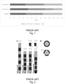
- DBS leads that are presently approved and used clinical treatment of PD symptoms are shown in Fig. 2 (starting from the left: Medtronic 3387, Medtronic 3389, 8-electrode Abbott Infinity ® directional lead, and 8-electrodes Boston Scientific directional lead Vercise Cartesia ® ).
- Fig. 2 represent the most commonly used leads in present DBS treatments, which have been used since DBS was first approved approximatively 30 years ago.
- the prior art leads shown in Fig. 2 include a quite limited number of electrodes (namely, 4 to 8 electrodes), each having a quite large area in the millimetric range (namely, between 1.5 and 6 mm 2 ).
- This segmented electrode design allows to achieve a degree of directionality.
- the subthalamic nucleus is generally assumed as the therapeutic target, while no directions regarding the specific placement of the lead within the STN are considered.
- the motor area of the STN is located in a dorso-lateral portion of the STN and occupies roughly 1/3 of the entire STN volume and less than half (approx. 2 to 4 mm) of the length of the STN along a normal lead trajectory based on beta-band local field potentials (LFPs) [14].
- LFPs beta-band local field potentials
- This area is therefore of a similar length to a single known DBS electrode, typically between 1 to 2 mm ( Fig. 3 ).
- the complexity of the anatomical structures within the basal ganglia requires precise delivery of stimulation.
- a poor therapeutic window (where for "therapeutic window” is intended to denote the difference in threshold between therapeutic effects and side effects) may result from non-optimal electrode placement. Surgical repositioning of the lead may be required to improve the therapeutic performance.
- a reduced therapeutic window may be the consequence of subject-specific anatomy.
- existing approaches are usually unable to implement a proper adaption, in particular due to the size of the electrodes.
- Stimulation of different areas of the STN may generate a better management of specific motor symptoms.
- tremor seems to be better controlled when leads are placed in the posterior part of the STN
- bradykinesia seems to be better controlled when leads are placed in a more anterior position.
- the Medtronic 3389 lead schematically illustrated in Fig. 2 , includes four relatively large ring electrodes (1.5 mm height x 1.27 mm diameter, with an overall area of 6 mm 2 ) for delivery of stimulation to the brain and sensing local electrophysiological signals (LFPs).
- LFPs local electrophysiological signals
- the field of stimulation obtained by activating one of said four electrodes is approximately spherical around the lead.
- the generation of such an omnidirectional electric field, in combination with sub-optimal lead positioning, results in poor selectivity of the anatomical structures surrounding the lead that are being activated, leading to the activation of unwanted anatomical structures and generation of side effects.
- directional leads have been designed containing up to 12metal electrodes (e.g., Medtronic SenSight ® , Abbott Infinity ® , Boston Scientific Vercise Cartesia ® , or directional Aleva ® ), where two ring electrodes are divided into three independent segments, each having an area of about 1-2 mm 2 , allowing to achieve a certain degree of directional selectivity.
- 12metal electrodes e.g., Medtronic SenSight ® , Abbott Infinity ® , Boston Scientific Vercise Cartesia ® , or directional Aleva ®
- DBS leads according to the prior art fail to accurately record and sense signals within such a small anatomical area, even more if the same electrodes are used for both stimulation and recording.
- Platinum (Pt) and platinum-iridium (Pt-Ir) constrain geometrical dimensions due to impedance limits.
- DBS systems are mostly adapted to deliver open-loop stimulation, this meaning that stimulation is delivered continuously (or at best according to a specific pre-established duty cycle), with no real-time adjustment of the stimulation parameters, and not specificity related to individual patient's biomarkers.
- symptomatic activity usually varies over time, e.g. during the day or over multiple days.
- reporting on a total number of hours per day does not give any qualitative information on those moments when patients are experiencing good or bad symptoms control, i.e. whether it is an activity or a rest moment.
- Horn et. al. [19] evaluated the connectivity between the stimulation site and other brain regions in PD patients treated with DBS by identifying the structural and functional connectivity profile of effective DBS to the STN.
- This evaluation was made by estimating, for each patient, the volume of tissue activated by the DBS electrode from individual postoperative images, and then applying a combination of normative and PD pathologic averaged brain connectome to estimate the brain networks activated from a given stimulated volume of tissue [19].
- the approach described in Horn et. al. [19] included the following steps: acquiring pre-/postoperative imaging ( Fig. 6A ) localizing DBS electrodes in standard space ( Fig. 6B ), calculating the volume of tissue activated (VTA) based on stimulation parameters ( Fig. 6C ), and calculating functional ( Fig. 6D ) and structural ( Fig. 6E ) connectivity from the VTA to the rest of the brain using high-quality normative connectome data.
- targets of therapeutic brain stimulation may be brain networks, and not individual brain regions such as the STN or GPi.
- the primary purpose of this study [20] was to identify specific markers of optimal DBS using fMRI to ease device fitting (in particular, to select the optimal electrode and voltage level).
- the activation of ipsilateral primary motor cortex, ipsilateral thalamus, and contralateral cerebellum was identified as a maker of optimal STN DBS, suggesting a large brain network activation to STN DBS, further optimized with a control of activation of this network at different levels [20].
- a neural interface for delivering neuromodulation/neurostimulation to brain structures of a patient and/or sensing brain signals of the patient comprises at least:
- a closed-loop system for delivery of neuromodulation/neurostimulation to brain structures of a patient comprises:
- the present invention provides a neural interface.
- said neural interface can be used for delivering neuromodulation/neurostimulation to brain structures of a patient and/or sensing brain signals of a patient.
- the same neural interface can be used for both delivering neuromodulation/neurostimulation and sensing brain signals.
- the neural interface comprises a support.
- the said support may at least partially be and/or comprise a thin film and/or be made of a nanostructure and/or a microstructure.
- thin film is to be understood as a stack of layers of materials ranging from fractions of a nanometer to several micrometers in thickness.
- Thin film layers can be obtained applying a variety of thin-film deposition/preparation techniques including sputtering, evaporation, chemical vapor deposition, molecular beam epitaxy, atomic layer deposition, spin coating, 3D printing, or the like.
- the stack of layers exhibits a variety of functionalities, such as conductive layers, insulating layers, passivating layers, or the like.
- the support e.g. a thin film, includes and/or carries an electrode array comprising a plurality of electrodes.
- the electrodes are made of reduced graphene oxide (rGO).
- each electrode of said plurality of electrodes is characterized by an area in the micrometer range.
- the invention is based on the basic idea that, by the provision of a neural interface comprising a dense arrangement of rGO electrodes with areas in the micrometer range, it is possible to deliver neuromodulation/neurostimulation with high level of stimulation selectivity and recording/decoding a broader neural signal bandwidth, enabling to sense biomarkers that metal electrodes according to the prior art cannot (or barely) detect.
- the use of rGO electrodes having areas down to the micrometer range allows stimulation to be directionally steered towards targeted areas, at the same time enabling 10 to 100 times higher spatial resolution and high signal-to-noise ratio, compared to the leads according to the prior art.
- rGO electrodes allow obtaining high charge injection, low electrical impedance (even though the electrode area is reduced), and high signal-to-noise ratio (allowing to sense high-frequency signals up to typical of single- and multi-unit activity).
- the physical properties of graphene allow obtaining micrometer-range electrodes while maintaining low electrical impedance and high-charge injection capacity.
- the neural interface is capable of sensing signals with high signal-to-noise ratio and delivering stimulation within the STN with high spatial selectivity and low angular resolution (down to 50-60°).
- each electrode is comprised between 1.0 and 0.0005 mm 2 .
- the electrode array comprises at least 20 electrodes.
- the electrode array may include 40 electrodes.
- the electrode array may include 50 electrodes.
- the electrode array may include from 128 to 1024 electrodes.
- micrometer-range rGO electrodes allows achieving precise stimulation while allowing decoding of neural signals with high signal-to-noise ratio, which cannot be obtained with the millimeter-range metal electrodes according to the prior art.
- the electrode array may comprise a plurality of electrodes having different areas in the micrometer range.
- one or more electrodes having a smaller area can be used for sensing smaller areas targeting single- and multi-unit activity, optimizing SNR and enlarging bandwidth.
- one or more electrodes having a larger area can be used for a more extended activation area targeting larger groups of neurons and measure e.g. LFPs.
- electrodes having different areas can be selected depending on characteristics of the stimulation target, spatial selectivity, charge injection, current density, required compliance voltage, and/or impedance.
- the neural interface may be configured and adapted to be implanted cortically.
- the neural interface may be configured and adapted to be implanted subcortically.
- the thin film is made of a flexible material.
- said flexible material may include metal or a metal alloy.
- said flexible material may include a polymer material.
- said polymer material may include one among Polylmide, Parylene, or Polydimethylsiloxane (PDMS).
- PDMS Polydimethylsiloxane
- the neural interface of the invention allows sensing a large variety of brain signals over time.
- said brain signals may include brain oscillations (e.g., beta, gamma, HFO).
- brain oscillations e.g., beta, gamma, HFO.
- said brain signals may include evoked responses to subcortical stimulation.
- Spatial high resolution is obtained thought the provision of a quite large number of rGO electrodes, arranged to form a high-density neural interface allowing recording/sensing activity of different locations nearby the implantation site.
- temporal high resolution is mainly enabled by the properties of graphene.
- rGO electrodes in the micrometer scale having low impedance allow for a high signal-to-noise ratio, enabling the recording/sensing of local neural activity at both low and high frequencies (with supporting sensing electronics).
- the invention further provides a closed-loop system for delivery of neuromodulation/neurostimulation to brain structures of a particular patient.
- said patient can be a Parkinson's Disease (PD) subject.
- PD Parkinson's Disease
- the system includes a stimulation unit.
- the stimulation unit comprises at least one neural interface as described above, said at least one neural interface being configured and adapted to deliver neuromodulation/neurostimulation to brain structures of the patient.
- the system further comprises a sensing unit.
- the sensing unit comprises at least one neural interface as described above, said at least one neural interface being configured and adapted to sense brain signals of the patient.
- the same neural interface can be used for both delivering neuromodulation/neurostimulation and sensing/ brain signals of the patient.
- system comprises a control unit.
- the control unit is operatively connected to the stimulation unit and the sensing unit.
- control unit is configured and adapted to perform real-time decoding of brain signals from the sensing unit and control one or more stimulation parameters for the stimulation unit based on said brain signals from the sensing unit.
- the system allows obtaining improved decoding and modulation of impaired brain networks of the nigrostriatal pathways, in a closed-loop fashion.
- the closed-loop system according to the invention is capable of bringing significant benefits to patients, e.g. PD patients, reducing multiple pathological symptoms in motor disorders conditions and allowing maintaining therapeutic achievements over time.
- the system of the invention allows achieving:
- the at least one neural interface of the stimulation unit may include a subcortical lead.
- the at least one neural interface of the stimulation unit may include a cortical lead.
- said subcortical and/or cortical lead may be configured and adapted to deliver selective stimulation to basal ganglia to elicit a therapeutic response, based on individual patient's brain signals.
- Stimulation is carried out with high-resolution, meaning that the target subcortical structure is stimulated at a very precise location (based on pathological biomarkers detection) more effectively and with limited adverse stimulation of surrounding, healthy structures.
- the sensing unit includes at least one cortical neural interface and/or at least one subcortical neural interface.
- the brain signals may include at least one among brain oscillation (e.g. LFP) and/or evoked responses to subcortical stimulation.
- brain oscillation e.g. LFP
- the sensing unit includes one cortical neural interface and one subcortical neural interface.
- the same neural interface can be used for both delivering neuromodulation/neurostimulation and sensing brain signals of the patient.
- subcortical stimulation can be delivered through a subcortical neural interface based on a combination of decoded cortical brain signals and subcortical brain signals.
- control unit may be configured and adapted to implement a machine learning algorithm for recording and processing brain signals of the patient and generate stimulation patterns for the stimulation unit based on the processed brain signals.
- control unit may be further configured and adapted to:
- the control unit may be implantable.
- control unit may be configured and adapted for implantation on the patient's skull.
- an implantable control unit allows to reduce the overall dimensions of the system, further than improving the patient's comfort during use.
- the system may further comprise an extra-corporeal power supply unit configured and adapted to wirelessly supply power to the implantable control unit.
- the implantable control unit may include a battery that can be charged through an extra-corporeal power supply unit.
- the system may further comprise a clinician computer.
- said clinician computer can be a portable device capable of implementing smart functionalities, e.g. a tablet computer or the like.
- the clinician computer may be implemented by means of a web-based portal, e.g. accessing at least one server.
- the clinician computer is configured and adapted to implement direct or indirect bi-directional communication with the control unit.
- the clinician computer comprises a control means configured and adapted to run a dedicated software means to:
- said software means of the clinician computer may be configured and adapted to implement web or cloud-based computing and/or machine learning algorithms and/or deep learning algorithms to:
- the patient's response to the delivered stimulation therapy is defined based on personalization algorithm suggestions, more preferably Al-based personalization algorithm suggestions.
- this allow anticipating on the role of modulation versus pharmacology, so as to adapt stimulation to increase the therapeutic effect while further reducing medication.
- the system implements high density decoding and modulation on its sensing unit , which renders almost impossible to program it with the current SOC programming methods, mostly due to the large number of combinations.
- the complexity of electrode selection in multi-electrode leads can be reduced by using brain decoding-driven methods.
- more than one brain network can be activated by basal ganglia stimulation in relation to different symptoms [23].
- the clinician computer may further comprise a display means.
- said display means may be configured and adapted to display information regarding the detected brain signals, current stimulation parameters and/or other information on the delivered stimulation therapy.
- the clinician is enabled to monitor the delivered stimulation therapy as well as the patient's response in a way that is intuitive and user-friendly.
- said software means of the clinician computer may be further configured and adapted to:
- said software means is configured and adapted to classify patients according to a respective health-risk level.
- patients may be classified according to the following ranking criteria:
- This approach enables the clinician to prioritize follow-up appointments with patients showing outcomes that are poorer than expected or degradation of performance, for a programming adjustment and/or a new medication schedule.
- the patient computer is configured and adapted to implement direct or indirect bi-directional communication with the control unit.
- the patient computer may comprise a control means configured and adapted to run a dedicated software means to:
- the user input from the patient may include information concerning one or more among drug intake, how he/she is feeling, mood and/or energy level, such that the delivered therapy can be adjusted accordingly.
- said user input means includes a display means of the touch-screen type.
- said user input means may be configured and adapted to receive a voice command and/or a user command provided by tapping on a predefined sensitive part.
- the display means may as well be used to display relevant information for the patient regarding the delivered therapy.
- the patient may be provided with a comprehensive overview on the ongoing stimulation therapy, which enables easily and intuitively monitoring therapeutic effects.
- the patient computer may be further configured and adapted to implement direct or indirect bi-directional communication with the clinician computer.
- control means of the patient computer may be further configured and adapted to:
- the patient's feedback may include information concerning one or more among drug intake, how he/she is feeling, mood and/or energy level.
- the clinician may be promptly informed regarding therapy effectiveness, possible side effects, or the like. Based on the received patient's feedback, the clinician may consider whether stimulation parameters and/or drugs intake need being adjusted.
- the clinician may efficiently plan follow-up appointments at relevant times when it is most needed.
- the clinician may provide information to the patient though the clinician computer.
- Information from the clinician may include, inter alia, risk-classification or the like.
- the clinician computer is implemented by means of a web-based portal and the patient computer is connected to the web (e.g., through a mobile device)
- communication between the clinician computer and the patient computer can be implemented without the provision of any dedicated hardware means.
- the patient computer may be a wearable device capable of implementing smart functionalities.
- the wearable device is provided with a sensor means that is configured and adapted to measure physiological data of the patient.
- the physiological data of the patient include one or more among heart rate, blood pressure, body temperature and/or body motion/acceleration.
- the patient computer can be a smartwatch or the like, which can be easily carried an intuitively used by the patient.
- the one or more stimulation parameters may include one or more among stimulation frequency, stimulation amplitude, stimulation pulse-width, ratio (pulse-width and intensity) between cathodic and anodic pulses, duty cycling, burst, and/or electrode combination selection.
- the patient P may be a PD subject.
- the patient P may be a subject affected by a psychiatric disorder, e.g. OCD.
- a psychiatric disorder e.g. OCD.
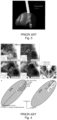
- the same neural interface 100, 200 can be used for both delivering neuromodulation/neurostimulation and sensing brain signals ( Figs. 7-8 ).
- Fig. 7 shows a configuration of the neural interface 100 according to the invention, where said neural interface 100 is configured to be cortically implanted.
- Fig. 8 shows an alternative configuration of the neural interface 200 according to the invention, where said neural interface 200 is configured to be subcortically implanted.
- said subcortical neural interface 200 is a subcortical lead configured to deliver neuromodulation/neurostimulation to brain structures of the patient P and/or sensing brain signals of the patient P.
- the neural interface 100, 200 comprises a support 10, 20, including and/or carrying at least an electrode array 12, 22.
- the support 10, 20 at least partially is and/or comprises a thin film and/or is made of a nanostructure and/or a microstructure.
- Figs. 7-8 show a configuration where the support 10, 20 is a thin film 10, 20.
- said thin film 10, 20 is made of a flexible material.
- said flexible material includes metal or a metal alloy and/or a polymer material.
- said polymer material includes one among Polylmide, Parylene, or Polydimethylsiloxane (PDMS).
- PDMS Polydimethylsiloxane
- the electrodes 14, 24 are made of reduced graphene oxide (rGO).
- each electrode 14, 24 of said plurality of electrodes 14, 24 has an area in the micrometer range.
- rGO electrodes 14, 24 having areas down to the micrometer range allows stimulation to be directionally steered towards targeted areas with a high level of selectivity ( Fig. 9 ).
- this configuration enables 10 to 100 times higher spatial and signal sensing and stimulation resolution compared to the DBS leads according to the state of the art.
- the neural interface 100, 200 allows decoding biomarkers that known metal electrodes, e.g. Platinum (Pt) and platinum-iridium (Pt-Ir) electrodes, cannot (or can barely) detect.
- metal electrodes e.g. Platinum (Pt) and platinum-iridium (Pt-Ir) electrodes
- rGO allows obtaining high charge injection, low electrical impedance (even in case of electrodes having reduced dimensions) as well as high signal-to noise ratio (up to high frequencies typical of single- and multi-unit activity).
- the physical properties of graphene allow the production of electrodes with a diameter in micrometer-range (such as electrodes 14, 24 shown in Figs. 7-8 ), while maintaining low electrical impedance and high-charge injection capacity.
- Each electrode 14, 24 has an area ranging between 1.0 and 0.0005 mm 2 .
- the electrode array 12, 22 comprises at least 20 electrodes 14, 24.
- the electrode array 12, 22 may include 50 electrodes 14, 24.
- the electrode array 12, 22 may include from 128 to 1024 electrodes 14, 24.
- the electrode array 12, 22 may comprise 40 electrodes 14, 24.
- the electrodes 14, 24 forming the electrode array 12, 22 have different areas ( Figs. 7-8 ).
- Fig. 7 shows a configuration where the electrode array 12 comprises electrodes 14 having an area of 0.0005 mm 2 for sensing only, and electrodes 14 having an area of 0.3 mm 2 for both sensing and stimulation.
- Fig. 8 shows a configuration where the electrode array 22 comprises electrodes 24 having an area of 0.0005 mm 2 for sensing only, and electrodes 24 having an area of 0.07 mm 2 for both sensing and stimulation.
- the electrodes 14, 24 forming the array 12, 22 may have the same area.
- the neural interface 100 can be configured and adapted to be implanted cortically ( Fig. 7 ).
- the neural interface 200 can be configured and adapted to be implanted subcortically ( Fig. 8 ).
- the invention further provides a closed-loop system 300 for delivery of neuromodulation/neurostimulation to brain structures of a patient P.
- the patient P may be a PD subject.
- the patient P may be a subject affected by a psychiatric disorder, e.g. OCD.
- a psychiatric disorder e.g. OCD.
- Fig. 10 schematically shows the different components of a closed-loop system 300 according to an embodiment of the invention and how these components interact.
- the system 300 comprises a stimulation unit 30.
- the stimulation unit 30 comprises at least one neural interface 100, 200 as described above.
- said at least one neural interface 100, 200 is configured and adapted to deliver neuromodulation/neurostimulation to brain structures of the patient P.
- the system 300 further comprises a sensing unit 32.
- the sensing unit 32 comprises at least one neural interface 100, 200 as described above.
- said brain signals of the patient P include at least one among brain oscillation and/or evoked responses to subcortical stimulation.
- the same neural interface 100, 200 can be used for both delivering neuromodulation/neurostimulation and sensing brain signals of the patient P ( Figs. 7-8 ).
- system 300 comprises a control unit 34.
- the control unit 34 is configured and adapted to perform real-time decoding of brain signals from the sensing unit 32 and control one or more stimulation parameters for the stimulation unit 30 based on said brain signals from the sensing unit 32.
- said one or more stimulation parameters include at least one among stimulation frequency, stimulation amplitude, stimulation pulse-width, ratio (pulse-width and intensity) between cathodic and anodic pulses, duty cycling, burst, and/or electrode combination selection.
- system 300 of the invention has proven effective in bringing significant benefits to patients, e.g. PD patients, reducing multiple symptoms in motor disorders conditions and allowing maintaining therapeutic achievements over time.
- the at least one neural interface 200 of the stimulation unit comprises is a subcortical lead 200.
- the at least one neural interface 100 of the stimulation unit may comprise a cortical lead 100.
- said cortical and/or subcortical lead 100, 200 is configured and adapted to deliver selective stimulation to basal ganglia to elicit a therapeutic response.
- the sensing unit 32 includes at least one cortical neural interface 100 and/or at least one subcortical neural interface 200.
- the sensing unit 32 includes one cortical neural interface 100, e.g. as shown in Fig. 7 , and one subcortical neural interface 200, e.g. as shown in Fig. 8 .
- control unit 34 is an implantable control unit 34, in particular adapted for implantation on the skull S of the patient P.
- control unit 34 may be non-implantable.
- FIG. 11 An overview of the implantable components of the system 300 in an implanted stated is schematically provided in Fig. 11 .
- extra-corporeal power supply unit 36 configured and adapted to wirelessly supply power to the control unit 34, while being implanted (as shown with dotted arrow in Fig. 10 ).
- control unit 34 is configured and adapted to implement a machine learning algorithm for recording and processing brain signals of the patient P and generate stimulation patterns for the stimulation unit 30 based on the processed brain signals.
- control unit 34 is further configured and adapted to:
- the system 300 further comprises a clinician computer 38.
- the clinician computer 38 is configured and adapted to implement direct or indirect bi-directional communication with the control unit 34 ( Fig. 10 ).
- the clinician computer 38 comprises a control means.
- control means is configured and adapted to run a dedicated software means to:
- said software means is further configured and adapted to implement web or cloud-based computing and/or machine learning algorithms and/or deep learning algorithms to:
- the patient's response to the delivered stimulation therapy is defined based on personalization algorithm suggestions, more preferably Al-based personalization algorithm suggestions.
- the clinician computer 38 further comprises a display means 40 ( Fig. 10 ).
- said display means 40 of the clinician computer 38 is configured and adapted to display information regarding the detected brain signals, current stimulation parameters and/or other information on the delivered stimulation therapy.
- the software means of the clinician computer 38 is configured and adapted to:
- patients P can be classified according to a respective health-risk level.
- patents P may be classified according to the following ranking criteria:
- the system 300 further comprises a patient computer 42.
- the patient computer 42 is configured and adapted to implement direct or indirect bi-directional communication with the control unit 32 ( Fig. 10 ).
- the patient computer 42 comprises a control means configured and adapted to run a dedicated software means to:
- Said user input means may include display means 44 of the touch-screen type.
- said user input means may be configured and adapted to receive a voice command and/or a user command provided by tapping on a predefined sensitive part.
- the display means 44 may as well be used to display relevant information for the patient P regarding the delivered therapy.
- the patient P may be provided with a comprehensive overview on the ongoing stimulation therapy, which enables easily and intuitively monitoring therapeutic effects.
- the patient computer 42 is also configured and adapted to implement direct or indirect bi-directional communication with the clinician computer 38 ( Fig. 10 ).
- the control means of the patient computer 42 is configured and adapted to:
- the patient computer 42 can be a wearable device capable of implementing smart functionalities.
- said wearable device is provided with a sensor means (not shown) that is configured and adapted to measure physiological data of the patient.
- said physiological data of the patient include one or more among heart rate, blood pressure, body temperature and/or body motion/acceleration.
- the patient computer 42 can be a smartwatch or the like, capable of being easily carried and intuitively used by the patient P.
- the therapy phases described below refer to a treatment performed using a preferred configuration of the system according to the invention on a PD subject whom chosen stimulation target is the STN.
- a PD subject may be referred for implantation if he/she is experiencing significant motor symptoms, such as tremors, stiffness, and difficulty with movement, despite treatment with medications. Implantation may also be considered when the subject is experiencing medication-related side effects that are affecting quality of life.
- Implantation candidacy criteria may be based on an evaluation process including physical examination, imaging tests such as MRI or CT, neuropsychological testing, and multidisciplinary review.
- the implantation procedure is planned.
- Patient selection process is similar to existing DBS referral process.
- Preoperative imaging is necessary to plan the insertion of subcortical neural interfaces on STN targets and the position of the cortical neural interface on the motor cortex.
- the implanted components of the system are surgically placed in a single-step procedure.
- the system includes two subcortical neural interfaces and a single cortical neural interface.
- the implantable control unit is secured on the skull, both subcortical neural interfaces are placed using state-of-the-art stereotactic surgery (e.g., by using a stereotactic robotic arm), and the cortical neural interface is positioned according to the planning.
- a set of neurophysiology recordings can be performed perioperatively to evaluate the accuracy of neural interfaces positioning, taking advantage of the neurophysiology sesning capabilities of the system.
- all electrodes on the subcortical neural interfaces are used to automatically measure STN-specific neurophysiological signature (e.g., evoked response, beta activity, high-frequency activity) to evaluate the position of the interface related to STN.
- STN-specific neurophysiological signature e.g., evoked response, beta activity, high-frequency activity
- This target response telemetry feature gives valuable information on neural interface placement as the percentage of electrodes on STN can be automatically estimated perioperatively.
- the clinician may then revise subcortical interface placement if the percentage of electrodes on target is estimated to be too low.
- electrodes from the cortical interface are used to evaluate the brain networks activated by subcortical stimulation on the target.
- the system After recovering from the surgery (typically 2 to 3 weeks postoperative), the system is ready to be activated and programmed to best suit the patient's specific PD symptoms.
- electrodes that are not on target must be identified and disregarded, and a reduced number of electrode clusters on target should be chosen as possible stimulating patterns, e.g. for the patient adapted treatment.
- neurophysiology measurements are performed while the subject is at rest and off medication. These measurements will be combined with perioperative measurements, cortical measurements, and imaging data, to reduce the number and the size of the chosen electrode clusters.
- a pre-defined machine learning algorithm will integrate data from all inputs to refine the electrode selection. Then, the neurologist can evaluate the clinical effects of stimulation delivered from each of selected electrode clusters and set the therapeutic stimulation level that is best suited to the patient's need.
- basal ganglia stimulation is associated to activation of brain network at different levels [19] with activation of specific brain areas [20].
- the intended mode of operation of the system is to capture and use this brain network activation to adapt the stimulation delivered at basal ganglia level.
- Such a feature allows the system to normalize the signatures of widespread brain network connectivity towards those found in healthy controls [19].
- FIG. 12 [62] A schematic view illustrating the functioning of an open-loop DBS system ( left panel ), a closed-loop system using subcortical LFP signals ( middle panel ), and a closed-loop system using a combination of subcortical and cortical recordings to regulate brain network response with subcortical high selectivity stimulation ( right panel ), is provided in Fig. 12 [62].
- the patient can go home and benefit from therapeutic optimization, which will now be adjusted in real-time in accordance with parameters and boundaries set by the clinician.
- Random stimulation patterns (combination of electrodes chosen among those defined to be on target) will be delivered within safety limits to evaluate the behavior of biomarkers for learning purposes.
- Patient direct inputs (e.g., through a dedicated application running on the patient computer) are also expected, e.g., to inform on symptoms type and magnitude, or drug intake.
- this learning phase can only occur at given moment during the day.
- extra usability constraints can be applied for power management (e.g., keep the external charger on for long period of time), for data storage (e.g., open the patient app on phone), or for the patient to give information on symptoms or state.
- This phase will be facilitated by specific motor tasks (e.g., finger tapping, balance exercise, shape drawing, rest) conducted from the patient application to trigger particular recording sessions.
- This learning phase will be executed after first postoperative programming, or anytime a few days prior to a follow-up programming visit.
- Analyzing neurophysiological signals coming from the system (and additionally also from other non-implantable wearable devices) is essential to set and control in real-time the optimal subcortical therapeutic modulation patterns and parameters, and get full benefit of high-density modulation and multiple decoding streams.
- the system will then embed a diagnosis and evaluation software designed to provide physiological and therapeutic metrics and an Al-based software assisting clinicians in system programming.
- the main function of this feature will be to help clinicians interpreting biological data and decide on the best therapy for each patient.
- Neural prostheses capable of multi-channel neural sensing, on-site signal processing, rapid symptom detection, and closed loop stimulation, are critical in order to further improve neuromodulation therapies in PD subjects.
- the system will combine artificial intelligence (machine and deep learning) with high resolution neural interfaces and decoding for treatment of PD symptoms.
- deep learning will enable personalized detection of symptoms-specific biomarkers to potentially adjust stimulation parameters to reduce accessible motor and non-motor symptoms selectively.
- the clinician accesses patient data that has been collected.
- Data collected during the learning phase are processed by the machine learning algorithm.
- Adjusted stimulation patterns, biomarkers, and closed-loop parameters are proposed as an optimization.
- This optimization can be related to stimulation levels, to stimulation patterns (i.e., a cluster of energized electrodes), but also to symptom-specific selective stimulation.
- this phase can occur before the patient's visit.
- the clinician can then apply the adjusted system programming as proposed by the algorithm and then evaluate its effective benefits with the patient.
- the patient benefits from personalized neuromodulation therapy, with expected maximized clinical benefits.
- the patient can use the patient computer to follow-up and visualize the medical outcomes of the delivered stimulation therapy, evolution of drug intake, mood, and symptoms.
- Thin film devices were fabricated and the electrochemical performance and stability of 25 ⁇ m diameter rGO micro-electrodes were assessed in vitro in a phosphate buffer saline solution.
- Electrochemical characterization of rGO is illustrated in Figs. 13a-f [24].
- the voltage drop at the electrode-electrolyte interface should always remain within the potential window set by the cyclic voltammetry in which no faradaic reactions occur.
- Fig. 13c shows the potential excursion that a 25 ⁇ m diameter rGO microelectrode experiences upon the injection of rectangular, biphasic current pulses (1 ms/phase) at charge densities of 1, 2, 3, and 4 mC/cm 2 .
- a quasi-linear voltage shift of maximum 0.9 V exists between the ohmic drops at the edges of the pulses, indicating a capacitive-like behavior of the electrode-electrolyte interface within the limits of the potential window.
- the charge injection limit of rGO electrodes was found to be 2.75 mC/cm 2 for 1-ms pulses.
- Fig. 13d shows a map of the voltage polarization experienced by a rGO microelectrode in response to biphasic current pulses between 0.1 ms and 1 ms and injected charge densities up to 5 mC/cm 2 .
- the charge injection limit of rGO is 2 orders of magnitude higher than that of Pt electrodes, which is typically in the range of 0.02-0.04 mC/cm 2 .
- Electrode stability was investigated during continuous electrical stimulation. rGO were stimulated with 1 ms biphasic (1 ms interphase gap), 3 mC/cm 2 pulses at 100 Hz while the impedance and CSC were monitored every 500k pulses. rGO was observed to be stable after 15 million pulses as indicated by the consistent impedance at 1 kHz ( Fig. 13e ).
- the average charge storage capacitance increased from 61.7 to 136.7 mC/cm 2 throughout the stimulation ( Fig. 13f ).
- the high performance and stability provided by rGO are attributed to the combination of the capacitive interaction in aqueous media with the large electrochemical surface area obtained during the hydrothermal reduction [24].
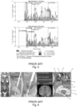
- rGO electrode arrays were used to demonstrate ability to detect cortical brain signals with high accuracy and with better performance than known Pt/lr electrodes by implanting thin film with rGO electrodes and by recording neural activity at the surface of the auditory cortex of anesthetized rats ( Figs 14a-f) .
- the used system included an array of 64 rGO microelectrodes ( ⁇ 25 ⁇ m, pitch 300 ⁇ m) that was positioned over the rat auditory cortex.
- Fig. 14b shows the 10 Hz high-pass filtered signal from each of the 64 rGO electrodes in the array over a 350 ms window in which a pure tone was presented acoustically at the ear of the animal.
- the density of the array allowed mapping of single-event LFPs as displayed in Fig. 14b, which reveals a spatial clustering with ON responses located on the top right corner and OFF responses grouped in the bottom part of the array.
- the amplitudes of the evoked LFPs ranged between 500 ⁇ V and 700 ⁇ V ( Fig. 14c ).
- High-pass filtered signals also showed the detection of high frequency multi-unit activity and the corresponding increase of the root mean square (RMS) amplitude (due to multi-unit activity) correlated to the evoked LFPs ( Fig. 14c ), indicating synchronous behavior of the neurons to the stimulus.
- RMS root mean square
- the signal to noise ratio was higher (2-3 times) and the noise floor was significantly lower, even for micro-size rGO electrodes (25-50 um diameter) that enable detection of high frequency signals.
- the small dimension of rGO electrodes allows obtaining high-density arrays for sensing with improved decoding capabilities compared to low-density, larger Pt/lr electrodes.
- rGO Due to its increased charge injection capacity, rGO allows to generate precise stimulation with microelectrodes. This is relevant for focal, selective stimulation in small structures like the STN.
- micro rGO electrodes implanted inside the sciatic nerve of rats were able to achieve selective activation of the main nerve fascicles.
- Focal stimulation with microelectrodes was able to achieve function specific targeted stimulation of submillimeter structures.
- the focal stimulation capability of the rGO electrodes is presently being tested in brain structures like the thalamus and STN in animals.
- Fig. 15 shows inter-spike frequency distribution of multi-unit activity measured by small rGO electrode in the STN in healthy control rodents (see left panel), and in PD rodent model (see middle panel). Left panel shows the inter-spike frequency distribution in PD rodent model before (pre) and after (post) therapeutic STN stimulation.
- PD symptomatic activity reveals different inter-spike peak frequency profile than healthy controls, and this PD-specific profile tends to show similar profile as controls when therapeutic stimulation is provided in the STN.
- rGO electrodes enable the development of cortical and subcortical neural interfaces with a high density of small electrodes, and allow for selective and safe charge injection in conjunction with advanced signal sensing capabilities.
- Such technology is critical for the development of a modulated stimulation system requiring focused selective stimulation for targeted therapy and high-resolution signal sensing for symptoms control and personalization.
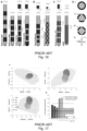
- Fig. 16B shows examples of directional leads that have not yet been approved by the FDA, but have already been implanted in human patients for testing. These leads include the 8-electrode direct STNAcute lead and the 40-electrode Medtronic-Sapiens lead.
- Fig. 16C provides examples of additional lead designs that have not yet been implanted in human patients for testing, including the 16-electrode segmented lead discussed in Buhlmann et. al. [25] and the micro-array-based DBS ( ⁇ DBS) lead with 1760 electrodes discussed in Willsie and Dorval [26].
- ⁇ DBS micro-array-based DBS
- FIG. 16D Exemplary cross-sections of DBS electrodes according to the prior art are illustrated in Fig. 16D (in detail: (i) Medtronics 3387/ Medtronics 3389; (ii) 8-electrode Abbott Infinity ® directional lead, 8-electrode Boston Scientific directional lead, and STNAcute directional lead; (iii) Medtronic-Sapiens and 16-electrode lead discussed in Buhlmann et. al. [25]; (iv) example of ⁇ DBS lead, with 32 electrode per rotation).
- Spectral and clinical topography of the STN on a large cohort of PD patients revealed that spectral features ranging from the delta-frequency band through HFOs are topographically segregated within the STN and may further differentiate in their spatial focality [29] as shown, e.g., in Fig. 17 [29].
- small electrodes demonstrated the capability to provide not only higher spatial resolution, but also do sensing in a broader and wider range, in particular sense signals with larger frequency content. In line with theoretical predictions, small electrodes can detect signals that are otherwise lost or not detectable when sensing with electrodes of the size of those that are used in current clinical settings [35].
- the subcortical neural interface according to the invention ( Fig. 8 ), which includes a relatively high number of electrodes having a reduced area, and which further benefits from the properties of rGO, allows obtaining a higher precision in stimulation selectivity as well as a higher precision in the identification of signal biomarkers, when compared to the subcortical leads according to the prior art.
- a double-blinded study evaluated intraoperatively the directional stimulation applied to STN in PD patients [30].
- a box plot representing the width of the therapeutic window (percentiles 0, 25, 75, 100), for omnidirectional stimulation and for stimulation in each single direction is provided in Fig. 18 , left panel [30].
- a box plot of the electrical current producing a meaningful therapeutic effect (percentiles 0, 25, 75, 100), for omnidirectional stimulation and stimulation in each single direction is provided in Fig. 18 , right panel [30].
- Fig. 19C shows that activation of an array of 4 electrodes yields a field of stimulation (b) which can be modulated so as to avoid stimulation of adjacent structures, thereby only stimulating the target area [31].
- directional leads offer proof of principle evidence that directionality can result in an increase of benefit with a reduction in side effects.
- preliminary evidence with smaller electrodes suggested an even more precise control that can provide greater benefit.
- High electrode density allows to access different zones of the STN selectively
- PD patients often benefit from DBS in terms of improved tremor, bradykinesia, rigidity, or motor fluctuations.
- the STN has a complex organization with a dorsolateral motor area, a central cognitive/associative area, and a limbic medial tip. Also, each of these regions show dense innervation with many projections to other brain regions [42], suggesting that selective stimulation of STN could activate various networks within each area.
- this personalized surgical targeting takes advantage of the identification of symptom-specific networks that have been investigated over the past years thanks to retrospective evaluation of DBS electrode location, connectomic network activation and clinical effects on corresponding symptoms across DBS cohorts, centers and disorders [3], [19].
- the placement of the DBS lead could be planned so that a set of cardinal symptoms could form the treatment focus, while additional comorbidities could be treated with lesser importance (i.e., weights could be applied based on empirical intensities of preoperatively experienced symptoms).
- preponderant tremor in one PD patient could shift the treatment focus toward maximized stimulation of the tremor network.
- bradykinesia and rigidity symptoms may weigh more heavily in another PD patient, drawing particular focus onto modulating the corresponding circuitry.
- beneficial networks could not only be weighted based on empirical symptom severity, but also based on individual preferences. For instance, some patients may experience tremor as a stronger subjective burden than bradykinesia, or vice versa.
- the neural interface according to the invention (e.g. the cortical lead shown in Fig. 8 ), including an electrode array having small rGO electrodes with high charge injection capabilities, allows taking even more advantage of such an atlas of networks that, when modulated, clearly associate with different therapeutic effects.
- Fig. 21 provides a comparative diagram showing a representation of STN somatotopy as schematized by Sasaki et al. [15] with overlapped illustration of a known subcortical Boston Scientific directional DBS lead in an implanted state (left panel), and a subcortical neural interface (subcortical lead) according to the invention in an implanted state (right panel).
- the subcortical neural interface of the invention ( Fig. 8 ) allows obtaining a high level of spatial resolution that cannot be achieved through the lead designs according to the prior art, this resulting in an improved therapeutic benefit for the patient.
- the neural interface according to the invention enables a more precise stimulation focus on the STN or GPi, with enhanced selectivity on the activated network.
- the large number of rGO micrometer-range electrodes allows selectively activating different parts of the STN independently. This enables getting precise access to specific networks associated with a particular symptom cluster, mapped on target area.
- Such symptomatotopy as introduced in Lozano and Lipsman [44], is accessible thanks to the high resolution and the high channel counts of the neural interface of the invention, as long as the neural interface has been well surgically placed within the STN.
- the definition symptomatotopy means that stimulation-related symptoms relief is distributed and organized along the STN.
- each network that is selectively activated with a cluster of small electrodes
- the neural interface of the invention is capable to selectively activating distinct areas of the STN (and possibly multiple therapeutic channels), thereby offering the neurologist the possibility to independently address different symptoms with the delivered therapy.
- subcortical and cortical interfaces as in the present invention allows for monitoring at cortical level of networks activated subcortically, which further improves stimulation resolution.
- image-based electrode activation profile also shows limitations that are determined by tissue contrast, spatial resolution, and signal-to-noise ratio of currently available (clinical) imaging protocols.
- a high-resolution subcortical neural interface Fig. 8
- a high-resolution cortical neural interface Fig. 7
- beta-band peaks corresponds to the brain oscillations of electrical activity between 12.5 Hz and 30 Hz
- [45] in the recorded LFPs may be used as triggers for onset and offset of stimulation.
- Increased brain response synchronization measured as increased beta-band power spectrum, is observed within the STN of PD patients and correlates with disease symptoms. This can also be reduced through Levodopa treatment [47], [48].
- Therapeutic DBS has proven effective in reducing such anomalous network synchronization (i.e., reduction of beta-band power spectrum).
- Adaptive DBS could therefore take advantage of such signals, in particular by initiating stimulation when those signals detected, and interrupt stimulation when those signals are suppressed, in a closed-loop manner [46].
- detection of LFPs may allow to select optimal stimulation electrode based on biomarker recording localization (e.g., highest beta-band power).
- beta band frequency range (approx. 13-35 Hz) is modulated by many other normal functions of the brain (including movement or even planning or imagination of it) wakefulness and decision making. Therefore, despite established reports of its modulations in PD, the multifaceted involvement of beta band in physiological processes might affect its performance as a biomarker by itself for the diseased state, especially in the freely behaving patients (in an ecological setting rather than in a controlled clinical setting) with a chronic DBS implant [49].
- Adaptive DBS only using beta bands is expected to reduce stimulation-induced side effects by limiting DBS stimulation to moments when it is needed. However, if patient tolerability (and, consequently, quality of life) is hypothesized to be improved, such adaptive stimulation is not intended to have any influence on therapeutic action and symptoms control.
- Adaptive DBS only using beta bands which is not yet clinically approved and therefore not implemented in the context of the clinical routine, is mainly aimed at reducing the patient risk profile of DBS as well as optimizing the existing approach.
- beta band oscillations at STN level may potentially affect the overall clinical benefit as this biomarker is influenced by physiological brain functions and does not seem to fully inform about the symptoms.
- the closed-loop system of the invention integrating real-time neural activity monitoring and feedback allows significantly improving precision and effectiveness of the delivered subcortical stimulation therapy.
- Measuring of the brain network activation to a subcortical stimulation with the implant system can be performed by introducing additional recording electrodes placed on other parts of the network.
- additional sensing electrodes can be placed at the cortical level (e.g., the motor cortex, M1) or subcortically at the thalamus level (e.g., GPi, GPe) to sense cortical response to therapeutic subcortical stimulation, monitor symptomatic brain network activity, and/or decode specific movements or functions in relation to symptoms.
- cortical level e.g., the motor cortex, M1
- thalamus level e.g., GPi, GPe
- cortical neural interfaces by combining cortical and subcortical neural interfaces, allows obtaining a more precise and targeted modulation of neural networks.
- cortical neural interfaces can decode specific neural signals related to motor control, but also sensory perception, and/or cognitive processes, allowing for real-time monitoring and feedback.
- This information can then be used to optimize subcortical stimulation parameters and patterns.
- cortical and subcortical neural interfaces revealed potential benefits in various applications.
- cortical neural interfaces can decode neural signals associated with movement intention, enabling the closed-loop system to provide subcortical stimulation only when necessary.
- This highly-adaptive approach provided by the invention allows improving symptom control as well as reducing side effects when compared with the solutions according to the prior art.
- a first, major clinical interest in cortical sensing is to evaluate efficacy of delivered subcortical therapeutic stimulation.
- cortical-evoked responses may greatly facilitate the assessment of STN stimulation in terms of targeted areas, stimulation orientation and amplitude.
- the stimulation amplitude was adapted based on patient-specific motor behavior, suppressing the pathological tremor on-demand based on a cortical lead detecting upper limb motor activity [59].
- Such stimulation paradigm was able to achieve clinical efficacy and tremor suppression in a range of movements (such as cup reaching, proximal and distal posture, water pouring, and/or writing) while having a consistent reduction in energy delivery (this reducing DBS-induced side effects) in comparison with standard DBS.
- an experimental setup with direct recording of potentials in the primary motor cortex using subdural ECoG together with synchronous capture of gait freezing using optoelectronic motion-tracking systems in freely-walking patients with PD receiving STN DBS was implemented [60].
- cortical recordings are related to the clinical efficacy of the subcortical stimulation, which may greatly ease device programming and stimulation optimization.
- symptomatic biomarkers sensed cortically, reflecting BNND at upper brain network levels, were shown to be suppressed with subcortical stimulation, suggesting the possibility to regulate brain network response with subcortical stimulation and cortical recording.
- the neural interface and closed-loop system of the invention addresses the above-mentioned drawbacks of the prior art, in particular by providing a solution that allows improving the decoding accuracy of cortical interfaces, optimizing stimulation parameters, and better understanding the complex interactions within neural networks.
- the closed-loop system of the invention allows improving decoding accuracy and providing more targeted and selective stimulation.
- the closed-loop system of the invention relying on a combination of cortical and subcortical neural interfaces, offers a powerful approach to modulate neural networks that overcomes the limitations of the prior art.
- the combined approach integrating real-time neural activity monitoring and feedback allows enhancing precision and effectiveness of subcortical therapeutic stimulation, thereby improving therapeutic outcome.
- control and estimation routines included herein can be used with various system configurations.
- the control methods and routines disclosed herein may be stored as executable instructions in non-transitory memory and may be carried out by a control unit such as a microcontroller (or a computer) in combination with the components of the system.
- the specific routines described herein may represent one or more of any number of processing strategies such as event-driven, interrupt-driven, multi-tasking, multi-threading, and the like.
- various actions, operations, and/or functions illustrated may be performed in the sequence illustrated, in parallel, or in some cases omitted.
- the order of processing is not necessarily required to achieve the features and advantages of the example embodiments described herein but is provided for ease of illustration and description.
Landscapes
- Health & Medical Sciences (AREA)
- Life Sciences & Earth Sciences (AREA)
- Veterinary Medicine (AREA)
- Public Health (AREA)
- General Health & Medical Sciences (AREA)
- Animal Behavior & Ethology (AREA)
- Engineering & Computer Science (AREA)
- Biomedical Technology (AREA)
- Heart & Thoracic Surgery (AREA)
- Biophysics (AREA)
- Molecular Biology (AREA)
- Neurosurgery (AREA)
- Pathology (AREA)
- Surgery (AREA)
- Physics & Mathematics (AREA)
- Medical Informatics (AREA)
- Psychology (AREA)
- Neurology (AREA)
- Nuclear Medicine, Radiotherapy & Molecular Imaging (AREA)
- Radiology & Medical Imaging (AREA)
- Psychiatry (AREA)
- Cardiology (AREA)
- Physiology (AREA)
- Electrotherapy Devices (AREA)
Priority Applications (3)
| Application Number | Priority Date | Filing Date | Title |
|---|---|---|---|
| EP23197897.4A EP4523731A1 (de) | 2023-09-18 | 2023-09-18 | Neuronale schnittstelle und geschlossenes regelkreis-decodierungs- und neuromodulations-/neurostimulationssystem damit |
| PCT/EP2024/076040 WO2025061731A1 (en) | 2023-09-18 | 2024-09-18 | System for delivery of neuromodulation/neurostimulation therapy to brain structures of a patient |
| PCT/EP2024/076033 WO2025061728A1 (en) | 2023-09-18 | 2024-09-18 | System for delivery of neuromodulation/neurostimulation therapy to brain structures of a patient |
Applications Claiming Priority (1)
| Application Number | Priority Date | Filing Date | Title |
|---|---|---|---|
| EP23197897.4A EP4523731A1 (de) | 2023-09-18 | 2023-09-18 | Neuronale schnittstelle und geschlossenes regelkreis-decodierungs- und neuromodulations-/neurostimulationssystem damit |
Publications (1)
| Publication Number | Publication Date |
|---|---|
| EP4523731A1 true EP4523731A1 (de) | 2025-03-19 |
Family
ID=88093815
Family Applications (1)
| Application Number | Title | Priority Date | Filing Date |
|---|---|---|---|
| EP23197897.4A Pending EP4523731A1 (de) | 2023-09-18 | 2023-09-18 | Neuronale schnittstelle und geschlossenes regelkreis-decodierungs- und neuromodulations-/neurostimulationssystem damit |
Country Status (2)
| Country | Link |
|---|---|
| EP (1) | EP4523731A1 (de) |
| WO (2) | WO2025061728A1 (de) |
Citations (3)
| Publication number | Priority date | Publication date | Assignee | Title |
|---|---|---|---|---|
| US20220144644A1 (en) * | 2019-02-27 | 2022-05-12 | Fundació Institut Català De Nanocièngia Inanotecnologia (Icn2) | Reduced graphene oxide film comprising a stack of rgo layers and its applications |
| EP4015033A1 (de) * | 2020-12-21 | 2022-06-22 | INBRAIN Neuroelectronics SL | Flexibler elektrodenträger |
| US20230191130A1 (en) * | 2021-12-17 | 2023-06-22 | Purdue Research Foundation | Reinforcement learning based closed-loop neuromodulation system |
Family Cites Families (9)
| Publication number | Priority date | Publication date | Assignee | Title |
|---|---|---|---|---|
| US9211417B2 (en) * | 2012-09-10 | 2015-12-15 | Great Lakes Neurotechnologies Inc | Movement disorder therapy system, devices and methods, and intelligent methods of tuning |
| US10842997B2 (en) | 2015-08-26 | 2020-11-24 | Boston Scientific Neuromodulation Corporation | Machine learning to optimize spinal cord stimulation |
| CN108289630A (zh) * | 2015-10-05 | 2018-07-17 | Mc10股份有限公司 | 用于神经调节和刺激的方法和系统 |
| EP3445227B1 (de) | 2016-03-15 | 2024-01-10 | Universität Bern | Verfahren und system zur optimierung der dbs-programmierung |
| WO2019118667A1 (en) * | 2017-12-14 | 2019-06-20 | Boston Scientific Neuromodulation Corporation | Systems for clinical effect-based neurostimulation |
| US20220401736A1 (en) * | 2021-06-22 | 2022-12-22 | University Of Washington | Apparatuses, systems and methods for implantable stimulator with externally trained classifier |
| US20250195877A1 (en) * | 2021-07-09 | 2025-06-19 | Cala Health, Inc. | Personalized therapy neurostimulation systems |
| US20230123383A1 (en) * | 2021-10-18 | 2023-04-20 | Advanced Neuromodulation Systems, Inc. | Systems and methods for providing neurostimulation therapy using multi-dimensional patient features |
| WO2023122083A1 (en) * | 2021-12-22 | 2023-06-29 | Boston Scientific Neuromodulation Corporation | Closed-loop feature optimization of biological signals |
-
2023
- 2023-09-18 EP EP23197897.4A patent/EP4523731A1/de active Pending
-
2024
- 2024-09-18 WO PCT/EP2024/076033 patent/WO2025061728A1/en active Pending
- 2024-09-18 WO PCT/EP2024/076040 patent/WO2025061731A1/en active Pending
Patent Citations (3)
| Publication number | Priority date | Publication date | Assignee | Title |
|---|---|---|---|---|
| US20220144644A1 (en) * | 2019-02-27 | 2022-05-12 | Fundació Institut Català De Nanocièngia Inanotecnologia (Icn2) | Reduced graphene oxide film comprising a stack of rgo layers and its applications |
| EP4015033A1 (de) * | 2020-12-21 | 2022-06-22 | INBRAIN Neuroelectronics SL | Flexibler elektrodenträger |
| US20230191130A1 (en) * | 2021-12-17 | 2023-06-22 | Purdue Research Foundation | Reinforcement learning based closed-loop neuromodulation system |
Non-Patent Citations (62)
| Title |
|---|
| ALKEMADE, A.: "Subdivisions and Anatomical Boundaries of the Subthalamic Nucleus", THE JOURNAL OF NEUROSCIENCE, vol. 33, no. 22, 2013, pages 9233 - 9234 |
| AMAN, J. E.JOHNSON, L. A.SANABRIA, D. E.WANG, J.PATRIAT, R.HILL, M.MARSHALL, E.MACKINNON, C. D.COOPER, S. E.SCHROCK, L. E.: "Directional deep brain stimulation leads reveal spatially distinct oscillatory activity in the globus pallidus internus of Parkinson's disease patients", NEUROBIOLOGY OF DISEASE, vol. 139, 2020, pages 104819, XP086121591, DOI: 10.1016/j.nbd.2020.104819 |
| ANDERSON, D. N.OSTING, B.VORWERK, J.DORVAL, A. D.BUTSON, C. R.: "Optimized programming algorithm for cylindrical and directional deep brain stimulation electrodes", JOURNAL OF NEURAL ENGINEERING, vol. 15, no. 2, 2018, pages 026005, XP020325720, DOI: 10.1088/1741-2552/aaa14b |
| APPLEBY, B. S.DUGGAN, P. S.REGENBERG, A.RABINS, P. V.: "Psychiatric and neuropsychiatric adverse events associated with deep brain stimulation: A meta-analysis of ten years' experience", MOVEMENT DISORDERS: OFFICIAL JOURNAL OF THE MOVEMENT DISORDER SOCIETY, vol. 22, no. 12, 2007, pages 1722 - 1728 |
| AVERNA, A.DEBOVE, I.NOWACKI, A.PETERMAN, K.DUCHET, B.SOUSA, M.BERNASCONI, E.ALVA, L.LACHENMAYER, M. L.SCHUEPBACH, M.: "Spectral Topography of the Subthalamic Nucleus to Inform Next-Generation Deep Brain Stimulation", MOVEMENT DISORDERS: OFFICIAL JOURNAL OF THE MOVEMENT DISORDER SOCIETY, vol. 38, no. 5, 2023, pages 818 - 830 |
| BOUR, L. J.LOURENS, M. A. J.VERHAGEN, R.BIE, R. M. A.VAN DEN MUNCKHOF, P.SCHUURMAN, P. R.CONTARINO, M. F.: "Directional Recording of Subthalamic Spectral Power Densities in Parkinson's Disease and the Effect of Steering Deep Brain Stimulation", BRAIN STIMULATION, vol. 8, no. 4, 2015, pages 730 - 741 |
| BOUTET, A.MADHAVAN, R.ELIAS, G. J. B.JOEL, S. E.GRAMER, R.RANJAN, M.PARAMANANDAM, V.XU, D.GERMANN, J.LOH, A.: "Predicting optimal deep brain stimulation parameters for Parkinson's disease using functional MRI and machine learning", NATURE COMMUNICATIONS, vol. 12, no. 1, 2021, pages 3043 |
| BRATSOS, S. P.KARPONIS, D.SALEH, S. N.: "Efficacy and Safety of Deep Brain Stimulation in the Treatment of Parkinson's Disease: A Systematic Review and Meta-analysis of Randomized Controlled Trials", CUREUS, 2018 |
| BROWN, P.: "Oscillatory nature of human basal ganglia activity: relationship to the pathophysiology of Parkinson's disease", MOVEMENT DISORDERS.: OFFICIAL JOURNAL OF THE MOVEMENT DISORDER SOCIETY, vol. 18, no. 4, 2003, pages 357 - 363 |
| BUHLMANN, J.HOFMANN, L.TASS, P. A.HAUPTMANN, C: "Modeling of a segmented electrode for desynchronizing deep brain stimulation", FRONTIERS IN NEUROENGINEERING, vol. 4, 2011, pages 15 |
| BULLMORE, E.SPORNS, O.: "The economy of brain network organization", NATURE REVIEWS NEUROSCIENCE, vol. 13, no. 5, 2012, pages 336 - 349 |
| CHOU, K. L.: "Adverse events from the treatment of Parkinson's disease", NEUROLOGIC CLINICS, vol. 26, no. 3, 2008, pages S65 - 83 |
| CONNOLLY, A. T.VETTER, R. J.HETKE, J. F.TEPLITZKY, B. A.KIPKE, D. R.PELLINEN, D. S.ANDERSON, D. J.BAKER, K. B.VITEK, J. L.JOHNSON,: "A Novel Lead Design for Modulation and Sensing of Deep Brain Structures", IEEE TRANSACTIONS ON BIO-MEDICAL ENGINEERING, vol. 63, no. 1, 2016, pages 148 - 157, XP011594624, DOI: 10.1109/TBME.2015.2492921 |
| CONTARINO, M. F.BOUR, L. J.VERHAGEN, R.LOURENS, M. A. J.DE BIE, R. M. A.VAN DEN MUNCKHOF, P.SCHUURMAN, P. R.: "Directional steering: A novel approach to deep brain stimulation", NEUROLOGY, vol. 83, no. 13, 2014, pages 1163 - 1169 |
| DE HEMPTINNE, C.RYAPOLOVA-WEBB, E. S.AIR, E. L.GARCIA, P. A.MILLER, K. J.OJEMANN, J. G.OSTREM, J. L.GALIFIANAKIS, N. B.STARR, P. A: "Exaggerated phase-amplitude coupling in the primary motor cortex in Parkinson disease", PROCEEDINGS OF THE NATIONAL ACADEMY OF SCIENCES OF THE UNITED STATES OF AMERICA, vol. 110, no. 12, 2013, pages 4780 - 4785 |
| DE HEMPTINNE, C.SWANN, N. C.OSTREM, J. L.RYAPOLOVA-WEBB, E. S.SAN LUCIANO, M.GALIFIANAKIS, N. B.STARR, P. A.: "Therapeutic deep brain stimulation reduces cortical phase-amplitude coupling in Parkinson's disease", NATURE NEUROSCIENCE, vol. 18, no. 5, 2015, pages 779 - 786, XP055547557, DOI: 10.1038/nn.3997 |
| DEMBEK, T. A.REKER, PVISSER-VANDEWALLE, V.WIRTHS, J.TREUER, H.KLEHR, M.ROEDIGER, J.DAFSARI, H. S.BARBE, M. T.TIMMERMANN, L.: "Directional DBS increases side-effect thresholds-A prospective, double-blind trial", MOVEMENT DISORDERS OFFICIAL JOURNAL OF THE MOVEMENT DISORDER SOCIETY, vol. 32, no. 10, 2017, pages 1380 - 1388 |
| FELDMANN, L. K.NEUMANN, W.-J.KRAUSE, P.LOFREDI, R.SCHNEIDER, G.-H.KUHN, A. A.: "Subthalamic beta band suppression reflects effective neuromodulation in chronic recordings", EUROPEAN JOURNAL OF NEUROLOGY, vol. 28, no. 7, 2021, pages 2372 - 2377, XP072032690, DOI: 10.1111/ene.14801 |
| GBD 2016 PARKINSON'S DISEASE COLLABORATORS: "Global, regional, and national burden of Parkinson's disease, 1990-2016: a systematic analysis for the Global Burden of Disease Study 2016", THE LANCET. NEUROLOGY, vol. 17, no. 11, 2018, pages 939 - 953 |
| HOLLUNDER, B.RAJAMANI, N.SIDDIQI, S. H.FINKE, C.KUHN, A. A.MAYBERG, H. S.FOX, M. D.NEUDORFER, C.HORN, A.: "Toward personalized medicine in connectomic deep brain stimulation", PROGRESS IN NEUROBIOLOGY, vol. 210, 2022, pages 102211 |
| HORN, A.REICH, M.VORWERK, J.LI, N.WENZEL, G.FANG, Q.SCHMITZ-HUBSCH, T.NICKL, R.KUPSCH, A.VOLKMANN, J.: "Connectivity Predicts deep brain stimulation outcome in Parkinson disease", ANNALS OF NEUROLOGY, vol. 82, no. 1, 2017, pages 67 - 78, XP071641130, DOI: 10.1002/ana.24974 |
| HORN, A.WENZEL, G.IRMEN, F.HUEBL, J.LI, N.NEUMANN, W.-J.KRAUSE, P.BOHNER, G.SCHEEL, MKUHN, A. A.: "Deep brain stimulation induced normalization of the human functional connectome in Parkinson's disease", BRAIN, vol. 142, no. 10, 2019, pages 3129 - 3143 |
| HOSAIN MD KAMAL ET AL: "Closed loop deep brain stimulation: an evolving technology", AUSTRALASIAN PHYSICAL AND ENGINEERING SCIENCES IN MEDICINE, MEDICINE, MELBOURNE, AU, vol. 37, no. 4, 7 September 2014 (2014-09-07), pages 619 - 634, XP035428098, ISSN: 0158-9938, [retrieved on 20140907], DOI: 10.1007/S13246-014-0297-2 * |
| HUSAIN, M.: "Targeting network dysfunction in neurodegenerative diseases", BRAIN, vol. 142, no. 12, 2019, pages 3661 - 3662 |
| JOHNSON, L. A., NEBECK, S. D., MURALIDHARAN, A., JOHNSON, M. D., BAKER, K. B., & VITEK, J. L.: "Closed-Loop Deep Brain Stimulation Effects on Parkinsonian Motor Symptoms in a Non-Human Primate - Is Beta Enough?", BRAIN STIMULATION, vol. 9, no. 6, 2016, pages 892 - 896, XP029789279, DOI: 10.1016/j.brs.2016.06.051 |
| KIM, J.CRIAUD, M.CHO, S. S.DIEZ-CIRARDA, M.MIHAESCU, A.COAKELEY, S.GHADERY, C.VALLI, M.JACOBS, M. F.HOULE, S.: "Abnormal intrinsic brain functional network dynamics in Parkinson's disease", BRAIN. A JOURNAL OF NEUROLOGY, vol. 140, no. 11, 2017, pages 2955 - 2967 |
| KURIAKOSE, R.SAHA, U.CASTILLO, G.UDUPA, K.NI, Z.GUNRAJ, C.MAZZELLA, F.HAMANI, C.LANG, A. E.MORO, E.: "The Nature and Time Course of Cortical Activation Following Subthalamic Stimulation in Parkinson's Disease", CEREBRAL CORTEX, vol. 20, no. 8, 2010, pages 1926 - 1936 |
| LI, Q.QIAN, Z.-M.ARBUTHNOTT, G. W.KE, Y.YUNG, W.-H.: "Cortical Effects of Deep Brain Stimulation", JAMA NEUROLOGY, vol. 71, no. 1, 2014, pages 100 |
| LOZANO, A. M.LIPSMAN, N.: "Probing and Regulating Dysfunctional Circuits Using Deep Brain Stimulation", NEURON, vol. 77, no. 3, 2013, pages 406 - 424, XP028970516, DOI: 10.1016/j.neuron.2013.01.020 |
| LYONS, K. E.PAHWA, R.: "Effects of bilateral subthalamic nucleus stimulation on sleep, daytime sleepiness, and early morning dystonia in patients with Parkinson disease", JOURNAL OF NEUROSURGERY, vol. 104, no. 4, 2006, pages 502 - 505 |
| MARTENS, H. C. F.TOADER, E.DECRE, M. M. J.ANDERSON, D. J.VETTER, R.KIPKE, D. R.BAKER, K. B.JOHNSON, M. D.VITEK, J. L.: "Spatial steering of deep brain stimulation volumes using a novel lead design", CLINICAL NEUROPHYSIOLOGY OFFICIAL JOURNAL OF THE, vol. 122, no. 3, 2011, pages 558 - 566, XP028151429, DOI: 10.1016/j.clinph.2010.07.026 |
| MERK, T.PETERSON, V.LIPSKI, W. J.BLANKERTZ, B.TURNER, R. S.LI, N.HORN, A.RICHARDSON, R. M.NEUMANN, W.-J.: "Electrocorticography is superior to subthalamic local field potentials for movement decoding in Parkinson's disease", ELIFE, vol. 11, 2022 |
| NEUMANN, W.-J.DEGEN, K.SCHNEIDER, G.-H.BRUCKE, C.HUEBL, J.BROWN, P.KUHN, A. A.: "Subthalamic synchronized oscillatory activity correlates with motor impairment in patients with Parkinson's disease", MOVEMENT DISORDERS. OFFICIAL JOURNAL OF THE MOVEMENT DISORDER SOCIETY, vol. 31, no. 11, 2016, pages 1748 - 1751 |
| NEUMANN, W.-JSTAUB-BARTELT, F.HORN, A.SCHANDA, J.SCHNEIDER, G.-H.BROWN, P.KUHN, A. A.: "Long term correlation of subthalamic beta band activity with motor impairment in patients with Parkinson's disease", CLINICAL NEUROPHYSIOLOGY. OFFICIAL JOURNAL OF THE INTERNATIONAL FEDERATION OF CLINICAL NEUROPHYSIOLOGY, vol. 128, no. 11, 2017, pages 2286 - 2291 |
| NGUYEN, T. A. K.DJILAS, M.NOWACKI, A.MERCANZINI, A.SCHUPBACH, M.RENAUD, P.POLLO, C.: "Analysis of patient-specific stimulation with segmented leads in the subthalamic nucleus", PLOS ONE, vol. 14, no. 6, 2019, pages e0217985 |
| OKUN, MICHAEL: "Deep-Brain Stimulation - Entering the Era of Human Neural-Network Modulation 371", THE NEW ENGLAND JOURNAL OF MEDICINE |
| OPRI, E.CERNERA, S.MOLINA, R.EISINGER, R. S.CAGLE, J. N.ALMEIDA, L.DENISON, T.OKUN, M. S.FOOTE, K. D.GUNDUZ, A.: "Chronic embedded cortico-thalamic closed-loop deep brain stimulation for the treatment of essential tremor", SCIENCE TRANSLATIONAL MEDICINE, vol. 12, no. 572, 2020, XP093038113, DOI: 10.1126/scitranslmed.aay7680 |
| OZTURK, M.VISWANATHAN, ASHETH, S. A.INCE, N. F.: "Electroceutically induced subthalamic high-frequency oscillations and evoked compound activity may explain the mechanism of therapeutic stimulation in Parkinson's disease", COMMUNICATIONS BIOLOGY, vol. 4, no. 1, 2021 |
| PEETERS, J.BOOGERS, A.VAN BOGAERT, T.DAVIDOFF, H.GRANSIER, R.WOUTERS, J.NUTTIN, B.MC LAUGHLIN, M.: "Electrophysiologic Evidence That Directional Deep Brain Stimulation Activates Distinct Neural Circuits in Patients With Parkinson Disease", NEUROMODULATION: JOURNAL OF THE INTERNATIONAL NEUROMODULATION SOCIETY, vol. 26, no. 2, 2023, pages 403 - 413 |
| PLANTINGA, B. R.TEMEL, Y.DUCHIN, Y.ULUDAG, K.PATRIAT, R.ROEBROECK, A.KUIJF, M.JAHANSHAHI, A.TER HAAR ROMENIJ, B.VITEK, J.: "Individualized parcellation of the subthalamic nucleus in patients with Parkinson's disease with 7T MRI", NEUROLMAGE, vol. 168, 2018, pages 403 - 411 |
| POLLO, C.KAELIN-LANG, A.OERTEL, M. F.STIEGLITZ, L.TAUB, E.FUHR, P.LOZANO, A. M.RAABE, A.SCHUPBACH, M.: "Directional deep brain stimulation: an intraoperative double-blind pilot study", BRAIN A JOURNAL OF NEUROLOGY, vol. 137, no. 7, 2014, pages 2015 - 2026 |
| POWERS, R.ETEZADI-AMOLI, M.ARNOLD, E. M.KIANIAN, S.MANCE, I.GIBIANSKY, M.TRIETSCH, D.ALVARADO, A. S.KRETLOW, J. D.HERRINGTON, T. M: "Smartwatch inertial sensors continuously monitor real-world motor fluctuations in Parkinson's disease", SCIENCE TRANSLATIONAL MEDICINE, vol. 13, no. 579, 2021 |
| RASIAH, N. P.MAHESHWARY, R.KWON, C.-S.BLOOMSTEIN, J. D.GIRGIS, F.: "Complications of Deep Brain Stimulation for Parkinson Disease and Relationship between Micro-electrode tracks and hemorrhage: Systematic Review and Meta-Analysis", WORLD NEUROSURGERY, vol. 171, 2023, pages e8 - e23 |
| REBELO, P., GREEN, A. L., AZIZ, T. Z., KENT, A., SCHAFER, D., VENKATESAN, L., & CHEERAN, B.: "Thalamic Directional Deep Brain Stimulation for tremor: Spend less, get more", BRAIN STIMULATION, vol. 11, no. 3, 2018, pages 600 - 606 |
| RODRIGUEZ-OROZ, M. C.MORO, E.KRACK, P.: "Long-term outcomes of surgical therapies for Parkinson's disease", MOVEMENT DISORDERS, vol. 27, no. 14, 2012, pages 1718 - 1728 |
| RODRIGUEZ-ROJAS, R.PINEDA-PARDO, J. A.MANEZ-MIRO, J.SANCHEZ-TUREL, A.MARTINEZ-FERNANDEZ, R.DEL ALAMO, M.DELONG, M.OBESO, J. A.: "Functional Topography of the Human Subthalamic Nucleus: Relevance for Subthalamotomy in Parkinson's Disease", MOVEMENT DISORDERS, vol. 37, no. 2, 2022, pages 279 - 290 |
| SABOURIN, S.KHAZEN, O.DIMARZIO, M.STAUDT, M. D.WILLIAMS, L.GILLOGLY, M.DURPHY, J.HANSPAL, E. K.ADAM, O. R.PILITSIS, J. G.: "Effect of Directional Deep Brain Stimulation on Sensory Thresholds in Parkinson's Disease", FRONTIERS IN HUMAN NEUROSCIENCE, vol. 14, 2020, pages 217 |
| SASAKI, T.KUWAHARA, K.KIN, I.OKAZAKI, M.SASADA, S.SHINKO, A.KAMEDA, M.YASUHARA, T.AGARI, T.DATE, I.: "Identification of Somatotopic Organization and Optimal Stimulation Site Within the Subthalamic Nucleus for Parkinson's Disease", OPERATIVE NEUROSURGERY (HAGERSTOWN, MD., vol. 17, no. 3, 2019, pages 239 - 246 |
| SCHNITZLER, A.MIR, P.BRODSKY, M. A.VERHAGEN, L.GROPPA, S.ALVAREZ, R.EVANS, A.BLAZQUEZ, M.NAGEL, S.PILITSIS, J. G.: "Directional Deep Brain Stimulation for Parkinson's Disease: Results of an International Crossover Study With Randomized, Double-Blind Primary Endpoint", NEUROMODULATION: TECHNOLOGY AT THE NEURAL INTERFACE, vol. 25, no. 6, 2022, pages 817 - 828 |
| SHEN, L.JIANG, C.HUBBARD, C. S.REN, J.HE, C.WANG, D.DAHMANI, L.GUO, Y.LIU, Y.XU, S.: "Subthalamic Nucleus Deep Brain Stimulation Modulates 2 Distinct Neurocircuits", ANNALS OF NEUROLOGY, vol. 88, no. 6, 2020, pages 1178 - 1193, XP071645490, DOI: 10.1002/ana.25906 |
| SWANN, N. C.DE HEMPTINNE, C.THOMPSON, M. C.MIOCINOVIC, S.MILLER, A. M.GILRON, R.OSTREM, J. L.CHIZECK, H. J.STARR, P. A.: "Adaptive deep brain stimulation for Parkinson's disease using motor cortex sensing", JOURNAL OF NEURAL ENGINEERING, vol. 15, no. 4, 2018, pages 046006, XP020329270, DOI: 10.1088/1741-2552/aabc9b |
| TELKES, I.SABOURIN, S.DURPHY, J.ADAM, O.SUKUL, V.RAVIV, N.STAUDT, M. D.PILITSIS, J. G.: "Functional Use of Directional Local Field Potentials in the Subthalamic Nucleus Deep Brain Stimulation", FRONTIERS IN HUMAN NEUROSCIENCE, vol. 14, 2020, pages 145 |
| VIANA, D.WALSTON, S. T.ILLA, X.DEL VALLE, J.PRATS, E.DE LA OLIVA, N.PALMA, M.DEL CORRO, E.RODRIGUEZ-MEANA, B.DEL PILAR BERNICOLA, : "Graphene-based thin film microelectrode technology for in vivo high resolution neural recording and stimulation", UNDER REVIEW AT ADVANCED MATERIALS. |
| VITEK, J. L.JAIN, R.CHEN, L.TRÖSTER, A. I.SCHROCK, L. E.HOUSE, P. A.GIROUX, M. L.HEBB, A. O.FARRIS, S. M.WHITING, D. M.: "Subthalamic nucleus deep brain stimulation with a multiple independent constant current-controlled device in Parkinson's disease (INTREPID): a multicentre, double-blind, randomised, sham-controlled study", THE LANCET. NEUROLOGY, vol. 19, no. 6, 2020, pages 491 - 501 |
| VIZCARRA, J. A.SITU-KCOMT, M.ARTUSI, C. A.DUKER, A. P.LOPIANO, L.OKUN, M. S.ESPAY, A. J.MEROLA, A.: "Subthalamic deep brain stimulation and levodopa in Parkinson's disease: a meta-analysis of combined effects", JOURNAL OF NEUROLOGY, vol. 266, no. 2, 2019, pages 289 - 297, XP036698424, DOI: 10.1007/s00415-018-8936-2 |
| WALKER, H. C.HUANG, H.GONZALEZ, C. L.BRYANT, J. E.KILLEN, J.CUTTER, G. R.KNOWLTON, R. C.MONTGOMERY, E. B.GUTHRIE, B. L.WATTS, R. L: "Short latency activation of cortex during clinically effective subthalamic deep brain stimulation for Parkinson's disease", MOVEMENT DISORDERS, vol. 27, no. 7, 2012, pages 864 - 873 |
| WANG, J. T.WANG, A. Y.PSARROS, C.DA CRUZ, M.: "Rates of revision and device failure in cochlear implant surgery: a 30-year experience", THE LARYNGOSCOPE, vol. 124, no. 10, 2014, pages 2393 - 2399 |
| WILLIAMS, N. R.FOOTE, K. D.OKUN, M. S.: "STN vs. GPi Deep Brain Stimulation: Translating the Rematch into Clinical Practice", MOVEMENT DISORDERS CLINICAL PRACTICE, vol. 1, no. 1, 2014, pages 24 - 35 |
| WILLSIE, A.DORVAL, A.: "Fabrication and initial testing of the pDBS: a novel Deep Brain Stimulation electrode with thousands of individually controllable contacts", BIOMEDICAL MICRODEVICES, vol. 17, no. 3, 2015, pages 9961 |
| YIN, Z.ZHU, G.LIU, Y.ZHAO, B.LIU, D.BAI, Y.ZHANG, Q.SHI, L.FENG, T.YANG, A.: "Cortical phase-amplitude coupling is key to the occurrence and treatment of freezing of gait", BRAIN: A JOURNAL OF NEUROLOGY, vol. 145, no. 7, 2022, pages 2407 - 2421 |
| YU, Y.HAN, F.WANG, Q: "Exploring phase-amplitude coupling from primary motor cortex-basal ganglia-thalamus network model", NEURAL NETWORKS: THE OFFICIAL JOURNAL OF THE INTERNATIONAL NEURAL NETWORK SOCIETY, vol. 153, 2022, pages 130 - 141, XP087130718, DOI: 10.1016/j.neunet.2022.05.027 |
| ZHANG, S.CONNOLLY, A. T.MADDEN, L. R.VITEK, J. L.JOHNSON, M. D.: "High-resolution local field potentials measured with deep brain stimulation arrays", JOURNAL OF NEURAL ENGINEERING, vol. 15, no. 4, 2018, pages 046019, XP020329283, DOI: 10.1088/1741-2552/aabdf5 |
Also Published As
| Publication number | Publication date |
|---|---|
| WO2025061728A1 (en) | 2025-03-27 |
| WO2025061731A1 (en) | 2025-03-27 |
Similar Documents
| Publication | Publication Date | Title |
|---|---|---|
| Frey et al. | Past, present, and future of deep brain stimulation: hardware, software, imaging, physiology and novel approaches | |
| AU2019291582B2 (en) | Systems and methods for treating mood disorders | |
| EP3787735B1 (de) | Neuralschnittstellensystem | |
| CN107921260B (zh) | 用于改善对可兴奋组织的刺激的方法和系统 | |
| Miocinovic et al. | History, applications, and mechanisms of deep brain stimulation | |
| CN109562262B (zh) | 用于改善对可兴奋组织的刺激的方法和系统 | |
| US10688303B2 (en) | Therapy target selection for psychiatric disorder therapy | |
| McIntyre et al. | Engineering the next generation of clinical deep brain stimulation technology | |
| EP3426336B1 (de) | Zieldefinition einer medizinischen therapie | |
| JP6730185B2 (ja) | 脳神経電位のモニタリング | |
| US8565886B2 (en) | Arousal state modulation with electrical stimulation | |
| US8761890B2 (en) | Electrical stimulation programming | |
| Hosain et al. | Closed loop deep brain stimulation: an evolving technology | |
| US20120271148A1 (en) | Brain condition monitoring based on co-activation of neural networks | |
| di Biase et al. | Adaptive, personalized closed-loop therapy for Parkinson’s disease: Biochemical, neurophysiological, and wearable sensing systems | |
| EP4523731A1 (de) | Neuronale schnittstelle und geschlossenes regelkreis-decodierungs- und neuromodulations-/neurostimulationssystem damit | |
| Raju et al. | MER based analysis of local field potentials with deep brain stimulation subthalamic nucleus in Parkinson\’s disease using coherence and entropy techniques | |
| US20250121193A1 (en) | Systems and methods for modulating brain activity | |
| Gunduz | Deep brain stimulation: emerging technologies and applications | |
| US20250114601A1 (en) | Epilepsy therapy using spinal cord stimulation | |
| KR102941919B1 (ko) | 기분 장애를 치료하기 위한 시스템 및 방법 | |
| Brinda | Development of model-based and sensor-based programming techniques for optimizing directional deep brain stimulation therapy for movement disorders | |
| Brinda et al. | Mechanisms and Targeting of Deep-Brain Stimulation Therapies | |
| Rosing | Characterization of evoked compound action potentials in targets of deep brain stimulation for Parkinson’s disease | |
| Johnson | Evoked resonant neural activity to guide deep brain stimulation for movement disordersEvoked resonant neural activity to guide deep brain stimulation for movement disorders |
Legal Events
| Date | Code | Title | Description |
|---|---|---|---|
| PUAI | Public reference made under article 153(3) epc to a published international application that has entered the european phase |
Free format text: ORIGINAL CODE: 0009012 |
|
| STAA | Information on the status of an ep patent application or granted ep patent |
Free format text: STATUS: THE APPLICATION HAS BEEN PUBLISHED |
|
| AK | Designated contracting states |
Kind code of ref document: A1 Designated state(s): AL AT BE BG CH CY CZ DE DK EE ES FI FR GB GR HR HU IE IS IT LI LT LU LV MC ME MK MT NL NO PL PT RO RS SE SI SK SM TR |
|
| STAA | Information on the status of an ep patent application or granted ep patent |
Free format text: STATUS: REQUEST FOR EXAMINATION WAS MADE |
|
| 17P | Request for examination filed |
Effective date: 20250919 |
|
| STAA | Information on the status of an ep patent application or granted ep patent |
Free format text: STATUS: EXAMINATION IS IN PROGRESS |