EP4516203A2 - Medical laser apparatus and system - Google Patents
Medical laser apparatus and system Download PDFInfo
- Publication number
- EP4516203A2 EP4516203A2 EP24211085.6A EP24211085A EP4516203A2 EP 4516203 A2 EP4516203 A2 EP 4516203A2 EP 24211085 A EP24211085 A EP 24211085A EP 4516203 A2 EP4516203 A2 EP 4516203A2
- Authority
- EP
- European Patent Office
- Prior art keywords
- controller
- endoscope
- aiming beam
- laser apparatus
- energy
- Prior art date
- Legal status (The legal status is an assumption and is not a legal conclusion. Google has not performed a legal analysis and makes no representation as to the accuracy of the status listed.)
- Pending
Links
Images


Classifications
-
- A—HUMAN NECESSITIES
- A61—MEDICAL OR VETERINARY SCIENCE; HYGIENE
- A61B—DIAGNOSIS; SURGERY; IDENTIFICATION
- A61B18/00—Surgical instruments, devices or methods for transferring non-mechanical forms of energy to or from the body
- A61B18/18—Surgical instruments, devices or methods for transferring non-mechanical forms of energy to or from the body by applying electromagnetic radiation, e.g. microwaves
- A61B18/20—Surgical instruments, devices or methods for transferring non-mechanical forms of energy to or from the body by applying electromagnetic radiation, e.g. microwaves using laser
- A61B18/22—Surgical instruments, devices or methods for transferring non-mechanical forms of energy to or from the body by applying electromagnetic radiation, e.g. microwaves using laser the beam being directed along or through a flexible conduit, e.g. an optical fibre; Couplings or hand-pieces therefor
- A61B18/26—Surgical instruments, devices or methods for transferring non-mechanical forms of energy to or from the body by applying electromagnetic radiation, e.g. microwaves using laser the beam being directed along or through a flexible conduit, e.g. an optical fibre; Couplings or hand-pieces therefor for producing a shock wave, e.g. laser lithotripsy
-
- A—HUMAN NECESSITIES
- A61—MEDICAL OR VETERINARY SCIENCE; HYGIENE
- A61B—DIAGNOSIS; SURGERY; IDENTIFICATION
- A61B1/00—Instruments for performing medical examinations of the interior of cavities or tubes of the body by visual or photographical inspection, e.g. endoscopes; Illuminating arrangements therefor
- A61B1/005—Flexible endoscopes
- A61B1/01—Guiding arrangements therefore
-
- A—HUMAN NECESSITIES
- A61—MEDICAL OR VETERINARY SCIENCE; HYGIENE
- A61B—DIAGNOSIS; SURGERY; IDENTIFICATION
- A61B1/00—Instruments for performing medical examinations of the interior of cavities or tubes of the body by visual or photographical inspection, e.g. endoscopes; Illuminating arrangements therefor
- A61B1/00002—Operational features of endoscopes
- A61B1/00004—Operational features of endoscopes characterised by electronic signal processing
- A61B1/00006—Operational features of endoscopes characterised by electronic signal processing of control signals
-
- A—HUMAN NECESSITIES
- A61—MEDICAL OR VETERINARY SCIENCE; HYGIENE
- A61B—DIAGNOSIS; SURGERY; IDENTIFICATION
- A61B1/00—Instruments for performing medical examinations of the interior of cavities or tubes of the body by visual or photographical inspection, e.g. endoscopes; Illuminating arrangements therefor
- A61B1/00002—Operational features of endoscopes
- A61B1/00043—Operational features of endoscopes provided with output arrangements
- A61B1/00055—Operational features of endoscopes provided with output arrangements for alerting the user
-
- A—HUMAN NECESSITIES
- A61—MEDICAL OR VETERINARY SCIENCE; HYGIENE
- A61B—DIAGNOSIS; SURGERY; IDENTIFICATION
- A61B1/00—Instruments for performing medical examinations of the interior of cavities or tubes of the body by visual or photographical inspection, e.g. endoscopes; Illuminating arrangements therefor
- A61B1/00064—Constructional details of the endoscope body
- A61B1/00071—Insertion part of the endoscope body
- A61B1/0008—Insertion part of the endoscope body characterised by distal tip features
- A61B1/00087—Tools
-
- A—HUMAN NECESSITIES
- A61—MEDICAL OR VETERINARY SCIENCE; HYGIENE
- A61B—DIAGNOSIS; SURGERY; IDENTIFICATION
- A61B1/00—Instruments for performing medical examinations of the interior of cavities or tubes of the body by visual or photographical inspection, e.g. endoscopes; Illuminating arrangements therefor
- A61B1/012—Instruments for performing medical examinations of the interior of cavities or tubes of the body by visual or photographical inspection, e.g. endoscopes; Illuminating arrangements therefor characterised by internal passages or accessories therefor
- A61B1/015—Control of fluid supply or evacuation
-
- A—HUMAN NECESSITIES
- A61—MEDICAL OR VETERINARY SCIENCE; HYGIENE
- A61B—DIAGNOSIS; SURGERY; IDENTIFICATION
- A61B1/00—Instruments for performing medical examinations of the interior of cavities or tubes of the body by visual or photographical inspection, e.g. endoscopes; Illuminating arrangements therefor
- A61B1/012—Instruments for performing medical examinations of the interior of cavities or tubes of the body by visual or photographical inspection, e.g. endoscopes; Illuminating arrangements therefor characterised by internal passages or accessories therefor
- A61B1/018—Instruments for performing medical examinations of the interior of cavities or tubes of the body by visual or photographical inspection, e.g. endoscopes; Illuminating arrangements therefor characterised by internal passages or accessories therefor for receiving instruments
-
- A—HUMAN NECESSITIES
- A61—MEDICAL OR VETERINARY SCIENCE; HYGIENE
- A61B—DIAGNOSIS; SURGERY; IDENTIFICATION
- A61B1/00—Instruments for performing medical examinations of the interior of cavities or tubes of the body by visual or photographical inspection, e.g. endoscopes; Illuminating arrangements therefor
- A61B1/04—Instruments for performing medical examinations of the interior of cavities or tubes of the body by visual or photographical inspection, e.g. endoscopes; Illuminating arrangements therefor combined with photographic or television appliances
- A61B1/043—Instruments for performing medical examinations of the interior of cavities or tubes of the body by visual or photographical inspection, e.g. endoscopes; Illuminating arrangements therefor combined with photographic or television appliances for fluorescence imaging
-
- A—HUMAN NECESSITIES
- A61—MEDICAL OR VETERINARY SCIENCE; HYGIENE
- A61B—DIAGNOSIS; SURGERY; IDENTIFICATION
- A61B1/00—Instruments for performing medical examinations of the interior of cavities or tubes of the body by visual or photographical inspection, e.g. endoscopes; Illuminating arrangements therefor
- A61B1/04—Instruments for performing medical examinations of the interior of cavities or tubes of the body by visual or photographical inspection, e.g. endoscopes; Illuminating arrangements therefor combined with photographic or television appliances
- A61B1/046—Instruments for performing medical examinations of the interior of cavities or tubes of the body by visual or photographical inspection, e.g. endoscopes; Illuminating arrangements therefor combined with photographic or television appliances for infrared imaging
-
- A—HUMAN NECESSITIES
- A61—MEDICAL OR VETERINARY SCIENCE; HYGIENE
- A61B—DIAGNOSIS; SURGERY; IDENTIFICATION
- A61B1/00—Instruments for performing medical examinations of the interior of cavities or tubes of the body by visual or photographical inspection, e.g. endoscopes; Illuminating arrangements therefor
- A61B1/04—Instruments for performing medical examinations of the interior of cavities or tubes of the body by visual or photographical inspection, e.g. endoscopes; Illuminating arrangements therefor combined with photographic or television appliances
- A61B1/05—Instruments for performing medical examinations of the interior of cavities or tubes of the body by visual or photographical inspection, e.g. endoscopes; Illuminating arrangements therefor combined with photographic or television appliances characterised by the image sensor, e.g. camera, being in the distal end portion
-
- A—HUMAN NECESSITIES
- A61—MEDICAL OR VETERINARY SCIENCE; HYGIENE
- A61B—DIAGNOSIS; SURGERY; IDENTIFICATION
- A61B1/00—Instruments for performing medical examinations of the interior of cavities or tubes of the body by visual or photographical inspection, e.g. endoscopes; Illuminating arrangements therefor
- A61B1/06—Instruments for performing medical examinations of the interior of cavities or tubes of the body by visual or photographical inspection, e.g. endoscopes; Illuminating arrangements therefor with illuminating arrangements
-
- A—HUMAN NECESSITIES
- A61—MEDICAL OR VETERINARY SCIENCE; HYGIENE
- A61B—DIAGNOSIS; SURGERY; IDENTIFICATION
- A61B1/00—Instruments for performing medical examinations of the interior of cavities or tubes of the body by visual or photographical inspection, e.g. endoscopes; Illuminating arrangements therefor
- A61B1/06—Instruments for performing medical examinations of the interior of cavities or tubes of the body by visual or photographical inspection, e.g. endoscopes; Illuminating arrangements therefor with illuminating arrangements
- A61B1/0638—Instruments for performing medical examinations of the interior of cavities or tubes of the body by visual or photographical inspection, e.g. endoscopes; Illuminating arrangements therefor with illuminating arrangements providing two or more wavelengths
-
- A—HUMAN NECESSITIES
- A61—MEDICAL OR VETERINARY SCIENCE; HYGIENE
- A61B—DIAGNOSIS; SURGERY; IDENTIFICATION
- A61B1/00—Instruments for performing medical examinations of the interior of cavities or tubes of the body by visual or photographical inspection, e.g. endoscopes; Illuminating arrangements therefor
- A61B1/12—Instruments for performing medical examinations of the interior of cavities or tubes of the body by visual or photographical inspection, e.g. endoscopes; Illuminating arrangements therefor with cooling or rinsing arrangements
- A61B1/126—Instruments for performing medical examinations of the interior of cavities or tubes of the body by visual or photographical inspection, e.g. endoscopes; Illuminating arrangements therefor with cooling or rinsing arrangements provided with means for cleaning in-use
-
- A—HUMAN NECESSITIES
- A61—MEDICAL OR VETERINARY SCIENCE; HYGIENE
- A61B—DIAGNOSIS; SURGERY; IDENTIFICATION
- A61B1/00—Instruments for performing medical examinations of the interior of cavities or tubes of the body by visual or photographical inspection, e.g. endoscopes; Illuminating arrangements therefor
- A61B1/307—Instruments for performing medical examinations of the interior of cavities or tubes of the body by visual or photographical inspection, e.g. endoscopes; Illuminating arrangements therefor for the urinary organs, e.g. urethroscopes, cystoscopes
-
- A—HUMAN NECESSITIES
- A61—MEDICAL OR VETERINARY SCIENCE; HYGIENE
- A61B—DIAGNOSIS; SURGERY; IDENTIFICATION
- A61B1/00—Instruments for performing medical examinations of the interior of cavities or tubes of the body by visual or photographical inspection, e.g. endoscopes; Illuminating arrangements therefor
- A61B1/31—Instruments for performing medical examinations of the interior of cavities or tubes of the body by visual or photographical inspection, e.g. endoscopes; Illuminating arrangements therefor for the rectum, e.g. proctoscopes, sigmoidoscopes, colonoscopes
-
- A—HUMAN NECESSITIES
- A61—MEDICAL OR VETERINARY SCIENCE; HYGIENE
- A61B—DIAGNOSIS; SURGERY; IDENTIFICATION
- A61B17/00—Surgical instruments, devices or methods
- A61B17/32—Surgical cutting instruments
- A61B17/320016—Endoscopic cutting instruments, e.g. arthroscopes, resectoscopes
- A61B17/32002—Endoscopic cutting instruments, e.g. arthroscopes, resectoscopes with continuously rotating, oscillating or reciprocating cutting instruments
-
- A—HUMAN NECESSITIES
- A61—MEDICAL OR VETERINARY SCIENCE; HYGIENE
- A61B—DIAGNOSIS; SURGERY; IDENTIFICATION
- A61B18/00—Surgical instruments, devices or methods for transferring non-mechanical forms of energy to or from the body
- A61B18/18—Surgical instruments, devices or methods for transferring non-mechanical forms of energy to or from the body by applying electromagnetic radiation, e.g. microwaves
- A61B18/20—Surgical instruments, devices or methods for transferring non-mechanical forms of energy to or from the body by applying electromagnetic radiation, e.g. microwaves using laser
-
- A—HUMAN NECESSITIES
- A61—MEDICAL OR VETERINARY SCIENCE; HYGIENE
- A61B—DIAGNOSIS; SURGERY; IDENTIFICATION
- A61B18/00—Surgical instruments, devices or methods for transferring non-mechanical forms of energy to or from the body
- A61B18/18—Surgical instruments, devices or methods for transferring non-mechanical forms of energy to or from the body by applying electromagnetic radiation, e.g. microwaves
- A61B18/20—Surgical instruments, devices or methods for transferring non-mechanical forms of energy to or from the body by applying electromagnetic radiation, e.g. microwaves using laser
- A61B18/22—Surgical instruments, devices or methods for transferring non-mechanical forms of energy to or from the body by applying electromagnetic radiation, e.g. microwaves using laser the beam being directed along or through a flexible conduit, e.g. an optical fibre; Couplings or hand-pieces therefor
- A61B18/24—Surgical instruments, devices or methods for transferring non-mechanical forms of energy to or from the body by applying electromagnetic radiation, e.g. microwaves using laser the beam being directed along or through a flexible conduit, e.g. an optical fibre; Couplings or hand-pieces therefor with a catheter
-
- A—HUMAN NECESSITIES
- A61—MEDICAL OR VETERINARY SCIENCE; HYGIENE
- A61B—DIAGNOSIS; SURGERY; IDENTIFICATION
- A61B90/00—Instruments, implements or accessories specially adapted for surgery or diagnosis and not covered by any of the groups A61B1/00 - A61B50/00, e.g. for luxation treatment or for protecting wound edges
- A61B90/36—Image-producing devices or illumination devices not otherwise provided for
- A61B90/37—Surgical systems with images on a monitor during operation
-
- A—HUMAN NECESSITIES
- A61—MEDICAL OR VETERINARY SCIENCE; HYGIENE
- A61M—DEVICES FOR INTRODUCING MEDIA INTO, OR ONTO, THE BODY; DEVICES FOR TRANSDUCING BODY MEDIA OR FOR TAKING MEDIA FROM THE BODY; DEVICES FOR PRODUCING OR ENDING SLEEP OR STUPOR
- A61M1/00—Suction or pumping devices for medical purposes; Devices for carrying-off, for treatment of, or for carrying-over, body-liquids; Drainage systems
- A61M1/71—Suction drainage systems
- A61M1/73—Suction drainage systems comprising sensors or indicators for physical values
-
- A—HUMAN NECESSITIES
- A61—MEDICAL OR VETERINARY SCIENCE; HYGIENE
- A61M—DEVICES FOR INTRODUCING MEDIA INTO, OR ONTO, THE BODY; DEVICES FOR TRANSDUCING BODY MEDIA OR FOR TAKING MEDIA FROM THE BODY; DEVICES FOR PRODUCING OR ENDING SLEEP OR STUPOR
- A61M1/00—Suction or pumping devices for medical purposes; Devices for carrying-off, for treatment of, or for carrying-over, body-liquids; Drainage systems
- A61M1/71—Suction drainage systems
- A61M1/74—Suction control
-
- A—HUMAN NECESSITIES
- A61—MEDICAL OR VETERINARY SCIENCE; HYGIENE
- A61M—DEVICES FOR INTRODUCING MEDIA INTO, OR ONTO, THE BODY; DEVICES FOR TRANSDUCING BODY MEDIA OR FOR TAKING MEDIA FROM THE BODY; DEVICES FOR PRODUCING OR ENDING SLEEP OR STUPOR
- A61M1/00—Suction or pumping devices for medical purposes; Devices for carrying-off, for treatment of, or for carrying-over, body-liquids; Drainage systems
- A61M1/71—Suction drainage systems
- A61M1/77—Suction-irrigation systems
-
- A—HUMAN NECESSITIES
- A61—MEDICAL OR VETERINARY SCIENCE; HYGIENE
- A61M—DEVICES FOR INTRODUCING MEDIA INTO, OR ONTO, THE BODY; DEVICES FOR TRANSDUCING BODY MEDIA OR FOR TAKING MEDIA FROM THE BODY; DEVICES FOR PRODUCING OR ENDING SLEEP OR STUPOR
- A61M1/00—Suction or pumping devices for medical purposes; Devices for carrying-off, for treatment of, or for carrying-over, body-liquids; Drainage systems
- A61M1/71—Suction drainage systems
- A61M1/77—Suction-irrigation systems
- A61M1/772—Suction-irrigation systems operating alternately
-
- H—ELECTRICITY
- H01—ELECTRIC ELEMENTS
- H01S—DEVICES USING THE PROCESS OF LIGHT AMPLIFICATION BY STIMULATED EMISSION OF RADIATION [LASER] TO AMPLIFY OR GENERATE LIGHT; DEVICES USING STIMULATED EMISSION OF ELECTROMAGNETIC RADIATION IN WAVE RANGES OTHER THAN OPTICAL
- H01S3/00—Lasers, i.e. devices using stimulated emission of electromagnetic radiation in the infrared, visible or ultraviolet wave range
- H01S3/05—Construction or shape of optical resonators; Accommodation of active medium therein; Shape of active medium
- H01S3/06—Construction or shape of active medium
- H01S3/063—Waveguide lasers, i.e. whereby the dimensions of the waveguide are of the order of the light wavelength
- H01S3/067—Fibre lasers
- H01S3/06708—Constructional details of the fibre, e.g. compositions, cross-section, shape or tapering
- H01S3/06716—Fibre compositions or doping with active elements
-
- H—ELECTRICITY
- H01—ELECTRIC ELEMENTS
- H01S—DEVICES USING THE PROCESS OF LIGHT AMPLIFICATION BY STIMULATED EMISSION OF RADIATION [LASER] TO AMPLIFY OR GENERATE LIGHT; DEVICES USING STIMULATED EMISSION OF ELECTROMAGNETIC RADIATION IN WAVE RANGES OTHER THAN OPTICAL
- H01S3/00—Lasers, i.e. devices using stimulated emission of electromagnetic radiation in the infrared, visible or ultraviolet wave range
- H01S3/14—Lasers, i.e. devices using stimulated emission of electromagnetic radiation in the infrared, visible or ultraviolet wave range characterised by the material used as the active medium
- H01S3/16—Solid materials
- H01S3/1601—Solid materials characterised by an active (lasing) ion
- H01S3/1603—Solid materials characterised by an active (lasing) ion rare earth
- H01S3/161—Solid materials characterised by an active (lasing) ion rare earth holmium
-
- A—HUMAN NECESSITIES
- A61—MEDICAL OR VETERINARY SCIENCE; HYGIENE
- A61B—DIAGNOSIS; SURGERY; IDENTIFICATION
- A61B1/00—Instruments for performing medical examinations of the interior of cavities or tubes of the body by visual or photographical inspection, e.g. endoscopes; Illuminating arrangements therefor
- A61B1/00002—Operational features of endoscopes
- A61B1/00043—Operational features of endoscopes provided with output arrangements
- A61B1/00045—Display arrangement
-
- A—HUMAN NECESSITIES
- A61—MEDICAL OR VETERINARY SCIENCE; HYGIENE
- A61B—DIAGNOSIS; SURGERY; IDENTIFICATION
- A61B1/00—Instruments for performing medical examinations of the interior of cavities or tubes of the body by visual or photographical inspection, e.g. endoscopes; Illuminating arrangements therefor
- A61B1/00064—Constructional details of the endoscope body
- A61B1/00105—Constructional details of the endoscope body characterised by modular construction
-
- A—HUMAN NECESSITIES
- A61—MEDICAL OR VETERINARY SCIENCE; HYGIENE
- A61B—DIAGNOSIS; SURGERY; IDENTIFICATION
- A61B17/00—Surgical instruments, devices or methods
- A61B17/32—Surgical cutting instruments
- A61B17/3205—Excision instruments
- A61B17/3207—Atherectomy devices working by cutting or abrading; Similar devices specially adapted for non-vascular obstructions
- A61B17/320783—Atherectomy devices working by cutting or abrading; Similar devices specially adapted for non-vascular obstructions through side-hole, e.g. sliding or rotating cutter inside catheter
-
- A—HUMAN NECESSITIES
- A61—MEDICAL OR VETERINARY SCIENCE; HYGIENE
- A61B—DIAGNOSIS; SURGERY; IDENTIFICATION
- A61B18/00—Surgical instruments, devices or methods for transferring non-mechanical forms of energy to or from the body
- A61B18/18—Surgical instruments, devices or methods for transferring non-mechanical forms of energy to or from the body by applying electromagnetic radiation, e.g. microwaves
- A61B18/20—Surgical instruments, devices or methods for transferring non-mechanical forms of energy to or from the body by applying electromagnetic radiation, e.g. microwaves using laser
- A61B18/22—Surgical instruments, devices or methods for transferring non-mechanical forms of energy to or from the body by applying electromagnetic radiation, e.g. microwaves using laser the beam being directed along or through a flexible conduit, e.g. an optical fibre; Couplings or hand-pieces therefor
-
- A—HUMAN NECESSITIES
- A61—MEDICAL OR VETERINARY SCIENCE; HYGIENE
- A61B—DIAGNOSIS; SURGERY; IDENTIFICATION
- A61B17/00—Surgical instruments, devices or methods
- A61B2017/00017—Electrical control of surgical instruments
- A61B2017/00022—Sensing or detecting at the treatment site
-
- A—HUMAN NECESSITIES
- A61—MEDICAL OR VETERINARY SCIENCE; HYGIENE
- A61B—DIAGNOSIS; SURGERY; IDENTIFICATION
- A61B18/00—Surgical instruments, devices or methods for transferring non-mechanical forms of energy to or from the body
- A61B2018/00315—Surgical instruments, devices or methods for transferring non-mechanical forms of energy to or from the body for treatment of particular body parts
- A61B2018/00482—Digestive system
-
- A—HUMAN NECESSITIES
- A61—MEDICAL OR VETERINARY SCIENCE; HYGIENE
- A61B—DIAGNOSIS; SURGERY; IDENTIFICATION
- A61B18/00—Surgical instruments, devices or methods for transferring non-mechanical forms of energy to or from the body
- A61B2018/00315—Surgical instruments, devices or methods for transferring non-mechanical forms of energy to or from the body for treatment of particular body parts
- A61B2018/00505—Urinary tract
-
- A—HUMAN NECESSITIES
- A61—MEDICAL OR VETERINARY SCIENCE; HYGIENE
- A61B—DIAGNOSIS; SURGERY; IDENTIFICATION
- A61B18/00—Surgical instruments, devices or methods for transferring non-mechanical forms of energy to or from the body
- A61B2018/00315—Surgical instruments, devices or methods for transferring non-mechanical forms of energy to or from the body for treatment of particular body parts
- A61B2018/00505—Urinary tract
- A61B2018/00511—Kidney
-
- A—HUMAN NECESSITIES
- A61—MEDICAL OR VETERINARY SCIENCE; HYGIENE
- A61B—DIAGNOSIS; SURGERY; IDENTIFICATION
- A61B18/00—Surgical instruments, devices or methods for transferring non-mechanical forms of energy to or from the body
- A61B2018/00315—Surgical instruments, devices or methods for transferring non-mechanical forms of energy to or from the body for treatment of particular body parts
- A61B2018/00559—Female reproductive organs
-
- A—HUMAN NECESSITIES
- A61—MEDICAL OR VETERINARY SCIENCE; HYGIENE
- A61B—DIAGNOSIS; SURGERY; IDENTIFICATION
- A61B18/00—Surgical instruments, devices or methods for transferring non-mechanical forms of energy to or from the body
- A61B2018/00571—Surgical instruments, devices or methods for transferring non-mechanical forms of energy to or from the body for achieving a particular surgical effect
- A61B2018/00607—Coagulation and cutting with the same instrument
-
- A—HUMAN NECESSITIES
- A61—MEDICAL OR VETERINARY SCIENCE; HYGIENE
- A61B—DIAGNOSIS; SURGERY; IDENTIFICATION
- A61B18/00—Surgical instruments, devices or methods for transferring non-mechanical forms of energy to or from the body
- A61B2018/00636—Sensing and controlling the application of energy
- A61B2018/00642—Sensing and controlling the application of energy with feedback, i.e. closed loop control
-
- A—HUMAN NECESSITIES
- A61—MEDICAL OR VETERINARY SCIENCE; HYGIENE
- A61B—DIAGNOSIS; SURGERY; IDENTIFICATION
- A61B18/00—Surgical instruments, devices or methods for transferring non-mechanical forms of energy to or from the body
- A61B2018/00636—Sensing and controlling the application of energy
- A61B2018/00666—Sensing and controlling the application of energy using a threshold value
- A61B2018/00672—Sensing and controlling the application of energy using a threshold value lower
-
- A—HUMAN NECESSITIES
- A61—MEDICAL OR VETERINARY SCIENCE; HYGIENE
- A61B—DIAGNOSIS; SURGERY; IDENTIFICATION
- A61B18/00—Surgical instruments, devices or methods for transferring non-mechanical forms of energy to or from the body
- A61B2018/00636—Sensing and controlling the application of energy
- A61B2018/00666—Sensing and controlling the application of energy using a threshold value
- A61B2018/00678—Sensing and controlling the application of energy using a threshold value upper
-
- A—HUMAN NECESSITIES
- A61—MEDICAL OR VETERINARY SCIENCE; HYGIENE
- A61B—DIAGNOSIS; SURGERY; IDENTIFICATION
- A61B18/00—Surgical instruments, devices or methods for transferring non-mechanical forms of energy to or from the body
- A61B2018/00636—Sensing and controlling the application of energy
- A61B2018/00696—Controlled or regulated parameters
-
- A—HUMAN NECESSITIES
- A61—MEDICAL OR VETERINARY SCIENCE; HYGIENE
- A61B—DIAGNOSIS; SURGERY; IDENTIFICATION
- A61B18/00—Surgical instruments, devices or methods for transferring non-mechanical forms of energy to or from the body
- A61B2018/00636—Sensing and controlling the application of energy
- A61B2018/00696—Controlled or regulated parameters
- A61B2018/00702—Power or energy
-
- A—HUMAN NECESSITIES
- A61—MEDICAL OR VETERINARY SCIENCE; HYGIENE
- A61B—DIAGNOSIS; SURGERY; IDENTIFICATION
- A61B18/00—Surgical instruments, devices or methods for transferring non-mechanical forms of energy to or from the body
- A61B2018/00636—Sensing and controlling the application of energy
- A61B2018/00773—Sensed parameters
-
- A—HUMAN NECESSITIES
- A61—MEDICAL OR VETERINARY SCIENCE; HYGIENE
- A61B—DIAGNOSIS; SURGERY; IDENTIFICATION
- A61B18/00—Surgical instruments, devices or methods for transferring non-mechanical forms of energy to or from the body
- A61B2018/00982—Surgical instruments, devices or methods for transferring non-mechanical forms of energy to or from the body combined with or comprising means for visual or photographic inspections inside the body, e.g. endoscopes
-
- A—HUMAN NECESSITIES
- A61—MEDICAL OR VETERINARY SCIENCE; HYGIENE
- A61B—DIAGNOSIS; SURGERY; IDENTIFICATION
- A61B18/00—Surgical instruments, devices or methods for transferring non-mechanical forms of energy to or from the body
- A61B18/18—Surgical instruments, devices or methods for transferring non-mechanical forms of energy to or from the body by applying electromagnetic radiation, e.g. microwaves
- A61B18/20—Surgical instruments, devices or methods for transferring non-mechanical forms of energy to or from the body by applying electromagnetic radiation, e.g. microwaves using laser
- A61B2018/2065—Multiwave; Wavelength mixing, e.g. using four or more wavelengths
-
- A—HUMAN NECESSITIES
- A61—MEDICAL OR VETERINARY SCIENCE; HYGIENE
- A61B—DIAGNOSIS; SURGERY; IDENTIFICATION
- A61B18/00—Surgical instruments, devices or methods for transferring non-mechanical forms of energy to or from the body
- A61B18/18—Surgical instruments, devices or methods for transferring non-mechanical forms of energy to or from the body by applying electromagnetic radiation, e.g. microwaves
- A61B18/20—Surgical instruments, devices or methods for transferring non-mechanical forms of energy to or from the body by applying electromagnetic radiation, e.g. microwaves using laser
- A61B18/22—Surgical instruments, devices or methods for transferring non-mechanical forms of energy to or from the body by applying electromagnetic radiation, e.g. microwaves using laser the beam being directed along or through a flexible conduit, e.g. an optical fibre; Couplings or hand-pieces therefor
- A61B2018/225—Features of hand-pieces
- A61B2018/2253—Features of hand-pieces characterised by additional functions, e.g. surface cooling or detecting pathological tissue
-
- A—HUMAN NECESSITIES
- A61—MEDICAL OR VETERINARY SCIENCE; HYGIENE
- A61B—DIAGNOSIS; SURGERY; IDENTIFICATION
- A61B90/00—Instruments, implements or accessories specially adapted for surgery or diagnosis and not covered by any of the groups A61B1/00 - A61B50/00, e.g. for luxation treatment or for protecting wound edges
- A61B90/06—Measuring instruments not otherwise provided for
- A61B2090/064—Measuring instruments not otherwise provided for for measuring force, pressure or mechanical tension
-
- A—HUMAN NECESSITIES
- A61—MEDICAL OR VETERINARY SCIENCE; HYGIENE
- A61B—DIAGNOSIS; SURGERY; IDENTIFICATION
- A61B2217/00—General characteristics of surgical instruments
- A61B2217/002—Auxiliary appliance
- A61B2217/005—Auxiliary appliance with suction drainage system
-
- A—HUMAN NECESSITIES
- A61—MEDICAL OR VETERINARY SCIENCE; HYGIENE
- A61B—DIAGNOSIS; SURGERY; IDENTIFICATION
- A61B2217/00—General characteristics of surgical instruments
- A61B2217/002—Auxiliary appliance
- A61B2217/007—Auxiliary appliance with irrigation system
-
- A—HUMAN NECESSITIES
- A61—MEDICAL OR VETERINARY SCIENCE; HYGIENE
- A61B—DIAGNOSIS; SURGERY; IDENTIFICATION
- A61B2562/00—Details of sensors; Constructional details of sensor housings or probes; Accessories for sensors
- A61B2562/02—Details of sensors specially adapted for in-vivo measurements
- A61B2562/0247—Pressure sensors
-
- A—HUMAN NECESSITIES
- A61—MEDICAL OR VETERINARY SCIENCE; HYGIENE
- A61M—DEVICES FOR INTRODUCING MEDIA INTO, OR ONTO, THE BODY; DEVICES FOR TRANSDUCING BODY MEDIA OR FOR TAKING MEDIA FROM THE BODY; DEVICES FOR PRODUCING OR ENDING SLEEP OR STUPOR
- A61M1/00—Suction or pumping devices for medical purposes; Devices for carrying-off, for treatment of, or for carrying-over, body-liquids; Drainage systems
- A61M1/71—Suction drainage systems
- A61M1/74—Suction control
- A61M1/75—Intermittent or pulsating suction
-
- A—HUMAN NECESSITIES
- A61—MEDICAL OR VETERINARY SCIENCE; HYGIENE
- A61M—DEVICES FOR INTRODUCING MEDIA INTO, OR ONTO, THE BODY; DEVICES FOR TRANSDUCING BODY MEDIA OR FOR TAKING MEDIA FROM THE BODY; DEVICES FOR PRODUCING OR ENDING SLEEP OR STUPOR
- A61M2205/00—General characteristics of the apparatus
- A61M2205/33—Controlling, regulating or measuring
- A61M2205/3331—Pressure; Flow
- A61M2205/3334—Measuring or controlling the flow rate
-
- A—HUMAN NECESSITIES
- A61—MEDICAL OR VETERINARY SCIENCE; HYGIENE
- A61M—DEVICES FOR INTRODUCING MEDIA INTO, OR ONTO, THE BODY; DEVICES FOR TRANSDUCING BODY MEDIA OR FOR TAKING MEDIA FROM THE BODY; DEVICES FOR PRODUCING OR ENDING SLEEP OR STUPOR
- A61M2205/00—General characteristics of the apparatus
- A61M2205/33—Controlling, regulating or measuring
- A61M2205/3331—Pressure; Flow
- A61M2205/3344—Measuring or controlling pressure at the body treatment site
-
- H—ELECTRICITY
- H01—ELECTRIC ELEMENTS
- H01S—DEVICES USING THE PROCESS OF LIGHT AMPLIFICATION BY STIMULATED EMISSION OF RADIATION [LASER] TO AMPLIFY OR GENERATE LIGHT; DEVICES USING STIMULATED EMISSION OF ELECTROMAGNETIC RADIATION IN WAVE RANGES OTHER THAN OPTICAL
- H01S3/00—Lasers, i.e. devices using stimulated emission of electromagnetic radiation in the infrared, visible or ultraviolet wave range
- H01S3/14—Lasers, i.e. devices using stimulated emission of electromagnetic radiation in the infrared, visible or ultraviolet wave range characterised by the material used as the active medium
- H01S3/16—Solid materials
- H01S3/1601—Solid materials characterised by an active (lasing) ion
- H01S3/1603—Solid materials characterised by an active (lasing) ion rare earth
- H01S3/1616—Solid materials characterised by an active (lasing) ion rare earth thulium
Definitions
- the light source 104 includes one or more light sources that emit light having one or more illumination characteristics, referred to as illumination modes, a condenser lens, and the like. Such light sources can be, for example, a Xenon lamp, an LED (Light-Emitting Diode), an LD (Laser Diode), or any combination thereof. Under the control of the endoscope processor 103, the light source 104 emits light from the one or more light sources thereof, and supplies the light to the endoscope 102 connected via the connector 114b and the light guide of the universal cord 114 as illumination light for the inside of the subject as an object.
- illumination modes referred to as illumination modes
- a condenser lens and the like.
- Such light sources can be, for example, a Xenon lamp, an LED (Light-Emitting Diode), an LD (Laser Diode), or any combination thereof.
- the light source 104 Under the control of the endoscope processor 103, the light source 104 emits light
- the display 108 includes, for example, a liquid crystal display, an organic electro luminescence (EL) display, or the like.
- the display 108 displays various kinds of information including the image having been subject to predetermined image processing by the information processing apparatus 103 via a video cable 108a. This allows an operator to observe and determine behavior of the desired position inside the subject by operating the endoscope 102 while watching the image (in-vivo image) displayed by the display 108.
- the laser apparatus 106 is for use with the energy guide 112, such as a laser fiber.
- the energy guide is disposed in the channel 102a through the treatment insertion port 107b and includes a distal end 112a that extends distally from the distal end opening 102b of the channel 102a so as to be configured to direct treatment energy to the target tissue.
- a proximal end of the energy guide 112 is operatively connected to the laser apparatus 106.
- the laser apparatus 106 includes two or more energy sources for generating laser energy coupled to the proximal end of the energy guide 112. Such energy sources can be selectable by a user by an input, such as a button 106a on the laser apparatus 106 or a foot switch (not shown), through software or a user interface on the display 108 or other inputs, manual or automatic as are known in the art.
- a first energy source 202 is optically coupled to the energy guide 112 and can be configured to generate energy for treating the target tissue through the energy guide 112.
- the first energy source 202 can be a thulium laser, used to generate laser light for delivery through the light guide 112 to the target tissue to operate in different treatment modes, such as a cutting (ablation) mode and a coagulation (hemostasis) mode.
- a cutting (ablation) mode and a coagulation (hemostasis) mode.
- Other energy sources known in the art for such treatment of tissue, or any other treatment modes, can also be used for the first energy source 202, such as Ho:YAG, Nd:YAG and CO 2 as well as others known in the art.
- the two or more energy sources can also include a second energy source 204 also optically coupled to the energy guide 112 and configured to emit at least two aiming beams to the target tissue through the energy guide 112, where the first aiming beam has at least one characteristic different from the second aiming beam.
- Such differing characteristics can be wavelength, power level and/or emitting pattern.
- the first aiming beam can have a wavelength in the range of 500 nm to 550 nm while the second aiming beam can have a wavelength in the range of 635 nm to 690 nm.
- the characteristics of the different aiming beams can be selected based on the visibility of the aiming beams in the image processed by the endoscope processor 103 and displayed on the display 108 under certain illumination modes provided by the light source 104.
- the laser apparatus 106 further includes a controller 206 comprising hardware, such as a CPU, that controls the operation of the laser apparatus 106 including the first and second energy sources 202, 204.
- the laser apparatus 106 can further include a sensor 208 operatively coupled to the energy guide 112 and under the control of the controller 206.
- the sensor 208 is configured, as known in the art, to detect reflected light through the energy guide 112 from the distal end 112a of the energy guide 112 and back to the sensor 208 such that the sensor 208 can determine an illumination mode output to the light guide 120 from the light source 104. That is, such sensor 208 detects the illumination mode being used to illuminate the target tissue.
- Such reflected light detection can be similar to that described in United States Patent 5,860,972 issued on January 19, 1999 , the contents of which is incorporated herein by reference.
- the light source 104 includes one or more light sources, such as a first light source 210 and a second light source 212 under the control of a controller 214.
- the light sources 210, 212 can be selected by a user through an input, such as a button 104a on the light source 104 or a foot switch (not shown), through software or a user interface on the display 108 or other inputs, manual or automatic as are known in the art.
- the first and second light sources 210, 212 are optically coupled to the light guide 120 to provide different illumination modes, as described above, to the light guide 120. Although a different light source is shown for each illumination mode, a single light source can be provided to produce illumination modes having different characteristics through the use of filters, lens and the like.
- the endoscope processor 103 also includes a controller 216 comprising hardware, such as a CPU, for control of the endoscope 102, display 108, light source 104 and/or laser apparatus 106.
- the controller 216 receives a signal from the image sensor 116 through line 124 in the universal cord 114 to process the same so as to generate an image/video for viewing on the display 108.
- image includes not only the target area of the tissue to be treated under the illumination of the light source 104 but also an aiming beam generated by the laser apparatus 106 when the first energy source 202 is active and the energy guide 112 is being used to treat the target tissue.
- the endoscope processor 103 includes one or more inputs, such as a button 103a on the endoscope processor 103 or a foot switch (not shown), through software or a user interface on the display 108 or other inputs, manual or automatic as are known in the art.
- FIG. 3 A use of the medical system 100 of FIG. 1 will now be described with regard to the flow chart illustrated in FIG. 3 .
- the user views the target tissue on the display 108 at 300.
- Such viewing of the target tissue is with an illumination mode set at 302 and output by one of the light sources 210, 212 of the light source 104.
- Such illumination mode can be set by selection by the user by any means known in the art or automatically provided by a determination made by either of controllers 214, 216 based on predetermined criteria.
- the controller 206 determines the illumination mode from the illumination modes used by the endoscope to illuminate the target tissue, such as receiving a signal indicating the type of illumination mode being used.
- the illumination mode signal provided to the controller 206 can be a manual input from the user at input 104a of the light source 104 to direct the light source controller 214 to output a signal to the controller 216 of the endoscope processor 103, which in turn outputs a signal to the laser apparatus controller 206.
- Such manual input can also be from the input, such as button 103a, of the endoscope processor 103.
- the light source controller 214 can also directly output a signal indicating the illumination mode to the laser apparatus controller 206.
- the input can also be via a button 107c on the endoscope through signal line 107d to the controller 216 of the endoscope processor 103, which is in turn relayed to the controller 206 of the laser apparatus.
- the controller 206 of the laser apparatus 106 can also receive a signal indicative of the illumination mode used by the endoscope from the sensor 208, which detects the illumination being used by reflected light through the energy guide 112 and the controller 206 processes such detection and determines the illumination mode based on the output from the sensor 208.
- the controller 216 of the endoscope processor 103 can analyze the image signal from the image sensor 116 and determine an illumination mode based on such image signal and output such determination to the controller 206 of the laser apparatus 106.
- Other sensors may also be employed for determination of the illumination mode being used by the endoscope, such as at the distal end of the endoscope 102 or in the endoscope processor 103.
- a predetermined event such as the first energy source being turned off and then again on.
- the second energy source 204 is controlled to change the aiming beam at 312 based on the indicated illumination mode. For example, when a white light illumination mode is determined, the controller 206 can control the second energy source 204 to emit a first aiming beam having a wavelength in the range of 500 nm to 550 nm. Alternatively, where a special light illumination mode is determined, the controller 206 can control the second energy source 204 to emit the second aiming beam having a wavelength in the range of 635 nm to 690 nm. As discussed above, the special light mode can be, for example, one of a narrow band imaging mode, an auto fluorescence imaging mode or an infrared imaging mode.
- FIG. 4 Another use of the medical system 100 of FIG. 1 will now be described with regard to the flow chart illustrated in FIG. 4 , in which 300, 302, 304 and 306 are substantially as described above.
- one of the aiming beams such as the first or second aiming beams is activated by the laser apparatus 106
- a determination is made at 314 as to whether a spot caused by the first or second aiming beam can be identified in an image from the endoscope 102. That is, the controller 216 analyzes the image signal from the image sensor 116 to determine if the aiming beam spot being used is visible in the image of the target tissue.
- Such determination of a spot in image data is well known in the art, such as by pixel comparison to determine a disparity (a discontinuity) in pixel data in an area of the image corresponding to an expected size and/or shape of the spot. Furthermore, such determination may consider the spot to not be visible if a spot is detected but the disparity is below some predetermined threshold where a user would have trouble identifying the spot clearly from the image.
- the imaging, illumination and display of the image and spot continues, at 314Y, until such determination changes or the laser apparatus no longer activates the first energy source 202.
- the controller 216 outputs a signal, at 316, to the laser apparatus controller 206 to switch to a different one of the first or second aiming beams.
- the above apparatus and methods also have utility for rigid type endoscopes.
- the laser apparatus 106 is described as a separate device, the features thereof can be incorporated into one or both of the light source and endoscope processor, in which a common controller can be used to make the determinations and control indicated herein.
Landscapes
- Health & Medical Sciences (AREA)
- Life Sciences & Earth Sciences (AREA)
- Surgery (AREA)
- Physics & Mathematics (AREA)
- Engineering & Computer Science (AREA)
- Heart & Thoracic Surgery (AREA)
- Veterinary Medicine (AREA)
- Public Health (AREA)
- Biomedical Technology (AREA)
- Animal Behavior & Ethology (AREA)
- General Health & Medical Sciences (AREA)
- Optics & Photonics (AREA)
- Nuclear Medicine, Radiotherapy & Molecular Imaging (AREA)
- Medical Informatics (AREA)
- Molecular Biology (AREA)
- Pathology (AREA)
- Radiology & Medical Imaging (AREA)
- Biophysics (AREA)
- Electromagnetism (AREA)
- Otolaryngology (AREA)
- Vascular Medicine (AREA)
- Hematology (AREA)
- Anesthesiology (AREA)
- Urology & Nephrology (AREA)
- Plasma & Fusion (AREA)
- Signal Processing (AREA)
- Pulmonology (AREA)
- Oral & Maxillofacial Surgery (AREA)
- Gynecology & Obstetrics (AREA)
- Orthopedic Medicine & Surgery (AREA)
- Laser Surgery Devices (AREA)
- Endoscopes (AREA)
- External Artificial Organs (AREA)
- Laser Beam Processing (AREA)
- Surgical Instruments (AREA)
- Semiconductor Lasers (AREA)
Abstract
Description
- This application claims the benefit of
U.S. Provisional Application No. 62/628,513 filed on February 9, 2018 - The invention relates generally to a medical laser apparatus and system and more particularly to a medical laser apparatus and system for use with an endoscope system having two or more illumination modes.
- Medical lasers have been utilized in a variety of treatment procedures including, for example, various endoscopic procedures. Generally, these procedures require precisely controlled delivery of energy in order to successfully accomplish the desired procedure.
- Generally, a surgical probe is utilized to deliver laser energy to a target tissue. The surgical probe generally comprises an energy guide, such as an optical fiber, coupled to an energy source, such as a laser, wherein the probe can be positioned such that the tip of the probe is positioned adjacent to the target tissue. Laser energy is directed out of the tip of the optical fiber onto desired portions of the target tissue. The laser optical fiber coupled to the laser source is required to be somewhat flexible such that the optical fiber can be manipulated. The laser system can include, for example, a Thulium Fiber Laser, which is used to generate the laser light for delivery through the optical fiber to the target tissue. The laser is capable of being operated in different treatment modes, such as a cutting (ablation) mode and a coagulation (hemostasis) mode.
- The medical professional performing the particular procedure manipulates the optical fiber into position near the targeted tissue and sets the laser power and mode for various treatments, which may require different power and mode settings depending on the treatment, such as vaporization mode or coagulation mode.
- The laser beam used for treating tissue is typically invisible to the human eye and to standard image sensors. Therefore, another illumination source can be used to generate a visible aiming beam. With the use of the aiming beam, an aiming beam spot can appear in the images formed when an endoscope is being used to view the target area.
- Also, an endoscopic video imaging system has functions to assist the early detection of minute lesions, such as cancer and preoperative accurate diagnosis of diseased areas. The system incorporates specific light imaging functions using specific light spectra in addition to normal light imaging. The endoscopic video imaging system can have at least two illumination modes, white light (normal light) illumination and a specific light illumination mode. The endoscope also has an illumination mode switching function that changes from the white light mode to specific light illumination mode or from specific light illumination mode to the white light mode.
- Accordingly, a medical laser apparatus is provided. The medical laser apparatus comprising: an energy guide; a first energy source configured to generate energy for treating a target tissue through the energy guide; a second energy source configured to emit first and second aiming beams to a target tissue through the energy guide, the second aiming beam having at least one characteristic different from the first aiming beam; and a controller comprising hardware, the controller being configured to: receive a signal indicating an illumination mode from at least two illumination modes used by an endoscope to illuminate the target tissue; and control the second energy source to output the first or second aiming beam based on the indicated illumination mode.
- Wherein when a white light illumination mode is indicated, the controller can control the second energy source to emit the first aiming beam having a wavelength in the range of 500 nm to 550 nm.
- Wherein when a special light illumination mode is indicated, the controller can control the second energy source to emit the first aiming beam having a wavelength in the range of 635 nm to 690 nm. The special light mode can be one of a narrow band imaging mode, an auto fluorescence imaging mode or an infrared imaging mode.
- The controller can be further configured to receive a signal indicating whether a spot caused by the first or second aiming beam can be identified in an image from the endoscope. When the spot cannot be identified in the image, the controller can be further configured to switch one of the first or second aiming beams to an other of the first or second aiming beams. The controller can be further configured to receive a signal indicating whether a spot caused by the other of the first or second aiming beam can be identified in the image from the endoscope. When the spot from the other of the first or second aiming beam cannot be identified in the image, the controller can be configured to control the first energy source to prohibit the first energy source from generating energy for treating the target tissue.
- The at least one characteristic can be selected from a group consisting of wavelength, power level and emitting pattern.
- The energy guide can be a laser fiber.
- The first energy source can be a treatment laser beam.
- Also provided is an endoscope controller comprising hardware, where the endoscope controller is for use with an endoscope. The endoscope controller being configured to: output a first signal indicating an illumination mode of the endoscope; detect whether a spot from an aiming beam generated by an aiming beam energy source is visible in an image captured by an image sensor in the endoscope; and outputting a second signal based on the detection.
- The second signal can be output only where the spot cannot be detected in the image.
- The aiming beam can be a first aiming beam; and where the spot cannot be detected in the image, the second signal can instruct a laser apparatus to one of change the first aiming beam to a second aiming beam having at least one characteristic different from the first aiming beam.
- Still further provided is a medical system comprising: a medical laser apparatus, comprising: an energy guide; a first energy source configured to generate energy for treating a target tissue through the energy guide; a second energy source configured to emit first and second aiming beams to a target tissue through the energy guide, the second aiming beam having at least one characteristic different from the first aiming beam; and a first controller comprising hardware, the first controller being configured to: receive a first signal indicating an illumination mode from at least two illumination modes used by an endoscope to illuminate the target tissue; control the second energy source to output the first or second aiming beam based on the indicated illumination mode; and a second controller comprising hardware, the second controller being for use with an endoscope, the second controller being configured to: output the first signal to the first controller indicating the illumination mode from the at least two illumination modes used by the endoscope.
- When a white light illumination mode is indicated, the first controller can control the second energy source to emit the first aiming beam having a wavelength in the range of 500 nm to 550 nm.
- When a special light illumination mode is indicated, the first controller can control the second energy source to emit the first aiming beam having a wavelength in the range of 635 nm to 690 nm. The special light mode can be one of a narrow band imaging mode, an auto fluorescence imaging mode or an infrared imaging mode.
- The second controller can be further configured to: output a second signal indicating whether a spot caused by the first or second aiming beam can be identified in an image from the endoscope; and the first controller can be further configured to: receive the second signal; and when the spot cannot be identified in the image, switch one of the first or second aiming beams to an other of the first or second aiming beams.
- The second controller can be further configured to: output a second signal indicating whether a spot caused by the first or second aiming beam can be identified in an image from the endoscope; and the first controller can be further configured to: receive the second signal; and when the spot from the other of the first or second aiming beam cannot be identified in the image, control the first energy source to prohibit the first energy source from generating energy for treating the target tissue.
- The at least one characteristic can be selected from a group consisting of wavelength, power level and emitting pattern.
- The energy guide can be a laser fiber.
- The first energy source can be a treatment laser beam.
- These and other features, aspects, and advantages of the present invention will become better understood when the following detailed description is read with reference to the accompanying drawings in which like characters represent like parts throughout the drawings, wherein:
-
FIG. 1 illustrates a medical system having an endoscope, laser apparatus, endoscope processor and endoscope light source. -
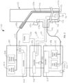
FIG. 2 illustrates a schematic view of the medical system ofFIG. 1 including a distal-end of the endoscope ofFIG. 1 . -
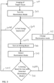
FIG. 3 illustrates a flow chart of a first method of operation of the medical system ofFIG. 1 . -
FIG. 4 illustrates a flow chart of a second method of operation of the medical system ofFIG. 1 . - Referring now to
FIG. 1 , the same illustrates an overall configuration of amedical system 100 having anendoscope 102, anendoscope processor 103, alight source 104, alaser apparatus 106 and adisplay 108. As illustrated inFIG. 1 , theendoscope 102 includes aninsertion section 110 configured to be inserted into a subject that images the inside of the subject and generates an image signal of the inside of the subject, theendoscope processor 103 that performs predetermined image processing on the image signal captured by theendoscope 102 and controls at least parts ofmedical system 100, thelight source 104 that generates illumination light of theendoscope 102 having at least two illumination modes, thelaser apparatus 106 having a first energy source for generating energy for treating a target tissue through anenergy guide 112 and a second energy source configured to emit two or more aiming beams to a target tissue through theenergy guide 112, and thedisplay device 108 that displays the aiming beam and an image of the image signal having been subject to the image processing performed by theendoscope processor 103. - The
endoscope 102 includes theinsertion portion 110 to be inserted into the subject, anoperating unit 107 to be held by an operator, which is on a proximal end portion side of theinsertion portion 110, and a flexibleuniversal cord 114 extended from theoperating unit 107. AlthoughFIG. 1 illustrates a Gastrointestinal (GI) endoscope, the apparatus and systems disclosed herein are not limited to a GI endoscope and also have particular utility for use with other types of endoscopes, such as ureteroscope or cystoscope or those used for a other treatment procedures. - The
insertion portion 110 is formed using a lighting fiber (light guide), an electric cable, an optical fiber, and the like. Theinsertion portion 110 includes a distal end portion 110a incorporating an imaging unit to be described later, a bendable bend portion 110b including a plurality of bend pieces, and a flexible tube portion 110c provided on a proximal end portion side of the bend portion 1 10b, which is flexible. The distal end portion 110a is provided with an illumination light guide 120 (seeFIG. 2 ) that illuminates the inside of the subject via an illumination lens 122 (seeFIG 2 ), an observation unit, including an image sensor, such as a CCD or CMOS and an objective lens system 118 (seeFIG. 2 ) that images the inside of the subject, an insertion port 107b that communicates with atreatment tool channel 102a (seeFIG. 2 ), and an air/water supply nozzle (not illustrated). - The
operating unit 107 includes a bending knob 107a for bending the bend portion 110b in the up and down direction and the right and left direction, the treatment tool insertion port 107b through which a treatment tool, such as medical forceps or theenergy guide 112 is inserted into a body cavity of the subject, and a plurality of switches 107c for operating a peripheral device such as theendoscope processor 103, thelight source device 104, an air supply device, a water supply device, and a gas supply device. The treatment tool, such as theenergy guide 112 can be inserted from the treatment tool insertion port 107b and through thechannel 102a such that a distal end thereof is exposed from an opening 102b (seeFIG. 2 ) of thechannel 102a at the distal end of theinsertion portion 110. - The
universal cord 114 includes a lighting fiber, a cable, and the like. Theuniversal cord 114 is branched at the proximal end thereof. One end of the branched ends is a connector 114a, and the other proximal end of the branched ends is aconnector 114b. The connector 114a is attachable/detachable to/from a connector of theendoscope processor 103. Theconnector 114b is attachable/detachable to/from thelight source 104. Theuniversal cord 114 propagates the illumination light emitted from thelight source 104 to the distal end portion 110a via theconnector 114b and the light guide 120 (seeFIG. 2 ). Further, theuniversal cord 114 transmits an image signal captured by the image sensor 116 (seeFIG. 2 ) to be described later to theendoscope processor 103 via a signal line 124 (seeFIG. 2 ) in the cable and via the connector 114a. - The
endoscope processor 103 executes predetermined image processing on the image signal output from the connector 114a, and controls at least part of the components making up themedical system 100. - The
light source 104 includes one or more light sources that emit light having one or more illumination characteristics, referred to as illumination modes, a condenser lens, and the like. Such light sources can be, for example, a Xenon lamp, an LED (Light-Emitting Diode), an LD (Laser Diode), or any combination thereof. Under the control of theendoscope processor 103, thelight source 104 emits light from the one or more light sources thereof, and supplies the light to theendoscope 102 connected via theconnector 114b and the light guide of theuniversal cord 114 as illumination light for the inside of the subject as an object. The illumination modes can be a white light illumination mode or a special light illumination mode, such as a narrow band imaging mode, an auto fluorescence imaging mode or an infrared imaging mode. A special light illumination can concentrate and intensify specific wavelengths of light, for example, resulting in a better visualization of a superficial micro-vessel and mucosal surface structures to enhance the subtle contrast of the irregularities of the mucosa. - The
display 108 includes, for example, a liquid crystal display, an organic electro luminescence (EL) display, or the like. Thedisplay 108 displays various kinds of information including the image having been subject to predetermined image processing by theinformation processing apparatus 103 via a video cable 108a. This allows an operator to observe and determine behavior of the desired position inside the subject by operating theendoscope 102 while watching the image (in-vivo image) displayed by thedisplay 108. - Referring now to
Figure 2 , themedical system 100 ofFIG. 1 is shown schematically. Thelaser apparatus 106 is for use with theenergy guide 112, such as a laser fiber. The energy guide is disposed in thechannel 102a through the treatment insertion port 107b and includes adistal end 112a that extends distally from the distal end opening 102b of thechannel 102a so as to be configured to direct treatment energy to the target tissue. A proximal end of theenergy guide 112 is operatively connected to thelaser apparatus 106. - The
laser apparatus 106 includes two or more energy sources for generating laser energy coupled to the proximal end of theenergy guide 112. Such energy sources can be selectable by a user by an input, such as a button 106a on thelaser apparatus 106 or a foot switch (not shown), through software or a user interface on thedisplay 108 or other inputs, manual or automatic as are known in the art. Afirst energy source 202 is optically coupled to theenergy guide 112 and can be configured to generate energy for treating the target tissue through theenergy guide 112. For example, thefirst energy source 202 can be a thulium laser, used to generate laser light for delivery through thelight guide 112 to the target tissue to operate in different treatment modes, such as a cutting (ablation) mode and a coagulation (hemostasis) mode. Other energy sources known in the art for such treatment of tissue, or any other treatment modes, can also be used for thefirst energy source 202, such as Ho:YAG, Nd:YAG and CO2 as well as others known in the art. - The two or more energy sources can also include a
second energy source 204 also optically coupled to theenergy guide 112 and configured to emit at least two aiming beams to the target tissue through theenergy guide 112, where the first aiming beam has at least one characteristic different from the second aiming beam. Such differing characteristics can be wavelength, power level and/or emitting pattern. For example, the first aiming beam can have a wavelength in the range of 500 nm to 550 nm while the second aiming beam can have a wavelength in the range of 635 nm to 690 nm. The characteristics of the different aiming beams can be selected based on the visibility of the aiming beams in the image processed by theendoscope processor 103 and displayed on thedisplay 108 under certain illumination modes provided by thelight source 104. - The
laser apparatus 106 further includes acontroller 206 comprising hardware, such as a CPU, that controls the operation of thelaser apparatus 106 including the first andsecond energy sources laser apparatus 106 can further include asensor 208 operatively coupled to theenergy guide 112 and under the control of thecontroller 206. Thesensor 208 is configured, as known in the art, to detect reflected light through theenergy guide 112 from thedistal end 112a of theenergy guide 112 and back to thesensor 208 such that thesensor 208 can determine an illumination mode output to thelight guide 120 from thelight source 104. That is,such sensor 208 detects the illumination mode being used to illuminate the target tissue. Such reflected light detection can be similar to that described inUnited States Patent 5,860,972 issued on January 19, 1999 , the contents of which is incorporated herein by reference. - The
light source 104 includes one or more light sources, such as a firstlight source 210 and a secondlight source 212 under the control of acontroller 214. Thelight sources light source 104 or a foot switch (not shown), through software or a user interface on thedisplay 108 or other inputs, manual or automatic as are known in the art. The first and secondlight sources light guide 120 to provide different illumination modes, as described above, to thelight guide 120. Although a different light source is shown for each illumination mode, a single light source can be provided to produce illumination modes having different characteristics through the use of filters, lens and the like. - The
endoscope processor 103 also includes acontroller 216 comprising hardware, such as a CPU, for control of theendoscope 102,display 108,light source 104 and/orlaser apparatus 106. Thecontroller 216, as discussed above, receives a signal from theimage sensor 116 throughline 124 in theuniversal cord 114 to process the same so as to generate an image/video for viewing on thedisplay 108. Such image includes not only the target area of the tissue to be treated under the illumination of thelight source 104 but also an aiming beam generated by thelaser apparatus 106 when thefirst energy source 202 is active and theenergy guide 112 is being used to treat the target tissue. Theendoscope processor 103 includes one or more inputs, such as a button 103a on theendoscope processor 103 or a foot switch (not shown), through software or a user interface on thedisplay 108 or other inputs, manual or automatic as are known in the art. - A use of the
medical system 100 ofFIG. 1 will now be described with regard to the flow chart illustrated inFIG. 3 . After insertion of theendoscope 102 to the target tissue site, the user views the target tissue on thedisplay 108 at 300. Such viewing of the target tissue is with an illumination mode set at 302 and output by one of thelight sources light source 104. Such illumination mode can be set by selection by the user by any means known in the art or automatically provided by a determination made by either ofcontrollers - A determination is made at 304 by the
controller 206 as to whether thefirst energy source 202 is activated (on) and delivering treatment energy to theenergy guide 112. Where it is determined that thefirst energy source 202 is not delivering treatment energy to theenergy guide 112, thecontroller 206, at 304N, does not activate thesecond energy source 204 to generate an aiming beam. Where it is determined that thefirst energy source 202 is delivering treatment energy to theenergy guide 112, thecontroller 206, at 304Y, activates thesecond energy source 204, at 306, to generate one of the first or second aiming beams. - At 308, the
controller 206 determines the illumination mode from the illumination modes used by the endoscope to illuminate the target tissue, such as receiving a signal indicating the type of illumination mode being used. The illumination mode signal provided to thecontroller 206 can be a manual input from the user at input 104a of thelight source 104 to direct thelight source controller 214 to output a signal to thecontroller 216 of theendoscope processor 103, which in turn outputs a signal to thelaser apparatus controller 206. Such manual input can also be from the input, such as button 103a, of theendoscope processor 103. Thelight source controller 214 can also directly output a signal indicating the illumination mode to thelaser apparatus controller 206. The input can also be via a button 107c on the endoscope through signal line 107d to thecontroller 216 of theendoscope processor 103, which is in turn relayed to thecontroller 206 of the laser apparatus. Thecontroller 206 of thelaser apparatus 106 can also receive a signal indicative of the illumination mode used by the endoscope from thesensor 208, which detects the illumination being used by reflected light through theenergy guide 112 and thecontroller 206 processes such detection and determines the illumination mode based on the output from thesensor 208. Furthermore, thecontroller 216 of theendoscope processor 103 can analyze the image signal from theimage sensor 116 and determine an illumination mode based on such image signal and output such determination to thecontroller 206 of thelaser apparatus 106. Other sensors (not shown) may also be employed for determination of the illumination mode being used by the endoscope, such as at the distal end of theendoscope 102 or in theendoscope processor 103. - At 310, a determination is made as to whether the aiming beam is appropriate for the determined illumination mode. Such determination can be based on historical data reflected in a look up table (LUT) operatively connected to the
controller 206, where the LUT corresponds illumination mode to aiming beam characteristic. Such LUT can store data of illumination modes (or the wavelength of the illumination light) and a corresponding wavelength of aiming beam for use with such illumination mode or wavelength of such illumination mode. Where it is determined that the aiming beam being used is appropriate for use with the illumination mode being used, no change is required in the aiming beam being used and the process continues at 310Y to image the target tissue until the determination is made at 310N that the aiming beam being used is not appropriate for use with the illumination mode being used. Such determination can be made upon predetermined intervals or upon an occurrence of a predetermined event, such as the first energy source being turned off and then again on. - However, where it is determined, at 3 10N, that the aiming beam being used is not appropriate for use with the illumination mode being used, the
second energy source 204 is controlled to change the aiming beam at 312 based on the indicated illumination mode. For example, when a white light illumination mode is determined, thecontroller 206 can control thesecond energy source 204 to emit a first aiming beam having a wavelength in the range of 500 nm to 550 nm. Alternatively, where a special light illumination mode is determined, thecontroller 206 can control thesecond energy source 204 to emit the second aiming beam having a wavelength in the range of 635 nm to 690 nm. As discussed above, the special light mode can be, for example, one of a narrow band imaging mode, an auto fluorescence imaging mode or an infrared imaging mode. - Another use of the
medical system 100 ofFIG. 1 will now be described with regard to the flow chart illustrated inFIG. 4 , in which 300, 302, 304 and 306 are substantially as described above. Where one of the aiming beams, such as the first or second aiming beams is activated by thelaser apparatus 106, a determination is made at 314 as to whether a spot caused by the first or second aiming beam can be identified in an image from theendoscope 102. That is, thecontroller 216 analyzes the image signal from theimage sensor 116 to determine if the aiming beam spot being used is visible in the image of the target tissue. Such determination of a spot in image data is well known in the art, such as by pixel comparison to determine a disparity (a discontinuity) in pixel data in an area of the image corresponding to an expected size and/or shape of the spot. Furthermore, such determination may consider the spot to not be visible if a spot is detected but the disparity is below some predetermined threshold where a user would have trouble identifying the spot clearly from the image. - Where the spot is detected, or is sufficiently detected in the image, the imaging, illumination and display of the image and spot continues, at 314Y, until such determination changes or the laser apparatus no longer activates the
first energy source 202. Where the spot cannot be detected or not sufficiently detected in the image at 314N, thecontroller 216 outputs a signal, at 316, to thelaser apparatus controller 206 to switch to a different one of the first or second aiming beams. - A similar determination is made by the
controller 216 at 318 to determine whether a spot caused by the different one of the first or second aiming beams can be identified in the image from theendoscope 102. Where the spot is detected, or is sufficiently detected in the image at 318Y, the imaging, illumination and display of the image and spot continues until such determination changes or the laser apparatus no longer activates thefirst energy source 202. Where the spot cannot be detected or not sufficiently detected in the image at 318N, as a safety measure, the controller can output a signal to thelaser apparatus controller 206 to control the first energy source to prohibit the first energy source from generating energy for treating the target tissue at 320. - Although described with regard to a flexible endoscope, the above apparatus and methods also have utility for rigid type endoscopes. In addition, although the
laser apparatus 106 is described as a separate device, the features thereof can be incorporated into one or both of the light source and endoscope processor, in which a common controller can be used to make the determinations and control indicated herein. - While there has been shown and described what is considered to be preferred embodiments, it will, of course, be understood that various modifications and changes in form or detail could readily be made without departing from the spirit of the invention. It is therefore intended that the invention be not limited to the exact forms described and illustrated, but should be constructed to cover all modifications that may fall within the scope of the appended claims.
- Hereinafter, some further examples are provided:
- Example 1. A medical laser apparatus, comprising:
- an energy guide;
- a first energy source configured to generate energy for treating a target tissue through the energy guide;
- a second energy source configured to emit first and second aiming beams to a target tissue through the energy guide, the second aiming beam having at least one characteristic different from the first aiming beam; and
- a controller comprising hardware, the controller being configured to:
- receive a signal indicating an illumination mode from at least two illumination modes used by an endoscope to illuminate the target tissue; and
- control the second energy source to output the first or second aiming beam based on the indicated illumination mode.
- Example 2. The medical laser apparatus according to Example 1, wherein when a white light illumination mode is indicated, the controller controls the second energy source to emit the first aiming beam having a wavelength in the range of 500 nm to 550 nm.
- Example 3. The medical laser apparatus according to Example 1, wherein when a special light illumination mode is indicated, the controller controls the second energy source to emit the second aiming beam having a wavelength in the range of 635 nm to 690 nm.
- Example 4. The medical laser apparatus according to Example 3, wherein the special light mode is one of a narrow band imaging mode, an auto fluorescence imaging mode or an infrared imaging mode.
- Example 5. The medical laser apparatus according to Example 1, wherein the controller is further configured to receive a signal indicating whether a spot caused by the first or second aiming beam can be identified in an image from the endoscope.
- Example 6. The medical laser apparatus according to Example 5, wherein, when the spot cannot be identified in the image, the controller being further configured to switch one of the first or second aiming beams to an other of the first or second aiming beams.
- Example 7. The medical laser apparatus according to Example 6, wherein the controller is further configured to receive a signal indicating whether a spot caused by the other of the first or second aiming beam can be identified in the image from the endoscope.
- Example 8. The medical laser apparatus according to Example 7, wherein, when the spot from the other of the first or second aiming beam cannot be identified in the image, the controller is configured to control the first energy source to prohibit the first energy source from generating energy for treating the target tissue.
- Example 9. The medical laser apparatus according to Example 1, wherein the at least one characteristic is selected from a group consisting of wavelength, power level and emitting pattern.
- Example 10. The medical laser apparatus according to Example 1, wherein the energy guide is a laser fiber.
- Example 11. The medical laser apparatus according to Example 1, wherein the first energy source is a treatment laser beam.
- Example 12. An endoscope controller comprising hardware, the endoscope controller being for use with an endoscope, the endoscope controller being configured to:
- output a first signal indicating an illumination mode of the endoscope;
- detect whether a spot from an aiming beam generated by an aiming beam energy source is visible in an image captured by an image sensor in the endoscope; and
- outputting a second signal based on the detection.
- Example 13. The endoscope controller of Example 12, wherein the second signal is output only where the spot cannot be detected in the image.
- Example 14. The endoscope controller of Example 12, wherein:
- the aiming beam is a first aiming beam; and
- where the spot cannot be detected in the image, the second signal instructs a laser apparatus to one of change the first aiming beam to a second aiming beam having at least one characteristic different from the first aiming beam.
- Example 15. A medical system comprising:
- a medical laser apparatus, comprising:
- an energy guide;
- a first energy source configured to generate energy for treating a target tissue through the energy guide;
- a second energy source configured to emit first and second aiming beams to a target tissue through the energy guide, the second aiming beam having at least one characteristic different from the first aiming beam; and
- a first controller comprising hardware, the first controller being configured to:
- receive a first signal indicating an illumination mode from at least two illumination modes used by an endoscope to illuminate the target tissue;
- control the second energy source to output the first or second aiming beam based on the indicated illumination mode; and
- a second controller comprising hardware, the second controller being for use with an endoscope, the second controller being configured to:
output the first signal to the first controller indicating the illumination mode from the at least two illumination modes used by the endoscope.
- a medical laser apparatus, comprising:
- Example 16. The medical system of Example 15, wherein when a white light illumination mode is indicated, the first controller controls the second energy source to emit the first aiming beam having a wavelength in the range of 500 nm to 550 nm.
- Example 17. The medical system according to Example 15, wherein when a special light illumination mode is indicated, the first controller controls the second energy source to emit the first aiming beam having a wavelength in the range of 635 nm to 690 nm.
- Example 18. The medical system according to Example 17, wherein the special light mode is one of a narrow band imaging mode, an auto fluorescence imaging mode or an infrared imaging mode.
- Example 19. The medical system according to Example 15, wherein:
- the second controller is further configured to:
output a second signal indicating whether a spot caused by the first or second aiming beam can be identified in an image from the endoscope; and - the first controller is further configured to:
- receive the second signal; and
- when the spot cannot be identified in the image, switch one of the first or second aiming beams to an other of the first or second aiming beams.
- the second controller is further configured to:
- Example 20. The medical system according to Example 15, wherein:
- the second controller is further configured to:
output a second signal indicating whether a spot caused by the first or second aiming beam can be identified in an image from the endoscope; and - the first controller is further configured to:
- receive the second signal; and
- when the spot from the other of the first or second aiming beam cannot be identified in the image, control the first energy source to prohibit the first energy source from generating energy for treating the target tissue.
- the second controller is further configured to:
- Example 21. The medical system according to Example 15, wherein the at least one characteristic is selected from a group consisting of wavelength, power level and emitting pattern.
- Example 22. The medical system according to Example 15, wherein the energy guide is a laser fiber.
- Example 23. The medical system according to Example 15, wherein the first energy source is a treatment laser beam.
Claims (15)
- A medical laser apparatus for use with an endoscope and a light source having two or more illumination modes, the apparatus comprising:an energy guide;a first energy source configured to generate energy for treating a target tissue through the energy guide;a second energy source, different from the light source, configured to emit first and second aiming beams to a target tissue through the energy guide, the second aiming beam having at least one characteristic different from the first aiming beam; anda first controller comprising hardware, the first controller being configured to:receive a signal indicating one of the at least two illumination modes currently illuminating the target tissue;select an aiming beam from the first and second aiming beams based on the illumination mode currently illuminating the target tissue; andcontrol the second energy source to emit the selected aiming beam to the target tissue.
- The medical laser apparatus according to claim 1, wherein when a white light illumination mode is indicated, the first controller controls the second energy source to emit the first aiming beam having a wavelength in a range of 500 nm to 550 nm.
- The medical laser apparatus according to claim 1, wherein when a special light illumination mode is indicated, the first controller controls the second energy source to emit the second aiming beam having a wavelength in a range of 635 nm to 690 nm.
- The medical laser apparatus according to claim 3, wherein the special light mode is one of a narrow band imaging mode, an auto fluorescence imaging mode or an infrared imaging mode.
- The medical laser apparatus according to claim 1, wherein the first controller is further configured to receive a signal indicating whether a spot caused by the first or second aiming beam can be identified in an image of the target tissue.
- The medical laser apparatus according to claim 5, wherein, when the spot cannot be identified in the image, the first controller being further configured to switch one of the first or second aiming beams to another of the first or second aiming beams.
- The medical laser apparatus according to claim 6, wherein the first controller is further configured to receive a signal indicating whether a spot caused by the other of the first or second aiming beam can be identified in the image of the target tissue.
- The medical laser apparatus according to claim 7, wherein, when the spot from the other of the first or second aiming beam cannot be identified in the image, the first controller is configured to control the first energy source to prohibit the first energy source from generating energy for treating the target tissue.
- The medical laser apparatus according to claim 1, wherein the at least one characteristic is selected from a group consisting of wavelength, power level and emitting pattern.
- The medical laser apparatus according to claim 1, wherein the energy guide is a laser fiber.
- The medical laser apparatus according to claim 1, wherein the first energy source is a treatment laser beam.
- An endoscope controller comprising hardware, the endoscope controller being for use with an endoscope, the endoscope controller being configured to:output a first signal indicating an illumination mode of the endoscope currently illuminating a target tissue;based on the illumination mode, select from a plurality of aiming beams generated by an aiming beam energy source an aiming beam to emit to the target tissue;detect whether a spot from the selected aiming beam is visible in an image captured by an image sensor in the endoscope; andoutputting a second signal based on the detection.
- The endoscope controller of claim 12, wherein the second signal is output only where the spot cannot be detected in the image.
- The endoscope controller of claim 12, wherein:the aiming beam is a first aiming beam; andwhere the spot cannot be detected in the image, the second signal instructs a laser apparatus to one of change the first aiming beam to a second aiming beam having at least one characteristic different from the first aiming beam.
- A medical system comprising:a medical laser apparatus according to claim 1,anda second controller comprising hardware, the second controller being for use with an endoscope, the second controller being configured to:
output the first signal to the first controller indicating the illumination mode from the at least two illumination modes used by the endoscope.
Applications Claiming Priority (3)
| Application Number | Priority Date | Filing Date | Title |
|---|---|---|---|
| US201862628513P | 2018-02-09 | 2018-02-09 | |
| PCT/US2019/017153 WO2019157247A1 (en) | 2018-02-09 | 2019-02-08 | Medical laser apparatus and system |
| EP19750915.1A EP3749167B1 (en) | 2018-02-09 | 2019-02-08 | Medical laser apparatus and system |
Related Parent Applications (1)
| Application Number | Title | Priority Date | Filing Date |
|---|---|---|---|
| EP19750915.1A Division EP3749167B1 (en) | 2018-02-09 | 2019-02-08 | Medical laser apparatus and system |
Publications (2)
| Publication Number | Publication Date |
|---|---|
| EP4516203A2 true EP4516203A2 (en) | 2025-03-05 |
| EP4516203A3 EP4516203A3 (en) | 2025-05-14 |
Family
ID=67548615
Family Applications (3)
| Application Number | Title | Priority Date | Filing Date |
|---|---|---|---|
| EP24211085.6A Pending EP4516203A3 (en) | 2018-02-09 | 2019-02-08 | Medical laser apparatus and system |
| EP19750915.1A Active EP3749167B1 (en) | 2018-02-09 | 2019-02-08 | Medical laser apparatus and system |
| EP19750838.5A Pending EP3749242A4 (en) | 2018-02-09 | 2019-02-09 | COMPUTER-READABLE STORAGE SYSTEM, METHOD AND DEVICE FOR CONTROLLING A LASER LIGHT SOURCE FROM A LITHOTRIPSY DEVICE |
Family Applications After (2)
| Application Number | Title | Priority Date | Filing Date |
|---|---|---|---|
| EP19750915.1A Active EP3749167B1 (en) | 2018-02-09 | 2019-02-08 | Medical laser apparatus and system |
| EP19750838.5A Pending EP3749242A4 (en) | 2018-02-09 | 2019-02-09 | COMPUTER-READABLE STORAGE SYSTEM, METHOD AND DEVICE FOR CONTROLLING A LASER LIGHT SOURCE FROM A LITHOTRIPSY DEVICE |
Country Status (11)
| Country | Link |
|---|---|
| US (9) | US12023096B2 (en) |
| EP (3) | EP4516203A3 (en) |
| JP (9) | JP7460526B2 (en) |
| KR (2) | KR102731868B1 (en) |
| CN (5) | CN119606285A (en) |
| AU (2) | AU2019217992B2 (en) |
| BR (2) | BR112020016159A2 (en) |
| CA (1) | CA3169535A1 (en) |
| DE (1) | DE112021001396T5 (en) |
| MX (2) | MX2020008321A (en) |
| WO (3) | WO2019157247A1 (en) |
Families Citing this family (33)
| Publication number | Priority date | Publication date | Assignee | Title |
|---|---|---|---|---|
| EP4516203A3 (en) | 2018-02-09 | 2025-05-14 | Gyrus ACMI, Inc. D.B.A. Olympus Surgical Technologies America | Medical laser apparatus and system |
| WO2019176171A1 (en) * | 2018-03-16 | 2019-09-19 | オリンパス株式会社 | Endoscope and endoscope system |
| CN112996549B (en) | 2018-09-24 | 2025-01-28 | 史赛克公司 | Systems and methods for improving control responsiveness during aspiration |
| JP7377769B2 (en) * | 2020-06-08 | 2023-11-10 | Hoya株式会社 | Program, information processing method, and information processing device |
| CN116249477B (en) | 2020-07-24 | 2026-01-23 | 捷锐士阿希迈公司(以奥林巴斯美国外科技术名义) | Image reconstruction and endoscope tracking |
| US12023044B2 (en) | 2020-08-10 | 2024-07-02 | Kunnskap Medical, LLC | Endoscopic system with medium management system control |
| DE112021005021T5 (en) * | 2020-09-25 | 2023-07-06 | Gyrus Acmi, Inc. D/B/A Olympus Surgical Technologies America | ANTI-CLOGGING DURING STONE EVACUATION |
| US20240049952A1 (en) * | 2020-10-30 | 2024-02-15 | The Regents Of The University Of Michigan | Devices, systems, and methods for treating kidney stones |
| US12070196B2 (en) * | 2020-11-23 | 2024-08-27 | Medos International Sarl | Arthroscopic medical implements and assemblies |
| GB2603904B (en) * | 2021-02-17 | 2023-07-26 | Keymed Medical & Industrial Equipment Ltd | Fluid management system |
| WO2022235762A1 (en) * | 2021-05-04 | 2022-11-10 | Ipg Photonics Corporation | Smart irrigation and aspiration system and method |
| US12290274B2 (en) * | 2021-05-25 | 2025-05-06 | Arthrex, Inc. | Dynamically controlling a distal window opening in a rotary surgical shaver |
| CA3225806A1 (en) * | 2021-07-19 | 2023-01-26 | Isaac Ostrovsky | Remote monitoring of fluid pressure in biological tissue |
| KR102359900B1 (en) * | 2021-07-30 | 2022-02-09 | 동성제약주식회사 | Photo dynamic and sono dynamic therapy apparatus based on complex luminous source |
| JP2024534141A (en) * | 2021-08-24 | 2024-09-18 | アイピージー フォトニクス コーポレーション | Methods and devices for the safe and effective treatment of urinary conditions using laser energy - Patents.com |
| DE102022126875A1 (en) | 2021-10-22 | 2023-04-27 | Gyrus Acmi, Inc. D/B/A Olympus Surgical Technologies America | CHARACTERIZATION OF TISSUE USING FLUORESCENCE EMISSION |
| US12564439B2 (en) | 2021-10-22 | 2026-03-03 | Gyrus Acmi, Inc. | Endoscope laser-triggered suction automatic on/off |
| DE102022126810A1 (en) * | 2021-10-22 | 2023-04-27 | Gyrus Acmi, Inc. D/B/A Olympus Surgical Technologies America | LASER COMBINATION WITH IN VIVO TARGET FEEDBACK ANALYSIS |
| US11963665B2 (en) * | 2021-10-27 | 2024-04-23 | Olympus Medical Systems Corp. | Endoscope and endoscope system |
| DE102021133193A1 (en) | 2021-12-15 | 2023-06-15 | Albert-Ludwigs-Universität Freiburg | Robotic system for wound cleaning |
| CN116649892A (en) * | 2022-02-25 | 2023-08-29 | 捷锐士阿希迈公司(以奥林巴斯美国外科技术名义) | Systems and methods for spectral signal detection |
| CN118900661A (en) * | 2022-03-22 | 2024-11-05 | 富士胶片株式会社 | Endoscope pipeline state determination method, endoscope pipeline state determination device and endoscope cleaning and disinfection device |
| CN114792927A (en) * | 2022-04-15 | 2022-07-26 | 深圳市雷迈科技有限公司 | Multi-wavelength laser system |
| CN115813510A (en) * | 2022-12-22 | 2023-03-21 | 新光维医疗科技(苏州)股份有限公司 | Surgical system and control method with cavity expansion, planing and suction functions |
| CN116077810B (en) * | 2023-04-11 | 2023-07-11 | 深圳核心医疗科技股份有限公司 | Control method and device of flushing device |
| CN116491885A (en) * | 2023-04-28 | 2023-07-28 | 浙江医高医疗科技有限公司 | Endoscope and endoscope pressure temperature control system |
| US12127804B1 (en) * | 2023-04-29 | 2024-10-29 | Syncrobotix, Inc. | Simplified highly maneuverable surgical catheter and bronchoscope |
| US12127805B1 (en) * | 2023-04-29 | 2024-10-29 | Syncrobotix, Inc. | Highly maneuverable surgical catheter and drive system |
| EP4620410A4 (en) * | 2023-06-16 | 2026-03-04 | Zhejiang Yigao Medical Tech Co Ltd | PRESSURE MAINTENANCE AND TEMPERATURE CONTROL METHOD FOR A FLUSHING AND SUCTION SYSTEM |
| CN116725659A (en) * | 2023-07-11 | 2023-09-12 | 锋迈(厦门)半导体科技有限公司 | Semiconductor laser operation system |
| CN117547339B (en) * | 2023-10-26 | 2024-11-29 | 邦士医疗科技股份有限公司 | A surgical power device capable of controlling constant pressure in a planing cavity and a control method thereof |
| CN121359953A (en) * | 2024-07-19 | 2026-01-20 | 蓝帆外科器械有限公司 | Medical water knife system |
| CN120324068B (en) * | 2025-06-12 | 2025-08-29 | 浙江归创医疗科技有限公司 | Medical pulse type thrombus removal system and method |
Citations (1)
| Publication number | Priority date | Publication date | Assignee | Title |
|---|---|---|---|---|
| US5860972A (en) | 1995-10-26 | 1999-01-19 | Xintec Corporation | Method of detection and destruction of urinary calculi and similar structures |
Family Cites Families (105)
| Publication number | Priority date | Publication date | Assignee | Title |
|---|---|---|---|---|
| DE3169553D1 (en) * | 1980-09-22 | 1985-05-02 | Olympus Optical Co | A laser device for an endoscope |
| JPS5971736A (en) * | 1982-07-30 | 1984-04-23 | オリンパス光学工業株式会社 | Length measuring apparatus for endoscope |
| US4971034A (en) | 1985-01-16 | 1990-11-20 | Asahi Kogaku Kogyo Kabushiki Kaisha | Body cavity pressure adjusting device for endoscope and laser medical treatment apparatus including body cavity pressure adjusting device |
| US4887600A (en) * | 1986-04-22 | 1989-12-19 | The General Hospital Corporation | Use of lasers to break down objects |
| DE3902943A1 (en) * | 1989-02-01 | 1990-08-09 | Wolf Gmbh Richard | DEVICE FOR INPUTING AND EXTRACTING LIQUIDS IN OR FROM BODY HOLLOW ORGANS |
| JPH03207371A (en) | 1990-01-10 | 1991-09-10 | Olympus Optical Co Ltd | Dissolution treatment apparatus |
| JP3110642B2 (en) * | 1995-01-30 | 2000-11-20 | 三菱電線工業株式会社 | Endoscope with dimension measurement function |
| US5830176A (en) | 1995-12-26 | 1998-11-03 | Mackool; Richard J. | Maintenance of pressure within a surgical site during a surgical procedure |
| US5836909A (en) * | 1996-09-13 | 1998-11-17 | Cosmescu; Ioan | Automatic fluid control system for use in open and laparoscopic laser surgery and electrosurgery and method therefor |
| US5785702A (en) | 1996-10-15 | 1998-07-28 | Eclipse Surgical Technologies, Inc. | Method for non-synchronous laser-assisted transmyocardial revascularization |
| US5931834A (en) | 1996-10-15 | 1999-08-03 | Eclipse Surgical Technologies, Inc. | Method for non-synchronous laser-assisted myocardial revascularization |
| US6527716B1 (en) | 1997-12-30 | 2003-03-04 | Altea Technologies, Inc. | Microporation of tissue for delivery of bioactive agents |
| US6156049A (en) | 1997-04-11 | 2000-12-05 | Coherent Inc. | Method and apparatus for transurethral resection of the prostate |
| DE19840346C2 (en) * | 1998-09-04 | 2000-09-07 | Med Laserzentrum Luebeck Gmbh | Method and device for diagnosing and monitoring the transmission quality of a fiber optic system |
| CN1249162A (en) | 1998-09-25 | 2000-04-05 | 中国科学院西安光学精密机械研究所 | A safe way to identify stones and human tissue |
| US6514242B1 (en) * | 1998-12-03 | 2003-02-04 | David Vasily | Method and apparatus for laser removal of hair |
| US6157661A (en) * | 1999-05-12 | 2000-12-05 | Laserphysics, Inc. | System for producing a pulsed, varied and modulated laser output |
| US20040229295A1 (en) | 1999-05-17 | 2004-11-18 | Marchitto Kevin S. | Activated delivery of biomolecules using electromagnetic energy |
| JP2001087304A (en) * | 1999-09-27 | 2001-04-03 | Nidek Co Ltd | Laser treatment apparatus |
| US7217266B2 (en) | 2001-05-30 | 2007-05-15 | Anderson R Rox | Apparatus and method for laser treatment with spectroscopic feedback |
| JP2003210485A (en) | 2002-01-23 | 2003-07-29 | Nidek Co Ltd | Laser therapeutic device |
| JP4421307B2 (en) * | 2004-01-09 | 2010-02-24 | 株式会社ニデック | Laser therapy device |
| US20060264995A1 (en) | 2004-02-18 | 2006-11-23 | Fanton Gary S | Apparatus and methods for clearing obstructions from surgical cutting instruments |
| US7297137B2 (en) | 2004-03-22 | 2007-11-20 | Alcon, Inc. | Method of detecting surgical events |
| JP4573555B2 (en) | 2004-03-30 | 2010-11-04 | オリンパス株式会社 | Endoscopic surgery system |
| DE102004028361B3 (en) * | 2004-06-11 | 2005-12-01 | Erbe Elektromedizin Gmbh | Flushing device and method for operating a purging device |
| US8062214B2 (en) | 2004-08-27 | 2011-11-22 | Smith & Nephew, Inc. | Tissue resecting system |
| EP1825802A4 (en) * | 2004-12-15 | 2009-05-27 | Olympus Medical Systems Corp | GAS SUPPLY DEVICE, GAS SUPPLY DEVICE CONTROL METHOD, GAS SUPPLY SYSTEM, AND ENDOSCOPIC SYSTEM |
| DE102005031125A1 (en) * | 2005-07-04 | 2007-01-11 | Siemens Ag | Method and lithotripsy apparatus for destroying a calculus in a patient |
| JP2007029603A (en) * | 2005-07-29 | 2007-02-08 | Fujinon Corp | Optical diagnostic treatment apparatus |
| GR20050100452A (en) | 2005-09-02 | 2007-04-25 | Estelle Enterprises Limited | Fluid exchange catheter's system |
| US20070073279A1 (en) * | 2005-09-29 | 2007-03-29 | Alcon, Inc. | Variable continuous wave laser |
| EP1991108B1 (en) | 2006-03-03 | 2018-08-01 | Cook Medical Technologies LLC | Endoscopic apparatus having an improved catheter |
| JP2007244679A (en) | 2006-03-16 | 2007-09-27 | Olympus Medical Systems Corp | Endoscope device |
| US7510542B2 (en) | 2006-12-20 | 2009-03-31 | Linvatec Corporation | Dual pump irrigation/aspiration system and method for determining joint pressure |
| US8591453B2 (en) | 2006-12-20 | 2013-11-26 | Linvatec Corporation | Dual pump arthroscopic irrigation/aspiration system with outflow control |
| US20080300662A1 (en) | 2007-06-01 | 2008-12-04 | Spectranetics | Custom Laser Sequences |
| JP5259141B2 (en) * | 2007-08-31 | 2013-08-07 | オリンパスメディカルシステムズ株式会社 | In-subject image acquisition system, in-subject image processing method, and in-subject introduction device |
| US8280496B2 (en) | 2007-12-13 | 2012-10-02 | Boston Scientific Scimed, Inc. | Extended spectral sensitivity endoscope system and method of using the same |
| JP5313523B2 (en) * | 2008-03-09 | 2013-10-09 | 邦男 粟津 | Calculus diagnosis and crushing device and calculus diagnosis and crushing method using infrared wavelength tunable laser |
| CN101273915B (en) | 2008-04-30 | 2010-11-03 | 爱科凯能科技(北京)有限公司 | laser lithotripsy device |
| JP5165479B2 (en) | 2008-07-02 | 2013-03-21 | オリンパスメディカルシステムズ株式会社 | Endoscope cleaning disinfection device |
| US9289541B2 (en) | 2008-08-22 | 2016-03-22 | Medtronic, Inc. | Surgical fluid management system |
| JP2010075314A (en) * | 2008-09-25 | 2010-04-08 | Fujifilm Corp | Invisible light irradiation apparatus, and method of controlling the same |
| EP3443922B1 (en) * | 2009-09-14 | 2020-07-29 | Memorial Sloan-Kettering Cancer Center | Apparatus and system for providing laser steering and focusing for incision, excision and ablation of tissue in minimally-invasive surgery |
| WO2011041629A2 (en) * | 2009-10-02 | 2011-04-07 | Cardiofocus, Inc. | Cardiac ablation system with pulsed aiming light |
| US9060775B2 (en) | 2009-10-09 | 2015-06-23 | Ethicon Endo-Surgery, Inc. | Surgical generator for ultrasonic and electrosurgical devices |
| US10441345B2 (en) | 2009-10-09 | 2019-10-15 | Ethicon Llc | Surgical generator for ultrasonic and electrosurgical devices |
| US20140074076A1 (en) * | 2009-10-12 | 2014-03-13 | Kona Medical, Inc. | Non-invasive autonomic nervous system modulation |
| US8665920B2 (en) * | 2010-01-27 | 2014-03-04 | Microvision, Inc. | Method and apparatus for laser diode compensation |
| US9066658B2 (en) | 2010-03-23 | 2015-06-30 | Stryker Corporation | Method and system for video based image detection/identification analysis for fluid and visualization control |
| US20120116168A1 (en) | 2010-09-13 | 2012-05-10 | Moellstam Anders | Method and device for flushing during endoscopic surgery |
| US9622911B2 (en) * | 2010-09-30 | 2017-04-18 | Cxl Ophthalmics, Llc | Ophthalmic treatment device, system, and method of use |
| US9737353B2 (en) * | 2010-12-16 | 2017-08-22 | Biosense Webster (Israel) Ltd. | System for controlling tissue ablation using temperature sensors |
| WO2012135073A2 (en) * | 2011-03-25 | 2012-10-04 | Board Of Trustees Of Michigan State University | Adaptive laser system for ophthalmic use |
| CN103429134B (en) | 2011-12-28 | 2015-12-09 | 奥林巴斯株式会社 | Body cavity pressure adjusting device and endoscope system |
| EP3308735B1 (en) | 2012-04-12 | 2019-09-11 | Boston Scientific Scimed, Inc. | Laser lithotripsy systems |
| NL2009424C2 (en) * | 2012-09-06 | 2014-03-10 | D O R C Dutch Ophthalmic Res Ct International B V | Irrigation/aspiration system, cartridge, pump unit, surgical machine, method for controlling. |
| US9968403B2 (en) * | 2012-10-16 | 2018-05-15 | Boston Scientific Scimed, Inc. | Surgical laser system and laser fiber |
| US10617470B2 (en) | 2012-10-16 | 2020-04-14 | Boston Scientific Scimed, Inc. | Laser ablation with electromagnetic energy feedback |
| US9119701B2 (en) | 2012-10-22 | 2015-09-01 | Alcon Research, Ltd. | Pressure control in phacoemulsification system |
| WO2014092851A1 (en) | 2012-12-11 | 2014-06-19 | Alcon Research, Ltd. | Phacoemulsification hand piece with integrated aspiration and irrigation pump |
| EP2967299B1 (en) | 2013-03-15 | 2022-11-30 | Stryker Corporation | Endoscopic light source and imaging system |
| CN108720909A (en) | 2013-04-08 | 2018-11-02 | 波士顿科学医学有限公司 | medical system and method |
| US9549850B2 (en) | 2013-04-26 | 2017-01-24 | Novartis Ag | Partial venting system for occlusion surge mitigation |
| US10265461B2 (en) | 2013-08-29 | 2019-04-23 | Motus Gi Medical Technologies Ltd. | Colon cleaning system with automatic self-purging features |
| WO2015066294A1 (en) | 2013-10-30 | 2015-05-07 | Faculty Physicians And Surgeons Of Loma Linda University School Of Medicine | Controlled pressure endoscopic and percutaneous surgery |
| JP5747368B1 (en) | 2013-11-08 | 2015-07-15 | オリンパス株式会社 | Endoscope reprocessing equipment |
| US9282985B2 (en) | 2013-11-11 | 2016-03-15 | Gyrus Acmi, Inc. | Aiming beam detection for safe laser lithotripsy |
| JP6002703B2 (en) | 2014-02-06 | 2016-10-05 | 富士フイルム株式会社 | Endoscope suction line switching device and endoscope |
| WO2015155776A2 (en) * | 2014-04-09 | 2015-10-15 | Motus Gi Medical Technologies Ltd. | Cleaning method for prepless colonoscopy |
| US9259231B2 (en) | 2014-05-11 | 2016-02-16 | Gyrus Acmi, Inc. | Computer aided image-based enhanced intracorporeal lithotripsy |
| CA2943563C (en) | 2014-05-22 | 2018-10-09 | Alcon Inc. | Technique for setting energy-related laser-pulse parameters |
| US10405924B2 (en) | 2014-05-30 | 2019-09-10 | The Spectranetics Corporation | System and method of ablative cutting and vacuum aspiration through primary orifice and auxiliary side port |
| US10828051B2 (en) | 2014-07-28 | 2020-11-10 | Shaw P. Wan | Suction evacuation device |
| JP2016043178A (en) | 2014-08-26 | 2016-04-04 | グンゼ株式会社 | Negative pressure treatment device |
| EP3200711A4 (en) | 2014-09-30 | 2018-06-27 | Shah, Kaushikkumar Vallabhadas | A sheath assembly and mutihole catheter for different fields of endoscopic surgery involving suction, irrigation and material removal. |
| JP2018501833A (en) * | 2014-11-14 | 2018-01-25 | ボストン サイエンティフィック サイムド,インコーポレイテッドBoston Scientific Scimed,Inc. | Surgical laser system and laser device |
| WO2016089900A2 (en) * | 2014-12-03 | 2016-06-09 | Cardiofocus, Inc. | System and method for visual confirmation of pulmonary vein isolation during ablation procedures |
| JP6540091B2 (en) | 2015-02-26 | 2019-07-10 | 株式会社ニデック | Ophthalmic laser treatment device |
| CN107635452B (en) * | 2015-06-02 | 2019-09-13 | 奥林巴斯株式会社 | Special light endoscope device |
| WO2017074740A1 (en) | 2015-10-27 | 2017-05-04 | Visumedics, Inc. | Laser system with pulse modulation and corresponding method of use |
| JP2017080348A (en) | 2015-10-30 | 2017-05-18 | Hoya株式会社 | Light source device |
| US11490990B2 (en) | 2015-11-12 | 2022-11-08 | Millennium Healtcare Technologies, Inc. | Laser-assisted periodontics |
| US10258415B2 (en) | 2016-01-29 | 2019-04-16 | Boston Scientific Scimed, Inc. | Medical user interfaces and related methods of use |
| US11273006B2 (en) | 2016-01-29 | 2022-03-15 | Millennium Healthcare Technologies, Inc. | Laser-assisted periodontics |
| US10471159B1 (en) * | 2016-02-12 | 2019-11-12 | Masimo Corporation | Diagnosis, removal, or mechanical damaging of tumor using plasmonic nanobubbles |
| CN114159013B (en) | 2016-03-15 | 2025-08-19 | 因特斯高普公司 | Surgical console, sample receiver and insertable endoscopic instrument for tissue removal |
| US10709472B2 (en) * | 2016-09-13 | 2020-07-14 | Biosense Webster (Israel) Ltd. | Debrider with multiple flushing orifices |
| US10610092B2 (en) | 2016-12-19 | 2020-04-07 | Sharon A Hibbs | Endoscope unblocking flush system |
| ES2937388T3 (en) | 2017-05-12 | 2023-03-28 | Convergent Dental Inc | System for the preventive treatment of dental hard tissue with a laser |
| AU2018289271B2 (en) * | 2017-06-19 | 2020-11-05 | Boston Scientific Scimed, Inc. | Automated fluid management system |
| US10478211B2 (en) | 2017-07-07 | 2019-11-19 | Ethicon Llc | Features to promote removal of debris from within ultrasonic surgical instrument |
| CN111565657B (en) | 2017-08-21 | 2023-11-07 | 锐凌公司 | Arthroscopic Devices and Methods |
| CN107485746A (en) | 2017-08-28 | 2017-12-19 | 重庆天如生物科技有限公司 | New perfusion suction system |
| CN109893209B (en) | 2017-11-17 | 2022-11-15 | 吉鲁斯·阿克米有限责任公司 | Coated endoscopic probe |
| WO2019157409A1 (en) | 2018-02-09 | 2019-08-15 | Performance Athlytics | Portable hydration monitoring device and methods |
| EP4516203A3 (en) | 2018-02-09 | 2025-05-14 | Gyrus ACMI, Inc. D.B.A. Olympus Surgical Technologies America | Medical laser apparatus and system |
| KR20190103589A (en) * | 2018-02-28 | 2019-09-05 | 서울대학교병원 | Continuous bladder irrigation control system |
| WO2019178180A1 (en) | 2018-03-13 | 2019-09-19 | Meditrina, Inc. | Endoscope and method of use |
| JP7503504B2 (en) * | 2018-06-08 | 2024-06-20 | クアンタ システム ソチエタ ペル アチオーニ | Photothermal targeted therapy system and related method integrating preconditioning and automatic triggering of photothermal targeted therapy via measurement of skin surface temperature |
| JP2021533895A (en) | 2018-08-16 | 2021-12-09 | モータス ジーアイ メディカル テクノロジーズ リミテッド | Integrated endoscopic cleansing system |
| US11389239B2 (en) | 2019-04-19 | 2022-07-19 | Elios Vision, Inc. | Enhanced fiber probes for ELT |
| CA3169405A1 (en) * | 2020-01-30 | 2021-08-05 | Boston Scientific Scimed, Inc. | Fluid management system and method for controlling intracavity pressure |
| JP7498287B2 (en) | 2020-02-27 | 2024-06-11 | ジャイラス エーシーエムアイ インク ディー/ビー/エー オリンパス サージカル テクノロジーズ アメリカ | Endoscope clearing system and method |
-
2019
- 2019-02-08 EP EP24211085.6A patent/EP4516203A3/en active Pending
- 2019-02-08 JP JP2020542995A patent/JP7460526B2/en active Active
- 2019-02-08 BR BR112020016159-4A patent/BR112020016159A2/en active Search and Examination
- 2019-02-08 AU AU2019217992A patent/AU2019217992B2/en active Active
- 2019-02-08 CN CN202411690325.7A patent/CN119606285A/en active Pending
- 2019-02-08 CN CN201980012086.0A patent/CN111683580B/en active Active
- 2019-02-08 US US16/968,800 patent/US12023096B2/en active Active
- 2019-02-08 KR KR1020207025950A patent/KR102731868B1/en active Active
- 2019-02-08 EP EP19750915.1A patent/EP3749167B1/en active Active
- 2019-02-08 MX MX2020008321A patent/MX2020008321A/en unknown
- 2019-02-08 WO PCT/US2019/017153 patent/WO2019157247A1/en not_active Ceased
- 2019-02-09 EP EP19750838.5A patent/EP3749242A4/en active Pending
- 2019-02-09 MX MX2020008318A patent/MX2020008318A/en unknown
- 2019-02-09 WO PCT/US2019/017391 patent/WO2019157406A1/en not_active Ceased
- 2019-02-09 JP JP2020542770A patent/JP7374911B2/en active Active
- 2019-02-09 AU AU2019216954A patent/AU2019216954B2/en active Active
- 2019-02-09 BR BR112020016161-6A patent/BR112020016161A2/en unknown
- 2019-02-09 CN CN202410652555.8A patent/CN118593117A/en active Pending
- 2019-02-09 KR KR1020207026082A patent/KR102712427B1/en active Active
- 2019-02-09 US US16/968,801 patent/US12059204B2/en active Active
- 2019-02-09 CN CN201980012090.7A patent/CN111683617B/en active Active
-
2020
- 2020-02-27 US US16/803,649 patent/US12369979B2/en active Active
- 2020-02-27 US US16/803,612 patent/US12016626B2/en active Active
-
2021
- 2021-02-25 CN CN202180016835.4A patent/CN115175626A/en active Pending
- 2021-02-25 JP JP2022551714A patent/JP7524336B2/en active Active
- 2021-02-25 CA CA3169535A patent/CA3169535A1/en active Pending
- 2021-02-25 WO PCT/US2021/019599 patent/WO2021173791A1/en not_active Ceased
- 2021-02-25 DE DE112021001396.3T patent/DE112021001396T5/en active Pending
-
2023
- 2023-06-27 JP JP2023105314A patent/JP2023115167A/en active Pending
-
2024
- 2024-03-21 JP JP2024045080A patent/JP2024073644A/en active Pending
- 2024-03-21 US US18/611,831 patent/US12414820B2/en active Active
- 2024-04-12 US US18/634,485 patent/US20240252245A1/en active Pending
- 2024-05-21 US US18/669,735 patent/US20240299093A1/en active Pending
- 2024-07-17 JP JP2024114017A patent/JP7783354B2/en active Active
-
2025
- 2025-02-14 JP JP2025022416A patent/JP2025081450A/en active Pending
- 2025-06-03 US US19/227,151 patent/US20250288354A1/en active Pending
- 2025-06-30 US US19/254,850 patent/US20250331920A1/en active Pending
- 2025-07-04 JP JP2025113865A patent/JP2025137525A/en active Pending
- 2025-11-27 JP JP2025207209A patent/JP2026031593A/en active Pending
Patent Citations (1)
| Publication number | Priority date | Publication date | Assignee | Title |
|---|---|---|---|---|
| US5860972A (en) | 1995-10-26 | 1999-01-19 | Xintec Corporation | Method of detection and destruction of urinary calculi and similar structures |
Also Published As
Similar Documents
| Publication | Publication Date | Title |
|---|---|---|
| US20240252245A1 (en) | Medical laser apparatus and system | |
| US9907462B2 (en) | Endoscope tip position visual indicator and heat management system | |
| JP7000590B2 (en) | Operation method of heat invasion observation device, endoscope system, heat invasion observation system, heat invasion observation device | |
| EP2996621B1 (en) | An endoscope tip position visual indicator and heat management system | |
| US9282985B2 (en) | Aiming beam detection for safe laser lithotripsy | |
| JP5394675B2 (en) | Endoscope system | |
| JP2017023604A (en) | Endoscope system | |
| JP5927077B2 (en) | Endoscope system | |
| EP4701556A1 (en) | Medical laser apparatus | |
| JP5784972B2 (en) | Exterior tube, laser transmission path, laser treatment instrument | |
| CN111035348A (en) | Endoscope system | |
| JP2026514798A (en) | Medical laser device | |
| DK202470125A1 (en) | Medical laser apparatus | |
| JP2012040105A (en) | Laser therapy apparatus, and laser output control method | |
| JP2004121487A (en) | Medical appliance |
Legal Events
| Date | Code | Title | Description |
|---|---|---|---|
| PUAI | Public reference made under article 153(3) epc to a published international application that has entered the european phase |
Free format text: ORIGINAL CODE: 0009012 |
|
| STAA | Information on the status of an ep patent application or granted ep patent |
Free format text: STATUS: THE APPLICATION HAS BEEN PUBLISHED |
|
| AC | Divisional application: reference to earlier application |
Ref document number: 3749167 Country of ref document: EP Kind code of ref document: P |
|
| AK | Designated contracting states |
Kind code of ref document: A2 Designated state(s): AL AT BE BG CH CY CZ DE DK EE ES FI FR GB GR HR HU IE IS IT LI LT LU LV MC MK MT NL NO PL PT RO RS SE SI SK SM TR |
|
| REG | Reference to a national code |
Ref country code: DE Ref legal event code: R079 Free format text: PREVIOUS MAIN CLASS: A61B0001307000 Ipc: A61B0001018000 |
|
| PUAL | Search report despatched |
Free format text: ORIGINAL CODE: 0009013 |
|
| AK | Designated contracting states |
Kind code of ref document: A3 Designated state(s): AL AT BE BG CH CY CZ DE DK EE ES FI FR GB GR HR HU IE IS IT LI LT LU LV MC MK MT NL NO PL PT RO RS SE SI SK SM TR |
|
| RIC1 | Information provided on ipc code assigned before grant |
Ipc: A61B 1/00 20060101ALN20250409BHEP Ipc: A61B 17/00 20060101ALN20250409BHEP Ipc: A61B 18/00 20060101ALN20250409BHEP Ipc: H01S 3/16 20060101ALN20250409BHEP Ipc: A61M 1/00 20060101ALN20250409BHEP Ipc: A61B 90/00 20160101ALN20250409BHEP Ipc: A61B 1/307 20060101ALI20250409BHEP Ipc: A61B 18/26 20060101ALI20250409BHEP Ipc: A61B 18/22 20060101ALI20250409BHEP Ipc: A61B 18/20 20060101ALI20250409BHEP Ipc: A61B 18/18 20060101ALI20250409BHEP Ipc: A61B 17/32 20060101ALI20250409BHEP Ipc: A61B 1/05 20060101ALI20250409BHEP Ipc: A61B 1/04 20060101ALI20250409BHEP Ipc: A61B 1/015 20060101ALI20250409BHEP Ipc: A61B 1/018 20060101AFI20250409BHEP |
|
| STAA | Information on the status of an ep patent application or granted ep patent |
Free format text: STATUS: REQUEST FOR EXAMINATION WAS MADE |
|
| 17P | Request for examination filed |
Effective date: 20251114 |