EP4513433A2 - Läsionsdetektionsverfahren und läsionsdetektionsprogramm - Google Patents
Läsionsdetektionsverfahren und läsionsdetektionsprogramm Download PDFInfo
- Publication number
- EP4513433A2 EP4513433A2 EP24182201.4A EP24182201A EP4513433A2 EP 4513433 A2 EP4513433 A2 EP 4513433A2 EP 24182201 A EP24182201 A EP 24182201A EP 4513433 A2 EP4513433 A2 EP 4513433A2
- Authority
- EP
- European Patent Office
- Prior art keywords
- region
- machine learning
- learning model
- data
- image
- Prior art date
- Legal status (The legal status is an assumption and is not a legal conclusion. Google has not performed a legal analysis and makes no representation as to the accuracy of the status listed.)
- Pending
Links
Images








Classifications
-
- G—PHYSICS
- G06—COMPUTING OR CALCULATING; COUNTING
- G06T—IMAGE DATA PROCESSING OR GENERATION, IN GENERAL
- G06T7/00—Image analysis
- G06T7/0002—Inspection of images, e.g. flaw detection
- G06T7/0012—Biomedical image inspection
-
- G—PHYSICS
- G06—COMPUTING OR CALCULATING; COUNTING
- G06T—IMAGE DATA PROCESSING OR GENERATION, IN GENERAL
- G06T7/00—Image analysis
- G06T7/10—Segmentation; Edge detection
- G06T7/11—Region-based segmentation
-
- G—PHYSICS
- G06—COMPUTING OR CALCULATING; COUNTING
- G06V—IMAGE OR VIDEO RECOGNITION OR UNDERSTANDING
- G06V10/00—Arrangements for image or video recognition or understanding
- G06V10/70—Arrangements for image or video recognition or understanding using pattern recognition or machine learning
- G06V10/764—Arrangements for image or video recognition or understanding using pattern recognition or machine learning using classification, e.g. of video objects
-
- G—PHYSICS
- G16—INFORMATION AND COMMUNICATION TECHNOLOGY [ICT] SPECIALLY ADAPTED FOR SPECIFIC APPLICATION FIELDS
- G16H—HEALTHCARE INFORMATICS, i.e. INFORMATION AND COMMUNICATION TECHNOLOGY [ICT] SPECIALLY ADAPTED FOR THE HANDLING OR PROCESSING OF MEDICAL OR HEALTHCARE DATA
- G16H30/00—ICT specially adapted for the handling or processing of medical images
- G16H30/40—ICT specially adapted for the handling or processing of medical images for processing medical images, e.g. editing
-
- G—PHYSICS
- G16—INFORMATION AND COMMUNICATION TECHNOLOGY [ICT] SPECIALLY ADAPTED FOR SPECIFIC APPLICATION FIELDS
- G16H—HEALTHCARE INFORMATICS, i.e. INFORMATION AND COMMUNICATION TECHNOLOGY [ICT] SPECIALLY ADAPTED FOR THE HANDLING OR PROCESSING OF MEDICAL OR HEALTHCARE DATA
- G16H50/00—ICT specially adapted for medical diagnosis, medical simulation or medical data mining; ICT specially adapted for detecting, monitoring or modelling epidemics or pandemics
- G16H50/20—ICT specially adapted for medical diagnosis, medical simulation or medical data mining; ICT specially adapted for detecting, monitoring or modelling epidemics or pandemics for computer-aided diagnosis, e.g. based on medical expert systems
-
- G—PHYSICS
- G16—INFORMATION AND COMMUNICATION TECHNOLOGY [ICT] SPECIALLY ADAPTED FOR SPECIFIC APPLICATION FIELDS
- G16H—HEALTHCARE INFORMATICS, i.e. INFORMATION AND COMMUNICATION TECHNOLOGY [ICT] SPECIALLY ADAPTED FOR THE HANDLING OR PROCESSING OF MEDICAL OR HEALTHCARE DATA
- G16H50/00—ICT specially adapted for medical diagnosis, medical simulation or medical data mining; ICT specially adapted for detecting, monitoring or modelling epidemics or pandemics
- G16H50/70—ICT specially adapted for medical diagnosis, medical simulation or medical data mining; ICT specially adapted for detecting, monitoring or modelling epidemics or pandemics for mining of medical data, e.g. analysing previous cases of other patients
-
- G—PHYSICS
- G06—COMPUTING OR CALCULATING; COUNTING
- G06T—IMAGE DATA PROCESSING OR GENERATION, IN GENERAL
- G06T2207/00—Indexing scheme for image analysis or image enhancement
- G06T2207/10—Image acquisition modality
- G06T2207/10072—Tomographic images
- G06T2207/10081—Computed x-ray tomography [CT]
-
- G—PHYSICS
- G06—COMPUTING OR CALCULATING; COUNTING
- G06T—IMAGE DATA PROCESSING OR GENERATION, IN GENERAL
- G06T2207/00—Indexing scheme for image analysis or image enhancement
- G06T2207/20—Special algorithmic details
- G06T2207/20021—Dividing image into blocks, subimages or windows
-
- G—PHYSICS
- G06—COMPUTING OR CALCULATING; COUNTING
- G06T—IMAGE DATA PROCESSING OR GENERATION, IN GENERAL
- G06T2207/00—Indexing scheme for image analysis or image enhancement
- G06T2207/20—Special algorithmic details
- G06T2207/20084—Artificial neural networks [ANN]
-
- G—PHYSICS
- G06—COMPUTING OR CALCULATING; COUNTING
- G06T—IMAGE DATA PROCESSING OR GENERATION, IN GENERAL
- G06T2207/00—Indexing scheme for image analysis or image enhancement
- G06T2207/30—Subject of image; Context of image processing
- G06T2207/30004—Biomedical image processing
- G06T2207/30096—Tumor; Lesion
Definitions
- Embodiments discussed herein are related to a lesion detection method and a lesion detection program.
- CT computed tomography
- MRI magnetic resonance imaging
- a doctor has to interpret a large number of images, which imposes a large burden on the doctor. For this reason, there is a demand for a technique for supporting diagnosis work of the doctor by a computer in some form.
- a technique for detecting a lesion region from a medical image by using a trained model (machine learning model) generated by machine learning.
- machine learning model For example, a system for heart segmentation that trains a neural network integrated with a long-short-term memory (LSTM) is proposed.
- LSTM long-short-term memory
- the machine learning model there is a machine learning model that classifies whether or not each pixel included in a captured tomographic image is a specific lesion region.
- a lesion region is classified in pixel units from the tomographic image.
- a problem with such a machine learning model is that classification accuracy may be decreased depending on an imaging position of the tomographic image in a human body.
- the actually captured tomographic image of the human body is used as learning data.
- the number of tomographic images used as the learning data is fixed regardless of the position in the human body.
- an appearance frequency of a lesion region may have a bias depending on the position inside the human body. Therefore, among the tomographic images used as the learning data, the number of tomographic images including the lesion region may vary depending on the position inside the human body. For a tomographic image captured at a position at which a sufficient number of tomographic images are not used as the learning data, there is a possibility that the classification accuracy of the lesion is decreased.
- an object of the present disclosure is to provide a lesion detection method and a lesion detection program capable of detecting a lesion region with a certain level of accuracy regardless of an imaging position of a tomographic image of an inside of a human body.
- a lesion detection method executed by a computer includes: acquiring a first tomographic image obtained by imaging a first imaging position of an inside of a first human body; selecting any one of a first machine learning model that classifies whether or not a pixel included in an input tomographic image is a specific lesion region in units of the pixel and a second machine learning model that classifies whether or not an image block obtained by dividing the input tomographic image into certain sizes is the specific lesion region in units of the image block, based on, among a plurality of tomographic images which are obtained by imaging an inside of one or more second human bodies, are used as learning data when the first machine learning model is generated by machine learning, and each of which includes the specific lesion region, the number of tomographic images captured at a second imaging position of the inside of the one or more second human bodies, which corresponds to the first imaging position; and detecting the specific lesion region from the first tomographic image by using a machine learning model selected between the first machine learning model and the
- a lesion region may be detected with a certain level of accuracy regardless of an imaging position of a tomographic image of an inside of a human body.
- FIG. 1 is a diagram illustrating a configuration example and a process example of a lesion detection apparatus according to a first embodiment.
- a lesion detection apparatus 1 illustrated in FIG. 1 is an information processing apparatus that detects a specific lesion region from a tomographic image obtained by imaging an inside of a human body. As an example in the following description, it is assumed that the tomographic image is an image captured at certain intervals in a height direction of the human body.
- the lesion detection apparatus 1 includes, for example, a storage unit 2 and a processing unit 3.
- the storage unit 2 is a storage region secured in a storage device (not illustrated) included in the lesion detection apparatus 1.
- the storage unit 2 stores data indicating a machine learning model 2a and data indicating a machine learning model 2b.
- the machine learning model 2a is a trained model that classifies whether or not a pixel included in an input tomographic image is a specific lesion region in units of the pixel. In the classification processing using the machine learning model 2a, when one tomographic image is input to the machine learning model 2a, a lesion region is classified in pixel units from the tomographic image.
- the machine learning model 2b is a trained model that classifies whether or not an image block obtained by dividing the input tomographic image into certain sizes is a specific lesion region in units of the image block.
- the tomographic image is divided into image blocks.
- the image blocks are input to the machine learning model 2b one by one, and it is determined whether or not the input image block is a lesion region each time.
- the processing unit 3 is, for example, a processor. In this case, the following process of the processing unit 3 is implemented by the processor executing a predetermined program. By selectively using any one of the machine learning models 2a and 2b, the processing unit 3 detects a lesion region from the captured tomographic image.
- a tomographic image group 12 is a set of tomographic images including a lesion region, among tomographic images used as learning data when the machine learning model 2a is generated by machine learning.
- a tomographic image included in the tomographic image group 12 is an image obtained by imaging an inside of human bodies 11a, 11b, and ....
- the processing unit 3 acquires a tomographic image 22a obtained by imaging a first imaging position of an inside a human body 21 as a target for lesion detection.
- the processing unit 3 selects any one of the machine learning models 2a and 2b, based on the number of tomographic images captured at a second imaging position corresponding to the first imaging position in the human bodies 11a, 11b, and ....
- the processing unit 3 detects a specific lesion region from the tomographic image 22a.
- An appearance frequency of the lesion region may have a bias depending on a position of the inside of the human body. Therefore, among the tomographic images used as learning data, the number of tomographic images including the lesion region may vary depending on the position of the inside of the human body. For a tomographic image captured at a position at which a sufficient number of tomographic images are not used as learning data, there is a possibility that classification accuracy of a lesion using the machine learning model 2a is decreased.
- the processing unit 3 selects the machine learning model 2a in a case where it is determined that the number of tomographic images captured at the second imaging position is a sufficient number with which a certain or higher level of lesion classification accuracy is obtained from the machine learning model 2a.
- the processing unit 3 selects the machine learning model 2b in a case where it is determined that the number of images is not sufficient to obtain the certain or higher level of lesion classification accuracy from the machine learning model 2a. Since the machine learning model 2b clasifies a lesion region based on a feature in a division region instead of the entire tomographic image, the lesion classification accuracy does not depend on the imaging position of the tomographic image. Therefore, by the model selection described above, it is possible to detect a lesion region with a certain level of accuracy regardless of the imaging position of the tomographic image of the inside of the human body.
- the lesion detection apparatus 1 or another information processing apparatus counts the number of tomographic images included in the tomographic image group 12 for each position in the human bodies 11a, 11b, and .... Specifically, a predetermined region of the human bodies 11a, 11b, and ... is divided into three regions at equal intervals in a direction perpendicular to the tomographic image, so that division regions A1 to A3 are set. For each of the division regions A1 to A3, the number of tomographic images is counted. In the example illustrated in FIG. 1 , it is assumed that the tomographic image group 12 includes 1000 tomographic images, the number of images in the division region A1 is 100, the number of images in the division region A2 is 800, and the number of images in the division region A3 is 100.
- the division regions A1 to A3 are obtained by dividing a region between predetermined reference points in each of the human bodies 11a, 11b, and .... As such a region, a specific organ region may be applied.
- a tomographic image group 22 is acquired by imaging a predetermined region inside the human body 21.
- division regions B1 to B3 are generated. It is assumed that among tomographic images included in the tomographic image group 22, a lesion detection process is executed on the tomographic image 22a belonging to the division region B2.
- the processing unit 3 acquires the number of tomographic images used as learning data, which is counted for the division region A2 corresponding to the division region B2, among the division regions A1 to A3.
- the number of images "800" is acquired for the division region A2.
- the processing unit 3 selects the machine learning model 2a. In this case, the processing unit 3 inputs the tomographic image 22a to the machine learning model 2a, and classifies whether or not each pixel of the tomographic image 22a is a lesion region.
- the processing unit 3 acquires the number of tomographic images used as learning data, which is counted for the division region A3 corresponding to the division region B3, among the division regions A1 to A3.
- the number of images "100" is acquired for the division region A3. Since a ratio of the acquired number "100" of images to the total number "1000" is 10%, which is equal to or less than the threshold value, the processing unit 3 selects the machine learning model 2b. In this case, the processing unit 3 divides the tomographic image into image blocks, and inputs the divided image blocks one by one to the machine learning model 2b to classify whether each image block is a lesion region.
- FIG. 2 is a diagram illustrating a configuration example of a diagnosis support system according to a second embodiment.
- the diagnosis support system illustrated in FIG. 2 is a system that supports image diagnosis work by CT imaging, and includes a CT apparatus 50 and a diagnosis support apparatus 100.
- the diagnosis support apparatus 100 is an example of the lesion detection apparatus 1 illustrated in FIG. 1 .
- the CT apparatus 50 captures an X-ray CT image of a human body. According to the present embodiment, the CT apparatus 50 captures a predetermined number of tomographic images of an axial plane in a chest region while changing a position (slice position) of a human body in a height direction (direction perpendicular to the axial plane) at a predetermined interval.
- the diagnosis support apparatus 100 extracts a lung field region from each tomographic image captured by the CT apparatus 50, and detects a lesion region based on image information of the extracted lung field region. According to the present embodiment, a shadow region is detected as the lesion region.
- the diagnosis support apparatus 100 may classify a type of shadow. For example, the diagnosis support apparatus 100 causes a display device to display information indicating a detection result of the lesion region. Thus, the diagnosis support apparatus 100 supports the image diagnosis work of a user (for example, a radiologist).
- the diagnosis support apparatus 100 is implemented as a computer illustrated in FIG. 2 , for example.
- the diagnosis support apparatus 100 includes a processor 101, a random-access memory (RAM) 102, a hard disk drive (HDD) 103, a graphics processing unit (GPU) 104, an input interface (I/F) 105, a reading device 106, and a communication interface (I/F) 107.
- a processor 101 a random-access memory (RAM) 102, a hard disk drive (HDD) 103, a graphics processing unit (GPU) 104, an input interface (I/F) 105, a reading device 106, and a communication interface (I/F) 107.
- RAM random-access memory
- HDD hard disk drive
- GPU graphics processing unit
- I/F input interface
- reading device 106 a reading device
- I/F communication interface
- the processor 101 comprehensively controls the entire diagnosis support apparatus 100.
- the processor 101 is, for example, a central processing unit (CPU), a microprocessor unit (MPU), a digital signal processor (DSP), an application-specific integrated circuit (ASIC), or a programmable logic device (PLD).
- the processor 101 may also be a combination of two or more elements among the CPU, the MPU, the DSP, the ASIC, and the PLD. Noted that the processor 101 is an example of the processing unit 3 illustrated in FIG. 1 .
- the RAM 102 is used as a main storage device of the diagnosis support apparatus 100.
- the RAM 102 temporarily stores at least a part of an operating system (OS) program and an application program to be executed by the processor 101.
- the RAM 102 also stores various kinds of data to be used in processes performed by the processor 101.
- OS operating system
- the RAM 102 also stores various kinds of data to be used in processes performed by the processor 101.
- the HDD 103 is used as an auxiliary storage device of the diagnosis support apparatus 100.
- the OS program, the application program, and various kinds of data are stored in the HDD 103.
- a different type of nonvolatile storage device such as a solid-state drive (SSD) may be used as the auxiliary storage device.
- SSD solid-state drive
- a display device 104a is coupled to the GPU 104.
- the GPU 104 causes the display device 104a to display an image in accordance with an instruction from the processor 101.
- Examples of the display device 104a include a liquid crystal display, an organic electroluminescence (EL) display, and the like.
- An input device 105a is coupled to the input interface 105.
- the input interface 105 transmits a signal output from the input device 105a to the processor 101.
- the input device 105a includes a keyboard, a pointing device, and the like.
- the pointing device includes a mouse, a touch panel, a tablet, a touch pad, a track ball, and the like.
- a portable-type recording medium 106a is removably attached to the reading device 106.
- the reading device 106 reads data recorded in the portable-type recording medium 106a, and transmits the data to the processor 101.
- the portable-type recording medium 106a includes an optical disc, a semiconductor memory, and the like.
- the communication interface 107 transmits and receives data to and from another apparatus such as the CT apparatus 50 via a network.
- the hardware configuration as described above is capable of implementing a processing function of the diagnosis support apparatus 100.
- a lesion classification process from a medical image may be executed by using a trained model generated by machine learning, for example.
- FIG. 3 is a diagram illustrating a first example of a trained model for lesion classification.
- a lesion classification model 60 illustrated in FIG. 3 is a trained model for classifying a lesion by a Segmentation method.
- the lesion classification model 60 may be referred to as a "Segmentation model 60".
- the Segmentation model 60 classifies whether or not a pixel of the tomographic image 62 is a specific lesion region (whether or not the pixel is a shadow in the present embodiment) in units of the pixel.
- the tomographic image 62 is a tomographic image of an axial plane.
- the Segmentation model 60 is generated by machine learning (for example, deep learning) using a tomographic image group 61 of the axial plane as learning data.
- a label indicating whether or not each pixel is a lesion region is added to each tomographic image used as learning data, and these labels are used as correct answer data in machine learning.
- the Segmentation model 60 is generated.
- the Segmentation model 60 may be a lesion classification model that classifies three or more types of lesions including a type indicating that the pixel is not a lesion.
- the Segmentation model 60 classifies, for each pixel, one of six types of shadows of a ground glass opacity, a consolidation, a honeycomb, emphysema, other shadows, and a normal (not a shadow).
- a label indicating which of the shadow types described above is added to each tomographic image used as the learning data for each pixel.
- the lesion classification process using such a Segmentation model 60 is effective for, for example, diagnosis of a diffuse pneumonia.
- FIG. 4 illustrates a second example of the trained model for lesion classification.
- a lesion classification model 70 illustrated in FIG. 4 is a trained model for classifying a lesion by a Classification method.
- the lesion classification model 70 may be referred to as a "Classification model 70".
- the Classification model 70 Upon receiving an input of a tomographic image 72, the Classification model 70 divides the tomographic image 72 into patch images (image blocks) having a certain size, and classifies whether or not the divided patch image is a specific lesion region (whether or not the divided patch image is a shadow in the present embodiment) in units of the divided patch image. Noted that in the same manner as the tomographic image 62 in FIG. 3 , it is assumed that the tomographic image 72 is a tomographic image of an axial plane.
- the classification model 70 is generated by machine learning (for example, deep learning) using a tomographic image group 71 of an axial plane as learning data. Meanwhile, unlike the Segmentation method in FIG. 3 , each tomographic image used as learning data is divided into the patch images described above, and a label indicating whether or not the patch image is a lesion region is added to each of the divided patch images.
- the Classification model 70 is generated by performing machine learning in which the patch image is used as a unit, by using each patch image as teaching data and using the label as correct answer data.
- the Classification model 70 may be a lesion classification model that classifies three or more types of lesions including a type indicating that the patch image is not a lesion.
- the Classification model 70 classifies, for each patch image, one of the six types of shadows described above. In this case, a label indicating one of the shadow types described above is added to each tomographic image used as the learning data for each patch image.
- the Classification model 70 classifies a lesion by using a patch image including a plurality of pixels as a unit.
- the Segmentation model 60 classifies a lesion in pixel units, it is possible to classify the lesion by using a finer region in a tomographic image as a unit, as compared with the Classification model 70.
- the Segmentation model 60 has a problem in that there may be a difference in classification accuracy for each position of tomographic images in an organ region.
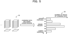
- FIG. 5 is a diagram illustrating a bias in the number of pieces of learning data in an organ region.
- a tomographic image of an organ region (lung field region in the present embodiment) among tomographic images obtained by imaging a chest region of a human body is used as learning data.
- a learning data set 81 illustrated in FIG. 5 is a set of pieces of learning data including, among such learning data, a tomographic image including a lesion region (a tomographic image including one or more pixels of the lesion region) as teaching data.
- the learning data set 81 does not include a tomographic image having no lesion region.
- the learning data included in the learning data set 81 may be referred to as "learning data of lesion region”.
- a histogram 82 illustrated in FIG. 5 is a graph illustrating the number of pieces of learning data included in the learning data set 81 for each of division regions obtained by equally dividing the organ region in a vertical direction.
- the histogram 82 in FIG. 5 indicates the number of pieces of learning data for each of division regions R1 to R6 obtained by dividing the organ region into six regions from an upper side.
- a lesion region is likely to appear in the organ region differs for each lesion as an classification target.
- the number of pieces of learning data of the lesion region included in the learning data set 81 described above also has a bias depending on the position of the organ region.
- the lesion region is likely to appear around a center of the organ region in the vertical direction. Therefore, in the histogram 82, the number of pieces of learning data around the center in the vertical direction is larger than the number of pieces of learning data at the upper portion or the lower portion.
- the Segmentation model 60 Based on an image feature in the entire one tomographic image such as a shape of an organ in the tomographic image or a positional relationship with another organ, the Segmentation model 60 classifies a lesion in pixel units. In order to classify the lesion with high accuracy, the sufficient number of pieces of learning data as learning data of the lesion region are desired for each position in the organ region. Meanwhile, in a case where there is a bias in the number of pieces of learning data for each position in the organ region as in the histogram 82, there is a problem in that, at a time of an inference, a difference in classification accuracy of the lesion occurs for each position in the organ region of the tomographic image input to the Segmentation model 60. As an example of the histogram 82, in a case where tomographic images of an upper portion and a lower portion in the organ region in the vertical direction are input, there is a possibility that the classification accuracy of the lesion is lowered.
- the Classification model 70 classifies a lesion by using a patch image obtained by dividing a tomographic image as a unit. Since such a Classification model 70 classifies a lesion based on an image feature such as light and dark of an image, in a partial region in a tomographic image called a patch image, classification accuracy does not depend on a position of the patch image in an organ region.
- the diagnosis support apparatus 100 selects and uses one of the Segmentation model 60 and the Classification model 70 at a time of an inference in accordance with the number of pieces of data in the organ region of the learning data of the lesion region, which is used at a time of training of the Segmentation model 60.
- the diagnosis support apparatus 100 selects and uses one of the Segmentation model 60 and the Classification model 70 at a time of an inference in accordance with the number of pieces of data in the organ region of the learning data of the lesion region, which is used at a time of training of the Segmentation model 60.
- the diagnosis support apparatus 100 selects and uses one of the Segmentation model 60 and the Classification model 70 at a time of an inference in accordance with the number of pieces of data in the organ region of the learning data of the lesion region, which is used at a time of training of the Segmentation model 60.
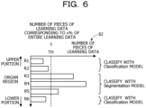
- FIG. 6 is a diagram illustrating an example of selecting a lesion classification model.
- a threshold value TH is set for the number of pieces of learning data of a lesion region for each division region.
- the threshold value TH for example, the number of pieces of learning data at a predetermined ratio (x%) with respect to a total number of pieces of learning data included in the learning data set 81 is set.
- the diagnosis support apparatus 100 executes lesion classification for a tomographic image included in a certain division region
- the diagnosis support apparatus 100 executes the lesion classification by using the Segmentation model 60.
- the diagnosis support apparatus 100 executes the lesion classification by using the Classification model 70.
- FIG. 7 is a diagram illustrating a specific example of model selection in accordance with the number of pieces of learning data of a lesion region.
- an organ region is divided into three regions of "upper organ portion", “middle organ portion", and “lower organ portion” in the vertical direction.
- a total number of pieces of learning data included in the learning data set 81 is 1000, and the threshold value TH is set to 200 corresponding to 20% of the total number.
- the number of pieces of learning data corresponding to the upper organ portion is 100.
- lesion classification for a tomographic image corresponding to the upper organ portion is executed by using the Classification model 70.
- the number of pieces of learning data corresponding to the middle organ portion is 800.
- lesion classification for a tomographic image corresponding to the middle organ portion is executed by using the Segmentation model 60.
- the number of pieces of learning data corresponding to the lower organ portion is 100.
- lesion classification for a tomographic image corresponding to the upper organ portion is executed by using the Classification model 70.
- the lesion may be classified with stable accuracy regardless of the position in the organ region in the tomographic image of the classification target.
- the threshold value TH may be set as an absolute number of pieces of learning data itself, for example, in addition to a value indicating a ratio to the total number of pieces of learning data.
- the threshold value TH may be the minimum number of pieces of learning data of the lesion region desired for obtaining the certain or higher level of classification accuracy.
- a division direction of the organ region for generating the division regions is not limited to the vertical direction as described above.
- the organ region may be equally divided in a direction perpendicular to the slice plane of the tomographic image (parallel direction of the tomographic image).
- FIG. 8 is a diagram illustrating a configuration example of the processing function provided by the diagnosis support apparatus.
- the diagnosis support apparatus 100 includes a learning data storage unit 110, a setting data storage unit 120, an aggregation data storage unit 130, a model data storage unit 140, a data aggregation unit 151, a model determination unit 152, a model generation unit 153, an organ region extraction unit 154, a model selection unit 155, and a lesion classification unit 156.
- the learning data storage unit 110, the setting data storage unit 120, the aggregation data storage unit 130, and the model data storage unit 140 are storage regions secured in a storage device included in the diagnosis support apparatus 100, such as the RAM 102 or the HDD 103.
- the learning data storage unit 110 stores learning data to be used when the Segmentation model 60 and the Classification model 70 are generated by machine learning.
- Setting data referred to in a process of determining a lesion classification model to be used for each division region is stored in the setting data storage unit 120.
- the aggregation data storage unit 130 stores aggregation data related to learning data of a lesion region (learning data including a tomographic image including the lesion region as teaching data). As the aggregation data, the number of pieces of learning data of the lesion region for each of the division regions obtained by dividing an organ region is stored. For each of the division regions, the aggregation data storage unit 130 also stores information indicating which of the Segmentation model 60 and the Classification model 70 is to be used.
- a model parameter corresponding to each of an organ region extraction model that is a trained model for extracting an organ region, the Segmentation model 60, and the Classification model 70 is stored in the model data storage unit 140.
- the processes of the data aggregation unit 151, the model determination unit 152, the model generation unit 153, the organ region extraction unit 154, the model selection unit 155, and the lesion classification unit 156 are implemented, for example, by the processor 101 executing a predetermined program.
- the data aggregation unit 151, the model determination unit 152, and the model generation unit 153 operate in a learning phase in which the Segmentation model 60 and the Classification model 70 are generated by machine learning.
- the data aggregation unit 151 counts the number of pieces of learning data of the lesion region for each division region, and registers the counting result in the aggregation data storage unit 130.
- the model determination unit 152 determines which one of the Segmentation model 60 and the Classification model 70 is to be used for each division region, and registers the determination result in the aggregation data storage unit 130.
- the model generation unit 153 By performing machine learning using the learning data stored in the model data storage unit 140, the model generation unit 153 generates the Segmentation model 60 and the Classification model 70, and registers a model parameter indicating the generated each lesion classification model in the learning data storage unit 110.
- the organ region extraction unit 154, the model selection unit 155, and the lesion classification unit 156 operate in an inference phase for classifying a lesion by using the Segmentation model 60 and the Classification model 70.
- the organ region extraction unit 154 extracts a tomographic image included in an organ region from a tomographic image set obtained by imaging a diagnosis target person by using the organ region extraction model.
- the model selection unit 155 selects any one of the Segmentation model 60 and the Classification model 70 as a lesion classification model to be used for lesion classification, based on information registered in the aggregation data storage unit 130.
- the lesion classification unit 156 classifies whether or not each pixel or each patch image is a lesion region from the tomographic image.
- FIG. 9 is a diagram illustrating an example of data stored in a learning data storage unit.
- Learning data tables 111 and 112 are stored in the learning data storage unit 110.
- Learning data to be used to generate the Segmentation model 60 is registered in the learning data table 111.
- a plurality of sets of learning data including image data and correct answer data are registered in the learning data table 111.
- the image data is data of a tomographic image
- an image data group corresponding to the disease case is an image data group of a plurality of tomographic images each obtained by one imaging for one person.
- the correct answer data is data in which a label indicating whether or not each pixel of the corresponding tomographic image is a lesion is associated with the pixel.
- Learning data to be used to generate the Classification model 70 are registered in the learning data table 112.
- a plurality of sets of learning data including image data and correct answer data as teaching data are registered in the learning data table 112.
- the image data is data of patch images obtained by dividing a tomographic image into predetermined sizes
- an image data group corresponding to the disease case is an image data group of patch images obtained from each of a plurality of tomographic images obtained by one imaging for one person.
- the correct answer data is a label indicating whether or not the corresponding patch image is a lesion.
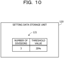
- FIG. 10 is a diagram illustrating an example of data stored in a setting data storage unit.
- setting data referred to in a process of determining a lesion classification model to be used for each division region is stored in the setting data storage unit 120.
- a setting data set 121 including the number of divisions and a threshold value is stored in the setting data storage unit 120.
- the number of divisions indicates the number of divisions of an organ region (for example, the number of divided division regions).
- the threshold value is a value indicating a ratio of a threshold value (the threshold value TH described above) used to determine which lesion classification model is to be used to the total number of pieces of learning data for a lesion region.
- the number of divisions and the threshold value may be freely set by a user.
- FIG. 11 is a diagram illustrating an example of data stored in an aggregation data storage unit.
- An aggregation data table 131 is stored in the aggregation data storage unit 130.
- a record for each division region is registered in the aggregation data storage unit 130.
- Each record includes a division region range, the number of pieces of learning data, and a model flag.
- the division region range is a value indicating a range of a division region in an organ region with a ratio based on an upper end of the organ region.
- the range of the division region is indicated by a ratio in which the upper end of the organ region is set to 0 and a lower end is set to 1.
- a division region range of a first division region is 0 or more and 0.33 or less
- a division region range of a second division region is more than 0.33 and 0.66 or less
- a division region range of a third division region is more than 0.66 and 1.0 or less.
- the number of pieces of learning data indicates the number of pieces of learning data including a tomographic image included in a corresponding division region among pieces of learning data of a lesion region used when the Segmentation model 60 is generated.
- the model flag is a flag indicating which of the Segmentation model 60 and the Classification model 70 is to be used when a lesion is classified for the tomographic image included in the corresponding division region. According to the present embodiment, it is assumed that the flag is set to 0 in a case where the Segmentation model 60 is to be used, and the flag is set to 1 in a case where the Classification model 70 is to be used.
- FIG. 12 is a diagram illustrating an example of data stored in a model data storage unit.
- a model data table 141 is stored in the model data storage unit 140. Model structure data and weight data are registered in the model data table 141 for each of the organ region extraction model, the Segmentation model 60, and the Classification model 70.
- the model structure data indicates a type of a machine learning model to be used and a configuration of a neural network.
- a file name of a file in which such information is described may be registered in the model data table 141 in practice.
- the weight data indicates a weight coefficient set between nodes in the neural network of the machine learning model. Actually, a file name of a file in which such a weight coefficient is described may be registered in the model data table 141.
- model structure data and the weight data for the organ region extraction model are registered in the model data table 141 in advance.
- Each model structure data for the Segmentation model 60 and the Classification model 70 is also registered in the model data table 141 in advance.
- each weight data for the Segmentation model 60 and the Classification model 70 is generated by the model generation unit 153, is registered in the model data table 141.
- FIG. 13 is an example of a flowchart illustrating a procedure of a data aggregation process.
- the data aggregation unit 151 assigns an image number starting from 0 to image data of a tomographic image included in the selected disease case, in order from image data at an upper end of the body. Among these pieces of image data, the data aggregation unit 151 sets an image number of the image data at the upper end of the organ region to I min , and sets an image number of the image data at a lower end of the organ region to I max .
- the image number x is normalized to a value equal to or more than 0 and equal to or less than 1.
- the image number x after calculated by Equation (1) indicates a ratio position of the selected image data in the entire organ region from the head side.
- the number of pieces of learning data for each division region is registered in the aggregation data table 131.
- the model determination process illustrated in FIG. 14 is executed.
- FIG. 14 is an example of a flowchart illustrating a procedure of a model determination process.
- the model determination unit 152 converts this threshold value into an absolute number of pieces of learning data, and compares the converted threshold value with the acquired number of pieces of learning data. Specifically, the model determination unit 152 sums up the numbers of pieces of learning data of the respective division regions registered in the aggregation data table 131, and multiplies the total value by a threshold value indicating a ratio to convert the total value into a threshold value indicating the absolute number of pieces of learning data.
- FIG. 15 is an example of a flowchart illustrating a procedure of a model generation process.
- FIG. 16 is an example of a flowchart illustrating a procedure of a lesion classification process.
- the model selection unit 155 sets the image number of a head (upper end) tomographic image to I min , and sets the image number of a last (lower end) tomographic image to I max .
- the model selection unit 155 acquires a model flag corresponding to the division region to which the selected tomographic image belongs, and determines whether the acquired model flag is 1. In a case where the model flag is 0, the process proceeds to step S46, and in a case where the model flag is 1, the process proceeds to step S47.
- the lesion classification unit 156 generates an classification result image to which information indicating whether or not each pixel of the selected tomographic image is a lesion is assigned. After that, in a case where a display of an classification result of the tomographic image is requested, the lesion classification unit 156 displays a region determined to have a lesion over the tomographic image in a manner distinguished from other regions based on the classification result image.
- the Segmentation model 60 and the Classification model 70 may be lesion classification models for classifying three or more types of lesions including a type indicating that the model is not a lesion.
- steps S46 and S48 for example, an classification result image to which information indicating one of the lesion types is assigned is generated for each pixel of the tomographic image.
- the lesion may be classified with stable accuracy regardless of the position in the organ region in the tomographic image of the classification target.
- Data stored in the setting data storage unit 120, the aggregation data storage unit 130, and the model data storage unit 140 is transferred to the second information processing apparatus via a network or a portable-type recording medium, and these pieces of data are referred to in the process in the inference phase.
- the lesion classification model is selected depending on whether or not the number of pieces of learning data of the lesion region used at the time of training is large for the division region to which the captured tomographic image belongs.
- the classification accuracy of the Segmentation model 60 is verified for each of the division regions.
- the lesion classification model is selected depending on whether or not the classification accuracy of the Segmentation model 60 is high for the division region to which the captured tomographic image belongs.
- FIG. 17 is a diagram illustrating a configuration example of a processing function provided by a diagnosis support apparatus according to the third embodiment.
- the same components as the components illustrated in FIG. 8 are indicated by the same reference signs, and will not be described.
- a diagnosis support apparatus 100a includes a learning data storage unit 110a, a setting data storage unit 120a, and an aggregation data storage unit 130a, instead of the learning data storage unit 110, the setting data storage unit 120, and the aggregation data storage unit 130 illustrated in FIG. 8 .
- the learning data storage unit 110a includes learning data to be used when the Segmentation model 60 is generated and learning data to be used when the Classification model 70 is generated. A part of the former learning data is used for verifying classification accuracy of the Segmentation model 60.
- the setting data storage unit 120a is different from the setting data storage unit 120 in that a value of the classification accuracy is set as a threshold value.
- the number of verifications and the number of correct answers at a time of verifying the classification accuracy of the Segmentation model 60 are registered in the aggregation data storage unit 130a.
- the diagnosis support apparatus 100a includes a data aggregation unit 151a and a model determination unit 152a, instead of the data aggregation unit 151 and the model determination unit 152 illustrated in FIG. 8 .
- the data aggregation unit 151a verifies the classification accuracy of the Segmentation model 60 for each of the division regions by using the learning data for verification registered in the learning data storage unit 110a.
- the model determination unit 152a determines a lesion classification model to be used for each of the division regions based on a result of verification by the data aggregation unit 151a.
- FIG. 18 is a diagram illustrating an example of data stored in an aggregation data storage unit.
- An aggregation data table 132 is stored in the aggregation data storage unit 130a.
- a record for each division region is registered in the aggregation data table 132.
- Each record includes a division region range, the number of verifications, the number of correct answers, and a model flag.
- the division region range and the model flag are the same as the division region range and the model flag in the aggregation data table 131 illustrated in FIG. 11 .
- the number of verifications indicates the number of tomographic images (or the number of pixels) for which classification accuracy is verified by using the Segmentation model 60.
- the number of correct answers indicates the number of tomographic images (or the number of pixels) determined to be correct by the verification.
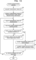
- FIG. 19 is another example of the flowchart illustrating the procedure of the data aggregation process.
- the data aggregation unit 151a executes the processes in steps S11 and S12 in FIG. 13 .
- step S12 a record is created for the aggregation data table 132.
- step S51 the process in and after next step S51 is executed.
- step S51 The data aggregation unit 151a selects one disease case for verification from a learning data table for the Segmentation model 60 stored in a learning data storage unit 110a.
- the data aggregation unit 151 assigns an image number starting from 0 to image data of a tomographic image included in the selected disease case, in order from image data at an upper end of the body. Among these pieces of image data, the data aggregation unit 151 sets an image number of the image data at the upper end of the organ region to I min , and sets an image number of the image data at a lower end of the organ region to I max .
- the data aggregation unit 151a determines in which division region range set in the aggregation data table 132 in step S12 the calculated image number x after is included. Thus, the data aggregation unit 151a determines a division region to which the selected image data belongs. Among the numbers of verifications in the aggregation data table 132, the data aggregation unit 151a counts up the number of verifications corresponding to the division region to which the selected image data belongs by 1.
- step S53 the number of pixels of the image data may be added to the corresponding number of verifications registered in the aggregation data table 132.
- step S55 the number of correct pixels may be added to the corresponding number of correct answers registered in the aggregation data table 132.
- the model determination process illustrated in FIG. 20 is executed.
- FIG. 20 is an example of a flowchart illustrating a procedure of a model determination process.
- step S23a The model determination unit 152a acquires the number of verifications and the number of correct answers from a record corresponding to the selected division region, among records in the aggregation data table 132.
- the model determination unit 152a calculates classification accuracy corresponding to the selected division region by a ratio of the number of correct answers to the number of verifications.
- next step S24 the calculated classification accuracy is compared with a threshold value.
- a lesion may be classified with stable accuracy, regardless of a position in an organ region in a tomographic image of an classification target.
- the processing function of the apparatus (for example, the lesion detection apparatus 1 and the diagnosis support apparatuses 100 and 100a) described in each of the embodiments described above may be implemented by a computer.
- a program describing the processing contents of the functions to be included in each apparatus is provided, and the processing functions described above are implemented over the computer by executing the program with the computer.
- the program describing the processing contents may be recorded in a computer-readable recording medium.
- the computer-readable recording medium include a magnetic storage device, an optical disc, a semiconductor memory, and the like.
- the magnetic storage device include a hard disk drive (HDD), a magnetic tape, and the like.
- the optical disc include a compact disc (CD), a Digital Versatile Disc (DVD), a Blu-ray disc (BD, registered trademark), and the like.
- a portable-type recording medium such as a DVD or a CD on which the program is recorded is sold.
- the program may be stored in a storage device of a server computer and transferred from the server computer to another computer via a network.
- the computer that executes the program stores, in a storage device thereof, the program recorded on the portable-type recording medium or the program transferred from the server computer, for example.
- the computer reads the program from the storage device thereof and executes the processing according to the program.
- the computer may also read the program directly from the portable-type recording medium and execute the processing according to the program.
- the computer may also sequentially execute the processing according to the received program.
Landscapes
- Engineering & Computer Science (AREA)
- Health & Medical Sciences (AREA)
- Medical Informatics (AREA)
- Theoretical Computer Science (AREA)
- General Health & Medical Sciences (AREA)
- Public Health (AREA)
- General Physics & Mathematics (AREA)
- Computer Vision & Pattern Recognition (AREA)
- Physics & Mathematics (AREA)
- Databases & Information Systems (AREA)
- Epidemiology (AREA)
- Primary Health Care (AREA)
- Biomedical Technology (AREA)
- Data Mining & Analysis (AREA)
- Radiology & Medical Imaging (AREA)
- Nuclear Medicine, Radiotherapy & Molecular Imaging (AREA)
- Pathology (AREA)
- Computing Systems (AREA)
- Evolutionary Computation (AREA)
- Software Systems (AREA)
- Artificial Intelligence (AREA)
- Multimedia (AREA)
- Quality & Reliability (AREA)
- Image Analysis (AREA)
- Apparatus For Radiation Diagnosis (AREA)
- Image Processing (AREA)
- Magnetic Resonance Imaging Apparatus (AREA)
Applications Claiming Priority (1)
| Application Number | Priority Date | Filing Date | Title |
|---|---|---|---|
| JP2023110444A JP2025008346A (ja) | 2023-07-05 | 2023-07-05 | 病変検出方法および病変検出プログラム |
Publications (2)
| Publication Number | Publication Date |
|---|---|
| EP4513433A2 true EP4513433A2 (de) | 2025-02-26 |
| EP4513433A3 EP4513433A3 (de) | 2025-04-23 |
Family
ID=91580699
Family Applications (1)
| Application Number | Title | Priority Date | Filing Date |
|---|---|---|---|
| EP24182201.4A Pending EP4513433A3 (de) | 2023-07-05 | 2024-06-14 | Läsionsdetektionsverfahren und läsionsdetektionsprogramm |
Country Status (3)
| Country | Link |
|---|---|
| US (1) | US20250014176A1 (de) |
| EP (1) | EP4513433A3 (de) |
| JP (1) | JP2025008346A (de) |
Citations (1)
| Publication number | Priority date | Publication date | Assignee | Title |
|---|---|---|---|---|
| US20190223725A1 (en) | 2018-01-25 | 2019-07-25 | Siemens Healthcare Gmbh | Machine Learning-based Segmentation for Cardiac Medical Imaging |
Family Cites Families (3)
| Publication number | Priority date | Publication date | Assignee | Title |
|---|---|---|---|---|
| JP7515502B2 (ja) * | 2019-03-15 | 2024-07-12 | ジェネンテック, インコーポレイテッド | 陽電子放射断層撮影を用いた腫瘍セグメンテーションのための深層畳み込みニューラルネットワーク |
| JP7715978B2 (ja) * | 2021-03-31 | 2025-07-31 | 富士通株式会社 | 画像識別プログラム、画像識別方法、画像識別装置および情報処理システム |
| JP7703408B2 (ja) * | 2021-09-15 | 2025-07-07 | 富士フイルム株式会社 | 画像処理装置、学習装置、画像処理方法、学習方法、画像処理プログラム、及び学習プログラム |
-
2023
- 2023-07-05 JP JP2023110444A patent/JP2025008346A/ja active Pending
-
2024
- 2024-06-14 EP EP24182201.4A patent/EP4513433A3/de active Pending
- 2024-06-18 US US18/746,301 patent/US20250014176A1/en active Pending
Patent Citations (1)
| Publication number | Priority date | Publication date | Assignee | Title |
|---|---|---|---|---|
| US20190223725A1 (en) | 2018-01-25 | 2019-07-25 | Siemens Healthcare Gmbh | Machine Learning-based Segmentation for Cardiac Medical Imaging |
Also Published As
| Publication number | Publication date |
|---|---|
| US20250014176A1 (en) | 2025-01-09 |
| JP2025008346A (ja) | 2025-01-20 |
| EP4513433A3 (de) | 2025-04-23 |
Similar Documents
| Publication | Publication Date | Title |
|---|---|---|
| EP3637320B1 (de) | Computerprogramm, lernvorrichtung, nachweisvorrichtung, lernverfahren und nachweisverfahren | |
| KR102043130B1 (ko) | 컴퓨터 보조 진단 방법 및 장치 | |
| US10452899B2 (en) | Unsupervised deep representation learning for fine-grained body part recognition | |
| JP5868231B2 (ja) | 医用画像診断支援装置、医用画像診断支援方法ならびにコンピュータプログラム | |
| Hogeweg et al. | Automatic detection of tuberculosis in chest radiographs using a combination of textural, focal, and shape abnormality analysis | |
| US10734107B2 (en) | Image search device, image search method, and image search program | |
| CN106340021B (zh) | 血管提取方法 | |
| US20130114867A1 (en) | Similar case searching apparatus and similar case searching method | |
| US9619888B2 (en) | Method for defining a region of interest in a radiation image of a breast | |
| JP7658429B2 (ja) | 情報処理装置、情報処理方法、医療映像識別装置及びプログラム | |
| US12300386B2 (en) | Information processing apparatus, training data generation device, and diagnosis support system | |
| Jansen et al. | Patient-specific fine-tuning of convolutional neural networks for follow-up lesion quantification | |
| JP6041781B2 (ja) | 医用画像処理装置およびその作動方法並びに医用画像処理プログラム | |
| US20230334868A1 (en) | Surgical phase recognition with sufficient statistical model | |
| US20250336070A1 (en) | Image identification method and non-transitory computer-readable storage medium storing computer program | |
| EP4513433A2 (de) | Läsionsdetektionsverfahren und läsionsdetektionsprogramm | |
| US20250086789A1 (en) | Lesion detection method and non-transitory computer-readable recording medium storing lesion detection program | |
| JP7807656B2 (ja) | 画像識別プログラム、画像識別方法、画像識別装置および情報処理システム | |
| US11923072B2 (en) | Image diagnosis supporting device and image processing method | |
| CN116091522A (zh) | 医学图像分割方法、装置、设备及可读存储介质 | |
| CN116934657A (zh) | 医用图像处理装置和医用图像处理方法 | |
| JP7598018B2 (ja) | 画像識別プログラム、画像識別方法、画像識別装置および情報処理システム | |
| JP2021087729A (ja) | 医用画像処理装置及びプログラム | |
| EP4379657A1 (de) | Verfahren zur erkennung von läsionen und programm zur erkennung von läsionen | |
| JP2023173115A (ja) | 画像認識プログラム、画像認識方法および画像認識装置 |
Legal Events
| Date | Code | Title | Description |
|---|---|---|---|
| PUAI | Public reference made under article 153(3) epc to a published international application that has entered the european phase |
Free format text: ORIGINAL CODE: 0009012 |
|
| STAA | Information on the status of an ep patent application or granted ep patent |
Free format text: STATUS: THE APPLICATION HAS BEEN PUBLISHED |
|
| AK | Designated contracting states |
Kind code of ref document: A2 Designated state(s): AL AT BE BG CH CY CZ DE DK EE ES FI FR GB GR HR HU IE IS IT LI LT LU LV MC ME MK MT NL NO PL PT RO RS SE SI SK SM TR |
|
| PUAL | Search report despatched |
Free format text: ORIGINAL CODE: 0009013 |
|
| AK | Designated contracting states |
Kind code of ref document: A3 Designated state(s): AL AT BE BG CH CY CZ DE DK EE ES FI FR GB GR HR HU IE IS IT LI LT LU LV MC ME MK MT NL NO PL PT RO RS SE SI SK SM TR |
|
| RIC1 | Information provided on ipc code assigned before grant |
Ipc: G06T 7/11 20170101AFI20250317BHEP |
|
| STAA | Information on the status of an ep patent application or granted ep patent |
Free format text: STATUS: REQUEST FOR EXAMINATION WAS MADE |
|
| 17P | Request for examination filed |
Effective date: 20250821 |