EP4512910A2 - Verfahren zum nachweis von bindehauterkrankungen unter verwendung von augenoberflächengewebe und alterungsbiomarker - Google Patents
Verfahren zum nachweis von bindehauterkrankungen unter verwendung von augenoberflächengewebe und alterungsbiomarker Download PDFInfo
- Publication number
- EP4512910A2 EP4512910A2 EP24213334.6A EP24213334A EP4512910A2 EP 4512910 A2 EP4512910 A2 EP 4512910A2 EP 24213334 A EP24213334 A EP 24213334A EP 4512910 A2 EP4512910 A2 EP 4512910A2
- Authority
- EP
- European Patent Office
- Prior art keywords
- genus
- conjunctival
- bacteria
- aging
- ocular surface
- Prior art date
- Legal status (The legal status is an assumption and is not a legal conclusion. Google has not performed a legal analysis and makes no representation as to the accuracy of the status listed.)
- Pending
Links
Images





Classifications
-
- C—CHEMISTRY; METALLURGY
- C12—BIOCHEMISTRY; BEER; SPIRITS; WINE; VINEGAR; MICROBIOLOGY; ENZYMOLOGY; MUTATION OR GENETIC ENGINEERING
- C12Q—MEASURING OR TESTING PROCESSES INVOLVING ENZYMES, NUCLEIC ACIDS OR MICROORGANISMS; COMPOSITIONS OR TEST PAPERS THEREFOR; PROCESSES OF PREPARING SUCH COMPOSITIONS; CONDITION-RESPONSIVE CONTROL IN MICROBIOLOGICAL OR ENZYMOLOGICAL PROCESSES
- C12Q1/00—Measuring or testing processes involving enzymes, nucleic acids or microorganisms; Compositions therefor; Processes of preparing such compositions
- C12Q1/68—Measuring or testing processes involving enzymes, nucleic acids or microorganisms; Compositions therefor; Processes of preparing such compositions involving nucleic acids
- C12Q1/6876—Nucleic acid products used in the analysis of nucleic acids, e.g. primers or probes
- C12Q1/6888—Nucleic acid products used in the analysis of nucleic acids, e.g. primers or probes for detection or identification of organisms
- C12Q1/689—Nucleic acid products used in the analysis of nucleic acids, e.g. primers or probes for detection or identification of organisms for bacteria
-
- C—CHEMISTRY; METALLURGY
- C12—BIOCHEMISTRY; BEER; SPIRITS; WINE; VINEGAR; MICROBIOLOGY; ENZYMOLOGY; MUTATION OR GENETIC ENGINEERING
- C12Q—MEASURING OR TESTING PROCESSES INVOLVING ENZYMES, NUCLEIC ACIDS OR MICROORGANISMS; COMPOSITIONS OR TEST PAPERS THEREFOR; PROCESSES OF PREPARING SUCH COMPOSITIONS; CONDITION-RESPONSIVE CONTROL IN MICROBIOLOGICAL OR ENZYMOLOGICAL PROCESSES
- C12Q1/00—Measuring or testing processes involving enzymes, nucleic acids or microorganisms; Compositions therefor; Processes of preparing such compositions
- C12Q1/02—Measuring or testing processes involving enzymes, nucleic acids or microorganisms; Compositions therefor; Processes of preparing such compositions involving viable microorganisms
- C12Q1/04—Determining presence or kind of microorganism; Use of selective media for testing antibiotics or bacteriocides; Compositions containing a chemical indicator therefor
- C12Q1/06—Quantitative determination
-
- C—CHEMISTRY; METALLURGY
- C12—BIOCHEMISTRY; BEER; SPIRITS; WINE; VINEGAR; MICROBIOLOGY; ENZYMOLOGY; MUTATION OR GENETIC ENGINEERING
- C12Q—MEASURING OR TESTING PROCESSES INVOLVING ENZYMES, NUCLEIC ACIDS OR MICROORGANISMS; COMPOSITIONS OR TEST PAPERS THEREFOR; PROCESSES OF PREPARING SUCH COMPOSITIONS; CONDITION-RESPONSIVE CONTROL IN MICROBIOLOGICAL OR ENZYMOLOGICAL PROCESSES
- C12Q1/00—Measuring or testing processes involving enzymes, nucleic acids or microorganisms; Compositions therefor; Processes of preparing such compositions
- C12Q1/68—Measuring or testing processes involving enzymes, nucleic acids or microorganisms; Compositions therefor; Processes of preparing such compositions involving nucleic acids
- C12Q1/6876—Nucleic acid products used in the analysis of nucleic acids, e.g. primers or probes
-
- C—CHEMISTRY; METALLURGY
- C12—BIOCHEMISTRY; BEER; SPIRITS; WINE; VINEGAR; MICROBIOLOGY; ENZYMOLOGY; MUTATION OR GENETIC ENGINEERING
- C12Q—MEASURING OR TESTING PROCESSES INVOLVING ENZYMES, NUCLEIC ACIDS OR MICROORGANISMS; COMPOSITIONS OR TEST PAPERS THEREFOR; PROCESSES OF PREPARING SUCH COMPOSITIONS; CONDITION-RESPONSIVE CONTROL IN MICROBIOLOGICAL OR ENZYMOLOGICAL PROCESSES
- C12Q1/00—Measuring or testing processes involving enzymes, nucleic acids or microorganisms; Compositions therefor; Processes of preparing such compositions
- C12Q1/68—Measuring or testing processes involving enzymes, nucleic acids or microorganisms; Compositions therefor; Processes of preparing such compositions involving nucleic acids
- C12Q1/6876—Nucleic acid products used in the analysis of nucleic acids, e.g. primers or probes
- C12Q1/6883—Nucleic acid products used in the analysis of nucleic acids, e.g. primers or probes for diseases caused by alterations of genetic material
Definitions
- the present invention relates to a method of detecting a conjunctival disease using an ocular surface tissue, and an aging biomarker.
- biomarkers Factors which quantitatively vary in correlation to states of living bodies or specific diseases are referred to as biomarkers, and serve as indicators for quantitatively recognizing biological changes in living bodies such as aging and diseases.
- Biomarkers which quantitatively vary in correlation to age and allow to predict aging are referred to as aging biomarkers.
- Biomarkers which quantitatively vary in correlation to specific diseases and can contribute to diagnosis of the diseases and to establishment of effective therapies are referred to as disease biomarkers.
- measures which allow to grasp health states of elderly people and to extend health life expectancy are being taken in an active manner.
- transcriptome/meta-transcriptome analysis which is an exhaustive gene expression analysis
- metabolome analysis which is an exhaustive metabolite analysis
- metagenomic analysis which is an exhaustive genome sequence analysis using next-generation sequencer, and the like
- attempts are being made to utilize the technology in exhaustive search for new biomarkers.
- health states and aging states of subjects can be inferred from abundance ratios of indigenous bacteria of skin surface, especially Propionibacterium acnes (occasionally abbreviated as "P. acnes" in the followings), and the like by using an exhaustive genome analysis using a next-generation sequencer (refer to Patent Literature 1).
- P. acnes Propionibacterium acnes
- acnes on the skin surface depends on physical conditions such as degree of skin oiliness, acne, thinning hair, hair loss, and menopause, other than age. Therefore, there is still a demand for search for aging biomarkers with which health states and aging states can be evaluated in an objective and highly reproducible manner.
- Disease biomarkers can contribute to diagnosis of specific diseases and to establishment of effective therapies. For example, there is a demand for search for disease biomarkers which allow to clarify a pathogenic mechanism and to predict an onset in diseases in which incidence increases in association with aging, such as conjunctival mucosa-associated lymphoid tissue (abbreviated as "MALT" in the followings) lymphoma, age-related macular degeneration, cataract, and glaucoma in an ophthalmic field.
- MALT conjunctival mucosa-associated lymphoid tissue
- conjunctival MALT lymphoma is known as localized tumor of low malignant potential, and extranodal marginal zone B-cell lymphoma of MALT type is the most typical histological subtype. It has been estimated that lymphoma in the ocular accessory gland has a frequency of approximately 8% of extranodal lymphoma. It has been reported that a prognosis of primary conjunctival lymphoma is favorable with long-term survival expected among patients with primary lymphoma. Histologically, conjunctival MALT lymphoma has characteristics similar to gastric MALT lymphoma and is thought to result from a chronic inflammatory response.
- dysbiosis which is an imbalance in a microbiota induces systemic diseases such as inflammatory bowel disease, obesity, and cardiovascular disease.
- systemic diseases such as inflammatory bowel disease, obesity, and cardiovascular disease.
- a relationship between the immune system and the indigenous microbiota of living organisms in biological defense and tissue repair has also been reported.
- Pathological changes in microbiotas of Staphylococcus epidermidis which is a skin indigenous bacterium, and the like, have been shown to cause opportunistic infections such as onset of catheter infection, endocarditis of artificial valves and endophthalmitis.
- dysbiosis and central nervous system disorders such as autism, multiple sclerosis, anxiety-depressive behavior, and functional gastrointestinal disorders has been reported at a clinical level, and a treatment in which the microbiota of these symptoms is targeted has also been suggested.
- the change in the microbiota causes various symptoms such as infection of pathogens or inflammation to living organisms, and may cause living organisms to fall into a fatal state.
- CALT Conjunctiva-associated lymphoid tissue
- Patent Literature 1 Japanese Unexamined Patent Application Publication No. 2017-29133
- the present invention aims to construct a technology in which the onset or progression of an aging state or a specific disease can be evaluated in an objective and highly reproducible manner, and in particular, to provide a method of detecting conjunctival diseases such as conjunctival MALT lymphoma, and to provide an aging biomarker that serves as an indicator of the aging state.
- the present inventors have conducted further studies to solve the above problem, and as a result, a finding has been obtained that dysbiosis of a conjunctival microbiota may cause immunological changes in the conjunctival mucosa and may be involved in the onset of conjunctival MALT lymphoma.
- an aging biomarker in which not only a physiological aged state (pre-symptomatic state) based on calendar age but also a pathological aging state that causes a pathological state due to abnormally accelerated aging or the like can be detected. Therefore, the biomarker of this configuration can be used not only for detecting the aging state but also for detecting the disease state that develops in association with aging, and can be used for elucidating the cause of a systemic disease resulting from an aging-related change.
- the method of detecting a conjunctival disease according to the present embodiment provides information on a pathological state of a conjunctival disease based on a change in a balance of a microbiota existing in an ocular surface tissue. That is, the method of detecting a conjunctival disease according to the present embodiment is based on a finding that a change in a microbial community structure of a microbiota existing in the ocular surface tissue is correlated with a pathology of a conjunctival disease.
- the conjunctival disease to be detected by the method of detecting a conjunctival disease generally means diseases that cause conjunctival abnormalities.
- diseases that cause abnormalities in the conjunctiva as a result of aging-related changes and in which incidence increases with aging are included.
- conjunctival MALT lymphoma, pterygium, dry eye, conjunctival relaxation, glaucoma, age-related macular degeneration (AMD), and the like are exemplified.
- the onset, progression degree and the like of a conjunctival disease are detected based on a change in a microbial community structure of a microbiota contained in an ocular surface tissue specimen derived from a subject. That is, it is possible to detect the ocular surface specimen sampled from the subject who has been evaluated as having the conjunctival disease based on the change in the microbial community structure of the microbiota, which allows to detect whether the subject has developed the conjunctival disease and whether there is a risk of developing the conjunctival disease in the future.
- Ocular surface tissues include a tissue on a surface of an eyeball, an appendage which is a structure close to a surface of the eyeball, and the like.
- the eyeball is protected by an eyelid, eyelashes at upper and lower edges of the eyelid and the like, and an outer membrane thereof is constituted by a cornea and a sclera.
- the sclera from the inside of the eyelid to the front of the eyeball is covered with a conjunctiva, and a Tenon's capsule is located between the conjunctiva and the sclera.
- the conjunctiva includes an eyelid conjunctiva that covers the inside of the eyelid, an eyeball conjunctiva that covers a part of the front surface of the eyeball, a fornix conjunctiva that is a transition part between the two, and the like.
- a meibomian gland opens at a margin of the eyelid
- a lacrimal gland opens on an ear side of a superior conjunctival fornix
- an accessory lacrimal gland opens at the superior and inferior conjunctival fornix.
- Examples of the ocular surface tissue include those described above, but are not limited thereto.
- the ocular surface tissue includes secretions and the like from the ocular surface tissue described above, and includes, for example, a tear film or eye oil composed of an oil layer and a liquid layer (a mucin layer and an aqueous layer), leakage/exudate from blood vessels and the like, cells/tissue fragments such as detached epithelial cells and others.
- a tear film or eye oil composed of an oil layer and a liquid layer (a mucin layer and an aqueous layer), leakage/exudate from blood vessels and the like, cells/tissue fragments such as detached epithelial cells and others.
- Collection of ocular surface tissue specimens can be performed using a technique known in the art as long as bacteria existing in the ocular surface tissue can be sampled. For example, rubbing an ocular surface tissue in an aseptic condition with a swab (cotton swab) or a spatula (ocular surface tissue scraping specimen), and collecting secretions such as leakage and exudate from the ocular surface tissue (ocular surface tissue secretion specimen) enable to perform the collection.
- a local anesthesia or the like may be given as needed.
- the obtained ocular surface tissue specimen can be dissolved or suspended in an appropriate liquid as needed.
- the amount of the ocular surface tissue specimen to be sampled is not particularly limited, but may be, for example, an amount obtained by rubbing with 1 swab.
- the ocular surface tissue specimen can be sampled by simple and non-invasive methods such as rubbing ocular surface tissues.
- a change in the microbiota existing in the ocular surface tissue can be determined using a known technique in the art.
- microbiota analysis methods targeting a 16S rRNA gene, and the like can be used.
- Microbiota analysis methods include metagenome analysis method, Terminal Restriction Fragment Length Polymorphism (T-RFLP), Denaturing Gradient Gel Electrophoresis (DGGE), Temperature Gradient Gel Electrophoresis (TGGE) and fluorescence in situ hybridization (FISH) -flow cytometry (FISH-FCM), but are not limited thereto.
- T-RFLP Terminal Restriction Fragment Length Polymorphism
- DGGE Denaturing Gradient Gel Electrophoresis
- TGGE Temperature Gradient Gel Electrophoresis
- FISH fluorescence in situ hybridization
- FISH-FCM fluorescence in situ hybridization
- a base sequence of 16S rRNA gene of for example, microbes (in particular, bacteria) contained in an ocular surface tissue specimen of a subject is analyzed, and it is possible to detect an ocular surface specimen sampled from the subject who has been evaluated as having the conjunctival disease based on the obtained base sequence data.
- the next-generation sequencer it becomes possible to identify the microbial species, its abundance ratio and the like while the microbiota which has been difficult to identify by conventional culture methods is made a single group.
- genomic DNA of a microbe contained in a specimen of ocular surface tissue of a subject is extracted.
- the method for extracting bacterial genomic DNA is not particularly limited, and it can be performed by using a known technique in the art. For example, heat extraction method, alkali heat extraction method, phenol/chloroform extraction method, or the like can be used.
- commercially available extraction kits such as PowerSoil (registered trademark) DNA Isolation Kit (MoBio, Carlsbad, CA) can be used.
- the base sequence of the 16S rRNA gene contained in the extracted genomic DNA is determined.
- the 16S rRNA gene may be sequenced in the entire region, but it is preferable to sequence a specific region as long as the sequence characteristics among each microbial species are reflected.
- Nine hypervariable regions called V1-V9 are adjacent to the 16S rRNA gene in a form locating predominantly in a region that is highly conserved across microbial species.
- identification of microbial species can be performed. Therefore, it is preferable to determine a base sequence of any of the hypervariable regions or of a region including a plurality of the regions. For example, a region including V1-V2, a region including V3-V4, and the like can be given; however, it is not limited to such a region.
- the region for determining the base sequence may be amplified, if necessary, and the obtained nucleic acid amplified fragment (amplicon) can be used as a base sequence determination target.
- Amplification of nucleic acids can be performed using techniques known in the art. For example, polymerase chain reaction (PCR) and the like can be used.
- Primers used in the nucleic acid amplification reaction can be designed based on a known technique in the art.
- the primer is preferably designed so as to include a region which is relatively universally conserved in the bacterial 16S rRNA genes of the ocular surface tissue.
- a universal primer for the 16S rRNA gene can be used.
- the primer may be designed so that a sequence required for base sequence determination is added to the amplified nucleic acid fragment. For example, a barcode sequence used for identification in samples, and the like are included.
- the amplified nucleic acid fragment may be previously purified by a technique known in the art.
- the base sequence can be determined using any of techniques known in the art. For example, it may be performed by a sequencer or the like based on the conventional Sanger method or the like, but from a viewpoint of ability to analyze the base sequence and the like, it is preferable to perform by a next-generation type sequencer or the like based on the Sequencing by Synthesis method, pyrosequencing method, ligase reaction sequencing method or the like.
- the next-generation sequencer for example, MiSeq (Illumina) or the like can be used, and sequence determination can be performed according to the manufacturer's protocol.
- the obtained base sequence data (read) may contain low-quality reads due to incomplete sequencing reaction and the like, removing low-quality reads by read trimming may be performed if necessary. Trimming can be performed using, for example, BBtrim or the like.
- Analyzing the microbiota is performed based on the determined base sequence.
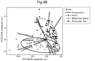
- Analysis of the microbiota can be performed using any of techniques known in the art, and can be analyzed by visualizing or digitizing the microbial community structure of the microbiota, and for example, principal component analysis (PCA), principal coordinate analysis (PCoA) or the like can be used.
- Analysis of the microbiota can be performed using analysis software, a database, or the like known in the art.
- analysis software for example, QIIME or the like can be used.
- As a database for example, Greengenes, SILVA, NCBI, or the like can be used, and homology analysis, phylogenetic analysis, or the like is performed on the database.
- the obtained base sequence data are classified into a plurality of clusters based on sequence similarity by OTU (Operational Taxonomic Unit) analysis, and the base sequence with the highest occurrence frequency in each OUT is set as a representative sequence.
- OTU Operaational Taxonomic Unit
- An analysis may be performed using such a representative sequence.
- the similarity of the base sequences for classification into the same cluster can be appropriately set based on the required reliability and the like. For example, 95% or more, 97% or more, and 99% or more can be set.
- UCLUST, UPARSE, USEARCH, or the like can be used as a software for OTU analysis.
- a conjunctival disease is detected by comparing the microbial community structure of the microbiota from the ocular surface tissue of the subject with that of healthy persons.
- the microbes include bacteria or funguses, and bacteria are particularly preferable.
- bacteria constituting the microbiota derived from ocular surface tissues the abundance or abundance ratio of bacteria belonging to a specific phylum, class, order, family, genus, or species can be used as an indicator.
- the abundance or the abundance ratio of the above of the subject is significantly increased or decreased as compared to that of healthy persons, it can be determined that there is a risk that the subject is suffering from a conjunctival disease or develops a conjunctival disease in the future.
- the abundance ratio can be a ratio, a difference, a sum, or a product of the abundance ratios of a plurality of bacteria belonging to a specific phylum, class, order, family, genus, or species.
- a determination as to whether the abundance or abundance ratio of the above of the subject is significantly increased or decreased as compared to that of healthy persons can be performed by predetermining a reference value of the abundance or abundance ratio of bacteria belonging to a specific phylum, class, order, family, genus or species in the microbiota derived from the ocular surface tissue and by comparing it with the reference value.
- a determination of suffering from a conjunctival disease may be a determination at any level of bacterial phylum, class, order, family, genus, and species. Also, it may be a determination based on a level of 1 phylum, class, order, family, genus, or species, or may be a determination based on a level of a plurality of phyla, classes, orders, families, genera, or species.
- a determination of conjunctival MALT lymphoma based on changes in the microbial community structure of, for example, an ocular surface tissue, particularly, a conjunctiva can be performed on healthy persons using an increase in the abundance or abundance ratio of bacteria belonging to the following genus as an indicator. If the abundance or abundance ratio of the following bacteria increases more than that in healthy persons, it can be determined that the subject has developed conjunctival MALT lymphoma, or has a risk of developing conjunctival MALT lymphoma in the future.
- Delftia genus bacteria Proteobacteria phylum, Betaproteobacteria class, Burkholderiales order, Comamonadaceae family, Delftia genus
- Xylophilus genus bacteria Proteobacteria phylum, Gamma Proteobacteria class, Xanthomonadales order, Xanthomonadaceae family, Xylophilus genus
- Simplicispira genus bacteria Proteobacteria phylum, Betaproteobacteria class, Burkholderiales order, Comamomonadaceae family, Simplicispira genus
- Rothia genus bacteria Actinobacteria phylum, Actinobacteria class, Micrococcales order, Micrococcaceae family, Rothia genus
- Xanthomonas genus bacteria Proteobacteria phylum, Gammaproteobacteria class, X
- conjunctival MALT lymphoma can be determined using an increase in the abundance or abundance ratio of Delftia genus bacteria as an indicator.
- Delftia genus bacteria are aerobic Gram-negative rods. Delftia genus bacteria exhibit resistance to ⁇ -lactam and aminoglycoside antibiotics. It is known that they adhere to contact lens cases and form biofilms, causing microbial and infiltrative keratitis to develop on the spherical surface of the eyeball.
- Delftia genus bacteria have an ability to oxidatively degrade and utilize glucose.
- conjunctival MALT lymphoma based on changes in the microbial community structure of ocular surface tissues, particularly the conjunctiva can be performed on healthy persons using a decrease in the abundance or abundance ratio of bacteria belonging to the following genera as an indicator. If the abundance or abundance ratio of the following bacteria decreases more than that of healthy persons, it can be determined that the subject has developed conjunctival MALT lymphoma, or has a risk of developing conjunctival MALT lymphoma in the future.
- Bacteroides genus bacteria Bacteroides genus bacteria (Bacteroidetes phylum, Bacteroidia class, Bacteroidales order, Bacteroidaceae family, Bacteroides genus); Clostridium genus bacteria (Firmicutes phylum, Clostridia class, Clostridiales order, Clostridiaceae family, Clostridium genus); Deinococcus genus bacteria (Deinococcus-Thermus phylum, Deinococci class, Deinococcales order, Deinococcaceae family, Deinococcus genus); Williamsia genus bacteria (Actinobacteria phylum, Actinobacteria class, Corynebacteriales order, Williamsisaceae family, Williamsia genus); Parabacteroides genus bacteria (Bacteroidetes phylum, Bacteroidia class, Bacteroidales order, Tannerellaceae family, Parabacteroides
- conjunctival MALT lymphoma can be determined based on a decrease in the abundance or abundance ratio of Bacteroides genus bacteria or Clostridium genus bacteria as an indicator.
- Bacteroides genus bacteria are known to produce bacterial polysaccharides and supervise maturation of immune system cells and the body during development. In the small intestine, Bacteroides interacts with dendritic cells in Peyer's patches of the intestine and induces a production and maturation of immunoglobulin A as a defense mechanism known as gut-associated lymphoid tissue (GALT).
- GALT gut-associated lymphoid tissue
- Bacteroides genus bacteria from birth is essential for maintaining intestinal immune system homeostasis and may also be useful as a local defense mechanism of ocular surface tissues.
- mucosa-associated Clostridium genus bacterial community plays an important role in inducing Tregs and IgA, and in suppressing inflammatory and allergic reactions.
- beneficial microbial group such as Bacteroides genus bacteria and Clostridium genus bacteria controls suppression of inflammatory response and allergic reaction as protective factors.
- a determination of MALT lymphoma based on changes in the microbial community structure of ocular surface tissues, particularly meibomian gland can be performed on healthy persons using an increase in the abundance or abundance ratio of bacteria belonging to the following genera as an indicator. If the abundance or abundance ratio of the following bacteria increases more than that in healthy persons, it can be determined that the subject has developed conjunctival MALT lymphoma, or has a risk of developing conjunctival MALT lymphoma in the future.
- Delftia genus bacteria Proteobacteria phylum, Betaproteobacteria class, Burkholderiales order, Comamonadaceae family, Delftia genus
- Clostridium genus bacteria Fermicutes phylum, Clostridia class, Clostridiales order, Clostridiaceae family, Clostridium genus
- Brevundimonas genus bacteria Proteobacteria phylum, Alphaproteobacteria class, Caulobacterales order, Caulobacteraceae family, Brevundimonas genus
- a determination of MALT lymphoma based on changes in the microbial community structure of the ocular surface tissues, particularly the meibomian gland can be performed in healthy persons using a decrease in the abundance or abundance ratio of bacteria belonging to the following genera as an indicator. If the abundance or abundance ratio of the following bacteria decreases more than that of healthy persons, it can be determined that the subject has developed conjunctival MALT lymphoma, or has a risk of developing conjunctival MALT lymphoma in the future.
- Lactobacillus genus bacteria (Firmicutes phylum, Bacilli class, Lactobacillales order, Lactobacillaceae family, Lactobacillus genus); Schlegelella genus bacteria (Proteobacteria phylum, Betaproteobacteria class, Burkholderiales order, Comamonadaceae family, Schlegelella genus)
- a determination of MALT lymphoma based on changes in the microbial community structure of the ocular surface tissues, particularly the periocular skin, can be performed in healthy persons using an increase in the abundance or abundance ratio of bacteria belonging to the following genus as an indicator. If the abundance or abundance ratio of the following bacteria increases more than that in healthy subjects, it can be determined that the subject has developed conjunctival MALT lymphoma, or has a risk of developing conjunctival MALT lymphoma in the future.
- Delftia genus bacteria Proteobacteria phylum, Betaproteobacteria class, Burkholderiales order, Comamonadaceae family, Delftia genus
- a determination of MALT lymphoma based on changes in the microbial community structure of the ocular surface tissues, particularly the periocular skin can be performed in healthy persons using a decrease in the abundance or abundance ratio of bacteria belonging to the following genus as an indicator. If the abundance or abundance ratio of the following bacteria decreases more than that in healthy subjects, it can be determined that the subject has developed conjunctival MALT lymphoma, or has a risk of developing conjunctival MALT lymphoma in the future.
- Exiguobacterium genus bacteria (Firmicutes phylum, Bacilli class, Bacillales order, Bacillaceae family, Exiguobacterium genus)
- a similarity distance among specific groups can be used as an indicator, when it is determined the similarity distance between the microbiota of the subject and that of a healthy person is equal to or more than a certain distance, it can be determined that the subject has developed a conjunctival disease, or has a risk of developing a conjunctival disease in the future.
- UniFrac distance analysis or the like can be used.
- the method of such analysis is to perform a phylogenetic tree analysis using the OTU representative sequence of the microbiota derived from the ocular surface tissue of the subject to be compared and that of a healthy person, and to calculate a difference in the microbial community structures as a distance (UniFrac distance) from the length of branches of OTU shared between both microbiotas and from the ratio of branches specific to each microbiota, and a similarity between the microbiotas can be presented 0 (100% similar) to 1 (100% dissimilar) of distance (UniFrac distance). Further, a two-dimensional scatter diagram may be created by principal coordinate analysis based on UniFrac distance.
- a determination whether the similarity distance between the microbiota of the subject and that of healthy persons is equal to or greater than a certain distance can be determined by, for example, comparison with reference data, based on which the group of developing conjunctival disease or the group of healthy persons is close to the similarity distance of the subject.
- the distance of the group of developing conjunctival disease is close to that of the subject, it can be determined that the subject has developed a conjunctival disease or has a risk of developing the disease in the future.
- the above two-dimensional scatter diagram may be used, and when the microbiota of the subject on the two-dimensional scatter diagram is located relatively closer to the group of developing a conjunctival disease compared to the group of healthy persons, it can be determined that the subject has developed a conjunctival disease or has a risk of developing a conjunctival disease in the future.
- a microbial species that can be an indicator of a change in the microbial community structure of the ocular surface tissue can be used as a disease biomarker for detecting a conjunctival disease, and such a disease biomarker also serves as a part of the present invention. It can be also used as a conjunctival disease detection kit for detecting a conjunctival disease by detecting a change in the microbial community structure, and such a disease detection kit also serves as a part of the present invention. For example, it can be configured to include an oligonucleotide probe and a primer for a microbe-specific gene, or a specific antibody, and the like.
- the aging biomarker according to the present embodiment provides information on an aging state of a living organism by a change in a balance of the microbiota existing in the ocular surface tissue. That is, the aging biomarker according to the present embodiment is based on the finding that a change in the microbial community structure of the microbiota existing in the ocular surface tissue correlates with aging.
- the aging biomarker according to the present embodiment means a factor that fluctuates depending on aging, that is, lapse of survival time, and can predict an aging state such as the degree of aging.
- Aging is a change in biological performance that occurs after a stage of maturity, means a decrease in functions that appear at all levels of individuals, organs, tissues, and cells, and includes not only aging based on calendar age but also aging based on biological age. Also, it does not matter whether aging is physiological aging or pathological aging. Therefore, the aging biomarker according to the present embodiment serves as an indicator of not only a physiological aging state but also a pathological aging state.
- physiological aging means a physiological function decline inevitably progressing with aging
- pathological aging means a function decline that causes a pathological condition due to abnormally accelerated aging or the like.
- the aging biomarker according to the present embodiment serves as an indicator of a pathological aging state, it can be used as an indicator of the onset and progression of a disease in which an abnormality occurs in a body as a result of aging-related changes.
- diseases include diseases in which incidence increases with age, and include conjunctival MALT lymphoma, pterygium, dry eye, conjunctival relaxation, glaucoma, age-related macular degeneration (AMD), and the like.
- the aging biomarker according to the present embodiment uses a change in the microbial community structure of the microbiota in the ocular surface tissue specimen derived from the subject, as an indicator.
- the details of the ocular surface tissue specimen and of the method of sampling it are as described in the above section (Method of Detecting Conjunctival Disease).
- the detection of the change in the microbiota in the ocular surface tissue specimen using the biomarker according to the present embodiment can be performed using a known technique in the art, and the details thereof are as described in the above section (Method of Detecting Conjunctival Disease).
- the aging biomarker uses a change in the microbial community structure of the microbiota derived from the ocular surface tissue of the subject, as an indicator.
- bacteria constituting the microbiota derived from the ocular surface tissue bacteria belonging to a specific phylum, class, order, family, genus, or species whose abundance or abundance ratio varies depending on aging can be used as the aging biomarker.
- the abundance ratio can be a ratio, a difference, a sum, or a product of the abundance ratios of a plurality of bacteria belonging to a specific phylum, class, order, family, genus, or species.
- the aging biomarker can be bacteria at any level of the phylum, class, order, family, genus, and species. Also, it may be bacteria at the level of 1 phylum, class, order, family, genus, or species, or bacteria at the level of a plurality of phyla, classes, orders, families, genera, or species.
- bacteria belonging to the following families can be used as the aging biomarker, all of which are indigenous bacteria of ocular surface tissues.
- Corynebacteriaceae family bacteria Actinobacteria phylum, Actinobacteria class, Corynebacteriales order, Corynebacteriaceae family
- Propionibacteriales family bacteria Actinobacteria phylum, Actinobacteria class, Propionibacteriales order, Propionibacteriales family
- the abundance, abundance ratio and the like of Corynebacteriaceae family bacteria and Propionibacteriales family bacteria are correlated with the aging state of the living organism. Specifically, with regard to Corynebacteriaceae family bacteria, the abundance, abundance ratio and the like increase with aging, while, with regard to Propionibacteriales bacteria, the abundance, abundance ratio and the like decrease with aging. Therefore, Propionibacteriales family bacteria predominate in the young people, while Corynebacteriaceae family bacteria predominate in old people.
- the abundance, the abundance ratio and the like of Propionibacteriales family bacteria in the ocular surface tissue specimen of the subject are predominant, it can be determined that the subject is in an aging state, and conversely, when the abundance, the abundance ratio and the like of Corynebacteriaceae family bacteria are predominant, it can be determined that the subject is not in an aging state.
- a ratio of the abundance ratios of a plurality of bacteria whose abundance fluctuates in an aging-dependent manner can be used as an aging biomarker.
- the ratio of Propionibacteriaceae family bacteria/Corynebacteriaceae family bacteria is used as the aging biomarker. If such a ratio is high, it can be determined that the subject is in an aging state.
- the ratio of Propionibacteriaceae family bacteria/Corynebacteriaceae family bacteria it can be configured to set a cut-off value and to determine the aging state of the subject.
- the cutoff value can be set appropriately according to the purpose of detection and the like, and can be set to, for example, 1. Further, specifically, a difference in the abundance ratios of a plurality of bacteria whose abundance fluctuates in an aging-dependent manner can be used as the aging biomarker.
- the difference of Propionibacteriaceae family bacteria - Corynebacteriaceae family bacteria can be used as the aging biomarker, and if such a difference is small, it can be determined that the subject is in an aging state. Therefore, in the difference of Propionibacteriaceae family bacteria - Corynebacteriaceae family bacteria, it can be configured to set a cut-off value and determine the aging state of the subject, and if it is smaller than the set cut-off value, it can be determined that the subject is in the aging state.
- the cutoff value can be set appropriately according to the purpose of detection and the like, and can be set to, for example, 0.
- the degree of aging e.g.
- +5 years older relative to actual age, and the like) can be determined based on the ratio of Propionibacteriaceae family bacteria/Corynebacteriaceae family bacteria and/or the difference of Propionibacteriaceae family bacteria - Corynebacteriaceae family bacteria. That is, with regard to being in the aging state, in addition to the qualitative evaluation of being in the aging state having a high risk of a conjunctival disease, a method of quantitatively evaluating the aging degree with respect to the actual age is also included.
- a similarity distance among specific groups can be used as an indicator.
- the similarity distance between the microbiota of the subject and, for example, that of a young healthy person is equal to or greater than a certain distance, it can be determined that the subject is in the aging state or the like.
- UniFrac distance analysis or the like can be used, the details are as described in the above section (Method of Detecting Conjunctival Disease).
- the aging biomarker according to the present embodiment can be used to detect the aging state (risk of a conjunctival disease) of the subject, and such an aging state detection method also forms a part of the present invention.
- it is a method of detecting a conjunctival disease, the method comprising a step of comparing a microbial community structure of a microbiota included in an ocular surface tissue specimen sampled from a healthy young person with a microbial community structure of a microbiota included in an ocular surface tissue specimen sampled from the subject to detect an ocular surface tissue specimen to be evaluated as being a conjunctival disease based on a change in the microbial community structure between the healthy young person and the subject.
- the biomarker according to the present embodiment can be used as an indicator on the change in the microbial community structure.
- the details of the step can be performed according to the above section (Method of Detecting Conjunctival Disease).
- a relationship between a change in the microbiota of the ocular surface tissue and the onset of conjunctival MALT lymphoma was examined. Specifically, the microbial community structure (bacteria) contained in a specimen sampled from human ocular surface tissues was analyzed by 16S rRNA gene, and healthy persons were compared with persons suffering from conjunctival MALT lymphoma.
- the healthy group consisted of 7 men and 18 women with an average age of 58.3 ⁇ 13.0 years.
- Table 1 shows clinical data and backgrounds of the disease group. Out of 25 persons in the disease group, 5 persons had a history of chemotherapy and radiotherapy, 6 persons had only a history of chemotherapy, and 3 persons had only a history of radiotherapy. Five persons had gastric lesions (2 cases with gastric MALT lymphoma, 1 case with gastric polyp, 1 case with gastric ulcer, 1 case with gastric cancer). The mean observation period was 50.0 ⁇ 6.2 months (5 to 93 months).
- Specimens were sampled in a clean room for ophthalmic treatment. Specimens were sampled from the superior and inferior conjunctival fornix of both eyes with DNA swab (Osaki Sterilized Cotton Swabs S0475-10, JAPAN) after each person of the above disease group and healthy group was instilled with sterilized local anesthetic proparacaine at baseline (reference time) and 1 month later. Specimens were similarly sampled from the hand, meibomian gland, and periocular skin to compare the conjunctiva with other sites. Therefore, the following experiments were performed on the specimens sampled from 4 places: conjunctiva, hand, meibomian gland, and periocular skin.
- Each sampled specimen was transferred to a tube (Eppendorf, Fremont, CA) and was frozen at - 80 degrees Celsius until DNA extraction.
- DNA extraction was performed using PowerSoil (registered trademark) DNA Isolation Kit (MoBio, Carlsbad, CA) according to the manufacturer's instructions.
- PowerSoil registered trademark
- Each extracted genomic DNA was eluted in 100 ⁇ l of a kit elution buffer and was stored at -20 degrees Celsius until analysis.
- BBtrim (bbmap.sourceforge.net). Twenty thousand reads per sample were randomly selected using random_sequence_sample.pl (ualberta.ca/-stothard/software.html) for further analysis. The processed reads were clustered into OTUs with a sequence similarity threshold defined at 97% using UCLUST version 1.2.22q. The representative sequence of each OUT was then taxonomically classified by using RDP Classifier version 2.2 with Greengenes 13_8 database. Bioinformatics pipeline QIIME version 1.9.1 was used as the informatics environment for all relevant processing of raw sequencing data.
- Fig. 1 shows the relative abundance of each bacterial species at each site. As a result, no significant change was observed in the bacterial composition at the baseline and one month after the baseline at any of the sites verified this time. From the results, it could be understood that stable microbiota was formed at the 4 sites examined this time, and it was found that stable microbiota such as intestinal microbiota also existed in ocular surface tissues such as conjunctiva.
- Figs. 2A and 2B show the main bacteria constituting the microbiota of the healthy group and that of the disease group, and the relative abundance ratios thereof are shown in the graphs.
- Microbiota diversity at the 4 sites was examined at family level in the healthy group and the disease group. The result is shown in Fig 3 . From the result, the numbers of conjunctiva, meibomian gland, periocular skin and hand microbiotas in the healthy group were 41.2 ⁇ 0.86 (range 33 to 52), 48.6 ⁇ 2.31 (range 32 to 65), 89.4 ⁇ 5.14 (range 50 to 131), and 83.5 ⁇ 5.11 (range 38 to 137), respectively.
- the numbers of conjunctiva, meibomian gland, periocular skin and hand microbiotas in the disease group were 39.4 ⁇ 0.86 (range 30 to 51), 48.2 ⁇ 2.48 (range 31 to 79), 80.8 ⁇ 6.09 (range 47 to 187), 87.4 ⁇ 6.57 (range 55 to 140), respectively.
- Statistical analysis between the healthy group and the disease group was performed by one-way ANOVA using Tukey post hoc analysis. There was no significant difference in ⁇ -diversity of the microbiota between the conjunctiva of the healthy group and that of the disease group.
- the conjunctiva and meibomian gland showed lower ⁇ -diversity than the periocular skin and hand (P ⁇ 0.01), while no significant difference was observed between conjunctiva and meibomian gland. From the result, it can be understood that the microbiota of the conjunctiva and the meibomian gland has less diversity than that of the body surface.
- PCA principal component analysis
- LEfSe analysis was performed to verify a difference in bacterial composition in the microbiota between the healthy group and the disease group.
- the results are shown in Fig. 5 as bar graphs showing LDA scores and phylogenetic diagrams including hierarchy of biological classification.
- the bacteria that significantly increase and decrease in the disease group were classified at a genus level, and the LDA scores were plotted on the phylogenetic diagrams.
- a difference in bacterial composition in the microbiota between the healthy group and the disease group was examined in more detail.
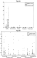
- the results are shown in Figs. 6A and 6B , and the abundance ratio for each bacterial species is shown by a box-and-whisker plot.
- Fig. 6A and Fig. 6B a relative occupancy ratio (%) of each bacterium with respect to the bacterium group existing at each site is shown on the vertical axis.
- Example 1 it was confirmed that, in the disease group of conjunctival MALT lymphoma, bacteria belonging to Delftia genus were significantly higher in the microbial community structure of the microbiota than that in the healthy group. It has been reported that Delftia genus bacteria are bacteria that exist in soil, water, and living environment ( Mahmood S. et al., J. Clin. Microbiol., 2012, 50 (11), p3799-3800 ), and has an ability to modify organic acid and amino acid residues ( Sabine Leibeling et al., Environ. Sci. Technol., 2010, 44 (10), p3793-3799 ).
- Example 1 it was confirmed that, in the disease group of conjunctival MALT lymphoma, the bacteria belonging to Bacteroides genus and Clostridium genus were significantly lower in the microbial community structure of the microbiota than that in the healthy group. It has been reported that Bacteroides genus bacteria constitute a gut microbiota, activate regulatory T cells (T-reg)/Th17, and trigger control of an inflammatory response ( Abby L. Geis et al., Cancer Discov., 5 (10), p1098-1109 ). In addition, there is also another report that it is involved in antibody production and inflammatory reaction control ( Rol N. et al., J. Biol.
- Delftia genus bacteria are an aggressive factor that can be a source of inflammation as a pathogen
- Bacteroides genus bacteria and Clostridium genus bacteria may be a protective factor through control of activation and inflammatory response of T-reg/Th17, induction of production of IgA, or the like.
- Example 2 As shown in Table 2, no significant difference was observed in the clinical parameters examined this time, and it is supported that the change in the microbial community structure confirmed in Example 1 does not depend on age, gender, or laterality, and is specific to conjunctival MALT lymphoma. Therefore, it is supported that changes in the community structure of microbes including Delftia genus bacteria, Bacteroides genus bacteria, and Clostridium genus bacteria are involved in the onset of conjunctival MALT lymphoma.
- microbial community structures contained in specimens sampled from human ocular surface tissues were analyzed by ITS region genes, and healthy persons were compared with persons suffering from conjunctival MALT lymphoma.
- Example 1 A disease group and healthy persons were selected in the same manner as in Example 1.
- the microbiota of the ocular surface tissue was analyzed by (sample collection and DNA isolation), (ITS regional sequencing and data processing) and (statistical analysis) in the same manner as in Example 1.
- each amplicon was prepared by using primer sets targeting the ITS region in rRNA (ITS1-F: 5'-CTTGGTCATTTAGAGGAAGTAA-3 '(SEQ ID NO: 3)) and (ITS2: 5'-GCATCGATGAAGAACGCAGC-3' (SEQ ID NO: 4)), and sequencing was performed with MiSeq (Illumina) using MiSeq v2 500 cycle kit.
- Figs. 9 , 10A to 10C Fungal compositions of specimens sampled from human ocular surface tissues of the healthy group and of the disease group were compared at a genus level. The results are shown in Figs. 9 , 10A to 10C.
- Fig. 9 main fungi constituting microbial layers of the healthy group and of the disease group are indicated, and their relative abundance ratios are shown in graphs.
- Figs. 10A to 10C show the results of comparison of the abundance ratios of 3 fungi of Malassezia genus, Byssochlamys genus, and Aspergillus genus, which are ranked higher in abundance ratio, in the healthy group and the disease group.
- a relationship between a change in the microbiota of ocular surface tissues and age was examined. Specifically, a microbial community structure contained in specimens sampled from human ocular surface tissues was analyzed by 16S rRNA gene, and a correlative relationship between the microbial community structure and age was verified.
- Example 2 analysis of microbiotas of ocular surface tissues of 38 healthy persons (78 eyes) aged 21 to 83 years was performed by (sample collection and DNA isolation), and (16S rRNA sequencing and data processing) and (statistical analysis) in the same manner as in Example 1.
- the breakdown of healthy subjects was 18 males and 21 females, mean age 53.0 ⁇ 19.8 years, and there were no history of ocular diseases, no wearing of contact lenses, and no history of ocular trauma.
- Fig. 11 Bacterial compositions of the conjunctival microbiota from healthy persons of each age were verified. The results are shown in Fig. 11 to Fig. 13 . As a result, it was confirmed that the bacterial composition of the microbiota of the conjunctiva derived from healthy persons of each age fluctuates with aging, and that with the aging, Propionibacteriaceae family bacteria decrease, while Corynebacteriaceae family bacteria increase.
- Fig. 11 is graphs showing changes with age in occupancy ratios of Propionibacteriaceae family bacteria and of Corynebacteriaceae family bacteria in the conjunctiva. Fig.
- Fig. 12 is graphs showing changes with age in the ratios (C/P) of Corynebacteriaceae family bacteria/Propionibacteriaceae family bacteria.
- Fig. 13 is a graph showing a change with age in a difference (C-P) of Corynebacteriaceae family bacteria - Propionibacteriaceae family bacteria. From all the results, it can be understood that significant correlative relationships with age changes were observed, and that the abundance ratio of Corynebacteriaceae family bacteria is higher than that of Propionibacteriaceae family bacteria around 60 years of age.
- Fig. 14 is graphs showing changes with age in the difference (C-P) of Corynebacteriaceae family bacteria - Propionibacteriaceae family bacteria in the conjunctiva, meibomian gland, hand, and periocular skin.
- the present invention can be used for detection of a pathological state of conjunctival disease, and all technical fields that require detection of an aging state, for example, onset of conjunctival disease, onset risk, and prediction of the degree of progression, investigation of the cause of conjunctival disease, confirming the therapeutic effect of conjunctival diseases by pharmaceuticals and the like, detecting physiological and pathological aging conditions, and the like, and can be used particularly in the medical field, the pharmaceutical field, and the like.
- a method of detecting a conjunctival disease using an ocular surface tissue comprising: a step of comparing a microbial community structure of a microbiota included in an ocular surface tissue specimen sampled from a healthy person, with a microbial community structure of a microbiota included in an ocular surface tissue specimen sampled from a subject to detect an ocular surface tissue specimen which is sampled from the subject evaluated as having the conjunctival disease based on a change in the microbial community structure between the healthy person and the subject.
- conjunctival disease is conjunctival mucosa-associated lymphoid tissue lymphoma.
- the change in the microbial community structure is a change in an abundance or an abundance ratio of a bacterial species which belongs to at least one genus selected from Delftia genus, Xylophilus genus, Simplicispira genus, Rothia genus, Xanthomonas genus, Bacteroides genus, Clostridium genus, Deinococcus genus, Williamsia genus, Parabacteroides genus, Chryseobacterium genus, Herbaspirillum genus, Brevundimonas genus, Lactobacillus genus, Schlegelella genus, and Exiguobacterium genus.
- the change in the microbial community structure is a change in the abundance or the abundance ratio of the bacterial species which belongs to the Delftia genus, and an increase in the abundance or the abundance ratio of the bacterial species which belongs to the Delftia genus is evaluated as the conjunctival disease.
- the change in the microbial community structure is a change in the abundance or the abundance ratio of the bacterial species which belongs to at least one genus selected from the Bacteroides genus and the Clostridium genus in a conjunctiva, and a decrease in the abundance or the abundance ratio of the bacterial species which belongs to at least one genus selected from the Bacteroides genus and the Clostridium genus is evaluated as the conjunctival disease.
- the change in the microbial community structure determines a base sequence of a 16S rRNA gene of a microbe constituting the microbiota, and is evaluated based on the base sequence.
- An aging biomarker for detecting an aging state comprising a bacterial species which belongs to at least one family selected from Corynebacteriaceae family and Propionibacteriales family in an ocular surface tissue.
Landscapes
- Chemical & Material Sciences (AREA)
- Life Sciences & Earth Sciences (AREA)
- Organic Chemistry (AREA)
- Proteomics, Peptides & Aminoacids (AREA)
- Health & Medical Sciences (AREA)
- Analytical Chemistry (AREA)
- Zoology (AREA)
- Wood Science & Technology (AREA)
- Engineering & Computer Science (AREA)
- Genetics & Genomics (AREA)
- Molecular Biology (AREA)
- Bioinformatics & Cheminformatics (AREA)
- Immunology (AREA)
- Biotechnology (AREA)
- Biophysics (AREA)
- Physics & Mathematics (AREA)
- Biochemistry (AREA)
- Microbiology (AREA)
- General Engineering & Computer Science (AREA)
- General Health & Medical Sciences (AREA)
- Toxicology (AREA)
- Pathology (AREA)
- Measuring Or Testing Involving Enzymes Or Micro-Organisms (AREA)
- Investigating Or Analysing Biological Materials (AREA)
Applications Claiming Priority (3)
| Application Number | Priority Date | Filing Date | Title |
|---|---|---|---|
| JP2018124504 | 2018-06-29 | ||
| PCT/JP2019/025827 WO2020004626A1 (ja) | 2018-06-29 | 2019-06-28 | 眼表面組織を用いる結膜疾患検出方法及び老化バイオマーカー |
| EP19825086.2A EP3816300A4 (de) | 2018-06-29 | 2019-06-28 | Verfahren zur detektion von bindehauterkrankungen unter verwendung von augenoberflächengewebe und biomarker für alterung |
Related Parent Applications (1)
| Application Number | Title | Priority Date | Filing Date |
|---|---|---|---|
| EP19825086.2A Division EP3816300A4 (de) | 2018-06-29 | 2019-06-28 | Verfahren zur detektion von bindehauterkrankungen unter verwendung von augenoberflächengewebe und biomarker für alterung |
Publications (2)
| Publication Number | Publication Date |
|---|---|
| EP4512910A2 true EP4512910A2 (de) | 2025-02-26 |
| EP4512910A3 EP4512910A3 (de) | 2025-06-11 |
Family
ID=68985241
Family Applications (2)
| Application Number | Title | Priority Date | Filing Date |
|---|---|---|---|
| EP24213334.6A Pending EP4512910A3 (de) | 2018-06-29 | 2019-06-28 | Verfahren zum nachweis von bindehauterkrankungen unter verwendung von augenoberflächengewebe und alterungsbiomarker |
| EP19825086.2A Pending EP3816300A4 (de) | 2018-06-29 | 2019-06-28 | Verfahren zur detektion von bindehauterkrankungen unter verwendung von augenoberflächengewebe und biomarker für alterung |
Family Applications After (1)
| Application Number | Title | Priority Date | Filing Date |
|---|---|---|---|
| EP19825086.2A Pending EP3816300A4 (de) | 2018-06-29 | 2019-06-28 | Verfahren zur detektion von bindehauterkrankungen unter verwendung von augenoberflächengewebe und biomarker für alterung |
Country Status (4)
| Country | Link |
|---|---|
| US (2) | US12365950B2 (de) |
| EP (2) | EP4512910A3 (de) |
| JP (2) | JP7312170B2 (de) |
| WO (1) | WO2020004626A1 (de) |
Families Citing this family (1)
| Publication number | Priority date | Publication date | Assignee | Title |
|---|---|---|---|---|
| CN121023090A (zh) * | 2025-10-31 | 2025-11-28 | 南昌大学第一附属医院 | 辅助预测鹦鹉热衣原体感染的呼吸道菌群标志物及其应用 |
Citations (1)
| Publication number | Priority date | Publication date | Assignee | Title |
|---|---|---|---|---|
| JP2017029133A (ja) | 2015-07-21 | 2017-02-09 | TAK−Circulator株式会社 | 身体状態の評価方法、情報の提示方法、および身体状態を改善又は予防する物質のスクリーニング方法 |
Family Cites Families (5)
| Publication number | Priority date | Publication date | Assignee | Title |
|---|---|---|---|---|
| US8110578B2 (en) | 2008-10-27 | 2012-02-07 | Signal Pharmaceuticals, Llc | Pyrazino[2,3-b]pyrazine mTOR kinase inhibitors for oncology indications and diseases associated with the mTOR/PI3K/Akt pathway |
| CN103687604A (zh) | 2011-03-01 | 2014-03-26 | 群体创新有限责任公司 | 用于治疗与致病生物膜相关之病症的材料和方法 |
| JP6637885B2 (ja) | 2013-07-21 | 2020-01-29 | ペンデュラム セラピューティクス, インコーポレイテッド | マイクロバイオームの特性解明、モニタリング、および処置のための方法およびシステム |
| JP2015188379A (ja) * | 2014-03-28 | 2015-11-02 | 森永乳業株式会社 | 腸内菌叢の老若判定方法及び菌群のスクリーニング方法 |
| JP6654326B2 (ja) | 2016-03-28 | 2020-02-26 | 国立大学法人大阪大学 | 眼科疾患のバイオマーカー |
-
2019
- 2019-06-28 US US17/252,982 patent/US12365950B2/en active Active
- 2019-06-28 JP JP2020527685A patent/JP7312170B2/ja active Active
- 2019-06-28 EP EP24213334.6A patent/EP4512910A3/de active Pending
- 2019-06-28 EP EP19825086.2A patent/EP3816300A4/de active Pending
- 2019-06-28 WO PCT/JP2019/025827 patent/WO2020004626A1/ja not_active Ceased
-
2023
- 2023-07-06 JP JP2023111475A patent/JP7594633B2/ja active Active
-
2025
- 2025-06-16 US US19/239,284 patent/US20250305071A1/en active Pending
Patent Citations (1)
| Publication number | Priority date | Publication date | Assignee | Title |
|---|---|---|---|---|
| JP2017029133A (ja) | 2015-07-21 | 2017-02-09 | TAK−Circulator株式会社 | 身体状態の評価方法、情報の提示方法、および身体状態を改善又は予防する物質のスクリーニング方法 |
Non-Patent Citations (8)
| Title |
|---|
| ABBY L. GEIS ET AL., CANCER DISCOV., vol. 5, no. 10, pages 1098 - 1109 |
| MAHMOOD S. ET AL., J. CLIN. MICROBIOL., vol. 50, no. 11, 2012, pages 3799 - 3800 |
| OUWEHAND AC. ET AL., WORLD J. GASTROENTEROL., vol. 15, no. 26, 2009, pages 3261 - 3268 |
| ROL N. ET AL., J. BIOL. CHEM., vol. 287, no. 47, 2012, pages 40074 - 40082 |
| SABINE LEIBELING ET AL., ENVIRON. SCI. TECHNOL., vol. 44, no. 10, 2010, pages 3793 - 3799 |
| SARA OMENETTI ET AL., FRONT. IMMUNOL., vol. 6, 2015 |
| STEFKAAT. ET AL., PROC. NATL. ACAD. SCI. USA, vol. 111, no. 36, pages 13145 - 13150 |
| TAYLOR FEEHLEY ET AL., CURR. OPIN. LMMUNOL., vol. 31, 2014, pages 79 - 86 |
Also Published As
| Publication number | Publication date |
|---|---|
| JPWO2020004626A1 (ja) | 2021-08-02 |
| US20250305071A1 (en) | 2025-10-02 |
| WO2020004626A1 (ja) | 2020-01-02 |
| US12365950B2 (en) | 2025-07-22 |
| JP2023130449A (ja) | 2023-09-20 |
| EP3816300A1 (de) | 2021-05-05 |
| EP3816300A4 (de) | 2022-03-16 |
| JP7312170B2 (ja) | 2023-07-20 |
| JP7594633B2 (ja) | 2024-12-04 |
| US20210214775A1 (en) | 2021-07-15 |
| EP4512910A3 (de) | 2025-06-11 |
Similar Documents
| Publication | Publication Date | Title |
|---|---|---|
| Li et al. | How ocular surface microbiota debuts in type 2 diabetes mellitus | |
| Suzuki et al. | The microbiome of the meibum and ocular surface in healthy subjects | |
| Ng et al. | Management and outcomes of postoperative endophthalmitis since the endophthalmitis vitrectomy study: the Endophthalmitis Population Study of Western Australia (EPSWA)’s fifth report | |
| Thompson III et al. | Fungal disease of the nose and paranasal sinuses | |
| Guler et al. | Risk factors for nosocomial candiduria | |
| Zysset-Burri et al. | Understanding the interactions between the ocular surface microbiome and the tear proteome | |
| Kim et al. | Cream cheese-derived Lactococcus chungangensis CAU 28 modulates the gut microbiota and alleviates atopic dermatitis in BALB/c mice | |
| US20250305071A1 (en) | Method of detecting conjunctival disease using ocular surface tissue, and aging biomarker | |
| Vergroesen et al. | Glaucoma patients have a lower abundance of butyrate-producing taxa in the gut | |
| Gupta et al. | Ocular conjunctival microbiome profiling in dry eye disease: A case control pilot study | |
| Tunç et al. | Corneal bacterial microbiome in patients with keratoconus using next-generation sequencing–based 16S rRNA gene analysis | |
| Zhao et al. | Conjunctival sac microbiome in anophthalmic patients: Flora diversity and the impact of ocular prosthesis materials | |
| Yang et al. | The microbiome of meibomian gland secretions from patients with internal hordeolum treated with hypochlorous acid eyelid wipes | |
| EP3748018A1 (de) | Verfahren zur diagnose von depression durch bakterielle metagenomanalyse | |
| Saleem et al. | Frequency of meningitis in neonatal late onset sepsis in Gangaram hospital, Lahore | |
| Agustina et al. | Prevalence and identification of oral candida species in patients with type 2 diabetes in Yogyakarta | |
| Guo et al. | Helicobacter pylori infection’s dual impact on the ocular surface: Tear secretion disruption and ocular surface microbiome alteration | |
| Gupta et al. | An alarming increase of fungal infections in intensive care unit: Challenges in the diagnosis and treatment | |
| Mobed | The role of gut microbiota in aging-associated diseases | |
| Ramakhrisnand | Exploring the Role of the Microbiome in Atopic Dermatitis: A Longitudinal Metagenomic Analysis in India | |
| US20190134123A1 (en) | Novel system and method for microbiome profiling and modulation by means of cannabis administration | |
| Tavakkolinasab | The impact of probiotics and prebiotics on dry eye disease | |
| Iglesias et al. | S1199 Prevalence of Alexithymia in Inflammatory Bowel Disease: Relationship With Sociodemographic and Clinical Parameters | |
| Megantara et al. | Microbiota profile in sinonasal mucosa of chronic rhinosinusitis as an indicator for therapeutic outcome | |
| JP2018088843A (ja) | デリケートエリアのかぶれ判定方法 |
Legal Events
| Date | Code | Title | Description |
|---|---|---|---|
| PUAI | Public reference made under article 153(3) epc to a published international application that has entered the european phase |
Free format text: ORIGINAL CODE: 0009012 |
|
| STAA | Information on the status of an ep patent application or granted ep patent |
Free format text: STATUS: REQUEST FOR EXAMINATION WAS MADE |
|
| 17P | Request for examination filed |
Effective date: 20241115 |
|
| AC | Divisional application: reference to earlier application |
Ref document number: 3816300 Country of ref document: EP Kind code of ref document: P |
|
| AK | Designated contracting states |
Kind code of ref document: A2 Designated state(s): AL AT BE BG CH CY CZ DE DK EE ES FI FR GB GR HR HU IE IS IT LI LT LU LV MC MK MT NL NO PL PT RO RS SE SI SK SM TR |
|
| REG | Reference to a national code |
Ref country code: DE Ref legal event code: R079 Free format text: PREVIOUS MAIN CLASS: C12Q0001689000 Ipc: C12Q0001687600 |
|
| PUAL | Search report despatched |
Free format text: ORIGINAL CODE: 0009013 |
|
| AK | Designated contracting states |
Kind code of ref document: A3 Designated state(s): AL AT BE BG CH CY CZ DE DK EE ES FI FR GB GR HR HU IE IS IT LI LT LU LV MC MK MT NL NO PL PT RO RS SE SI SK SM TR |
|
| RIC1 | Information provided on ipc code assigned before grant |
Ipc: C12Q 1/689 20180101ALI20250507BHEP Ipc: C12Q 1/6883 20180101ALI20250507BHEP Ipc: C12Q 1/6876 20180101AFI20250507BHEP |