EP4512343A2 - Echokardiographie-führungsverfahren und echokardiographie-führungsvorrichtung damit - Google Patents
Echokardiographie-führungsverfahren und echokardiographie-führungsvorrichtung damit Download PDFInfo
- Publication number
- EP4512343A2 EP4512343A2 EP23792184.6A EP23792184A EP4512343A2 EP 4512343 A2 EP4512343 A2 EP 4512343A2 EP 23792184 A EP23792184 A EP 23792184A EP 4512343 A2 EP4512343 A2 EP 4512343A2
- Authority
- EP
- European Patent Office
- Prior art keywords
- cross
- section
- guide
- echocardiography
- image
- Prior art date
- Legal status (The legal status is an assumption and is not a legal conclusion. Google has not performed a legal analysis and makes no representation as to the accuracy of the status listed.)
- Pending
Links
Images


Classifications
-
- G—PHYSICS
- G06—COMPUTING OR CALCULATING; COUNTING
- G06T—IMAGE DATA PROCESSING OR GENERATION, IN GENERAL
- G06T7/00—Image analysis
- G06T7/0002—Inspection of images, e.g. flaw detection
- G06T7/0012—Biomedical image inspection
-
- A—HUMAN NECESSITIES
- A61—MEDICAL OR VETERINARY SCIENCE; HYGIENE
- A61B—DIAGNOSIS; SURGERY; IDENTIFICATION
- A61B8/00—Diagnosis using ultrasonic, sonic or infrasonic waves
- A61B8/08—Clinical applications
- A61B8/0883—Clinical applications for diagnosis of the heart
-
- A—HUMAN NECESSITIES
- A61—MEDICAL OR VETERINARY SCIENCE; HYGIENE
- A61B—DIAGNOSIS; SURGERY; IDENTIFICATION
- A61B8/00—Diagnosis using ultrasonic, sonic or infrasonic waves
- A61B8/46—Ultrasonic, sonic or infrasonic diagnostic devices with special arrangements for interfacing with the operator or the patient
- A61B8/461—Displaying means of special interest
-
- A—HUMAN NECESSITIES
- A61—MEDICAL OR VETERINARY SCIENCE; HYGIENE
- A61B—DIAGNOSIS; SURGERY; IDENTIFICATION
- A61B8/00—Diagnosis using ultrasonic, sonic or infrasonic waves
- A61B8/52—Devices using data or image processing specially adapted for diagnosis using ultrasonic, sonic or infrasonic waves
- A61B8/5207—Devices using data or image processing specially adapted for diagnosis using ultrasonic, sonic or infrasonic waves involving processing of raw data to produce diagnostic data, e.g. for generating an image
-
- G—PHYSICS
- G06—COMPUTING OR CALCULATING; COUNTING
- G06N—COMPUTING ARRANGEMENTS BASED ON SPECIFIC COMPUTATIONAL MODELS
- G06N3/00—Computing arrangements based on biological models
- G06N3/02—Neural networks
- G06N3/04—Architecture, e.g. interconnection topology
- G06N3/045—Combinations of networks
-
- G—PHYSICS
- G06—COMPUTING OR CALCULATING; COUNTING
- G06N—COMPUTING ARRANGEMENTS BASED ON SPECIFIC COMPUTATIONAL MODELS
- G06N3/00—Computing arrangements based on biological models
- G06N3/02—Neural networks
- G06N3/08—Learning methods
-
- A—HUMAN NECESSITIES
- A61—MEDICAL OR VETERINARY SCIENCE; HYGIENE
- A61B—DIAGNOSIS; SURGERY; IDENTIFICATION
- A61B8/00—Diagnosis using ultrasonic, sonic or infrasonic waves
- A61B8/48—Diagnostic techniques
- A61B8/486—Diagnostic techniques involving arbitrary m-mode
-
- A—HUMAN NECESSITIES
- A61—MEDICAL OR VETERINARY SCIENCE; HYGIENE
- A61B—DIAGNOSIS; SURGERY; IDENTIFICATION
- A61B8/00—Diagnosis using ultrasonic, sonic or infrasonic waves
- A61B8/48—Diagnostic techniques
- A61B8/488—Diagnostic techniques involving Doppler signals
-
- A—HUMAN NECESSITIES
- A61—MEDICAL OR VETERINARY SCIENCE; HYGIENE
- A61B—DIAGNOSIS; SURGERY; IDENTIFICATION
- A61B8/00—Diagnosis using ultrasonic, sonic or infrasonic waves
- A61B8/52—Devices using data or image processing specially adapted for diagnosis using ultrasonic, sonic or infrasonic waves
- A61B8/5269—Devices using data or image processing specially adapted for diagnosis using ultrasonic, sonic or infrasonic waves involving detection or reduction of artifacts
-
- G—PHYSICS
- G06—COMPUTING OR CALCULATING; COUNTING
- G06T—IMAGE DATA PROCESSING OR GENERATION, IN GENERAL
- G06T2207/00—Indexing scheme for image analysis or image enhancement
- G06T2207/10—Image acquisition modality
- G06T2207/10132—Ultrasound image
-
- G—PHYSICS
- G06—COMPUTING OR CALCULATING; COUNTING
- G06T—IMAGE DATA PROCESSING OR GENERATION, IN GENERAL
- G06T2207/00—Indexing scheme for image analysis or image enhancement
- G06T2207/20—Special algorithmic details
- G06T2207/20081—Training; Learning
-
- G—PHYSICS
- G06—COMPUTING OR CALCULATING; COUNTING
- G06T—IMAGE DATA PROCESSING OR GENERATION, IN GENERAL
- G06T2207/00—Indexing scheme for image analysis or image enhancement
- G06T2207/20—Special algorithmic details
- G06T2207/20084—Artificial neural networks [ANN]
-
- G—PHYSICS
- G06—COMPUTING OR CALCULATING; COUNTING
- G06T—IMAGE DATA PROCESSING OR GENERATION, IN GENERAL
- G06T2207/00—Indexing scheme for image analysis or image enhancement
- G06T2207/30—Subject of image; Context of image processing
- G06T2207/30004—Biomedical image processing
- G06T2207/30048—Heart; Cardiac
Definitions
- the present invention relates to a method for providing a guide for echocardiography measurement and a device for providing a guide for echocardiography using the same.
- An echocardiography examination is performed by a scheme of projecting ultrasound waves on the heart having a three-dimensional structure in multiple-dimensional planes to acquire images of the heart and measure hemodynamic variables.
- a medical staff positions an ultrasound probe in a location where it is easy to acquire ultrasound images, so as to acquire multi-face images through anatomical structures around the heart, such as between the ribs, and records the images by finding an appropriate monolayer through rotation and tilting.
- an echocardiography examination process is divided into examination preparation, image acquisition, image analysis, and report writing, and in particular, the image acquisition and analysis process can take a lot of time.
- visualization functions such as two-dimensional image navigation for a process in which the medical staff moves a probe based on a virtual three-dimensional structure of the entire heart can be required.
- the inventors of the present invention recognized a fact that there is difficulty in acquiring an image corresponding to a standard measurement cross section (view) during an image analysis process.
- the inventors of the present invention provided whether a cross section of an ultrasound image currently acquired through the recognition corresponds to a standard measurement cross section as a matching rate to conceive that the higher the matching rate, the closer the matching rate is to the standard measurement cross section.
- the inventors of the present invention could develop a guide system for acquiring a measurement value by segmenting a plurality of regions with respect to an echocardiographic image, calculating a matching rate by comparing the measurement value and the standard measurement cross section, and acquiring the standard measurement cross section based thereon.
- the inventors of the present invention could expect that by providing a new guide system, it is possible to overcome the limitation that it is difficult to acquire an accurate measurement value of a cardiac structure in the related art by facilitating image acquisition corresponding to the standard measurement cross section.
- the inventors of the present invention could expect to overcome the limitation of low reliability and being provided for an entry-level medical staff or for an educational purpose because the conventional echocardiography guide system presents a uniform guide for the direction, rotation, angle, etc., of a probe.
- visualization functions such as two-dimensional image navigation for a process in which the medical staff moves a probe based on a virtual three-dimensional structure of the entire heart are provided to provide an intuitive echocardiographic image guide.
- an object to be achieved by the present invention is to provide an echocardiography guide method and an echocardiography guide device using an artificial neural network-based guide system configured to present a guideline for acquiring measurement values by segmenting an echocardiographic image into a plurality of regions, calculating a matching rate by comparing the measurement values with a standard measurement cross section (view), and acquiring the standard measurement cross section (view) based on the calculated matching rate.
- the inventors of the present invention conceived that when a guide for determining a relative angle between a standard image cross section received in advance and the received cross section by using an Euler angle through the recognition described above, and reducing the relative angle based on the relative angle is generated, and the guide is provided as a graphic interface, the measurement value may be closer to the standard measurement cross section.
- the inventors of the present invention could develop the guide system by generating a guide for determining a relative angle between a standard image cross section received in advance and the received cross section by using an Euler angle, and reducing the relative angle based on the relative angle.
- the inventors of the present invention could expect that by providing a new guide system, it is possible to overcome the limitation that it is difficult to acquire an accurate measurement value of a cardiac structure in the related art by facilitating image acquisition corresponding to the standard measurement cross section.
- the inventors of the present invention recognized the need for a guide system applicable to spectral Doppler requiring fine tuning as well as echocardiography B-mode and M-mode.
- the inventors of the present invention were able to develop an artificial neural network-based guide system capable of presenting a guideline by evaluating a cross section matching degree and evaluating the relative angle matching rate to enable fine tuning.
- the inventors of the present invention could expect that it is possible to overcome the limitation of low reliability and being provided for an entry-level medical staff or for an educational purpose because the guide system in the related art presents a guide based on a threshold.
- an object to be achieved by the present invention is to provide a guide providing method and a guide providing device configured to receive an echocardiographic image of an individual, classify a cross section for the echocardiographic image, determine a relative angle, and provide a guide based on an evaluation result.
- an echocardiography guide method according to an exemplary embodiment of the present invention.
- the guide method includes: receiving an echocardiographic image of an individual; segmenting a cross section of the echocardiographic image into a plurality of cardiac monolayer regions; calculating a matching rate for the cross section from which the plurality of cardiac monolayer regions is segmented based on a standard cross section received in advance; determining whether to provide a guide based on the matching rate for the cross section; and providing the guide for acquiring a cross section having a preset matching rate or higher in response to whether to provide the guide.
- the segmenting of the cross section of the echocardiographic image into the cardiac monolayer regions may further include segmenting each of the plurality of cardiac monolayer regions by using a first prediction model trained to segment the cross section into each of the plurality of cardiac monolayer regions.
- the calculating of the matching rate for the cross section based on the standard cross section received in advance may further include calculating a matching rate by using a matching rate calculation model configured to calculate the matching rate by outputting measurement values from the segmented cardiac structure regions and comparing the measurement value with a measurement value determined in the cross section based on the standard cross section received in advance.
- the plurality of cardiac monolayer regions may be any one or more selected from structures observable in a specific cardiac cross section, such as the right ventricle (RV), the left ventricle (LV), the Aorta, the left atrium (LA), the left ventricle posterior wall (LVPW), and the intect ventricular septum (IVS).
- RV right ventricle
- LV left ventricle
- LA left atrium
- LVPW left ventricle posterior wall
- IVS intect ventricular septum
- the first prediction model which is trained to segment the cross section into each of the plurality of cardiac monolayer regions may include a first feature extraction unit configured to input a received echocardiographic image and segment a plurality of regions based on the anatomical structure within the echocardiographic image, and a second feature extraction unit configured to input the received echocardiographic image and predict positions of a plurality of points within the echocardiographic image.
- the first prediction model may be trained to segment the cross section into each of the plurality of cardiac monolayer regions, and may be further configured to evaluate the region segmentation result by inputting an entropy, and mask information including an anatomical mask for each region (inferenced chamber mask anatomy) and an area for each region (inferenced chamber mask ratio) with respect to the segmented regions.
- the matching rate calculation model may be configured to calculate a measurement value corresponding to an area and a size of each of the segmented cardiac monolayer regions by inputting the plurality of points in the echocardiographic image determined by the first prediction model, and further configured to calculate the matching rate by comparing the measurement value determined from the cross section based on the standard cross section received in advance.
- the matching rate calculation model may be further configured to calculate the matching rate by evaluating measurement values measured in the segmented cardiac monolayer regions by inputting data from an entropy meter, a capacity meter, and a connectivity meter, and comparing the measurement value determined in the cross section based on the standard cross section received in advance.
- a visualization function such as a navigation of a two-dimensional image according to a position and a direction in which a probe moves in a virtual three-dimensional structure for the entire heart may be included with respect to movement of the probe.
- the guide device includes: a communication unit configured to receive an echocardiographic image of an individual; and a processor functionally connected to the communication unit.
- the processor is configured to segment the cross section of the echocardiography image into a plurality of cardiac monolayer regions, calculate a matching rate for the cross section in which the plurality of cardiac monolayer regions are segmented based on a previously received standard cross section, and determines whether to provide the guide based on the matching rate for the cross section, and provide a guide for acquiring a cross section having a preset matching rate or higher in response to whether to provide the guide.
- the processor may be further configured to segment each of the plurality of cardiac monolayer regions using a first prediction model trained to segment the cross section into the plurality of cardiac monolayer regions, respectively.
- the processor may be further configured to calculate a matching rate by using a matching rate calculation model configured to calculate the matching rate by outputting measurement values from the segmented cardiac structure regions and comparing the measurement value with a measurement value determined in the cross section based on a standard cross section received in advance.
- the plurality of cardiac monolayer regions may be one or more selected from structures observable in a specific cardiac cross section, such as the right ventricle (RV), the left ventricle (LV), the aorta, the left atrium (LA), the left ventricle posterior wall (LVPW), and the intect ventricular septum (IVS).
- RV right ventricle
- LV left ventricle
- LA left atrium
- LVPW left ventricle posterior wall
- IVS intect ventricular septum
- the first prediction model may be trained to segment the cross section into each of a plurality of cardiac monolayer regions, and may include a first feature extraction unit configured to input a received echocardiographic image and segment a plurality of regions based on the anatomical structure within the echocardiographic image, and a second feature extraction unit configured to input the received echocardiographic image and predict positions of a plurality of points within the echocardiographic image.
- the first prediction model may be trained to segment the cross section into each of the plurality of cardiac monolayer regions, and may be further configured to evaluate the region segmentation result by inputting an entropy, and mask information including an anatomical mask for each region (inferenced chamber mask anatomy) and an area for each region (inferenced chamber mask ratio) with respect to the segmented regions.
- the matching rate calculation model may be configured to calculate a measurement value corresponding to an area and a size of each of the segmented cardiac monolayer regions by inputting the plurality of points in the echocardiographic image determined by the first prediction model, and further configured to calculate the matching rate by comparing the measurement value determined from the cross section based on the standard cross section received in advance.
- the matching rate calculation model may be further configured to calculate the matching rate by evaluating measurement values measured in the segmented cardiac monolayer regions by inputting data from an entropy meter, a capacity meter, and a connectivity meter, and comparing the measurement value determined in the cross section based on the standard cross section received in advance.
- the guide may include the matching rate calculated by comparing the measurement value determined in the cross section based on the standard cross section received in advance, and movement information of the probe for acquiring the standard measurement cross section (view).
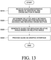
- the guide method which is implemented by a processor, includes: receiving an echocardiographic image of an individual and a cross section of the image; determining the relative angle of the received cross section with respect to a standard image cross section received in advance; generating, based on the relative angle, a guide for decreasing the relative angle; and providing the guide as a graphic interface.
- an Euler angle may be used.
- the provided guide may further include a two-dimensional relative angle for a three-dimensional image determined using the Euler angle.
- the provided guide may provide a matching degree between relative angles of the cross section and a standard image.
- the guide device includes: a communication unit configured to receive an echocardiographic image of an individual and a cross section in the image; and a processor functionally connected to the communication unit, and the processor is configured to classify a cross section for the echocardiographic image, determine a relative angle between a standard image cross section received in advance and the received cross section, generate a guide for reducing the relative angle based on the relative angle, and provide the guide as a graphic interface.
- the processor may be further configured to determine the relative angle using an Euler angle.
- the processor may be further configured to provide further include a two-dimensional relative angle for a three-dimensional image determined using the Euler angle.
- the processor may be further configured to provide a guide that provides a matching degree between the relative angles of the cross section and the standard image.
- the present invention may provide a guide system for an echocardiography cross section based on an artificial neural network configured to segment a cross section of the echocardiographic image into a plurality of cardiac monolayer regions, calculate a matching rate for the cross section from which the plurality of cardiac monolayer regions is segmented based on a standard cross section received in advance, determine whether to provide a guide based on the matching rate for the cross section, and provide the guide for acquiring a cross section having a preset matching rate or higher in response to whether to provide the guide to provide a guideline for highly reliable echocardiography measurement.
- the present invention may overcome the limitations that it is difficult to acquire an accurate measurement value for a cardiac structure in the related art by providing a new guide system.
- the present invention may overcome the limitation of low reliability and being provided for an entry-level medical staff or for an educational purpose because the conventional echocardiography guide system presents a uniform guide for the direction, rotation, angle, etc., of a probe.
- the present invention provides a guide system which determines a relative angle between a standard image cross section received in advance and the received cross section, and generates a guide for reducing the relative angle based on the relative angle to provide the guide as a graphic interface to provide a guideline for highly reliable echocardiography measurement.
- the present invention may overcome the limitations of a guide system using a threshold-based cross section (view) classification model in the related art by providing a new guide system.
- the present invention may overcome the limitation of low reliability and being provided for an entry-level medical staff or for an educational purpose because the guide system in the related art presents a guide based on a threshold.
- a 'standard image' may mean an image corresponding to a standard measurement cross section (view).
- FIGS. 1 , 2A , and 2B a guide system for a cross section (view) of an echocardiography using an echocardiography guide device and an echocardiography guide device according to an exemplary embodiment of the present invention will be described.
- FIG. 1 illustrates a guide system for a cross section of an echocardiography using an echocardiography guide device according to an exemplary embodiment of the present invention.
- FIG. 2A is a block diagram illustrating a configuration of a medical staff device according to an exemplary embodiment of the present invention.
- FIG. 2B is a block diagram illustrating a configuration of a guide server according to an exemplary embodiment of the present invention.
- the guide system 1000 may be a system configured to provide a guide for echocardiography measurement based on an echocardiographic image of an individual.
- the guide system 1000 may be constituted by a medical staff device 100 receiving a guide for echocardiography measurement, an echocardiographic image diagnosis device 200 providing the echocardiographic image, and a guide server 300 generating the guide for echocardiography measurement based on the received echocardiographic image.
- the medical staff device 100 as an electronic device providing a user interface for representing the guide for echocardiography measurement may include at least one of a smartphone, a tablet personal computer (PC), a laptop computer, and/or a PC.
- a smartphone a tablet personal computer (PC)
- PC personal computer
- laptop computer a laptop computer
- PC personal computer
- the medical staff device 100 may receive prediction results associated with a matching rate with an electrocardiography cross section for the individual and a standard cross section from the guide server 300, and display the received results through a display unit to be described below.
- the guide server 300 may include a general-purpose computer, laptop, and/or data server that perform various operations for determining the guide for echocardiography measurement based on the echocardiographic image provided from the echocardiographic image diagnosis device 200 such as an ultrasound diagnosis device.
- the guide server 300 may be a device for accessing a web server providing a web page or a mobile web server providing a mobile website, but is not limited thereto.
- the guide server 300 receives the echocardiographic image of the individual from the echocardiographic image diagnosis device 200, segments the cross section of the echocardiographic image into a plurality of cardiac monolayer regions, calculates a matching rate for the cross section in which the plurality of cardiac monolayer regions is segmented based on a standard cross section received in advance, and determines whether to provide the guide based on the matching rate for the cross section to provide a guide for acquiring a cross section (view) having a preset matching rate or higher in response to whether to provide the guide.
- the guide server 300 may provide the prediction results to the medical staff device 100.
- Information provided from the guide server 300 may be provided to a web page through a web browser installed in the medical staff device 100, or provided in the form of an application or a program.
- the data may be provided in a form that is embedded in a platform in a client-server environment.
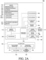
- the medical staff device 100 may include a memory interface 110, one or more processors 120, and a peripheral interface 130. Various components within the medical staff device 100 may be connected by one or more communication buses or signal lines.
- the memory interface 110 is connected to the memory 150 to transmit various data to the processor 120.
- the memory 150 may include at least one type of storage medium among flash memory type, hard disk type, multimedia card micro type, card type memory (for example, SD or XD memory, etc.), RAM, SRAM, ROM, EEPROM, PROM, network storage, cloud, and blockchain data.
- the memory 150 may store at least one of an operating system 151, a communication module 152, a graphical user interface module (GUI) 153, a sensor processing module 154, a phone module 155, and an application module 156.
- the operating system 151 may include instructions for processing basic system services and instructions for performing hardware tasks.
- the communication module 152 may communicate with at least one of one or more other devices, computers, and servers.
- the graphical user interface module (GUI) 153 may process a graphical user interface.
- the sensor processing module 154 may process sensor-related functions (e.g., processing a voice input received using one or more microphones 192).
- the phone module 155 may process phone-related functions.
- the application module 156 may perform various functions of the user application, such as electronic messaging, web browsing, media processing, navigation, imaging, and other processing functions.
- the medical staff device 100 may store one or more software applications 156-1 and 156-2 (e.g., a guide application) associated with any one type of service in the memory 150.
- the memory 150 may store a digital assistant client module (157) (hereinafter, referred to as DA client module), and thus store instructions for performing client-side functions of a digital assistant and various user data 158.
- DA client module digital assistant client module
- the DA client module 157 may acquire the user's voice input, text input, touch input, and/or gesture input through various user interfaces (e.g., I/O subsystem 140) provided in the medical staff device 100.
- various user interfaces e.g., I/O subsystem 140
- the DA client module 157 may output data in audiovisual and tactile forms.
- the DA client module 157 may output data consisting of a combination of at least two of voice, sound, notification, text message, menu, graphic, video, animation, and vibration.
- the DA client module 157 may communicate with a digital assistant server (not illustrated) using a communication subsystem 180.
- the DA client module 157 may collect additional information about a surrounding environment of the medical staff device 100 from various sensors, subsystems, and peripheral devices to construct a context associated with the user input.
- the DA client module 157 may infer a user's intention by providing context information to a digital assistance server jointly with the user input.
- the context information that may accompany the user input may include sensor information, such as lighting, ambient noise, ambient temperature, images of the surrounding environment, video, etc.
- the context information may include a physical state of the medical staff device 100 (e.g., device orientation, device position, device temperature, power level, speed, acceleration, motion pattern, cellular signal strength, etc.).
- the context information may include information related to the software state of the medical staff device 100 (e.g., processes running on the medical staff device 100, installed programs, past and current network activity, background services, error logs, resource usage, etc.).
- the memory 150 may include additional or deleted instructions, and may further include additional components other than those illustrated in FIG. 2A of the medical staff device 100, or may exclude some components.
- the processor 120 may control the overall operation of the medical staff device 100 and execute various instructions to implement an interface that provides the guide for echocardiography measurement by running an application or program stored in the memory 150.
- the processor 120 may correspond to an operation device such as a central processing unit (CPU) or an application processor (AP). Further, the processor 120 may be implemented in the form of an integrated chip (IC) such as a system on chip (SoC) in which various operation devices such as a neural processing unit (NPU) are integrated.
- IC integrated chip
- SoC system on chip
- NPU neural processing unit
- the peripheral interface 130 is connected to various sensors, subsystems, and peripheral devices to provide data so that the medical staff device 100 may perform various functions. Here, it may be understood that any function performed by the medical staff device 100 is performed by the processor 120.
- the peripheral interface 130 may receive data from a motion sensor 160, an illumination sensor (optical sensor) 161, and a proximity sensor 162, and through this, the medical staff device 100 may perform orientation, light, and proximity detection functions.
- the peripheral interface 130 may receive data from other sensors 163 (positioning system - GPS receiver, temperature sensor, biometric sensor), thereby enabling the medical staff device 100 to perform functions related to the other sensors 163.
- the medical staff device 100 may include a camera subsystem 170 connected to the peripheral interface 130, and an optical sensor 171 connected thereto, which may enable the medical staff device 100 to perform various photographing functions, such as taking photographs and recording video clips.
- the medical staff device 100 may include a communication subsystem 180 connected to the peripheral interface 130.
- the communication subsystem 180 consists of one or more wired/wireless networks and may include various communication ports, radio frequency transceivers, and optical transceivers.
- the medical staff device 100 includes an audio subsystem 190 connected to the peripheral interface 130, the audio subsystem 190 includes one or more speakers 191 and one or more microphones 192, such that the medical staff device 100 may perform voice-activated functions, such as speech recognition, voice replication, digital recording, and telephony functions.
- voice-activated functions such as speech recognition, voice replication, digital recording, and telephony functions.
- the medical staff device 100 may include an I/O subsystem 140 connected to the peripheral interface 130.
- the I/O subsystem 140 may control a touch screen 143 included in the medical staff device 100 via a touch screen controller 141.
- the touch screen controller 141 may detect a user's contact and movement or cessation of contact and movement using any one of a plurality of touch sensing technologies, such as capacitive, resistive, infrared, surface acoustic wave technology, proximity sensor array, etc.
- the I/O subsystem 140 may control other input/control devices 144 included in the medical staff device 100 via other input controller(s) 142.
- the other input controller(s) 142 may control one or more buttons, rocker switches, thumb-wheels, infrared ports, USB ports, and pointer devices such as a stylus.
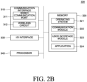
- the guide server 300 may include a communication interface 310, a memory 320, an I/O interface 330, and a processor 340, and respective components may communicate with each other through one or more communication buses or signal lines.
- the communication interface 310 may be connected to the medical staff device 100 and the echocardiographic image diagnosis device 200 via a wired/wireless communication network to exchange data.
- the communication interface 310 may receive the echocardiographic image from the echocardiographic image diagnosis device 200, and transmit determined information to the medical staff device 100.
- the communication interface 310 that enables transmission and reception of such data may include a communication port 311 and a wireless circuit 312, and here, the wired communication port 311 may include one or more wired interfaces, for example, Ethernet, universal serial bus (USB), FireWire, etc. Further, the wireless circuit 312 may transmit and receive data with an external device via RF signals or optical signals. Moreover, the wireless communication may use at least one of a plurality of communication standards, protocols and technologies, such as GSM, EDGE, CDMA, TDMA, Bluetooth, Wi-Fi, VoIP, Wi-MAX, or any other suitable communication protocol.
- the memory 320 may store various data used in the guide server 300.
- the memory 320 may store the echocardiographic image, store a model trained to classify and segment the cross section for the echocardiographic image, or store models trained to compare and evaluate a measurement value acquired from the echocardiography cross section based on a previously received standard cross section, and calculate a matching rate.
- the memory 320 may include a volatile or nonvolatile storage medium capable of storing various data, instructions, and information.
- the memory 320 may include at least one type of storage medium among flash memory type, hard disk type, multimedia card micro type, card type memory (for example, SD or XD memory, etc.), RAM, SRAM, ROM, EEPROM, PROM, network storage, cloud, and blockchain data.
- the memory 320 may store at least one component of an operating system 321, a communication module 322, a user interface module 323, and one or more applications 324.
- the operating system 321 may include various software components and drivers for controlling and managing general system tasks (e.g., memory management, storage device control, power management, etc.) and may support communication between various hardware, firmware, and software components.
- general system tasks e.g., memory management, storage device control, power management, etc.
- the communication module 323 may support communication with other devices through the communication interface 310.
- the communication module 320 may include various software components for processing data received by the wired communication port 311 or wireless circuit 312 of the communication interface 310.
- the user interface module 323 may receive a user's request or input from a keyboard, touch screen, microphone, etc., through the I/O interface 330 and provide a user interface on a display.
- the application 324 may include a program or module configured to be executed by one or more processors 330.
- the application for providing the guide for echocardiography measurement may be implemented on a server farm.
- the I/O interface 330 may connect at least one of an input/output device (not illustrated) of the guide server 300, such as a display, a keyboard, a touch screen, and a microphone, to the user interface module 323.
- the I/O interface 330 may receive a user input (e.g., a voice input, a keyboard input, a touch input, etc.) together with the user interface module 323, and process an instruction according to the received input.
- the processor 340 is connected to the communication interface 310, the memory 320, and the I/O interface 330 to control the overall operation of the guide server 300, and may perform various instructions for the guide through the application or program stored in the memory 320.
- the processor 340 may correspond to an operation device such as a central processing unit (CPU) or an application processor (AP). Further, the processor 340 may be implemented in the form of an integrated chip (IC) such as a system on chip (SoC) in which various operation devices are integrated. Alternatively, the processor 340 may include a module for calculating an artificial neural network model, such as a neural processing unit (NPU).
- CPU central processing unit
- AP application processor
- IC integrated chip
- SoC system on chip
- NPU neural processing unit
- the processor 340 may be configured to receive the echocardiographic image of the individual, segment the cross section of the echocardiographic image into a plurality of cardiac monolayer regions, calculate a matching rate for the cross section in which the plurality of cardiac monolayer regions is segmented based on a standard cross section received in advance, and determines whether to provide the guide based on the matching rate for the cross section, and provide a guide for acquiring a cross section having a preset matching rate or higher in response to whether to provide the guide.
- the processor 340 may be configured to provide a measurement guide for each echocardiography cross section.
- the processor 340 may be further configured to segment the plurality of cardiac monolayer regions using a first prediction model trained to segment the plurality of cardiac monolayer regions in the cross section, respectively.
- the processor 340 may be further configured to calculate a matching rate by using a matching rate calculation model configured to calculate the matching rate by outputting measurement values from the segmented cardiac structure regions and comparing the measurement value with a measurement value determined in the cross section based on a standard cross section received in advance.
- the first prediction model may be trained to segment each of a plurality of cardiac monolayer regions in the cross section, and may include a first feature extraction unit configured to input a received echocardiographic image and segment a plurality of regions based on the anatomical structure within the echocardiographic image, and a second feature extraction unit configured to input the received echocardiographic image and predict positions of a plurality of points within the echocardiographic image.
- the first prediction model may be trained to segment the cross section into each of the plurality of cardiac monolayer regions, and may be further configured to evaluate the region segmentation result by inputting an entropy, and mask information including an anatomical mask for each region (inferenced chamber mask anatomy) and an area for each region (inferenced chamber mask ratio) with respect to the segmented regions.
- the matching rate calculation model may be configured to calculate a measurement value corresponding to an area and a size of each of the segmented cardiac monolayer regions by inputting the plurality of points in the echocardiographic image determined by the first prediction model, and may be further configured to calculate the matching rate by comparing the measurement value determined in the cross section based on the standard cross section received in advance.
- the matching rate calculation model may be further configured to evaluate measurement values measured in the segmented cardiac monolayer regions by inputting data from an entropy meter, a capacity meter, and a connectivity meter, and calculate the matching rate by comparing the measurement value determined in the cross section based on the standard cross section received in advance.
- the processor 340 may be further configured to provide a guide including the matching rate calculated by comparing the measurement value determined in the cross section based on the standard cross section received in advance, and movement information of the probe for acquiring the standard measurement cross section (view).
- FIG. 3 illustrates a procedure of an echocardiography guide method according to an exemplary embodiment of the present invention.
- a guide method including a step of receiving the echocardiographic image of the individual, a step of segmenting the cross section of the echocardiographic image into a plurality of cardiac monolayer regions, a step of calculating a matching rate for the cross section in which the plurality of cardiac monolayer regions is segmented based on a standard cross section received in advance, a step of determining whether to provide the guide based on the matching rate for the cross section, and a step of providing a guide for acquiring a cross section having a preset matching rate or higher in response to whether to provide the guide.
- the step of segmenting the monolayer region in the cross section may further include a step of segmenting the plurality of cardiac monolayer regions using the first prediction model trained to segment the plurality of cardiac monolayer regions in the cross section, respectively.
- the step of calculating the matching rate for the cross section based on the standard cross section received in advance may further include a step of calculating the matching rate by using the matching rate calculation model configured by outputting measurement values in the segmented cardiac structure regions and comparing the measurement values with the measurement value determined in the cross section based on the standard cross section received in advance.
- the plurality of cardiac monolayer regions may be at least one selected from structures observable in a specific cross section of the heart, such as the right ventricle (RV), the left ventricle (LV), the aorta (Aorta), the left atrium (LA), the left ventricle posterior wall (LVPW), and the intect ventricular septum (IVS).
- RV right ventricle
- LV left ventricle
- Aorta aorta
- LA left atrium
- LVPW left ventricle posterior wall
- IVS intect ventricular septum
- the first prediction model may be trained to segment a plurality of cardiac monolayer regions in the cross section, respectively, and may include a first feature extraction unit configured to input a received echocardiographic image and segment a plurality of regions based on the anatomical structure within the echocardiographic image, and a second feature extraction unit configured to input the received echocardiographic image and predict positions of a plurality of points within the echocardiographic image.
- the first prediction model may be trained to segment the cross section into each of the plurality of cardiac monolayer regions, and may be further configured to evaluate the region segmentation result by inputting an entropy, and mask information including an anatomical mask for each region (inferenced chamber mask anatomy) and an area for each region (inferenced chamber mask ratio) with respect to the segmented regions.
- the matching rate calculation model may be configured to calculate a measurement value corresponding to an area and a size of each of the segmented cardiac monolayer regions by inputting the plurality of points in the echocardiographic image determined by the first prediction model, and may be further configured to calculate the matching rate by comparing the measurement value determined in the cross section based on the standard cross section received in advance.
- the matching rate calculation model may be further configured to calculate the matching rate by evaluating measurement values measured in segmented cardiac monolayer regions by inputting data of an entropy meter, a capacity meter, and a connectivity meter by using a measurement value corresponding to an area and a size of each of the segmented cardiac monolayer regions by inputting the plurality of points in the echocardiographic image determined by the first prediction model, and comparing the measurement value determined in the cross section based on the standard cross section received in advance.
- the region segmentation result is evaluated by inputting an entropy, and mask information including an anatomical mask for each region (inferenced chamber mask anatomy) and an area for each region (inferenced chamber mask ratio) to provide an evaluation result with respect to the segmented regions.
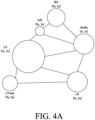
- FIG. 4A is a diagram illustrating an example of a chamber anatomy meter for a result of segmenting a cardiac monolayer region based on a standard cross section received in advance according to an exemplary embodiment of the present invention.
- FIG. 4B is a diagram illustrating an example of a ratio meter for the result of segmenting the cardiac monolayer region based on the standard cross section received in advance according to an exemplary embodiment of the present invention.
- the chamber anatomy meter for the result of segmenting the cardiac monolayer region based on the standard cross section received in advance may be previously predicted in advance to probabilistically distribute coordinates of respective points corresponding to the center of each region of the aorta (Aorta), the intect ventricular septum (IVS), the left atrium (LA), the left ventricle (LV), the right ventricle (RV), and the left ventricle posterior wall (LVPW) by inputting the standard measurement cross section received in advance, and for example, in respect to the coordinates at which the respective points may be located, a point corresponding to a center of the Aorta may correspond to (X 1 , Y 1 ), a point corresponding to a center of the intect ventricular septum (IVS) may correspond to (X 2 , Y 2 ), a point corresponding to a center of the left atrium (LA) may correspond to (X 3 , Y 3 ),
- area ratios of each of the plurality of cardiac monolayer regions segmented in the cross section may be measured in advance as 18% for the right ventricle (RV), 32% for the left ventricle (LV), 19% for the aorta, 19% for the left atrium (LA), 11% for the left ventricle posterior wall (LVPW), and 5% for the intect ventricular septum (IVS) by inputting the standard measurement cross section received in advance.
- RV right ventricle
- LV left ventricle
- LA left atrium
- LVPW left ventricle posterior wall
- IVS intect ventricular septum
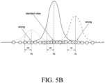
- FIGS. 5A and 5B are diagrams illustrating examples of evaluating the result of segmenting the cardiac monolayer region using an entropy meter according to an exemplary embodiment of the present invention.
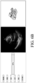
- FIGS. 6A to 6C are diagrams illustrating examples of evaluating the result of segmenting the cardiac monolayer region using a capacity meter according to an exemplary embodiment of the present invention.
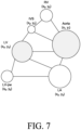
- FIG. 7 is a diagram illustrating an example of evaluating the result of segmenting the cardiac monolayer region using a connectivity meter according to an exemplary embodiment of the present invention.
- the matching rate calculation model of the present invention may be configured to calculate a measurement value corresponding to an area and a size of each of the segmented cardiac monolayer regions by inputting the plurality of points in the echocardiographic image determined by the first prediction model, and may be further configured to calculate the matching rate by comparing the measurement value determined in the cross section based on the standard cross section received in advance.
- the matching rate calculation model may be configured to calculate the matching rate by evaluating measurement values measured in segmented cardiac monolayer regions by inputting data of an entropy meter, a capacity meter, and a connectivity meter by using a measurement value corresponding to an area and a size of each of the segmented cardiac monolayer regions by inputting the plurality of points in the echocardiographic image, and comparing the measurement value determined from the cross section based on the standard cross section received in advance.
- FIGS. 5A and 5B illustrate examples of evaluating the results of segmenting the cardiac monolayer region according to an exemplary embodiment of the present invention using an entropy meter, and by inputting data such as an entropy distribution in the standard measurement cross section (standard image, view), an entropy in a plurality of segmented cardiac monolayer regions acquired from a real-time ultrasound image is measured and compared with the entropy distribution in the standard measurement cross section (standard image, view), so it is possible to provide a guideline for probe manipulation for acquiring a cross section such as the standard measurement cross section (standard image, view).
- Equation 1 above p may represent uncertainty
- Equation 2 above x t may represent a pixel
- c may represent a classification
- h may represent a height
- w may represent a width
- p may represent a probability
- FIG. 6A is a diagram illustrating the result of segmenting the cardiac monolayer region, and an example of measuring the capacity of each segmented heart region in the previously received standard cross section according to an exemplary embodiment of the present invention
- FIGS. 6B and 6C are diagrams illustrating examples of evaluating the result of segmenting the cardiac monolayer region by using the connectivity meter according to an exemplary embodiment of the present invention.
- coordinates of a plurality of points in the echocardiographic image may be predicted in advance by inputting the standard measurement cross section received in advance, and referring back to FIG. 4B , area ratios of the plurality of respective cardiac monolayer regions segmented in the cross section may be predicted in advance as 18% for the right ventricle (RV), 32% for the left ventricle (LV), 19% for the Aorta, 19% for the left atrium (LA), 11% for the left ventricle posterior wall (LVPW), and 5% for the intect ventricular septum (IVS), so that it is possible to provide the movement guide of the probe for acquiring the standard measurement cross section by comparing the coordinates of the plurality of points and the area of each region measured in each of the segmented regions with the coordinates of the points and the area of each region in the standard measurement cross section received in advance.
- RV right ventricle
- LV left ventricle
- Aorta 19% for the Aorta
- LA left atrium
- LVPW left
- FIG. 7 is a diagram illustrating an example of evaluating the result of segmenting the cardiac monolayer region using the connectivity meter according to an exemplary embodiment of the present invention.
- coordinates of respective points corresponding to the center of each region of the aorta, the intect ventricular septum (IVS), the left atrium (LA), the left ventricle (LV), the right ventricle (RV), and the left ventricle posterior wall (LVPW) may be predicted in advance to be probabilistically distributed by inputting the standard measurement cross section received in advance, and for example, in respect to the coordinates at which the respective points may be located, a point corresponding to a center of the aorta may correspond to (X 1 , Y 1 ), a point corresponding to a center of the intect ventricular septum (IVS) may correspond to (X 2 , Y 2 ), a point corresponding to a center of the left atrium (LA) may correspond to (X 3 , Y 3 ), a point corresponding to a center of the left ventricle (LV) may correspond to (X 4 , Y 4 ), a point corresponding to a
- the matching rate may be calculated by comparing the connectivity of each point corresponding to the center of each region with that in the standard measurement cross section received in advance, and in FIG. 7 , since it may be confirmed that the point corresponding to the center of the Aorta is enlarged compared to the point corresponding to the center of the Aorta in FIG. 4A , it can be seen that the probe should be moved to the left to acquire a point close to FIG. 4A .
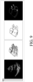
- FIGS. 8 to 10 a guideline presented by utilizing the echocardiography guide method according to various exemplary embodiments of the present invention will be described with reference to FIGS. 8 to 10 .
- a real-time echocardiographic image is received, the cross section is segmented into each of a plurality of cardiac monolayer regions, and the received echocardiographic image is input to predict the positions of a plurality of points within the echocardiographic image, thereby providing a movement guide of the probe for acquiring the standard measurement cross section.
- FIG. 9 illustrates that the cross section is segmented into a plurality of cardiac monolayer regions in the standard measurement cross section, and the positions and entropy of a plurality of points are measured in each region
- FIG. 10 is a diagram illustrating a result of segmenting a cardiac monolayer region when the probe acquires the echocardiographic image at a wrong position, and the positions and entropy of the plurality of points measured.
- the segmentation result of the cardiac monolayer region and a plurality of predicted point measurement values are compared with the measurement values determined in the cross section based on the standard cross section received in advance by inputting data of an entropy meter, a capacity meter, and a connectivity meter, and evaluated, and a matching rate with the standard cross section may be calculated based thereon.
- FIG. 9 indicates a case where a matching rate is 99%, and it is possible to acquire a cross section close to the standard cross section, and referring to FIG. 10, FIG. 10 illustrates a result that the matching rate is calculated as 35%, and in the present invention, as the matching rate in the real-time echocardiographic image may be confirmed, the probe may be enabled to be guided to a clinically meaningful position.
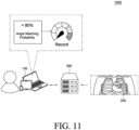
- FIG. 11 illustrates a guide system for a cross section of an echocardiography using an echocardiography guide device according to an exemplary embodiment of the present invention.
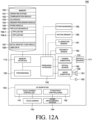
- FIG. 12A is a block diagram illustrating a configuration of a medical staff device according to an exemplary embodiment of the present invention.
- FIG. 12B is a block diagram illustrating a configuration of a guide server according to an exemplary embodiment of the present invention.
- the guide system 1000 may be a system configured to provide a guide for echocardiography measurement based on an echocardiographic image of an individual.
- the guide system 1000 may be constituted by a medical staff device 100 receiving a guide for echocardiography measurement, an echocardiographic image diagnosis device 200 providing the echocardiographic image, and a guide server 300 generating the guide for echocardiography measurement based on the received echocardiographic image.
- the medical staff device 100 as an electronic device providing a user interface for representing the guide for echocardiography measurement may include at least one of a smartphone, a tablet personal computer (PC), a laptop computer, and/or a PC.
- a smartphone a tablet personal computer (PC)
- PC personal computer
- laptop computer a laptop computer
- PC personal computer
- the medical staff device 100 may receive prediction results associated with an electrocardiography cross section for the individual and an angle from the guide server 300, and display the received results through a display unit to be described below.
- the guide server 300 may include a general-purpose computer, laptop, and/or data server that perform various operations for determining the guide for echocardiography measurement based on the echocardiographic image provided from the echocardiographic image diagnosis device 200 such as an ultrasound diagnosis device.
- the guide server 300 may be a device for accessing a web server providing a web page or a mobile web server providing a mobile website, but is not limited thereto.
- the guide server 300 receives an echocardiographic image of an individual and a cross section in the image from the echocardiographic image diagnosis device 200, and determines a relative angle between a standard image cross section received in advance and the received cross section to generate a guide for reducing the relative angle based on the relative angle, and finally provide the guide as a graphic interface.
- the guide server 300 may determine the relative angle using an Euler angle.
- the provided guide may further include a two-dimensional relative angle for a three-dimensional image determined using the Euler angle.
- the provided guide may provide a matching degree between relative angles of the cross section and a standard image.
- an angle at a current position of the probe is compared with the relative angle in the standard image and the cross section to provide a matching degree, so the matching degree may be used as a navigation for acquiring the standard image and the cross section, and it is possible to provide a guideline for highly reliable echocardiography measurement.
- the guide server 300 may provide the prediction results to the medical staff device 100.
- Information provided from the guide server 300 may be provided to a web page through a web browser installed in the medical staff device 100, or provided in the form of an application or a program.
- the data may be provided in a form that is embedded in a platform in a client-server environment.
- the medical staff device 100 may include a memory interface 110, one or more processors 120, and a peripheral interface 130. Various components within the medical staff device 100 may be connected by one or more communication buses or signal lines.
- the memory interface 110 is connected to the memory 150 to transmit various data to the processor 120.
- the memory 150 may include at least one type of storage medium among flash memory type, hard disk type, multimedia card micro type, card type memory (for example, SD or XD memory, etc.), RAM, SRAM, ROM, EEPROM, PROM, network storage, cloud, and blockchain data.
- the memory 150 may store at least one of an operating system 151, a communication module 152, a graphical user interface module (GUI) 153, a sensor processing module 154, a phone module 155, and an application module 156.
- the operating system 151 may include instructions for processing basic system services and instructions for performing hardware tasks.
- the communication module 152 may communicate with at least one of one or more other devices, computers, and servers.
- the graphical user interface module (GUI) 153 may process a graphical user interface.
- the sensor processing module 154 may process sensor-related functions (e.g., processing a voice input received using one or more microphones 192).
- the phone module 155 may process phone-related functions.
- the application module 156 may perform various functions of the user application, such as electronic messaging, web browsing, media processing, navigation, imaging, and other processing functions.
- the medical staff device 100 may store one or more software applications 156-1 and 156-2 (e.g., a guide application) associated with any one type of service in the memory 150.
- the memory 150 may store a digital assistant client module 157 (hereinafter, referred to as DA client module), and thus store instructions for performing client-side functions of a digital assistant and various user data 158.
- DA client module digital assistant client module
- the DA client module 157 may acquire the user's voice input, text input, touch input, and/or gesture input through various user interfaces (e.g., I/O subsystem 140) provided in the medical staff device 100.
- various user interfaces e.g., I/O subsystem 140
- the DA client module 157 may output data in audiovisual and tactile forms.
- the DA client module 157 may output data consisting of a combination of at least two of voice, sound, notification, text message, menu, graphic, video, animation, and vibration.
- the DA client module 157 may communicate with a digital assistant server (not illustrated) using a communication subsystem 180.
- the DA client module 157 may collect additional information about a surrounding environment of the medical staff device 100 from various sensors, subsystems, and peripheral devices to construct a context associated with the user input.
- the DA client module 157 may infer a user's intention by providing context information to a digital assistance server jointly with the user input.
- the context information that may accompany the user input may include sensor information, such as lighting, ambient noise, ambient temperature, images of the surrounding environment, video, etc.
- the context information may include a physical state of the medical staff device 100 (e.g., device orientation, device position, device temperature, power level, speed, acceleration, motion pattern, cellular signal strength, etc.).
- the context information may include information related to the software state of the medical staff device 100 (e.g., processes running on the medical staff device 100, installed programs, past and current network activity, background services, error logs, resource usage, etc.).
- the memory 150 may include additional or deleted instructions, and may further include additional components other than those illustrated in FIG. 12A of the medical staff device 100, or may exclude some components.
- the processor 120 may control the overall operation of the medical staff device 100 and execute various instructions to implement an interface that provides the guide for echocardiography measurement by running an application or program stored in the memory 150.
- the processor 120 may correspond to an operation device such as a central processing unit (CPU) or an application processor (AP). Further, the processor 120 may be implemented in the form of an integrated chip (IC) such as a system on chip (SoC) in which various operation devices such as a neural processing unit (NPU) are integrated.
- IC integrated chip
- SoC system on chip
- NPU neural processing unit
- the peripheral interface 130 is connected to various sensors, subsystems, and peripheral devices to provide data so that the medical staff device 100 may perform various functions. Here, it may be understood that any function performed by the medical staff device 100 is performed by the processor 120.
- the peripheral interface 130 may receive data from a motion sensor 160, an illumination sensor (optical sensor) 161, and a proximity sensor 162, and through this, the medical staff device 100 may perform orientation, light, and proximity detection functions.
- the peripheral interface 130 may receive data from other sensors 163 (positioning system - GPS receiver, temperature sensor, biometric sensor), thereby enabling the medical staff device 100 to perform functions related to the other sensors 163.
- the medical staff device 100 may include a communication subsystem 180 connected to the peripheral interface 130.
- the communication subsystem 180 consists of one or more wired/wireless networks and may include various communication ports, radio frequency transceivers, and optical transceivers.
- the medical staff device 100 includes an audio subsystem 190 connected to the peripheral interface 130, the audio subsystem 190 includes one or more speakers 191 and one or more microphones 192, such that the medical staff device 100 may perform voice-activated functions, such as speech recognition, voice replication, digital recording, and telephony functions.
- voice-activated functions such as speech recognition, voice replication, digital recording, and telephony functions.
- the medical staff device 100 may include an I/O subsystem 140 connected to the peripheral interface 130.
- the I/O subsystem 140 may control a touch screen 143 included in the medical staff device 100 via a touch screen controller 141.
- the touch screen controller 141 may detect a user's contact and movement or cessation of contact and movement using any one of a plurality of touch sensing technologies, such as capacitive, resistive, infrared, surface acoustic wave technology, proximity sensor array, etc.
- the I/O subsystem 140 may control other input/control devices 144 included in the medical staff device 100 via other input controller(s) 142.
- the other input controller(s) 142 may control one or more buttons, rocker switches, thumb-wheels, infrared ports, USB ports, and pointer devices such as a stylus.
- the guide server 300 may include a communication interface 310, a memory 320, an I/O interface 330, and a processor 340, and respective components may communicate with each other through one or more communication buses or signal lines.
- the communication interface 310 may be connected to the medical staff device 100 and the echocardiographic image diagnosis device 200 via a wired/wireless communication network to exchange data.
- the communication interface 310 may receive the echocardiographic image from the echocardiographic image diagnosis device 200, and transmit determined information to the medical staff device 100.
- the communication interface 310 that enables transmission and reception of such data may include a communication port 311 and a wireless circuit 312, and here, the wired communication port 311 may include one or more wired interfaces, for example, Ethernet, universal serial bus (USB), FireWire, etc. Further, the wireless circuit 312 may transmit and receive data with an external device via RF signals or optical signals. Moreover, the wireless communication may use at least one of a plurality of communication standards, protocols and technologies, such as GSM, EDGE, CDMA, TDMA, Bluetooth, Wi-Fi, VoIP, Wi-MAX, or any other suitable communication protocol.
- the memory 320 may store various data used in the guide server 300.
- the memory 320 may store an echocardiographic image, or models trained to determine ultrasound cross sections and relative angles within the echocardiographic image.
- the memory 320 may include a volatile or nonvolatile storage medium capable of storing various data, instructions, and information.
- the memory 320 may include at least one type of storage medium among flash memory type, hard disk type, multimedia card micro type, card type memory (for example, SD or XD memory, etc.), RAM, SRAM, ROM, EEPROM, PROM, network storage, cloud, and blockchain data.
- the memory 320 may store at least one component of an operating system 321, a communication module 322, a user interface module 323, and one or more applications 324.
- the operating system 321 may include various software components and drivers for controlling and managing general system tasks (e.g., memory management, storage device control, power management, etc.) and may support communication between various hardware, firmware, and software components.
- general system tasks e.g., memory management, storage device control, power management, etc.
- the communication module 323 may support communication with other devices through the communication interface 310.
- the communication module 320 may include various software components for processing data received by the wired communication port 311 or wireless circuit 312 of the communication interface 310.
- the user interface module 323 may receive a user's request or input from a keyboard, touch screen, microphone, etc., through the I/O interface 330 and provide a user interface on a display.
- the application 324 may include a program or module configured to be executed by one or more processors 330.
- the application for providing the guide for echocardiography measurement may be implemented on a server farm.
- the I/O interface 330 may connect at least one of an input/output device (not illustrated) of the guide server 300, such as a display, a keyboard, a touch screen, and a microphone, to the user interface module 323.
- the I/O interface 330 may receive a user input (e.g., a voice input, a keyboard input, a touch input, etc.) together with the user interface module 323, and process an instruction according to the received input.
- the processor 340 is connected to the communication interface 310, the memory 320, and the I/O interface 330 to control the overall operation of the guide server 300, and may perform various instructions for the guide through the application or program stored in the memory 320.
- the processor 340 may correspond to an operation device such as a central processing unit (CPU) or an application processor (AP). Further, the processor 340 may be implemented in the form of an integrated chip (IC) such as a system on chip (SoC) in which various operation devices are integrated. Alternatively, the processor 340 may include a module for calculating an artificial neural network model, such as a neural processing unit (NPU).
- CPU central processing unit
- AP application processor
- IC integrated chip
- SoC system on chip
- NPU neural processing unit
- the processor 340 may be configured to classify a cross section for the echocardiographic image, determine a relative angle between the standard image cross section received in advance and the received cross section, generate a guide for reducing the relative angle based on the relative angle, and provide the guide as a graphic interface.
- the processor 340 may be further configured to determine the relative angle using an Euler angle.
- the processor 340 may be further configured to provide a guide that provides a matching degree between the relative angles of the cross section and the standard image.
- FIG. 13 illustrates a procedure of an echocardiography guide method according to an exemplary embodiment of the present invention.
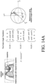
- the Euler angle is a three-angle system introduced by Leonhard Euler to represent a direction in which a rigid body is located in three-dimensional space, and a three-dimensional or two-dimensional standard position for each cross section of an echocardiography may be set in advance, and based on this, a relative angle is determined with respect to a current probe position compared to a three-dimensional or two-dimensional baseline.
- FIG. 14 is an exemplary diagram illustrating determining a relative angle using an Euler angle according to an exemplary embodiment of the present invention.
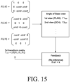
- FIG. 15 illustrates a procedure for reducing a relative angle according to various exemplary embodiments of the present invention.
- information on a cross section in which an angle of a CT image is labeled is received together with a training ultrasound image to determine a relative angle, and feature extraction for an image and a masking region may be performed, and then the angle may be determined through feature stitching layers.
- the angle may be expressed in a transform of a 3D transform matrix, and the feature stitching layers may include a layer for mesh feature extraction.
- a relative angle between a two-dimensional cross section acquired from a three-dimensional image and a preset baseline is determined using the Euler angle
- a relative angle between an echocardiographic image for a three-dimensional or two-dimensional standard position and an individual for each cross section of a preset echocardiography, and the cross section in the image is determined to generate and provide a guide including a feedback for reducing the relative angle based on the relative angle, thereby making it possible to more easily acquire an image corresponding to a standard measurement cross section.
- the present invention provides a guide for determining a relative angle based on a baseline acquired from a standard measurement image and for reducing the relative angle for a current echocardiographic image acquired by the probe, so that the guide may be utilized as a guide when acquiring a 3D echocardiographic image for acquiring a standard cross section.
Landscapes
- Engineering & Computer Science (AREA)
- Health & Medical Sciences (AREA)
- Life Sciences & Earth Sciences (AREA)
- Physics & Mathematics (AREA)
- General Health & Medical Sciences (AREA)
- Biomedical Technology (AREA)
- Biophysics (AREA)
- Theoretical Computer Science (AREA)
- Molecular Biology (AREA)
- Medical Informatics (AREA)
- Nuclear Medicine, Radiotherapy & Molecular Imaging (AREA)
- Radiology & Medical Imaging (AREA)
- Public Health (AREA)
- Surgery (AREA)
- General Physics & Mathematics (AREA)
- Animal Behavior & Ethology (AREA)
- Heart & Thoracic Surgery (AREA)
- Pathology (AREA)
- Veterinary Medicine (AREA)
- Computer Vision & Pattern Recognition (AREA)
- Data Mining & Analysis (AREA)
- Software Systems (AREA)
- Mathematical Physics (AREA)
- General Engineering & Computer Science (AREA)
- Computing Systems (AREA)
- Evolutionary Computation (AREA)
- Computational Linguistics (AREA)
- Artificial Intelligence (AREA)
- Cardiology (AREA)
- Quality & Reliability (AREA)
- Ultra Sonic Daignosis Equipment (AREA)
- Geometry (AREA)
Applications Claiming Priority (4)
| Application Number | Priority Date | Filing Date | Title |
|---|---|---|---|
| KR20220048018 | 2022-04-19 | ||
| KR1020230051036A KR20240154402A (ko) | 2023-04-18 | 2023-04-18 | 심장 초음파에 대한 가이드 방법 및 이를 이용한 심장 초음파에 대한 가이드용 디바이스 |
| KR1020230051037A KR102917172B1 (ko) | 2022-04-19 | 2023-04-18 | 심장 초음파에 대한 가이드 방법 및 이를 이용한 심장 초음파에 대한 가이드용 디바이스 |
| PCT/KR2023/005325 WO2023204610A2 (ko) | 2022-04-19 | 2023-04-19 | 심장 초음파에 대한 가이드 방법 및 이를 이용한 심장 초음파에 대한 가이드용 디바이스 |
Publications (2)
| Publication Number | Publication Date |
|---|---|
| EP4512343A2 true EP4512343A2 (de) | 2025-02-26 |
| EP4512343A4 EP4512343A4 (de) | 2026-04-15 |
Family
ID=88420130
Family Applications (1)
| Application Number | Title | Priority Date | Filing Date |
|---|---|---|---|
| EP23792184.6A Pending EP4512343A4 (de) | 2022-04-19 | 2023-04-19 | Echokardiographie-führungsverfahren und echokardiographie-führungsvorrichtung damit |
Country Status (3)
| Country | Link |
|---|---|
| US (1) | US20250272844A1 (de) |
| EP (1) | EP4512343A4 (de) |
| WO (1) | WO2023204610A2 (de) |
Families Citing this family (2)
| Publication number | Priority date | Publication date | Assignee | Title |
|---|---|---|---|---|
| JP2025134210A (ja) * | 2024-03-04 | 2025-09-17 | 富士フイルム株式会社 | 超音波診断装置および超音波診断装置の制御方法 |
| EP4620405A1 (de) * | 2024-03-20 | 2025-09-24 | Ontact Health Co., Ltd. | Verfahren zur bereitstellung einer richtlinie für ein kardiales ultraschallbild und vorrichtung zur bereitstellung einer richtlinie für ein kardiales ultraschallbild damit |
Family Cites Families (7)
| Publication number | Priority date | Publication date | Assignee | Title |
|---|---|---|---|---|
| US8073215B2 (en) * | 2007-09-18 | 2011-12-06 | Siemens Medical Solutions Usa, Inc. | Automated detection of planes from three-dimensional echocardiographic data |
| JPWO2013108592A1 (ja) * | 2012-01-19 | 2015-05-11 | コニカミノルタ株式会社 | 超音波診断装置および超音波診断装置の制御方法 |
| KR20150082945A (ko) * | 2014-01-08 | 2015-07-16 | 삼성메디슨 주식회사 | 초음파 진단 장치 및 그 동작방법 |
| KR102698916B1 (ko) * | 2017-01-19 | 2024-08-26 | 뉴욕 유니버시티 | 초음파 분석을 위한 시스템, 방법 및 컴퓨터-접근가능 매체 |
| JP6824125B2 (ja) * | 2017-07-28 | 2021-02-03 | 株式会社日立製作所 | 医用撮像装置及び画像処理方法 |
| JP7193979B2 (ja) * | 2018-10-29 | 2022-12-21 | 富士フイルムヘルスケア株式会社 | 医用撮像装置、画像処理装置、および、画像処理方法 |
| KR102852582B1 (ko) * | 2020-02-05 | 2025-08-29 | 삼성메디슨 주식회사 | 초음파 진단 장치 및 그 동작 방법 |
-
2023
- 2023-04-19 WO PCT/KR2023/005325 patent/WO2023204610A2/ko not_active Ceased
- 2023-04-19 EP EP23792184.6A patent/EP4512343A4/de active Pending
- 2023-04-19 US US18/858,308 patent/US20250272844A1/en active Pending
Also Published As
| Publication number | Publication date |
|---|---|
| WO2023204610A3 (ko) | 2023-12-14 |
| US20250272844A1 (en) | 2025-08-28 |
| WO2023204610A2 (ko) | 2023-10-26 |
| EP4512343A4 (de) | 2026-04-15 |
Similar Documents
| Publication | Publication Date | Title |
|---|---|---|
| EP4512343A2 (de) | Echokardiographie-führungsverfahren und echokardiographie-führungsvorrichtung damit | |
| WO2021018101A1 (zh) | 数据处理方法、装置、设备及存储介质 | |
| KR20250069510A (ko) | 의료 영상 분할 방법 및 이를 수행하는 장치 | |
| KR102846159B1 (ko) | 심장 초음파 영상에 대한 정보 제공 방법 및 이를 이용한 심장 초음파 영상에 대한 정보 제공용 디바이스 | |
| KR102819588B1 (ko) | 도플러 초음파 영상에 대한 정보 제공 방법 및 이를 이용한 도플러 초음파 영상에 대한 정보 제공용 디바이스 | |
| KR20250102794A (ko) | 척추 정렬 상태에 대한 평가 방법 및 이를 이용한 척추 정렬 상태에 대한 평가용 디바이스 | |
| KR102917172B1 (ko) | 심장 초음파에 대한 가이드 방법 및 이를 이용한 심장 초음파에 대한 가이드용 디바이스 | |
| KR20240154402A (ko) | 심장 초음파에 대한 가이드 방법 및 이를 이용한 심장 초음파에 대한 가이드용 디바이스 | |
| US20250295377A1 (en) | Method for providing guideline for cardiac ultrasound image and device for providing guideline for cardiac ultrasound image using the same | |
| US20250295381A1 (en) | Method for providing user interface for cardiac ultrasound imaging guideline and device for providing user interface using the same | |
| US20260114848A1 (en) | Method for providing information about doppler ultrasound image, and device for providing information about doppler ultrasound image using same | |
| KR102951463B1 (ko) | Ood 데이터 탐지 기반 초음파 영상 뷰에 대한 정보 제공 방법 및 이를 이용한 초음파 영상 뷰에 대한 정보 제공용 디바이스 | |
| KR102823321B1 (ko) | 자궁 경부에 대한 정보 제공 방법 및 이를 이용한 자궁 경부에 대한 정보 제공용 디바이스 | |
| KR102732544B1 (ko) | M-모드 초음파 영상에 대한 정보 제공 방법 및 이를 이용한 m-모드 초음파 영상에 대한 정보 제공용 디바이스 | |
| US20250295376A1 (en) | Method for providing information on m-mode ultrasound image, and device for providing information on m-mode ultrasound image by using same | |
| KR102771062B1 (ko) | 대상체에 대한 표준 평면 정보 제공 방법 및 이를 수행하는 디바이스 | |
| KR20250141628A (ko) | 심장 초음파 영상에 대한 가이드라인 제공 방법 및 이를 이용한 심장 초음파 영상에 대한 가이드라인 제공용 디바이스 | |
| KR102947965B1 (ko) | 도플러 초음파 영상 분류에 대한 정보 제공 방법 및 이를 이용한 도플러 초음파 영상 분류에 대한 정보 제공용 디바이스 | |
| KR102874388B1 (ko) | M-모드 초음파 영상에 대한 정보 제공 방법 및 이를 이용한 장치 | |
| EP4534018A1 (de) | Verfahren zur bereitstellung von informationen über eine ultraschallbildansicht und vorrichtung zur bereitstellung von informationen über eine ultraschallbildansicht damit | |
| KR20250142220A (ko) | 측정치 결정을 위한 사용자 인터페이스 제공 방법 및 이를 이용한 사용자 인터페이스 제공용 디바이스 | |
| KR20250071604A (ko) | 심장 초음파 영상에 대한 정보 제공 방법 및 이를 이용한 심장 초음파 영상에 대한 정보 제공용 디바이스 | |
| US20250387102A1 (en) | Method for providing information on severity of aortic stenosis and device using the same | |
| US20250378560A1 (en) | Method and device for providing information on strain quantification | |
| KR20250179174A (ko) | 대동맥 협착증 중증도에 대한 정보 제공 방법 및 이를 이용한 대동맥 협착증 중증도에 대한 정보 제공용 디바이스 |
Legal Events
| Date | Code | Title | Description |
|---|---|---|---|
| STAA | Information on the status of an ep patent application or granted ep patent |
Free format text: STATUS: THE INTERNATIONAL PUBLICATION HAS BEEN MADE |
|
| PUAI | Public reference made under article 153(3) epc to a published international application that has entered the european phase |
Free format text: ORIGINAL CODE: 0009012 |
|
| STAA | Information on the status of an ep patent application or granted ep patent |
Free format text: STATUS: REQUEST FOR EXAMINATION WAS MADE |
|
| 17P | Request for examination filed |
Effective date: 20241113 |
|
| AK | Designated contracting states |
Kind code of ref document: A2 Designated state(s): AL AT BE BG CH CY CZ DE DK EE ES FI FR GB GR HR HU IE IS IT LI LT LU LV MC ME MK MT NL NO PL PT RO RS SE SI SK SM TR |
|
| DAV | Request for validation of the european patent (deleted) | ||
| DAX | Request for extension of the european patent (deleted) | ||
| A4 | Supplementary search report drawn up and despatched |
Effective date: 20260313 |
|
| RIC1 | Information provided on ipc code assigned before grant |
Ipc: A61B 8/00 20060101AFI20260309BHEP Ipc: A61B 8/08 20060101ALI20260309BHEP Ipc: G06T 7/00 20170101ALI20260309BHEP Ipc: G06N 3/08 20230101ALI20260309BHEP |