EP4502601A1 - Immunological detection method and immunological detection kit - Google Patents
Immunological detection method and immunological detection kit Download PDFInfo
- Publication number
- EP4502601A1 EP4502601A1 EP23774938.7A EP23774938A EP4502601A1 EP 4502601 A1 EP4502601 A1 EP 4502601A1 EP 23774938 A EP23774938 A EP 23774938A EP 4502601 A1 EP4502601 A1 EP 4502601A1
- Authority
- EP
- European Patent Office
- Prior art keywords
- antibody
- immunological detection
- collagen
- complex
- detection method
- Prior art date
- Legal status (The legal status is an assumption and is not a legal conclusion. Google has not performed a legal analysis and makes no representation as to the accuracy of the status listed.)
- Pending
Links
Images



Classifications
-
- G—PHYSICS
- G01—MEASURING; TESTING
- G01N—INVESTIGATING OR ANALYSING MATERIALS BY DETERMINING THEIR CHEMICAL OR PHYSICAL PROPERTIES
- G01N33/00—Investigating or analysing materials by specific methods not covered by groups G01N1/00 - G01N31/00
- G01N33/48—Biological material, e.g. blood, urine; Haemocytometers
- G01N33/50—Chemical analysis of biological material, e.g. blood, urine; Testing involving biospecific ligand binding methods; Immunological testing
- G01N33/68—Chemical analysis of biological material, e.g. blood, urine; Testing involving biospecific ligand binding methods; Immunological testing involving proteins, peptides or amino acids
- G01N33/6887—Chemical analysis of biological material, e.g. blood, urine; Testing involving biospecific ligand binding methods; Immunological testing involving proteins, peptides or amino acids from muscle, cartilage or connective tissue
-
- C—CHEMISTRY; METALLURGY
- C07—ORGANIC CHEMISTRY
- C07K—PEPTIDES
- C07K16/00—Immunoglobulins [IG], e.g. monoclonal or polyclonal antibodies
- C07K16/18—Immunoglobulins [IG], e.g. monoclonal or polyclonal antibodies against material from animals or humans
-
- G—PHYSICS
- G01—MEASURING; TESTING
- G01N—INVESTIGATING OR ANALYSING MATERIALS BY DETERMINING THEIR CHEMICAL OR PHYSICAL PROPERTIES
- G01N33/00—Investigating or analysing materials by specific methods not covered by groups G01N1/00 - G01N31/00
- G01N33/48—Biological material, e.g. blood, urine; Haemocytometers
- G01N33/50—Chemical analysis of biological material, e.g. blood, urine; Testing involving biospecific ligand binding methods; Immunological testing
- G01N33/53—Immunoassay; Biospecific binding assay; Materials therefor
- G01N33/577—Immunoassay; Biospecific binding assay; Materials therefor involving monoclonal antibodies binding reaction mechanisms characterised by the use of monoclonal antibodies
-
- G—PHYSICS
- G01—MEASURING; TESTING
- G01N—INVESTIGATING OR ANALYSING MATERIALS BY DETERMINING THEIR CHEMICAL OR PHYSICAL PROPERTIES
- G01N33/00—Investigating or analysing materials by specific methods not covered by groups G01N1/00 - G01N31/00
- G01N33/48—Biological material, e.g. blood, urine; Haemocytometers
- G01N33/50—Chemical analysis of biological material, e.g. blood, urine; Testing involving biospecific ligand binding methods; Immunological testing
- G01N33/58—Chemical analysis of biological material, e.g. blood, urine; Testing involving biospecific ligand binding methods; Immunological testing involving labelled substances
-
- C—CHEMISTRY; METALLURGY
- C07—ORGANIC CHEMISTRY
- C07K—PEPTIDES
- C07K2317/00—Immunoglobulins specific features
- C07K2317/30—Immunoglobulins specific features characterized by aspects of specificity or valency
- C07K2317/34—Identification of a linear epitope shorter than 20 amino acid residues or of a conformational epitope defined by amino acid residues
-
- G—PHYSICS
- G01—MEASURING; TESTING
- G01N—INVESTIGATING OR ANALYSING MATERIALS BY DETERMINING THEIR CHEMICAL OR PHYSICAL PROPERTIES
- G01N2333/00—Assays involving biological materials from specific organisms or of a specific nature
- G01N2333/435—Assays involving biological materials from specific organisms or of a specific nature from animals; from humans
- G01N2333/78—Connective tissue peptides, e.g. collagen, elastin, laminin, fibronectin, vitronectin, cold insoluble globulin [CIG]
-
- G—PHYSICS
- G01—MEASURING; TESTING
- G01N—INVESTIGATING OR ANALYSING MATERIALS BY DETERMINING THEIR CHEMICAL OR PHYSICAL PROPERTIES
- G01N2800/00—Detection or diagnosis of diseases
- G01N2800/34—Genitourinary disorders
- G01N2800/347—Renal failures; Glomerular diseases; Tubulointerstitial diseases, e.g. nephritic syndrome, glomerulonephritis; Renovascular diseases, e.g. renal artery occlusion, nephropathy
Definitions
- the present invention relates to an immunological detection method and an immunological detection kit.
- Type I procollagen N-propeptide (hereinafter referred to as "P1NP” or “PINP”) is a polypeptide with a molecular weight of approximately 35 kDa that is cleaved from the N-terminus when type I collagen, the main component of bone matrix protein, is produced from the precursor type I procollagen.
- PINP released into the blood is known to be a bone formation marker that sensitively reflects early stage of bone formation. Therefore, blood PINP concentration is useful for determining and monitoring the therapeutic effects of osteoporosis drugs such as PTH (parathyroid hormone) preparations and bisphosphonate preparations.
- PTH parthyroid hormone
- bisphosphonate preparations bisphosphonate preparations.
- the measurement of PINP (total PINP) sometimes results in detection of monomers as well as intact PINP as a trimer.
- procollagen-N-propeptide kit for example, Procollagen Intact PINP (product name, manufactured by Fujirebio Inc., Non-Patent Document 1) and Elecsys (registered trademark) Reagent Total P1NP (product name, Roche Diagnostics, Non-Patent Document 2) are commercially available.
- Procollagen Intact PINP product name, manufactured by Fujirebio Inc., Non-Patent Document 1
- Elecsys registered trademark
- Reagent Total P1NP product name, Roche Diagnostics, Non-Patent Document 2
- Patent Document 1 discloses a method and a test kit for determining the presence of amino-terminal propeptides of type I procollagen in individual persons.
- Patent Literature 2 discloses an assay method for the ⁇ 1-homotrimer amino-terminal propeptide which is an intact trimer of type I procollagen or the heterotrimer amino-terminal propeptide which is an intact classical type I procollagen, and an assay kit suitable for use in the assay method.
- Non-Patent Literature 3 discloses that a trimeric PINP and a monomeric PINP have different clearance pathways (trimer: liver, monomer: kidney), and that patients with renal disease tend to lack ability of form clearance of the monomer and have high monomer concentrations in the blood.
- Non-Patent Literature 4 discloses that a monomeric PINP reflects the degradation product of pN-collagen rather than the thermal denaturation product of the intact propeptide.
- Intact PINP Procollagen Intact PINP (hereinafter abbreviated as "Intact PINP") described in Non-Patent Literature 1 can detect only the trimer of the ⁇ -chain of type I collagen N-terminal propeptide.
- Intact PINP is a measurement kit based on radioimmunoassay method, and uses an antibody labeled with a radioisotope (125I). Therefore, the measurement needs to be performed in a radiation controlled area and requires about 3-hour measurement time, which is cumbersome.
- the half-life of the radioisotope (125I) is as short as 59.4 days, the shelf life of the kit is also as short as 6 weeks at 2 to 8 °C.
- the Elecsys reagent total P1NP (hereinafter abbreviated as "total P1NP") described in Non-Patent Literature 2 can detect the trimer and monomer of the ⁇ -chain of type I collagen N-terminal propeptide.
- the total P1NP is a measurement kit based on electrochemiluminescence immunoassay (ECLIA), and is widely used because it is easy to handle with no need for a radiation control area and a measurement time of about 20 minutes.
- the kit also has a long shelf life of about 18 months at 2 to 8 °C.
- the present invention has been made in consideration of the above circumstances, and one of its objects is to provide an immunological detection method and an immunological detection kit that are easy to handle and can specifically (selectively) measure the trimer in biological samples containing the trimer and monomer of the ⁇ -chain of PINP.
- the present invention can provide an immunological detection method and an immunological detection kit that are easy to handle and can specifically (selectively) measure the trimer in biological samples containing the trimer and monomer of the ⁇ -chain of PINP.
- the terms "react,” “recognize,” and “bind” are used synonymously in relation to the reactivity of an antibody with an antigen.
- an antibody disclosed herein is described as not reactive with respect to a certain compound, it means that the antibody does not substantially react with the certain compound.
- the antibody is described as not reactive when the measured value is below the detection limit.
- the terms “detection” and “measurement” are used in a sense encompassing the verification of the presence and/or quantification of trimeric type I collagen N-terminal propeptide.
- the immunological detection method of the present invention is an immunological detection method for trimeric type I collagen N-terminal propeptide in a biological sample, and uses a first antibody that binds to a first specific portion of a pro- ⁇ 1 chain in the trimeric type I collagen N-terminal propeptide and a second antibody that binds to a second specific portion of a pro- ⁇ 1 chain in the trimeric type I collagen N-terminal propeptide.
- the biological sample that can be used in the immunological detection method of the present invention is not particularly limited as long as it is a sample derived from a living body, but is preferably a liquid sample such as a body fluid, more preferably at least one selected from the group consisting of blood, plasma, and serum, and even more preferably any one selected from the group consisting of blood, plasma, and serum.
- the living body is preferably a human or a mammal (e.g., a mouse, a guinea pig, a rat, a monkey, a dog, a cat, a hamster, a horse, a cow, and a pig), and more preferably a human.
- the biological sample may be one collected or prepared during the implementation of the present invention, or may be one collected or prepared in advance and stored.
- the biological sample may usually contain both the trimer and monomer of type I collagen N-terminal propeptide, or may contain only one of the trimer and the monomer, or may contain neither the trimer nor the monomer.
- the biological sample is preferably one derived from a subject with impaired renal function.
- subjects with impaired renal function include patients with chronic kidney disease.
- Renal function can be evaluated, for example, by eGFR value (estimated glomerular filtration rate, unit: mL/min/1.73m 2).
- Trimeric type I collagen N-terminal propeptide is a polypeptide with a molecular weight of approximately 35 kDa, which is cleaved from the N-terminus during the formation of type I collagen from its precursor, i.e., type I procollagen.
- This polypeptide is a heterotrimer consisting of two ⁇ 1 chains (pro- ⁇ 1 chains) with a molecular weight of approximately 15 kDa and one ⁇ 2 chain (pro- ⁇ 2 chain) with a molecular weight of approximately 5 kDa.
- the first specific portion of the pro- ⁇ 1 chain is a portion having a specific primary structure (amino acid sequence) to which the first antibody binds.
- the primary structure (amino acid sequence) of the first specific portion of the pro- ⁇ 1 chain is, for example, QEEGQVEGQD (SEQ ID NO: 1) shown in FIG. 1 .
- the second specific portion of the pro- ⁇ 1 chain is a portion having a specific primary structure (amino acid sequence) to which the second antibody binds.
- the primary structure (amino acid sequence) of the second specific portion of the pro- ⁇ 1 chain is, for example, PDGSESPTDQETT (SEQ ID NO: 2) shown in FIG. 1 .
- the first antibody is not particularly limited as long as it binds to the first specific portion of the pro- ⁇ 1 chain, but is preferably an antibody that binds to the primary structure (amino acid sequence) of QEEGQVEGQD (SEQ ID NO: 1) shown in FIG. 1 , such as an anti-C1A1-01 antibody.
- the first antibody is preferably a monoclonal antibody.
- a monoclonal antibody can be produced, for example, by fusing a B lymphocyte with an immortalized cultured cell to form a hybridoma.
- Monoclonal antibodies are preferable because they are specific and are expected to produce significantly lower background signals than polyclonal antibodies.
- the anti-C1A1-01 antibody is an antibody prepared by the method of the Examples described below using an immunogen obtained by binding ovalbumin, a carrier protein, to a peptide having a primary structure represented by SEQ ID NO: 3: QEEGQVEGQDC.
- the hybridoma producing the antibody S24206R shown in FIG. 1 has been deposited at the National Institute of Technology and Evaluation, Patent Microorganism Depositary (NPMD) (Room 122, 2-5-8 Kazusakamatari, Kisarazu, Chiba Prefecture, Japan) under the name NITE BP-03606 (deposit date: February 15, 2022).
- NPMD Patent Microorganism Depositary
- the second antibody is not particularly limited as long as it binds to the second specific portion of the pro- ⁇ 1 chain, but is preferably an antibody that binds to the primary structure (amino acid sequence) of PDGSESPTDQETT (SEQ ID NO: 2) shown in FIG. 1 , such as an anti-C1A1-02 antibody.
- the anti-C1A1-02 is an antibody prepared by the method of the Examples described below using an immunogen obtained by binding ovalbumin, a carrier protein, to a peptide having a primary structure represented by SEQ ID NO: 4: PDGSESPTDQETTC.
- the hybridoma producing the antibody S24217 shown in FIG. 1 has been deposited at the NPMD as NITE BP-03607, and the hybridoma producing the antibody S24219 has been deposited at the NPMD as NITE BP-03608 (deposit date: February 15, 2022).
- first antibody or the second antibody binds to a monomer of type I collagen N-terminal propeptide derived from a patient with renal disease.
- the detection method is not particularly limited as long as it is an immunological method, but is preferably any one selected from the group consisting of an electrochemiluminescence method, latex agglutination immunoturbidimetry, and enzyme-linked immunosorbent assay (ELISA).
- the first antibody and the second antibody is labeled with a labeling substance.
- the labeling substance include enzymes, fluorescent substances, chemiluminescent substances, biotin, avidin, gold colloid particles, and colored latex.
- the method for detecting a signal derived from a labeling substance can be appropriately selected depending on the respective labeling substances.
- the immunological detection kit of the present invention is an immunological detection kit for trimeric type I collagen N-terminal propeptide in a biological sample, and includes a first antibody that binds to a first specific portion of a pro- ⁇ 1 chain in the trimeric type I collagen N-terminal propeptide, and a second antibody that binds to a second specific portion of a pro- ⁇ 1 chain in the trimeric type I collagen N-terminal propeptide.
- the biological sample, trimeric type I collagen N-terminal propeptide, the first antibody, and the second antibody are as described for the immunological detection method of the present invention.
- the immunological detection kit of the present invention may further include a protocol, a detection device, an analysis device, etc.
- the immunological detection kit of the present invention is preferable for carrying out the above-mentioned immunological detection method of the present invention.
- the following peptides were bound to the carrier protein ovalbumin (OVA) to prepare immunogens.
- OVA ovalbumin
- C1A1-01-OVA, and C1A2-01-OVA were used as immunogens, and were mixed at 1:1 with Freund's Complete Adjuvant (Difco Laboratories) for the first immunization and Freund's Incomplete Adjuvant (Difco Laboratories) for the second and subsequent immunizations.
- 6-week-old (female) Balb/c mice (CLEA Japan, Inc.) or F344/Jc1 rats (CLEA Japan, Inc.) were subcutaneously immunized every 2 weeks with 40 ⁇ g of the immunogen for the first immunization and 20 ⁇ g for the second and subsequent immunizations. The immunization was continued for 8 weeks.
- Antigen-immobilized ELISA was performed according to the following procedure to determine the antibody titer in the blood.
- the samples were measured using Procollagen Intact PINP (Fujirebio Inc.) and Elecsys Reagent Total P1NP (Roche Diagnostics K.K.) by an outside contractor.
- S24206R antibody was prepared in PBS to a concentration of 2.0 mg/mL, followed by adding, with stirring, 20 equivalents of bis(2,2'-bipyridine) [4'-methyl-4-(4-NHS-4-oxobutyl)-2,2'bipyridine] ruthenium (II) bis(hexafluorophosphate) (hereinafter referred to as Ru complex NHS, manufactured by Tokyo Chemical Industry Co., Ltd.) per mole of antibody, which had been dissolved in a small amount of DMSO, and the resulting was stirred for 30 minutes at room temperature in the dark.
- Ru complex NHS manufactured by Tokyo Chemical Industry Co., Ltd.
- a fully automated electrochemiluminescence immunoassay device PicoLumi III (manufactured by Sekisui Medical Co., Ltd.) was used for the measurement. 100 ⁇ L of blocking solution containing mouse IgG and 5 ⁇ L of the measurement sample were added to a reaction tube. 25 ⁇ L of the S24217 antibody-immobilized magnetic particle suspension or the S24219 antibody-immobilized magnetic particle suspension was added and reacted at 28 °C for 5 minutes. After separation of BF using the BF separation solution specified for the device, 100 ⁇ L of Ru complex-labeled S24206R antibody solution was added and the resulting was reacted for 3 minutes. After the BF separation, the magnetic particles were suspended in the substrate (tripropylamine) solution specified for the device, and electricity was applied at a predetermined position on the device.
- Sample 1 contains a large amount of monomeric P1NP.
- Monomeric P1NP and trimeric P1NP in the sample were separated by size exclusion chromatography using Prominence (LC-20A Series manufactured by Shimadzu Corporation) and TSKgel G2000SW XL (Tosoh Corporation).
- PBST phosphate-buffered saline with Tween (registered trademark)
- Tween registered trademark
- FIG. 12 shows the results for the sample prepared by mixing an equal amount of sample 1 and sample 2
- FIG. 13 shows the results for sample 1.
- the trimeric P1NP was mainly contained in fraction No. 7 and the monomeric P1NP was contained in fraction No. 10.
- Each fraction was measured by electrochemiluminescence using a combination of S24217 as the solid-phase antibody and S24206R as the Ru-labeled antibody, and by Elecsys reagent total P1NP.
- serial dilutions of serum from patients with cancer bone metastasis with high P1NP levels, valued with the Elecsys reagent total P1NP were used as standard to calculate the measured values.
- Both S24217 and S24206R are antibodies that bind to the ⁇ 1 chain, but when the fraction containing monomeric P1NP was measured by electrochemiluminescence using the combination of these antibodies, no significant increase in the measured value was observed. Therefore, the antibody combination of S24217 and S24206R can specifically detect trimer P1NP.
- the results are shown in FIGs. 2 to 5 .
- the results are shown in FIGs. 6 to 8 .
- the results are shown in FIG. 9 and FIG. 11 .
- r 0.646 for all samples
- r' 0.528 for samples from patients at stages 4 and 5 ( FIG. 11 ).
- the correlation between the measurement values by the Elecsys reagent total P1NP and the measurement values by the Procollagen Intact PINP was particularly poor in samples from patients with severe renal disease.
- NPMD is an acronym for the National Institute of Technology and Evaluation, Patent Microorganism Depositary (Room 122, 2-5-8 Kazusakamatari, Kisarazu, Chiba Prefecture, Japan).
Landscapes
- Health & Medical Sciences (AREA)
- Life Sciences & Earth Sciences (AREA)
- Engineering & Computer Science (AREA)
- Chemical & Material Sciences (AREA)
- Immunology (AREA)
- Biomedical Technology (AREA)
- Molecular Biology (AREA)
- Urology & Nephrology (AREA)
- Hematology (AREA)
- General Health & Medical Sciences (AREA)
- Medicinal Chemistry (AREA)
- Biochemistry (AREA)
- Biotechnology (AREA)
- General Physics & Mathematics (AREA)
- Food Science & Technology (AREA)
- Cell Biology (AREA)
- Pathology (AREA)
- Analytical Chemistry (AREA)
- Physics & Mathematics (AREA)
- Microbiology (AREA)
- Proteomics, Peptides & Aminoacids (AREA)
- Organic Chemistry (AREA)
- Chemical Kinetics & Catalysis (AREA)
- Biophysics (AREA)
- Genetics & Genomics (AREA)
- Peptides Or Proteins (AREA)
Abstract
The present invention provides an immunological detection method for trimeric type I collagen N-terminal propeptide in a biological sample, which uses a first antibody that binds to a first specific portion of a pro-α1 chain in the trimeric type I collagen N-terminal propeptide and a second antibody that binds to a second specific portion of a pro-α1 chain in the trimeric type I collagen N-terminal propeptide. The immunological detection method is easy to handle and can specifically (selectively) measure the trimer in biological samples containing the trimer and monomer of the α-chain of PINP.
Description
- The present invention relates to an immunological detection method and an immunological detection kit.
- Priority is claimed on
Japanese Patent Application No. 2022-050846, filed March 25, 2022 - Type I procollagen N-propeptide (hereinafter referred to as "P1NP" or "PINP") is a polypeptide with a molecular weight of approximately 35 kDa that is cleaved from the N-terminus when type I collagen, the main component of bone matrix protein, is produced from the precursor type I procollagen. PINP released into the blood is known to be a bone formation marker that sensitively reflects early stage of bone formation. Therefore, blood PINP concentration is useful for determining and monitoring the therapeutic effects of osteoporosis drugs such as PTH (parathyroid hormone) preparations and bisphosphonate preparations. The measurement of PINP (total PINP) sometimes results in detection of monomers as well as intact PINP as a trimer.
- As type I procollagen-N-propeptide kit (generic name), for example, Procollagen Intact PINP (product name, manufactured by Fujirebio Inc., Non-Patent Document 1) and Elecsys (registered trademark) Reagent Total P1NP (product name, Roche Diagnostics, Non-Patent Document 2) are commercially available.
-
Patent Document 1 discloses a method and a test kit for determining the presence of amino-terminal propeptides of type I procollagen in individual persons. -
Patent Literature 2 discloses an assay method for the α1-homotrimer amino-terminal propeptide which is an intact trimer of type I procollagen or the heterotrimer amino-terminal propeptide which is an intact classical type I procollagen, and an assay kit suitable for use in the assay method. - Non-Patent
Literature 3 discloses that a trimeric PINP and a monomeric PINP have different clearance pathways (trimer: liver, monomer: kidney), and that patients with renal disease tend to lack ability of form clearance of the monomer and have high monomer concentrations in the blood. - Non-Patent
Literature 4 discloses that a monomeric PINP reflects the degradation product of pN-collagen rather than the thermal denaturation product of the intact propeptide. -
- Patent Literature 1:
Japanese Unexamined Patent Application Publication (Translation of PCT Application) No. H7-504896 - Patent Literature 2:
Japanese Unexamined Patent Application Publication No. H8-291197 -
- Non-Patent Literature 1: Package insert (13th edition) of "Type I Procollagen-N-propeptide Kit Procollagen Intact PINP", Fujirebio Inc., February 2020
- Non-Patent Literature 2: Package insert (9th edition) of "Type I Procollagen-N-propeptide Kit Elecsys Reagent (R) Total P1NP", Roche Diagnostics K.K., March 2021
- Non-Patent Literature 3: Koivula, M. -K. , and 2 others, "Measurement of aminoterminal propeptide of type I procollagen (PINP) in serum", Clinical Biochemistry, August 2012, Vol. 45, No. 12, pp. 920-927
- Non-Patent Literature 4: Koivula, M. -K. , and 7 others, "Difference between total and intact assays for N-terminal propeptide of type I procollagen reflects degradation of pN-collagen rather than denaturation of intact propeptide," Annals of Clinical Biochemistry, January 2010, Vol. 47, No. 1, p. 67-71
- Procollagen Intact PINP (hereinafter abbreviated as "Intact PINP") described in
Non-Patent Literature 1 can detect only the trimer of the α-chain of type I collagen N-terminal propeptide. Intact PINP is a measurement kit based on radioimmunoassay method, and uses an antibody labeled with a radioisotope (125I). Therefore, the measurement needs to be performed in a radiation controlled area and requires about 3-hour measurement time, which is cumbersome. In addition, since the half-life of the radioisotope (125I) is as short as 59.4 days, the shelf life of the kit is also as short as 6 weeks at 2 to 8 °C. - The Elecsys reagent total P1NP (hereinafter abbreviated as "total P1NP") described in Non-Patent
Literature 2 can detect the trimer and monomer of the α-chain of type I collagen N-terminal propeptide. The total P1NP is a measurement kit based on electrochemiluminescence immunoassay (ECLIA), and is widely used because it is easy to handle with no need for a radiation control area and a measurement time of about 20 minutes. The kit also has a long shelf life of about 18 months at 2 to 8 °C. - There is a good correlation between the Intact PINP and the total P1NP, and it is assumed that there is no significant difference in performance between these products.
- However, in patients with renal disease, clearance of the α-chain monomer of type I collagen N-terminal propeptide cannot be formed, so that the PINP value of samples from patients with renal disease is measured to be high when using total P1NP.
- The present invention has been made in consideration of the above circumstances, and one of its objects is to provide an immunological detection method and an immunological detection kit that are easy to handle and can specifically (selectively) measure the trimer in biological samples containing the trimer and monomer of the α-chain of PINP.
-
- [1] An immunological detection method for trimeric type I collagen N-terminal propetide in a biological sample, comprising utilizing:
- a first antibody that binds to a first specific portion of a pro-α1 chain in the trimeric type I collagen N-terminal propetide; and
- a second antibody that binds to a second specific portion of a pro-α1 chain in the trimeric type I collagen N-terminal propetide.
- [2] The immunological detection method according to [1], wherein at least one of the first antibody and the second antibody is labeled with a labeling substance, and the method comprises:
- a step of contacting the biological sample and the first antibody to form a first complex;
- a step of contacting the first complex and the second antibody to form a second complex; and
- a step of detecting a signal derived from the labeling substance.
- [3] The immunological detection method according to [1], wherein at least one of the first antibody and the second antibody is labeled with a labeling substance, and the method comprises:
- a step of contacting the biological sample and the second antibody to form a first complex;
- a step of contacting the first complex and the first antibody to form a second complex; and
- a step of detecting a signal derived from the labeling substance.
- [4] The immunological detection method according to any one of [1] to [3], wherein a primary structure of the first specific portion of the pro-α1 chain in the trimeric type I collagen N-terminal propetide comprises a sequence represented by SEQ ID NO: 1, and
a primary structure of the second specific portion of the pro-α1 chain in the trimeric type I collagen N-terminal propetide comprises a sequence represented by SEQ ID NO: 2:- SEQ ID NO: 1 QEEGQVEGQD
- SEQ ID NO: 2 PDGSESPTDQETT.
- [5] The immunological detection method according to any one of [1] to [4], wherein at least one of the first antibody and the second antibody is a monoclonal antibody.
- [6] The immunological detection method according to [2] or [3], wherein the biological sample is at least one selected from the group consisting of blood, plasma, and serum.
- [7] The immunological detection method according to any one of [1] to [6], wherein the immunological detection method is any one selected from the group consisting of an electrochemiluminescence method, latex agglutination immunoturbidimetry, and enzyme-linked immunosorbent assay (ELISA).
- [8] The immunological detection method according to any one of [2], [3] and [6], wherein the biological sample is derived from a subject with an eGFR value of less than 30 mL/min/1.73 m2.
- [9] The immunological detection method according to any one of [1] to [8], which does not detect a monomer of type I collagen N-terminal propetide derived from a renal disease patient.
- [10] An immunological detection kit for trimeric type I collagen N-terminal propetide in a biological sample, comprising:
- a first antibody that binds to a first specific portion of a pro-α1 chain in the trimeric type I collagen N-terminal propetide; and
- a second antibody that binds to a second specific portion of a pro-α1 chain in the trimeric type I collagen N-terminal propetide.
- [11] The immunological detection kit according to [10], wherein at least one of the first antibody and the second antibody is labeled with a labeling substance, and wherein:
- the biological sample and the first antibody are contacted to form a first complex;
- the first complex and the second antibody are contacted to form a second complex; and
- a signal derived from the labeling substance is detected.
- [12] The immunological detection kit according to [10], wherein at least one of the first antibody and the second antibody is labeled with a labeling substance, and wherein:
- the biological sample and the second antibody are contacted to form a first complex;
- the first complex and the first antibody are contacted form a second complex; and
- a signal derived from the labeling substance is detected.
- [15] The immunological detection kit according to any one of [10] to [12], wherein a primary structure of the first specific portion of the pro-α1 chain in the trimeric type I collagen N-terminal propetide comprises a sequence represented by SEQ ID NO: 1, and
a primary structure of the second specific portion of the pro-α1 chain in the trimeric type I collagen N-terminal propetide comprises a sequence represented by SEQ ID NO: 2:- SEQ ID NO: 1 QEEGQVEGQD
- SEQ ID NO: 2 PDGSESPTDQETT.
- [14] The immunological detection kit according to any one of [10] to [13], wherein at least one of the first antibody and the second antibody is a monoclonal antibody.
- [15] The immunological detection kit according to [11] or [12], wherein the biological sample is at least one selected from the group consisting of blood, plasma, and serum.
- [16] The immunological detection kit according to any one of [10] to [15], which utilizes any one immunological detection method selected from the group consisting of an electrochemiluminescence method, latex agglutination immunoturbidimetry, and enzyme-linked immunosorbent assay (ELISA).
- [17] The immunological detection kit according to any one of [11], [12] and [15], wherein the biological sample is derived from a subject with an eGFR value of less than 30 mL/min/1.73 m2.
- [18] The immunological detection kit according to any one of [10] to [17], which does not detect a monomer of type I collagen N-terminal propetide derived from a renal disease patient.
- The present invention can provide an immunological detection method and an immunological detection kit that are easy to handle and can specifically (selectively) measure the trimer in biological samples containing the trimer and monomer of the α-chain of PINP.
-
-
FIG. 1 is a schematic diagram illustrating the first and second specific portions of pro-α1 chain. -
FIG. 2 is a graph showing the measurement results for samples from osteoporosis patients and healthy subjects by ELISA. -
FIG. 3 is a graph showing the measurement results for samples from osteoporosis patients and healthy subjects by ELISA. -
FIG. 4 is a graph showing the measurement results for samples from osteoporosis patients and healthy subjects by ELISA. -
FIG. 5 is a graph showing the measurement results for samples from osteoporosis patients and healthy subjects using commercially available reagents. -
FIG. 6 is a graph showing the measurement results for samples from osteoporosis patients and renal disease patients by electrochemiluminescence. -
FIG. 7 is a graph showing the measurement results for samples from osteoporosis patients and renal disease patients by electrochemiluminescence. -
FIG. 8 is a graph showing the measurement results for samples from osteoporosis patients and renal disease patients by electrochemiluminescence. -
FIG. 9 is a graph showing the measurement results for samples from renal disease patients by ELISA. -
FIG. 10 is a graph showing the measurement results for samples from renal disease patients by ELISA. -
FIG. 11 is a graph showing the measurement results for samples from renal disease patients using commercially available reagents. -
FIG. 12 is a graph showing the measurement results for P1NP separated by HPLC. -
FIG. 13 is a graph showing the measurement results for P1NP separated by HPLC. - Hereinbelow, embodiments of the present invention are described, but the present invention is not limited to such embodiments, and various modifications can be made as long as such modifications do not deviate from the substance of the present invention.
- In the present invention, the one-letter abbreviations for amino acids follow those described in IUPAC-IUB Joint Commission on Biochemical Nomenclature (JCBN), Nomenclature and Symbolism for Amino Acids and Peptides Recommendations 1983, European Journal of Biochemistry, 1984, Vol. 138, pp. 9-37. The amino acid sequence is described in the order of from N-terminus to C-terminus.
- In the present invention, the terms "react," "recognize," and "bind" are used synonymously in relation to the reactivity of an antibody with an antigen. When an antibody disclosed herein is described as not reactive with respect to a certain compound, it means that the antibody does not substantially react with the certain compound. For example, in the Examples described below, the antibody is described as not reactive when the measured value is below the detection limit.
- In the present invention, the terms "detection" and "measurement" are used in a sense encompassing the verification of the presence and/or quantification of trimeric type I collagen N-terminal propeptide.
- The immunological detection method of the present invention is an immunological detection method for trimeric type I collagen N-terminal propeptide in a biological sample, and uses a first antibody that binds to a first specific portion of a pro-α1 chain in the trimeric type I collagen N-terminal propeptide and a second antibody that binds to a second specific portion of a pro-α1 chain in the trimeric type I collagen N-terminal propeptide.
- The biological sample that can be used in the immunological detection method of the present invention is not particularly limited as long as it is a sample derived from a living body, but is preferably a liquid sample such as a body fluid, more preferably at least one selected from the group consisting of blood, plasma, and serum, and even more preferably any one selected from the group consisting of blood, plasma, and serum. The living body is preferably a human or a mammal (e.g., a mouse, a guinea pig, a rat, a monkey, a dog, a cat, a hamster, a horse, a cow, and a pig), and more preferably a human. The biological sample may be one collected or prepared during the implementation of the present invention, or may be one collected or prepared in advance and stored. The biological sample may usually contain both the trimer and monomer of type I collagen N-terminal propeptide, or may contain only one of the trimer and the monomer, or may contain neither the trimer nor the monomer.
- The biological sample is preferably one derived from a subject with impaired renal function. Examples of subjects with impaired renal function include patients with chronic kidney disease. Renal function can be evaluated, for example, by eGFR value (estimated glomerular filtration rate, unit: mL/min/1.73m2).
- eGFR ≥ 90 Normal
- eGFR = 60 to 89 Mild renal impairment
- eGFR = 30 to 59 Moderate renal impairment
- eGFR = 15 to 29 Severe renal impairment
- eGFR < 15 End-stage renal disease
- Trimeric type I collagen N-terminal propeptide is a polypeptide with a molecular weight of approximately 35 kDa, which is cleaved from the N-terminus during the formation of type I collagen from its precursor, i.e., type I procollagen. This polypeptide is a heterotrimer consisting of two α1 chains (pro-α1 chains) with a molecular weight of approximately 15 kDa and one α2 chain (pro-α2 chain) with a molecular weight of approximately 5 kDa.
- The first specific portion of the pro-α1 chain is a portion having a specific primary structure (amino acid sequence) to which the first antibody binds. The primary structure (amino acid sequence) of the first specific portion of the pro-α1 chain is, for example, QEEGQVEGQD (SEQ ID NO: 1) shown in
FIG. 1 . - The second specific portion of the pro-α1 chain is a portion having a specific primary structure (amino acid sequence) to which the second antibody binds. The primary structure (amino acid sequence) of the second specific portion of the pro-α1 chain is, for example, PDGSESPTDQETT (SEQ ID NO: 2) shown in
FIG. 1 . - The first antibody is not particularly limited as long as it binds to the first specific portion of the pro-α1 chain, but is preferably an antibody that binds to the primary structure (amino acid sequence) of QEEGQVEGQD (SEQ ID NO: 1) shown in
FIG. 1 , such as an anti-C1A1-01 antibody. - The first antibody is preferably a monoclonal antibody. A monoclonal antibody can be produced, for example, by fusing a B lymphocyte with an immortalized cultured cell to form a hybridoma. Monoclonal antibodies are preferable because they are specific and are expected to produce significantly lower background signals than polyclonal antibodies.
- The anti-C1A1-01 antibody is an antibody prepared by the method of the Examples described below using an immunogen obtained by binding ovalbumin, a carrier protein, to a peptide having a primary structure represented by SEQ ID NO: 3: QEEGQVEGQDC. The hybridoma producing the antibody S24206R shown in
FIG. 1 has been deposited at the National Institute of Technology and Evaluation, Patent Microorganism Depositary (NPMD) (Room 122, 2-5-8 Kazusakamatari, Kisarazu, Chiba Prefecture, Japan) under the name NITE BP-03606 (deposit date: February 15, 2022). - The second antibody is not particularly limited as long as it binds to the second specific portion of the pro-α1 chain, but is preferably an antibody that binds to the primary structure (amino acid sequence) of PDGSESPTDQETT (SEQ ID NO: 2) shown in
FIG. 1 , such as an anti-C1A1-02 antibody. - The anti-C1A1-02 is an antibody prepared by the method of the Examples described below using an immunogen obtained by binding ovalbumin, a carrier protein, to a peptide having a primary structure represented by SEQ ID NO: 4: PDGSESPTDQETTC. The hybridoma producing the antibody S24217 shown in
FIG. 1 has been deposited at the NPMD as NITE BP-03607, and the hybridoma producing the antibody S24219 has been deposited at the NPMD as NITE BP-03608 (deposit date: February 15, 2022). - It is preferable that either the first antibody or the second antibody, or neither the first antibody nor the second antibody, binds to a monomer of type I collagen N-terminal propeptide derived from a patient with renal disease.
- The detection method is not particularly limited as long as it is an immunological method, but is preferably any one selected from the group consisting of an electrochemiluminescence method, latex agglutination immunoturbidimetry, and enzyme-linked immunosorbent assay (ELISA).
- For detection, it is preferable that at least one of the first antibody and the second antibody is labeled with a labeling substance. Examples of the labeling substance include enzymes, fluorescent substances, chemiluminescent substances, biotin, avidin, gold colloid particles, and colored latex.
- In the detection method of the present invention, the following embodiments (1) or (2) are preferred.
- (1) The first antibody is labeled with a labeling substance, the biological sample is contacted with the first antibody to form a first complex, the first complex is contacted with the second antibody to form a second complex, and a signal derived from the labeling substance is detected.
- (2) The second antibody is labeled with a labeling substance, the biological sample is contacted with the second antibody to form a first complex, the first complex is contacted with the first antibody to form a second complex, and a signal derived from the labeling substance is detected.
- The method for detecting a signal derived from a labeling substance can be appropriately selected depending on the respective labeling substances.
- The immunological detection kit of the present invention is an immunological detection kit for trimeric type I collagen N-terminal propeptide in a biological sample, and includes a first antibody that binds to a first specific portion of a pro-α1 chain in the trimeric type I collagen N-terminal propeptide, and a second antibody that binds to a second specific portion of a pro-α1 chain in the trimeric type I collagen N-terminal propeptide.
- The biological sample, trimeric type I collagen N-terminal propeptide, the first antibody, and the second antibody are as described for the immunological detection method of the present invention.
- The immunological detection kit of the present invention may further include a protocol, a detection device, an analysis device, etc.
- The immunological detection kit of the present invention is preferable for carrying out the above-mentioned immunological detection method of the present invention.
- Hereinbelow, the present invention will be described with reference to Examples which, however, should not be construed as limiting the present invention.
- The following peptides were bound to the carrier protein ovalbumin (OVA) to prepare immunogens.
- C1A1-01: QEEGQVEGQDC (SEQ ID NO: 3)
- Uniprot ID: P02452, type I procollagen α1, Gln23-Asp32+Cys
- C1A1-02: PDGSESPTDQETTC (SEQ ID NO: 4)
- Uniprot ID: P02452, type I procollagen α1, Pro96-Thr108+Cys
-
- (1) 0.2 mL of purified water was added to Imject Maleimide Activated OVA (Thermo Fischer) to obtain a 10 mg/mL solution.
- (2) 2 mg of peptides (C1A1-01, C1A1-02) were each dissolved in 0.2 mL of PBS (phosphate buffered saline) to obtain peptide solutions with a concentration of 10 mg/mL.
- (3) The three peptide solutions obtained in (1) and (2) were individually stirred at room temperature for 2 hours.
- (4) The solutions were dialyzed twice against 500 mL of PBS to obtain C1A1-01-OVA and C1A2-01-OVA solutions.
- C1A1-01-OVA, and C1A2-01-OVA were used as immunogens, and were mixed at 1:1 with Freund's Complete Adjuvant (Difco Laboratories) for the first immunization and Freund's Incomplete Adjuvant (Difco Laboratories) for the second and subsequent immunizations. 6-week-old (female) Balb/c mice (CLEA Japan, Inc.) or F344/Jc1 rats (CLEA Japan, Inc.) were subcutaneously immunized every 2 weeks with 40 µg of the immunogen for the first immunization and 20 µg for the second and subsequent immunizations. The immunization was continued for 8 weeks. Antigen-immobilized ELISA was performed according to the following procedure to determine the antibody titer in the blood.
-
- (1) 50 µL/well each of C1A1-01-OVA, C1A2-01-OVA and C1A1-02-OVA solutions, each adjusted to 1 µg/mL with PBS, were dispensed into a 96-well plate and allowed to stand at room temperature for 2 hours.
- (2) After washing three times with 400 µL/well of PBST (phosphate-buffered saline with Tween®), 1% BSA/PBST (blocking solution) was dispensed and the resulting was allowed to stand at room temperature for 1 hour.
- (3) After removing the blocking solution, 50 µL/well each of antiserum solutions from respective immunized animals, diluted 500 to 121,500-fold with 1% BSA/PBST, were dispensed, and the resulting was allowed to stand at room temperature for 1 hour.
- (4) The antiserum solutions were removed, and the plate was washed three times with 400 µL/well of PBST. Then, 50 µL/well of Gt anti-Mouse IgG (H+L) PAb-HRP (SouthernBiotech) diluted 95,000-fold with 1% BSA/PBST or 50 µL/well of Gt anti-Rat IgG (H+L) PAb-HRP (SouthernBiotech) diluted 8,500-fold with 1% BSA/PBST was dispensed, and the resulting was allowed to stand at room temperature for 1 hour.
- (5) The antibody solution was removed, and the plate was washed three times with 400 µL/well of PBST. Then, 50 µL/well of OPD (o-phenylenediamine dihydrochloride) coloring solution was dispensed, and the resulting was allowed to stand at room temperature for 10 minutes.
- (6) After dispensing 50 µL/well of stop solution to stop the reaction, the absorbance at a wavelength of 492 nm was measured using a plate reader.
- Individuals showing a sufficient increase in antibody titer were intraperitoneally immunized with 50 µg of each immunogen diluted in
PBS 1 to 3 days before dissection. Then, spleen cells, iliac lymph node cells and inguinal lymph node cells were collected and fused with myeloma cell SP2/0 by electrofusion method. The fused cells were cultured in a 96-well plate, and the culture supernatant was collected 7 or 8 days after the fusion. The above-mentioned antigen-immobilized ELISA was performed while replacing the antiserum solution with the culture supernatant, and cell lines producing antibodies that reacted with the immunogens were selected to obtain the following 3 antibody clones.[Table 1] Target Immunogen Immunogen peptide sequence Acquired antibody Hybridoma deposit No. α1 chain C1A1-01 QEEGQVEGQDC S24206R NITE BP-03606 C1A1-02 PDGSESPTDQETTC S24217 NITE BP-03607 S24219 NITE BP-03608 - The samples were measured using Procollagen Intact PINP (Fujirebio Inc.) and Elecsys Reagent Total P1NP (Roche Diagnostics K.K.) by an outside contractor.
-
- (1) A solid-phase antibody prepared at 5 µg/mL in PBS was dispensed onto an ELISA plate (50 µL/well) and allowed to stand at room temperature for 2 hours.
- (2) After washing three times with PBST (400 µL/well), 1% BSA/PBST (blocking solution) was dispensed (100 µL/well) and the resulting was allowed to stand at room temperature for 1 hour.
- (3) After removing the blocking solution, a sample diluted 10-fold with 1% BSA/PBST was dispensed (50 µL/well) and the resulting was allowed to stand at room temperature for 1 hour.
- (4) After washing three times (400 µL/well), a solution of biotinylated antibody, which had been biotinylated as per standard method using Biotin Labeling Kit-NH2 (Dojindo Laboratories), and adjusted to 0.2 µg/mL in 1% BSA/PBST, was dispensed (50 µL/well), and the resulting was allowed to stand at room temperature for 1 hour.
- (5) After washing three times (400 µL/well), HRP-Streptavidin adjusted to 0.2 µg/mL in PBS was dispensed (50 µL/well) and the resulting was allowed to stand at room temperature for 30 minutes.
- (6) After washing 3 times, an OPD coloring solution was dispensed (50 µL/well) and the resulting was allowed to stand at room temperature for 10 minutes.
- (7) A stop solution was dispensed (50 µL/well), and the absorbance was measured using a plate reader (wavelength: 492 nm).
- 10 mg of magnetic particles (manufactured by Sekisui Medical Co., Ltd.) with a diameter of 3 µm were washed with PBS to remove the solution. A S24217 antibody solution or S24219 antibody solution diluted with PBS to give an absorbance of 0.5 at 280 nm was added to the magnetic particles and the resulting was stirred at room temperature for 20 hours. The antibody solution was removed, and the magnetic particles were washed three times with 1 mL of blocking solution (0.1% BSA, 50 mM HEPES, 150 mM NaCl, 0.01% Tween-20, 0.05
% ProClin 300, pH 7.2). 500 µL of the blocking solution was added, and after stirring at room temperature for 16 hours, the resulting was diluted with the blocking solution to 0.5 mg/mL to obtain an S24217 antibody-immobilized magnetic particle suspension and an S24219 antibody-immobilized magnetic particle suspension. - S24206R antibody was prepared in PBS to a concentration of 2.0 mg/mL, followed by adding, with stirring, 20 equivalents of bis(2,2'-bipyridine) [4'-methyl-4-(4-NHS-4-oxobutyl)-2,2'bipyridine] ruthenium (II) bis(hexafluorophosphate) (hereinafter referred to as Ru complex NHS, manufactured by Tokyo Chemical Industry Co., Ltd.) per mole of antibody, which had been dissolved in a small amount of DMSO, and the resulting was stirred for 30 minutes at room temperature in the dark. 2M glycine was added while stirring so that the molar ratio of glycine to Ru complex NHS was 2:1, and the resulting was stirred for 20 minutes in the dark. Using Sephadex G-25 equilibrated with PBS, Ru complexes not bound to the antibody were removed from the resulting solution, and the solution was diluted to 1 µg/mL with a blocking solution to obtain a Ru complex-labeled S24206R antibody solution.
- A fully automated electrochemiluminescence immunoassay device PicoLumi III (manufactured by Sekisui Medical Co., Ltd.) was used for the measurement. 100 µL of blocking solution containing mouse IgG and 5 µL of the measurement sample were added to a reaction tube. 25 µL of the S24217 antibody-immobilized magnetic particle suspension or the S24219 antibody-immobilized magnetic particle suspension was added and reacted at 28 °C for 5 minutes. After separation of BF using the BF separation solution specified for the device, 100 µL of Ru complex-labeled S24206R antibody solution was added and the resulting was reacted for 3 minutes. After the BF separation, the magnetic particles were suspended in the substrate (tripropylamine) solution specified for the device, and electricity was applied at a predetermined position on the device.
- The samples used were a mixture of equal amounts of
sample 1 andsample 2, as well assample 1, as shown in the table below.Sample 1 contains a large amount of monomeric P1NP.[Table 2] Sample Remarks Procollagen Intact PINP measurement value [ng/ml] Elecsys reagent total P1NP measurement value [ng/ml] 1 Serum from patients with cancer bone metastasis 863.6 990 No renal disease 2 Serum from stage 5 renal disease patients58.5 934 - Monomeric P1NP and trimeric P1NP in the sample were separated by size exclusion chromatography using Prominence (LC-20A Series manufactured by Shimadzu Corporation) and TSKgel G2000SW XL (Tosoh Corporation). PBST (phosphate-buffered saline with Tween (registered trademark)) was delivered at a flow rate of 1.0 mL/min, and 20 µL of the sample was injected. 500 µL fractions were collected in test tubes to which 100 µL of 1% BSA/PBST had been added beforehand.
-
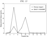
FIG. 12 shows the results for the sample prepared by mixing an equal amount ofsample 1 andsample 2, andFIG. 13 shows the results forsample 1. - From the elution times of the molecular weight marker MW-Marker (HPLC) (Oriental Yeast Co., Ltd.), the trimeric P1NP was mainly contained in fraction No. 7 and the monomeric P1NP was contained in fraction No. 10. Each fraction was measured by electrochemiluminescence using a combination of S24217 as the solid-phase antibody and S24206R as the Ru-labeled antibody, and by Elecsys reagent total P1NP. In the former, serial dilutions of serum from patients with cancer bone metastasis with high P1NP levels, valued with the Elecsys reagent total P1NP, were used as standard to calculate the measured values. Both S24217 and S24206R are antibodies that bind to the α1 chain, but when the fraction containing monomeric P1NP was measured by electrochemiluminescence using the combination of these antibodies, no significant increase in the measured value was observed. Therefore, the antibody combination of S24217 and S24206R can specifically detect trimer P1NP.
- 15 Samples from healthy subjects and 5 samples from osteoporosis patients without renal disease were used.
- Sandwich ELISA was performed using the antibody combinations shown in Table 3. The same samples were also measured with Procollagen Intact PINP and Elecsys Reagent Total P1NP.
[Table 3] Solid-phase antibody Biotinylated antibody S24217 x S24206R S24217 S24206R S24206R x S24217 S24206R S24217 S24219 x S24206R S24219 S24206R - The results are shown in
FIGs. 2 to 5 . The Pearson's product moment correlation coefficient between the ELISA absorbance values using the antibody combination S24217 x S24206R and the Procollagen Intact PINP measurement values was: r = 0.981 (FIG. 2 ). The Pearson's product moment correlation coefficient between the ELISA absorbance values using the antibody combination S24206R x S24217 and the Procollagen Intact PINP measurement values was: r = 0.978 (FIG. 3 ). The Pearson's product moment correlation coefficient between the ELISA absorbance values using the antibody combination S24219 x S24206R and the Procollagen Intact PINP measurement values was: r = 0.975 (FIG. 4 ). The Pearson's product moment correlation coefficient between the Elecsys reagent total P1NP measurement values and the Procollagen Intact PINP measurement values was: r = 0.962 (FIG. 5 ). Therefore, there was a good correlation between the absorbance values by the sandwich ELISA for each antibody combination and the measurement values by the Procollagen Intact PINP. - 10 samples from patients with renal disease and 7 samples from patients with osteoporosis were used. Serial dilutions of serum from patients with cancer bone metastasis with high P1NP levels, valued with the Elecsys reagent total P1NP, were used as standard, and P1NP measurements were calculated by electrochemiluminescence method using the antibody combinations shown in Table 4. The same samples were also measured with Procollagen Intact PINP and Elecsys Reagent Total P1NP.
[Table 4] Solid-phase antibody Labeled antibody S24217 x S24206R S24217 S24206R S24219 x S24206R S24219 S24206R - The results are shown in
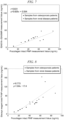
FIGs. 6 to 8 . The correlation between the electrochemiluminescence measurement values by the antibody combination S24217 x S24206R and the Procollagen Intact PINP measurement values was: r = 0.902, y = 0.940x - 2.84 in terms of Pearson's product moment correlation coefficient (FIG. 6 ). The correlation between the electrochemiluminescence measurement values by the antibody combination S24219 x S24206R and the Procollagen Intact PINP measurement values was: r = 0.831, y = 0.826x + 0.594 in terms of Pearson's product moment correlation coefficient (FIG. 7 ). On the other hand, the correlation between the valuesmeasured by the Elecsys reagent total P1NP and the valuesmeasured by Procollagen Intact PINP was: r = 0.773, y = 1.64x - 17.4 in terms of Pearson's product moment correlation coefficient (FIG. 8 ). Therefore, even when a group of samples including samples from patients with renal disease was measured, there was a good correlation between the values measured by the electrochemiluminescence method of the present invention and the values measured by Procollagen Intact PINP. - 3 Samples from healthy individuals, 10 samples from osteoporosis patients without renal disease, and 75 samples from patients with renal disease were used. Sandwich ELISA was performed using the antibody combinations shown in Table 3. The same samples were also measured with Procollagen Intact PINP and Elecsys Reagent Total P1NP.
[Table 5] Solid-phase antibody Biotinylated antibody S24217 x S24206R S24217 S24206R S24219 x S24206R S24219 S24206R - The results are shown in
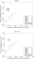
FIG. 9 andFIG. 11 . The Pearson's product moment correlation coefficient between the ELISA absorbance values using the antibody combination S24217 x S24206R and the Procollagen Intact PINP measurement values was: r = 0.820 for all samples, and r' = 0.841 for samples from patients atstages 4 and 5 (FIG. 9 ). The Pearson's product moment correlation coefficient between the ELISA absorbance values using the antibody combination S24219 x S24206R and the Procollagen Intact PINP measurement values was: r = 0.831 for all samples, and r' = 0.836 for samples from patients atstages 4 and 5 (FIG. 10 ). The Pearson's product moment correlation coefficient between the Elecsys reagent total P1NP measurement values and the Procollagen Intact PINP measurement values was: r = 0.646 for all samples, and r' = 0.528 for samples from patients atstages 4 and 5 (FIG. 11 ). Thus, there was a good correlation between the absorbance values by the sandwich ELISA for each antibody combination and the measurement values by the Procollagen Intact PINP. On the other hand, the correlation between the measurement values by the Elecsys reagent total P1NP and the measurement values by the Procollagen Intact PINP was particularly poor in samples from patients with severe renal disease. - The relationship between renal disease (chronic renal disease) stage and eGFR (unit: mL/min/1.73 m2) is as follows:
- Stage 1: eGFR ≥ 90
- Stage 2: eGFR = 60 to 89
- Stage 3a: eGFR = 45 to 59
- Stage 3b: eGFR = 30 to 44
- Stage 4: eGFR = 15 to 29
- Stage 5: eGFR < 15
-
[Table 6] SEQ ID NO. Amino acid sequence (N terminus to C terminus) 1 QEEGQVEGQD 2 PDGSESPTDQETT 3 QEEGQVEGQDC 4 PDGSESPTDQETTC -
- NPMD NITE BP-03606
- NPMD NITE BP-03607
- NPMD NITE BP-03608
- "NPMD" is an acronym for the National Institute of Technology and Evaluation, Patent Microorganism Depositary (Room 122, 2-5-8 Kazusakamatari, Kisarazu, Chiba Prefecture, Japan).
Claims (18)
- An immunological detection method for trimeric type I collagen N-terminal propetide in a biological sample, comprising utilizing:a first antibody that binds to a first specific portion of a pro-α1 chain in the trimeric type I collagen N-terminal propetide; anda second antibody that binds to a second specific portion of a pro-α1 chain in the trimeric type I collagen N-terminal propetide.
- The immunological detection method according to claim 1, wherein at least one of the first antibody and the second antibody is labeled with a labeling substance, and the method comprises:a step of contacting the biological sample and the first antibody to form a first complex;a step of contacting the first complex and the second antibody to form a second complex; anda step of detecting a signal derived from the labeling substance.
- The immunological detection method according to claim 1, wherein at least one of the first antibody and the second antibody is labeled with a labeling substance, and the method comprises:a step of contacting the biological sample and the second antibody to form a first complex;a step of contacting the first complex and the first antibody to form a second complex; anda step of detecting a signal derived from the labeling substance.
- The immunological detection method according to any one of claims 1 to 3, wherein a primary structure of the first specific portion of the pro-α1 chain in the trimeric type I collagen N-terminal propetide comprises a sequence represented by SEQ ID NO: 1, and
a primary structure of the second specific portion of the pro-α1 chain in the trimeric type I collagen N-terminal propetide comprises a sequence represented by SEQ ID NO: 2:SEQ ID NO: 1 QEEGQVEGQDSEQ ID NO: 2 PDGSESPTDQETT. - The immunological detection method according to claim 4, wherein at least one of the first antibody and the second antibody is a monoclonal antibody.
- The immunological detection method according to claim 2 or 3, wherein the biological sample is at least one selected from the group consisting of blood, plasma, and serum.
- The immunological detection method according to claim 4, wherein the immunological detection method is any one selected from the group consisting of an electrochemiluminescence method, latex agglutination immunoturbidimetry, and enzyme-linked immunosorbent assay (ELISA).
- The immunological detection method according to claim 2 or 3, wherein the biological sample is derived from a subject with an eGFR value of less than 30 mL/min/1.73 m2.
- The immunological detection method according to claim 4, which does not detect a monomer of type I collagen N-terminal propetide derived from a renal disease patient.
- An immunological detection kit for trimeric type I collagen N-terminal propetide in a biological sample, comprising:a first antibody that binds to a first specific portion of a pro-α1 chain in the trimeric type I collagen N-terminal propetide; anda second antibody that binds to a second specific portion of a pro-α1 chain in the trimeric type I collagen N-terminal propetide.
- The immunological detection kit according to claim 10, wherein at least one of the first antibody and the second antibody is labeled with a labeling substance, and wherein:the biological sample and the first antibody are contacted to form a first complex;the first complex and the second antibody are contacted to form a second complex; anda signal derived from the labeling substance is detected.
- The immunological detection kit according to claim 10, wherein at least one of the first antibody and the second antibody is labeled with a labeling substance, and wherein:the biological sample and the second antibody are contacted to form a first complex;the first complex and the first antibody are contacted form a second complex; anda signal derived from the labeling substance is detected.
- The immunological detection kit according to any one of claims 10 to 12, wherein a primary structure of the first specific portion of the pro-α1 chain in the trimeric type I collagen N-terminal propetide comprises a sequence represented by SEQ ID NO: 1, and
a primary structure of the second specific portion of the pro-α1 chain in the trimeric type I collagen N-terminal propetide comprises a sequence represented by SEQ ID NO: 2:SEQ ID NO: 1 QEEGQVEGQDSEQ ID NO: 2 PDGSESPTDQETT. - The immunological detection kit according to claim 13, wherein at least one of the first antibody and the second antibody is a monoclonal antibody.
- The immunological detection kit according to claim 11 or 12, wherein the biological sample is at least one selected from the group consisting of blood, plasma, and serum.
- The immunological detection kit according to any one of claims 10 to 12, which utilizes any one immunological detection method selected from the group consisting of an electrochemiluminescence method, latex agglutination immunoturbidimetry, and enzyme-linked immunosorbent assay (ELISA).
- The immunological detection kit according to claim 11 or 12, wherein the biological sample is derived from a subject with an eGFR value of less than 30 mL/min/1.73 m2.
- The immunological detection kit according to claim 13, which does not detect a monomer of type I collagen N-terminal propetide derived from a renal disease patient.
Applications Claiming Priority (2)
| Application Number | Priority Date | Filing Date | Title |
|---|---|---|---|
| JP2022050846 | 2022-03-25 | ||
| PCT/JP2023/011188 WO2023182343A1 (en) | 2022-03-25 | 2023-03-22 | Immunological detection method and immunological detection kit |
Publications (2)
| Publication Number | Publication Date |
|---|---|
| EP4502601A1 true EP4502601A1 (en) | 2025-02-05 |
| EP4502601A4 EP4502601A4 (en) | 2026-03-25 |
Family
ID=88101125
Family Applications (1)
| Application Number | Title | Priority Date | Filing Date |
|---|---|---|---|
| EP23774938.7A Pending EP4502601A4 (en) | 2022-03-25 | 2023-03-22 | Immunological detection method and immunological detection kit |
Country Status (5)
| Country | Link |
|---|---|
| US (1) | US20250155453A1 (en) |
| EP (1) | EP4502601A4 (en) |
| JP (1) | JPWO2023182343A1 (en) |
| CN (1) | CN118829882A (en) |
| WO (1) | WO2023182343A1 (en) |
Family Cites Families (8)
| Publication number | Priority date | Publication date | Assignee | Title |
|---|---|---|---|---|
| GB9014220D0 (en) * | 1990-06-26 | 1990-08-15 | Farmos Yhtymy Oy | Method for the immunlolgical determinition of the carboxyterminal propeptide of type i procollagen |
| CA2128877A1 (en) | 1992-01-31 | 1993-08-05 | David J. Baylink | Amino procollagen 1(1) peptides |
| DK104093D0 (en) * | 1993-09-17 | 1993-09-17 | Osteometer A S | PROCEDURE FOR DETERMINING COLLAGEN FRAGMENTS IN BODY LIQUIDS, TEST KITS AND MEANS FOR EXERCISING THE PROCEDURE AND USING THE PROCEDURE FOR DIAGNOSTICING THE DISEASES OF TABLET METAL |
| US6008325A (en) * | 1995-04-19 | 1999-12-28 | Orion Diagnostica | Antibody to aminoterminal propeptide of type 1 procollagen |
| GB9507985D0 (en) * | 1995-04-19 | 1995-06-07 | Orion Yhtymae Oy | Antibody to aminoterminal propeptide to type 1 procollagen and assay method using it |
| CN108152507A (en) * | 2017-11-28 | 2018-06-12 | 泰州泽成生物技术有限公司 | A kind of I type precollagen amino terminal peptides detection kit and its detection method |
| JP7489278B2 (en) | 2020-09-18 | 2024-05-23 | ダイキン工業株式会社 | Ventilation system |
| CN112326972A (en) * | 2020-10-30 | 2021-02-05 | 安徽理工大学 | Chemiluminescence quantitative detection kit for detecting complete PINP in serum |
-
2023
- 2023-03-22 US US18/835,821 patent/US20250155453A1/en active Pending
- 2023-03-22 CN CN202380020121.XA patent/CN118829882A/en active Pending
- 2023-03-22 EP EP23774938.7A patent/EP4502601A4/en active Pending
- 2023-03-22 JP JP2024509155A patent/JPWO2023182343A1/ja active Pending
- 2023-03-22 WO PCT/JP2023/011188 patent/WO2023182343A1/en not_active Ceased
Also Published As
| Publication number | Publication date |
|---|---|
| EP4502601A4 (en) | 2026-03-25 |
| WO2023182343A1 (en) | 2023-09-28 |
| CN118829882A (en) | 2024-10-22 |
| JPWO2023182343A1 (en) | 2023-09-28 |
| US20250155453A1 (en) | 2025-05-15 |
Similar Documents
| Publication | Publication Date | Title |
|---|---|---|
| JP2008513536A (en) | Monoclonal antibody against progastrin | |
| JP5576592B2 (en) | Monoclonal antibody against gastrin hormone | |
| RU2511033C2 (en) | Novel bnp (1-32) epitope and antibodies directed against said epitope | |
| KR20180055823A (en) | Method of measuring PIVKA-II, and method of producing PIVKA-II immunoassay reagent or kit | |
| JP5583648B2 (en) | Monoclonal antibody against gastrin hormone | |
| CA2785678C (en) | Human insulin assay and assay reagent | |
| US10954298B2 (en) | Method of obtaining a binder to prepro-vasopressin or fragments thereof | |
| CN113727995A (en) | Method for detecting cancer and detection reagent | |
| CA2088354C (en) | Pancreas-elastasis-1-specific antibody, a process for obtaining it and a test kit containing such antibodies | |
| US20250155453A1 (en) | Immunological detection method and immunological detection kit | |
| US7094551B2 (en) | Immunoassays, assay methods, antibodies and method of creating antibodies for detecting FGF-23 | |
| EP4502602A1 (en) | Immunological detection method and immunological detection kit | |
| KR20120118412A (en) | Human liver carboxylesterase 1-specific indicating monoclonal antibody, hybridoma cell line producing the same and use thereof | |
| JPH10226700A (en) | Immunoassay for detection of MIA | |
| EP1712564A1 (en) | Anti-nc1 monoclonal antibody | |
| CN119119270B (en) | Anti-Human CD24 monoclonal antibody, preparation method and application thereof | |
| EP3019875B1 (en) | Augurin immunoassay | |
| JP3778979B2 (en) | Sandwich immunoassay for N-peptide | |
| EP4317172A1 (en) | Immunological analysis method for type-i collagen c-terminal telopeptide | |
| JP4423426B2 (en) | Method of using increased concentration of adrenomedullin precursor C-terminal peptide as an indicator of cardiovascular disease or inflammatory disease | |
| Ashby et al. | Anomalies in the assay of parathyroid hormone in normocalcaemic patients with renal stone disease | |
| JP2001343389A (en) | Inspection method of cancer by measurement of autoantibody against mdm2, and its reagent | |
| JPH0674954A (en) | Anti-β2-microglobulin-derived substance antibody and method for immunoassay of β2-microglobulin-derived substance using the same | |
| JPH0843398A (en) | Diagnosis of cancer | |
| CN114891104A (en) | Monoclonal antibody for identifying AMH and application thereof |
Legal Events
| Date | Code | Title | Description |
|---|---|---|---|
| STAA | Information on the status of an ep patent application or granted ep patent |
Free format text: STATUS: THE INTERNATIONAL PUBLICATION HAS BEEN MADE |
|
| PUAI | Public reference made under article 153(3) epc to a published international application that has entered the european phase |
Free format text: ORIGINAL CODE: 0009012 |
|
| STAA | Information on the status of an ep patent application or granted ep patent |
Free format text: STATUS: REQUEST FOR EXAMINATION WAS MADE |
|
| 17P | Request for examination filed |
Effective date: 20240805 |
|
| AK | Designated contracting states |
Kind code of ref document: A1 Designated state(s): AL AT BE BG CH CY CZ DE DK EE ES FI FR GB GR HR HU IE IS IT LI LT LU LV MC ME MK MT NL NO PL PT RO RS SE SI SK SM TR |
|
| DAV | Request for validation of the european patent (deleted) | ||
| DAX | Request for extension of the european patent (deleted) | ||
| A4 | Supplementary search report drawn up and despatched |
Effective date: 20260220 |