CN113727995A - Method for detecting cancer and detection reagent - Google Patents
Method for detecting cancer and detection reagent Download PDFInfo
- Publication number
- CN113727995A CN113727995A CN202080030591.0A CN202080030591A CN113727995A CN 113727995 A CN113727995 A CN 113727995A CN 202080030591 A CN202080030591 A CN 202080030591A CN 113727995 A CN113727995 A CN 113727995A
- Authority
- CN
- China
- Prior art keywords
- antibody
- cancer
- azu1
- reagent
- azu
- Prior art date
- Legal status (The legal status is an assumption and is not a legal conclusion. Google has not performed a legal analysis and makes no representation as to the accuracy of the status listed.)
- Pending
Links
- 238000000034 method Methods 0.000 title claims abstract description 85
- 206010028980 Neoplasm Diseases 0.000 title claims abstract description 62
- 201000011510 cancer Diseases 0.000 title claims abstract description 56
- 239000003153 chemical reaction reagent Substances 0.000 title claims abstract description 51
- 238000001514 detection method Methods 0.000 title claims abstract description 31
- 208000005718 Stomach Neoplasms Diseases 0.000 claims abstract description 40
- 206010017758 gastric cancer Diseases 0.000 claims abstract description 40
- 201000011549 stomach cancer Diseases 0.000 claims abstract description 40
- 208000006265 Renal cell carcinoma Diseases 0.000 claims abstract description 18
- 206010006187 Breast cancer Diseases 0.000 claims abstract description 17
- 208000026310 Breast neoplasm Diseases 0.000 claims abstract description 17
- 206010009944 Colon cancer Diseases 0.000 claims abstract description 16
- 206010058467 Lung neoplasm malignant Diseases 0.000 claims abstract description 16
- 201000005202 lung cancer Diseases 0.000 claims abstract description 16
- 208000020816 lung neoplasm Diseases 0.000 claims abstract description 16
- 208000001333 Colorectal Neoplasms Diseases 0.000 claims abstract description 15
- 238000012360 testing method Methods 0.000 claims abstract description 6
- 239000003550 marker Substances 0.000 claims description 31
- 230000003248 secreting effect Effects 0.000 claims description 25
- TZCPCKNHXULUIY-RGULYWFUSA-N 1,2-distearoyl-sn-glycero-3-phosphoserine Chemical compound CCCCCCCCCCCCCCCCCC(=O)OC[C@H](COP(O)(=O)OC[C@H](N)C(O)=O)OC(=O)CCCCCCCCCCCCCCCCC TZCPCKNHXULUIY-RGULYWFUSA-N 0.000 claims description 14
- ZWZWYGMENQVNFU-UHFFFAOYSA-N Glycerophosphorylserin Natural products OC(=O)C(N)COP(O)(=O)OCC(O)CO ZWZWYGMENQVNFU-UHFFFAOYSA-N 0.000 claims description 14
- 102100025222 CD63 antigen Human genes 0.000 claims description 7
- 102100027221 CD81 antigen Human genes 0.000 claims description 7
- 102100037904 CD9 antigen Human genes 0.000 claims description 7
- 101000934368 Homo sapiens CD63 antigen Proteins 0.000 claims description 7
- 101000914479 Homo sapiens CD81 antigen Proteins 0.000 claims description 7
- 101000738354 Homo sapiens CD9 antigen Proteins 0.000 claims description 7
- 239000002245 particle Substances 0.000 claims description 3
- 239000003795 chemical substances by application Substances 0.000 claims description 2
- 108010007337 Azurin Proteins 0.000 claims 4
- YBJHBAHKTGYVGT-ZKWXMUAHSA-N (+)-Biotin Chemical compound N1C(=O)N[C@@H]2[C@H](CCCCC(=O)O)SC[C@@H]21 YBJHBAHKTGYVGT-ZKWXMUAHSA-N 0.000 description 82
- 210000004027 cell Anatomy 0.000 description 50
- 238000002835 absorbance Methods 0.000 description 48
- 241000282414 Homo sapiens Species 0.000 description 42
- 229960002685 biotin Drugs 0.000 description 41
- 235000020958 biotin Nutrition 0.000 description 41
- 239000011616 biotin Substances 0.000 description 41
- 210000002966 serum Anatomy 0.000 description 33
- 239000000126 substance Substances 0.000 description 33
- 239000007790 solid phase Substances 0.000 description 32
- 238000005259 measurement Methods 0.000 description 28
- 238000002965 ELISA Methods 0.000 description 27
- 239000011859 microparticle Substances 0.000 description 24
- 239000000203 mixture Substances 0.000 description 23
- 239000000523 sample Substances 0.000 description 23
- 239000006180 TBST buffer Substances 0.000 description 17
- 102000004169 proteins and genes Human genes 0.000 description 17
- 108090000623 proteins and genes Proteins 0.000 description 17
- 101000793686 Homo sapiens Azurocidin Proteins 0.000 description 16
- 210000004408 hybridoma Anatomy 0.000 description 15
- 238000003118 sandwich ELISA Methods 0.000 description 15
- 230000027455 binding Effects 0.000 description 14
- 238000006243 chemical reaction Methods 0.000 description 14
- 108020003175 receptors Proteins 0.000 description 14
- 102000005962 receptors Human genes 0.000 description 14
- 102100030009 Azurocidin Human genes 0.000 description 12
- 102100025475 Carcinoembryonic antigen-related cell adhesion molecule 5 Human genes 0.000 description 10
- 101000914324 Homo sapiens Carcinoembryonic antigen-related cell adhesion molecule 5 Proteins 0.000 description 10
- 101000914321 Homo sapiens Carcinoembryonic antigen-related cell adhesion molecule 7 Proteins 0.000 description 10
- NBIIXXVUZAFLBC-UHFFFAOYSA-N Phosphoric acid Chemical compound OP(O)(O)=O NBIIXXVUZAFLBC-UHFFFAOYSA-N 0.000 description 10
- 150000001413 amino acids Chemical group 0.000 description 10
- 108090000765 processed proteins & peptides Proteins 0.000 description 10
- 239000000243 solution Substances 0.000 description 9
- 241000282412 Homo Species 0.000 description 8
- 229920001213 Polysorbate 20 Polymers 0.000 description 8
- 210000004369 blood Anatomy 0.000 description 8
- 239000008280 blood Substances 0.000 description 8
- 239000012228 culture supernatant Substances 0.000 description 8
- 238000013211 curve analysis Methods 0.000 description 8
- 238000002372 labelling Methods 0.000 description 8
- 239000002609 medium Substances 0.000 description 8
- 230000036961 partial effect Effects 0.000 description 8
- 235000010486 polyoxyethylene sorbitan monolaurate Nutrition 0.000 description 8
- 239000000256 polyoxyethylene sorbitan monolaurate Substances 0.000 description 8
- 238000000585 Mann–Whitney U test Methods 0.000 description 7
- 241000699666 Mus <mouse, genus> Species 0.000 description 7
- 230000000052 comparative effect Effects 0.000 description 7
- 238000010790 dilution Methods 0.000 description 7
- 239000012895 dilution Substances 0.000 description 7
- 238000002360 preparation method Methods 0.000 description 7
- 239000000047 product Substances 0.000 description 7
- XZWYTXMRWQJBGX-VXBMVYAYSA-N FLAG peptide Chemical compound NCCCC[C@@H](C(O)=O)NC(=O)[C@H](CC(O)=O)NC(=O)[C@H](CC(O)=O)NC(=O)[C@H](CC(O)=O)NC(=O)[C@H](CC(O)=O)NC(=O)[C@H](CCCCN)NC(=O)[C@@H](NC(=O)[C@@H](N)CC(O)=O)CC1=CC=C(O)C=C1 XZWYTXMRWQJBGX-VXBMVYAYSA-N 0.000 description 6
- 108010090804 Streptavidin Proteins 0.000 description 6
- 239000007864 aqueous solution Substances 0.000 description 6
- 238000004113 cell culture Methods 0.000 description 6
- 102000043635 human AZU1 Human genes 0.000 description 6
- 230000031700 light absorption Effects 0.000 description 6
- 230000035945 sensitivity Effects 0.000 description 6
- 239000006228 supernatant Substances 0.000 description 6
- 102100023962 Bifunctional arginine demethylase and lysyl-hydroxylase JMJD6 Human genes 0.000 description 5
- 108091026890 Coding region Proteins 0.000 description 5
- 241001465754 Metazoa Species 0.000 description 5
- 229910000147 aluminium phosphate Inorganic materials 0.000 description 5
- 210000000628 antibody-producing cell Anatomy 0.000 description 5
- 210000001124 body fluid Anatomy 0.000 description 5
- 239000010839 body fluid Substances 0.000 description 5
- 239000001110 calcium chloride Substances 0.000 description 5
- 229910001628 calcium chloride Inorganic materials 0.000 description 5
- 238000010586 diagram Methods 0.000 description 5
- 210000001808 exosome Anatomy 0.000 description 5
- 239000013613 expression plasmid Substances 0.000 description 5
- 230000003053 immunization Effects 0.000 description 5
- 238000003018 immunoassay Methods 0.000 description 5
- 108010077613 phosphatidylserine receptor Proteins 0.000 description 5
- 238000012216 screening Methods 0.000 description 5
- 238000005199 ultracentrifugation Methods 0.000 description 5
- 102000004190 Enzymes Human genes 0.000 description 4
- 108090000790 Enzymes Proteins 0.000 description 4
- 108010001336 Horseradish Peroxidase Proteins 0.000 description 4
- 102100020870 La-related protein 6 Human genes 0.000 description 4
- 108050008265 La-related protein 6 Proteins 0.000 description 4
- 230000000903 blocking effect Effects 0.000 description 4
- 239000007853 buffer solution Substances 0.000 description 4
- 238000011088 calibration curve Methods 0.000 description 4
- 238000010367 cloning Methods 0.000 description 4
- 238000007796 conventional method Methods 0.000 description 4
- 238000011156 evaluation Methods 0.000 description 4
- 230000014509 gene expression Effects 0.000 description 4
- 238000002649 immunization Methods 0.000 description 4
- 238000004949 mass spectrometry Methods 0.000 description 4
- 239000012528 membrane Substances 0.000 description 4
- 239000013612 plasmid Substances 0.000 description 4
- 238000000926 separation method Methods 0.000 description 4
- 101710154607 Azurocidin Proteins 0.000 description 3
- 101100453350 Candida albicans (strain SC5314 / ATCC MYA-2876) HBR1 gene Proteins 0.000 description 3
- BVKZGUZCCUSVTD-UHFFFAOYSA-L Carbonate Chemical compound [O-]C([O-])=O BVKZGUZCCUSVTD-UHFFFAOYSA-L 0.000 description 3
- 108060003951 Immunoglobulin Proteins 0.000 description 3
- 108010038807 Oligopeptides Proteins 0.000 description 3
- 102000015636 Oligopeptides Human genes 0.000 description 3
- 241000283973 Oryctolagus cuniculus Species 0.000 description 3
- 108010033276 Peptide Fragments Proteins 0.000 description 3
- 102000007079 Peptide Fragments Human genes 0.000 description 3
- 101100337984 Scheffersomyces stipitis (strain ATCC 58785 / CBS 6054 / NBRC 10063 / NRRL Y-11545) GSM1 gene Proteins 0.000 description 3
- 210000003719 b-lymphocyte Anatomy 0.000 description 3
- 239000003054 catalyst Substances 0.000 description 3
- 238000005516 engineering process Methods 0.000 description 3
- 230000005284 excitation Effects 0.000 description 3
- 230000036541 health Effects 0.000 description 3
- 230000002163 immunogen Effects 0.000 description 3
- 102000018358 immunoglobulin Human genes 0.000 description 3
- 210000003734 kidney Anatomy 0.000 description 3
- 238000004519 manufacturing process Methods 0.000 description 3
- 239000008188 pellet Substances 0.000 description 3
- 108040007629 peroxidase activity proteins Proteins 0.000 description 3
- 102000013415 peroxidase activity proteins Human genes 0.000 description 3
- 239000002244 precipitate Substances 0.000 description 3
- 239000000439 tumor marker Substances 0.000 description 3
- 102000002260 Alkaline Phosphatase Human genes 0.000 description 2
- 108020004774 Alkaline Phosphatase Proteins 0.000 description 2
- 108090000672 Annexin A5 Proteins 0.000 description 2
- 102000004121 Annexin A5 Human genes 0.000 description 2
- 108090001008 Avidin Proteins 0.000 description 2
- 102400000667 Brain natriuretic peptide 32 Human genes 0.000 description 2
- 101800000407 Brain natriuretic peptide 32 Proteins 0.000 description 2
- 101800002247 Brain natriuretic peptide 45 Proteins 0.000 description 2
- 101100369802 Caenorhabditis elegans tim-1 gene Proteins 0.000 description 2
- 241000699802 Cricetulus griseus Species 0.000 description 2
- MYMOFIZGZYHOMD-UHFFFAOYSA-N Dioxygen Chemical compound O=O MYMOFIZGZYHOMD-UHFFFAOYSA-N 0.000 description 2
- 101150046249 Havcr2 gene Proteins 0.000 description 2
- 102100034458 Hepatitis A virus cellular receptor 2 Human genes 0.000 description 2
- 206010061218 Inflammation Diseases 0.000 description 2
- AGPKZVBTJJNPAG-WHFBIAKZSA-N L-isoleucine Chemical compound CC[C@H](C)[C@H](N)C(O)=O AGPKZVBTJJNPAG-WHFBIAKZSA-N 0.000 description 2
- 101710191666 Lactadherin Proteins 0.000 description 2
- 102100039648 Lactadherin Human genes 0.000 description 2
- 239000000232 Lipid Bilayer Substances 0.000 description 2
- 238000012408 PCR amplification Methods 0.000 description 2
- 206010035226 Plasma cell myeloma Diseases 0.000 description 2
- 239000002202 Polyethylene glycol Substances 0.000 description 2
- ONIBWKKTOPOVIA-UHFFFAOYSA-N Proline Natural products OC(=O)C1CCCN1 ONIBWKKTOPOVIA-UHFFFAOYSA-N 0.000 description 2
- 108010076504 Protein Sorting Signals Proteins 0.000 description 2
- QAOWNCQODCNURD-UHFFFAOYSA-N Sulfuric acid Chemical compound OS(O)(=O)=O QAOWNCQODCNURD-UHFFFAOYSA-N 0.000 description 2
- 108700031126 Tetraspanins Proteins 0.000 description 2
- 102000043977 Tetraspanins Human genes 0.000 description 2
- 101710120037 Toxin CcdB Proteins 0.000 description 2
- 102100027904 Zinc finger protein basonuclin-1 Human genes 0.000 description 2
- KYIKRXIYLAGAKQ-UHFFFAOYSA-N abcn Chemical compound C1CCCCC1(C#N)N=NC1(C#N)CCCCC1 KYIKRXIYLAGAKQ-UHFFFAOYSA-N 0.000 description 2
- 230000009471 action Effects 0.000 description 2
- 229940024606 amino acid Drugs 0.000 description 2
- 238000004458 analytical method Methods 0.000 description 2
- 239000000427 antigen Substances 0.000 description 2
- 102000036639 antigens Human genes 0.000 description 2
- 108091007433 antigens Proteins 0.000 description 2
- 238000003556 assay Methods 0.000 description 2
- 239000011324 bead Substances 0.000 description 2
- 230000015572 biosynthetic process Effects 0.000 description 2
- 239000000872 buffer Substances 0.000 description 2
- 210000004899 c-terminal region Anatomy 0.000 description 2
- 238000005119 centrifugation Methods 0.000 description 2
- 238000004132 cross linking Methods 0.000 description 2
- 238000012258 culturing Methods 0.000 description 2
- 201000010099 disease Diseases 0.000 description 2
- 208000037265 diseases, disorders, signs and symptoms Diseases 0.000 description 2
- 230000001605 fetal effect Effects 0.000 description 2
- 239000010419 fine particle Substances 0.000 description 2
- 238000002875 fluorescence polarization Methods 0.000 description 2
- 108020001507 fusion proteins Proteins 0.000 description 2
- 102000037865 fusion proteins Human genes 0.000 description 2
- 239000001963 growth medium Substances 0.000 description 2
- 238000000338 in vitro Methods 0.000 description 2
- 230000004054 inflammatory process Effects 0.000 description 2
- 229960000310 isoleucine Drugs 0.000 description 2
- AGPKZVBTJJNPAG-UHFFFAOYSA-N isoleucine Natural products CCC(C)C(N)C(O)=O AGPKZVBTJJNPAG-UHFFFAOYSA-N 0.000 description 2
- 230000000670 limiting effect Effects 0.000 description 2
- 210000004962 mammalian cell Anatomy 0.000 description 2
- 238000001840 matrix-assisted laser desorption--ionisation time-of-flight mass spectrometry Methods 0.000 description 2
- 238000000691 measurement method Methods 0.000 description 2
- 201000000050 myeloid neoplasm Diseases 0.000 description 2
- 239000013642 negative control Substances 0.000 description 2
- HPNRHPKXQZSDFX-OAQDCNSJSA-N nesiritide Chemical compound C([C@H]1C(=O)NCC(=O)N[C@@H](CCCNC(N)=N)C(=O)N[C@@H](CCCCN)C(=O)N[C@@H](CCSC)C(=O)N[C@@H](CC(O)=O)C(=O)N[C@@H](CCCNC(N)=N)C(=O)N[C@H](C(N[C@@H](CO)C(=O)N[C@@H](CO)C(=O)N[C@@H](CO)C(=O)N[C@@H](CO)C(=O)NCC(=O)N[C@@H](CC(C)C)C(=O)NCC(=O)N[C@@H](CSSC[C@@H](C(=O)N1)NC(=O)CNC(=O)[C@H](CO)NC(=O)CNC(=O)[C@H](CCC(N)=O)NC(=O)[C@@H](NC(=O)[C@H](CCSC)NC(=O)[C@H](CCCCN)NC(=O)[C@H]1N(CCC1)C(=O)[C@@H](N)CO)C(C)C)C(=O)N[C@@H](CCCCN)C(=O)N[C@@H](C(C)C)C(=O)N[C@@H](CC(C)C)C(=O)N[C@@H](CCCNC(N)=N)C(=O)N[C@@H](CCCNC(N)=N)C(=O)N[C@@H](CC=1N=CNC=1)C(O)=O)=O)[C@@H](C)CC)C1=CC=CC=C1 HPNRHPKXQZSDFX-OAQDCNSJSA-N 0.000 description 2
- 210000000440 neutrophil Anatomy 0.000 description 2
- 210000001672 ovary Anatomy 0.000 description 2
- 229920001223 polyethylene glycol Polymers 0.000 description 2
- 108091033319 polynucleotide Proteins 0.000 description 2
- 102000040430 polynucleotide Human genes 0.000 description 2
- 239000002157 polynucleotide Substances 0.000 description 2
- 239000013641 positive control Substances 0.000 description 2
- 238000011084 recovery Methods 0.000 description 2
- 238000011160 research Methods 0.000 description 2
- 210000003296 saliva Anatomy 0.000 description 2
- 241000894007 species Species 0.000 description 2
- 210000004989 spleen cell Anatomy 0.000 description 2
- 238000001920 surface-enhanced laser desorption--ionisation mass spectrometry Methods 0.000 description 2
- 238000004885 tandem mass spectrometry Methods 0.000 description 2
- 210000001138 tear Anatomy 0.000 description 2
- 210000002700 urine Anatomy 0.000 description 2
- BRZYSWJRSDMWLG-DJWUNRQOSA-N (2r,3r,4r,5r)-2-[(1s,2s,3r,4s,6r)-4,6-diamino-3-[(2s,3r,4r,5s,6r)-3-amino-4,5-dihydroxy-6-[(1r)-1-hydroxyethyl]oxan-2-yl]oxy-2-hydroxycyclohexyl]oxy-5-methyl-4-(methylamino)oxane-3,5-diol Chemical compound O1C[C@@](O)(C)[C@H](NC)[C@@H](O)[C@H]1O[C@@H]1[C@@H](O)[C@H](O[C@@H]2[C@@H]([C@@H](O)[C@H](O)[C@@H]([C@@H](C)O)O2)N)[C@@H](N)C[C@H]1N BRZYSWJRSDMWLG-DJWUNRQOSA-N 0.000 description 1
- QKNYBSVHEMOAJP-UHFFFAOYSA-N 2-amino-2-(hydroxymethyl)propane-1,3-diol;hydron;chloride Chemical compound Cl.OCC(N)(CO)CO QKNYBSVHEMOAJP-UHFFFAOYSA-N 0.000 description 1
- FWMNVWWHGCHHJJ-SKKKGAJSSA-N 4-amino-1-[(2r)-6-amino-2-[[(2r)-2-[[(2r)-2-[[(2r)-2-amino-3-phenylpropanoyl]amino]-3-phenylpropanoyl]amino]-4-methylpentanoyl]amino]hexanoyl]piperidine-4-carboxylic acid Chemical compound C([C@H](C(=O)N[C@H](CC(C)C)C(=O)N[C@H](CCCCN)C(=O)N1CCC(N)(CC1)C(O)=O)NC(=O)[C@H](N)CC=1C=CC=CC=1)C1=CC=CC=C1 FWMNVWWHGCHHJJ-SKKKGAJSSA-N 0.000 description 1
- 108010088751 Albumins Proteins 0.000 description 1
- 102000009027 Albumins Human genes 0.000 description 1
- 102100024321 Alkaline phosphatase, placental type Human genes 0.000 description 1
- 238000012815 AlphaLISA Methods 0.000 description 1
- 206010003445 Ascites Diseases 0.000 description 1
- 241000894006 Bacteria Species 0.000 description 1
- 108091003079 Bovine Serum Albumin Proteins 0.000 description 1
- 241000282693 Cercopithecidae Species 0.000 description 1
- ZXCAQANTQWBICD-DCAQKATOSA-N Cys-Lys-Val Chemical compound CC(C)[C@@H](C(=O)O)NC(=O)[C@H](CCCCN)NC(=O)[C@H](CS)N ZXCAQANTQWBICD-DCAQKATOSA-N 0.000 description 1
- 238000008157 ELISA kit Methods 0.000 description 1
- 241000588724 Escherichia coli Species 0.000 description 1
- 241000287828 Gallus gallus Species 0.000 description 1
- 102000002812 Heat-Shock Proteins Human genes 0.000 description 1
- 108010004889 Heat-Shock Proteins Proteins 0.000 description 1
- 241000238631 Hexapoda Species 0.000 description 1
- NTRAGDHVSGKUSF-AVGNSLFASA-N Leu-Arg-Arg Chemical compound CC(C)C[C@H](N)C(=O)N[C@@H](CCCN=C(N)N)C(=O)N[C@@H](CCCN=C(N)N)C(O)=O NTRAGDHVSGKUSF-AVGNSLFASA-N 0.000 description 1
- 241000124008 Mammalia Species 0.000 description 1
- 108010052285 Membrane Proteins Proteins 0.000 description 1
- 241000699670 Mus sp. Species 0.000 description 1
- 206010030113 Oedema Diseases 0.000 description 1
- 206010060862 Prostate cancer Diseases 0.000 description 1
- 208000000236 Prostatic Neoplasms Diseases 0.000 description 1
- 206010038389 Renal cancer Diseases 0.000 description 1
- 240000004808 Saccharomyces cerevisiae Species 0.000 description 1
- 206010040047 Sepsis Diseases 0.000 description 1
- 102000012479 Serine Proteases Human genes 0.000 description 1
- 108010022999 Serine Proteases Proteins 0.000 description 1
- 108090000901 Transferrin Proteins 0.000 description 1
- 102000004338 Transferrin Human genes 0.000 description 1
- 238000001042 affinity chromatography Methods 0.000 description 1
- 125000003172 aldehyde group Chemical group 0.000 description 1
- 238000003016 alphascreen Methods 0.000 description 1
- 238000012870 ammonium sulfate precipitation Methods 0.000 description 1
- 210000004102 animal cell Anatomy 0.000 description 1
- 230000000844 anti-bacterial effect Effects 0.000 description 1
- 230000000845 anti-microbial effect Effects 0.000 description 1
- 239000012472 biological sample Substances 0.000 description 1
- 238000001574 biopsy Methods 0.000 description 1
- 239000002981 blocking agent Substances 0.000 description 1
- 210000000601 blood cell Anatomy 0.000 description 1
- 239000012503 blood component Substances 0.000 description 1
- 229940098773 bovine serum albumin Drugs 0.000 description 1
- 125000002091 cationic group Chemical group 0.000 description 1
- 230000007910 cell fusion Effects 0.000 description 1
- 230000001413 cellular effect Effects 0.000 description 1
- 210000001175 cerebrospinal fluid Anatomy 0.000 description 1
- 230000008859 change Effects 0.000 description 1
- 239000005515 coenzyme Substances 0.000 description 1
- 208000029742 colonic neoplasm Diseases 0.000 description 1
- 230000002860 competitive effect Effects 0.000 description 1
- 239000000306 component Substances 0.000 description 1
- 230000007123 defense Effects 0.000 description 1
- 238000003745 diagnosis Methods 0.000 description 1
- 238000002405 diagnostic procedure Methods 0.000 description 1
- 239000012470 diluted sample Substances 0.000 description 1
- 238000003113 dilution method Methods 0.000 description 1
- 230000000694 effects Effects 0.000 description 1
- 239000013604 expression vector Substances 0.000 description 1
- 238000000684 flow cytometry Methods 0.000 description 1
- 239000012530 fluid Substances 0.000 description 1
- 238000002523 gelfiltration Methods 0.000 description 1
- 239000008187 granular material Substances 0.000 description 1
- 230000002209 hydrophobic effect Effects 0.000 description 1
- 238000004191 hydrophobic interaction chromatography Methods 0.000 description 1
- 238000001727 in vivo Methods 0.000 description 1
- 208000015181 infectious disease Diseases 0.000 description 1
- 102000006495 integrins Human genes 0.000 description 1
- 108010044426 integrins Proteins 0.000 description 1
- 238000005342 ion exchange Methods 0.000 description 1
- 238000004255 ion exchange chromatography Methods 0.000 description 1
- 201000011061 large intestine cancer Diseases 0.000 description 1
- 238000004895 liquid chromatography mass spectrometry Methods 0.000 description 1
- 238000001294 liquid chromatography-tandem mass spectrometry Methods 0.000 description 1
- 108010026228 mRNA guanylyltransferase Proteins 0.000 description 1
- 239000006249 magnetic particle Substances 0.000 description 1
- 230000036210 malignancy Effects 0.000 description 1
- 108020004999 messenger RNA Proteins 0.000 description 1
- 230000000813 microbial effect Effects 0.000 description 1
- 210000001616 monocyte Anatomy 0.000 description 1
- 230000009871 nonspecific binding Effects 0.000 description 1
- 239000003330 peritoneal dialysis fluid Substances 0.000 description 1
- 238000001050 pharmacotherapy Methods 0.000 description 1
- 239000012071 phase Substances 0.000 description 1
- 239000008055 phosphate buffer solution Substances 0.000 description 1
- 150000003905 phosphatidylinositols Chemical class 0.000 description 1
- 108010031345 placental alkaline phosphatase Proteins 0.000 description 1
- 210000004180 plasmocyte Anatomy 0.000 description 1
- 230000004481 post-translational protein modification Effects 0.000 description 1
- 230000002980 postoperative effect Effects 0.000 description 1
- 102000004196 processed proteins & peptides Human genes 0.000 description 1
- 238000001959 radiotherapy Methods 0.000 description 1
- 238000006268 reductive amination reaction Methods 0.000 description 1
- 201000010174 renal carcinoma Diseases 0.000 description 1
- 238000002271 resection Methods 0.000 description 1
- 238000004366 reverse phase liquid chromatography Methods 0.000 description 1
- 210000004739 secretory vesicle Anatomy 0.000 description 1
- 235000020183 skimmed milk Nutrition 0.000 description 1
- BEOOHQFXGBMRKU-UHFFFAOYSA-N sodium cyanoborohydride Chemical compound [Na+].[B-]C#N BEOOHQFXGBMRKU-UHFFFAOYSA-N 0.000 description 1
- 230000009870 specific binding Effects 0.000 description 1
- 230000000707 stereoselective effect Effects 0.000 description 1
- 239000000758 substrate Substances 0.000 description 1
- 238000002198 surface plasmon resonance spectroscopy Methods 0.000 description 1
- 230000001225 therapeutic effect Effects 0.000 description 1
- 210000001519 tissue Anatomy 0.000 description 1
- 239000012581 transferrin Substances 0.000 description 1
- 230000001052 transient effect Effects 0.000 description 1
- 230000010474 transient expression Effects 0.000 description 1
- 238000000108 ultra-filtration Methods 0.000 description 1
- 230000002792 vascular Effects 0.000 description 1
- 238000005406 washing Methods 0.000 description 1
Images


Classifications
-
- C—CHEMISTRY; METALLURGY
- C07—ORGANIC CHEMISTRY
- C07K—PEPTIDES
- C07K14/00—Peptides having more than 20 amino acids; Gastrins; Somatostatins; Melanotropins; Derivatives thereof
- C07K14/435—Peptides having more than 20 amino acids; Gastrins; Somatostatins; Melanotropins; Derivatives thereof from animals; from humans
- C07K14/46—Peptides having more than 20 amino acids; Gastrins; Somatostatins; Melanotropins; Derivatives thereof from animals; from humans from vertebrates
- C07K14/47—Peptides having more than 20 amino acids; Gastrins; Somatostatins; Melanotropins; Derivatives thereof from animals; from humans from vertebrates from mammals
- C07K14/4701—Peptides having more than 20 amino acids; Gastrins; Somatostatins; Melanotropins; Derivatives thereof from animals; from humans from vertebrates from mammals not used
- C07K14/4723—Cationic antimicrobial peptides, e.g. defensins
-
- C—CHEMISTRY; METALLURGY
- C07—ORGANIC CHEMISTRY
- C07K—PEPTIDES
- C07K16/00—Immunoglobulins [IGs], e.g. monoclonal or polyclonal antibodies
- C07K16/18—Immunoglobulins [IGs], e.g. monoclonal or polyclonal antibodies against material from animals or humans
-
- G—PHYSICS
- G01—MEASURING; TESTING
- G01N—INVESTIGATING OR ANALYSING MATERIALS BY DETERMINING THEIR CHEMICAL OR PHYSICAL PROPERTIES
- G01N33/00—Investigating or analysing materials by specific methods not covered by groups G01N1/00 - G01N31/00
- G01N33/48—Biological material, e.g. blood, urine; Haemocytometers
- G01N33/50—Chemical analysis of biological material, e.g. blood, urine; Testing involving biospecific ligand binding methods; Immunological testing
- G01N33/53—Immunoassay; Biospecific binding assay; Materials therefor
- G01N33/531—Production of immunochemical test materials
- G01N33/532—Production of labelled immunochemicals
- G01N33/535—Production of labelled immunochemicals with enzyme label or co-enzymes, co-factors, enzyme inhibitors or enzyme substrates
-
- G—PHYSICS
- G01—MEASURING; TESTING
- G01N—INVESTIGATING OR ANALYSING MATERIALS BY DETERMINING THEIR CHEMICAL OR PHYSICAL PROPERTIES
- G01N33/00—Investigating or analysing materials by specific methods not covered by groups G01N1/00 - G01N31/00
- G01N33/48—Biological material, e.g. blood, urine; Haemocytometers
- G01N33/50—Chemical analysis of biological material, e.g. blood, urine; Testing involving biospecific ligand binding methods; Immunological testing
- G01N33/53—Immunoassay; Biospecific binding assay; Materials therefor
- G01N33/574—Immunoassay; Biospecific binding assay; Materials therefor for cancer
- G01N33/57484—Immunoassay; Biospecific binding assay; Materials therefor for cancer involving compounds serving as markers for tumor, cancer, neoplasia, e.g. cellular determinants, receptors, heat shock/stress proteins, A-protein, oligosaccharides, metabolites
- G01N33/57488—Immunoassay; Biospecific binding assay; Materials therefor for cancer involving compounds serving as markers for tumor, cancer, neoplasia, e.g. cellular determinants, receptors, heat shock/stress proteins, A-protein, oligosaccharides, metabolites involving compounds identifable in body fluids
-
- C—CHEMISTRY; METALLURGY
- C07—ORGANIC CHEMISTRY
- C07K—PEPTIDES
- C07K2319/00—Fusion polypeptide
- C07K2319/01—Fusion polypeptide containing a localisation/targetting motif
- C07K2319/02—Fusion polypeptide containing a localisation/targetting motif containing a signal sequence
-
- C—CHEMISTRY; METALLURGY
- C07—ORGANIC CHEMISTRY
- C07K—PEPTIDES
- C07K2319/00—Fusion polypeptide
- C07K2319/01—Fusion polypeptide containing a localisation/targetting motif
- C07K2319/035—Fusion polypeptide containing a localisation/targetting motif containing a signal for targeting to the external surface of a cell, e.g. to the outer membrane of Gram negative bacteria, GPI- anchored eukaryote proteins
-
- C—CHEMISTRY; METALLURGY
- C07—ORGANIC CHEMISTRY
- C07K—PEPTIDES
- C07K2319/00—Fusion polypeptide
- C07K2319/40—Fusion polypeptide containing a tag for immunodetection, or an epitope for immunisation
- C07K2319/43—Fusion polypeptide containing a tag for immunodetection, or an epitope for immunisation containing a FLAG-tag
-
- G—PHYSICS
- G01—MEASURING; TESTING
- G01N—INVESTIGATING OR ANALYSING MATERIALS BY DETERMINING THEIR CHEMICAL OR PHYSICAL PROPERTIES
- G01N2333/00—Assays involving biological materials from specific organisms or of a specific nature
- G01N2333/435—Assays involving biological materials from specific organisms or of a specific nature from animals; from humans
- G01N2333/46—Assays involving biological materials from specific organisms or of a specific nature from animals; from humans from vertebrates
- G01N2333/47—Assays involving proteins of known structure or function as defined in the subgroups
- G01N2333/4701—Details
- G01N2333/4721—Cationic antimicrobial peptides, e.g. defensins
-
- G—PHYSICS
- G01—MEASURING; TESTING
- G01N—INVESTIGATING OR ANALYSING MATERIALS BY DETERMINING THEIR CHEMICAL OR PHYSICAL PROPERTIES
- G01N2470/00—Immunochemical assays or immunoassays characterised by the reaction format or reaction type
- G01N2470/04—Sandwich assay format
- G01N2470/06—Second binding partner specifically binding complex of analyte with first binding partner
Abstract
The problem is to provide a method for simply and accurately detecting cancer and a reagent that can be used in the method. A method for detecting cancer (excluding renal cell carcinoma), which comprises the step of measuring the amount of azuridin (AZU1) in a test subject, and considering cancer detection in the case that the measured value exceeds a predetermined reference value. Preferably, the cancer is selected from the group consisting of gastric cancer, breast cancer, colorectal cancer and lung cancer. Use of a reagent comprising an antibody specifically recognizing AZU1 for the detection of cancer, excluding renal cell carcinoma.
Description
Technical Field
The present invention relates to a method and a reagent for detecting cancer, in which Azurocidin (アズロシジン, hereinafter referred to as "AZU 1") is used as a measurement target.
Background
The tumor markers for detecting cancer are generally listed in table 1. However, there are many problems that any marker has a low positive rate in the early stage of cancer, is false positive in benign tumor and inflammation, and cannot be detected in cancer with high malignancy. Therefore, it is desired to find a tumor marker capable of detecting such cancers with high accuracy and to develop a detection method.
[ Table 1]
(Table 1)

Furthermore, AZU1 is an inactive serine protease known as the alias heparin-binding protein (HBP) or the 37kDa cationic antimicrobial protein (CAP 37). As a function thereof, AZU1 has a chemomigration action on monocytes and an antibacterial action on gram-negative bacteria. AZU1 is present in azurophil granules of neutrophils, and is released from neutrophils migrating to the infected site to induce vascular leakage and edema formation, promote inflammation, and contribute to biological defense (non-patent documents 1 to 5).
For the association of AZU1 with diseases, a diagnostic method for diagnosing infection and sepsis by measuring the amount of AZU1 in body fluids has been disclosed (patent documents 1 to 3). In recent years, a method for diagnosing renal cell carcinoma by isolating extracellular vesicles in a body fluid and detecting AZU1 has been reported (patent document 4 and non-patent document 6).
As for the association of AZU1 with cancer, it was reported that the messenger RNA expression level of AZU1 was increased in breast cancer, prostate cancer, and large intestine cancer (non-patent document 6). However, to date, there has been no report on the dynamics of AZU1 in body fluids collected less invasively than in biopsy in cancers other than renal cell carcinoma including these, and it has not been clear whether AZU1 in body fluids can be used for detecting cancers other than renal cell carcinoma.
Documents of the prior art
Patent document
Patent document 1: japanese patent No. 5166522
Patent document 2: japanese patent No. 5488885
Patent document 3: japanese patent No. 5818916
Patent document 4: international publication No. 2018/079689 pamphlet
Non-patent document
Non-patent document 1: j Leukoc biol.2009Mar; 85(3): 344-51
Non-patent document 2: trends immunol.2009Nov; 30(11): 538-46
Non-patent document 3: nat Med.2001Oct; 7(10): 1123-7.
Non-patent document 4: trends immunol.2009Nov; 30(11): 547-56
Non-patent document 5: thromb Haemost.2009Aug; 102(2): 198-205
Non-patent document 6: int J cancer.2018feb 1; 142(3): 607-617
Disclosure of Invention
Problems to be solved by the invention
The present invention addresses the problem of providing a method for detecting cancer simply and accurately, and a reagent that can be used in the method.
Means for solving the problems
As a result of intensive studies to solve the above problems, the present inventors have found that AZU1 in body fluids of cancer patients exhibits a significantly higher value than that of healthy persons, and have found that AZU1 can be a cancer detection marker, and have completed the present invention.
That is, the present invention includes the following aspects.
[1] A method for detecting cancer (excluding renal cell carcinoma), which comprises the step of measuring the amount of azuridin (AZU1) in a test subject, and considering cancer detection in the case that the measured value exceeds a predetermined reference value.
[2] The method according to [1], wherein the cancer is selected from the group consisting of gastric cancer, breast cancer, colorectal cancer and lung cancer.
[3] The method according to [1] or [2], wherein the determination of the amount of AZU1 is performed using an antibody specifically recognizing AZU 1.
[4] The method according to any one of [1] to [3], which comprises a step of further detecting a second marker present on the same cell-secreting microparticle as a cell-secreting microparticle in which AZU1 is present in the sample, wherein the second marker is at least any one of 1 marker shown in Table 2.
[5] The method according to [4], wherein the aforementioned second marker comprises at least 1 of CD81, CD63, CD9 and phosphatidylserine.
[6] The method according to [4] or [5], wherein the detection of the second marker is performed using an antibody or a receptor that specifically recognizes the second marker.
[7] A reagent for detecting cancer (excluding renal cell carcinoma) comprising an antibody that specifically recognizes AZU 1.
[8] The reagent according to [7], wherein the cancer is selected from the group consisting of gastric cancer, breast cancer, colorectal cancer and lung cancer.
[9] The reagent according to [7] or [8], further comprising an antibody or a receptor that specifically recognizes any of the second markers described in Table 2.
[10] The reagent according to [9], wherein the aforementioned second marker comprises at least 1 of CD81, CD63, CD9 and phosphatidylserine.
ADVANTAGEOUS EFFECTS OF INVENTION
The present invention provides a method for detecting cancer easily and with high accuracy, and a reagent that can be used in the method.
Drawings
FIG. 1 is a graph showing the results of screening of hybridoma cell culture supernatants obtained in example 8 using CELISA constitutively expressing CHO-K1 cells using GPI-anchored AZU 1.
Fig. 2 is a graph showing the screening results of the hybridoma cell culture supernatants obtained by ELISA using secreted AZU1 in example 9.
Fig. 3 is a graph showing the results of performance evaluation of anti-AZU 1 monoclonal antibody as a solid phase antibody using ELISA using cell secreting microparticles in example 13.
Fig. 4 is a graph showing the results of evaluating the performance of the anti-AZU 1 monoclonal antibody as a biotin-labeled antibody obtained by ELISA using cell-secreting microparticles in example 13.
Fig. 5 is a diagram showing that in example 14, even when any of an anti-CD 81 antibody, an anti-CD 9 antibody, and an anti-CD 63 antibody was used as a solid phase antibody for ELISA, cell-secreted microparticles including AZU1 could be detected.
Fig. 6 is a graph showing box plots of ELISA absorbance for the healthy human group and the stomach cancer patient group in example 15-1 using an anti-CD 81 antibody as the solid-phase antibody and an anti-AZU 1 antibody as the biotin-labeled antibody.
Fig. 7 is a graph showing box plots of ELISA absorbance for the healthy human group and the group of gastric cancer patients in example 15-2 using an anti-CD 9 antibody as the solid-phase antibody and an anti-AZU 1 antibody as the biotin-labeled antibody.
Fig. 8 is a graph showing box plots of ELISA absorbance for the healthy human group and the group of gastric cancer patients in example 15-3 using the anti-CD 63 antibody as the solid-phase antibody and the anti-AZU 1 antibody as the biotin-labeled antibody.
Fig. 9 is a graph showing box plots of ELISA absorbance for the healthy human group and the group of gastric cancer patients in example 15-4 using the anti-AZU 1 antibody as the solid-phase antibody and the anti-CD 81 antibody as the biotin-labeled antibody.
Fig. 10 is a graph showing box plots of ELISA absorbance for the healthy human group and the group of gastric cancer patients in example 15-5 using the anti-AZU 1 antibody as the solid-phase antibody and the anti-CD 9 antibody as the biotin-labeled antibody.
Fig. 11 is a graph showing box plots of ELISA absorbance for the healthy human group and the group of gastric cancer patients in example 15-6 using the anti-AZU 1 antibody as the solid-phase antibody and the anti-CD 63 antibody as the biotin-labeled antibody.
Fig. 12 is a graph showing box plots of ELISA absorbance for the healthy human group and the stomach cancer patient group in example 16 using Tim4-hFc as the solid phase receptor and using anti-AZU 1 antibody as the biotin-labeled antibody.
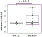
FIG. 13 is a graph showing a box chart of CEA measurement values in a healthy human group and a gastric cancer patient group in comparative example 1.
Fig. 14 is a graph showing the results of analysis of Receiver Operating Characteristic (ROC) curves of ELISA absorbances between the group of healthy humans and the group of patients with gastric cancer in example 17 using an anti-CD 81 antibody as a solid phase antibody and an anti-AZU 1 antibody as a biotin-labeled antibody.
Fig. 15 is a graph showing the results of ROC curve analysis of ELISA absorbance between the group of healthy humans and the group of patients with gastric cancer in example 17 using an anti-CD 9 antibody as a solid phase antibody and an anti-AZU 1 antibody as a biotin-labeled antibody.
Fig. 16 is a graph showing the results of ROC curve analysis of ELISA absorbance between the group of healthy humans and the group of patients with gastric cancer in example 17 using an anti-CD 63 antibody as a solid phase antibody and an anti-AZU 1 antibody as a biotin-labeled antibody.
Fig. 17 is a graph showing the results of ROC curve analysis of ELISA absorbance between the group of healthy humans and the group of patients with gastric cancer in example 17 using an anti-AZU 1 antibody as a solid phase antibody and an anti-CD 81 antibody as a biotin-labeled antibody.
Fig. 18 is a graph showing the results of ROC curve analysis of ELISA absorbance between the group of healthy humans and the group of patients with gastric cancer in example 17 using an anti-AZU 1 antibody as a solid phase antibody and an anti-CD 9 antibody as a biotin-labeled antibody.
Fig. 19 is a graph showing the results of ROC curve analysis of ELISA absorbance between the group of healthy humans and the group of patients with gastric cancer in example 17 using an anti-AZU 1 antibody as a solid phase antibody and an anti-CD 63 antibody as a biotin-labeled antibody.
Fig. 20 is a graph showing the results of ROC curve analysis of ELISA absorbance between the group of healthy humans and the group of patients with gastric cancer in example 17 using Tim4-hFc as a solid phase receptor and using an anti-AZU 1 antibody as a biotin-labeled antibody.
FIG. 21 is a graph showing the results of ROC curve analysis of CEA measurement values between a healthy human group and a gastric cancer patient group in example 17.
Fig. 22 is a graph showing box plots of ELISA absorbance for the healthy human group and the breast cancer patient group in example 18-1 using an anti-CD 81 antibody as a solid-phase antibody and an anti-AZU 1 antibody as a biotin-labeled antibody.
Fig. 23 is a graph showing box plots of ELISA absorbance for the healthy human group and the breast cancer patient group in example 18-2 using an anti-CD 9 antibody as a solid-phase antibody and an anti-AZU 1 antibody as a biotin-labeled antibody.
Fig. 24 is a graph showing box plots of ELISA absorbance for the healthy human group and the breast cancer patient group in example 18-3 using an anti-CD 63 antibody as a solid-phase antibody and an anti-AZU 1 antibody as a biotin-labeled antibody.
FIG. 25 is a box plot showing ELISA absorbances of healthy humans and colorectal cancer patients in example 19-1, using an anti-CD 81 antibody as a solid-phase antibody and an anti-AZU 1 antibody as a biotin-labeled antibody.
FIG. 26 is a box chart showing ELISA absorbances of a healthy human group and a colorectal cancer patient group in example 19-2, using an anti-CD 9 antibody as a solid phase antibody and an anti-AZU 1 antibody as a biotin-labeled antibody.
Fig. 27 is a box plot showing ELISA absorbances of the healthy human group and the colorectal cancer patient group in example 19-3 using an anti-CD 63 antibody as a solid phase antibody and an anti-AZU 1 antibody as a biotin-labeled antibody.
Fig. 28 is a graph showing box plots of ELISA absorbance for the healthy human group and the lung cancer patient group in example 20-1 using an anti-CD 81 antibody as a solid-phase antibody and an anti-AZU 1 antibody as a biotin-labeled antibody.
Fig. 29 is a graph showing box plots of ELISA absorbance for the healthy human group and the lung cancer patient group in example 20-2 using an anti-CD 9 antibody as a solid-phase antibody and an anti-AZU 1 antibody as a biotin-labeled antibody.
Fig. 30 is a graph showing box plots of ELISA absorbance for the healthy human group and the lung cancer patient group in example 20-3 using an anti-CD 63 antibody as a solid-phase antibody and an anti-AZU 1 antibody as a biotin-labeled antibody.
FIG. 31 is a box chart showing the concentration of free AZU1 in serum specimens of healthy persons, lung cancer, colorectal cancer, breast cancer and gastric cancer in example 21.
Detailed Description
[1] The method of the present invention for detecting cancer
The first mode of the present invention is a method for detecting cancer (excluding renal cell carcinoma), which comprises the step of measuring the amount of AZU1 in a specimen. This is a method based on the fact that AZU1 is characteristically present in a biological sample such as cancer blood as compared with a healthy specimen. The determination of the amount of AZU1 in a sample is usually performed in vitro (in vitro).
As shown in examples described below, the method of the present invention can detect cancer with high sensitivity and specificity as compared with the case of measuring a conventionally known tumor marker such as CEA.
The method of the present invention includes a stage up to the detection of cancer, and does not include a final judgment action related to cancer diagnosis. The doctor refers to the detection results or the like based on the method of the present invention to diagnose cancer or to develop a treatment policy.
Usually, a subject to be detected for cancer (test animal) is a human.
Examples of the sample to be measured in the present invention include blood, urine, saliva, tears, ascites, peritoneal lavage fluid, cerebrospinal fluid, and an extract solution of cells or tissues. In view of the ease of collection of the specimen, blood, urine, saliva, and tears are preferable. In view of versatility with other test items, blood is more preferable. As the blood, whole blood may be used as it is, or blood components such as serum, plasma, and blood cells may be separated and used. The dilution ratio of the sample is not particularly limited, and may be appropriately selected depending on the type and state of the sample to be used, for example, in the range of from undiluted to 100-fold dilution.
The sample usually contains microparticles secreted from cells (cell-secreting microparticles) described later.
The disease to which the present invention is directed is cancer (excluding renal cell carcinoma). Preferably stomach cancer, breast cancer, colorectal cancer and/or lung cancer, more preferably stomach cancer.
In the present invention, AZU1 to be measured is a peptide including at least a sequence from isoleucine at residue 27 to proline at residue 248 in the amino acid sequence of human AZU1 protein disclosed in accession No. P20160 of UniPlotKB, or a peptide including an amino acid sequence having 80% or more identity to the aforementioned sequence. The uniformity is preferably 90% or more, more preferably 95% or more. The peptide may be a peptide consisting of amino acids in which 1 or several amino acids are deleted, substituted, inserted, or added to the above sequence. In addition, several means: preferably 2 to 20, more preferably 2 to 10, and further preferably 2 to 5. In addition, other peptide fragments may also be present on both sides of the aforementioned sequence.
AZU1 determined in the present invention may determine AZU1 present as a soluble protein, may determine AZU1 present on microparticles secreted by cells, or both. In the case of determining AZU1 present on the microparticles, AZU1 co-existing with the second marker on the microparticles may be determined.
Examples of microparticles secreted from cells include exosomes. The exosome is a membrane vesicle composed of a lipid bilayer membrane, usually with a diameter of 50-200 nm. Exosomes are known to contain a large number of membrane proteins such as tetraspanins (tetraspanins) and integrins, proteins involved in the formation of multicellular bodies, heat shock proteins, and the like. In addition, the lipid bilayer membrane constituting the exosome is known to have phosphatidylserine on the membrane surface thereof. Representative molecules contained in large amounts in exosomes are shown in table 2.
The second marker in the present invention is not particularly limited as long as it is a molecule present on a cell secretory microparticle, and preferably means at least 1 contained in the group consisting of the proteins shown in table 2 and phosphatidylserine, and more preferably at least 1 contained in CD81, CD63, CD9, and phosphatidylserine. The proteins shown in table 2 also include peptides having amino acid sequences with high homology (80% or more, preferably 90% or more, and more preferably 95% or more) thereto.
The second marker may be more than 2, and thus the term "second" is also to be understood as meaning "other than AZU 1".
The second marker to be detected in the present invention is not particularly limited as long as it is a molecule present on a cell-secreting microparticle, and is preferably a protein and phosphatidylserine shown in table 2, which are present on the same cell-secreting microparticle as the cell-secreting microparticle in which AZU1 is present.
[ Table 2-1]
(Table 2)

[ tables 2-2]
(continuation table 2)

Among the detection methods of the present invention, the method for measuring the amount of AZU1, and the method for measuring (detecting) at least 1 of the second marker coexisting on the secretory microparticle of a cell and AZU1 are not particularly limited as long as they do not interfere with the measurement of the amount of AZU 1. Examples thereof include an immunoassay method using an antibody specifically recognizing AZU1 and a method using mass spectrometry.
Specific examples of the immunoassay method using an antibody that specifically recognizes AZU1 include the following.
[a] Using an antibody specifically recognizing AZU1 and labeled AZU1, a competition method was used in which labeled AZU1 and AZU1 contained in the test object competitively bind to the aforementioned antibody.
[b] A sample was brought into contact with a chip on which an antibody specifically recognizing AZU1 was immobilized, and a method of surface plasmon resonance for detecting a signal depending on the binding of the antibody to AZU1 was used.
[c] A fluorescence polarization immunoassay method in which a fluorescence-labeled antibody specifically recognizing AZU1 is used and the degree of fluorescence polarization of the antibody increases when the antibody binds to AZU1 is used.
[d] A sandwich method of forming complexes of 2 antibodies specifically recognizing AZU1 (1 of which is a labeled antibody) with 3 of AZU1 was used. In this case, the 2 antibodies are preferably 2 antibodies having different epitopes.
[e] As a pretreatment, a method of detecting a conjugate of AZU1 and an antibody using a mass spectrometer after concentrating AZU1 in a sample by an antibody that specifically recognizes AZU 1.
[f] A flow cytometry method in which a fluorescently labeled antibody that specifically recognizes AZU1 is used, and after the antibody is bound to AZU1, measurement objects are arranged in a flow channel, and a complex in which the antibody is bound to the measurement objects is counted based on scattered light and fluorescence obtained when each particle is irradiated with excitation light.
Of these, the methods of [ d ] and [ e ] are simple and highly versatile, but the method of [ d ] is more preferable in terms of handling a large amount of a specimen because the techniques related to reagents and apparatuses are well established.
The antibody specifically recognizing AZU1 is not particularly limited, and can be obtained by immunizing an animal with AZU1 protein itself, an oligopeptide consisting of a partial region of AZU1, a polynucleotide encoding the full length or partial region of AZU1, or the like as an immunogen.
When the AZU1 protein itself or an oligopeptide consisting of a partial region of AZU1 is used as an immunogen, the structure of the protein or the oligopeptide may be changed during the preparation process. Therefore, the obtained antibody may not have high specificity and binding force to a desired antigen, and as a result, it may be impossible to prepare an antibody for quantitative determination of AZU1 contained in a sample. On the other hand, when an expression vector comprising a polynucleotide encoding the full-length or partial region of AZU1 is used as an immunogen, AZU1 is expressed in vivo in an immunized animal without structural change, and therefore an antibody having high specificity and binding force (i.e., high affinity) to a desired antigen can be obtained, which is preferable.
The animal used for immunization is not particularly limited as long as it has an antibody-producing ability, and may be a mammal used for ordinary immunization such as a mouse, a rat, and a rabbit, or a bird such as a chicken.
The antibody specifically recognizing AZU1 may be a monoclonal antibody or a polyclonal antibody, but is preferably a monoclonal antibody.
The hybridoma producing an antibody that specifically recognizes AZU1 may be established by appropriately selecting from among established techniques. As an example, a hybridoma producing a monoclonal antibody that specifically recognizes AZU1 can be established by collecting B cells from an animal immunized by the above method, electrofusing the B cells with myeloma cells or fusing the B cells in the presence of polyethylene glycol, selecting a hybridoma producing a desired antibody using HAT medium, and cloning the selected hybridoma using a limiting dilution method.
The selection of antibodies specifically recognizing AZU1 for use in the present invention may be based on affinity towards Glycosylated Phosphatidylinositol (GPI) -anchored AZU1 derived from the host expression system.
The host is not particularly limited, and those skilled in the art may suitably select from microbial cells such as Escherichia coli and yeast, insect cells, and animal cells, which are generally used for expression of proteins, and preferably use mammalian cells capable of expressing a protein having a structure close to that of natural AZU1 by post-translational modification such as disulfide bond or sugar chain addition. Examples of mammalian cells include conventionally used 293T cell lines derived from fetal kidney, COS-7 cell lines derived from monkey kidney, CHO-K1 cell lines derived from Chinese hamster ovary, and cancer cells isolated from human body.
The antibody used in the cancer detection method of the present invention may be purified by appropriately selecting from among established techniques. For example, after culturing the antibody-producing hybridoma cells established by the above-described method, the antibody can be purified by collecting the culture supernatant, concentrating the antibody by ammonium sulfate precipitation as necessary, and then performing ion exchange chromatography, hydrophobic interaction chromatography, or affinity chromatography using a carrier on which protein a, protein G, protein L, or the like is immobilized.
In the present invention, when the amount of AZU1 present on the secretory microparticle of the same cell and the detection of the second marker are performed simultaneously, it is preferable to use an antibody or a receptor (hereinafter also referred to as "antibody or the like") that specifically recognizes the second marker in addition to the antibody that recognizes AZU 1.
As the second marker in the present invention, at least 1 of CD81, CD63, CD9, and phosphatidylserine is preferably contained, and as an antibody or receptor specifically recognizing these, an anti-CD 81 antibody, an anti-CD 63 antibody, an anti-CD 9 antibody, or a phosphatidylserine receptor is preferably used.
The anti-CD 81 antibody, anti-CD 63 antibody, and anti-CD 9 antibody used in the present invention can be obtained by the same method as that for the aforementioned antibody recognizing AZU 1.
The phosphatidylserine receptor used in the present invention is not particularly limited, and examples thereof include annexin V, MFG-E8, Tim1, Tim3 and Tim4, and Tim4 having high specificity and binding force to phosphatidylserine is preferable (International publication No. 2016/088689). The Tim4 may have at least an amino acid sequence of a binding domain (IgV domain) to phosphatidylserine, and for example, a Tim4 protein itself, a peptide consisting of a partial region of the IgV domain including Tim4, or a fusion protein in which another peptide fragment is bound to a partial region of the IgV domain including Tim4 may be used.
The labeled antibody or the like used in the binding quantitation method by the sandwich method described above may be labeled with: an enzyme such as peroxidase or alkaline phosphatase, a fluorescent substance, a chemiluminescent substance, a radioisotope or a functional fine particle, which can be detected by a detection device; or another substance that specifically binds to avidin and biotin, and the like, the labeling can be performed by a method of an established technique.
The method of detecting AZU1 by mass spectrometry and quantitatively determining it in the method of the present invention will be described in detail below.
When the sample is blood, as the pretreatment step, it is preferable to: proteins such as albumin, immunoglobulin, and transferrin contained in a large amount in blood are removed by Agilent Human 14 or the like, and then further fractionated by ion exchange, gel filtration, reverse phase chromatography, or the like.
The measurement can be performed by tandem mass spectrometry (MS/MS), liquid chromatography/tandem mass spectrometry (LC/MS/MS), matrix-assisted laser desorption ionization time-of-flight mass spectrometry (MALDI-TOF/MS), surface-enhanced laser desorption ionization mass spectrometry (SELDI-MS), and the like.
In the detection method of the present invention, it is preferable that: when the amount of AZU1 obtained by the measurement exceeds a reference value (cutoff value) calculated from the control, it is determined that cancer is detected.
The amount of AZU1 used for the determination may be any of a measured value or a converted concentration value. The converted concentration value is: values converted from the measured values based on a calibration curve prepared using AZU1 as a standard sample. The concentration of the standard sample can be determined by converting the measured value based on a standard curve of a standard peptide obtained by mass spectrometry.
For the cutoff value, samples collected from healthy persons and cancer patients can be measured, and the measured value showing the optimum sensitivity and specificity can be set appropriately by a Receiver Operating Characteristic (ROC) curve.
The method of detecting cancer of the present invention can be used in a method of treating cancer. That is, according to the present invention, there is provided a method of treating cancer (excluding renal cell carcinoma) in a patient, the method comprising the steps of:
(i) identifying a patient as a person whose measured value of the AZU1 amount exceeds a preset reference value; and
(ii) and (c) performing a treatment on the patient identified above.
In the identification in the aforementioned step (i), the amount of AZU1 may be measured using an antibody that specifically recognizes AZU1, or may be measured using mass spectrometry.
Examples of the treatment in the step (ii) include surgical resection, pharmacotherapy, radiotherapy, and the like, but are not particularly limited.
[2] The reagent for detecting cancer of the present invention
The second mode of the present invention is a reagent for detecting cancer (excluding renal cell carcinoma) which comprises an antibody specifically recognizing AZU 1.
The reagent of the present invention further preferably further comprises an antibody or receptor specifically recognizing the second marker shown in table 2. The antibody or receptor (antibody or the like) specifically recognizing the second marker is not particularly limited, and is preferably an antibody or receptor specifically recognizing at least 1 of, for example, CD81, CD63, CD9, and phosphatidylserine, and more preferably any of an anti-CD 81 antibody, an anti-CD 63 antibody, an anti-CD 9 antibody, or a phosphatidylserine receptor. The phosphatidylserine receptor is not particularly limited, and examples thereof include annexin V, MFG-E8, Tim1, Tim3 and Tim4, and Tim4 having high specificity and binding force to phosphatidylserine is preferable. The Tim4 may have at least the amino acid sequence of the binding domain to phosphatidylserine (IgV domain), and for example, a Tim4 protein itself, a peptide consisting of a partial region of the IgV domain including Tim4, or a fusion protein in which another peptide fragment is bound to a partial region of the IgV domain including Tim4 may be used.
The reagent and the measurement method of the present invention are specifically described below in 3 modes of the sandwich method described above. However, the reagent and the measurement method of the present invention are not limited to these 3 modes.
[ mode 1] two-step Sandwich method
The reagent used in the present embodiment includes 2 kinds of antibodies (hereinafter referred to as "antibody etc. 1" and "antibody etc. 2"). The binding sites of the antibody or the like 1 and the antibody or the like 2 are preferably different from each other with respect to the substance to be measured. Examples of the combination of the antibody or the like 1 and the antibody or the like 2 include three groups [ a ] to [ c ] described below.
[a] Antibody and the like 1: antibody, antibody or the like that specifically recognizes AZU1 2: an antibody that specifically recognizes AZU1 and is the same as or different from antibody 1
[b] Antibody and the like 1: antibody, antibody or the like that specifically recognizes AZU1 2: antibodies or receptors specifically recognizing the second marker
[c] Antibody and the like 1: antibody or receptor specifically recognizing the second marker, antibody, etc. 2: antibody specifically recognizing AZU1
The reagent of the present embodiment can be produced by the following methods [1] to [3 ].
[1] First, antibodies or the like 1 are bound to a carrier capable of B/F separation such as an immunoplate or magnetic particles. The binding method may be physical binding using a hydrophobic bond, or chemical binding using a linking agent or the like capable of crosslinking two substances.
[2] After the antibody or the like 1 is bound to the carrier, the carrier surface is blocked with bovine serum albumin, skim milk, a commercially available blocking agent for immunization, or the like as a reagent for 1 time in order to avoid non-specific binding.
[3] The other, i.e., antibody or the like 2 is labeled, and a solution containing the obtained labeled antibody or the like is prepared as a reagent for 2 times. As a substance for labeling 2 such as an antibody, preferred are: an enzyme such as peroxidase or alkaline phosphatase, a fluorescent substance, a chemiluminescent substance, a radioisotope or a functional fine particle, which can be detected by a detection device; or another substance having specific binding such as avidin and biotin. The solution of the 2-time reagent is preferably a buffer solution capable of satisfactorily performing the antigen-antibody reaction, for example, a phosphate buffer solution, a Tris-HCl buffer solution, or the like.
The reagent of the present embodiment thus produced can be lyophilized as necessary.
Next, the detection and measurement of AZU1 using the reagent prepared in the present embodiment can be carried out by the methods shown in the following [4] to [6 ].
[4] The 1 st reagent prepared in [2] above was brought into contact with the specimen at a constant temperature for a constant time. The reaction conditions are such that the reaction is carried out at a temperature in the range of 4 ℃ to 40 ℃ for 5 minutes to 180 minutes.
[5] Removing unreacted substances by B/F separation, and then contacting the resulting mixture with the 2-time reagent prepared in [3] at a constant temperature for a constant time to form a sandwich complex. As for the reaction conditions, it is sufficient to react them at a temperature ranging from 4 ℃ to 40 ℃ for 5 minutes to 180 minutes.
[6] Unreacted substances were removed by B/F separation, a marker substance such as a labeled antibody was quantified, and the concentration of AZU1 in a sample was quantified using a calibration curve prepared using AZU1 of a known concentration as a standard.
[ mode 2] one-step Sandwich method
The reagent used in this embodiment includes an antibody or the like 1 and an antibody or the like 2, as in embodiment 1 described above.
In the preparation of the reagent of the present embodiment, a substance obtained by binding 1 such as an antibody to a carrier and performing a blocking treatment can be prepared by the methods described in [1] to [2] of embodiment 1, and a buffer solution containing 2 such as a labeled antibody can be further added to the immobilized carrier such as an antibody.
The reagent of the present embodiment thus produced can be lyophilized as necessary.
Next, the detection and measurement of AZU1 using the reagent prepared in the present embodiment can be carried out by the methods shown in the following [7] to [8 ].
[7] The reagent prepared in the above method is brought into contact with a sample at a certain temperature for a certain time to form a sandwich complex. As for the reaction conditions, it is sufficient to react them at a temperature ranging from 4 ℃ to 40 ℃ for 5 minutes to 180 minutes.
[8] Unreacted substances were removed by B/F separation, a marker substance such as a labeled antibody was quantified, and the concentration of AZU1 in a sample was quantified using a calibration curve prepared using AZU1 of a known concentration as a standard.
[ mode 3] homogeneous phase Sandwich method
The reagent used in this embodiment includes antibodies 1 and 2, and streptavidin-coated labeling substances that can be excited by excitation light, as in embodiments 1 and 2 described above. As the streptavidin-coated marker substance, for example, AlphaScreen streptavidin donor beads (Perkinelmer) can be suitably used.
The reagent of the present embodiment can be produced by the following methods described in [9] to [10 ].
[9] First, an antibody or the like is labeled with biotin 1. The Biotin labeling may be carried out by a conventionally known method, and for example, a method using a Biotin labeling Kit-NH2 labeling Kit (manufactured by Dojindo laboratories) is used.
[10] The other, antibody or the like 2 is labeled with a substance that emits a signal by singlet oxygen. The singlet oxygen is generated when the streptavidin-coated marker substance is excited. The signal is preferably a fluorescent signal. As the signal-generating substance, for example, AlphaLISA receptor beads (Perkinelmer) can be suitably used. The method of binding the antibody or the like 2 to the signal-generating substance is not particularly limited, and examples thereof include reductive amination crosslinking using sodium cyanoborohydride and an aldehyde group on the surface of the signal-generating substance.
Next, the detection and measurement of AZU1 using the reagent prepared in the present embodiment can be carried out by the methods shown in [11] to [13] below.
[11] A sandwich complex is formed by bringing the aforementioned biotin-labeled antibody or the like 1 produced in [9] and the signal generating substance-labeled antibody or the like 2 produced in [10] into contact with a specimen at a predetermined temperature under a light-shielding condition for a predetermined time. As for the reaction conditions, it is sufficient to react them at a temperature ranging from 4 ℃ to 40 ℃ for 5 minutes to 180 minutes.
[12] Then adding streptavidin covering mark substance to make it contact for a certain time under the condition of certain temperature and shading to make the biotin labeled antibody and streptavidin covering mark substance combine. As for the reaction conditions, it is sufficient to react them at a temperature ranging from 4 ℃ to 40 ℃ for 5 minutes to 180 minutes.
[13] The signal emitted from the signal generating substance-labeled antibody or the like 21 upon irradiation with excitation light is quantified using an analyzer, and the concentration of AZU1 in the sample is quantified using a calibration curve prepared using AZU1 of known concentration as a standard. As the analyzer, for example, EnSpire (Perkinelmer) can be suitably used.
The amount of the reagent component contained in the reagent of the present invention may be appropriately set depending on various conditions such as the amount of the sample, the type of the reagent, and the method of measurement. For example, in the case of measuring the amount of AZU1 by a sandwich method using 20. mu.L of serum or plasma as a sample, the amount of 1 such as an antibody bound to a carrier may be 100ng to 1000. mu.g, and the amount of 2 such as a labeled antibody may be 2ng to 20. mu.g, using 20. mu.L of the sample as a reaction system for reacting with the antibody or the like.
The reagent of the present invention can be used for measurement by a manual method (manual measurement) or measurement using an automatic immunodiagnostic apparatus. In particular, the measurement using the automatic immunodiagnostic apparatus is preferable because the measurement can be performed without being affected by endogenous measurement-interfering factors and competitive enzymes contained in the specimen, and the amount of AZU1 in the specimen can be measured in a short time.
Another embodiment of the second mode of the present invention is a use of an antibody specifically recognizing AZU1 for the manufacture of a reagent for detecting cancer (excluding renal cell carcinoma). Further, use of an antibody that specifically recognizes AZU1 and an antibody or receptor that specifically recognizes any of the second markers set forth in table 2 in the manufacture of a reagent for detecting cancer (excluding renal cell carcinoma).
In addition, another embodiment of the present invention is the use of an antibody specifically recognizing AZU1 for the detection of cancer, excluding renal cell carcinoma. In addition, use of an antibody specifically recognizing AZU1, and an antibody or receptor specifically recognizing any of the second markers described in Table 2, for the detection of cancer (excluding renal cell carcinoma).
Examples
The present invention will be described in more detail with reference to examples, which are provided for illustrative purposes only and are not intended to limit the scope of the present invention.
EXAMPLE 1 preparation of GPI-anchored AZU1 expression plasmid
A region encoding a sequence from isoleucine at residue 27 to proline at residue 248 in the amino acid sequence of human AZU1 protein was amplified by PCR. The PCR amplification product was inserted into a plasmid pFLAG1 (manufactured by Sigma-Aldrich) containing the coding region for the FLAG tag peptide consisting of the amino acid sequence DYKDDDDK (SEQ ID NO: 1) and the coding region for the GPI-anchored signal peptide derived from placental alkaline phosphatase using In-fusion HD cloning kit (manufactured by Takara Bio Inc.), thereby preparing a plasmid capable of expressing GPI-anchored AZU1 In which the FLAG tag peptide was added to the N-terminal side and the GPI-anchored signal peptide was added to the C-terminal side.
EXAMPLE 2 preparation of secretory AZU1 expression plasmid
The PCR amplification product of the AZU1 coding region prepared In example 1 was inserted into a plasmid pFLAG1 (manufactured by Sigma-Aldrich) containing a coding region for a FLAG tag peptide and a coding region for a BNC peptide (Japanese patent application laid-open No. 2009-240300) consisting of the C-terminal side 7 amino acid sequence CKVLRRH (SEQ ID NO: 2) of BNP (brain natriuretic peptide) using an In-fusion HD cloning kit (manufactured by Takara Bio Inc.), to prepare a plasmid capable of expressing a secretory AZU1 In which the FLAG tag peptide was added to the N-terminal side and the BNC-terminal side was added with the BNC peptide.
EXAMPLE 3 production of CHO-K1 cells constitutively expressing AZU1 in GPI-anchored form
The GPI-anchored AZU1 expression plasmid prepared in example 1 was introduced into CHO-K1 cell line derived from Chinese hamster ovary according to a conventional method, and then cultured in Ham's F12 medium (manufactured by FUJIFILM Wako Pure Chemical Corporation) supplemented with 10% FBS at 5% CO2The cells were incubated at 37 ℃ for 24 hours in an incubator. After the culture, an antibiotic G418 solution (manufactured by Thermo Fisher Scientific) was added to the cells so as to make the cells 250. mu.g/mL, and the cells were further cultured for 3 weeks. CHO-K1 cells constitutively expressing GPI-anchored AZU1 were obtained by cell sorting using anti-FLAG antibody.
EXAMPLE 4 preparation of 293T cells transiently expressing secreted AZU1
The secretory AZU1 expression plasmid prepared in example 2 was introduced into human fetal kidney-derived 293T cell line by a conventional method, and then cultured in 5% CO using D-MEM medium (FUJIFILM Wako Pure Chemical Corporation) supplemented with 10% FBS2The cells were incubated at 37 ℃ in an incubator and AZU1 was transiently expressed.
EXAMPLE 5 recovery of secreted AZU1 solution
The secretory AZU1 transient expression 293T cells prepared in example 4 were cultured for 72 hours, and then the culture medium was centrifuged to collect the supernatant as a secretory AZU1 solution.
EXAMPLE 6 recovery of cell secretory Microparticles by ultracentrifugation
293T cell lines, secretory AZU1 transient-expressing 293T cells prepared in example 4, ACHN cell lines derived from human renal carcinoma, and AZU 1-FLAG-expressing ACHN cells (patent document 4 and non-patent document 6) were subjected to ultrafiltration using AMICON ULTRA-15 and 100kDa cut-off (Merck Millipore) in 10% FBS-added D-MEM medium in 5% CO2After culturing at 37 ℃ for 48 hours in an incubator, the cells were recovered by the following methodSecreting microparticles.
[1] 60mL of the culture medium was centrifuged at 2000 Xg at 4 ℃ for 30 minutes, and the supernatant was collected.
[2] The supernatant was collected by centrifugation at 16000 Xg for 30 minutes at 4 ℃.
[3] The supernatant obtained by the above centrifugation was ultracentrifuged at 100000 Xg at 4 ℃ for 16 hours, and the supernatant was removed to recover the precipitate.
[4] PBS was added to the ultracentrifugation precipitate, ultracentrifugation was performed at 100000 Xg at 4 ℃ for 16 hours, and the supernatant was removed to recover the precipitate.
[5] To the ultracentrifugation pellet, 200. mu.L of PBS was added, and the pellet was suspended by pipetting to collect cell secretory microparticles.
[ example 7] immunization and cell fusion
4 Balb/c mice were administered 40. mu.g/mouse every 7 days with the GPI-anchored AZU1 expression plasmid constructed in example 1, 6 times in total, and then spleen cells were collected. The recovered spleen cells and mouse myeloma Sp2/0 cell line were fused in the presence of polyethylene glycol according to a conventional method, and cultured in GIT medium (manufactured by FUJIFILM Wako Pure Chemical Corporation) supplemented with HAT (manufactured by Sigma-Aldrich) for about 10 days, thereby selecting hybridoma cells.
EXAMPLE 8 screening of anti-AZU 1 antibody-producing cells (1)
Using the GPI-anchored AZU1 constitutively expressing CHO-K1 cells prepared in example 3, hybridoma cells producing anti-AZU 1 antibody were screened using the cellular enzyme immunoassay (CELISA) shown below.
[1]GPI-anchored AZU1 constitutive expression of CHO-K1 cells in 96-well microplate (Falcon Co., Ltd.) at 5X 10⁴Each well was cultured in Ham's F12 medium (Fujifilm Wako Pure Chemical Corporation) supplemented with 10% FBS at 5% CO2The cells were incubated at 37 ℃ for 24 hours in an incubator.
[2] After 3 washes with PBS, mouse IgG (negative control, NC) diluted to 2 μ g/mL with PBS containing 1% BSA, a commercially available mouse anti-AZU 1 antibody (R & D Systems) (positive control, PC), and a hybridoma cell culture supernatant without dilution were added at 50 μ L/well, and the mixture was left at room temperature for 1 hour.
[3] After 3 washes with PBS, 20000-fold horseradish peroxidase (HRP) -labeled anti-mouse immunoglobulin G-Fc antibody (manufactured by Sigma-Aldrich) diluted with PBS containing 1% BSA was added at 50. mu.L/well, and the mixture was left at room temperature for 1 hour.
[4] After 3 washes with PBS, TMB Microwell Peroxidase Substrate (manufactured by SeraCare Life Sciences) was added at 50. mu.L/well, and the mixture was left at room temperature for 10 minutes.
[5] A1 mol/L phosphoric acid aqueous solution was added to 50. mu.L/well to stop the reaction.
[6] Absorbance at 450nm was measured by a light absorption microplate reader.
The measurement results are shown in fig. 1. High signals were detected from 6 hybridoma cell culture supernatants (1-2, 1-7, 1-8, 1-13, 1-14, and 1-15).
EXAMPLE 9 screening of anti-AZU 1 antibody-producing cells (2)
Using the secreted AZU1 solution prepared in example 5, hybridomas producing anti-AZU 1 antibodies were screened using a sandwich ELISA as shown below.
[1] An anti-FLAG antibody diluted to 2. mu.g/mL with a carbonate buffer solution (pH9.8) was added to a Maxisorp 96-well microplate (manufactured by Thermo Fisher Scientific) at 50. mu.L/well, and the mixture was allowed to stand overnight at 4 ℃ to be immobilized.
[2] After 3 washes with TBS (TBST) containing 0.05% Tween20, SuperBlock (PBS) (manufactured by Thermo Fisher Scientific) was added at 200. mu.L/well, and the mixture was left at room temperature for 1 hour.
[3] After 3 washes with TBST, 5-fold dilution of secreted AZU1 prepared in example 5 with PBS containing 1% BSA was added to 50 μ L/well, and the mixture was left at room temperature for 2 hours.
[4] After 3 washes with TBST, mouse IgG (negative control, NC) diluted to 2. mu.g/mL with PBS containing 1% BSA, a commercially available mouse anti-AZU 1 antibody (R & D Systems) (positive control, PC) and a hybridoma cell culture supernatant without dilution were added at 50. mu.L/well, and the mixture was left at room temperature for 1 hour.
[5] After 3 washes with TBST, 20000-fold horseradish peroxidase (HRP) -labeled anti-mouse immunoglobulin G-Fc antibody (manufactured by Sigma-Aldrich) diluted with PBS containing 1% BSA was added at 50. mu.L/well, and the mixture was left at room temperature for 1 hour.
[6] After 3 washes with TBST, SureBlue Reserve TMB (manufactured by SeraCare Life Sciences) was added to 50. mu.L/well, and the mixture was left at room temperature for 10 minutes.
[7] A1 mol/L phosphoric acid aqueous solution was added to 50. mu.L/well to stop the reaction.
[8] Absorbance at 450nm was measured by a light absorption microplate reader.
The measurement results are shown in fig. 2. High signals were detected from 7 hybridoma cell culture supernatants (1-2, 1-5, 1-7, 1-8, 1-13, 1-14, and 1-15).
EXAMPLE 10 cloning of anti-AZU 1 antibody-producing cells
3 hybridoma cells (1-2, 1-8 and 1-14) selected based on the results of example 8 and example 9 were cloned by limiting dilution, and acclimatized culture from HT (manufactured by Sigma-Aldrich) added GIT medium to HT-free GIT medium was performed, and finally 3 anti-AZU 1 antibody-producing cell lines (clones 1-2, 1-8 and 1-14) were established.
EXAMPLE 11 preparation of anti-AZU 1 monoclonal antibody
From the culture supernatants of 3 anti-AZU 1 antibody-producing cells (clones 1-2, 1-8 and 1-14) established in example 10, monoclonal antibodies were purified using a protein G column and according to a conventional method. After each purified antibody was dialyzed with PBS, the absorbance at 280nm was measured and the protein concentration was quantified.
EXAMPLE 12 preparation of biotin-labeled antibody
The Biotin labeling Kit-NH was used2Labeling kits (manufactured by Homopchemistry research Co., Ltd.), and anti-CD 81 antibody, anti-CD 9 antibody and anti-CD 63 antibody (all manufactured by Frontier Institute Co., Ltd.) and 3 anti-AZU 1 antibodies (clones 1-2, 1-8 and 1-14) prepared in example 11 were biotin-labeled according to the protocol described in the product instructions, respectively.
EXAMPLE 13 evaluation of Performance of anti-AZU 1 monoclonal antibody (1)
The secretory microparticles derived from 293T, 293T-AZU1, ACHN, and ACHN-AZU1 cells prepared in example 6 were measured by 8 kinds of sandwich ELISA described in Table 3.
[ Table 3]
(Table 3)

Specific methods are shown below.
[1] The solid phase antibody described in Table 3 diluted to 4. mu.g/mL with carbonate buffer (pH9.8) was added to a Maxisorp 96-well microplate (manufactured by Thermo Fisher Scientific) at 50. mu.L/well, and the mixture was allowed to stand overnight at 4 ℃ to be immobilized.
[2] After 3 washes with PBS, SuperBlock (PBS) (manufactured by Thermo Fisher Scientific) was added at 300. mu.L/well, and the mixture was left at room temperature for 1 hour.
[3] After 3 washes with PBS, the pellet was separated by ultracentrifugation diluted 10-fold with PBS containing 1% BSA at 50. mu.L/well and allowed to stand at room temperature for 2 hours.
[4] After 3 washes with PBS, the biotin-labeled antibody described in Table 3 diluted to 2. mu.g/mL with PBS containing 1% BSA was added at 50. mu.L/well, and the mixture was left at room temperature for 1 hour.
[5] After 3 washes with PBS, 50. mu.L/well of Streptavidin-PolyHRP40 (manufactured by Stereospermic Detection technologies) diluted 50000-fold with PBS containing 1% BSA was added, and the mixture was left at room temperature for 30 minutes.
[6] After 3 washes with PBS, SureBlue Reserve TMB (manufactured by SeraCare Life Sciences) was added to 50. mu.L/well, and the mixture was left at room temperature for 10 minutes.
[7] A1 mol/L phosphoric acid aqueous solution was added to 50. mu.L/well to stop the reaction.
[8] Absorbance at 450nm was measured by a light absorption microplate reader.
The measurement results are shown in fig. 3 and 4(NC is negative target). Shows that: by using an anti-AZU 1 antibody for either the solid phase antibody or the biotin-labeled antibody, it is possible to detect cells secreting microparticles containing AZU 1. In particular, clones 1 to 14 were shown to have high detection sensitivity.
Hereinafter, unless otherwise specified, the anti-AZU 1 antibody refers to clones 1 to 14.
EXAMPLE 14 evaluation of Performance of anti-AZU 1 monoclonal antibody (2)
The secretory microparticles derived from 293T, 293T-AZU1, ACHN, and ACHN-AZU1 cells prepared in example 6 were measured by 3 kinds of sandwich ELISA described in Table 4.
[ Table 4]
(Table 4)

The specific procedure was the same as in example 13.
The measurement results are shown in FIG. 5(NC is negative target). Shows that: even when any of an anti-CD 81 antibody, an anti-CD 9 antibody, and an anti-CD 63 antibody was used as a solid phase antibody, cell secretory microparticles containing AZU1 could be detected.
EXAMPLE 15 AZU1 assay of serum samples from healthy persons and gastric cancer patients (1)
Table 5 shows details of the human serum samples for health use in the present example. Human serum was collected by health management center of Tokyo research center, Tokyo, Japan, with approval from ethical committee of the biological science division of Tokyo corporation and with approval from the provider of the subject.
[ Table 5]
(Table 5)

Table 6 shows details of the samples of the serum of the gastric cancer patient used in the present example. Serum for gastric cancer patients was purchased from PROMEDDX and BioIVT. The product-accompanying documentation for each company is well documented as collected on protocols approved by the ethical committee.
[ Table 6]
(Table 6)

The serum samples were measured by 6 kinds of sandwich ELISA described in Table 7.
[ Table 7]
(Table 7)

Specific methods are shown below.
[1] The solid phase antibody described in Table 7 diluted to 4. mu.g/mL with carbonate buffer (pH9.8) was added to a Maxisorp 96-well microplate (manufactured by Thermo Fisher Scientific) at 50. mu.L/well, and allowed to stand overnight at 4 ℃ to be immobilized.
[2] After 3 washes with PBS, SuperBlock (PBS) (manufactured by Thermo Fisher Scientific) was added at 300. mu.L/well, and the mixture was left at room temperature for 1 hour.
[3] After 3 washes with PBS, a 5-fold diluted sample of serum was added to 50. mu.L/well of the sample in PBS containing 1% BSA and 0.05mg/mL heterophilic blocking reagent HBR1 (manufactured by Scantibodies laboratories), and the sample was left at room temperature for 2 hours.
[4] After 3 washes with PBS, the biotin-labeled detection antibody described in Table 7 diluted to 2. mu.g/mL with PBS containing 1% BSA was added at 50. mu.L/well, and the mixture was left at room temperature for 1 hour.
[5] After 3 washes with PBS, 50. mu.L/well of Streptavidin-PolyHRP40 (manufactured by Stereospermic Detection technologies) diluted 50000-fold with PBS containing 1% BSA was added, and the mixture was left at room temperature for 1 hour.
[6] After 3 washes with PBS, SureBlue Reserve TMB (manufactured by SeraCare Life Sciences) was added to 50. mu.L/well, and the mixture was left at room temperature for 10 minutes.
[7] A1 mol/L phosphoric acid aqueous solution was added to 50. mu.L/well to stop the reaction.
[8] Absorbance at 450nm was measured by a light absorption microplate reader.
The box plots of the absorbance of the 6 sandwich ELISAs are shown in FIGS. 6 to 11. The minimum, 25 percentile, median, 75 percentile, maximum, range of absorbance within the 95% confidence interval, and p-value of the mann-whitney U-test of the absorbance of the healthy human group and the stomach cancer patient group in the 6 sandwich ELISAs are shown in table 8. In any of the sandwich ELISAs, the gastric cancer patient group also showed a tendency of high value compared to the healthy human group, and particularly in the case of using the combination of the solid phase antibody and the biotin labeled antibody shown in examples 15-1, 15-2, and 15-6, p < 0.05, and a statistically significant difference was observed.
[ Table 8]

EXAMPLE 16 AZU1 assay of serum samples from healthy persons and gastric cancer patients (2)
The same serum samples as those of the user in example 15 were measured by the sandwich ELISA described below.
[1] Phosphatidylserine receptor Tim4-hFc (manufactured by FUJIFILM Wako Pure Chemical Corporation) diluted to 4. mu.g/mL with PBS was added to a Maxisorp 96-well microplate (manufactured by Thermo Fisher Scientific) at 50. mu.L/well and allowed to stand overnight at 4 ℃ for immobilization.
[2] After 3 washes with PBS, SuperBlock (PBS) (manufactured by Thermo Fisher Scientific) was added at 300. mu.L/well, and the mixture was left at room temperature for 1 hour.
[3]After 3 washes with TBS, 50. mu.L/well of 1% BSA, 2mM CaCl was added2And 0.05mg/mL of a heterophilic blocking reagent HBR1 (manufactured by Scantifoods laboratories) in TBS for 2 hours at room temperature.
[4]With a catalyst containing 2mM CaCl2TBS of (5) was washed 3 times, and 50. mu.L/well of the reagent was added with a solution containing 1% BSA and 2mM CaCl2TBS of (5) was diluted to 2. mu.g/mL of biotin-labeled anti-AZU 1 antibody (clones 1 to 14) and allowed to stand at room temperature for 1 hour.
[5]With a catalyst containing 2mM CaCl2TBS of (5) was washed 3 times, and 50. mu.L/well of the reagent was added with a solution containing 1% BSA and 2mM CaCl2The TBS was diluted 50000 times with Streptavidin-PolyHRP40 (manufactured by Stereospecific Detection technologies) and allowed to stand at room temperature for 1 hour.
[6]With a catalyst containing 2mM CaCl2The TBS (5) was washed 3 times, and SureBlue Reserve TMB (manufactured by SeraCare Life Sciences) was added to 50. mu.L/well, and the mixture was left at room temperature for 10 minutes.
[7] A1 mol/L phosphoric acid aqueous solution was added to 50. mu.L/well to stop the reaction.
[8] Absorbance at 450nm was measured by a light absorption microplate reader.
Fig. 12 shows a box plot of absorbance. The minimum, 25 percentile, median, 75 percentile, maximum, range of absorbance within the 95% confidence interval, and p-value of the mann-whitney U test of absorbance for the healthy human group and the gastric cancer patient group are shown in table 9. The group of gastric cancer patients showed a high value trend, p < 0.05, compared to the healthy group, and a statistically significant difference was observed.
[ Table 9]
(watch 9)

Comparative example 1 CEA measurement of serum samples of healthy persons and gastric cancer patients
In the same serum samples as those used in examples 15 and 16, CEA, which is a representative gastric cancer marker, was measured using a carcinoembryonic antigen CEA measurement reagent (made by Tosoh, approval No. 20100EZZ00112000) using a fully automatic coenzyme immunoassay device AIA-2000 (made by Tosoh) as an evaluation device.
Fig. 13 is a box diagram of the measurement values. The minimum, 25 percentile, median, 75 percentile, maximum, range of absorbance within the 95% confidence interval, and p-value of the mann-whitney U test of absorbance for the healthy human group and the gastric cancer patient group are shown in table 10.
[ Table 10]
(watch 10)

EXAMPLE 17 comparison of gastric cancer detection Performance of AZU1 with CEA
The results of analysis of Receiver Operating Characteristic (ROC) curves between the healthy human group and the group of patients with gastric cancer based on the measurement results of examples 15 to 16 and comparative example 1 are shown in fig. 14 to 21, and the area under the ROC curve (AUC) and the range of AUC within the 95% confidence interval are shown in table 11. In examples 15-1, 15-2, 15-5, 15-6, and 16, AUC was increased as compared with CEA, showing excellent gastric cancer detection performance.
[ Table 11]
(watch 11)

In examples 15 and 16, the maximum Youden Index (Youden Index) calculated from sensitivity + specificity-1 in ROC curve analysis was set as a cutoff value, and in comparative example 1, 5.0ng/mL, which is a standard value of CEA, was set as a cutoff value, and the sensitivity and specificity in this case are shown in Table 12. Examples 15-1, 15-2, 15-6, and 16 showed an increased sensitivity and an equivalent specificity as compared with CEA, and showed excellent gastric cancer detection performance.
[ Table 12]
(watch 12)

EXAMPLE 18 measurement of serum specimen for breast cancer patient
The human serum samples used in this example were the same as those used in examples 15 to 16 and comparative example 1. The details of the breast cancer patient serum samples used in the present example are shown in table 13. Serum samples for breast cancer patients were purchased from PROMEDDX, and their collection according to an ethical committee-approved protocol was well documented in the accompanying data.
[ Table 13]
(watch 13)

The above samples were measured by sandwich ELISA described in conditions 1 to 3 of Table 7. The specific procedure was the same as in example 15.
FIGS. 22 to 24 show box diagrams of absorbance. The minimum, 25 percentile, median, 75 percentile, maximum, range of absorbance within the 95% confidence interval, and p-value for the mann-whitney U test of absorbance for the healthy human group and the breast cancer patient group are shown in table 14. In any sandwich ELISA, the breast cancer patient group showed a high value trend compared to the healthy human group, with p < 0.05, and statistically significant differences were observed.
[ Table 14]

EXAMPLE 19 measurement of serum specimen for colorectal cancer patient
The human serum samples used in this example were the same as those used in examples 15 to 16 and comparative example 1. Table 15 shows the details of the samples of the serum of the colorectal cancer patients used in the present example. Serum samples of colorectal cancer patients were purchased from PROMEDDX, and collection according to an ethical committee approved protocol is described in the product-associated data.
[ Table 15]
(watch 15)

The above samples were measured by sandwich ELISA described in conditions 1 to 3 of Table 7. The specific procedure was the same as in example 15.
FIGS. 25 to 27 show box diagrams of absorbance. The minimum value, 25 percentile, median, 75 percentile, maximum value, range of absorbance in 95% confidence interval, and p-value of the mann-whitney U test of absorbance in the healthy human group and the colorectal cancer patient group are shown in table 16. In any of the sandwich ELISAs, the colon cancer patient groups showed a tendency of higher value than the healthy human groups, and particularly, in the case of using the combination of the solid phase antibody and the biotin labeled antibody shown in examples 20-1 and 20-3, p < 0.05, a statistically significant difference was observed.
[ Table 16]

EXAMPLE 20 measurement of serum specimen for Lung cancer patient
The human serum samples used in this example were the same as those used in examples 15 to 16 and comparative example 1. The details of the lung cancer patient serum samples used in this example are shown in table 17. Serum samples for lung cancer patients were purchased from PROMEDDX, and their collection according to a protocol approved by the ethical committee is well documented in the accompanying data of products.
[ Table 17]
(watch 17)

The above samples were measured by sandwich ELISA described in conditions 1 to 3 of Table 7. The specific procedure was the same as in example 15.
FIGS. 28 to 30 show box diagrams of absorbance. The minimum, 25 percentile, median, 75 percentile, maximum, range of absorbance within the 95% confidence interval, and p-value of the mann-whitney U test of absorbance of the healthy human group and the lung cancer patient group are shown in table 18. In any of the sandwich ELISAs, the lung cancer patient group showed a tendency of high value compared with the healthy human group, particularly p < 0.05 in the case of using the combination of the solid phase antibody and the biotin-labeled antibody shown in example 20-2, and a statistically significant difference was observed.
[ Table 18]

EXAMPLE 21 determination of free AZU1 in serum samples from healthy persons and various cancer patients
Table 19 shows the details of the serum samples used in the present example. All the human serum samples were approved by the ethical committee of the biological science division of Tosoh corporation and approved by the sample provider, and collected by the health management center of Tosoh research center. Specimens of serum for lung cancer, colorectal cancer, breast cancer and gastric cancer were purchased from PROMEDDX and BioIVT, and the accompanying data of the products of each company are clearly recorded and collected according to a protocol approved by the ethical committee.
[ Table 19]
(watch 19)

The concentration of free AZU1 contained in the serum specimen was measured by sandwich ELISA using a commercially available AZU1 ELISA kit (product No. SEK10660, manufactured by Sino Biological Co., Ltd.). Note that AZU1 determined in this example was both AZU1 present as a soluble protein and AZU1 present on cell secretory granules.
Specific methods are shown below.
[1] A rabbit anti-human AZU1 monoclonal antibody diluted to 2. mu.g/mL with PBS was added to a Maxisorp 96-well microplate (manufactured by Thermo Fisher Scientific) at 100. mu.L/well, and the mixture was allowed to stand overnight at 4 ℃ to be immobilized.
[2] The washing was performed 3 times with TBS containing 0.05% Tween20, and TBS containing 2% BSA and 0.05% Tween20 was added at 300. mu.L/well and allowed to stand at room temperature for 1 hour.
[3] After 3 washes with TBS containing 0.05% Tween20, serum samples diluted 10-fold with TBS containing 0.1% BSA, 0.05% Tween20 and 0.05mg/mL heterophilic blocking reagent HBR1 (manufactured by Scantibodies laboratories) or standard samples prepared by adding recombinant human AZU1 to the above-mentioned dilution were added to 100. mu.L/well, and the samples were left at room temperature for 2 hours.
[4] The rabbit anti-human AZU1 polyclonal antibody diluted to 0.5. mu.g/mL with TBS containing 0.5% BSA and 0.05% Tween20 was added at 100. mu.L/well in 3 washes with TBS containing 0.05% Tween20 and left at room temperature for 1 hour.
[5] After 3 washes with TBS containing 0.05% Tween20, SureBlue Reserve TMB (manufactured by SeraCare Life Sciences) was added at 200. mu.L/well, and the mixture was left at room temperature for 10 minutes.
[6] A1 mol/L aqueous solution of sulfuric acid was added to 50. mu.L/well to stop the reaction.
[7] Absorbance at 450nm was measured by a light absorption microplate reader.
[8] And (4) preparing a standard curve by using the recombinant AZU1 as a standard substance, and calculating the concentration of free AZU1 in the sample to be detected.
FIG. 31 is a box plot of the concentration of free AZU1 contained in serum samples. The minimum, 25 percentile, median, 75 percentile, maximum, range of free AZU1 concentrations within the 95% confidence interval for the absorbance of the healthy and various cancer patient groups, and the p-value of the mann-whitney U test when various cancer species groups were compared to the healthy group are shown in table 20. Any cancer species showed a high value trend compared to the healthy group, p < 0.05, and statistically significant differences were observed.
[ Table 20]
(watch 20)

Industrial applicability
The present invention provides a method for detecting cancer easily and with high accuracy, and a reagent that can be used in the method. These methods are industrially very useful because they can be suitably used for screening various cancers, determining therapeutic effects, and post-operative medical follow-up.
Sequence listing
<110> public welfare group law human Cancer Research society (Japanese Foundation for Cancer Research)
National University corporate, Osaka University (Osaka University)
Tosoh Corporation of Tosoh
<120> method for detecting cancer and detection reagent
<130> 2190012-0366
<150> JP2019-082912
<151> 2019-04-24
<160> 2
<170> PatentIn version 3.5
<210> 1
<211> 8
<212> PRT
<213> Intelligent (Homo sapiens)
<400> 1
Asp Tyr Lys Asp Asp Asp Asp Lys
1 5
<210> 2
<211> 7
<212> PRT
<213> Intelligent (Homo sapiens)
<400> 2
Cys Lys Val Leu Arg Arg His
1 5
Claims (10)
1. A method for detecting cancer, excluding renal cell carcinoma,
the method comprises the step of determining the amount of azurin (AZU1) in a test subject,
when the measured value exceeds a preset reference value, it is considered that cancer has been detected.
2. The method of claim 1, wherein the cancer is selected from the group consisting of gastric cancer, breast cancer, colorectal cancer, and lung cancer.
3. The method of claim 1 or 2, wherein the determination of the amount of AZU1 is performed using an antibody that specifically recognizes AZU 1.
4. The method according to any one of claims 1 to 3, comprising the further step of detecting a second marker present on the same cell-secreting particle as the cell-secreting particle in which AZU1 is present in the sample,
the second marker is at least any 1 of those described in table 2.
5. The method of claim 4, wherein the second marker comprises at least 1 of CD81, CD63, CD9, and phosphatidylserine.
6. The method according to claim 4 or 5, wherein the detection of the second marker is performed using an antibody or receptor specifically recognizing the second marker.
7. A reagent for detecting cancer (excluding renal cell carcinoma) comprising an antibody that specifically recognizes AZU 1.
8. The agent according to claim 7, wherein the cancer is selected from the group consisting of gastric cancer, breast cancer, colorectal cancer, and lung cancer.
9. The reagent according to claim 7 or 8, further comprising an antibody or receptor specifically recognizing any of the second markers set forth in Table 2.
10. The reagent of claim 9, wherein the second marker comprises at least 1 of CD81, CD63, CD9, and phosphatidylserine.
Applications Claiming Priority (3)
| Application Number | Priority Date | Filing Date | Title |
|---|---|---|---|
| JP2019-082912 | 2019-04-24 | ||
| JP2019082912 | 2019-04-24 | ||
| PCT/JP2020/016607 WO2020218121A1 (en) | 2019-04-24 | 2020-04-15 | Cancer detection method and detection reagent |
Publications (1)
| Publication Number | Publication Date |
|---|---|
| CN113727995A true CN113727995A (en) | 2021-11-30 |
Family
ID=72942515
Family Applications (1)
| Application Number | Title | Priority Date | Filing Date |
|---|---|---|---|
| CN202080030591.0A Pending CN113727995A (en) | 2019-04-24 | 2020-04-15 | Method for detecting cancer and detection reagent |
Country Status (5)
| Country | Link |
|---|---|
| US (1) | US20220178932A1 (en) |
| EP (1) | EP3960758A4 (en) |
| JP (1) | JPWO2020218121A1 (en) |
| CN (1) | CN113727995A (en) |
| WO (1) | WO2020218121A1 (en) |
Citations (4)
| Publication number | Priority date | Publication date | Assignee | Title |
|---|---|---|---|---|
| WO2013162368A1 (en) * | 2012-04-26 | 2013-10-31 | Stichting Vu-Vumc | Biomarkers |
| EP2803673A1 (en) * | 2011-12-26 | 2014-11-19 | Shionogi & Co., Ltd. | Monoclonal antibody for detecting exosomes |
| US20170153240A1 (en) * | 2015-11-30 | 2017-06-01 | Sungkwang Medical Foundation | Composition, kit, and method for diagnosing and treating ovarian cancer |
| WO2018217087A1 (en) * | 2017-05-23 | 2018-11-29 | Stichting Het Nederlands Kanker Instituut-Antoni van Leeuwenhoek Ziekenhuis | Novel stool-based protein biomarkers for colorectal cancer screening |
Family Cites Families (9)
| Publication number | Priority date | Publication date | Assignee | Title |
|---|---|---|---|---|
| GB1502522A (en) | 1976-11-25 | 1978-03-01 | Gestetner Ltd | Stencil ejector on a stencil duplicator |
| GB0711327D0 (en) | 2007-06-12 | 2007-07-25 | Hansa Medical Ab | Diagnostic method |
| JP5663834B2 (en) | 2008-03-14 | 2015-02-04 | 東ソー株式会社 | Method for producing recombinant antibody |
| JP5488885B2 (en) | 2009-10-28 | 2014-05-14 | 国立大学法人 東京大学 | Disease markers for infectious diseases |
| WO2012031008A2 (en) * | 2010-08-31 | 2012-03-08 | The General Hospital Corporation | Cancer-related biological materials in microvesicles |
| CA2809282C (en) * | 2010-09-27 | 2017-09-12 | Somalogic, Inc. | Mesothelioma biomarkers and uses thereof |
| KR102582192B1 (en) | 2014-12-05 | 2023-09-25 | 후지필름 가부시키가이샤 | Tim protein-bound carrier, methods for obtaining, removing and detecting extracellular membrane vesicles and viruses using said carrier, and kit including said carrier |
| JP6712737B2 (en) | 2016-10-28 | 2020-06-24 | 公益財団法人がん研究会 | Biomarker, disease-related gene search method, and renal cancer marker |
| CN109490528A (en) * | 2017-10-05 | 2019-03-19 | 香港科技大学 | The analysis of excretion body and Method for cancer diagnostics |
-
2020
- 2020-04-15 US US17/605,327 patent/US20220178932A1/en active Pending
- 2020-04-15 EP EP20795091.6A patent/EP3960758A4/en active Pending
- 2020-04-15 CN CN202080030591.0A patent/CN113727995A/en active Pending
- 2020-04-15 JP JP2021516037A patent/JPWO2020218121A1/ja active Pending
- 2020-04-15 WO PCT/JP2020/016607 patent/WO2020218121A1/en unknown
Patent Citations (4)
| Publication number | Priority date | Publication date | Assignee | Title |
|---|---|---|---|---|
| EP2803673A1 (en) * | 2011-12-26 | 2014-11-19 | Shionogi & Co., Ltd. | Monoclonal antibody for detecting exosomes |
| WO2013162368A1 (en) * | 2012-04-26 | 2013-10-31 | Stichting Vu-Vumc | Biomarkers |
| US20170153240A1 (en) * | 2015-11-30 | 2017-06-01 | Sungkwang Medical Foundation | Composition, kit, and method for diagnosing and treating ovarian cancer |
| WO2018217087A1 (en) * | 2017-05-23 | 2018-11-29 | Stichting Het Nederlands Kanker Instituut-Antoni van Leeuwenhoek Ziekenhuis | Novel stool-based protein biomarkers for colorectal cancer screening |
Non-Patent Citations (1)
| Title |
|---|
| RAN X. ET AL.: "A quantitative proteomics study on olfactomedin 4 in the development of gastric cancer", INT. J. ONCOL, vol. 47, no. 5, pages 1932 - 1944, XP093009833, DOI: 10.3892/ijo.2015.3168 * |
Also Published As
| Publication number | Publication date |
|---|---|
| JPWO2020218121A1 (en) | 2020-10-29 |
| US20220178932A1 (en) | 2022-06-09 |
| EP3960758A4 (en) | 2023-02-15 |
| WO2020218121A1 (en) | 2020-10-29 |
| EP3960758A1 (en) | 2022-03-02 |
Similar Documents
| Publication | Publication Date | Title |
|---|---|---|
| AU2015254257B2 (en) | Anti-MUC1 antibody or antigen-binding fragment of same, and use thereof | |
| US20120271124A1 (en) | Thrombospondin Fragments and Uses Thereof In Clinical Assays for Cancer and Generation of Antibodies and Other Binding Agents | |
| JP6737504B2 (en) | Test method and test agent for clear cell adenocarcinoma of the ovary | |
| US9347954B2 (en) | Antibody capable of binding to specific region of periostin, and method of measuring periostin using the same | |
| EP2515114B1 (en) | Method for diagnosing malignant tumor | |
| JP6829444B2 (en) | Methods and Reagents for Detecting Castration-Resistant Prostate Cancer | |
| RU2607588C2 (en) | Method of producing agent, binding with pre-vasopressin or its fragments | |
| JP7127422B2 (en) | Method and detection reagent for detecting cancer | |
| CN111303289B (en) | Anti-human Tn-type glycosylated MUC1 antibody and application thereof | |
| WO2020218121A1 (en) | Cancer detection method and detection reagent | |
| JP7306661B2 (en) | Methods and reagents for detecting gastrointestinal stromal tumors | |
| JP6760562B2 (en) | Information provision method for predicting the prognosis of patients with clear cell ovarian cancer | |
| CN115232211A (en) | Novel anti-soluble CD14 subtype antibody and application thereof | |
| US9127054B2 (en) | Immunoassay of cofilin 1 protein | |
| WO2021246153A1 (en) | Method and reagent for detecting pancreatic cancers | |
| EP4317172A1 (en) | Immunological analysis method for type-i collagen c-terminal telopeptide | |
| EP4063845A1 (en) | Method for detecting cancer bone metastasis and detection reagent | |
| CN115746134A (en) | Galectin-3 immunoassay | |
| CN115151821A (en) | Method and reagent for detecting ovarian malignant tumor |
Legal Events
| Date | Code | Title | Description |
|---|---|---|---|
| PB01 | Publication | ||
| PB01 | Publication | ||
| SE01 | Entry into force of request for substantive examination | ||
| SE01 | Entry into force of request for substantive examination |