EP4497449A1 - Injizierbares hydrogel zur behandlung von segmentknochendefekten - Google Patents
Injizierbares hydrogel zur behandlung von segmentknochendefekten Download PDFInfo
- Publication number
- EP4497449A1 EP4497449A1 EP24187085.6A EP24187085A EP4497449A1 EP 4497449 A1 EP4497449 A1 EP 4497449A1 EP 24187085 A EP24187085 A EP 24187085A EP 4497449 A1 EP4497449 A1 EP 4497449A1
- Authority
- EP
- European Patent Office
- Prior art keywords
- composition
- bone
- weight
- hydrogels
- gelatin
- Prior art date
- Legal status (The legal status is an assumption and is not a legal conclusion. Google has not performed a legal analysis and makes no representation as to the accuracy of the status listed.)
- Pending
Links
Images






Classifications
-
- A—HUMAN NECESSITIES
- A61—MEDICAL OR VETERINARY SCIENCE; HYGIENE
- A61L—METHODS OR APPARATUS FOR STERILISING MATERIALS OR OBJECTS IN GENERAL; DISINFECTION, STERILISATION OR DEODORISATION OF AIR; CHEMICAL ASPECTS OF BANDAGES, DRESSINGS, ABSORBENT PADS OR SURGICAL ARTICLES; MATERIALS FOR BANDAGES, DRESSINGS, ABSORBENT PADS OR SURGICAL ARTICLES
- A61L27/00—Materials for grafts or prostheses or for coating grafts or prostheses
- A61L27/50—Materials characterised by their function or physical properties, e.g. injectable or lubricating compositions, shape-memory materials, surface modified materials
- A61L27/52—Hydrogels or hydrocolloids
-
- A—HUMAN NECESSITIES
- A61—MEDICAL OR VETERINARY SCIENCE; HYGIENE
- A61L—METHODS OR APPARATUS FOR STERILISING MATERIALS OR OBJECTS IN GENERAL; DISINFECTION, STERILISATION OR DEODORISATION OF AIR; CHEMICAL ASPECTS OF BANDAGES, DRESSINGS, ABSORBENT PADS OR SURGICAL ARTICLES; MATERIALS FOR BANDAGES, DRESSINGS, ABSORBENT PADS OR SURGICAL ARTICLES
- A61L27/00—Materials for grafts or prostheses or for coating grafts or prostheses
- A61L27/14—Macromolecular materials
- A61L27/26—Mixtures of macromolecular compounds
-
- A—HUMAN NECESSITIES
- A61—MEDICAL OR VETERINARY SCIENCE; HYGIENE
- A61L—METHODS OR APPARATUS FOR STERILISING MATERIALS OR OBJECTS IN GENERAL; DISINFECTION, STERILISATION OR DEODORISATION OF AIR; CHEMICAL ASPECTS OF BANDAGES, DRESSINGS, ABSORBENT PADS OR SURGICAL ARTICLES; MATERIALS FOR BANDAGES, DRESSINGS, ABSORBENT PADS OR SURGICAL ARTICLES
- A61L27/00—Materials for grafts or prostheses or for coating grafts or prostheses
- A61L27/36—Materials for grafts or prostheses or for coating grafts or prostheses containing ingredients of undetermined constitution or reaction products thereof, e.g. transplant tissue, natural bone, extracellular matrix
- A61L27/3604—Materials for grafts or prostheses or for coating grafts or prostheses containing ingredients of undetermined constitution or reaction products thereof, e.g. transplant tissue, natural bone, extracellular matrix characterised by the human or animal origin of the biological material, e.g. hair, fascia, fish scales, silk, shellac, pericardium, pleura, renal tissue, amniotic membrane, parenchymal tissue, fetal tissue, muscle tissue, fat tissue, enamel
- A61L27/3616—Blood, e.g. platelet-rich plasma
-
- A—HUMAN NECESSITIES
- A61—MEDICAL OR VETERINARY SCIENCE; HYGIENE
- A61L—METHODS OR APPARATUS FOR STERILISING MATERIALS OR OBJECTS IN GENERAL; DISINFECTION, STERILISATION OR DEODORISATION OF AIR; CHEMICAL ASPECTS OF BANDAGES, DRESSINGS, ABSORBENT PADS OR SURGICAL ARTICLES; MATERIALS FOR BANDAGES, DRESSINGS, ABSORBENT PADS OR SURGICAL ARTICLES
- A61L27/00—Materials for grafts or prostheses or for coating grafts or prostheses
- A61L27/36—Materials for grafts or prostheses or for coating grafts or prostheses containing ingredients of undetermined constitution or reaction products thereof, e.g. transplant tissue, natural bone, extracellular matrix
- A61L27/3641—Materials for grafts or prostheses or for coating grafts or prostheses containing ingredients of undetermined constitution or reaction products thereof, e.g. transplant tissue, natural bone, extracellular matrix characterised by the site of application in the body
- A61L27/3645—Connective tissue
- A61L27/365—Bones
-
- A—HUMAN NECESSITIES
- A61—MEDICAL OR VETERINARY SCIENCE; HYGIENE
- A61L—METHODS OR APPARATUS FOR STERILISING MATERIALS OR OBJECTS IN GENERAL; DISINFECTION, STERILISATION OR DEODORISATION OF AIR; CHEMICAL ASPECTS OF BANDAGES, DRESSINGS, ABSORBENT PADS OR SURGICAL ARTICLES; MATERIALS FOR BANDAGES, DRESSINGS, ABSORBENT PADS OR SURGICAL ARTICLES
- A61L27/00—Materials for grafts or prostheses or for coating grafts or prostheses
- A61L27/40—Composite materials, i.e. containing one material dispersed in a matrix of the same or different material
- A61L27/44—Composite materials, i.e. containing one material dispersed in a matrix of the same or different material having a macromolecular matrix
- A61L27/48—Composite materials, i.e. containing one material dispersed in a matrix of the same or different material having a macromolecular matrix with macromolecular fillers
-
- A—HUMAN NECESSITIES
- A61—MEDICAL OR VETERINARY SCIENCE; HYGIENE
- A61L—METHODS OR APPARATUS FOR STERILISING MATERIALS OR OBJECTS IN GENERAL; DISINFECTION, STERILISATION OR DEODORISATION OF AIR; CHEMICAL ASPECTS OF BANDAGES, DRESSINGS, ABSORBENT PADS OR SURGICAL ARTICLES; MATERIALS FOR BANDAGES, DRESSINGS, ABSORBENT PADS OR SURGICAL ARTICLES
- A61L27/00—Materials for grafts or prostheses or for coating grafts or prostheses
- A61L27/50—Materials characterised by their function or physical properties, e.g. injectable or lubricating compositions, shape-memory materials, surface modified materials
- A61L27/54—Biologically active materials, e.g. therapeutic substances
-
- A—HUMAN NECESSITIES
- A61—MEDICAL OR VETERINARY SCIENCE; HYGIENE
- A61L—METHODS OR APPARATUS FOR STERILISING MATERIALS OR OBJECTS IN GENERAL; DISINFECTION, STERILISATION OR DEODORISATION OF AIR; CHEMICAL ASPECTS OF BANDAGES, DRESSINGS, ABSORBENT PADS OR SURGICAL ARTICLES; MATERIALS FOR BANDAGES, DRESSINGS, ABSORBENT PADS OR SURGICAL ARTICLES
- A61L2400/00—Materials characterised by their function or physical properties
- A61L2400/06—Flowable or injectable implant compositions
-
- A—HUMAN NECESSITIES
- A61—MEDICAL OR VETERINARY SCIENCE; HYGIENE
- A61L—METHODS OR APPARATUS FOR STERILISING MATERIALS OR OBJECTS IN GENERAL; DISINFECTION, STERILISATION OR DEODORISATION OF AIR; CHEMICAL ASPECTS OF BANDAGES, DRESSINGS, ABSORBENT PADS OR SURGICAL ARTICLES; MATERIALS FOR BANDAGES, DRESSINGS, ABSORBENT PADS OR SURGICAL ARTICLES
- A61L2430/00—Materials or treatment for tissue regeneration
- A61L2430/02—Materials or treatment for tissue regeneration for reconstruction of bones; weight-bearing implants
Definitions
- the invention relates to an injectable hydrogel composition that is derived from natural sources and exhibits high biocompatibility, injectability and in-situ gelation ability at bone defect sites.
- the invention also relates to a formulation comprising the injectable hydrogel composition in combination with an osteogenic substance, as well as the medical use thereof in the treatment of segmental bone defect.
- Segmental bone defects refer to a partial or circumferential absence of bone tissues at defect sites, usually being attributed to high-energy trauma, tumor resection, skeletal reconstructions, infections, developmental deformities or osteomyelitis.
- critical segmental bone defects the areas of bone loss are so large that the defects may cause significant patient morbidity and will fail to heal and result in delayed union or non-union of bone tissue without medical intervention.
- bone grafting and internal/external fixation have been used widely as the main therapeutic strategy in the defect-filling applications, healing of segmental bone defects remains an enormous challenge in clinical orthopedics.
- Various tissue engineering approaches have been proposed to facilitate segmental bone healing, including implantation of osteoconductive scaffolds and delivery of osteoinductive growth factors and osteogenic cells.
- PMMA bone cement has the advantages of possessing high compressive strength and good ability to bond to bone tissue, it is not biodegradable and usually needs to be removed from the defect site through a secondary surgery, which may pose additional risks to the patient.
- the non-biodegradability of PMMA bone cement can also lead to long-term complications such as stress shielding, where the surrounding bone may weaken due to lack of mechanical loading, and the cement may also cause chronic inflammation.
- hydrogels have gained growing attention as they are promising candidates for use as three-dimensional osteogenic scaffolds for cell adhesion and as osteogenic carriers for delivery of cells, stimulating factors or medicaments (for review articles, please see, for example, Bai, X. et al., Bioactive Materials, (2016), 3: 401-417 ; Zhang Y.-F. et al., Front Pharmacol., (2020) 11 :622 ).
- Hydrogels are viscoelastic polymeric material with highly hydrated three-dimensional crosslinked networks mimicking bone extracellular matrices (ECM) and providing an appropriate microenvironment for bone regeneration.
- Hydrogels may be derived from protein-based or polysaccharide-based natural biomaterials, such as gelatin, collagen, chitosan and hyaluronic acid, synthetic polymers, such as polyethylene glycol, polyacrylamide, polyvinyl alcohol, and their hybrid combinations.
- synthetic polymers such as polyethylene glycol, polyacrylamide, polyvinyl alcohol, and their hybrid combinations.
- natural hydrogels are often preferred over synthetic hydrogels in tissue engineering applications.
- Gelatin-based hydrogels are among the most commonly used natural hydrogels for bone tissue engineering.
- Gelatin is a hydrolyzed product of collagen-the main structural protein in the ECM of bone tissue, and can be functionalized via its reactive amino groups. However, as it tends to dissolve spontaneously at the physiological temperature and therefore shows insufficient mechanical properties to support bone regeneration, gelatin is not suitable for use alone in the treatment of segmental bone defects where high mechanical stability is required.
- the gelation times of gelatin hydrogels should also be adjusted to suit the specific needs for the treatment of segmental bone defects. That is to say, the gelatin hydrogel need be tailored to have a suitable gelation time that allows for easy injection into the bone defect site during a surgical procedure while ensuring that it transitions into a gel state at the defect site within an appropriate time frame, typically around 30 minutes, to allow orthopedists to complete the surgical procedure.
- the mechanical properties and gelation time of a gelatin hydrogel can be adjusted through various means such as modifying the chemical composition of the hydrogel, adjusting the degree of cross-linking and incorporating additional materials. In practice, however, it remains unsuccessful to fabricate a hydrogel formulation which meets all of the requirements described above.
- CN104582747A discloses an injectable composition where genipin crosslinked-gelatin microparticles are suspended in a degradable alginate hydrogel.
- CN103007345A discloses an antibacterial wound healing composition which includes gelatin microspheres and a genipin crosslinked-collagen scaffold. While gelatin-based hydrogels have shown promise in the art, there is still a need for developing an improved biomaterial and method for treatment of segmental bone defect.
- the hydrogel composition herein includes a blend of gelatin and hyaluronic acid in combination with a specific concentration of genipin as a cross-linking agent to constitute an injectable formulation.
- the precursor combination remains in a generally liquid state at ambient temperature before being injected into the bone defect site but is adapted to undergo transition into gel state under physiological conditions, such as at about 37°C and pH 7.0, within 10-40 minutes, such as between 20-30 minutes.
- Such injectability and in-situ gelation ability are crucial properties for the treatment of segmental bone defect, as they make the hydrogel herein deliverable directly to the defect site through minimally invasive procedures and minimize the risk of infection and other complications.
- the mechanical properties and the in-situ gelation time of the hydrogel herein are tuned to provide structural support for bone growth and regeneration in segmental bone defects while also serving as a carrier for delivery of osteogenic substances.
- an injectable hydrogel precursor composition comprising, preferably consisting essentially of, and more preferably consisting of:
- a formulation comprising the injectable hydrogel precursor composition above in combination with an osteogenic substance selected from the group consisting of a mesenchymal stem cell, a stem cell secretome, a platelet-rich fibrin, a platelet-rich plasma, a chemotherapeutic drug, a growth factor, a cytokine, an antibiotic and a combination thereof.
- an osteogenic substance selected from the group consisting of a mesenchymal stem cell, a stem cell secretome, a platelet-rich fibrin, a platelet-rich plasma, a chemotherapeutic drug, a growth factor, a cytokine, an antibiotic and a combination thereof.
- a method for treating a segmental bone defect having a defect site in a patient between a first bone end and a second bone end disconnected to the first bone end comprising injecting the formulation described above to the defect site and allowing the formulation to transition to a hydrogel in situ.
- hyaluronic acid is in an amount of about 1% by weight based on the total weight of the composition, and genipin is in an amount of about 0.5% by weight based on the total weight of the composition.
- the hyaluronic acid has a molecular weight of between about 8,000 kDa to about 10,000 kDa.
- gelatin is selected from the group consisting of type-A gelatin, type-B gelatin, and a combination thereof. In more preferred embodiments, the gelatin comprises type-B gelatin.
- the aqueous medium is selected from the group consisting of water, water-based solutions and water-based buffers.
- hydrogel refers to a three-dimensional polymeric network of gelatin and hyaluronic acid crosslinked covalently by genipin and hydrated by an aqueous medium.
- hydrogel precursor composition as used herein may refer to a mixture of about 10wt% gelatin, about 0.5-2wt% hyaluronic acid and about 0.5-1.0wt% genipin in the aqueous medium, which is in form of an injectable fluid and is adapted to transition into a hydrogel through crosslinking under physiological conditions, namely at about 37°C and pH 7.
- the composition may be present in form of two or more separate parts to be mixed prior to use, so as to prevent premature gelation.
- injectable means that the composition herein has sufficient flowability to be delivered to a patient with an injection device, such as a syringe and a cartridge.
- the composition is suitable for administration to a segmental bone defect site through injection.
- GLT collagen
- collagen or abbreviated as "GLT” is intended to encompass all of the protein substances obtained by acid, alkaline or enzymatic hydrolysis of collagen taken from skin, bones and connective tissues of animals. Compared with collagen, GLT exhibits higher biocompatibility, biodegradability, cost effectiveness, low immunogenicity, and easy-to-obtain properties. GLT is rich in glycine, proline and hydroxyproline in terms of primary structure and, therefore, is suitable for cell attachment and growth, and maintenance of the physiological functions of cells.
- the GLT useful in the invention may include, but be not limited to, type-A GLT which is derived from porcine, poultry or fish source and has an isoelectric point of 7.0-9.0, type-B GLT which is derived from bovine skin and has an isoelectric point of about 5.0, and a combination thereof.
- the amount of GLT contained in the composition herein is about 5 wt% to about 20 wt%, and more preferably about 10 wt%, based on the total weight of the composition.
- hyaluronic acid or “hyaluronan”, or abbreviated as “HA”
- HA a linear glycosaminoglycan polymer composed of repeating units of N-acetyl D-glucosamine and D-glucuronic acid with the monosaccharide units linked together via alternating -1,3 and -1,4 glycosidic bonds, as well as alkali metal salts thereof, including sodium, potassium and lithium salts thereof.
- the HA used herein has a molecular weight of at least about 1,000 kDa, and more preferably has a molecular weight of between about 5,000 kDa to about 50,000 kDa, such as having a molecular weight of between about 8,000 kDa to about 10,000 kDa.
- the amount of HA contained in the hydrogel precursor composition herein is about 0.5 wt% to about 5 wt%, and more preferably from about 0.5 wt% to about 2.0 wt%, based on the total weight of the composition. Because a certain portion of GLT in the composition may start to gel at ambient temperature and reduce the flowability of the composition, the presence of the specified amount of HA in the composition is crucial for maintaining the injectability of the composition.
- highly hydrophilic HA can enhance the overall solubility of the hydrogel precursors in the aqueous medium and may be distributed randomly among and interlace with GLT molecules, by which the GLT molecules are no longer arranged in a regular manner, and the flowability of the composition is increased accordingly.
- GP is used herein to refer to a chemical compound found in Genipa americana fruit extract and having the following chemical structure: GP is known to be a naturally occurring cross-linker for proteins and chitosan. It exhibits a low toxicity to mammals, with LD 50 (intravenous) 382 mg/kg in mice, and therefore is much less toxic than glutaraldehyde and many other commonly used synthetic crosslinking reagents.
- the amount of GP contained in the hydrogel precursor composition herein is about 0.1 wt% to about 1.0 wt%, more preferably from about 0.5 wt% to about 1.0 wt%, and most preferably about 0.5 wt%, based on the total weight of the composition.
- aqueous medium is intended to encompass water, such as distilled water, and water-based solutions or buffers, such as normal saline and phosphate buffered saline (PBS).
- PBS phosphate buffered saline
- the aqueous medium has a pH of 6-8, such as about 7.
- a claimed composition consisting essentially of GLT, HA, GP and an aqueous medium would not exclude trace contaminants from the preparation steps of the substances, such as preservatives and salts, which do not materially affect the injectability and in-situ gelation ability of the precursor composition, as well as the osteoconductive and osteoinductive properties of the hydrogel cured from the composition for the treatment of a segmental bone defect.
- the injectable hydrogel precursor composition can be prepared by mixing GLT, HA and GP thoroughly with the aqueous medium.
- GLT, HA and GP may be added to the aqueous medium together at ambient temperature under agitation, such as under constant stirring.
- GLT and HA may be dissolved in the aqueous medium at higher temperature until a clear solution is obtained, followed by suspending GP in the solution at ambient temperature before use.
- the composition herein even when all of the constitutive elements are combined, will still maintain high flowability at ambient temperature to ensure high injectability and is adapted for translation to a hydrogel upon increasing the temperature to 37°C.
- the sol-gel transition of the composition herein occurs and completes within a time interval between 10-40 minutes 37°C, indicating that the composition has a gelation time between 10-40 minutes at the physiological temperature, such as between 20-30 minutes.
- gelation time is defined herein as the time needed to reach the gel point of the composition counted from the time point when GP is added into the composition to initiate the crosslinking reaction.
- the gelation time refers to the time needed for the storage modulus (G') of the composition to crossover the loss modulus (G") thereof after the addition of GP, as measured by a rheometer, as illustrated in Example 4 below.
- the gelation time refers to the time required for the composition, in an inverted state, shows no considerable fluidity.
- the composition herein when all of the constitutive elements are mixed together, will remain injectable at ambient temperature for at least an hour, which allows orthopedists to have ample time to complete the surgical procedure, and will rapidly undergo phase transition into gel form under physiological temperature.
- Such gelation kinetics are particularly advantageous for the treatment of a segmental bone defect, as they provide a workable time window that allows applying the composition to the defect site through an injection device and further makes the composition usable for in-situ gel formation during surgery.
- the hydrogel formed from the composition herein has sufficient mechanical strength and stiffness to maintain its structural stability to serve as scaffolding material and provide support for bone regeneration to avoid stress shielding, subsequent bone loss and refracture, while also being able to withstand the repetitive deformation that occurs in the mechanically dynamic environment in the bone defect site.
- the hydrogel thus formed may have a compressive strength ranging from 300 kPa to 500 kPa, such as about 400 kPa, as measured by a texture analyzer.
- the term “compressive strength” refers to the value calculated from a load-deformation curve derived from a tensile test on a cured hydrogel, where a compression force of 5 kg is applied at a speed of 1.0 mm/second.
- the hydrogel herein is also suitable for serving as a carrier for delivery of osteogenic substances. Therefore, the invention further contemplates a formulation which comprises the injectable hydrogel precursor composition herein in combination with an osteogenic substance.
- osteogenic substance refers to a substance that can actively promote bone formation or ameliorate a condition which is thought to be unfavorable to bone formation. Examples of the osteogenic substances include, but are not limited to, mesenchymal stem cells, stem cell secretomes, platelet-rich fibrins, platelet-rich plasma, chemotherapeutic drugs, growth factors, cytokines, antibiotics, and combinations thereof.
- the osteogenic substance can, if appropriate and desired, be lyophilized and stored in a sterile ampoule ready for reconstitution by addition of an aqueous medium, such as distilled water.
- the osteogenic substance comprises a platelet-rich fibrin (PRF), which is a platelet concentrate originated from autologous or donor blood obtained immediately after centrifugation through erythrocyte removal and is rich in growth factors, including transforming growth factor-beta (TGF- ⁇ ), bone morphogenetic protein (BMP), insulin-like growth factor-1 (IGF-1), fibroblast growth factor (FGF), vascular endothelial growth factor (VEGF) and platelet derived growth factors (PDGFs).
- TGF- ⁇ transforming growth factor-beta
- BMP bone morphogenetic protein
- IGF-1 insulin-like growth factor-1
- FGF fibroblast growth factor
- VEGF vascular endothelial growth factor
- PDGFs platelet derived growth factors
- the osteogenic substance comprises an antibiotic for treating a patient afflicted with or at risk of a bacterial infection, such as vancomycin.
- an antibiotic for treating a patient afflicted with or at risk of a bacterial infection such as vancomycin.
- Methods for combining the injectable hydrogel herein with the osteogenic substance are generally known in the art, which involve preparing the injectable hydrogel precursor composition as described above, following by adding the osteogenic substance to the composition and mixing thoroughly, until the osteogenic substance is evenly distributed in the composition.
- the obtained formulation is ready for injection to, for example, a segmental bone defect site, where it transitions to gel form.
- the mechanical strength of the hydrogel herein will not be reduced considerably by loading an osteogenic substance to the hydrogel.
- the hydrogel disclosed herein is shown to exhibit low cytotoxicity and high biocompatibility in vitro and in vivo. From the experimental results shown in Examples 8 and 9, it can be appreciated that the hydrogel herein, either loaded with or without an osteogenic substance, exhibits suitable biodegradation kinetics in vitro and in vivo for serving as an orthopedic implant. It is further demonstrated in Example 10 below that such biodegradation kinetics advantageously result in timely release of the osteogenic substance from the hydrogel to promote bone formation.
- the invention further contemplates the medical use of the formulation disclosed herein for the treatment of a segmental bone defect, as well as a therapeutic method for treating a segmental bone defect in a patient, comprising administering to the patient the medical formulation disclosed herein.
- segmental bone defect refers to a partial or circumferential loss of bone tissues, which creates a defect site between a first bone end and a second bone end disconnected to the first bone end, and the bone marrow cavity at the defect site is completely exposed.
- Segmental bone defects can occur in various bones, which include but are not limited to femur, tibia, humerus, radius ulna, clavicle, and mandibular bones.
- the segmental bone defect is a critical segmental bone defect, which is defined herein as the minimum amount of bone loss that will not heal spontaneously by bone formation in the lifetime of a patient.
- a critical segmental bone defect has a >50% circumferential loss or a length of >2 cm in adult human patients.
- treating includes providing structural support and/or promotion of bone formation at a bone defect site, reduction of the severity of a segmental bone defect, amelioration of one or more of the symptoms caused by a segmental bone defect, or deceleration or inhibition of the deterioration of a segmental bone defect.
- Patients suitable for the treatment encompass human or non-human vertebrates, such as non-human mammals.
- Non-human mammals include livestock animals, companion animals, laboratory animals, and non-human primates.
- Non-human patients also include, without limitation, horses, cows, pigs, goats, dogs, cats, mice, rats, guinea pigs, gerbils, hamsters, mink, and rabbits. It is understood that the preferred patient is a human patient, especially a human patient suffering from a segmental bone defect.
- lyophilized PRF fresh PRF was first frozen and stored at -80 °C for 24 hours, and the frozen PRF was then lyophilized overnight using a Freeze Dryer System (KingMech Scientific Co., LTD., New Taipei City, Taiwan) at -51°C. The LPRF was ground into powder for subsequent use.
- Example 1 was repeated to prepare colloidal suspensions of GLT/HA hydrogel containing 0.5 wt% GP (referred to as GLT/HA-0.5%GP hereafter), to which 25mg and 50mg of LPRF powder prepared in Example 2 were added, respectively.
- the hydrogel suspensions were continuously stirred under magnetic stirring at 37°C until powder was fully dissolved.
- the LPRF-loaded hydrogels thus obtained were referred to hereafter as GLT/HA-0.5%GP hydrogel loaded with 25 mg and 50 mg of LPRF, respectively.
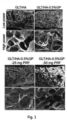
- Fig. 1 shows scan electron microscopic images of the GLT/HA-0.5%GP hydrogel formed from the composition prepared in Example 1 and the two LPRF-loaded hydrogels formed from the compositions prepared in Example 3.
- the LPRF-loaded hydrogels have a similar microstructure to GLT/HA-0.5%GP hydrogel, indicating that they share a similar porous structure with a similar pore size distribution. Moreover, a honeycomb-like porous morphology with interconnected pores was observed in those hydrogels.
- the open porosity of the scaffold will function to support the cellular on/ingrowth of osteoblasts and bone mineralization.
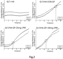
- the storage moduli (G') and loss modulus (G") of GLT/HA-0.5%GP was constantly higher than those of GLT/HA-GP with a crossover point of G' and G" of about 23 minutes, indicating that GLT/HA-0.5%GP was stably crosslinked in the presence of GP and had a gelation time of about 23 minutes.
- GLT/HA-0.5%GP hydrogels loaded with 25 or 50 mg of LPRF had crossover points of G' and G" of about 12 and 15 minutes, respectively, and the average values of G' and G" for the LPRF-loaded hydrogels were higher than those of GLT/HA-0.5%GP, indicating that mechanical strength increased with incorporation with PRF into GLT/HA-0.5%GP.
- Example 1 To simulate the working conditions and time frame for a segmental bone defect surgery, which require that a hydrogel precursor composition have sufficient flowability at room temperature before being applied to a bone defect site through an injection device, the hydrogels prepared in Example 1 were tested for syringeability at 25°C. In the test, each hydrogel was loaded into a 3 mL syringe with 21G needle (30 ⁇ 0.8 mm in diameter), and the syringeability was measured at 0 minute, 15 minutes and 60 minutes post loading, in terms of the residual mass of the hydrogel retained in the syringe by applying manual force for 5 seconds.
- the bottle inversion test was further used to measure the solution-gelation transition times of the hydrogels prepared in Examples 1 at physiological temperature.
- the test was initiated at T0, when the hydrogels prepared in Example 1 were placed in capped bottles, respectively, and allowed to stand for 1 minute.
- the flowabilities of the respective mixtures at T10 (10 minutes from T0), T20 (20 minutes from T0) and T30 (30 minutes from T0) at 37°C were determined by tilting the respective bottles upside-down, and the gelation times were determined when the mixtures stop flowing.
- thermoresponsive behaviors of the hydrogels prepared in Example 1 are related to the degree of GP crosslinking.
- GLT/HA-0.025%GP and - 0.05%GP hydrogels were shown injectable at ambient temperature even after 24 hours post-formulation.
- the two hydrogels gradually gelled and formed stable construct after 40 minutes as demonstrated by the bottle inversion test.
- GLT/HA-0.5%GP and -1%GP hydrogels exhibited relatively rapid gelation time of about 20-25 minutes at 37°C, which is consistent with the values obtained by using a rheometer as shown in Example 4 above, while remaining injectable for at least 60 minutes at ambient temperature.
- the gelation times of the hydrogels decreased from 37.5 minutes to 29, 26.5, 25, and 20.5 minutes at 37°C when GP concentration gradually increased from 0.025% to 0.05%, 0.1%, 0.5% and 1%, respectively.
- the hydrogels prepared in Example 1, the LPRF-loaded hydrogels prepared in Example 3, and the non-crosslinked GLT/HA hydrogel were tested for mechanical property using a texture analyzer (model TA.XT Plus, Texture Technologies Corp., Scarsdale, NY, USA) at ambient temperature with a cylinder probe of SMS P/25 to compress the samples. Plate samples (1 cm 3 in size) were subjected to a tensile test using a 5 kg load cell at a test speed of 1.0 mm/sec. The individual tensile modulus and fracture strength were obtained from the stress-strain curve, where ⁇ is the stress (N/mm 2 ) and ⁇ is the strain (mm/mm) under tensile measurement. All of the hydrogels were tested in triplicate for each group.
- the non-crosslinked GLT/HA hydrogel exhibited a poor mechanical strength of less than 50 kPa, rendering it unsuitable for use in bone tissue engineering applications.
- the mechanical strengths of the GLT/HA hydrogels prepared in Example 1 increased with the increasing of the GP concentration, indicating that the mechanical property of the hydrogel herein can be tuned by adjusting the extent of GP crosslinking.
- GP was added in an amount from 0.5 wt% to 1.0 wt%
- the hydrogels herein exhibited a compressive strength of about 400 kPa.
- the GP-crosslinked hydrogels including GLT/HA-0.5%GP hydrogel and the LPRF-loaded hydrogels, were able to withstand compressive stress of hundreds kPa without breaking down or collapsing.
- the LPRF-loaded hydrogels had a compressive strength of about 275 kPa, suggesting that the loading of LPRF did not considerably interfere with the structural integrity or impair the mechanical strength of the composite hydrogels.
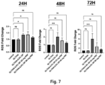
- L929 fibroblast cells (purchased from National Institute of Health, Taiwan) were seeded in a 96-well microplate at a density of 1 ⁇ 10 3 cells per well in DMEM supplemented with 10% fetal bovine serum (FBS) (Gibco ® , Grand Island, NY, USA) and allowed to attach overnight. The DMEM was removed, and the supernatants were added to the cell culture. After a 24-hour incubation, the viable cell number in each well was counted with thiazolyl blue tetrazolium bromide (MTT; Sigma-Aldrich, Saint Louis, MO, USA). The absorbance of each solution was measured at a wavelength of 595 nm with a microplate reader (Bio-Rad) in quadruplicate.
- FBS fetal bovine serum
- ROS reactive oxygen species
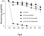
- the in vitro biodegradation features of the GLT/HA-0.5%GP hydrogel prepared in Example 1, the LPRF-loaded hydrogels prepared in Example 3 and the non-crosslinked GLT/HA hydrogel were evaluated by first measured the initial weight of 1 cm 3 cube-shaped hydrogels in each group. Subsequently, the hydrogels were incubated at 37°C in 3 mL of PBS buffer with gentle agitation in an orbital shaker. At each time point (0 hour, 12 nd hour, 24 th hour, 48 th hour, 72 nd hour, Day 6, Day 9, Day 12), the hydrogels were taken from the medium, blotting with a filter paper, and finally weighed accordingly. After that, the medium was replaced with fresh PBS. Percentage of weight loss was plotted against the degradation time.
- the biodegradation rates of the hydrogels tested decreased with addition of either GP or GP plus LPRF, suggesting that the addition of GP and LPRF slowed down the biodegradation rate of the hydrogels.
- the data reveal that the LPRF-loaded hydrogels herein exhibit desirable biodegradation performance for further in vivo applications as an orthopedic implant.
- Example 9 In vivo Subcutaneous Hydrogel Implantation
- Fig. 9 The remaining volume analysis shown in Fig. 9 indicates that the sizes of GLT/HA-GP and GLT/HA-GP-LPRF hydrogels gradually decreased in the first 14 days. Histological analysis of the subcutaneous tissues implanted with hydrogels shows that these hydrogels were encapsulated by thin fibrous tissues but without notable inflammatory cells aggregation ( Fig. 10 ). The systemic toxicity (e.g., hyperemia, edema, necrosis) of the implanted hydrogels to vital visceral organs including heart, liver, spleen, lung and kidney was not detected either ( Fig. 11 ).
- hyperemia edema, necrosis
- group A the defect in vivo was left untreated which served as control (sham); in group B, the defect in vivo was implanted with GLT/HA-0.5%GP hydrogel; and in group C, the defect in vivo was implanted with GLT/HA-0.5%GP-LPRF hydrogel.
- An 8-hole mini plate (VariAx Hand, Stryker) was applied laterally for fixation.
- Penicillin 2.2 mg/kg for 3 days
- ketoprofen 40,000 IU/kg for 3 days
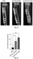
- Fig. 15 shows macroscopic assessment of the tibia harvested. More new bone regeneration and better bone defect repair were observed visually in the rabbit groups implanted with GLT/HA-0.5%GP-LPRF hydrogel than in GLT/HA-0.5%GP and control hydrogels. In the rabbit group implanted with GLT/HA-0.5%GP-LPRF hydrogel, the defects were filled with reparative tissues, and the bony cortical surface was mostly continuous. In contrast, the untreated control showed very limited repair in the defect area, leaving a remarkable central depression on the bony surface. In GLT/HA-0.5%GP group, partial union of bony defect was noted.
- tibias were scanned by micro-CT (Skyscan 1176; Bruker, Billerica, MA, USA). Micro-CT scanning of the femurs was done at 90 kV, 8000 ⁇ 8000 pixels/slice, and a resolution of 35 ⁇ m. Once scanning, the micro-CT images were reconstructed to produce a 3D volume rendering using reconstruction software (NRecon Reconstruction; Bruker). The tibial defect area was selected by identifying the relative position between the third and fourth screw holes and was denoted as the region of interest (ROI). Images of new bone formation in the ROI were captured and analyzed. Microstructural measurements included bone volume (BV, mm3), the percentage of bone volume versus total tissue volume (BV/TV, %), bone thickness (mm), bone separation (mm), bone surface (mm), and bone surface/total tissue volume (1/mm).
- GP-crosslinked GLT/HA hydrogel can be used for delivering PRF to the bone defect site, where beneficial growth factors are released due to biodegradation of the hydrogel, thus promoting osteogenic differentiations and mineralization of osteoblasts.
- GP crosslinking can help maintaining the stability of the hydrogel to ensure sustained stimulation of MSCs and gradual release of growth factors.
- Specimens were then processed for decalcification. For histological evaluation, samples were embedded in paraffin, and blocks were cut to serial thick sections (around 5 ⁇ m for each slide). Tissue sections were then stained by hematoxylin and eosin (H&E), Alizarin red S (ARS) and immunohistochemical staining using type I collagen (CoIl) antibody. Specimen sections were also observed under light microscopy (Leica) to evaluate new bone formation.
- H&E hematoxylin and eosin
- ARS Alizarin red S
- CoIl type I collagen
- GLT/HA-0.5%GP-LPRF hydrogel may serve as an osteoconductive biomaterial to allow ingrowth of osteoprogenitor cells from marrow and adjacent bony bed into the 3D structure of the porous scaffold.
Landscapes
- Health & Medical Sciences (AREA)
- Chemical & Material Sciences (AREA)
- Life Sciences & Earth Sciences (AREA)
- Medicinal Chemistry (AREA)
- Transplantation (AREA)
- Dermatology (AREA)
- Oral & Maxillofacial Surgery (AREA)
- Veterinary Medicine (AREA)
- Epidemiology (AREA)
- Animal Behavior & Ethology (AREA)
- General Health & Medical Sciences (AREA)
- Public Health (AREA)
- Engineering & Computer Science (AREA)
- Biomedical Technology (AREA)
- Molecular Biology (AREA)
- Botany (AREA)
- Chemical Kinetics & Catalysis (AREA)
- Composite Materials (AREA)
- Materials Engineering (AREA)
- Orthopedic Medicine & Surgery (AREA)
- Vascular Medicine (AREA)
- Hematology (AREA)
- Urology & Nephrology (AREA)
- Zoology (AREA)
- Dispersion Chemistry (AREA)
- Materials For Medical Uses (AREA)
Applications Claiming Priority (1)
| Application Number | Priority Date | Filing Date | Title |
|---|---|---|---|
| TW112127852A TWI835690B (zh) | 2023-07-26 | 2023-07-26 | 用於治療節段性骨缺損的可注射型水膠 |
Publications (1)
| Publication Number | Publication Date |
|---|---|
| EP4497449A1 true EP4497449A1 (de) | 2025-01-29 |
Family
ID=91269759
Family Applications (1)
| Application Number | Title | Priority Date | Filing Date |
|---|---|---|---|
| EP24187085.6A Pending EP4497449A1 (de) | 2023-07-26 | 2024-07-08 | Injizierbares hydrogel zur behandlung von segmentknochendefekten |
Country Status (3)
| Country | Link |
|---|---|
| US (1) | US12220501B1 (de) |
| EP (1) | EP4497449A1 (de) |
| TW (1) | TWI835690B (de) |
Citations (3)
| Publication number | Priority date | Publication date | Assignee | Title |
|---|---|---|---|---|
| US20110150823A1 (en) * | 2009-12-22 | 2011-06-23 | National Cheng Kung Univeristy | Cell Tissue Gel Containing Collagen and Hyaluronan |
| CN103007345A (zh) | 2012-12-20 | 2013-04-03 | 深圳清华大学研究院 | 抗菌生物活性支架及其制备方法 |
| CN104582747A (zh) | 2012-08-08 | 2015-04-29 | 南洋理工大学 | 用于制造具有活细胞的水凝胶微粒的方法和用于制造组织工程支架的组合物 |
Family Cites Families (3)
| Publication number | Priority date | Publication date | Assignee | Title |
|---|---|---|---|---|
| WO2012134540A2 (en) * | 2010-10-22 | 2012-10-04 | Vanderbilt University | Injectable synthetic pur composite |
| US12383627B2 (en) * | 2019-03-07 | 2025-08-12 | Board Of Regents Of The University Of Nebraska | Antimicrobial scaffolds |
| US20240024387A1 (en) * | 2020-10-15 | 2024-01-25 | University Of Louisville Research Foundation, Inc. | Probiotic delivery systems and methods of making and using |
-
2023
- 2023-07-26 TW TW112127852A patent/TWI835690B/zh active
- 2023-08-29 US US18/239,216 patent/US12220501B1/en active Active
-
2024
- 2024-07-08 EP EP24187085.6A patent/EP4497449A1/de active Pending
Patent Citations (3)
| Publication number | Priority date | Publication date | Assignee | Title |
|---|---|---|---|---|
| US20110150823A1 (en) * | 2009-12-22 | 2011-06-23 | National Cheng Kung Univeristy | Cell Tissue Gel Containing Collagen and Hyaluronan |
| CN104582747A (zh) | 2012-08-08 | 2015-04-29 | 南洋理工大学 | 用于制造具有活细胞的水凝胶微粒的方法和用于制造组织工程支架的组合物 |
| CN103007345A (zh) | 2012-12-20 | 2013-04-03 | 深圳清华大学研究院 | 抗菌生物活性支架及其制备方法 |
Non-Patent Citations (6)
| Title |
|---|
| "Biomaterials Applications for Nanomedicine", 16 November 2011, INTECH, ISBN: 978-953-30-7661-4, article GORGIEVA SELESTINA ET AL: "Collagen- vs. Gelatine-Based Biomaterials and Their Biocompatibility: Review and Perspectives", pages: 17 - 52, XP093226381, DOI: 10.5772/24118 * |
| BAI, X. ET AL., BIOACTIVE MATERIALS, vol. 3, 2018, pages 401 - 417 |
| CHANG KAI-CHI ET AL: "Characterization of genipin-crosslinked gelatin/hyaluronic acid-based hydrogel membranes and loaded with hinokitiol: In vitro evaluation of antibacterial activity and biocompatibility", MATERIALS SCIENCE AND ENGINEERING C, ELSEVIER SCIENCE S.A, CH, vol. 105, 12 August 2019 (2019-08-12), XP085832164, ISSN: 0928-4931, [retrieved on 20190812], DOI: 10.1016/J.MSEC.2019.110074 * |
| GILARSKA ADRIANA ET AL: "Bioactive yet antimicrobial structurally stable collagen/chitosan/lysine functionalized hyaluronic acid - based injectable hydrogels for potential bone tissue engineering applications", INTERNATIONAL JOURNAL OF BIOLOGICAL MACROMOLECULES, vol. 155, 1 July 2020 (2020-07-01), NL, pages 938 - 950, XP055868097, ISSN: 0141-8130, DOI: 10.1016/j.ijbiomac.2019.11.052 * |
| ZHANG Y.-F. ET AL., FRONT PHARMACOL., vol. 11, 2020, pages 622 |
| ZHENG ZIRUI ET AL: "Hyaluronic acid-based materials for bone regeneration: A review", REACTIVE AND FUNCTIONAL POLYMERS, ELSEVIER, AMSTERDAM, NL, vol. 171, 28 December 2021 (2021-12-28), XP086945081, ISSN: 1381-5148, [retrieved on 20211228], DOI: 10.1016/J.REACTFUNCTPOLYM.2021.105151 * |
Also Published As
| Publication number | Publication date |
|---|---|
| US20250032675A1 (en) | 2025-01-30 |
| TWI835690B (zh) | 2024-03-11 |
| US12220501B1 (en) | 2025-02-11 |
| TW202504648A (zh) | 2025-02-01 |
Similar Documents
| Publication | Publication Date | Title |
|---|---|---|
| Li et al. | A bone regeneration strategy via dual delivery of demineralized bone matrix powder and hypoxia-pretreated bone marrow stromal cells using an injectable self-healing hydrogel | |
| Fu et al. | Injectable and thermo-sensitive PEG-PCL-PEG copolymer/collagen/n-HA hydrogel composite for guided bone regeneration | |
| CA2557386C (en) | Gel suitable for implantation and delivery system | |
| Kouhi et al. | Injectable gellan gum/lignocellulose nanofibrils hydrogels enriched with melatonin loaded forsterite nanoparticles for cartilage tissue engineering: Fabrication, characterization and cell culture studies | |
| JP2010273847A (ja) | 高密度多孔質複合体 | |
| Jung et al. | Multivalent ion-based in situ gelling polysaccharide hydrogel as an injectable bone graft | |
| EP3344304B1 (de) | Bioaktives polymer zur knochenregeneration | |
| JP5315233B2 (ja) | 注入可能な骨空隙充填剤 | |
| EP4178634B1 (de) | Integrierte bioaktive kern-schale-struktur zur regeneration von knochen- und osteochondralen geweben | |
| Ma et al. | Poly (propylene fumarate)/β-calcium phosphate composites for enhanced bone repair | |
| CA2934470C (en) | Fgf-18 formulation in xyloglucan gels | |
| US20060198863A1 (en) | Ceramic composition for filling bone defects | |
| US12220501B1 (en) | Injectable hydrogel for treatment of segmental bone defect | |
| CN118649288A (zh) | 一种自固化的可吸收骨填充材料和应用 | |
| Dabbarh et al. | Chitosan based biocomposites for hard tissue engineering | |
| CN118416290A (zh) | 一种生物基骨胶水及制备方法和应用 | |
| CN121265864A (zh) | 用于治疗节段性骨缺损的可注射型水胶 | |
| US9474828B2 (en) | Self-hardening bioactive cement compositions with partially deacetylated chitin as bone graft substitutes | |
| Chen et al. | Effects of chitosan-coated pressed calcium sulfate pellets combined with recombinant human bone morphogenetic protein 2 on bone formation in femoral condyle-contained bone defects | |
| Apinun et al. | Evaluation of Bone Regeneration Using Injectable Surfactant-Induced Thai Silk Fibroin/Collagen In Situ-Forming Hydrogel in Segmental Bone Defects in Rats. | |
| Sadek et al. | The efficiency of intra-articular injection of silver nanoparticles (AgNPs) on repair of experimentally induced avascular meniscal tear in dogs | |
| He et al. | Fabrication of injectable calcium sulfate bone graft material | |
| CN117582548A (zh) | 一种具有原位修复前交叉韧带功能的可注射多交联复合水凝胶支架及其制备方法 | |
| Marion | DEVELOPMENT OF ALGINATE HYDROGELS FOR BONE REGENERATION |
Legal Events
| Date | Code | Title | Description |
|---|---|---|---|
| PUAI | Public reference made under article 153(3) epc to a published international application that has entered the european phase |
Free format text: ORIGINAL CODE: 0009012 |
|
| STAA | Information on the status of an ep patent application or granted ep patent |
Free format text: STATUS: THE APPLICATION HAS BEEN PUBLISHED |
|
| AK | Designated contracting states |
Kind code of ref document: A1 Designated state(s): AL AT BE BG CH CY CZ DE DK EE ES FI FR GB GR HR HU IE IS IT LI LT LU LV MC ME MK MT NL NO PL PT RO RS SE SI SK SM TR |
|
| STAA | Information on the status of an ep patent application or granted ep patent |
Free format text: STATUS: REQUEST FOR EXAMINATION WAS MADE |
|
| 17P | Request for examination filed |
Effective date: 20250307 |
|
| RIC1 | Information provided on ipc code assigned before grant |
Ipc: A61L 27/26 20060101AFI20251017BHEP Ipc: A61L 27/54 20060101ALI20251017BHEP |
|
| GRAP | Despatch of communication of intention to grant a patent |
Free format text: ORIGINAL CODE: EPIDOSNIGR1 |
|
| STAA | Information on the status of an ep patent application or granted ep patent |
Free format text: STATUS: GRANT OF PATENT IS INTENDED |
|
| INTG | Intention to grant announced |
Effective date: 20260116 |
|
| GRAS | Grant fee paid |
Free format text: ORIGINAL CODE: EPIDOSNIGR3 |