EP4495263A1 - Verfahren zur bestimmung des x-chromosom-reaktivierungsniveaus einer weiblichen person - Google Patents
Verfahren zur bestimmung des x-chromosom-reaktivierungsniveaus einer weiblichen person Download PDFInfo
- Publication number
- EP4495263A1 EP4495263A1 EP23382740.1A EP23382740A EP4495263A1 EP 4495263 A1 EP4495263 A1 EP 4495263A1 EP 23382740 A EP23382740 A EP 23382740A EP 4495263 A1 EP4495263 A1 EP 4495263A1
- Authority
- EP
- European Patent Office
- Prior art keywords
- female subject
- determining
- level
- methylation
- dna
- Prior art date
- Legal status (The legal status is an assumption and is not a legal conclusion. Google has not performed a legal analysis and makes no representation as to the accuracy of the status listed.)
- Withdrawn
Links
Images





Classifications
-
- C—CHEMISTRY; METALLURGY
- C12—BIOCHEMISTRY; BEER; SPIRITS; WINE; VINEGAR; MICROBIOLOGY; ENZYMOLOGY; MUTATION OR GENETIC ENGINEERING
- C12Q—MEASURING OR TESTING PROCESSES INVOLVING ENZYMES, NUCLEIC ACIDS OR MICROORGANISMS; COMPOSITIONS OR TEST PAPERS THEREFOR; PROCESSES OF PREPARING SUCH COMPOSITIONS; CONDITION-RESPONSIVE CONTROL IN MICROBIOLOGICAL OR ENZYMOLOGICAL PROCESSES
- C12Q1/00—Measuring or testing processes involving enzymes, nucleic acids or microorganisms; Compositions therefor; Processes of preparing such compositions
- C12Q1/68—Measuring or testing processes involving enzymes, nucleic acids or microorganisms; Compositions therefor; Processes of preparing such compositions involving nucleic acids
- C12Q1/6876—Nucleic acid products used in the analysis of nucleic acids, e.g. primers or probes
- C12Q1/6883—Nucleic acid products used in the analysis of nucleic acids, e.g. primers or probes for diseases caused by alterations of genetic material
-
- C—CHEMISTRY; METALLURGY
- C12—BIOCHEMISTRY; BEER; SPIRITS; WINE; VINEGAR; MICROBIOLOGY; ENZYMOLOGY; MUTATION OR GENETIC ENGINEERING
- C12Q—MEASURING OR TESTING PROCESSES INVOLVING ENZYMES, NUCLEIC ACIDS OR MICROORGANISMS; COMPOSITIONS OR TEST PAPERS THEREFOR; PROCESSES OF PREPARING SUCH COMPOSITIONS; CONDITION-RESPONSIVE CONTROL IN MICROBIOLOGICAL OR ENZYMOLOGICAL PROCESSES
- C12Q1/00—Measuring or testing processes involving enzymes, nucleic acids or microorganisms; Compositions therefor; Processes of preparing such compositions
- C12Q1/68—Measuring or testing processes involving enzymes, nucleic acids or microorganisms; Compositions therefor; Processes of preparing such compositions involving nucleic acids
- C12Q1/6876—Nucleic acid products used in the analysis of nucleic acids, e.g. primers or probes
- C12Q1/6883—Nucleic acid products used in the analysis of nucleic acids, e.g. primers or probes for diseases caused by alterations of genetic material
- C12Q1/6886—Nucleic acid products used in the analysis of nucleic acids, e.g. primers or probes for diseases caused by alterations of genetic material for cancer
-
- C—CHEMISTRY; METALLURGY
- C12—BIOCHEMISTRY; BEER; SPIRITS; WINE; VINEGAR; MICROBIOLOGY; ENZYMOLOGY; MUTATION OR GENETIC ENGINEERING
- C12Q—MEASURING OR TESTING PROCESSES INVOLVING ENZYMES, NUCLEIC ACIDS OR MICROORGANISMS; COMPOSITIONS OR TEST PAPERS THEREFOR; PROCESSES OF PREPARING SUCH COMPOSITIONS; CONDITION-RESPONSIVE CONTROL IN MICROBIOLOGICAL OR ENZYMOLOGICAL PROCESSES
- C12Q2600/00—Oligonucleotides characterized by their use
- C12Q2600/154—Methylation markers
Definitions
- the invention relates to methods for quantifying the global X chromosome reactivation level of a female subject, in particular to methods based on determining the DNA methylation status.
- the invention also provides to the use of X chromosome reactivation levels as a biomarker of several aspects of female health and disease.
- the X chromosome in the cells of therian mammals can inactivate.
- one of the two X chromosomes in female cells is randomly inactivated, leading to the formation of Barr bodies, which are visible in cell nuclei.
- Chromosome X inactivation thus achieves gene dosage compensation with males, who carry only one X chromosome per cell. It is estimated that up to 23% of genes on chromosome X naturally escape inactivation in the inactivated X chromosome (XI).
- XCI is an epigenetic process that occurs early in development and it is thought to be stably maintained throughout the lifespan of females.
- some genes subject to XCI have been described to reactivate under some circumstances.
- XCI and its reverse process, chromosome X reactivation (X-Ra) have been mostly measured as an individual property of genes, but not as a global property of the X chromosome, let alone as a global parameter a female subject.
- the present inventors have developed a novel method for calculating a biomarker value that provides accurate information regarding numerous aspects of women's health and disease.
- the inventors have devised a simple method for accurately and reliable calculating a quantitative value representing the global level of X chromosome reactivation (X-Ra) in a female subject, the method being based on determining the fraction of hypomethylated CpGs in a particular set of genes of the X chromosome.
- the method of the invention allows calculating the global X-Ra level in a female subject using complex samples containing different cell types, such as whole blood or tumor biopsies, which is a noteworthy advantage when it comes to the design of practical diagnostics and for testing blood samples collected from other studies.
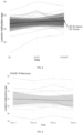
- the present inventors have surprisingly found that the global X-Ra biomarker calculated by the method of the invention is strongly associated with female age ( Figure 2A-2C ) and shorter telomere length ( Figure 2D ). They have also found that the X-Ra biomarker provides more information than other known aging biomarkers based on general methylation levels (e.g. those disclosed in M. E. Levine, et al. 2018). In particular, X-Ra level is strongly associated with age-associated diseases in women, such as cancer ( Figure 2E , 3A , 7A, 7B ) or Alzheimer's disease ( Figure 3C ), while global methylation levels are not ( Figure 4-5 ).
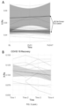
- X-Ra levels were also found to correlate with low survival of breast cancer patients ( Figure 6B ), particularly in triple-negative breast cancer patients. The inventors also found that X-Ra levels correlate with recovery from infection, in particular COVID 19 infections ( Figure 3B ), while global methylation levels did not. In addition to the above, the inventors found that the X-Ra levels correlate with the stage of pregnancy ( Figure 3D ) and female fertility ( Figure 2F ).
- the results herein provided demonstrate that the global X-Ra level of a female subject calculated by the method of the invention represents a multipurpose biomarker of female health which is highly robust, reliable and informative for a range of applications, including the determination of aging, pregnancy, fertility, and various types of diseases.
- the invention provides an in vitro method for determining the X chromosome reactivation (X-Ra) level of a female subject, the method comprising the steps of: (a) determining the methylation status of at least 10 CpG sites of Table 1 in a DNA-containing sample from the female subject; and (b) calculating the fraction of the CpG sites analyzed in step (a) (i.e., whose methylation status is determined in step (a)) that are hypomethylated.
- X-Ra X chromosome reactivation
- the invention provides an in vitro method for determining the biological age of a female subject, the method comprising the steps of determining the X chromosome reactivation (X-Ra) level in a DNA-containing sample from the female subject with a method as defined in the first aspect and comparing the determined X-Ra level with a reference value or a range of reference values.
- X-Ra X chromosome reactivation
- the invention provides an in vitro method for the diagnosis and/or prognosis of an age-related disease or disorder in a female subject, the method comprising the steps of determining the X chromosome reactivation (X-Ra) level in a DNA-containing sample from the female subject with a method as defined in the first aspect and comparing the determined X-Ra level with a reference value or a range of reference values; optionally, wherein the age-related disease or disorder is selected from the group consisting of cancer, neurodegenerative disease, sarcopenia, osteopenia, osteoporosis, arthritis, atherosclerosis, cardiovascular disease, hypertension, cataracts, presbyopia, glaucoma, type 2 diabetes, metabolic syndrome, chronic inflammation, immunosenescence, menopausal symptoms, and combinations thereof.
- X-Ra X chromosome reactivation
- the invention provides an in vitro method for the diagnosis and/or prognosis of an infection in a female subject, the method comprising the steps of determining the X chromosome reactivation (X-Ra) level in a DNA-containing sample from the female subject with a method as defined in the first aspect and comparing the determined X-Ra level with a reference value or a range of reference values.
- X-Ra X chromosome reactivation
- the invention provides an in vitro method for deciding or recommending whether to initiate a therapeutic intervention in a female subject suspicious of suffering an age-related disease or disorder, or an infection, wherein the method comprises the steps of determining the X chromosome reactivation (X-Ra) level in a DNA-containing sample from the female subject with a method as defined in the first aspect and comparing the determined X-Ra level with a reference value or a range of reference values.
- X-Ra X chromosome reactivation
- the invention provides an in vitro method for determining the efficacy of a therapeutic intervention in a female subject already diagnosed of suffering an age-related disease or disorder, or an infection, the method comprising determining the X chromosome reactivation (X-Ra) level in a DNA-containing sample from the female subject with a method as defined in the first aspect before and after starting the therapeutic intervention.
- X-Ra X chromosome reactivation
- the invention provides an in vitro method for determining the ER- status, PR- status, and/or HER2-status of a breast cancer in a female subject, the method comprising the steps of determining the X chromosome reactivation (X-Ra) level in a DNA-containing sample from the female subject with a method as defined in the first aspect and comparing the determined X-Ra level with a reference value or a range of reference values.
- X-Ra X chromosome reactivation
- the invention provides an in vitro method for determining the gestational age of a fetus, the method comprising the steps of determining the X chromosome reactivation (X-Ra) level in a DNA-containing sample from a female subject pregnant with the fetus with a method as defined in the first aspect and comparing the determined X-Ra level with a reference value or a range of reference values.
- X-Ra X chromosome reactivation
- the invention provides an in vitro method for determining the fertility status of a female subject, the method comprising the steps of determining the X chromosome reactivation (X-Ra) level in a DNA-containing sample from the female subject with a method as defined in the first aspect and comparing the determined X-Ra level with a reference value or a range of reference values.
- X-Ra X chromosome reactivation
- the invention provides the use of means for determining, in a DNA-containing sample from a female subject, the methylation status of at least 10 CpG sites of Table 1 in a method for determining the X chromosome reactivation (X-Ra) level as defined in the first aspect; optionally, wherein the means are a methylation chip array.
- the invention provides the use of the methylation status of at least 10 CpG sites of Table 1 as a biomarker for: determining the X chromosome reactivation (X-Ra) level of a female subject; determining the biological age of a female subject (i.e., as an aging biomarker); determining the gestational age of a fetus; diagnosing and/or prognosing an age-related disease or disorder, or an infection, in a female subject; deciding or recommending whether to initiate a therapeutic intervention in a female subject suspicious of suffering an age-related disease or disorder, or an infection; determining the efficacy of a therapeutic intervention in a female subject already diagnosed of suffering an age-related disease or disorder, or an infection; or determining the fertility status of a female subject.
- X-Ra X chromosome reactivation
- the invention provides a computer-implemented method comprising the steps of: (a) receiving information corresponding to the methylation status of at least 10 CpG sites of Table 1 from a DNA-containing sample from a female subject; (b) calculating the fraction of the CpG sites of step (a) that are hypomethylated, which corresponds to the X-Ra level of the female subject.
- the invention provides a data processing apparatus comprising means for carrying out the method of the twelfth aspect.
- the invention provides a computer program comprising instructions which, when the program is executed by a computer, cause the computer to carry out the method of the twelfth aspect.
- the invention provides a computer-readable medium comprising instructions which, when executed by a computer, cause the computer to carry out the method of the twelfth aspect.
- the invention provides a method of identifying a candidate molecule that modulates the X chromosome reactivation (X-Ra) level, the method comprising the steps of contacting the candidate molecule with a cell from a female subject and determining the X chromosome reactivation (X-Ra) level with a method as defined in the first aspect.
- any ranges given include both the lower and the upper end-points of the range. Ranges given should be considered approximate, unless specifically stated. The term “about” refers to a deviation of plus/minus 10 %, particularly plus/minus 5 %.
- the present invention provides an in vitro method for determining the X chromosome reactivation (X-Ra) level of a female subject comprising (a) determining the methylation status of at least 10 CpG sites of Table 1 in a DNA-containing sample from the female subject; and (b) calculating the fraction of the CpG sites analyzed in step (a) that are hypomethylated.
- the calculated fraction of CpGs that are hypomethylated represents the X-Ra level of the female subject. While increasing the number of CpG sites analyzed increases the accuracy of the method, as shown in the examples below and Figure 8 , it is sufficient to only determine the methylation status of a low number of CpGs of Table 1 -e.g. as low as 10 CpGs-to obtain a reliable X-Ra level.
- X chromosome inactivation refers to the transcriptional silencing of one of the two X chromosomes in female somatic cells.
- X chromosome reactivation or “X-Ra” refers to the reverse process of XCI, where the transcription of the inactivated X chromosome is reactivated, for example, by an external process or by XCI deficiency.
- both the terms XCI and X-Ra refer to global (intra individual and chromosome-wide) XCI and X-Ra levels, respectively, that is, to the individual's tendency to inactivate or reactivate of the whole X chromosome, and not a single gene, across the cells in a given biological sample. Therefore, the first aspect can also be formulated as an in vitro method for determining the global X chromosome reactivation (X-Ra) level of a female subject.
- X-Ra level and "X-Ra value" are used interchangeably in the context of X-Ra determination.
- methylation status of a CpG site refers to the presence or absence of a methylated cytosine, or the ratio of methylated cytosine to unmethylated cytosine, in said CpG site.
- CpG site refers to a region of a DNA molecule where a cytosine nucleotide is followed by a guanine nucleotide in the linear sequence of bases along its 5' to 3' direction.
- CpG is a shorthand for 5'-C-phosphate-G-3' that is cytosine and guanine separated by only one phosphate group; phosphate links any two nucleotides together in DNA.
- the CpG site may be present in a CpG island, a CpG doublet, a promoter, an intron, or an exon of gene.
- the CpG sites whose methylation status is determined in step (a) are located in promoters.
- the CpG sites are well-defined genomic regions and they are indexed in database giving to said regions an identification number (ID) as cg_number, according to Illumina ® 's CpG Loci Identification wherein flanking sequences regions around the CpG dinucleotide are used to generate unique CpG cluster IDs (cg_number).
- the location of all CpG sites in human chromosome X provided herein are according to the chromosome map and sequence entries of database UCSC Genome Browser on Human February 2009, GRCh37/hg19 assembly of the University of California Santa Cruz (UCSC).
- the methylation status of DNA, and in particular of any CpG site can optionally be represented or indicated by a "methylation value” or "methylation level.”
- a methylation value, score or level can be generated, for example, by quantifying the methylation in a cytosine using, for example a beta value ranging from 0 to 1, as explained below.
- polynucleotide is interchangeably used with “nucleic acid” and refers to a polymer of nucleotides, either ribonucleotides or deoxyribonucleotides.
- a polynucleotide formed by ribonucleotides may be referred to as “RNA polynucleotide”, “ribonucleic acid” or simply “RNA”; and a polynucleotide formed by deoxyribonucleotides may be referred to as "DNA polynucleotide", “deoxyribonucleic acid” or simply “DNA”.
- genomic DNA refers to the chromosomal DNA, particularly to human chromosomal DNA.
- the method consists of the steps of:
- the female subject is a mammal, particularly a human or a non-human mammal.
- the female subject is a non-human primate.
- the female subject is a rodent.
- the female subject is a sheep, a goat, cattle, a cat, or a dog. The female subject may be at any stage of development.
- the method comprises determining the methylation status of at least 10 CpG sites of Table 1, optionally wherein the CpG sites are in at least 2, at least 3, at least 4, at least 5, at least 6, at least 7, at least 8, at least 9, at least 10, at least 20, at least 30, at least 40, at least 50, at least 60, at least 70, at least 80, at least 90, at least 100, at least 100, at least 110, at least 120, at least 130, at least 140, at least 150, at least 160, at least 170, at least 180, at least 190, at least 200, at least 210, at least 220, at least 230, at least 240, at least 250, or 277 genes of Table 1.
- the method comprises determining the methylation status of at least 20, at least 30, at least 40, at least 50, at least 100, at least 150, at least 200, at least 250, at least 300, at least 350, at least 400, at least 450, or 498 CpG sites of Table 1, optionally wherein the CpG sites are in at least 2, at least 3, at least 4, at least 5, at least 6, at least 7, at least 8, at least 9, at least 10, at least 20, at least 30, at least 40, at least 50, at least 60, at least 70, at least 80, at least 90, at least 100, at least 100, at least 110, at least 120, at least 130, at least 140, at least 150, at least 160, at least 170, at least 180, at least 190, at least 200, at least 210, at least 220, at least 230, at least 240, at least 250, or 277 genes of Table 1.
- the method comprises determining the methylation status of at least one CpG site in at least 2, at least 5, at least 10, at least 20, at least 30, at least 40, at least 50, at least 60, at least 70, at least 80, at least 90, at least 100, at least 100, at least 110, at least 120, at least 130, at least 140, at least 150, at least 160, at least 170, at least 180, at least 190, at least 200, at least 210, at least 220, at least 230, at least 240, at least 250, or 277 genes of Table 1.
- the method comprises determining the methylation status of a least 10 CpG sites of Table 2, optionally wherein the CpG sites are in at least 2, at least 3, at least 4, at least 5, at least 6, at least 7, at least 8, at least 9, at least 10, at least 20, at least 30, at least 40, at least 50, at least 60, at least 70, at least 80, at least 90, at least 100, at least 100, at least 110, at least 120, at least 130, at least 140, at least 150, at least 160, at least 170, at least 180, at least 190, at least 200, at least 210, at least 220, at least 230, at least 240, at least 250, at least 300, at least 350, or 362 genes of Table 2.
- the method comprises determining the methylation status of a least 10, at least 20, at least 30, at least 40, at least 50, at least 100, at least 150, at least 200, at least 250, at least 300, at least 350, at least 400, at least 450, at least 500, at least 1000, at least 1500, at least 2000, at least 2500, at least 3000, at least 3500, at least 4000, at least 4500, or 4758 CpG sites of Table 2, optionally wherein the CpG sites are in at least 2, at least 3, at least 4, at least 5, at least 6, at least 7, at least 8, at least 9, at least 10, at least 20, at least 30, at least 40, at least 50, at least 60, at least 70, at least 80, at least 90, at least 100, at least 100, at least 110, at least 120, at least 130, at least 140, at least 150, at least 160, at least 170, at least 180, at least 190, at least 200, at least 210, at least 220, at least 230, at least
- the method comprises determining the methylation status of at least one CpG site in at least 2, at least 3, at least 4, at least 5, at least 6, at least 7, at least 8, at least 9, at least 10, at least 20, at least 30, at least 40, at least 50, at least 60, at least 70, at least 80, at least 90, at least 100, at least 100, at least 110, at least 120, at least 130, at least 140, at least 150, at least 160, at least 170, at least 180, at least 190, at least 200, at least 210, at least 220, at least 230, at least 240, at least 250, at least 300, at least 350, or 362 genes of Table 2.
- step (b) the fraction of the CpG sites is the weighted fraction of the CpG sites analyzed in step (a) that are hypomethylated.
- the methylation status of the CpGs is determined by a method selected from the group consisting of methylation chip array (e.g. Illumina's Infimum HumanMethylation27 BeadChip array, Illumina's Infimum HumanMethylation450 BeadChip array, or Illumina's Infimum MethylationEPIC), bisulfite sequencing (e.g.
- methods for the detection of DNA methylation first involve converting the DNA to be analyzed so that the unmethylated cytosine is converted to uracil.
- a chemical reagent that selectively modifies either the methylated or non-methylated form of CpG dinucleotide motifs may be used.
- Suitable chemical reagents include hydrazine and bisulphite ions and the like.
- isolated DNA can be treated with sodium bisulfite (NaHS03) which converts unmethylated cytosine to uracil, while methylated cytosines are maintained.
- uracil is recognized as a thymine by DNA polymerase. Therefore, after PCR or sequencing, the resultant product contains cytosine only at the position where 5-methylcytosine occurs in the starting template DNA. This makes the discrimination between unmethylated and methylated cytosine possible by multiple methods.
- methylation chip arrays There are numerous commercial methylation chip arrays that can be used in the methods of the invention, for example Illumina's Infimum HumanMethylation450 BeadChip array or Illumina's Infimum HumanMethylation27 BeadChip array. These arrays generate fluorescent signals that are scanned, and the intensities of the signals produced by the unmethylated and methylated bead types are measured. Software is generally used to record DNA methylation values, described as "beta values," for each locus in each sample. DNA methylation beta values are continuous variables between 0 and 1, representing the ratio of the intensity of the methylated bead type to the combined locus intensity.
- Methylation-Specific PCR is based on a chemical reaction of sodium bisulfite with DNA, converting unmethylated cytosines of CpG dinucleotides to uracil (UpG), followed by traditional PCR. Methylated cytosines will not be converted by the sodium bisulfite, and specific nucleotide primers designed to overlap with the CpG site of interest will allow determining the methylation status as methylated or unmethylated, based on the amount of PCR product formed.
- the HELP assay is based on the differential ability of restriction enzymes to recognize and cleave methylated and unmethylated CpG DNA sites.
- ChIP-on-chip assays based on the ability of commercially prepared antibodies to bind to DNA methylation-associated proteins like MCP2, can be used to determine the methylation status.
- restriction landmark genomic scanning also based upon differential recognition of methylated and unmethylated CpG sites by restriction enzymes can be used.
- Methylated DNA immunoprecipitation (MeDIP) analogous to chromatin immunoprecipitation, can be used to isolate methylated DNA fragments for input into DNA detection methods such as DNA microarrays (MeDIP-chip) or DNA sequencing (MeDIP-seq). The unmethylated DNA is not precipitated.
- molecular break light assay for DNA adenine methyltransferase activity can be used. This is an assay that uses the specificity of the restriction enzyme Dpnl for fully methylated (adenine methylation) GATC sites in an oligonucleotide labeled with a fluorophore and quencher.
- the adenine methyltransferase methylates the oligonucleotide making it a substrate for Dpnl. Cutting of the oligonucleotide by Dpnl gives rise to a fluorescence increase.
- Pyrosequencing of bisulfite treated DNA is a sequencing of an amplicon made by a normal forward primer but a biotinylated reverse primer to PCR the gene of choice.
- the Pyrosequencer analyses the sample by denaturing the DNA and adding one nucleotide at a time to the mix according to a sequence given by the user. If there is a mismatch, it is recorded and the percentage of DNA for which the mismatch is present is noted. This gives the user a percentage methylation per CpG island.
- Next generation genome bisulfite sequencing also known as NGBS, is a genome-wide analysis of DNA methylation.
- NGS Next-Generation Sequencing
- the hypomethylated CpG sites are the CpG sites with a beta value equal to or lower than 0.01, 0.02, 0.03, 0.04, 0.05, 0.06, 0.07, 0.08, 0.09, 0.1, 0.11, 0.12, 0.13, 0.14, 0.15, 0.16, 0.17, 0.18, 0.19, or 0.2, particularly where the methylation status is determined by a methylation chip array (such as the Illumina's Infimum HumanMethylation450 BeadChip array or Infimum HumanMethylation27 BeadChip array).
- the hypomethylated CpG sites are the CpG sites with a beta value equal to or lower than about 0.2, where the methylation status is determined by a methylation chip array.
- the hypomethylated CpG sites are the CpG sites with a methylation equal to or lower than 20%, 19%, 18%, 17%, 16%, 15%, 14%, 13%, 12%, 11%, 10%, 9%, 8%, 7%, 6%, 5%, 4%, 3%, 2%, or, 1%, particularly where the methylation status is determined by bisulfite sequencing.
- the hypomethylated CpG sites are the CpG sites with a methylation equal to or lower than about 20%, where the methylation status is determined by bisulfite sequencing.
- the hypomethylated CpG sites are the CpG sites with a methylation equal to or lower than 5%, 4%, 3%, 2%, or, 1%, particularly where the methylation status is determined by next-generation bisulfite sequencing.
- the hypomethylated CpG sites are the CpG sites with a methylation equal to or lower than about 5%, where the methylation status is determined by next-generation bisulfite sequencing.
- the DNA-containing sample is a genomic DNA-containing sample, optionally selected from the group consisting of whole blood, plasma, serum, urine, saliva, sputum, nasal mucous, respiratory lavage, tears, chorionic villi, amniotic fluid, cerebrospinal fluid, spinal fluid, lavage fluid, stool, lymphatic fluid, bile, sweat, breast milk, breast fluid, cultured cells, tissue biopsy (e.g. tumor biopsy), tissue swabs, cervical cytology, synovial fluid, and combinations thereof.
- the whole blood is peripheral whole blood.
- the method further comprises determining the biological age of the female subject by comparing the determined X-Ra level with a reference value or a range of reference values.
- the method further comprises diagnosing and/or prognosing an age-related disease or disorder in the female subject by comparing the determined X-Ra level with a reference value or a range of reference values.
- the method further comprises diagnosing and/or prognosing an infection in the female subject by comparing the determined X-Ra level with a reference value or a range of reference values.
- the method further comprises determining the gestational age of a fetus by comparing the determined X-Ra level with a reference value or a range of reference values.
- the method further comprises determining the fertility status of the female subject by comparing the determined X-Ra level with a reference value or a range of reference values.
- the method further comprises reporting the X-Ra level of the female subject, optionally wherein said reporting comprises preparing a written or electronic report
- the method is with the proviso that the method does not comprise determining the methylation status of any CpG site in an autosome (non-sexual chromosome) and/or a Y chromosome.
- the method is with the proviso that it does not comprise determining the methylation status of any CpG site outside the X chromosome.
- the invention provides an in vitro method for determining the biological age of a female subject, the method comprising the steps of determining the X chromosome reactivation (X-Ra) level in a DNA-containing sample from the female subject with a method as defined in the first aspect and comparing the determined X-Ra level with a reference value or a range of reference values.
- X-Ra X chromosome reactivation
- biological age refers to the physiological state of a female subject, or of a tissue from said subject, relative to the physiological changes that occur throughout the subject's lifespan. Therefore, the skilled person would appreciate that to determine the biological age of a subject means to determine the degree of aging of said subject.
- chronological age refers to the age of a subject as measured by a time scale, such as months and years. If the subject has premature aging, the subject's biological age is greater than their chronological age. If a subject has delayed aging, the subject's biological age is less than their chronological age.
- the term "reference value or a range of reference values" referred to in the methods of the invention is to be understood as a predefined value or range of values of X-Ra derived from a control sample or group of samples.
- the control samples are taken from a subject or group of subjects wherein the biological age; gestational age of a fetus; fertility; or presence, absence, stage, histological subtype or grade, or course of a disease; has been properly performed previously. This value is used as a threshold to discriminate the female subjects assayed.
- the subject or subjects from whom the reference value is derived may include subject/s wherein the condition is absent, subject/s wherein the condition is present, or both.
- reference value is a cut-off value defined by means of a conventional ROC analysis (Receiver Operating Characteristic analysis).
- optimal cut-off value will be defined according to the particular applications of the method - e.g., purpose, target population for the diagnosis or prognosis, balance between specificity and sensibility, etc.
- reference value can be an absolute value; a relative value; a value that has an upper or a lower limit; a range of values; an average value; a median value, a mean value, or a value as compared to a particular control or baseline value.
- the method further comprises the step of determining the age of the female subject based on the determined X-Ra level, optionally by applying a statistical prediction algorithm.
- the reference value or a range of reference values are derived from one or more female subjects of known ages.
- the method further comprises reporting the biological age of the female subject, optionally wherein said reporting comprises preparing a written or electronic report.
- the invention provides an in vitro method for the diagnosis and/or prognosis of an age-related disease or disorder in a female subject, the method comprising the steps of determining the X chromosome reactivation (X-Ra) level in a DNA-containing sample from the female subject with a method as defined in the first aspect and comparing the determined X-Ra level with a reference value or a range of reference values.
- X-Ra X chromosome reactivation
- the method further comprises the step of diagnosing and/or prognosing the age-related disease or disorder based on the determined X-Ra level, optionally by applying a statistical prediction algorithm.
- the reference value or a range of reference values are derived from one or more female subjects where the age-related disease or disorder is absent. In another embodiment, the reference value or a range of reference values are derived from one or more female subjects where the age-related disease or disorder is present.
- diagnosis is understood as becoming aware of a particular medical condition complication or risk in a subject; the determination of the nature of the disease or condition; or the distinguishing of one disease or condition from another. It refers both to the process of attempting to determine or identify the possible disease or disorder, and to the opinion reached by this process.
- a diagnosis in the sense of diagnostic procedure, can be regarded as an attempt at classification of an individual's condition into separate and distinct categories that allow medical decisions about treatment and prognosis to be made. Subsequently, a diagnostic opinion is often described in terms of a disease or other condition. However, a diagnosis can take many forms. It might be a matter of detecting the presence and naming the disease, lesion, dysfunction or disability.
- Prognosis refers to the prediction of the probable progression and outcome of a disease, including the death of the subject.
- a “disease” is a state of health of an animal wherein the animal cannot maintain homeostasis, and wherein if the disease is not ameliorated then the animal's health continues to deteriorate.
- a “disorder” in an animal is a state of health in which the animal is able to maintain homeostasis, but in which the animal's state of health is less favorable than it would be in the absence of the disorder. Left untreated, a disorder does not necessarily cause a further decrease in the animal's state of health.
- age-related disease or disorder refers to diseases and disorders often associated with aging, that is, diseases or disorders in which aging is a major risk factor, for example cancers (e.g., gliomas, leukemia, lymphoma, breast cancer, prostate cancer, lung cancer, etc.) and neurodegenerative diseases (e.g., Parkinson's disease, Alzheimer's disease, Huntington's disease, dementia, etc.).
- cancers e.g., gliomas, leukemia, lymphoma, breast cancer, prostate cancer, lung cancer, etc.
- neurodegenerative diseases e.g., Parkinson's disease, Alzheimer's disease, Huntington's disease, dementia, etc.
- the age-related disease or disorder is selected from the group consisting of cancer, neurodegenerative disease (e.g. Alzheimer's disease, dementia), sarcopenia, osteopenia, osteoporosis, arthritis, atherosclerosis, cardiovascular disease, hypertension, cataracts, presbyopia, glaucoma, type 2 diabetes, metabolic syndrome, chronic inflammation, immunosenescence, menopausal symptoms, and combinations thereof.
- neurodegenerative disease e.g. Alzheimer's disease, dementia
- sarcopenia e.g. Alzheimer's disease, dementia
- osteopenia e.g., osteopenia
- osteoporosis e.g., arthritis
- atherosclerosis e.g., atherosclerosis
- cardiovascular disease e.g. Alzheimer's disease, dementia
- hypertension e.g. Alzheimer's disease, dementia
- cataracts e.g., presbyopia
- glaucoma e.g., type 2 diabetes, metabolic syndrome, chronic inflammation, immunosenescence
- the determined X-Ra level is upper or lower than the reference value, this is indicative that the female subject suffers from an age-related disease or disorder.
- the determined X-Ra level is upper than the reference value derived from one or more female subjects where the age-related disease or disorder is absent, this is indicative that the female subject suffers from an age-related disease or disorder, particularly cancer.
- the determined X-Ra level is lower than the reference value derived from one or more female subjects where the age-related disease or disorder is present, this is indicative that the female subject suffers from an age-related disease or disorder, particularly Alzheimer's disease.
- the determined X-Ra level is upper or lower than the reference value, this is indicative that the female subject has a bad prognosis.
- the determined X-Ra level is upper than the reference value derived from one or more female subjects where the age-related disease or disorder is absent, this is indicative that the female subject has a bad prognosis.
- the determined X-Ra level is lower than the reference value derived from one or more female subjects where the age-related disease or disorder is present, this is indicative that the female subject has a bad prognosis.
- the cancer is breast cancer; particularly triple-negative breast cancer (TNBC).
- TNBC triple-negative breast cancer
- ER estrogen receptor
- PR progesterone receptor
- HER-2 human epidermal growth factor receptor 2
- the method further comprises reporting the diagnosis or prognosis of the age-related disease or disorder, optionally wherein said reporting comprises preparing a written or electronic report.
- the method further comprises treating the female subject with an anticancer drug.
- the invention provides an in vitro method for the diagnosis and/or prognosis of an infection in a female subject, the method comprising the steps of determining the X chromosome reactivation (X-Ra) level in a DNA-containing sample from the female subject with a method as defined in the first aspect and comparing the determined X-Ra level with a reference value or a range of reference values.
- X-Ra X chromosome reactivation
- the method further comprises the step of diagnosing and/or prognosing the infection based on the determined X-Ra level, optionally by applying a statistical prediction algorithm.
- the reference value or a range of reference values are derived from one or more female subjects where the infection is absent. In another embodiment, the reference value or a range of reference values are derived from one or more female subjects where the infection is present.
- the method further comprises reporting the diagnosis or prognosis of the infection, optionally wherein said reporting comprises preparing a written or electronic report.
- the determined X-Ra level is upper or lower than the reference value, this is indicative that the female subject suffers from an infection. In a particular embodiment of the fourth aspect, if the determined X-Ra level is upper or lower than the reference value, this is indicative that the female subject has a bad prognosis.
- the invention also provides in a fifth and sixth aspects, a method for deciding or recommending whether to initiate a therapeutic intervention, and a method for determining the efficacy of a therapeutic intervention, respectively. All embodiments above disclosed for the third and fourth aspects of the invention are also meant to apply to the fifth and sixth aspects of the invention.
- the determined X-Ra level is the same or different from the reference value, this is indicative that the female subject has to start a therapeutic intervention.
- the X-Ra level determined once started the intervention is different than the level determined before starting the intervention, and optionally the same or different than the reference value, it is indicative that the therapeutic intervention is effective.
- the invention provides an in vitro method for determining the fertility status of a female subject.
- this is indicative that the female subject has a low fertility.
- this is indicative that the female subject has a high fertility.
- the invention also provides, in a tenth aspect, the use of means for determining, in a DNA-containing sample from a female subject, the methylation status of at least 10 CpG sites of Table 1 in a method for determining the X chromosome reactivation (X-Ra) level as defined in the first aspect.
- the means are for determining the methylation status of:
- These means may be, in a particular embodiment, DNA oligonucleotides that are complementary to a sequence comprising the cytosine of each CpG and producing a differential signal if the cytosine in a determined position is methylated or unmethylated.
- the differential signal is selected from fluorescence signal, chemiluminescence signal and combinations thereof. This signal is mainly the result of the emission of either fluorescence or chemiluminescence by a compound associated, in particular, covalently bonded, to the oligonucleotides complementary to the sequences to be detected.
- the means comprise one or more DNA oligonucleotides that are complementary to a sequence comprising the methylated cytosine of each CpG site, and one or more DNA oligonucleotides that are complementary to a sequence comprising the unmethylated cytosine of each CpG site.
- the DNA oligonucleotides are provided together with other reagents, such as buffers, fluorescent or chemiluminescent labels, and instructions to use them in the determination of the methylation status of the one or more CpG sites of interest.
- the means are a methylation chip array, in particular Illumina's Infimum HumanMethylation27 BeadChip array or Illumina's Infimum HumanMethylation450 BeadChip array.
- the means form part of a kit which further comprises instructions for its use.
- kit refers to a product containing the different reagents (or reagent means) necessary for carrying out the methods of the invention packed so as to allow their transport and storage.
- Materials suitable for packing the components of the kit include crystal, plastic (e.g. polyethylene, polypropylene, polycarbonate), bottles, vials, paper, or envelopes.
- the instructions in the kit are for the simultaneous, sequential or separate use of the different components which are in the kit.
- Said instructions can be in the form of printed material or in the form of an electronic support capable of storing instructions susceptible of being read or understood, such as, for example, electronic storage media (e.g. pendrive), or optical media (e.g. CD-ROM, DVD), or audio materials. Additionally, or alternatively, the media can contain internet addresses that provide said instructions.
- the invention provides the use of the methylation status of at least 10 CpG sites of Table 1 as a biomarker for several purposes.
- the eleventh aspect provides the use of the methylation status of:
- the invention provides in a twelfth aspect a computer-implemented method comprising the steps of (a) receiving information corresponding to the methylation status of at least 10 CpG sites of Table 1 in a DNA-containing sample from the female subject; (b) calculating the fraction of the CpG sites analyzed in step (a) that are hypomethylated, which corresponds to the X-Ra level of the female subject.
- the method further comprises:
- the method further comprising collecting the information regarding the X-Ra level of the female subject and saving the information in a data carrier and/or reporting the information.
- the invention provides a method of identifying a candidate molecule that modulates the X chromosome reactivation (X-Ra) level, the method comprising the steps of contacting the candidate molecule with a cell from a female subject and determining the X chromosome reactivation (X-Ra) level with a method as defined in the first aspect.
- X-Ra X chromosome reactivation
- the method is an in vitro method.
- the candidate molecule increases X-Ra level.
- the candidate molecule decreases X-Ra level.
- the method further comprises comparing the determined X-Ra level with a reference value or a range of reference values.
- the reference value corresponds to the X-Ra level of the cells before contacting the candidate molecule.
- an increased X-Ra level identifies the candidate molecule as a molecule that increases X-Ra (i.e., inhibits XCI), and a decreased X-Ra level identifies the candidate molecule as a molecule that decreases X-Ra (i.e., promotes XCI).
- the cell is a somatic cell.
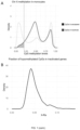
- the data was obtained from the Gene Expression Omnibus (https://www.ncbi.nlm.nih.gov/geo/) with the reference GSE56046. It is a large population study with gene expression (Illumina HumanHT-12 v4 Expression) and methylation (Illumina HumanMethylation450) data in purified human monocytes from blood. The data included covariates such as race, gender, study site, and sample contamination with B-cells, T-cells, natural killer cells, and neutrophils. As race, gender, and study site were encoded in a categorical variable with 18 different categories, sex was inferred from the mean expression of chromosome Y divided by the mean expression from the whole genome (Ry).
- Beta 2 M / 2 M + 1
- a total of 11,648 CpG sites from chromosome X were analyzed.
- the association between X-Ra level and age was assessed adjusting by leukocyte content and well.
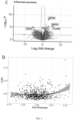
- 47,308 transcripts were analyzed and detected surrogate variables with SVA, adjusting by age, cohort and contamination with B-cells, T-cells, natural killer cells, and neutrophils.
- the Genetic Epidemiology Network of Arteriopathy (GENOA) study is a large community-based study of hypertensive siblings, including African Americans from Jackson, USA. Genomic DNA was extracted from stored peripheral blood leukocytes collected using AutoGen FlexStar (AutoGen, Holliston, MA). Bisulfite conversion was performed with the EZ DNA Methylation Kit (Zymo Research, Irvine, CA), and methylation was assessed using the Illumina Infinium HumanMethylationEPIC BeadChip. After quality control and filtering, a total of 1,394 samples were included in the analysis.
- the TruDiagnostic DNA biobank is a USA population-based cohort aged between 13 and 97 years old.
- the biobank includes 3,590 individuals recruited between October 2020 and February 2022, who have chosen TruDiagnostic for DNA methylation analysis for biological aging.
- Clinical data was collected, including anthropometric measures and diagnosis of common diseases.
- Methylation data was analyzed from 1,414 females from 3,259 individuals who passed quality control.
- the Infinium HumanMethylationEPIC BeadChip was used for DNA methylation assessment following the manufacturer's protocol.
- Several quality controls and functional normalizations were performed using the meffil package, resulting in 745,150 probes, as described in detail elsewhere ( Carreras-Gallo, A. et al, "The early-life exposome modulates the effect of polymorphic inversions on DNA methylation". Commun. Biol. 2022 51 5, 1-13 (2022 )).
- CpG sites were annotated to genes using the EPIC Illumina annotation ilm10b4.hg19.
- Blood cell types were estimated using the blood gse35069 reference panel from the meffil package.
- methylation data were collected on11 elderly women (mean age 74.18), at two time points. Five patients were diagnosed with AD before the first visit.
- methylation data was collected in buffy coat from individuals who recovered from severe COVID 19 infection and were admitted to the UCI. Methylation was assayed at admission, during and previous discharge from UCI and at follow-up after discharge.
- methylation data was collected in the blood of 14 pregnant women at early, midterm, delivery and post-partum.
- TCGA Cancer Genome Atlas
- the cancer sites included breast (BRCA), bladder (BLCA), colon (COAD), head and neck (HNSC), kidney (KIRC), liver (LIHC), lung adenocarcinoma (LUAD), lung squamous cell carcinoma (LUSC), pancreas (PAAD), rectum (READ), thyroid (THCA), uterus (UCEC), skin (SKCM) and ovary (OV).
- BRCA breast
- bladder BLCA
- COAD head and neck
- HNSC kidney
- LIHC liver
- LAD lung adenocarcinoma
- LUSC lung squamous cell carcinoma
- PAAD pancreas
- READ pancreas
- RTD rectum
- TCA thyroid
- uterus uterus
- SKCM skin
- OV ovary
- RNA-seq and CNV data for the 12 selected cancer sites was downloaded to perform associations with X-Ra.
- data was downloaded using TCGAbiolinks version 2.22.4 and fitted proportional hazards regression models for survival data adjusting by age for each independent cancer site.
- the association between X-Ra and hormonal status (ER, PR, HER2) of breast cancer, adjusting by age was also tested.
- surrogate variables were inferred with svaseq from the SVA R library for each individual cancer.
- the association of transcription levels of 20,532 genes with FIGO.2 was tested adjusting by age and surrogate variables, transforming the expression levels with voom and fitting regression models with limma. In each cancer site, genes with less than 15 counts in more than 75% of the samples were removed from the analysis.
- Table 1 shows the panel of 468 CpGs used to calculate X-Ra levels in data derived from HM27 platform, and their corresponding position in the chromosome X and the associated gene.
- Table 1 Probe ID Associated gene Position in ChrX (hg19) Probe ID Associated gene Position in ChrX (hg19) cg00029931 RPL36A 100645741 cg13286902 TRMT2B 100306780 cg00032666 MAMLD1 149613398 cg13340269 TMEM35 100333778 cg00121904 MAOB 43741826 cg13370916 STARD8 67867403 cg00126698 BTK 100640503 cg13482233 HEPH 65382529 cg00128197 HSD17B10 53461455 cg13658777 PDHA1 19361740 cg00141845 UPRT 74493352 cg13674559 WDR44 117479752
- Table 2 shows the panel of 468 CpGs used to calculate X-Ra levels in data derived from HM450 platform, and their corresponding position in the chromosome X and the associated gene Table 2
- Probe ID Gene Position in ChrX (hg19)
- Probe ID Gene Position in ChrX (hg19)
- Probe ID Gene Position in ChrX (hg19)
- cg00005617 EFNB1 68059898 cg08065271
- PDZD4 153090978 cg17703220 XIAP 122993978 cg00011891 UBL4A 153714660 cg08070147 SCML1 17756762 cg17705081
- GPKOW 48980694 cg00014152 WDR13 48457128 cg08076508 PHF8 54070176 cg17710066 SCML2 18373455 cg00016522 MTM1 149737876 cg08076861 ZNF275 1525993
- the CpGs in the reference panel were selected and the beta methylation levels for each individual were extracted.
- X-Ra was defined as the fraction of selected CpGs that were hypomethylated. Hypomethylation status was defined for those CpGs whose beta values were below 0.2.
- Rxci was computed as the mean transcription residual across the inactive genes
- Rxescape was computed as the mean transcription residual across the escapees. The difference between both means is a measure of the global transcription inactivation relative to those genes not under XCI.
- X-Ra and age >65 was also tested using a linear regression model, adjusting for cohort and immune cell contamination. Dichotomization of age was taken as the median age of cancer at any site according to SEER 22 2015-2019.
- TruDiagnostic data the association with telomere length, height, weight, number of children and cancer diagnosis at any site was also tested.
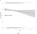
- a logistic regression model was fitted on X-Ra and the interaction between X-Ra and height. It was tested the interaction taking height as continuous and also dichotomizing at the first quartile (1.60 cm).
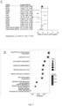
- X-Ra For each of the 12 cancer sites, it was inferred the value of X-Ra and tested the association with cancer status and survival using a meta-analysis with fixed and random effects models. The prediction power of X-Ra on cancer status was tested with a ROC area under the curve statistic. The optimal X-Ra was defined as the Youden distance, which was used to compute the attributable fraction of X-Ra of affected tissue on cancer risk, using the AFglm function of the AF library. For transcriptome-wide associations of X-Ra in each cancer site, it used limma and adjusted for SVA and age. Log2FC with their standard error were then meta-analyzed across cancer sites, and the associations were adjusted for multiple comparisons for the number of transcripts tested, with Bonferroni correction. On these associations, enrichment analyses on biological processes were performed on genes with nominal significance lower than 0.01, using the enrichGO from clusterProfiler.
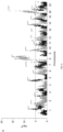
- transcriptome-wide differential expression analysis was performed on monocytes of the 605-adult woman. It was found significant genes relevant to cancer and histone modifications ( FIG. 1C ). Specifically, it was observed significant upregulation of ABTB2 expression, which has recently been associated with treatment response in breast cancer chemotherapy. It was also observed significant downregulation of SOGA1, which is regulated by the IncRNA TSIX and is a predictor of hepatocellular carcinoma. Additionally, it was observed upregulation of the Histone-binding protein RBBP4 gene and RSPO2 encoding for R-spondin 2, which synergizes with WNT to activate beta-catenin.
- X-Ra in blood is associated with aging, age-associated diseases and transient states
- X-Ra difference 2.48 ⁇ 10 -4
- X-Ra as a marker of breast cancer in tumor DNA
- X-Ra levels in cancer tissue are associated with specific chromosomal copy number variants (CNVs) across the genome.
- CNVs chromosomal copy number variants
- the genome was scanned using 0.05MB windows to detect CNVs in BRCA samples. It was then tested the association between X-Ra and frequent deletions and gains (>0.01) while adjusting for age. While a significant association of X-Ra with deletions was not observed, it was found 492 significant regions with overlapping gains after correcting for multiple comparisons ( Fig. 6C ). Remarkably, the several of top associations in each chromosome included loci associated with cancer. The most significant association was found with copy number gains at TNFRSF68 , believed to prevent apoptosis and promote tumor growth.
Landscapes
- Chemical & Material Sciences (AREA)
- Life Sciences & Earth Sciences (AREA)
- Health & Medical Sciences (AREA)
- Organic Chemistry (AREA)
- Proteomics, Peptides & Aminoacids (AREA)
- Engineering & Computer Science (AREA)
- Analytical Chemistry (AREA)
- Zoology (AREA)
- Genetics & Genomics (AREA)
- Wood Science & Technology (AREA)
- Immunology (AREA)
- Pathology (AREA)
- Physics & Mathematics (AREA)
- Biotechnology (AREA)
- Microbiology (AREA)
- Molecular Biology (AREA)
- Biophysics (AREA)
- Biochemistry (AREA)
- Bioinformatics & Cheminformatics (AREA)
- General Engineering & Computer Science (AREA)
- General Health & Medical Sciences (AREA)
- Hospice & Palliative Care (AREA)
- Oncology (AREA)
- Measuring Or Testing Involving Enzymes Or Micro-Organisms (AREA)
Priority Applications (3)
| Application Number | Priority Date | Filing Date | Title |
|---|---|---|---|
| EP23382740.1A EP4495263A1 (de) | 2023-07-19 | 2023-07-19 | Verfahren zur bestimmung des x-chromosom-reaktivierungsniveaus einer weiblichen person |
| EP24742268.6A EP4747403A1 (de) | 2023-07-19 | 2024-07-18 | Verfahren zur bestimmung des x-chromosom-reaktivierungsniveaus einer weiblichen person |
| PCT/EP2024/070423 WO2025017136A1 (en) | 2023-07-19 | 2024-07-18 | Method for determining the x chromosome reactivation level of a female subject |
Applications Claiming Priority (1)
| Application Number | Priority Date | Filing Date | Title |
|---|---|---|---|
| EP23382740.1A EP4495263A1 (de) | 2023-07-19 | 2023-07-19 | Verfahren zur bestimmung des x-chromosom-reaktivierungsniveaus einer weiblichen person |
Publications (1)
| Publication Number | Publication Date |
|---|---|
| EP4495263A1 true EP4495263A1 (de) | 2025-01-22 |
Family
ID=87426776
Family Applications (2)
| Application Number | Title | Priority Date | Filing Date |
|---|---|---|---|
| EP23382740.1A Withdrawn EP4495263A1 (de) | 2023-07-19 | 2023-07-19 | Verfahren zur bestimmung des x-chromosom-reaktivierungsniveaus einer weiblichen person |
| EP24742268.6A Pending EP4747403A1 (de) | 2023-07-19 | 2024-07-18 | Verfahren zur bestimmung des x-chromosom-reaktivierungsniveaus einer weiblichen person |
Family Applications After (1)
| Application Number | Title | Priority Date | Filing Date |
|---|---|---|---|
| EP24742268.6A Pending EP4747403A1 (de) | 2023-07-19 | 2024-07-18 | Verfahren zur bestimmung des x-chromosom-reaktivierungsniveaus einer weiblichen person |
Country Status (2)
| Country | Link |
|---|---|
| EP (2) | EP4495263A1 (de) |
| WO (1) | WO2025017136A1 (de) |
Citations (1)
| Publication number | Priority date | Publication date | Assignee | Title |
|---|---|---|---|---|
| WO2018072705A1 (en) * | 2016-10-19 | 2018-04-26 | The Chinese University Of Hong Kong | Gestational age assessment by methylation and size profiling of maternal plasma dna |
-
2023
- 2023-07-19 EP EP23382740.1A patent/EP4495263A1/de not_active Withdrawn
-
2024
- 2024-07-18 EP EP24742268.6A patent/EP4747403A1/de active Pending
- 2024-07-18 WO PCT/EP2024/070423 patent/WO2025017136A1/en not_active Ceased
Patent Citations (1)
| Publication number | Priority date | Publication date | Assignee | Title |
|---|---|---|---|---|
| WO2018072705A1 (en) * | 2016-10-19 | 2018-04-26 | The Chinese University Of Hong Kong | Gestational age assessment by methylation and size profiling of maternal plasma dna |
Non-Patent Citations (11)
| Title |
|---|
| BURTIS C. A. ET AL.: "Statistical Treatment of Reference Values", 2008 |
| CARRERAS-GALLO, A. ET AL.: "The early-life exposome modulates the effect of polymorphic inversions on DNA methylation", COMMUN. BIOL., vol. 51, no. 5, 2022, pages 1 - 13 |
| KANANEN LAURA ET AL: "Ageing-associated changes in DNA methylation in X and Y chromosomes", EPIGENETICS & CHROMATIN, vol. 14, no. 1, 2 July 2021 (2021-07-02), London, UK, XP093113393, ISSN: 1756-8935, Retrieved from the Internet <URL:https://link.springer.com/article/10.1186/s13072-021-00407-6/fulltext.html> DOI: 10.1186/s13072-021-00407-6 * |
| KANG JUN ET AL: "Cancer-Testis Antigen Expression in Serous Endometrial Cancer with Loss of X Chromosome Inactivation", PLOS ONE, vol. 10, no. 9, 11 September 2015 (2015-09-11), US, pages e0137476, XP093113524, ISSN: 1932-6203, DOI: 10.1371/journal.pone.0137476 * |
| LI SHUXIA ET AL: "Exploratory analysis of age and sex dependent DNA methylation patterns on the X-chromosome in whole blood samples", GENOME MEDICINE, vol. 12, no. 1, 28 April 2020 (2020-04-28), XP093113332, ISSN: 1756-994X, Retrieved from the Internet <URL:https://link.springer.com/article/10.1186/s13073-020-00736-3/fulltext.html> DOI: 10.1186/s13073-020-00736-3 * |
| M. E. LEVINE ET AL.: "An epigenetic biomarker of aging for lifespan and healthspan", AGING (ALBANY. NY, vol. 10, 2018 |
| MCCARTNEY DANIEL L. ET AL: "An epigenome-wide association study of sex-specific chronological ageing", GENOME MEDICINE, vol. 12, no. 1, 31 December 2019 (2019-12-31), XP093113421, ISSN: 1756-994X, Retrieved from the Internet <URL:http://link.springer.com/article/10.1186/s13073-019-0693-z/fulltext.html> DOI: 10.1186/s13073-019-0693-z * |
| MORRIS TJ ET AL.: "Analysis pipelines and packages for Infinium HumanMethylation450 BeadChip (450k) data", METHODS, vol. 72, 15 January 2015 (2015-01-15), pages 3 - 8, XP055790540, DOI: 10.1016/j.ymeth.2014.08.011 |
| RONAN CHALIGN? ET AL: "X-chromosome inactivation in development and cancer", FEBS LETTERS, ELSEVIER, AMSTERDAM, NL, vol. 588, no. 15, 14 June 2014 (2014-06-14), pages 2514 - 2522, XP071254249, ISSN: 0014-5793, DOI: 10.1016/J.FEBSLET.2014.06.023 * |
| SUN ZHIFU ET AL: "Chromosome X genomic and epigenomic aberrations and clinical implications in breast cancer by base resolution profiling", EPIGENOMICS, vol. 7, no. 7, 18 May 2015 (2015-05-18), United Kingdom, pages 1099 - 1110, XP093113534, ISSN: 1750-1911, DOI: 10.2217/epi.15.43 * |
| TANG WALFRED W C ET AL: "A Unique Gene Regulatory Network Resets the Human Germline Epigenome for Development", CELL, ELSEVIER, AMSTERDAM NL, vol. 161, no. 6, 4 June 2015 (2015-06-04), pages 1453 - 1467, XP029171297, ISSN: 0092-8674, DOI: 10.1016/J.CELL.2015.04.053 * |
Also Published As
| Publication number | Publication date |
|---|---|
| WO2025017136A1 (en) | 2025-01-23 |
| EP4747403A1 (de) | 2026-05-27 |
Similar Documents
| Publication | Publication Date | Title |
|---|---|---|
| JP7799607B2 (ja) | 卵巣癌の検出 | |
| US20230366034A1 (en) | Compositions and methods for diagnosing lung cancers using gene expression profiles | |
| TWI806621B (zh) | 來自血漿之胚胎或腫瘤甲基化模式組(methylome)之非侵入性測定 | |
| Ehrich et al. | Noninvasive detection of fetal trisomy 21 by sequencing of DNA in maternal blood: a study in a clinical setting | |
| EP3421613B1 (de) | Identifikation und verwendung von zirkulierenden nukleinsäure-tumormarkern | |
| AU2011302004B2 (en) | Molecular diagnostic test for cancer | |
| JP2022518762A (ja) | 子宮内膜癌の検出 | |
| US20250349386A1 (en) | Non-invasive detection of tissue abnormality using methylation | |
| EP4209598A1 (de) | Beurteilung des gestationsalters durch methylierung und grössenprofilierung von dna aus maternalem plasma | |
| EP2977467B1 (de) | Verfahren, verwendung von marker und bestimmungsvorrichtung zur gewinnung von informationen über eine vielzahl von krebsarten | |
| AU2021291586B2 (en) | Multimodal analysis of circulating tumor nucleic acid molecules | |
| US20220380853A1 (en) | Prostate cancer detection methods | |
| US20250137066A1 (en) | Compostions and methods for diagnosing lung cancers using gene expression profiles | |
| EP2780476B1 (de) | Verfahren zur diagnose und/oder prognose von gynäkologischen karzinomen | |
| Michel et al. | Noninvasive multicancer detection using DNA hypomethylation of LINE-1 retrotransposons | |
| US20240093302A1 (en) | Non-invasive cancer detection based on dna methylation changes | |
| US20240229158A1 (en) | Dna methylation biomarkers for hepatocellular carcinoma | |
| WO2017119510A1 (ja) | 乳がんの診断のための検査方法、遺伝子マーカー、および検査薬 | |
| EP2138589A1 (de) | Molekulare Signatur des Lebertumorgrades und Verwendung zur Bewertung der Prognose und therapeutischen Durchführung | |
| KR20260011755A (ko) | 체세포 돌연변이 검출을 위한 비침습적 시험관 내 방법 | |
| WO2025017136A1 (en) | Method for determining the x chromosome reactivation level of a female subject | |
| US20240141435A1 (en) | Methods for detecting and predicting cancer | |
| KR20230134473A (ko) | 암의 진단, 예후 및 요법과 관련된 새로운 바이오마커공급원으로서의 리보솜 rna 2'o-메틸화 | |
| US11473131B2 (en) | Methods for detecting genomic DNA methylation | |
| US20260139325A1 (en) | Classification of colorectal tumors using dna methylation from liquid biopsy |
Legal Events
| Date | Code | Title | Description |
|---|---|---|---|
| PUAI | Public reference made under article 153(3) epc to a published international application that has entered the european phase |
Free format text: ORIGINAL CODE: 0009012 |
|
| STAA | Information on the status of an ep patent application or granted ep patent |
Free format text: STATUS: THE APPLICATION HAS BEEN PUBLISHED |
|
| AK | Designated contracting states |
Kind code of ref document: A1 Designated state(s): AL AT BE BG CH CY CZ DE DK EE ES FI FR GB GR HR HU IE IS IT LI LT LU LV MC ME MK MT NL NO PL PT RO RS SE SI SK SM TR |
|
| STAA | Information on the status of an ep patent application or granted ep patent |
Free format text: STATUS: THE APPLICATION IS DEEMED TO BE WITHDRAWN |
|
| 18D | Application deemed to be withdrawn |
Effective date: 20250723 |