EP4470544A2 - Verfahren und zusammensetzungen zur reprogrammierung von zellen - Google Patents
Verfahren und zusammensetzungen zur reprogrammierung von zellen Download PDFInfo
- Publication number
- EP4470544A2 EP4470544A2 EP24188635.7A EP24188635A EP4470544A2 EP 4470544 A2 EP4470544 A2 EP 4470544A2 EP 24188635 A EP24188635 A EP 24188635A EP 4470544 A2 EP4470544 A2 EP 4470544A2
- Authority
- EP
- European Patent Office
- Prior art keywords
- cells
- reprogramming
- cell
- composition
- mrna
- Prior art date
- Legal status (The legal status is an assumption and is not a legal conclusion. Google has not performed a legal analysis and makes no representation as to the accuracy of the status listed.)
- Pending
Links
Images







Classifications
-
- C—CHEMISTRY; METALLURGY
- C12—BIOCHEMISTRY; BEER; SPIRITS; WINE; VINEGAR; MICROBIOLOGY; ENZYMOLOGY; MUTATION OR GENETIC ENGINEERING
- C12N—MICROORGANISMS OR ENZYMES; COMPOSITIONS THEREOF; PROPAGATING, PRESERVING, OR MAINTAINING MICROORGANISMS; MUTATION OR GENETIC ENGINEERING; CULTURE MEDIA
- C12N15/00—Mutation or genetic engineering; DNA or RNA concerning genetic engineering, vectors, e.g. plasmids, or their isolation, preparation or purification; Use of hosts therefor
- C12N15/09—Recombinant DNA-technology
- C12N15/11—DNA or RNA fragments; Modified forms thereof; Non-coding nucleic acids having a biological activity
- C12N15/113—Non-coding nucleic acids modulating the expression of genes, e.g. antisense oligonucleotides; Antisense DNA or RNA; Triplex- forming oligonucleotides; Catalytic nucleic acids, e.g. ribozymes; Nucleic acids used in co-suppression or gene silencing
-
- A—HUMAN NECESSITIES
- A61—MEDICAL OR VETERINARY SCIENCE; HYGIENE
- A61K—PREPARATIONS FOR MEDICAL, DENTAL OR TOILETRY PURPOSES
- A61K31/00—Medicinal preparations containing organic active ingredients
- A61K31/70—Carbohydrates; Sugars; Derivatives thereof
- A61K31/7088—Compounds having three or more nucleosides or nucleotides
- A61K31/713—Double-stranded nucleic acids or oligonucleotides
-
- A—HUMAN NECESSITIES
- A61—MEDICAL OR VETERINARY SCIENCE; HYGIENE
- A61K—PREPARATIONS FOR MEDICAL, DENTAL OR TOILETRY PURPOSES
- A61K35/00—Medicinal preparations containing materials or reaction products thereof with undetermined constitution
- A61K35/12—Materials from mammals; Compositions comprising non-specified tissues or cells; Compositions comprising non-embryonic stem cells; Genetically modified cells
- A61K35/28—Bone marrow; Haematopoietic stem cells; Mesenchymal stem cells of any origin, e.g. adipose-derived stem cells
-
- A—HUMAN NECESSITIES
- A61—MEDICAL OR VETERINARY SCIENCE; HYGIENE
- A61P—SPECIFIC THERAPEUTIC ACTIVITY OF CHEMICAL COMPOUNDS OR MEDICINAL PREPARATIONS
- A61P17/00—Drugs for dermatological disorders
-
- C—CHEMISTRY; METALLURGY
- C12—BIOCHEMISTRY; BEER; SPIRITS; WINE; VINEGAR; MICROBIOLOGY; ENZYMOLOGY; MUTATION OR GENETIC ENGINEERING
- C12N—MICROORGANISMS OR ENZYMES; COMPOSITIONS THEREOF; PROPAGATING, PRESERVING, OR MAINTAINING MICROORGANISMS; MUTATION OR GENETIC ENGINEERING; CULTURE MEDIA
- C12N15/00—Mutation or genetic engineering; DNA or RNA concerning genetic engineering, vectors, e.g. plasmids, or their isolation, preparation or purification; Use of hosts therefor
- C12N15/09—Recombinant DNA-technology
- C12N15/11—DNA or RNA fragments; Modified forms thereof; Non-coding nucleic acids having a biological activity
-
- C—CHEMISTRY; METALLURGY
- C12—BIOCHEMISTRY; BEER; SPIRITS; WINE; VINEGAR; MICROBIOLOGY; ENZYMOLOGY; MUTATION OR GENETIC ENGINEERING
- C12N—MICROORGANISMS OR ENZYMES; COMPOSITIONS THEREOF; PROPAGATING, PRESERVING, OR MAINTAINING MICROORGANISMS; MUTATION OR GENETIC ENGINEERING; CULTURE MEDIA
- C12N15/00—Mutation or genetic engineering; DNA or RNA concerning genetic engineering, vectors, e.g. plasmids, or their isolation, preparation or purification; Use of hosts therefor
- C12N15/09—Recombinant DNA-technology
- C12N15/87—Introduction of foreign genetic material using processes not otherwise provided for, e.g. co-transformation
- C12N15/90—Stable introduction of foreign DNA into chromosome
- C12N15/902—Stable introduction of foreign DNA into chromosome using homologous recombination
- C12N15/907—Stable introduction of foreign DNA into chromosome using homologous recombination in mammalian cells
-
- C—CHEMISTRY; METALLURGY
- C12—BIOCHEMISTRY; BEER; SPIRITS; WINE; VINEGAR; MICROBIOLOGY; ENZYMOLOGY; MUTATION OR GENETIC ENGINEERING
- C12N—MICROORGANISMS OR ENZYMES; COMPOSITIONS THEREOF; PROPAGATING, PRESERVING, OR MAINTAINING MICROORGANISMS; MUTATION OR GENETIC ENGINEERING; CULTURE MEDIA
- C12N5/00—Undifferentiated human, animal or plant cells, e.g. cell lines; Tissues; Cultivation or maintenance thereof; Culture media therefor
- C12N5/06—Animal cells or tissues; Human cells or tissues
- C12N5/0602—Vertebrate cells
- C12N5/0652—Cells of skeletal and connective tissues; Mesenchyme
-
- C—CHEMISTRY; METALLURGY
- C12—BIOCHEMISTRY; BEER; SPIRITS; WINE; VINEGAR; MICROBIOLOGY; ENZYMOLOGY; MUTATION OR GENETIC ENGINEERING
- C12N—MICROORGANISMS OR ENZYMES; COMPOSITIONS THEREOF; PROPAGATING, PRESERVING, OR MAINTAINING MICROORGANISMS; MUTATION OR GENETIC ENGINEERING; CULTURE MEDIA
- C12N5/00—Undifferentiated human, animal or plant cells, e.g. cell lines; Tissues; Cultivation or maintenance thereof; Culture media therefor
- C12N5/06—Animal cells or tissues; Human cells or tissues
- C12N5/0602—Vertebrate cells
- C12N5/0696—Artificially induced pluripotent stem cells, e.g. iPS
-
- C—CHEMISTRY; METALLURGY
- C12—BIOCHEMISTRY; BEER; SPIRITS; WINE; VINEGAR; MICROBIOLOGY; ENZYMOLOGY; MUTATION OR GENETIC ENGINEERING
- C12N—MICROORGANISMS OR ENZYMES; COMPOSITIONS THEREOF; PROPAGATING, PRESERVING, OR MAINTAINING MICROORGANISMS; MUTATION OR GENETIC ENGINEERING; CULTURE MEDIA
- C12N9/00—Enzymes; Proenzymes; Compositions thereof; Processes for preparing, activating, inhibiting, separating or purifying enzymes
- C12N9/14—Hydrolases (3)
- C12N9/16—Hydrolases (3) acting on ester bonds (3.1)
- C12N9/22—Ribonucleases [RNase]; Deoxyribonucleases [DNase]
-
- C—CHEMISTRY; METALLURGY
- C12—BIOCHEMISTRY; BEER; SPIRITS; WINE; VINEGAR; MICROBIOLOGY; ENZYMOLOGY; MUTATION OR GENETIC ENGINEERING
- C12N—MICROORGANISMS OR ENZYMES; COMPOSITIONS THEREOF; PROPAGATING, PRESERVING, OR MAINTAINING MICROORGANISMS; MUTATION OR GENETIC ENGINEERING; CULTURE MEDIA
- C12N9/00—Enzymes; Proenzymes; Compositions thereof; Processes for preparing, activating, inhibiting, separating or purifying enzymes
- C12N9/14—Hydrolases (3)
- C12N9/16—Hydrolases (3) acting on ester bonds (3.1)
- C12N9/22—Ribonucleases [RNase]; Deoxyribonucleases [DNase]
- C12N9/222—Clustered regularly interspaced short palindromic repeats [CRISPR]-associated [CAS] enzymes
-
- C—CHEMISTRY; METALLURGY
- C12—BIOCHEMISTRY; BEER; SPIRITS; WINE; VINEGAR; MICROBIOLOGY; ENZYMOLOGY; MUTATION OR GENETIC ENGINEERING
- C12N—MICROORGANISMS OR ENZYMES; COMPOSITIONS THEREOF; PROPAGATING, PRESERVING, OR MAINTAINING MICROORGANISMS; MUTATION OR GENETIC ENGINEERING; CULTURE MEDIA
- C12N2310/00—Structure or type of the nucleic acid
- C12N2310/10—Type of nucleic acid
- C12N2310/14—Type of nucleic acid interfering nucleic acids [NA]
- C12N2310/141—MicroRNAs, miRNAs
-
- C—CHEMISTRY; METALLURGY
- C12—BIOCHEMISTRY; BEER; SPIRITS; WINE; VINEGAR; MICROBIOLOGY; ENZYMOLOGY; MUTATION OR GENETIC ENGINEERING
- C12N—MICROORGANISMS OR ENZYMES; COMPOSITIONS THEREOF; PROPAGATING, PRESERVING, OR MAINTAINING MICROORGANISMS; MUTATION OR GENETIC ENGINEERING; CULTURE MEDIA
- C12N2310/00—Structure or type of the nucleic acid
- C12N2310/10—Type of nucleic acid
- C12N2310/20—Type of nucleic acid involving clustered regularly interspaced short palindromic repeats [CRISPR]
-
- C—CHEMISTRY; METALLURGY
- C12—BIOCHEMISTRY; BEER; SPIRITS; WINE; VINEGAR; MICROBIOLOGY; ENZYMOLOGY; MUTATION OR GENETIC ENGINEERING
- C12N—MICROORGANISMS OR ENZYMES; COMPOSITIONS THEREOF; PROPAGATING, PRESERVING, OR MAINTAINING MICROORGANISMS; MUTATION OR GENETIC ENGINEERING; CULTURE MEDIA
- C12N2330/00—Production
- C12N2330/30—Production chemically synthesised
- C12N2330/31—Libraries, arrays
-
- C—CHEMISTRY; METALLURGY
- C12—BIOCHEMISTRY; BEER; SPIRITS; WINE; VINEGAR; MICROBIOLOGY; ENZYMOLOGY; MUTATION OR GENETIC ENGINEERING
- C12N—MICROORGANISMS OR ENZYMES; COMPOSITIONS THEREOF; PROPAGATING, PRESERVING, OR MAINTAINING MICROORGANISMS; MUTATION OR GENETIC ENGINEERING; CULTURE MEDIA
- C12N2500/00—Specific components of cell culture medium
- C12N2500/60—Buffer, e.g. pH regulation, osmotic pressure
-
- C—CHEMISTRY; METALLURGY
- C12—BIOCHEMISTRY; BEER; SPIRITS; WINE; VINEGAR; MICROBIOLOGY; ENZYMOLOGY; MUTATION OR GENETIC ENGINEERING
- C12N—MICROORGANISMS OR ENZYMES; COMPOSITIONS THEREOF; PROPAGATING, PRESERVING, OR MAINTAINING MICROORGANISMS; MUTATION OR GENETIC ENGINEERING; CULTURE MEDIA
- C12N2501/00—Active agents used in cell culture processes, e.g. differentation
- C12N2501/10—Growth factors
- C12N2501/115—Basic fibroblast growth factor (bFGF, FGF-2)
-
- C—CHEMISTRY; METALLURGY
- C12—BIOCHEMISTRY; BEER; SPIRITS; WINE; VINEGAR; MICROBIOLOGY; ENZYMOLOGY; MUTATION OR GENETIC ENGINEERING
- C12N—MICROORGANISMS OR ENZYMES; COMPOSITIONS THEREOF; PROPAGATING, PRESERVING, OR MAINTAINING MICROORGANISMS; MUTATION OR GENETIC ENGINEERING; CULTURE MEDIA
- C12N2501/00—Active agents used in cell culture processes, e.g. differentation
- C12N2501/60—Transcription factors
- C12N2501/602—Sox-2
-
- C—CHEMISTRY; METALLURGY
- C12—BIOCHEMISTRY; BEER; SPIRITS; WINE; VINEGAR; MICROBIOLOGY; ENZYMOLOGY; MUTATION OR GENETIC ENGINEERING
- C12N—MICROORGANISMS OR ENZYMES; COMPOSITIONS THEREOF; PROPAGATING, PRESERVING, OR MAINTAINING MICROORGANISMS; MUTATION OR GENETIC ENGINEERING; CULTURE MEDIA
- C12N2501/00—Active agents used in cell culture processes, e.g. differentation
- C12N2501/60—Transcription factors
- C12N2501/603—Oct-3/4
-
- C—CHEMISTRY; METALLURGY
- C12—BIOCHEMISTRY; BEER; SPIRITS; WINE; VINEGAR; MICROBIOLOGY; ENZYMOLOGY; MUTATION OR GENETIC ENGINEERING
- C12N—MICROORGANISMS OR ENZYMES; COMPOSITIONS THEREOF; PROPAGATING, PRESERVING, OR MAINTAINING MICROORGANISMS; MUTATION OR GENETIC ENGINEERING; CULTURE MEDIA
- C12N2501/00—Active agents used in cell culture processes, e.g. differentation
- C12N2501/60—Transcription factors
- C12N2501/604—Klf-4
-
- C—CHEMISTRY; METALLURGY
- C12—BIOCHEMISTRY; BEER; SPIRITS; WINE; VINEGAR; MICROBIOLOGY; ENZYMOLOGY; MUTATION OR GENETIC ENGINEERING
- C12N—MICROORGANISMS OR ENZYMES; COMPOSITIONS THEREOF; PROPAGATING, PRESERVING, OR MAINTAINING MICROORGANISMS; MUTATION OR GENETIC ENGINEERING; CULTURE MEDIA
- C12N2501/00—Active agents used in cell culture processes, e.g. differentation
- C12N2501/60—Transcription factors
- C12N2501/605—Nanog
-
- C—CHEMISTRY; METALLURGY
- C12—BIOCHEMISTRY; BEER; SPIRITS; WINE; VINEGAR; MICROBIOLOGY; ENZYMOLOGY; MUTATION OR GENETIC ENGINEERING
- C12N—MICROORGANISMS OR ENZYMES; COMPOSITIONS THEREOF; PROPAGATING, PRESERVING, OR MAINTAINING MICROORGANISMS; MUTATION OR GENETIC ENGINEERING; CULTURE MEDIA
- C12N2501/00—Active agents used in cell culture processes, e.g. differentation
- C12N2501/60—Transcription factors
- C12N2501/606—Transcription factors c-Myc
-
- C—CHEMISTRY; METALLURGY
- C12—BIOCHEMISTRY; BEER; SPIRITS; WINE; VINEGAR; MICROBIOLOGY; ENZYMOLOGY; MUTATION OR GENETIC ENGINEERING
- C12N—MICROORGANISMS OR ENZYMES; COMPOSITIONS THEREOF; PROPAGATING, PRESERVING, OR MAINTAINING MICROORGANISMS; MUTATION OR GENETIC ENGINEERING; CULTURE MEDIA
- C12N2501/00—Active agents used in cell culture processes, e.g. differentation
- C12N2501/60—Transcription factors
- C12N2501/608—Lin28
-
- C—CHEMISTRY; METALLURGY
- C12—BIOCHEMISTRY; BEER; SPIRITS; WINE; VINEGAR; MICROBIOLOGY; ENZYMOLOGY; MUTATION OR GENETIC ENGINEERING
- C12N—MICROORGANISMS OR ENZYMES; COMPOSITIONS THEREOF; PROPAGATING, PRESERVING, OR MAINTAINING MICROORGANISMS; MUTATION OR GENETIC ENGINEERING; CULTURE MEDIA
- C12N2501/00—Active agents used in cell culture processes, e.g. differentation
- C12N2501/65—MicroRNA
-
- C—CHEMISTRY; METALLURGY
- C12—BIOCHEMISTRY; BEER; SPIRITS; WINE; VINEGAR; MICROBIOLOGY; ENZYMOLOGY; MUTATION OR GENETIC ENGINEERING
- C12N—MICROORGANISMS OR ENZYMES; COMPOSITIONS THEREOF; PROPAGATING, PRESERVING, OR MAINTAINING MICROORGANISMS; MUTATION OR GENETIC ENGINEERING; CULTURE MEDIA
- C12N2506/00—Differentiation of animal cells from one lineage to another; Differentiation of pluripotent cells
- C12N2506/13—Differentiation of animal cells from one lineage to another; Differentiation of pluripotent cells from connective tissue cells, from mesenchymal cells
- C12N2506/1307—Differentiation of animal cells from one lineage to another; Differentiation of pluripotent cells from connective tissue cells, from mesenchymal cells from adult fibroblasts
-
- C—CHEMISTRY; METALLURGY
- C12—BIOCHEMISTRY; BEER; SPIRITS; WINE; VINEGAR; MICROBIOLOGY; ENZYMOLOGY; MUTATION OR GENETIC ENGINEERING
- C12N—MICROORGANISMS OR ENZYMES; COMPOSITIONS THEREOF; PROPAGATING, PRESERVING, OR MAINTAINING MICROORGANISMS; MUTATION OR GENETIC ENGINEERING; CULTURE MEDIA
- C12N2510/00—Genetically modified cells
-
- C—CHEMISTRY; METALLURGY
- C12—BIOCHEMISTRY; BEER; SPIRITS; WINE; VINEGAR; MICROBIOLOGY; ENZYMOLOGY; MUTATION OR GENETIC ENGINEERING
- C12N—MICROORGANISMS OR ENZYMES; COMPOSITIONS THEREOF; PROPAGATING, PRESERVING, OR MAINTAINING MICROORGANISMS; MUTATION OR GENETIC ENGINEERING; CULTURE MEDIA
- C12N2800/00—Nucleic acids vectors
- C12N2800/80—Vectors containing sites for inducing double-stranded breaks, e.g. meganuclease restriction sites
Definitions
- the present disclosure relates generally to methods and compositions for cell reprogramming.
- the present disclosure includes methods and compositions for reprogramming primary human fibroblast cells into induced pluripotent stem cells (iPSCs) in the absence of feeder cells.
- iPSCs induced pluripotent stem cells
- a method for cell reprogramming is provided.
- the cell is a primary human fibroblast cell.
- the primary human fibroblast cell is a primary human neonatal fibroblast cell.
- the primary human fibroblast cell is a primary human adult fibroblast cell.
- the cells are seeded at a density of less than about 1000 cells/cm 2 . In some embodiments, the cells ( e . g ., primary human fibroblast cells) are seeded at a density of less than about 100 cells/cm 2 . In some embodiments, the cells ( e . g ., primary human fibroblast cells) are seeded at a density of less than about 10 cells/cm 2 . In some embodiments, the cells ( e . g ., primary human fibroblast cells) are seeded at a density of less than about 1 cell/cm 2 .
- the reprogramming methods disclosed herein are applied to a single individually plated cell (e . g ., a primary human fibroblast cell).
- the cells e . g ., the primary human fibroblast cells
- the cells are transfected with a composition comprising a complexation buffer and a reprogramming mRNA cocktail.
- the reprogramming mRNA cocktail comprises 100 to 1500 ng/10 cm 2 reprogramming mRNA.
- the reprogramming mRNA cocktail comprises 50 to 600 ng/10 cm 2 reprogramming mRNAs.
- the composition further comprises reprogramming miRNAs.
- the composition comprises 1 to 40 pmoles/10 cm 2 reprogramming miRNAs.
- the reprogramming mRNA cocktail of the composition comprises Oct4 or Myo-D-Oct4 (M 3 O), Sox2, and Klf4 in a 3:1:1 molar ratio.
- the reprogramming mRNA cocktail further comprises one or more of c-Myc, Lin28A, or Nanog in a 1:1 molar ratio with Sox2 and Klf4.
- the composition includes reprogramming miRNA which comprises miR-367 and miR-302s.
- the pH of the complexation buffer is about 7.3 to 8.4. In one embodiment, the pH of the complexation buffer is about 7.3. In one embodiment, the pH of the complexation buffer is about 7.4. In one embodiment, the pH of the complexation buffer is about 7.5. In one embodiment, the pH of the complexation buffer is about 7.6. In one embodiment, the pH of the complexation buffer is about 7.7. In one embodiment, the pH of the complexation buffer is about 7.8. In one embodiment, the pH of the complexation buffer is about 7.9. In one embodiment, the pH of the complexation buffer is about 8.0. In one embodiment, the pH of the complexation buffer is about 8.1. In one embodiment, the pH of the complexation buffer is about 8.2. In one embodiment, the pH of the complexation buffer is about 8.3. In one embodiment, the pH of the complexation buffer is about 8.4. In one embodiment, the pH of the complexation buffer is about 8.5. In one embodiment, the pH of the complexation buffer is about 8.6.
- the complexation buffer comprises OPTI-MEM ® reduced serum medium. In one embodiment, the complexation buffer comprises phosphate-buffered saline (PBS).
- PBS phosphate-buffered saline
- the transfection is performed at least 3 times. In one embodiment, the transfection is performed every other day. In one embodiment, the transfection is performed every third day.
- the cells are grown under conditions selected to increase levels of expression of endogenous pluripotency genes and chromatin modifiers, and to limit the increase in levels of innate immune response genes.
- the endogenous pluripotency genes include NANOG, LIN28A, OCT4, SOX2, SALL4, GDF3, PRDM14, UTF1, NR5A2, DPPA2, DPPA4, NR6A1, TRIM71, LEFTY1, LEFTY2, ZFP42, and ZIC3.
- the chromatin modifier genes include ASF1A, DNMT3A, DNMT3B, DNMT3L, and TET1.
- the reprogramming methods of the present technology are applied to a single primary human fibroblast cell individually plated in the absence of feeder cells. In one embodiment, reprogramming occurs in greater than 15% of individually plated cells. In one embodiment, reprogramming occurs in greater than 50% of individually plated cells. In one embodiment, reprogramming occurs in greater than 90% of individually plated cells.
- the methods of the present technology yield an unprecedented reprogramming efficiency and produce clinically relevant iPSC lines from a variety of human primary fibroblast cells, including individually plated single cells.
- cell density refers to the density of cells on a given substrate, plate, well, dish, container, or the like, in which the cells are grown/seeded.
- Containers in which cells may include, but are not limited to, tissue culture plates, tissue culture dishes, and tissue culture wells, including tissue culture wells of various sizes, such as, 384-well, 96-well, 48-well, 24-well, 12-well, 6-well, and 100 mm dishes.
- cell density may be expressed as the number of cells per plate, dish, well, and the like.
- complexation buffer refers to a buffer, including OPTI-MEM ® or PBS, to which a transfection reagent and a molecule to be transfected are added and in which the transfection reagent associates or complexes with the molecule to be transfected.
- the terms “complexes with,” “complexed with,” and “complexed to” are used interchangeably, and refer to any method by which a nucleic acid molecule interacts with (e . g . binds to, comes into contact with, adheres to) a cationic lipid. Such an interaction can include, but is not limited to encapsulation of a nucleic acid molecule into a cationic liposome, association of a nucleic acid molecule and cationic lipid characterized by non-covalent, ionic charge interactions, and other types of associations between nucleic acid molecules and cationic lipids known by those skilled in the art.
- differentiated refers to a cell that takes on a more committed (“differentiated”) position within a given cell lineage.
- “Dedifferentiated” refers to a cell that reverts to a less committed position within a cell lineage.
- the term "effective amount” refers to a quantity of a composition, compound, nucleic acid, or a number of cells sufficient to achieve a desired therapeutic and/or prophylactic effect, e . g ., an amount which results in the prevention of, or amelioration of a disease or medical condition or one or more symptoms associated with a disease or medical condition or an amount which results in reprogramming of somatic cells.
- the amount of a composition, compound, nucleic acid, or a number of cells administered to the subject will depend on the composition, compound, nucleic acid, or type of cell, the type and severity of the disease, and on the characteristics of the individual, such as general health, age, sex, body weight and tolerance to drugs. It will also depend on the degree, severity and type of disease. The skilled artisan will be able to determine appropriate dosages depending on these and other factors.
- feeder cells refers to cells of one type that are co-cultured with cells of another type, to provide an environment in which the cells of the second type can grow.
- Feeder cells include, but are not limited to 3001 G irradiated neonatal human foreskin fibroblasts (GloabalStem) and FibroGRO mitomycin C-inactivated xeno-free human neonatal fibroblasts (Millipore).
- iPSCs induced pluripotent stem cells
- ESCs embryonic stem cells
- undifferentiated cells artificially derived by reprogramming differentiated, non-pluripotent cells, typically adult somatic cells.
- isolated means that materials naturally accompanying in normal circumstances are at least reduced, or preferably substantially completely eliminated. Therefore, the term “isolated cell” refers to a cell substantially free from other accompanying in natural circumstances substances ( e.g., other cells, proteins, nucleic acids, etc. ) .
- isolated in relation to nucleic acids or polypeptides means that, for example, the nucleic acids or the polypeptides are substantially free from cellular substances or culture media when they are produced by recombinant DNA techniques; or precursory chemical substances or other chemical substances when they are chemically synthesized.
- Isolated nucleic acids are typically free from sequences naturally flanking the nucleic acid within an organism from which the nucleic acid is derived ( i . e ., sequences positioned at the 5' terminus and the 3' terminus of the nucleic acid).
- miRNA also referred to as miRNA or miR
- miRNA refers to small RNA molecules (about 19-27 nucleotides) that regulate gene expression by targeting one or more mRNAs for translational repression or cleavage. They are small inhibitory RNAs capable of suppressing the translation of target genes with high complementarity. Certain miRNA clusters, including miR-302-367, can promote reprogramming into induced pluripotent stem cells (iPSCs).
- iPSCs induced pluripotent stem cells
- microRNAs include, but are not limited to miR200s, miR290s, miR371s, miR302s, miR367, miR369s, and biologically active fragments, analogues, and variants thereof.
- miRNA includes microRNA mimics (m-miRNAs).
- Mature microRNA may include microRNA that has been cleaved from a microRNA precursor (pre-miRNA), or that has been synthesized.
- pre-miRNA microRNA mimics are chemically modified RNA molecules designed to mimic endogenous mature microRNAs.
- pluripotent stem cell refers to a cell capable of continued self-renewal, and, under appropriate conditions, of producing progeny of several different cell types.
- PSCs are capable of producing progeny that are derivatives of each of the three germ layers: endoderm, mesoderm, and ectoderm, according to a standard art-accepted test, such as the ability to form a teratoma in a suitable host, or the ability to differentiate into cells stainable for markers representing tissue types of all three germ layers in culture.
- Included in the definition of PSCs are embryonic cells of various types, such as embryonic stem cells (ESCs), as well as induced pluripotent stem cells (iPSCs) that have been reprogrammed from an adult somatic cell.
- ESCs embryonic stem cells
- iPSCs induced pluripotent stem cells
- PSCs include primary tissue and established lines that bear phenotypic characteristics of PSCs, and derivatives of such lines that still have the capacity of producing progeny of each of the three germ layers.
- PSC cultures are described as "undifferentiated” or “substantially undifferentiated” when a substantial proportion of stem cells and their derivatives in the population display morphological characteristics of undifferentiated cells, clearly distinguishing them from differentiated cells of embryo or adult origin.
- Undifferentiated PSCs are easily recognized by those skilled in the art, and typically appear in the two dimensions of a microscopic view with high nuclear/cytoplasmic ratios and prominent nucleoli. It is understood that colonies of undifferentiated cells within the population will often be surrounded by neighboring cells that are differentiated.
- prevention or “preventing” of a disease or medical condition refers to the reduction of the occurrence of a disease or medical condition in a treated sample relative to an untreated control sample, or a delay in the onset of one or more symptoms of the disease or medical condition relative to the untreated control sample.
- reprogramming and grammatical equivalents refer to a process that alters or reverses the differentiation status of a somatic cell that is either partially or terminally differentiated.
- Reprogramming of a somatic cell may be a partial or complete reversion of the differentiation status of the somatic cell.
- reprogramming is complete when a somatic cell is reprogrammed into an induced pluripotent stem cell.
- reprogramming may be partial, such as reversion into any less differentiated state. For example, reverting a terminally differentiated cell into a cell of a less differentiated state, such as a multipotent cell.
- reprogramming efficiency refers to the number of iPSC colonies generated per somatic input cell. For example, reprogramming efficiency can be calculated by dividing the average number of Tra-1-60 + colonies per well on a multi-well plate by the initial number of cells plated.
- Tra-1-60 is a pluripotent stem cell-specific protein expressed on the surface of undifferentiated embryonic and induced pluripotent stem cells.
- reprogramming factor refers to a molecule, which when contacted with a cell (e . g ., expressed by a cell, transformed into a cell for expression, exogenously provided to a cell, etc. ) , can, either alone or in combination with other molecules, cause reprogramming.
- reprogramming factors include, but are not limited to Oct3 protein, Oct4 protein, Myo-D-Oct4 (M 3 O) protein, Sox1 protein, Sox2 protein, Sox3 protein, Sox15 protein, Klfl, protein, Klf2 protein, Klf3 protein, Klf4 protein, Klf5 protein, c-Myc protein, L-Myc protein, N-Myc protein, Nanog protein, Lin28A protein, Tert protein, Utfl protein, Aicda protein, Glis1, Sall4, Esrrb, Tet1, Tet2, Zfp42, Prdm14, Nr5a2, Gata6, Sox7, Pax1, Gata4, Gata3, cEBPa, HNF4a, GMNN, SNAIL, Grb2, Trim71, and biologically active fragments, analogues, variants, and family members thereof.
- M 3 O Myo-D-Oct4
- the term "reprogramming mRNA cocktail" refers to a composition including one or more mRNAs that encode one or more proteins that may enhance the efficiency of iPSC generation.
- the reprogramming mRNA cocktail comprises reprogramming mRNAs e .
- M 3 O Myo-D-Oct4
- the reprogramming mRNA cocktail comprises mRNAs encoding reprogramming factors Oct4 protein or Myo-D-Oct4 (M 3 O) protein, Sox2 protein, and Klf4 protein in a 3:1:1 molar ratio. In some embodiments, the reprogramming mRNA cocktail comprises mRNAs encoding reprogramming factors Oct4 protein or Myo-D-Oct4 (M 3 O) protein, Sox2 protein, and Klf4 protein in a 2:1:1 molar ratio.
- the reprogramming mRNA cocktail comprises mRNAs encoding reprogramming factors Oct4 protein or Myo-D-Oct4 (M 3 O) protein, Sox2 protein, and Klf4 protein in a 1:1:1 molar ratio.
- the reprogramming mRNA cocktail comprises mRNAs encoding Oct4 protein or Myo-D-Oct4 (M 3 O) protein, Sox2 protein, Klf4 protein, c-Myc protein, Lin28A protein, and Nanog protein in a 3:1:1:1:1:1 molar ratio with Sox2 and Klf4.
- the reprogramming mRNA cocktail comprises mRNAs encoding Oct4 protein or Myo-D-Oct4 (M 3 O) protein, Sox2 protein, Klf4 protein, c-Myc protein, Lin28A protein, and Nanog protein in a 2:1:1:1:1:1 molar ratio with Sox2 and Klf4.
- the reprogramming mRNA cocktail comprises mRNAs encoding Oct4 protein or Myo-D-Oct4 (M 3 O) protein, Sox2 protein, Klf4 protein, c-Myc protein, Lin28A protein, and Nanog protein in a 1:1:1:1:1:1 molar ratio with Sox2 and Klf4.
- mRNAs encoding Oct4 or Myo-D-Oct4 (M 3 O) are replaced with mRNAs encoding Utfl or Gata4.
- the reprogramming mRNA cocktail comprises a mixture of modified mRNAs incorporating modified ribonucleoside bases comprising pseudouridine ( ⁇ ) and/or 5-methylcytidine (m 5 C) residues, wherein the modified mRNAs encode for one or more of Oct3 protein, Oct4 protein, Myo-D-Oct4 (M 3 O) protein, Sox1 protein, Sox2 protein, Sox3 protein, Sox15 protein, Klfl, protein, Klf2 protein, Klf3 protein, Klf4 protein, Klf5 protein, c-Myc protein, L-Myc protein, N-Myc protein, Nanog protein, Lin28A protein, Tert protein, Utfl protein, Aicda protein, Glis1, Sall4, Esrrb, Tet1, Tet2, Zfp42, Prdm14, Nr5a2, Gata6, Sox7, Pax1, Gata4, Gata3, cEBPa, HNF4a,
- the reprogramming mRNA cocktail comprises a mixture of modified mRNAs incorporating modified ribonucleoside bases comprising a pseudouridine ( ⁇ ) or a modified nucleoside, wherein the modified nucleoside is 5-methylcytidine (m 5 C), 5-methyluridine (m 5 U), N6-methyladenosine (m 6 A), inosine and 2'-O-methylated nucleosides, in addition to N7-methylguanosine (m 7 G), 2-thiouridine (s 2 U), pseudouridine ( ⁇ ), or 2'-O-methyl-U.
- the modified nucleoside is 5-methylcytidine (m 5 C), 5-methyluridine (m 5 U), N6-methyladenosine (m 6 A), inosine and 2'-O-methylated nucleosides, in addition to N7-methylguanosine (m 7 G), 2-thiouridine (s 2 U), pseudouridine ( ⁇ ), or 2'-O-methyl-U.
- the modified nucleoside is m 1 A (1-methyladenosine); m 2 A (2-methyladenosine); Am (2'-O-methyladenosine); ms 2 m 6 A (2-methylthio-N 6 -methyladenosine); i 6 A (N 6 -isopentenyladenosine); ms 2 i6A (2-methylthio-N 6 isopentenyladenosine); io 6 A (N 6 -(cis-hydroxyisopentenyl)adenosine); ms 2 i 6 A (2-methylthio-N 6 -(cis-hydroxyisopentenyl)adenosine); g 6 A (N 6 -glycinylcarbamoyladenosine); t 6 A (N 6 -threonylcarbamoyladenosine); ms 2 t 6 A (2-methylthio-N 6 -threonyl carbam
- somatic cell refers to any cell other than pluripotent stem cells or germ cells.
- the cells may be any type of somatic cells, of any origin, including cells derived from humans or animals.
- somatic cells may include, but are not limited to fibroblast cells, epithelial cells, osteocytes, chondrocytes, neurons, muscle cells, hepatic cells, intestinal cells, spleen cells, and adult stem cells, including, but not limited to hematopoietic stem cells, vascular endothelial stem cells, cardiac stem cells, muscle-derived stem cells, mesenchymal stem cells, epidermal stem cells, adipose-derived stem cells, intestinal stem cells, neural stem cells, renal epithelium cells, urothelial cells, and hepatic stem cells.
- a “synergistic effect” refers to a greater-than-additive effect that is produced by a combination of at least two agents, and which exceeds that which would otherwise result from the individual administration of the agents.
- transfection refers to the delivery of exogenous nucleic acid molecules to a cell, either in vivo or in vitro, whereby the nucleic acid is taken up by the cell and is functional within the cell.
- a cell that has taken up the exogenous nucleic acid is referred to as a "host cell” or "transfected cell.”
- transfection reagent refers to a substance or mixture of substances that associates with a molecule and facilitates the delivery of the molecule to and/or internalization of the molecule by a cell.
- transfection reagents include cationic polymers, cationic lipids, or cell-penetrating peptides.
- Treating covers the treatment of a disease or medical condition described herein, in a subject, such as a human, an animal, or a mammal, and includes: (i) inhibiting a disease or disorder, i . e ., arresting its development; (ii) relieving a disease or disorder, i . e ., causing regression of the disorder; (iii) slowing progression of the disorder; and/or (iv) inhibiting, relieving, or slowing progression of one or more symptoms of the disease or medical condition.
- RNA-based approaches include the use of adenoviral vectors, nonintegrating DNA plasmid-based vectors, direct protein transduction, Sendai viral vectors, and modified mRNA-based approaches. Delivery of mRNA into mammalian cells can be accomplished by complexing the RNA with a cationic vehicle, such as a lipid, to facilitate uptake by endocytosis.
- a cationic vehicle such as a lipid
- RNAs encoding reprogramming factors e . g ., Oct4, Sox2, c-Myc, Klf4, and Lin28A
- modified mRNAs encoding reprogramming factors e . g ., Oct4, Sox2, c-Myc, Klf4, and Lin28A
- the feeder-free combinatorial mod-mRNA/m-miRNA reprogramming methods of the present technology allow for the successful reprogramming of low density primary human fibroblast cultures, starting from as little as a single cell, into clinically relevant, genome unmodified iPSCs at an unprecedented efficiency.
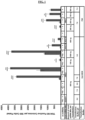
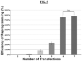
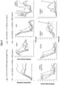
- FIG. 1 the application of the methods of the present technology to primary neonatal fibroblasts resulted in the production of over 4,000 Tra-1-60 + iPSC colonies from only 500 starting cells in a 6-well dish format in 18 days after as few as 7 transfections. Over 60 Tra-1-60 + iPSC colonies are produced from only 500 starting cells in 18 days after as few as 3 transfections ( FIG. 5 ) .
- the ability to reprogram cells using a low starting cell density may be particularly advantageous in the clinical setting.
- Lower input cell density will shorten the time between patient biopsy and iPSC generation and will reduce the risk of contamination and the accumulation of mutations due to extensive cell culturing.
- the reduced number of RNA transfections required for our protocol significantly reduces the cost of reprogramming for both research and clinical applications as compared to other RNA-based reprogramming protocols.
- the methods disclosed herein are also useful to reprogram individually plated single human primary fibroblasts on a feeder-free system. As shown in Table 1 , the methods of the present technology reprogram greater than 90% of individually plated primary human neonatal fibroblasts. By contrast, recent reports have demonstrated that the reprogramming efficiency of human iPSCs is approximately 2.1% for known mRNA methods using a starting cell density of 50,000 cells. ( Schlaeger, et al. Nature Biotechnology 33:58-63 (2015 )). In addition, the combination of mod-mRNA cocktail and m-miRNAs in the methods disclosed herein produces a synergistic effect on reprogramming efficiency as shown in FIG. 1 and Table 1.
- the methods disclosed herein are also useful to reprogram or dedifferentiate cells prior to re-differentiation.
- the methods include generating differentiated cells by exposing pluripotent cells generated by the methods described herein to cell lineage differentiation factors.
- the differentiated cells comprise neuronal lineage cells.
- the differentiated cells comprise endodermal lineage cells.
- the differentiated cells comprise cardiomyocyte lineage cells.
- the differentiated cells comprise keratinocytes.
- the differentiated cells comprise mesenchymal stem cells.
- the mesenchymal stem cells are able to further differentiate to fibroblasts, chondrocytes and/or osteoblasts.
- the differentiated cells comprise fibroblasts.
- the methods disclosed herein can be used to generate iPSCs that can be further modulated to form any type of somatic cells by culturing the iPSCs under cell-type specific conditions known in the art.
- Cell-type or cell lineage specific conditions may include contacting the iPSCs with cell and cell lineage differentiation factors under conditions known in the art and described herein.
- iPSCs can be differentiated toward a neuronal lineage by exposing them to one or more factors that include, but are not limited to, N2 and B27 supplements, Noggin, SB431542, DMEM/F 12 medium, laminin, cyclic adenosine monophosphate (cAMP), ascorbic acid, brain-derived neurotrophic factor (BDNF), glial cell-derived neurotrophic factor (GDNF), insulin-like growth factor I (IGF-I), fibroblast growth factor (FGF)-8, transforming growth factor (TGF) beta 3 (TGF- ⁇ 3), or retinoic acid.
- N2 and B27 supplements Noggin, SB431542, DMEM/F 12 medium, laminin, cyclic adenosine monophosphate (cAMP), ascorbic acid, brain-derived neurotrophic factor (BDNF), glial cell-derived neurotrophic factor (GDNF), insulin-like growth factor I (IGF-I), fibroblast growth factor (FGF)-8, transforming
- iPSCs can be differentiated toward an endodermal lineage (such as hepatocytes, pancreatic cells, intestinal epithelial, lung cells) by exposing them to specific differentiation factors and media, which include, but are not limited to, RPMI medium, SFD medium, N2B27 medium, glutamine, monothioglycerol (MTG), CHIR 99021, activin A, ascorbic acid, bone morphogenetic protein (BMP)- 4, vascular endothelial growth factor (VEGF), epidermal growth factor (EGF), basic FGF (bFGF), hepatocyte growth factor (HGF), dexamethasone, TGF- ⁇ , hydrocortisone, FGF-7, or Exendin-4.
- endodermal lineage such as hepatocytes, pancreatic cells, intestinal epithelial, lung cells
- specific differentiation factors and media include, but are not limited to, RPMI medium, SFD medium, N2B27 medium, glutamine, monothioglycerol (MTG
- Cardiomyocyte lineage differentiation factors and media include, but are not limited to, StemPro medium, DMEM/F12 medium, BMP4, Activin A, bFGF, VEGF, Dickkopf-related protein 1 (DKK1), Transferrin, MTG, or ascorbic acid.
- iPSC can be exposed to fetal serum and differentiation factors which include, but are not limited to, bFGF, BMP-4, EGF, retinoic acid, or platelet derived growth factor (PDGF).
- iPSC-derived MSC can subsequently be differentiated toward (1) bone progenitors (osteocytes) through exposure to one or more factors such as ascorbic-acid-2-phosphate, ⁇ -glycerophosphate, M dexamethasone or BMP-2, (2) chondrogenic progenitor (chondrocytes) through exposure to one or more factors such as dexamethasone, ascorbic-acid-2-phosphate, proline, pyruvate, TGF- ⁇ 3, or insulin/transferrin/selenious acid supplement (ITS) (3) adipogenic progenitors through exposure to one or more factors such as hydrocortisone, isobutylmethylxanthine or indomethacin; and (4) fibroblasts through exposure to connective tissue growth factor (CTGF).
- osteoblasts bone progenitors
- CGF connective tissue growth factor
- Fibroblasts can also be derived directly from iPSCs via exposure to one or more factors such as TGF- ⁇ 2, ascorbic acid, connective tissue growth factor (CTGF), ITS reagents, or fetal serum.
- Keratinocyte lineage differentiation factors include, but are not limited to, BMP4, retinoic acid, ascorbic acid, insulin, hydrocortisone, bovine pituitary extract, IGF-1 or EGF.
- the methods disclosed herein are also useful to reprogram or dedifferentiate cells prior to re-differentiation of cells and organ formation.
- the methods include generating organs by exposing pluripotent cells generated by the methods described herein to differentiating factors as described herein and combining one or more of the differentiated cells and cell types under conditions sufficient to encourage organ formation.
- iPSCs generated using the methods described herein can be differentiated to cells that can be used to make skin as well as other organs such as liver, bones, and cartilage.
- Such methods include combining one or more of the lineages and/or cell types that form an organ under conditions sufficient to encourage organ formation.
- conditions sufficient to form skin may include but are not limited to co-culture or in vivo co-grafting of iPSC-derived keratinocytes and fibroblasts.
- fibroblasts are grown on extracellular protein matrix (such as collagen, laminin, fibronectin, etc. ) to form a dermis-like structure followed by overlaying with keratinocytes to produce epidermis.
- extracellular protein matrix such as collagen, laminin, fibronectin, etc.
- a silicone grafting chamber can be surgically inserted onto the muscle fascia of recipient severe combined immunodeficiency (SCID) mice.
- SCID severe combined immunodeficiency
- a cell slurry consisting of keratinocytes and fibroblasts derived from human iPSCs is introduced into this chamber.
- the cells and factors necessary to generate human skin equivalents ex vivo and in vivo include, but are not limited to, iPSC derived keratinocytes, fibroblasts, melanocytes and derma papilla cells, EGF, insulin, fetal serum, ascorbic acid, hydrocortisone, bovine pituitary extract, IGF-1, or DMEM medium.
- Bones can be grown ex vivo by culturing iPSC-derived osteocytes in the presence of ascorbic-acid-2-phosphate, ⁇ -glycerophosphate and fetal serum.
- Cartilage can be generated by culturing iPSC-derived chondrocytes as micromasses in the presence of ITS, dexamethasone, ascorbic-acid-2-phosphate, proline, pyruvate and TGF- ⁇ 3.
- Liver can be generated via the formation of liver buds.
- Conditions sufficient to form liver buds may include, but are not limited to, the combination of mesenchymal stem cells with hepatic progenitors (both can be derived from iPSCs as described above) in the presence of endothelial growth medium and/or hepatocyte culture medium supplemented with dexamethasone, oncostatin, HGF, and matrigel.
- the methods disclosed herein can also be used to generate iPSCs that can be further genetically manipulated via homologous recombination or other appropriate techniques known in the art using, for example, TALENs, zinc-finger nucleases, or CRISPR/Cas9 systems with the goal to model a variety of genetic disorders as well as to generate isogenic wild-type controls when iPSCs are generated from patients with genetic defects.
- TALENs zinc-finger nucleases
- CRISPR/Cas9 systems CRISPR/Cas9 systems with the goal to model a variety of genetic disorders as well as to generate isogenic wild-type controls when iPSCs are generated from patients with genetic defects.
- These genetically manipulated iPSC clones can be differentiated into relevant cell types for research studies or for therapeutic applications to be returned back to the patient as an autograft.
- somatic cells that may be reprogrammed by the methods disclosed herein are not limiting, and any kind of somatic cells may be used.
- mature somatic cells may be used.
- somatic cells are from an embryonic stage.
- somatic cells may be primary cells (non-immortalized cells), such as those freshly isolated from an animal, or may be derived from a cell line (immortalized cells).
- the somatic cells are mammalian cells, such as, for example, human cells or mouse cells.
- the somatic cells are canine, feline, equine, or bovine cells.
- somatic cells may be obtained by well-known methods, from different organs, such as, but not limited to, skin, lung, pancreas, liver, stomach, intestine, heart, reproductive organs, bladder, kidney, urethra and other urinary organs, or generally from any organ or tissue containing living somatic cells, or from blood cells.
- fibroblasts are used.
- keratinocytes are used.
- melanocytes are used.
- cells isolated from the blood and/or bone marrow which include, but are not limited to, endothelial cells, lymphocytes, myeloid cells, leukocytes, mesenchymal stem cells, and hematopoietic stem cells
- mesenchymal stem cells are used.
- urine-derived renal epithelium cells are used.
- somatic cell as used herein, is also intended to include adult stem cells.
- cells are reprogrammed for an intended therapeutic use, and are derived from the patient subject.
- Somatic cells can be derived from a healthy or diseased subject, which include, but are not limited to, patients suffering from epidermolysis bullosa (EB), subtypes of skin blistering disorders such as EB simplex (EBS), junctional EB (JEB), dominant dystrophic EB (DDEB) and recessive dystrophic EB (RDEB), Kindler syndrome, acquired and congenital ichthyoses such as epidermolytic ichthyosis (EI), formerly known as epidermolytic hyperkeratosis (EHK), and Lamellar Ichthyosis (LI); Ectrodactyly, Ectodermal dysplasia, and Cleft lip/palate (EEC) syndrome; Dyskeratosis Congenita (DC); connective tissue diseases and injuries, type I diabetes, Down Syndrome, Danon disease.
- EB epidermolysis bullosa
- methods for obtaining human somatic cells include obtaining a cellular sample, e.g. , by a biopsy (e.g. , a skin sample).
- somatic cells selected for reprogramming are plated at low density. In some embodiments, cells are plated at a density of about 10,000 cells/cm 2 or less. In some embodiments, cells are plated at a density of about 9,000 cells/cm 2 or less. In some embodiments, cells are plated at a density of about 8,000 cells/cm 2 or less. In some embodiments, cells are plated at a density of about 7,000 cells/cm 2 or less. In some embodiments, cells are plated at a density of about 6,000 cells/cm 2 or less. In some embodiments, cells are plated at a density of about 5,000 cells/cm 2 or less.
- cells are plated at a density of about 4,000 cells/cm 2 or less. In some embodiments, cells are plated at a density of about 3,000 cells/cm 2 or less. In some embodiments, cells are plated at a density of about 2,000 cells/cm 2 or less. In some embodiments, cells are plated at a density of about 1,000 cells/cm 2 or less. In some embodiments, cells are plated at a density of about 900 cells/cm 2 or less. In some embodiments, cells are plated at a density of about 800 cells/cm 2 or less. In some embodiments, cells are plated at a density of about 700 cells/cm 2 or less.
- cells are plated at a density of about 600 cells/cm 2 or less. In some embodiments, cells are plated at a density of about 500 cells/cm 2 or less. In some embodiments, cells are plated at a density of about 400 cells/cm 2 or less. In some embodiments, cells are plated at a density of about 300 cells/cm 2 or less. In some embodiments, cells are plated at a density of about 200 cells/cm 2 or less. In some embodiments, cells are plated at a density of about 100 cells/cm 2 or less. In some embodiments, cells are plated at a density of about 50 cells/cm 2 or less. In some embodiments, cells are plated at a density of about 25 cells/cm 2 or less.
- cells are plated at a density of about 10 cells/cm 2 or less. In some embodiments, cells are plated at a density of about 5 cells/cm 2 or less. In some embodiments, cells are plated at a density of about 2 cells/cm 2 or less. In some embodiments, cells are plated at a density of about 1 cell/cm 2 or less. In some embodiments, cells are plated at a density of about 0.5 cells/cm 2 or less. In some embodiments, cells are plated at a density of about 0.25 cells/cm 2 or less. In some embodiments, low density is in the range of about 0.1-5,000 cells/cm 2 .
- low density is in the range of about 0.1-3,000 cells/cm 2 . In some embodiments, low density is in the range of about 0.1-2,000 cells/cm 2 . In some embodiments, low density is in the range of about 0.1-1,000 cells/cm 2 . In some embodiments, the reprogrammed cells are single cell derived colonies.
- the reprogrammed cells generated by the methods described herein have a variety of applications and therapeutic uses.
- the methods disclosed herein are directed to reprogramming cells suitable for therapeutic applications, including transplantation into patients, as the iPSCs have been reprogrammed without providing exogenous DNA.
- the methods of the present technology yield iPSCs that have not been exposed to animal-derived, human-derived, or allogeneic materials.
- the methods disclosed herein yield reprogrammed cells with normal karyotypes or with karyotypes that are the same as the patient from whom they were derived.
- uncorrected iPSCs from patients can be differentiated into cell types relevant to the genetic disorder for modeling the disease in organotypic cultures or for recapitulating the disease phenotype in vivo by transplanting onto immunodeficient mice as a xenograft.
- a particular mutation of interest can be introduced into normal healthy iPSCs, as another approach to modeling a disorder.
- therapeutic applications include, but are not limited to, autologous iPSC-derived bone and cartilage transplantation/repair for non-healing fractures, and the generation of autologous bioengineered skin equivalents from iPSCs for treating non-healing wounds, among others.
- a method for treating or preventing one or more symptoms of a disease or disorder in a subject comprising dedifferentiating cells in vitro and administering a therapeutically effective amount of the dedifferentiated cells to a subject in need thereof.
- a method for treating or preventing one or more symptoms of a disease or disorder in a subject comprises reprogramming cells to pluripotency in vitro and administering a therapeutically effective amount of the reprogrammed cells to a subject in need thereof.
- the method comprises obtaining one or more somatic cells from a subject and reprogramming the cells into iPSCs or dedifferentiated cells.
- a method for treating or preventing one or more symptoms of a disease or disorder in a subject comprises reprogramming cells to pluripotency in vitro, differentiating the cells to one or more appropriate cell types, and administering a therapeutically effective amount of the differentiated cells to a subject in need thereof.
- the method comprises obtaining one or more somatic cells from a subject and reprogramming the cells into iPSCs or dedifferentiated cells.
- the cells are then cultured under conditions that allow for the cells to differentiate into a desired cell type suitable for treating or preventing a condition.
- the differentiated cells may then be introduced into the subject to treat or prevent the condition.
- a method for treating or preventing one or more symptoms of a disease or disorder in a subject comprising (a) reprogramming cells to pluripotency in vitro , (b) differentiating the cells to one or more appropriate cell types, (c) combining one or more differentiated cell types to form an organ, and (d) administering a therapeutically effective amount of the organ to a subject in need thereof.
- the method comprises obtaining one or more somatic cells from a subject and reprogramming the cells into iPSCs or dedifferentiated cells.
- the cells are then cultured under conditions that allow for the cells to differentiate into a desired cell type suitable for treating or preventing a condition.
- the one or more differentiated cell types are combined under conditions that support formation of an organ.
- the differentiated organ may then be introduced into the subject to treat or prevent the condition.
- iPSCs generated using the methods described herein are genetically modified to correct a genetic abnormality or to improve or change cellular functioning. Methods of genetically modifying cells are well-known in the art, and described herein. In some embodiments, the genetic modification is performed prior to generation of the iPSCs, after generation of the iPSCs, prior to differentiation of the iPSCs, or after differentiation of the iPSCs.
- a method of treating a patient suffering from a genetic abnormality comprises: (a) reprogramming one or more primary adult cells from the patient to a pluripotent state using any of the methods described herein to form iPSCs, (b) genetically correcting one or more mutations in the iPSCs (prior to generation of the iPSCs, after generation of the iPSCs, prior to differentiation of the iPSCs, or after differentiation of the iPSCs), (c) differentiating the iPSCs, (d) providing the corrected differentiated cells to the patient.
- the primary adult cells are fibroblast cells.
- the corrected iPSCs are differentiated to fibroblasts and keratinocytes.
- the method further comprises generating skin from the differentiated cells.
- the patient is suffering from one or more of genetic diseases such as Epidermolysis Bullosa simplex (EBS), recessive dystrophic EB (RDEB), junctional EB (JEB), or Epidermolytic Ichthyosis.
- EBS Epidermolysis Bullosa simplex
- RDEB recessive dystrophic EB
- JEB junctional EB
- the one or more mutations includes K14, K1, collagen type VII, laminin332, collagen type XVII.
- generation of iPSCs allows for the development of cell or stem cell replacement strategies to treat genetic disorders, and for modeling these diseases with the goal of producing novel therapeutic options for patients.
- cells are isolated from a patient with a genetic disorder. This can be accomplished by many means, for example through a skin biopsy, blood draw, or a bone marrow aspirate. Suitable cell types for reprogramming include keratinocytes, melanocytes, fibroblasts, mesenchymal stem cells, etc.
- the generated iPSCs are genetically corrected by, for example, homologous recombination or other appropriate techniques known in the art using, for example, TALENs, zinc-finger nucleases, or CRISPR/Cas9 systems.
- the corrected iPSC clones are differentiated into relevant cell types for transplantation (typically somatic cells) and returned back to the patient as an autograft.
- therapeutic applications of the present technology include but are not limited to skin repair, skin transplantation, cartilage repair, bone repair, neuron transplantation, hematopoietic stem cell transplantation, mesenchymal stem cell transplantation, tissue reconstruction, organ reconstruction, or beta cell transplantation in a subject in need thereof.
- the methods disclosed herein can be used to generate cells for the treatment and/or prevention of a variety of diseases or disorders, including, but not limited to, skin injuries (wounds) and diseases, including epidermolysis bullosa (EB), subtypes of skin blistering disorders such as EB simplex (EBS), junctional EB (JEB), dominant dystrophic EB (DDEB) and recessive dystrophic EB (RDEB), Kindler syndrome, acquired and congenital ichthyoses such as epidermolytic ichthyosis (EI), formerly known as epidermolytic hyperkeratosis (EHK), and Lamellar Ichthyosis (LI); Ectrodactyly, Ectodermal dysplasia, and Cleft lip/palate (EEC) syndrome; Dyskeratosis Congenita (DC); connective tissue diseases and injuries (for example, osteoarthritis, bone fractures, and lipodystrophy); eye injuries and diseases such macular
- the methods disclosed herein can generate cells that can be utilized for repairing or regenerating a tissue or differentiated cell lineage in a subject.
- the method comprises generating a reprogrammed or dedifferentiated cell as described herein and administering the reprogrammed and/or dedifferentiated cell to a subject in need thereof.
- the subject has a disease or disorder in which an increase or replacement of a particular cell type or cellular dedifferentiation is desirable, such as skin injuries (wounds) and diseases, including epidermolysis bullosa (EB), subtypes of skin blistering disorders such as EB simplex (EBS), junctional EB (JEB), dominant dystrophic EB (DDEB) and recessive dystrophic EB (RDEB), Kindler syndrome, acquired and congenital ichthyoses such as epidermolytic ichthyosis (EI), formerly known as epidermolytic hyperkeratosis (EHK), and Lamellar Ichthyosis (LI); Ectrodactyly, Ectodermal dysplasia, and Cleft lip/palate (EEC) syndrome; Dyskeratosis Congenita (DC); connective tissue diseases and injuries (for example, osteoarthritis, bone fractures, and lipodystrophy); eye injuries and diseases such macular degeneration
- the subject has damage to a tissue or organ. In some embodiments, the subject has a deficiency of a particular cell type. In some embodiments, differentiated cells generated from the iPSCs produced by the methods disclosed herein can be used for tissue reconstitution or regeneration in a subject in need thereof.
- the methods of the present technology allow for successful reprogramming with less exogenous RNA (ExoRNA) (i.e. , 700-10,500 ng mod-mRNA for the entire reprogramming; 5-1000 pmoles m-miRNAs for the entire reprogramming).
- ExoRNA exogenous RNA
- reprogramming factors include, but are not limited to, one or more of Oct3 protein, Oct4 protein, Myo-D-Oct4 (M 3 O) protein, Sox1 protein, Sox2 protein, Sox3 protein, Sox15 protein, Klfl, protein, Klf2 protein, Klf3 protein, Klf4 protein, Klf5 protein, c-Myc protein, L-Myc protein, N-Myc protein, Nanog protein, Lin28A protein, Tert protein, Utfl protein, Aicda protein, Glis1, Sall4, Esrrb, Tet1, Tet2, Zfp42, Prdm14, Nr5a2, Gata6, Sox7, Pax1, Gata4, Gata3, cEBPa, HNF4a
- mature human microRNAs include, but are not limited to, one or more of miR200s, miR290s, miR371s, miR302s, miR367, miR369s, and biologically active fragments, analogues, and variants thereof.
- the primary human fibroblasts are transfected with 1500 ng/10 cm 2 of a reprogramming mRNA cocktail. In some embodiments, the primary human fibroblasts are transfected with 1400 ng/10 cm 2 of a reprogramming mRNA cocktail. In some embodiments, the primary human fibroblasts are transfected with 1300 ng/10 cm 2 of a reprogramming mRNA cocktail. In some embodiments, the primary human fibroblasts are transfected with 1200 ng/10 cm 2 of a reprogramming mRNA cocktail. In some embodiments, the primary human fibroblasts are transfected with 1100 ng/10 cm 2 of a reprogramming mRNA cocktail.
- the primary human fibroblasts are transfected with 1000 ng/10 cm 2 of a reprogramming mRNA cocktail. In some embodiments, the primary human fibroblasts are transfected with 900 ng/10 cm 2 of a reprogramming mRNA cocktail. In some embodiments, the primary human fibroblasts are transfected with 800 ng/10 cm 2 of a reprogramming mRNA cocktail. In some embodiments, the primary human fibroblasts are transfected with 700 ng/10 cm 2 of a reprogramming mRNA cocktail. In some embodiments, the primary human fibroblasts are transfected with 600 ng/10 cm 2 of a reprogramming mRNA cocktail.
- the primary human fibroblasts are transfected with 500 ng/10 cm 2 of a reprogramming mRNA cocktail. In some embodiments, the primary human fibroblasts are transfected with 400 ng/10 cm 2 of a reprogramming mRNA cocktail. In some embodiments, the primary human fibroblasts are transfected with 300 ng/10 cm 2 of a reprogramming mRNA cocktail. In some embodiments, the primary human fibroblasts are transfected with 200 ng/10 cm 2 of a reprogramming mRNA cocktail. In some embodiments, the primary human fibroblasts are transfected with 100 ng/10 cm 2 of a reprogramming mRNA cocktail.
- the primary human fibroblasts are transfected with 50 ng/10 cm 2 of a reprogramming mRNA cocktail. In some embodiments, the primary human fibroblasts are transfected with 100 to 1500 ng/10 cm 2 of a reprogramming mRNA cocktail. In other embodiments, the primary human fibroblasts are transfected with 100 to 600 ng/10 cm 2 of the reprogramming mRNA cocktail.
- the reprogramming mRNA cocktail comprises Oct4 or M 3 O, Sox2, and Klf4 in a 3:1:1 molar ratio. In another aspect the reprogramming mRNA cocktail comprises Oct4 or M 3 O, Sox2, Klf4, and one or more of c-Myc, Lin28A, or Nanog in a 1:1 molar ratio with Sox2 and Klf4.
- the reprogramming modified mRNA cocktail comprises human Klf4, c-Myc, M 3 O, Sox2, Lin28A, Nanog, and mWasabi. In some embodiments, the modified mRNA cocktail comprises 10% mWasabi modified mRNA, and Klf4, c-Myc, M 3 O, Sox2, Lin28A, and Nanog in a 1:1:3:1:1:1 molar ratio.
- the primary human fibroblasts are transfected with 600 ng/10 cm 2 of a reprogramming mRNA cocktail comprising Klf4, c-Myc, M 3 O, Sox2, Lin28A, and Nanog in a 1:1:3:1:1:1 molar ratio ( i . e ., 80.8 ng Klf4, 76.5 ng c-Myc, 221.1 ng M 3 O, 63.8 ng Sox2, 42.5 ng Lin28A, 55.3 ng Nanog, and 60 ng mWasabi).
- the primary human fibroblasts are transfected with a reprogramming mRNA cocktail alone or in combination with a miRNA mix.
- cells are transfected with 50 pmoles/10 cm 2 reprogramming miRNA (e.g., miR-367 and miR-302s).
- cells are transfected with 40 pmoles/10 cm 2 reprogramming miRNA.
- cells are transfected with 30 pmoles/10 cm 2 reprogramming miRNA.
- cells are transfected with 20 pmoles/10 cm 2 reprogramming miRNA.
- cells are transfected with 15 pmoles/10 cm 2 reprogramming miRNA.
- cells are transfected with 10 pmoles/10 cm 2 reprogramming miRNA. In some embodiments, cells are transfected with 5 pmoles/10 cm 2 reprogramming miRNA. In some embodiments, cells are transfected with 2 pmoles/10 cm 2 reprogramming miRNA. In some embodiments, cells are transfected with 1 pmoles/10 cm 2 reprogramming miRNA. In some embodiments, cells are transfected with 1 to 50 pmoles/10 cm 2 reprogramming miRNA. In some embodiments, cells are transfected with 10 to 40 pmoles/10 cm 2 reprogramming miRNA. In some embodiments, cells are transfected with 5 to 40 pmoles/10 cm 2 reprogramming miRNA.
- the primary human fibroblasts are transfected with miRNA367, miRNA302a, miRNA302b, miRNA302c, and miRNA302d in a 1:1:1:1:1 molar ratio.
- the reprogramming efficiency of the present technology is enhanced by tailoring the cell transfection regimen and cell seeding conditions specifically to primary human fibroblasts.
- the primary human fibroblasts are typically cultured in medium containing Knock-Out Serum Replacement (KOSR medium) and RNA transfections are performed with the transfection reagent, LIPOFECTAMINE ® RNAiMAX TM .
- reprogramming efficiency is enhanced by the addition of a complexation buffer that mediates that formation of transfection complexes between RNAs and cationic lipids.
- the complexation buffer is OPTI-MEM ® or PBS.
- cells are seeded at a density of about 10,000 cells/cm 2 or less and transfected with 1,000 ng/10 cm 2 of a reprogramming mRNA cocktail and 20 pmoles/10 cm 2 of reprogramming miRNAs per transfection every 48 hrs for a duration of 13 days (7 transfections). In some embodiments, cells are seeded at a density of about 10,000 cells/cm 2 or less and transfected with 1,000 ng/10 cm 2 of a reprogramming mRNA cocktail and 10 pmoles/10 cm 2 of reprogramming miRNAs. In some embodiments, cells are seeded at a density of about 10,000 cells/cm 2 or less and transfected with 1,000 ng/10 cm 2 of a reprogramming mRNA cocktail and 5 pmoles/10 cm 2 of reprogramming miRNAs.
- cells are seeded at a density of about 10,000 cells/cm 2 or less and transfected with 600 ng/10 cm 2 of a reprogramming mRNA cocktail and 20 pmoles/10 cm 2 of reprogramming miRNAs. In some embodiments, cells are seeded at a density of about 10,000 cells/cm 2 or less and transfected with 600 ng/10 cm 2 of a reprogramming mRNA cocktail and 10 pmoles/10 cm 2 of reprogramming miRNAs. In some embodiments, cells are seeded at a density of about 10,000 cells/cm 2 or less and transfected with 600 ng/10 cm 2 of a reprogramming mRNA cocktail and 5 pmoles/10 cm 2 of reprogramming miRNAs.
- cells are seeded at a density of about 10,000 cells/cm 2 or less and transfected with 300 ng/10 cm 2 of a reprogramming mRNA cocktail and 20 pmoles/10 cm 2 of reprogramming miRNAs. In some embodiments, cells are seeded at a density of about 10,000 cells/cm 2 or less and transfected with 300 ng/10 cm 2 of a reprogramming mRNA cocktail and 10 pmoles/10 cm 2 of reprogramming miRNAs. In some embodiments, cells are seeded at a density of about 10,000 cells/cm 2 or less and transfected with 300 ng/10 cm 2 of a reprogramming mRNA cocktail and 5 pmoles/10 cm 2 of reprogramming miRNAs.
- cells are seeded at a density of about 10,000 cells/cm 2 or less and transfected with 100 ng/10 cm 2 of a reprogramming mRNA cocktail and 20 pmoles/10 cm 2 of reprogramming miRNAs. In some embodiments, cells are seeded at a density of about 10,000 cells/cm 2 or less and transfected with 100 ng/10 cm 2 of a reprogramming mRNA cocktail and 10 pmoles/10 cm 2 of reprogramming miRNAs. In some embodiments, cells are seeded at a density of about 10,000 cells/cm 2 or less and transfected with 100 ng/10 cm 2 of a reprogramming mRNA cocktail and 5 pmoles/10 cm 2 of reprogramming miRNAs.
- cells are seeded at a density of about 9,000 cells/cm 2 or less and transfected with 1,000 ng/10 cm 2 of a reprogramming mRNA cocktail and 20 pmoles/10 cm 2 of reprogramming miRNAs per transfection every 48 hrs for the duration of 13 days (7 transfections). In some embodiments, cells are seeded at a density of about 10,000 cells/cm 2 or less and transfected with 1,000 ng/10 cm 2 of a reprogramming mRNA cocktail and 10 pmoles/10 cm 2 of reprogramming miRNAs. In some embodiments, cells are seeded at a density of about 10,000 cells/cm 2 or less and transfected with 1,000 ng/10 cm 2 of a reprogramming mRNA cocktail and 5 pmoles/10 cm 2 of reprogramming miRNAs.
- cells are seeded at a density of about 9,000 cells/cm 2 or less and transfected with 600 ng/10 cm 2 of a reprogramming mRNA cocktail and 20 pmoles/10 cm 2 of reprogramming miRNAs. In some embodiments, cells are seeded at a density of about 10,000 cells/cm 2 or less and transfected with 600 ng/10 cm 2 of a reprogramming mRNA cocktail and 10 pmoles/10 cm 2 of reprogramming miRNAs. In some embodiments, cells are seeded at a density of about 10,000 cells/cm 2 or less and transfected with 600 ng/10 cm 2 of a reprogramming mRNA cocktail and 5 pmoles/10 cm 2 of reprogramming miRNAs.
- cells are seeded at a density of about 9,000 cells/cm 2 or less and transfected with 300 ng/10 cm 2 of a reprogramming mRNA cocktail and 20 pmoles/10 cm 2 of reprogramming miRNAs. In some embodiments, cells are seeded at a density of about 10,000 cells/cm 2 or less and transfected with 300 ng/10 cm 2 of a reprogramming mRNA cocktail and 10 pmoles/10 cm 2 of reprogramming miRNAs. In some embodiments, cells are seeded at a density of about 10,000 cells/cm 2 or less and transfected with 300 ng/10 cm 2 of a reprogramming mRNA cocktail and 5 pmoles/10 cm 2 of reprogramming miRNAs.
- cells are seeded at a density of about 0.1-10,000 cells/cm 2 or less and transfected with 1,000 ng/10 cm 2 of a reprogramming mRNA cocktail and 20 pmoles/10 cm 2 of reprogramming miRNAs per transfection every 48 hrs for the duration of 13 days (7 transfections). In some embodiments, cells are seeded at a density of about 0.1-10,000 cells/cm 2 or less and transfected with 1,000 ng/10 cm 2 of a reprogramming mRNA cocktail and 10 pmoles/10 cm 2 of reprogramming miRNAs.
- cells are seeded at a density of about 0.1-10,000 cells/cm 2 or less and transfected with 1,000 ng/10 cm 2 of a reprogramming mRNA cocktail and 5 pmoles/10 cm 2 of reprogramming miRNAs.
- cells are seeded at a density of about 0.1-10,000 cells/cm 2 or less and transfected with 600 ng/10 cm 2 of a reprogramming mRNA cocktail and 20 pmoles/10 cm 2 of reprogramming miRNAs. In some embodiments, cells are seeded at a density of about 0.1-10,000 cells/cm 2 or less and transfected with 600 ng/10 cm 2 of a reprogramming mRNA cocktail and 10 pmoles/10 cm 2 of reprogramming miRNAs.
- cells are seeded at a density of about 0.1-10,000 cells/cm 2 or less and transfected with 600 ng/10 cm 2 of a reprogramming mRNA cocktail and 5 pmoles/10 cm 2 of reprogramming miRNAs.
- cells are seeded at a density of about 0.1-10,000 cells/cm 2 or less and transfected with 300 ng/10 cm 2 of a reprogramming mRNA cocktail and 20 pmoles/10 cm 2 of reprogramming miRNAs. In some embodiments, cells are seeded at a density of about 0.1-10,000 cells/cm 2 or less and transfected with 300 ng/10 cm 2 of a reprogramming mRNA cocktail and 10 pmoles/10 cm 2 of reprogramming miRNAs.
- cells are seeded at a density of about 0.1-10,000 cells/cm 2 or less and transfected with 300 ng/10 cm 2 of a reprogramming mRNA cocktail and 5 pmoles/10 cm 2 of reprogramming miRNAs.
- cells are seeded at a density of about 0.1-10,000 cells/cm 2 or less and transfected with 100 ng/10 cm 2 of a reprogramming mRNA cocktail and 20 pmoles/10 cm 2 of reprogramming miRNAs. In some embodiments, cells are seeded at a density of about 0.1-10,000 cells/cm 2 or less and transfected with 100 ng/10 cm 2 of a reprogramming mRNA cocktail and 10 pmoles/10 cm 2 of reprogramming miRNAs.
- cells are seeded at a density of about 0.1-10,000 cells/cm 2 or less and transfected with 100 ng/10 cm 2 of a reprogramming mRNA cocktail and 5 pmoles/10 cm 2 of reprogramming miRNAs.
- Transcripts of six reprogramming factors (OCT4 or Myo-D-OCT4 (M 3 O), SOX2, KLF4, c-MYC, NANOG and LIN28A (abbreviated as "O m SKMNL”)) were prepared as modified mRNA (mod-mRNA) as follows.
- Modified mRNA was synthesized as described in Warren, et al. Cell Stem Cell 7:618-630 (2010 ), with slight modifications. Briefly, MEGAscript T7 kit (Life Technologies, Grand Island, NY) was used and 1.6 ⁇ g of template PCR product for each 40 ⁇ l reaction was used.
- the mod-mRNA mix used for reprogramming contained 6 reprogramming factors, M 3 O , SOX2, KLF4, c-MYC, NANOG and LIN28A (abbreviated as "O m SKMNL”), at a molar stoichiometry of Myo-D-Oct4 (M 3 O) to the other 5 factors as 3:1:1:1:1:1 and included 10% mWasabi mod-mRNA to control for the transfection.
- a m-miRNA mix was prepared as follows.
- MiR-367/302s as miScript miRNA mimic (Syn-has-miR-367-3p, Syn-has-miR-302a-3p, Syn-has-miR-302b-3p, Syn-has-miR-302c-3p, and Syn-has-miR-302d-3p) or controls (AllStars Neg. Control siRNA and fluorescently labeled AllStars Neg. siRNA AF 488) were purchased from QIAGEN. Lyophilized products were dissolved to 5 ⁇ M final concentration in dH 2 O supplemented with 1 U/ ⁇ l of RIBOGUARD TM RNase Inhibitor. Stocks were frozen at -70°C until further use. Individual m-miRNA-367/302s stocks were mixed in 1:1:1:1:1 ratio to prepare a 5 ⁇ M m-miRNA mix.
- the plating medium contained modified DMEM/F12 with L-glutamine and no HEPES (catalog # 11320), 20% KNOCKOUT TM Serum Replacement, 5% Heat Inactivated FBS, 0.2% of 100x MEM Non-Essential Amino Acids Solution, 0.4 mM GLUTAMAX TM Supplement, 55 ⁇ M of 2-Mercaptoethanol, 1% of 100x Antibiotic-Antimycotic solution (all from Life Technologies, Grand Island, NY), 50 ⁇ g/mL L-Ascorbic Acid (Sigma-Aldrich, St.
- KOSR medium containing modified DMEM/F12 with L-glutamine and no HEPES (catalog # 11320), 20% KNOCKOUT TM Serum Replacement, 0.2% of 100x MEM Non-Essential Amino Acids Solution, 0.4 mM GLUTAMAX TM Supplement, 55 ⁇ M of 2-Mercaptoethanol, 1% of 100x Antibiotic-Antimycotic solution (all from Life Technologies, Grand Island, NY), 50 ⁇ g/mL L-Ascorbic Acid (Sigma-Aldrich, St. Louis, MO).
- KOSR Medium was equilibrated overnight at 5% O 2 before each medium change and supplemented with fresh 100 ng/mL bFGF (Life Technologies, Grand Island, NY) and 200 ng/mL B18R.
- the volume used per well in a 6-well dish format was 1 mL.
- RNAiMAX TM LIPOFECTAMINE ® (Life Technologies, Grand Island, NY)
- RNA molecules for mWasabi transfections, modified mRNA transfections, and m-miRNAs transfections
- complexation buffer was combined as described below.
- RNAiMAX TM LIPOFECTAMINE ® was performed in either pH-adjusted OPTI-MEM ® or pH-adjusted-PBS. pH values evaluated are shown in Tables 2 and 3 below.
- the pH of commercially supplied OPTI-MEM ® is 7.2-7.3.
- the pH of OPTI-MEM ® was adjusted to the indicated value with 1M NaOH.
- the pH of 1xPBS was adjusted using either 1M NaOH or 1M HCl. Strict RNase-free conditions were maintained for OPTI-MEM ® and 1xPBS preparation, and following pH adjustment both OPTI-MEM ® and 1x PBS were filter sterilized.
- RNA and RNAiMAX TM LIPOFECTAMINE ® reagent were first diluted in pH-adjusted OPTI-MEM ® reduced serum medium (Life Technologies, Grand Island, NY) or 1x pH-adjusted PBS.
- OPTI-MEM ® reduced serum medium Life Technologies, Grand Island, NY
- 1x pH-adjusted PBS 100 ng/ ⁇ l RNA was diluted 5x and 5 ⁇ l of RNAiMAX TM LIPOFECTAMINE ® per microgram of RNA was diluted 10x. After dilution these components were combined together and incubated for 15 min at room temperature (RT).
- RNAiMAX TM LIPOFECTAMINE ® For the m-miRNA transfections, a 5 ⁇ M (5 pmol/ ⁇ l) m-miRNA mix was diluted to 0.6 pmol/ ⁇ l and 1 ⁇ l of RNAiMAX TM LIPOFECTAMINE ® per 6 picomoles was diluted 10x. The diluted m-miRNA mix and RNAiMAX TM LIPOFECTAMINE ® were mixed together and incubated for 15 minutes at RT. After incubation at RT, mixtures of mod-RNA mix and/or m-miRNA mix and RNAiMAX TM LIPOFECTAMINE ® were applied to the cell culture.
- KOSR medium was changed 24 hours after each transfection.
- the medium (KOSR Medium supplemented with 100 ng/mL bFGF (Life Technologies, Grand Island, NY)) was changed every day and the cells were grown up to Day 18, at which point the wells were stained with anti-Tra-1-60 antibody by methods well-known in the art, and the number of Tra-1-60 positive, iPSC colonies was counted.
- results are shown in FIG. 1 .
- neonatal fibroblast cells were plated at a density of 500 cells per well of the 6-well dish format.
- "5fM 3 O” represents a 6 factor cocktail containing M 3 O, Sox2, Klf4, c-Myc, Lin28A and Nanog.
- "5fOCT4" represents a 6 factor cocktail containing Oct4, Sox2, Klf4, c-Myc, Lin28A and Nanog.
- “4fOCT4" represents a 5 factor cocktail containing Oct4, Sox2, Klf4, c-Myc and Lin28A.
- the "+” stands for 20 pmoles of m-miRNA367/302s.
- the "-” stands for no m-miRNA.
- “NEG” stands for the negative m-miRNA (All Stars Neg. Control siRNA, Qiagen).
- Each mod-mRNA cocktail also contained 10% of mWasabi mod-mRNA to monitor the transfection efficiency.
- RNAs were adjusted to the surface area as well as the volume of medium used throughout the regimen.
- KOSR medium supplemented with bFGF and B 18R were used, and 100 ng of mod-mRNA mix comprising M 3 O and 2 pmoles of m-miRNA mix comprising miRNA367/302s were used per well per transfection. Seven transfections were performed at 48 hour intervals as described in Example 1, and KOSR medium was changed 24 hours after each transfection. After completing the series of transfections, the medium was changed every day and the cells were grown up to Day 18, at which point the wells were stained with anti-Tra-1-60 antibody and the number of wells with resulting iPSC colonies was counted. The experiment was performed for 3 independent human primary neonatal lines (FN1, FN2, and FN3).
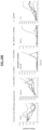
- Results are shown in Table 4 below and in FIGS. 2 and 3 .
- TABLE 4 Primary Neonatal Cell Line Mature microRNAs Wells with an individually plated single cell Wells with dividing cells throughout reprogramming Wells with Tra-1-60 + colonies Reprogramming efficiency (%) Wells with Tra-1-60 + colonies/wells with dividing cells x 100% FN1 + 157 144 106 73.6% - 134 116 0 0% FN2 + 141 130 101 77.7% - 107 98 8 8.2% FN5 + 110 108 98 90.7% - 111 110 16 14.5%
- Example 3 Evaluation of plating density and reprogramming efficiency
- a series of reprogramming experiments using human primary neonatal fibroblasts plated at different densities were performed.
- Cells were transformed according to the methods described in Example 1, using OPTI-MEM ® at pH 8.2, 20 pmole m-miRNA mix, 600 ng of mod-mRNA mix (including M 3 O), with 7 transfections, one every 48 hours.
- KOSR medium was changed 24 hours after each transfection. After the last transfection, the medium was changed every day and cells were grown to 18 days and stained for Tra-1-60 + expression.
- the initial seeding density which allows for the generation of Tra-1-60 + colonies was 10,000 cells/well in a 6-well dish format. The colonies did not form at an initial density above 100,000 cells/well.
- Reprogramming efficiency is traditionally calculated as the number of resulting iPSC colonies divided by the number of input cells. If applied to some embodiments of the methods disclosed herein, these calculations would indicate up to 800% of reprogramming efficiency ( see Table 5 above). This number is achieved when multiple sister iPSC colonies derive from a single parental cell.
- Example 4 The number of transfections influences reprogramming efficiency
- Example 1 500 human neonatal fibroblast cells were plated in a 6-well dish and transfected as described in Example 1 (using OPTI-MEM ® at pH 8.2, 20 pmole m-miRNA mix and 600 ng of mod-mRNA mix (including M 3 O)). Cells were transfected every other day starting at Day 1 after plating; however, the total number of transfections was varied for each sample. Sample 1 was transfected 2x (on Day 1 and Day 3); sample 2 was transfected 3x (Day 1, Day 3 and Day 5); sample 4 was transfected 4x (Day 1, 3, 5 and 7); sample 5 was transfected 6x (Day 1, 3, 5, 7, 9 and 11) and sample 6 was transfected 7x (Day 1, 3, 5, 7, 9, 11 and 13). The medium was changed 24 hours after each transfection. After the last transfection, the medium was changed daily to 18 days. At Day 18, cells were stained for Tra-1-60 expression and positive colonies were counted. Results are shown in FIG. 5 .
- Fibroblasts derived from 3 different inherited skin blistering disorders were successfully reprogrammed. Successful, high efficiency reprogramming was also achieved with fibroblasts derived from an individual suffering from Danon disease (FDanon); 2 patients with Down Syndrome (FDown1 and FDown2), 4 healthy individuals of 34 (F34), 40 (F40), 41(F41) and 50 (F50) years of age; senescent line derived from F50 (F50S), and 3 healthy neonatal lines (FN1, FN2, and FN5) (Table 6, below).
- the inherited skin blistering disorders were EHK (FEH1), EBS (FEB1), RDEB (FRD1 and FRD2).
- the adult fibroblast line derived from a 50 year old patient was serially passaged until more than 91% of cells exhibited a senescent phenotype. These cells were then reprogrammed into iPSCs according to the method described above. The reprogramming of this senescent line took only 16 days and resulted in an efficiency of approximately 0.33% (Table 6) , which surpasses previous reports on the reprogramming of senescent fibroblasts using an integrating lentiviral approach (-40 days). Thus, as shown here for the first time, senescent human cells were reprogrammed with an integration-free approach.
- telomerase The activity of telomerase was measured using the Trapeze Telomerase Detection Kit (Chemicon). CHAPS (1x) lysis buffer was used to obtain extracts from parental fibroblasts (F50), iPSC lines derived from F50 (150-2 and I50-3), iPSC lines derived from senescent F50S line (I50S-1 and I50S-2) and H1 ESC line. About 2000 cells were assayed for each telomeric repeat amplification protocol assay, and 800 cell equivalents were loaded into each well of a 15% non-denaturing TBE polyacrylamide gel. Reactions were performed in triplicate for each cell line. Each sample was heat inactivated for 15 min at 85 0 C to assess the background of the assay.
- telomere activity was high in all iPSCs, including the lines generated from senescent fibroblasts, and ESCs but not in fibroblasts or upon heat inactivation of the samples ( FIG. 6A ) .
- telomere length was determined by qPCR as previously shown in Cawthon, R.M., Nucleic Acids Res, 2002. 30(10): p. e47 . Briefly, DNA was isolated from low passage (F50) and senescent (F50S) parental fibroblasts, iPSC lines derived from F50 (150-2 and 150-3) and F50S (I50S-1 and I50S-2), and human ESCs (H1). qPCR analysis was performed with telomere-specific primers using LightCycle 480 (Roche). Telomeres were significantly longer in all iPSC lines as compared to corresponding parental fibroblasts, indicating an activation of rejuvenation processes upon reprogramming with approach described herein ( FIG. 6B ) .
- the expression of p21 was assessed in low passage fibroblasts (F50), senescent (F50S) derived from F50, iPSC lines derived from F50 (I50-2 and 150-3) and F50S (I50S-1 and I50S-2), and human ESCs (H1) using Western blot analysis. Briefly, the cells were lysed with RIPA buffer supplemented with a protease inhibitor cocktail (Sigma). Protein extracts were resolved by 15% SDS-PAGE and transferred to PVDF membranes. Membranes were blocked in 2.5% milk powder, 2.5% BSA in PBS for 1 hr and incubated with primary antibodies at 4°C overnight.
- Example 6 The combinatorial RNA-based approach reduces the expression of innate immunity genes, leads to the robust activation of pluripotency-associated genes and exhibits increased cell population doubling
- a time-point experiment was performed and the transcript level of a set of genes was analyzed during the first sixteen days of the reprogramming method as described in Example 1 using human primary neonatal cells plated at an initial density of 500 cells per well of a 6-well plate, OPTI-MEM ® at pH 8.2, 20 pmole m-miRNA mix, 600 ng of mod-mRNA mix, with 7 transfections, one every 48 hours. KOSR medium was changed 24 hours after each transfection. As compared to the previously published feeder-free mod-mRNA reprogramming protocol ( Warren L, et al. Sci Rep. 2:657. (2012 )), the level of transcripts encoding innate immunity genes was lower with the methods disclosed herein ( see e.g. , FIG.
- FIG. 8 This correlated with the lower level of exogenous reprogramming mod-mRNAs detected for our optimal regimen ( FIG. 8 ) .
- An elevated expression level of several cell cycle-promoting genes e.g. , CDK4, CCNE1, E2F1, CCNA2, and CDC20
- a reduced expression level of several cell cycle inhibitors e.g. , CDKN1A (p21) and CDKN1C (p57) was also observed ( FIG. 9 ) . This level was similar for all conditions initiated at a low seeding density as compared to the reprogramming initiated from 10,000 and 50,000 cells.
- CDK inhibitors such as p21 CIP1 and p57 are activated in response to redundant signaling of reprogramming factors expression, thus creating a reprogramming barrier.
- the expression of both p21 CIP1 and p57 remains low in our regimen as compared to the basic mod-mRNA reprogramming protocol ( FIG. 9 ) .
- the transcript level of Sall4 increased on Day 3 of the present combinatorial protocol and only on Day 11 of the basic mod-mRNA reprogramming approach (data not shown).
- the activation of NANOG, LIN28A, OCT4, SALL4, GDF3, PRDM14, UTF1, NR5A2, DPPA2, DPPA4, NR6A1, TRIM71, LEFTY1, LEFTY2, ZFP42, ASFIA, DNMT3A, DNMT3B, DNMT3L, TET1, and EPCAM occurred at least several days earlier in the combinatorial approach disclosed herein as compared to the previously published mod-mRNA reprogramming protocol ( FIG. 10A ) .
- the activation of genes involved in mesenchymal-to-epithelial transition (MET) also exhibited robust activation ( FIG. 11 ) .
- Example 7 Differentiation of iPSCs generated by methods described herein into a neuronal lineage
- iPSCs generated using our method were assessed. The ability of iPSCs generated using our method to differentiate into a variety of cell types including cells from a neuronal lineage was assessed.
- iPSCs were differentiated into cells from a neuronal lineage using a protocol adapted from work by Hua et al. and Chambers et al. ( Bao-Yang Hua, et al. PNAS 107:4335-4340 (2010 ); Chambers, S.M. et al. Nature Biotechnology 3:275-280 (2009 )). Briefly, iPSC cultures were disaggregated using Collagenase Type I and feeder-depleted on gelatin for 30 min at 37°C. The non-adherent cells were collected and plated on a Geltrex (Gibco) covered dish at a density of 25-30,000 cells/cm 2 in N2/B27 Medium (as described by Liu et al.
- the formed aggregates were induced with neural induction media containing Neurobasal medium (Gibco) supplemented with N2, B27 and heparin for 14 days.
- the induction phase was followed by the differentiation stage in Neurobasal medium supplemented with N2 (Gibco), B27 (Gibco), 1 ug/ml laminin (Sigma), 100 nM cAMP (Sigma), 200 ng/ml ascorbic acid (Sigma), 10 ng/ml BDNF (Peprotech), 10 ng/ml GDNF (Peprotech) and 10 ng/ml IGF-I (Peprotech).
- the staining shows the presence of the neuron specific type III ⁇ tubulin (TUJ1), which is indicative of a successful differentiation of iPSCs into neuronal progenitors ( FIG. 12 ) .
- TUJ1 neuron specific type III ⁇ tubulin
- Example 8 Differentiation of iPSCs generated by methods described herein into an endodermal lineage
- iPSCs generated using our method were assessed. The ability of iPSCs generated using our method to differentiate into a variety of cell types including cells from an endodermal lineage was assessed.
- iPSCs were differentiated into cells from an endodermal lineage using a previously published protocol ( Cheng, X., et al. ESCs Stembook, Cambridge (MA):Harvard Stem Cell Institute (2012 )). Briefly, iPSCs were feeder-depleted and seeded on a Geltrex-covered dish. These cells were then differentiated as a monolayer at 70% confluency.
- RPMI medium (Gibco) supplemented with 1x Glutamine (Gibco), 450 ⁇ M MTG (Gibco), 2 uM CHIR 99021 (Stemgent), 100 ng/ml Activin A (R&D), 50 ug/ml Ascorbic Acid (Sigma), 25 ng/ml BMP4 (R&D), 5 ng/ml bFGF (Gibco) and 10 ng/ml VEGF (R&D) was used.
- cells were disaggregated with Accutase and seeded at 250,000 cells/well on a Geltrex (Gibco) coated dish.
- cytokines were fed every 2 days, until Day 18 with media supplemented with cytokines provided above at varied concentrations.
- cells were replated onto Fibronectin coated chamber slides (BD) and immunostained with an antibody against endoderm specific cytokeratin Endo-A (Developmental Studies Hybridoma Bank, Iowa City, IA) using an immunostaining protocol as described for neuronal differentiation in Example 7.
- DAPI-containing mounting medium was used to visualize nuclei. Images were acquired using a Nikon Eclipse 90i upright microscope with a 10x objective (100X magnification).
- Endo-A is an early marker of endodermal progenitors, which in turn give rise to the epithelia of gastrointestinal and respiratory tracts, the urinary system, liver, and pancreas ( FIG. 13 ) .
- Example 9 Differentiation of iPSCs generated by methods described herein into a cardiomyocyte lineage
- iPSCs generated using our method were assessed. The ability of iPSCs generated using our method to differentiate into a variety of cell types including cells from a cardiomyocyte lineage was assessed.
- the iPSCs were differentiated into cells from a cardiomyocyte lineage using a previously published protocol ( Yang et al. Nature, 453:524-528 (2008 )). Briefly, cells were feeder-depleted and seeded on a Geltrex covered dish. These cells were then dissociated using Collagenase Type I and placed in a suspension culture for embryoid body formation in differentiation medium as previously described by Yang et al. Nature, 453:524-528 (2008 ).
- cytokines were used in combination with StemPro medium (Gibco): Day 0: BMP4 (20 ng/ml) (R&D); Day1: Activin A (6 ng/ml) (R&D), BMP4 (20 ng/ml) (R&D), bFGF (2.5 ng/ml) (Gibco); Day 3 and Day5: VEGF (1.25 ng/ml) (R&D) and DKK1 (150 ng/ml) (R&D); Day 8, Day 11 and Day 14: VEGF (1.25 ng/ml), DKK1 (150 ng/ml), and bFGF (2.5 ng/ml).
- Example 10 Differentiation of iPSCs generated by methods described herein into specific skin cell types
- iPSCs generated using our method were assessed.
- keratinocytes The ability of iPSCs generated using our method to differentiate into an ectodermal lineage, keratinocytes, mesenchymal stem cells, and fibroblasts was assessed.
- iPSCs were differentiated into cells from an ectodermal lineage and then to keratinocytes using a previously published protocol using BMP4 and retinoic acid treatments ( Bilousova G. et al. J. Invest. Dermatol. 131(4):857-64 (2011 )). Briefly, iPSCs were plated in N2B27 medium supplemented with 100 ng/ml bFGF onto Geltrex and Collagen I (Sigma) covered tissue culture plates.
- DKSFM Keratinocyte Serum Free medium
- R&D 25 ng/ml BMP4
- the medium was changed every other day for 17 days.
- the cells were replated onto Collagen type IV/Collagen type I coated dishes in CnT07 medium (Lonza) ( FIG. 15A ) .
- the left panel shows cells at Day 10 of differentiation, which are morphologically similar to ectoderm.
- the right panel shows cells at Day 30 of differentiation, which look identical to normal human keratinocytes.
- the cells also stained positive for the marker of basal layer keratinocytes keratin 14 (K14) with the immunostaining protocol as described in Example 7 (not shown).
- the images were taken with Nikon Eclipse TE2000-S inverted microscope with a 10x objective (100X magnification).
- the iPSCs can be differentiated either directly into a fibroblast lineage via exposure to TGF- ⁇ 2 or BMP-4 in the presence of fetal serum or through the intermediate mesenchymal stem cell stage.
- the later approach allows for the assessment of iPSC capacity to differentiate into both mesenchymal stem cells and fibroblasts.
- the differentiation into mesenchymal stem cells was achieved using previously published protocols ( West, J.D. et al. Am. J. Physiol. Cell Physiol. May 28 (2014 ); Lee, C.H. et al. J. Clin Invest. 120(9):3340-3349 )).
- iPSCs were exposed to medium containing 10% knock out serum replacement (GIBCO) in the presence of 10 ng/ml PDGF and 10 ng/ml EGF for 4 days. The medium was then switched to ⁇ MEM (HyClone) supplemented with 20% FBS. The cells were cultured for additional 2 weeks. The phenotype of iPSC derived mesenchymal stem cells was confirmed morphologically ( FIG. 15B , left) and by flow cytometric analysis for the expression of mesenchymal stem cell markers CD105 and CD90 (not shown).
- GEBCO knock out serum replacement
- iPSC-derived mesenchymal stem cells were then differentiated into a fibroblast lineage by exposing the cells to 100 ng/ml of connective tissue growth factor (CTGF).
- CTGF connective tissue growth factor
- the fibroblast phenotype was confirmed by a morphological analysis ( FIG. 15B , right) and functionally in the grafting assay described below and in FIG. 16 . Images were taken with Nikon Eclipse TE2000-S microscope with a 10x objective (100X magnification).
- Example 11 Use of cells differentiated from iPSCs to form human skin
- the ability to use cells differentiated from iPSCs generated using our method to create organs was assessed using human skin as a model system.
- a similar approach can be used for other organs, including but not limited to liver, pancreas, neurons, bones, and cartilage.
- Keratinocytes and fibroblasts generated from iPSCs as described in Example 10 were used to create skin using a method adapted from Lichti et al. ( Lichti, et al. Nat Protoc. 3 (5):799-810 (2008 )). Briefly, keratinocytes and fibroblasts derived from human iPSCs were mixed together and transplanted into a silicon chamber that was surgically inserted onto the muscle fascia of recipient severe combined immunodeficiency (SCID) mice. One week later, the chamber protecting the skin graft was removed.
- SCID severe combined immunodeficiency
- mice Four weeks post-transplantation, the recipient mice were euthanized and the grafting area was excised. The tissue was fixed in 10% Formalin, paraffinized, and sectioned. The sections were either stained with hematoxylin/eosin (H&E) or with antibody against epidermal specific markers Keratin (K) 14 and K1. For immunostaining, slides containing paraffin embedded sections were deparaffinized by submerging in Xylene and then rehydrated through a gradient ethanol bath. To retrieve the antigen, the sections were boiled for 10 minutes in 10mM Citrate Buffer pH 6.0 (Abcam).
- H&E hematoxylin/eosin
- K epidermal specific markers Keratin
- FIG. 16A The H&E image ( FIG. 16A ) shows the formation of human skin on a mouse. Note the thicker epidermis in the graft area which is indicative of human skin.
- FIG. 16B The H&E image ( FIG. 16A ) shows the formation of human skin on a mouse. Note the thicker epidermis in the graft area which is indicative of human skin.
- FIG. 16B shows the results of immunostaining using antibodies to both mouse and human K14 and to only mouse K1.
- the area to the right of the white arrow where there is no staining with antibody to mouse K1 shows the human xenograft area.
- FIG. 16B lower panel shows immuno-staining using antibodies to both mouse and human K14 and human K1 of the portion of FIG. 16B upper panel indicated by the white box.
- the K1 staining in this image indicates the formation of correct skin layers in a human iPSC-derived xenograft. Images were acquired using a Nikon Eclipse 90i upright microscope with either 4 x (40x magnification) or 10x (100x magnification) objective where indicated.
- Example 12 Genetic correction of Epidermolysis bullosa mutant gene using iPSCs
- EBS Epidermolysis bullosa
- iPSCs Cells from a patient suffering from EB were obtained using standard techniques, and used to generate iPSCs as described in Example 5. The iPSCs were then subjected to TALEN-mediated deletion of Exon 1 from the mutant K14 gene. To achieve the specific disruption of the mutant allele, we focused on a TALEN-based approach designed to introduce a knockout (KO) deletion of the K14 mt allele, and to leave the wild-type K14 allele intact and functional.
- TALEN-based approach designed to introduce a knockout (KO) deletion of the K14 mt allele, and to leave the wild-type K14 allele intact and functional.
- the point mutation C373T in the K14 mt allele in EBS iPSCs allows for the design of allele-specific TALENs, which would preferentially target the K14 mt allele and could be used for all EBS patients with the C373T mutation.
- ssODN single-stranded oligonucleotide
- the TALENs were delivered as mod-mRNA into target iPSCs.
- the efficiency of transfection of modified mRNA encoding eGFP, the K14 TALENs and ssODNs in EBS iPSCs is shown in FIG. 17A .
- the ssODNs were introduced using Fugene 6 (Roche), and 2 hrs later, 200 ng of TALEN mod-mRNA was delivered via RNAiMAX Lipofectamine (Invitrogen) in the presence of B18R, an interferon inhibitor to reduce the toxicity of mRNAs. Images were taken with Nikon Eclipse TE2000-S microscope with a 10x objective (100X magnification). DNA sequencing data ( FIG.
- a range includes each individual member.
- a group having 1-3 cells refers to groups having 1, 2, or 3 cells.
- a group having 1-5 cells refers to groups having 1, 2, 3, 4, or 5 cells, and so forth.
Landscapes
- Health & Medical Sciences (AREA)
- Life Sciences & Earth Sciences (AREA)
- Engineering & Computer Science (AREA)
- Chemical & Material Sciences (AREA)
- Genetics & Genomics (AREA)
- Biomedical Technology (AREA)
- Organic Chemistry (AREA)
- Bioinformatics & Cheminformatics (AREA)
- Zoology (AREA)
- Wood Science & Technology (AREA)
- Biotechnology (AREA)
- Molecular Biology (AREA)
- General Health & Medical Sciences (AREA)
- General Engineering & Computer Science (AREA)
- Biochemistry (AREA)
- Microbiology (AREA)
- Medicinal Chemistry (AREA)
- Cell Biology (AREA)
- Public Health (AREA)
- Veterinary Medicine (AREA)
- Animal Behavior & Ethology (AREA)
- Pharmacology & Pharmacy (AREA)
- Plant Pathology (AREA)
- Developmental Biology & Embryology (AREA)
- Biophysics (AREA)
- Physics & Mathematics (AREA)
- Epidemiology (AREA)
- Immunology (AREA)
- Nuclear Medicine, Radiotherapy & Molecular Imaging (AREA)
- Dermatology (AREA)
- General Chemical & Material Sciences (AREA)
- Chemical Kinetics & Catalysis (AREA)
- Transplantation (AREA)
- Virology (AREA)
- Hematology (AREA)
- Rheumatology (AREA)
- Mycology (AREA)
- Micro-Organisms Or Cultivation Processes Thereof (AREA)
Applications Claiming Priority (3)
| Application Number | Priority Date | Filing Date | Title |
|---|---|---|---|
| US201562258801P | 2015-11-23 | 2015-11-23 | |
| EP16869157.4A EP3384038B1 (de) | 2015-11-23 | 2016-11-22 | Verfahren und zusammensetzungen zur reprogrammierung von zellen |
| PCT/US2016/063258 WO2017091547A1 (en) | 2015-11-23 | 2016-11-22 | Methods and compositions for reprogramming cells |
Related Parent Applications (1)
| Application Number | Title | Priority Date | Filing Date |
|---|---|---|---|
| EP16869157.4A Division EP3384038B1 (de) | 2015-11-23 | 2016-11-22 | Verfahren und zusammensetzungen zur reprogrammierung von zellen |
Publications (2)
| Publication Number | Publication Date |
|---|---|
| EP4470544A2 true EP4470544A2 (de) | 2024-12-04 |
| EP4470544A3 EP4470544A3 (de) | 2025-06-18 |
Family
ID=58763571
Family Applications (2)
| Application Number | Title | Priority Date | Filing Date |
|---|---|---|---|
| EP24188635.7A Pending EP4470544A3 (de) | 2015-11-23 | 2016-11-22 | Verfahren und zusammensetzungen zur reprogrammierung von zellen |
| EP16869157.4A Active EP3384038B1 (de) | 2015-11-23 | 2016-11-22 | Verfahren und zusammensetzungen zur reprogrammierung von zellen |
Family Applications After (1)
| Application Number | Title | Priority Date | Filing Date |
|---|---|---|---|
| EP16869157.4A Active EP3384038B1 (de) | 2015-11-23 | 2016-11-22 | Verfahren und zusammensetzungen zur reprogrammierung von zellen |
Country Status (4)
| Country | Link |
|---|---|
| US (3) | US11459560B2 (de) |
| EP (2) | EP4470544A3 (de) |
| CA (1) | CA3011692A1 (de) |
| WO (1) | WO2017091547A1 (de) |
Families Citing this family (12)
| Publication number | Priority date | Publication date | Assignee | Title |
|---|---|---|---|---|
| US10648002B2 (en) * | 2016-11-22 | 2020-05-12 | Regents Of The University Of Minnesota | Method for correcting a genetic sequence |
| US20210324335A1 (en) * | 2018-09-04 | 2021-10-21 | Academia Sinica | Medium composition and method for culturing mesenchymal stem cells |
| CN112094804B (zh) * | 2019-06-18 | 2024-05-14 | 中国医学科学院基础医学研究所 | 一种异质性干细胞群、其制备方法及用途 |
| CN114667348A (zh) * | 2019-08-30 | 2022-06-24 | 细流而希佳股份有限公司 | 一种尿路上皮细胞的诱导剂和尿路上皮细胞的诱导方法 |
| WO2021133724A1 (en) * | 2019-12-23 | 2021-07-01 | The Board Of Trustees Of The Leland Stanford Junior University | Genetically corrected cells for therapeutic use |
| EP3851532A1 (de) * | 2020-01-20 | 2021-07-21 | Universidad Carlos III de Madrid | Gen-editing zur behandlung von epidermolysis bullosa |
| EP3875581A1 (de) * | 2020-03-02 | 2021-09-08 | Centre d'Etude des Cellules Souches (CECS) | Automatisiertes verfahren zur herstellung von keratinozyten |
| EP3875580A1 (de) * | 2020-03-02 | 2021-09-08 | Centre d'Etude des Cellules Souches (CECS) | Verfahren zur herstellung von keratinozyten |
| JP2024515805A (ja) * | 2021-04-27 | 2024-04-10 | アヴィータ・メディカル・インコーポレイテッド | 新たに脱凝集された組織に由来する再生生物活性懸濁液および臨床治療における使用方法 |
| WO2024012502A1 (zh) * | 2022-07-12 | 2024-01-18 | 南京瑞初医药有限公司 | 组合物及其使用方法 |
| CN116790665A (zh) * | 2023-06-09 | 2023-09-22 | 臻赫医药(杭州)有限公司 | 一种基于基因编辑的病毒抵抗猪的构建方法 |
| CN117431258B (zh) * | 2023-12-20 | 2024-06-28 | 上海元戊医学技术有限公司 | 使用含有Tet1基因的重编程因子诱导人体细胞重编程的方法 |
Family Cites Families (12)
| Publication number | Priority date | Publication date | Assignee | Title |
|---|---|---|---|---|
| JP5626619B2 (ja) | 2008-12-08 | 2014-11-19 | 国立大学法人京都大学 | 効率的な核初期化方法 |
| EP3231869A1 (de) | 2008-06-13 | 2017-10-18 | Whitehead Institute for Biomedical Research | Programmierung und umprogrammierung von zellen |
| US20130189741A1 (en) * | 2009-12-07 | 2013-07-25 | Cellscript, Inc. | Compositions and methods for reprogramming mammalian cells |
| US8802438B2 (en) * | 2010-04-16 | 2014-08-12 | Children's Medical Center Corporation | Compositions, kits, and methods for making induced pluripotent stem cells using synthetic modified RNAs |
| US9580689B2 (en) * | 2010-07-22 | 2017-02-28 | Regents Of The University Of Minnesota | Induced pluripotent stem cells |
| AU2012347919B2 (en) | 2011-12-05 | 2017-02-02 | Factor Bioscience Inc. | Methods and products for transfecting cells |
| EP3561054B1 (de) | 2012-05-13 | 2023-07-05 | Allele Biotechnology And Pharmaceuticals, Inc. | Feeder-freie ableitung von menschgemachten pluripotenten stammzellen mit synthetischer botenrns |
| JP2015522257A (ja) * | 2012-06-13 | 2015-08-06 | ステムジェント, インコーポレイテッドStemgent, Inc. | 多能性幹細胞を調製する方法 |
| KR102121086B1 (ko) * | 2012-11-01 | 2020-06-09 | 팩터 바이오사이언스 인크. | 세포에서 단백질을 발현시키는 방법들과 생성물들 |
| EP2953647B1 (de) * | 2013-02-05 | 2024-07-17 | B.G. Negev Technologies and Applications Ltd. | Positiv geladene polysaccharide zur rna-transfektion |
| EP3099801B1 (de) | 2014-01-31 | 2020-03-18 | Factor Bioscience Inc. | Synthetische rna zur verwendung in der behandlung von epidermolysis bullosa dystrophica |
| EP3371300A1 (de) | 2015-11-05 | 2018-09-12 | Centro en Investigación Biomédica en Red | Verfahren zum gen-editing von zellen, die aus einer person isoliert wurden, die an einer die erythroide lineage beeinflussenden stoffwechselerkrankung leidet, durch besagtes verfahren erhaltene zellen und verwendungen davon |
-
2016
- 2016-11-22 CA CA3011692A patent/CA3011692A1/en active Pending
- 2016-11-22 US US16/072,797 patent/US11459560B2/en active Active
- 2016-11-22 EP EP24188635.7A patent/EP4470544A3/de active Pending
- 2016-11-22 WO PCT/US2016/063258 patent/WO2017091547A1/en not_active Ceased
- 2016-11-22 EP EP16869157.4A patent/EP3384038B1/de active Active
-
2018
- 2018-04-20 US US15/958,965 patent/US11566242B2/en active Active
-
2022
- 2022-07-20 US US17/869,514 patent/US12600968B2/en active Active
Non-Patent Citations (25)
| Title |
|---|
| "Current Protocols in Molecular Biology", vol. 1-3, 1997 |
| "DNA Cloning: A Practical Approach", vol. 1-2, 1985 |
| "Gene Transfer Vectors for Mammalian Cells", 1987, COLD SPRING HARBOR LABORATORY |
| "Immobilized Cells and Enzymes", 1986, IRL PRESS |
| BAO-YANG HUA ET AL., PNAS, vol. 107, 2010, pages 4335 - 4340 |
| BILOUSOVA G. ET AL., J. INVEST. DERMATOL., vol. 131, no. 4, 2011, pages 857 - 64 |
| CAWTHON, R.M., NUCLEIC ACIDS RES, vol. 30, no. 10, 2002, pages e47 |
| CELL, vol. 126, 2006, pages 663 - 676 |
| CELL, vol. 131, 2007, pages 861 - 872 |
| CHAMBERS, S.M. ET AL., NATURE BIOTECHNOLOGY, vol. 3, 2009, pages 275 - 280 |
| CHENG, X. ET AL.: "ESCs Stembook,", 2012, HARVARD STEM CELL INSTITUTE |
| LEE, C.H. ET AL., J. CLIN INVEST., vol. 120, no. 9, pages 3340 - 3349 |
| LICHTI ET AL., NAT PROTOC., vol. 3, no. 5, 2008, pages 799 - 810 |
| LIU ET AL., BIOCHEM. BIOPHYS. RES. COMMUN., vol. 346, no. 1, 2006, pages 131 - 139 |
| MANDAL ET AL., NATURE PROTOCOLS, vol. 8, no. 3, 2013, pages 568 - 582 |
| PERBAL: "Transcription and Translation", 1984, ACADEMIC PRESS, INC., article "A Practical Guide to Molecular Cloning; the series" |
| SAMBROOK ET AL.: "Molecular Cloning: A Laboratory Manual", 1989, COLD SPRING HARBOR LABORATORY PRESS |
| SCHANTZNG: "A Manual for Primary Human Cell Culture", 2004, WORLD SCIENTIFIC PUBLISHING CO., PTE, LTD. |
| SCHLAEGER ET AL., NATURE BIOTECHNOLOGY, vol. 33, 2015, pages 58 - 63 |
| STRACHANREAD: "Human Molecular Genetics", vol. 154, 155, 1999, JOHN WILEY AND SONS, INC. |
| WARREN ET AL., CELL STEM CELL, vol. 7, 2010, pages 618 - 630 |
| WARREN L ET AL., SCI REP, vol. 2, 2012, pages 657 |
| WARREN L ET AL., SCI REP., vol. 2, 2012, pages 657 |
| WEST, J.D. ET AL., AM. J. PHYSIOL. CELL PHYSIOL., 28 May 2014 (2014-05-28) |
| YANG ET AL., NATURE, vol. 453, 2008, pages 524 - 528 |
Also Published As
| Publication number | Publication date |
|---|---|
| EP3384038B1 (de) | 2024-07-24 |
| US20220403390A1 (en) | 2022-12-22 |
| EP3384038A4 (de) | 2019-12-04 |
| EP3384038A1 (de) | 2018-10-10 |
| US20180346933A1 (en) | 2018-12-06 |
| WO2017091547A1 (en) | 2017-06-01 |
| US11566242B2 (en) | 2023-01-31 |
| EP4470544A3 (de) | 2025-06-18 |
| US20180362983A1 (en) | 2018-12-20 |
| CA3011692A1 (en) | 2017-06-01 |
| US12600968B2 (en) | 2026-04-14 |
| US11459560B2 (en) | 2022-10-04 |
Similar Documents
| Publication | Publication Date | Title |
|---|---|---|
| US12600968B2 (en) | Methods and compositions for reprogramming cells | |
| US20240158757A1 (en) | Novel and efficient method for reprogramming immortalized lymphoblastoid cell lines to induced pluripotent stem cells | |
| US8962331B2 (en) | Method of making induced pluripotent stem cell from adipose stem cells using minicircle DNA vectors | |
| AU2016250905B2 (en) | Generation of muscle-lineage cells from stem cells | |
| JP5846558B2 (ja) | 多能性幹細胞から骨格筋前駆細胞への分化誘導方法 | |
| US20150232810A1 (en) | Methods of preparing pluripotent stem cells | |
| EP3064577B1 (de) | Neuartiges chondrozyteninduktionsverfahren | |
| KR102143320B1 (ko) | 합성 메신저 rna를 이용하여 소변세포를 신경줄기세포로 직접 역분화하는 방법 | |
| US20230138022A1 (en) | Methods of making pluripotent stem cells and uses thereof | |
| WO2023127824A1 (ja) | 神経堤細胞の培養方法及び製造方法 | |
| JP7762490B2 (ja) | 細胞リプログラミングのための組成物および方法 | |
| WO2011111588A1 (en) | Method of inducing the differentiation of germline stem cells, method of expanding the cells, and culture media therefor | |
| US20250188421A1 (en) | Methods of making induced pluripotent stem cells | |
| US20240342218A1 (en) | Neural progenitor cells and therapeutic uses of same | |
| CN120787257A (zh) | 始发态多能干细胞的制造方法 | |
| Tsai | Pluripotency reprogramming potential of dermal papilla cells and the role of Wnt signaling in dermal papilla cells during hair follicle morphogenesis |
Legal Events
| Date | Code | Title | Description |
|---|---|---|---|
| PUAI | Public reference made under article 153(3) epc to a published international application that has entered the european phase |
Free format text: ORIGINAL CODE: 0009012 |
|
| STAA | Information on the status of an ep patent application or granted ep patent |
Free format text: STATUS: THE APPLICATION HAS BEEN PUBLISHED |
|
| AC | Divisional application: reference to earlier application |
Ref document number: 3384038 Country of ref document: EP Kind code of ref document: P |
|
| AK | Designated contracting states |
Kind code of ref document: A2 Designated state(s): AL AT BE BG CH CY CZ DE DK EE ES FI FR GB GR HR HU IE IS IT LI LT LU LV MC MK MT NL NO PL PT RO RS SE SI SK SM TR |
|
| REG | Reference to a national code |
Ref country code: DE Ref legal event code: R079 Free format text: PREVIOUS MAIN CLASS: A61K0031710500 Ipc: C12N0015870000 |
|
| PUAL | Search report despatched |
Free format text: ORIGINAL CODE: 0009013 |
|
| AK | Designated contracting states |
Kind code of ref document: A3 Designated state(s): AL AT BE BG CH CY CZ DE DK EE ES FI FR GB GR HR HU IE IS IT LI LT LU LV MC MK MT NL NO PL PT RO RS SE SI SK SM TR |
|
| RIC1 | Information provided on ipc code assigned before grant |
Ipc: A61K 31/7105 20060101ALI20250515BHEP Ipc: C12N 5/10 20060101ALI20250515BHEP Ipc: C12N 15/09 20060101ALI20250515BHEP Ipc: C12N 15/87 20060101AFI20250515BHEP |
|
| STAA | Information on the status of an ep patent application or granted ep patent |
Free format text: STATUS: REQUEST FOR EXAMINATION WAS MADE |
|
| 17P | Request for examination filed |
Effective date: 20251215 |