EP4470531A1 - Pharmazeutische zusammensetzung zur behandlung von retinitis pigmentosa - Google Patents
Pharmazeutische zusammensetzung zur behandlung von retinitis pigmentosa Download PDFInfo
- Publication number
- EP4470531A1 EP4470531A1 EP23747041.4A EP23747041A EP4470531A1 EP 4470531 A1 EP4470531 A1 EP 4470531A1 EP 23747041 A EP23747041 A EP 23747041A EP 4470531 A1 EP4470531 A1 EP 4470531A1
- Authority
- EP
- European Patent Office
- Prior art keywords
- statin
- pvs
- pharmaceutical composition
- mice
- pbs
- Prior art date
- Legal status (The legal status is an assumption and is not a legal conclusion. Google has not performed a legal analysis and makes no representation as to the accuracy of the status listed.)
- Pending
Links
Images


Classifications
-
- A—HUMAN NECESSITIES
- A61—MEDICAL OR VETERINARY SCIENCE; HYGIENE
- A61K—PREPARATIONS FOR MEDICAL, DENTAL OR TOILETRY PURPOSES
- A61K9/00—Medicinal preparations characterised by special physical form
- A61K9/48—Preparations in capsules, e.g. of gelatin, of chocolate
- A61K9/50—Microcapsules having a gas, liquid or semi-solid filling; Solid microparticles or pellets surrounded by a distinct coating layer, e.g. coated microspheres, coated drug crystals
- A61K9/51—Nanocapsules; Nanoparticles
- A61K9/5107—Excipients; Inactive ingredients
- A61K9/513—Organic macromolecular compounds; Dendrimers
- A61K9/5146—Organic macromolecular compounds; Dendrimers obtained otherwise than by reactions only involving carbon-to-carbon unsaturated bonds, e.g. polyethylene glycol, polyamines, polyanhydrides
- A61K9/5153—Polyesters, e.g. poly(lactide-co-glycolide)
-
- A—HUMAN NECESSITIES
- A61—MEDICAL OR VETERINARY SCIENCE; HYGIENE
- A61K—PREPARATIONS FOR MEDICAL, DENTAL OR TOILETRY PURPOSES
- A61K31/00—Medicinal preparations containing organic active ingredients
- A61K31/33—Heterocyclic compounds
- A61K31/395—Heterocyclic compounds having nitrogen as a ring hetero atom, e.g. guanethidine or rifamycins
- A61K31/435—Heterocyclic compounds having nitrogen as a ring hetero atom, e.g. guanethidine or rifamycins having six-membered rings with one nitrogen as the only ring hetero atom
- A61K31/47—Quinolines; Isoquinolines
-
- A—HUMAN NECESSITIES
- A61—MEDICAL OR VETERINARY SCIENCE; HYGIENE
- A61K—PREPARATIONS FOR MEDICAL, DENTAL OR TOILETRY PURPOSES
- A61K9/00—Medicinal preparations characterised by special physical form
- A61K9/0012—Galenical forms characterised by the site of application
- A61K9/0019—Injectable compositions; Intramuscular, intravenous, arterial, subcutaneous administration; Compositions to be administered through the skin in an invasive manner
-
- A—HUMAN NECESSITIES
- A61—MEDICAL OR VETERINARY SCIENCE; HYGIENE
- A61K—PREPARATIONS FOR MEDICAL, DENTAL OR TOILETRY PURPOSES
- A61K9/00—Medicinal preparations characterised by special physical form
- A61K9/14—Particulate form, e.g. powders, Processes for size reducing of pure drugs or the resulting products, Pure drug nanoparticles
-
- A—HUMAN NECESSITIES
- A61—MEDICAL OR VETERINARY SCIENCE; HYGIENE
- A61P—SPECIFIC THERAPEUTIC ACTIVITY OF CHEMICAL COMPOUNDS OR MEDICINAL PREPARATIONS
- A61P27/00—Drugs for disorders of the senses
- A61P27/02—Ophthalmic agents
-
- B—PERFORMING OPERATIONS; TRANSPORTING
- B82—NANOTECHNOLOGY
- B82Y—SPECIFIC USES OR APPLICATIONS OF NANOSTRUCTURES; MEASUREMENT OR ANALYSIS OF NANOSTRUCTURES; MANUFACTURE OR TREATMENT OF NANOSTRUCTURES
- B82Y5/00—Nanobiotechnology or nanomedicine, e.g. protein engineering or drug delivery
Definitions
- the present disclosure relates to treatment of retinitis pigmentosa.
- Retinitis pigmentosa is a disease in which photoreceptor cells and pigment epithelial cells of the retina degenerate due to genetic mutations. Its typical symptoms include night blindness and visual field constriction due to photoreceptor cell damage. The frequency of retinitis pigmentosa is considered one in 4,000 to 8,000 people. Currently, there is no established treatment that restores retinal function or inhibits disease progression and only symptomatic treatment is available.
- Statins lower blood cholesterol by inhibiting HMG-CoA reductase and are widely used as therapeutic agents for hypercholesterolemia.
- Statins are also known to have pro-angiogenic effect. This effect was initially observed in animal models only at doses much higher than the clinical dose. Then, it was reported that pitavastatin-loaded PLGA nanoparticles were selectively delivered to vascular endothelial cells and induced angiogenesis at low doses in a lower limb ischemia model, and that they improved symptoms of pulmonary hypertension at low doses in a pulmonary hypertension model. Clinical trials are now underway for the treatment of critical limb ischemia and pulmonary hypertension, respectively. Pitavastatin-loaded PLGA nanoparticles have also been reported to inhibit tissue damage in an atherosclerosis model.
- An object of the present disclosure is to provide a pharmaceutical composition for treating retinitis pigmentosa.
- the inventors found that inflammatory monocytes in the peripheral blood and peripherally-derived macrophages in the retina increased in retinitis pigmentosa, and that statin-loaded nanoparticles reduced peripheral blood inflammatory monocytes and peripherally-derived retinal macrophages and inhibited cone cell death in an animal model of retinitis pigmentosa, and then accomplish the present invention.
- the present disclosure provides a pharmaceutical composition for treating retinitis pigmentosa comprising statin-loaded nanoparticles.
- the present disclosure provides a pharmaceutical composition for treating retinitis pigmentosa comprising statin-loaded nanoparticles.
- the present disclosure provides a pharmaceutical composition for treating retinitis pigmentosa comprising statin-loaded nanoparticles.
- statin refers to a compound having HMG-CoA (3-hydroxy-3-methylglutaryl-coenzyme A) reductase inhibitory activity.
- Statin includes, for example, pitavastatin, atorvastatin, pravastatin, simvastatin, fluvastatin, and rosuvastatin.
- the statin is pitavastatin.
- statin referred in the present disclosure is used in the sense that it encompasses the free form and its pharmaceutically acceptable salts, and their solvates.
- Examples of pharmaceutically acceptable salts include alkali metal salts such as sodium and potassium salts, alkaline earth metal salts such as calcium and magnesium salts, organic amine salts such as phenethylamine salts, and ammonium salts.
- Examples of solvates include solvents with water or alcohol.
- pharmaceutically acceptable salts of pitavastatin includes pitavastatin calcium
- solvates of pitavastatin or a pharmaceutically acceptable salt thereof include pitavastatin calcium hydrate (for example, pentahydrate).
- the statin-loaded nanoparticles may comprise one or more statins, and may comprise any other agent than the statin(s).
- the statin-loaded nanoparticles of the present disclosure contain statin inside a nanoparticle composed of a biocompatible polymer.
- the biocompatible polymer may be a polymer of one or more monomers selected from D,L-lactide, D-lactide, L-lactide, D,L-lactic acid, D-lactic acid, L-lactic acid, glycolide, glycolic acid, ⁇ -caprolactone, ⁇ -hydroxyhexanoic acid, ⁇ -butyrolactone, ⁇ -hydroxybutyric acid, ⁇ -valerolactone, ⁇ -hydroxyvaleric acid, hydroxybutyric acid, and malic acid.
- biocompatible polymers include, for example, polylactic acid, polyglycolic acid, lactic acid-glycolic acid copolymer (also called as poly lactide-co-glycolide, PLGA), and lactic acid-aspartic acid copolymer.
- the biocompatible polymer is PLGA.
- PLGA includes polymers containing lactic acid or lactide and glycolic acid or glycolide in various proportions.
- the ratio of lactic acid or lactide to glycolic acid or glycolide may be, for example, 1:99 to 99:1, preferably 3:1.
- the weight-average molecular weight of PLGA may be, for example, 5,000 to 200,000 or 15,000 to 25,000.
- PLGA can be synthesized by any known process. Commercially available PLGA may also be used.
- the particle size of statin-loaded nanoparticles may be, for example, 1 to 1000 nm, 2 to 500 nm, 3 to 300 nm, 10 to 300 nm, or 50 to 300 nm.
- the particle size may also be 100 to 300 nm or 200 to 300 nm. In one embodiment, the particle size is 50 to 300 nm.
- the particle size as used herein refers to the sphere equivalent diameter measured by dynamic light scattering and is expressed as a median diameter (D 50 ).
- the median diameter (D 50 ) is the particle diameter at the point that is 50% in the cumulative curve where the total volume of the population of particles is 100% (50% diameter).
- the cumulative curve and D 50 can be determined using a particle size analyzer such as Nanotrac Wave-EX150 (MictotracBEL Corp.).
- the surface of the statin-loaded nanoparticles may be modified with polyethylene glycol (PEG).
- PEG polyethylene glycol
- nanoparticles whose surface is modified with PEG can be obtained by using a PEG-modified biocompatible polymer for the production of nanoparticles.
- the modification of the particle surface with PEG can improve the stability of nanoparticles in blood.
- the statin-loaded nanoparticles can be produced by any production process.
- the statin-loaded nanoparticles can be produced by the water-emulsion method.
- the water-emulsion method uses two types of solvents: a good solvent in which the biocompatible polymer is dissolved, and a poor solvent in which the biocompatible polymer is not dissolved.
- the good solvent and the poor solvent can be selected by a person skilled in the art according to the nanoparticles to be produced.
- Examples of poor solvents include water.
- a surfactant may be added to the water.
- examples of surfactants include, for example, polyvinyl alcohol (PVA), lecithin, hydroxymethylcellulose, and hydroxypropylcellulose.
- PVA polyvinyl alcohol
- the poor solvent is water and the surfactant is PVA.
- good solvents include haloalkanes, acetone, methanol, ethanol, ethyl acetate, diethyl ether, cyclohexane, benzene, toluene, and mixtures of these organic solvents, which have a low boiling point and are water insoluble.
- the good solvent is acetone or a mixture of acetone and ethanol at a ratio of 2:1.
- a biocompatible polymer is first dissolved in a good solvent, and a solution containing an agent is then added to and mixed with the good solvent.
- the good solvent containing the polymer and agent is dropped into a poor solvent under agitation, the good solvent diffuses into the poor solvent rapidly.
- emulsification of the good solvent in the poor solvent occurs to form emulsion drops of the good solvent.
- the organic solvent continuously diffuses from inside of the emulsion into the poor solvent, reducing the solubility of the biocompatible polymer and agent in the emulsion drops and producing nanoparticles of spherical crystalline particles that encapsulate the agent.
- the good solvent, an organic solvent is then separated by centrifugation or removed under reduced pressure. The resulting nanoparticles can be used per se or after being powdered by lyophilization or other operations.
- the statin-loaded nanoparticles may be produced by using a forced-thin-film microreactor.
- a good solvent containing the polymer and agent and a poor solvent are first introduced between the processing planes, which are arranged facing each other, and at least one of which rotates relative to the other.
- the good solvent is mixed with the poor solvent in the thin-film fluid formed by this process, and nanoparticles encapsulating the agent are deposited in the thin-film fluid.
- the forced thin-film microreactor may be ULREA SS-11 (M Technique Co., Ltd.), for example.
- the statin-loaded nanoparticles may contain, for example, 0.01 to 99% by weight, 0.1 to 30% by weight, 0.5 to 20% by weight, or 1 to 15% by weight of statin. In one embodiment, the statin-loaded nanoparticles contain 1 to 15% by weight of statin.
- the statin content is expressed as a ratio of the weight of statin to the weight of statin-loaded nanoparticles. The statin content can be determined by measuring the weight of statin extracted from a given weight of statin-loaded nanoparticles and calculating the ratio of the weight of statin to the weight of statin-loaded nanoparticles.
- the statin-loaded nanoparticles can be complexed into redispersible aggregates (nanocomposites) when powdered by lyophilization or other operations.
- nanoparticles can be prepared as redispersible composites when they are dried with organic or inorganic material. This compositing allows nanoparticles to be aggregates that are easy to handle and can disperse and exhibit their properties when exposed to moisture at the time of use.
- the statin-loaded nanoparticles are composites with sugar alcohol or sucrose. The use of a substance such as sugar alcohol can reduce the variability of the inclusion rate, and this substance can also serve as an excipient to enhance the handling of nanoparticles.
- sugar alcohol examples include mannitol, trehalose, sorbitol, erythritol, maltitol, and xylitol.
- the sugar alcohol is trehalose.
- the compositing can be done by fluid bed dry granulation.
- statin-loaded nanoparticles can be used for the treatment of retinitis pigmentosa.
- Retinitis pigmentosa is a disease in which photoreceptor cells and pigment epithelial cells of the retina degenerate due to genetic mutations.
- Retinitis pigmentosa includes conditions in which any one of rod cells, cone cells, and pigment epithelial cells, or two or more cells selected from these cells, degenerate.
- rod cell degeneration is often preceded by gradual cone cell degeneration.
- the condition in which only rod cells degenerate is called rod dystrophy, and the condition in which both rod cells and cone cells degenerate is called rod-cone dystrophy, both of which are included in retinitis pigmentosa of the present disclosure.
- Retinitis pigmentosa may be caused by any genetic mutation, and there may be one or more causative genetic mutations.
- the treatment of retinitis pigmentosa includes improvement, inhibition and delay of exacerbation of one or more symptoms or findings of retinitis pigmentosa, and inhibition and delay of disease progression.
- Symptoms of retinitis pigmentosa include night blindness, visual field constriction, visual acuity loss, photophobia, day blindness, color blindness, and photopsia.
- Findings of retinitis pigmentosa include (1) fundus findings (retinal vascular narrowing, coarse retinal pigmentation, ossicle-like pigmentation, multiple white spots, optic nerve atrophy, macular degeneration), (2) abnormal electroretinograms (weakening type, negative type, vanishing type), (3) fundus autofluorescence findings of hyperfluorescence or hypo fluorescence due to retinal pigment epithelial atrophy, and (4) optical coherence tomography findings of abnormal ellipsoid zones (IS/OS) in the central fossa (discontinuity or disappearance).
- fundus findings retina vascular narrowing, coarse retinal pigmentation, ossicle-like pigmentation, multiple white spots, optic nerve atrophy, macular degeneration
- abnormal electroretinograms weakening type, negative type, vanishing type
- fundus autofluorescence findings of hyperfluorescence or hypo fluorescence due to retinal pigment epithelial atrophy and (4) optical coherence tomography findings
- the statin-loaded nanoparticles can be comprised in a pharmaceutical composition.
- the pharmaceutical composition may comprise, for example, 0.000001 to 99.9% by weight, 0.00001 to 99.8% by weight, 0.0001 to 99.7% by weight, 0.001 to 99.6% by weight, 0.01 to 99.5% by weight, 0.1 to 99% by weight, 1 to 50% by weight, 1 to 40% by weight, 1 to 30% by weight, 1 to 20% by weight, or 1 to 15% by weight of statin-loaded nanoparticles.
- the pharmaceutical composition may further comprise a pharmaceutically acceptable additive as needed.
- Examples of pharmaceutical acceptable additives include, for example, excipients, lubricants, binders, disintegrants, solubilizers, suspending agents, isotonic agents, buffers, painless agents, preservatives, antioxidants, coloring agents, and sweetening agents.
- the pharmaceutical composition may be, for example, in the form of tablets, capsules, dispersions, granules, liquids, suspensions, emulsions, inhalants, injections, eye drops, and ophthalmic ointments.
- injections include solution injections, suspension injections, emulsion injections, and ready-to-use injections.
- the pharmaceutical composition is an injection.
- statin-loaded nanoparticles or the pharmaceutical composition comprising the statin-loaded nanoparticles is administered to a subject in an amount that can exert the desired effect (herein referred to as an effective amount).
- an effective amount an amount that can exert the desired effect
- the dosage and duration of administration may be determined by a person skilled in the art according to factors such as age, weight, and health condition of the subject.
- the statin-loaded nanoparticles or the pharmaceutical composition comprising the statin-loaded nanoparticles may be administered, for example, at a dose of 0.001 mg/kg to 100 mg/kg, 0.003 mg/kg to 10 mg/kg, 0.005 mg/kg to 5 mg/kg, 0.01 mg/kg to 3 mg/kg, 0.01 mg/kg to 1 mg/kg, 0.01 mg/kg to 0.75 mg/kg, 0.01 mg/kg to 0.5 mg/kg, 0.01 mg/kg to 0.3mg/kg, 0.01 mg/kg to 0.25 mg/kg, 0.01 mg/kg to 0.1 mg/kg, 0.01 mg/kg to 0.09 mg/kg, 0.01 mg/kg to 0.08 mg/kg, 0.01 mg/kg to 0.07 mg/kg, 0.01 mg/kg to 0.06 mg/kg, 0.01 mg/kg to 0.05 mg/kg, 0.01 mg/kg to 0.04 mg/kg, 0.01 mg/kg to 0.03 mg/kg, 0.03 mg/kg to 1 mg/kg, 0.03 mg/
- statin-loaded nanoparticles or the pharmaceutical composition comprising the statin-loaded nanoparticles is administered at a dose of 0.01 mg/kg to 0.3 mg/kg or 0.03 mg/kg to 0.1 mg/kg of statin (for example, pitavastatin calcium) per day.
- statin-loaded nanoparticles or the pharmaceutical composition comprising the statin-loaded nanoparticles may be administered at a dose of 1 to 10 mg/body (for example, 1, 2, 4, 8, or 10 mg/body) or 1 to 8 mg/body (for example, 1, 2, 4, or 8 mg/body) of statin (for example, pitavastatin calcium) per adult per day.
- the administration may be in a single dose or multiple doses.
- the administration may be done once a day, once every several days (for example, 2, 3, 4, 5 or 6 days), once a week or once every several weeks (for example, 2, 3, 4, 5 or 6 weeks), once a month or once every several months (for example, 2, 3, 4, 5 or 6 months).
- the administration is twice a week (for example, once every 3 or 4 days) or once every 1 to 4 weeks (for example, 1, 2, 3, or 4 weeks).
- the duration of administration may be, for example, one day or several days (for example, 2, 3, 4, 5 or 6 days), one week or several weeks (for example, 2, 3, 4, 5 or 6 weeks), one month or several months (for example, 2, 3, 4, 5 or 6 months), or longer.
- the administration may be systemic or topical, and may be oral or parenteral (for example, intravenous, intramuscular, intratracheal, intranasal, intraocular).
- the statin-loaded nanoparticles or the pharmaceutical composition comprising the statin-loaded nanoparticles is administered intravenously.
- the subject is a mammal (for example, human, mouse, rat, hamster, rabbit, cat, dog, cow, sheep, monkey), preferably a human.
- the subject is a human subject suffering from retinitis pigmentosa (also referred to as a retinitis pigmentosa patient).
- the present disclosure provides a method for treating retinitis pigmentosa, comprising administering an effective amount of statin-loaded nanoparticles to a subject in need thereof.
- the present disclosure provides statin-loaded nanoparticles for treating retinitis pigmentosa.
- the present disclosure provides use of statin-loaded nanoparticles for the manufacture of a medicament for treating retinitis pigmentosa.
- the present disclosure provides use of statin-loaded nanoparticles for treating retinitis pigmentosa.
- WT mice C57BL/6J mice, B6.CXB1-Pde6 ⁇ rd10 /J (rd10) mice, and B6.129S4-Ccl2 tmlRol /J (Ccl2 -/- ) mice were purchased from The Jackson Laboratory (West Grove, PA). Rd10 mice were crossed with Ccl2 -/- mice to generate rd10;Ccl2 -/- mice.
- RP Retinitis pigmentosa
- the subjects' best corrected visual acuity was measured with a Landolt decimal VA chart (CV-6000; Tomey, Nagoya, Japan) at 5 m or with single Landolt test cards (HP-1258; Handaya, Tokyo), and the values were converted to the logarithm of the minimum angle of resolution (logMAR) units.
- the refractive error was corrected at each visit using multiples lenses with different diopters to confirm that the VA was best-corrected.
- the VA was based on the minimum Landolt C letter that the subject was able to correctly answer >60% (3/5) of the time.
- Immunolabeled cells were analyzed by the BD FACSVerse system (BD Biosciences, Franklin Lakes, NJ) using FlowJo software. The samples were prepared and assayed as follows.
- Peripheral blood was drawn from the mouse via a cardiac puncture, and red blood cells were lysed with VersaLyse Lysing solution (Becton Dickinson Biosciences, San Jose, CA) for 10 min at room temperature. The cells were washed twice in ice-cold FACS buffer, i.e., phosphate buffered saline (PBS) with 2% fetal bovine serum (FBS).
- FACS buffer i.e., phosphate buffered saline (PBS) with 2% fetal bovine serum (FBS).
- Fc receptors were blocked with anti-mouse CD16/CD32 (eBioscience) for 5 min at 4°C, followed by incubation for 20 min on ice with antibodies against mouse CD192 (CCR2) (Alexa Fluor 647-conjugated, clone SA203G11; Biolegend, San Diego, CA), CDllb (BV421-conjugated, clone M1/70; Biolegend), Ly-6C (Alexa fluor 488-conjugated, clone HK1.4; Biolegend), Ly-6G (allophycocyanin [APC]-cy7-conjugated, clone 1A8; Biolegend) and CX3CR1 (phycoerythrin [PE]-conjugated, clone SA011F11, Biolegend). Dead cells were excluded with the fluorescent marker 7-AAD (BD Pharmingen, San Diego, CA). Inflammatory monocytes were identified as CD11b + Ly-6C hi Ly-6G lo-n
- the mouse retina was harvested from enucleated eyes, minced and digested (with 1.2 mg/ml collagenase D [Roche Diagnostics, Indianapolis, IN] and 40 ⁇ g/ml DNase I [Sigma-Aldrich, St. Louis, MO]) in a water bath at 37°C for 30 min. Following digestion, the tissue was dissociated into single-cell suspensions by pipetting. The cells were washed twice in ice-cold FACS buffer (PBS with 2% FBS).
- Fc receptors were blocked with anti-mouse CD16/CD32 (eBioscience) for 10 min at 4°C, followed by incubation for 20 min on ice with antibodies against mouse CD11c (PE-Cy7-conjugated, clone N418; Biolegend), CD45 (APC-conjugated, clone 30-F11; Biolegend), Ly-6C (APC-cy7-conjugated, clone HK1.4; Biolegend), Ly-6G (APC-cy7-conjugated, clone 1A8; Biolegend), CDllb (PE-conjugated, clone M1/70; Biolegend), CD192 (CCR2) (FITC-conjugated, clone SA203G11; Biolegend), and CX3CR1 (BV421-conjugated, clone SA011F11; Biolegend).
- CD11c PE-Cy7-conjugated, clone N4
- Microglia were defined as CD11b hi CD11c mid CD45 mid Ly-6G lo Ly-6C lo cells, and macrophages were defined as CD11b hi CD11c hi CD45 hi Ly-6G lo Ly-6C lo cells.
- WT retinas were used as a control for gating microglia and macrophages at each experiment.
- Fc receptors were blocked with anti-mouse CD16/CD32 (eBioscience) for 5 min at 4°C, followed by incubation for 20 min on ice with antibodies against human CD56 (NCAM) (PerCP/Cy5.5-conjugated, clone HCD56; Biolegend), CD19 (PerCP/Cy5.5-conjugated, clone HIB19; Biolegend), HLA-DR (APC-conjugated, clone L243; Biolegend), CD14 (PE-conjugated, clone M5E2; Biolegend), CD16 (BV421-conjugated, clone 3G8; Biolegend), CD192 (CCR2) (FITC-conjugated, clone K036C2; Biolegend), and CX3CR1 (PEcy7-conjugated, clone 2A9-1, Biolegend). Dead cells were excluded with 7-AAD (BD Pharmingen). Monocytes were
- the numbers of PNA-positive cone cells were counted by using Image J software, ver. 1.52a (U.S. National Institutes of Health [NIH]) in 0.015625-mm 2 retinal areas in the superior, inferior, temporal, and nasal areas located 200, 250, 500 or 750 ⁇ m from the optic disc and each number was averaged for each area. The names and conditions of the samples were masked from the observers.
- Mouse eyes were enucleated, fixed with 4% paraformaldehyde in PBS for 24 hr, and then mounted in paraffin. Sections (5 ⁇ m thick) were prepared along the horizontal meridian. The sections were subsequently stained with hematoxylin and eosin (H&E). Five sections were randomly selected from each eye. The number of cells in the outer nuclear layer (ONL) was counted in a 100 ⁇ m 2 square area in the central region (200 ⁇ m from the optic disc), mid-peripheral region (500 ⁇ m from the optic disc), and peripheral region (1000 ⁇ m from the optic disc) of the retina in the nasal and temporal hemispheres. The tissue samples were assigned numbers and letters, and the conditions were masked from the observers.
- TUNEL staining was performed using an ApopTag Fluorescein In Situ Apoptosis Detection Kit (Merck Millipore, Darmstadt, Germany) according to the manufacturer's instructions. Immunofluorescence images were acquired using a fluorescence microscope (BZ-X700; Keyence), and the numbers of TUNEL-positive cells in 10,000 ⁇ m 2 areas in the central region (200 ⁇ m from the optic nerve), mid-peripheral region (500 ⁇ m from the optic nerve), and peripheral region (1000 ⁇ m from the optic nerve) of the retina in the nasal and temporal hemispheres were counted by using Image J software, ver. 1.52a. The ONL areas in each square area were measured, and the density of TUNEL-positive cells in the ONL was calculated and expressed as cells/mm 2 . The names and conditions of the samples were masked from the observers.
- Electroretinogram (ERG) Electroretinogram
- Photopic ERG were recorded through LED contact lenses using a PuREC system (PC-100; Mayo Corporation, Aichi, Japan). Mice were anesthetized with an intraperitoneal injection of ketamine (100 mg/ kg) and xylazine (10 mg/kg), and the body temperature was maintained at 37 °C with a heating pad. The pupils were dilated with 0.5% tropicamide and 0.5% phenylephrine hydrochloride. After topical oxbuprocaine application, LED contact lenses were attached on the mouse cornea. A reference electrode was placed to the tongue and a ground electrode was clipped to the tail. Mice were adapted for 10 min to a background of white light at an intensity of 30 cd/m 2 . Sixteen photopic flashes were taken at 3.0 cd ⁇ s/m 2 and averaged.
- PLGA polymer with an average molecular weight of 20,000 and a lactide-to-glycolide copolymer ratio of 75:25 was used to prepare the nanoparticles (NP).
- FITC Diojindo Laboratories, Kumamoto, Japan
- pitavastatin calcium (Wako, Osaka, Japan) (referred “pitavastatin” or "PVS” herein) was incorporated into the PLGA nanoparticles.
- the nanoparticles were created by using ULREA SS-11 (M Technique Co., Osaka, Japan).
- FITC-NP For FITC-NP, first, a tank containing liquid A (an aqueous solution containing 2.0% polyvinyl alcohol (PVA)) was pressurized to 0.3 MPa and then flowed at a set temperature of 43°C (measured value: approximately 40°C) and a rate of 120 ml/min. Then, liquid B (a solution containing PLGA, FITC, acetone, and ethanol in a weight ratio of 4.04 : 0.20 : 63.84 : 31.92) was flowed at a set temperature of 41°C (measured value: approximately 30°C) at 100 ml/min. The liquids A and B were reacted on a spinning disc rotating at 1,000 rpm with back pressure of 0.02 MPa.
- PVA polyvinyl alcohol
- liquid B (a solution containing PLGA, PVS, acetone, and ethanol in a weight ratio of 0.7:0.15:66.10:33.05) was flowed at a set temperature of 25°C (measured value: approximately 24°C) at 100 ml/min.
- the liquids A and B were reacted on a spinning disc rotating at 400 rpm with back pressure of 0.02 MPa.
- the solvent in the resultant mixture was removed by distillation using an evaporator and the resultant was powdered by the freeze-drying method.
- the FITC-NP, and PVS-NP contained 6.8 ⁇ 0.4% (w/v) FITC, and 2.79 ⁇ 0.05% (w/v) PVS, respectively.
- the diameters were measured by Nanotrac Wave-EX150 (MicrotracBEL Corp). The diameters are as follows: FITC-NP, 252 nm; and PVS-NP, 202 nm.
- FITC-NP 0.5 mg FITC-NP/100 ⁇ l PBS
- FITC incorporation in IMo was analyzed by flow cytometry.
- the retinas were also collected 24 hr after an intravenous injection of FITC-NP, and the FITC incorporation in macrophages and microglia was analyzed.
- the blood cells were labeled with mouse Ly-6C (APC-cy7-conjugated, clone HK1.4; Biolegend) and Ly-6G (PerCP/Cy5.5-conjugated, clone 1A8; Biolegend) and the following antibodies: CD192 (CCR2) (Alexa Fluor 647-conjugated, clone SA203G11; Biolegend), CD11b (BV421-conjugated, clone M1/70; Biolegend), and CX3CR1 (PE-conjugated, clone SA011F11, Biolegend).
- CD192 CCR2
- CD11b BV421-conjugated, clone M1/70; Biolegend
- CX3CR1 PE-conjugated, clone SA011F11, Biolegend.
- the FITC fluorescence was evaluated in CD11b + Ly-6C hi Ly-6G lo-neg IMo.
- CD192 CCR2 (FITC-conjugated, clone SA203G11; Biolegend) was not used.
- the retinal cells were stained with the following antibodies: CD11c (PE-Cy7-conjugated, clone N418; Biolegend), CD45 (APC-conjugated, clone 30-F11; Biolegend), Ly-6C (APC-cy7-conjugated, clone HK1.4; Biolegend), Ly-6G (APC-cy7-conjugated, clone 1A8; Biolegend), CD11b (PE-conjugated, clone M1/70, Biolegend), and CX3CR1 (BV421-conjugated, clone SA011F11; Biolegend). The fluorescence of FITC in microglia and macrophages was then measured.
- mice We divided rd10 mice into three groups at P21: the PBS group (100 ⁇ l PBS), the FITC-NP group (0.5 mg FITC-NP/100 ⁇ l PBS), and the PVS-NP group (0.0065 mg PVS/0.5 mg PVS-NP/100 ⁇ l PBS; equivalent to 0.325 mg PVS/kg body weight).
- PBS, FITC-NP, or PVS-NP was administered intravenously via the tail vein twice a week (once every three days or four days) from P21 until the end of each experiment.
- mice To determine the optimal dosage of PVS-NP, we divided rd10 mice into four groups at P21 to perform a dose-finding study: the PBS group (100 ⁇ l PBS), the PVS-NP low group (0.1 mg PVS/kg), the PVS-NP middle group (0.3 mg PVS/kg), and the PVS-NP high group (1.0 mg PVS/kg).
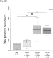
- PBS or PVS-NP was administered intravenously via the tail vein once a week from P21 until the end of each experiment. Analyses for the photopic ERG and the number of PNA-positive cone cells were performed at P49.
- mice To determine the optimal administration schedule of PVS-NP, we divided rd10 mice into three groups at P21 to perform a schedule-finding study: the PBS (100 ⁇ l PBS) every two weeks group, the PVS-NP (0.75 mg PVS/kg) every four weeks group, and the PVS-NP (0.5 mg PVS/kg) every two weeks group.
- PBS or PVS-NP was administered intravenously via the tail vein for each administration period from P21 until the end of each experiment. Analyses for the photopic ERG and the number of PNA-positive cone cells were performed at P49.
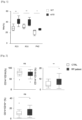
- the correlation coefficients between the MD slope in the RP patients' HFA10-2 tests and the percentage of monocyte subsets in the patients were analyzed by Spearman's rank correlation test. Comparisons of the data between pairs of groups were performed using the Wilcoxon rank sum test. Ap-value ⁇ 0.05 was accepted as significant.
- the statistical analyses of the data were performed with JMP1 Pro 13.0.0 software (SAS, Cary, NC).
- RP inflammatory monocytes
- TUNEL staining at P21 when the rod cell death peaked, showed no significant difference in the number of TUNEL-positive cells in the outer nuclear layer (ONL) between rd10;Ccl2 -/- and rd10;Ccl2 +/+ mice ( Fig. 7 ).
- the HE staining at P26 demonstrated that there was no significant difference in ONL thickness in the presence or absence of Ccl2 ( Fig. 8 ), suggesting that Ccl2 deficiency may not influence rod degeneration in rd10 mice.
- cone cell density as assessed with peanut agglutinin (PNA) labeling, was significantly higher in the P52 rd10;Ccl2 -/- mice compared with the rd10;Ccl2 +/+ mice ( Fig. 9 ).
- PNA peanut agglutinin
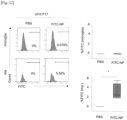
- FITC-NP efficiently delivered FITC to a large number of IMo (p ⁇ 0.05; Fig. 11 ).
- the drug delivery efficiency to retinal microglia and macrophages was analyzed 24 hr after the intravenous injection of the nanoparticles.
- introduction of FITC was observed in 3.1 ⁇ 1.7 % of macrophages.
- no FITC was detected in microglia after the administration of FITC-NP ( Fig. 12 ).
- PVS-NP pitavastatin-loaded nanoparticles
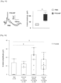
- mice To determine the optimal administration schedule of PVS-NP, we divided rd10 mice into three groups at P21 to perform a schedule-finding study: the PBS (100 ⁇ l PBS) every two weeks group, the PVS-NP (0.75 mg PVS/kg) every four weeks group, and the PVS-NP (0.5 mg PVS/kg) every two weeks group.
- PBS or PVS-NP was administered intravenously via the tail vein for each administration period from P21 until the end of each experiment.
- Analyses for the photopic ERG and the number of PNA-positive cone cells were performed at P49.
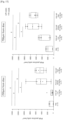
- the photopic ERG b-wave was significantly higher in the PVS-NP (0.75 mg PVS/kg) every four weeks group compared to the PBS group (p ⁇ 0.01; Fig.
- statin-loaded nanoparticles are a promising therapeutic option for retinitis pigmentosa.
Landscapes
- Health & Medical Sciences (AREA)
- Chemical & Material Sciences (AREA)
- Medicinal Chemistry (AREA)
- Pharmacology & Pharmacy (AREA)
- Life Sciences & Earth Sciences (AREA)
- Animal Behavior & Ethology (AREA)
- General Health & Medical Sciences (AREA)
- Public Health (AREA)
- Veterinary Medicine (AREA)
- Engineering & Computer Science (AREA)
- Epidemiology (AREA)
- Bioinformatics & Cheminformatics (AREA)
- Dermatology (AREA)
- Ophthalmology & Optometry (AREA)
- Chemical Kinetics & Catalysis (AREA)
- General Chemical & Material Sciences (AREA)
- Nuclear Medicine, Radiotherapy & Molecular Imaging (AREA)
- Organic Chemistry (AREA)
- Physics & Mathematics (AREA)
- Biomedical Technology (AREA)
- Nanotechnology (AREA)
- Optics & Photonics (AREA)
- Pharmaceuticals Containing Other Organic And Inorganic Compounds (AREA)
Applications Claiming Priority (2)
| Application Number | Priority Date | Filing Date | Title |
|---|---|---|---|
| JP2022012146 | 2022-01-28 | ||
| PCT/JP2023/002470 WO2023145831A1 (ja) | 2022-01-28 | 2023-01-26 | 網膜色素変性症を治療するための医薬組成物 |
Publications (2)
| Publication Number | Publication Date |
|---|---|
| EP4470531A1 true EP4470531A1 (de) | 2024-12-04 |
| EP4470531A4 EP4470531A4 (de) | 2026-01-14 |
Family
ID=87471563
Family Applications (1)
| Application Number | Title | Priority Date | Filing Date |
|---|---|---|---|
| EP23747041.4A Pending EP4470531A4 (de) | 2022-01-28 | 2023-01-26 | Pharmazeutische zusammensetzung zur behandlung von retinitis pigmentosa |
Country Status (5)
| Country | Link |
|---|---|
| US (1) | US20250144041A1 (de) |
| EP (1) | EP4470531A4 (de) |
| JP (1) | JPWO2023145831A1 (de) |
| CN (1) | CN119031907A (de) |
| WO (1) | WO2023145831A1 (de) |
Family Cites Families (7)
| Publication number | Priority date | Publication date | Assignee | Title |
|---|---|---|---|---|
| JP2004149480A (ja) * | 2002-10-31 | 2004-05-27 | Saisentan Igaku Kenkyusho:Kk | 経口又は経皮投与用の眼科疾患治療剤 |
| US20040248957A1 (en) * | 2003-05-16 | 2004-12-09 | Ambit Biosciences Corporation | Heterocyclic compounds and uses thereof |
| EP2057987B1 (de) | 2006-08-30 | 2015-03-04 | Kyushu University, National University Corporation | Pharmazeutische zusammensetzung mit statin-verkapseltem nanopartikel |
| WO2008044339A1 (en) * | 2006-10-13 | 2008-04-17 | Aqumen Biopharmaceuticals K.K. | Therapeutic/preventive agent for intraocular disease containing statin compound |
| EP2226083A4 (de) * | 2007-12-25 | 2013-01-23 | Univ Yamaguchi | Wirkstofffreisetzungssystem |
| WO2021180867A1 (en) * | 2020-03-11 | 2021-09-16 | Universität Regensburg | A nanoparticle for use in the treatment of an ocular disease |
| JP6978016B1 (ja) | 2020-07-01 | 2021-12-08 | 株式会社システムフォワード | 来場者管理システム及び来場者管理方法 |
-
2023
- 2023-01-26 EP EP23747041.4A patent/EP4470531A4/de active Pending
- 2023-01-26 JP JP2023576985A patent/JPWO2023145831A1/ja active Pending
- 2023-01-26 CN CN202380030920.5A patent/CN119031907A/zh active Pending
- 2023-01-26 WO PCT/JP2023/002470 patent/WO2023145831A1/ja not_active Ceased
- 2023-01-26 US US18/832,713 patent/US20250144041A1/en active Pending
Also Published As
| Publication number | Publication date |
|---|---|
| CN119031907A (zh) | 2024-11-26 |
| US20250144041A1 (en) | 2025-05-08 |
| JPWO2023145831A1 (de) | 2023-08-03 |
| WO2023145831A1 (ja) | 2023-08-03 |
| EP4470531A4 (de) | 2026-01-14 |
Similar Documents
| Publication | Publication Date | Title |
|---|---|---|
| Parker et al. | Three‐Year Safety Results of SAR422459 (EIAV‐ABCA4) Gene Therapy in Patients With ABCA4‐Associated Stargardt Disease: An Open‐Label Dose‐Escalation Phase I/IIa Clinical Trial, Cohorts 1‐5 | |
| Yurkewicz et al. | The effect of the selective NMDA receptor antagonist traxoprodil in the treatment of traumatic brain injury | |
| KR20190067156A (ko) | 리포솜 작제물을 사용한 황반 및 망막의 세포에의 우레아의 전달 | |
| KR102713615B1 (ko) | 인간에서의 신경퇴화 및 녹내장에 대한 진단적 마커로서의 망막 및 시신경 내 지방 알갱이 | |
| WO2024215999A1 (en) | Polysialic acid-polymer conjugate and nanoparticle | |
| Li et al. | Orchestrating the frontline: HDAC3-miKO recruits macrophage reinforcements for accelerated myelin debris clearance after stroke | |
| EP4470531A1 (de) | Pharmazeutische zusammensetzung zur behandlung von retinitis pigmentosa | |
| Szamier et al. | Retinal histopathology of a carrier of X-chromosome-linked retinitis pigmentosa | |
| EP3478364B1 (de) | Diagnosemittel oder vorläufer von schubförmig wiederkehrender multipler sklerose | |
| Taylor et al. | Roles of adhesion molecules ICAM-1 and α4 integrin in antigen-induced changes in microvascular permeability associated with lung inflammation in sensitized brown Norway rats | |
| US20230036161A1 (en) | Agent for use in the treatment and prophylaxis of postischemic tissue injury | |
| Rorke-Adams et al. | Head trauma | |
| Yancey et al. | Cysticercosis: recent advances in diagnosis and management of neurocysticercosis | |
| JP7591831B2 (ja) | 好中球損傷用ポリマー粒子 | |
| US20210137969A1 (en) | Compositions and methods for the treatment and prevention of cerebral atrophy | |
| Haller | Modulation of pharmacokinetics of nanodimensional drug carriers | |
| KR102369155B1 (ko) | 근적외선 분광분석법을 이용한 지연성 뇌허혈 진단 방법 | |
| Azad et al. | Retinal Changes in Severe Malaria in Bangladeshi Children | |
| Ahmed et al. | Evaluation of Macular and Peripapillary Capillary Density in Patients with Iron Deficiency Anemia | |
| SINGH | SURGICO-THERAPEUTIC MANAGEMENT OF OCULAR AFFECTIONS IN DOG THESIS | |
| JP7235658B2 (ja) | 疼痛治療のためのang(1-7)誘導体オリゴペプチド | |
| CN119700982A (zh) | Mcp1抑制剂在制备治疗高度近视相关炎症药物中的应用 | |
| CN118286229A (zh) | (+)-jq1在制备治疗增生性视网膜病变的药物中的应用 | |
| WO2022056437A1 (en) | Compositions and methods of use for infusible extracellular matrix | |
| JP2020160058A (ja) | 静脈血栓モデル非ヒト哺乳動物 |
Legal Events
| Date | Code | Title | Description |
|---|---|---|---|
| STAA | Information on the status of an ep patent application or granted ep patent |
Free format text: STATUS: THE INTERNATIONAL PUBLICATION HAS BEEN MADE |
|
| PUAI | Public reference made under article 153(3) epc to a published international application that has entered the european phase |
Free format text: ORIGINAL CODE: 0009012 |
|
| STAA | Information on the status of an ep patent application or granted ep patent |
Free format text: STATUS: REQUEST FOR EXAMINATION WAS MADE |
|
| 17P | Request for examination filed |
Effective date: 20240823 |
|
| AK | Designated contracting states |
Kind code of ref document: A1 Designated state(s): AL AT BE BG CH CY CZ DE DK EE ES FI FR GB GR HR HU IE IS IT LI LT LU LV MC ME MK MT NL NO PL PT RO RS SE SI SK SM TR |
|
| DAV | Request for validation of the european patent (deleted) | ||
| DAX | Request for extension of the european patent (deleted) | ||
| A4 | Supplementary search report drawn up and despatched |
Effective date: 20251216 |
|
| RIC1 | Information provided on ipc code assigned before grant |
Ipc: A61K 9/51 20060101AFI20251210BHEP Ipc: A61K 9/00 20060101ALI20251210BHEP Ipc: A61K 9/14 20060101ALI20251210BHEP Ipc: A61K 31/47 20060101ALI20251210BHEP Ipc: A61P 27/02 20060101ALI20251210BHEP |