EP4446338A2 - Zellen und verfahren zur zellkultur - Google Patents
Zellen und verfahren zur zellkultur Download PDFInfo
- Publication number
- EP4446338A2 EP4446338A2 EP24190440.8A EP24190440A EP4446338A2 EP 4446338 A2 EP4446338 A2 EP 4446338A2 EP 24190440 A EP24190440 A EP 24190440A EP 4446338 A2 EP4446338 A2 EP 4446338A2
- Authority
- EP
- European Patent Office
- Prior art keywords
- cell
- cells
- gene
- concentration
- genes
- Prior art date
- Legal status (The legal status is an assumption and is not a legal conclusion. Google has not performed a legal analysis and makes no representation as to the accuracy of the status listed.)
- Pending
Links
Images


Classifications
-
- C—CHEMISTRY; METALLURGY
- C12—BIOCHEMISTRY; BEER; SPIRITS; WINE; VINEGAR; MICROBIOLOGY; ENZYMOLOGY; MUTATION OR GENETIC ENGINEERING
- C12P—FERMENTATION OR ENZYME-USING PROCESSES TO SYNTHESISE A DESIRED CHEMICAL COMPOUND OR COMPOSITION OR TO SEPARATE OPTICAL ISOMERS FROM A RACEMIC MIXTURE
- C12P7/00—Preparation of oxygen-containing organic compounds
- C12P7/62—Carboxylic acid esters
-
- C—CHEMISTRY; METALLURGY
- C07—ORGANIC CHEMISTRY
- C07K—PEPTIDES
- C07K16/00—Immunoglobulins [IGs], e.g. monoclonal or polyclonal antibodies
-
- C—CHEMISTRY; METALLURGY
- C12—BIOCHEMISTRY; BEER; SPIRITS; WINE; VINEGAR; MICROBIOLOGY; ENZYMOLOGY; MUTATION OR GENETIC ENGINEERING
- C12P—FERMENTATION OR ENZYME-USING PROCESSES TO SYNTHESISE A DESIRED CHEMICAL COMPOUND OR COMPOSITION OR TO SEPARATE OPTICAL ISOMERS FROM A RACEMIC MIXTURE
- C12P17/00—Preparation of heterocyclic carbon compounds with only O, N, S, Se or Te as ring hetero atoms
- C12P17/18—Preparation of heterocyclic carbon compounds with only O, N, S, Se or Te as ring hetero atoms containing at least two hetero rings condensed among themselves or condensed with a common carbocyclic ring system, e.g. rifamycin
- C12P17/182—Heterocyclic compounds containing nitrogen atoms as the only ring heteroatoms in the condensed system
-
- C—CHEMISTRY; METALLURGY
- C12—BIOCHEMISTRY; BEER; SPIRITS; WINE; VINEGAR; MICROBIOLOGY; ENZYMOLOGY; MUTATION OR GENETIC ENGINEERING
- C12P—FERMENTATION OR ENZYME-USING PROCESSES TO SYNTHESISE A DESIRED CHEMICAL COMPOUND OR COMPOSITION OR TO SEPARATE OPTICAL ISOMERS FROM A RACEMIC MIXTURE
- C12P7/00—Preparation of oxygen-containing organic compounds
- C12P7/40—Preparation of oxygen-containing organic compounds containing a carboxyl group including Peroxycarboxylic acids
- C12P7/42—Hydroxy-carboxylic acids
-
- C—CHEMISTRY; METALLURGY
- C12—BIOCHEMISTRY; BEER; SPIRITS; WINE; VINEGAR; MICROBIOLOGY; ENZYMOLOGY; MUTATION OR GENETIC ENGINEERING
- C12P—FERMENTATION OR ENZYME-USING PROCESSES TO SYNTHESISE A DESIRED CHEMICAL COMPOUND OR COMPOSITION OR TO SEPARATE OPTICAL ISOMERS FROM A RACEMIC MIXTURE
- C12P7/00—Preparation of oxygen-containing organic compounds
- C12P7/40—Preparation of oxygen-containing organic compounds containing a carboxyl group including Peroxycarboxylic acids
- C12P7/52—Propionic acid; Butyric acids
-
- C—CHEMISTRY; METALLURGY
- C12—BIOCHEMISTRY; BEER; SPIRITS; WINE; VINEGAR; MICROBIOLOGY; ENZYMOLOGY; MUTATION OR GENETIC ENGINEERING
- C12P—FERMENTATION OR ENZYME-USING PROCESSES TO SYNTHESISE A DESIRED CHEMICAL COMPOUND OR COMPOSITION OR TO SEPARATE OPTICAL ISOMERS FROM A RACEMIC MIXTURE
- C12P7/00—Preparation of oxygen-containing organic compounds
- C12P7/40—Preparation of oxygen-containing organic compounds containing a carboxyl group including Peroxycarboxylic acids
- C12P7/56—Lactic acid
-
- C—CHEMISTRY; METALLURGY
- C12—BIOCHEMISTRY; BEER; SPIRITS; WINE; VINEGAR; MICROBIOLOGY; ENZYMOLOGY; MUTATION OR GENETIC ENGINEERING
- C12N—MICROORGANISMS OR ENZYMES; COMPOSITIONS THEREOF; PROPAGATING, PRESERVING, OR MAINTAINING MICROORGANISMS; MUTATION OR GENETIC ENGINEERING; CULTURE MEDIA
- C12N2500/00—Specific components of cell culture medium
- C12N2500/30—Organic components
- C12N2500/32—Amino acids
-
- C—CHEMISTRY; METALLURGY
- C12—BIOCHEMISTRY; BEER; SPIRITS; WINE; VINEGAR; MICROBIOLOGY; ENZYMOLOGY; MUTATION OR GENETIC ENGINEERING
- C12N—MICROORGANISMS OR ENZYMES; COMPOSITIONS THEREOF; PROPAGATING, PRESERVING, OR MAINTAINING MICROORGANISMS; MUTATION OR GENETIC ENGINEERING; CULTURE MEDIA
- C12N2500/00—Specific components of cell culture medium
- C12N2500/30—Organic components
- C12N2500/34—Sugars
-
- C—CHEMISTRY; METALLURGY
- C12—BIOCHEMISTRY; BEER; SPIRITS; WINE; VINEGAR; MICROBIOLOGY; ENZYMOLOGY; MUTATION OR GENETIC ENGINEERING
- C12N—MICROORGANISMS OR ENZYMES; COMPOSITIONS THEREOF; PROPAGATING, PRESERVING, OR MAINTAINING MICROORGANISMS; MUTATION OR GENETIC ENGINEERING; CULTURE MEDIA
- C12N2510/00—Genetically modified cells
- C12N2510/02—Cells for production
-
- C—CHEMISTRY; METALLURGY
- C12—BIOCHEMISTRY; BEER; SPIRITS; WINE; VINEGAR; MICROBIOLOGY; ENZYMOLOGY; MUTATION OR GENETIC ENGINEERING
- C12N—MICROORGANISMS OR ENZYMES; COMPOSITIONS THEREOF; PROPAGATING, PRESERVING, OR MAINTAINING MICROORGANISMS; MUTATION OR GENETIC ENGINEERING; CULTURE MEDIA
- C12N2511/00—Cells for large scale production
Definitions
- the invention relates to a method of cell culture where the cells are modified to reduce the level of synthesis of growth and/or productivity inhibitors by the cell.
- the invention also relates to a method of cell culture for improving cell growth and productivity, in particular in fed-batch culture of mammalian cells at high cell density.
- the invention further relates to a method of producing cells with improved cell growth and/or productivity in cell culture and to cells obtained or obtainable by such methods.
- Proteins have become increasingly important as diagnostic and therapeutic agents. In most cases, proteins for commercial applications are produced in cell culture, from cells that have been engineered and/or selected to produce unusually high levels of a particular protein of interest. Optimization of cell culture conditions is important for successful commercial production of proteins. Mammalian cells have inefficient metabolism which causes them to consume large amounts of nutrients and convert a significant amount of them to byproducts. The byproducts are released into the culture and accumulate over the course of the culture. Lactate and ammonia, known to be the conventional inhibitors of cells in culture, are the two major byproducts of cellular metabolism that accumulate to high levels in culture and beyond certain concentrations, they start inhibiting the growth and productivity of cells in culture.
- the invention relates to a method of cell culture comprising (i) providing cells in a cell culture medium to start a cell culture process, wherein the cells are modified to reduce the level of synthesis of growth and/or productivity inhibitors by the cell.
- the invention also relates to method of producing cells with improved cell growth and/or productivity in cell culture comprising the steps of:
- the invention further relates to a cell comprising one or more modified genes which reduces the level of synthesis of growth and/or productivity inhibitors by the cell, in particular wherein the one or more modified genes is selected from Bcat1, Bcat2, Auh, Mccc1, Mccc2, Ivd, PCDB1, QDPR, Hpd, Hgd and Pah, wherein the modification increases or decreases the gene expression, and the use of the foregoing cells for the expression of a recombinant protein or polypeptide.
- the present invention provides methods and media for cell culture.
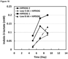
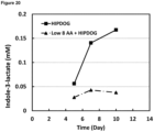
- the present invention provides cell culture methods where the concentration of at least one metabolite selected from 3-(4-hydroxyphenyl)lactate, 4-hydroxyphenylpyruvate, phenyllactate, phenylpyruvate, indolelactate (indole-3-lactate), indolecarboxylic acid (indole-3-carboxylate), homocysteine, 2-hydroxybutyric acid, isovalerate, 2-methyl butyrate, isobutyrate and formate, and/or at least one amino acid selected from phenylalanine, tyrosine, tryptophan, methionine, leucine, serine, threonine and glycine is maintained at low levels in the cell culture medium.
- the inventors have unexpectedly discovered that, in cell culture, and in particular in high density cell culture, such as for example fed-batch cell culture aiming at producing high amount of a recombinant protein of interest, the growth of cells was inhibited by the accumulation of metabolites such as 3-(4-hydroxyphenyl)lactate, 4-hydroxyphenylpyruvate, phenyllactate, phenylpyruvate, indolelactate, indolecarboxylic acid, homocysteine, 2-hydroxybutyric acid, isovalerate, 2-methyl butyrate, isobutyrate and formate in the cell culture medium.
- the inhibitory effect of these metabolites can be limited by maintaining their concentration in the cell culture medium below levels where they inhibit cell growth.
- the inhibitory effects of these metabolites have also be can be limited by modifying one or more genes in the cell to reduce the level of synthesis of growth and/or productivity inhibitors by the cell, in particular where the one or more modified genes is selected from Bcat1, Bcat2, Auh, Mccc1, Mccc2, Ivd, Hpd, Hgd, Pah, PCBD1, and QDPR.
- Methods comprising controlling the metabolite concentration in the cell culture medium at low levels
- the invention provides a method of cell culture comprising (i) providing cells in a cell culture medium to start a cell culture process, wherein the cells are modified to reduce the level of synthesis of growth and/or productivity inhibitors by the cell.
- the cells are modified to modify expression of one or more genes, in one embodiment the genes are in the cell metabolic pathway or pathways which synthesise the growth inhibitors or metabolic intermediates thereof, or metabolites of growth inhibitors or metabolic intermediates thereof, or metabolite of the metabolic intermediate.
- Metal intermediates are understood to comprise molecules which are the precursors or metabolites required for the synthesis of a cell growth and/or productivity inhibitor and include reactant, product and cofactor molecules of enzymes of the cell metabolic pathway or pathways.
- Relevant cofactors include for example BH4 (tetrahydrobiopterin), or BH4-4a (carbinolamine), or q-BH2.
- Metabolic intermediates may be synthesised in the same pathway(s) as the cell metabolic pathway or pathways encoding an enzyme which catalyses the synthesis of the cell growth and/or productivity inhibitor or may be synthesised in a branching pathway.
- Metabolites are understood to comprise the products of metabolic reactions catalysed by the enzymes of the cell metabolic pathway or pathways and include reactant, product and cofactor molecules of said enzymes such as for example BH4 (tetrahydrobiopterin), or BH4-4a (carbinolamine), or q-BH2. Metabolites may arise in the same pathway(s) as the cell metabolic pathway or pathways encoding an enzyme which catalyses the synthesis of the cell growth and/or productivity inhibitor or intermediate thereof or may be synthesised in a branching pathway. The branching pathway may arise at a node situated above or below the one or more one or more genes in the cell metabolic pathway or pathways encoding an enzyme which catalyses the synthesis of the cell growth and/or productivity inhibitor or intermediate thereof.
- the one or more genes modified encodes an enzyme that catalyses the synthesis of 3-(4-hydroxyphenyl)lactate (HPLA), 4-hydroxyphenylpyruvate, phenyllactate (PLA), phenyl pyruvate, indole-3-carboxylate (indolecarboxylic acid), indole-3-lactate (indolelactate), 2-hydroxybutyric acid, homocysteine, isovalerate, 2-methyl butyrate, isobutyrate, butyrate, formate, or a metabolic intermediate thereof or metabolites of said molecules or metabolic intermediates thereof, or metabolite of the metabolic intermediate.
- HPLA 3-(4-hydroxyphenyl)lactate
- PDA phenyllactate
- PDA phenyl pyruvate
- indole-3-carboxylate indolecarboxylic acid
- indole-3-lactate indolelactate
- 2-hydroxybutyric acid homocystein
- the one or more genes modified encodes an enzyme that catalyses the synthesis of 4-hydroxyphenylpyruvate or phenyllactate (PLA), metabolite thereof or a metabolic intermediate thereof, or metabolite of the metabolic intermediate.
- PHA 4-hydroxyphenylpyruvate or phenyllactate
- the one or more genes modified encodes an enzyme that catalyses the synthesis of isovalerate, 2-methylbutyrate, isobutyrate or butyrate, metabolite thereof or a metabolic intermediate thereof, or metabolite of the metabolic intermediate.
- the one or more genes modified is selected from; PCDB1, QDPR, Pah, Mif, Got1, Got2, Nup62-il4i1, Hpd, Hgd, Gstz1, Fah.
- the one or more genes modified is selected from; PCDB1, QDPR Hpd, Hgd and Pah; in one embodiment wherein the one or more genes modified is selected from; ; (i) PCDB1, (ii) Pah, (iii) QDPR, (iv) PCBD1 and QDPR, (v) PCBD1 and Pah, (vi) Pah and QDPR, (vii) PCDB1 and Pah, and QDPR, (viii) any of (i) to (vii) and Hpd and/or Hgd.
- the one or more genes modified is selected from; Bcat1, Bcat2, Bckdha/b, Dbt/Dld, Ivd, Acadm, Mccc1, Mccc2, Auh, Hmgcl, Fasn.
- the one or more genes modified is selected from, Bcat1, Bcat2, Auh, Mccc1, Mccc2, Ivd.
- the one or more genes modified is;
- the one or more genes are modified to increase or decrease gene expression.
- the one or more genes are modified to increase gene expression, in one embodiment wherein the one or more genes are Pah and/or PCDB1 and/or QDPR and/or Hpd and/or Hgd.
- the one or more genes are modified to decrease gene expression, in one embodiment wherein the one or more genes are Bcat1 and/or Bcat2.
- the one or more genes modified is selected from; (i) PCDB1, (ii) Pah, (iii) QDPR, (iv) PCBD1 and QDPR, (v) PCBD1 and Pah, (vi) Pah and QDPR, (vii) PCDB1 and Pah, and QDPR, (viii) any of (i) to (vii) and Hpd and/or Hgd.
- the cell comprises;
- the method further comprises; (ii) maintaining at least one metabolite selected from 3-(4-hydroxyphenyl)lactate, 4-hydroxyphenylpyruvate, phenyllactate, indolelactate (indole-3-lactate), indolecarboxylic acid (indole-3-carboxylate), homocysteine, 2-hydroxybutyric acid, isovalerate, 2-methylbutyrate, isobutyrate and formate below a concentration C1 in the cell culture medium, wherein C1 is 3mM.
- at least one metabolite selected from 3-(4-hydroxyphenyl)lactate, 4-hydroxyphenylpyruvate, phenyllactate, indolelactate (indole-3-lactate), indolecarboxylic acid (indole-3-carboxylate), homocysteine, 2-hydroxybutyric acid, isovalerate, 2-methylbutyrate, isobutyrate and formate below a concentration C1 in
- C1 is 2.5mM, 2mM, 1.5mM, 1mM, 0.9mM, 0.8mM, 0.7mM, 0.6mM, 0.5mM, 0.4mM, 0.3mM, 0.2mM or 0.1mM. In some embodiments, C1 is 1mM. In some embodiments, C1 is 0.5mM.
- step (ii) comprises the step of measuring the concentration of said at least one metabolite.
- the concentration of precursor of said at least one metabolite in the cell culture medium is decreased by reducing the amount of precursor provided to the cells.
- Said predefined value is selected so that the decrease of concentration of said precursor prevents the concentration of metabolite from rising above C1.
- the predefined value can be equal to C1 or can be a percentage of C1. In some embodiments the percentage is 50, 55, 60, 65, 70, 75, 80, 85, 90 or 95% of C1. In some embodiments the percentage is 80% of C1.
- the concentration of phenylalanine is decreased in the cell culture medium.
- the concentration of tyrosine is decreased in the cell culture medium.
- the concentrations of tyrosine and phenylalanine are decreased in the cell culture medium.
- the concentration of tryptophan is decreased in the cell culture medium.
- the concentration of methionine is decreased in the cell culture medium.
- the concentration of leucine is decreased in the cell culture medium.
- the concentration of isoleucine is decreased in the cell culture medium.
- the concentration of valine is decreased in the cell culture medium.
- the concentration of serine, threonine and/or glycine is decreased in the cell culture medium.
- the concentration of serine is decreased in the cell culture medium.
- the concentration of threonine is decreased in the cell culture medium.
- the concentration of glycine is decreased in the cell culture medium.
- the concentration of precursor in the cell culture medium can be decreased by reducing the amount of precursor provided to the cells, for example by reducing the concentration of said precursor in the feed medium, reducing the feed rate, or reducing the number or volume of feeds.
- the feed medium can be replaced by a feed medium comprising a lower concentration of precursor.
- step (ii) comprises maintaining 1, 2, 3, 4, 5, 6, 7, 8, 9, 10 or 11 of 3-(4-hydroxyphenyl)lactate, 4-hydroxyphenylpyruvate, phenyllactate, indolelactate (indole-3-lactate), indolecarboxylic acid (indole-3-carboxylate), homocysteine, 2-hydroxybutyric acid, isovalerate, 2-methylbutyrate, isobutyrate and formate below C1 in the cell culture medium.
- step (ii) comprises maintaining 1, 2, 3, 4, 5, 6, or 7 of 3-(4-hydroxyphenyl)lactate, 4-hydroxyphenylpyruvate, phenyllactate, indolelactate, indolecarboxylic acid, homocysteine and 2-hydroxybutyric acid below C1 in the cell culture medium.
- step (ii) comprises maintaining 3-(4-hydroxyphenyl)lactate, 4-hydroxyphenylpyruvate and phenyllactate below C1 in the cell culture medium.
- step (ii) comprises maintaining indolelactate (indole-3-lactate) and indolecarboxylic acid (indole-3-carboxylate) below C1 in the cell culture medium.
- step (ii) comprises maintaining homocysteine and 2-hydroxybutyric acid below C1 in the cell culture medium.
- step (ii) comprises maintaining isovalerate, 2-methylbutyrate, and isobutyrate below C1 in the cell culture medium.
- step (ii) comprises maintaining isobutyrate below C1 in the cell culture medium.
- step (ii) comprises maintaining 2-methylbutyrate below C1 in the cell culture medium.
- step (ii) comprises maintaining isovalerate below C1 in the cell culture medium.
- step (ii) comprises maintaining formate below C1 in the cell culture medium.
- step (ii) comprises maintaining isovalerate and 4-hydroxyphenylpyruvate below C1 in the cell culture medium.
- step (ii) comprises maintaining 3-(4-hydroxyphenyl)lactate, 4-hydroxyphenylpyruvate, phenyllactate, indolelactate (indole-3-lactate), indolecarboxylic acid (indole-3-carboxylate), homocysteine, 2-hydroxybutyric acid isovalerate, 2-methylbutyrate, isobutyrate and formate below C1 in the cell culture medium.
- step (ii) the concentration of 3-(4-hydroxyphenyl)lactate is maintained below 0.5mM, 0.4mM, 0.3mM, 0.2mM, or 0.1mM.
- step (ii) the concentration of 3-(4- hydroxyphenyl)lactate is maintained below 0.3mM.
- step (ii) the concentration of 3-(4-hydroxyphenyl)lactate is maintained below 0.1mM.
- step (ii) the concentration of 4-hydroxyphenylpyruvate is maintained below 0.1mM, 0.08mM, 0.06mM, 0.05mM, 0.04mM, 0.03mM or 0.02mM.
- step (ii) the concentration of 4-hydroxyphenylpyruvate is maintained below 0.05mM.
- step (ii) the concentration of 4-hydroxyphenylpyruvate is maintained below 0.02mM.
- step (ii) the concentration of phenyllactate is maintained below 0.5mM, 0.4mM, 0.3mM, 0.2mM, or 0.1mM.
- step (ii) the concentration of phenyllactate is maintained below 0.2mM.
- step (ii) the concentration of phenyllactate is maintained below 0.1mM.
- step (ii) the concentration of indolelactate (indole-3-lactate), is maintained below 3mM, 2mM, 1mM, 0.5mM, 0.3mM or 0.1mM.
- step (ii) the concentration of indolelactate is maintained below 1mM.
- step (ii) the concentration of indolelactate is maintained below 0.3mM.
- step (ii) the concentration of indolelactate is maintained below 0.1mM.
- step (ii) the concentration of indolecarboxylic acid (indole-3-carboxylate), is maintained below 1mM, 0.8mM, 0.6mM, 0.4mM or 0.2mM.
- step (ii) the concentration of indolecarboxylic acid is maintained below 0.5mM.
- step (ii) the concentration of indolecarboxylic acid is maintained below 0.2mM.
- step (ii) the concentration of homocysteine is maintained below 0.5mM, 0.4mM, 0.3mM, 0.2mM, or 0.1mM.
- step (ii) the concentration of homocysteine is maintained below 0.3mM.
- step (ii) the concentration of homocysteine is maintained below 0.1mM.
- step (ii) the concentration of 2-hydroxybutyric acid is maintained below 1mM, 0.8mM, 0.6mM, 0.4mM or 0.2mM.
- step (ii) the concentration of 2-hydroxybutyric acid is maintained below 0.5mM.
- step (ii) the concentration of 2-hydroxybutyric acid is maintained below 0.2mM.
- step (ii) the concentration of isovalerate and/or, 2-methylbutyrate, and/or isobutyrate is maintained below 2mM, 1mM, 0.8mM, 0.6mM, 0.4mM or 0.2mM.
- step (ii) the concentration of isovalerate and/or, 2-methylbutyrate, and/or isobutyrate is maintained below 1mM.
- step (ii) the concentration of isovalerate and/or, 2-methylbutyrate, and/or isobutyrate is maintained below 0.5mM.
- step (ii) the concentration of formate is maintained below 4mM, 3mM, 2mM, 1mM, 0.5mM or 0.2mM.
- step (ii) the concentration of formate is maintained below 3mM.
- step (ii) the concentration of formate is maintained below 1mM.
- Methods comprising controlling the amino acid concentration in the cell culture medium at low levels
- the method of cell culture further comprises; (iii) maintaining at least one amino acid selected from phenylalanine, tyrosine, tryptophan, methionine, leucine, isoleucine, valine, serine, threonine and glycine below a concentration C2 in the cell culture medium, wherein C2 is 2mM.
- said concentration is maintained between 0.1mM and C2, 0.2mM and C2, 0.3mM and C2, 0.4mM and C2, or 0.5mM and C2. In some embodiments, said concentration is maintained between 0.5mM and C2.
- C2 is 2mM, 1.5mM, 1mM, 0.9mM, 0.8mM, 0.7mM, 0.6mM, 0.5mM, 0.4mM, 0.3mM, 0.2mM, 0.1mM, 0.05mM. In some embodiments, C2 is 1mM.
- Methods comprising measuring the amino acid concentration in the cell culture medium
- step (iii) comprises the step of measuring the concentration of said at least one amino acid.
- concentration of amino acid can be measured by any method known to the skilled person, including off line and on line measurement methods.
- the concentration of amino acids can be measured once or several times during the cell culture. In some embodiments, the amino acid concentration is measured continuously, intermittently, every 30min, every hour, every two hours, twice a day, daily, or every two days. In a preferred embodiment, the concentration of amino acid is measured daily.
- the concentration of amino acid can be measured by any method known to the skilled person.
- Preferred methods to measure the concentration of amino acids in online or offline methods include for example Liquid Chromatography such HPLC, UPLC or LCMS, NMR or GCMS.
- the concentration of amino acid is measured off line by taking a sample of the cell culture medium and measuring the concentration of said at least one amino acid in said sample. In some embodiments, the concentration of amino acid is measured as disclosed in Example 4.
- a preferred method to measure the concentration of amino acids in an off line method is UPLC.
- the concentration of amino acid is measured online. In some embodiments, the concentration of amino acid is measured on-line using Raman spectroscopy. In some embodiments, the concentration of amino acid is measured on-line using Raman spectroscopy as disclosed in Example 7. In some embodiments, the concentration of amino acid is measured online using HPLC or UPLC based technology with an auto-sampler that draws sample from reactor and transfers to the equipment in a programmed manner.
- the concentration of said at least one amino acid in the cell culture medium is decreased.
- the predefined value can be equal C2 or can be a percentage of C2. In some embodiments the percentage is 50, 55, 60, 65, 70, 75, 80, 85, 90 or 95% of C2. In some embodiments the percentage is 80% of C2.
- the concentration of amino acid in the cell culture medium can be decreased by reducing the amount of amino acid provided to the cells, for example by reducing the concentration of said amino acid in the feed medium, reducing the feed rate, or reducing the number or volume of feeds.
- the feed medium can be replaced by a feed medium comprising a lower concentration of amino acid.
- the concentration of phenylalanine is maintained below 2mM in the cell culture medium. In a preferred embodiment, the concentration of phenylalanine is maintained between 0.1 and 2mM in the cell culture medium. In a preferred embodiment, the concentration of phenylalanine is maintained between 0.1 and 1mM in the cell culture medium. In a preferred embodiment, the concentration of phenylalanine is maintained between 0.2 and 1mM in the cell culture medium. In a preferred embodiment, the concentration of phenylalanine is maintained between 0.5 and 1mM in the cell culture medium.
- the concentration of tyrosine is maintained below 2mM in the cell culture medium. In a preferred embodiment, the concentration of tyrosine is maintained between 0.1 and 2mM in the cell culture medium. In a preferred embodiment, the concentration of tyrosine is maintained between 0.1 and 1mM in the cell culture medium. In a preferred embodiment, the concentration of tyrosine is maintained between 0.2 and 1mM in the cell culture medium. In a preferred embodiment, the concentration of tyrosine is maintained between 0.5 and 1mM in the cell culture medium.
- the concentration of tryptophan is maintained below 2mM in the cell culture medium. In a preferred embodiment, the concentration of tryptophan is maintained between 0.1 and 2mM in the cell culture medium. In a preferred embodiment, the concentration of tryptophan is maintained between 0.1 and 1mM in the cell culture medium. In a preferred embodiment, the concentration of tryptophan is maintained between 0.2 and 1mM in the cell culture medium. In a preferred embodiment, the concentration of tryptophan is maintained between 0.5 and 1mM in the cell culture medium.
- the concentration of methionine is maintained below 2mM in the cell culture medium. In a preferred embodiment, the concentration of methionine is maintained between 0.1 and 2mM in the cell culture medium. In a preferred embodiment, the concentration of methionine is maintained between 0.1 and 1mM in the cell culture medium. In a preferred embodiment, the concentration of methionine is maintained between 0.2 and 1mM in the cell culture medium. In a preferred embodiment, the concentration of methionine is maintained between 0.5 and 1mM in the cell culture medium.
- the concentration of leucine is maintained below 2mM in the cell culture medium. In a preferred embodiment, the concentration of leucine is maintained between 0.1 and 2mM in the cell culture medium. In a preferred embodiment, the concentration of leucine is maintained between 0.1 and 1mM in the cell culture medium. In a preferred embodiment, the concentration of leucine is maintained between 0.2 and 1mM in the cell culture medium. In a preferred embodiment, the concentration of leucine is maintained between 0.5 and 1mM in the cell culture medium.
- the concentration of isoleucine is maintained below 2mM in the cell culture medium. In a preferred embodiment, the concentration of isoleucine is maintained between 0.1 and 2mM in the cell culture medium. In a preferred embodiment, the concentration of isoleucine is maintained between 0.1 and 1mM in the cell culture medium. In a preferred embodiment, the concentration of isoleucine is maintained between 0.2 and 1mM in the cell culture medium. In a preferred embodiment, the concentration of isoleucine is maintained between 0.5 and 1mM in the cell culture medium.
- the concentration of valine is maintained below 2mM in the cell culture medium. In a preferred embodiment, the concentration of valine is maintained between 0.1 and 2mM in the cell culture medium. In a preferred embodiment, the concentration of valine is maintained between 0.1 and 1mM in the cell culture medium. In a preferred embodiment, the concentration of valine is maintained between 0.2 and 1mM in the cell culture medium. In a preferred embodiment, the concentration of valine is maintained between 0.5 and 1mM in the cell culture medium.
- the concentration of serine is maintained below 2mM in the cell culture medium. In a preferred embodiment, the concentration of serine is maintained between 0.1 and 2mM in the cell culture medium. In a preferred embodiment, the concentration of serine is maintained between 0.1 and 1mM in the cell culture medium. In a preferred embodiment, the concentration of serine is maintained between 0.2 and 1mM in the cell culture medium. In a preferred embodiment, the concentration of serine is maintained between 0.5 and 1mM in the cell culture medium.
- the concentration of threonine is maintained below 2mM in the cell culture medium. In a preferred embodiment, the concentration of threonine is maintained between 0.1 and 2mM in the cell culture medium. In a preferred embodiment, the concentration of threonine is maintained between 0.1 and 1mM in the cell culture medium. In a preferred embodiment, the concentration of threonine is maintained between 0.2 and 1mM in the cell culture medium. In a preferred embodiment, the concentration of threonine is maintained between 0.5 and 1mM in the cell culture medium.
- the concentration of glycine is maintained below 2mM in the cell culture medium. In a preferred embodiment, the concentration of glycine is maintained between 0.1 and 2mM in the cell culture medium. In a preferred embodiment, the concentration of glycine is maintained between 0.1 and 1mM in the cell culture medium. In a preferred embodiment, the concentration of glycine is maintained between 0.2 and 1mM in the cell culture medium. In a preferred embodiment, the concentration of glycine is maintained between 0.5 and 1mM in the cell culture medium.
- the concentration of tyrosine, phenylalanine and leucine is maintained below 2mM, preferably between 0.1 and 2mM, between 0.1 and 1mM, between 0.2 and 1mM or between 0.5 and 1mM in the cell culture medium.
- the cell culture medium comprises 1, 2, 3, 4, 5, 6, 7, 8, 9, or 10, of glycine, , proline, serine, threonine, lysine, arginine, histidine, aspartate, glutamate and asparagine at a concentration above 1mM, 1.5mM, 2mM, 3mM or 5mM.
- the cell culture medium comprises one of glycine, , proline, serine, threonine, lysine, arginine, histidine, aspartate, glutamate and asparagine at a concentration above 2mM.
- the cell culture medium comprises one of glycine, , proline, serine, threonine, lysine, arginine, histidine, aspartate, glutamate and asparagine at a concentration above 5mM.
- the cell culture medium comprises 1, 2, 3, 4, 5, 6,or 7, of, proline, lysine, arginine, histidine, aspartate, glutamate and asparagine at a concentration above 1mM, 1.5mM, 2mM, 3mM or 5mM.
- the cell culture medium comprises one of, proline, lysine, arginine, histidine, aspartate, glutamate and asparagine at a concentration above 2mM.
- the cell culture medium comprises one of, proline, lysine, arginine, histidine, aspartate, glutamate and asparagine at a concentration above 5mM.
- the cell culture medium comprises no tyrosine. In some embodiments, the cell culture is a HiPDOG culture wherein the medium comprises no tyrosine. In some embodiments, the cell culture medium is HiPDOG medium, medium used in the HipDOG process, but comprising no tyrosine. In some embodiments the cell culture medium comprises no tyrosine wherein a HiPDOG culture or process is used which comprises no tyrosine.
- lactate and ammonia are also maintained at low levels in the cell culture medium.
- Methods to keep lactate and ammonia at low levels are known to the skilled person.
- lactate can be kept at low levels in cell culture by using methods disclosed in WO2004104186 , Gagnon et al, Biotechnology and Bioengineering, Vol. 108, No. 6, June, 2011 (Gagnon et Al) or WO2004048556 .
- Various other strategies can be employed to restrict lactate production and/or induce lactate consumption. These include culturing cells under slightly reduced pH (6.7 -7.0), culturing cells at low glucose concentrations by using alternative carbon sources including but not limited to fructose (Wlaschin & Hu, 2007) and galactose (Altamirano et al, 2006), using a cell line that has reduced protein levels of glycolytic enzymes including but not limited to hexose transporter or lactate dehydrogenase (Kim & Lee, 2007a), employing a cell line with suppressed cellular protein levels of both lactate dehydrogenase and pyruvate dehydrogenase kinase (Zhou et al, 2011), or cell line with over-expression of pyruvate carboxylase enzyme (Kim & Lee, 2007b), or with the use of inhibitors (small molecule or protein based) for signaling pathways (such as AKT (Mulukutla et al,
- lactate is maintained at low levels by using the high-end pH-controlled delivery of glucose (HIPDOG process / HIPDOG culture) disclosed in Gagnon et al.
- lactate is maintained at low levels in the cell culture medium. In some embodiments, concentration of lactate in the cell culture medium is maintained below 90 mM. In a preferred embodiment, the concentration of lactate in the cell culture medium is maintained below 70 mM. In some embodiments, the concentration of lactate in the cell culture medium is maintained below 50 mM. In some embodiments, the concentration of lactate in the cell culture medium is maintained below 40 mM. In some embodiments, lactate is maintained at low levels by controlling the amount of glucose provided to the cell culture. In some embodiments, lactate is maintained at low levels by using the HIPDOG process / culture. In some embodiments, a pH sensor is used to monitor pH of the cell culture, and, in response to a rise above a predetermined pH value, glucose is fed to the cell culture. In some embodiments, the predetermined pH value is approximately 7.
- Ammonia can be kept at low levels in cell culture by any method known to the skilled person such as for example the methods disclosed in Butler et Al, Cytotechnology 15: 87-94, 1994 or Hong et Al, Appl Microbiol Biotechnol (2010) 88:869-876 .
- ammonia can be kept at low levels by using a glutamine synthetase (GS) expression system.
- GS glutamine synthetase
- Such systems are commercially available (Lonza) and can be used to generate recombinant cell lines.
- Cell lines using GS expression system demonstrate the gain of function to synthesize glutamine in vivo, thereby completely relieving cellular dependence on the externally supplied glutamine. Since the major fraction of ammonia produced in culture is from the catabolysis of externally supplied glutamine, such a gain of metabolic function reduces the levels of ammonia produced in culture.
- any of the ammonia is maintained at low levels in the cell culture medium.
- concentration of ammonia in the cell culture medium is maintained below 20 mM.
- concentration of ammonia in the cell culture medium is maintained below 10 mM.
- concentration of ammonia in the cell culture medium is maintained below 8 mM.
- culture and "cell culture” as used herein refer to a cell population that is suspended in a medium under conditions suitable to survival and/or growth of the cell population. As will be clear to those of ordinary skill in the art, in some embodiments, these terms as used herein refer to the combination comprising the cell population and the medium in which the population is suspended. In some embodiments, the cells of the cell culture comprise mammalian cells.
- the present invention may be used with any cell culture method that is amenable to the desired process (e.g., production of a recombinant protein (e.g., antibody)).
- a recombinant protein e.g., antibody
- cells may be grown in batch or fed-batch cultures, where the culture is terminated after sufficient expression of the recombinant protein (e.g., antibody), after which the expressed protein (e.g., antibody) is harvested.
- cells may be grown in batch-refeed, where the culture is not terminated and new nutrients and other components are periodically or continuously added to the culture, during which the expressed recombinant protein (e.g., antibody) is harvested periodically or continuously.
- Other suitable methods e.g., spin-tube cultures are known in the art and can be used to practice the present invention.
- a cell culture suitable for the present invention is a fed-batch culture.
- fed-batch culture refers to a method of culturing cells in which additional components are provided to the culture at a time or times subsequent to the beginning of the culture process. Such provided components typically comprise nutritional components for the cells which have been depleted during the culturing process.
- a fed-batch culture is typically stopped at some point and the cells and/or components in the medium are harvested and optionally purified.
- the fed-batch culture comprises a base medium supplemented with feed media.
- Cells may be grown in any convenient volume chosen by the practitioner. For example, cells may be grown in small scale reaction vessels ranging in volume from a few milliliters to several liters. Alternatively, cells may be grown in large scale commercial Bioreactors ranging in volume from approximately at least 1 liter to 10, 50, 100, 250, 500, 1000, 2500, 5000, 8000, 10,000, 12,000, 15000, 20000 or 25000 liters or more, or any volume in between.
- the temperature of a cell culture will be selected based primarily on the range of temperatures at which the cell culture remains viable and the range in which a high level of desired product (e.g., a recombinant protein) is produced.
- desired product e.g., a recombinant protein
- most mammalian cells grow well and can produce desired products (e.g., recombinant proteins) within a range of about 25°C to 42°C, although methods taught by the present disclosure are not limited to these temperatures.
- desired products e.g., recombinant proteins or antibodies
- a cell culture is grown at a temperature of 20, 21, 22, 23, 24, 25, 26, 27, 28, 29, 30, 31, 32, 33, 34, 35, 36, 37, 38, 39, 40, 41, 42, 43, 44 or 45°C at one or more times during the cell culture process.
- the cells may be grown for any amount of time, depending on the needs of the practitioner and the requirement of the cells themselves.
- the cells are grown at 37°C. In some embodiments, the cells are grown at 36.5°C.
- the cells may be grown during the initial growth phase (or growth phase) for a greater or lesser amount of time, depending on the needs of the practitioner and the requirement of the cells themselves. In some embodiments, the cells are grown for a period of time sufficient to achieve a predefined cell density. In some embodiments, the cells are grown for a period of time sufficient to achieve a cell density that is a given percentage of the maximal cell density that the cells would eventually reach if allowed to grow undisturbed. For example, the cells may be grown for a period of time sufficient to achieve a desired viable cell density of 1, 5, 10, 15, 20, 25, 30, 35, 40, 45, 50, 55, 60, 65, 70, 75, 80, 85, 90, 95 or 99 percent of maximal cell density.
- the cells are grown until the cell density does not increase by more than 15%, 14%, 13%, 12%, 11%, 10%, 9%, 8%, 7%, 6%, 5%, 4%, 3%, 2% or 1% per day of culture. In some embodiments, the cells are grown until the cell density does not increase by more than 5% per day of culture.
- the cells are allowed to grow for a defined period of time. For example, depending on the starting concentration of the cell culture, the temperature at which the cells are grown, and the intrinsic growth rate of the cells, the cells may be grown for 0, 1, 2, 3, 4, 5, 6, 7, 8, 9, 10, 11, 12, 13, 14, 15, 16, 17, 18, 19, 20 or more days, preferably for 4 to 10 days. In some cases, the cells may be allowed to grow for a month or more. The practitioner of the present invention will be able to choose the duration of the initial growth phase depending on protein production requirements and the needs of the cells themselves.
- the cell culture may be agitated or shaken during the initial culture phase in order to increase oxygenation and dispersion of nutrients to the cells.
- agitated or shaken during the initial culture phase in order to increase oxygenation and dispersion of nutrients to the cells.
- certain internal conditions of the bioreactor during the initial growth phase including but not limited to pH, temperature, oxygenation, etc.
- a metabolic shift can be accomplished by, e.g., a change in the temperature, pH, osmolality or chemical inductant level of the cell culture.
- the culture conditions are shifted by shifting the temperature of the culture.
- shifting temperature is not the only mechanism through which an appropriate metabolic shift can be achieved.
- such a metabolic shift can also be achieved by shifting other culture conditions including, but not limited to, pH, osmolality, and sodium butyrate levels.
- the timing of the culture shift will be determined by the practitioner of the present invention, based on protein production requirements or the needs of the cells themselves.
- the temperature shift may be gradual. For example, it may take several hours or days to complete the temperature change. Alternatively, the temperature shift may be abrupt. For example, the temperature change may be complete in less than several hours. Given the appropriate production and control equipment, such as is standard in the commercial large-scale production of polypeptides or proteins, the temperature change may even be complete within less than an hour.
- the cell culture is maintained for a subsequent production phase under a second set of culture conditions conducive to the survival and viability of the cell culture and appropriate for expression of the desired polypeptide or protein at commercially adequate levels.
- the culture may be shifted by shifting one or more of a number of culture conditions including, but not limited to, temperature, pH, osmolality, and sodium butyrate levels.
- the temperature of the culture is shifted.
- the culture is maintained at a temperature or temperature range that is lower than the temperature or temperature range of the initial growth phase.
- multiple discrete temperature shifts may be employed to increase cell density or viability or to increase expression of the recombinant protein.
- the cells may be maintained in the subsequent production phase until a desired cell density or production titer is reached.
- the cells are allowed to grow for a defined period of time during the subsequent production phase. For example, depending on the concentration of the cell culture at the start of the subsequent growth phase, the temperature at which the cells are grown, and the intrinsic growth rate of the cells, the cells may be grown for 1, 2, 3, 4, 5, 6, 7, 8, 9, 10, 11, 12, 13, 14, 15, 16, 17, 18, 19, 20 or more days. In some cases, the cells may be allowed to grow for a month or more. The practitioner of the present invention will be able to choose the duration of the subsequent production phase depending on polypeptide or protein production requirements and the needs of the cells themselves.
- the cell culture may be agitated or shaken during the subsequent production phase in order to increase oxygenation and dispersion of nutrients to the cells.
- agitated or shaken during the subsequent production phase in order to increase oxygenation and dispersion of nutrients to the cells.
- certain internal conditions of the bioreactor during the subsequent growth phase including but not limited to pH, temperature, oxygenation, etc.
- the cells express a recombinant protein and the cell culture method of the invention comprises a growth phase and a production phase.
- step (ii) and/or (iii) is applied during the totality of the cell culture method. In some embodiments step (ii) and/or (iii) is applied during a part of the cell culture method. In some embodiments, step (ii) and/or (iii) is applied until a predetermined viable cell density is obtained.
- the cell culture method of the invention comprises a growth phase and a production phase and step (ii) and/or (iii) is applied during the growth phase. In some embodiments, the cell culture method of the invention comprises a growth phase and a production phase and step (ii) and/or (iii) is applied during a part of the growth phase. In some embodiments, the cell culture method of the invention comprises a growth phase and a production phase and step (ii) and/or (iii) is applied during the growth phase and the production phase.
- step (ii) and/or (iii) can refer to maintaining the concentration of amino acid or metabolite below C1 or C2 for the entire culture process (until harvesting) or for a part of the culture process such as for example the growth phase, a part of the growth phase or until a predetermined cell density is obtained.
- the present invention also provides a method of producing cells with improved cell growth and/or productivity in cell culture comprising the steps of:
- the identification of cell metabolites which are cell growth and/or productivity inhibitors comprises;
- Identification of cell metabolites which are highly expressed metabolites or are metabolites that accumulate to high levels/concentrations can be performed using methods for identifying and/or measuring the metabolite concentration in the cell culture medium or in the cells, for example in the cell pellet for example using one or more of or a combination of NMR, LC/MS and GC/MS techniques.
- spent media samples and the cell pellet samples can be collected and analyzed for example from single or from duplicate reactors runs, performed for each condition taken at time points throughout culture, for example up to and/or including maximum viable cell density, for example at time points of days selected from 0, 1, 2, 3, 4, 5, 7, 8, 9, 11, and 12.
- the relative levels (fold changes) of all measurable metabolites are measured and calculated.
- the relative levels are determined in both the spent media and/or cell pellet samples, and are calculated based on fold changes compared to the level of the metabolite when first detected until the desired time endpoint, usually maximum viable cell density. Metabolites accumulating to high levels are judged from a fold change cut-off of any one of greater than or equal to 10, 15, 20, 25, 30, 35, 40, 45, 50, 60, 70, 80 90 or 100 fold between the first time point the metabolite was measured and the desired time endpoint, for example maximum viable cell density. In some embodiments it is greater than or equal to 20 fold.
- the increase in rate of metabolite production or the increased level/concentration of metabolite production is greater than or equal 2, 3, 4, 5, 6, 7, 8, 9, or 10 fold or more increased during cell culture up to maximum viable cell density. In some embodiments it is greater than or equal to 15, 20, 25, 30, 35, 40, 45 or 50 fold. In some embodiments it is greater than or equal to 40 fold. In some embodiments it is greater than or equal to 50 fold.
- the identification of cell metabolites which are cell growth (productivity) inhibitors further comprises
- Quantifying the highly expressed metabolite production/concentration at maximum viable cell density can be performed using methods for identifying and/or measuring the metabolite concentration in the cell culture medium or in the cells.
- the measurement of and/or the quantification of the metabolites, including highly expressed metabolites is performed using liquid chromatography with mass spectrometry (LC/MS), gas chromatography with mass spectrometry (GC/MS), or nuclear magnetic resonance (NMR).
- the cell metabolites and/or highly expressed metabolites are identified from samples of the culture medium or from the culture cells during cell culture.
- the measurement of and/or the quantification of metabolites or highly expressed metabolites can be carried out by using obtained purified compound forms of the metabolite or highly expressed metabolite. These compounds can be used to prepare calibration curves for the compounds at known concentrations in order to precisely correlate the measurements obtained from the methods used with the concentrations of metabolite. In this way a precise measurement of the concentration of the metabolite is determinable at any given time point in the cell culture, for example at maximum viable cell density.
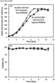
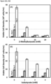
- the effect of quantified concentrations of highly expressed metabolite can be determined on the cell growth and/or productivity of cells in cell culture by introduction of the determined concentration into a culture of cells and comparing the effect on the cell growth and/or productivity with an otherwise identical culture lacking the introduced metabolite.
- cells which can be cells producing a recombinant protein, can be inoculated into a culture medium spiked-in with different selected concentrations of a highly expressed metabolite, either alone or in combination with other different highly expressed metabolites at selected concentrations, these concentrations can be for example the determined concentration at maximum viable cell density or serial dilutions thereof.
- the pH of the culture medium may also be adjusted to 7 before inoculating the cells.
- the comparison can determine the existence of a negative effect of a metabolite on cell growth and/or productivity, and/or the existence of synergistic effect between metabolites on the same measures and thereby such highly expressed metabolites can be determined to be inhibitors of cell growth and/or productivity.
- the cell metabolic pathway or pathways is determined from the nutrient source or component of the culture medium of the highly expressed metabolite or cell metabolite or metabolites which are cell growth and/or productivity inhibitors, these fall broadly into amino acids, vitamins, inorganic salts, trace elements, vitamins, energy sources, lipids, and nucleotides, further disclosure is provided herein.
- the skilled person possessing the knowledge of the identity of a highly expressed metabolite which is an inhibitory metabolite and knowledge of the nutrient sources for the metabolite can apply biochemical knowledge of metabolic pathways common to the cell to deduce the relevant cell metabolic pathway or pathways which contribute to the synthesis of the growth and/or productivity inhibitors and which can include pathways leading to intermediates of the metabolite as well as pathways branching therefrom.
- the identification of one or more genes in the cell metabolic pathway or pathways encoding an enzyme which catalyses the synthesis of the cell growth and/or productivity inhibitors or metabolic intermediates or one or more genes encoding an enzyme in metabolic pathways branching therefrom further comprises;
- BH4 tetrahydrobiopterin
- BH4-4a carbbinolamine
- the identification of the one or more genes comprises gene expression analysis, in some embodiments it comprises gene expression analysis of one or more genes in the identified pathway or pathways, in some embodiments it comprises gene expression analysis of all the genes in the identified pathway or pathways. In some embodiments it comprises gene expression analysis by measuring transcript abundance, in some embodiments it comprises real time quantitative PCR assay or RT-qPCR assay.
- RT qPCR can measure transcript abundance by amplifying a target cDNA sequence using PCR in combination with a detection reagent, for example SYBR Green. Relative gene expression levels can be determined by measuring the number of PCR cycles required for the signal from detection reagent to surpass the background and increase logarithmically. This cycle number is commonly referred to as the C T (Threshold Cycle). Low abundance transcripts have a high C T values in comparison to known control standards of transcript abundance, such as beta-actin, and vice-versa.
- the relative gene expression level is determined by real time quantitative PCR, RT-qPCR, In some embodiments the identification of gene mutation is determined by mRNA sequencing.
- the identified gene is expressed at an increased or decreased level higher or lower than the expression levels of a control gene, in some embodiments the level is greater than or equal to any one of 5, 10, 15, 20, 25, 30, 35, 40, 45, 50, 55, 60, 65 70,75, 80, 85 90 or 95 percent higher or lower than the expression levels of a control gene, for example as judged by Ct value or dCt value.
- the identified gene is expressed 10% higher or lower than the expression levels of a control gene, in some embodiments 15% higher or lower, in some embodiments 20% higher or lower than the expression levels of a control gene.
- control gene has a Ct value of equal to or between 14 and 17 and or has a Ct value of equal to or between 15 and 16 as measured by RT-qPCR, in some embodiments the control gene is beta actin.
- the identified gene has a Ct value of greater than or equal to any one of 23, 24, 25, 26, 27, 28, 29, 30 or greater than 30 as measured by RT-qPCR, in some embodiments the identified gene has a Ct value of less than or equal to any one of 14, 13, 12, 11, 10, 9, 8, 7, 6, 5, 4, 3, 2, 1 or les than 14 as measured by RT-qPCR.
- the identified gene has a dCt value of less than or equal to -1 less than or equal to 0 or less than or equal to 1 or more than or equal to any one of 1, 2, 3, 4, 5, 6, 7, 8, 9, 10 or 11 or more than or equal to 11 as measured by RT-qPCR.
- the identification of one or more genes in the cell metabolic pathway or pathways encoding an enzyme which catalyses the synthesis of the cell growth and/or productivity inhibitors or metabolic intermediates thereof or one or more genes encoding an enzyme in a metabolic pathway branching therefrom additionally or alternatively comprises gene mutation analysis of one or more genes in the identified pathway or pathways. In some embodiments it comprises gene mutation analysis of all the genes in the identified pathway or pathways.
- the expression of the one or more genes in the cell metabolic pathway or encoding an enzyme which catalyses the synthesis of the cell growth and/or productivity inhibitors or metabolic intermediates thereof or one or more genes encoding an enzyme in a metabolic pathway branching therefrom is expressed at an increased or decreased level higher or lower than the expression levels of a control gene and/or is mutated, the expression of the one or more genes is/may be modified.
- the expression of the one or more one or more genes in the cell metabolic pathway or pathways encoding an enzyme which catalyses the synthesis of the cell growth and/or productivity inhibitor or metabolic intermediates thereof is modified.
- one or more genes encoding an enzyme in a metabolic pathway branching from the cell metabolic pathway or pathways comprising the one or more one or more genes encoding an enzyme which catalyses the synthesis of the cell growth and/or productivity inhibitor or metabolic intermediates thereof is modified.
- the branch arises in a node situated above the one or more one or more genes in the cell metabolic pathway or pathways encoding an enzyme which catalyses the synthesis of the cell growth and/or productivity inhibitor or metabolic intermediate thereof. In some embodiments the branch arises in a node situated below.
- the branch arises in a node situated above the one or more one or more gene in the cell metabolic pathway or pathways encoding an enzyme which catalyses the synthesis of the cell growth and/or productivity inhibitor, in some embodiments the branch arises in a node situated below.
- the method of producing cells with improved cell growth and/or productivity in cell culture involves identifying the metabolite inhibitors produced by the cell.
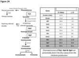
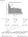
- the method further involves identifying metabolic pathways leading to the synthesis, the point of inhibitor synthesis or leading away from this point such that metabolism is channelled to and/or from the point of inhibitor synthesis (see Figure 24 ).
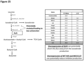
- pathways branching from these metabolic pathways are identified (see Figure 25 ).
- Such branches may arise at nodes in the pathway situated above, below or at the point of inhibitor synthesis and likewise serve to channel metabolism towards the inhibitor and/or away from the inhibitor point of synthesis, or serve to channel metabolism towards or away from the point of synthesis of intermediates of the inhibitor synthesis.
- genes encoding enzymes in these above mentioned pathways which includes the branches or branching pathways.
- These genes may encode enzymes which synthesise the inhibitor; they may encode enzymes which synthesise intermediates or upstream intermediates for the synthesis of the inhibitor; they may encode enzymes for which the inhibitor is an intermediate or an upstream intermediate for the enzyme action; or they may encode enzymes for which the inhibitor is direct intermediate or substrate for the enzyme action (for example this is illustrated in Figure 24 by genes Hpd and Hgd) or may encode enzmes which generate cofactors in the cell metabolic pathway or pathways for example BH4 (tetrahydrobiopterin), or BH4-4a (carbinolamine), or q-BH2.
- BH4 tetrahydrobiopterin
- BH4-4a carbbinolamine
- the method further involves modifying the expression of the one or more above mentioned genes to reduce the level of synthesis of the cell growth and/or productivity inhibitors.
- This objective can be achieved in a number of ways as the examples illustrate. Genes encoding enzymes which synthesise the inhibitor or which synthesis intermediates in the pathway leading to the inhibitor or pathways branching therefrom, may be modified to reduce gene expression, hence reducing the metabolic channelling towards production of the inhibitor, particularly in the case when the one or more gene or genes is highly expressed.
- Genes encoding enzymes for which the inhibitor is a substrate or an intermediate or an upstream intermediate in the pathway leading from the inhibitor or pathways branching therefrom, may be modified to increase gene expression, hence increasing the metabolic channelling away from production of the inhibitor, particularly in the case when the one or gene or genes is under expressed and/or is mutated and/or suffers loss of function or enzyme activity.
- genes in a pathway branching from a node located at an intermediate synthesis point upstream of the inhibitor synthesis point may be modified to increase gene expression, hence increasing the metabolic channelling away from the pathway leading to the production of the inhibitor, particularly in the case when the one or gene or genes is under expressed and/or is mutated and/or suffers loss of function or enzyme activity.
- the one or more genes identified encode an enzyme either directly synthesising the inhibitor or an intermediate to the inhibitor synthesis or metabolites of either inhibitor or intermediate it may not be desirable to modify such genes if they are involved in other important metabolic processes, This is illustrated in Example 4 ( Figure 24 ) with reference to genes Got1, Got2, Nup62-il4i1 and Mif, which may be expressed at normal or high levels.
- one or more genes in a branching pathway may be modified, for example in a pathway branching from a node located at an intermediate synthesis point upstream of the inhibitor synthesis point, to increase the metabolic channelling away from the pathway leading to the production of the inhibitor.
- genes encoding enzymes for which the inhibitor is a substrate or an upstream intermediate may be modified to increase gene expression, hence increasing the metabolic channelling away production of the inhibitor, particularly in the case when the one or gene or genes is under expressed and/or is mutated and/or suffers loss of function or enzyme activity.
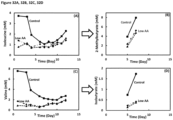
- the metabolic targets for the phenylalaine/tyrosine pathway are one or more of Pah, Hpd and/or Hgd genes and/or PCDB1 and/or QDPR ( Figures 26 and 27 ). These are targets for increasing expression, which can be achieved by mutation of the gene to correct loss of activity or increase activity and/or by providing an additional wild type copy or copies of the one or more genes in an expressible vector which can be introduced into the cell.
- the one or more genes identified encode an enzyme either directly synthesising the inhibitor or an intermediate to the inhibitor synthesis or metabolites of either inhibitor or intermediate it may desirable to modify such genes to prevent metabolic channelling towards production of the inhibitor synthesis. As illustrated in Example 24 this can be achieved by modifying the one or more genes directly upstream from production of the inhibitor as shown for genes Bcat1 and/or Bcat2. Such modification can be by gene knockdown or gene knockout.
- gene knockdown reduces gene expression or activity or activity of the encoded molecule to less than or equal to any of, 90, 80, 70, 60, 50, 45, 40, 35, 30, 25, 20, 15, 10, 5, 2.5 percent level of the expression or activity compared to unmodified cells.
- Bcat1 and/or Bcat2 knockdown is to less than or equal to any of, 90, 80, 70, 60, 50, 45, 40, 35, 30, 25, 20, 15, 10, 5, 2.5 percent level of Bcat1 and/or Bcat2 expression or activity compared to unmodified cells.
- Knockdown can be achieved by any one or more of of gene deletion, disruption, substitution, point mutation, multiple point mutation, insertion mutation or frameshift mutation applied to the identified gene to be expressed at a decreased level or by repression of gene expression by use of CRISPR/CAS9 or CRISPR interference or interfering RNA, interfering mRNA or interfering aptamer, or siRNA or siRNA interference or a zinc finger transcription factor or a zinc finger nuclease or a transcription activator-like effector nucleases (TALEN) or by use of an inhibitor such as a an inhibitor molecule or small molecule inhibitor, for example an activity inhibitor of protein or enzyme activity.
- CRISPR/CAS9 or CRISPR interference or interfering RNA, interfering mRNA or interfering aptamer, or siRNA or siRNA interference or a zinc finger transcription factor or a zinc finger nuclease or a transcription activator-like effector nucleases (TALEN) or by use of an inhibitor such as a an inhibitor
- increasing the metabolic channelling away from production of the inhibitor synthesis can be achieved by modifying one or more genes encoding enzymes which themselves share an intermediate of inhibitor production as their own intermediate. As illustrated in Example 5 this can be achieved by modifying the one or more genes directly downstream from, or downstream from a node which branches out towards, the inhibitor production ( Figure 25 ) to increase expression. This is particularly where the one or more genes is under expressed or is mutated and suffers loss of function or enzyme activity. This is illustrated for genes Ivd, Mccc1 and/or Mccc2 where the enzymes have suffered activity altering mutation and gene Auh which is under expressed.
- the metabolic targets for the leucine pathway are one or more of Ivd, Mccc1, Mccc2 and Auh genes. These are targets for increasing expression which can be achieved by mutation of the gene to correct loss of activity or increase activity and/or by providing a wild type copy of the one or more genes in an expressible vector which can be introduced into the cell.
- the modification suppresses, reduces, prevents the biosynthesis of the growth and/or productivity inhibitor and/or an intermediate thereof, in some embodiments the modification suppresses, reduces, prevents the biosynthesis of the growth and/or productivity inhibitor. According to some embodiments the modification produces cells with improved cell growth and/or productivity in cell culture.
- modifying the expression of the one or more genes comprises;
- RNAi interfering RNA
- suitable RNAi include RNAi that decreases or increases the level of a gene product, i.e. targets the one or more genes.
- an RNAi can be an shRNA or siRNA.
- a "small interfering" or “short interfering RNA” or siRNA is a RNA duplex of nucleotides that is targeted to a gene interest or the one or more genes.
- An "RNA duplex" refers to the structure formed by the complementary pairing between two regions of a RNA molecule.
- siRNA is "targeted" to a gene in that the nucleotide sequence of the duplex portion of the siRNA is complementary to a nucleotide sequence of the targeted gene.
- the length of the duplex of siRNAs is less than 30 nucleotides.
- the duplex can be 29, 28, 27, 26, 25, 24, 23, 22, 21, 20, 19, 18, 17, 16, 15, 14, 13, 12, 11 or 10 nucleotides in length.
- the length of the duplex is 19-25 nucleotides in length.
- the RNA duplex portion of the siRNA can be part of a hairpin structure.
- the hairpin structure may contain a loop portion positioned between the two sequences that form the duplex. The loop can vary in length.
- the loop is 5, 6, 7, 8, 9, 10, 11, 12 or 13 nucleotides in length.
- the hairpin structure can also contain 3' or 5' overhang portions.
- the overhang is a 3' or a 5' overhang 0, 1, 2, 3, 4 or 5 nucleotides in length.
- a "short hairpin RNA,” or shRNA is a polynucleotide construct that can be made to express an interfering RNA such as siRNA.
- the vector contains one or more of a promoter sequence, a directional cloning site, an epitope tag, a polyadenylation sequence, and antibiotic resistance gene.
- the promoter sequence is Human cytomegalovirus immediate early promoter
- the directional cloning site is TOPO
- the epitope tag is V5 for detection using anti-V5 antibodies
- the polyadenylation sequence is from Herpes Simplex Virus thymidine kinase
- antibiotic resistance gene is Blasticidin.
- the cell growth and/or productivity inhibitor is selected from the group of consisting of: 3-(4-hydroxyphenyl)lactate (HPLA), 4-hydroxyphenylpyruvate, phenyllactate (PLA), indole carboxylate (indole-3-carboxylate), indole lactate (indole-3-lactate), 2-hydroxybutyric acid, homocysteine, isovalerate, 2-methylbutyrate, isobutyrate, butyrate, formate.
- HPLA 3-(4-hydroxyphenyl)lactate
- PDA 4-hydroxyphenylpyruvate
- PDA phenyllactate
- indole carboxylate indole-3-carboxylate
- indole lactate indole-3-lactate
- 2-hydroxybutyric acid homocysteine
- isovalerate 2-methylbutyrate
- isobutyrate butyrate
- butyrate formate.
- the cell metabolic pathway or pathways synthesise 3-(4-hydroxyphenyl)lactate (HPLA), 4-hydroxyphenylpyruvate, phenyllactate (PLA), indole carboxylate (indole-3-carboxylate), indole lactate (indole-3-lactate), 2-hydroxybutyric acid, homocysteine, isovalerate, 2-methylbutyrate, isobutyrate, butyrate, formate, or metabolites thereof or metabolic intermediates thereof, or metabolite of the metabolic intermediate.
- HPLA 3-(4-hydroxyphenyl)lactate
- PDA 4-hydroxyphenylpyruvate
- PDA phenyllactate
- indole carboxylate indole-3-carboxylate
- indole lactate indole-3-lactate
- 2-hydroxybutyric acid homocysteine
- isovalerate 2-methylbutyrate
- isobutyrate isobutyrate
- butyrate formate
- the cell metabolic pathway is the leucine pathway and/or the isoleucine pathway and/or the valine pathway or the phenylalanine / tyrosine pathway or the acetoacetate/fumerate. In some embodiments the cell metabolic pathway is the leucine pathway or the phenylalanine / tyrosine pathway. In some embodiments the cell metabolic pathway is the leucine pathway and the isoleucine pathway and the valine pathway and/or the phenylalanine / tyrosine pathway.
- the gene modified encodes an enzyme that catalyses the synthesis of 3-(4-hydroxyphenyl)lactate (HPLA), 4-hydroxyphenylpyruvate, phenyllactate (PLA), indole carboxylate (indole-3-carboxylate), indole lactate (indole-3-lactate), 2-hydroxybutyric acid, homocysteine, isovalerate, 2-methylbutyrate, isobutyrate, butyrate, formate, or metabolites thereof or metabolic intermediates thereof, or metabolite of the metabolic intermediate.
- HPLA 3-(4-hydroxyphenyl)lactate
- PDA phenyllactate
- indole carboxylate indole-3-carboxylate
- indole lactate indole-3-lactate
- 2-hydroxybutyric acid homocysteine
- isovalerate 2-methylbutyrate
- isobutyrate isobutyrate
- butyrate formate
- the gene modified encodes an enzyme that catalyses the synthesis of 4-hydroxyphenylpyruvate or phenyllactate (PLA) or metabolites thereof or metabolic intermediates thereof, or metabolite of the metabolic intermediate.
- PHA 4-hydroxyphenylpyruvate or phenyllactate
- the gene modified encodes an enzyme that catalyses the synthesis of s isovalerate, 2-methylbutyrate, isobutyrate, or butyrate, or metabolites thereof or metabolic intermediates thereof, or metabolite of the metabolic intermediate.
- the one or genes modified is selected from; PCDB1, QDPR, Pah, Mif, Got1, Got2, Nup62-il4i1, Hpd, Hgd, Gstz1, Fah.
- the one or more genes modified is selected from; Bcat1, Bcat2, Bckdha/b, Dbt/Dld, Ivd, Acadm, Mccc1, Mccc2, Auh, Hmgcl, Fasn.
- the one or genes modified is selected from; PCDB1, QDPR, Pah, Mif, Got1, Got2, Nup62-il4i1, Hpd, Hgd, Gstz1, Fah, Bcat1, Bcat2, Bckdha/b, Dbt/Dld, Ivd, Acadm, Mccc1, Mccc2, Auh, Hmgcl, Fasn, Fasn.
- the one or more genes modified is selected from; Hpd, Hgd and Pah, PCDB1, QDPR.
- the one or more genes modified is selected from, Bcat1, Bcat2, Auh, Mccc1, Mccc2, Ivd.
- the one or more genes modified is selected from, Bcat1, Bcat2, Auh, Mccc1, Mccc2, Ivd, Hpd, Hgd and Pah, PCDB1, QDPR.
- the gene is modified to increase or decrease gene expression.
- the gene is modified to decrease gene expression.
- the modification increases gene expression
- (b) increases gene expression except wherein the gene is Bcat1 and/or Bcat2, when modification decreases gene expression.
- a cell comprising one or more modified genes which reduces the level of synthesis of growth and/or productivity inhibitors by the cell.
- the cell comprises one or more modified genes selected from Bcat1, Bcat2, Auh, Mccc1/2, Ivd, Hpd, Hgd and Pah, PCDB1, QDPR, wherein the modification increases or decreases the gene expression, in some embodiments increases it, in some embodiments the gene expression of Bcat1 and/or Bcat2 is reduced, in some embodiments the level of synthesis of growth and/or productivity inhibitors by the cell is also reduced.
- the one or more genes modified is Auh, or is Bcat1, or is Bcat2, or is Bcat1 and Bcat2, or is one or more of Mccc1, Mccc2 and Ivd, for example is Mccc1 or Mccc2 or Ivd optionally in combination with Auh and/or Bcat1 and/or Bcat2, or is Auh and one or more Mccc1, Mccc2 and Ivd, or is Auh, Mccc1, Mccc2 and Ivd, optionally in combination with Bcat1 and/or Bcat2.
- the one or more genes is modified to increase gene expression, in some embodiments by mutation of the gene, in some embodiments by introduction of a copy of the wild type gene into the cell optionally as an expressible vector.
- Auh gene expression can be increased by introduction of a copy of the wild type gene, in some embodiments by mutation, alternatively by both.
- Mccc1, Mccc2, Ivd gene expression can be increased by mutation, in some embodiments by introduction of a copy of the wild type gene, alternatively by both.
- Auh gene expression is increased by introduction of a copy of the wild type gene and/or Mccc1, Mccc2, and Ivd gene expression is increased by mutation.
- the gene expression of Bcat1 and/or Bcat2 is reduced, either by gene knockdown or knockout, in some embodiments Bcat1 and/or Bcat2 knockdown is to less than or equal to any of, 90, 80, 70, 60, 50, 45, 40, 35, 30, 25, 20, 15, 10, 5, 2.5 percent level of Bcat1 and/or Bcat2 expression or activity compared to unmodified cells.
- the one or more modified gene(s) selected from (a) Pah, PCBD1, QDPR, Mif, Got1, Got2, Nup62-il4i1, Hpd, Hgd, Gstz1, Fah, Bcat1, Bcat2, Bckdha/b, Dbt/Dld, Ivd, Acadm, Mccc1, Mccc2 , Auh, Hmgcl, Fasn, Fasn, (b) Pah, PCBD1, QDPR, Mif, Got1, Got2, Nup62-il4i1, Hpd, Hgd, Gstz1, Fah, (c) Bcat1, Bcat2, Bckdha/b, Dbt/Dld, Ivd, Acadm, Mccc1, Mccc2 , Auh, Hmgcl, Fasn, Fasn, (d) Bcat1, Bcat2, Auh, Mccc1, Mccc2, Ivd, Hpd, Hgd and Pah
- the one or more genes modified is Pah and/or PCDB1 and/or QDPR, or is one or more of Hpd and Hgd, for example Hpd, or Hgd optionally in combination with Pah and/or PCDB1 and/or QDPR, or is Pah and one or more of Hpd and Hgd or,is Pah,Hpd and Hgd.
- the one or more genes modified is selected from; (i) PCDB1, (ii) Pah, (iii) QDPR, (iv) PCBD1 and QDPR, (v) PCBD1 and Pah, (vi) Pah and QDPR, (vii) PCDB1 and Pah, and QDPR, (viii) any of (i) to (vii) and Hpd and/or Hgd.
- the one or more genes is modified to increase gene expression.
- any one or more of Pah, PCDB1, QDPR, Hpd, Hgd gene expression can be increased by introduction of a copy of the wild type gene, in some embodiments by mutation, alternatively by both.
- Pah, PCDB1, QDPR, Hpd and Hgd gene expression is increased by introduction of a copy of the wild type gene. In some embodiments Hpd and Hgd gene expression is increased by introduction of a copy of the wild type gene. In some embodiments Pah, PCDB1 and/or QDPR gene expression is increased by introduction of a copy of the wild type gene.
- the one or more genes modified is Auh, Mccc1, Mccc2, Ivd, Pah,Hpd and Hgd. In some embodiments, the one or more genes modified is Auh, Mccc1, Mccc2, Ivd, Pah, PCDB1, Hpd and Hgd and optionally QDPR. In some embodiments, the one or more genes modified is Bcat1 and/or Bcat2, Pah, PCDB1, Hpd and Hgd and optionally QDPR. In some embodiments of the preceding embodiments the one or more genes is modified to increase gene expression by way of the relevant method of introduction of a copy of the wild type gene, by mutation, alternatively by both. In some embodiments Auh gene expression is increased by introduction of a copy of the wild type gene and Mccc1, Mccc2, Ivd, Pah, PCDB1, QDPR, Hpd and Hgd gene expression is increased by mutation.
- the present invention further provides a cell obtained or obtainable by the method of producing cells.
- the cell comprises one or more modified genes which reduces the level of synthesis of growth and/or productivity inhibitors by the cell.
- the cell comprises one or more modified genes selected from Bcat1, Bcat2, Auh, Mccc1, Mccc2, Ivd, Hpd, Hgd and Pah, PCDB1, QDPR, wherein the modification increases or decreases the gene expression.
- gene expression is increased.
- the level of synthesis of growth and/or productivity inhibitors by the cell is also reduced.
- the one or more genes modified is Auh, or is Bcat1, or is Bcat2, or is Bcat1 and Bcat2,, or is one or more of Mccc1, Mccc2 and Ivd, for example is Mccc1 or Mccc2 or Ivd optionally in combination with Auh and/or Bcat1 and/or Bcat2, or is Auh and one or more Mccc1, Mccc2 and Ivd, or is Auh, Mccc1, Mccc2 and Ivd, optionally in combination with Bcat1 and/or Bcat2.
- the one or more genes is modified to increase gene expression, in some embodiments by mutation of the gene, in some embodiments by introduction of a copy of the wild type gene into the cell optionally as an expressible vector.
- Auh gene expression can be increased by introduction of a copy of the wild type gene, in some embodiments by mutation, alternatively by both.
- Mccc1, Mccc2, Ivd gene expression can be increased by mutation, in some embodiments by introduction of a copy of the wild type gene, alternatively by both.
- Auh gene expression is increased by introduction of a copy of the wild type gene and/or Mccc1, Mccc2, and Ivd gene expression is increased by mutation.
- the gene expression of Bcat1 and/or Bcat2 is reduced, either by gene knockdown or knockout, in some embodiments Bcat1 and/or Bcat2 knockdown is to less than or equal to any of, 90, 80, 70, 60, 50, 45, 40, 35, 30, 25, 20, 15, 10, 5, 2.5 percent level of Bcat1 and/or Bcat2 expression or activity compared to unmodified cells.
- the one or more genes modified is Pah, and/or PCDB1 and/or QDPR, or is one or more of Hpd and Hgd, for example Hpd, or Hgd optionally in combination with Pah, and/or PCDB1 and/or QDPR, or is Pah and one or more of Hpd and Hgd. or,is Pah,Hpd and Hgd.
- the one or more genes modified is selected from; (i) PCDB1, (ii) Pah, (iii) QDPR, (iv) PCBD1 and QDPR, (v) PCBD1 and Pah, (vi) Pah and QDPR, (vii) PCDB1 and Pah, and QDPR, (viii) any of (i) to (vii) and Hpd and/or Hgd.
- the one or more genes is modified to increase gene expression.
- Pah, PCDB1, QDPR, Hpd, Hgd gene expression can be increased by introduction of a copy of the wild type gene, in some embodiments by mutation, alternatively by both.
- Pah, PCDB1, QDPR, Hpd and Hgd gene expression is increased by introduction of a copy of the wild type gene. In some embodiments Hpd and Hgd gene expression is increased by introduction of a copy of the wild type gene. In some embodiments Pah, PCDB1, QDPR, Hpd and Hgd gene expression is increased by introduction of a copy of the wild type gene.
- the one or more genes modified is Auh, Mccc1, Mccc2, Ivd, Pah,Hpd and Hgd. In some embodiments, the one or more genes modified is Auh, Mccc1, Mccc2, Ivd, Pah, PCDB1, Hpd and Hgd and optionally QDPR. In some embodiments, the one or more genes modified is Bcat1 and/or Bcat2, Pah, PCDB1, Hpd and Hgd and optionally QDPR. In some embodiments of the preceding embodiments the one or more genes is modified to increase gene expression by way of the relevant method of introduction of a copy of the wild type gene, by mutation, alternatively by both. In some embodiments Auh gene expression is increased by introduction of a copy of the wild type gene and Mccc1, Mccc2, Ivd, Pah, PCDB1, QDPR, Hpd and Hgd gene expression is increased by mutation.
- the one or more genes modified is selected from Bcat1, Bcat2, Auh, Mccc1, Mccc2, Ivd, Hpd, Hgd, PCBD1, QDPR and Pah, wherein the modification increases the gene expression of one or more of Auh, Mccc1, Mccc2, Ivd, Hpd, Hgd, PCBD1, QDPR and Pah and/or reduces the gene expression of Bcat1 or Bcat2 and/or reduces the level of synthesis of growth and/or productivity inhibitors by the cell.
- the one or more genes modified is, (i) Auh, (ii) Bcat1, (iii) Bcat2, (iv) one or more of Mccc1, Mccc2 and Ivd, (v) Bcat1 and/or Bcat2 and/or Auh and one or more Mccc1, Mccc2 and Ivd, (vi) Pah, (vii) PCBD1,(viii) PCBD1 and/or QDPR, (ix) one or more of Hpd and Hgd, (x) Pah and/or PCBD1 and one or more of Hpd and Hgd, (xi) Pah and PCBD1, and optionally QDPR, (xii) Pah, PCBD1, Hpd and Hgd, and optionally QDPR, (xiii) Pah, PCBD1, Hpd and Hgd, and optionally QDPR, (xiii) Bcat1 and/or Bcat2, Auh, Mccc1, Mccc2 and Ivd or, (xi
- the modification increases gene expression
- (b) increases gene expression except wherein the gene is Bcat1 and/or Bcat2, when modification decreases gene expression, optionally Bcat1 and/or Bcat2 knockdown is to less than or equal to any of, 90, 80, 70, 60, 50, 45, 40, 35, 30, 25, 20, 15, 10, 5, 2.5 percent level of Bcat1 and/or Bcat2 expression or activity compared to unmodified cells.
- the cells express a recombinant protein or heterologous recombinant protein.
- the cell is a mammalian cell.
- mammalian cells that may be used in accordance with the present invention include BALB/c mouse myeloma line (NSO/I, ECACC No: 85110503); human retinoblasts (PER.C6, CruCell, Leiden, The Netherlands); monkey kidney CV1 line transformed by SV40 (COS-7, ATCC CRL 1651); human embryonic kidney line (293 or 293 cells subcloned for growth in suspension culture, Graham et al., J.
- monkey kidney cells (CV1 ATCC CCL 70); African green monkey kidney cells (VERO-76, ATCC CRL-1 587); human cervical carcinoma cells (HeLa, ATCC CCL 2); canine kidney cells (MDCK, ATCC CCL 34); buffalo rat liver cells (BRL 3A, ATCC CRL 1442); human lung cells (W138, ATCC CCL 75); human liver cells (Hep G2, HB 8065); mouse mammary tumor (MMT 060562, ATCC CCL51); TRI cells ( Mather et al., Annals N.Y. Acad.
- hybridoma refers to a cell or progeny of a cell resulting from fusion of an immortalized cell and an antibody-producing cell. Such a resulting hybridoma is an immortalized cell that produces antibodies.
- a hybridoma is a trioma cell line, which results when progeny of heterohybrid myeloma fusions, which are the product of a fusion between human cells and a murine myeloma cell line, are subsequently fused with a plasma cell.
- a hybridoma is any immortalized hybrid cell line that produces antibodies such as, for example, quadromas (See, e.g., Milstein et al., Nature, 537:3053, 1983 ).
- quadromas See, e.g., Milstein et al., Nature, 537:3053, 1983 ).
- hybridoma cell lines might have different nutrition requirements and/or might require different culture conditions for optimal growth, and will be able to modify conditions as needed.
- Methods comprising identifying and / or measuring the metabolite concentration in the cell culture medium or in cells (cell pellet sample)
- Metabolites can be identified and/or the concentration of metabolites can be measured by any method known to the skilled person, including off line and on line measurement methods, such measurements also constitute metabolomic analysis or metabolomic measurement. Applied to the cell culture medium or to samples of the cells for instance the cell pellet
- the identification and/or concentration of metabolites can be measured once or several times during the cell culture.
- the metabolite concentration is measured continuously, intermittently, every 30 min, every hour, every two hours, twice a day, daily, or every two days.
- the identification and/or concentration of metabolite is measured daily.
- An off line measurement method as used herein refers to a method where the measurement of a parameter such as a concentration is not automated and integrated to the cell culture method.
- a measurement method where a sample is manually taken from the cell culture medium so that a specific concentration can be measured in said sample is considered as an off line measurement method.
- Online measurement methods refer to methods where the measurement of a parameter, such as a concentration, is automated and integrated to the cell culture method.
- a method using the Raman spectroscopy as disclosed in Example 7 is an on-line measurement method.
- the use of High Performance Liquid Chromatography (HPLC) or Ultra Performance Liquid Chromatography (UPLC) based technology with an auto-sampler that draws samples from reactor and transfers them to the equipment in a programmed manner is an online measurement method.
- the identification and/or concentration of metabolites can be measured by any method known to the skilled person.
- Preferred methods to identify and/or measure the concentration of metabolites in online or offline methods include for example Liquid Chromatography such as High-Performance Liquid Chromatography (HPLC), Ultra Performance Liquid Chromatography (UPLC) or Liquid Chromatography - Mass Spectrometry (LCMS), Nuclear Magnetic Resonance (NMR) or Gas Chromatography - Mass Spectrometry (GCMS).
- Liquid Chromatography such as High-Performance Liquid Chromatography (HPLC), Ultra Performance Liquid Chromatography (UPLC) or Liquid Chromatography - Mass Spectrometry (LCMS), Nuclear Magnetic Resonance (NMR) or Gas Chromatography - Mass Spectrometry (GCMS).
- the identification and/or concentration of metabolite is measured off line by taking a sample of the cell culture medium and measuring the concentration of said at least one metabolite in said sample. In some embodiments, the identification and/or concentration of metabolites is measured as disclosed in Example 2. A preferred method to measure the identification and/or concentration of metabolites in an offline method is LCMS.
- the identification and/or concentration of metabolite is measured on-line. In some embodiments, the identification and/or concentration of metabolite is measured online using Raman spectroscopy. In some embodiments, the identification and/or concentration of metabolite is measured on-line using Raman spectroscopy as disclosed in Example 7. In some embodiments, the identification and/or concentration of metabolite is measured online using HPLC or UPLC based technology with an auto-sampler that draws samples from reactor and transfers them to the equipment in a programmed manner. Identification of a metabolite includes determining the presence and/or identity of the metabolite.
- cell growth and/or productivity are increased as compared to a control culture, said control culture being identical except that it does not comprise step (ii) and/or (iii) and/or does not comprise the modified cells or the cells produced by the method of producing cells. i.e. the cells are unmodified.
- the method of the invention is a method for improving cell growth. In some embodiments, the method of the invention is a method for improving cell growth in high density cell culture at high cell density.
- High cell density refers to cell density above 1 ⁇ 10 6 cells/mL, 5 ⁇ 10 6 cells/mL, 1 ⁇ 10 7 cells /mL, 5 ⁇ 10 7 cells/mL, 1 ⁇ 10 8 cells/mL or 5 ⁇ 10 8 cells/mL, preferably above 1 ⁇ 10 7 cells /mL, more preferably above 5 ⁇ 10 7 cells/mL.
- the the above described methods are for improving cell growth in a cell culture where cell density is above 1 ⁇ 10 6 cells/mL, 5 ⁇ 10 6 cells/mL, 1 ⁇ 10 7 cells /mL, 5 ⁇ 10 7 cells/mL, 1 ⁇ 10 8 cells/mL or 5 ⁇ 10 8 cells/mL .
- the methods are for improving cell growth in a cell culture where maximum cell density is above 1 ⁇ 10 6 cells/mL, 5 ⁇ 10 6 cells/mL, 1 ⁇ 10 7 cells /mL, 5 ⁇ 10 7 cells/mL, 1 ⁇ 10 8 cells/mL or 5 ⁇ 10 8 cells/mL.
- cell growth is determined by viable cell density (VCD), maximum viable cell density, or Integrated viable cell count (IVCC). In some embodiments, cell growth is determined by maximum viable cell density.
- VCD viable cell density
- IVCC Integrated viable cell count
- Viable cell density refers to the number of cells present in a given volume of medium. Viable cell density can be measured by any method known to the skilled person. Preferably, Viable cell density is measured using an automated cell counter such as Bioprofile Flex ® .
- maximum cell density refers to the maximum cell density achieved during the cell culture.
- cell viability refers to the ability of cells in culture to survive under a given set of culture conditions or experimental variations. Those of ordinary skill in the art will appreciate that one of many methods for determining cell viability are encompassed in this invention. For example, one may use a dye (e.g., trypan blue) that does not pass through the membrane of a living cell, but can pass through the disrupted membrane of a dead or dying cell in order to determine cell viability.
- a dye e.g., trypan blue
- IVCC Integrated viable cell count
- VCD viable cell density
- Titer refers, for example, to the total amount of recombinantly expressed protein produced by a cell culture in a given amount of medium volume. Titer is typically expressed in units of grams of protein per liter of medium.
- cell growth is increased by at least 5%, 10%, 15%, 20% or 25% as compared to the control culture. In some embodiments, cell growth is increased by at least 10% as compared to the control culture. In some embodiments, cell growth is increased by at least 20% as compared to the control culture.
- the productivity is determined by titer and/or volumetric productivity.
- Titer refers, for example, to the total amount of recombinantly expressed protein produced by a cell culture in a given amount of medium volume. Titer is typically expressed in units of grams of protein per liter of medium.
- the productivity is determined by titer. In some embodiments, the productivity is increased by at least 5%, 10%, 15%, 20% or 25% as compared to the control culture. In some embodiments, the productivity is increased by at least 10% as compared to a control culture. In some embodiments, the productivity is increased by at least 20% as compared to a control culture.
- the maximum cell density of the cell culture is greater than 1 ⁇ 10 6 cells/mL, 5 ⁇ 10 6 cells/mL, 1 ⁇ 10 7 cells /mL, 5 ⁇ 10 7 cells/mL, 1 ⁇ 10 8 cells/mL or 5 ⁇ 10 8 cells/mL. In some embodiments, the maximum cell density of the cell culture is greater than 5 ⁇ 10 6 cells/mL. In some embodiments, the maximum cell density of the cell culture is greater than 1 ⁇ 10 8 cells/mL.
- medium refers to a solution containing components or nutrients which nourish growing mammalian cells.
- nutrients include essential and non-essential amino acids, vitamins, energy sources, lipids, and trace elements required by the cell for minimal growth and/or survival.
- Such a solution may also contain further nutrients or supplementary components that enhance growth and/or survival above the minimal rate, including, but not limited to, hormones and/or other growth factors, particular ions (such as sodium, chloride, calcium, magnesium, and phosphate), buffers, vitamins, nucleosides or nucleotides, trace elements (inorganic compounds usually present at very low final concentrations), inorganic compounds present at high final concentrations (e.g., iron), amino acids, lipids, and/or glucose or other energy source.
- a medium is advantageously formulated to a pH and salt concentration optimal for cell survival and proliferation.
- a medium is a feed medium that is added after the beginning of the cell culture.
- cells may be grown in one of a variety of chemically defined media, wherein the components of the media are both known and controlled.
- cells may be grown in a complex medium, in which not all components of the medium are known and/or controlled.
- Chemically defined growth media for mammalian cell culture have been extensively developed and published over the last several decades. All components of defined media are well characterized, and so defined media do not contain complex additives such as serum or hydrolysates. Early media formulations were developed to permit cell growth and maintenance of viability with little or no concern for protein production. More recently, media formulations have been developed with the express purpose of supporting highly productive recombinant protein producing cell cultures. Such media are preferred for use in the method of the invention. Such media generally comprises high amounts of nutrients and in particular of amino acids to support the growth and/or the maintenance of cells at high density. If necessary, these media can be modified by the skilled person for use in the method of the invention. For example, the skilled person may decrease the amount of phenylalanine, tyrosine, tryptophan and/or methionine in these media for their use as base media or feed media in a method as disclosed herein.
- complex media may contain additives such as simple and/or complex carbon sources, simple and/or complex nitrogen sources, and serum, among other things.
- complex media suitable for the present invention contains additives such as hydrolysates in addition to other components of defined medium as described herein.
- defined media typically includes roughly fifty chemical entities or components at known concentrations in water. Most of them also contain one or more well-characterized proteins such as insulin, IGF-1, transferrin or BSA, but others require no protein components and so are referred to as protein-free defined media. Typical chemical components of the media fall into five broad categories: amino acids, vitamins, inorganic salts, trace elements, and a miscellaneous category that defies neat categorization.
- Cell culture medium may be optionally supplemented with supplementary components.
- supplementary components refers to components that enhance growth and/or survival above the minimal rate, including, but not limited to, hormones and/or other growth factors, particular ions (such as sodium, chloride, calcium, magnesium, and phosphate), buffers, vitamins, nucleosides or nucleotides, trace elements (inorganic compounds usually present at very low final concentrations), amino acids, lipids, and/or glucose or other energy source.
- supplementary components may be added to the initial cell culture.
- supplementary components may be added after the beginning of the cell culture.
- components which are trace elements refer to a variety of inorganic salts included at micromolar or lower levels.
- trace elements are zinc, selenium, copper, and others.
- iron (ferrous or ferric salts) can be included as a trace element in the initial cell culture medium at micromolar concentrations.
- Manganese is also frequently included among the trace elements as a divalent cation (MnCl 2 or MnSO 4 ) in a range of nanomolar to micromolar concentrations. Numerous less common trace elements are usually added at nanomolar concentrations.
- the medium used in the methods of the invention is a medium suitable for supporting high cell density, such as for example 1 ⁇ 10 6 cells/mL, 5 ⁇ 10 6 cells/mL, 1 ⁇ 10 7 cells /mL, 5 ⁇ 10 7 cells/mL, 1 ⁇ 10 8 cells/mL or 5 ⁇ 10 8 cells/mL, in a cell culture.
- the cell culture is a mammalian cell fed-batch culture, preferably a CHO cells fed-batch culture.
- the cell culture medium comprises phenylalanine at a concentration below 2mM, below 1mM, between 0.1 and 2mM, between 0.1 to 1mM, between 0.5 and 1.5mM or between 0.5 to 1mM.
- the cell culture medium comprises tyrosine at a concentration below 2mM, below 1mM, between 0.1 and 2mM, between 0.1 to 1mM, between 0.5 and 1.5mM or between 0.5 to 1mM. In some embodiments, the cell culture medium comprises no tyrosine. In some embodiments, the cell culture medium is HiPDOG medium, medium used in the HipDOG process, but comprising no tyrosine. In some embodiments the cell culture medium comprises no tyrosine wherein a HiPDOG culture or process is used which comprises no tyrosine.
- the cell culture medium comprises tryptophan at a concentration below 2mM, below 1mM, between 0.1 and 2mM, between 0.1 to 1mM, between 0.5 and 1.5mM or between 0.5 to 1mM.
- the cell culture medium comprises methionine at a concentration below 2mM, below 1mM, between 0.1 and 2mM, between 0.1 to 1mM, between 0.5 and 1.5mM or between 0.5 to 1mM.
- the cell culture medium comprises leucine at a concentration below 2mM, below 1mM, between 0.1 and 2mM, between 0.1 to 1mM, between 0.5 and 1.5mM or between 0.5 to 1mM.
- the cell culture medium comprises isoleucine at a concentration below 2mM, below 1mM, between 0.1 and 2mM, between 0.1 to 1mM, between 0.5 and 1.5mM or between 0.5 to 1mM.
- the cell culture medium comprises valine at a concentration below 2mM, below 1mM, between 0.1 and 2mM, between 0.1 to 1mM, between 0.5 and 1.5mM or between 0.5 to 1mM.
- the cell culture medium comprises serine at a concentration below 2mM, below 1mM, between 0.1 and 2mM, between 0.1 to 1mM, between 0.5 and 1.5mM or between 0.5 to 1mM.
- the cell culture medium comprises threonine at a concentration below 2mM, below 1mM, between 0.1 and 2mM, between 0.1 to 1mM, between 0.5 and 1.5mM or between 0.5 to 1mM.
- the cell culture medium comprises glycine at a concentration below 2mM, below 1mM, between 0.1 and 2mM, between 0.1 to 1mM, between 0.5 and 1.5mM or between 0.5 to 1mM.
- the cell culture medium comprises two of phenylalanine, tyrosine, tryptophan, methionine, leucine, isoleucine, valine, serine, threonine and glycine at a concentration below 2mM, below 1mM, between 0.1 and 2mM, between 0.1 to 1mM, between 0.5 and 1.5mM or between 0.5 to 1mM.
- the cell culture medium comprises phenylalanine and tyrosine at a concentration below 2mM, below 1mM, between 0.1 and 2mM, between 0.1 to 1mM, between 0.5 and 1.5mM or between 0.5 to 1mM.
- the cell culture medium comprises phenylalanine and tryptophan at a concentration below 2mM, below 1mM, between 0.1 and 2mM, between 0.1 to 1mM, between 0.5 and 1.5mM or between 0.5 to 1mM.
- the cell culture medium comprises phenylalanine and methionine at a concentration below 2mM, below 1mM, between 0.1 and 2mM, between 0.1 to 1mM, between 0.5 and 1.5mM or between 0.5 to 1mM.
- the cell culture medium comprises tyrosine and tryptophan at a concentration below 2mM, below 1mM, between 0.1 and 2mM, between 0.1 to 1mM, between 0.5 and 1.5mM or between 0.5 to 1mM.
- the cell culture medium comprises tyrosine and methionine at a concentration below 2mM, below 1mM, between 0.1 and 2mM, between 0.1 to 1mM, between 0.5 and 1.5mM or between 0.5 to 1mM.
- the cell culture medium comprises tryptophan and methionine at a concentration below 2mM, below 1mM, between 0.1 and 2mM, between 0.1 to 1mM, between 0.5 and 1.5mM or between 0.5 to 1mM.
- the cell culture medium comprises three of phenylalanine, tyrosine, tryptophan, methionine, leucine, serine, threonine and glycine at a concentration below 2mM, below 1mM, between 0.1 and 2mM, between 0.1 to 1mM, between 0.5 and 1.5mM or between 0.5 to 1mM.
- the cell culture medium comprises phenylalanine, tyrosine and tryptophan at a concentration below 2mM, below 1mM, between 0.1 and 2mM, between 0.1 to 1mM, between 0.5 and 1.5mM or between 0.5 to 1mM.
- the cell culture medium comprises phenylalanine, tyrosine and methionine at a concentration below 2mM, below 1mM, between 0.1 and 2mM, between 0.1 to 1mM, between 0.5 and 1.5mM or between 0.5 to 1mM.
- the cell culture medium comprises phenylalanine, tryptophan and methionine at a concentration below 2mM, below 1mM, between 0.1 and 2mM, between 0.1 to 1mM, between 0.5 and 1.5mM or between 0.5 to 1mM.
- the cell culture medium comprises tyrosine, tryptophan and methionine at a concentration below 2mM, below 1mM, between 0.1 and 2mM, between 0.1 to 1mM, between 0.5 and 1.5mM or between 0.5 to 1mM.
- the cell culture medium comprises four of phenylalanine, tyrosine, tryptophan, methionine, leucine, isoleucine, valine, serine, threonine and glycine at a concentration below 2mM, below 1mM, between 0.1 and 2mM, between 0.1 to 1mM, between 0.5 and 1.5mM or between 0.5 to 1mM.
- the cell culture medium comprises phenylalanine, tyrosine, tryptophan and methionine at a concentration below 2mM, below 1mM, between 0.1 and 2mM, between 0.1 to 1mM, between 0.5 and 1.5mM or between 0.5 to 1mM.
- the cell culture medium comprises five of phenylalanine, tyrosine, tryptophan, methionine, leucine, isoleucine, valine, serine, threonine and glycine at a concentration below 2mM, below 1mM, between 0.1 and 2mM, between 0.1 to 1mM, between 0.5 and 1.5mM or between 0.5 to 1mM.
- the cell culture medium comprises six of phenylalanine, tyrosine, tryptophan, methionine, leucine, isoleucine, valine, serine, threonine and glycine at a concentration below 2mM, below 1mM, between 0.1 and 2mM, between 0.1 to 1mM, between 0.5 and 1.5mM or between 0.5 to 1mM.
- the cell culture medium comprises seven of phenylalanine, tyrosine, tryptophan, methionine, leucine, isoleucine, valine, serine, threonine and glycine at a concentration below 2mM, below 1mM, between 0.1 and 2mM, between 0.1 to 1mM, between 0.5 and 1.5mM or between 0.5 to 1mM.
- the cell culture medium comprises phenylalanine, tyrosine, tryptophan, methionine, leucine, isoleucine, valine, serine, threonine and glycine at a concentration below 2mM, below 1mM, between 0.1 and 2mM, between 0.1 to 1mM, between 0.5 and 1.5mM or between 0.5 to 1mM.
- the cell culture medium further comprises at least 1, 2, 3, 4, 5, 6, 7, 8, or 9, of glycine, proline, serine, threonine, lysine, arginine, histidine, aspartate, glutamate and asparagine at a concentration above 2mM, 3mM, 4mM, 5mM, 10mM, 15mM, preferably 2mM.
- the cell culture medium further comprises at least 5 of glycine, proline, serine, threonine, lysine, arginine, histidine, aspartate, glutamate and asparagine at a concentration above 2mM, 3mM, 4mM, 5mM, 10mM, 15mM, preferably 2mM.
- the cell culture medium further comprises glycine, proline, serine, threonine, lysine, arginine, histidine, aspartate, glutamate and asparagine at a concentration above 2mM, 3mM, 4mM, 5mM, 10mM, 15mM, preferably 2mM.
- the cell culture medium further comprises at least 1, 2, 3, 4, 5, 6, or 7, of proline, lysine, arginine, histidine, aspartate, glutamate and asparagine at a concentration above 2mM, 3mM, 4mM, 5mM, 10mM, 15mM, preferably 2mM.
- the cell culture medium further comprises at least 5 of proline, lysine, arginine, histidine, aspartate, glutamate and asparagine at a concentration above 2mM, 3mM, 4mM, 5mM, 10mM, 15mM, preferably 2mM.
- the cell culture medium further comprises proline, lysine, arginine, histidine, aspartate, glutamate and asparagine at a concentration above 2mM, 3mM, 4mM, 5mM, 10mM, 15mM, preferably 2mM.
- the cell culture medium comprises serine at a concentration above 3mM, 5mM, 7mM, 10mM, 15mM or 20mM, preferably 10mM.
- the cell culture medium comprises cysteine at a concentration above 3mM, 5mM, 7mM, 10mM, 15mM or 20mM, preferably 10mM.
- the above cell culture medium is for use in a method as disclosed herein.
- the above cell culture medium is used in a method as disclosed herein as a base media.
- the above cell culture medium is used a method as disclosed herein as a feed media.
- the cells will be selected or engineered to produce high levels of desired products (e.g., recombinant protein or antibody).
- desired products e.g., recombinant protein or antibody.
- cells will be manipulated by the hand of man to produce high levels of recombinant protein, for example by introduction of a gene encoding the protein of interest and/or by introduction of genetic control elements that regulate expression of that gene (whether endogenous or introduced).
- Certain proteins may have detrimental effects on cell growth, cell viability or some other characteristic of the cells that ultimately limits production of the protein of interest in some way. Even amongst a population of cells of one particular type engineered to express a specific protein, variability within the cellular population exists such that certain individual cells will grow better, produce more protein of interest, or produce a protein with higher activity levels (e.g., enzymatic activity).
- a cell line is empirically selected by the practitioner for robust growth under the particular conditions chosen for culturing the cells.
- individual cells engineered to express a particular protein are chosen for large-scale production based on cell growth, final cell density, percent cell viability, titer of the expressed protein or any combination of these or any other conditions deemed important by the practitioner.
- Any protein that is expressible in a host cell may be produced in accordance with the present teachings and may be produced according to the methods of the invention or by the cells of the invention.
- the term "host cell” as used herein refers to a cell that is manipulated according to the present invention to produce a protein of interest as described herein.
- a protein may be expressed from a gene that is endogenous to the cell, or from a heterologous gene that is introduced into the cell.
- a protein may be one that occurs in nature, or may alternatively have a sequence that was engineered or selected by the hand of man.
- Proteins that may desirably be expressed in accordance with the present invention will often be selected on the basis of an interesting or useful biological or chemical activity.
- the present invention may be employed to express any pharmaceutically or commercially relevant enzyme, receptor, antibody, hormone, regulatory factor, antigen, binding agent, etc.
- the protein expressed by cells in culture are selected from antibodies, or fragments thereof, nanobodies, single domain antibodies, glycoproteins, therapeutic proteins, growth factors, clotting factors, cytokines, fusion proteins, pharmaceutical drug substances, vaccines, enzymes, or Small Modular ImmunoPharmaceuticals TM (SMIPs).
- SIPs Small Modular ImmunoPharmaceuticals TM
- Antibodies are proteins that have the ability to specifically bind a particular antigen. Any antibody that can be expressed in a host cell may be produced in accordance with the present invention and may be produced according to the methods of the invention or by the cells of the invention. In some embodiments, the antibody to be expressed is a monoclonal antibody.
- the monoclonal antibody is a chimeric antibody.
- a chimeric antibody contains amino acid fragments that are derived from more than one organism.
- Chimeric antibody molecules can include, for example, an antigen binding domain from an antibody of a mouse, rat, or other species, with human constant regions.
- a variety of approaches for making chimeric antibodies have been described. See e.g., Morrison et al., Proc. Natl. Acad. Sci. U.S.A. 81, 6851 (1985 ); Takeda et al., Nature 314, 452 (1985 ), Cabilly et al., U.S. Patent No. 4,816,567 ; Boss et al., U.S. Patent No. 4,816,397 ; Tanaguchi et al., European Patent Publication EP171496 ; European Patent Publication 0173494 , United Kingdom Patent GB 2177096B .
- the monoclonal antibody is a human antibody derived, e.g., through the use of ribosome-display or phage-display libraries (see, e.g., Winter et al., U.S. Patent No. 6,291,159 and Kawasaki, U.S. Patent No. 5,658,754 ) or the use of xenographic species in which the native antibody genes are inactivated and functionally replaced with human antibody genes, while leaving intact the other components of the native immune system (see, e.g., Kucherlapati et al., U.S. Patent No. 6,657,103 ).
- the monoclonal antibody is a humanized antibody.
- a humanized antibody is a chimeric antibody wherein the large majority of the amino acid residues are derived from human antibodies, thus minimizing any potential immune reaction when delivered to a human subject.
- amino acid residues in the complementarity determining regions are replaced, at least in part, with residues from a non-human species that confer a desired antigen specificity or affinity.
- Such altered immunoglobulin molecules can be made by any of several techniques known in the art, ( e . g ., Teng et al., Proc. Natl. Acad. Sci.
- the monoclonal, chimeric, or humanized antibodies described above may contain amino acid residues that do not naturally occur in any antibody in any species in nature. These foreign residues can be utilized, for example, to confer novel or modified specificity, affinity or effector function on the monoclonal, chimeric or humanized antibody.
- the antibodies described above may be conjugated to drugs for systemic pharmacotherapy, such as toxins, low-molecular-weight cytotoxic drugs, biological response modifiers, and radionuclides (see e.g., Kunz et al., Calicheamicin derivative-carrier conjugates, US20040082764 A1 ).
- Any given protein that is to be expressed in accordance with the present invention may have its own particular characteristics and may influence the cell density or viability of the cultured cells, may be expressed at lower levels than another protein grown under identical culture conditions, and may have different biological activity depending on the exact culture conditions and steps performed.
- One of ordinary skill in the art will be able to appropriately modify the steps and compositions used to produce a particular protein according to the teachings of the present invention in order to optimize cell growth and the production and/or activity level of any given expressed protein.
- a nucleic acid molecule introduced into the cell encodes the protein desired to be expressed according to the present invention and may be introduced and expressed according to the methods of the invention or into and by the cells of the invention.
- a nucleic acid molecule may encode a gene product that induces the expression of the desired protein by the cell.
- introduced genetic material may encode a transcription factor that activates transcription of an endogenous or heterologous protein.
- an introduced nucleic acid molecule may increase the translation or stability of a protein expressed by the cell.
- a nucleic acid to be introduced is in the form of a naked nucleic acid molecule.
- the nucleic acid molecule introduced into a cell may consist only of the nucleic acid encoding the protein and the necessary genetic control elements.
- a nucleic acid encoding the protein may be contained within a plasmid vector.
- suitable vectors for expression of proteins in mammalian cells include pCDNA1; pCD, see Okayama, et al. Mol. Cell Biol. 5:1136-1142, 1985 ; pMClneo Poly-A, see Thomas, et al.
- a nucleic acid molecule to be introduced into a cell is contained within a viral vector.
- a nucleic acid encoding the protein may be inserted into the viral genome (or a partial viral genome).
- Regulatory elements directing the expression of the protein may be included with the nucleic acid inserted into the viral genome (i.e., linked to the gene inserted into the viral genome) or can be provided by the viral genome itself.
- Naked DNA can be introduced into cells by forming a precipitate containing the DNA and calcium phosphate.
- naked DNA can also be introduced into cells by forming a mixture of the DNA and DEAE-dextran and incubating the mixture with the cells or by incubating the cells and the DNA together in an appropriate buffer and subjecting the cells to a high-voltage electric pulse (e.g., by electroporation).
- a further method for introducing naked DNA cells is by mixing the DNA with a liposome suspension containing cationic lipids. The DNA/liposome complex is then incubated with cells.
- naked DNA can also be directly injected into cells by, for example, microinjection.
- naked DNA can also be introduced into cells by complexing the DNA to a cation, such as polylysine, which is coupled to a ligand for a cell-surface receptor (see for example Wu, G. and Wu, C.H. J. Biol. Chem. 263:14621, 1988 ; Wilson et al. J. Biol. Chem. 267:963-967, 1992 ; and U.S. Patent No. 5,166,320 , each of which is hereby incorporated by reference in its entirety). Binding of the DNA-ligand complex to the receptor facilitates uptake of the DNA by receptor-mediated endocytosis.
- a cation such as polylysine
- viral vectors containing particular nucleic acid sequences e.g., a cDNA encoding a protein
- Infection of cells with a viral vector has the advantage that a large proportion of cells receive the nucleic acid, which can obviate the need for selection of cells which have received the nucleic acid.
- molecules encoded within the viral vector e.g., by a cDNA contained in the viral vector, are generally expressed efficiently in cells that have taken up viral vector nucleic acid.
- Defective retroviruses are well characterized for use in gene transfer for gene therapy purposes (for a review see Miller, A.D. Blood 76:271, 1990 ).
- a recombinant retrovirus can be constructed having a nucleic acid encoding a protein of interest inserted into the retroviral genome. Additionally, portions of the retroviral genome can be removed to render the retrovirus replication defective. Such a replication defective retrovirus is then packaged into virions which can be used to infect a target cell through the use of a helper virus by standard techniques.
- adenovirus The genome of an adenovirus can be manipulated such that it encodes and expresses a protein of interest but is inactivated in terms of its ability to replicate in a normal lytic viral life cycle. See, for example, Berkner et al. BioTechniques 6:616, 1988 ; Rosenfeld et al. Science 252:431-434, 1991 ; and Rosenfeld et al. Cell 68:143-155, 1992 .
- Suitable adenoviral vectors derived from the adenovirus strain Ad type 5 dl324 or other strains of adenovirus are known to those skilled in the art.
- Recombinant adenoviruses are advantageous in that they do not require dividing cells to be effective gene delivery vehicles and can be used to infect a wide variety of cell types, including airway epithelium (Rosenfeld et al., 1992, cited supra), endothelial cells ( Lemarchand et al., Proc. Natl. Acad. Sci. USA 89:6482-6486, 1992 ), hepatocytes ( Herz and Gerard, Proc. Natl. Acad. Sci. USA 90:2812-2816, 1993 ) and muscle cells ( Quantin et al., Proc. Natl. Acad. Sci. USA 89:2581-2584, 1992 ).
- introduced adenoviral DNA (and foreign DNA contained therein) is not integrated into the genome of a host cell but remains episomal, thereby avoiding potential problems that can occur as a result of insertional mutagenesis in situations where introduced DNA becomes integrated into the host genome (e.g., retroviral DNA).
- the carrying capacity of the adenoviral genome for foreign DNA is large (up to 8 kilobases) relative to other gene delivery vectors (Berkner et al. cited supra; Haj-Ahmand and Graham, J. Virol. 57:267, 1986 ).
- Adeno-associated virus is a naturally occurring defective virus that requires another virus, such as an adenovirus or a herpes virus, as a helper virus for efficient replication and a productive life cycle.
- AAV Adeno-associated virus
- Vectors containing as little as 300 base pairs of AAV can be packaged and can integrate. Space for exogenous DNA is limited to about 4.5 kb.
- An AAV vector such as that described in Tratschin et al. (Mol. Cell. Biol. 5:3251-3260, 1985 ) can be used to introduce DNA into cells.
- a variety of nucleic acids have been introduced into different cell types using AAV vectors (see for example Hermonat et al., Proc. Natl. Acad.
- the modified population of cells may be used without further isolation or subcloning of individual cells within the population. That is, there may be sufficient production of the protein by the population of cells such that no further cell isolation is needed and the population can be immediately be used to seed a cell culture for the production of the protein. Alternatively, it may be desirable to isolate and expand a homogenous population of cells from a few cells or a single cell that efficiently produce(s) the protein.
- the introduced nucleic acid may encode another polypeptide or protein that induces or increases the level of expression of the protein produced endogenously by a cell.
- a cell may be capable of expressing a particular protein but may fail to do so without additional treatment of the cell.
- the cell may express insufficient amounts of the protein for the desired purpose.
- an agent that stimulates expression of the protein of interest can be used to induce or increase expression of that protein by the cell.
- the introduced nucleic acid molecule may encode a transcription factor that activates or upregulates transcription of the protein of interest. Expression of such a transcription factor in turn leads to expression, or more robust expression of the protein of interest.
- a nucleic acid that directs expression of the protein is stably introduced into the host cell. In certain embodiments, a nucleic acid that directs expression of the protein is transiently introduced into the host cell.
- a nucleic acid that directs expression of the protein is transiently introduced into the host cell.
- a gene encoding a protein of interest may optionally be linked to one or more regulatory genetic control elements.
- a genetic control element directs constitutive expression of the protein.
- a genetic control element that provides inducible expression of a gene encoding the protein of interest can be used.
- the use of an inducible genetic control element e.g., an inducible promoter
- potentially useful inducible genetic control elements for use in eukaryotic cells include hormone- regulated elements (e.g., see Mader, S. and White, J.H., Proc. Natl. Acad. Sci.
- the expressed protein is secreted into the medium and thus cells and other solids may be removed, as by centrifugation or filtering for example, as a first step in the purification process.
- the expressed protein may be bound to the surface of the host cell.
- the media may be removed and the host cells expressing the protein are lysed as a first step in the purification process. Lysis of mammalian host cells can be achieved by any number of means well known to those of ordinary skill in the art, including physical disruption by glass beads and exposure to high pH conditions.
- the expressed protein may be isolated and purified by standard methods including, but not limited to, chromatography (e.g., ion exchange, affinity, size exclusion, and hydroxyapatite chromatography), gel filtration, centrifugation, or differential solubility, ethanol precipitation and/or by any other available technique for the purification of proteins (See, e.g., Scopes, Protein Purification Principles and Practice 2nd Edition, Springer-Verlag, New York, 1987 ; Higgins, S.J. and Hames, B.D. (eds.), Protein Expression : A Practical Approach, Oxford Univ Press, 1999 ; and Deutscher, M.P., Simon, M.I., Abelson, J.N.
- the protein may be isolated by binding it to an affinity column comprising antibodies that were raised against that protein and were affixed to a stationary support.
- affinity tags such as an influenza coat sequence, poly-histidine, or glutathione-S-transferase can be attached to the protein by standard recombinant techniques to allow for easy purification by passage over the appropriate affinity column.
- Protease inhibitors such as phenyl methyl sulfonyl fluoride (PMSF), leupeptin, pepstatin or aprotinin may be added at any or all stages in order to reduce or eliminate degradation of the protein during the purification process. Protease inhibitors are particularly advantageous when cells must be lysed in order to isolate and purify the expressed protein.
- compositions as described above may be administered to a subject or may first be formulated for delivery by any available route including, but not limited to parenteral (e.g., intravenous), intradermal, subcutaneous, oral, nasal, bronchial, ophthalmic, transdermal (topical), transmucosal, rectal, and vaginal routes.
- parenteral e.g., intravenous
- intradermal subcutaneous
- oral nasal
- bronchial ophthalmic
- transdermal topical
- transmucosal rectal, and vaginal routes.
- compositions typically include a purified polypeptide or protein expressed from a mammalian cell line, a delivery agent (i.e., a cationic polymer, peptide molecular transporter, surfactant, etc., as described above) in combination with a pharmaceutically acceptable carrier.
- a delivery agent i.e., a cationic polymer, peptide molecular transporter, surfactant, etc., as described above
- pharmaceutically acceptable carrier includes solvents, dispersion media, coatings, antibacterial and antifungal agents, isotonic and absorption delaying agents, and the like, compatible with pharmaceutical administration. Supplementary active compounds can also be incorporated into the compositions.
- a pharmaceutical composition is formulated to be compatible with its intended route of administration.
- Solutions or suspensions used for parenteral, intradermal, or subcutaneous application can include the following components: a sterile diluent such as water for injection, saline solution, fixed oils, polyethylene glycols, glycerine, propylene glycol or other synthetic solvents; antibacterial agents such as benzyl alcohol or methyl parabens; antioxidants such as ascorbic acid or sodium bisulfite; chelating agents such as ethylenediaminetetraacetic acid; buffers such as acetates, citrates or phosphates and agents for the adjustment of tonicity such as sodium chloride or dextrose. pH can be adjusted with acids or bases, such as hydrochloric acid or sodium hydroxide.
- the parenteral preparation can be enclosed in ampoules, disposable syringes or multiple dose vials made of glass or plastic.
- compositions suitable for injectable use typically include sterile aqueous solutions (where water soluble) or dispersions and sterile powders for the extemporaneous preparation of sterile injectable solutions or dispersion.
- suitable carriers include physiological saline, bacteriostatic water, Cremophor EL TM (BASF, Parsippany, NJ) or phosphate buffered saline (PBS).
- the composition should be sterile and should be fluid to the extent that easy syringability exists.
- Preferred pharmaceutical formulations are stable under the conditions of manufacture and storage and must be preserved against the contaminating action of microorganisms such as bacteria and fungi.
- the relevant carrier can be a solvent or dispersion medium containing, for example, water, ethanol, polyol (for example, glycerol, propylene glycol, and liquid polyetheylene glycol, and the like), and suitable mixtures thereof.
- the proper fluidity can be maintained, for example, by the use of a coating such as lecithin, by the maintenance of the required particle size in the case of dispersion and by the use of surfactants.
- Prevention of the action of microorganisms can be achieved by various antibacterial and antifungal agents, for example, parabens, chlorobutanol, phenol, ascorbic acid, thimerosal, and the like.
- isotonic agents for example, sugars, polyalcohols such as manitol, sorbitol, or sodium chloride in the composition.
- Prolonged absorption of the injectable compositions can be brought about by including in the composition an agent which delays absorption, for example, aluminum monostearate and gelatin.
- Sterile injectable solutions can be prepared by incorporating the purified polypeptide or protein in the required amount in an appropriate solvent with one or a combination of ingredients enumerated above, as required, followed by filtered sterilization.
- dispersions are prepared by incorporating the purified polypeptide or protein expressed from a mammalian cell line into a sterile vehicle which contains a basic dispersion medium and the required other ingredients from those enumerated above.
- the preferred methods of preparation are vacuum drying and freeze-drying which yields a powder of the active ingredient plus any additional desired ingredient from a previously sterile-filtered solution thereof.
- Oral compositions generally include an inert diluent or an edible carrier.
- the purified polypeptide or protein can be incorporated with excipients and used in the form of tablets, troches, or capsules, e.g ., gelatin capsules.
- Oral compositions can also be prepared using a fluid carrier for use as a mouthwash.
- Pharmaceutically compatible binding agents, and/or adjuvant materials can be included as part of the composition.
- the tablets, pills, capsules, troches and the like can contain any of the following ingredients, or compounds of a similar nature: a binder such as microcrystalline cellulose, gum tragacanth or gelatin; an excipient such as starch or lactose, a disintegrating agent such as alginic acid, Primogel, or corn starch; a lubricant such as magnesium stearate or Sterotes; a glidant such as colloidal silicon dioxide; a sweetening agent such as sucrose or saccharin; or a flavoring agent such as peppermint, methyl salicylate, or orange flavoring.
- a binder such as microcrystalline cellulose, gum tragacanth or gelatin
- an excipient such as starch or lactose, a disintegrating agent such as alginic acid, Primogel, or corn starch
- a lubricant such as magnesium stearate or Sterotes
- a glidant such as colloidal silicon dioxide
- the inventive compositions comprising a purified polypeptide or protein expressed from a mammalian cell line and a delivery agent are preferably delivered in the form of an aerosol spray from a pressured container or dispenser which contains a suitable propellant, e . g ., a gas such as carbon dioxide, or a nebulizer.
- a suitable propellant e . g ., a gas such as carbon dioxide, or a nebulizer.
- the present invention particularly contemplates delivery of the compositions using a nasal spray, inhaler, or other direct delivery to the upper and/or lower airway.
- Intranasal administration of DNA vaccines directed against influenza viruses has been shown to induce CD8 T cell responses, indicating that at least some cells in the respiratory tract can take up DNA when delivered by this route, and the delivery agents of the invention will enhance cellular uptake.
- the compositions comprising a purified polypeptide expressed from a mammalian cell line and a delivery agent are formulated as large porous particles
- Systemic administration can also be by transmucosal or transdermal means.
- penetrants appropriate to the barrier to be permeated are used in the formulation.
- penetrants are generally known in the art, and include, for example, for transmucosal administration, detergents, bile salts, and fusidic acid derivatives.
- Transmucosal administration can be accomplished through the use of nasal sprays or suppositories.
- the purified polypeptide or protein and delivery agents are formulated into ointments, salves, gels, or creams as generally known in the art.
- compositions can also be prepared in the form of suppositories (e . g ., with conventional suppository bases such as cocoa butter and other glycerides) or retention enemas for rectal delivery.
- suppositories e . g ., with conventional suppository bases such as cocoa butter and other glycerides
- retention enemas for rectal delivery.
- the compositions are prepared with carriers that will protect the polypeptide or protein against rapid elimination from the body, such as a controlled release formulation, including implants and microencapsulated delivery systems.
- a controlled release formulation including implants and microencapsulated delivery systems.
- Biodegradable, biocompatible polymers can be used, such as ethylene vinyl acetate, polyanhydrides, polyglycolic acid, collagen, polyorthoesters, and polylactic acid. Methods for preparation of such formulations will be apparent to those skilled in the art.
- the materials can also be obtained commercially from Alza Corporation and Nova Pharmaceuticals, Inc.
- Liposomal suspensions (including liposomes targeted to infected cells with monoclonal antibodies to viral antigens) can also be used as pharmaceutically acceptable carriers. These can be prepared according to methods known to those skilled in the art, for example, as described in U.S. Patent No. 4,522,811 .
- Dosage unit form refers to physically discrete units suited as unitary dosages for the subject to be treated; each unit containing a predetermined quantity of active polypeptide or protein calculated to produce the desired therapeutic effect in association with the required pharmaceutical carrier.
- the polypeptide or protein expressed according to the present invention can be administered at various intervals and over different periods of time as required, e.g., one time per week for between about 1 to 10 weeks, between 2 to 8 weeks, between about 3 to 7 weeks, about 4, 5, or 6 weeks, etc.
- the skilled artisan will appreciate that certain factors can influence the dosage and timing required to effectively treat a subject, including but not limited to the severity of the disease or disorder, previous treatments, the general health and/or age of the subject, and other diseases present.
- treatment of a subject with a polypeptide or protein as described herein can include a single treatment or, in many cases, can include a series of treatments.
- appropriate doses may depend upon the potency of the polypeptide or protein and may optionally be tailored to the particular recipient, for example, through administration of increasing doses until a preselected desired response is achieved. It is understood that the specific dose level for any particular animal subject may depend upon a variety of factors including the activity of the specific polypeptide or protein employed, the age, body weight, general health, gender, and diet of the subject, the time of administration, the route of administration, the rate of excretion, any drug combination, and the degree of expression or activity to be modulated.
- the present invention includes the use of compositions for treatment of nonhuman animals. Accordingly, doses and methods of administration may be selected in accordance with known principles of veterinary pharmacology and medicine. Guidance may be found, for example, in Adams, R. (ed.), Veterinary Pharmacology and Therapeutics, 8th edition, Iowa State University Press; ISBN: 0813817439; 2001 .
- compositions can be included in a container, pack, or dispenser together with instructions for administration.
- This experiment was carried out to identify the major growth inhibitors (metabolic byproducts) accumulating in the glucose restricted and conventional fed-batch cultures of mammalian cells, using global metabolite profiling approaches.
- CHO cells comprising a glutamine synthase expression system (commercially available from Lonza) (hereafter GS-CHO, Cell line A) and expressing a recombinant antibody were used in the current experiment.
- GS-CHO, Cell line A glutamine synthase expression system
- Two types of medium were used in this experiment.
- First medium is “Medium A” which is used for inoculation of the experiment on day 0 of the culture.
- Second medium is "Medium B” which is the enriched nutrient media used as a feed medium for conventional and HIPDOG fed-batch processes for culture (described in the next section).
- Medium A is a fortified version of insulin-free Medium 9 ( US7294484 , table 14), with slight differences in concentrations of sodium bicarbonate and potassium chloride, and containing Pluronic F68 instead of polyvinyl alcohol. It was fortified by adding 10% glutamine-free Medium 5 ( US7294484 , table 7), and by further raising the concentrations of eight amino acids (Glu, Tyr, Gly, Phe, Pro, Thr, Trp and Val). The concentrations of amino acids are listed in the Table 1 below.
- Table 1 Concentration of Amino Acids in Medium A Amino Acids Concentration in Medium A (mM) alanine 0.4 arginine 5.3 asparagine•H2O 21.1 aspartic acid 2.3 cysteine•HCl•H2O 0.4 cystine•2HCl 1.5 glutamic acid 0 monosodium glutamate 2.0 glutamine 0 glycine 3.6 histidine•HCl•H2O 2.7 isoleucine 5.4 leucine 9.4 lysine•HCl 8.9 methionine 3.1 phenylalanine 4.5 proline 9.1 serine 11.8 threonine 10.8 tryptophan 2.3 tyrosine•2Na•2H2O 5.1 valine 10.3
- Medium B has the same composition as Medium 5 ( US7294484 , table 7), but with higher levels of the amino acids (by a factor of 2.5).
- concentrations of amino acids in Medium B are shown in Table 2.
- Table 2 Concentration of Amino Acids in Medium B Amino Acids Concnetration in Medium B (mM) alanine 6.0 arginine 32.9 asparagine•H 2 O 54.0 aspartic acid 15.0 cysteine•HCl•H 2 O 0.0 cystine•2HCl 4.7 glutamic acid 6.0 monosodium glutamate 0.0 glutamine 0.0 glycine 6.0 histidine•HCl•H 2 O 10.5 isoleucine 27.0 eucine 38.9 lysine•HCl 30.0 methionine 12.0 phenylalanine 15.0 proline 18.0 serine 45.2 threonine 24.0 tryptophan 4.8 tyrosine•2Na•2H 2 O 12.0 valine 24.0
- glucose restricted fed-batch process Two conditions were employed including conventional fed-batch process and a glucose restricted fed-batch process.
- glucose restricted fed batch process hereafter HIPDOG culture
- glucose was limited by using the HIPDOG technology (Gagnon et al, 2011).
- the pH dead-band used while the HIPDOG control was operational was 7.025 +/- 0.025.
- the conventional process was identical to the HIPDOG process /culture with respect to inoculum cell density targeted (1E6 cells/mL), the media used, culture volume (1L), the amount of feed added daily to the culture, and the process parameters including the temperature (36.5 C), pH (6.9-7.2) and the agitation rate (267 rpm).
- the two cultures only differed in their glucose levels.
- glucose was maintained at greater than 2g/L between days 2 through 5, while in the HIPDOG culture glucose was consumed by the cells naturally until the glucose level fell to a point at which the cells began to also consume lactic acid (observed by a slight rise in pH of the culture) and the HIPDOG technology/feeding strategy commenced.
- the glucose levels in both the conditions were maintained at concentrations above 2g/L by feeding glucose as necessary and were treated similarly until day 12.
- Viable cell density, lactate and ammonia concentration in the cell culture medium were measured on a daily basis for both the conditions.
- the base medium used is Medium A and the feed medium used was Medium B.
- Sample preparation was conducted using a methanol extraction to remove the protein fraction while allowing maximum recovery of small molecules.
- the resulting extract was dried under vacuum and subsequently used for sample preparation for the appropriate instrument, either LC/MS or GC/MS.
- the LC/MS portion of the platform was based on a Waters ACQUITY UPLC and a Thermo-Finnigan LTQ mass spectrometer, which consisted of an electrospray ionization (ESI) source and linear ion-trap (LIT) mass analyzer.
- ESI electrospray ionization
- LIT linear ion-trap
- the sample was analyzed independently in both positive and negative ion modes. Sample was reconstituted in acidic conditions for positive ion mode and was gradient eluted using water and methanol, both containing 0.1% Formic acid, whereas for negative ion mode sample was reconstituted in basic extracts, which also used water/methanol, contained 6.5mM ammonium bicarbonate for gradient elution.
- the MS analysis alternated between MS and data-dependent MS 2 scans using dynamic exclusion.
- the samples destined for GC/MS analysis were re-dried under vacuum desiccation for a minimum of 24 hours prior to being derivatized under dried nitrogen using bistrimethyl-silyl-triflouroacetamide (BSTFA).
- BSTFA bistrimethyl-silyl-triflouroacetamide
- the GC column was 5% phenyl and the temperature ramp is from 40° to 300° C in a 16 minute period.
- Samples were analyzed on a Thermo-Finnigan Trace DSQ fast-scanning single-quadrupole mass spectrometer using electron impact ionization. The instrument was tuned and calibrated for mass resolution and mass accuracy on a frequent basis.
- the data was extracted from the raw mass spec data files and peaks were identified. Subsequently, the peaks were annotated and quantified (arbitrary intensity values) with compound information by comparison to library entries of purified standards or recurrent unknown entities. The combination of chromatographic properties and mass spectra gave an indication of a match to the specific compound or an isobaric entity.
- NMR spectra were acquired on a Varian four-channel VNMRS 700 MHz NMR spectrometer equipped with a cryogenically cooled 1H/13C triple resonance biomolecular probe with auto tuning.
- the pulse sequence used was a 1D-tnnoesy with a 990 ms presaturation on water and a 4 s acquisition time. Spectra were collected with 32 transients and 4 steady-state scans at 298 K.
- Spectra were processed and .cnx files were generated using the Processor module in Chenomx NMR Suite 8.0.
- Compounds were identified and quantified using the Profiler module in Chenomx NMR Suite 8.0 with the Chenomx Compound Library version 9, containing 332 compounds.
- the profiled concentrations have been corrected to reflect the composition of the original sample, instead of the contents of the NMR tube.
- each sample is diluted by introducing an internal standard and, where necessary, to increase the analyzed volume of a small sample.
- the metabolites that were accumulating to high levels on day 7 of the HIPDOG culture were identified based on the fold changes measured using the global metabolite profiling techniques.
- concentration at which the metabolite affects the growth of the cell negatively was determined through spike-in experiments using purified compounds (see example 3). The results of these experiments were used to narrow the list of the putative novel inhibitors. A total of 9 inhibitors were identified by this process. The list of the 9 metabolites identified as potential inhibitors as well as their potential metabolic source in the cell culture medium are reported in Table 3.
- Table 3 shows the names and the functional classes of the nine metabolites identified as putative growth inhibitors accumulating in the GS-CHO fed-batch cultures
- Group Metabolite Functional Class / Metabolic Source 1 3-(4-hydroxyphenyl)lactate (HPLA) Phenylalanine & tyrosine metabolism 4-hydroxyphenylpyruvate Phenyllactate (PLA) Phenylalanine metabolism 2
- Indolelactate indole-3-lactate
- Indolecarboxylic acid (indole-3-carboxylic acid) 3 Homocysteine Methionine metabolism 2-hydroxybutyric acid 4
- Example 2 Determination of the concentration to which the putative inhibitors accumulate in late stages of HIPDOG Fed-batch cultures
- the concentrations of newly identified metabolites from the first two groups of Table 3 were determined using the calibration curves developed, and the concentrations for the metabolites listed in groups 4 and 5 were determined by NMR technology as discussed in the Materials and Methods section of Example 1.
- the concentration of six putative inhibitors (phenyllactate, 3-(4-hydroxyphenyl)lactate, 4-hydroxyphenylpyruvate, indolelactate, isovalerate and formate) on day 7 of the HIPDOG fed-batch cultures are listed Table 4.
- GS-CHO cells producing a recombinant antibody were inoculated at low viable cell densities (0.1E6 cells/mL) in various conditions in 5ml volume, 6-well plate cultures. These conditions include fresh Medium A or fresh Medium A spiked-in with four putative inhibitors at concentrations detected on day 7 of the HIPDOG culture of Example 1 (phenyllactate at 0.2mM, 3-(4-hydroxyphenyl)lactate at 0.38mM , 4-hydroxyphenylpyruvate at 0.08mM and indolelactate at 0.26mM).
- GS-CHO cells producing a recombinant antibody were inoculated at low viable cell densities in fresh Medium A spiked-in with different concentrations of 9 inhibitors (indolecarboxylic acid, homocysteine, 2-hydroxybutyric acid, phenyllactate, 3-(4-hydroxyphenyl)lactate, 4-hydroxyphenylpyruvate, indolelactate, isovalerate and formate) with one inhibitor at a time per condition.
- the pH of the Medium A, spiked-in with any one of the nine metabolites or any combination of the nine metabolites was adjusted to 7 before inoculating the cells (on day 0).
- the concentrations tested are:
- indolelactate the stock solution (500mM) was prepared in DMSO.
- pure DMSO spike-in conditions ⁇ DMSO cont' or DMSO control
- All the conditions were run in duplicates or triplicates. Growth of the cells in above described conditions was monitored for 5 or 6 days.
- the above mentioned nine metabolites are by-products of phenylalanine, tyrosine, tryptophan, leucine, serine, threonine methionine and glycine metabolism. Identifying the enzymes in these pathways that contribute to the biosynthesis of the inhibitors provides a strategy to genetically modify the cells to suppress the production of the same by gene modification.
- This experiment was performed to reduce the formation of newly identified inhibitors by limiting the supply of the carbon sources responsible for their biosynthesis.
- the goal of this experiment was to assess if such reduction in inhibitor formation relieves the growth suppression in the late stages of the culture, resulting in increased maximum viable cell densities in the fed-batch cultures.
- GS-CHO cells expressing a recombinant antibody were used in the current experiment.
- Two conditions were tested as part of this experiment: A) HIPDOG fed-batch culture with low levels of four amino acids ((tyrosine, methionine, phenylalanine and tryptophan) (Low AA condition)), B) HIPDOG fed-batch culture with normal amino acids concentrations (control HIPDOG condition /culture). Exponentially growing cells from seed culture were inoculated at 1 ⁇ 10 6 cells/mL into each production bioreactor. For both the conditions, HIPDOG strategy was in operation between day 2 and day 7 of the culture.
- the concentrations of tyrosine, tryptophan, phenylalanine and methionine were maintained between 0.5mM and 1mM for first seven days of the culture after which they were adjusted to the levels of each of those amino acids in the control HIPDOG condition. Post day 7 both the conditions were treated similarly. Viable cell density, lactate, ammonia and amino acid concentrations were measured on a daily basis (amino acids measured only for first seven days). For both conditions, the inoculum viable cell density targeted (1 ⁇ 10 6 cells/mL), and the culture volume (1L) and the process parameters including the temperature (36.5 C), pH (6.9-7.2) and the agitation rate (267 rpm) were identical.
- the base medium used in the control HIPDOG condition was Medium A and that used in Low AA condition was the modified version of Medium A with low concentrations of the four amino acids (tyrosine, tryptophan, phenylalanine and methionine at approximately 0.6mM).
- the feed medium used for control HIPDOG culture was Medium B.
- either the original Medium B or a modified version of Medium B with higher concentration of four amino acids (tyrosine, tryptophan, methionine and phenylalanine) was formulated and used as feed media (60% higher for methionine and ⁇ 100% higher levels for tyrosine, tryptophan and phenylalanine as compared to original feed Medium B).
- the levels of the four amino acids in the modified version of Medium B was configured based on the cell specific consumption rates for the four amino acids and the previously determined feeding schedule for the cell line A in a HIPDOG process of culture. Amino acid concentrations were measured every day using a UPLC based amino acid method which is described in detail below. Based on the level of amino acids at a given sampling point and the feeding schedule, one of the two types of medium B (original or higher concentration) was chosen as the feed medium till next sampling point such that the concentration for the four amino acids are between 0.5mM-1mM at the next sampling time point.
- the concentrations of the amino acids were successfully maintained between 0.5mM - 1mM in Low AA conditions ( Figure 8 and Figure 9 ).
- the amino acid concentrations in the control HIPDOG process remained high over the course of the culture.
- the cell densities in the Low AA condition peaked around 35 ⁇ 10 6 cells/ml on day 7 whereas the cell densities in control HIPDOG condition peaked around 32 ⁇ 10 6 cells/mL.
- the harvest titer levels in the Low AA condition (5.3g/L) were 18% higher than the control condition (4.5 g/L).
- limiting the amino acid supply increased the cell densities (and thereby the titer) in the late stages of the cultures.
- Example 5 demonstrates that such an increase in growth and productivity can be explained as a result of reduced production of the newly identified inhibitors.
- the main goal of this example was to demonstrate the reduction in the accumulation of the newly identified inhibitors in fed-batch cultures by limiting the supply of the carbon sources (amino acids) responsible for their biosynthesis.
- two experiments are included to demonstrate that such reduction in the levels of the newly identified inhibitors through limitation of four amino acids or eight amino acids relieves the growth suppression in the late stages of the fed-batch culture resulting in increased maximum viable cell densities.
- the base medium used in the HIPDOG condition was Medium A and that used in low amino acid conditions was the modified version of Medium A with low concentrations of either four amino acids (tyrosine, tryptophan, phenylalanine and methionine at approximately 0.6mM) or eight amino acids (tyrosine, tryptophan, phenylalanine, methionine, leucine, serine, threonine and glycine at approximately 0.6mM).
- the feed medium used for all the conditions was Medium B.
- Amino acid concentrations were measured every day using UPLC based amino acid method as described in Example 4.
- concentrated solutions of the amino acids were supplemented to the conditions such that the concentration for the four amino acids or the eight amino acids in corresponding low amino acid conditions are between 0.5mM-1mM at the next sampling time point.
- Spent medium samples from various conditions across both the experiments were analyzed for the levels of the newly identified inhibitors using the NMR technology described in the Materials and Methods section of Example 1.
- the second experiment was performed to verify and reproduce the increased positive effect of limiting leucine, serine, glycine and threonine in Low 8 AA condition when compared to Low 4 AA condition ( Figure 17 ). Only two conditions were tested in this experiment including the Low 8 AA and Low 4 AA condition using the cell line A. The eight amino acids or the four amino acids were controlled between 0.5mM and 1mM until day 7 of the culture. Amino acid data is not shown for this experiment but is similar to the amino acid profiles observed for the two conditions in above experiment ( Figures 11 - 14 ) except for tyrosine which was not exhausted in this experiment.
- This experiment was performed to demonstrate that the growth beneficial effects of limiting the levels of certain amino acids on the growth of cells were not specific to one cell line (cell line A) but are more general and can be applied to other cell lines.
- Two nutrient limitations experiments were performed as part of this example using a different CHO cell line (cell line B) producing a different recombinant antibody. The goal of these experiments was to show that in fed-batch cultures, the control of amino acids at lower levels results in reduced inhibitor accumulations and such low accumulations of inhibitors can explain the increased viable cell densities and protein titers that were seen in the low amino acid fed-batch cultures.
- a new GS-CHO cell line (cell line B) expressing a different recombinant antibody was used in this example.
- Two experiments were performed to understand the effect of simultaneously limiting either four or eight amino acids.
- two conditions were tested: A) HIPDOG fed-batch culture with low levels of eight amino acids including tyrosine, methionine, phenylalanine, tryptophan, leucine, serine, glycine and threonine (Low 8 AA + HIPDOG condition) and B) HIPDOG fed-batch culture with normal amino acids concentrations (HIPDOG condition).
- HIPDOG fed-batch culture with low levels of four amino acids including tyrosine, methionine, phenylalanine and tryptophan (Low 4 AA + HIPDOG condition)
- B) HIPDOG fed-batch culture with normal amino acids concentrations (HIPDOG condition).
- the concentrations of above mentioned four or eight amino acids were maintained between 0.5mM and 1mM for first seven days of the culture after which they were adjusted to the levels closer to the respective amino acids in the control HIPDOG condition. Post day 7, both the conditions were treated similarly.
- Viable cell, ammonia and amino acid concentrations were measured on daily basis throughout the culture.
- the inoculum viable cell density targeted (1 ⁇ 10 6 cells/mL), and the culture volume (1L) and the process parameters including the temperature (36.5 C), pH (6.9-7.2) and the agitation rate (259 rpm) were identical.
- the base medium used in the HIPDOG condition was Medium A and that used in Low AA conditions was the modified version of Medium A with low concentrations of either four amino acids (tyrosine, tryptophan, phenylalanine and methionine at approximately 0.6mM) or eight amino acids (tyrosine, tryptophan, phenylalanine, methionine, leucine, serine, threonine and glycine at approximately 0.6mM).
- the feed medium used for all the conditions was Medium B. Amino acid concentrations were measured every day using UPLC based amino acid method as described in Example 4.
- the second experiment was performed to investigate the effect of controlling the levels of four amino acids (tyrosine, phenylalanine, tryptophan and methionine) between 0.5mM - 1mM (Low 4 AA + HIPDOG) on growth and productivity of cell line B, when compared to control HIPDOG condition ( Figures 21-23 ).
- Amino acid culture profiles are not shown for this experiment but are similar to those observed for the similar conditions in Experiment 1 of Example 5 ( Figures 11 - 14 ) except for tyrosine which was not exhausted in this experiment.
- Such limitation of amino acid levels in the Low 4 AA condition resulted in lower levels of biosynthesis and reduced accumulation of the newly identified metabolites.
- Example 7 Use of RAMAN spectroscopy for online measurement of the four (phenylalanine, tyrosine, tryptophan and methionine) or eight amino acids (phenylalanine, tyrosine, tryptophan, methionine, leucine, serine, glycine, and threonine) and newly identified inhibitors
- Raman spectroscopy is based on the inelastic scattering of monochromatic light (photons) by a molecule.
- the technology uses the frequency shift in the light, due to a change in energy of the photon when it is absorbed and reemitted by the molecule, to determine the characteristics of the molecule.
- This technology has been successfully used in microbial and mammalian cell culture bioreactors with success to measure the levels of various process parameters.
- Raman spectroscopy has also been used to determine the concentrations of glucose, lactate, ammonia, glutamine and glutamate in CHO cell cultures.
- Raman spectra for all the amino acids have been previously reported in the literature.
- Raman spectra for the newly identified inhibitory metabolites is characterized using a solution of the purified metabolites.
- An empirical model is built by employing a training set of spectral data generated using known concentrations of each of the four or eight amino acids or the newly identified inhibitors, individually.
- the sample matrix (background) used for of the preparation of calibration samples (used as the training set) is a mix of spent media samples taken from different time points of different cell culture processes.
- the model developed using such a training set is more general and can be applied to any other cell culture process.
- This model is used to measure the concentration of the four or eight amino acids or the metabolites (inhibitors) in the culture (online) and accordingly control the levels of amino acids through feedback control feeding strategies.
- Example 8 Suppression of inhibitor formation through control of amino acids at low levels in fed-batch cultures by using online measurement of inhibitor concentration
- a fed-batch process is designed to reduce the formation of the inhibitors through control of four (phenylalanine, tyrosine, tryptophan and methionine) or eight amino acids (phenylalanine, tyrosine, tryptophan, methionine, leucine, serine, glycine, and threonine) at low levels, for example between 0.2-1mM in the cell culture medium.
- a control of the inhibitor production is attained by a feeding strategy that operates as a feed-back loop based on the online measurement of the inhibitors themselves.
- Such online measurements are in the form of RAMAN spectroscopy or through use of HPLC/UPLC based technology with an auto-sampler that draws sample from reactor and transfers it to the equipment in a programmed manner.
- concentration of the inhibitors rises above a specified level (example: 0.2mM)
- the amount of phenylalanine, tyrosine, tryptophan, methionine, leucine, serine, glycine, and threonine fed to the cells is decreased until the concentration of inhibitors falls below a predefined level (for example 0.1mM).
- Example 9 Suppression of inhibitor formation through online or offline measurement and control of amino acids at low levels in fed-batch cultures
- a fed-batch process is designed to reduce the formation of the inhibitors through control of the four (phenylalanine, tyrosine, tryptophan and methionine) or eight amino acids (phenylalanine, tyrosine, tryptophan, methionine, leucine, serine, glycine, and threonine) at low levels (0.2-1mM).
- a control of inhibitor formation is attained by a feeding strategy that operates as a feed-back loop based on the online measurement of the amino acids.
- Such online measurements is in the form of RAMAN spectroscopy or through use of HPLC/UPLC based technology with an auto-sampler that draws sample from reactor and transfers it to the equipment in a programmed manner.
- estimated amounts of feed medium similar to Medium B is added to the culture so as to maintain the concentrations of the four or eight amino acids.
- the samples are taken on a once per day basis and amino acid concentrations are measured offline using a UPLC/HPLC method.
- the amino acid concentrations obtained are used to calculate the cell specific uptake rates of the four (phenylalanine, tyrosine, tryptophan and methionine) or eight amino acids (phenylalanine, tyrosine, tryptophan, methionine, leucine, serine, glycine, and threonine) between previous two sampling time points.
- the amount of feed medium (example: Medium B) to be added till the next sampling point is determined and provided to the cells, such that the concentrations of the amino acids are always within the desired range (0.2mM-1 mM).
- Example 10 Suppression of inhibitor formation through programmed feeding so as to keep the amino acids at low levels in fed-batch cultures
- the formation of the newly identified inhibitors in culture is kept low by maintaining the concentration of the four (phenylalanine, tyrosine, tryptophan and methionine) or eight amino acids (phenylalanine, tyrosine, tryptophan, methionine, leucine, serine, glycine, and threonine) at low levels in culture (0.2-1mM).
- concentration of the four (phenylalanine, tyrosine, tryptophan and methionine) or eight amino acids (phenylalanine, tyrosine, tryptophan, methionine, leucine, serine, glycine, and threonine) at low levels in culture (0.2-1mM).
- This is attained by designing a programmed feeding strategy (using modified Medium B) such that the concentrations of the amino acids, at any given time point in the culture are always within the desired range (0.2mM-1mM).
- Such a feeding strategy is contrived by assessing the specific consumption rates of amino acids at different time points along the culture and modifying the concentration in the feed medium (Medium B) such that with the above defined feeding strategy/schedule, sufficient amounts of amino acids are provided to the culture to maintain the amino acids concentrations within the desired range (0.2-1mM).
- Example 11 Experiment to determine the gene targets for metabolic engineering for suppressing the biosynthesis of the inhibitors related to phenylalanine / tyrosine pathway
- the experiment was performed to determine the cause for production of inhibitor molecules including phenyllactate, 4-hydroxyphenylpyruvate and 3-(4-hydroxyphenyl)lactate.
- Gene expression of all the enzymes in the phenylalanine/tyrosine pathway was probed using Real Time Quantitative Polymerase Chain Reaction (RT-qPCR) assay. Based on the gene expression and the biochemistry of the phenylalanine/tyrosine catabolic pathway, gene targets for metabolic engineering of the CHO cell line A that suppress the biosynthesis of above mentioned inhibitors, was identified.
- RT-qPCR Real Time Quantitative Polymerase Chain Reaction
- RT-qPCR assay was used to assess relative gene expression levels of enzymes in the leucine and phenylalanine / tyrosine metabolic pathways.
- RT qPCR measures transcript abundance, and hence, gene expression by amplifying a target cDNA sequence using PCR in combination with a detection reagent (i.e. SYBR Green).
- SYBR green is a molecule that fluoresces when bound to double stranded DNA and the fluorescence can be measured in real time during the RT qPCR assay.
- the amount of fluorescence is directly proportional to the amount of double stranded PCR product (also called amplicon) in the reaction.
- Relative gene expression levels are determined by measuring the number of PCR cycles required for SYBR green fluorescence to surpass the background fluorescence and increase logarithmically. This cycle number is commonly referred to as the C T (Threshold Cycle).
- C T Threshold Cycle
- a transcript in high abundance would have a lower C T value as it would require fewer PCR cycles for the fluorescence to surpass the background fluorescence where, conversely, a transcript in lower abundance would have a higher C T value as it would require more PCR cycles for the fluorescence to surpass the background level.
- the C T values of the targeted metabolic genes were tabulated and compared to the C T value of a well characterized housekeeping gene, Beta Actin. The difference between the C T of the target gene and the C T of Beta Actin was reported as the ⁇ C T . High ⁇ C T value indicates low gene expression level.
- the C T and ⁇ C T values for the genes in the phenylalanine / tyrosine pathway is shown in Figure 24 .
- the gene expression data from the phenylalanine/tyrosine pathway indicates that CHO cell line A has low expression of the PAH, HPD and HGD genes.
- the expressed protein (or enzyme) levels correlate to the transcript levels, low levels of PAH enzyme can result in the phenylalanine being converted to phenylpyruvate and subsequently to phenyllactate by catalytic action of GOT1/GOT2 and MIF enzymes, both of which are expressed at high levels in CHO cell line A.
- tyrosine is channeled towards production of 4-hydroxyphenylpyruvate and 3-(4-hydroxyphenyl)lactate by catalytic action of GOT1/GOT2 and MIF enzymes. Reducing the channeling of the phenylalaine and tyrosine flux towards inhibitor production requires the down-regulation of the expression of GOT1/GOT2 and MIF genes. However, GOT1/GOT2 and MIF are critical for other physiologically important metabolic functions. Therefore, down-regulation of the two enzymes wouldn't potentially yield a viable cell line.
- Example 12 Experiment to determine the gene targets for metabolic engineering for suppressing the biosynthesis of the inhibitors related to leucine pathway
- the experiment was performed to determine the cause for the biosynthesis and accumulation of isovaleric acid, which is a byproduct of leucine metabolism and accumulates to very high levels in fed-batch cultures of CHO cells.
- Gene expression of all the enzymes in the leucine pathway was probed using Real Time Quantitative Polymerase Chain Reaction (RT-qPCR) assay.
- RT-qPCR Real Time Quantitative Polymerase Chain Reaction
- production of isovaleric acid in human disease conditions such as isovaleric academia has been reported to be due to 'loss of function' mutations in enzymes down stream of isovaryl-CoA, an intermediate in leucine catabolism pathway.
- These enzymes include Ivd, Mccc1 , and Mccc2, which can harbor loss of activity mutations though expressed at high levels.
- the C T and ⁇ C T values for the genes in the leucine pathway are shown in Figure 25 .
- the gene expression data from the phenylalanine/tyrosine pathway indicates that CHO cell line A has lower levels of AUH enzyme relative to the other enzymes in the pathway.
- this data doesn't strongly explain the high levels of isovaleric acid production as reported in the earlier experiments, as the enzyme is further downstream of the node which branches out towards isovaleric acid production ( Figure 25 ). Therefore, loss of function (enzyme acitivity) of IVD, MCCC1 and/or MCCC2 could give a better rationale for isovaleric acid production.
- DNA sequencing results reveal the mutation status and the likely level of function of the encoded enzyme.
- Table 5 Genes of the phenylanaline/tyrosine and leucine pathways as also illustrated in Figures 24 and 25 Symbol Name Pah phenylalanine hydroxylase Mif macrophage migration inhibitory factor Got1 glutamic-oxaloacetic transaminase 1, soluble Got2 glutamatic-oxaloacetic transaminase 2, mitochondrial Hpd 4-hydroxyphenylpyruvic acid dioxygenase Hgd homogentisate 1, 2-dioxygenase Gstz1 glutathione transferase zeta 1 (maleylacetoacetate isomerase) Fah fumarylacetoacetate hydrolase Bcat1 branched chain aminotransferase 1, cytosolic Bckdha branched chain ketoacid dehydrogenase E1, alpha polypeptide Bckdhb branched chain ketoacid dehydrogenas
- Example 13 Experiment to ectopically express in CHO cell line A, the mouse genes of HPD, HGD, PAH, AUH and wild type form of any other gene target determined in Example 12 through mutation analysis
- the resulting cell lines have a high level of expression of the mouse AUH, HPD, HGD, or PAH gene and by extension, a higher level of AUH, HPD, HGD, or PAH enzyme activity. Increased activity in these key enzymes will likely improve metabolic flux and reduce the cellular concentration of certain known inhibitory substances.
- Overexpression cassettes were constructed using mouse cDNA sequences from the MGC collection (http://genecollections.nci.nih.gov/MGC/) of AUH, HPD, HGD, and/or PAH genes.
- the sequences were provided as E . coli glycerol stocks containing shuttle vectors with cDNAs of the target genes (GE Dharmacon).
- PCR primers were designed using the Primer3 algorithm (http://bioinfo.ut.ee/primer3-0.4.0/primer3/) to amplify the coding regions of the cDNAs in reactions with a proof-reading polymerase called Pfu Turbo HotStart 2X Master Mix (Agilent).
- the PCR products were directionally cloned into the pcDNA Gateway Direction TOPO Expression vector (Invitrogen).
- the vector contains a viral promoter sequence (Human cytomegalovirus immediate early promoter), a directional TOPO cloning site, an epitope tag (V5) for detection using anti-V5 antibodies, a polyadenylation sequence (Herpes Simplex Virus thymidine kinase), and antibiotic resistance gene (Blasticidin).
- the vectors are sequence confirmed by WyzerBio and transfected into CHO Cell Line A for over expression using the GenePulser XCell eletroporator (BioRad) and recovered in the presence of blasticidin for selective pressure.
- the engineered CHO cells are assessed for expression of the trans-gene by RT qPCR, western hybridization, and enzyme assay. Upon selection with medium containing blasticidin the cells are cryopreserved for later experimentation in bioreactors.
- the transfected cell lines are therefore engineered to express mouse AUH, HPD, HGD, and/or PAH genes at high levels and by extension, have increased AUH, HPD, HGD, or PAH enzymatic activity.
- Analysis by RT qPCR, Western hybridization, and enzyme assay is performed to reveal increase in the total Auh, Hpd, Hgd, or Pah gene expression and enzyme activity (both endogenous and transgenic) in the newly created transgenic cell lines.
- Example 14 Experiment to probe the suppression of inhibitor formation in genetically engineered cell lines that expresses the target genes determined in Examples 11 and 12
- Phenotype determination mainly entails the peak cell densities and productivity of these clones in HIPDOG fed-batch culture and/or the level of inhibitor molecule accumulation in the culture.
- CHO cells used in the experiment are the engineered form of CHO cell line A obtained from overexpression of the target genes PAH, HPD, HGD, AUH and wild type form of any other gene target determined in Example 11 and 12 by mutation analysis.
- Two conditions are be tested as part of this experiment: A) HIPDOG fed-batch culture using the CHO cells expression all the target genes and B) HIPDOG fed-batch culture with CHO cells expressing GFP protein. Details on the use HIPDOG technology are described in the material and methods section of Example 1. Exponentially growing cells from seed culture are inoculated at 1 ⁇ 10 6 cells/mL into production bioreactors that employes HiPDOG process. For both the conditions, HiPDOG control is in operation between day 2 and day 7 of the culture.
- Viable cell density, lactate, ammonia and amino acid concentrations are measured on daily basis until day 12 (amino acids measured only for first seven days).
- the inoculum viable cell density targeted (1 ⁇ 10 6 cells/mL), and the culture volume (1L) and the process parameters including the temperature (36.5 C), pH (6.9-7.2) and the agitation rate (267 rpm) were identical.
- the base media used will be Medium A and the feed medium is Medium B.
- Supernatant samples from various conditions across both the experiments are analyzed for the levels of the newly identified inhibitors using the NMR technology described in the Materials and Methods section of Example 1. Amino acid concentrations are measured for samples using UPLC based amino acid method which is described in detail below.
- Cells expressing the target gene channel lower carbon (phenylalanie, tyrosine and leucine) towards biosynthesis of inhibitor resulting in lower accumulations of the compounds in the culture medium.
- the cells expressing the GFP molecule negative control
- Such differences in the levels of inhibitor production and accumulation results in growth differences leading to higher peak cell densities in the cells expressing the target gene set compared to cell expressing GFP.
- Such increase in the total cells also leads to higher over yield of the recombinant protein produced by the cells.
- Example 15 Experiment to determine the gene targets for metabolic engineering for suppressing the biosynthesis of the inhibitors related to phenylalanine / tyrosine pathway
- This experiment was performed to determine the cause for production of inhibitor molecules including phenyllactate, 4-hydroxyphenylpyruvate and 3-(4-hydroxyphenyl)lactate.
- Gene expression of all the enzymes in the phenylalanine/tyrosine pathway was probed using Real Time Quantitative Polymerase Chain Reaction (RT-qPCR) assay. Based on the gene expression and the biochemistry of the phenylalanine/tyrosine catabolic pathway, gene targets for metabolic engineering of the CHO cells (CHO cell line A, CHO cell line B and parental cell line) that suppress the biosynthesis of above mentioned inhibitors, were identified.
- RT-qPCR assay was used to assess relative gene expression levels of enzymes in the phenylalanine / tyrosine metabolic pathways.
- RT qPCR measures transcript abundance, and hence, gene expression by amplifying a target cDNA sequence using PCR in combination with a detection reagent (i.e. SYBR Green).
- SYBR green is a molecule that fluoresces when bound to double stranded DNA and the fluorescence can be measured in real time during the RT qPCR assay.
- the amount of fluorescence is directly proportional to the amount of double stranded PCR product (also called amplicon) in the reaction.
- Relative gene expression levels are determined by measuring the number of PCR cycles required for SYBR green fluorescence to surpass the background fluorescence and increase logarithmically. This cycle number is commonly referred to as the C T (Threshold Cycle).
- C T Threshold Cycle
- a transcript in high abundance would have a lower C T value as it would require fewer PCR cycles for the fluorescence to surpass the background fluorescence where, conversely, a transcript in lower abundance would have a higher C T value as it would require more PCR cycles for the fluorescence to surpass the background level.
- the C T values of the targeted metabolic genes were tabulated and compared to the C T value of a well characterized housekeeping gene, beta-Actin (B-Actin). The difference between the C T of the target gene and the C T of B-Actin was reported as the ⁇ C T . High ⁇ C T value indicates low gene expression level.
- CHO cell line B has similar level of gene expression as CHO cell line A (data not shown).
- the gene expression data from the phenylalanine/tyrosine pathway indicates that CHO cell line A and parental cell line have low expression of the PAH, HPD and HGD genes.
- the protein (or enzyme) levels correlate to the transcript levels, low levels of PAH enzyme can result in the phenylalanine being converted to phenylpyruvate and subsequently to phenyllactate by catalytic action of GOT1/GOT2 and MIF enzymes (or non-specific enzymatic reactions), both of which are expressed at high levels in CHO cell line A and parental cell line.
- GOT1/GOT2 and MIF enzymes or non-specific enzymatic reactions
- tyrosine is channeled towards production of 4-hydroxyphenylpyruvate and 3-(4-hydroxyphenyl)lactate by catalytic action of GOT1/GOT2 and MIF enzymes (or non-specific enzymatic reactions).
- GOT1/GOT2 and MIF are critical for other physiologically important metabolic functions. Therefore, down-regulation or knocking-out of these two enzymes wouldn't potentially yield a viable cell line.
- An alternative way to reduce inhibitor biosynthesis is through overexpression of the PAH, HPD and HGD genes which would then channel the flux away from inhibitor production and towards production of Krebs cycle metabolites (which are used for energy synthesis in mitochondria).
- the metabolic targets chosen for phenylalanine/tyrosine pathway are PAH, HPD and HGD genes.
- BH 4 tetrahydrobiopterin
- Mammalian cells synthesize BH 4 using GTP as a substrate ( Figure 27A ).
- the enzymes involved in the biosynthesis of BH 4 include GCH1, PTS and SPR.
- BH 4 is converted to BH 4 - 4a- carbinolamine during the reaction catalyzed by PAH activity, which is recycled back to BH 4 by the activity of PCBD1 and QDPR enzymes.
- Example 16 Experiment to overexpress PAH, HPD, HGD and PCBD1 in CHO cell lines
- Expression vectors for HPD, HGD, PAH and PCBD1 genes were constructed using mouse cDNA sequences from the MGC collection ( http://qenecollections.nci.nih.gov/MGC/ ) ( Figure 28 ). The sequences were provided by GE Dharmacon as E. coli glycerol stocks containing shuttle vectors with cDNAs of the target genes. PCR primers were designed using the Primer3 algorithm (http://bioinfo.ut.ee/primer3-0.4.0/primer3/) to amplify the coding regions of the cDNAs in reactions with a proof-reading polymerase called Pfu Turbo HotStart 2X Master Mix (Agilent).
- the PCR products were cloned into commercially available constitutive expression vectors with different antibiotic resistance genes to allow for individual selection of the expression plasmids shown in Table 6.
- control vectors were also provided by the vendor to serve as a negative control (transfection control).
- the vectors were sequence confirmed by WyzerBiosciences (Cambridge, MA).
- the expression and control plasmids were transfected into CHO parental cells, CHO cell line A and CHO cell line B using the GenePulser XCell eletroporator (BioRad) and recovered in the presence of antibiotics for selective pressure. A stepwise approach was taken whereby the cells were first transfected with the PAH expression vector and recovered.
- the PAH expressing cell pools were then transfected with HPD and HGD together, and the resultant cell pool was transfected with PCBD1 until the final product, a 4-times transfected cell line, was achieved.
- the cell pools were cryopreserved for later experimentation in bioreactors.
- Figure 28 shows the expressed mouse genes, the expression vectors created in Pfizer's laboratories and the antibiotic selection gene.
- CHO cell line A was only transfected with PAH and was not taken through subsequent set of transfections.
- Table 6 shows a summary of the plasmids used for each gene expressed, including the commercial expression vector used, the antibiotic used as selection pressure, and the vendor of the expression vector. Also listed are the null vectors used to generate negative transfection control cell lines.
- 4x-tfxn cell pools were generated after transfection with PAH, HPD, HGD, and PCBD1 expression vectors (called 4x-tfxn cell pools) along with the accompanying negative control vectors (either empty vector or vector containing chloramphenicol acetyltransferase (CAT)) (called 4x-control-tfxn cell pools).
- 4x-tfxn cell pools obtained from transfection were designed to express mouse PAH, HPD, HGD and PCBD1 genes at high levels and have increased PAH, HPD, HGD and PCBD1 enzymatic activity. Analysis by RT qPCR (see Example 17), and western hybridization (data not shown) was used to determine changes in PAH, HPD, HGD and PCBD1 gene expression (both endogenous and transgenic) in the newly created transgenic cell pools.
- Example 17 Experiment to probe the expression of mouse transgenes (PAH, HPD, HGD and PCBD1) in the cell pools generated from the quadruple transfections
- RT qPCR assay was performed to assess relative gene expression levels of mouse transgenes PAH, HPD, HGD, and PCBD1 in 4x-tfxn cell pools derived from parental cell line or CHO cell line B.
- the RT qPCR assay was performed using an Applied Biosystems 7500 Real Time PCR system (Applied Biosystems) and the PowerUP SYBR Green Master Mix reagent (Life Technologies).
- the PCR primers were designed using the Primer3 algorithm (http://bioinfo.ut.ee/primer3-0.4.0/primer3/) based on the mouse cDNA sequences from the MGC collection (http://genecollections.nci.nih.gov/MGC/) provided by GE Dharmacon.
- RNA was prepared from the 4x-tfxn cells pools and 4x-control-tfxn cell pools of CHO cell line B or parental cell line using the Qiagen RNeasy Kit (Qiagen), which was in turn used as template for oligo dT primed cDNA synthesis using the SuperScript III First-Strand Synthesis System for RT-PCR (Life Technologies).
- the C T values of the mouse PAH, HPD, HGD, and PCBD1 genes were tabulated and compared to the C T value of a well characterized housekeeping gene, B-Actin. The delta between the C T of the target gene and the C T of B-Actin was reported as the ⁇ C T .
- the C T values for mouse PAH, HPD, HGD and PCBD1 genes in 4x-tfxn cell pools and the corresponding negative control pools (4x-control-tfxn) are shown in Table 7.
- Lower ⁇ C T values were observed for the mouse transgenes in the 4x-tfxn cell pools of CHO cell line B and parental cell line compared to the corresponding negative control cell pools (4x-control-tfxn). This indicates that the gene expression of the targets was higher in 4x-tfxn pools and therefore promotes the channeling of amino acid flux away from the phenyllactate, 3-(4-hydroxyphenyl)lactate and 4-hydroxyphenyllactate inhibitor biosynthesis pathway (see Example 19).
- Table 7 shows the expression levels, as assayed by RT-qPCR, of mouse PAH, HPD, HGD and PCBD1 genes in quadruple transfected (4x-tfxn) or negative quadruple transfection control (4x-control-tfxn) cell pools of CHO parental cell line and CHO cell line B.
- Example 18 Probing the ability of cell pools that express the four transfected mouse genes including PAH, HPD, HGD and PCBD1 (4x-tfxn) to grow in tyrosine-free medium
- PAH and PCBD1 enzyme activity conferred the ability to cells to catalyze the synthesis of tyrosine from phenylalanine and therefore to promote the channeling of amino acid flux away from the phenyllactate, 3-(4-hydroxyphenyl)lactate and 4-hydroxyphenyllactate inhibitor biosynthesis pathway (see Example 19).
- the goal of this experiment was to test if 4x-tfxn cell pools expressing the four mouse genes including PAH, HPD, HGD and PCBD1, derived from CHO cell line B or parental host cell line, have the ability to proliferate in tyrosine-free medium conditions.
- Medium D Medium C without tyrosine, but with supplementation of additional amount (2mM) of phenylalanine.
- Medium C is approximately a third in concentration of various amino acids of the levels in Medium A.
- Medium A is a fortified version of insulin-free Medium 9 as disclosed in US7294484 , table 14, with slight differences in concentrations of sodium bicarbonate and potassium chloride, and containing Pluronic F68 instead of polyvinyl alcohol.
- Table 8 Concentration of Amino Acids in Medium A Amino Acids Concentration in Medium A (mM) alanine 0.4 arginine 5.3 asparagine•H2O 21.1 aspartic acid 2.3 cysteine•HCl•H2O 0.4 cystine•2HCl 1.5 glutamic acid 0 monosodium glutamate 2.0 glutamine 0 glycine 3.6 histidine•HCl•H2O 2.7 isoleucine 5.4 leucine 9.4 lysine•HCl 8.9 methionine 3.1 phenylalanine 4.5 proline 9.1 serine 11.8 threonine 10.8 tryptophan 2.3 tyrosine•2Na•2H2O 5.1 valine 10.3
- Cells were inoculated at 0.3E6 cells/mL in 25 mL working volume in 125 mL shake flasks. Shake flasks were incubated on a shaking platform (125 rpm) at 36.5 C and 5% carbon dioxide environment. Cell growth was monitored for 3 days. On day 3, cells were spun down and the cell pellets were re-inoculated into fresh Medium D at 0.3E6 cells/mL and cell growth was monitored for next 5 days.
- Example 19 Demonstrating (i) similar or better growthlproductivity of 4x-tfnx cell pools in tyrosine-free fed-batch cultures when compared 4x-control-tfxn cell pools in tyrosine supplemented cultures and, (ii) reduced accumulation of 3-(4-hydroxyphenyl)lactate, a byproduct of tyrosine pathway, in 4x-tfxn cultures
- the goal of this experiment was to demonstrate that 4x-tfxn cell pools can grow in tyrosine-free HiPDOG cultures at similar or higher growth rates and reach similar or higher peak cell densities and produce similar or higher titers, when compared to 4x-control-tfxn cell pools in typical (tyrosine-supplemented) HiPDOG cultures.
- Another goal was to establish that the combination of PAH, HPD, HGD and PCBD1 expression along with cultivation in tyrosine-free media suppresses the production of the metabolite byproduct of tyrosine pathway, 3-(4-hydroxyphenyl)lactate.
- 4x-tfxn cells and 4x-control-tfxn cell pools derived from the CHO cell line B, expressing a recombinant antibody, were used in this example.
- 4x-tfxn cell pools expressed the four mouse enzymes (PAH, HPD, HGD and PCBD1), whereas the 4x-control-tfxn cell pools are the control cells that expressed only the resistance marker for the selection pressure.
- Cell line B derived 4x-tfnx cell pools or the 4x-control-tfnx cell pools were inoculated at 1.2E6 cells/mL in HiPDOG fed-batch cultures using tyrosine-free Medium A or original formulation of Medium A (containing tyrosine), respectively (for information on Medium A, see Example 18).
- tyrosine-free Feed Medium B was used for 4x-tfxn cell pool fed-batch culture whereas the original composition of Feed Medium B (containing tyrosine) was used for 4x-control-tfxn fed-batch cultures (for information on Medium B, see below).
- the culture volume (1L) and the process parameters including the temperature (36.5 C), pH (until day 2: 7.15-7.20, from day 2 onwards: 7.10-7.15) and the agitation rate (259 rpm) were identical.
- HiPDOG strategy was employed throughout the run. Viable cell density, glucose, lactate, ammonia and amino acid concentrations were measured on daily basis until day 12. Supernatant samples from both the conditions were analyzed for the levels of the 3-(4-hydroxylphenyl)lactate using the NMR technology described below. Culture amino acid levels were measured using amino acid analysis method described below.
- Medium B has the same composition as Medium 5 ( US7294484 , table 7), but with higher levels of the amino acids (by a factor of 2.5).
- concentrations of amino acids in Medium B are shown in Table 9.
- Table 9 Concentration of Amino Acids in Medium B Amino Acids Concnetration in Medium B (mM) alanine 6.0 arginine 32.9 asparagine•H 2 O 54.0 aspartic acid 15.0 cysteine•HCl•H2O 0.0 cystine•2HCl 4.7 glutamic acid 6.0 monosodium glutamate 0.0 glutamine 0.0 glycine 6.0 histidine•HCl•H 2 O 10.5 isoleucine 27.0 leucine 38.9 lysine•HCl 30.0 methionine 12.0 phenylalanine 15.0 proline 18.0 serine 45.2 threonine 24.0 tryptophan 4.8 tyosine•2Na•2H 2 O 12.0 valine 24.0
- NMR spectra were acquired on a Varian four-channel VNMRS 700 MHz NMR spectrometer equipped with a cryogenically cooled 1H/13C triple resonance biomolecular probe with auto tuning.
- the pulse sequence used was a 1D-tnnoesy with a 990 ms presaturation on water and a 4 s acquisition time. Spectra were collected with 32 transients and 4 steady-state scans at 298 K.
- Spectra were processed and .cnx files were generated using the Processor module in Chenomx NMR Suite 8.0.
- Compounds were identified and quantified using the Profiler module in Chenomx NMR Suite 8.0 with the Chenomx Compound Library version 9, containing 332 compounds.
- the profiled concentrations have been corrected to reflect the composition of the original sample, instead of the contents of the NMR tube.
- each sample is diluted by introducing an internal standard and, where necessary, to increase the analyzed volume of a small sample.
- 4x-tfxn cells consumed phenylalanine at a faster rate than the 4x-control-tfxn cells (data not shown).
- Tyrosine accumulation was observed in 4x-tfxn cultures (data not shown). This suggested that 4x-tfxn cells converted phenylalanine to tyrosine to supply the same for their physiological needs (biomass and protein synthesis). Excess tyrosine was secreted into the culture. 4x-control-tfxn culture was supplemented with exogenous tyrosine in inoculation and feed media.
- 2-methylbutyrate and isobutyrate are byproducts of isoleucine and valine pathways, which were observed to accumulate in the HiPDOG fed-batch cultures of CHO cell line A.
- the level of accumulation on day 7 were about 9mM for 2-methylbutyrate and 2mM for isobutyrate (data not shown; accumulation for CHO cell line C are in the same order (see Exmaple 21)).
- This experiment was setup to probe the effect of these two compounds individually on growth of CHO cell line A in the concentration range observed in the HiPDOG fed-batch cultures.
- CHO cell line A producing a recombinant antibody were inoculated at low cell densities (0.1E6 cells/mL) in various conditions in triplicates in 6-well plate cultures. The working volume for each well on day 0 was 4mL.
- the conditions tested include fresh Medium A or fresh Medium A supplemented individually with 2-methylbutyrate or isobutyrate at various concentrations. The concentrations tested for 2-methylbutyrate are [0, 5, 10 and 20mM] and for isobutyrate are [0, 1, 2 and 4mM].
- the 6-well plates were incubated on a shaking platform in 36.5C and 5% carbon dioxide. Cell growth in all the conditions was monitored for 5 days.
- Figure 31 shows the independent effect of the 2-methylbutyrate or isobutyrate on growth of the CHO cell line A.
- Cells cultured in fresh medium grew very well.
- Cell growth was suppressed when cells were cultured in fresh media supplemented with 2-methylbutyrate ( Figure 31A ) or isobutyrate ( Figure 31B ) at concentrations higher than 5mM or 1mM, respectively.
- 2-methylbutyrate and isobutyrate inhibit cell growth.
- 2-methylbutyrate is a metabolic byproduct of isoleucine metabolism
- isobutyrate is a metabolic byproduct of valine metabolism.
- Example 21 Demonstrating the reduction in the accumulation of 2-methylbutyrate or isobutyrate through limitation of isoleucine or valine, respectively, in fed-batch cultures of CHO cells (CHO cell line C).
- the main goal of this example was to demonstrate reduction in the accumulation of the 2-methylbutyrate or isobutyrate in fed-batch cultures of CHO cells by limiting the supply of the isoleucine or valine, respectively.
- the GS-CHO cell line (cell line C) expressing a recombinant antibody was used in this example.
- Exponentially growing cells from seed cultures were inoculated at about 4E6 cells/mL into production bioreactors that employed a typical fed-batch process (with typical levels of amino acids) or the low amino acid fed-batch process.
- the concentrations of isoleucine and valine were maintained between 0.5mM and 1mM for first seven days of the culture after which they were allowed to increase beyond 1mM.
- Viable cell density, glucose, lactate, ammonia and amino acid concentrations were measured on daily basis until day 12.
- the culture volume (1L) and the process parameters including the temperature (36.5 C), pH (6.9-7.2) and the agitation rate (259 rpm) were identical.
- the base media used in control condition was Medium A and that used in low amino acid conditions was the modified version of Medium A with low concentrations of amino acids, including tyrosine, phenylalanine, methionine, tryptophan, serine, glycine, threonine, leucine, isoleucine and valine.
- the feed medium used for both the conditions was Medium B.
- Amino acid levels in Medium B were adjusted such that the amount of amino acid delivered through feeding Medium B, in a semi-continuous fashion, is approximately equal to the amount of amino acid taken up by the culture.
- the feed rate used was proportional to integral viable cells in the culture.
- Supernatant samples from both the conditions were analyzed for the levels of the 2-methylbutyrate and isobutyrate using the NMR technology described in example 19. Culture amino acid levels were measured using amino acid analysis method described in example 19.
- Example 22 Experiment to determine the gene targets for metabolic engineering for suppressing the biosynthesis of the inhibitors (isovalerate, 2-methylbutyrate and isobutyrate) produced from the branched chain amino acid pathway
- RT qPCR analysis was performed on leucine catabolic pathway genes.
- the log expression values for the genes in the leucine pathway are shown in Figure 33A .
- the gene expression data from the leucine pathway indicate that all enzymes in the pathway were expressed at similar levels.
- Gene expression for enzymes in isoleucine and valine pathway was also observed to be relatively high in the parental cell line (data not shown). The data did not point towards a clear target in the BCAA pathway that can explain the high rates of isovalerate, isobutyrate and 2-methylbutyrate biosynthesis and secretion.
- Example 23 Generation of transient BCAT1 knockdown CHO cell pools to suppress the production of BCAA pathway inhibitor (isovalerate, 2-methylbutyrate and isobutyrate) biosynthesis
- This experiment was setup to knockdown BCAT1 gene expression in CHO cell line B and CHO parental cell line in order to limit the production of isovalerate, isobutyrate and 2-methylbutyrate in respective cell cultures.
- miRNA knockdown works by expressing small RNA sequences (approximately 20 - 25 bases) that are complimentary to a target gene sequence (e.g. BCAT1).
- the miRNA binds to the messenger RNA (mRNA) of the target gene forming a region of double stranded RNA.
- This double stranded RNA is targeted for cleavage and degradation by the cell resulting in a net decrease in mRNA to be translated, and hence, protein to be produced.
- Micro RNA knockdown of BCAT1 was performed using the BLOCK-iT Inducible Pol II miR RNAi Expression Vector Kit with EmGFP (Invitrogen).
- RNAi Designer https://rnaidesigner.thermofisher.com/rnaiexpress/
- DNA oligonucleotides were prepared by Integrated DNA technologies (www.idtdna.com).
- the oligonucleotide pairs were annealed and ligated into the pcDNA TM 6.2-GW/EmGFP-miR vector.
- the vectors (five of them) were sequence confirmed by WyzerBiosciences (Cambridge, MA).
- the five miRNA vectors or a negative control vector named pcDNA TM 6.2-GW/EmGFP-miR-neg were transfected into CHO parental cell line or CHO cell line B using the GenePulser XCell eletroporator (BioRad) and were left to recover for two days.
- cells were either subjected to selection pressure by antibiotic (blasticidin, 10ug/mL) for generation of stable cell pools (see Example 24), or cell lysates from the transfected cells (or untransfected cells) were prepared in M-PER protein extraction reagent (Thermo Fisher) supplemented with cOmplete, Mini, EDTA-free Protease Inhibitor Cocktail (Roche). The samples were analyzed by western blot using the Novex NuPage system (Thermo Fisher). The system provides polyacrylamide gels, sample loading buffer, sample reductant, gel running buffer, protein mass standard, nitrocellulose membrane, and chemiluminescence detection reagent.
- the blots were probed with a rabbit polyclonal anti-BCAT1 antibody (AbCam, Cat# ab110761) or a B-Actin antibody (AbCam, Cat# ab8227).
- the western blots were imaged using a BioRad ChemiDoc system and analyzed for levels of B-actin and BCAT1.
- FIG 34 shows western blot for BCAT1 and B-Actin protein levels in transiently transfected cells.
- B-Actin was included as a house keeping gene (or protein) and was used as an internal comparator of protein expression. Levels of B-Actin were similar across all the five miRNA (transient) transfections, negative miRNA control transfection and untransfected cells (for both CHO cell line B and parental cell line). This indicated even protein loading across all the conditions ( Figure 34A and 34C ).
- CHO parental cell line transient transfections lower level of BCAT1 protein were observed in case of knock-down using miRNA Seq 2.1a, miRNA Seq 3.1a, miRNA Seq 4.1a and miRNA Seq 5.1a.
- lower level of BCAT1 protein were observed in all the five knock-down conditions tested. Untransfected control and the negative control showed more intense banding pattern indicating higher levels of the BCAT1 proteins.
- Example 24 Generation of stable BCAT1 knockdown CHO cell pools to suppress the production of BCAA pathway inhibitor (isovalerate, 2-methylbutyrate and isobutyrate) biosynthesis
- the main goal of this experiment was to develop stable CHO cells pools with reduced levels of BCAT1.
- the objective was to further demonstrate that these stable pools had reduced capability to produce the BCAA pathway inhibitor metabolites (isovalerate, 2-methylbutyrate and isobutyrate), the reduction of which resulted in better growth and productivity.
- the parental and the CHO cell line B cells transfected with the five BCAT1 miRNA knock-down vectors were selected for stable cell pools. Stable pools were then probed for protein levels of BCAT1. All stable pools of parental cell line, generated from transfections with the five miRNAs, had reduced protein levels of BCAT1, to varying extent, when compared to the stable pools generated from transfection with the negative miRNA sequence control ( Figure 35A and 35B ). This indicated successful generation of stable cell pools with knockdown of BCAT1. Pools with lower protein levels of BCAT1 were selected and tested for production of inhibitors including isovalerate, 2-methylbutyrate and isobutyrate in fed-batch cultures (with and without employing HiPDOG strategy).
- BCAT2 is an isoenzyme form of BCAT1, which is catalytically and functionally equivalent to BCAT1 and performs the same function in the leucine, isoleucine and valine pathways depicted in Figure 33B .
Landscapes
- Chemical & Material Sciences (AREA)
- Organic Chemistry (AREA)
- Life Sciences & Earth Sciences (AREA)
- Engineering & Computer Science (AREA)
- Wood Science & Technology (AREA)
- Zoology (AREA)
- Health & Medical Sciences (AREA)
- Genetics & Genomics (AREA)
- General Health & Medical Sciences (AREA)
- Biochemistry (AREA)
- Bioinformatics & Cheminformatics (AREA)
- Microbiology (AREA)
- General Chemical & Material Sciences (AREA)
- General Engineering & Computer Science (AREA)
- Chemical Kinetics & Catalysis (AREA)
- Biotechnology (AREA)
- Biophysics (AREA)
- Immunology (AREA)
- Medicinal Chemistry (AREA)
- Molecular Biology (AREA)
- Proteomics, Peptides & Aminoacids (AREA)
- Micro-Organisms Or Cultivation Processes Thereof (AREA)
- Preparation Of Compounds By Using Micro-Organisms (AREA)
- Measuring Or Testing Involving Enzymes Or Micro-Organisms (AREA)
- Apparatus Associated With Microorganisms And Enzymes (AREA)
Applications Claiming Priority (4)
| Application Number | Priority Date | Filing Date | Title |
|---|---|---|---|
| US201562222555P | 2015-09-23 | 2015-09-23 | |
| US201662396475P | 2016-09-19 | 2016-09-19 | |
| EP16778886.8A EP3353313B1 (de) | 2015-09-23 | 2016-09-22 | Zellen und verfahren zur zellkultur |
| PCT/IB2016/055666 WO2017051347A2 (en) | 2015-09-23 | 2016-09-22 | Cells and method of cell culture |
Related Parent Applications (1)
| Application Number | Title | Priority Date | Filing Date |
|---|---|---|---|
| EP16778886.8A Division EP3353313B1 (de) | 2015-09-23 | 2016-09-22 | Zellen und verfahren zur zellkultur |
Publications (2)
| Publication Number | Publication Date |
|---|---|
| EP4446338A2 true EP4446338A2 (de) | 2024-10-16 |
| EP4446338A3 EP4446338A3 (de) | 2025-05-07 |
Family
ID=57121458
Family Applications (2)
| Application Number | Title | Priority Date | Filing Date |
|---|---|---|---|
| EP16778886.8A Active EP3353313B1 (de) | 2015-09-23 | 2016-09-22 | Zellen und verfahren zur zellkultur |
| EP24190440.8A Pending EP4446338A3 (de) | 2015-09-23 | 2016-09-22 | Zellen und verfahren zur zellkultur |
Family Applications Before (1)
| Application Number | Title | Priority Date | Filing Date |
|---|---|---|---|
| EP16778886.8A Active EP3353313B1 (de) | 2015-09-23 | 2016-09-22 | Zellen und verfahren zur zellkultur |
Country Status (6)
| Country | Link |
|---|---|
| US (3) | US10738334B2 (de) |
| EP (2) | EP3353313B1 (de) |
| JP (5) | JP7001585B2 (de) |
| CA (2) | CA2999224C (de) |
| ES (1) | ES2989780T3 (de) |
| WO (1) | WO2017051347A2 (de) |
Families Citing this family (12)
| Publication number | Priority date | Publication date | Assignee | Title |
|---|---|---|---|---|
| US10544395B2 (en) * | 2014-03-19 | 2020-01-28 | Pfizer Inc. | Method of cell culture |
| US10738334B2 (en) | 2015-09-23 | 2020-08-11 | Pfizer Inc. | Cells having reduced BCAT1 |
| WO2017196902A2 (en) * | 2016-05-10 | 2017-11-16 | Genentech, Inc. | Methods of decreasing trisulfide bonds during recombinant production of polypeptides |
| EP3596206A1 (de) * | 2017-03-16 | 2020-01-22 | Pfizer Inc | Tyrosinprototrophie |
| WO2019106097A1 (en) * | 2017-11-30 | 2019-06-06 | F.Hoffmann-La Roche Ag | Process for culturing mammalian cells |
| CN111465686B (zh) | 2017-12-11 | 2024-03-22 | 富士胶片株式会社 | 动物细胞、动物细胞的制造方法及靶蛋白的制造方法 |
| BR112020024296A2 (pt) | 2018-08-27 | 2021-03-09 | Regeneron Pharmaceuticals, Inc. | Métodos para produção de um intermediário de purificação de proteína concentrado, para monitoramento e controle dos atributos de qualidade críticos em um intermediário de purificação de proteína e para monitorar e controlar os níveis de excipientes no fluido de cultura de células colhido e/ou intermediário de purificação de proteína, e, intermediário de purificação de proteína |
| WO2020115655A1 (en) | 2018-12-06 | 2020-06-11 | Pfizer Inc. | Cells with reduced inhibitor production and methods of use thereof |
| EP3896168A4 (de) * | 2018-12-11 | 2022-03-02 | FUJIFILM Corporation | Tierzellen, verfahren zur herstellung von tierzellen und verfahren zur herstellung von zielprotein |
| US20220325235A1 (en) * | 2019-08-14 | 2022-10-13 | The University Of Massachusetts | Cell culture methods |
| CN113308447B (zh) * | 2021-05-31 | 2022-09-30 | 西南大学 | 拟南芥ugt74f2在催化苯乳酸合成苯乳酰葡萄糖中的应用 |
| WO2023043916A1 (en) * | 2021-09-15 | 2023-03-23 | Genentech, Inc. | Metabolomic analysis |
Citations (19)
| Publication number | Priority date | Publication date | Assignee | Title |
|---|---|---|---|---|
| US4399216A (en) | 1980-02-25 | 1983-08-16 | The Trustees Of Columbia University | Processes for inserting DNA into eucaryotic cells and for producing proteinaceous materials |
| EP0117060A2 (de) | 1983-01-19 | 1984-08-29 | Genentech, Inc. | Verfahren zum Aussieben und Amplifizieren in eukarotischen Wirtzellen und Nukleotidensequenzen und Expressionsvektoren zur Verwendung in diesem Verfahren |
| EP0117058A2 (de) | 1983-01-19 | 1984-08-29 | Genentech, Inc. | Verfahren zur Herstellung von reifen Proteinen in Wirtzellen von Wirbeltieren |
| US4522811A (en) | 1982-07-08 | 1985-06-11 | Syntex (U.S.A.) Inc. | Serial injection of muramyldipeptides and liposomes enhances the anti-infective activity of muramyldipeptides |
| EP0171496A2 (de) | 1984-08-15 | 1986-02-19 | Research Development Corporation of Japan | Verfahren zur Herstellung von chimärischen monoklonalen Antikörpern |
| EP0173494A2 (de) | 1984-08-27 | 1986-03-05 | The Board Of Trustees Of The Leland Stanford Junior University | Chimäre Rezeptoren durch Verbindung und Expression von DNS |
| EP0239400A2 (de) | 1986-03-27 | 1987-09-30 | Medical Research Council | Rekombinante Antikörper und Verfahren zu deren Herstellung |
| US4816567A (en) | 1983-04-08 | 1989-03-28 | Genentech, Inc. | Recombinant immunoglobin preparations |
| US4816397A (en) | 1983-03-25 | 1989-03-28 | Celltech, Limited | Multichain polypeptides or proteins and processes for their production |
| GB2177096B (en) | 1984-09-03 | 1989-05-17 | Celltech Ltd | Production of chimeric antibodies |
| WO1992006193A1 (en) | 1990-10-05 | 1992-04-16 | Scott David Gorman | Antibodies directed against cd3 |
| US5166320A (en) | 1987-04-22 | 1992-11-24 | University Of Connecticut | Carrier system and method for the introduction of genes into mammalian cells |
| US5658754A (en) | 1989-10-05 | 1997-08-19 | Optein, Inc. | Cell-free synthesis and isolation of novel genes and polypeptides |
| US6291159B1 (en) | 1989-05-16 | 2001-09-18 | Scripps Research Institute | Method for producing polymers having a preselected activity |
| US6657103B1 (en) | 1990-01-12 | 2003-12-02 | Abgenix, Inc. | Human antibodies derived from immunized xenomice |
| US20040082764A1 (en) | 2002-05-02 | 2004-04-29 | Wyeth Holdings Corporation | Calicheamicin derivative-carrier conjugates |
| WO2004048556A1 (de) | 2002-11-27 | 2004-06-10 | Forschungszentrum Juelich Gmbh | Verfahren zur kultivierung von zellen zur produktion von substanzen |
| WO2004104186A1 (en) | 2003-05-15 | 2004-12-02 | Wyeth | Restricted glucose feed for animal cell culture |
| US7294484B2 (en) | 2004-08-27 | 2007-11-13 | Wyeth Research Ireland Limited | Production of polypeptides |
Family Cites Families (12)
| Publication number | Priority date | Publication date | Assignee | Title |
|---|---|---|---|---|
| EE9900030A (et) * | 1996-07-25 | 1999-08-16 | American Cyanamid Company | HPPD geen ja inhibiitorid |
| WO2001068822A2 (en) | 2000-03-14 | 2001-09-20 | Nilab Aps | Method of treating phenylketonuria and means therefor |
| US20090186358A1 (en) | 2007-12-21 | 2009-07-23 | Wyeth | Pathway Analysis of Cell Culture Phenotypes and Uses Thereof |
| WO2010132341A2 (en) | 2009-05-11 | 2010-11-18 | Pfenex, Inc. | Production of recombinant proteins utilizing non-antibiotic selection methods and the incorporation of non-natural amino acids therein |
| US9234210B2 (en) | 2010-08-25 | 2016-01-12 | Intrexon Ceu, Inc. | Selectable markers and related methods |
| EP2481801A1 (de) * | 2011-01-28 | 2012-08-01 | Deutsches Krebsforschungszentrum | Inhibitoren von verzweigtkettiger Aminotransferase-1 (BCAT1) zur Behandlung von Neoplasie |
| WO2012145682A1 (en) | 2011-04-21 | 2012-10-26 | Amgen Inc. | A method for culturing mammalian cells to improve recombinant protein production |
| WO2012151346A1 (en) | 2011-05-03 | 2012-11-08 | Dermachip Inc. | Expression signatures of genes and gene networks associated with skin aging |
| EP3683319A1 (de) | 2011-06-01 | 2020-07-22 | Precision Biosciences, Inc. | Verfahren und produkte zur herstellung manipulierter säugetierzelllinien mit verstärkten transgenen |
| US9422224B2 (en) * | 2011-06-13 | 2016-08-23 | Ergon Pharmaceuticals Llc | Methods of treatment using a BCAT1 inhibitor |
| US10738334B2 (en) | 2015-09-23 | 2020-08-11 | Pfizer Inc. | Cells having reduced BCAT1 |
| EP3596206A1 (de) * | 2017-03-16 | 2020-01-22 | Pfizer Inc | Tyrosinprototrophie |
-
2016
- 2016-09-22 US US15/762,345 patent/US10738334B2/en active Active
- 2016-09-22 EP EP16778886.8A patent/EP3353313B1/de active Active
- 2016-09-22 CA CA2999224A patent/CA2999224C/en active Active
- 2016-09-22 JP JP2018514809A patent/JP7001585B2/ja active Active
- 2016-09-22 ES ES16778886T patent/ES2989780T3/es active Active
- 2016-09-22 EP EP24190440.8A patent/EP4446338A3/de active Pending
- 2016-09-22 CA CA3214798A patent/CA3214798A1/en active Pending
- 2016-09-22 WO PCT/IB2016/055666 patent/WO2017051347A2/en not_active Ceased
-
2020
- 2020-06-30 US US16/917,099 patent/US11718864B2/en active Active
-
2021
- 2021-01-05 JP JP2021000555A patent/JP7046235B2/ja active Active
-
2022
- 2022-03-22 JP JP2022045715A patent/JP2022088471A/ja active Pending
-
2023
- 2023-03-30 JP JP2023055168A patent/JP2023082111A/ja not_active Ceased
- 2023-07-12 US US18/350,986 patent/US20240093248A1/en active Pending
-
2024
- 2024-09-30 JP JP2024170062A patent/JP2025004071A/ja not_active Withdrawn
Patent Citations (19)
| Publication number | Priority date | Publication date | Assignee | Title |
|---|---|---|---|---|
| US4399216A (en) | 1980-02-25 | 1983-08-16 | The Trustees Of Columbia University | Processes for inserting DNA into eucaryotic cells and for producing proteinaceous materials |
| US4522811A (en) | 1982-07-08 | 1985-06-11 | Syntex (U.S.A.) Inc. | Serial injection of muramyldipeptides and liposomes enhances the anti-infective activity of muramyldipeptides |
| EP0117060A2 (de) | 1983-01-19 | 1984-08-29 | Genentech, Inc. | Verfahren zum Aussieben und Amplifizieren in eukarotischen Wirtzellen und Nukleotidensequenzen und Expressionsvektoren zur Verwendung in diesem Verfahren |
| EP0117058A2 (de) | 1983-01-19 | 1984-08-29 | Genentech, Inc. | Verfahren zur Herstellung von reifen Proteinen in Wirtzellen von Wirbeltieren |
| US4816397A (en) | 1983-03-25 | 1989-03-28 | Celltech, Limited | Multichain polypeptides or proteins and processes for their production |
| US4816567A (en) | 1983-04-08 | 1989-03-28 | Genentech, Inc. | Recombinant immunoglobin preparations |
| EP0171496A2 (de) | 1984-08-15 | 1986-02-19 | Research Development Corporation of Japan | Verfahren zur Herstellung von chimärischen monoklonalen Antikörpern |
| EP0173494A2 (de) | 1984-08-27 | 1986-03-05 | The Board Of Trustees Of The Leland Stanford Junior University | Chimäre Rezeptoren durch Verbindung und Expression von DNS |
| GB2177096B (en) | 1984-09-03 | 1989-05-17 | Celltech Ltd | Production of chimeric antibodies |
| EP0239400A2 (de) | 1986-03-27 | 1987-09-30 | Medical Research Council | Rekombinante Antikörper und Verfahren zu deren Herstellung |
| US5166320A (en) | 1987-04-22 | 1992-11-24 | University Of Connecticut | Carrier system and method for the introduction of genes into mammalian cells |
| US6291159B1 (en) | 1989-05-16 | 2001-09-18 | Scripps Research Institute | Method for producing polymers having a preselected activity |
| US5658754A (en) | 1989-10-05 | 1997-08-19 | Optein, Inc. | Cell-free synthesis and isolation of novel genes and polypeptides |
| US6657103B1 (en) | 1990-01-12 | 2003-12-02 | Abgenix, Inc. | Human antibodies derived from immunized xenomice |
| WO1992006193A1 (en) | 1990-10-05 | 1992-04-16 | Scott David Gorman | Antibodies directed against cd3 |
| US20040082764A1 (en) | 2002-05-02 | 2004-04-29 | Wyeth Holdings Corporation | Calicheamicin derivative-carrier conjugates |
| WO2004048556A1 (de) | 2002-11-27 | 2004-06-10 | Forschungszentrum Juelich Gmbh | Verfahren zur kultivierung von zellen zur produktion von substanzen |
| WO2004104186A1 (en) | 2003-05-15 | 2004-12-02 | Wyeth | Restricted glucose feed for animal cell culture |
| US7294484B2 (en) | 2004-08-27 | 2007-11-13 | Wyeth Research Ireland Limited | Production of polypeptides |
Non-Patent Citations (67)
| Title |
|---|
| "Methods in Enzymology Series", vol. 182, 1997, ACADEMIC PRESS, article "Guide to Protein Purification : Methods in Enzymology" |
| "Protein Expression : A Practical Approach", 1999, OXFORD UNIV PRESS |
| "Veterinary Pharmacology and Therapeutics", 2001, IOWA STATE UNIVERSITY PRESS |
| ALTAMIRANO CILLANES ABECERRA SCAIRO JJGODIA F: "Considerations on the lactate consumption by CHO cells in the presence of galactose.", JOURNAL OF BIOTECHNOLOGY, vol. 125, 2006, pages 547 - 556, XP024956649, DOI: 10.1016/j.jbiotec.2006.03.023 |
| BERKNER ET AL., BIOTECHNIQUES, vol. 6, 1988, pages 616 |
| BERTONI JM: "Competitive inhibition of rat brain hexokinase by 2-deoxyglucose, glucosamine, and metrizamide", JOURNAL OF NEUROCHEMISTRY, vol. 37, 1981, pages 1523 - 1528 |
| BUTLER ET AL., CYTOTECHNOLOGY, vol. 15, 1994, pages 87 - 94 |
| CHO, URLAUB AND CHASIN, PROC. NATL. ACAD. SCI. USA, vol. 77, 1980, pages 4216 |
| CLEM BTELANG SCLEM AYALCIN AMEIER JSIMMONS ARASKU MAARUMUGAM SDEAN WLEATON J: "Small-molecule inhibition of 6-phosphofructo-2-kinase activity suppresses glycolytic flux and tumor growth.", MOLECULAR CANCER THERAPEUTICS, vol. 7, 2008, pages 110 - 120, XP002496579, DOI: 10.1158/1535-7163.MCT-07-0482 |
| DUVEL KYECIES JLMENON SRAMAN PLIPOVSKY ALSOUZA ALTRIANTAFELLOW EMA QGORSKI RCLEAVER S: "Activation of a metabolic gene regulatory network downstream of mTOR complex 1.", MOLECULAR CELL, vol. 39, 2010, pages 171 - 183 |
| FLOTTE ET AL., AM. J. RESPIR. CELL. MOL. BIOL., vol. 7, 1992, pages 349 - 356 |
| FLOTTE ET AL., J. BIOL. CHEM., vol. 268, 1993, pages 3781 - 3790 |
| GAGNON ET AL., BIOTECHNOLOGY AND BIOENGINEERING, vol. 108, no. 6, June 2011 (2011-06-01) |
| GAGNON MHILLER GLUAN YTKITTREDGE ADEFELICE JDRAPEAU D: "High-end pH-controlled delivery of glucose effectively suppresses lactate accumulation in CHO fed-batch cultures.", BIOTECHNOLOGY AND BIOENGINEERING, vol. 108, 2011, pages 1328 - 1337, XP055187906, DOI: 10.1002/bit.23072 |
| GETHING ET AL., NATURE, vol. 293, 1981, pages 620 - 625 |
| GRAHAM ET AL., J. GEN VIROL., vol. 36, 1977, pages 59 |
| HAJ-AHMANDGRAHAM, J. VIROL., vol. 57, 1986, pages 267 |
| HERMONAT ET AL., PROC. NATL. ACAD. SCI. USA, vol. 81, 1984, pages 6466 - 6470 |
| HONG, AL, APPL MICROBIOL BIOTECHNOL, vol. 88, 2010, pages 869 - 876 |
| JONES ET AL., NATURE, vol. 321, 1986, pages 522 - 525 |
| KAUFMAN ET AL., EMBO J., vol. 6, 1987, pages 187 - 195 |
| KEOWN ET AL., METHODS IN ENZYMOLOGY, 1989 |
| KEOWN ET AL., METHODS IN ENZYMOLOGY, vol. 185, 1990, pages 527 - 537 |
| KIM SHLEE GM: "Down-regulation of lactate dehydrogenase-A by siRNAs for reduced lactic acid formation of Chinese hamster ovary cells producing thrombopoietin.", APPLIED MICROBIOLOGY AND BIOTECHNOLOGY, vol. 74, 2007, pages 152 - 159 |
| KIM SHLEE GM: "Functional expression of human pyruvate carboxylase for reduced lactic acid formation of Chinese hamster ovary cells (DG44", APPLIED MICROBIOLOGY AND BIOTECHNOLOGY, vol. 76, 2007, pages 659 - 665 |
| KOZBOR ET AL., IMMUNOLOGY TODAY, vol. 4, 1983, pages 7279 |
| LEE HLTBOCCAZZI PGORRET NRAM RJSINSKEY AJ: "In situ bioprocess monitoring of Escherichia coli bioreactions using Raman spectroscopy.", VIBRATIONAL SPECTROSCOPY, vol. 35, 2004, pages 131 - 137, XP093116962, DOI: 10.1016/j.vibspec.2003.12.015 |
| LEE JSLEE GM: "Rapamycin treatment inhibits CHO cell death in a serum-free suspension culture by autophagy induction.", BIOTECHNOLOGY AND BIOENGINEERING, vol. 109, 2012, pages 3093 - 3102 |
| LEMARCHAND ET AL., PROC. NATL. ACAD. SCI. USA, vol. 89, 1992, pages 10149 - 10153 |
| LI BRYAN PWRAY BHLEISTER KJSIRIMUTHU NMRYDER AG: "Rapid characterization and quality control of complex cell culture media solutions using raman spectroscopy and chemometrics.", BIOTECHNOLOGY AND BIOENGINEERING, vol. 107, 2010, pages 290 - 301, XP055194693, DOI: 10.1002/bit.22813 |
| MADER, S.WHITE, J.H., PROC. NATL. ACAD. SCI. USA, vol. 90, 1993, pages 5603 - 5607 |
| MANOME, Y. ET AL., BIOCHEMISTRY, vol. 32, 1993, pages 10607 - 10613 |
| MANTEI ET AL., NATURE, vol. 281, 1979, pages 40 - 46 |
| MATHER ET AL., ANNALS N.Y. ACAD. SCI., vol. 383, 1982, pages 44 - 68 |
| MCLAUGHLIN ET AL., J. VIROL., vol. 62, 1989, pages 1963 - 1973 |
| MILLER, A.D., BLOOD, vol. 76, 1990, pages 271 |
| MILSTEIN ET AL., NATURE, vol. 537, 1983, pages 3053 |
| MORGAN HPO'REILLY FJWEAR MAO'NEILL JRFOTHERGILL-GILMORE LAHUPP TWALKINSHAW MD: "M2 pyruvate kinase provides a mechanism for nutrient sensing and regulation of cell proliferation", PROCEEDINGS OF THE NATIONAL ACADEMY OF SCIENCES OF THE UNITED STATES OF AMERICA, vol. 110, 2013, pages 5881 - 5886 |
| MORRISON ET AL., PROC. NATL. ACAD. SCI. U.S.A., vol. 81, 1985, pages 6851 |
| MULUKUTLA BCGRAMER MHU WS: "On metabolic shift to lactate consumption in fed-batch culture of mammalian cells.", METABOLIC ENGINEERING, vol. 14, 2012, pages 138 - 149, XP028466094, DOI: 10.1016/j.ymben.2011.12.006 |
| MUZYCZKA ET AL., CURR. TOPICS IN MICRO. AND IMMUNOL., vol. 158, 1992, pages 97 - 129 |
| OKAYAMA ET AL., MOL. CELL BIOL., vol. 5, 1985, pages 1136 - 1142 |
| OLSSON ET AL., METH. ENZYMOL., vol. 92, 1982, pages 3 - 16 |
| PARNIAK, M.A.DAVIS, M.D.KAUFMAN, S.: "Effect of alkaline pH on the activity of rat liver phenylalanine hydroxylase.", THE JOURNAL OF BIOLOGICAL CHEMISTRY, vol. 263, 1988, pages 1223 - 1230 |
| PRESTA, CURR. OP. STRUCT. BIOL., vol. 2, 1992, pages 593 - 596 |
| RIECHMANN ET AL., NATURE, vol. 336, 1988, pages 348 - 352 |
| ROSENFELD ET AL., CELL, vol. 68, 1992, pages 143 - 155 |
| ROSENFELD ET AL., SCIENCE, vol. 252, 1991, pages 431 - 434 |
| SCOPES: "Protein Purification Principles and Practice", 1987, SPRINGER-VERLAG |
| SEED, B., NATURE, vol. 329, 1987, pages 840 |
| SPENCER, D.M. ET AL., SCIENCE, vol. 262, 1993, pages 1019 - 1024 |
| TAKEDA ET AL., NATURE, vol. 314, 1985, pages 452 |
| TENG ET AL., PROC. NATL. ACAD. SCI. U.S.A., vol. 80, 1983, pages 7308 - 7312 |
| THOMAS ET AL., CELL, vol. 51, 1987, pages 503 - 512 |
| TM4, MATHER, BIOL. REPROD., vol. 23, 1980, pages 243 - 251 |
| TRATSCHIN ET AL., J. VIROL., vol. 51, 1984, pages 611 - 619 |
| TRATSCHIN ET AL., MOL. CELL. BIOL., vol. 4, 1985, pages 2072 - 2081 |
| VIROLOGY, vol. 52, 1978, pages 456 - 457 |
| WHELAN JCRAVEN SGLENNON B: "In situ Raman spectroscopy for simultaneous monitoring of multiple process parameters in mammalian cell culture bioreactors.", BIOTECHNOLOGY PROGRESS, vol. 28, 2012, pages 1355 - 1362, XP055447080, DOI: 10.1002/btpr.1590 |
| WHITEHOUSE SCOOPER RHRANDLE PJ: "Mechanism of activation of pyruvate dehydrogenase by dichloroacetate and other halogenated carboxylic acids.", THE BIOCHEMICAL JOURNAL, vol. 141, 1974, pages 761 - 774, XP000886200 |
| WILSON ET AL., J. BIOL. CHEM., vol. 267, 1992, pages 963 - 967 |
| WLASCHIN KFHU WS: "Engineering cell metabolism for high-density cell culture via manipulation of sugar transport.", JOURNAL OF BIOTECHNOLOGY, vol. 131, 2007, pages 168 - 176, XP022184732, DOI: 10.1016/j.jbiotec.2007.06.006 |
| WONDISFORD ET AL., MOL. ENDOCRINOL., vol. 2, 1988, pages 32 - 39 |
| WU, G.WU, C.H., J. BIOL. CHEM., vol. 263, 1988, pages 14621 |
| YI WCLARK PMMASON DEKEENAN MCHILL CGODDARD WA, 3RDPETERS ECDRIGGERS EMHSIEH-WILSON LC: "Phosphofructokinase 1 glycosylation regulates cell growth and metabolism", SCIENCE, vol. 337, 2012, pages 975 - 980, XP055145205, DOI: 10.1126/science.1222278 |
| ZHOU MCRAWFORD YNG DTUNG JPYNN AFMEIER AYUK IHVIJAYASANKARAN NLEACH KJOLY J: "Decreasing lactate level and increasing antibody production in Chinese Hamster Ovary cells (CHO) by reducing the expression of lactate dehydrogenase and pyruvate dehydrogenase kinases", JOURNAL OF BIOTECHNOLOGY, vol. 153, 2011, pages 27 - 34, XP002697412, DOI: 10.1016/j.jbiotec.2011.03.003 |
| ZHU GZHU XFAN QWAN X: "Raman spectra of amino acids and their aqueous solutions.", SPECTROCHIMICA ACTA PART A, MOLECULAR AND BIOMOLECULAR SPECTROSCOPY, vol. 78, 2011, pages 1187 - 1195, XP028139048, DOI: 10.1016/j.saa.2010.12.079 |
Also Published As
| Publication number | Publication date |
|---|---|
| JP7046235B2 (ja) | 2022-04-01 |
| ES2989780T3 (es) | 2024-11-27 |
| JP2025004071A (ja) | 2025-01-14 |
| JP2022088471A (ja) | 2022-06-14 |
| JP2018527009A (ja) | 2018-09-20 |
| WO2017051347A3 (en) | 2017-05-04 |
| CA2999224A1 (en) | 2017-03-30 |
| US10738334B2 (en) | 2020-08-11 |
| JP7001585B2 (ja) | 2022-02-04 |
| US20210017553A1 (en) | 2021-01-21 |
| JP2023082111A (ja) | 2023-06-13 |
| JP2021121186A (ja) | 2021-08-26 |
| EP3353313B1 (de) | 2024-08-07 |
| US20180265904A1 (en) | 2018-09-20 |
| CA2999224C (en) | 2023-10-24 |
| CA3214798A1 (en) | 2017-03-30 |
| US20240093248A1 (en) | 2024-03-21 |
| EP4446338A3 (de) | 2025-05-07 |
| US11718864B2 (en) | 2023-08-08 |
| EP3353313A2 (de) | 2018-08-01 |
| WO2017051347A2 (en) | 2017-03-30 |
Similar Documents
| Publication | Publication Date | Title |
|---|---|---|
| US20240093248A1 (en) | Cells and method of cell culture | |
| EP3119876B1 (de) | Zellkultur-verfahren | |
| JP2025087746A (ja) | 細胞培養方法 | |
| US20220073942A1 (en) | Cells with reduced inhibitor production and methods of use thereof | |
| US20250109377A1 (en) | Cell culture methods | |
| WO2025163323A1 (en) | Novel biologicals production system | |
| EA049609B1 (ru) | Способ культивирования клеток млекопитающих |
Legal Events
| Date | Code | Title | Description |
|---|---|---|---|
| PUAI | Public reference made under article 153(3) epc to a published international application that has entered the european phase |
Free format text: ORIGINAL CODE: 0009012 |
|
| STAA | Information on the status of an ep patent application or granted ep patent |
Free format text: STATUS: THE APPLICATION HAS BEEN PUBLISHED |
|
| AC | Divisional application: reference to earlier application |
Ref document number: 3353313 Country of ref document: EP Kind code of ref document: P |
|
| AK | Designated contracting states |
Kind code of ref document: A2 Designated state(s): AL AT BE BG CH CY CZ DE DK EE ES FI FR GB GR HR HU IE IS IT LI LT LU LV MC MK MT NL NO PL PT RO RS SE SI SK SM TR |
|
| P01 | Opt-out of the competence of the unified patent court (upc) registered |
Free format text: CASE NUMBER: APP_56623/2024 Effective date: 20241016 |
|
| REG | Reference to a national code |
Ref country code: DE Ref legal event code: R079 Free format text: PREVIOUS MAIN CLASS: C07K0014435000 Ipc: C12P0021000000 |
|
| RIC1 | Information provided on ipc code assigned before grant |
Ipc: C07K 14/435 20060101ALI20250107BHEP Ipc: C07K 16/00 20060101ALI20250107BHEP Ipc: C12P 21/00 20060101AFI20250107BHEP |
|
| PUAL | Search report despatched |
Free format text: ORIGINAL CODE: 0009013 |
|
| AK | Designated contracting states |
Kind code of ref document: A3 Designated state(s): AL AT BE BG CH CY CZ DE DK EE ES FI FR GB GR HR HU IE IS IT LI LT LU LV MC MK MT NL NO PL PT RO RS SE SI SK SM TR |
|
| RIC1 | Information provided on ipc code assigned before grant |
Ipc: C07K 14/435 20060101ALI20250328BHEP Ipc: C07K 16/00 20060101ALI20250328BHEP Ipc: C12P 21/00 20060101AFI20250328BHEP |
|
| STAA | Information on the status of an ep patent application or granted ep patent |
Free format text: STATUS: REQUEST FOR EXAMINATION WAS MADE |