EP4438112A2 - Verfahren zur herstellung eines transkraniellen kopfteils für diagnostische oder therapeutische verfahren - Google Patents
Verfahren zur herstellung eines transkraniellen kopfteils für diagnostische oder therapeutische verfahren Download PDFInfo
- Publication number
- EP4438112A2 EP4438112A2 EP24188947.6A EP24188947A EP4438112A2 EP 4438112 A2 EP4438112 A2 EP 4438112A2 EP 24188947 A EP24188947 A EP 24188947A EP 4438112 A2 EP4438112 A2 EP 4438112A2
- Authority
- EP
- European Patent Office
- Prior art keywords
- patient
- transducers
- head
- ultrasound transducers
- transducer
- Prior art date
- Legal status (The legal status is an assumption and is not a legal conclusion. Google has not performed a legal analysis and makes no representation as to the accuracy of the status listed.)
- Pending
Links
Images


Classifications
-
- A—HUMAN NECESSITIES
- A61—MEDICAL OR VETERINARY SCIENCE; HYGIENE
- A61B—DIAGNOSIS; SURGERY; IDENTIFICATION
- A61B5/00—Measuring for diagnostic purposes; Identification of persons
- A61B5/05—Detecting, measuring or recording for diagnosis by means of electric currents or magnetic fields; Measuring using microwaves or radio waves
- A61B5/055—Detecting, measuring or recording for diagnosis by means of electric currents or magnetic fields; Measuring using microwaves or radio waves involving electronic [EMR] or nuclear [NMR] magnetic resonance, e.g. magnetic resonance imaging
-
- A—HUMAN NECESSITIES
- A61—MEDICAL OR VETERINARY SCIENCE; HYGIENE
- A61B—DIAGNOSIS; SURGERY; IDENTIFICATION
- A61B5/00—Measuring for diagnostic purposes; Identification of persons
- A61B5/40—Detecting, measuring or recording for evaluating the nervous system
- A61B5/4058—Detecting, measuring or recording for evaluating the nervous system for evaluating the central nervous system
- A61B5/4064—Evaluating the brain
-
- A—HUMAN NECESSITIES
- A61—MEDICAL OR VETERINARY SCIENCE; HYGIENE
- A61B—DIAGNOSIS; SURGERY; IDENTIFICATION
- A61B5/00—Measuring for diagnostic purposes; Identification of persons
- A61B5/68—Arrangements of detecting, measuring or recording means, e.g. sensors, in relation to patient
- A61B5/6801—Arrangements of detecting, measuring or recording means, e.g. sensors, in relation to patient specially adapted to be attached to or worn on the body surface
- A61B5/6802—Sensor mounted on worn items
- A61B5/6803—Head-worn items, e.g. helmets, masks, headphones or goggles
-
- A—HUMAN NECESSITIES
- A61—MEDICAL OR VETERINARY SCIENCE; HYGIENE
- A61B—DIAGNOSIS; SURGERY; IDENTIFICATION
- A61B5/00—Measuring for diagnostic purposes; Identification of persons
- A61B5/68—Arrangements of detecting, measuring or recording means, e.g. sensors, in relation to patient
- A61B5/6801—Arrangements of detecting, measuring or recording means, e.g. sensors, in relation to patient specially adapted to be attached to or worn on the body surface
- A61B5/6813—Specially adapted to be attached to a specific body part
- A61B5/6814—Head
-
- A—HUMAN NECESSITIES
- A61—MEDICAL OR VETERINARY SCIENCE; HYGIENE
- A61B—DIAGNOSIS; SURGERY; IDENTIFICATION
- A61B5/00—Measuring for diagnostic purposes; Identification of persons
- A61B5/68—Arrangements of detecting, measuring or recording means, e.g. sensors, in relation to patient
- A61B5/6801—Arrangements of detecting, measuring or recording means, e.g. sensors, in relation to patient specially adapted to be attached to or worn on the body surface
- A61B5/683—Means for maintaining contact with the body
- A61B5/6835—Supports or holders, e.g., articulated arms
-
- A—HUMAN NECESSITIES
- A61—MEDICAL OR VETERINARY SCIENCE; HYGIENE
- A61B—DIAGNOSIS; SURGERY; IDENTIFICATION
- A61B5/00—Measuring for diagnostic purposes; Identification of persons
- A61B5/70—Means for positioning the patient in relation to the detecting, measuring or recording means
- A61B5/702—Posture restraints
-
- A—HUMAN NECESSITIES
- A61—MEDICAL OR VETERINARY SCIENCE; HYGIENE
- A61B—DIAGNOSIS; SURGERY; IDENTIFICATION
- A61B6/00—Apparatus or devices for radiation diagnosis; Apparatus or devices for radiation diagnosis combined with radiation therapy equipment
- A61B6/02—Arrangements for diagnosis sequentially in different planes; Stereoscopic radiation diagnosis
- A61B6/03—Computed tomography [CT]
-
- A—HUMAN NECESSITIES
- A61—MEDICAL OR VETERINARY SCIENCE; HYGIENE
- A61B—DIAGNOSIS; SURGERY; IDENTIFICATION
- A61B8/00—Diagnosis using ultrasonic, sonic or infrasonic waves
- A61B8/08—Clinical applications
- A61B8/0808—Clinical applications for diagnosis of the brain
-
- A—HUMAN NECESSITIES
- A61—MEDICAL OR VETERINARY SCIENCE; HYGIENE
- A61B—DIAGNOSIS; SURGERY; IDENTIFICATION
- A61B8/00—Diagnosis using ultrasonic, sonic or infrasonic waves
- A61B8/42—Details of probe positioning or probe attachment to the patient
-
- A—HUMAN NECESSITIES
- A61—MEDICAL OR VETERINARY SCIENCE; HYGIENE
- A61B—DIAGNOSIS; SURGERY; IDENTIFICATION
- A61B8/00—Diagnosis using ultrasonic, sonic or infrasonic waves
- A61B8/42—Details of probe positioning or probe attachment to the patient
- A61B8/4272—Details of probe positioning or probe attachment to the patient involving the acoustic interface between the transducer and the tissue
- A61B8/4281—Details of probe positioning or probe attachment to the patient involving the acoustic interface between the transducer and the tissue characterised by sound-transmitting media or devices for coupling the transducer to the tissue
-
- A—HUMAN NECESSITIES
- A61—MEDICAL OR VETERINARY SCIENCE; HYGIENE
- A61B—DIAGNOSIS; SURGERY; IDENTIFICATION
- A61B8/00—Diagnosis using ultrasonic, sonic or infrasonic waves
- A61B8/44—Constructional features of the ultrasonic, sonic or infrasonic diagnostic device
- A61B8/4483—Constructional features of the ultrasonic, sonic or infrasonic diagnostic device characterised by features of the ultrasound transducer
- A61B8/4488—Constructional features of the ultrasonic, sonic or infrasonic diagnostic device characterised by features of the ultrasound transducer the transducer being a phased array
-
- A—HUMAN NECESSITIES
- A61—MEDICAL OR VETERINARY SCIENCE; HYGIENE
- A61B—DIAGNOSIS; SURGERY; IDENTIFICATION
- A61B90/00—Instruments, implements or accessories specially adapted for surgery or diagnosis and not covered by any of the groups A61B1/00 - A61B50/00, e.g. for luxation treatment or for protecting wound edges
- A61B90/10—Instruments, implements or accessories specially adapted for surgery or diagnosis and not covered by any of the groups A61B1/00 - A61B50/00, e.g. for luxation treatment or for protecting wound edges for stereotaxic surgery, e.g. frame-based stereotaxis
- A61B90/14—Fixators for body parts, e.g. skull clamps; Constructional details of fixators, e.g. pins
-
- A—HUMAN NECESSITIES
- A61—MEDICAL OR VETERINARY SCIENCE; HYGIENE
- A61N—ELECTROTHERAPY; MAGNETOTHERAPY; RADIATION THERAPY; ULTRASOUND THERAPY
- A61N7/00—Ultrasound therapy
-
- A—HUMAN NECESSITIES
- A61—MEDICAL OR VETERINARY SCIENCE; HYGIENE
- A61N—ELECTROTHERAPY; MAGNETOTHERAPY; RADIATION THERAPY; ULTRASOUND THERAPY
- A61N7/00—Ultrasound therapy
- A61N7/02—Localised ultrasound hyperthermia
-
- A—HUMAN NECESSITIES
- A61—MEDICAL OR VETERINARY SCIENCE; HYGIENE
- A61B—DIAGNOSIS; SURGERY; IDENTIFICATION
- A61B2560/00—Constructional details of operational features of apparatus; Accessories for medical measuring apparatus
- A61B2560/04—Constructional details of apparatus
- A61B2560/0406—Constructional details of apparatus specially shaped apparatus housings
- A61B2560/0425—Ergonomically shaped housings
-
- A—HUMAN NECESSITIES
- A61—MEDICAL OR VETERINARY SCIENCE; HYGIENE
- A61B—DIAGNOSIS; SURGERY; IDENTIFICATION
- A61B2562/00—Details of sensors; Constructional details of sensor housings or probes; Accessories for sensors
- A61B2562/12—Manufacturing methods specially adapted for producing sensors for in-vivo measurements
-
- A—HUMAN NECESSITIES
- A61—MEDICAL OR VETERINARY SCIENCE; HYGIENE
- A61B—DIAGNOSIS; SURGERY; IDENTIFICATION
- A61B2562/00—Details of sensors; Constructional details of sensor housings or probes; Accessories for sensors
- A61B2562/16—Details of sensor housings or probes; Details of structural supports for sensors
-
- A—HUMAN NECESSITIES
- A61—MEDICAL OR VETERINARY SCIENCE; HYGIENE
- A61B—DIAGNOSIS; SURGERY; IDENTIFICATION
- A61B2576/00—Medical imaging apparatus involving image processing or analysis
- A61B2576/02—Medical imaging apparatus involving image processing or analysis specially adapted for a particular organ or body part
- A61B2576/026—Medical imaging apparatus involving image processing or analysis specially adapted for a particular organ or body part for the brain
-
- A—HUMAN NECESSITIES
- A61—MEDICAL OR VETERINARY SCIENCE; HYGIENE
- A61B—DIAGNOSIS; SURGERY; IDENTIFICATION
- A61B5/00—Measuring for diagnostic purposes; Identification of persons
- A61B5/48—Other medical applications
- A61B5/4848—Monitoring or testing the effects of treatment, e.g. of medication
-
- A—HUMAN NECESSITIES
- A61—MEDICAL OR VETERINARY SCIENCE; HYGIENE
- A61N—ELECTROTHERAPY; MAGNETOTHERAPY; RADIATION THERAPY; ULTRASOUND THERAPY
- A61N7/00—Ultrasound therapy
- A61N2007/0078—Ultrasound therapy with multiple treatment transducers
-
- G—PHYSICS
- G16—INFORMATION AND COMMUNICATION TECHNOLOGY [ICT] SPECIALLY ADAPTED FOR SPECIFIC APPLICATION FIELDS
- G16H—HEALTHCARE INFORMATICS, i.e. INFORMATION AND COMMUNICATION TECHNOLOGY [ICT] SPECIALLY ADAPTED FOR THE HANDLING OR PROCESSING OF MEDICAL OR HEALTHCARE DATA
- G16H30/00—ICT specially adapted for the handling or processing of medical images
- G16H30/40—ICT specially adapted for the handling or processing of medical images for processing medical images, e.g. editing
Definitions
- the present disclosure relates to transcranial diagnostic and therapeutic procedures.
- the patient-specific headset may include a patient-specific frame that is fabricated, according to volumetric image data, to conform to an anatomical curvature of a portion of a patient's head.
- the patient-specific frame is configured to support a plurality of transducers in pre-selected positions and orientations, which may be spatially registered to the volumetric image data. This spatial registration may be employed to control at least a portion of the transducers to focus energy at a pre-selected tissue region.
- a system for performing diagnostic or therapeutic transcranial procedures comprising:
- a method of fabricating a transcranial headset for diagnostic or therapeutic procedures comprising:
- kit for performing diagnostic or therapeutic transcranial procedures comprising:
- the terms “comprises” and “comprising” are to be construed as being inclusive and open ended, and not exclusive. Specifically, when used in the specification and claims, the terms “comprises” and “comprising” and variations thereof mean the specified features, steps or components are included. These terms are not to be interpreted to exclude the presence of other features, steps or components.
- exemplary means “serving as an example, instance, or illustration,” and should not be construed as preferred or advantageous over other configurations disclosed herein.
- the terms “about” and “approximately” are meant to cover variations that may exist in the upper and lower limits of the ranges of values, such as variations in properties, parameters, and dimensions. Unless otherwise specified, the terms “about” and “approximately” mean plus or minus 25 percent or less.
- any specified range or group is as a shorthand way of referring to each and every member of a range or group individually, as well as each and every possible sub-range or sub -group encompassed therein and similarly with respect to any sub-ranges or sub-groups therein. Unless otherwise specified, the present disclosure relates to and explicitly incorporates each and every specific member and combination of sub-ranges or sub-groups.
- the term "on the order of”, when used in conjunction with a quantity or parameter, refers to a range spanning approximately one tenth to ten times the stated quantity or parameter.
- pre-operative refers to an action, process, method, event or step that occurs or is carried out prior to a medical procedure.
- Pre-operative is not limited to surgical procedures, and may refer to other types of medical procedures, such as diagnostic and therapeutic procedures.
- Intraoperative refers to an action, process, method, event or step that occurs or is carried out during at least a portion of a medical procedure. Intraoperative, as defined herein, is not limited to surgical procedures, and may refer to other types of medical procedures, such as diagnostic and therapeutic procedures.
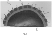
- a patient-specific headset 100 for performing transcranial diagnostic or therapeutic procedures is shown worn on the head 50 of a patient.
- the patient-specific headset 100 which includes a patient-specific frame (support structure) 110 that supports a plurality of transducers 120, conforms to the anatomical contour of at least a portion of the patient's head.
- the patient-specific frame 110 which is shown in cross-section in FIG. 1 , mechanically supports transducers 120 in pre-selected positions and orientations.
- the transducers 120 may be used to transmit and/or receive energy for brain diagnostic or therapeutic purposes or for localization of the skull surface.
- the patient-specific frame 110 includes a plurality of attachment interfaces for receiving and supporting the transducers 120.
- the attachment interfaces are provided as apertures (recesses) into which the transducers 120 are placed.
- the transducers 120 may be affixed to the patient-specific frame 110 according to a wide variety of different means, such as, but not limited to, with an attachment mechanism (e.g. via fasteners that extend into the patient-specific frame 110, optionally into pre-formed holes), or an adhesive such as a glue.
- the transducers 120 are remotely interfaced with electronics through wires or through a flexible printed circuit board 140.
- the transducers 120 may be removably attachable to the patient-specific frame 110.
- the patient-specific headset may also include a coupling layer 130 that is provided adjacent to an inner surface of the patient-specific frame.
- the outer surface of the coupling layer 130 contacts distal surfaces of the transducers 120, and the inner surface of the coupling layer contacts the patient's head 50, thereby facilitating coupling of energy between the transducers in the patient-specific frame and the patient's head.
- the inclusion of the coupling layer 130, and the composition and/or geometry of the coupling layer may be dependent on the type of transducers 120.
- the coupling layer 130 may be an acoustic coupling layer that facilitates propagation of acoustic waves and reduces reflections at interfaces.
- the coupling layer 130 includes an elastic membrane that retains a liquid layer between the transducer surfaces and the elastic membrane, such that coupling to the skin is achieved.
- FIG. 1 shows an example embodiment in which the transducers are ultrasound transducers, it will be understood that the transducers may be any transducers capable of emitting or receiving energy.
- Non-limiting examples of different types of transducers include ultrasound transducers configured to emit and/or receive ultrasound energy, magnetic resonance coils (radio-frequency coils) and optical transducers such as lasers, light emitting diodes, and optical fibers coupled to sources and/or detectors.
- the transducers need not all be of the same type, and a first portion of the transducers may be selected to emit and/or detect a first type of energy (e.g. ultrasound), and another portion of the transducers may be selected to emit and/or detect a second type of energy (e.g. optical or electromagnetic waves).
- a first type of energy e.g. ultrasound
- second type of energy e.g. optical or electromagnetic waves
- a first subset of the transducers may be ultrasound transducers, and a second subset of the transducers may be optical transducers (e.g. optical fibers in optical communication with a source and/or detector), such that the patient-specific headset is capable of performing photoacoustic imaging.
- optical transducers e.g. optical fibers in optical communication with a source and/or detector
- a first subset of the transducers may be ultrasound transducers, and a second subset of the transducers may be MRI coils, such that the system is suitable for performing simultaneous ultrasound and MR imaging or sonications while using MR imaging.
- a first subset of the transducers may be MRI coils, and a second subset of the transducers may be positron emission detector (PET), such that the system is suitable for performing both MR and PET imaging.
- PET positron emission detector
- a first subset of the transducers may be MRI coils, and wherein a second subset of the transducers may be positron emission detector (PET), and third subset of the transducers may be ultrasound transducer, such that said system is suitable for performing both MR and PET imaging while delivering ultrasound therapy or imaging.
- PET positron emission detector
- the transducers are may be transducer elements for forming a phased array (i.e. the transducers may be phased-array transducers).
- Such phased-array transducers may be spatially arranged on the patient-specific headset to provide a full phased array, or a sparse phased array.
- the phased-array transducers may be provided as a plurality of sub-arrays, where each sub-array is mechanically supported as a separate transducer module, such that each transducer module is mechanically supported on the patient-specific frame 110 by a respective attachment interface.
- each transducer 120 may be a transducer module housing a sub-array of transducers, such that the sub-arrays, spatially distributed on the patient-specific headset 100, separately form distinct phased arrays, or collectively form a composite phased-array.
- the transducer modules, and their respective attachment interfaces may have unique shapes (i.e.
- the transducer modules may be respectively keyed), such that a given transducer module fits uniquely with its respective attachment interface. If the phased array is formed by a set of transducer modules housing respective sub-arrays, the transducer modules may be spatially distributed on the patient-specific frame to reduce or minimize the formation of grating lobes.
- the patient-specific frame conforms to the anatomical contour of at least a portion of the patient's head.
- a conformal frame may be fabricated based on volumetric image data of the patient's head.
- FIG. 2 illustrates an example method for fabricating a patient-specific frame based on volumetric image data associated with the patient.
- steps 200 and 210 volumetric image data of a patient's head is obtained and processed to provide surface data characterizing an anatomical curvature (e.g. skin or bone surface) of a portion of the patient's head.
- the volumetric data may be obtained, for example, by performing imaging using an imaging modality such as, but not limited to, magnetic resonance (MR) imaging and computed tomography (CT) imaging.
- MR magnetic resonance
- CT computed tomography
- the volumetric image data may be obtained based on a previously performed imaging procedure.
- the volumetric image data may be processed and segmented to obtain surface data characterizing the surface of a portion of the patient's skull.
- Such surface segmentation may be performed, for example, using imaging processing software such as the Mimics TM software platform (Materialise, Belgium).
- imaging processing software such as the Mimics TM software platform (Materialise, Belgium).
- Such software enables the creation of a 3D model (the surface data) of the surface of a portion of the patient's head.
- the model may be created using known techniques, such as using the steps of thresholding, region growing and manual editing. Automatic thresholding may be performed to achieve a first approximation of the bony surfaces of the skull, followed by manual editing to obtain a refined model.
- Haptic modeling for example using a modeling software platform such as the PHANTOM TM Desktop Haptic Device, may be used to further refine the model. Additional example methods of image processing and segmentation of volumetric image data are disclosed in US Patent No. 8,086,336 .
- the surface data is used to produce a digital model of the patient-specific frame.
- a suitable software platform such as the software package Surfacer TM
- the model is then modified or refined (e.g. updated) to include a plurality of transducer attachment interfaces for receiving and supporting a plurality of transducers in pre-selected positions and orientations relative to the patient's head, and for supporting the transducers such that energy is coupled transcranially.
- the positions and orientations of the transducer attachment interfaces may be selected, for example, to form one or more phased arrays of transducers.
- the transducer modules may be spatially distributed on the patient-specific frame to reduce or minimize the formation of grating lobes.
- the digital model may be further refined to include one or more additional features, such as, but not limited to, an attachment interface for the attachment of one or more fiducial markers, an aperture to permit surgical access to a selected region of the patient's head when the patient-specific frame is worn, markers for identifying reference directions, and one or more positioning features such as external handles.
- additional features such as, but not limited to, an attachment interface for the attachment of one or more fiducial markers, an aperture to permit surgical access to a selected region of the patient's head when the patient-specific frame is worn, markers for identifying reference directions, and one or more positioning features such as external handles.
- the digital model, updated to include the transducer attachment interfaces, is then employed to fabricate the patient-specific frame, as shown at step 240.
- the patient-specific frame may be fabricated from the model using 3D printing.
- the model may be employed to produce a mold suitable for forming the patient-specific frame, and the mold may be subsequently employed to fabricate the patient-specific frame.
- the transducers After having fabricated the patient-specific frame, the transducers (or transducer assemblies or modules) are secured (attached, adhered, etc.) to the respective transducer attachment interfaces of the patient-specific frame, as shown at step 250.
- a relationship may be established between the positions and orientations of the transducers and the volumetric image data (i.e. so that both can be represented within a common reference frame).
- the known positions and orientations of the transducers are spatially registered relative to the volumetric image data, thereby generating transducer registration data characterizing the positions and orientations of the transducers relative to the volumetric image data.
- transducer registration data may include the spatial coordinates of the transducers, and vectors identifying their respective orientations, in the reference frame of the volumetric data.
- the transducer registration data may include a coordinate transformation for transforming the positions and orientations of the transducers from a first reference frame to the reference frame of the volumetric image data.
- the transducer registration data enables the determination of the positions and orientations of the transducers relative to the volumetric image data, enabling, for example, the determination of suitable beamforming parameters to pulse one or more phased arrays (e.g. sub-arrays) of transducers to focus an energy beam at a specific location or region within the patient's head.
- a subset of transducers may be configured to emit an energy beam toward the skull of the patient and to detect energy that is reflected from the skull in order to facilitate the detection, for each transducer in the subset of transducers, of a local spatial offset of the skull of the patient relative to the patient-specific frame.
- the detected spatial offsets may then be employed to correct a spatial registration of the transducers relative to the patient anatomy (e.g. the skull and/or one or more internal tissue regions of interest) or to perform corrections based on the detected signals, for example, as disclosed in US Patent No. 6,612,988 .
- the subset of transducers may be ultrasound transducers, or, for example, optical fibers operably connected to an optical coherence tomography system.
- the registration between the headset and the head and brain can be achieved by performing imaging (for example MRI, CT, thomosynthesis, or x-ray) with the headset placed on the subjects head, allowing the transducer locations to be determined from the imaging visible fiducial markers in the headset.
- imaging for example MRI, CT, thomosynthesis, or x-ray
- FIG. 3 provides a block diagram illustrating an example implementation of a system for performing diagnostic or therapeutic transcranial procedures.
- Control and processing hardware 300 is operably connected to the patient-specific transcranial headset 100, optionally via transducer driver electronics/circuitry 380.
- the control and processing hardware 300 which includes one or more processors 310 (for example, a CPU/microprocessor), bus 305, memory 315, which may include random access memory (RAM) and/or read only memory (ROM), a data acquisition interface 320, a display 325, external storage 330, one more communications interfaces 335, a power supply 340, and one or more input/output devices and/or interfaces 345 (e.g. a speaker, a user input device, such as a keyboard, a keypad, a mouse, a position tracked stylus, a position tracked probe, a foot switch, and/or a microphone for capturing speech commands).
- processors 310 for example, a CPU/microprocessor
- bus 305 memory 315, which may include random access memory (RAM) and/or read only memory (ROM), a data acquisition interface 320, a display 325, external storage 330, one more communications interfaces 335, a power supply 340, and one or more input/output devices and/or interfaces
- Volumetric image data 370 and transducer registration data 375 may be stored on an external database or stored in memory 315 or storage 330 of control and processing hardware 300.
- the tracking system 365 may optionally be employed to track the position and orientation of the patient, via detection of one or more fiducial markers 160 attached to the patient-specific headset 100, and optionally one or more medical instruments or devices also having fiducial markers attached thereto. For example, passive or active signals emitted from the fiducial markers may be detected by a stereographic tracking system employing two tracking cameras.
- the transducer driving electronics/circuitry 380 may include, for example, but is not limited to, Tx/Rx switches, transmit and/or receive beamformers.
- the control and processing hardware 300 may be programmed with programs, subroutines, applications or modules 350, which include executable instructions, which when executed by the one or more processors 310, causes the system to perform one or more methods described in the present disclosure. Such instructions may be stored, for example, in memory 315 and/or other storage.
- the transducer control module 355 includes executable instructions for controlling the transducers of the patient-specific transcranial headset 100 to deliver energy to a target location or region of interest, based on the registration of the transducer positions and orientations with the volumetric image data as per the transducer registration data 375.
- the patient-specific headset 100 may support a plurality of phased-array transducers, and transducer control module 355 may control the beamforming applied (on transmit and/or receive) to deliver, based on the known positions and orientations of the phased array transducers relative to the volumetric image data, one or more focused energy beams to a region of interest.
- the region of interest may be specified intraoperatively by a user (e.g. via a user interface controlled by control and processing hardware 300) or according to a pre-established surgical plan.
- the registration module 360 may optionally be employed for registering volumetric image data 370 to an intraoperative reference frame associated with tracking system 365.
- the optional guidance user interface module 362 includes executable instructions for displaying a user interface showing spatially registered volumetric images for image-guided procedures.
- the registration module 360 may also intraoperatively receive spatial correction information based on a detected spatial offset between the patient-specific frame and the patient's head (which, as described above, may be provided by a subset of distance-sensing transducers) and employ this spatial correction information to dynamically adjust (e.g. correct) the registration between the transducers and the volumetric image data.
- each component is illustrated in FIG. 3
- any number of each component can be included in the control and processing hardware 300.
- a computer typically contains a number of different data storage media.
- bus 305 is depicted as a single connection between all of the components, it will be appreciated that the bus 305 may represent one or more circuits, devices or communication channels which link two or more of the components.
- bus 305 often includes or is a motherboard.
- Control and processing hardware 300 may include many more or less components than those shown.
- control and processing hardware 300 may be implemented as one or more physical devices that are coupled to processor 310 through one of more communications channels or interfaces.
- control and processing hardware 300 can be implemented using application specific integrated circuits (ASICs).
- ASICs application specific integrated circuits
- control and processing hardware 300 can be implemented as a combination of hardware and software, where the software is loaded into the processor from the memory or over a network connection.
- Some aspects of the present disclosure can be embodied, at least in part, in software, which, when executed on a computing system, transforms a computing system into a specialty-purpose computing system that is capable of performing the methods disclosed herein. That is, the techniques can be carried out in a computer system or other data processing system in response to its processor, such as a microprocessor, executing sequences of instructions contained in a memory, such as ROM, volatile RAM, non-volatile memory, cache, magnetic and optical disks, or a remote storage device. Further, the instructions can be downloaded into a computing device over a data network in a form of compiled and linked version.
- the logic to perform the processes as discussed above could be implemented in additional computer and/or machine readable media, such as discrete hardware components as large-scale integrated circuits (LSI's), application-specific integrated circuits (ASIC's), or firmware such as electrically erasable programmable read-only memory (EEPROM's) and field-programmable gate arrays (FPGAs).
- LSI's large-scale integrated circuits
- ASIC's application-specific integrated circuits
- firmware such as electrically erasable programmable read-only memory (EEPROM's) and field-programmable gate arrays (FPGAs).
- a computer readable medium can be used to store software and data which when executed by a data processing system causes the system to perform various methods.
- the executable software and data can be stored in various places including for example ROM, volatile RAM, non-volatile memory and/or cache. Portions of this software and/or data can be stored in any one of these storage devices.
- a machine readable medium includes any mechanism that provides (i.e., stores and/or transmits) information in a form accessible by a machine (e.g., a computer, network device, personal digital assistant, manufacturing tool, any device with a set of one or more processors, etc.).
- Examples of computer-readable media include but are not limited to recordable and non-recordable type media such as volatile and non-volatile memory devices, read only memory (ROM), random access memory (RAM), flash memory devices, floppy and other removable disks, magnetic disk storage media, optical storage media (e.g., compact discs (CDs),digital versatile disks (DVDs), etc.), among others.
- the instructions can be embodied in digital and analog communication links for electrical, optical, acoustical or other forms of propagated signals, such as carrier waves, infrared signals, digital signals, and the like.
- the phrases “computer readable material” and “computer readable storage medium” refer to all computer-readable media, except for a transitory propagating signal per se.
- the patient-specific headset and associated transducer registration data may be employed for a wide variety of transcranial procedures, including, but not limited to, neuromodulation, neurostimulation, neuroimaging, neuro-monitoring, focused-ultrasound transcranial ablation, mild heating (hyperthermia), neuromodulation, neurostimulation, mechanical excitation of the brain for diagnostic or therapeutic purposes, manipulation, control, excitation or sensing of gas bubbles, liquid droplets, solid particles, cells, nanoparticles, quantum dots or electronic circuits or devices, focused-ultrasound transcranial excitation or sensing of brain implants, devices, electronic circuits or sensors and transcranial procedures involving the use of focused ultrasound to disruption and opening of the blood-brain barrier for delivery of therapeutic or diagnostic agents, cells, particles, droplets, bubbles, electronic devices, transmitters, sensors or other foreign material for diagnostic purposes.
- the systems, devices and method disclosed herein may be adapted to provide a patient-specific apparatus for performing diagnostic or therapeutic procedures on other parts or portions of the body.
- the patient-specific frame may be fabricated according to volumetric image data of other body regions or body portions.
- a patient-specific frame may be fabricated, based on volumetric image data of a patient's knee, such that the patient-specific frame conforms to the contour of the patient's knee, for performing a diagnostic or therapeutic procedure on the knee using the transducer supported by the patient-specific frame.
- a patient-specific frame may be fabricated, based on volumetric image data of a patient's spine, such that the patient-specific frame conforms to the contour of the patient's spine, for performing a diagnostic or therapeutic procedure on the spine using the transducer supported by the patient-specific frame.
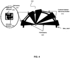
- an example patient-specific headset 400 is shown, where the patient specific frame 410 (sub-array holder) supports a plurality of focused ultrasound phased array transducers.
- This example system may be employed, for example, for neuromodulation experiments or treatments in humans or large animals.
- ultrasound beams 420 are generated by multiple transducer sub-array modules 430, where each transducer sub-array module includes, in one example implementation, a set of 64 transducer elements spaced at the center-to-center distance of half-wavelength, making complete electronic beam steering possible.
- each transducer sub-array module includes, in one example implementation, a set of 64 transducer elements spaced at the center-to-center distance of half-wavelength, making complete electronic beam steering possible.
- the module size of the aforementioned example implementation would be approximately 8.5 mm x 8.5 mm, but will scale inversely with frequency. Larger center-to-center spacing could be used by limiting the steering range, accepting some grating lobes, or by making the array surface curved such that it and only steering the array focus to a limited volume of tissue.
- Each of the modules may be connected to a 64-channel RF-driving board via a flex circuit.
- a photograph of an example of such a sub-array module is shown in FIG. 5A.
- FIGS. 5B and 5C shows an image of a plurality of sub-modules assembled and supported on a patient-specific frame.
- the aforementioned modules may be provided as sub-modules 440 that form a sub-array module 430 connected to a rigid base that will support and hold the module in place.
- Each sub-array module 430 may also house one or more (e.g. four) PVDF-wideband receivers 450 to detect reflections from the skull surfaces and scattering from the brain tissue.
- the location and number of these modules may be selected, on a per-patient basis, based on computer simulations, such that they all form a sparse (e.g. optimal) array where the distances between the modules have suitable (e.g. maximal) variability to prevent grating lobe formation.
- the computer simulations employ volumetric image data (e.g. MRI or CT-scans) of the subject's head to obtain a model of the surface of the patient's head, and to perform the calculations of the spatial distribution of the transducers modules relative to the patient anatomy.
- This information may be used with a 3D-printer to form a plastic support (frame) that fits snugly over the subject's head, like a custom helmet that has mounting holes for the insertion of the sub-arrays.
- the sub-arrays may have unique shapes, so they would fit only in the locations determined by the computer plan.
- high-frequency transmit-receive elements are also integrated in the sub-array assemblies (e.g. 128 high-frequency elements), which can be used to determine the distance of the skull from the array. As noted above, such distances can be used to improve the registration of previously obtained volumetric imaging data to the array co-ordinates, and potential avoiding the need for additional intraoperative imaging. These high-frequency elements can be used continuously to track any head motion, and thus, there is no need for invasive pin fixation, as is the case with the current devices.
- an elastic membrane may be provided that secures a liquid layer between the module surfaces and the membrane, thereby facilitating coupling to the skin when the patient-specific headset is worn.
- a headset fabricated according to the design described in the present example may be capable of driving 256 sub-arrays, resulting in an array of over 16,000 individual elements and 64 modules, although fewer elements may be required if adequate focusing can be achieved.
- the maximal area of human skull cap that can be used to propagate ultrasound is expected to vary approximately between 450 and 700 cm 2 ; thus, the complete array may not be fully populated, and may instead be sparse, which still maintains the high focus quality with the cost of increased power requirements. It is expected that such sparse array implementations will be compatible with all of the applications, which typically require low power during exposures (for example, such as opening of the blood-brain barrier and neuromodulation).
Landscapes
- Health & Medical Sciences (AREA)
- Life Sciences & Earth Sciences (AREA)
- Engineering & Computer Science (AREA)
- Public Health (AREA)
- Veterinary Medicine (AREA)
- Biomedical Technology (AREA)
- Animal Behavior & Ethology (AREA)
- General Health & Medical Sciences (AREA)
- Medical Informatics (AREA)
- Surgery (AREA)
- Physics & Mathematics (AREA)
- Pathology (AREA)
- Heart & Thoracic Surgery (AREA)
- Molecular Biology (AREA)
- Biophysics (AREA)
- Nuclear Medicine, Radiotherapy & Molecular Imaging (AREA)
- Radiology & Medical Imaging (AREA)
- Neurology (AREA)
- Neurosurgery (AREA)
- High Energy & Nuclear Physics (AREA)
- Acoustics & Sound (AREA)
- Oral & Maxillofacial Surgery (AREA)
- Psychology (AREA)
- Physiology (AREA)
- Gynecology & Obstetrics (AREA)
- Optics & Photonics (AREA)
- Physical Education & Sports Medicine (AREA)
- Ultra Sonic Daignosis Equipment (AREA)
- Magnetic Resonance Imaging Apparatus (AREA)
- Surgical Instruments (AREA)
- Nuclear Medicine (AREA)
Applications Claiming Priority (3)
| Application Number | Priority Date | Filing Date | Title |
|---|---|---|---|
| US201662298873P | 2016-02-23 | 2016-02-23 | |
| PCT/CA2017/050230 WO2017143444A1 (en) | 2016-02-23 | 2017-02-23 | Patient-specific headset for diagnostic and therapeutic transcranial procedures |
| EP17755681.8A EP3419548B1 (de) | 2016-02-23 | 2017-02-23 | Verfahren zur herstellung eines transkraniellen kopfteils für diagnostische oder therapeutische verfahren |
Related Parent Applications (2)
| Application Number | Title | Priority Date | Filing Date |
|---|---|---|---|
| PCT/CA2017/050230 Previously-Filed-Application WO2017143444A1 (en) | 2016-02-23 | 2017-02-23 | Patient-specific headset for diagnostic and therapeutic transcranial procedures |
| EP17755681.8A Division EP3419548B1 (de) | 2016-02-23 | 2017-02-23 | Verfahren zur herstellung eines transkraniellen kopfteils für diagnostische oder therapeutische verfahren |
Publications (2)
| Publication Number | Publication Date |
|---|---|
| EP4438112A2 true EP4438112A2 (de) | 2024-10-02 |
| EP4438112A3 EP4438112A3 (de) | 2024-12-11 |
Family
ID=59684665
Family Applications (2)
| Application Number | Title | Priority Date | Filing Date |
|---|---|---|---|
| EP17755681.8A Active EP3419548B1 (de) | 2016-02-23 | 2017-02-23 | Verfahren zur herstellung eines transkraniellen kopfteils für diagnostische oder therapeutische verfahren |
| EP24188947.6A Pending EP4438112A3 (de) | 2016-02-23 | 2017-02-23 | Verfahren zur herstellung eines transkraniellen kopfteils für diagnostische oder therapeutische verfahren |
Family Applications Before (1)
| Application Number | Title | Priority Date | Filing Date |
|---|---|---|---|
| EP17755681.8A Active EP3419548B1 (de) | 2016-02-23 | 2017-02-23 | Verfahren zur herstellung eines transkraniellen kopfteils für diagnostische oder therapeutische verfahren |
Country Status (10)
| Country | Link |
|---|---|
| US (2) | US11771370B2 (de) |
| EP (2) | EP3419548B1 (de) |
| JP (1) | JP6832958B2 (de) |
| KR (1) | KR102814985B1 (de) |
| CN (1) | CN109219415B (de) |
| AU (1) | AU2017222925B2 (de) |
| BR (1) | BR112018017326B1 (de) |
| CA (1) | CA3015001A1 (de) |
| IL (1) | IL261285B (de) |
| WO (1) | WO2017143444A1 (de) |
Families Citing this family (47)
| Publication number | Priority date | Publication date | Assignee | Title |
|---|---|---|---|---|
| US10974078B2 (en) | 2012-12-27 | 2021-04-13 | Brainsonix Corporation | Treating degenerative dementia with low intensity focused ultrasound pulsation (LIFUP) device |
| KR102677468B1 (ko) * | 2015-02-25 | 2024-06-27 | 디시전 사이선씨즈 메디컬 컴패니, 엘엘씨 | 음향 신호 전송 접촉매질 및 결합 매체 |
| WO2016210133A1 (en) | 2015-06-24 | 2016-12-29 | The Regents Of The Universtiy Of Michigan | Histotripsy therapy systems and methods for the treatment of brain tissue |
| CH711264A2 (de) * | 2015-06-29 | 2016-12-30 | St Gallen Kantonsspital | Implantierbare Ultraschallsender-Anordnung zur Behandlung einer Person. |
| CA3001315C (en) * | 2015-10-08 | 2023-12-19 | Decision Sciences Medical Company, LLC | Acoustic orthopedic tracking system and methods |
| WO2018026738A1 (en) * | 2016-08-01 | 2018-02-08 | Bhaskar Ramamurthy | Ultrasound guided opening of blood-brain barrier |
| WO2018112664A1 (en) * | 2016-12-22 | 2018-06-28 | Sunnybrook Research Institute | Systems and methods for performing transcranial ultrasound therapeutic and imaging procedures |
| US11872415B2 (en) * | 2017-07-06 | 2024-01-16 | John Moshe GOMORI | Device system and method for trans-cranial focused ultrasound without hair shaving |
| US20190083065A1 (en) * | 2017-09-19 | 2019-03-21 | Shuki Vitek | Focal cavitation signal measurement |
| US10265234B1 (en) | 2018-04-20 | 2019-04-23 | Neural Analytics, Inc. | Device pad |
| WO2019209389A1 (en) * | 2018-04-23 | 2019-10-31 | Mr Instruments, Inc. | Wearable open and adjustable mri head coil |
| EP3886737A4 (de) | 2018-11-28 | 2022-08-24 | Histosonics, Inc. | Histotripsiesysteme und -verfahren |
| IL313600A (en) | 2019-02-13 | 2024-08-01 | Alpheus Medical Inc | Non-invasive sonodynamic therapy |
| US11957936B2 (en) * | 2019-05-14 | 2024-04-16 | Vanderbilt University | Passive wire reflectors for improved image quality in MR-guided focused ultrasound |
| KR20220017941A (ko) | 2019-05-29 | 2022-02-14 | 손알라센스, 인코포레이티드 | 초음파감응화 |
| CA3138023A1 (en) | 2019-05-31 | 2020-12-03 | Sunnybrook Research Institute | Systems and methods for reducing thermal skull-induced aberrations during transcranial ultrasound therapeutic procedures |
| EP3979942A1 (de) * | 2019-06-06 | 2022-04-13 | Insightec Ltd. | Verbesserte magnetresonanz(mr)-leistung in mr-geführten ultraschallsystemen |
| US11464497B2 (en) * | 2019-10-09 | 2022-10-11 | Acoustiic Inc. | Modular ultrasonic transducers and frame |
| CN110840395B (zh) * | 2019-10-21 | 2021-06-04 | 同济大学 | 一种头箍式脑功能成像仪 |
| TW202128251A (zh) * | 2019-11-26 | 2021-08-01 | 美商閾限科學公司 | 治療神經系統疾病和腦部狀況的裝置和方法 |
| CN219512390U (zh) | 2019-11-26 | 2023-08-11 | Mr仪器有限公司 | 可佩戴的mri成像系统 |
| CN115209812B (zh) * | 2020-01-27 | 2026-01-27 | 戈尔丹斯医疗公司 | 超声换能器组件 |
| US11813485B2 (en) | 2020-01-28 | 2023-11-14 | The Regents Of The University Of Michigan | Systems and methods for histotripsy immunosensitization |
| KR102433536B1 (ko) * | 2020-02-28 | 2022-08-17 | 중앙대학교 산학협력단 | 심장 초음파 검사기 및 그를 포함하는 3차원 심장 초음파 검사 시스템 |
| US11759661B2 (en) * | 2020-05-20 | 2023-09-19 | Brainsonix Corporation | Ultrasonic transducer treatment device |
| EP4167876A4 (de) | 2020-06-18 | 2024-07-17 | Histosonics, Inc. | Akustische histotripsie- und patientenkopplungssysteme und -verfahren |
| EP4182857A2 (de) * | 2020-07-15 | 2023-05-24 | Google LLC | Generative modellierung von quantenhardware |
| US12112488B2 (en) | 2020-08-06 | 2024-10-08 | Canon U.S.A., Inc. | Methods and systems for image synchronization |
| IL300335A (en) | 2020-08-07 | 2023-04-01 | Alpheus Medical Inc | Ultrasound arrays for improved sonodynamic therapy for cancer treatment |
| US12257446B2 (en) | 2020-08-24 | 2025-03-25 | Brainsonix Corporation | Systems and methods for neuromodulation of neuronal circuits using transcranial focused microwave pulses |
| AU2021332372A1 (en) | 2020-08-27 | 2023-03-16 | The Regents Of The University Of Michigan | Ultrasound transducer with transmit-receive capability for histotripsy |
| EP4026584A1 (de) * | 2021-01-11 | 2022-07-13 | Koninklijke Philips N.V. | Neuromodulation des gehirns eines patienten mit einer kopfspulenvorrichtung |
| GB202100449D0 (en) * | 2021-01-14 | 2021-03-03 | Ucl Business Ltd | Subject positioning |
| US12582848B2 (en) | 2021-06-07 | 2026-03-24 | The Regents Of The University Of Michigan | Minimally invasive histotripsy systems and methods |
| CN113303823A (zh) * | 2021-07-06 | 2021-08-27 | 南京市第一医院 | 一种应用于帕金森氏病诊断的pet/mri一体式成像设备 |
| EP4373435A1 (de) * | 2021-07-19 | 2024-05-29 | The Cleveland Clinic Foundation | Systeme und verfahren zur verwendung mit mrt-geführtem fokussiertem ultraschall |
| EP4169470B1 (de) * | 2021-10-25 | 2024-05-08 | Erbe Vision GmbH | Vorrichtung und verfahren zur positionierung eines patientenkörpers und zur verfolgung der position des patienten während einer operation |
| KR20240127472A (ko) | 2022-01-04 | 2024-08-22 | 유니버시티 오브 유타 리써치 파운데이션 | 심부 뇌 회로들에 대한 조절을 위한 시스템 및 방법 |
| KR102750439B1 (ko) * | 2022-02-03 | 2025-01-09 | 주식회사 딥슨바이오 | 브레인 초음파자극기 고정형 장치 |
| KR102800662B1 (ko) * | 2022-02-03 | 2025-04-30 | 주식회사 딥슨바이오 | 브레인 자극 트랜스듀서 고정형 장치 |
| CN114712735B (zh) * | 2022-04-18 | 2025-02-14 | 上海市第六人民医院 | 一种神经退行性疾病超声治疗仪及其使用方法 |
| CN120129515A (zh) * | 2022-07-21 | 2025-06-10 | 森尼布鲁克研究院 | 通过经颅聚焦超声治疗高血压的方法 |
| JP2026508049A (ja) | 2022-10-28 | 2026-03-10 | ヒストソニックス,インコーポレーテッド | ヒストトリプシシステムおよび方法 |
| KR102626932B1 (ko) * | 2023-01-03 | 2024-01-23 | 주식회사 뉴머스 | 환자 맞춤형 헬멧 및 이의 제조 방법 |
| KR20260003742A (ko) | 2023-04-20 | 2026-01-07 | 히스토소닉스, 인크. | 치료 계획 및 요법을 위한 사용자 인터페이스들 및 작업 흐름들을 포함하는 히스토트립시 시스템들 및 연관된 방법들 |
| US20250189643A1 (en) * | 2023-12-11 | 2025-06-12 | California Institute Of Technology | Nonlinear ultrasound imaging of acoustic biomolecules using coded excitations |
| DE102024122859A1 (de) | 2024-08-09 | 2026-02-12 | Fraunhofer-Gesellschaft zur Förderung der angewandten Forschung eingetragener Verein | Vorrichtung zur nicht-invasiven ultraschallbehandlung einer zielregion innerhalb eines körperteils eines benutzers |
Citations (2)
| Publication number | Priority date | Publication date | Assignee | Title |
|---|---|---|---|---|
| US6612988B2 (en) | 2000-08-29 | 2003-09-02 | Brigham And Women's Hospital, Inc. | Ultrasound therapy |
| US8086336B2 (en) | 2002-09-30 | 2011-12-27 | Medical Modeling Inc. | Method for design and production of a custom-fit prosthesis |
Family Cites Families (25)
| Publication number | Priority date | Publication date | Assignee | Title |
|---|---|---|---|---|
| US7747312B2 (en) * | 2000-01-04 | 2010-06-29 | George Mason Intellectual Properties, Inc. | System and method for automatic shape registration and instrument tracking |
| US6733450B1 (en) * | 2000-07-27 | 2004-05-11 | Texas Systems, Board Of Regents | Therapeutic methods and apparatus for use of sonication to enhance perfusion of tissue |
| US6613005B1 (en) * | 2000-11-28 | 2003-09-02 | Insightec-Txsonics, Ltd. | Systems and methods for steering a focused ultrasound array |
| EP1429659A4 (de) * | 2001-08-24 | 2009-11-11 | Brigham & Womens Hospital | Ultraschalltherapie |
| CN100562293C (zh) | 2003-04-17 | 2009-11-25 | 布赖汉姆妇女医院 | 剪切式诊断超声波系统 |
| US7344509B2 (en) | 2003-04-17 | 2008-03-18 | Kullervo Hynynen | Shear mode therapeutic ultrasound |
| US7175599B2 (en) | 2003-04-17 | 2007-02-13 | Brigham And Women's Hospital, Inc. | Shear mode diagnostic ultrasound |
| WO2006096755A2 (en) * | 2005-03-07 | 2006-09-14 | The Brigham And Women's Hospital, Inc. | Adaptive ultrasound delivery system |
| US20100113959A1 (en) * | 2006-03-07 | 2010-05-06 | Beth Israel Deaconess Medical Center, Inc. | Transcranial magnetic stimulation (tms) methods and apparatus |
| EP2051777B1 (de) | 2006-08-11 | 2019-01-16 | Koninklijke Philips N.V. | Ultraschallsystem zur strömungsabbildung von hirnblut und mikrobläschen-blutgerinnungslyse |
| WO2008065561A1 (en) * | 2006-11-28 | 2008-06-05 | Koninklijke Philips Electronics, N.V. | Apparatus for 3d ultrasound imaging and therapy |
| US7999945B2 (en) | 2007-07-18 | 2011-08-16 | The George Washington University | Optical coherence tomography / acoustic radiation force imaging probe |
| CN101502418B (zh) * | 2008-02-05 | 2011-05-04 | 周常安 | 耳戴式脑电检测装置 |
| US8425424B2 (en) * | 2008-11-19 | 2013-04-23 | Inightee Ltd. | Closed-loop clot lysis |
| GB2470455B (en) * | 2009-04-13 | 2013-07-17 | 1072 Technology Ltd | Phototherapy apparatus |
| BR112012015620A2 (pt) * | 2009-12-28 | 2017-10-17 | Koninl Philips Electronics Nv | ferramenta de planejamento de terapia e método de planejamento da ablação ultrassônica focalizada de alta intensidade (hifu) guiada por ressonância magnética |
| US8603014B2 (en) | 2010-10-05 | 2013-12-10 | Cerevast Therapeutics, Inc. | Hands-free operator-independent transcranial ultrasound apparatus and methods |
| WO2014139029A1 (en) | 2013-03-15 | 2014-09-18 | Neurogate Medical Systems Inc. | Device and method for transcranial magnetic stimulation coil positioning with data integration |
| US9924889B2 (en) * | 2013-10-03 | 2018-03-27 | Medical University Of Vienna | Method and system for combined transcranial magnetic simulation (TMS) and functional magnetic resonance imaging (fMRI) studies |
| JP6124216B2 (ja) * | 2013-10-30 | 2017-05-10 | 国立研究開発法人量子科学技術研究開発機構 | ヘルメット型pet装置 |
| WO2015075603A1 (en) * | 2013-11-21 | 2015-05-28 | Koninklijke Philips N.V. | Ultrasound headset |
| CN105828876A (zh) * | 2013-12-18 | 2016-08-03 | 皇家飞利浦有限公司 | 用于超声溶栓处置的超声图像和计算机断层摄影图像配准的系统和方法 |
| CN104548390B (zh) * | 2014-12-26 | 2018-03-23 | 中国科学院深圳先进技术研究院 | 一种获得用于发射穿颅聚焦超声的超声发射序列的方法及系统 |
| CN104799950A (zh) | 2015-04-30 | 2015-07-29 | 上海昕健医疗技术有限公司 | 基于医学图像的个性化最小创伤膝关节定位导板 |
| WO2018112664A1 (en) * | 2016-12-22 | 2018-06-28 | Sunnybrook Research Institute | Systems and methods for performing transcranial ultrasound therapeutic and imaging procedures |
-
2017
- 2017-02-23 US US16/078,903 patent/US11771370B2/en active Active
- 2017-02-23 BR BR112018017326-6A patent/BR112018017326B1/pt active IP Right Grant
- 2017-02-23 CA CA3015001A patent/CA3015001A1/en active Pending
- 2017-02-23 CN CN201780022821.7A patent/CN109219415B/zh active Active
- 2017-02-23 JP JP2018562396A patent/JP6832958B2/ja active Active
- 2017-02-23 KR KR1020187027197A patent/KR102814985B1/ko active Active
- 2017-02-23 AU AU2017222925A patent/AU2017222925B2/en active Active
- 2017-02-23 EP EP17755681.8A patent/EP3419548B1/de active Active
- 2017-02-23 WO PCT/CA2017/050230 patent/WO2017143444A1/en not_active Ceased
- 2017-02-23 EP EP24188947.6A patent/EP4438112A3/de active Pending
-
2018
- 2018-08-21 IL IL261285A patent/IL261285B/en unknown
-
2023
- 2023-08-29 US US18/239,288 patent/US12426836B2/en active Active
Patent Citations (2)
| Publication number | Priority date | Publication date | Assignee | Title |
|---|---|---|---|---|
| US6612988B2 (en) | 2000-08-29 | 2003-09-02 | Brigham And Women's Hospital, Inc. | Ultrasound therapy |
| US8086336B2 (en) | 2002-09-30 | 2011-12-27 | Medical Modeling Inc. | Method for design and production of a custom-fit prosthesis |
Also Published As
| Publication number | Publication date |
|---|---|
| IL261285A (en) | 2018-10-31 |
| US12426836B2 (en) | 2025-09-30 |
| AU2017222925A1 (en) | 2018-08-30 |
| EP4438112A3 (de) | 2024-12-11 |
| CN109219415B (zh) | 2021-10-26 |
| EP3419548A4 (de) | 2020-03-18 |
| KR20180116345A (ko) | 2018-10-24 |
| BR112018017326B1 (pt) | 2022-12-13 |
| JP6832958B2 (ja) | 2021-02-24 |
| JP2019510599A (ja) | 2019-04-18 |
| CN109219415A (zh) | 2019-01-15 |
| US20190021666A1 (en) | 2019-01-24 |
| KR102814985B1 (ko) | 2025-05-30 |
| IL261285B (en) | 2022-02-01 |
| US11771370B2 (en) | 2023-10-03 |
| BR112018017326A2 (pt) | 2019-04-09 |
| EP3419548A1 (de) | 2019-01-02 |
| WO2017143444A1 (en) | 2017-08-31 |
| AU2017222925B2 (en) | 2021-11-04 |
| EP3419548B1 (de) | 2024-07-17 |
| US20230397881A1 (en) | 2023-12-14 |
| CA3015001A1 (en) | 2017-08-31 |
Similar Documents
| Publication | Publication Date | Title |
|---|---|---|
| US12426836B2 (en) | Patient-specific headset for diagnostic and therapeutic transcranial procedures | |
| US12290403B2 (en) | Systems and methods for performing transcranial ultrasound therapeutic and imaging procedures | |
| JP5522741B2 (ja) | 治療の超音波振動子の位置追跡のための方法及び装置 | |
| US20230398381A1 (en) | Multiparametric optimization for ultrasound procedures | |
| US20200085409A1 (en) | Ultrasound focusing utilizing a 3d-printed skull replica | |
| Joe et al. | Development of a subject-specific guide system for Low-Intensity Focused Ultrasound (LIFU) brain stimulation | |
| US8360978B2 (en) | Ultrasound device for medical applications |
Legal Events
| Date | Code | Title | Description |
|---|---|---|---|
| PUAI | Public reference made under article 153(3) epc to a published international application that has entered the european phase |
Free format text: ORIGINAL CODE: 0009012 |
|
| STAA | Information on the status of an ep patent application or granted ep patent |
Free format text: STATUS: REQUEST FOR EXAMINATION WAS MADE |
|
| 17P | Request for examination filed |
Effective date: 20240716 |
|
| AC | Divisional application: reference to earlier application |
Ref document number: 3419548 Country of ref document: EP Kind code of ref document: P |
|
| AK | Designated contracting states |
Kind code of ref document: A2 Designated state(s): AL AT BE BG CH CY CZ DE DK EE ES FI FR GB GR HR HU IE IS IT LI LT LU LV MC MK MT NL NO PL PT RO RS SE SI SK SM TR |
|
| REG | Reference to a national code |
Ref country code: DE Ref legal event code: R079 Free format text: PREVIOUS MAIN CLASS: A61N0007000000 Ipc: A61B0090140000 |
|
| PUAL | Search report despatched |
Free format text: ORIGINAL CODE: 0009013 |
|
| AK | Designated contracting states |
Kind code of ref document: A3 Designated state(s): AL AT BE BG CH CY CZ DE DK EE ES FI FR GB GR HR HU IE IS IT LI LT LU LV MC MK MT NL NO PL PT RO RS SE SI SK SM TR |
|
| RIC1 | Information provided on ipc code assigned before grant |
Ipc: A61N 7/00 20060101ALI20241105BHEP Ipc: A61B 8/13 20060101ALI20241105BHEP Ipc: A61B 6/03 20060101ALI20241105BHEP Ipc: A61B 5/055 20060101ALI20241105BHEP Ipc: A61B 90/14 20160101AFI20241105BHEP |
|
| GRAP | Despatch of communication of intention to grant a patent |
Free format text: ORIGINAL CODE: EPIDOSNIGR1 |
|
| STAA | Information on the status of an ep patent application or granted ep patent |
Free format text: STATUS: GRANT OF PATENT IS INTENDED |
|
| INTG | Intention to grant announced |
Effective date: 20260216 |