EP4403626A1 - Kulturmedium und kulturverfahren für lungenkrebsepithelzellen und anwendung davon - Google Patents
Kulturmedium und kulturverfahren für lungenkrebsepithelzellen und anwendung davon Download PDFInfo
- Publication number
- EP4403626A1 EP4403626A1 EP21957266.6A EP21957266A EP4403626A1 EP 4403626 A1 EP4403626 A1 EP 4403626A1 EP 21957266 A EP21957266 A EP 21957266A EP 4403626 A1 EP4403626 A1 EP 4403626A1
- Authority
- EP
- European Patent Office
- Prior art keywords
- lung cancer
- cells
- culture medium
- alkyl
- culture
- Prior art date
- Legal status (The legal status is an assumption and is not a legal conclusion. Google has not performed a legal analysis and makes no representation as to the accuracy of the status listed.)
- Pending
Links
Images


Classifications
-
- C—CHEMISTRY; METALLURGY
- C12—BIOCHEMISTRY; BEER; SPIRITS; WINE; VINEGAR; MICROBIOLOGY; ENZYMOLOGY; MUTATION OR GENETIC ENGINEERING
- C12Q—MEASURING OR TESTING PROCESSES INVOLVING ENZYMES, NUCLEIC ACIDS OR MICROORGANISMS; COMPOSITIONS OR TEST PAPERS THEREFOR; PROCESSES OF PREPARING SUCH COMPOSITIONS; CONDITION-RESPONSIVE CONTROL IN MICROBIOLOGICAL OR ENZYMOLOGICAL PROCESSES
- C12Q1/00—Measuring or testing processes involving enzymes, nucleic acids or microorganisms; Compositions therefor; Processes of preparing such compositions
- C12Q1/02—Measuring or testing processes involving enzymes, nucleic acids or microorganisms; Compositions therefor; Processes of preparing such compositions involving viable microorganisms
-
- C—CHEMISTRY; METALLURGY
- C12—BIOCHEMISTRY; BEER; SPIRITS; WINE; VINEGAR; MICROBIOLOGY; ENZYMOLOGY; MUTATION OR GENETIC ENGINEERING
- C12N—MICROORGANISMS OR ENZYMES; COMPOSITIONS THEREOF; PROPAGATING, PRESERVING, OR MAINTAINING MICROORGANISMS; MUTATION OR GENETIC ENGINEERING; CULTURE MEDIA
- C12N5/00—Undifferentiated human, animal or plant cells, e.g. cell lines; Tissues; Cultivation or maintenance thereof; Culture media therefor
- C12N5/06—Animal cells or tissues; Human cells or tissues
- C12N5/0602—Vertebrate cells
- C12N5/0693—Tumour cells; Cancer cells
-
- C—CHEMISTRY; METALLURGY
- C12—BIOCHEMISTRY; BEER; SPIRITS; WINE; VINEGAR; MICROBIOLOGY; ENZYMOLOGY; MUTATION OR GENETIC ENGINEERING
- C12N—MICROORGANISMS OR ENZYMES; COMPOSITIONS THEREOF; PROPAGATING, PRESERVING, OR MAINTAINING MICROORGANISMS; MUTATION OR GENETIC ENGINEERING; CULTURE MEDIA
- C12N5/00—Undifferentiated human, animal or plant cells, e.g. cell lines; Tissues; Cultivation or maintenance thereof; Culture media therefor
- C12N5/06—Animal cells or tissues; Human cells or tissues
-
- C—CHEMISTRY; METALLURGY
- C12—BIOCHEMISTRY; BEER; SPIRITS; WINE; VINEGAR; MICROBIOLOGY; ENZYMOLOGY; MUTATION OR GENETIC ENGINEERING
- C12N—MICROORGANISMS OR ENZYMES; COMPOSITIONS THEREOF; PROPAGATING, PRESERVING, OR MAINTAINING MICROORGANISMS; MUTATION OR GENETIC ENGINEERING; CULTURE MEDIA
- C12N5/00—Undifferentiated human, animal or plant cells, e.g. cell lines; Tissues; Cultivation or maintenance thereof; Culture media therefor
- C12N5/10—Cells modified by introduction of foreign genetic material
-
- C—CHEMISTRY; METALLURGY
- C12—BIOCHEMISTRY; BEER; SPIRITS; WINE; VINEGAR; MICROBIOLOGY; ENZYMOLOGY; MUTATION OR GENETIC ENGINEERING
- C12N—MICROORGANISMS OR ENZYMES; COMPOSITIONS THEREOF; PROPAGATING, PRESERVING, OR MAINTAINING MICROORGANISMS; MUTATION OR GENETIC ENGINEERING; CULTURE MEDIA
- C12N2500/00—Specific components of cell culture medium
- C12N2500/05—Inorganic components
- C12N2500/10—Metals; Metal chelators
- C12N2500/20—Transition metals
- C12N2500/24—Iron; Fe chelators; Transferrin
-
- C—CHEMISTRY; METALLURGY
- C12—BIOCHEMISTRY; BEER; SPIRITS; WINE; VINEGAR; MICROBIOLOGY; ENZYMOLOGY; MUTATION OR GENETIC ENGINEERING
- C12N—MICROORGANISMS OR ENZYMES; COMPOSITIONS THEREOF; PROPAGATING, PRESERVING, OR MAINTAINING MICROORGANISMS; MUTATION OR GENETIC ENGINEERING; CULTURE MEDIA
- C12N2501/00—Active agents used in cell culture processes, e.g. differentation
- C12N2501/10—Growth factors
- C12N2501/11—Epidermal growth factor [EGF]
-
- C—CHEMISTRY; METALLURGY
- C12—BIOCHEMISTRY; BEER; SPIRITS; WINE; VINEGAR; MICROBIOLOGY; ENZYMOLOGY; MUTATION OR GENETIC ENGINEERING
- C12N—MICROORGANISMS OR ENZYMES; COMPOSITIONS THEREOF; PROPAGATING, PRESERVING, OR MAINTAINING MICROORGANISMS; MUTATION OR GENETIC ENGINEERING; CULTURE MEDIA
- C12N2501/00—Active agents used in cell culture processes, e.g. differentation
- C12N2501/10—Growth factors
- C12N2501/15—Transforming growth factor beta (TGF-β)
-
- C—CHEMISTRY; METALLURGY
- C12—BIOCHEMISTRY; BEER; SPIRITS; WINE; VINEGAR; MICROBIOLOGY; ENZYMOLOGY; MUTATION OR GENETIC ENGINEERING
- C12N—MICROORGANISMS OR ENZYMES; COMPOSITIONS THEREOF; PROPAGATING, PRESERVING, OR MAINTAINING MICROORGANISMS; MUTATION OR GENETIC ENGINEERING; CULTURE MEDIA
- C12N2501/00—Active agents used in cell culture processes, e.g. differentation
- C12N2501/30—Hormones
- C12N2501/345—Gastrin; Cholecystokinins [CCK]
-
- C—CHEMISTRY; METALLURGY
- C12—BIOCHEMISTRY; BEER; SPIRITS; WINE; VINEGAR; MICROBIOLOGY; ENZYMOLOGY; MUTATION OR GENETIC ENGINEERING
- C12N—MICROORGANISMS OR ENZYMES; COMPOSITIONS THEREOF; PROPAGATING, PRESERVING, OR MAINTAINING MICROORGANISMS; MUTATION OR GENETIC ENGINEERING; CULTURE MEDIA
- C12N2501/00—Active agents used in cell culture processes, e.g. differentation
- C12N2501/70—Enzymes
- C12N2501/72—Transferases [EC 2.]
- C12N2501/727—Kinases (EC 2.7.)
-
- C—CHEMISTRY; METALLURGY
- C12—BIOCHEMISTRY; BEER; SPIRITS; WINE; VINEGAR; MICROBIOLOGY; ENZYMOLOGY; MUTATION OR GENETIC ENGINEERING
- C12N—MICROORGANISMS OR ENZYMES; COMPOSITIONS THEREOF; PROPAGATING, PRESERVING, OR MAINTAINING MICROORGANISMS; MUTATION OR GENETIC ENGINEERING; CULTURE MEDIA
- C12N2503/00—Use of cells in diagnostics
- C12N2503/02—Drug screening
-
- C—CHEMISTRY; METALLURGY
- C12—BIOCHEMISTRY; BEER; SPIRITS; WINE; VINEGAR; MICROBIOLOGY; ENZYMOLOGY; MUTATION OR GENETIC ENGINEERING
- C12N—MICROORGANISMS OR ENZYMES; COMPOSITIONS THEREOF; PROPAGATING, PRESERVING, OR MAINTAINING MICROORGANISMS; MUTATION OR GENETIC ENGINEERING; CULTURE MEDIA
- C12N2533/00—Supports or coatings for cell culture, characterised by material
- C12N2533/90—Substrates of biological origin, e.g. extracellular matrix, decellularised tissue
Definitions
- the invention relates to the field of medical technology, in particular to a culture medium and a culture method for culturing or expanding primary lung cancer epithelial cells in vitro, and also to a method and application of the cultured cells in the efficacy evaluating and screening of drugs.
- Lung cancer is currently the most common respiratory tract tumor in the world. There are more than 1.8 million new patients of lung cancer worldwide every year. As the most common malignant tumor in clinical practice, lung cancer is mainly treated by surgery, chemotherapy, radiotherapy, molecular targeted therapy and immunotherapy, among which surgery, chemotherapy and radiotherapy are the most commonly used means. However, there are limitations in the suitable population for these clinical treatments. In recent years, with the rise and development of molecular biology, pharmacotherapy of tumor has shown a diversified trend, among which molecular targeted drugs have become a hot research topic in clinical treatment of lung cancer due to their strong targeting and high safety. However, among the many clinical treatment options, it is particularly important to choose the one that suits the patient.
- Functional testing refers to the method of detecting the sensitivity of anti-tumor drugs on cells of cancer patient in vitro.
- the key to apply this method is to develop tumor cell models that have short growth cycle and can represent the biological characteristics of patients with lung cancer.
- the cell model should be easy to operate to quickly and efficiently predict the efficacy of clinical medication, so as to give precise medication guidance to cancer patients in a timely manner.
- the normally low success rates and long growth cycles of in vitro establishment of cell models from the primary tumor cells of cancer patients, and the problems such as excessive proliferation of mesenchymal cells (such as fibroblasts, and the like), all restrict the development in this field.
- Cell reprogramming is a technique in which the autologous primary epithelial cells from a patient are cultured with mouse-derived feeder cells.
- the primary cells from a patient are tested for drug sensitivity, the presence of these mouse-derived cells may interfere with the drug sensitivity testing results of the patient's autologous primary cells; however, if the mouse-derived feeder cells are removed, the patient's autologous primary cells will detach from the reprogramming environment, and the cell proliferation rate and intracellular signaling pathways will undergo significant changes ( Liu et al., Am J Pathol, 183(6): 1862-1870, 2013 ; Liu et al., Cell Death Dis., 9(7) : 750, 2018 ), thereby greatly affecting the response of the patient's autologous primary cells to the drug.
- Organoid is a technique that embeds the patient's autologous primary epithelial cells in the extracellular matrix for three-dimensional culture in vitro, and there is no interference problem from mouse-derived feeder cells because the feeder cells are not required in this technique.
- the culture medium in the organoid technique needs to be added with a variety of specific growth factors (such as Wnt proteins and R-spondin family proteins), which is expensive and unsuitable for extensive use in clinical.
- the organoid needs to be embedded in the extracellular matrix gel during the entire culture process, and the plating steps of cell inoculation, passage, and drug sensitivity test are cumbersome and time-consuming as compared with 2D culture operations.
- the size of the organoid formed by this technique is difficult to control, and some organoids may grow too large and cause an internal necrosis. Therefore, the organoid technique has poorer operability and applicability than the 2D culture technique. It requires professional technicians to operate, and thus, is not suitable for extensive and wide use in clinical for in vitro drug sensitivity testing ( Nick Barker, Nat Cell Biol, 18(3): 246-54, 2016 ).
- the cultured lung cancer cells can represent the biological characteristics of the lung cancer patients themselves.
- the invention intends to provide a culture medium for culturing primary lung cancer epithelial cells and a method for culturing primary lung cancer epithelial cells using the culture medium.
- the culture medium and the culture method for primary lung cancer epithelial cells of the invention can achieve the goal of short in vitro culture period, controllable cost, and convenient operation with no interference from exogenous cells.
- the technique is applied to construct a primary tumor cell model of lung cancer, the primary lung cancer cells with the biological characteristics of the lung cancer patients can be obtained, which can be applied in new drug screening and in vitro drug sensitivity test.
- One aspect of the invention is to provide a primary cell culture medium for culturing primary lung cancer epithelial cells, comprising an MST1/2 kinase inhibitor, a ROCK kinase inhibitor selected from at least one of Y27632, Fasudil, and H-1152; a fibroblast growth factor; at least one additive selected from B27 additive and N2 additive; an epidermal growth factor; transferrin; gastrin; and a TGF ⁇ type I receptor inhibitor selected from at least one of A83-01, SB431542, Repsox, SB505124, SB525334, SD208, LY36494, and SJN2511, wherein the MST1/2 kinase inhibitor comprises a compound of Formula (I) or a pharmaceutically acceptable salt, or a solvate thereof, wherein,
- the MST1/2 kinase inhibitor comprises a compound of Formula (Ia) or a pharmaceutically acceptable salt, or a solvate thereof, wherein,
- the MST1/2 kinase inhibitor is at least one selected from the following compounds or a pharmaceutically acceptable salt, or a solvate thereof. 1 2 3 4 5 6 7 8 9 10 11 12 13 14 15 16 17 18 19 20 21 22 23 24 25 26 27 28 29 30 31 32 33 34 35 36 37 38 39 40 41 42 43 44 45 46 47 48 49 50 51 52 53 54 55 56 57 58 59
- the MST1/2 kinase inhibitor of the invention is Compound 1.
- the amount of the MST1/2 kinase inhibitor in the culture medium is usually 2.5 ⁇ M-15 ⁇ M, preferably 2.5 ⁇ M-10 ⁇ M.
- the ROCK kinase inhibitor is preferably Y27632.
- the amount of the ROCK kinase inhibitor in the culture medium is usually 2.5 ⁇ M-18 ⁇ M, preferably 5 ⁇ M-15 ⁇ M.
- the amount of the fibroblast growth factor is 2.5-80 ng/ml, preferably 5-40 ng/ml; the volume concentration of the B27 additive or the N2 additive in the culture medium is 1 :25-1 :800, preferably 1:25-1:200; the amount of the epidermal growth factor is 2.5-80 ng/ml, preferably 10-40 ng/ml; the amount of transferrin is 2.5-80 ng/ml, preferably 5-80 ng/ml; the amount of gastrin is 2.5-80 ng/ml, preferably 5-40 ng/ml; the amount of the TGF ⁇ type I receptor inhibitor is 62.5-800 nM, preferably 125-500 nM.
- the medium formulation of the invention also contains an initial medium selected from the group consisting of DMEM/F12, DMEM, F12 or RPMI-1640; and one or more antibiotics selected from the group consisting of streptomycin / penicillin, amphotericin B and Primocin.
- the initial medium is preferably DMEM/F12
- the antibiotic is preferably Primocin.
- the amount of Primocin in the culture medium is 25 ⁇ g/ml-400 ⁇ g/ml, preferably 50 ⁇ g/ml-200 ⁇ g/ml.
- the composition of the medium of the invention is supplemented with a MST1/2 kinase inhibitor, but does not contain uncertain components such as serum, bovine pituitary extract or the like, niche factors that are necessary for culture of organoid such as Wnt agonists, R-spondin family proteins, BMP inhibitors or the like, nor does it contain nicotinamide or N-acetylcysteine, thereby greatly reducing the cost of the medium, simplifying the operation process of preparing the medium, and realizing the in vitro culture of the primary lung cancer epithelial cells with controllable cost and convenient operation.
- a MST1/2 kinase inhibitor but does not contain uncertain components such as serum, bovine pituitary extract or the like, niche factors that are necessary for culture of organoid such as Wnt agonists, R-spondin family proteins, BMP inhibitors or the like, nor does it contain nicotinamide or N-acetylcysteine, thereby greatly reducing the cost
- the primary lung cancer epithelial cells can be lung cancer cells, normal lung cancer epithelial cells, or lung cancer epithelial stem cells.
- One aspect of the invention is to provide a method for culturing the primary lung cancer epithelial cells, comprising the following steps:
- a low growth factor type extracellular matrix gel can be used, for example, commercially available Matrigel (purchased from Corning) or BME (purchased from Trevigen) can be used. More specifically, the extracellular matrix gel is diluted with a serum-free culture medium, which may be DMEM/F12 (purchased from Corning). The dilution ratio of the extracellular matrix gel is 1:50-1:400, preferably 1:100-1:200.
- the coating method comprises adding the diluted extracellular matrix gel into the culture vessel to cover the bottom of the culture vessel completely, and standing for 30 minutes or more, preferably standing and coating at 37°C, preferably for a coating time of 30-60 minutes. After the coating is completed, the excess extracellular matrix gel diluent is discarded and the culture vessel is ready for later use.
- the primary lung cancer epithelial cells can be derived, for example, from the lung cancer surgery samples and biopsy samples.
- the lung cancer tissue samples are derived from the cancer tissue samples by surgical resection from lung cancer patients who have given informed consent, and the biopsy samples are collected from intrapulmonary lesion under ultrasound guidance. Collection of the aforementioned tissue samples is performed within half an hour of a patient's surgical excision or biopsy. More specifically, in a sterile environment, the tissue sample from non-necrotic sites is cut, with its volume of 5 mm 3 or more, and then the tissue sample is placed in a pre-cooled tissue transport fluid, which is contained in a plastic sterile centrifuge tube with a lid, and transported to the laboratory on ice.
- the tissue transport fluid contains DMEM/F12, the MST1/2 kinase inhibitor (e.g., Compound 1) of the invention and 0.2-0.4 vol% of Primocin.
- the concentration range of the MST1/2 kinase inhibitor of the invention is 0.3-10 ⁇ M, preferably 2-5 ⁇ M, and more preferably 3 ⁇ M; and the concentration range of Primocin is 25-400 ⁇ g/ml, preferably 50-200 ⁇ g/ml, more preferably 100 ⁇ g/ml.
- the tissue sample is transferred to a cell culture dish, which is then rinsed with the tissue transport fluid, and the blood cells on the surface of the tissue sample are washed away.
- the rinsed tissue sample is transferred to another new culture dish, with the addition of 1-3 ml of the tissue transport fluid, and the tissue sample is divided into tissue fragments less than 3 mm 3 in volume using a sterile scalpel blade and a forceps.
- the tissue sample fragments are transferred to a centrifuge tube, which is centrifuged at 1000-3000 rpm for 3-5 minutes in a tabletop centrifuge (Sigma, 3-18K); after discarding the supernatant, the tissue transport fluid and the tissue digestive solution are added in a ratio of 1:1 (the dosage is about 5 ml of tissue digestive solution per 10 mg of tissue; the preparation method of the tissue digestive solution comprises: dissolving 1-2 mg/ml collagenase II, 1-2 mg/ml collagenase IV, 50-100 U/ml deoxyribonucleic acid I, 0.5-1 mg/ml hyaluronidase, 0.1-0.5 mg/ml calcium chloride, 5-10 mg/ml bovine serum albumin in HBSS and RPMI-1640 with a volume ratio of 1:1); then the sample is numbered and sealed with sealing film, and is then digested in a constant-temperature shaker (Zhichu Instrument ZQLY-180N) at 37°C, 200-300 revolutions
- undigested tissue blocks are filtered through a cell filter screen (the cell screen mesh size is, for example 70 ⁇ m); the tissue blocks on the filter screen are rinsed with the tissue transport fluid; the residual cells are rinsed into a centrifuge tube and centrifuged with a tabletop centrifuge at 1000-3000 rpm for 3-5 minutes. After discarding the supernatant, the remaining cell pellet is observed to determine whether blood cells are remained; if there are blood cells, 3-5 ml blood cell lysate (purchased from Sigma) is added, which is then mixed well, lysed at 4°C for 10-20 minutes, with shaking and mixing well once every 5 minutes; after lysis, the resultant is take out and centrifuged at 1000-3000 rpm for 3-5 minutes. After discarding the supernatant, the primary cell culture medium of the invention is added for resuspension. The total number of cells is obtained by counting with a flow imaging counter (JIMBIO FIL, Jiangsu Jimbio Technology Co., Ltd.).
- step (3) Inoculating the primary lung cancer epithelial cells isolated in step (3) in the coated culture vessel, and culturing by using the primary cell culture medium obtained in step (1).
- primary lung cancer cells are inoculated in one well of a multi-well plate at a density of 2 ⁇ 10 4 to 8 ⁇ 10 4 cells/cm 2 (e.g., 4 ⁇ 10 4 cells/cm 2 ); 2-3 ml of primary epithelial cell culture medium is added, and then the plate is cultured in a cell incubator for 8-16 days, for example, under the conditions of 37°C, 5% CO 2 ; fresh primary cell culture medium is used for replacing every 4 days during the culture; digestion and passaging are performed when the primary lung cancer epithelial cells grow to a cell density that accounts for about 80% to 90% of the bottom area of the multi-well plate.
- a density of 2 ⁇ 10 4 to 8 ⁇ 10 4 cells/cm 2 e.g., 4 ⁇ 10 4 cells/cm 2
- 2-3 ml of primary epithelial cell culture medium is added, and then the plate is cultured in a cell incubator for 8-16 days, for example, under the conditions of 37°C, 5% CO 2 ; fresh primary cell
- This inoculation step does not require the use of feeder cells, and thus eliminates the steps of culturing and irradiating feeder cells compared with the cell conditional reprogramming technique. Compared with the organoid technique, this step does not need to mix the primary cells with the matrix gel uniformly on ice to form gel droplets and wait for the solidification of the gel droplets before adding the medium, either, and thus the pre-coated culture vessel can be directly used for inoculation of primary cells. In addition, compared with the organoid technique, the coated culture vessel only requires a small amount of diluted extracellular matrix gel, which saves the amount of the expensive extracellular matrix gel and also simplifies the operation steps.
- the inoculated primary lung cancer epithelial cells after culturing the inoculated primary lung cancer epithelial cells for 8-16 days, when the cell clones formed in the culture container converge to cover 80% of the bottom area, the supernatant is discarded, and 0.5-2 ml of 0.05% trypsin (purchased from Thermo Fisher) is added for cell digestion, which is then incubated at room temperature for 5-20 minutes.
- the digested cells are resuspended in 1-4 ml of DMEM/F12 culture liquid containing, for example, 5% (v/v) fetal bovine serum, 100 U/ml penicillin, and 100 ⁇ g/ml streptomycin, and are centrifuged at 1000-3000 rpm for 3-5 minutes.
- the digested single cells are resuspended using the primary cell culture medium of the invention, and the obtained cell suspension is placed in a T25 cell culture flask coated with extracellular matrix gel for continuous culture.
- the coating step of T25 cell culture flask is the same as that in step (2).
- the expanded lung cancer epithelial cells grow in 2D, avoiding the non-uniform size of organoids and internal necrosis in overgrown organoids that may occur in the expansion using organoid technique.
- the lung cancer epithelial cells especially the lung cancer tumor cells, cultured by the culturing method for primary lung cancer epithelial cells of the invention, can be used for efficacy evaluating and screening of drugs, which comprises the following steps:
- This step avoids the problem of the cell reprogramming technique that the presence of feeder cells may interfere with the primary cell counting and the subsequent primary cell viability assay, and eliminates the need of the tedious step of mixing, embedding and then plating the cell suspension with matrix gel on ice as that in the organoid technique, which greatly simplifies the operation process and enhances the operability and practicality of the technique. Since the inoculated cells are single-cell suspensions rather than 3D structures like organoid, this technique may result in a more uniform number of plating cells and less variation in cell numbers between wells when compared with the organoid technique, making it more suitable for subsequent high-throughput drug screening operations.
- step (4) Adding the selected candidate drugs such as traditional chemotherapeutic drugs, targeted drugs, antibody drugs, or combination thereof with gradient dilutions, to the adherent cells obtained in step (4), using a high-throughput automated workstation.
- selected candidate drugs such as traditional chemotherapeutic drugs, targeted drugs, antibody drugs, or combination thereof with gradient dilutions
- Cell-Titer Glo Luminescent Cell Viability Assay Kit (purchased from Promega) is used for detection of the survival rate of the lung cancer epithelial cells to screen for drug activity.
- each well is added with, for example, 10 ⁇ L of Cell Titer-Glo reagent (purchased from Promega), and after uniformly shaking, the chemiluminescence intensity is measured with a fluorescence microplate reader for each well. Taking the drug concentration as the abscissa and the fluorescence intensity as the ordinate, GraphPad Prism software is used to draw the drug dose-effect curve based on the measured values, and the inhibitory intensity of the drugs on the proliferation of the tested cells are calculated.
- Cell Titer-Glo reagent purchased from Promega
- the feeder cells will not interfere with the detection results as in the cell reprogramming technique because a cell co-culture system is not used.
- the interaction time with drug is shorter than the time of drug detection in the organoid technique (the average administration time in the organoid technique is 6 days).
- the lung cancer epithelial cells derived from humans or other mammals including lung cancer cells, normal lung epithelial cells, lung cancer epithelial stem cells, or tissues comprising at least any of these cells, can be cultured.
- the culture medium of the invention can also be used to develop a kit for expansion and culture of primary lung cancer cells in vitro.
- the cells obtained by the culture method of this embodiment can be used in regenerative medicine, basic medical research of lung cancer epithelial cells, screening of drug responses, and development of new drugs for lung cancer, and the like.
- epithelial cells include differentiated epithelial cells and epithelial stem cells obtained from epithelial tissues.
- Epithelial stem cells refers to the cells having the ability of long-term self-renewal and to differentiate towards epithelial cells, and to the stem cells which are originated from epithelial tissues.
- epithelial tissues include cornea, oral mucosa, skin, conjunctiva, bladder, renal tubule, kidney, digestive organs (esophagus, stomach, duodenum, small intestine (including jejunum and ileum), large intestine (including colon)), liver, pancreas, mammary gland, salivary gland, lacrimal gland, prostate, hair root, trachea, lung, etc.
- the cell culture medium of the embodiment is preferably the culture medium for culturing lung originated epithelial cells.
- epithelial tumor cells refers to the cells obtained by tumorigenesis of cells originated from the aforementioned epithelial tissues.
- organs refers to a three-dimensional, organ-like cellular tissue formed by spontaneously organizing and aggregating cells in high density within a controlled space.
- MST1/2 kinase inhibitor refers to any inhibitor that directly or indirectly negatively regulates MST1/2 signaling.
- MST1/2 kinase inhibitors reduce the activity of MST1/2 kinase by, for example, binding to the same. Since MST1 and MST2 have similar structures, MST1/2 kinase inhibitors may be, for example, compounds that bind to MST1 or MST2 and reduce the activity thereof.
- Lung cancer tissue samples were derived from the cancer tissue samples by surgical resection from lung cancer patients who have given informed consent.
- One of the samples (No. B4) are described as below.
- tissue samples were collected within half an hour after a surgical resection. More specifically, in a sterile environment, tissue samples from non-necrotic sites were cut with a volume of 0.5 cm 3 or more, and were placed in 4 ml of pre-cooled tissue transport fluid (specific formulation shown in Table 1). The transport fluid was placed in a 5 ml plastic sterile cryopreservation tube with a lid (purchased from Guangzhou Jet Bio-Filtration Co., Ltd.) and cold chain (0-10°C) transported to the laboratory. Table 1.
- tissue transport fluid Components of tissue transport fluid Supplier Final concentration DMEM/F12 Corning 97.8 vol.% Primocin Invivogen 0.2 vol.% (concentration of commercial product: 50 mg/ml) Compound 1 Self prepared 3 ⁇ M Table 2.
- tissue digestive solution Components of tissue digestive solution Supplier Final concentration HBSS Gibco 50 vol.% RPMI-1640 Corning 50 vol.% collagenase II Sigma 2 mg/ml collagenase IV Sigma 2 mg/ml deoxyribonucleic acid I Sigma 50 U/ml hyaluronidase Sigma 0.5 mg/ml calcium chloride Sangon Biotech 0.33 mg/ml bovine serum albumin Sangon Biotech 10 mg/ml
- the tissue sample (No. B4) was transferred to a 100 mm cell-culture dish (purchased from NEST). The tissue sample was rinsed with the tissue transport fluid. The residual blood on the surface of the tissue sample was washed away. Excess tissues such as fat on the surface of the tissue sample were removed. The rinsed tissue sample was transferred to another new 100 mm culture dish; 2 ml of transport fluid was added, and a sterile scalpel blade and a forceps were used to divide the tissue sample into tissue fragments less than 3 mm 3 in volume.
- the tissue sample fragments were transferred to a 15 ml centrifuge tube, and centrifuged at 1500 rpm for 4 minutes in a tabletop centrifuge (Sigma, 3-18K); after discarding the supernatant, the tissue transport fluid and the tissue digestive solution were added in a ratio of 1:1 (the dosage is about 5 ml of tissue digestive solution per 10 mg of tissue; specific formulation was shown in Table 2); then the sample was numbered and sealed with sealing film, and was then digested in a constant-temperature shaker (Zhichu Instrument ZQLY-180N) at 37°C, 300 revolutions; whether the digestion was completed was determined via observation every 1 hour.
- a constant-temperature shaker Zhichu Instrument ZQLY-180N
- tissue blocks were filtered out by a 70 ⁇ m filter screen; the tissue blocks on the filter screen were rinsed with the tissue transport fluid; the residual cells were rinsed into a centrifuge tube and centrifuged at 1500 rpm for 4 minutes.
- the remaining cell pellet was observed to determine whether blood cells were remained; if there were blood cells, 3 ml blood cell lysate (purchased from Sigma) was added, which was then mixed well, lysed at 4°C for 15 minutes, with shaking and mixing well once every 5 minutes; after lysis, the resultant was take out and centrifuged at 1500 rpm for 4 minutes. The supernatant was discarded to provide digested and isolated primary lung cancer cells, which were added with basic medium (BM) for resuspension.
- BM basic medium
- the basic medium was prepared by adding 0.2 vol.% of Primocin (purchased from Invivogen, with a concentration of 50 mg/ml) to the commercial DMEM/F-12 medium to provide a final concentration of 100 ⁇ g/ml.
- the total number of cells was 1,020,000, which was obtained by counting with a flow imaging counter (JIMBIO FIL, Jiangsu Jimbio Technology Co., Ltd.).
- Extracellular matrix gel (Matrigel ® ) (manufactured by Corning) was diluted with serum-free DMEM/F12 medium at a ratio of 1:100 to prepare an extracellular matrix diluent.
- the extracellular matrix diluent was added to a 48-well culture plate with 500 ⁇ l/well to completely cover the bottom of the wells of the culture plate. The culture plate was let stand for 1 hour in a 37°C incubator. After 1 hour, the extracellular matrix diluent was removed to provide a Matrigel-coated plate.
- BM basic medium
- Primocin purchased from Invivogen, with a concentration of 50 mg/ml
- DMEM/F-12 commercial DMEM/F-12 medium
- BM+80 40, 20, 10, 5, 2.5 ng/ml transferrin BM + fibroblast growth factor (FGF) Sino Biological Inc. BM+80, 40, 20, 10, 5, 2.5 ng/ml FGF BM + N2 additive Gibco BM+1/25, 1/50, 1/100, 1/200, 1/400, 1/800 diluting ratio of N2 BM + B27 additive Gibco BM+1/25, 1/50, 1/100, 1/200, 1/400, 1/800 diluting ratio of B27 BM + Y27632 MCE BM+40, 20, 10, 5, 2.5, 1.25 ⁇ M Y27632 BM + Compound 1 Preparation Example BM+40, 20, 10, 5, 2.5, 1.25 ⁇ M Compound 1 BM + A83-01 MCE BM+4000, 2000, 1000, 500, 250, 125 nM A83-01
- the culture mediums with different components were added at a volume of 500 ⁇ l/well to 48-well plates which were coated with extracellular matrix gel (Matrigel).
- Lung cancer cells (No. B18) isolated from lung cancer tissues according to the same method as described in Example 1 were inoculated at a cell density of 2 ⁇ 10 4 cells/cm 2 in the above-mentioned 48-well culture plates which were coated with Matrigel. After surface disinfection, the plates were placed in a 37°C, 5% CO 2 incubator (purchased from Thermo Fisher), and the same number of freshly isolated lung cancer tumor cells (No. B18) were cultured under different medium formulations. The culture mediums were replaced every 4 days after the start of culture. After 12 days of culture, cell counts were performed.
- the basic medium (BM) without addition of any additive factor was used as a control.
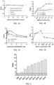
- the results were shown in Figures 1A-1D .
- the ordinate in the figures represents the ratio of the number of cells obtained after culture in different mediums to the number of cells obtained after culture in basic medium BM.
- adding different concentrations of different factors according to Table 3 to BM all resulted in the proliferation of cells but with varying degrees.
- B27 additive, N2 additive, fibroblast growth factor, gastrin, epidermal growth factor, transferrin, Compound 1, Y27632 and A83-01 provided certain promoting effects on cell proliferation in specific concentration ranges.
- Extracellular matrix gel (Matrigel ® ) was diluted with serum-free DMEM/F12 medium at a ratio of 1:100 to prepare an extracellular matrix diluent.
- the extracellular matrix diluent was added to a 48-well culture plate with 500 ⁇ l/well to completely cover the bottom of the wells of the culture plate. The culture plate was let stand for 1 hour in a 37°C incubator. After 1 hour, the extracellular matrix diluent was removed to provide a Matrigel-coated plate.
- the culture mediums with different components were added at a volume of 500 ⁇ l/well to 48-well plates which were coated with extracellular matrix gel (Matrigel), and simultaneously, the BM medium was used as a control.
- Lung cancer cells (No. B22) isolated from lung cancer tissues according to the method described in Example 1 were inoculated at a cell density of 2 ⁇ 10 4 cells/cm 2 in the 48-well culture plates which were coated with Matrigel. After surface disinfection, the plates were placed in a 37°C, 5% CO 2 incubator (purchased from Thermo Fisher), and the same number of freshly isolated lung cancer tumor cells (No. B22) were cultured under different medium formulations. After 10 days of culture, cell counts were performed. The results were shown in Figure 2 . As shown in the figure, with the sequential addition of new additive factors, the cell proliferation effect of the culture medium formulation was continuously improved, and it was finally determined that No. 8 culture medium formulation was the most preferable culture medium in this application for culturing and expanding primary lung cancer cells.
- Extracellular matrix gel (Matrigel ® ) was diluted with serum-free DMEM/F12 medium at a ratio of 1:100 to prepare an extracellular matrix diluent.
- the extracellular matrix diluent was added to a 48-well culture plate with 200 ⁇ l/well to completely cover the bottom of the wells of the culture plate. The culture plate was let stand for 1 hour in a 37°C incubator. After 1 hour, the extracellular matrix diluent was removed to provide a Matrigel-coated plate.
- Lung cancer epithelial cells derived from cancer tissues were isolated from the cancer tissue of a lung cancer patient (Sample No. B26) using the same method as in Example 1. Next, lung cancer epithelial cells derived from cancer tissues were counted with a flow imaging counter (JIMBIO FIL, Jiangsu Jimbio Technology Co., Ltd.) to get the total number of cells. Then, the cells were inoculated at a density of 4 ⁇ 10 4 cells/cm 2 in a 48-well plate which was coated with Matrigel ® . 2 ml of the prepared No.
- a flow imaging counter JIMBIO FIL, Jiangsu Jimbio Technology Co., Ltd.
- the centrifuged cell pellet was resuspended in the basic medium BM and the cells were counted with a flow imaging counter (JIMBIO FIL, Jiangsu Jimbio Technology Co., Ltd.) to get the total number of cells.
- the obtained cells were used in the following cultivation experiments.
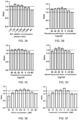
- the digested cell suspension was diluted with the above Formulations 1-8 respectively, and then inoculated into a 48-well plate at a volume of 250 ⁇ l with 10,000 cells per well.
- the medium of Formulation 2 When using the medium of Formulation 2, to a 48-well plate inoculated with primary cells was added the prepared fibroblast growth factor, with 250 ⁇ l per well, the final concentrations of fibroblast growth factor were 80 ng/ml, 40 ng/ml, 20 ng/ml, 10 ng/ml, 5 ng/ml, 2.5 ng/ml, respectively; a well of Blank Control (BC) was set using the medium of Formulation 2.
- BC Blank Control
- the medium of Formulation 4 When using the medium of Formulation 4, to a 48-well plate inoculated with primary cells was added the prepared epidermal growth factor, with 250 ⁇ l per well, the final concentrations of epidermal growth factor were 80 ng/ml, 40 ng/ml, 20 ng/ml, 10 ng/ml, 5 ng/ml, 2.5 ng/ml, respectively; a well of Blank Control (BC) was set using the medium of Formulation 4.
- BC Blank Control
- the ratios were calculated by referring to the cell numbers in the well of Blank Control (BC), and the results were shown in Figures 3A-3H .
- the ratio represents a ratio of the number of cells of the first passage cultured by using each culture medium to the number of cells of the first passage cultured by the corresponding well of Blank Control.
- the ratio is greater than 1, it indicates that the proliferation promoting effect of the prepared medium containing different concentrations of factors or small molecular compounds is preferable over that of the medium in the well of Blank Control; if the ratio is less than 1, it indicates that the proliferation promoting effect of the prepared medium containing different concentrations of factors or small molecular compounds is poorer than that of the medium in the well of Blank Control.
- the volume concentration of B27 additive in the culture medium is preferably 1:25-1:800, more preferably 1:25-1:200, and most preferably 1:50;
- the amount of fibroblast growth factor is preferably 2.5 ng/ml-80 ng/ml, more preferably 5 ng/ml-40 ng/ml, and most preferably 10 ng/ml;
- the amount of transferrin is preferably 2.5 ng/ml-80 ng/ml, more preferably 5 ng/ml-80 ng/ml, and most preferably 20 ng/ml;
- the amount of epidermal growth factor is preferably 2.5 ng/ml-80 ng/ml, more preferably 10 ng/ml-40 ng/ml, and most preferably 20 ng/ml;
- the amount of Y27632 is preferably 2.5 ⁇ M-18 ⁇ M, more preferably 5 ⁇ M-15 ⁇ M, and most preferably 10 ⁇ M;
- the amount of MST1/2 is preferably
- FLM The optimal concentration combination of various additive factors was used as the most preferred culture medium formulation for culturing and expanding primary lung cancer cells in the invention (hereinafter referred to as FLM): basic medium (BM) + 10 ng/ml fibroblast growth factor (FGF) + 20 ng/ml epidermal growth factor (EGF) + 20 ng/ml transferrin + 1:50 volume ratio of B27 additive + 5 ⁇ M Compound 1 + 10 ⁇ M Y27632 + 500 nM A83-01 + 10 ng/ml gastrin.
- BM basic medium
- FGF ng/ml fibroblast growth factor
- EGF epidermal growth factor
- transferrin 1:50 volume ratio of B27 additive + 5 ⁇ M Compound 1 + 10 ⁇ M Y27632 + 500 nM A83-01 + 10 ng/ml gastrin.
- Lung cancer epithelial cells derived from cancer tissues were isolated from cancer tissues of lung cancer patients (Sample No. B21) using the same method as in Example 1. Next, cancer tissue-derived lung cancer epithelial cells were counted with a flow imaging counter (JIMBIO FIL, Jiangsu Jimbio Technology Co., Ltd.) to get the total number of cells. Then, the cells were inoculated in a 12-well plate which was coated with Matrigel ® (purchased from BD Biosciences) at a density of 4 ⁇ 10 4 cells/cm 2 . 2 ml of the prepared culture medium FLM for primary lung cancer epithelial cells was added to the 12-well plate, which was then placed in a 37°C, 5% CO 2 incubator (purchased from Thermo Fisher) for culture.
- a flow imaging counter JIMBIO FIL, Jiangsu Jimbio Technology Co., Ltd.
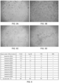
- Figure 4A is a microscopy image (photographed by a 40x inverted phase contrast microscope) of the cells which were cultured to day 4 after inoculation at a density of 4 ⁇ 10 4 cells/cm 2 into the 12-well plate which was coated with Matrigel. Microscopic observation shows that the cultured primary lung cancer cells derived from cancer tissues had high purity, without fibroblasts.
- Figure 4B is a microscopy image (photographed by a 40x inverted phase contrast microscope) of the cells which were cultured to day 12 after inoculation.
- Culture medium FLM for primary lung cancer epithelial cell was prepared in the same method of Example 2, and basic medium BM was prepared as control.
- a culture medium FM used in the cell conditional reprogramming technique was prepared additionally.
- the formulation of the culture medium is shown in Table 5.
- a commercial medium EpiMCult TM Plus Medium hereinafter also referred to as "EpiM medium" was purchased from stem cell, and the formulation of the culture medium is shown in Table 6. Table 5.
- composition of culture medium FM used in the cell conditional reprogramming technique Medium composition Supplier Final concentration DMEM medium Corning 65 vol.% Fetal bovine serum Gibico 10 vol.% Ham's F12 Nutrient Solution Gibico 25 vol.% Hydrocortisone Sigma-Aldrich 25 ng/ml Epidermal growth factor R&D 0.125 ng/ml Insulin Sigma-Aldrich 5 ⁇ g/ml Amphotericin B Sigma-Aldrich 250 ng/ml Gentamicin Gibico 10 ⁇ g/ml Cholera Toxin Sigma-Aldrich 0.1 nM Y27632 Enzo 10 ⁇ M Table 6.
- composition of commercial medium EpiMCult TM Plus Medium (EpiM) Medium composition Supplier Final concentration EpiMCult TM Plus Basal Medium stem cell 98 vol.% EpiMCult TM Plus Supplement stem cell 2 vol.% hydrocortisone stem cell 480 ng/ml
- the primary lung cancer cells No. B8 derived from lung cancer tissues were obtained.
- the cells were cultured at the same density (4 ⁇ 10 4 cells/cm 2 ) under the following 5 culture conditions:
- the centrifuged cell pellet was resuspended in the culture medium of the invention and the cells were counted with a flow imaging counter (JIMBIO FIL, Jiangsu Jimbio Technology Co., Ltd.) to obtain the total number of cells, which was 464,000.
- the cells cultured under the other three culture conditions were digested and counted in the same operation process as above.
- the total number of cells cultured by using the mediums FM, EpiM and BM were 350,000, 110,000 and 68,000, respectively.
- Figures 5A-5D are microscopic photos (under a 40x inverted phase contrast microscope) of the sample numbered B8 cultured under four different culture conditions until day 12:
- Figure 5A is the microscopic photo of B8 cultured to day 12 using basic medium BM;
- Figure 5B is the microscopic photo of B8 cultured to day 12 using the culture medium FLM of the invention;
- Figure 5C is the microscopic photo of B8 cultured to day 12 using the commercial culture medium EpiM;
- Figure 5D is the microscopic photo of B8 cultured to day 12 using the conditioned reprogramming medium FM.
- sample B8 cannot form cell clones when cultured with basic medium BM ( Figure 5A ) for 12 days; only a small number of cell clones are formed when cultured with EpiM ( Figure 5C ) for 12 days, and the cells are in poor condition; the cells show certain expansion when cultured with the conditional reprogramming medium FM ( Figure 5D ) for 12 days, but the cell density and cell number are not comparable to those in the medium FLM of the invention ( Figure 5B ).
- Figure 6 is a comparative diagram of cell proliferation effects obtained by culturing the primary lung cancer cells isolated from ten lung cancer patient samples according to Example 1 for 14 days under the conditions of four different culture mediums, where V represents a moderate clone formation ability and proliferation promoting effect, ⁇ represents a significant clone formation ability and proliferation promoting effect, ⁇ represents an extremely significant clone formation ability and proliferation promoting effect, and ⁇ represents no clone formation. It can be confirmed from Figure 6 that the culture medium FLM of the invention has significant superiority over the other three culture conditions in terms of clone formation ability, cell proliferation promoting effect and successful rate of culture in cultivation of the lung cancer tissue-derived primary cells.
- the culture medium FLM for primary lung cancer epithelial cells and the culture mediums BM, FM, and EpiM as controls were obtained by using the same method as in (1) of this Example.
- the lung cancer tissue-devired primary lung cancer cells (No. B16) were cultured under four culture conditions by using the same method as in (1) of this Example, and then digested, passaged and counted.
- the cultured cells were digested, collected and counted according to the above operating method.
- the cells were inoculated at a density of 4 ⁇ 10 4 cells/well again and cultured continuously.
- Population Doubling (PD) 3.32 * log 10 (total number of digested cells / the number of cells at initial inoculation), see Chapman et al., Stem Cell Research & Therapy 2014, 5: 60 .
- Figure 7 shows the growth curves of B16 cells under four different culture conditions drawn by Graphpad Prism software.
- the abscissa represents the days of cell culture, and the ordinate represents the multiple of cumulative cell expansion, i.e., the multiple of cell expansion in the culture period. The larger the value, the more multiples the cell expands in certain period, that is, the more cells are expanded.
- the slope represents the rate of cell expansion. It can be confirmed from the figure that the proliferation rates of lung cancer epithelial cells cultured with the culture medium FLM of the invention was superior to the other four culture conditions, and it can also be confirmed that the culture medium of the invention can continuously culture the primary lung cancer epithelial cells, and the rate remains unchanged for expanding of more than 20 days.
- Example No. B16 Cancer tissue about the size of a mung bean was taken from a surgical resection sample of a lung cancer patient, and immersed in 1 ml of 4% paraformaldehyde.
- Lung cancer epithelial cells (Sample No. B16) were obtained from the remaining cancer tissue in the same method of Example 1.
- Sample B16 was continuously cultured to the fourth passage using the culture medium FLM of the invention according to the method of Example 3.
- Immunohistochemistry assay was used to detect the expression of important lung cancer-related biomarkers in the original tissue of Sample B16 and the primary cells obtained by continuous culture to the fourth passage.
- the tissue was fixed with 4% paraformaldehyde, embedded in paraffin, and cut into tissue sections of 4 ⁇ m thickness with a microtome. Routine immunohistochemical detection was then performed (for detailed steps, see Li et al., Nature Communication, (2016) 9: 2983 ).
- the primary antibodies used were P63 antibody (purchased from CST) and Ki67 antibody (purchased from R&D).
- Figure 8 confirms that the expression of lung cancer-related biomarkers on the cells that were cultured from the lung cancer cells (Sample No. B16) with the culture medium of the invention to the 4th passage, was substantially consistent with the expression of markers on the original tissue section from which the cells were derived. This suggests that the cells cultured with the culture medium of the invention maintain the original pathological characteristics of the cancer tissues of the lung cancer patients.
- the culture medium FLM for primary lung cancer epithelial cells was prepared using the same method as Example 2. As a control, the same method as Example 2 was used to prepare basic medium BM. In addition, other 8 different culture mediums were prepared according to Table 7. Table 7 Culture mediums with different compositions (final concentrations are shown) Culture Medium Composition FLM without Compound 1 DMEM/F12 + 100 ⁇ g/mL Primocin + 1:50 B27 additive + 10 ng/ml fibroblast growth factor + 20 ng/ml epidermal growth factor + 20 ng/ml transferrin + 10 ⁇ M Y27632 + 500 nM A83-01 + 10 ng/ml gastrin FLM without Y27632 DMEM/F12 + 100 ⁇ g/mL Primocin + 1:50 B27 additive + 10 ng/ml fibroblast growth factor + 20 ng/ml epidermal growth factor + 20 ng/ml transferrin + 5 ⁇ M Compound 1 + 500 nM A83-01
- Example 2 One case of primary lung cancer cells (No. B18) derived from lung cancer tissues was obtained using the same method as in Example 1.
- the primary lung cancer cells were inoculated into a 48-well plate coated with Matrigel ® at a density of 4 ⁇ 10 4 cells/cm 2 , and cultured with 2 ml of the culture medium (FLM) for primary lung cancer epithelial cells of the invention, in a 37°C, 5% CO 2 incubator (purchased from Thermo Fisher).
- FLM culture medium
- the supernatant of medium in the 48-well plate was discarded, and 200 ⁇ l of 0.05% trypsin (purchased from Gibco) was added for cell digestion,which was then incubated at 37°C for 10 minutes, until the cells were completely digested as observed under a microscope (EVOS M500, Invitrogen); then the digestion was terminated by using 800 ⁇ l of DMEM/F12 culture solution containing 5% (v/v) fetal bovine serum, 100 U/ml penicillin, and 100 ⁇ g/ml streptomycin; the resultant was collected into a 15 ml centrifuge tube and centrifuged at 1500 rpm for 4 minutes, and then the supernatant was discarded.
- trypsin purchased from Gibco
- the centrifuged cell pellet was resuspended in the culture medium of the invention, and the cells were counted with a flow imaging counter (JIMBIO FIL, Jiangsu Jimbio Technology Co., Ltd.) to get the total number of cells.
- the cells were inoculated into another 48-well plate coated with extracellular matrix gel at a density of 2 ⁇ 10 4 cells/cm 2 for further culture.
- Cells cultured with the other 8 culture mediums and BM were digested, passaged and counted using the same methods as above, and cultured using different mediums.
- the cultured cells were digested, collected and counted according to the above operating method.
- the cells were inoculated at a density of 2 ⁇ 10 4 cells/cm 2 again and cultured continuously.
- Population Doubling (PD) 3.32 * log 10 (total number of digested cells / the number of cells at initial inoculation), see Chapman et al., Stem Cell Research & Therapy 2014, 5: 60 .
- Figure 9 is a graph of cell growth curves drawn under ten different culture medium conditions using Graphpad Prism software.
- the abscissa represents the days of cell culture, and the ordinate represents the multiple of cumulative cell expansion, i.e., the multiple of cell expansion in the culture period. The larger the value, the more multiples the cell expands in certain period, that is, the more cells are expanded.
- the slope represents the rate of cell expansion.
- Freshly isolated primary lung cancer epithelial cell sample (No. B16) was obtained according to the steps described in Example 1. Then, the primary epithelial cells were inoculated onto a 6-well plate coated with Matrigel TM . 3mL of the culture medium FLM and 3mL of the culture medium FLM without Compound 1 were respectively added to the wells inoculated with the aforementioned epithelial cells. The plate was placed in a 37°C, 5% CO 2 incubator (purchased from Thermo Fisher) for culture. The culture mediums were changed every 4 days during the culture process. After 12 days of cultivation, cells of each group were collected and the expression of Compound 1 on Hippo pathway related proteins YAP and TAZ in the nucleus was detected using conventional immunoblotting method.
- Yes associated protein YAP
- PDZ-binding motif TEAD transcription factors
- the testing result in Figure 10 shows that compared with the culture medium without Compound 1, the culture medium FLM with the addition of Compound 1 showed a significant increase in the expression of YAP and TAZ proteins in the nucleus. This indicates that Compound 1 can activate the Hippo pathway, making YAP/TAZ translocate to the nucleus and bind to TEAD transcription factors, and thus, promoting the sustained proliferation of cells.
- Lung cancer cells (No. B16) were isolated and obtained from the cancer tissues of one pathologically diagnosed lung cancer patient by using the same method as in Example 1.
- B16 was cultured using the culture medium FLM of the invention according to the method of Example 3, and when the number of lung cancer cells reached 1 ⁇ 10 7 , the lung cancer cells were digested and collected by using the same method of Example 4.
- the culture medium FLM for lung cancer cells of the invention and Matrigel ® were mixed at a ratio of 1:1, and 100 ⁇ l of the culture medium mixed with Matrigel was used to resuspend 5 ⁇ 10 6 lung cancer cells, and the resultant was injected into the lung cancer fat pad and the axilla of the right forelimb of 6-week-old female highly immunodeficient mice (NCG) (purchased from Nanjing Model Animal Research Center), respectively.
- NCG highly immunodeficient mice
- Tumor formation can be observed in both of the two tumor cell inoculation sites of the mice on day 15 after tumor cell inoculation. From day 15 to day 30, the tumor proliferation in mice was obvious. This indicates that the cancer tissue-derived lung cancer cells cultured by the culture method of the invention have tumorigenicity in mice.
- the lung cancer cells cultured from the patient-derived lung cancer samples can be used to test the sensitivity of the tumor cells of the patient to different drugs.
- Figures 11A and 11B respectively represent the sensitivity of the lung cancer cells cultured from surgically resected cancer tissue samples of two different lung cancer patients (Sample No. B25 and Sample No. B26) to two chemotherapeutic drugs Paclitaxel and Gemcitabine, and to two targeted drugs Afatinib and Erlotinib.
- Figure 11A shows the sensitivity results of lung cancer cells cultured from Sample No. B25 on four drugs
- Figure 11B shows the sensitivity results of lung cancer cells cultured from Sample No. B26 on four drugs. The results show that the cells from the same patient have different sensitivities to different drugs, and the cells from different patients also have different sensitivities to the same drug.
- the invention provides a culture medium and a culture method for culturing primary lung cancer epithelial cells.
- the cultured lung cancer epithelial cells can be used for the efficacy evaluating and screening of drugs. Therefore, the invention is applicable in the industry.
Landscapes
- Health & Medical Sciences (AREA)
- Engineering & Computer Science (AREA)
- Life Sciences & Earth Sciences (AREA)
- Chemical & Material Sciences (AREA)
- Biomedical Technology (AREA)
- Organic Chemistry (AREA)
- Wood Science & Technology (AREA)
- Genetics & Genomics (AREA)
- Zoology (AREA)
- Biotechnology (AREA)
- Bioinformatics & Cheminformatics (AREA)
- Microbiology (AREA)
- Biochemistry (AREA)
- General Engineering & Computer Science (AREA)
- General Health & Medical Sciences (AREA)
- Cell Biology (AREA)
- Oncology (AREA)
- Proteomics, Peptides & Aminoacids (AREA)
- Physics & Mathematics (AREA)
- Analytical Chemistry (AREA)
- Biophysics (AREA)
- Immunology (AREA)
- Molecular Biology (AREA)
- Measuring Or Testing Involving Enzymes Or Micro-Organisms (AREA)
- Micro-Organisms Or Cultivation Processes Thereof (AREA)
- Medicines That Contain Protein Lipid Enzymes And Other Medicines (AREA)
- Pharmaceuticals Containing Other Organic And Inorganic Compounds (AREA)
Applications Claiming Priority (2)
| Application Number | Priority Date | Filing Date | Title |
|---|---|---|---|
| CN202111079965.0A CN115806936B (zh) | 2021-09-15 | 2021-09-15 | 一种用于肺癌上皮细胞的培养基、培养方法及其应用 |
| PCT/CN2021/126229 WO2023039999A1 (zh) | 2021-09-15 | 2021-10-26 | 一种用于肺癌上皮细胞的培养基、培养方法及其应用 |
Publications (2)
| Publication Number | Publication Date |
|---|---|
| EP4403626A1 true EP4403626A1 (de) | 2024-07-24 |
| EP4403626A4 EP4403626A4 (de) | 2025-10-15 |
Family
ID=85481794
Family Applications (1)
| Application Number | Title | Priority Date | Filing Date |
|---|---|---|---|
| EP21957266.6A Pending EP4403626A4 (de) | 2021-09-15 | 2021-10-26 | Kulturmedium und kulturverfahren für lungenkrebsepithelzellen und anwendung davon |
Country Status (5)
| Country | Link |
|---|---|
| US (1) | US20240392251A1 (de) |
| EP (1) | EP4403626A4 (de) |
| JP (1) | JP7766370B2 (de) |
| CN (1) | CN115806936B (de) |
| WO (1) | WO2023039999A1 (de) |
Families Citing this family (1)
| Publication number | Priority date | Publication date | Assignee | Title |
|---|---|---|---|---|
| JP7735022B1 (ja) * | 2024-01-04 | 2025-09-08 | 学校法人藤田学園 | がん細胞の培養用および/または保存用の培地、がん細胞の初代培養方法、がん細胞の継代培養方法およびがん細胞の保存方法 |
Family Cites Families (9)
| Publication number | Priority date | Publication date | Assignee | Title |
|---|---|---|---|---|
| CN105801582A (zh) | 2016-04-12 | 2016-07-27 | 合肥工业大学 | 一类新型二氢蝶啶酮类衍生物及其制备方法和在医药上的用途 |
| GB201609663D0 (en) * | 2016-06-02 | 2016-07-20 | Stemtek Therapeutics Sl | Methods for producing cancer stem cell spheroids |
| CN107151645A (zh) * | 2017-05-16 | 2017-09-12 | 武汉大学深圳研究院 | 一种为肺癌提供离体个体化药物测试的方法及培养基 |
| CN108486059A (zh) * | 2018-05-02 | 2018-09-04 | 深圳市因诺转化医学研究院 | 一种细胞培养液及人原代肺癌细胞株的建立方法 |
| CN108624561B (zh) * | 2018-05-26 | 2021-09-17 | 复旦大学 | 原代肿瘤细胞培养基、培养方法以及应用 |
| CN111039944B (zh) * | 2018-10-12 | 2021-11-23 | 中国科学院合肥物质科学研究院 | Mst1激酶抑制剂及其用途 |
| US20220144775A1 (en) | 2019-01-30 | 2022-05-12 | Nissan Chemical Corporation | Hydrazide compound and kinase inhibitor |
| CN112779209B (zh) | 2019-11-08 | 2023-01-24 | 合肥中科普瑞昇生物医药科技有限公司 | 原代乳腺上皮细胞培养基、培养方法及其应用 |
| CN113969262B (zh) * | 2020-07-22 | 2024-03-26 | 合肥中科普瑞昇生物医药科技有限公司 | 一种用于肺癌上皮细胞的培养基、培养方法及其应用 |
-
2021
- 2021-09-15 CN CN202111079965.0A patent/CN115806936B/zh active Active
- 2021-10-26 JP JP2024516559A patent/JP7766370B2/ja active Active
- 2021-10-26 EP EP21957266.6A patent/EP4403626A4/de active Pending
- 2021-10-26 US US18/692,569 patent/US20240392251A1/en active Pending
- 2021-10-26 WO PCT/CN2021/126229 patent/WO2023039999A1/zh not_active Ceased
Also Published As
| Publication number | Publication date |
|---|---|
| US20240392251A1 (en) | 2024-11-28 |
| EP4403626A4 (de) | 2025-10-15 |
| WO2023039999A1 (zh) | 2023-03-23 |
| CN115806936B (zh) | 2025-09-26 |
| JP2024535032A (ja) | 2024-09-26 |
| CN115806936A (zh) | 2023-03-17 |
| JP7766370B2 (ja) | 2025-11-10 |
Similar Documents
| Publication | Publication Date | Title |
|---|---|---|
| JP7461674B2 (ja) | 食道扁平上皮癌の上皮細胞用の培養培地、培養方法、及びその用途 | |
| EP4056685A1 (de) | Primäres brustepithelzellen-kulturmedium, kulturverfahren und dessen verwendung | |
| CN113969262B (zh) | 一种用于肺癌上皮细胞的培养基、培养方法及其应用 | |
| CN115975934B (zh) | 卵巢癌类器官的培养基、培养方法及其应用 | |
| EP4403626A1 (de) | Kulturmedium und kulturverfahren für lungenkrebsepithelzellen und anwendung davon | |
| CN115772498B (zh) | 一种用于肝癌类器官培养的培养基、及其培养方法和应用 | |
| EP4368706A1 (de) | Kulturmedium und kulturverfahren für lungenkrebsepithelzellen und anwendung davon | |
| CN115975935B (zh) | 一种用于宫颈癌原代细胞的培养基和培养方法 | |
| JP7503662B2 (ja) | 喉頭癌上皮細胞用の培養培地、培養方法及びその用途 | |
| RU2842810C2 (ru) | Культуральная среда и способ культивирования эпителиальных клеток рака легкого и их применение | |
| CN115960815B (zh) | 肺癌胸水来源类器官的培养基、培养方法及其应用 | |
| RU2838489C2 (ru) | Среда для культивирования эпителиальных клеток рака лёгкого, способ их культивирования и применение | |
| WO2023060764A1 (zh) | 胃癌原代细胞的培养基和培养方法 | |
| RU2814719C1 (ru) | Культуральная среда для эпителиальных клеток рака гортани, способ культивирования и их применение | |
| RU2816529C1 (ru) | Культуральная среда для эпителиальных клеток плоскоклеточной карциномы пищевода, способ культивирования и их применение | |
| CN115975940B (zh) | 胃癌原代细胞的培养基和培养方法 | |
| CN115975910B (zh) | 胃癌类器官的培养基及培养方法 | |
| CN115975931A (zh) | 食管癌类器官的培养基、培养方法及其应用 |
Legal Events
| Date | Code | Title | Description |
|---|---|---|---|
| STAA | Information on the status of an ep patent application or granted ep patent |
Free format text: STATUS: THE INTERNATIONAL PUBLICATION HAS BEEN MADE |
|
| PUAI | Public reference made under article 153(3) epc to a published international application that has entered the european phase |
Free format text: ORIGINAL CODE: 0009012 |
|
| STAA | Information on the status of an ep patent application or granted ep patent |
Free format text: STATUS: REQUEST FOR EXAMINATION WAS MADE |
|
| 17P | Request for examination filed |
Effective date: 20240311 |
|
| AK | Designated contracting states |
Kind code of ref document: A1 Designated state(s): AL AT BE BG CH CY CZ DE DK EE ES FI FR GB GR HR HU IE IS IT LI LT LU LV MC MK MT NL NO PL PT RO RS SE SI SK SM TR |
|
| DAV | Request for validation of the european patent (deleted) | ||
| DAX | Request for extension of the european patent (deleted) | ||
| A4 | Supplementary search report drawn up and despatched |
Effective date: 20250915 |
|
| RIC1 | Information provided on ipc code assigned before grant |
Ipc: C12N 5/09 20100101AFI20250909BHEP Ipc: C12Q 1/02 20060101ALI20250909BHEP |