EP4356932A9 - Verfahren und arzneimittel zur behandlung von neuronaler ceroid-lipofuszinose - Google Patents
Verfahren und arzneimittel zur behandlung von neuronaler ceroid-lipofuszinose Download PDFInfo
- Publication number
- EP4356932A9 EP4356932A9 EP22824303.6A EP22824303A EP4356932A9 EP 4356932 A9 EP4356932 A9 EP 4356932A9 EP 22824303 A EP22824303 A EP 22824303A EP 4356932 A9 EP4356932 A9 EP 4356932A9
- Authority
- EP
- European Patent Office
- Prior art keywords
- cln3
- disease
- compound
- keap1
- neurons
- Prior art date
- Legal status (The legal status is an assumption and is not a legal conclusion. Google has not performed a legal analysis and makes no representation as to the accuracy of the status listed.)
- Withdrawn
Links
Images






Classifications
-
- A—HUMAN NECESSITIES
- A61—MEDICAL OR VETERINARY SCIENCE; HYGIENE
- A61K—PREPARATIONS FOR MEDICAL, DENTAL OR TOILETRY PURPOSES
- A61K45/00—Medicinal preparations containing active ingredients not provided for in groups A61K31/00 - A61K41/00
-
- A—HUMAN NECESSITIES
- A61—MEDICAL OR VETERINARY SCIENCE; HYGIENE
- A61P—SPECIFIC THERAPEUTIC ACTIVITY OF CHEMICAL COMPOUNDS OR MEDICINAL PREPARATIONS
- A61P3/00—Drugs for disorders of the metabolism
-
- A—HUMAN NECESSITIES
- A61—MEDICAL OR VETERINARY SCIENCE; HYGIENE
- A61K—PREPARATIONS FOR MEDICAL, DENTAL OR TOILETRY PURPOSES
- A61K31/00—Medicinal preparations containing organic active ingredients
- A61K31/56—Compounds containing cyclopenta[a]hydrophenanthrene ring systems; Derivatives thereof, e.g. steroids
- A61K31/57—Compounds containing cyclopenta[a]hydrophenanthrene ring systems; Derivatives thereof, e.g. steroids substituted in position 17 beta by a chain of two carbon atoms, e.g. pregnane or progesterone
-
- A—HUMAN NECESSITIES
- A61—MEDICAL OR VETERINARY SCIENCE; HYGIENE
- A61K—PREPARATIONS FOR MEDICAL, DENTAL OR TOILETRY PURPOSES
- A61K31/00—Medicinal preparations containing organic active ingredients
- A61K31/13—Amines
-
- A—HUMAN NECESSITIES
- A61—MEDICAL OR VETERINARY SCIENCE; HYGIENE
- A61K—PREPARATIONS FOR MEDICAL, DENTAL OR TOILETRY PURPOSES
- A61K31/00—Medicinal preparations containing organic active ingredients
- A61K31/13—Amines
- A61K31/135—Amines having aromatic rings, e.g. ketamine, nortriptyline
- A61K31/137—Arylalkylamines, e.g. amphetamine, epinephrine, salbutamol, ephedrine or methadone
-
- A—HUMAN NECESSITIES
- A61—MEDICAL OR VETERINARY SCIENCE; HYGIENE
- A61K—PREPARATIONS FOR MEDICAL, DENTAL OR TOILETRY PURPOSES
- A61K31/00—Medicinal preparations containing organic active ingredients
- A61K31/16—Amides, e.g. hydroxamic acids
- A61K31/164—Amides, e.g. hydroxamic acids of a carboxylic acid with an aminoalcohol, e.g. ceramides
-
- A—HUMAN NECESSITIES
- A61—MEDICAL OR VETERINARY SCIENCE; HYGIENE
- A61K—PREPARATIONS FOR MEDICAL, DENTAL OR TOILETRY PURPOSES
- A61K31/00—Medicinal preparations containing organic active ingredients
- A61K31/21—Esters, e.g. nitroglycerine, selenocyanates
- A61K31/215—Esters, e.g. nitroglycerine, selenocyanates of carboxylic acids
- A61K31/22—Esters, e.g. nitroglycerine, selenocyanates of carboxylic acids of acyclic acids, e.g. pravastatin
-
- A—HUMAN NECESSITIES
- A61—MEDICAL OR VETERINARY SCIENCE; HYGIENE
- A61K—PREPARATIONS FOR MEDICAL, DENTAL OR TOILETRY PURPOSES
- A61K31/00—Medicinal preparations containing organic active ingredients
- A61K31/21—Esters, e.g. nitroglycerine, selenocyanates
- A61K31/215—Esters, e.g. nitroglycerine, selenocyanates of carboxylic acids
- A61K31/22—Esters, e.g. nitroglycerine, selenocyanates of carboxylic acids of acyclic acids, e.g. pravastatin
- A61K31/225—Polycarboxylic acids
-
- A—HUMAN NECESSITIES
- A61—MEDICAL OR VETERINARY SCIENCE; HYGIENE
- A61K—PREPARATIONS FOR MEDICAL, DENTAL OR TOILETRY PURPOSES
- A61K31/00—Medicinal preparations containing organic active ingredients
- A61K31/33—Heterocyclic compounds
- A61K31/335—Heterocyclic compounds having oxygen as the only ring hetero atom, e.g. fungichromin
-
- A—HUMAN NECESSITIES
- A61—MEDICAL OR VETERINARY SCIENCE; HYGIENE
- A61K—PREPARATIONS FOR MEDICAL, DENTAL OR TOILETRY PURPOSES
- A61K31/00—Medicinal preparations containing organic active ingredients
- A61K31/33—Heterocyclic compounds
- A61K31/395—Heterocyclic compounds having nitrogen as a ring hetero atom, e.g. guanethidine or rifamycins
- A61K31/40—Heterocyclic compounds having nitrogen as a ring hetero atom, e.g. guanethidine or rifamycins having five-membered rings with one nitrogen as the only ring hetero atom, e.g. sulpiride, succinimide, tolmetin, buflomedil
- A61K31/403—Heterocyclic compounds having nitrogen as a ring hetero atom, e.g. guanethidine or rifamycins having five-membered rings with one nitrogen as the only ring hetero atom, e.g. sulpiride, succinimide, tolmetin, buflomedil condensed with carbocyclic rings, e.g. carbazole
-
- A—HUMAN NECESSITIES
- A61—MEDICAL OR VETERINARY SCIENCE; HYGIENE
- A61K—PREPARATIONS FOR MEDICAL, DENTAL OR TOILETRY PURPOSES
- A61K31/00—Medicinal preparations containing organic active ingredients
- A61K31/33—Heterocyclic compounds
- A61K31/395—Heterocyclic compounds having nitrogen as a ring hetero atom, e.g. guanethidine or rifamycins
- A61K31/495—Heterocyclic compounds having nitrogen as a ring hetero atom, e.g. guanethidine or rifamycins having six-membered rings with two or more nitrogen atoms as the only ring heteroatoms, e.g. piperazine or tetrazines
-
- A—HUMAN NECESSITIES
- A61—MEDICAL OR VETERINARY SCIENCE; HYGIENE
- A61K—PREPARATIONS FOR MEDICAL, DENTAL OR TOILETRY PURPOSES
- A61K31/00—Medicinal preparations containing organic active ingredients
- A61K31/33—Heterocyclic compounds
- A61K31/395—Heterocyclic compounds having nitrogen as a ring hetero atom, e.g. guanethidine or rifamycins
- A61K31/495—Heterocyclic compounds having nitrogen as a ring hetero atom, e.g. guanethidine or rifamycins having six-membered rings with two or more nitrogen atoms as the only ring heteroatoms, e.g. piperazine or tetrazines
- A61K31/496—Non-condensed piperazines containing further heterocyclic rings, e.g. rifampin, thiothixene or sparfloxacin
-
- A—HUMAN NECESSITIES
- A61—MEDICAL OR VETERINARY SCIENCE; HYGIENE
- A61K—PREPARATIONS FOR MEDICAL, DENTAL OR TOILETRY PURPOSES
- A61K31/00—Medicinal preparations containing organic active ingredients
- A61K31/56—Compounds containing cyclopenta[a]hydrophenanthrene ring systems; Derivatives thereof, e.g. steroids
- A61K31/565—Compounds containing cyclopenta[a]hydrophenanthrene ring systems; Derivatives thereof, e.g. steroids not substituted in position 17 beta by a carbon atom, e.g. estrane, estradiol
- A61K31/568—Compounds containing cyclopenta[a]hydrophenanthrene ring systems; Derivatives thereof, e.g. steroids not substituted in position 17 beta by a carbon atom, e.g. estrane, estradiol substituted in positions 10 and 13 by a chain having at least one carbon atom, e.g. androstanes, e.g. testosterone
-
- A—HUMAN NECESSITIES
- A61—MEDICAL OR VETERINARY SCIENCE; HYGIENE
- A61K—PREPARATIONS FOR MEDICAL, DENTAL OR TOILETRY PURPOSES
- A61K31/00—Medicinal preparations containing organic active ingredients
- A61K31/70—Carbohydrates; Sugars; Derivatives thereof
- A61K31/7088—Compounds having three or more nucleosides or nucleotides
- A61K31/713—Double-stranded nucleic acids or oligonucleotides
-
- A—HUMAN NECESSITIES
- A61—MEDICAL OR VETERINARY SCIENCE; HYGIENE
- A61P—SPECIFIC THERAPEUTIC ACTIVITY OF CHEMICAL COMPOUNDS OR MEDICINAL PREPARATIONS
- A61P25/00—Drugs for disorders of the nervous system
-
- A—HUMAN NECESSITIES
- A61—MEDICAL OR VETERINARY SCIENCE; HYGIENE
- A61P—SPECIFIC THERAPEUTIC ACTIVITY OF CHEMICAL COMPOUNDS OR MEDICINAL PREPARATIONS
- A61P25/00—Drugs for disorders of the nervous system
- A61P25/14—Drugs for disorders of the nervous system for treating abnormal movements, e.g. chorea, dyskinesia
-
- A—HUMAN NECESSITIES
- A61—MEDICAL OR VETERINARY SCIENCE; HYGIENE
- A61P—SPECIFIC THERAPEUTIC ACTIVITY OF CHEMICAL COMPOUNDS OR MEDICINAL PREPARATIONS
- A61P25/00—Drugs for disorders of the nervous system
- A61P25/14—Drugs for disorders of the nervous system for treating abnormal movements, e.g. chorea, dyskinesia
- A61P25/16—Anti-Parkinson drugs
-
- A—HUMAN NECESSITIES
- A61—MEDICAL OR VETERINARY SCIENCE; HYGIENE
- A61P—SPECIFIC THERAPEUTIC ACTIVITY OF CHEMICAL COMPOUNDS OR MEDICINAL PREPARATIONS
- A61P25/00—Drugs for disorders of the nervous system
- A61P25/18—Antipsychotics, i.e. neuroleptics; Drugs for mania or schizophrenia
-
- A—HUMAN NECESSITIES
- A61—MEDICAL OR VETERINARY SCIENCE; HYGIENE
- A61P—SPECIFIC THERAPEUTIC ACTIVITY OF CHEMICAL COMPOUNDS OR MEDICINAL PREPARATIONS
- A61P25/00—Drugs for disorders of the nervous system
- A61P25/28—Drugs for disorders of the nervous system for treating neurodegenerative disorders of the central nervous system, e.g. nootropic agents, cognition enhancers, drugs for treating Alzheimer's disease or other forms of dementia
-
- A—HUMAN NECESSITIES
- A61—MEDICAL OR VETERINARY SCIENCE; HYGIENE
- A61P—SPECIFIC THERAPEUTIC ACTIVITY OF CHEMICAL COMPOUNDS OR MEDICINAL PREPARATIONS
- A61P43/00—Drugs for specific purposes, not provided for in groups A61P1/00-A61P41/00
-
- C—CHEMISTRY; METALLURGY
- C12—BIOCHEMISTRY; BEER; SPIRITS; WINE; VINEGAR; MICROBIOLOGY; ENZYMOLOGY; MUTATION OR GENETIC ENGINEERING
- C12N—MICROORGANISMS OR ENZYMES; COMPOSITIONS THEREOF; PROPAGATING, PRESERVING, OR MAINTAINING MICROORGANISMS; MUTATION OR GENETIC ENGINEERING; CULTURE MEDIA
- C12N15/00—Mutation or genetic engineering; DNA or RNA concerning genetic engineering, vectors, e.g. plasmids, or their isolation, preparation or purification; Use of hosts therefor
- C12N15/09—Recombinant DNA-technology
- C12N15/11—DNA or RNA fragments; Modified forms thereof; Non-coding nucleic acids having a biological activity
- C12N15/113—Non-coding nucleic acids modulating the expression of genes, e.g. antisense oligonucleotides; Antisense DNA or RNA; Triplex- forming oligonucleotides; Catalytic nucleic acids, e.g. ribozymes; Nucleic acids used in co-suppression or gene silencing
-
- C—CHEMISTRY; METALLURGY
- C12—BIOCHEMISTRY; BEER; SPIRITS; WINE; VINEGAR; MICROBIOLOGY; ENZYMOLOGY; MUTATION OR GENETIC ENGINEERING
- C12N—MICROORGANISMS OR ENZYMES; COMPOSITIONS THEREOF; PROPAGATING, PRESERVING, OR MAINTAINING MICROORGANISMS; MUTATION OR GENETIC ENGINEERING; CULTURE MEDIA
- C12N15/00—Mutation or genetic engineering; DNA or RNA concerning genetic engineering, vectors, e.g. plasmids, or their isolation, preparation or purification; Use of hosts therefor
- C12N15/09—Recombinant DNA-technology
- C12N15/87—Introduction of foreign genetic material using processes not otherwise provided for, e.g. co-transformation
- C12N15/90—Stable introduction of foreign DNA into chromosome
-
- G—PHYSICS
- G01—MEASURING; TESTING
- G01N—INVESTIGATING OR ANALYSING MATERIALS BY DETERMINING THEIR CHEMICAL OR PHYSICAL PROPERTIES
- G01N15/00—Investigating characteristics of particles; Investigating permeability, pore-volume or surface-area of porous materials
- G01N15/10—Investigating individual particles
- G01N15/14—Optical investigation techniques, e.g. flow cytometry
- G01N15/1434—Optical arrangements
-
- G—PHYSICS
- G01—MEASURING; TESTING
- G01N—INVESTIGATING OR ANALYSING MATERIALS BY DETERMINING THEIR CHEMICAL OR PHYSICAL PROPERTIES
- G01N15/00—Investigating characteristics of particles; Investigating permeability, pore-volume or surface-area of porous materials
- G01N15/10—Investigating individual particles
- G01N15/14—Optical investigation techniques, e.g. flow cytometry
- G01N15/1456—Optical investigation techniques, e.g. flow cytometry without spatial resolution of the texture or inner structure of the particle, e.g. processing of pulse signals
- G01N15/1459—Optical investigation techniques, e.g. flow cytometry without spatial resolution of the texture or inner structure of the particle, e.g. processing of pulse signals the analysis being performed on a sample stream
-
- G—PHYSICS
- G01—MEASURING; TESTING
- G01N—INVESTIGATING OR ANALYSING MATERIALS BY DETERMINING THEIR CHEMICAL OR PHYSICAL PROPERTIES
- G01N33/00—Investigating or analysing materials by specific methods not covered by groups G01N1/00 - G01N31/00
- G01N33/48—Biological material, e.g. blood, urine; Haemocytometers
- G01N33/50—Chemical analysis of biological material, e.g. blood, urine; Testing involving biospecific ligand binding methods; Immunological testing
- G01N33/5005—Chemical analysis of biological material, e.g. blood, urine; Testing involving biospecific ligand binding methods; Immunological testing involving human or animal cells
- G01N33/5008—Chemical analysis of biological material, e.g. blood, urine; Testing involving biospecific ligand binding methods; Immunological testing involving human or animal cells for testing or evaluating the effect of chemical or biological compounds, e.g. drugs, cosmetics
- G01N33/502—Chemical analysis of biological material, e.g. blood, urine; Testing involving biospecific ligand binding methods; Immunological testing involving human or animal cells for testing or evaluating the effect of chemical or biological compounds, e.g. drugs, cosmetics for testing non-proliferative effects
- G01N33/5035—Chemical analysis of biological material, e.g. blood, urine; Testing involving biospecific ligand binding methods; Immunological testing involving human or animal cells for testing or evaluating the effect of chemical or biological compounds, e.g. drugs, cosmetics for testing non-proliferative effects on sub-cellular localization
-
- A—HUMAN NECESSITIES
- A01—AGRICULTURE; FORESTRY; ANIMAL HUSBANDRY; HUNTING; TRAPPING; FISHING
- A01K—ANIMAL HUSBANDRY; AVICULTURE; APICULTURE; PISCICULTURE; FISHING; REARING OR BREEDING ANIMALS, NOT OTHERWISE PROVIDED FOR; NEW BREEDS OF ANIMALS
- A01K2217/00—Genetically modified animals
- A01K2217/07—Animals genetically altered by homologous recombination
- A01K2217/075—Animals genetically altered by homologous recombination inducing loss of function, i.e. knock out
-
- A—HUMAN NECESSITIES
- A01—AGRICULTURE; FORESTRY; ANIMAL HUSBANDRY; HUNTING; TRAPPING; FISHING
- A01K—ANIMAL HUSBANDRY; AVICULTURE; APICULTURE; PISCICULTURE; FISHING; REARING OR BREEDING ANIMALS, NOT OTHERWISE PROVIDED FOR; NEW BREEDS OF ANIMALS
- A01K2227/00—Animals characterised by species
- A01K2227/10—Mammal
- A01K2227/105—Murine
-
- C—CHEMISTRY; METALLURGY
- C12—BIOCHEMISTRY; BEER; SPIRITS; WINE; VINEGAR; MICROBIOLOGY; ENZYMOLOGY; MUTATION OR GENETIC ENGINEERING
- C12N—MICROORGANISMS OR ENZYMES; COMPOSITIONS THEREOF; PROPAGATING, PRESERVING, OR MAINTAINING MICROORGANISMS; MUTATION OR GENETIC ENGINEERING; CULTURE MEDIA
- C12N2310/00—Structure or type of the nucleic acid
- C12N2310/50—Physical structure
- C12N2310/53—Physical structure partially self-complementary or closed
- C12N2310/531—Stem-loop; Hairpin
-
- C—CHEMISTRY; METALLURGY
- C12—BIOCHEMISTRY; BEER; SPIRITS; WINE; VINEGAR; MICROBIOLOGY; ENZYMOLOGY; MUTATION OR GENETIC ENGINEERING
- C12N—MICROORGANISMS OR ENZYMES; COMPOSITIONS THEREOF; PROPAGATING, PRESERVING, OR MAINTAINING MICROORGANISMS; MUTATION OR GENETIC ENGINEERING; CULTURE MEDIA
- C12N2740/00—Reverse transcribing RNA viruses
- C12N2740/00011—Details
- C12N2740/10011—Retroviridae
- C12N2740/16011—Human Immunodeficiency Virus, HIV
- C12N2740/16041—Use of virus, viral particle or viral elements as a vector
- C12N2740/16043—Use of virus, viral particle or viral elements as a vector viral genome or elements thereof as genetic vector
-
- G—PHYSICS
- G01—MEASURING; TESTING
- G01N—INVESTIGATING OR ANALYSING MATERIALS BY DETERMINING THEIR CHEMICAL OR PHYSICAL PROPERTIES
- G01N15/00—Investigating characteristics of particles; Investigating permeability, pore-volume or surface-area of porous materials
- G01N15/10—Investigating individual particles
- G01N2015/1006—Investigating individual particles for cytology
-
- G—PHYSICS
- G01—MEASURING; TESTING
- G01N—INVESTIGATING OR ANALYSING MATERIALS BY DETERMINING THEIR CHEMICAL OR PHYSICAL PROPERTIES
- G01N15/00—Investigating characteristics of particles; Investigating permeability, pore-volume or surface-area of porous materials
- G01N15/10—Investigating individual particles
- G01N15/14—Optical investigation techniques, e.g. flow cytometry
- G01N2015/1497—Particle shape
Definitions
- the present disclosure relates to a method and medicament for the treatment of neurodegenerative diseases such as neuronal ceroid lipofuscinosis, in particular juvenile neuronal ceroid lipofuscinosis (JNCL).
- neurodegenerative diseases such as neuronal ceroid lipofuscinosis, in particular juvenile neuronal ceroid lipofuscinosis (JNCL).
- JNCL juvenile neuronal ceroid lipofuscinosis
- NCL Neuronal ceroid lipofuscinosis
- LSD lysosomal storage diseases
- Symptoms of NCLs occur mostly in infancy and childhood, and most patients present with a progressive neurodegenerative clinical course.
- JNCL juvenile neuronal ceroid lipofuscinosis
- Batten disease is one of the most common inherited juvenile neurological diseases, and is caused by CLN3 gene mutation.
- JNCL patients show obvious lysosomal defect features, and many substances such as subunit c of mitochondrial ATP synthase (SCMAS), lipoprotein and glycoprotein cannot be degraded normally and thus gradually accumulate in lysosomes.
- Neurons are one of the most affected cell types by CLN3 mutations because they are highly lysosomal dependent for substance metabolism. Patients exhibit severe visual impairment around the age of 2-10 years, followed by epilepsy, progressive motor deficits and cognitive decline, and usually die of the disease between the ages of 20-30 years.
- Lysosome is an organelle that plays a key role in the degradation and recycling of intracellular and intracellular substances (Luzio et al., 2009; Mizushima and Komatsu, 2011).
- a network consisting of lysosomes, endosomes, autophagosomes, and other cellular components performs the function of degrading materials inside and outside the cell (Saftig and Klumperman, 2009).
- Changes in lysosomal acidity affect the activity of a variety of enzymes in the lumen, thereby disrupting their substrate clearance processes, leading to the development of a variety of neurodegenerative diseases, including NCL (Song et al., 2020).
- One aspect of the present disclosure relates to a method for treating a disease associated with autophagy functional deficiency and/or lysosomal functional deficiency, the method comprising a process of inhibiting the activity of Kelch-like ECH-associated protein 1 (KEAP1), and/or enhancing the activity of nuclear erythroid 2-related factor 2 (NRF2) and/or the downstream proteins thereof.
- KEAP1 Kelch-like ECH-associated protein 1
- NEF2 nuclear erythroid 2-related factor 2
- the disease associated with autophagy functional deficiency and/or lysosomal functional deficiency is a neurodegenerative disease.
- the neurodegenerative disease is selected from the group consisting of amyotrophic lateral sclerosis (ALS), Alzheimer's dementia, Alexander disease, Alper's disease, ataxia-telangiectasia, bovine spongiform encephalopathy (BSE), Canavan disease, Cockayne syndrome, corticobasal degeneration, Creutzfeldt-Jakob disease, Huntington's disease, HIV-related dementia, Kennedy's disease, Krabbedisease, dementia with Lewy bodies, Machado-Joseph disease (spinocerebellar ataxias 3), multiple sclerosis, multiple system atrophy, neurospirillosis, Parkinson's disease, Péme's disease, Pick's disease, primary lateral sclerosis, Prion's disease, Refsum's disease, Sandhoff disease, Hield's disease, schizophrenia, Spielmeyer-Vogt-Sjogren-Batten disease, spinocerebellar ataxias, spinal muscular at
- the neurodegenerative disease is selected from Parkinson's disease, Alzheimer's disease, Huntington's disease, Kennedy's disease, amyotrophic lateral sclerosis, primary lateral sclerosis, multiple sclerosis, frontotemporal dementia, or neuronal ceroid lipofuscinosis (NCL).
- Parkinson's disease Alzheimer's disease, Huntington's disease, Kennedy's disease, amyotrophic lateral sclerosis, primary lateral sclerosis, multiple sclerosis, frontotemporal dementia, or neuronal ceroid lipofuscinosis (NCL).
- the neurodegenerative disease is preferably neuronal ceroid lipofuscinosis, more preferably juvenile neuronal ceroid lipofuscinosis (JNCL).
- JNCL juvenile neuronal ceroid lipofuscinosis
- the disease is caused by a mutation in CLN3 gene, such as caused by CLN3 ⁇ ex7 / 8 , a deletion mutation in exons 7 and 8 of CLN3 gene, wherein the deletion mutation is about 1.02 kb.
- KEAP1 gene is silenced or knocked down with siRNA, sgRNA or vectors constructed with shRNA, thereby enhancing the activity of NRF2 and/or the downstream proteins thereof.
- the therapeutic method according to the present disclosure wherein the expression of NRF2 and/or the downstream genes thereof is enhanced with a KEAP1 inhibitor and/or NRF2 activator, thereby enhancing the activity of NRF2 and/or the downstream proteins thereof.
- the KEAP1 inhibitor and/or NRF2 activator is selected from reagents covalently reacting with cysteine residues on KEAP1, including cysteine at the positon on 151, 272 and/or 288 on KEAP1.
- the therapeutic method according to the present disclosure wherein the KEAP1 inhibitor and/or NRF2 activator is selected from the group consisting of Carvedilol ( CAS: 72956-09-3 ), ketoconazole ( CAS: 65277-42-1 ), GANT61, CAS (500579-04-4 ), protriptyline hydrochloride ( CAS: 1225-55-4 ), LP 44 ( CAS: 824958-12-5 ), Doxepin HCI ( CAS: 1229-29-4 ), dimethyl fumarate (DMF), acetyl-11-keto- ⁇ -boswellic acid (AKBA), isothiocyanates, bardoxolone (CDDO), bardoxo
- KEAP1 inhibitor is Compound G, namely (Z)-guggulsterone, having the formula:
- Another aspect of the present disclosure relates to the use of a KEAP1 inhibitor and/or NRF2 activator in the manufacture of a medicament for inhibiting the activity of KEAP1, and/or enhancing the activity of nuclear erythroid 2-related factor 2 (NRF2) and/or the downstream proteins thereof to treat diseases associated with autophagy functional deficiency and/or lysosomal functional deficiency.
- NRF2 nuclear erythroid 2-related factor 2
- the disease associated with autophagy functional deficiency and/or lysosomal functional deficiency is a neurodegenerative disease.
- the neurodegenerative disease is selected from the group consisting of amyotrophic lateral sclerosis (ALS), Alzheimer's dementia, Alexander disease, Alper's disease, ataxia-telangiectasia, bovine spongiform encephalopathy (BSE), Canavan disease, Cockayne syndrome, neurospirillosis, Parkinson's disease, Péme's disease, Pick's disease, primary lateral sclerosis, Prion's disease, Refsum's disease, Sandhoff disease, Hield's disease, schizophrenia, Spielmeyer-Vogt-Sjogren-Batten disease, spinocerebellar ataxias, spinal muscular atrophy, or neuronal ceroid lipofuscinosis (NCL).
- ALS amyotrophic lateral sclerosis
- Alzheimer's dementia Alexander disease
- Alper's disease ataxia-telangiectasia
- bovine spongiform encephalopathy BSE
- Canavan disease Cockayne syndrome
- neurospirillosis
- neurodegenerative disease is preferably neuronal ceroid lipofuscinosis, more preferably juvenile neuronal ceroid lipofuscinosis (JNCL).
- JNCL juvenile neuronal ceroid lipofuscinosis
- the disease is caused by a mutation in CLN3 gene, such as caused by CLN3 ⁇ ex7 / 8 , a deletion mutation in exons 7 and 8 of CLN3 gene, wherein the deletion mutation is about 1.02 kb.
- KEAP1 gene is silenced or knocked down with siRNA, sgRNA or vectors constructed with shRNA, thereby enhancing the activity of NRF2 and/or the downstream proteins thereof.
- NRF2 and/or the downstream genes thereof are enhanced with a KEAP1 inhibitor and/or NRF2 activator, thereby enhancing the activity of NRF2 and/or the downstream proteins thereof.
- KEAP1 inhibitor and/or NRF2 activator is selected from reagents covalently reacting with cysteine residues on KEAP1, including cysteine at the positon 151, 272 and/or 288 on KEAP1.
- the KEAP1 inhibitor and/or NRF2 activator is selected from the group consisting of Carvedilol ( CAS: 72956-09-3 ), ketoconazole ( CAS: 65277-42-1 ), GANT61, CAS (500579-04-4 ), protriptyline hydrochloride ( CAS: 1225-55-4 ), LP 44 ( CAS: 824958-12-5 ), Doxepin HCI ( CAS: 1229-29-4 ), dimethyl fumarate (DMF), acetyl-11-keto- ⁇ -boswellic acid (AKBA), isothiocyanates, bardoxolone (CDDO), bardoxolonemethyl (CDDO-Me) and the derivatives or analogues thereof, or selected from one or more of ⁇ , ⁇ -unsaturated carbonyl compounds, phenols and polyphenolic compounds.
- Carvedilol CAS: 72956-09-3
- ketoconazole CAS: 65277-42-1
- KEAP1 inhibitor is Compound G, namely (Z)-guggulsterone, having the formula:
- Yet another aspect of the present disclosure relates to a pharmaceutical composition for treating a disease associated with autophagy functional deficiency and/or lysosomal functional deficiency, comprising a therapeutically effective amount of one or more of KEAP1 inhibitor and/or NRF2 activator, and a pharmaceutically acceptable carrier and/or excipient.
- composition according to the present disclosure wherein the KEAP 1 inhibitor and/or NRF2 activator is selected from reagents covalently reacting with cysteine residues on KEAP1, including cysteine at the positon 151, 272 and/or 288 on KEAP1.
- KEAP1 inhibitor and/or NRF2 activator is selected from the group consisting of Carvedilol ( CAS: 72956-09-3 ), ketoconazole ( CAS: 65277-42-1 ), GANT61, CAS (500579-04-4 ), protriptyline hydrochloride ( CAS: 1225-55-4 ), LP 44 ( CAS: 824958-12-5 ), Doxepin HCI ( CAS: 1229-29-4 ), dimethyl fumarate (DMF), acetyl-11-keto- ⁇ -boswellic acid (AKBA), isothiocyanates, bardoxolone (CDDO), bardoxolonemethyl (CDDO-Me) and the derivatives or analogues thereof, or selected from one or more of ⁇ , ⁇ -unsaturated carbonyl compounds, phenols and polyphenolic compounds.
- Carvedilol CAS: 72956-09-3
- ketoconazole CAS: 65277-42-1
- KEAP1 inhibitor is Compound G, namely (Z)-guggulsterone, having the formula:
- Still another aspect of the present disclosure relates to a kit for treating a disease associated with autophagy functional deficiency and/or lysosomal functional deficiency, comprising a reagent for inhibiting KEAP1 gene expression.
- the kit according to the present disclosure comprises siRNA, sgRNA or a vector constructed with shRNA for silencing or knocking down KEAP1 gene to inhibit the expression of KEAP1 gene.
- Another aspect of the present disclosure relates to a method for alleviating or eliminating the differentiation defect of neural stem cells into neurons in a subject, comprising administering a medicament inhibiting KEAP1 activity and/or enhancing the activity of NRF2 and/or the downstream proteins thereof, wherein the subject is suffering from a disease associated with autophagy functional deficiency and/or lysosomal functional deficiency.
- the disease is caused by a mutation in CLN3 gene, such as caused by a deletion mutation in exons 7 and 8 of CLN3 gene, the disease is preferably JNCL.
- the medicament is selected from one or more of the following medicaments: Compound G, namely (Z)-guggulsterone, Carvedilol ( CAS: 72956-09-3 ), Ketoconazole ( CAS: 65277-42-1 ), GANT61, CAS (500579-04-4 ), Protriptyline hydrochloride ( CAS: 1225-55-4 ), LP 44 ( CAS: 824958-12-5 ), Doxepin HCl ( CAS: 1229-29-4 ), dimethyl fumarate (DMF), acetyl-11-keto- ⁇ -boswellic acid (AKBA), isothiocyanates, bardoxolone (CDDO), bardoxolonemethyl (CDDO-Me) and the derivatives or analogues thereof, or selected from other ⁇ , ⁇ -unsaturated carbonyl compounds, phenols and polyphenolic compounds.
- Compound G namely (Z)-guggulsterone
- Carvedilol
- the medicament is preferably Compound G, namely (Z)-guggulsterone, having the formula:
- Another aspect of the present disclosure relates to a system for screening substances activating autophagic flux and/or enhancing lysosomal function, wherein the system comprises a Tandem LC3 reporter system.
- the screening system wherein when autophagosomes with the dual fluorescently labeled LC3 is located in the cytoplasm, the autophagosomes present, for example red and green, dual fluorescence while when autophagosomes fuse with lysosomes to form an autolysosome, the autophagosomes only present, for example red, single fluorescence.
- the screening system according to the present disclosure comprises an NRK cell line stably expressing tandem LC3.
- Yet another aspect of the present disclosure relates to a method for screening substances activating autophagic flux and/or enhancing lysosomal function, wherein the aforementioned screening system is used, if substances to be screened increases both the number of dual fluorescence and single fluorescence labeled autophagosomes and the number of autolysosomes in the reporter system, then the substance is a target substance activating autophagic flux and/or enhancing lysosomal function.
- Still another aspect of the present disclosure relates to an in vitro model based on neural stem cells or neurons, wherein the model comprises a deletion mutation of exons 7 and 8 of the CLN3 gene, namely CLN3 ⁇ ex7 / 8 .
- Another aspect of the present disclosure relates to use of the in vitro model for screening compounds inducing autophagy and/or verifying the effect of said compounds.
- the present disclosure by inhibiting the expression of KEAP1 gene or activating the expression of NRF2, directly or indirectly activates or enhances the expression of NRF2 and/or the downstream genes thereof, increases the lysosomal acidic environment of neurons and the enzymatic activity of cathepsins in neurons, decreasse the abnormal storage of proteins, protects mitochondrial homeostasis in neurons, and increases the ability of mitochondria to produce ATP, thereby achieving the effect of preventing or treating neurodegenerative diseases associated with autophagy functional deficiency and/or lysosomal functional deficiency.
- the terms "subject” and “patient” are used interchangeably and refer to a mammal in need of treatment, such as pets (e.g., dogs, cats, and the like), livestock (e.g., cows, pigs, horses, sheep, goats, and the like), and laboratory animals (e.g., rats, mice, guinea pigs, and the like).
- the subject is a human in need of treatment.
- treatment/treating refers to obtaining a desired pharmacological and/or physiological effect.
- the effect may be therapeutic, which includes achieving, partially or substantially, one or more of the following: partially or completely reducing the severity of a disease, disorder, or syndrome; alleviating or improving a clinical symptom or indicator associated with the disorder; and delaying, inhibiting, or reducing the likelihood of progression of the disease, disorder, or syndrome.
- the compounds of the present disclosure may be used to treat neurodegenerative disorders.
- neurodegenerative disorders include, but are not limited to: amyotrophic lateral sclerosis (ALS), Alzheimer's dementia, Alexander disease, Alper's disease, ataxia-telangiectasia, bovine spongiform encephalopathy (BSE), Canavan disease, Cockayne syndrome, corticobasal degeneration, Creutzfeldt-Jakob disease, Huntington's disease, HIV-related dementia, Kennedy's disease, Krabbedisease, dementia with Lewy bodies, Machado-Joseph disease (spinocerebellar ataxias 3), multiple sclerosis, multiple system atrophy, neurospirillosis, Parkinson's disease, Péme's disease, Pick's disease, primary lateral sclerosis, Prion's disease, Refsum's disease, Sandhoff disease, Hield's disease, schizophrenia, Spielmeyer-Vogt-Sjogren-Batten disease, spinocere
- a pharmaceutical composition refers to a product of one or more of the active ingredients described herein with other chemical components, such as physiologically suitable carriers and excipients.
- the purpose of a pharmaceutical composition is to facilitate administration of a compound to an organism.
- Administration of the compound described herein, or a pharmaceutically acceptable salt thereof, or other forms, to a subject may be performed using any suitable delivery method, including topical, enteral, parenteral, transdermal, transmucosal, via inhalation, intracisternal, epidural, intravaginal, intravenous, intramuscular, subcutaneous, intradermal, and intravitreal administration of a compound described herein, or a pharmaceutically acceptable salt thereof, to a subject.
- Administration of a compound described herein, or a pharmaceutically acceptable salt thereof, or other forms, to a subject further includes topical, enteral, parenteral, transdermal, transmucosal, via inhalation, intracisternal, epidural, intravaginal, intravenous, intramuscular, subcutaneous, intradermal, and intravitreal administration of a compound that can be metabolized to the compound described herein, or a pharmaceutically acceptable salt thereof, or other forms, to a subject in or on its body surface.
- compositions of the present disclosure may be manufactured by processes well known in the art, e.g. by conventional mixing, dissolving, granulating, dragee-making, levigating, emulsifying, encapsulating, entrapping or lyophilizing.
- the compound described herein, or a pharmaceutically acceptable salt thereof can be administered systemically (e.g. orally) in combination with a pharmaceutically acceptable carrier, such as an inert diluent or an assimilable edible carrier. They may be enclosed in hard or soft shell gelatin capsules, may be compressed into tablets, or may be incorporated directly with the patient's diet.
- a pharmaceutically acceptable carrier such as an inert diluent or an assimilable edible carrier.
- the compounds, substances, or pharmaceutically acceptable salts thereof, or other forms described herein may be combined with one or more excipients and administered in the form of ingestible tablets, buccal tablets, lozenges, capsules, elixirs, suspensions, syrups, or oblaat, and the like.
- the tablets, lozenges, pills, capsules and the like may include the following: binders such as gum tragacanth, acacia, corn starch and gelatin; excipients such as dicalcium phosphate; disintegrating agents such as corn starch, potato starch, alginic acid and the like; lubricants, such as magnesium stearate; sweetening agents such as sucrose, fructose, lactose and aspartame; and flavoring agents.
- binders such as gum tragacanth, acacia, corn starch and gelatin
- excipients such as dicalcium phosphate
- disintegrating agents such as corn starch, potato starch, alginic acid and the like
- lubricants such as magnesium stearate
- sweetening agents such as sucrose, fructose, lactose and aspartame
- flavoring agents such as sucrose, fructose, lactose and aspartame
- Exemplary pharmaceutical dosage forms for intravenous, intramuscular, subcutaneous, intradermal, etc., injection or infusion include sterile aqueous solutions or dispersions and sterile powders comprising active ingredients, and the sterile powders is suitable for the extemporaneous preparation of sterile injectable or infusible solutions or dispersions.
- the final dosage form should be sterile, fluid and stable under the conditions of manufacture and storage.
- Sterile injectable solutions can be prepared by incorporating the active ingredient in the required amount in the appropriate solvent together with various of the other required ingredients enumerated herein, followed by sterilization by filtration.
- the preferred methods of preparation may be vacuum drying and lyophilization techniques, the techniques can yield a powder of the active ingredient plus any additional desired ingredient present in the sterile-filtered solutions.
- compositions and preparations should contain at least about 0.1% of the active ingredient.
- the percentage of the compositions and preparations may, of course, be varied and may conveniently be between about 2-60% of the weight of a given unit dosage form.
- the amount of active ingredient in therapeutically useful pharmaceutical compositions is a level at which an effective dose can be obtained.
- the amount of a compound described herein, or a pharmaceutically acceptable salt thereof, or other forms required for treatment may vary with the particular form selected, and may vary with the route of administration, the nature of the condition being treated and the age and condition of the patient, and may ultimately be at the discretion of the attendant physician or clinician.
- a dose of a compound provided herein, or a pharmaceutically acceptable salt, or other forms, administered to a subject can range from 10 ⁇ g ⁇ 5,000mg; 10 ⁇ g ⁇ 1mg; 1 ⁇ 500mg; or 500-5,000 mg, etc.
- Each unit dosage form comprises, for example, 0.01 ⁇ 10mg or 0.05 ⁇ 1mg of active ingredient.
- the dose is 5 mg/kg or less.
- the desired dose may be presented in a single dose or as divided doses administered at appropriate intervals.
- Suitable dosages of the compounds described herein, or a pharmaceutically acceptable salt or other forms can be determined by comparing the in vitro activity with in vivo activity thereof in animal models. Methods for extrapolating effective dosages in mice and other animals to humans are known in the art.
- physiologically acceptable carrier and “pharmaceutically acceptable carrier”, which may be used interchangeably, refer to a carrier or a diluent, including an adjuvant, that does not cause significant irritation to an organism and does not abrogate the biological activity and properties of the administered compound.
- Exemplary solid carriers can include finely divided solids such as talc, clay, microcrystalline cellulose, silica, alumina and the like.
- Suitable liquid carriers include water, alcohols, glycols and water-alcohol/glycol blends.
- the compounds described herein, or pharmaceutically acceptable salts, or other forms, can be dissolved or dispersed at effective levels, optionally with the aid of non-toxic surfactants.
- excipient refers to an inert substance added to a pharmaceutical composition to further facilitate administration of an active ingredient.
- excipients include calcium carbonate, calcium phosphate, various sugars and various types of starch, cellulose derivatives, gelatin, vegetable oils and polyethylene glycols, and the like.
- the present disclosure constructs a human neuronal model of JNCL disease by introducing homozygous deletion mutations in exons 7 and 8 of CLN3 gene, which is the most common in JNCL patients, into hESCs using CRISPR/Cas9 gene editing technology and inducing them to differentiate into neural lineages.
- This model recapitulates several JNCL-associated phenotypes, such as lysosomal and autophagy functional defects and dysregulation of mitochondrial homeostasis. Since the normal functions of autophagy and lysosomes are essential for maintaining neuronal homeostasis, high-throughput screening systems aimed at discovering novel autophagy-promoting small molecules have been established.
- guggulsterone could, through inhibiting KEAP1 protein, enhance autophagy and lysosomal function, and alleviate JNCL-accosicated disease phenotype.
- Neurobasal ® medium, Advanced DMEM/F-12 medium, DMEM/F12 medium, DMEM basal medium, DPBS buffer, opti-MEM medium, fetal bovine serum (FBS), GlutaMAXTM (100 ⁇ ), N2 Supplement (100 ⁇ ), B27 Supplement (50 ⁇ ), Penicillin/Streptomycin (100 ⁇ ), Trypsin-EDTA, and StemPro Accutase were all purchased from Gibco; mTeSRTM1 medium was purchased from STEMCELL; borate buffer was purchased from Thermo Fisher Scientific; Matrigel was purchased from BD; Poly-D-lysine, PDL was purchased from Sigma Aldrich.
- CHIR99021 (S1263), SB431542 (S1067), rapamycin (S1039), Bafilomycin A1 (S1413), Y27632 2HCl (S1049) were purchased from Selleck; CAMP (A9501), L-ascorbic acid (A4403) were purchased from Sigma Aldrich; Compound G ((Z)-Guggulsterone) was purchased from Tocris; Human GDNF (450-10-500), Human/Murine/Rat BDNF (450-02-500) were purchased from PeproTech; Recombinant Mouse EGF Protein (2028-EG), Recombinant Mouse FGF basic (3139-FB) were purchased from R&D; Leukemia Inhibitory Factor Human (hLIF, LIF1010) was purchased from Millipore.
- Lipofectamine ® 3000 Reagent, Hochest 33342, ECL developer and substrate luminescence solution (Super SignalTM West Dura Extended Duration Substrate), PageRuler TM Prestained Protein Ladder were purchased from Thermo Fisher Scientific; DMSO, paraformaldehyde, absolute ethanol, isopropanol, ammonium persulfate (AP), 30% Acr/Bis solution, tetramethylethylenediamine, sodium lauryl sulfate, iodoacetamide (IAA) were purchased from Sigma Aldrich; NEBuilder ® HiFi DNA Assembly kit was purchased from NEB; Proteinase K, Taq DNA polymerase, 6 ⁇ DNA Loading Buffer, 100bp DNA Ladder, Trans2K ® DNA Marker, PerfectStart TM Green qPCR SuperMix, EasyScript ® One-Step gDNA Removal and cDNA Synthesis SuperMix, competent cells were purchased from TransGen Biotech; Tris, glycine
- p62 antibody (PM045) was purchased from MBL; LC3 antibody (2775), ⁇ 3-Tubulin antibody (5568P) were purchased from Cell Signaling Technology; ⁇ -Actin (C4) antibody (sc-47778) was purchased from Santa Cruz; Recombinant Anti-ATP synthase C antibody (ab181243), Pax6 antibody (ab195045) were purchased from Abeam; Sox1 antibody (AF3369) was purchased from R&D; ⁇ -actin antibody (sc-47778) was purchased from Santa Cruz; Nestin antibody (MAB353, MAB5326), Sox2 antibody (AB5603) were purchased from Millipore; MAP2 antibody (M9942) were purchased from Sigma Aldrich; Oct4 antibody (MA5-14845) was purchased from Thermo Fisher Scientific.
- Table 1 QPCR primer information for detecting gene expression
- Gene Upstream primer Downstream primer GAPDH TGCACCACCAACTGCTTAGC GGCATGGACTGTGGTCATGAG CLN3 GGTTGCCTTTTCTCATTCTGTG CACCAGGAGATCACGGC SOX2 GTCATTTGCTGTGGGTGATG AGAAAAACGAGGGAAATGGG PAX6 CCAGAAAGGATGCCTCATAAA TCTGCGCGCCCCTAGTTA NOGGIN TCGAACACCCAGACCCTATC CATGAAGCCTGGGTCGTAGT SOX1 CAATGCGGGGAGGAGAAGTC CTCTGGACCAAACTGTGGCG OCT4 CAGTGCCCGAAACCCACAC GGAGACCCAGCAGCCTCAAA KEAP1 CTGGAGGATCATACCAAGCAGG GGATACCCTCAATGGACACCAC HMOX1 AAGACTGCGTTCCTGCTCAAC AAAGCCCTACAGCAACTGTCG NQO1 GAAGAGCACTGATCGTACT
- pMXS-IP GFP-OMP25 (#38249), pBABE-puro-mCherry-EGFP-LC3B (#22418), pSpCas9(BB)-2A-Puro (PX459, #48139) were purchased from Addgene;
- ATG5 shRNA plasmid, KEAP1 shRNA plasmid, CLN3 shRNA plasmid were selected from MISSION ® shRNA plasmid library (MERCK); human p62-GFP plasmid (a plasmid that overexpresses p62 protein in mammalian cells driven by the efla promoter), Flag plasmid and Flag-KEAP1 plasmid (plasmids that overexpress FLAG and FLAG-KEAP1 proteins in mammalian cells, respectively, both driven by the CMV promoter).
- Cln3 knockout mice B6.129S6-Cln3tm1Nbm/J (029471) was purchased from Jackson Laboratory and bred on a C57BL/6 background; wild-type C57BL/6 mice were purchased from Beijing Vital River Laboratory Animal Technology Co. Ltd. Animals were housed in independent ventilated cages with a maximum of 6 mice per cage. The mouse feeding environment had a 12/12 hour light/dark cycle and the ambient temperature was maintained at 22-26 °C. Mice had free access to sterile pelleted food and water. All animal experiments were approved by the Institutional Animal Care and Use Committee (IACUC).
- IACUC Institutional Animal Care and Use Committee
- HEK293T/17 cell medium (DMEM medium + 10% FBS + 1% Penicillin/Streptomycin) was used for adherent culture in a cell culture dish or cell culture plate with medium exchange daily, and it was passaged when the cell density reaches 95%-100%. During passage, the medium was aspirated and the cells were washed once with dPBS and then digested to single cells with 0.25% Trypsin-EDTA for 1 min at room temperature. HEK293T/17 cell culture was added with five times the volume of Trypsin-EDTA. Passage was performed at a ratio of 1:5 after resuspension by centrifugation.
- Rat kidney NRK cells Rat kidney NRK cells:
- NRK cell medium (NRK rat kidney cell medium: DMEM medium + 10% FBS + 1% Penicillin/Streptomycin) was used for adherent culture in a cell culture dish or cell culture plate with medium exchange daily, and it was passaged when the cell density reaches 95%-100%. During passage, the medium was aspirated and the cells were washed once with dPBS and then digested to single cells with 0.25% Trypsin-EDTA for 3 min at room temperature. NRK cell culture was added with five times the volume of Trypsin-EDTA. Passage was performed at a ratio of 1:10 after resuspension by centrifugation.
- H9 human embryonic stem cells were cultured on cell culture plates precoated with 2% matrigel.
- the medium was changed daily with fresh mTeSRTM1 medium. Passage was performed when the cell colony density reached 70%-80%. During passage, the medium was aspirated and the cells were washed once with dPBS before the addition of Versene and digestion at 37°C in a cell incubator for 5-7 min. After the cells had rounded but not yet floated, Versene was aspirated and the cells were blown into single cells using mTeSRTM1 medium with 10 ⁇ M Y2763 and transferred to plates precoated with 2% matrigel at a ratio of 1:10-1:20.
- the medium on the day of passage was mTeSRTM1 medium with 10 ⁇ M Y27632, which was removed after 24 hours.
- a human embryonic stem cell (hESC) medium MTeSRTM 1 Medium + 1% Penicillin/Streptomycin.
- the medium on the day of passage was mTeSRTM1 medium with 10 ⁇ M Y27632, which was changed to human neural stem cell induction medium 24 hours later
- human neural stem cell induction medium Advanced DMEM/F12 medium: Neurobasal ® medium (1:1) + 3 ⁇ M CHIR99021 + 2 ⁇ M SB431542 + 0.1 ⁇ M Compound E + 5 ⁇ g/mL BSA + 10ng/mL hLIF + 1 ⁇ N2 + 1 ⁇ B27 + 1% GlutaMAXTM + 1% Penicillin/Streptomycin), and continusly cultured for 7 days, to induce the formation of human neural stem cells, during which the medium was changed daily.
- human neural stem cell maintenance medium Human neural stem cell maintenance medium : Advanced DMEM/F12 medium: Neurobasal ® medium (1:1) + 3 ⁇ M CHIR99021 + 2 ⁇ M SB431542 + 5 ⁇ g/mL BSA + 10ng/mL hLIF + 1 ⁇ N2 + 1 ⁇ B27 + 1% GlutaMAXTM + 1% Penicillin/Streptomycin, then transferred to cell culture plates precoated 2% matrigel at a ratio of 1:3).
- human neural stem cell maintenance medium Advanced DMEM/F12 medium: Neurobasal ® medium (1:1) + 3 ⁇ M CHIR99021 + 2 ⁇ M SB431542 + 5 ⁇ g/mL BSA + 10ng/mL hLIF + 1 ⁇ N2 + 1 ⁇ B27 + 1% GlutaMAXTM + 1% Penicillin/Streptomycin
- Y27632 was removed after 24 hours. Thereafter, the cells were cultured in human neural stem cell and were passaged according to the above method when the cell density reached 85%-95%. Human neural stem cells can be maintained in vitro for more than 10 passages or can be stored in liquid nitrogen for later use.
- the medium on the day of passage was human neural stem cell maintenance medium with 10 ⁇ M Y27632, which was removed after 24h, and culture continued in human neural stem cell maintenance medium for 48 hours.
- the medium was changed to human neuronal differentiation medium (human neuronal medium: DMEM/F12 medium + 10ng/ml hBDNF + 10ng/ml hGDNF + 300ng/mL cAMP + 0.2mM vitamin C + 1 ⁇ N2 + 1 ⁇ B27 + 1% Penicillin/Streptomycin), with medium change halfly every other day.
- Human neurons highly expressing MAP2 and TUJ1 proteins were obtained on Day 17 of differentiation. Thereafter, the human neurons could be continuously cultured in the human neuron differentiation medium, with medium change halfly every other day.
- the cell medium was aspirated off, after wash with dPBS once, the digestive enzyme corresponding to the cell type was added to digest into single cells; the supernatant was removed by centrifugation, and the cells were resuspended with 1mL of fresh medium; the cell suspension with filtered through 40 ⁇ m filter membrane and the single cell filtrate was collected; the single cell filtrate was transferred to a flow tube and analyzed or sorted using BD FACS Aria III; in the analysis, the fluorescence intensity of 1 ⁇ 10 4 cells for each sample was analyzed to obtain the mean fluorescence intensity (MFI); for sorting, cells were received in 96 well plates with medium, one cell per well to establish a cell line.
- MFI mean fluorescence intensity
- transfection was performed with Amaxa TM P3 primary cell 4D Nucleofector TM electrotransfection kit, taking 12-well cell culture plate as an example: the Amaxa TM P3 primary cell 4D Nucleofector TM electrotransfection kit was equilibrated to room temperature in advance; human embryonic stem cells were digested with versene at 37°C for 10 minutes after growing to a density of about 70%; cells were blown into single cells with 1mL of mTeSRTM1 medium with 10 ⁇ M Y27632; centrifugation at the rotation speed of 500g for 5 minutes with a desk centrifuge to remove the supernatant as clean as possible; 82 ⁇ L Nucleofector TM Solution and 18 ⁇ L Supplement contained in the kit were mixed, and PX459 plasmid and two sgRNA plasmids targeting CLN3 were added at 1 ⁇ g, respectively, followed by mixing with blow.
- Said human embryonic stem cells were resuspended with the obtained solution and transferred to an electrotransfection cup, and bubble generation was avoided duting the transfer process; electrotransformation was performed with Lonza electrotron, and the electrotransformation program is CB-150; the electroporated cells were transferred into 5mL of fresh mTeSRTM1 medium comprising 10 ⁇ M Y27632 and seeded into cell culture plates precoated with 2% Matrigel; after 24 hours of electroporation, Y27632 was removed and switched to fresh mTeSRTM1 medium, and the electroporation was completed.
- HEK293T/17 cells used for co-immunoprecipitation were mainly transfected by Lipofectamine 3000 kit.
- 10cm cell culture dish HEK293T/17 cells were cultured to 40-50% density; firstly, 10 ⁇ g of plasmid and 20 ⁇ L of P3000 reagent were added into 500 ⁇ L of opti-MEM medium and mixed by shaking; 30 ⁇ L of Liposome 3000 was added into 500 ⁇ L of opti-MEM medium in another tube and mixed by reversal; Liposome 3000 was added into the above opti-MEM medium containing the plasmid and mixed by reversal; after leaving at room temperature for 15 minutes, it was dropwised to the HEK293T/17 cell plate. 8 hours after transfection, the cell medium was aspirated and fresh HEK293T/17 cell medium was added, and the transfection completed; 36 hours after transfection, cells can be collected for co-immunoprecipitation experiments.
- Lentivirus packaging was performed in HEK293T/17 cells: HEK293T/17 cells were plated in cell culture plates, until a density of 40-60%; the lentivirus plasmid and two viral packaging plasmids psPAX2 and pMD2.G were mixed in a ratio of 5:3:2 for transfection, and the transfection step referred to the transfection step of liposome 3000 cells; after 8 hours of transfection, liposome 3000 was aspirated and replaced with fresh HEK293T/17 cell medium; 32-56 hours after transfection, the HEK293T/17 cell medium was collected and the cell debris was filtered off with a 0.45 ⁇ M filter; according to the required amount of virus, the filtered virus solution was added into the medium of target cells, and the remaining virus can be quickly frozen in liquid nitrogen and stored at -80°C for subsequent use, and repeated freezing and thawing should be avoided; 8 hours after infection the medium was replaced with fresh medium of the target cells, and the
- Sample preparation for adherent cultured cells, exemplified by cells cultured in a six-well cell plate, the cell medium was aspirated and the cells were washed once with dPBS. About 100uL of RIPA lysate (50 mM Tris-HCl, pH 7.5, 150 mM NaCl, 0.25% Na-deoxycholate, 0.1% NP-40, 0.1% Triton X-100) containing protease inhibitors and phosphatase inhibitors was added to each well. The cell lysate was scraped off completely with a cell scraper and collected into 1.5mL EP tubes. Lysis was continued for 30 minutes on ice.
- RIPA lysate 50 mM Tris-HCl, pH 7.5, 150 mM NaCl, 0.25% Na-deoxycholate, 0.1% NP-40, 0.1% Triton X-100
- the weight of the animal tissue was weighed, and RIPA lysate containing protease inhibitors and phosphatase inhibitors was added with five times the volume of the animal tissue.
- the tissue was grinded on ice with a tissue grinder until there is no obvious tissue mass, collected into 1.5mL EP tubes, and placed on ice for further lysis for 30 min.
- the cell or tissue lysate was centrifuged at 13,000 rpm at 4°C for 5 minutes, and the supernatant was collected; the protein concentration in the lysis solution was determined using BCA Protein Assay Kit for concentration, and the sample was diluted to the target concentration; 4 volumes of lysate supernatant was mixed with 1 volume of 5 ⁇ loading buffer (0.3125M Tris-HCl, pH 6.8, 10% SDS, 50% glycerol, 0.25% bromophenol blue, 0.5M DTT) and heated at 98°C for 10 minutes; polyacrylamide gel electrophoresis: the protein samples were electrophoresed by SDS-PAGE (7.5% concentration gel + 10% separation gel for p62, CLN3; 7.5% concentration gum + 15% separation gum for LC3, SCMAS); the composition of electrophoresis buffer is 25 mM Tris-Base, 0.2 M glycine, 1% SDS, pH 8.3; the electrophoresis conditions were 80 V for 30 minutes and 120 V for 1 hour; transme
- Fixation the cell medium was aspirated and the cells were washed once with dPBS, then 4% paraformaldehyde solution was added to fix the cells, which were then incubated at room temperature for 20 min and washed 3 times with dPBS for 5 min each; blocking and permeabilization: blocking solution (5% BSA and 0.3% Trinton-X100 dissolved in dPBS) was added, incubation at room temperature for 1 hour followed by washing with dPBS 3 times for 5 minutes each; primary antibody incubation: the antibody was diluted with dPBS containing 5% BSA according to the specification, incubated overnight at 4°C or 2 hours at room temperature, and washed 3 times with dPBS for 5 minutes each; secondary antibody incubation: the secondary antibody of the corresponding species was diluted with dPBS containing 5% BSA at a ratio of 1:1000, incubated at room temperature for 1 hour, and then washed with dPBS 3 times for 5 minutes each; nuclear staining: DAPI was
- mice were anesthetized by intraperitoneal injection of Avertin anesthetic, and following thoracotomy, the heart was perfused with 20mL of pre-cooled dPBS to wash out the blood in the miceand them was perfused with 20mL of pre-cooled 4% paraformaldehyde solution to pre-fix the tissues; the complete mouse brain tissue was removed through dissection and placed in a 50mL centrifuge tube containing 4% paraformaldehyde solution, fixing at 4°C overnight; 4% paraformaldehyde solution was discarded, wash was performed 3 times with dPBS; dPBS solution containing 15% sucrose was added, dehydrated at 4°C for 12h until brain tissue sank to the bottom of 50mL centrifuge tube; dPBS solution containing 15% sucrose was discarded and dPBS solution containing 30% sucrose was added, dehydrated at 4°C for 12h to 24h until brain tissue sank to
- blocking and permeabilization blocking solution (5% BSA and 0.3% Trinton-X100 dissolved in dPBS) was added, incubation at room temperature for 1 hour followed by washing with dPBS 3 times for 5 minutes each; primary antibody incubation: the antibody was diluted with dPBS containing 5% BSA according to the specification, incubated overnight at 4°C, then washed 3 times with dPBS for 5 minutes each; secondary antibody incubation: the secondary antibody of the corresponding species was diluted with dPBS containing 5% BSA at a ratio of 1:1000, incubated at room temperature for 1 hour, and then washed with dPBS 3 times for 5 minutes each; nuclear staining: DAPI was diluted to 100ng/mL with dPBS, incubated at room temperature for 5 minutes, and washed 3 times with dPBS for 5 minutes each; the brain tissue was plated on a glass slide, an anti-quenching agent was dripped, then a cover glass was covered, the surrounding area
- Neural stem cells or neurons were digested into single cells and were counted using a hemocytometer. The cell suspension was then diluted to the same cell concentration (10 5 ⁇ 10 6 /mL). 100 ⁇ L of cell suspension was taken out into a white 96-well analysis plate, and 100 ⁇ L of CellTiter-Lumi TM Steady Plus reagent that had been equilibrated to room temperature was added, shaken to mix, and incubated at room temperature for 15 minutes. The chemiluminescence intensity of the sample was then read using a multifunctional microplate reader to indicate the intracellular relative ATP content.
- the medium in the cell culture plate was aspirated and the cells were digested after washing once with dPBS. After stopping the digestion with cell medium, the supernatant was removed by centrifugation and the cells were resuspended with 200 ⁇ L dPBS. DNeasy ® Blood & Tissue Kit was used to extract DNA from cells. Then, referring to the qRT-PCR system and reaction procedure in Tables 2 and 3, the qRT-PCR reaction was performed using the extracted DNA instead of the cDNA. Data analysis was performed in Prism 7 software using the 2 - ⁇ Ct process with genomic gene b2M as internal reference for relative quantification of the number of mitochondrial sequences MinArc. Primers for determining intracellular mitochondria number were as follows:
- RNA extraction and Real-Time Quantitative Reverse Transcription PCR qRT-PCR
- the cells in the cell-culture dish were collected, extracted using AxyPrep Total RNA Mini Prep Kit or Qiagen AxyPrep Total RNA Mini Prep Kit, and the extracted RNA concentration is measured using Nanodrop; the extracted mRNA was reverse transcribed into cDNA by Transgen's EasyScript ® One-Step gDNA Removal and cDNA Synthesis SuperMix and the primers used for reverse transcription were Oligo dT primers; the completion of reverse transcription followed by qRT-PCR reaction through PerfectStart TM Green qPCR SuperMix to detect the expression of the target gene; the qRT-PCR reaction was performed on the Bio-Rad CFX96 Real-Time PCR system; see Table 2 and Table 3 for the 20 ⁇ L qRT-PCR reaction system and reaction procedures; data analysis was performed in Prism 7 software using the 2 - ⁇ Ct process with GAPDH as internal reference for relative quantification of the expression of the target gene.
- Table 2 qRT-PCR Reaction System Component Volume ( ⁇ l) PerfectStart TM Green qPCR SuperMix 10 Forward primer (10 pmol) 1 Reverse primer (10 pmol) 1 cDNA X dd H 2 O 12-X Total volume 20
- Table 3 qRT-PCR Reaction Procedures Step Temperature Time/Cycle Cycle Number Thermal activation of DNA polymerase and template denaturation 94°C 3 minutes 1 Denaturation 94°C 10 seconds Annealing/Extension 60°C 30 seconds 40 Melt Curve Ramp from 55°C to 95°C, increasing by 0.5°C per cycle, 5 seconds per cycle
- the activity of intracellular protease cathepsin D was measured using Abcam's Cathepsin D Activity Assay kit. Each sample was prepared in more than 3 replicates. The specific steps were as follows: the medium in the cell culture plate was aspirated and the cells were digested into single cell and resuspended in fresh cell medium after washing once with dPBS; a blood cell counting plate was used to count the cells, for each sample, 10 6 cells were taken and washed once with 100 ⁇ L dPBS after centrifuging to remove the supernatant, and centrifuged at 4°C to remove the supernatant; 200 ⁇ L of CD cell lysate contained in the kit was added followed by incubation on ice for 10 minutes; centrifugation at 13000 rpm for 5 minutes at 4°C and the supernatant was collected into a new 1.5mL EP tube; 50 ⁇ L reaction solution was mixed with 2 ⁇ L substrate, 40 ⁇ L CD lysis buffer and 10 ⁇ L cell supernatant from step 4) were
- the IS methanol solution in step 4 was added to the ground brain tissue in a volume of 5mL per gram of brain tissue and was fully mixed by shaking; the supernatant was collected after centrifuging at 13000 g for 8 minutes at 4°C; 100 ⁇ L of the supernatant was mixed with 100 ⁇ L of 10% aqueous methanol and transferred to an HPLC sample vial; the supernatant of blank tissue was mixed with 100 ⁇ L of 10% aqueous methanol and, and the standard of Compound G was added to the following concentration nd transferred to HPLC sample vial as standard curve: 1000ng/mL, 500ng/mL, 200 ng/mL, 100ng/mL, 50ng/mL, 20ng/mL, 10ng/mL, 5ng/mL, 2ng/mL, 1ng/mL, 0.5ng/mL, 0.2ng/mL, 0.1ng/mL; UPLC-MS/MS was used to identify
- the chromatographic conditions were as follows: the chromatographic column was Waters XSelect HSS T3 column (100 ⁇ 2.1mm, 1.8 ⁇ m); mobile phase A was 0.1% formic acid methanol, mobile phase B was 0.1% formic acid water; gradient elution: 40% mobile phase A to 100% mobile phase A over 0-3 minutes, then a hold for 2 minutes, then returned to the initial 40% mobile phase A at Minute 5.1, and ended one run at Minute 8; the flow rate was 0.2mL/min; the injection volume was 10 ⁇ L; the column temperature was 30°C.
- the mass spectrometry conditions were as follows: electrospray ionization (ESI), in positive ion mode, multiple reaction monitor (MRM), with an ion source temperature of 500°C, curtain gas of 30 psi, collision gas of medium, ion voltage of 5500 V, spray gas and auxiliary heating gas of 50 psi; the peak area of Compound G was calculated by AB SCIEX Analyst 1.6.3 Software and a standard curve was formed with standards, to calculate the drug concentration in the sample.
- Table 4 Mass spectrometry parameters Sample Q1-Q3 DP CE Compund G 313.1/97* 170 27 Compund G 313.1/109 170 24 IS 361.1/343.3* 105 14 IS 361.1/147.1 105 28 The maker * indicates quantitative fragment ion.
- a Compound G-binding proteomic analysis with the following steps was performed: neural stem cells were cultured in 10cm cell culture dishes to a cell density of 90%. 1-2 mg of protein could be obtained after the neural stem cells in a 10 cm cell culture dish are lysed; the supernatant was aspirated, fresh medium containing 30 ⁇ M Compound G or medium containing an equal volume of solvent DMSO was added, followed by incubation in a cell culture incubator at 37°C for 2 hours; the cell medium was aspirated followed by washing once with 10mL of dPBS, and the aspiration should be as clean as possible.
- the cell culture dish was placed on ice and added with RIPA cell lysis buffer with protease inhibitors and phosphatase inhibitors (for the formulation, please refer to the flow cytometry analysis and sorting section); all cells were scraped off on ice using a cell scraper and collected into 1.5mL EP tubes; the sample was placed on ice and ultrasonically broken with an ultrasonic power of 20% and with ultrasound 5 times for 3 seconds each time; ENE probe at a final concentration of 100 ⁇ M was added to each sample, followed by incubation for 1 hour in the dark while rotating. ENE could react with sulfhydryl groups in protein cysteine residues to produce stable thioether bonds.
- reaction was stopped by the addition of ENE probe at a final concentration of 100 ⁇ M to each sample, followed by incubation for 1 hour in the dark while rotating; each sample was added with freshly prepared iodoacetamide (IAA) at a final concentration of 32mM and incubated in the dark with rotation for 0.5 hours to alkylate the reduced cysteine; each sample was added with 500 ⁇ L of pre-cooled anhydrous methanol and 125 ⁇ L of pre-cooled chloroform and mixed by inversion, followed by centrifugation at 1700g for 30 minutes at 4°C to remove the supernatant; each sample was added with 500 ⁇ L of pre-cooled anhydrous methanol and 500 ⁇ L of pre-cooled chloroform and mixed by inversion, followed by centrifugation at 1700g for 30 minutes at 4°C to remove the supernatant; repeated.
- IAA iodoacetamide
- the collected peptides were centrifuged using a vacuum centrifuge at 45°C for about 2 hours to evaporate to dryness; 20 ⁇ L HPLC grade water and 10 ⁇ L acetonitrile were added to the peptide fragments being evaporated to dryness, followed by reconstitution through shaking; L Az-UV-biotin (samples treated with Compound G) or H Az-UV-biotin (control treated with DMSO) with a final concentration of 1mM, 4mM sodium ascorbate, 1.25mM TBTA and 4mM copper sulfate were added in sequence and shaken gently to mix, followed by incubation at room temperature in the dark for 2 hours; 120 ⁇ L streptomycin binding buffer (50mM NaOAc, pH 4.5) was added to dilute the reaction mixture, and immediately vortexed for 30 seconds to mix; centrifugation at room temperature for five minutes at 14,000g, and equal amounts of the supernatant of the Compound G-treated sample and the control sample were taken out and mixed in
- the supernatant was collected and transferred to a new 1.5mL EP tube; then concentrated to approximately 200 ⁇ L using a vacuum centrifuge. Final desalination was performed with C18 StageTips filters. 1 mL of HPLC grade acetonitrile was added to the filter, allowing it to flow through the filter through gravity; 1 mL HPLC grade water was added to equilibrate the filter; repeated. The above concentrated liquid was added to the adsorption bed of the filter element, which was then washed with 1mL HPLC grade water; the peptides were eluted with 750 ⁇ L HLB solvent A and 250 ⁇ L HLB solvent B, and the eluates were mixed in a new EP tube.
- the eluted peptide sample was dried with a vacuum centrifuge at 45°C, which took about 2 hours; 12 ⁇ L of LC-MS/MS sampling buffer (0.1% formic acid and 5% acetonitrile in water) was added to resuspend the peptides; centrifugation at 20,000g for 10 minutes at room temperature, and 10 ⁇ L of the supernatant was transferred to a sampling bottle with a cannula; the sampling bottle was placed into the LC autosampler pre-cooled to 4°C, and the injection program was set and started. For each analysis, half (5 ⁇ L) of each sample was injected into the LC-MS/MS and the flow rate was set to 600 nL/min; LC-MS/MS results were searched and analyzed using pFind 3.
- Flag plasmid and Flag-KEAP1 plasmid were transfected into cells using Lipofectamine 3000; 36-48 hours after transfection, the cell culture medium was removed and the cells were washed once with pre-chilled dPBS; taking a 10cm cell culture dish as an example, PBS was aspirated, and 1 ml of pre-cooled non-denaturing cell lysis solution containing protease inhibitors and phosphatase inhibitors (2mM Tris•HCl (pH 7.4), 150mM NaCl, 1% NP-40, 1mM EDTA, 5% glycerol) was added, followed by incubation on ice for 5 minutes; cells were scraped off on ice using a cell scraper, collected into 1.5mL EP tubes, and placed on ice; the sample could be sonicated 3 times for 5 seconds on ice; centrifugation at 14,000 g for 10 minutes at 4°C, then the supernatant was transferred to a new tube, which was placed
- mice were isolated from breeding cages and treated with continuous addition of 1 mg/mL Compound G in their drinking water.
- the control group was Cln3 KO mice whose drinking water contained an equal amount of solvent. Body weight changes were recorded during administration. At the age of Month 2, the mice were euthanized and immunohistochemical staining of brain tissue was performed to observe the abnormal accumulation of SCMAS proteins in the brain.
- mice to be tested were kept in single cages for more than 1 week to adapt to the environment; the maze used in this study was a circular pool with a diameter of 120cm and a depth of 40cm; the pool wall is divided into four quadrants including northeast (NE), northwest (NW), southwest (SW) and southeast (SE) with circles, squares, triangles and crosses respectively.
- the platform was placed at a fixed position in the SW quadrant with a diameter of 11 cm and a height of 18 cm; milky white water containing titanium dioxide was added to the pool, the amount of added water being just short of the platform is suitable; the water temperature was adjusted to about 22°C; the mouse was gently put into the water from the designated position shown in Table 5, and the movement of the mouse within 60 seconds was recorded through the video equipment. If the mouse found the platform within 60 seconds, its time to find the platform was recorded; if the mouse could not find the platform within 60 seconds, it was guided to the platform and held for 15 seconds.
- mice were removed from the platform, wiped clean with a dry towel and returned to their cages. Each mouse was trained 4 times a day, with a 30-minute interval between each training, for a total of 6 days, and mice that cannot swim were excluded; on the Day 7, the platform in the water was taken out, and then the mice were gently put into the water from the NE direction, and the movement of the mouse within 60 seconds was recorded with a video equipment. The time it took for the mouse to find the platform for the first time and the time the mouse stayed in the SW quadrant were analyzed and counted using Noldus software. In addition, the experimenter manually counted the number of times the mouse crossed the location of the original platform within 60 seconds.
- mice placed in the water maze Training days 1 st time 2 nd time 3 rd time 4 th time Day 1 N E SE NW Day 2 E SE NW N Day 3 SE NW N E Day 4 NW N E SE Day 5 N E SE NW Day 6 E SE NW N Day 7 (Probe) NE
- mice to be tested were kept in single cages for more than 1 week to adapt to the environment; the rotation speed of the wheel was set to 10 rpm, and the mice were gently placed on the device to adapt for 1 minute; the rotation speed was increased continuously and reached 30 rpm within 300 seconds, and if the mice fell from the roller during this period, the device senced and recorded the time of the mice's first falling, thereafter, the dropped mice were put back into the device for continue training; if the mice did not fall during the period, it was recorded as 300 seconds; the mice was returned to the cage if falling more than 3 times within 300 seconds; the mice were trained three times a day, with an interval of 30 minutes each time, and the training lasted for a total of 4 days; on Day 4, the average value of the three training results could be used as an indicator to measure the mouse
- mice to be tested were kept in single cages for more than 1 week to adapt to the environment; the central 25 cm ⁇ 25 cm area of the box was divided into a central area. The mice were placed gently in the central area facing away from the experimenter, and the video device was started to record the movement track of the mice within 10 minutes; the boxes were thoroughly wiped twice with 75% ethanol to eliminate the odor of the mouse, and then the next mouse was placed for movement track record within 10 minutes; the movement track of mice was analyzed with the Noldus software, and the analysis indicators included the movement speed and total movement distance of mice. The number of times the mice stood upright on their hind legs within 10 minutes was manually counted by the experimenter.
- shRNA knockdown experiments of genes such as CLN3 and KEAP1 were conducted in induced neural stem cells and neurons, with specific steps as follows: the neural stem cells were planted into a desired cell culture plate to a cell density of about 50% or the neural stem cells were differentiated into human neurons in the cell culture plate according to experimental requirements; the shRNA lentiviral plasmids of CLN3 and KEAP1 genes and the control lentiviral plasmid expressing non-target sequences were selected from the MISSIOH human shRNA library of Sigma Aldrich, and the sequences were shown in Table 6; Lentiviruses was packaged in 10 cm cell culture plates and virus-containing supernatant was collected by filtration according to the method described above; the supernatant and lentivirus concentrate (40% PEG-8000 and 1.2M sodium chloride dissolved in dPBS solution, pH ⁇ 7.0) were mixed in a volume ratio of 1:3, and incubated overnight at 4°C with slow rotation; centrifugation at 13000 g for 30 minutes at
- transcriptome sequencing was performed on neural stem cells treated with Compound G, with specific steps as follows: CLN3 ⁇ ex7 / 8 human neural stem cells were seeded into 2% Matrigel-coated 6-well cell plates to a cell density of 80%; fresh cell culture medium containing 6 ⁇ M Compound G or an equal volume of DMSO was added, and each treatment condition for two biological replicates; the neural stem cells were incubated in a 37°C cell culture incubator for 6 hours; the cell culture medium was aspirated, following by washing once with dPBS, and then intracellular RNA was extracted by Qiagen RNeazy Mini Plus Kit; the extracted RNA was sent to ANNOROAD for RNA quality testing first.
- the Aligent 2100 RNA Nano 6000 Assay kit and qPCR were used for quality detection; Illumina's Hiseq PE Cluster kit was used for clustering, and the Hiseq 2500 sequencing platform was used for paired-end sequencing to obtain 150 bp paired-end sequencing reads; FastQC software was used to evaluate the quality of the raw data obtained from sequencing, then TrimGalore was used to remove adapter sequences and low-quality sequences, and filtered data was obtained for subsequent analysis.
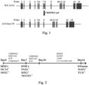
- the method for efficiently differentiating hESC into neural stem cells and neurons was performed as shown in Figure 2 .
- CHIR99021, SB431542, hLIF, and Compound E were used to induce hESC to differentiate into neural lineage.
- both wild-type and CLN3 ⁇ ex7 / 8 hESC were efficiently induced to become PAX6, SOX1, and NESTIN-positive human neural stem cells (hNSC).
- hNSC were then treated with CHIR99021, SB431542, and hLIF for 3 days, followed by changing the culture medium to contain hBDNF, hGDNF, cAMP, and vitamin C, and continuing for another 14 days of induction.
- MAP2 and TUJ1-positive human neurons were then obtained.
- Example 3 In vitro recapitulation of disease-associated phenotypes in CLN3 ⁇ ex7 / 8 human neural stem cells and neurons
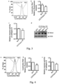
- the cells were stained with Lysotracker, a fluorescent dye that is enriched in acidic lysosomes and emits red fluorescence. The fluorescence intensity of the stained cells was then analyzed using flow cytometry.
- the CLN3 gene mutation in neural stem cells obtained by differentiation can result in JNCL-associated lysosomal defects such as lysosomal acidity dysregulation, protease activity reduction, and abnormal protein storage.
- Lysosomal and autophagy functions were further examined in CLN3 ⁇ ex7 / 8 neurons. After differentiating wild-type and CLN3 ⁇ ex7 / 8 human neural stem cells into neurons, the neurons were first stained with Lysotracker Red dye, and the fluorescence intensity of wild-type and CLN3 ⁇ ex7 / 8 neurons was then analyzed by flow cytometry, respectively.
- CLN3 knockdown was performed in wild-type neural stem cells using short hairpin RNA (shRNA) targeting the CLN3 transcript, and wild-type neural stem cells infected with target-free shRNA (Ctrl shRNA) were used as a control, which were both differentiated into human neurons later.
- shRNA short hairpin RNA
- Ctrl shRNA target-free shRNA
- the changes of CLN3 protein level in neurons expressing CLN3 shRNA were analyzed by western blot. The results showed that the use of CLN3 shRNA significantly reduced the CLN3 protein expression level in neurons compared to the control shRNA ( Figure 6 (c) ).
- the number of autophagosomes in the cells was counted before and after treatment with the autophagy blocker bafilomycin A1 (Baf-A1).
- the results showed that the changes of the number of autophagosomes in wild-type neural stem cells (number of autophagosomes after treatment with Baf-A1 - number of autophagosomes before treatment with Baf-A1) was significantly higher than that in CLN3 ⁇ ex7 / 8 neural stem cells ( Figure 7 (b) ), confirming that autophagic flux in CLN3 ⁇ ex7 / 8 human neural stem cells was defective.
- Example 4 The compounds of the present disclosure are autophagy inducers capable of increasing lysosomal acidity
- the inventors constructed a normal rat kidney (NRK) cell line that stably expresses tandem LC3.
- the tandem LC3 system is an LC3 reporter system that uses both red and green fluorescent labels.
- the autophagosomes simultaneously show red fluorescence and green fluorescence (yellow).
- the acidic environment in lysosomes quenches the green fluorescence of tandem LC3, and thus autolysosome only present red fluorescence (Shunsuke Kimura et al., 2007).
- this system is able to distinguish between autophagy inducers and lysosomal inhibitors.
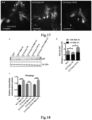
- the inventors obtained seven small molecule compounds which were able to increase the number of red fluorescent and green fluorescent double-positive autophagosomes in cells by two-fold and more simultaneously, and to keep the number of red fluorescent single-positive autophagosomes from decreasing, when tested with the tandem LC3 system described above, suggesting that all of them are autophagy inducers rather than lysosomal inhibitors. Moreover, after treating BFP-LC3 stably expressing NRK cells with these seven small molecule compounds for 18 hours, staining with Lysotracker dye was performed. It could be observed that, compared with control cells, these seven small molecule compounds could significantly increase intracellular Lysotracker staining ( Figures 9, 10 ), indicating that they increased intracellular lysosomal acidity.
- Said seven small molecule compounds are:
- Example 5 Compound G alleviates disease phenotypes in JNCL human neural cell model
- Compound G alleviates the differentiation defects in CLN3 ⁇ ex7 / 8 neural stem cells
- CLN3 ⁇ ex7 / 8 neural stem cells eventually generated a lower number of neurons than wild-type cells.
- the seven small molecule compounds obtained above were each added to the culture medium and observed for their effects on the differentiation process of CLN3 ⁇ ex7 / 8 neural stem cells into neurons.
- Compound G was found to be effective in alleviating the differentiation defects of CLN3 ⁇ ex7 / 8 neural stem cells to neurons.
- Compound G-treated CLN3 ⁇ ex7 / 8 neural stem cells were able to generate more MAP2 and TUJ1-positive neurons, which were close to the density of neurons generated by differentiation from wild-type neural stem cells ( Figure 11 ).
- Compound G alleviates the lysosomal and autophagy defects in CLN3 ⁇ ex7 / 8 neurons
- the treatment of the screened compound G can effectively alleviate the lysosomal acidity dysregulation in CLN3 ⁇ ex7 / 8 human neurons, increase the lysosomal protease activity, and reduce the abnormal intracellular protein storage, i.e., the compound G can alleviate the JNCL disease-associated phenotypes to a certain extent in CLN3 ⁇ ex7 / 8 human neuronal models.
- the inventors also tested several other potentially useful compounds and found that, similar to Compound G, they were also capable of alleviating the JNCL disease-associated phenotypes to some extent in the CLN3 ⁇ ex7 / 8 human neuronal model.
- the inventors tested dimethyl fumarate (DMF): using Lysotracker staining to characterize the lysosomal acidity in the cells and analyzing the average fluorescence intensity of the staining using flow cytometry.
- the inventors found that the average fluorescence intensity of the Lysotracker in the DMF-treated CLN3 ⁇ ex7 / 8 human neurons was significantly increased, suggesting that DMF can increase the lysosomal acidity in CLN3 ⁇ ex7 / 8 human neurons.
- Tests for the effect of DMF on SCMAS protein storage in CLN3 ⁇ ex7 / 8 human neurons showed that DMF treatment significantly reduced SCMAS protein storage in CLN3 ⁇ ex7 / 8 human neurons ( Figure 16 ).
- Compound G increases the level of autophagy and mitochondrial autophagy in CLN3 ⁇ ex7 / 8 neurons and maintains the function and homeostasis of mitochondria
- the p62 protein expressed by p62 is a cellular autophagy-associated protein responsible for attaching autophagic substrates to LC3 proteins which are located on the membrane of autophagosomes. Since p62 can be transported with autophagosomes to lysosomes and degraded, the intensity of intracellular autophagic flux can be characterized by comparing the changes in the amount of intracellular p62 protein after treatment with the lysosomal inhibitor Baf-A1 (p62 (Baf-A1-treated group) - p62 (Baf-A1-untreated group)).
- Damaged mitochondria within a cell can be eliminated through an autophagic process named mitochondrial autophagy.
- Functional defects in autophagy affect the elimination of damaged mitochondria, which in turn disrupts mitochondrial homeostasis in neurons.
- mitochondrial autophagy After inducing mitochondrial damage in wild-type and CLN3 ⁇ ex7 / 8 neurons using the mitochondrial uncoupler CCCP, and then inhibiting the elimination of damaged mitochondria by Baf-A1, the increases of intracellular mitochondrial number (mtDNA/nDNA) after Baf-A1 treatment can represent the intracellular mitochondrial autophagy level.
- Example 6 Compound G improved the disease phenotype of JNCL mice
- the Cln3 knockout ( Cln3 KO ) JNCL mouse model was purchased from Jackson Lab.
- Compound G improved the disease-associated phenotypes of JNCL mouse model
- Compound G could penetrate the blood-brain barrier
- Compound G was tested firstly whether it could penetrate the mice's blood-brain barrier: it was added to the drinking water of Cln3 KO mice, the mice were anesthetized at different time points. After using dPBS to completely perfuse the systemic blood, the mice were euthanized and the brain tissues thereof were taken out. The drug concentrations of Compound G in the mouse brain tissues were measured. The results showed that the presence of Compound G could be detected in the mouse brain tissues obtained at different time points, and its average concentration in the mouse brain was 814.74 ⁇ 834.19ng/g brain tissue ( Figure 20 ). That is, Compound G could penetrate the blood-brain barrier.
- Compound G could reduce the abnormal storage of the proteins in the brain of JNCL mouse model
- Compound G could improve the learning and memory ability of JNCL mice
- mice were trained in the water maze for 6 days, and the time it took for them to find the underwater platform was recorded. On the 7th day, the underwater platform was removed and the mice were tested for 60 seconds. The time that the mice spent reaching the position of the original platform for the first time, the number of shuttles at the position of the original platform and the time stayed in the quadrant of the original platform were recorded, which characterized whether they have learned the position of the underwater platform through the first 6 days of training.
- roller experiment was conducted. Treatment of Compound G was continuously provided to the mice after weaned on day 21.
- the Cln3 KO mice and the Compound G-treated Cln3 KO mice reached 4-5 months of age, roller training for 3 days was conducted, and test was conducted on the 4th day for the motor ability of the mice on the roller.
- Example 7 Compound G inhibited KEAP1 and enhanced the functions of autolysosome
- the known targets of Compound G were barely expressed in nerve cells
- Compound G could increase the transcription level of the transcription factor EB (TFEB) and the autophagy-related genes and the lysosomal genes by inhibiting the activities of the known target farnesoid X receptor (FXR).
- Compound G was added to the mouse tail tip fibroblasts (TTF), after incubation for 6 hours, mRNA was extracted, and the transcription level of the autophagy-related genes and the lysosomal genes in the cells was detected. The results showed that treatment with Compound G could significantly increase the transcription level of the autophagy-related genes and the lysosomal genes in TTF ( Figure 25 (a, b)).
- shRNA was used to knock down the level of FXR in TTF. After testing the efficiency of the knockdown of FXR by shRNA, shRNA-2 with the best knockdown efficiency was selected for subsequent experiments ( Figure 28 (a) ).
- lentivirus packaged with control shRNA or FXR shRNA-2 was used to infect the mouse TTF. After treatment with Compound G, mRNA was extracted for detection of gene transcription. It was found that Compound G could significantly increase the expression of the autophagy-related genes in control shRNA-infected cells, while in TTF infected with FXR shRNA-2, treatment with Compound G failed to increase the expression of these genes ( Figure 28 (b) ). The data showed that the knockdown of FXR itself could significantly increase the expression level of Tfeb and autophagy-related genes ( Figure 28 (b) ).
- Compound G activated the expression of NRF2 downstream genes
- transcriptome sequencing experiments were conducted on wild-type human neural stem cells, CLN3 ⁇ ex7 / 8 human neural stem cells and CLN3 ⁇ ex7 / 8 human neural stem cells after treated with Compound G for 6 hours, and differentially expressed genes were analyzed.
- PCA analysis showed that in CLN3 ⁇ ex7 / 8 human neural stem cells, treatment with Compound G for 6 hours did not produce a significant impact on the gene expression profile thereof.
- CLN3 ⁇ ex7 / 8 human neural stem cells treated and untreated with Compound G were clustered together ( Figure 29 (b, c)).
- Compound G inhibited KEAP1 protein activity
- NRF2 protein was mainly regulated by KEAP1 protein.
- Multiple electrophiles or electrophile precursor compounds could increase NRF2 activity by inhibiting KEAP1, such as isothiocyanates, ⁇ , ⁇ -unsaturated carbonyl compounds, phenols and polyphenolic compounds, etc.
- the basic action mode involved covalent reactions with the cysteine residues on KEAP1, mainly including cysteine residues at the positon on 151, 272 and 288 on KEAP1, resulting in changes in the binding mode of KEAP1 and NRF2, thereby activating NRF2 (Wells, 2015).
- Compound G contained an ⁇ , ⁇ -unsaturated carbonyl group, and a quantitative thiol reactivity analysis was conducted to determine whether Compound G could covalently bind to the cysteine on KEAP1. After treating CLN3 ⁇ ex7 / 8 human neural stem cells with Compound G for 2 hours, the cells were lysed, mass spectrometry was used to analyze the thiol reactivity of the cysteine residues on KEAP1 in the lysate.
- shRNA targeting KEAP1 was used to knock down the KEAP1 protein in the CLN3 ⁇ ex7 / 8 human neuron model.
- Five shRNA lentiviral plasmids targeting KEAP1 were selected from the human shRNA library, followed by using HEK293T cells to package them into lentivirus, and infecting the human neural stem cells.
- To detect the knockdown efficiency of shRNA mRNA was extracted, and qRT-PCR was used to detect the relative abundance of the KEAP1 mRNA in cells. The results showed that KEAP1 shRNA-3 had the highest knockdown efficiency ( Figure 34 ), then this shRNA was used to conduct subsequent experiments, detecting the impact of the knockdown of KEAP1 on the CLN3 ⁇ ex7 / 8 human neurons.
- Lysotracker staining was used to indicate the acidity of the lysosomes in cells, and flow cytometry was used to analyze the average fluorescence intensity of the staining.
- the Lysotracker average fluorescence intensity of the KEAP1 -knockdown CLN3 ⁇ ex7 / 8 human neurons was significantly enhanced, while adding Compound G to the KEAP1 -knockdown CLN3 ⁇ ex7 / 8 human neurons could not further increase the Lysotracker average fluorescence intensity ( Figure 35 ).
- This data demonstrated that knockdown of KEAP1 could increase the lysosomal acidity in CLN3 ⁇ ex7 / 8 human neurons, while the ability of Compound G to increase the lysosomal acidity was dependent on KEAP1.
- tandem LC3 protein was overexpressed in human neural stem cells to mark the intracellular autophagosomes, and imaging was conducted using the high-content laser confocal microscopy.
- the results showed that, similar to Compound G, the KEAP1 knockdown could significantly increase the number of the red-fluorescence single-positive autophagosomes in the CLN3 ⁇ ex7 / 8 human neural stem cells. This result suggested that the KEAP1 knockdown could effectively alleviate the defect of the autophagic flux in the CLN3 ⁇ ex7 / 8 human neural stem cells ( Figure 38 (a, b)).
- the knockdown of KEAP1 could mimic the effects of Compound G on lysosomal functions, autophagic flux intensities, and mitochondrial homeostasis.
- Compound G exerted the above functions in a KEAP1-dependent manner.
- Compound G enhanced the lysosomal acidity through the p62-KEAP1 pathway
- the present disclosure detected the impact of the Compound G treatment or the KEAP1 knockdown on the intracellular p62 protein level.
- Compound G was added to human neurons, after incubation for 24 hours, the cell lysate was extracted, qRT-PCR was used to detect the transcription level of the p62 gene.
- the results showed that treatment with Compound G or knockdown of KEAP1 could significantly increase the transcription level of p62 in human neurons ( Figure 41 (a) ).
- treatment with Compound G or knockdown of KEAP1 could also increase the p62 protein level ( Figure 41 (b) ).
- Example 8 Compound G reduced the accumulation of toxic proteins in human neuronal model of Alzheimer's dementia
- the present disclosure detected the therapeutic effect of the treatment of Compound G on Alzheimer's dementia.
- This model exhibited the deposition of the extracellular A ⁇ plaques and the increased levels of toxic A ⁇ 42 secreted by the cells into the culture medium.
- APP-gene mutant human neurons after treatment with Compound G showed a significant reduction in the deposition of A ⁇ plaques and a significant reduction in the level of toxic A ⁇ 42 secreted by the cells into the culture medium ( Figure 44 ).
- the present disclosure reveals that small molecule compounds including Compound G enhance the autophagic flux and the lysosomal functions in human neurons by inhibiting KEAP1, thereby benefiting the treatment of JNCL. It is demonstrated at the same time that knockdown of the KEAP1 gene can also achieve the same effects. In addition to JNCL, lysosomal functional deficiency and autophagy functional reduction are also involved in the pathogenesis of a variety of NCLs and other late-onset neurodegenerative diseases. It is reasonable to infer that inhibiting the activity of KEAP1 also has certain therapeutic effects on other neurodegenerative diseases related to autophagy and lysosomal functional deficiencies. Means of inhibiting KEAP1 include using small molecule compounds identified in the present disclosure including Compound G or knocking down the KEAP1 gene.
Landscapes
- Health & Medical Sciences (AREA)
- Life Sciences & Earth Sciences (AREA)
- Chemical & Material Sciences (AREA)
- General Health & Medical Sciences (AREA)
- Medicinal Chemistry (AREA)
- Engineering & Computer Science (AREA)
- Animal Behavior & Ethology (AREA)
- Pharmacology & Pharmacy (AREA)
- Public Health (AREA)
- Veterinary Medicine (AREA)
- Bioinformatics & Cheminformatics (AREA)
- Biomedical Technology (AREA)
- Organic Chemistry (AREA)
- Epidemiology (AREA)
- Neurosurgery (AREA)
- Neurology (AREA)
- Chemical Kinetics & Catalysis (AREA)
- General Chemical & Material Sciences (AREA)
- Nuclear Medicine, Radiotherapy & Molecular Imaging (AREA)
- Genetics & Genomics (AREA)
- Molecular Biology (AREA)
- Biochemistry (AREA)
- Immunology (AREA)
- Biotechnology (AREA)
- Physics & Mathematics (AREA)
- Wood Science & Technology (AREA)
- Zoology (AREA)
- General Engineering & Computer Science (AREA)
- Psychology (AREA)
- Psychiatry (AREA)
- Emergency Medicine (AREA)
- Microbiology (AREA)
- Hematology (AREA)
- Analytical Chemistry (AREA)
- General Physics & Mathematics (AREA)
- Pathology (AREA)
- Biophysics (AREA)
- Plant Pathology (AREA)
- Dispersion Chemistry (AREA)
- Urology & Nephrology (AREA)
Applications Claiming Priority (2)
| Application Number | Priority Date | Filing Date | Title |
|---|---|---|---|
| CN202110674245.2A CN115487297A (zh) | 2021-06-17 | 2021-06-17 | 治疗神经元蜡样质脂褐质沉积症的方法和药物 |
| PCT/CN2022/099308 WO2022262830A1 (zh) | 2021-06-17 | 2022-06-17 | 治疗神经元蜡样质脂褐质沉积症的方法和药物 |
Publications (3)
| Publication Number | Publication Date |
|---|---|
| EP4356932A1 EP4356932A1 (de) | 2024-04-24 |
| EP4356932A9 true EP4356932A9 (de) | 2024-08-21 |
| EP4356932A4 EP4356932A4 (de) | 2025-04-16 |
Family
ID=84464824
Family Applications (1)
| Application Number | Title | Priority Date | Filing Date |
|---|---|---|---|
| EP22824303.6A Withdrawn EP4356932A4 (de) | 2021-06-17 | 2022-06-17 | Verfahren und arzneimittel zur behandlung von neuronaler ceroid-lipofuszinose |
Country Status (4)
| Country | Link |
|---|---|
| US (1) | US20250152605A1 (de) |
| EP (1) | EP4356932A4 (de) |
| CN (2) | CN115487297A (de) |
| WO (1) | WO2022262830A1 (de) |
Family Cites Families (7)
| Publication number | Priority date | Publication date | Assignee | Title |
|---|---|---|---|---|
| ZA989365B (en) * | 1997-10-15 | 1999-04-15 | Boehringer Mannheim Pharm Corp | Preparation for treating alzheimer's disease |
| CN101686725A (zh) * | 2007-04-03 | 2010-03-31 | 刚迦·拉贾·高卡拉贾 | 消炎和抗氧化协同作用的膳食补充组合物 |
| JP2012525385A (ja) * | 2009-04-29 | 2012-10-22 | バイオジェン・アイデック・エムエイ・インコーポレイテッド | 神経変性および神経炎症の治療 |
| EP2806948B1 (de) * | 2012-01-27 | 2020-01-15 | New York University | Verfahren zur verbesserung von remyelinisierung unter verwendung von gli1-inhibitoren |
| MX2014014993A (es) * | 2012-06-05 | 2015-06-17 | Int Stem Cell Corp | Metodo para la prevencion de enfermedades neurologicas. |
| WO2016100575A1 (en) * | 2014-12-16 | 2016-06-23 | Board Of Regents Of The University Of Nebraska | Gene therapy for juvenile batten disease |
| WO2019108773A1 (en) * | 2017-11-29 | 2019-06-06 | Stc.Unm | Substituted stilbenes as inhibitors of nf-kb and activators of nrf2 |
-
2021
- 2021-06-17 CN CN202110674245.2A patent/CN115487297A/zh active Pending
-
2022
- 2022-06-17 EP EP22824303.6A patent/EP4356932A4/de not_active Withdrawn
- 2022-06-17 CN CN202280043165.XA patent/CN117561083A/zh active Pending
- 2022-06-17 US US18/571,108 patent/US20250152605A1/en active Pending
- 2022-06-17 WO PCT/CN2022/099308 patent/WO2022262830A1/zh not_active Ceased
Also Published As
| Publication number | Publication date |
|---|---|
| CN117561083A (zh) | 2024-02-13 |
| EP4356932A1 (de) | 2024-04-24 |
| WO2022262830A1 (zh) | 2022-12-22 |
| EP4356932A4 (de) | 2025-04-16 |
| CN115487297A (zh) | 2022-12-20 |
| US20250152605A1 (en) | 2025-05-15 |
Similar Documents
| Publication | Publication Date | Title |
|---|---|---|
| CN107405378B (zh) | 用于治疗与非降解异常蛋白的积聚相关的病症或癌症的蛋白酶体抑制剂 | |
| JP7182852B2 (ja) | 神経変性疾患のための細胞性発見プラットフォーム | |
| Oh et al. | S-Nitrosylation of p62 inhibits autophagic flux to promote α-synuclein secretion and spread in Parkinson's disease and Lewy body dementia | |
| US10105420B2 (en) | Methods, compositions and screens for therapeutics for the treatment of synovial sarcoma | |
| US10201558B2 (en) | Compositions of selenoorganic compounds and methods of use thereof | |
| US9696306B2 (en) | Methods of reducing levels of tau | |
| KR20130128018A (ko) | 새로운 노화 방지 물질 및 그 확인 방법 | |
| EP2841067A2 (de) | Arzneimittelscreeningplattform für das rett-syndrom | |
| AU2020203955B2 (en) | Composition and method for inhibiting amyloid beta accumulation and/or aggregation | |
| Hou et al. | Acute spinal cord injury in rats should target activated autophagy | |
| Valverde et al. | Dipeptidyl peptidase 4 contributes to Alzheimer’s disease–like defects in a mouse model and is increased in sporadic Alzheimer’s disease brains | |
| Ahmed et al. | Amplifying the heat shock response ameliorates ALS and FTD pathology in mouse and human models | |
| WO2016138300A1 (en) | Compositions and methods of treating fanconi anemia | |
| EP3947344A1 (de) | Aminoguanidinhydrazone als retromerstabilisatoren zur behandlung von neurologischen erkrankungen | |
| EP3890771B1 (de) | Verfahren zur verminderung der konzentration des neuronalen mikrotubulus-bindenden proteins tau | |
| EP4356932A9 (de) | Verfahren und arzneimittel zur behandlung von neuronaler ceroid-lipofuszinose | |
| EP3979989A1 (de) | Herstellung und verwendung von in extrazellulären vesikeln enthaltenen enampt | |
| US20240409890A1 (en) | Ufmylation inhibition to target tauopathy in human neurons | |
| US11382872B2 (en) | LSD1 inhibitors as skeletal muscle hypertrophy inducers | |
| US12226410B2 (en) | Methods for enhancing cellular clearance of pathological molecules via activation of the cellular protein ykt6 | |
| US20250032425A1 (en) | First in class use of estrogen receptor beta agonists in treating angelman syndrome and relevant autism spectrum disorders | |
| Tamilselvam et al. | Small peptide P110 mitigates axonal degeneration of SPG15 patient iPSC-derived neurons by targeting mitochondrial dysfunction | |
| US20240238385A1 (en) | Isolated or artificial nucleotide sequences for use in neurodegenerative diseases | |
| Martín Masegosa | Simultaneous activation of two complementary targets, Kv7. 2/3 AND TSPO: a novel treatment for amyotrophic lateral sclerosis | |
| Schmoll | Network hyperexcitability and dopaminergic neuron loss in a novel A30P-driven Parkinson’s midbrain organoid model |
Legal Events
| Date | Code | Title | Description |
|---|---|---|---|
| STAA | Information on the status of an ep patent application or granted ep patent |
Free format text: STATUS: THE INTERNATIONAL PUBLICATION HAS BEEN MADE |
|
| PUAI | Public reference made under article 153(3) epc to a published international application that has entered the european phase |
Free format text: ORIGINAL CODE: 0009012 |
|
| STAA | Information on the status of an ep patent application or granted ep patent |
Free format text: STATUS: REQUEST FOR EXAMINATION WAS MADE |
|
| 17P | Request for examination filed |
Effective date: 20231215 |
|
| AK | Designated contracting states |
Kind code of ref document: A1 Designated state(s): AL AT BE BG CH CY CZ DE DK EE ES FI FR GB GR HR HU IE IS IT LI LT LU LV MC MK MT NL NO PL PT RO RS SE SI SK SM TR |
|
| DAV | Request for validation of the european patent (deleted) | ||
| DAX | Request for extension of the european patent (deleted) | ||
| REG | Reference to a national code |
Ref country code: DE Ref legal event code: R079 Free format text: PREVIOUS MAIN CLASS: A61K0048000000 Ipc: A61K0031403000 |
|
| A4 | Supplementary search report drawn up and despatched |
Effective date: 20250317 |
|
| RIC1 | Information provided on ipc code assigned before grant |
Ipc: A61P 25/00 20060101ALI20250311BHEP Ipc: A61K 31/713 20060101ALI20250311BHEP Ipc: A61K 31/164 20060101ALI20250311BHEP Ipc: A61K 31/568 20060101ALI20250311BHEP Ipc: A61K 31/335 20060101ALI20250311BHEP Ipc: A61K 31/225 20060101ALI20250311BHEP Ipc: A61P 25/14 20060101ALI20250311BHEP Ipc: A61K 31/22 20060101ALI20250311BHEP Ipc: A61P 3/00 20060101ALI20250311BHEP Ipc: A61P 25/28 20060101ALI20250311BHEP Ipc: A61P 25/18 20060101ALI20250311BHEP Ipc: A61P 25/16 20060101ALI20250311BHEP Ipc: G01N 33/50 20060101ALI20250311BHEP Ipc: A61P 43/00 20060101ALI20250311BHEP Ipc: C12N 15/90 20060101ALI20250311BHEP Ipc: A61K 31/57 20060101ALI20250311BHEP Ipc: G01N 15/14 20240101ALI20250311BHEP Ipc: A61K 48/00 20060101ALI20250311BHEP Ipc: A61K 31/403 20060101AFI20250311BHEP |
|
| STAA | Information on the status of an ep patent application or granted ep patent |
Free format text: STATUS: THE APPLICATION IS DEEMED TO BE WITHDRAWN |
|
| 18D | Application deemed to be withdrawn |
Effective date: 20251007 |