EP4345160A2 - Method for reproducible differentiation of clinical-grade retinal pigment epithelium cells - Google Patents
Method for reproducible differentiation of clinical-grade retinal pigment epithelium cells Download PDFInfo
- Publication number
- EP4345160A2 EP4345160A2 EP23214033.5A EP23214033A EP4345160A2 EP 4345160 A2 EP4345160 A2 EP 4345160A2 EP 23214033 A EP23214033 A EP 23214033A EP 4345160 A2 EP4345160 A2 EP 4345160A2
- Authority
- EP
- European Patent Office
- Prior art keywords
- cells
- rpe
- retinal
- medium
- cell
- Prior art date
- Legal status (The legal status is an assumption and is not a legal conclusion. Google has not performed a legal analysis and makes no representation as to the accuracy of the status listed.)
- Pending
Links
- 238000000034 method Methods 0.000 title claims abstract description 161
- 230000004069 differentiation Effects 0.000 title claims abstract description 73
- 210000003583 retinal pigment epithelium Anatomy 0.000 title description 306
- 210000004027 cell Anatomy 0.000 claims abstract description 723
- 238000012258 culturing Methods 0.000 claims abstract description 42
- 101000670189 Homo sapiens Ribulose-phosphate 3-epimerase Proteins 0.000 claims abstract description 11
- 239000002609 medium Substances 0.000 claims description 158
- 210000004263 induced pluripotent stem cell Anatomy 0.000 claims description 103
- 230000002207 retinal effect Effects 0.000 claims description 89
- 239000003112 inhibitor Substances 0.000 claims description 85
- 230000037361 pathway Effects 0.000 claims description 62
- 108090000623 proteins and genes Proteins 0.000 claims description 57
- 102000004169 proteins and genes Human genes 0.000 claims description 44
- WRKPZSMRWPJJDH-UHFFFAOYSA-N N-(6-methyl-1,3-benzothiazol-2-yl)-2-[(4-oxo-3-phenyl-6,7-dihydrothieno[3,2-d]pyrimidin-2-yl)thio]acetamide Chemical compound S1C2=CC(C)=CC=C2N=C1NC(=O)CSC1=NC=2CCSC=2C(=O)N1C1=CC=CC=C1 WRKPZSMRWPJJDH-UHFFFAOYSA-N 0.000 claims description 43
- XEYBRNLFEZDVAW-ARSRFYASSA-N dinoprostone Chemical group CCCCC[C@H](O)\C=C\[C@H]1[C@H](O)CC(=O)[C@@H]1C\C=C/CCCC(O)=O XEYBRNLFEZDVAW-ARSRFYASSA-N 0.000 claims description 39
- 230000035800 maturation Effects 0.000 claims description 38
- 102000004887 Transforming Growth Factor beta Human genes 0.000 claims description 37
- 108090001012 Transforming Growth Factor beta Proteins 0.000 claims description 37
- FHYUGAJXYORMHI-UHFFFAOYSA-N SB 431542 Chemical group C1=CC(C(=O)N)=CC=C1C1=NC(C=2C=C3OCOC3=CC=2)=C(C=2N=CC=CC=2)N1 FHYUGAJXYORMHI-UHFFFAOYSA-N 0.000 claims description 30
- CDOVNWNANFFLFJ-UHFFFAOYSA-N 4-[6-[4-(1-piperazinyl)phenyl]-3-pyrazolo[1,5-a]pyrimidinyl]quinoline Chemical compound C1CNCCN1C1=CC=C(C2=CN3N=CC(=C3N=C2)C=2C3=CC=CC=C3N=CC=2)C=C1 CDOVNWNANFFLFJ-UHFFFAOYSA-N 0.000 claims description 29
- 229940121396 wnt pathway inhibitor Drugs 0.000 claims description 29
- 210000002242 embryoid body Anatomy 0.000 claims description 28
- DFPAKSUCGFBDDF-UHFFFAOYSA-N Nicotinamide Chemical compound NC(=O)C1=CC=CN=C1 DFPAKSUCGFBDDF-UHFFFAOYSA-N 0.000 claims description 26
- 229940124647 MEK inhibitor Drugs 0.000 claims description 24
- 239000002829 mitogen activated protein kinase inhibitor Substances 0.000 claims description 24
- 239000000203 mixture Substances 0.000 claims description 23
- 239000011159 matrix material Substances 0.000 claims description 22
- 230000006698 induction Effects 0.000 claims description 20
- OGKYMFFYOWUTKV-UHFFFAOYSA-N N-(2-aminoethyl)-5-chloroisoquinoline-8-sulfonamide Chemical compound C1=NC=C2C(S(=O)(=O)NCCN)=CC=C(Cl)C2=C1 OGKYMFFYOWUTKV-UHFFFAOYSA-N 0.000 claims description 19
- 238000004519 manufacturing process Methods 0.000 claims description 19
- 239000001963 growth medium Substances 0.000 claims description 18
- 108700028369 Alleles Proteins 0.000 claims description 16
- 230000001413 cellular effect Effects 0.000 claims description 16
- 150000001875 compounds Chemical class 0.000 claims description 15
- 208000002780 macular degeneration Diseases 0.000 claims description 15
- NOESYZHRGYRDHS-UHFFFAOYSA-N insulin Chemical compound N1C(=O)C(NC(=O)C(CCC(N)=O)NC(=O)C(CCC(O)=O)NC(=O)C(C(C)C)NC(=O)C(NC(=O)CN)C(C)CC)CSSCC(C(NC(CO)C(=O)NC(CC(C)C)C(=O)NC(CC=2C=CC(O)=CC=2)C(=O)NC(CCC(N)=O)C(=O)NC(CC(C)C)C(=O)NC(CCC(O)=O)C(=O)NC(CC(N)=O)C(=O)NC(CC=2C=CC(O)=CC=2)C(=O)NC(CSSCC(NC(=O)C(C(C)C)NC(=O)C(CC(C)C)NC(=O)C(CC=2C=CC(O)=CC=2)NC(=O)C(CC(C)C)NC(=O)C(C)NC(=O)C(CCC(O)=O)NC(=O)C(C(C)C)NC(=O)C(CC(C)C)NC(=O)C(CC=2NC=NC=2)NC(=O)C(CO)NC(=O)CNC2=O)C(=O)NCC(=O)NC(CCC(O)=O)C(=O)NC(CCCNC(N)=N)C(=O)NCC(=O)NC(CC=3C=CC=CC=3)C(=O)NC(CC=3C=CC=CC=3)C(=O)NC(CC=3C=CC(O)=CC=3)C(=O)NC(C(C)O)C(=O)N3C(CCC3)C(=O)NC(CCCCN)C(=O)NC(C)C(O)=O)C(=O)NC(CC(N)=O)C(O)=O)=O)NC(=O)C(C(C)CC)NC(=O)C(CO)NC(=O)C(C(C)O)NC(=O)C1CSSCC2NC(=O)C(CC(C)C)NC(=O)C(NC(=O)C(CCC(N)=O)NC(=O)C(CC(N)=O)NC(=O)C(NC(=O)C(N)CC=1C=CC=CC=1)C(C)C)CC1=CN=CN1 NOESYZHRGYRDHS-UHFFFAOYSA-N 0.000 claims description 14
- 108010031318 Vitronectin Proteins 0.000 claims description 13
- 102100035140 Vitronectin Human genes 0.000 claims description 13
- 239000000411 inducer Substances 0.000 claims description 13
- 229960003966 nicotinamide Drugs 0.000 claims description 13
- 235000005152 nicotinamide Nutrition 0.000 claims description 13
- 239000011570 nicotinamide Substances 0.000 claims description 13
- 108010085895 Laminin Proteins 0.000 claims description 12
- 102000007547 Laminin Human genes 0.000 claims description 12
- 102100028972 HLA class I histocompatibility antigen, A alpha chain Human genes 0.000 claims description 11
- 108010075704 HLA-A Antigens Proteins 0.000 claims description 11
- 210000004081 cilia Anatomy 0.000 claims description 11
- JUAVTXYOCISSSL-UHFFFAOYSA-N n-(2-aminoethyl)-5-chloroisoquinoline-8-sulfonamide;dihydrochloride Chemical group Cl.Cl.C1=NC=C2C(S(=O)(=O)NCCN)=CC=C(Cl)C2=C1 JUAVTXYOCISSSL-UHFFFAOYSA-N 0.000 claims description 11
- LBPKYPYHDKKRFS-UHFFFAOYSA-N 1,5-naphthyridine, 2-[3-(6-methyl-2-pyridinyl)-1h-pyrazol-4-yl]- Chemical compound CC1=CC=CC(C2=C(C=NN2)C=2N=C3C=CC=NC3=CC=2)=N1 LBPKYPYHDKKRFS-UHFFFAOYSA-N 0.000 claims description 10
- AXNUEXXEQGQWPA-UHFFFAOYSA-N 2,5-dichloro-N-(2-methyl-4-nitrophenyl)benzenesulfonamide Chemical compound CC1=CC([N+]([O-])=O)=CC=C1NS(=O)(=O)C1=CC(Cl)=CC=C1Cl AXNUEXXEQGQWPA-UHFFFAOYSA-N 0.000 claims description 10
- JQGOCCALXFSRHZ-UHFFFAOYSA-N 2-[4-[2-fluoro-5-[5-(6-methylpyridin-2-yl)-1h-pyrazol-4-yl]phenyl]pyrazol-1-yl]ethanol Chemical compound CC1=CC=CC(C=2C(=CNN=2)C=2C=C(C(F)=CC=2)C2=CN(CCO)N=C2)=N1 JQGOCCALXFSRHZ-UHFFFAOYSA-N 0.000 claims description 10
- DXLXRNZCYAYUED-UHFFFAOYSA-N 4-[2-[4-(3-quinolin-4-ylpyrazolo[1,5-a]pyrimidin-6-yl)phenoxy]ethyl]morpholine Chemical compound C=1C=C(C2=CN3N=CC(=C3N=C2)C=2C3=CC=CC=C3N=CC=2)C=CC=1OCCN1CCOCC1 DXLXRNZCYAYUED-UHFFFAOYSA-N 0.000 claims description 10
- DPDZHVCKYBCJHW-UHFFFAOYSA-N 4-[4-(2,3-dihydro-1,4-benzodioxin-6-yl)-5-(2-pyridinyl)-1H-imidazol-2-yl]benzamide Chemical compound C1=CC(C(=O)N)=CC=C1C1=NC(C=2C=C3OCCOC3=CC=2)=C(C=2N=CC=CC=2)N1 DPDZHVCKYBCJHW-UHFFFAOYSA-N 0.000 claims description 10
- FVRYPYDPKSZGNS-UHFFFAOYSA-N 5-[6-(4-methoxyphenyl)pyrazolo[1,5-a]pyrimidin-3-yl]quinoline Chemical compound C1=CC(OC)=CC=C1C1=CN2N=CC(C=3C4=CC=CN=C4C=CC=3)=C2N=C1 FVRYPYDPKSZGNS-UHFFFAOYSA-N 0.000 claims description 10
- DKPQHFZUICCZHF-UHFFFAOYSA-N 6-[2-tert-butyl-5-(6-methyl-2-pyridinyl)-1H-imidazol-4-yl]quinoxaline Chemical compound CC1=CC=CC(C2=C(N=C(N2)C(C)(C)C)C=2C=C3N=CC=NC3=CC=2)=N1 DKPQHFZUICCZHF-UHFFFAOYSA-N 0.000 claims description 10
- JMIFGARJSWXZSH-UHFFFAOYSA-N DMH1 Chemical compound C1=CC(OC(C)C)=CC=C1C1=CN2N=CC(C=3C4=CC=CC=C4N=CC=3)=C2N=C1 JMIFGARJSWXZSH-UHFFFAOYSA-N 0.000 claims description 10
- 108010067306 Fibronectins Proteins 0.000 claims description 10
- 102000016359 Fibronectins Human genes 0.000 claims description 10
- HEKAIDKUDLCBRU-UHFFFAOYSA-N N-[4-[2-ethyl-4-(3-methylphenyl)-5-thiazolyl]-2-pyridinyl]benzamide Chemical compound S1C(CC)=NC(C=2C=C(C)C=CC=2)=C1C(C=1)=CC=NC=1NC(=O)C1=CC=CC=C1 HEKAIDKUDLCBRU-UHFFFAOYSA-N 0.000 claims description 10
- REFJWTPEDVJJIY-UHFFFAOYSA-N Quercetin Chemical compound C=1C(O)=CC(O)=C(C(C=2O)=O)C=1OC=2C1=CC=C(O)C(O)=C1 REFJWTPEDVJJIY-UHFFFAOYSA-N 0.000 claims description 10
- KLGQSVMIPOVQAX-UHFFFAOYSA-N XAV939 Chemical compound N=1C=2CCSCC=2C(O)=NC=1C1=CC=C(C(F)(F)F)C=C1 KLGQSVMIPOVQAX-UHFFFAOYSA-N 0.000 claims description 10
- ACWZRVQXLIRSDF-UHFFFAOYSA-N binimetinib Chemical compound OCCONC(=O)C=1C=C2N(C)C=NC2=C(F)C=1NC1=CC=C(Br)C=C1F ACWZRVQXLIRSDF-UHFFFAOYSA-N 0.000 claims description 10
- 230000000694 effects Effects 0.000 claims description 10
- SAGZIBJAQGBRQA-UHFFFAOYSA-N n-(oxan-4-yl)-4-[4-(5-pyridin-2-yl-1h-pyrazol-4-yl)pyridin-2-yl]benzamide Chemical compound C=1C=C(C=2N=CC=C(C=2)C2=C(NN=C2)C=2N=CC=CC=2)C=CC=1C(=O)NC1CCOCC1 SAGZIBJAQGBRQA-UHFFFAOYSA-N 0.000 claims description 10
- JJEDWBQZCRESJL-DEDYPNTBSA-N n-[(e)-(5-methylfuran-2-yl)methylideneamino]-2-phenoxybenzamide Chemical compound O1C(C)=CC=C1\C=N\NC(=O)C1=CC=CC=C1OC1=CC=CC=C1 JJEDWBQZCRESJL-DEDYPNTBSA-N 0.000 claims description 10
- 239000000790 retinal pigment Substances 0.000 claims description 10
- CYOHGALHFOKKQC-UHFFFAOYSA-N selumetinib Chemical compound OCCONC(=O)C=1C=C2N(C)C=NC2=C(F)C=1NC1=CC=C(Br)C=C1Cl CYOHGALHFOKKQC-UHFFFAOYSA-N 0.000 claims description 10
- LIRYPHYGHXZJBZ-UHFFFAOYSA-N trametinib Chemical compound CC(=O)NC1=CC=CC(N2C(N(C3CC3)C(=O)C3=C(NC=4C(=CC(I)=CC=4)F)N(C)C(=O)C(C)=C32)=O)=C1 LIRYPHYGHXZJBZ-UHFFFAOYSA-N 0.000 claims description 10
- 102100028976 HLA class I histocompatibility antigen, B alpha chain Human genes 0.000 claims description 9
- 108010058607 HLA-B Antigens Proteins 0.000 claims description 9
- 108010023082 activin A Proteins 0.000 claims description 9
- 239000003102 growth factor Substances 0.000 claims description 9
- 102000004877 Insulin Human genes 0.000 claims description 7
- 108090001061 Insulin Proteins 0.000 claims description 7
- SUDAHWBOROXANE-SECBINFHSA-N PD 0325901 Chemical compound OC[C@@H](O)CONC(=O)C1=CC=C(F)C(F)=C1NC1=CC=C(I)C=C1F SUDAHWBOROXANE-SECBINFHSA-N 0.000 claims description 7
- 229940125396 insulin Drugs 0.000 claims description 7
- WQUIJVKNPYBZOF-UHFFFAOYSA-N 4-[4-(1,3-benzodioxol-5-yl)-5-pyridin-2-yl-1h-imidazol-2-yl]benzamide;hydrate Chemical compound O.C1=CC(C(=O)N)=CC=C1C1=NC(C=2C=C3OCOC3=CC=2)=C(C=2N=CC=CC=2)N1 WQUIJVKNPYBZOF-UHFFFAOYSA-N 0.000 claims description 6
- 230000015572 biosynthetic process Effects 0.000 claims description 6
- 230000006378 damage Effects 0.000 claims description 6
- 208000014674 injury Diseases 0.000 claims description 6
- RHUJMHOIQBDFQR-UHFFFAOYSA-N 2-[[3-(2-methoxyphenyl)-4-oxo-6,7-dihydrothieno[3,2-d]pyrimidin-2-yl]sulfanyl]-n-(6-methyl-1,3-benzothiazol-2-yl)acetamide Chemical compound COC1=CC=CC=C1N1C(=O)C(SCC2)=C2N=C1SCC(=O)NC1=NC2=CC=C(C)C=C2S1 RHUJMHOIQBDFQR-UHFFFAOYSA-N 0.000 claims description 5
- HIJMSZGHKQPPJS-UHFFFAOYSA-N 3-(6-methylpyridin-2-yl)-n-phenyl-4-quinolin-4-ylpyrazole-1-carbothioamide Chemical compound CC1=CC=CC(C=2C(=CN(N=2)C(=S)NC=2C=CC=CC=2)C=2C3=CC=CC=C3N=CC=2)=N1 HIJMSZGHKQPPJS-UHFFFAOYSA-N 0.000 claims description 5
- PCCDKTWDGDFRME-UHFFFAOYSA-N 4-[6-(4-piperazin-1-ylphenyl)pyrazolo[1,5-a]pyrimidin-3-yl]quinoline;hydrochloride Chemical group Cl.C1CNCCN1C1=CC=C(C2=CN3N=CC(=C3N=C2)C=2C3=CC=CC=C3N=CC=2)C=C1 PCCDKTWDGDFRME-UHFFFAOYSA-N 0.000 claims description 5
- RJDVIJJQKMGPMV-UHFFFAOYSA-N 6-[4-(2-piperidin-1-ylethoxy)phenyl]-3-pyridin-4-ylpyrazolo[1,5-a]pyrimidine;dihydrochloride Chemical compound Cl.Cl.C=1C=C(C2=CN3N=CC(=C3N=C2)C=2C=CN=CC=2)C=CC=1OCCN1CCCCC1 RJDVIJJQKMGPMV-UHFFFAOYSA-N 0.000 claims description 5
- 108010058597 HLA-DR Antigens Proteins 0.000 claims description 5
- 102000006354 HLA-DR Antigens Human genes 0.000 claims description 5
- IBCXZJCWDGCXQT-UHFFFAOYSA-N LY 364947 Chemical compound C=1C=NC2=CC=CC=C2C=1C1=CNN=C1C1=CC=CC=N1 IBCXZJCWDGCXQT-UHFFFAOYSA-N 0.000 claims description 5
- 102000002092 Left-Right Determination Factor Human genes 0.000 claims description 5
- 108050009437 Left-Right Determination Factor Proteins 0.000 claims description 5
- ZVOLCUVKHLEPEV-UHFFFAOYSA-N Quercetagetin Natural products C1=C(O)C(O)=CC=C1C1=C(O)C(=O)C2=C(O)C(O)=C(O)C=C2O1 ZVOLCUVKHLEPEV-UHFFFAOYSA-N 0.000 claims description 5
- HWTZYBCRDDUBJY-UHFFFAOYSA-N Rhynchosin Natural products C1=C(O)C(O)=CC=C1C1=C(O)C(=O)C2=CC(O)=C(O)C=C2O1 HWTZYBCRDDUBJY-UHFFFAOYSA-N 0.000 claims description 5
- 102100030053 Secreted frizzled-related protein 3 Human genes 0.000 claims description 5
- 108010020277 WD repeat containing planar cell polarity effector Proteins 0.000 claims description 5
- 208000027418 Wounds and injury Diseases 0.000 claims description 5
- NOFOAYPPHIUXJR-APNQCZIXSA-N aphidicolin Chemical compound C1[C@@]23[C@@]4(C)CC[C@@H](O)[C@@](C)(CO)[C@@H]4CC[C@H]3C[C@H]1[C@](CO)(O)CC2 NOFOAYPPHIUXJR-APNQCZIXSA-N 0.000 claims description 5
- SEKZNWAQALMJNH-YZUCACDQSA-N aphidicolin Natural products C[C@]1(CO)CC[C@]23C[C@H]1C[C@@H]2CC[C@H]4[C@](C)(CO)[C@H](O)CC[C@]34C SEKZNWAQALMJNH-YZUCACDQSA-N 0.000 claims description 5
- XHBVYDAKJHETMP-UHFFFAOYSA-N dorsomorphin Chemical compound C=1C=C(C2=CN3N=CC(=C3N=C2)C=2C=CN=CC=2)C=CC=1OCCN1CCCCC1 XHBVYDAKJHETMP-UHFFFAOYSA-N 0.000 claims description 5
- DKAGJZJALZXOOV-UHFFFAOYSA-N hydrate;hydrochloride Chemical compound O.Cl DKAGJZJALZXOOV-UHFFFAOYSA-N 0.000 claims description 5
- MWDZOUNAPSSOEL-UHFFFAOYSA-N kaempferol Natural products OC1=C(C(=O)c2cc(O)cc(O)c2O1)c3ccc(O)cc3 MWDZOUNAPSSOEL-UHFFFAOYSA-N 0.000 claims description 5
- RDSACQWTXKSHJT-UHFFFAOYSA-N n-[3,4-difluoro-2-(2-fluoro-4-iodoanilino)-6-methoxyphenyl]-1-(2,3-dihydroxypropyl)cyclopropane-1-sulfonamide Chemical compound C1CC1(CC(O)CO)S(=O)(=O)NC=1C(OC)=CC(F)=C(F)C=1NC1=CC=C(I)C=C1F RDSACQWTXKSHJT-UHFFFAOYSA-N 0.000 claims description 5
- RDSACQWTXKSHJT-NSHDSACASA-N n-[3,4-difluoro-2-(2-fluoro-4-iodoanilino)-6-methoxyphenyl]-1-[(2s)-2,3-dihydroxypropyl]cyclopropane-1-sulfonamide Chemical compound C1CC1(C[C@H](O)CO)S(=O)(=O)NC=1C(OC)=CC(F)=C(F)C=1NC1=CC=C(I)C=C1F RDSACQWTXKSHJT-NSHDSACASA-N 0.000 claims description 5
- 229960001285 quercetin Drugs 0.000 claims description 5
- 235000005875 quercetin Nutrition 0.000 claims description 5
- XELRMPRLCPFTBH-UHFFFAOYSA-N quinazoline-2,4-diamine Chemical compound C1=CC=CC2=NC(N)=NC(N)=C21 XELRMPRLCPFTBH-UHFFFAOYSA-N 0.000 claims description 5
- 238000012216 screening Methods 0.000 claims description 5
- 229960004066 trametinib Drugs 0.000 claims description 5
- 208000007014 Retinitis pigmentosa Diseases 0.000 claims description 4
- ZGSXEXBYLJIOGF-UHFFFAOYSA-N iwr-1-endo Chemical compound C=1C=CC2=CC=CN=C2C=1NC(=O)C(C=C1)=CC=C1N1C(=O)C2C(C=C3)CC3C2C1=O ZGSXEXBYLJIOGF-UHFFFAOYSA-N 0.000 claims description 4
- 208000035719 Maculopathy Diseases 0.000 claims description 3
- 208000015122 neurodegenerative disease Diseases 0.000 claims description 3
- 206010012689 Diabetic retinopathy Diseases 0.000 claims description 2
- 208000010412 Glaucoma Diseases 0.000 claims description 2
- 101150088952 IGF1 gene Proteins 0.000 claims 1
- MFEADGGBEDIZRE-MGIYQXMMSA-N endo-iwr 1 Chemical compound C=1C=CC2=CC=CN=C2C=1NC(=O)C(C=C1)=CC=C1N1C(=O)[C@@H]2[C@H](C=C3)C[C@H]3[C@@H]2C1=O.C=1C=CC2=CC=CN=C2C=1NC(=O)C(C=C1)=CC=C1N1C(=O)[C@@H]2[C@H](C=C3)C[C@H]3[C@@H]2C1=O MFEADGGBEDIZRE-MGIYQXMMSA-N 0.000 claims 1
- 210000001778 pluripotent stem cell Anatomy 0.000 abstract description 57
- 239000006285 cell suspension Substances 0.000 abstract description 38
- 108060007030 Ribulose-phosphate 3-epimerase Proteins 0.000 abstract description 3
- 238000005516 engineering process Methods 0.000 description 118
- 239000011604 retinal Substances 0.000 description 70
- 235000020945 retinal Nutrition 0.000 description 69
- NCYCYZXNIZJOKI-UHFFFAOYSA-N vitamin A aldehyde Natural products O=CC=C(C)C=CC=C(C)C=CC1=C(C)CCCC1(C)C NCYCYZXNIZJOKI-UHFFFAOYSA-N 0.000 description 69
- DAEPDZWVDSPTHF-UHFFFAOYSA-M sodium pyruvate Chemical compound [Na+].CC(=O)C([O-])=O DAEPDZWVDSPTHF-UHFFFAOYSA-M 0.000 description 58
- 239000000306 component Substances 0.000 description 43
- 235000018102 proteins Nutrition 0.000 description 39
- 229960002986 dinoprostone Drugs 0.000 description 35
- XEYBRNLFEZDVAW-UHFFFAOYSA-N prostaglandin E2 Natural products CCCCCC(O)C=CC1C(O)CC(=O)C1CC=CCCCC(O)=O XEYBRNLFEZDVAW-UHFFFAOYSA-N 0.000 description 35
- 101100247004 Rattus norvegicus Qsox1 gene Proteins 0.000 description 34
- 108700021430 Kruppel-Like Factor 4 Proteins 0.000 description 31
- CIWBSHSKHKDKBQ-JLAZNSOCSA-N Ascorbic acid Chemical compound OC[C@H](O)[C@H]1OC(=O)C(O)=C1O CIWBSHSKHKDKBQ-JLAZNSOCSA-N 0.000 description 30
- JYGXADMDTFJGBT-VWUMJDOOSA-N hydrocortisone Chemical compound O=C1CC[C@]2(C)[C@H]3[C@@H](O)C[C@](C)([C@@](CC4)(O)C(=O)CO)[C@@H]4[C@@H]3CCC2=C1 JYGXADMDTFJGBT-VWUMJDOOSA-N 0.000 description 30
- XOAAWQZATWQOTB-UHFFFAOYSA-N taurine Chemical compound NCCS(O)(=O)=O XOAAWQZATWQOTB-UHFFFAOYSA-N 0.000 description 30
- 239000012580 N-2 Supplement Substances 0.000 description 29
- 101150086694 SLC22A3 gene Proteins 0.000 description 29
- 229940054269 sodium pyruvate Drugs 0.000 description 29
- 238000002826 magnetic-activated cell sorting Methods 0.000 description 25
- 230000008672 reprogramming Effects 0.000 description 25
- 108091003079 Bovine Serum Albumin Proteins 0.000 description 24
- 239000013598 vector Substances 0.000 description 24
- 239000000872 buffer Substances 0.000 description 23
- 210000001082 somatic cell Anatomy 0.000 description 23
- 101000884271 Homo sapiens Signal transducer CD24 Proteins 0.000 description 22
- 241000699666 Mus <mouse, genus> Species 0.000 description 22
- 102100038081 Signal transducer CD24 Human genes 0.000 description 22
- 239000012091 fetal bovine serum Substances 0.000 description 22
- 210000000130 stem cell Anatomy 0.000 description 22
- 239000006144 Dulbecco’s modified Eagle's medium Substances 0.000 description 21
- 239000010410 layer Substances 0.000 description 20
- 210000001519 tissue Anatomy 0.000 description 20
- 102000013814 Wnt Human genes 0.000 description 19
- 108050003627 Wnt Proteins 0.000 description 19
- 210000002966 serum Anatomy 0.000 description 19
- 102000003974 Fibroblast growth factor 2 Human genes 0.000 description 18
- 108090000379 Fibroblast growth factor 2 Proteins 0.000 description 18
- 238000000684 flow cytometry Methods 0.000 description 18
- 230000014509 gene expression Effects 0.000 description 18
- 150000007523 nucleic acids Chemical class 0.000 description 18
- 101000581981 Homo sapiens Neural cell adhesion molecule 1 Proteins 0.000 description 17
- 102100027347 Neural cell adhesion molecule 1 Human genes 0.000 description 17
- 102000010834 Extracellular Matrix Proteins Human genes 0.000 description 16
- 108010037362 Extracellular Matrix Proteins Proteins 0.000 description 16
- 101000800116 Homo sapiens Thy-1 membrane glycoprotein Proteins 0.000 description 16
- -1 Nanog Proteins 0.000 description 16
- 102100033523 Thy-1 membrane glycoprotein Human genes 0.000 description 16
- 239000003550 marker Substances 0.000 description 16
- 239000011668 ascorbic acid Substances 0.000 description 15
- 229960005070 ascorbic acid Drugs 0.000 description 15
- 235000010323 ascorbic acid Nutrition 0.000 description 15
- 229960000890 hydrocortisone Drugs 0.000 description 15
- 102000039446 nucleic acids Human genes 0.000 description 15
- 108020004707 nucleic acids Proteins 0.000 description 15
- 239000000126 substance Substances 0.000 description 15
- 229960003080 taurine Drugs 0.000 description 15
- AUYYCJSJGJYCDS-LBPRGKRZSA-N Thyrolar Chemical compound IC1=CC(C[C@H](N)C(O)=O)=CC(I)=C1OC1=CC=C(O)C(I)=C1 AUYYCJSJGJYCDS-LBPRGKRZSA-N 0.000 description 14
- 102000054766 genetic haplotypes Human genes 0.000 description 14
- KWGKDLIKAYFUFQ-UHFFFAOYSA-M lithium chloride Chemical compound [Li+].[Cl-] KWGKDLIKAYFUFQ-UHFFFAOYSA-M 0.000 description 14
- 210000001525 retina Anatomy 0.000 description 14
- 239000012591 Dulbecco’s Phosphate Buffered Saline Substances 0.000 description 13
- 239000000427 antigen Substances 0.000 description 13
- 108091007433 antigens Proteins 0.000 description 13
- 102000036639 antigens Human genes 0.000 description 13
- 102100031176 Retinoid isomerohydrolase Human genes 0.000 description 12
- 101150111214 lin-28 gene Proteins 0.000 description 12
- 230000008569 process Effects 0.000 description 12
- 108010054126 retinoid isomerohydrolase Proteins 0.000 description 12
- 238000010186 staining Methods 0.000 description 12
- 239000013589 supplement Substances 0.000 description 12
- 210000002744 extracellular matrix Anatomy 0.000 description 11
- 238000007747 plating Methods 0.000 description 11
- 239000000243 solution Substances 0.000 description 11
- 238000002054 transplantation Methods 0.000 description 11
- IJGRMHOSHXDMSA-UHFFFAOYSA-N Atomic nitrogen Chemical compound N#N IJGRMHOSHXDMSA-UHFFFAOYSA-N 0.000 description 10
- IAZDPXIOMUYVGZ-UHFFFAOYSA-N Dimethylsulphoxide Chemical compound CS(C)=O IAZDPXIOMUYVGZ-UHFFFAOYSA-N 0.000 description 10
- 102000004190 Enzymes Human genes 0.000 description 10
- 108090000790 Enzymes Proteins 0.000 description 10
- 108090000581 Leukemia inhibitory factor Proteins 0.000 description 10
- 102000004058 Leukemia inhibitory factor Human genes 0.000 description 10
- 241001465754 Metazoa Species 0.000 description 10
- 102100028001 Retinaldehyde-binding protein 1 Human genes 0.000 description 10
- 101710101931 Retinaldehyde-binding protein 1 Proteins 0.000 description 10
- 238000010494 dissociation reaction Methods 0.000 description 10
- 230000005593 dissociations Effects 0.000 description 10
- 210000002950 fibroblast Anatomy 0.000 description 10
- 230000006870 function Effects 0.000 description 10
- 239000012533 medium component Substances 0.000 description 10
- 239000013612 plasmid Substances 0.000 description 10
- 238000012360 testing method Methods 0.000 description 10
- 238000011282 treatment Methods 0.000 description 10
- 108060000903 Beta-catenin Proteins 0.000 description 9
- 102000015735 Beta-catenin Human genes 0.000 description 9
- 238000003556 assay Methods 0.000 description 9
- 208000037265 diseases, disorders, signs and symptoms Diseases 0.000 description 9
- 210000001671 embryonic stem cell Anatomy 0.000 description 9
- 239000000463 material Substances 0.000 description 9
- 238000011160 research Methods 0.000 description 9
- 239000002356 single layer Substances 0.000 description 9
- OYPRJOBELJOOCE-UHFFFAOYSA-N Calcium Chemical compound [Ca] OYPRJOBELJOOCE-UHFFFAOYSA-N 0.000 description 8
- 108010035532 Collagen Proteins 0.000 description 8
- 102000008186 Collagen Human genes 0.000 description 8
- FYYHWMGAXLPEAU-UHFFFAOYSA-N Magnesium Chemical compound [Mg] FYYHWMGAXLPEAU-UHFFFAOYSA-N 0.000 description 8
- 239000011575 calcium Substances 0.000 description 8
- 229910052791 calcium Inorganic materials 0.000 description 8
- 229920001436 collagen Polymers 0.000 description 8
- 210000000805 cytoplasm Anatomy 0.000 description 8
- 239000003814 drug Substances 0.000 description 8
- 239000011777 magnesium Substances 0.000 description 8
- 229910052749 magnesium Inorganic materials 0.000 description 8
- 102100030310 5,6-dihydroxyindole-2-carboxylic acid oxidase Human genes 0.000 description 7
- 108010088751 Albumins Proteins 0.000 description 7
- 102000009027 Albumins Human genes 0.000 description 7
- 102000012304 Bestrophin Human genes 0.000 description 7
- 108050002823 Bestrophin Proteins 0.000 description 7
- 108050003623 Bestrophin-1 Proteins 0.000 description 7
- 241000701044 Human gammaherpesvirus 4 Species 0.000 description 7
- 102100022430 Melanocyte protein PMEL Human genes 0.000 description 7
- 108010050345 Microphthalmia-Associated Transcription Factor Proteins 0.000 description 7
- 102100030157 Microphthalmia-associated transcription factor Human genes 0.000 description 7
- 102100038895 Myc proto-oncogene protein Human genes 0.000 description 7
- 101710135898 Myc proto-oncogene protein Proteins 0.000 description 7
- 101710150448 Transcriptional regulator Myc Proteins 0.000 description 7
- 108060008724 Tyrosinase Proteins 0.000 description 7
- 206010064930 age-related macular degeneration Diseases 0.000 description 7
- 239000003795 chemical substances by application Substances 0.000 description 7
- 238000005138 cryopreservation Methods 0.000 description 7
- 210000005260 human cell Anatomy 0.000 description 7
- 238000011534 incubation Methods 0.000 description 7
- 210000001161 mammalian embryo Anatomy 0.000 description 7
- 108091008695 photoreceptors Proteins 0.000 description 7
- ZRKFYGHZFMAOKI-QMGMOQQFSA-N tgfbeta Chemical compound C([C@H](NC(=O)[C@H](C(C)C)NC(=O)CNC(=O)[C@H](CCC(O)=O)NC(=O)[C@H](CCCNC(N)=N)NC(=O)[C@H](CC(N)=O)NC(=O)[C@H](CC(C)C)NC(=O)[C@H]([C@@H](C)O)NC(=O)[C@H](CCC(O)=O)NC(=O)[C@H]([C@@H](C)O)NC(=O)[C@H](CC(C)C)NC(=O)CNC(=O)[C@H](C)NC(=O)[C@H](CO)NC(=O)[C@H](CCC(N)=O)NC(=O)[C@@H](NC(=O)[C@H](C)NC(=O)[C@H](C)NC(=O)[C@@H](NC(=O)[C@H](CC(C)C)NC(=O)[C@@H](N)CCSC)C(C)C)[C@@H](C)CC)C(=O)N[C@@H]([C@@H](C)O)C(=O)N[C@@H](C(C)C)C(=O)N[C@@H](CC=1C=CC=CC=1)C(=O)N[C@@H](C)C(=O)N1[C@@H](CCC1)C(=O)N[C@@H]([C@@H](C)O)C(=O)N[C@@H](CC(N)=O)C(=O)N[C@@H](CCC(O)=O)C(=O)N[C@@H](C)C(=O)N[C@@H](CC=1C=CC=CC=1)C(=O)N[C@@H](CCCNC(N)=N)C(=O)N[C@@H](C)C(=O)N[C@@H](CC(C)C)C(=O)N1[C@@H](CCC1)C(=O)N1[C@@H](CCC1)C(=O)N[C@@H](CCCNC(N)=N)C(=O)N[C@@H](CCC(O)=O)C(=O)N[C@@H](CCCNC(N)=N)C(=O)N[C@@H](CO)C(=O)N[C@@H](CCCNC(N)=N)C(=O)N[C@@H](CC(C)C)C(=O)N[C@@H](CC(C)C)C(O)=O)C1=CC=C(O)C=C1 ZRKFYGHZFMAOKI-QMGMOQQFSA-N 0.000 description 7
- 241001529453 unidentified herpesvirus Species 0.000 description 7
- 108010059616 Activins Proteins 0.000 description 6
- 239000012583 B-27 Supplement Substances 0.000 description 6
- 102100026818 Inhibin beta E chain Human genes 0.000 description 6
- 241000829100 Macaca mulatta polyomavirus 1 Species 0.000 description 6
- 101710130208 Melanocyte protein PMEL Proteins 0.000 description 6
- 102000003425 Tyrosinase Human genes 0.000 description 6
- 239000000488 activin Substances 0.000 description 6
- 210000002459 blastocyst Anatomy 0.000 description 6
- LZAXPYOBKSJSEX-UHFFFAOYSA-N blebbistatin Chemical compound C1CC2(O)C(=O)C3=CC(C)=CC=C3N=C2N1C1=CC=CC=C1 LZAXPYOBKSJSEX-UHFFFAOYSA-N 0.000 description 6
- 201000010099 disease Diseases 0.000 description 6
- 239000003623 enhancer Substances 0.000 description 6
- 239000003797 essential amino acid Substances 0.000 description 6
- 235000020776 essential amino acid Nutrition 0.000 description 6
- 238000001727 in vivo Methods 0.000 description 6
- 210000004940 nucleus Anatomy 0.000 description 6
- 239000008188 pellet Substances 0.000 description 6
- 210000000844 retinal pigment epithelial cell Anatomy 0.000 description 6
- 150000003384 small molecules Chemical class 0.000 description 6
- 239000000758 substrate Substances 0.000 description 6
- 241000701822 Bovine papillomavirus Species 0.000 description 5
- 102000004127 Cytokines Human genes 0.000 description 5
- 108090000695 Cytokines Proteins 0.000 description 5
- 101000773083 Homo sapiens 5,6-dihydroxyindole-2-carboxylic acid oxidase Proteins 0.000 description 5
- 206010028980 Neoplasm Diseases 0.000 description 5
- 238000004458 analytical method Methods 0.000 description 5
- 230000008901 benefit Effects 0.000 description 5
- 229920002988 biodegradable polymer Polymers 0.000 description 5
- 239000004621 biodegradable polymer Substances 0.000 description 5
- 210000000349 chromosome Anatomy 0.000 description 5
- 230000001965 increasing effect Effects 0.000 description 5
- 239000007788 liquid Substances 0.000 description 5
- 238000012423 maintenance Methods 0.000 description 5
- 229910052757 nitrogen Inorganic materials 0.000 description 5
- 229920000642 polymer Polymers 0.000 description 5
- 230000011664 signaling Effects 0.000 description 5
- 210000001578 tight junction Anatomy 0.000 description 5
- 239000011534 wash buffer Substances 0.000 description 5
- 208000037663 Best vitelliform macular dystrophy Diseases 0.000 description 4
- 101150099612 Esrrb gene Proteins 0.000 description 4
- 101000979001 Homo sapiens Methionine aminopeptidase 2 Proteins 0.000 description 4
- 101000969087 Homo sapiens Microtubule-associated protein 2 Proteins 0.000 description 4
- 101000767631 Human papillomavirus type 16 Protein E7 Proteins 0.000 description 4
- 102100023174 Methionine aminopeptidase 2 Human genes 0.000 description 4
- 102100035846 Pigment epithelium-derived factor Human genes 0.000 description 4
- 229920000954 Polyglycolide Polymers 0.000 description 4
- 208000017442 Retinal disease Diseases 0.000 description 4
- FPIPGXGPPPQFEQ-OVSJKPMPSA-N all-trans-retinol Chemical compound OC\C=C(/C)\C=C\C=C(/C)\C=C\C1=C(C)CCCC1(C)C FPIPGXGPPPQFEQ-OVSJKPMPSA-N 0.000 description 4
- 230000004888 barrier function Effects 0.000 description 4
- 230000024245 cell differentiation Effects 0.000 description 4
- 239000013553 cell monolayer Substances 0.000 description 4
- 210000002304 esc Anatomy 0.000 description 4
- 239000000017 hydrogel Substances 0.000 description 4
- 238000000338 in vitro Methods 0.000 description 4
- 230000001939 inductive effect Effects 0.000 description 4
- 150000002500 ions Chemical class 0.000 description 4
- 230000000670 limiting effect Effects 0.000 description 4
- 150000002632 lipids Chemical class 0.000 description 4
- 238000005259 measurement Methods 0.000 description 4
- 210000004379 membrane Anatomy 0.000 description 4
- 239000012528 membrane Substances 0.000 description 4
- 210000002569 neuron Anatomy 0.000 description 4
- 238000010899 nucleation Methods 0.000 description 4
- 235000015097 nutrients Nutrition 0.000 description 4
- 239000008194 pharmaceutical composition Substances 0.000 description 4
- 210000000608 photoreceptor cell Anatomy 0.000 description 4
- 108090000102 pigment epithelium-derived factor Proteins 0.000 description 4
- 239000004633 polyglycolic acid Substances 0.000 description 4
- 238000003860 storage Methods 0.000 description 4
- 230000004083 survival effect Effects 0.000 description 4
- 230000001225 therapeutic effect Effects 0.000 description 4
- 230000032258 transport Effects 0.000 description 4
- 201000007790 vitelliform macular dystrophy Diseases 0.000 description 4
- FHVDTGUDJYJELY-UHFFFAOYSA-N 6-{[2-carboxy-4,5-dihydroxy-6-(phosphanyloxy)oxan-3-yl]oxy}-4,5-dihydroxy-3-phosphanyloxane-2-carboxylic acid Chemical compound O1C(C(O)=O)C(P)C(O)C(O)C1OC1C(C(O)=O)OC(OP)C(O)C1O FHVDTGUDJYJELY-UHFFFAOYSA-N 0.000 description 3
- 102000002260 Alkaline Phosphatase Human genes 0.000 description 3
- 108020004774 Alkaline Phosphatase Proteins 0.000 description 3
- KCXVZYZYPLLWCC-UHFFFAOYSA-N EDTA Chemical compound OC(=O)CN(CC(O)=O)CCN(CC(O)=O)CC(O)=O KCXVZYZYPLLWCC-UHFFFAOYSA-N 0.000 description 3
- WSFSSNUMVMOOMR-UHFFFAOYSA-N Formaldehyde Chemical compound O=C WSFSSNUMVMOOMR-UHFFFAOYSA-N 0.000 description 3
- 102000003886 Glycoproteins Human genes 0.000 description 3
- 108090000288 Glycoproteins Proteins 0.000 description 3
- 101001111320 Homo sapiens Nestin Proteins 0.000 description 3
- 101000685824 Homo sapiens Probable RNA polymerase II nuclear localization protein SLC7A6OS Proteins 0.000 description 3
- 102000043129 MHC class I family Human genes 0.000 description 3
- 108091054437 MHC class I family Proteins 0.000 description 3
- 241000282560 Macaca mulatta Species 0.000 description 3
- 102100024014 Nestin Human genes 0.000 description 3
- 102400001093 PAK-2p27 Human genes 0.000 description 3
- 108010032788 PAX6 Transcription Factor Proteins 0.000 description 3
- 102100037506 Paired box protein Pax-6 Human genes 0.000 description 3
- 102100023136 Probable RNA polymerase II nuclear localization protein SLC7A6OS Human genes 0.000 description 3
- 238000010240 RT-PCR analysis Methods 0.000 description 3
- 210000001744 T-lymphocyte Anatomy 0.000 description 3
- 102000040945 Transcription factor Human genes 0.000 description 3
- 108091023040 Transcription factor Proteins 0.000 description 3
- 102100022356 Tyrosine-protein kinase Mer Human genes 0.000 description 3
- 239000012190 activator Substances 0.000 description 3
- 229940072056 alginate Drugs 0.000 description 3
- 229920000615 alginic acid Polymers 0.000 description 3
- 235000010443 alginic acid Nutrition 0.000 description 3
- 239000007640 basal medium Substances 0.000 description 3
- 210000004369 blood Anatomy 0.000 description 3
- 239000008280 blood Substances 0.000 description 3
- 108010018804 c-Mer Tyrosine Kinase Proteins 0.000 description 3
- 230000010261 cell growth Effects 0.000 description 3
- 210000000170 cell membrane Anatomy 0.000 description 3
- 238000012512 characterization method Methods 0.000 description 3
- 239000003153 chemical reaction reagent Substances 0.000 description 3
- 230000007850 degeneration Effects 0.000 description 3
- 235000014113 dietary fatty acids Nutrition 0.000 description 3
- 208000035475 disorder Diseases 0.000 description 3
- 229930195729 fatty acid Natural products 0.000 description 3
- 239000000194 fatty acid Substances 0.000 description 3
- 150000004665 fatty acids Chemical class 0.000 description 3
- 230000001605 fetal effect Effects 0.000 description 3
- 210000004602 germ cell Anatomy 0.000 description 3
- 150000004676 glycans Chemical class 0.000 description 3
- 210000003958 hematopoietic stem cell Anatomy 0.000 description 3
- ZGSXEXBYLJIOGF-BOPNQXPFSA-N iwr-1 Chemical compound C=1C=CC2=CC=CN=C2C=1NC(=O)C(C=C1)=CC=C1N1C(=O)[C@@H]2C(C=C3)CC3[C@@H]2C1=O ZGSXEXBYLJIOGF-BOPNQXPFSA-N 0.000 description 3
- 108020004999 messenger RNA Proteins 0.000 description 3
- 239000011325 microbead Substances 0.000 description 3
- 230000001537 neural effect Effects 0.000 description 3
- 108010008217 nidogen Proteins 0.000 description 3
- 210000000287 oocyte Anatomy 0.000 description 3
- 229920001282 polysaccharide Polymers 0.000 description 3
- 239000005017 polysaccharide Substances 0.000 description 3
- 239000000843 powder Substances 0.000 description 3
- 238000002360 preparation method Methods 0.000 description 3
- 102000004196 processed proteins & peptides Human genes 0.000 description 3
- 108090000765 processed proteins & peptides Proteins 0.000 description 3
- 238000000746 purification Methods 0.000 description 3
- 230000003362 replicative effect Effects 0.000 description 3
- 238000007423 screening assay Methods 0.000 description 3
- 239000002904 solvent Substances 0.000 description 3
- 238000010374 somatic cell nuclear transfer Methods 0.000 description 3
- 239000006228 supernatant Substances 0.000 description 3
- 238000012546 transfer Methods 0.000 description 3
- 241000701161 unidentified adenovirus Species 0.000 description 3
- 230000004382 visual function Effects 0.000 description 3
- 229940088594 vitamin Drugs 0.000 description 3
- 229930003231 vitamin Natural products 0.000 description 3
- 235000013343 vitamin Nutrition 0.000 description 3
- 239000011782 vitamin Substances 0.000 description 3
- 208000020938 vitelliform macular dystrophy 2 Diseases 0.000 description 3
- XLYOFNOQVPJJNP-UHFFFAOYSA-N water Substances O XLYOFNOQVPJJNP-UHFFFAOYSA-N 0.000 description 3
- KIUKXJAPPMFGSW-DNGZLQJQSA-N (2S,3S,4S,5R,6R)-6-[(2S,3R,4R,5S,6R)-3-Acetamido-2-[(2S,3S,4R,5R,6R)-6-[(2R,3R,4R,5S,6R)-3-acetamido-2,5-dihydroxy-6-(hydroxymethyl)oxan-4-yl]oxy-2-carboxy-4,5-dihydroxyoxan-3-yl]oxy-5-hydroxy-6-(hydroxymethyl)oxan-4-yl]oxy-3,4,5-trihydroxyoxane-2-carboxylic acid Chemical compound CC(=O)N[C@H]1[C@H](O)O[C@H](CO)[C@@H](O)[C@@H]1O[C@H]1[C@H](O)[C@@H](O)[C@H](O[C@H]2[C@@H]([C@@H](O[C@H]3[C@@H]([C@@H](O)[C@H](O)[C@H](O3)C(O)=O)O)[C@H](O)[C@@H](CO)O2)NC(C)=O)[C@@H](C(O)=O)O1 KIUKXJAPPMFGSW-DNGZLQJQSA-N 0.000 description 2
- LAQPKDLYOBZWBT-NYLDSJSYSA-N (2s,4s,5r,6r)-5-acetamido-2-{[(2s,3r,4s,5s,6r)-2-{[(2r,3r,4r,5r)-5-acetamido-1,2-dihydroxy-6-oxo-4-{[(2s,3s,4r,5s,6s)-3,4,5-trihydroxy-6-methyloxan-2-yl]oxy}hexan-3-yl]oxy}-3,5-dihydroxy-6-(hydroxymethyl)oxan-4-yl]oxy}-4-hydroxy-6-[(1r,2r)-1,2,3-trihydrox Chemical compound O[C@H]1[C@H](O)[C@H](O)[C@H](C)O[C@H]1O[C@H]([C@@H](NC(C)=O)C=O)[C@@H]([C@H](O)CO)O[C@H]1[C@H](O)[C@@H](O[C@]2(O[C@H]([C@H](NC(C)=O)[C@@H](O)C2)[C@H](O)[C@H](O)CO)C(O)=O)[C@@H](O)[C@@H](CO)O1 LAQPKDLYOBZWBT-NYLDSJSYSA-N 0.000 description 2
- FPIPGXGPPPQFEQ-UHFFFAOYSA-N 13-cis retinol Natural products OCC=C(C)C=CC=C(C)C=CC1=C(C)CCCC1(C)C FPIPGXGPPPQFEQ-UHFFFAOYSA-N 0.000 description 2
- QFWCYNPOPKQOKV-UHFFFAOYSA-N 2-(2-amino-3-methoxyphenyl)chromen-4-one Chemical compound COC1=CC=CC(C=2OC3=CC=CC=C3C(=O)C=2)=C1N QFWCYNPOPKQOKV-UHFFFAOYSA-N 0.000 description 2
- DDLZLOKCJHBUHD-WAVHTBQISA-N 6-bromoindirubin-3'-oxime Chemical compound O=C/1NC2=CC(Br)=CC=C2C\1=C\1/C(=N/O)/C2=CC=CC=C2N/1 DDLZLOKCJHBUHD-WAVHTBQISA-N 0.000 description 2
- 201000004569 Blindness Diseases 0.000 description 2
- 108010007726 Bone Morphogenetic Proteins Proteins 0.000 description 2
- 102000007350 Bone Morphogenetic Proteins Human genes 0.000 description 2
- 108010077544 Chromatin Proteins 0.000 description 2
- 108020004414 DNA Proteins 0.000 description 2
- 238000002965 ELISA Methods 0.000 description 2
- 101150033269 ESRRG gene Proteins 0.000 description 2
- 108010073385 Fibrin Proteins 0.000 description 2
- 102000009123 Fibrin Human genes 0.000 description 2
- BWGVNKXGVNDBDI-UHFFFAOYSA-N Fibrin monomer Chemical compound CNC(=O)CNC(=O)CN BWGVNKXGVNDBDI-UHFFFAOYSA-N 0.000 description 2
- 102000001267 GSK3 Human genes 0.000 description 2
- 108010014905 Glycogen Synthase Kinase 3 Proteins 0.000 description 2
- 241000282412 Homo Species 0.000 description 2
- 108091006905 Human Serum Albumin Proteins 0.000 description 2
- 101100540311 Human papillomavirus type 16 E6 gene Proteins 0.000 description 2
- 101710128836 Large T antigen Proteins 0.000 description 2
- 108700005092 MHC Class II Genes Proteins 0.000 description 2
- 102000043131 MHC class II family Human genes 0.000 description 2
- 108091054438 MHC class II family Proteins 0.000 description 2
- 108700018351 Major Histocompatibility Complex Proteins 0.000 description 2
- XUMBMVFBXHLACL-UHFFFAOYSA-N Melanin Chemical compound O=C1C(=O)C(C2=CNC3=C(C(C(=O)C4=C32)=O)C)=C2C4=CNC2=C1C XUMBMVFBXHLACL-UHFFFAOYSA-N 0.000 description 2
- 102000004232 Mitogen-Activated Protein Kinase Kinases Human genes 0.000 description 2
- 108090000744 Mitogen-Activated Protein Kinase Kinases Proteins 0.000 description 2
- 241000699670 Mus sp. Species 0.000 description 2
- 108010069196 Neural Cell Adhesion Molecules Proteins 0.000 description 2
- 102100023616 Neural cell adhesion molecule L1-like protein Human genes 0.000 description 2
- 102100037369 Nidogen-1 Human genes 0.000 description 2
- 102100038454 Noggin Human genes 0.000 description 2
- 108091028043 Nucleic acid sequence Proteins 0.000 description 2
- 108020005187 Oligonucleotide Probes Proteins 0.000 description 2
- 102000007079 Peptide Fragments Human genes 0.000 description 2
- 108010033276 Peptide Fragments Proteins 0.000 description 2
- LCTONWCANYUPML-UHFFFAOYSA-N Pyruvic acid Chemical compound CC(=O)C(O)=O LCTONWCANYUPML-UHFFFAOYSA-N 0.000 description 2
- 201000007737 Retinal degeneration Diseases 0.000 description 2
- 206010057430 Retinal injury Diseases 0.000 description 2
- 102100039270 Ribulose-phosphate 3-epimerase Human genes 0.000 description 2
- 239000006146 Roswell Park Memorial Institute medium Substances 0.000 description 2
- 240000004808 Saccharomyces cerevisiae Species 0.000 description 2
- 241000701062 Saimiriine gammaherpesvirus 2 Species 0.000 description 2
- 241000700584 Simplexvirus Species 0.000 description 2
- 108020004459 Small interfering RNA Proteins 0.000 description 2
- 102000009618 Transforming Growth Factors Human genes 0.000 description 2
- 108010009583 Transforming Growth Factors Proteins 0.000 description 2
- XSQUKJJJFZCRTK-UHFFFAOYSA-N Urea Chemical compound NC(N)=O XSQUKJJJFZCRTK-UHFFFAOYSA-N 0.000 description 2
- 238000004115 adherent culture Methods 0.000 description 2
- 230000000735 allogeneic effect Effects 0.000 description 2
- 235000001014 amino acid Nutrition 0.000 description 2
- 229940024606 amino acid Drugs 0.000 description 2
- 150000001413 amino acids Chemical class 0.000 description 2
- 230000003321 amplification Effects 0.000 description 2
- 239000003963 antioxidant agent Substances 0.000 description 2
- 235000006708 antioxidants Nutrition 0.000 description 2
- 210000003719 b-lymphocyte Anatomy 0.000 description 2
- 210000002469 basement membrane Anatomy 0.000 description 2
- 230000009286 beneficial effect Effects 0.000 description 2
- 230000003115 biocidal effect Effects 0.000 description 2
- 229920001400 block copolymer Polymers 0.000 description 2
- 210000004204 blood vessel Anatomy 0.000 description 2
- 229940112869 bone morphogenetic protein Drugs 0.000 description 2
- 229940098773 bovine serum albumin Drugs 0.000 description 2
- 125000000484 butyl group Chemical group [H]C([*])([H])C([H])([H])C([H])([H])C([H])([H])[H] 0.000 description 2
- 239000004202 carbamide Substances 0.000 description 2
- 230000015556 catabolic process Effects 0.000 description 2
- 238000004113 cell culture Methods 0.000 description 2
- 230000009744 cell cycle exit Effects 0.000 description 2
- 230000003915 cell function Effects 0.000 description 2
- 210000003850 cellular structure Anatomy 0.000 description 2
- 239000007795 chemical reaction product Substances 0.000 description 2
- 210000003483 chromatin Anatomy 0.000 description 2
- 239000002577 cryoprotective agent Substances 0.000 description 2
- 230000003247 decreasing effect Effects 0.000 description 2
- 238000006731 degradation reaction Methods 0.000 description 2
- 230000001419 dependent effect Effects 0.000 description 2
- 238000009795 derivation Methods 0.000 description 2
- 238000013461 design Methods 0.000 description 2
- 238000001514 detection method Methods 0.000 description 2
- 238000009826 distribution Methods 0.000 description 2
- 235000013601 eggs Nutrition 0.000 description 2
- 210000002257 embryonic structure Anatomy 0.000 description 2
- 208000030533 eye disease Diseases 0.000 description 2
- 230000004438 eyesight Effects 0.000 description 2
- 238000000855 fermentation Methods 0.000 description 2
- 230000004151 fermentation Effects 0.000 description 2
- 239000012894 fetal calf serum Substances 0.000 description 2
- 229950003499 fibrin Drugs 0.000 description 2
- 238000009472 formulation Methods 0.000 description 2
- 230000004927 fusion Effects 0.000 description 2
- 238000001476 gene delivery Methods 0.000 description 2
- 230000009368 gene silencing by RNA Effects 0.000 description 2
- 230000002068 genetic effect Effects 0.000 description 2
- 230000012010 growth Effects 0.000 description 2
- 230000036541 health Effects 0.000 description 2
- 229920002674 hyaluronan Polymers 0.000 description 2
- 229960003160 hyaluronic acid Drugs 0.000 description 2
- 230000001506 immunosuppresive effect Effects 0.000 description 2
- 238000010348 incorporation Methods 0.000 description 2
- 238000002347 injection Methods 0.000 description 2
- 239000007924 injection Substances 0.000 description 2
- 108010028309 kalinin Proteins 0.000 description 2
- 238000011031 large-scale manufacturing process Methods 0.000 description 2
- 210000004962 mammalian cell Anatomy 0.000 description 2
- 230000002503 metabolic effect Effects 0.000 description 2
- 238000012986 modification Methods 0.000 description 2
- 230000004048 modification Effects 0.000 description 2
- 229920005615 natural polymer Polymers 0.000 description 2
- 238000003199 nucleic acid amplification method Methods 0.000 description 2
- 239000002773 nucleotide Substances 0.000 description 2
- 239000002751 oligonucleotide probe Substances 0.000 description 2
- 210000000056 organ Anatomy 0.000 description 2
- 230000000144 pharmacologic effect Effects 0.000 description 2
- 230000035479 physiological effects, processes and functions Effects 0.000 description 2
- 229920002627 poly(phosphazenes) Polymers 0.000 description 2
- 229920000058 polyacrylate Polymers 0.000 description 2
- 239000002243 precursor Substances 0.000 description 2
- 239000000047 product Substances 0.000 description 2
- 230000005855 radiation Effects 0.000 description 2
- 230000001172 regenerating effect Effects 0.000 description 2
- 230000004258 retinal degeneration Effects 0.000 description 2
- 230000001177 retroviral effect Effects 0.000 description 2
- 102000000568 rho-Associated Kinases Human genes 0.000 description 2
- 108010041788 rho-Associated Kinases Proteins 0.000 description 2
- 150000003839 salts Chemical class 0.000 description 2
- 239000000523 sample Substances 0.000 description 2
- 230000028327 secretion Effects 0.000 description 2
- 230000000405 serological effect Effects 0.000 description 2
- 239000007787 solid Substances 0.000 description 2
- 230000020382 suppression by virus of host antigen processing and presentation of peptide antigen via MHC class I Effects 0.000 description 2
- 239000000725 suspension Substances 0.000 description 2
- 238000002560 therapeutic procedure Methods 0.000 description 2
- 230000030968 tissue homeostasis Effects 0.000 description 2
- 230000017423 tissue regeneration Effects 0.000 description 2
- 229940035722 triiodothyronine Drugs 0.000 description 2
- 108010014402 tyrosinase-related protein-1 Proteins 0.000 description 2
- DGVVWUTYPXICAM-UHFFFAOYSA-N β‐Mercaptoethanol Chemical compound OCCS DGVVWUTYPXICAM-UHFFFAOYSA-N 0.000 description 2
- DOKZLKDGUQWMSX-HKBQPEDESA-N (2S)-2-[[2-(2,3-dihydro-1H-inden-5-yloxy)-9-[(4-phenylphenyl)methyl]-6-purinyl]amino]-3-phenyl-1-propanol Chemical compound C([C@@H](CO)NC=1C=2N=CN(CC=3C=CC(=CC=3)C=3C=CC=CC=3)C=2N=C(OC=2C=C3CCCC3=CC=2)N=1)C1=CC=CC=C1 DOKZLKDGUQWMSX-HKBQPEDESA-N 0.000 description 1
- YDRYQBCOLJPFFX-REOHCLBHSA-N (2r)-2-amino-3-(1,1,2,2-tetrafluoroethylsulfanyl)propanoic acid Chemical compound OC(=O)[C@@H](N)CSC(F)(F)C(F)F YDRYQBCOLJPFFX-REOHCLBHSA-N 0.000 description 1
- ALJIEVIJBAJISI-PLRJNAJWSA-N (2z)-2-[(4-acetylphenyl)hydrazinylidene]-2-(3,3-dimethyl-4h-isoquinolin-1-yl)acetamide Chemical compound C1=CC(C(=O)C)=CC=C1N\N=C(/C(N)=O)C1=NC(C)(C)CC2=CC=CC=C12 ALJIEVIJBAJISI-PLRJNAJWSA-N 0.000 description 1
- 108091032973 (ribonucleotides)n+m Proteins 0.000 description 1
- NCYCYZXNIZJOKI-IOUUIBBYSA-N 11-cis-retinal Chemical compound O=C/C=C(\C)/C=C\C=C(/C)\C=C\C1=C(C)CCCC1(C)C NCYCYZXNIZJOKI-IOUUIBBYSA-N 0.000 description 1
- NCYCYZXNIZJOKI-HPNHMNAASA-N 11Z-retinal Natural products CC(=C/C=O)C=C/C=C(C)/C=C/C1=C(C)CCCC1(C)C NCYCYZXNIZJOKI-HPNHMNAASA-N 0.000 description 1
- YTOPFCCWCSOHFV-UHFFFAOYSA-N 2,6-dimethyl-4-tridecylmorpholine Chemical compound CCCCCCCCCCCCCN1CC(C)OC(C)C1 YTOPFCCWCSOHFV-UHFFFAOYSA-N 0.000 description 1
- JCSGFHVFHSKIJH-UHFFFAOYSA-N 3-(2,4-dichlorophenyl)-4-(1-methyl-3-indolyl)pyrrole-2,5-dione Chemical compound C12=CC=CC=C2N(C)C=C1C(C(NC1=O)=O)=C1C1=CC=C(Cl)C=C1Cl JCSGFHVFHSKIJH-UHFFFAOYSA-N 0.000 description 1
- 102100036009 5'-AMP-activated protein kinase catalytic subunit alpha-2 Human genes 0.000 description 1
- FHIIMEXCWLFXBU-UHFFFAOYSA-N 6-(2,6-dichlorophenyl)-2-[4-[2-(diethylamino)ethoxy]anilino]-8-methylpyrido[2,3-d]pyrimidin-7-one;hydrate;dihydrochloride Chemical compound O.Cl.Cl.C1=CC(OCCN(CC)CC)=CC=C1NC1=NC=C(C=C(C=2C(=CC=CC=2Cl)Cl)C(=O)N2C)C2=N1 FHIIMEXCWLFXBU-UHFFFAOYSA-N 0.000 description 1
- 108091007504 ADAM10 Proteins 0.000 description 1
- 101150037123 APOE gene Proteins 0.000 description 1
- 102000007469 Actins Human genes 0.000 description 1
- 108010085238 Actins Proteins 0.000 description 1
- 102100038568 Age-related maculopathy susceptibility protein 2 Human genes 0.000 description 1
- 102100040202 Apolipoprotein B-100 Human genes 0.000 description 1
- 102100029470 Apolipoprotein E Human genes 0.000 description 1
- DCXYFEDJOCDNAF-UHFFFAOYSA-N Asparagine Natural products OC(=O)C(N)CC(N)=O DCXYFEDJOCDNAF-UHFFFAOYSA-N 0.000 description 1
- 206010003694 Atrophy Diseases 0.000 description 1
- 208000023275 Autoimmune disease Diseases 0.000 description 1
- 241000894006 Bacteria Species 0.000 description 1
- 102100021257 Beta-secretase 1 Human genes 0.000 description 1
- 102000004152 Bone morphogenetic protein 1 Human genes 0.000 description 1
- 108090000654 Bone morphogenetic protein 1 Proteins 0.000 description 1
- 102100028726 Bone morphogenetic protein 10 Human genes 0.000 description 1
- 102100028727 Bone morphogenetic protein 15 Human genes 0.000 description 1
- 102100024506 Bone morphogenetic protein 2 Human genes 0.000 description 1
- 102100024504 Bone morphogenetic protein 3 Human genes 0.000 description 1
- 102100024505 Bone morphogenetic protein 4 Human genes 0.000 description 1
- 102100022526 Bone morphogenetic protein 5 Human genes 0.000 description 1
- 102100022525 Bone morphogenetic protein 6 Human genes 0.000 description 1
- 102100022544 Bone morphogenetic protein 7 Human genes 0.000 description 1
- 238000011740 C57BL/6 mouse Methods 0.000 description 1
- 102100022002 CD59 glycoprotein Human genes 0.000 description 1
- 101001059929 Caenorhabditis elegans Forkhead box protein O Proteins 0.000 description 1
- 101100161935 Caenorhabditis elegans act-4 gene Proteins 0.000 description 1
- 101100493820 Caenorhabditis elegans best-1 gene Proteins 0.000 description 1
- 101100257372 Caenorhabditis elegans sox-3 gene Proteins 0.000 description 1
- OKTJSMMVPCPJKN-UHFFFAOYSA-N Carbon Chemical compound [C] OKTJSMMVPCPJKN-UHFFFAOYSA-N 0.000 description 1
- 102100025064 Cellular tumor antigen p53 Human genes 0.000 description 1
- 208000030275 Chondronectin Diseases 0.000 description 1
- 208000033810 Choroidal dystrophy Diseases 0.000 description 1
- 102000012422 Collagen Type I Human genes 0.000 description 1
- 108010022452 Collagen Type I Proteins 0.000 description 1
- 102100024203 Collagen alpha-1(XIV) chain Human genes 0.000 description 1
- 101710106877 Collagen alpha-1(XIV) chain Proteins 0.000 description 1
- 208000035473 Communicable disease Diseases 0.000 description 1
- 102100030135 Complement C1q tumor necrosis factor-related protein 5 Human genes 0.000 description 1
- 102100025680 Complement decay-accelerating factor Human genes 0.000 description 1
- 102100035432 Complement factor H Human genes 0.000 description 1
- 102100035431 Complement factor I Human genes 0.000 description 1
- 108091035707 Consensus sequence Proteins 0.000 description 1
- 108010079245 Cystic Fibrosis Transmembrane Conductance Regulator Proteins 0.000 description 1
- 241000450599 DNA viruses Species 0.000 description 1
- 229920002307 Dextran Polymers 0.000 description 1
- 102100039673 Disintegrin and metalloproteinase domain-containing protein 10 Human genes 0.000 description 1
- 241000255581 Drosophila <fruit fly, genus> Species 0.000 description 1
- 101100364845 Drosophila melanogaster salm gene Proteins 0.000 description 1
- 102100031480 Dual specificity mitogen-activated protein kinase kinase 1 Human genes 0.000 description 1
- 101710146526 Dual specificity mitogen-activated protein kinase kinase 1 Proteins 0.000 description 1
- 102100023266 Dual specificity mitogen-activated protein kinase kinase 2 Human genes 0.000 description 1
- 101710146529 Dual specificity mitogen-activated protein kinase kinase 2 Proteins 0.000 description 1
- 206010058314 Dysplasia Diseases 0.000 description 1
- 108010031111 EBV-encoded nuclear antigen 1 Proteins 0.000 description 1
- 108010014258 Elastin Proteins 0.000 description 1
- 102000016942 Elastin Human genes 0.000 description 1
- 102100040897 Embryonic growth/differentiation factor 1 Human genes 0.000 description 1
- 241000196324 Embryophyta Species 0.000 description 1
- 241000588724 Escherichia coli Species 0.000 description 1
- 102100020903 Ezrin Human genes 0.000 description 1
- 102000018233 Fibroblast Growth Factor Human genes 0.000 description 1
- 108050007372 Fibroblast Growth Factor Proteins 0.000 description 1
- 108090000331 Firefly luciferases Proteins 0.000 description 1
- 241000701047 Gallid alphaherpesvirus 2 Species 0.000 description 1
- 108010010803 Gelatin Proteins 0.000 description 1
- WQZGKKKJIJFFOK-GASJEMHNSA-N Glucose Natural products OC[C@H]1OC(O)[C@H](O)[C@@H](O)[C@@H]1O WQZGKKKJIJFFOK-GASJEMHNSA-N 0.000 description 1
- 108010043121 Green Fluorescent Proteins Proteins 0.000 description 1
- 102000004144 Green Fluorescent Proteins Human genes 0.000 description 1
- 208000007698 Gyrate Atrophy Diseases 0.000 description 1
- 102100028971 HLA class I histocompatibility antigen, C alpha chain Human genes 0.000 description 1
- 108010008553 HLA-B*07 antigen Proteins 0.000 description 1
- 108010052199 HLA-C Antigens Proteins 0.000 description 1
- 108010010378 HLA-DP Antigens Proteins 0.000 description 1
- 102000015789 HLA-DP Antigens Human genes 0.000 description 1
- 108010062347 HLA-DQ Antigens Proteins 0.000 description 1
- 102000003693 Hedgehog Proteins Human genes 0.000 description 1
- 108090000031 Hedgehog Proteins Proteins 0.000 description 1
- 102000008055 Heparan Sulfate Proteoglycans Human genes 0.000 description 1
- 229920002971 Heparan sulfate Polymers 0.000 description 1
- 102000008949 Histocompatibility Antigens Class I Human genes 0.000 description 1
- 108010088652 Histocompatibility Antigens Class I Proteins 0.000 description 1
- 102100030634 Homeobox protein OTX2 Human genes 0.000 description 1
- 101000783681 Homo sapiens 5'-AMP-activated protein kinase catalytic subunit alpha-2 Proteins 0.000 description 1
- 101000808726 Homo sapiens Age-related maculopathy susceptibility protein 2 Proteins 0.000 description 1
- 101000889953 Homo sapiens Apolipoprotein B-100 Proteins 0.000 description 1
- 101000894895 Homo sapiens Beta-secretase 1 Proteins 0.000 description 1
- 101000695367 Homo sapiens Bone morphogenetic protein 10 Proteins 0.000 description 1
- 101000695360 Homo sapiens Bone morphogenetic protein 15 Proteins 0.000 description 1
- 101000762366 Homo sapiens Bone morphogenetic protein 2 Proteins 0.000 description 1
- 101000762375 Homo sapiens Bone morphogenetic protein 3 Proteins 0.000 description 1
- 101000762379 Homo sapiens Bone morphogenetic protein 4 Proteins 0.000 description 1
- 101000899388 Homo sapiens Bone morphogenetic protein 5 Proteins 0.000 description 1
- 101000899390 Homo sapiens Bone morphogenetic protein 6 Proteins 0.000 description 1
- 101000899361 Homo sapiens Bone morphogenetic protein 7 Proteins 0.000 description 1
- 101000897400 Homo sapiens CD59 glycoprotein Proteins 0.000 description 1
- 101000721661 Homo sapiens Cellular tumor antigen p53 Proteins 0.000 description 1
- 101000794265 Homo sapiens Complement C1q tumor necrosis factor-related protein 5 Proteins 0.000 description 1
- 101000856022 Homo sapiens Complement decay-accelerating factor Proteins 0.000 description 1
- 101000737574 Homo sapiens Complement factor H Proteins 0.000 description 1
- 101000893552 Homo sapiens Embryonic growth/differentiation factor 1 Proteins 0.000 description 1
- 101000584400 Homo sapiens Homeobox protein OTX2 Proteins 0.000 description 1
- 101001020544 Homo sapiens LIM/homeobox protein Lhx2 Proteins 0.000 description 1
- 101000620359 Homo sapiens Melanocyte protein PMEL Proteins 0.000 description 1
- 101000961414 Homo sapiens Membrane cofactor protein Proteins 0.000 description 1
- 101000989653 Homo sapiens Membrane frizzled-related protein Proteins 0.000 description 1
- 101000582998 Homo sapiens Rab effector MyRIP Proteins 0.000 description 1
- 101000801643 Homo sapiens Retinal-specific phospholipid-transporting ATPase ABCA4 Proteins 0.000 description 1
- 101000831496 Homo sapiens Toll-like receptor 3 Proteins 0.000 description 1
- 101000669447 Homo sapiens Toll-like receptor 4 Proteins 0.000 description 1
- 101000837837 Homo sapiens Transcription factor EC Proteins 0.000 description 1
- 101000687905 Homo sapiens Transcription factor SOX-2 Proteins 0.000 description 1
- 101000808011 Homo sapiens Vascular endothelial growth factor A Proteins 0.000 description 1
- 101000976622 Homo sapiens Zinc finger protein 42 homolog Proteins 0.000 description 1
- 108010001336 Horseradish Peroxidase Proteins 0.000 description 1
- 108090000144 Human Proteins Proteins 0.000 description 1
- 102000003839 Human Proteins Human genes 0.000 description 1
- 102000008100 Human Serum Albumin Human genes 0.000 description 1
- 101000756400 Human T-cell leukemia virus 2 Protein Rex Proteins 0.000 description 1
- 241000701806 Human papillomavirus Species 0.000 description 1
- 208000032578 Inherited retinal disease Diseases 0.000 description 1
- 108010004250 Inhibins Proteins 0.000 description 1
- 102000002746 Inhibins Human genes 0.000 description 1
- 108010028750 Integrin-Binding Sialoprotein Proteins 0.000 description 1
- 102000016921 Integrin-Binding Sialoprotein Human genes 0.000 description 1
- 108010083687 Ion Pumps Proteins 0.000 description 1
- 102000006391 Ion Pumps Human genes 0.000 description 1
- 108091006975 Iron transporters Proteins 0.000 description 1
- 208000007766 Kaposi sarcoma Diseases 0.000 description 1
- 101150072501 Klf2 gene Proteins 0.000 description 1
- DCXYFEDJOCDNAF-REOHCLBHSA-N L-asparagine Chemical compound OC(=O)[C@@H](N)CC(N)=O DCXYFEDJOCDNAF-REOHCLBHSA-N 0.000 description 1
- 102100031413 L-dopachrome tautomerase Human genes 0.000 description 1
- 101710093778 L-dopachrome tautomerase Proteins 0.000 description 1
- 102100036132 LIM/homeobox protein Lhx2 Human genes 0.000 description 1
- 102100022744 Laminin subunit alpha-3 Human genes 0.000 description 1
- 241000254158 Lampyridae Species 0.000 description 1
- 201000003533 Leber congenital amaurosis Diseases 0.000 description 1
- 108090001090 Lectins Proteins 0.000 description 1
- 102000004856 Lectins Human genes 0.000 description 1
- 102000043136 MAP kinase family Human genes 0.000 description 1
- 108091054455 MAP kinase family Proteins 0.000 description 1
- 230000037364 MAPK/ERK pathway Effects 0.000 description 1
- 102100039373 Membrane cofactor protein Human genes 0.000 description 1
- 102100029357 Membrane frizzled-related protein Human genes 0.000 description 1
- 102100026261 Metalloproteinase inhibitor 3 Human genes 0.000 description 1
- 241000711408 Murine respirovirus Species 0.000 description 1
- 241000699660 Mus musculus Species 0.000 description 1
- 101100364840 Mus musculus Sall3 gene Proteins 0.000 description 1
- 101100310657 Mus musculus Sox1 gene Proteins 0.000 description 1
- 101100257376 Mus musculus Sox3 gene Proteins 0.000 description 1
- FABQUVYDAXWUQP-UHFFFAOYSA-N N4-(1,3-benzodioxol-5-ylmethyl)-6-(3-methoxyphenyl)pyrimidine-2,4-diamine Chemical compound COC1=CC=CC(C=2N=C(N)N=C(NCC=3C=C4OCOC4=CC=3)C=2)=C1 FABQUVYDAXWUQP-UHFFFAOYSA-N 0.000 description 1
- 108010057466 NF-kappa B Proteins 0.000 description 1
- 102000003945 NF-kappa B Human genes 0.000 description 1
- 241001045988 Neogene Species 0.000 description 1
- 101100538425 Neurospora crassa (strain ATCC 24698 / 74-OR23-1A / CBS 708.71 / DSM 1257 / FGSC 987) trp-1 gene Proteins 0.000 description 1
- 238000000636 Northern blotting Methods 0.000 description 1
- 102000002584 Octamer Transcription Factor-3 Human genes 0.000 description 1
- 108010068425 Octamer Transcription Factor-3 Proteins 0.000 description 1
- 108091034117 Oligonucleotide Proteins 0.000 description 1
- 108090000573 Osteocalcin Proteins 0.000 description 1
- 102000004067 Osteocalcin Human genes 0.000 description 1
- 108010081689 Osteopontin Proteins 0.000 description 1
- 102000004264 Osteopontin Human genes 0.000 description 1
- 102000007354 PAX6 Transcription Factor Human genes 0.000 description 1
- 101150081664 PAX6 gene Proteins 0.000 description 1
- 238000012408 PCR amplification Methods 0.000 description 1
- NHJSWORVNIOXIT-UHFFFAOYSA-N PD-166866 Chemical compound COC1=CC(OC)=CC(C=2C(=NC3=NC(N)=NC=C3C=2)NC(=O)NC(C)(C)C)=C1 NHJSWORVNIOXIT-UHFFFAOYSA-N 0.000 description 1
- 101710126211 POU domain, class 5, transcription factor 1 Proteins 0.000 description 1
- 208000037273 Pathologic Processes Diseases 0.000 description 1
- 208000034247 Pattern dystrophy Diseases 0.000 description 1
- 206010057249 Phagocytosis Diseases 0.000 description 1
- 229920002732 Polyanhydride Polymers 0.000 description 1
- 239000004698 Polyethylene Substances 0.000 description 1
- 229920001710 Polyorthoester Polymers 0.000 description 1
- 108010009736 Protein Hydrolysates Proteins 0.000 description 1
- 108010029485 Protein Isoforms Proteins 0.000 description 1
- 102000001708 Protein Isoforms Human genes 0.000 description 1
- 108010076504 Protein Sorting Signals Proteins 0.000 description 1
- 239000012980 RPMI-1640 medium Substances 0.000 description 1
- 102100030371 Rab effector MyRIP Human genes 0.000 description 1
- 101100230432 Rattus norvegicus H1-5 gene Proteins 0.000 description 1
- 102000007056 Recombinant Fusion Proteins Human genes 0.000 description 1
- 108010008281 Recombinant Fusion Proteins Proteins 0.000 description 1
- 108010052090 Renilla Luciferases Proteins 0.000 description 1
- 108020005091 Replication Origin Proteins 0.000 description 1
- 206010038848 Retinal detachment Diseases 0.000 description 1
- 208000032430 Retinal dystrophy Diseases 0.000 description 1
- 102100033617 Retinal-specific phospholipid-transporting ATPase ABCA4 Human genes 0.000 description 1
- 206010038923 Retinopathy Diseases 0.000 description 1
- 241000235343 Saccharomycetales Species 0.000 description 1
- 206010039491 Sarcoma Diseases 0.000 description 1
- 108010071390 Serum Albumin Proteins 0.000 description 1
- 102000007562 Serum Albumin Human genes 0.000 description 1
- 229920002472 Starch Polymers 0.000 description 1
- 108090000054 Syndecan-2 Proteins 0.000 description 1
- 238000003917 TEM image Methods 0.000 description 1
- 102000043168 TGF-beta family Human genes 0.000 description 1
- 108091085018 TGF-beta family Proteins 0.000 description 1
- 101150052863 THY1 gene Proteins 0.000 description 1
- 102000013530 TOR Serine-Threonine Kinases Human genes 0.000 description 1
- 108010065917 TOR Serine-Threonine Kinases Proteins 0.000 description 1
- 108010008125 Tenascin Proteins 0.000 description 1
- 102000007000 Tenascin Human genes 0.000 description 1
- 108060008245 Thrombospondin Proteins 0.000 description 1
- 102000002938 Thrombospondin Human genes 0.000 description 1
- 108010002321 Tight Junction Proteins Proteins 0.000 description 1
- 102000000591 Tight Junction Proteins Human genes 0.000 description 1
- 108010031429 Tissue Inhibitor of Metalloproteinase-3 Proteins 0.000 description 1
- 102100024324 Toll-like receptor 3 Human genes 0.000 description 1
- 102100039360 Toll-like receptor 4 Human genes 0.000 description 1
- 102100028503 Transcription factor EC Human genes 0.000 description 1
- 102100024270 Transcription factor SOX-2 Human genes 0.000 description 1
- 102000004338 Transferrin Human genes 0.000 description 1
- 108090000901 Transferrin Proteins 0.000 description 1
- 102000046299 Transforming Growth Factor beta1 Human genes 0.000 description 1
- 102000011117 Transforming Growth Factor beta2 Human genes 0.000 description 1
- 101800002279 Transforming growth factor beta-1 Proteins 0.000 description 1
- 101800000304 Transforming growth factor beta-2 Proteins 0.000 description 1
- 102000056172 Transforming growth factor beta-3 Human genes 0.000 description 1
- 108090000097 Transforming growth factor beta-3 Proteins 0.000 description 1
- 206010052779 Transplant rejections Diseases 0.000 description 1
- 102100039037 Vascular endothelial growth factor A Human genes 0.000 description 1
- 241000251539 Vertebrata <Metazoa> Species 0.000 description 1
- 108700005077 Viral Genes Proteins 0.000 description 1
- 241000700605 Viruses Species 0.000 description 1
- ITBGJNVZJBVPLJ-UHFFFAOYSA-N WAY-316606 Chemical compound FC(F)(F)C1=CC=C(S(=O)(=O)C=2C=CC=CC=2)C=C1S(=O)(=O)NC1CCNCC1 ITBGJNVZJBVPLJ-UHFFFAOYSA-N 0.000 description 1
- 101150010310 WNT-4 gene Proteins 0.000 description 1
- 101150019524 WNT2 gene Proteins 0.000 description 1
- 102000052547 Wnt-1 Human genes 0.000 description 1
- 108700020987 Wnt-1 Proteins 0.000 description 1
- 102000052556 Wnt-2 Human genes 0.000 description 1
- 108700020986 Wnt-2 Proteins 0.000 description 1
- 102000052549 Wnt-3 Human genes 0.000 description 1
- 108700020985 Wnt-3 Proteins 0.000 description 1
- 102000052548 Wnt-4 Human genes 0.000 description 1
- 108700020984 Wnt-4 Proteins 0.000 description 1
- 101100485097 Xenopus laevis wnt11b gene Proteins 0.000 description 1
- 102100023550 Zinc finger protein 42 homolog Human genes 0.000 description 1
- JLCPHMBAVCMARE-UHFFFAOYSA-N [3-[[3-[[3-[[3-[[3-[[3-[[3-[[3-[[3-[[3-[[3-[[5-(2-amino-6-oxo-1H-purin-9-yl)-3-[[3-[[3-[[3-[[3-[[3-[[5-(2-amino-6-oxo-1H-purin-9-yl)-3-[[5-(2-amino-6-oxo-1H-purin-9-yl)-3-hydroxyoxolan-2-yl]methoxy-hydroxyphosphoryl]oxyoxolan-2-yl]methoxy-hydroxyphosphoryl]oxy-5-(5-methyl-2,4-dioxopyrimidin-1-yl)oxolan-2-yl]methoxy-hydroxyphosphoryl]oxy-5-(6-aminopurin-9-yl)oxolan-2-yl]methoxy-hydroxyphosphoryl]oxy-5-(6-aminopurin-9-yl)oxolan-2-yl]methoxy-hydroxyphosphoryl]oxy-5-(6-aminopurin-9-yl)oxolan-2-yl]methoxy-hydroxyphosphoryl]oxy-5-(6-aminopurin-9-yl)oxolan-2-yl]methoxy-hydroxyphosphoryl]oxyoxolan-2-yl]methoxy-hydroxyphosphoryl]oxy-5-(5-methyl-2,4-dioxopyrimidin-1-yl)oxolan-2-yl]methoxy-hydroxyphosphoryl]oxy-5-(4-amino-2-oxopyrimidin-1-yl)oxolan-2-yl]methoxy-hydroxyphosphoryl]oxy-5-(5-methyl-2,4-dioxopyrimidin-1-yl)oxolan-2-yl]methoxy-hydroxyphosphoryl]oxy-5-(5-methyl-2,4-dioxopyrimidin-1-yl)oxolan-2-yl]methoxy-hydroxyphosphoryl]oxy-5-(6-aminopurin-9-yl)oxolan-2-yl]methoxy-hydroxyphosphoryl]oxy-5-(6-aminopurin-9-yl)oxolan-2-yl]methoxy-hydroxyphosphoryl]oxy-5-(4-amino-2-oxopyrimidin-1-yl)oxolan-2-yl]methoxy-hydroxyphosphoryl]oxy-5-(4-amino-2-oxopyrimidin-1-yl)oxolan-2-yl]methoxy-hydroxyphosphoryl]oxy-5-(4-amino-2-oxopyrimidin-1-yl)oxolan-2-yl]methoxy-hydroxyphosphoryl]oxy-5-(6-aminopurin-9-yl)oxolan-2-yl]methoxy-hydroxyphosphoryl]oxy-5-(4-amino-2-oxopyrimidin-1-yl)oxolan-2-yl]methyl [5-(6-aminopurin-9-yl)-2-(hydroxymethyl)oxolan-3-yl] hydrogen phosphate Polymers Cc1cn(C2CC(OP(O)(=O)OCC3OC(CC3OP(O)(=O)OCC3OC(CC3O)n3cnc4c3nc(N)[nH]c4=O)n3cnc4c3nc(N)[nH]c4=O)C(COP(O)(=O)OC3CC(OC3COP(O)(=O)OC3CC(OC3COP(O)(=O)OC3CC(OC3COP(O)(=O)OC3CC(OC3COP(O)(=O)OC3CC(OC3COP(O)(=O)OC3CC(OC3COP(O)(=O)OC3CC(OC3COP(O)(=O)OC3CC(OC3COP(O)(=O)OC3CC(OC3COP(O)(=O)OC3CC(OC3COP(O)(=O)OC3CC(OC3COP(O)(=O)OC3CC(OC3COP(O)(=O)OC3CC(OC3COP(O)(=O)OC3CC(OC3COP(O)(=O)OC3CC(OC3COP(O)(=O)OC3CC(OC3COP(O)(=O)OC3CC(OC3CO)n3cnc4c(N)ncnc34)n3ccc(N)nc3=O)n3cnc4c(N)ncnc34)n3ccc(N)nc3=O)n3ccc(N)nc3=O)n3ccc(N)nc3=O)n3cnc4c(N)ncnc34)n3cnc4c(N)ncnc34)n3cc(C)c(=O)[nH]c3=O)n3cc(C)c(=O)[nH]c3=O)n3ccc(N)nc3=O)n3cc(C)c(=O)[nH]c3=O)n3cnc4c3nc(N)[nH]c4=O)n3cnc4c(N)ncnc34)n3cnc4c(N)ncnc34)n3cnc4c(N)ncnc34)n3cnc4c(N)ncnc34)O2)c(=O)[nH]c1=O JLCPHMBAVCMARE-UHFFFAOYSA-N 0.000 description 1
- 230000005856 abnormality Effects 0.000 description 1
- 239000002253 acid Substances 0.000 description 1
- 230000003213 activating effect Effects 0.000 description 1
- 230000001154 acute effect Effects 0.000 description 1
- 239000000654 additive Substances 0.000 description 1
- SHGAZHPCJJPHSC-YCNIQYBTSA-N all-trans-retinoic acid Chemical compound OC(=O)\C=C(/C)\C=C\C=C(/C)\C=C\C1=C(C)CCCC1(C)C SHGAZHPCJJPHSC-YCNIQYBTSA-N 0.000 description 1
- 229940100609 all-trans-retinol Drugs 0.000 description 1
- 239000011717 all-trans-retinol Substances 0.000 description 1
- 235000019169 all-trans-retinol Nutrition 0.000 description 1
- 238000010171 animal model Methods 0.000 description 1
- 239000005557 antagonist Substances 0.000 description 1
- 239000003242 anti bacterial agent Substances 0.000 description 1
- 230000003466 anti-cipated effect Effects 0.000 description 1
- 230000000947 anti-immunosuppressive effect Effects 0.000 description 1
- 230000003110 anti-inflammatory effect Effects 0.000 description 1
- 230000000692 anti-sense effect Effects 0.000 description 1
- 230000003078 antioxidant effect Effects 0.000 description 1
- 230000006907 apoptotic process Effects 0.000 description 1
- 238000013459 approach Methods 0.000 description 1
- 239000012736 aqueous medium Substances 0.000 description 1
- 229960001230 asparagine Drugs 0.000 description 1
- 235000009582 asparagine Nutrition 0.000 description 1
- 208000006673 asthma Diseases 0.000 description 1
- QVGXLLKOCUKJST-UHFFFAOYSA-N atomic oxygen Chemical compound [O] QVGXLLKOCUKJST-UHFFFAOYSA-N 0.000 description 1
- 230000037444 atrophy Effects 0.000 description 1
- 230000001363 autoimmune Effects 0.000 description 1
- 230000001580 bacterial effect Effects 0.000 description 1
- WQZGKKKJIJFFOK-VFUOTHLCSA-N beta-D-glucose Chemical compound OC[C@H]1O[C@@H](O)[C@H](O)[C@@H](O)[C@@H]1O WQZGKKKJIJFFOK-VFUOTHLCSA-N 0.000 description 1
- 239000000227 bioadhesive Substances 0.000 description 1
- 238000010256 biochemical assay Methods 0.000 description 1
- 239000003124 biologic agent Substances 0.000 description 1
- 230000008512 biological response Effects 0.000 description 1
- 210000004703 blastocyst inner cell mass Anatomy 0.000 description 1
- 210000000601 blood cell Anatomy 0.000 description 1
- 210000004155 blood-retinal barrier Anatomy 0.000 description 1
- 230000004378 blood-retinal barrier Effects 0.000 description 1
- 210000000988 bone and bone Anatomy 0.000 description 1
- 210000001185 bone marrow Anatomy 0.000 description 1
- 210000004556 brain Anatomy 0.000 description 1
- 239000006172 buffering agent Substances 0.000 description 1
- DQXBYHZEEUGOBF-UHFFFAOYSA-N but-3-enoic acid;ethene Chemical compound C=C.OC(=O)CC=C DQXBYHZEEUGOBF-UHFFFAOYSA-N 0.000 description 1
- 244000309466 calf Species 0.000 description 1
- 201000011510 cancer Diseases 0.000 description 1
- 229910052799 carbon Inorganic materials 0.000 description 1
- 239000000969 carrier Substances 0.000 description 1
- 230000022131 cell cycle Effects 0.000 description 1
- 230000032823 cell division Effects 0.000 description 1
- 230000004663 cell proliferation Effects 0.000 description 1
- 210000003986 cell retinal photoreceptor Anatomy 0.000 description 1
- 238000002659 cell therapy Methods 0.000 description 1
- 230000003833 cell viability Effects 0.000 description 1
- 238000005119 centrifugation Methods 0.000 description 1
- 238000006243 chemical reaction Methods 0.000 description 1
- 108010059385 chemotactic factor inactivator Proteins 0.000 description 1
- 210000003161 choroid Anatomy 0.000 description 1
- 208000003571 choroideremia Diseases 0.000 description 1
- 239000011247 coating layer Substances 0.000 description 1
- 239000000512 collagen gel Substances 0.000 description 1
- 239000000515 collagen sponge Substances 0.000 description 1
- 230000004456 color vision Effects 0.000 description 1
- 230000000295 complement effect Effects 0.000 description 1
- 239000002299 complementary DNA Substances 0.000 description 1
- 230000001143 conditioned effect Effects 0.000 description 1
- 239000000356 contaminant Substances 0.000 description 1
- 238000011109 contamination Methods 0.000 description 1
- 239000002826 coolant Substances 0.000 description 1
- 238000001816 cooling Methods 0.000 description 1
- XUJNEKJLAYXESH-UHFFFAOYSA-N cysteine Natural products SCC(N)C(O)=O XUJNEKJLAYXESH-UHFFFAOYSA-N 0.000 description 1
- 235000018417 cysteine Nutrition 0.000 description 1
- 125000000151 cysteine group Chemical group N[C@@H](CS)C(=O)* 0.000 description 1
- 230000001086 cytosolic effect Effects 0.000 description 1
- 238000011161 development Methods 0.000 description 1
- 230000018109 developmental process Effects 0.000 description 1
- 206010012601 diabetes mellitus Diseases 0.000 description 1
- 238000007865 diluting Methods 0.000 description 1
- 229940079593 drug Drugs 0.000 description 1
- 238000009509 drug development Methods 0.000 description 1
- 230000009977 dual effect Effects 0.000 description 1
- 230000004064 dysfunction Effects 0.000 description 1
- 210000003981 ectoderm Anatomy 0.000 description 1
- 230000002900 effect on cell Effects 0.000 description 1
- 229920002549 elastin Polymers 0.000 description 1
- 238000000635 electron micrograph Methods 0.000 description 1
- 210000002308 embryonic cell Anatomy 0.000 description 1
- 210000001900 endoderm Anatomy 0.000 description 1
- 230000007613 environmental effect Effects 0.000 description 1
- 238000001952 enzyme assay Methods 0.000 description 1
- 108010015749 epinectin Proteins 0.000 description 1
- 239000005038 ethylene vinyl acetate Substances 0.000 description 1
- 239000013604 expression vector Substances 0.000 description 1
- 108010055671 ezrin Proteins 0.000 description 1
- 230000002349 favourable effect Effects 0.000 description 1
- 210000003754 fetus Anatomy 0.000 description 1
- 102000013370 fibrillin Human genes 0.000 description 1
- 108060002895 fibrillin Proteins 0.000 description 1
- 239000012530 fluid Substances 0.000 description 1
- MHMNJMPURVTYEJ-UHFFFAOYSA-N fluorescein-5-isothiocyanate Chemical compound O1C(=O)C2=CC(N=C=S)=CC=C2C21C1=CC=C(O)C=C1OC1=CC(O)=CC=C21 MHMNJMPURVTYEJ-UHFFFAOYSA-N 0.000 description 1
- 239000006260 foam Substances 0.000 description 1
- 238000007710 freezing Methods 0.000 description 1
- 230000008014 freezing Effects 0.000 description 1
- 239000012737 fresh medium Substances 0.000 description 1
- 201000006321 fundus dystrophy Diseases 0.000 description 1
- 239000000499 gel Substances 0.000 description 1
- 229920000159 gelatin Polymers 0.000 description 1
- 239000008273 gelatin Substances 0.000 description 1
- 235000019322 gelatine Nutrition 0.000 description 1
- 235000011852 gelatine desserts Nutrition 0.000 description 1
- 238000010353 genetic engineering Methods 0.000 description 1
- 238000012248 genetic selection Methods 0.000 description 1
- 210000004392 genitalia Anatomy 0.000 description 1
- 210000001654 germ layer Anatomy 0.000 description 1
- 239000008103 glucose Substances 0.000 description 1
- 235000001727 glucose Nutrition 0.000 description 1
- 239000008187 granular material Substances 0.000 description 1
- 239000005090 green fluorescent protein Substances 0.000 description 1
- 230000003394 haemopoietic effect Effects 0.000 description 1
- 208000019622 heart disease Diseases 0.000 description 1
- 238000013537 high throughput screening Methods 0.000 description 1
- 238000012615 high-resolution technique Methods 0.000 description 1
- 229940088597 hormone Drugs 0.000 description 1
- 239000005556 hormone Substances 0.000 description 1
- 108700020610 human chondronectin Proteins 0.000 description 1
- 102000043667 human chondronectin Human genes 0.000 description 1
- 238000009396 hybridization Methods 0.000 description 1
- 239000000416 hydrocolloid Substances 0.000 description 1
- 239000001257 hydrogen Substances 0.000 description 1
- 229910052739 hydrogen Inorganic materials 0.000 description 1
- 230000002209 hydrophobic effect Effects 0.000 description 1
- 150000001261 hydroxy acids Chemical class 0.000 description 1
- 230000028993 immune response Effects 0.000 description 1
- 230000036039 immunity Effects 0.000 description 1
- 238000003365 immunocytochemistry Methods 0.000 description 1
- 238000012744 immunostaining Methods 0.000 description 1
- 229960003444 immunosuppressant agent Drugs 0.000 description 1
- 230000001861 immunosuppressant effect Effects 0.000 description 1
- 239000003018 immunosuppressive agent Substances 0.000 description 1
- 208000015181 infectious disease Diseases 0.000 description 1
- 230000002458 infectious effect Effects 0.000 description 1
- 230000002757 inflammatory effect Effects 0.000 description 1
- 208000017532 inherited retinal dystrophy Diseases 0.000 description 1
- 208000030603 inherited susceptibility to asthma Diseases 0.000 description 1
- 239000000893 inhibin Substances 0.000 description 1
- 230000002401 inhibitory effect Effects 0.000 description 1
- 230000005764 inhibitory process Effects 0.000 description 1
- 230000003993 interaction Effects 0.000 description 1
- 102000008371 intracellularly ATP-gated chloride channel activity proteins Human genes 0.000 description 1
- PGHMRUGBZOYCAA-ADZNBVRBSA-N ionomycin Chemical compound O1[C@H](C[C@H](O)[C@H](C)[C@H](O)[C@H](C)/C=C/C[C@@H](C)C[C@@H](C)C(/O)=C/C(=O)[C@@H](C)C[C@@H](C)C[C@@H](CCC(O)=O)C)CC[C@@]1(C)[C@@H]1O[C@](C)([C@@H](C)O)CC1 PGHMRUGBZOYCAA-ADZNBVRBSA-N 0.000 description 1
- PGHMRUGBZOYCAA-UHFFFAOYSA-N ionomycin Natural products O1C(CC(O)C(C)C(O)C(C)C=CCC(C)CC(C)C(O)=CC(=O)C(C)CC(C)CC(CCC(O)=O)C)CCC1(C)C1OC(C)(C(C)O)CC1 PGHMRUGBZOYCAA-UHFFFAOYSA-N 0.000 description 1
- 230000000366 juvenile effect Effects 0.000 description 1
- 229930027917 kanamycin Natural products 0.000 description 1
- SBUJHOSQTJFQJX-NOAMYHISSA-N kanamycin Chemical compound O[C@@H]1[C@@H](O)[C@H](O)[C@@H](CN)O[C@@H]1O[C@H]1[C@H](O)[C@@H](O[C@@H]2[C@@H]([C@@H](N)[C@H](O)[C@@H](CO)O2)O)[C@H](N)C[C@@H]1N SBUJHOSQTJFQJX-NOAMYHISSA-N 0.000 description 1
- 229960000318 kanamycin Drugs 0.000 description 1
- 229930182823 kanamycin A Natural products 0.000 description 1
- 210000002510 keratinocyte Anatomy 0.000 description 1
- 238000011813 knockout mouse model Methods 0.000 description 1
- 239000002523 lectin Substances 0.000 description 1
- 230000031700 light absorption Effects 0.000 description 1
- 108700041430 link Proteins 0.000 description 1
- 210000005229 liver cell Anatomy 0.000 description 1
- 230000007257 malfunction Effects 0.000 description 1
- 241001515942 marmosets Species 0.000 description 1
- 108010082117 matrigel Proteins 0.000 description 1
- 230000001404 mediated effect Effects 0.000 description 1
- 210000002901 mesenchymal stem cell Anatomy 0.000 description 1
- 210000003716 mesoderm Anatomy 0.000 description 1
- 230000031864 metaphase Effects 0.000 description 1
- 239000012982 microporous membrane Substances 0.000 description 1
- 239000002991 molded plastic Substances 0.000 description 1
- 230000001002 morphogenetic effect Effects 0.000 description 1
- 230000000921 morphogenic effect Effects 0.000 description 1
- 230000000877 morphologic effect Effects 0.000 description 1
- 210000002894 multi-fate stem cell Anatomy 0.000 description 1
- 201000006417 multiple sclerosis Diseases 0.000 description 1
- 230000035772 mutation Effects 0.000 description 1
- 108700043045 nanoluc Proteins 0.000 description 1
- 210000000822 natural killer cell Anatomy 0.000 description 1
- 101150091879 neo gene Proteins 0.000 description 1
- 210000005036 nerve Anatomy 0.000 description 1
- 210000003061 neural cell Anatomy 0.000 description 1
- 210000003757 neuroblast Anatomy 0.000 description 1
- 108700007229 noggin Proteins 0.000 description 1
- 125000003729 nucleotide group Chemical group 0.000 description 1
- 238000011580 nude mouse model Methods 0.000 description 1
- 229920001542 oligosaccharide Polymers 0.000 description 1
- 150000002482 oligosaccharides Chemical class 0.000 description 1
- 210000001328 optic nerve Anatomy 0.000 description 1
- 229920000620 organic polymer Polymers 0.000 description 1
- 208000014380 ornithine aminotransferase deficiency Diseases 0.000 description 1
- 230000002018 overexpression Effects 0.000 description 1
- 239000001301 oxygen Substances 0.000 description 1
- 229910052760 oxygen Inorganic materials 0.000 description 1
- 229940118537 p53 inhibitor Drugs 0.000 description 1
- 230000008506 pathogenesis Effects 0.000 description 1
- 230000009054 pathological process Effects 0.000 description 1
- 230000007170 pathology Effects 0.000 description 1
- 230000002093 peripheral effect Effects 0.000 description 1
- 230000008782 phagocytosis Effects 0.000 description 1
- 238000007539 photo-oxidation reaction Methods 0.000 description 1
- 230000016732 phototransduction Effects 0.000 description 1
- 239000000049 pigment Substances 0.000 description 1
- 230000019612 pigmentation Effects 0.000 description 1
- 230000003169 placental effect Effects 0.000 description 1
- 239000013600 plasmid vector Substances 0.000 description 1
- 230000010287 polarization Effects 0.000 description 1
- 229920001200 poly(ethylene-vinyl acetate) Polymers 0.000 description 1
- 229920000573 polyethylene Polymers 0.000 description 1
- 229920000193 polymethacrylate Polymers 0.000 description 1
- 102000054765 polymorphisms of proteins Human genes 0.000 description 1
- 229920001184 polypeptide Polymers 0.000 description 1
- 229920001451 polypropylene glycol Polymers 0.000 description 1
- 229920002451 polyvinyl alcohol Polymers 0.000 description 1
- 235000019422 polyvinyl alcohol Nutrition 0.000 description 1
- 229920000036 polyvinylpyrrolidone Polymers 0.000 description 1
- 235000013855 polyvinylpyrrolidone Nutrition 0.000 description 1
- 239000001267 polyvinylpyrrolidone Substances 0.000 description 1
- 238000004321 preservation Methods 0.000 description 1
- 125000002924 primary amino group Chemical group [H]N([H])* 0.000 description 1
- 238000012545 processing Methods 0.000 description 1
- 230000000750 progressive effect Effects 0.000 description 1
- 230000002062 proliferating effect Effects 0.000 description 1
- 230000035755 proliferation Effects 0.000 description 1
- 230000002035 prolonged effect Effects 0.000 description 1
- 230000000644 propagated effect Effects 0.000 description 1
- 239000003531 protein hydrolysate Substances 0.000 description 1
- 208000005069 pulmonary fibrosis Diseases 0.000 description 1
- 229940107700 pyruvic acid Drugs 0.000 description 1
- 239000001397 quillaja saponaria molina bark Substances 0.000 description 1
- 108010033990 rab27 GTP-Binding Proteins Proteins 0.000 description 1
- 102000006581 rab27 GTP-Binding Proteins Human genes 0.000 description 1
- 108091006082 receptor inhibitors Proteins 0.000 description 1
- 108010054624 red fluorescent protein Proteins 0.000 description 1
- 230000001105 regulatory effect Effects 0.000 description 1
- 238000009256 replacement therapy Methods 0.000 description 1
- 230000004044 response Effects 0.000 description 1
- 230000000717 retained effect Effects 0.000 description 1
- 230000004264 retinal detachment Effects 0.000 description 1
- 230000004283 retinal dysfunction Effects 0.000 description 1
- 230000004243 retinal function Effects 0.000 description 1
- 210000001164 retinal progenitor cell Anatomy 0.000 description 1
- 229930002330 retinoic acid Natural products 0.000 description 1
- 229960003471 retinol Drugs 0.000 description 1
- 235000020944 retinol Nutrition 0.000 description 1
- 239000011607 retinol Substances 0.000 description 1
- 230000002441 reversible effect Effects 0.000 description 1
- 238000012552 review Methods 0.000 description 1
- 210000003705 ribosome Anatomy 0.000 description 1
- 101150065508 salA gene Proteins 0.000 description 1
- 229930182490 saponin Natural products 0.000 description 1
- 150000007949 saponins Chemical class 0.000 description 1
- 230000003248 secreting effect Effects 0.000 description 1
- 238000000926 separation method Methods 0.000 description 1
- 239000012679 serum free medium Substances 0.000 description 1
- 102000035025 signaling receptors Human genes 0.000 description 1
- 108091005475 signaling receptors Proteins 0.000 description 1
- 210000001626 skin fibroblast Anatomy 0.000 description 1
- 210000000813 small intestine Anatomy 0.000 description 1
- 229940126586 small molecule drug Drugs 0.000 description 1
- 230000000392 somatic effect Effects 0.000 description 1
- 210000001988 somatic stem cell Anatomy 0.000 description 1
- 230000006641 stabilisation Effects 0.000 description 1
- 238000011105 stabilization Methods 0.000 description 1
- 238000003153 stable transfection Methods 0.000 description 1
- 239000008107 starch Substances 0.000 description 1
- 235000019698 starch Nutrition 0.000 description 1
- 210000002784 stomach Anatomy 0.000 description 1
- 238000007920 subcutaneous administration Methods 0.000 description 1
- 230000001629 suppression Effects 0.000 description 1
- 238000001356 surgical procedure Methods 0.000 description 1
- 238000004114 suspension culture Methods 0.000 description 1
- 208000024891 symptom Diseases 0.000 description 1
- 229920001059 synthetic polymer Polymers 0.000 description 1
- 210000004876 tela submucosa Anatomy 0.000 description 1
- 238000010257 thawing Methods 0.000 description 1
- 230000002110 toxicologic effect Effects 0.000 description 1
- 231100000723 toxicological property Toxicity 0.000 description 1
- 239000011573 trace mineral Substances 0.000 description 1
- 235000013619 trace mineral Nutrition 0.000 description 1
- 238000013518 transcription Methods 0.000 description 1
- 230000035897 transcription Effects 0.000 description 1
- 230000005030 transcription termination Effects 0.000 description 1
- 108091006106 transcriptional activators Proteins 0.000 description 1
- 238000010361 transduction Methods 0.000 description 1
- 230000026683 transduction Effects 0.000 description 1
- 238000001890 transfection Methods 0.000 description 1
- 239000012581 transferrin Substances 0.000 description 1
- 230000009261 transgenic effect Effects 0.000 description 1
- 238000013519 translation Methods 0.000 description 1
- 230000014621 translational initiation Effects 0.000 description 1
- 238000004627 transmission electron microscopy Methods 0.000 description 1
- 230000008736 traumatic injury Effects 0.000 description 1
- 229960001727 tretinoin Drugs 0.000 description 1
- 210000000143 trophectoderm cell Anatomy 0.000 description 1
- 239000012588 trypsin Substances 0.000 description 1
- 230000002477 vacuolizing effect Effects 0.000 description 1
- 239000012808 vapor phase Substances 0.000 description 1
- 239000013603 viral vector Substances 0.000 description 1
- 230000003612 virological effect Effects 0.000 description 1
- 150000003722 vitamin derivatives Chemical class 0.000 description 1
- 238000001262 western blot Methods 0.000 description 1
Images


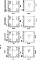


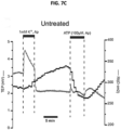


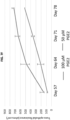

Classifications
-
- C—CHEMISTRY; METALLURGY
- C12—BIOCHEMISTRY; BEER; SPIRITS; WINE; VINEGAR; MICROBIOLOGY; ENZYMOLOGY; MUTATION OR GENETIC ENGINEERING
- C12N—MICROORGANISMS OR ENZYMES; COMPOSITIONS THEREOF; PROPAGATING, PRESERVING, OR MAINTAINING MICROORGANISMS; MUTATION OR GENETIC ENGINEERING; CULTURE MEDIA
- C12N5/00—Undifferentiated human, animal or plant cells, e.g. cell lines; Tissues; Cultivation or maintenance thereof; Culture media therefor
- C12N5/06—Animal cells or tissues; Human cells or tissues
- C12N5/0602—Vertebrate cells
- C12N5/0618—Cells of the nervous system
- C12N5/0621—Eye cells, e.g. cornea, iris pigmented cells
-
- C—CHEMISTRY; METALLURGY
- C12—BIOCHEMISTRY; BEER; SPIRITS; WINE; VINEGAR; MICROBIOLOGY; ENZYMOLOGY; MUTATION OR GENETIC ENGINEERING
- C12N—MICROORGANISMS OR ENZYMES; COMPOSITIONS THEREOF; PROPAGATING, PRESERVING, OR MAINTAINING MICROORGANISMS; MUTATION OR GENETIC ENGINEERING; CULTURE MEDIA
- C12N2500/00—Specific components of cell culture medium
- C12N2500/30—Organic components
-
- C—CHEMISTRY; METALLURGY
- C12—BIOCHEMISTRY; BEER; SPIRITS; WINE; VINEGAR; MICROBIOLOGY; ENZYMOLOGY; MUTATION OR GENETIC ENGINEERING
- C12N—MICROORGANISMS OR ENZYMES; COMPOSITIONS THEREOF; PROPAGATING, PRESERVING, OR MAINTAINING MICROORGANISMS; MUTATION OR GENETIC ENGINEERING; CULTURE MEDIA
- C12N2501/00—Active agents used in cell culture processes, e.g. differentation
- C12N2501/10—Growth factors
- C12N2501/105—Insulin-like growth factors [IGF]
-
- C—CHEMISTRY; METALLURGY
- C12—BIOCHEMISTRY; BEER; SPIRITS; WINE; VINEGAR; MICROBIOLOGY; ENZYMOLOGY; MUTATION OR GENETIC ENGINEERING
- C12N—MICROORGANISMS OR ENZYMES; COMPOSITIONS THEREOF; PROPAGATING, PRESERVING, OR MAINTAINING MICROORGANISMS; MUTATION OR GENETIC ENGINEERING; CULTURE MEDIA
- C12N2501/00—Active agents used in cell culture processes, e.g. differentation
- C12N2501/10—Growth factors
- C12N2501/15—Transforming growth factor beta (TGF-β)
-
- C—CHEMISTRY; METALLURGY
- C12—BIOCHEMISTRY; BEER; SPIRITS; WINE; VINEGAR; MICROBIOLOGY; ENZYMOLOGY; MUTATION OR GENETIC ENGINEERING
- C12N—MICROORGANISMS OR ENZYMES; COMPOSITIONS THEREOF; PROPAGATING, PRESERVING, OR MAINTAINING MICROORGANISMS; MUTATION OR GENETIC ENGINEERING; CULTURE MEDIA
- C12N2501/00—Active agents used in cell culture processes, e.g. differentation
- C12N2501/10—Growth factors
- C12N2501/155—Bone morphogenic proteins [BMP]; Osteogenins; Osteogenic factor; Bone inducing factor
-
- C—CHEMISTRY; METALLURGY
- C12—BIOCHEMISTRY; BEER; SPIRITS; WINE; VINEGAR; MICROBIOLOGY; ENZYMOLOGY; MUTATION OR GENETIC ENGINEERING
- C12N—MICROORGANISMS OR ENZYMES; COMPOSITIONS THEREOF; PROPAGATING, PRESERVING, OR MAINTAINING MICROORGANISMS; MUTATION OR GENETIC ENGINEERING; CULTURE MEDIA
- C12N2501/00—Active agents used in cell culture processes, e.g. differentation
- C12N2501/10—Growth factors
- C12N2501/16—Activin; Inhibin; Mullerian inhibiting substance
-
- C—CHEMISTRY; METALLURGY
- C12—BIOCHEMISTRY; BEER; SPIRITS; WINE; VINEGAR; MICROBIOLOGY; ENZYMOLOGY; MUTATION OR GENETIC ENGINEERING
- C12N—MICROORGANISMS OR ENZYMES; COMPOSITIONS THEREOF; PROPAGATING, PRESERVING, OR MAINTAINING MICROORGANISMS; MUTATION OR GENETIC ENGINEERING; CULTURE MEDIA
- C12N2501/00—Active agents used in cell culture processes, e.g. differentation
- C12N2501/40—Regulators of development
- C12N2501/415—Wnt; Frizzeled
-
- C—CHEMISTRY; METALLURGY
- C12—BIOCHEMISTRY; BEER; SPIRITS; WINE; VINEGAR; MICROBIOLOGY; ENZYMOLOGY; MUTATION OR GENETIC ENGINEERING
- C12N—MICROORGANISMS OR ENZYMES; COMPOSITIONS THEREOF; PROPAGATING, PRESERVING, OR MAINTAINING MICROORGANISMS; MUTATION OR GENETIC ENGINEERING; CULTURE MEDIA
- C12N2506/00—Differentiation of animal cells from one lineage to another; Differentiation of pluripotent cells
- C12N2506/45—Differentiation of animal cells from one lineage to another; Differentiation of pluripotent cells from artificially induced pluripotent stem cells
-
- C—CHEMISTRY; METALLURGY
- C12—BIOCHEMISTRY; BEER; SPIRITS; WINE; VINEGAR; MICROBIOLOGY; ENZYMOLOGY; MUTATION OR GENETIC ENGINEERING
- C12N—MICROORGANISMS OR ENZYMES; COMPOSITIONS THEREOF; PROPAGATING, PRESERVING, OR MAINTAINING MICROORGANISMS; MUTATION OR GENETIC ENGINEERING; CULTURE MEDIA
- C12N2533/00—Supports or coatings for cell culture, characterised by material
- C12N2533/50—Proteins
- C12N2533/52—Fibronectin; Laminin
Definitions
- the present invention was made as a result of activities undertaken within the scope of a joint research agreement that was in effect at the time the present invention was made.
- the parties to said joint research agreement are The Government of the United States of America, U.S. Department of Health and Human Services, as represented by the National Eye Institute, an institute of the National Institutes of Health and Cellular Dynamics International, Inc.
- This disclosure relates generally to the field of stem cell biology. More particularly, it concerns methods of efficient production of stem cell-derived retinal pigment epithelial cell populations for use as a cell therapy.
- the retina is a light-sensitive layer of tissue that lines the inner surface of the eye.
- Photoreceptor cells either rods or cones, in the retina are directly sensitive to light and transform chemical light signals into electrical events that trigger nerve impulses.
- the retinal pigment epithelium (RPE) is a layer of pigmented cells that forms the blood-retinal barrier.
- the RPE cells play important roles in the maintenance of visual function and the transport of ions, water, and metabolic end products from the subretinal space to the blood (Strauss et al., 2005). Further, RPE cells establish the immune privilege of the eye by secreting immunosuppressive factors.
- a disorder or injury to the RPE cells can result in degeneration of the retina, loss of visual function, and blindness.
- Several disorders of the retina including acute and age-related macular degeneration and Best disease, involve degeneration of the RPE; therefore, cell replacement therapy is a possible therapeutic option for preservation of vision (Buchholz et al., 2009).
- stem cells are undifferentiated cells which can give rise to a succession of mature functional cells.
- a hematopoietic stem cell may give rise to any of the different types of terminally differentiated blood cells.
- Embryonic stem (ES) cells are derived from the embryo and are pluripotent, thus possessing the capability of developing into any organ or tissue type, including RPE cells.
- iPSCs induced pluripotent stem cells
- iPSCs have been shown to give rise to ocular cells, including RPE cells (Hirami et al ., 2009).
- RPE cells Hirami et al ., 2009.
- all of the techniques known to date for the production of iPSC- or ESC-derived RPE cells are dependent on using a starting population of embryoid bodies.
- RPEs from pluripotent cells such as iPSCs or ESCs
- iPSCs or ESCs retinal pigment epithelial
- the starting cell population of pluripotent stem cells may be, for example, embryonic stem cells or induced pluripotent stem cells.
- the pluripotent stem cells are induced pluripotent stem cells (iPSCs).
- iPSCs induced pluripotent stem cells
- a method for producing human RPE cells comprising (a) obtaining a starting population comprising human induced pluripotent stem cells (iPSCs) that are dissociated into essentially single cells; (b) culturing the iPSCs in a retinal induction medium to initiate differentiation of the cells into retinal lineage cells; (c) further culturing the retinal lineage cells in a retinal differentiation medium to further differentiate the retinal lineage cells; (d) culturing the cells in retinal medium to form differentiating RPE cells; and (e) culturing the RPE cells in a RPE maturation medium, thereby producing human RPE cells.
- the method does not include the formation of embryoid bodies.
- the RPE cells does not include the formation of embryoid bodies.
- the RPE cells does not include the formation of embryoid bodies.
- the iPSCs are cultured on a matrix.
- the matrix comprises at least one recombinant cellular adhesion protein such as laminin, vitronectin or fibronectin.
- the at least one cellular adhesion protein is human.
- the iPSCs are cultured without a feeder layer. In some aspects, the iPSCs are cultured in a fully-defined culture medium. In other aspects, the iPSCs are cultured in a xeno-free culture medium.
- the retinal induction medium comprises a WNT pathway inhibitor, a BMP pathway inhibitor, a TGF ⁇ pathway inhibitor and insulin growth factor 1 (IGF1).
- the WNT pathway inhibitor is selected from the group consisting of N-(2-Aminoethyl)-5-chloroisoquinoline-8-sulphonamide dihydrochloride (CKI-7), N-(6-Methyl-2-benzothiazolyl)-2-[(3,4,6,7-tetrahydro-4-oxo-3-phenylthieno[3,2-d]pyrimidin-2-yl)thio]-acetamide (IWP2), N-(6-Methyl-2-benzothiazolyl)-2-[(3,4,6,7-tetrahydro-3-(2-methoxyphenyl)-4-oxothieno[3,2-d]pyrimidin-2-yl)thio]-acetamide (IWP4), 2-Phenoxybenzoic acid-[(5
- the WNT pathway inhibitor is CKI-7.
- the BMP pathway inhibitor is 4-(6-(4-(piperazin-1-yl)phenyl)pyrazolo[1,5-a]pyrimidin-3-yl)quinoline hydrochloride (LDN193189), 6-[4-[2-(1-Piperidinyl)ethoxy]phenyl]-3-(4-pyridinyl)-pyrazolo[1,5-a]pyrimidine dihydrochloride (Dorsomorphin), 4-[6-[4-(1-Methylethoxy)phenyl]pyrazolo[1,5-a]pyrimidin-3-yl]-quinoline (DMH1), 4-[6-[4-[2-(4-Morpholinyl)ethoxy]phenyl]pyrazolo[1,5-a]pyrimidin-3-yl]quinoline (DMH-2), or 5-[6-(4-Methoxyphenyl)pyrazolo
- the BMP pathway inhibitor is LDN193189.
- the TGF ⁇ pathway inhibitor is 4-[4-(1,3-benzodioxol-5-yl)-5-(2-pyridinyl)-1H-imidazol-2-yl]benzamide (SB431542), 6-[2-(1,1-Dimethylethyl)-5-(6-methyl-2-pyridinyl)-1H-imidazol-4-yl]quinoxaline (SB525334), 2-(5-Benzo[l,3]dioxol-5-yl-2-ieri-butyl-3H-imidazol-4-yl)-6-methylpyridine hydrochloride hydrate (SB-505124), 4-(5-Benzol[l,3]dioxol- 5-yl-4-pyridin-2-yl-lH-imidazol-2-yl)-benzamide hydrate, 4-[4-(1,3-Benzodioxol-5
- the retinal differentiation medium comprises a WNT pathway inhibitor, a BMP pathway inhibitor, a TGF ⁇ pathway inhibitor, a MEK inhibitor and IGF1.
- the WNT pathway inhibitor is selected from the group consisting of N-(2-Aminoethyl)-5-chloroisoquinoline-8-sulphonamide dihydrochloride (CKI-7), N-(6-Methyl-2-benzothiazolyl)-2-[(3,4,6,7-tetrahydro-4-oxo-3-phenylthieno[3,2-d]pyrimidin-2-yl)thio]-acetamide (IWP2), N-(6-Methyl-2-benzothiazolyl)-2-[(3,4,6,7-tetrahydro-3-(2-methoxyphenyl)-4-oxothieno[3,2-d]pyrimidin-2-yl)thio]-acetamide (IWP4), 2-Phenoxybenzoic acid-[(5
- the WNT pathway inhibitor is CKI-7.
- the BMP pathway inhibitor is 4-(6-(4-(piperazin-1-yl)phenyl)pyrazolo[1,5-a]pyrimidin-3-yl)quinoline hydrochloride (LDN193189), 6-[4-[2-(1-Piperidinyl)ethoxy]phenyl]-3-(4-pyridinyl)-pyrazolo[1,5-a]pyrimidine dihydrochloride (Dorsomorphin), 4-[6-[4-(1-Methylethoxy)phenyl]pyrazolo[1,5-a]pyrimidin-3-yl]-quinoline (DMH1), 4-[6-[4-[2-(4-Morpholinyl)ethoxy]phenyl]pyrazolo[1,5-a]pyrimidin-3-yl]quinoline (DMH-2), or 5-[6-(4-Methoxyphenyl)pyrazolo
- the BMP pathway inhibitor is LDN193189.
- the TGF ⁇ pathway inhibitor is 4-[4-(1,3-benzodioxol-5-yl)-5-(2-pyridinyl)-1H-imidazol-2-yl]benzamide (SB431542), 6-[2-(1,1-Dimethylethyl)-5-(6-methyl-2-pyridinyl)-1H-imidazol-4-yl]quinoxaline (SB525334), 2-(5-Benzo[l,3]dioxol-5-yl-2-ieri-butyl-3H-imidazol-4-yl)-6-methylpyridine hydrochloride hydrate (SB-505124), 4-(5-Benzol[1,3]dioxol- 5-yl-4-pyridin-2-yl-lH-imidazol-2-yl)-benzamide hydrate, 4-[4-(1,3-Benzodioxol-5-
- the TGF ⁇ pathway inhibitor is SB431542.
- the MEK inhibitor is N-[(2R)-2,3-Dihydroxypropoxy]-3,4-difluoro-2-[(2-fluoro-4-iodophenyl)amino]- benzamide (PD0325901), N-[3-[3-cyclopropyl-5-(2-fluoro-4-iodoanilino)-6,8-dimethyl-2,4,7-trioxopyrido[4,3-d]pyrimidin-1-yl]phenyl]acetamide (GSK1120212), 6-(4-bromo-2-fluoroanilino)-7-fluoro-N-(2-hydroxyethoxy)-3-methylbenzimidazole-5-carboxamide (MEK162), N-[3,4-difluoro-2-(2-fluoro-4-iodoanilino)-6-methoxyphenyl]-1-(2,3
- the RPE cells are dissociated after culture in the RPE maturation medium.
- the dissociated RPE cells are seeded and cultured in RPE maturation medium.
- the RPE maturation medium comprises a MEK inhibitor.
- the MEK inhibitor is N-[(2R)-2,3-Dihydroxypropoxy]-3,4-difluoro-2-[(2-fluoro-4-iodophenyl)amino]- benzamide (PD0325901), N-[3-[3-cyclopropyl-5-(2-fluoro-4-iodoanilino)-6,8-dimethyl-2,4,7-trioxopyrido[4,3-d]pyrimidin-1-yl]phenyl]acetamide (GSK1120212), 6-(4-bromo-2-fluoroanilino)-7-fluoro-N-(2-hydroxyethoxy)-3-methylbenzimidazole-5-carboxamide (MEK162), N-[3,4-difluoro-2-(2-fluoro-4-iodoanilino)-6-methoxyphenyl]-1-(2,3-dihydroxypropyl)cyclopropane-1
- the RPE cells are dissociated after culture in the RPE medium and reseeded on a degradable scaffold in the RPE maturation medium thereby producing mature RPE cells.
- the RPE maturation medium may comprise at least one primary cilium inducer.
- the at least one primary cilium inducer is prostaglandin E2 (PGE2) or aphidicolin.
- the RPE maturation medium may comprise N-(6-Methyl-2-benzothiazolyl)-2-[(3,4,6,7-tetrahydro-4-oxo-3-phenylthieno[3,2-d]pyrimidin-2-yl)thio]-acetamide (IWP2) or 4-(1,3,3a,4,7,7a-Hexahydro-1,3-dioxo-4,7-methano-2H-isoindol-2-yl)-N-8-quinolinyl-Benzamide (endo-IWR1).
- the starting population of iPSCs is pre-confluent cells that have been dissociated into single cells.
- the iPSCs are cultured at an initial cell density of about 5,000 to 40,000 cells/cm 2 .
- the iPSCs are cultured at an initial cell density of 5,000, 10,000, 20,000, 30,000, or 40,000 cells/cm 2 .
- the starting population of iPSCs is MHC haplotype-matched to a subject in need thereof.
- the iPSCs are homozygous for at least one HLA allele.
- the iPSCs are homozygous at HLA-A, HLA-B or HLA-DR.
- the iPSCs are homozygous at HLA-A and HLA-B.
- a method for producing human retinal pigment epithelial (RPE) cells comprising (a) obtaining a starting population comprising human induced pluripotent stem cells (iPSCs) that are dissociated into essentially single cells in a fully defined medium; (b) culturing the iPSCs on laminin in a retinal induction medium comprising LDN193189, CKI-7, and SB431542 to initiate differentiation of the cells into retinal lineage cells; (c) further culturing the retinal lineage cells in a retinal differentiation medium comprising LDN193189, CKI-7, SB431542, and PD0325901 to further differentiate the retinal lineage cells; (d) culturing the cells in retinal medium comprising nicotinamide and Activin A to form differentiating RPE cells; and (e) culturing the RPE cells in a RPE maturation medium, thereby producing human RPE cells.
- the method does not include the formation
- RPE cells can be derived from pluripotent stem cells such as ES cells and iPSC cells; however, current methods are dependent on a starting population of embryoid bodies.
- the present disclosure provides a highly efficient and reproducible method of differentiating pluripotent stem cells (PSCs) into functional and mature RPE cells without the use of embryoid bodies. Further embodiments and advantages are described below.
- purified does not require absolute purity; rather, it is intended as a relative term. Thus, a purified population of cells is greater than about 90%, 91%, 92%, 93%, 94%, 95%, 96%, 97%, 98%, 99% or 100% pure, or, most preferably, essentially free other cell types.
- essentially or “essentially free,” in terms of a specified component is used herein to mean that none of the specified component has been purposefully formulated into a composition and/or is present only as a contaminant or in trace amounts.
- the total amount of the specified component resulting from any unintended contamination of a composition is therefore well below 0.05%, preferably below 0.01%.
- Most preferred is a composition in which no amount of the specified component can be detected with standard analytical methods.
- cell is herein used to refer to a structural and functional unit of an organism that can replicate independently, is enclosed by a membrane, and contains biomolecules and genetic material.
- Cells used herein may be naturally-occurring cells or artificially modified cells (e.g ., fusion cells, genetically modified cells, etc .).
- cell population is used herein to refer to a group of cells, typically of a common type.
- the cell population can be derived from a common progenitor or may comprise more than one cell type.
- An "enriched” cell population refers to a cell population derived from a starting cell population (e.g ., an unfractionated, heterogeneous cell population) that contains a greater percentage of a specific cell type than the percentage of that cell type in the starting population.
- the cell populations may be enriched for one or more cell types and depleted of one or more cell types.
- stem cell refers herein to a cell that under suitable conditions is capable of differentiating into a diverse range of specialized cell types, while under other suitable conditions is capable of self-renewing and remaining in an essentially undifferentiated pluripotent state.
- stem cell also encompasses a pluripotent cell, multipotent cell, precursor cell and progenitor cell.
- Exemplary human stem cells can be obtained from hematopoietic or mesenchymal stem cells obtained from bone marrow tissue, embryonic stem cells obtained from embryonic tissue, or embryonic germ cells obtained from genital tissue of a fetus.
- Exemplary pluripotent stem cells can also produced from somatic cells by reprogramming them to a pluripotent state by the expression of certain transcription factors associated with pluripotency; these cells are called "induced pluripotent stem cells" or "iPSCs”.
- pluripotent refers to the property of a cell to differentiate into all other cell types in an organism, with the exception of extraembryonic, or placental, cells. Pluripotent stem cells are capable of differentiating to cell types of all three germ layers (e.g., ectodermal, mesodermal, and endodermal cell types) even after prolonged culture.
- a pluripotent stem cell is an embryonic stem cell derived from the inner cell mass of a blastocyst. In other embodiments, the pluripotent stem cell is an induced pluripotent stem cell derived by reprogramming somatic cells.
- the term "differentiation” refers to the process by which an unspecialized cell becomes a more specialized type with changes in structural and/or functional properties.
- the mature cell typically has altered cellular structure and tissue-specific proteins. More specifically, in the context of the present methods indicates the process of a human stem cell acquiring the cell type of a retinal pigment epithelial (RPE) cell with features indicative that said RPE cell is a mature, terminally differentiated cell.
- RPE retinal pigment epithelial
- undifferentiated refers to cells that display characteristic markers and morphological characteristics of undifferentiated cells that clearly distinguish them from terminally differentiated cells of embryo or adult origin.
- Embryoid bodies are aggregates of pluripotent stem cells that can undergo differentiation into cells of the endoderm, mesoderm, and ectoderm germ layers.
- the spheroid structures form when pluripotent stem cells aggregate and enable the non-adherent culture of EBs in suspension.
- Isolated cell has been substantially separated or purified from others cells in an organism or culture. Isolated cells can be, for example, at least 99%, at least 98% pure, at least 95% pure or at least 90% pure.
- an “embryo” refers to a cellular mass obtained by one or more divisions of a zygote or an activated oocyte with an artificially reprogrammed nucleus.
- an “embryonic stem (ES) cell” is an undifferentiated pluripotent cell which is obtained from an embryo in an early stage, such as the inner cell mass at the blastocyst stage, or produced by artificial means (e.g . nuclear transfer) and can give rise to any differentiated cell type in an embryo or an adult, including germ cells ( e.g. sperm and eggs).
- iPSCs are cells generated by reprogramming a somatic cell by expressing or inducing expression of a combination of factors (herein referred to as reprogramming factors). iPSCs can be generated using fetal, postnatal, newborn, juvenile, or adult somatic cells.
- factors that can be used to reprogram somatic cells to pluripotent stem cells include, for example, Oct4 (sometimes referred to as Oct 3/4), Sox2, c-Myc, and Klf4, Nanog, and Lin28.
- somatic cells are reprogrammed by expressing at least two reprogramming factors, at least three reprogramming factors, or four reprogramming factors to reprogram a somatic cell to a pluripotent stem cell.
- An “allele” refers to one of two or more forms of a gene. Diploid organisms such as humans contain two copies of each chromosome, and thus carry one allele on each.
- homozygous is defined as containing two of the same alleles at a particular locus.
- heterozygous refers to as containing two different alleles at a particular locus.
- haplotype refers to a combination of alleles at multiple loci along a single chromosome.
- a haplotype can be based upon a set of single-nucleotide polymorphisms (SNPs) on a single chromosome and/or the alleles in the major histocompatibility complex.
- SNPs single-nucleotide polymorphisms
- haplotype-matched is defined as the cell (e.g. iPSC cell) and the subject being treated share one or more major histocompatibility locus haplotypes.
- the haplotype of the subject can be readily determined using assays well known in the art.
- the haplotype-matched iPSC cell can be autologous or allogeneic.
- the autologous cells which are grown in tissue culture and differentiated to RPE cells inherently are haplotype-matched to the subject.
- Substantially the same HLA type indicates that the HLA type of donor matches with that of a patient to the extent that the transplanted cells, which have been obtained by inducing differentiation of iPSCs derived from the donor's somatic cells, can be engrafted when they are transplanted to the patient.
- Super donors are referred to herein as individuals that are homozygous for certain MHC class I and II genes. These homozygous individuals can serve as super donors and their cells, including tissues and other materials comprising their cells, can be transplanted in individuals that are either homozygous or heterozygous for that haplotype.
- the super donor can be homozygous for the HLA-A, HLA-B, HLA-C, HLA-DR, HLA-DP or HLA-DQ locus/loci alleles, respectively.
- Feeer-free or feeder-independent is used herein to refer to a culture supplemented with cytokines and growth factors (e.g ., TGF ⁇ , bFGF, LIF) as a replacement for the feeder cell layer.
- cytokines and growth factors e.g ., TGF ⁇ , bFGF, LIF
- feeder-free or feeder-independent culture systems and media may be used to culture and maintain pluripotent cells in an undifferentiated and proliferative state.
- feeder-free cultures utilize an animal-based matrix (e.g . MATRIGEL TM ) or are grown on a substrate such as fibronectin, collagen or vitronectin.
- Feeer layers are defined herein as a coating layer of cells such as on the bottom of a culture dish.
- the feeder cells can release nutrients into the culture medium and provide a surface to which other cells, such as pluripotent stem cells, can attach.
- a defined medium refers to a medium, an extracellular matrix, or a culture condition in which the chemical composition and amounts of approximately all the components are known.
- a defined medium does not contain undefined factors such as in fetal bovine serum, bovine serum albumin or human serum albumin.
- a defined medium comprises a basal media (e.g ., Dulbecco's Modified Eagle's Medium (DMEM), F12, or Roswell Park Memorial Institute Medium (RPMI) 1640, containing amino acids, vitamins, inorganic salts, buffers, antioxidants and energy sources) which is supplemented with recombinant albumin, chemically defined lipids, and recombinant insulin.
- DMEM Dulbecco's Modified Eagle's Medium
- RPMI Roswell Park Memorial Institute Medium
- An exemplary fully defined medium is Essential 8 TM medium.
- Xeno-Free (XF) when used in relation to a medium, an extracellular matrix, or a culture condition, refers to a medium, an extracellular matrix, or a culture condition which is essentially free from heterogeneous animal-derived components. For culturing human cells, any proteins of a non-human animal, such as mouse, would be xeno components. In certain aspects, the Xeno-free matrix may be essentially free of any non-human animal-derived components, therefore excluding mouse feeder cells or MATRIGEL TM .
- MATRIGEL TM is a solubilized basement membrane preparation extracted from the Engelbreth-Holm-Swarm (EHS) mouse sarcoma, a tumor rich in extracellular matrix proteins to include laminin (a major component), collagen IV, heparan sulfate proteoglycans, and entactin/nidogen.
- EHS Engelbreth-Holm-Swarm
- KNOCKOUT TM serum replacement referred to herein as a serum-free formulation optimized to grow and maintain undifferentiated cells, such as stem cell, in culture.
- Pre-confluent refers to a cell culture in which the proportion of the culture surface which is covered by cells is about 60-80%. Usually, pre-confluent refers to a culture in which about 70% of the culture surface is covered by cells.
- the "retina” refers to a light-sensitive layer of tissue which lines the inner surface of the eye.
- Retinal pigment epithelium refers to a monolayer of pigmented cells between the choroid, a layer filled with blood vessels, and the retina.
- Retinal lineage cells herein refer to cells that can give rise or differentiate to RPE cells.
- RIM Retinal Induction Medium
- the "Retinal Differentiation Medium (RDM)” is defined herein as a medium that comprises a WNT pathway inhibitor, a BMP pathway inhibitor and a MEK inhibitor and differentiates retinal cells.
- the RDM also comprises a TGF ⁇ pathway inhibitor.
- the "Retinal Medium (RM)” is defined as a growth medium for the culture of retinal cells comprising Activin A and Nicotinamide.
- RPE-Maturation Medium herein refers to a medium for the maturation of RPE cells comprising taurine and hydrocortisone.
- the RPE-MM also comprises triiodothyronine.
- the RPE-MM may also comprise PD0325901 or PGE2.
- RPE cells are referred to herein as RPE cells which have downregulated expression of immature RPE markers such as Pax6 and upregulated expression of mature RPE markers such as RPE65.
- RPE cell “maturation” refers herein to the process by which RPE developmental pathways are modulated to generate mature RPE cells. For example, modulation of cilia function can result in RPE maturation.
- a “therapeutically effective amount” used herein refers to the amount of a compound that, when administered to a subject for treatment of a disease or condition, is sufficient to effect such treatment.
- Inducer is defined herein as a molecule that regulates gene expression such as activating genes within a cell.
- An inducer can bind to repressors or activators. Inducers functions by disabling repressors.
- ES cells are derived from the inner cell mass of blastocysts and have a high in vitro differentiating capability.
- ES cells can be isolated by removing the outer trophectoderm layer of a developing embryo, then culturing the inner mass cells on a feeder layer of non-growing cells. The replated cells can continue to proliferate and produce new colonies of ES cells which can be removed, dissociated, replated again and allowed to grow. This process of "subculturing" undifferentiated ES cells can be repeated a number of times to produce cell lines containing undifferentiated ES cells ( U.S. Patent Nos. 5,843,780 ; 6,200,806 ; 7,029,913 ).
- ES cells have the potential to proliferate while maintaining their pluripotency.
- ES cells are useful in research on cells and on genes which control cell differentiation.
- the pluripotency of ES cells combined with genetic manipulation and selection can be used for gene analysis studies in vivo via the generation of transgenic, chimeric, and knockout mice.
- mouse ES cells Methods for producing mouse ES cells are well known.
- a preimplantation blastocyst from the 129 strain of mice is treated with mouse antiserum to remove the trophoectoderm, and the inner cell mass is cultured on a feeder cell layer of chemically inactivated mouse embryonic fibroblasts in medium containing fetal calf serum. Colonies of undifferentiated ES cells that develop are subcultured on mouse embryonic fibroblast feeder layers in the presence of fetal calf serum to produce populations of ES cells.
- mouse ES cells can be grown in the absence of a feeder layer by adding the cytokine leukemia inhibitory factor (LIF) to serum-containing culture medium (Smith, 2000).
- LIF cytokine leukemia inhibitory factor
- mouse ES cells can be grown in serum-free medium in the presence of bone morphogenetic protein and LIF (Ying et al., 2003).
- Human ES cells can be produced or derived from a zygote or blastocyst-staged mammalian embryo produced by the fusion of a sperm and egg cell, nuclear transfer, pathogenesis, or the reprogramming of chromatin and subsequent incorporation of the reprogrammed chromatin into a plasma membrane to produce an embryonic cell by previously described methods (Thomson and Marshall, 1998; Reubinoff et al., 2000).
- human blastocysts are exposed to antihuman serum, and trophectoderm cells are lysed and removed from the inner cell mass which is cultured on a feeder layer of mouse embryonic fibroblasts.
- clumps of cells derived from the inner cell mass are chemically or mechanically dissociated, replated, and colonies with undifferentiated morphology are selected by micropipette, dissociated, and replated ( U.S. Patent No. 6,833,269 ).
- human ES cells can be grown without serum by culturing the ES cells on a feeder layer of fibroblasts in the presence of basic fibroblast growth factor (Amit et al ., 2000).
- human ES cells can be grown without a feeder cell layer by culturing the cells on a protein matrix such as MATRIGEL TM or laminin in the presence of "conditioned" medium containing basic fibroblast growth factor (Xu et al., 2001).
- a protein matrix such as MATRIGEL TM or laminin
- ES cells can also be derived from other organisms including rhesus monkey and marmoset by previously described methods (Thomson, and Marshall, 1998; Thomson et al., 1995; Thomson and Odorico, 2000), as well as from established mouse and human cell lines.
- established human ES cell lines include MAOI, MA09, ACT-4, HI, H7, H9, H13, H14 and ACT30.
- mouse ES cell lines that have been established include the CGR8 cell line established from the inner cell mass of the mouse strain 129 embryos, and cultures of CGR8 cells can be grown in the presence of LIF without feeder layers.
- ES stem cells can be detected by protein markers including transcription factor Oct4, alkaline phosphatase (AP), stage-specific embryonic antigen SSEA-1, stage-specific embryonic antigen SSEA-3, stage-specific embryonic antigen SSEA-4, transcription factor NANOG, tumor rejection antigen 1-60 (TRA-1-60), tumor rejection antigen 1-81 (TRA-1-81), SOX2, or REX1.
- protein markers including transcription factor Oct4, alkaline phosphatase (AP), stage-specific embryonic antigen SSEA-1, stage-specific embryonic antigen SSEA-3, stage-specific embryonic antigen SSEA-4, transcription factor NANOG, tumor rejection antigen 1-60 (TRA-1-60), tumor rejection antigen 1-81 (TRA-1-81), SOX2, or REX1.
- pluripotency was originally achieved in 2006 using mouse cells (Yamanaka et al. 2006) and in 2007 using human cells (Yu et al. 2007; Takahashi et al. 2007) by reprogramming of somatic cells via the introduction of transcription factors that are linked to pluripotency.
- Pluripotent stem cells can be maintained in an undifferentiated state and are capable of differentiating into almost any cell type.
- the use of iPSCs circumvents most of the ethical and practical problems associated with large-scale clinical use of ES cells, and patients with iPSC-derived autologous transplants may not require lifelong immunosuppressive treatments to prevent graft rejection.
- any cell can be used as a starting point for iPSCs.
- cell types could be keratinocytes, fibroblasts, hematopoietic cells, mesenchymal cells, liver cells, or stomach cells.
- T cells may also be used as a source of somatic cells for reprogramming ( U.S. Patent No. 8,741,648 ).
- somatic cell is itself a RPE cells such as a human RPE cell.
- the RPE cell can be an adult or a fetal RPE cell.
- iPSCs can be grown under conditions that are known to differentiate human ES cells into specific cell types, and express human ES cell markers including: SSEA-1, SSEA-3, SSEA-4, TRA-1-60, and TRA-1-81.
- Somatic cells can be reprogrammed to produce induced pluripotent stem cells (iPSCs) using methods known to one of skill in the art.
- iPSCs induced pluripotent stem cells
- One of skill in the art can readily produce induced pluripotent stem cells, see for example, Published U.S. Patent Application No. 20090246875 , Published U.S. Patent Application No. 2010/0210014 ; Published U.S. Patent Application No. 20120276636 ; U.S. Patent No. 8,058,065 ; U.S. Patent No. 8,129,187 ; U.S. Patent No. 8,278,620 ; PCT Publication NO. WO 2007/069666 A1 , and U.S. Patent No. 8,268,620 , which are incorporated herein by reference.
- nuclear reprogramming factors are used to produce pluripotent stem cells from a somatic cell.
- at least three, or at least four, of Klf4, c-Myc, Oct3/4, Sox2, Nanog, and Lin28 are utilized.
- Oct3/4, Sox2, c-Myc and Klf4 are utilized.
- the cells are treated with a nuclear reprogramming substance, which is generally one or more factor(s) capable of inducing an iPSC from a somatic cell or a nucleic acid that encodes these substances (including forms integrated in a vector).
- the nuclear reprogramming substances generally include at least Oct3/4, Klf4 and Sox2 or nucleic acids that encode these molecules.
- a functional inhibitor of p53, L-myc or a nucleic acid that encodes L-myc, and Lin28 or Lin28b or a nucleic acid that encodes Lin28 or Lin28b, can be utilized as additional nuclear reprogramming substances.
- Nanog can also be utilized for nuclear reprogramming. As disclosed in published U.S. Patent Application No.
- exemplary reprogramming factors for the production of iPSCs include (1) Oct3/4, Klf4, Sox2, L-Myc (Sox2 can be replaced with Sox1, Sox3, Soxl5, Soxl7 or Soxl8; Klf4 is replaceable with Klfl, Klf2 or HIf5); (2) Oct3/4, Klf4, Sox2, L-Myc, TERT, SV40 Large T antigen (SV40LT); (3) Oct3/4, Klf4, Sox2, L-Myc, TERT, human papilloma virus (HPV)16 E6; (4) Oct3/4, Klf4, Sox2, L-Myc, TERT, HPV16 E7 (5) Oct3/4, Klf4, Sox2, L-Myc, TERT, HPV16 E6, HPV16 E7; (6) Oct3/4, Klf4, Sox2, L-Myc, TERT, Bmil; (7) Oct3/4, Klf4, Sox2, L-Myc, Lin28;
- Oct3/4, Klf4, Sox2, and c-Myc are utilized.
- Oct4, Nanog, and Sox2 are utilized, see for example, U.S. Patent No. 7,682,828 , which is incorporated herein by reference.
- these factors include, but are not limited to, Oct3/4, Klf4 and Sox2.
- the factors include, but are not limited to Oct 3/4, Klf4 and Myc.
- Oct3/4, Klf4, c-Myc, and Sox2 are utilized.
- Oct3/4, Klf4, Sox2 and Sal 4 are utilized.
- telomeres like Nanog, Lin28, Klf4, or c-Myc can increase reprogramming efficiency and can be expressed from several different expression vectors.
- an integrating vector such as the EBV element-based system can be used ( U.S. Patent No. 8,546,140 ).
- reprogramming proteins could be introduced directly into somatic cells by protein transduction.
- Reprogramming may further comprise contacting the cells with one or more signaling receptors including glycogen synthase kinase 3 (GSK-3) inhibitor, a mitogen-activated protein kinase kinase (MEK) inhibitor, a transforming growth factor beta (TGF- ⁇ ) receptor inhibitor or signaling inhibitor, leukemia inhibitory factor (LIF), a p53 inhibitor, an NF-kappa B inhibitor, or a combination thereof.
- GSK-3 glycogen synthase kinase 3
- MEK mitogen-activated protein kinase kinase
- TGF- ⁇ transforming growth factor beta
- LIF leukemia inhibitory factor
- p53 inhibitor a p53 inhibitor
- NF-kappa B inhibitor a combination thereof.
- Those regulators may include small molecules, inhibitory nucleotides, expression cassettes, or protein factors. It is anticipated that virtually any iPS cells or cell lines may be used..
- iPSCs can be cultured in a medium sufficient to maintain pluripotency.
- the iPSCs may be used with various media and techniques developed to culture pluripotent stem cells, more specifically, embryonic stem cells, as described in U.S. Patent No. 7,442,548 and U.S. Patent Pub. No. 2003/0211603 .
- LIF Leukemia Inhibitory Factor
- bFGF basic fibroblast growth factor
- pluripotent cells may be cultured on fibroblast feeder cells or a medium that has been exposed to fibroblast feeder cells in order to maintain the stem cells in an undifferentiated state.
- the cell is cultured in the co-presence of mouse embryonic fibroblasts treated with radiation or an antibiotic to terminate the cell division, as feeder cells.
- pluripotent cells may be cultured and maintained in an essentially undifferentiated state using a defined, feeder-independent culture system, such as a TESR TM medium (Ludwig et al., 2006a; Ludwig et al. , 2006b) or E8 TM medium (Chen et al ., 2011).
- the iPSC can be modified to express exogenous nucleic acids, such as to include a tyrosinase enhancer operaby linked to a promoter and a nucleic acid sequence encoding a first marker.
- exogenous nucleic acids such as to include a tyrosinase enhancer operaby linked to a promoter and a nucleic acid sequence encoding a first marker.
- the tyrosinase gene is disclosed, for example, in GENBANK ® Accession No. 22173, as available on January 1, 2013. This sequence aligns to chromosome 7 of mouse strain C57BL/6 location 5286971-5291691 (invert orientation). A 4721 base pair sequence is sufficient for expression in RPE cells, see Murisier et al., Dev. Biol. 303: 838-847, 2007 , which is incorporated herein by reference.
- This construct is expressed in retinal pigment epithelial cells.
- Other enhancers can be utilized.
- Other RPE-specific enhancers include D-MITF, DCT, TYRP1, RPE65, VMD2, MERTK, MYRIP, and RAB27A.
- Suitable promoters include, but are not limited to, any promoter expressed in retinal pigment epithelial cells including the tyrosinase promoter.
- the construct can also include other elements, such as a ribosome binding site for translational initiation (internal ribosomal binding sequences), and a transcription/translation terminator. Generally, it is advantageous to transfect cells with the construct.
- Suitable vectors for stable transfection include, but are not limited to retroviral vectors, lentiviral vectors and Sendai virus.
- Plasmids have been designed with a number of goals in mind, such as achieving regulated high copy number and avoiding potential causes of plasmid instability in bacteria, and providing means for plasmid selection that are compatible with use in mammalian cells, including human cells. Particular attention has been paid to the dual requirements of plasmids for use in human cells. First, they are suitable for maintenance and fermentation in E. coli, so that large amounts of DNA can be produced and purified. Second, they are safe and suitable for use in human patients and animals. The first requirement calls for high copy number plasmids that can be selected for and stably maintained relatively easily during bacterial fermentation. The second requirement calls for attention to elements such as selectable markers and other coding sequences.
- plasmids that encode a marker are composed of: (1) a high copy number replication origin, (2) a selectable marker, such as, but not limited to, the neo gene for antibiotic selection with kanamycin, (3) transcription termination sequences, including the tyrosinase enhancer and (4) a multicloning site for incorporation of various nucleic acid cassettes; and (5) a nucleic acid sequence encoding a marker operably linked to the tyrosinase promoter.
- plasmid vectors that are known in the art for inducing a nucleic acid encoding a protein. These include, but are not limited to, the vectors disclosed in U.S. Patent No. 6,103,470 ; U.S. Patent No. 7,598,364 ; U.S. Patent No. 7,989,425 ; and U.S. Patent No. 6,416,998 , which are incorporated herein by reference.
- a viral gene delivery system can be an RNA-based or DNA-based viral vector.
- An episomal gene delivery system can be a plasmid, an Epstein-Barr virus (EBV)-based episomal vector, a yeast-based vector, an adenovirus-based vector, a simian virus 40 (SV40)-based episomal vector, a bovine papilloma virus (BPV)-based vector, or a lentiviral vector.
- Markers include, but are not limited to, fluorescence proteins (for example, green fluorescent protein or red fluorescent protein), enzymes (for example, horse radish peroxidase or alkaline phosphatase or firefly/renilla luciferase or nanoluc), or other proteins.
- a marker may be a protein (including secreted, cell surface, or internal proteins; either synthesized or taken up by the cell); a nucleic acid (such as an mRNA, or enzymatically active nucleic acid molecule) or a polysaccharide. Included are determinants of any such cell components that are detectable by antibody, lectin, probe or nucleic acid amplification reaction that are specific for the marker of the cell type of interest.
- the markers can also be identified by a biochemical or enzyme assay or biological response that depends on the function of the gene product. Nucleic acid sequences encoding these markers can be operably linked to the tyrosinase enhancer. In addition, other genes can be included, such as genes that may influence stem cell to RPE differentiation, or RPE function, or physiology, or pathology.
- a nucleic acid is included that encodes one or more of MITF, PAX6, TFEC, OTX2, LHX2, VMD2, CFTR, RPE65, MFRP, CTRP5, CFH, C3, C2B, APOE, APOB, mTOR, FOXO, AMPK, SIRT1-6, HTRP1, ABCA4, TIMP3, VEGFA, CFI, TLR3, TLR4, APP, CD46, BACE1, ELOLV4, ADAM 10, CD55, CD59, and ARMS2.
- Major Histocompatibility Complex is the main cause of immune-rejection of allogeneic organ transplants.
- the HLA loci are highly polymorphic and are distributed over 4 Mb on chromosome 6.
- the ability to haplotype the HLA genes within the region is clinically important since this region is associated with autoimmune and infectious diseases and the compatibility of HLA haplotypes between donor and recipient can influence the clinical outcomes of transplantation.
- HLAs corresponding to MHC class I present peptides from inside the cell and HLAs corresponding to MHC class II present antigens from outside of the cell to T-lymphocytes.
- HLA-matched stem cell lines may overcome the risk of immune rejection.
- HLA loci are usually typed by serology and PCR for identifying favorable donor-recipient pairs.
- Serological detection of HLA class I and II antigens can be accomplished using a complement mediated lymphocytotoxicity test with purified T or B lymphocytes. This procedure is predominantly used for matching HLA-A and -B loci.
- Molecular-based tissue typing can often be more accurate than serologic testing.
- Low resolution molecular methods such as SSOP (sequence specific oligonucleotide probes) methods, in which PCR products are tested against a series of oligonucleotide probes, can be used to identify HLA antigens, and currently these methods are the most common methods used for Class II-HLA typing.
- High resolution techniques such as SSP (sequence specific primer) methods which utilize allele specific primers for PCR amplification can identify specific MHC alleles.
- MHC compatibility between a donor and a recipient increases significantly if the donor cells are HLA homozygous, i.e. contain identical alleles for each antigen-presenting protein. Most individuals are heterozygous for MHC class I and II genes, but certain individuals are homozygous for these genes. These homozygous individuals can serve as super donors and grafts generated from their cells can be transplanted in all individuals that are either homozygous or heterozygous for that haplotype. Furthermore, if homozygous donor cells have a haplotype found in high frequency in a population, these cells may have application in transplantation therapies for a large number of individuals.
- iPSCs can be produced from somatic cells of the subject to be treated, or another subject with the same or substantially the same HLA type as that of the patient.
- the major HLAs e.g., the three major loci of HLA-A, HLA-B and HLA-DR
- the somatic cell donor may be a super donor; thus, iPSCs derived from a MHC homozygous super donor may be used to generate RPE cells.
- the iPSCs derived from a super donor may be transplanted in subjects that are either homozygous or heterozygous for that haplotype.
- the iPSCs can be homozygous at two HLA alleles such as HLA-A and HLA-B.
- iPSCs produced from super donors can be used in the methods disclosed herein, to produce RPE cells that can potentially "match" a large number of potential recipients.
- reprogramming factors are expressed from expression cassettes comprised in one or more exogenous episiomal genetic elements (see U.S. Patent Publication 2010/0003757 , incorporated herein by reference).
- iPSCs can be essentially free of exogenous genetic elements, such as from retroviral or lentiviral vector elements.
- These iPSCs are prepared by the use of extra-chromosomally replicating vectors (i.e., episomal vectors), which are vectors capable of replicating episomally to make iPSCs essentially free of exogenous vector or viral elements (see U.S. PatentNo. 8,546,140 , incorporated herein by reference; Yu et al., 2009).
- a number of DNA viruses such as adenoviruses, Simian vacuolating virus 40 (SV40) or bovine papilloma virus (BPV), or budding yeast ARS (Autonomously Replicating Sequences)-containing plasmids replicate extra-chromosomally or episomally in mammalian cells. These episomal plasmids are intrinsically free from all these disadvantages (Bode et al., 2001) associated with integrating vectors.
- a lymphotrophic herpes virus-based including or Epstein Barr Virus (EBV) as defined above may replicate extra-chromosomally and help deliver reprogramming genes to somatic cells.
- EBV Epstein Barr Virus
- EBV elements are OriP and EBNA-1, or their variants or functional equivalents.
- An additional advantage of episomal vectors is that the exogenous elements will be lost with time after being introduced into cells, leading to self-sustained iPSCs essentially free of these elements.
- lymphotrophic herpes virus is a herpes virus that replicates in a lymphoblast (e.g., a human B lymphoblast) and becomes a plasmid for a part of its natural life-cycle.
- Herpes simplex virus HSV
- exemplary lymphotrophic herpes viruses include, but are not limited to EBV, Kaposi's sarcoma herpes virus (KSHV); Herpes virus saimiri (HS) and Marek's disease virus (MDV).
- KSHV Kaposi's sarcoma herpes virus
- HS Herpes virus saimiri
- MDV Marek's disease virus
- episome-based vectors are contemplated, such as yeast ARS, adenovirus, SV40, or BPV.
- Pluripotent stem cells can be prepared through the method of somatic cell nuclear transfer.
- Somatic cell nuclear transfer involves the transfer of a donor nucleus into a spindle-free oocyte.
- donor fibroblast nuclei from skin fibroblasts of a rhesus macaque are introduced into the cytoplasm of spindle-free, mature metaphase II rhesus macaque ooctyes by electrofusion (Byrne et al., 2007).
- the fused oocytes are activated by exposure to ionomycin, and then incubated until the blastocyst stage.
- the inner cell mass of selected blastocysts are then cultured to produce embryonic stem cell lines.
- the embryonic stem cell lines show normal ES cell morphology, express various ES cell markers, and differentiate into multiple cell types both in vitro and in vivo.
- RPE cells are produced in the methods disclosed herein.
- the cells in the retina that are directly sensitive to light are the photoreceptor cells.
- Photoreceptors are photosensitive neurons in the outer part of the retina and can be either rods or cones. In the process of phototransduction, the photoreceptor cells convert incident light energy focused by the lens to electric signals which are then sent via the optic nerve to the brain.
- Vertebrates have two types of photoreceptor cells including cones and rods. Cones are adapted to detect fine detail, central and color vision and function well in bright light. Rods are responsible for peripheral and dim light vision. Neural signals from the rods and cones undergo processing by other neurons of the retina.
- the retinal pigment epithelium acts as a barrier between the bloodstream and the retina and closely interacts with photoreceptors in the maintenance of visual function.
- the retinal pigment epithelium is composed of a single layer of hexagonal cells that are densely packed with granules of melanin that absorbs light energy that arrives to the retina.
- the main functions of the specialized RPE cells include: transport of nutrients such as glucose, retinol, and fatty acids from the blood to the photoreceptors; transport of water, metabolic end products, and ions from the subretinal space to the blood; absorption of light and protection against photooxidation; reisomerization of all-trans-retinol into 11-cis-retinal; phagocytosis of shed photoreceptor membranes; and secretion of various essential factors for the structural integrity of the retina.
- the retinal pigment epithelium expresses markers such as cellular retinaldehyde-binding protein (CRALBP), RPE65, best vitelliform macular dystrophy gene (VMD2), and pigment epithelium derived factor (PEDF).
- CRALBP cellular retinaldehyde-binding protein
- RPE65 RPE65
- VMD2 best vitelliform macular dystrophy gene
- PEDF pigment epithelium derived factor
- Malfunction of the retinal pigment epithelium is associated with a number of vision-altering conditions, such as retinal pigment epithelium detachment, dysplasia, atrophy, retinopathy, retinitis pigmentosa, macular dystrophy, or degeneration.
- Retinal pigment epithelial (RPE) cells can be characterized based upon their pigmentation, epithelial morphology, and apical-basal polarity. Differentiated RPE cells can be visually recognized by their cobblestone morphology and the initial appearance of pigment. In addition, differentiated RPE cells have transepithelial resistance/TER, and trans-epithelial potential/TEP across the monolayer (TER >100 ohms. cm2; TEP >2 mV), transport fluid and C0 2 from the apical to basal side, and regulate a polarized secretion of cytokines.
- RPE cells express several proteins that can serve as markers for detection by the use of methodologies, such as immunocytochemistry, Western blot analysis, flow cytometry, and enzyme-linked immunoassay (ELISA).
- RPE-specific markers may include: cellular retinaldehyde binding protein (CRALBP), microphthalmia-associated transcription factor (MITF), tyrosinase-related protein 1 (TYRP-1), retinal pigment epithelium-specific 65 kDa protein (RPE65), premelanosome protein (PMEL17), bestrophin 1 (BEST1), and c-mer proto-oncogene tyrosine kinase (MERTK).
- CRALBP retinaldehyde binding protein
- MITF microphthalmia-associated transcription factor
- TYRP-1 tyrosinase-related protein 1
- RPE65 retinal pigment epithelium-specific 65 kDa protein
- PMEL17 premelanosome protein
- BEST1 bestrophin 1
- RPE cells do not express (at any detectable level) the embryonic stem cells markers Oct-4, nanog or Rex-2. Specifically, expression of these genes is approximately 100-1000 fold lower in RPE cells than in ES cells or iPSC cells, when assessed by quantitative RT-PCR.
- RPE cell markers may be detected at the mRNA level, for example, by reverse transcriptase polymerase chain reaction (RT-PCR), Northern blot analysis, or dot-blot hybridization analysis using sequence-specific primers in standard amplification methods using publicly available sequence data (GENBANK ® ).
- RT-PCR reverse transcriptase polymerase chain reaction
- Northern blot analysis or dot-blot hybridization analysis using sequence-specific primers in standard amplification methods using publicly available sequence data (GENBANK ® ).
- Expression of tissue-specific markers as detected at the protein or mRNA level is considered positive if the level is at least or about 2-, 3-, 4-, 5-, 6-, 7-, 8-, or 9-fold, and more particularly more than 10-, 20-, 30, 40-, 50-fold or higher above that of a control cell, such as an undifferentiated pluripotent stem cell or other unrelated cell type.
- Dysfunction, injury, and loss of RPE cells are factors of many eye diseases and disorders including age-related macular degeneration (AMD), hereditary macular degenerations including Best disease, and retinitis pigmentosa.
- a potential treatment for such diseases is the transplantation of RPE cells into the retina of those in need of such treatment. It is speculated that the replenishment of RPE cells by their transplantation may delay, halt or reverse degradation, improve retinal function and prevent blindness stemming from such conditions.
- obtaining RPE cells directly from human donors and embryos is a challenge.
- iPSCs reprogrammed using well-known reprogramming factors can give rise to ocular cells of neuronal lineage, including RPE cells (Hirami et al., 2009).
- PCT Publication No. 2014/121077 incorporated by reference herein in its entirety, discloses methods wherein embryoid bodies (EBs) produced from iPSCs are treated with Wnt and Nodal antagonists in suspension culture to induce expression of markers of retinal progenitor cells.
- This publication discloses methods wherein RPE cells are derived from iPSCs through a process of differentiation of EBs of the iPSCs into cultures highly enriched for RPE cells.
- embryoid bodies are produced from iPSCs by the addition of a rho-associated coiled-coil kinase (ROCK) inhibitor and cultured in a first medium comprising two WNT pathway inhibitors and a Nodal pathway inhibitor.
- the EBs are plated on a MATRIGEL TM coated tissue culture in a second medium that does not comprise basic fibroblast growth factor (bFGF), comprises a Nodal pathway inhibitor, comprises about 20 ng to about 90 ng of Noggin, and comprises about 1 to about 5% knock out serum replacement to form differentiating RPE cells.
- the differentiating RPE cells are cultured in a third medium comprising ACTIVIN and WNT3a.
- the RPE cells are then cultured in RPE medium that includes about 5% fetal serum, a canonical WNT inhibitor, a non-canonical WNT inhibitor, and inhibitors of the Sonic Hedgehog and FGF pathways to produce human RPE cells.
- EBs for the production of differentiated cell type.
- the production of EBs is a non-consistent and non-reproducible process as the efficiency varies.
- the size and shape of EBs produced from iPSCs or ES cells is not homogenous, and the production of EBS also involves a rate-limiting centrifugation treatment.
- the present disclosure provides methods that allow large-scale production of iPSC- or ES-derived cells needed for clinical, research or therapeutic applications that are independent of EBs.
- methods are provided for producing RPE cells from an essentially single cell suspension of pluripotent stem cells (PSCs) such as human iPSCs.
- PSCs pluripotent stem cells
- the PSCs are cultured to pre-confluency to prevent any cell aggregates.
- the PSCs are dissociated by incubation with a cell dissociation enzyme, such as exemplified by TRYPSIN TM or TRYPLE TM .
- PSCs can also be dissociated into an essentially single cell suspension by pipetting.
- Blebbistatin e.g ., about 2.5 ⁇ M
- a ROCK inhibitor instead of Blebbistatin may alternatively used to increase PSC survival after dissociated into single cells.
- the single cell suspension of PSCs is generally counted before seeding.
- the single cell suspension of PSCs is counted by a hemocytometer or an automated cell counter, such as VICELL ® or TC20.
- the cells may be diluted to a cell density of about 10,000 to about 500,000 cells/mL, about 50,000 to about 200,000 cells/mL, or about 75,000 to about 150,000 cells/mL.
- the single cell suspension of PSCs is diluted to a density of about 100,000 cells/mL in a fully defined cultured medium such as ESSENTIAL 8 TM (E8 TM ) medium.
- a culture vessel used for culturing the cell(s) can include, but is particularly not limited to: flask, flask for tissue culture, dish, petri dish, dish for tissue culture, multi dish, micro plate, micro-well plate, multi plate, multi-well plate, micro slide, chamber slide, tube, tray, CELLSTACK ® Chambers, culture bag, and roller bottle, as long as it is capable of culturing the stem cells therein.
- the cells may be cultured in a volume of at least or about 0.2, 0.5, 1, 2, 5, 10, 20, 30, 40, 50 ml, 100 ml, 150 ml, 200 ml, 250 ml, 300 ml, 350 ml, 400 ml, 450 ml, 500 ml, 550 ml, 600 ml, 800 ml, 1000 ml, 1500 ml, or any range derivable therein, depending on the needs of the culture.
- the culture vessel may be a bioreactor, which may refer to any device or system ex vivo that supports a biologically active environment such that cells can be propagated.
- the bioreactor may have a volume of at least or about 2, 4, 5, 6, 8, 10, 15, 20, 25, 50, 75, 100, 150, 200, 500 liters, 1, 2, 4, 6, 8, 10, 15 cubic meters, or any range derivable therein.
- the PSCs are plated at a cell density appropriate for efficient differentiation.
- the cells are plated at a cell density of about 1,000 to about 75,000 cells/cm 2 , such as of about 5,000 to about 40,000 cells/cm 2 .
- the cells may be seeded at a cell density of about 50,000 to about 400,000 cells per well.
- the cells are seeded at a cell density of about 100,000, about 150,00, about 200,000, about 250,000, about 300,000 or about 350,000 cells per well, such as about 200,00 cells per well.
- the PSCs such as iPSCs, are generally cultured on culture plates coated by one or more cellular adhesion proteins to promote cellular adhesion while maintaining cell viability.
- preferred cellular adhesion proteins include extracellular matrix proteins such as vitronectin, laminin, collagen and/or fibronectin which may be used to coat a culturing surface as a means of providing a solid support for pluripotent cell growth.
- extracellular matrix is recognized in the art.
- Its components include one or more of the following proteins: fibronectin, laminin, vitronectin, tenascin, entactin, thrombospondin, elastin, gelatin, collagen, fibrillin, merosin, anchorin, chondronectin, link protein, bone sialoprotein, osteocalcin, osteopontin, epinectin, hyaluronectin, undulin, epiligrin, and kalinin.
- the PSCs are grown on culture plates coated with vitronectin or fibronectin.
- the cellular adhesion proteins are human proteins.
- the extracellular matrix (ECM) proteins may be of natural origin and purified from human or animal tissues or, alternatively, the ECM proteins may be genetically engineered recombinant proteins or synthetic in nature.
- the ECM proteins may be a whole protein or in the form of peptide fragments, native or engineered. Examples of ECM protein that may be useful in the matrix for cell culture include laminin, collagen I, collagen IV, fibronectin and vitronectin.
- the matrix composition includes synthetically generated peptide fragments of fibronectin or recombinant fibronectin.
- the matrix composition is xeno-free. For example, in the xeno-free matrix to culture human cells, matrix components of human origin may be used, wherein any non-human animal components may be excluded.
- the total protein concentration in the matrix composition may be about 1 ng/mL to about 1 mg/mL. In some preferred embodiments, the total protein concentration in the matrix composition is about 1 ⁇ g/mL to about 300 ⁇ g/mL. In more preferred embodiments, the total protein concentration in the matrix composition is about 5 ⁇ g/mL to about 200 ⁇ g/mL.
- Cells such as RPE cells or PSC, can be cultured with the nutrients necessary to support the growth of each specific population of cells.
- the cells are cultured in growth media including a carbon source, a nitrogen source and a buffer to maintain pH.
- the medium can also contain fatty acids or lipids, amino acids (such as non-essential amino acids), vitamin(s), growth factors, cytokines, antioxidant substances, pyruvic acid, buffering agents, and inorganic salts.
- An exemplary growth medium contains a minimal essential media, such as Dulbecco's Modified Eagle's medium (DMEM) or ESSENTIAL 8 TM (E8 TM ) medium, supplemented with various nutrients, such as non-essential amino acids and vitamins, to enhance stem cell growth.
- DMEM Dulbecco's Modified Eagle's medium
- E8 TM ESSENTIAL 8 TM
- minimal essential media examples include, but are not limited to, Minimal Essential Medium Eagle (MEM) Alpha medium, Dulbecco's modified Eagle medium (DMEM), RPMI-1640 medium, 199 medium, and F12 medium. Additionally, the minimal essential media may be supplemented with additives such as horse, calf or fetal bovine serum. Alternatively, the medium can be serum free. In other cases, the growth media may contain "knockout serum replacement," referred to herein as a serum-free formulation optimized to grow and maintain undifferentiated cells, such as stem cell, in culture. KNOCKOUT TM serum replacement is disclosed, for example, in U.S. Patent Application No. 2002/0076747 , which is incorporated herein by reference. Preferably, the PSCs are cultured in a fully defined and feeder free media.
- the single cell PSCs are generally cultured in a fully defined culture medium after plating.
- the medium is aspirated and fresh medium, such as E8 TM medium, is added to the culture.
- the single cell PSCs are cultured in the fully defined culture medium for about 1, 2 or 3 days after plating.
- the single cells PSCs are cultured in the fully defined culture medium for about 2 days before proceeding with the differentiation process.
- the medium may contain or may not contain any alternatives to serum.
- the alternatives to serum can include materials which appropriately contain albumin (such as lipid-rich albumin, albumin substitutes such as recombinant albumin, plant starch, dextrans and protein hydrolysates), transferrin (or other iron transporters), fatty acids, insulin, collagen precursors, trace elements, 2-mercaptoethanol, 3'-thiolgiycerol, or equivalents thereto.
- the alternatives to serum can be prepared by the method disclosed in International Publication No. WO 98/30679 , for example.
- any commercially available materials can be used for more convenience.
- the commercially available materials include KNOCKOUT TM Serum Replacement (KSR), Chemically-defined Lipid concentrated (Gibco), and GLUTAMAX TM (Gibco).
- the culturing temperature can be about 30 to 40°C, for example, at least or about 31, 32, 33, 34, 35, 36, 37, 38, 39°C but particularly not limited to them.
- the cells are cultured at 37°C.
- the CO 2 concentration can be about 1 to 10%, for example, about 2 to 5%, or any range derivable therein.
- the oxygen tension can be at least, up to, or about 1, 2, 3, 4, 5, 6, 7, 8, 9, 10, 20%, or any range derivable therein.
- the Retinal Induction Medium comprises a WNT pathway inhibitor and can result in the differentiation of PSCs to retinal lineage cells.
- the RIM additionally comprises a TGF ⁇ pathway inhibitor and a BMP pathway inhibitor.
- Table 3 One exemplary RIM medium is shown in Table 3.
- the RIM can include DMEM and F12 at about a 1:1 ratio.
- a WNT pathway inhibitor is included in the RIM, such as CKI-7
- a BMP pathway inhibitor is included, such as LDN193189
- the TGF ⁇ pathway inhibitor is included, such as SB431542.
- the RIM comprises about 5 nM to about 50 nM, such as about 10 nM, of LDN193189, about 0.1 ⁇ M to about 5 ⁇ M, such as about 0.5 ⁇ M, of CKI-7, and about 0.5 ⁇ M to about 10 ⁇ M, such as about 1 ⁇ M, of SB431542.
- the RIM can include knockout serum replacement, such as about 1% to about 5%, MEM non-essential amino acids (NEAA), sodium pyruvate, N-2 supplement, B-27 supplement, ascorbic acid, and insulin growth factor 1 (IGF1).
- the IGF1 is animal free IGF1 (AF-IGF1) and is comprised in the RIM from about 0.1 ng/mL to about 10 ng/mL, such as about 1 ng/mL.
- the media is such as aspirated each day and replaced with fresh RIM.
- the cells are generally cultured in the RIM for about 1 to about 5 days, such as about 1, 2, 3, 4 or 5 days, such as for about 2 days to produce retinal lineage cells.
- the retinal lineage cells can then be cultured in Retinal Differentiation Medium (RDM) for further differentiation.
- RDM comprises a WNT pathway inhibitor, a BMP pathway inhibitor, a TGF ⁇ pathway inhibitor and a MEK inhibitor.
- the RDM comprises a WNT pathway inhibitor, such as CKI-7, a BMP pathway inhibitor, such as LDN193189, a TGF ⁇ pathway inhibitor, such as SB431542, and a MEK inhibitor, such as PD0325901.
- the RDM can comprise a WNT pathway inhibitor, a BMP pathway inhibitor, a TGF ⁇ pathway inhibitor and a bFGF inhibitor.
- the concentrations of the Wnt pathway inhibitor, BMP pathway inhibitor and TGF ⁇ pathway inhibitor are higher in the RDM as compared to the RIM, such as about 9 to about 11 times higher, such as about 10 times higher.
- the RDM comprises about 50 nM to about 200 nM, such as about 100 nM of LDN193189, about 1 ⁇ M to about 10 ⁇ M, such as about 5 ⁇ M, of CKI-7, about 1 ⁇ M to about 50 ⁇ M, such as about 10 ⁇ M, of SB431542, and about 0.1 ⁇ M to about 10 ⁇ M, such as about 1 ⁇ M, 2 ⁇ M, 3 ⁇ M, 4 ⁇ M, 5 ⁇ M, 6 ⁇ M, 7 ⁇ M, 8 ⁇ M, or 9 ⁇ M of PD0325901.
- Table 3 One exemplary RDM medium is shown in Table 3.
- the RDM comprises DMEM and F12 at about a 1:1 ratio, knockout serum replacement (e.g., about 1% to about 5%, such as about 1.5%), MEM NEAA, sodium pyruvate, N-2 supplement, B-27 supplement, ascorbic acid and IGF1 (e.g., about 1 ng/mL to about 50 ng/mL, such as about 10 ng/mL).
- knockout serum replacement e.g., about 1% to about 5%, such as about 1.5%)
- MEM NEAA sodium pyruvate
- N-2 supplement e.g., sodium pyruvate
- N-2 supplement e.g., sodium pyruvate
- B-27 supplement e.g., sodium pyruvate
- ascorbic acid e.g., about 1 ng/mL to about 50 ng/mL, such as about 10 ng/mL
- IGF1 e.g., about 1 ng/mL to about 50 ng/
- the differentiated retinal cells can be even further differentiated by culturing the cells in Retinal Medium (RM).
- the Retinal Medium comprises Activin A and can additionally comprise Nicotinamide.
- the RM can comprise about 50 to about 200 ng/mL, such as about 100 ng/mL, of ACTIVIN A, and about 1 mM to about 50 mM, such as about 10 mM, of nicotinamide.
- the RM can comprise other TGF- ⁇ pathway activators such as GDF1 and/or WNT pathway activators such as WAY-316606, IQ1, QS11, SB-216763, BIO (6-bromoindirubin-3'-oxime), or 2-amino-4-[3,4-(methylenedioxy)benzyl-amino]-6-(3-methoxyphenyl) pyrimidine.
- the RM can additionally comprise WNT3a.
- One exemplary RM medium is shown in Table 3.
- the RM can include DMEM and F12 at about a 1:1 ratio, knockout serum replacement at about 1% to about 5%, such as about 1.5%, MEM non-essential amino acids (NEAA), sodium pyruvate, N-2 supplement, B-27 supplement, and ascorbic acid.
- the medium can be changed daily with room temperature RM.
- the cells are generally cultured in the RM for about 8, 9, 10, 11, 12, 13, 14, 15, 16 or 17 days, such as for about 10 days to derive differentiating RPE cells.
- the cells are preferably cultured in RPE Maturation Medium (RPE-MM).
- RPE-MM media are shown in Table 3.
- the RPE-Maturation Medium can comprise about 100 ⁇ g/mL to about 300 ⁇ g/mL, such as about 250 ⁇ g/mL, of taurine, about 10 ⁇ g/L to about 30 ⁇ g/L, such as about 20 ⁇ g/L, of hydrocortisone and about 0.001 ⁇ g/L to about 0.1 ⁇ g/L, such as about 0.013 ⁇ g/L, of triiodothyronine.
- the RPE-MM can comprise MEM Alpha, N-2 supplement, MEM non-essential amino acids (NEAA), and sodium pyruvate, and fetal bovine serum (e.g ., about 0.5% to about 10%, such as about 1% to about 5%).
- the medium can be changed every other day with room temperature RPE-MM.
- the cells are generally cultured in RPE-MM for about 5 to about 10 days, such as about 5 days.
- the cells can then be dissociated, such as with a cell dissociation enzyme, reseeded, and cultured for an additional period of time, such as an additional about 5 to about 30 days, such as about 15 to 20 days, for further differentiation into RPE cells.
- the RPE-MM does not include a WNT pathway inhibitor.
- RPE cells can be cryopreserved at this stage.
- the RPE cells can then be cultured in the RPE-MM for a continued period of time for maturation.
- the RPE cells are grown in wells, such as a 6-well, 12-well, 24-well, or 10 cm plate.
- the RPE cells can be maintained in RPE medium for about four to about ten weeks, such as for about six to eight weeks, such as for six, seven, or eight weeks.
- the cells can be dissociated in a cell dissociated enzyme such as TRYPLE TM and reseeded on a degradable scaffold assembly such as in a specialized SNAPWELL TM design for about one to two weeks in RPE-MM with a MEK inhibitor such as PD0325901.
- the RPE-MM can comprise a bFGF inhibitor instead of the MEK inhibitor.
- the methods for culturing RPE cells on a degradable scaffold are taught and described in PCT Publication No. WO 2014/121077 , which is incorporated herein by reference in its entirety. Briefly, the main components of this method are a CORNING ® COSTAR ® SNAPWELL TM plate, a bioinert 0-ring, and a biodegradable scaffold. SNAPWELL TM plates provide the structure and platform for the biodegradable scaffolds. The microporous membrane that creates an apical and basal side is ideal for providing support to the scaffold as well as isolating the distinct sides of the polarized layer of cells.
- the ability of the SNAPWELL TM insert to detach the membrane allows the support ring of the insert to be used an anchor for the scaffold.
- the resulting differentiated, polarized, and confluent monolayers of functional RPE cells can be cryopreserved at this stage (e.g., in xenofree CS10 medium).
- mature RPE cells can be further developed into functional RPE cell monolayers that behave as intact RPE tissue by continued culture in the RPE-MM with additional chemicals or small molecules that promote RPE maturation.
- these small molecules are primary cilium inducers such as prostaglandin E2 (PGE2) or aphidicolin.
- PGE2 may be added to the medium at a concentration of about 25 ⁇ M to about 250 ⁇ M , such as about 50 ⁇ M to about 100 ⁇ M.
- the RPE-MM can comprise canonical WNT pathway inhibitors.
- Exemplary canonical WNT pathway inhibitors are N-(6-Methyl-2-benzothiazolyl)-2-[(3,4,6,7-tetrahydro-4-oxo-3-phenylthieno[3,2-d]pyrimidin-2-yl)thio]-acetamide (IWP2) or 4-(1,3,3a,4,7,7a-Hexahydro-1,3-dioxo-4,7-methano-2H-isoindol-2-yl)-N-8-quinolinyl-Benzamide (endo-IWR1).
- the cells are can be cultured in this medium for an additional period of time, such as an additional about one week to about five weeks, such as about another two to four weeks to obtain mature and functional RPE cell monolayers.
- an additional period of time such as an additional about one week to about five weeks, such as about another two to four weeks to obtain mature and functional RPE cell monolayers.
- the retinal pigment epithelial cells produced by the methods disclosed herein can be cryopreserved, see for example, PCT Publication No. 2012/149484 A2 , which is incorporated by reference herein.
- the cells can be cryopreserved with or without a substrate.
- the storage temperature ranges from about -50°C to about -60°C, about - 60°C to about -70°C, about -70°C to about -80°C, about -80°C to about -90°C, about -90°C to about - 100°C, and overlapping ranges thereof.
- lower temperatures are used for the storage (e.g ., maintenance) of the cryopreserved cells.
- liquid nitrogen (or other similar liquid coolant) is used to store the cells.
- the cells are stored for greater than about 6 hours. In additional embodiments, the cells are stored about 72 hours. In several embodiments, the cells are stored 48 hours to about one week. In yet other embodiments, the cells are stored for about 1, 2, 3, 4, 5, 6, 7, or 8 weeks. In further embodiments, the cells are stored for 1, 2, 3, 4, 5, 67, 8, 9, 10, 11 or 12 months. The cells can also be stored for longer times.
- the cells can be cryopreserved separately or on a substrate, such as any of the substrates disclosed herein.
- cryoprotectants can be used.
- the cells can be cryopreserved in a cryopreservation solution comprising one or more cryoprotectants, such as DM80, serum albumin, such as human or bovine serum albumin.
- the solution comprises about 1 %, about 1.5%, about 2%, about 2.5%, about 3%, about 4%, about 5%, about 6%, about 7% ⁇ , about 8%, about 9%, or about 10% DMSO.
- the solution comprises about 1% to about 3%, about 2% to about 4%, about 3% to about 5%, about 4% to about 6%, about 5% to about 7%, about 6% to about 8%, about 7% to about 9%, or about 8% ⁇ to about 10% dimethylsulfoxide (DMSO) or albumin.
- DMSO dimethylsulfoxide
- the solution comprises 2.5% DMSO.
- the solution comprises 10% DMSO.
- Cells may be cooled, for example, at about 1° C minute during cryopreservation.
- the cryopreservation temperature is about -80° C to about -180° C, or about -125° C to about -140° C.
- the cells are cooled to 4 °C prior to cooling at about 1 °C/minute.
- Cryopreserved cells can be transferred to vapor phase of liquid nitrogen prior to thawing for use. In some embodiments, for example, once the cells have reached about -80° C, they are transferred to a liquid nitrogen storage area. Cryopreservation can also be done using a controlled-rate freezer.
- Cryopreserved cells may be thawed, e.g ., at a temperature of about 25° C to about 40° C, and typically at a temperature of about 37° C.
- WNT is a family of highly conserved secreted signaling molecules that regulate cell-to-cell interactions and are related to the Drosophila segment polarity gene, wingless.
- the WNT family of genes encodes 38 to 43 kDa cysteine rich glycoproteins.
- the WNT proteins have a hydrophobic signal sequence, a conserved asparagine-linked oligosaccharide consensus sequence (see e.g ., Shimizu et al Cell Growth Differ 8: 1349-1358 (1997 )) and 22 conserved cysteine residues. Because of their ability to promote stabilization of cytoplasmic beta-catenin, WNT proteins can act as transcriptional activators and inhibit apoptosis. Overexpression of particular WNT proteins has been shown to be associated with certain cancers.
- a WNT inhibitor herein refers to WNT inhibitors in general.
- a WNT inhibitor refers to any inhibitor of a member of the WNT family proteins including Wnt1, Wnt2, Wnt2b, Wnt3, Wnt4, Wnt5A, Wnt6, Wnt7A, Wnt7B, Wnt8A, Wnt9A, Wnt10a, Wnt11, and Wnt16.
- Certain embodiments of the present methods concern a WNT inhibitor in the differentiation medium.
- WNT inhibitors include N-(2-Aminoethyl)-5-chloroisoquinoline-8-sulphonamide dihydrochloride (CKI-7), N-(6-Methyl-2-benzothiazolyl)-2-[(3,4,6,7-tetrahydro-4-oxo-3-phenylthieno[3,2-d]pyrimidin-2-yl)thio]-acetamide (IWP2), N-(6-Methyl-2-benzothiazolyl)-2-[(3,4,6,7-tetrahydro-3-(2-methoxyphenyl)-4-oxothieno[3,2-d]pyrimidin-2-yl)thio]-acetamide (IWP4), 2-Phenoxybenzoic acid-[(5-methyl-2-furanyl)methylene]hydrazide (PNU 74654) 2,4-diamino-quinazoline, quercetin, 3,5,
- Bone morphogenic proteins are multi-functional growth factors that belong to the transforming growth factor beta (TGF ⁇ ) superfamily. BMPs are considered to constitute a group of pivotal morphogenetic signals, orchestrating architecture through the body. The important functioning of BMP signals in physiology is emphasized by the multitude of roles for dysregulated BMP signaling in pathological processes.
- BMP pathway inhibitors may include inhibitors of BMP signaling in general or inhibitors specific for BMP1, BMP2, BMP3, BMP4, BMP5, BMP6, BMP7, BMP8a, BMP8b, BMP10 or BMP15.
- Exemplary BMP inhibitors include 4-(6-(4-(piperazin-1-yl)phenyl)pyrazolo[1,5-a]pyrimidin-3-yl)quinoline hydrochloride (LDN193189), 6-[4-[2-(1-Piperidinyl)ethoxy]phenyl]-3-(4-pyridinyl)-pyrazolo[1,5-a]pyrimidine dihydrochloride (Dorsomorphin), 4-[6-[4-(1-Methylethoxy)phenyl]pyrazolo[1,5-a]pyrimidin-3-yl]-quinoline (DMH1), 4-[6-[4-[2-(4-Morpholinyl)ethoxy]
- TGF ⁇ Transforming growth factor beta
- TGF- ⁇ is a secreted protein that controls proliferation, cellular differentiation, and other functions in most cells. It is a type of cytokine which plays a role in immunity, cancer, bronchial asthma, lung fibrosis, heart disease, diabetes, and multiple sclerosis. TGF- ⁇ exists in at least three isoforms called TGF- ⁇ 1, TGF- ⁇ 2 and TGF- ⁇ 3.
- the TGF- ⁇ family is part of a superfamily of proteins known as the transforming growth factor beta superfamily, which includes inhibins, activin, anti-müllerian hormone, bone morphogenetic protein, decapentaplegic and Vg-1.
- TGF ⁇ pathway inhibitors may include any inhibitors of TGF ⁇ signaling in general.
- the TGF ⁇ pathway inhibitor is 4-[4-(1,3-benzodioxol-5-yl)-5-(2-pyridinyl)-1H-imidazol-2-yl]benzamide (SB431542), 6-[2-(1,1-Dimethylethyl)-5-(6-methyl-2-pyridinyl)-1H-imidazol-4-yl]quinoxaline (SB525334), 2-(5- Benzo[l,3]dioxol-5-yl-2-ieri-butyl-3H-imidazol-4-yl)-6-methylpyridine hydrochloride hydrate (SB- 505124), 4-(5-Benzol[l,3]dioxol- 5-yl-4-pyridin-2-yl-lH-imidazol-2-yl)-benzamide hydrate, 4-[4-(l,3-Benzo
- MEK inhibitor is a chemical or drug that inhibits the mitogen-activated protein kinase enzymes MEK1 or MEK2. They can be used to affect the MAPK/ERK pathway.
- MEK inhibitors include N-[(2R)-2,3-Dihydroxypropoxy]-3,4-difluoro-2-[(2-fluoro-4-iodophenyl)amino]- benzamide (PD0325901), N-[3-[3-cyclopropyl-5-(2-fluoro-4-iodoanilino)-6,8-dimethyl-2,4,7-trioxopyrido[4,3-d]pyrimidin-1-yl]phenyl]acetamide (GSK1120212), 6-(4-bromo-2-fluoroanilino)-7-fluoro-N-(2-hydroxyethoxy)-3-methylbenzimidazole-5-carboxamide (MEK162), N-[3,4-difluor
- Basic fibroblast growth factor also known as bFGF, FGF2 or FGF- ⁇
- bFGF is present in basement membranes and in the subendothelial extracellular matrix of blood vessels.
- bFGF is a common component of human ESC culture medium in which it is necessary for the cells to remain in an undifferentiated state.
- bFGF inhibitors herein refer to bFGF inhibitors in general.
- bFGF inhibitors include, but are not limited to N-[2-[[4-(Diethylamino)butyl]amino-6-(3,5-dimethoxyphenyl)pyrido[2,3-d]pyrimidin-7-yl]-N'-(l,l-dimethylethyl)urea (PD173074), 2-(2-Amino-3-methoxyphenyl)-4H-l-benzopyran-4-one (PD 98059), l-tert-Butyl-3-[6-(2,6-dichlorophenyl)-2-[[4-(diethylamino)butyl]amino]pyrido[2,3-d]pyrimidin-7-yl]urea (PD161570), 6-(2,6-Dichlorophenyl)-2-[[4-[2-(diethylamino)ethoxy]
- Certain aspects provide a method to produce an RPE or RPE-enriched cell population which can be used for a number of important research, development, and commercial purposes.
- the methods disclosed herein result in a cell population of at least or about 10 6 , 10 7 , 10 8 , 5 ⁇ 10 8 , 10 9 , 10 10 cells (or any range derivable therein) comprising at least or about 90% (for example, at least or about 90%, 95%, 96%, 97%, 98%, 99%, 99.5%, or any range derivable therein) RPE cells.
- starting cells for the present methods may comprise the use of at least or about 10 4 , 10 5 , 10 6 , 10 7 , 10 8 , 10 9 , 10 11 , 10 11 , 10 12 , 10 13 cells or any range derivable therein.
- the starting cell population may have a seeding density of at least or about 10, 10 1 , 10 2 , 10 3 , 10 4 , 10 5 , 10 6 , 10 7 , 10 8 cells/ml, or any range derivable therein.
- RPE cells produced by the methods disclosed herein may be used in any methods and applications currently known in the art for RPE cells.
- a method of assessing a compound may be provided, comprising assaying a pharmacological or toxicological property of the compound on the RPE cell.
- a method of assessing a compound for an effect on a RPE cell comprising: a) contacting the RPE cells provided herein with the compound; and b) assaying an effect of the compound on the RPE cells.
- RPE cells can be used commercially to screen for factors (such as solvents, small molecule drugs, peptides, oligonucleotides) or environmental conditions (such as culture conditions or manipulation) that affect the characteristics of such cells and their various progeny.
- factors such as solvents, small molecule drugs, peptides, oligonucleotides
- environmental conditions such as culture conditions or manipulation
- test compounds may be chemical compounds, small molecules, polypeptides, growth factors, cytokines, or other biological agents.
- a method includes contacting a RPE cell with a test agent and determining if the test agent modulates activity or function of RPE cells within the population.
- screening assays are used for the identification of agents that modulate RPE cell proliferation or alter RPE cell differentiation. Screening assays may be performed in vitro or in vivo. Methods of screening and identifying ocular agents or RPE agents include those suitable for high-throughput screening.
- RPE cells can be positioned or placed on a culture dish, flask, roller bottle or plate (e.g., a single multi-well dish or dish such as 8, 16, 32, 64, 96, 384 and 1536 multi-well plate or dish), optionally at defined locations, for identification of potentially therapeutic molecules. Libraries that can be screened include, for example, small molecule libraries, siRNA libraries, and adenoviral transfection vector libraries.
- screening applications relate to the testing of pharmaceutical compounds for their effect on retinal tissue maintenance or repair. Screening may be done either because the compound is designed to have a pharmacological effect on the cells, or because a compound designed to have effects elsewhere may have unintended side effects on cells of this tissue type.
- RPE cells can also provide use of RPE cells to enhance ocular tissue maintenance and repair for any condition in need thereof, including retinal degeneration or significant injury.
- the cells can first be tested in a suitable animal model.
- the RPE cells are evaluated for their ability to survive and maintain their phenotype in vivo.
- Cell compositions are administered to immunodeficient animals (e.g ., nude mice or animals rendered immunodeficient chemically or by irradiation). Tissues are harvested after a period of growth, and assessed as to whether the pluripotent stem cell-derived cells are still present.
- RPE cell suitability and survival can be determined by transplantation (e.g. subcutaneous or subretinal) in matrigel in immunodeficient animals such as NOG mice (Kanemura et al., 2014).
- the human RPE cells described herein, or a pharmaceutical composition including these cells can be used for the manufacture of a medicament to treat a condition in a patient in need thereof.
- the RPE cells can be previously cryopreserved.
- the disclosed RPE cells are derived from iPSCs, and thus can be used to provide "personalized medicine" for patients with eye diseases.
- somatic cells obtained from patients can be genetically engineered to correct the disease causing mutation, differentiated into RPE, and engineered to form an RPE tissue. This RPE tissue can be used to replace the endogenous degenerated RPE of the same patient.
- iPSCs generated from a healthy donor or from HLA homozygous "super- donors" can be used.
- RPE cells can be treated in vitro with certain factors, such as pigment epithelium-derived factor (PEDF), transforming growth factor (TGF)-beta, and/or retinoic acid to generate an anti-inflammatory and immunosuppressive environment in vivo.
- PEDF pigment epithelium-derived factor
- TGF transforming growth factor
- retinoic acid to generate an anti-inflammatory and immunosuppressive environment in vivo.
- Various eye conditions may be treated or prevented by the introduction of the RPE cells obtained using the methods disclosed herein.
- the conditions include retinal diseases or disorders generally associated with retinal dysfunction or degradation, retinal injury, and/or loss of retinal pigment epithelium.
- Conditions that can be treated include, without limitation, degenerative diseases of the retina, such as Stargardt's macular dystrophy, retinitis pigmentosa, macular degeneration (such as age related macular degeneration), glaucoma, and diabetic retinopathy.
- Additional conditions include Lebers congenital amaurosis, hereditary or acquired macular degeneration, Best disease, retinal detachment, gyrate atrophy, choroideremia, pattern dystrophy, other dystrophies of the RPE, and RPE and retinal damage due to damage caused by any one of photic, laser, inflammatory, infectious, radiation, neovascular or traumatic injury.
- methods are provided for treating or preventing a condition characterized by retinal degeneration, comprising administering to a subject in need thereof an effective amount of a composition comprising RPE cells. These methods can include selecting a subject with one or more of these conditions, and administering a therapeutically effective amount of the RPE cells sufficient to treat the condition and/or ameliorate symptoms of the condition.
- the RPE cells may be transplanted in various formats.
- the RPE cells may be introduced into the target site in the form of cell suspension, or adhered onto a matrix, extracellular matrix or substrate such as a biodegradable polymer, as a monolayer, or a combination.
- the RPE cells may also be transplanted together (co-transplantation) with other retinal cells, such as with photoreceptors.
- the RPE cells are produced from iPSCs from the subject to be treated, and thus are autologous.
- the RPE cells are produced from an MHC-matched donor.
- the RPE cells can be used for autologous RPE grafts to those subjects suitable for receiving regenerative medicine.
- the RPE cells may be transplanted in combination with other retinal cells, such as with photoreceptors.
- Transplantation of the RPE cells produced by the disclosed methods can be performed by various techniques known in the art. For example, methods for performed RPE transplants are described in U.S. Patent No. 5,962,027 and U.S. Patent No. 6,045,791 , each of which is incorporated herein by reference in its entirety.
- the transplantation is performed via pars pana vitrectomy surgery followed by delivery of the cells through a small retinal opening into the sub-retinal space or by direct injection.
- the RPE cells can be introduced into the target site in the form of cell suspension, adhered onto a matrix, such as extracellular matrix, or provided on substrate such as a biodegradable polymer.
- the RPE cells can also be transplanted together (co-transplantation) with other cells, such as retinal cells with photoreceptors.
- a composition comprising RPE cells obtained by the methods disclosed herein is provided.
- these RPE cells include a tyrosinase enhancer operably linked to a promoter and a nucleic acid encoding a marker.
- the RPE cells also include a second constitutive promoter operably linked to a nucleic acid encoding a second marker.
- compositions of the RPE cells produced by the methods disclosed herein can include at least about 1 ⁇ 10 3 RPE cells, about 1 ⁇ 10 4 RPE cells, about 1 ⁇ 10 5 RPE cells, about 1 ⁇ 10 6 RPE cells, about 1 ⁇ 10 7 RPE cells, about 1 ⁇ 10 8 RPE cells, or about 1 ⁇ 10 9 RPE cells.
- the compositions are substantially purified (with respect to non-RPE cells) preparations comprising differentiated RPE cells produced by the methods disclosed herein.
- Compositions are also provided that include a scaffold, such as a polymeric carrier and/or an extracellular matrix, and an effective amount of the RPE cells produced by the methods disclosed herein .
- the cells are provided as a monolayer of cells.
- the matrix material if generally physiologically acceptable and suitable for use in in vivo applications.
- the physiologically acceptable materials include, but are not limited to, solid matrix materials that are absorbable and/or non-absorbable, such as small intestine submucosa (SIS), crosslinked or non-crosslinked alginate, hydrocolloid, foams, collagen gel, collagen sponge, polyglycolic acid (PGA) mesh, fleeces and bioadhesives.
- SIS small intestine submucosa
- PGA polyglycolic acid
- Suitable polymeric carriers also include porous meshes or sponges formed of synthethic or natural polymers, as well as polymer solutions.
- the matrix is a polymeric mesh or sponge, or a polymeric hydrogel.
- Natural polymers that can be used include proteins such as collagen, albumin, and fibrin; and polysaccharides such as alginate and polymers of hyaluronic acid.
- Synthetic polymers include both biodegradable and non-biodegradable polymers.
- biodegradable polymers include polymers of hydroxy acids such as polyactic acid (PLA), polyglycolic acid (PGA) and polylactic acid-glycolic acid (PGLA), polyorthoesters, polyanhydrides, polyphosphazenes, and combinations thereof.
- Non-biodegradable polymers include polyacrylates, polymethacrylates, ethylene vinyl acetate, and polyvinyl alcohols.
- a hydrogel is a substance formed when an organic polymer (natural or synthetic) is cross- linked via covalent, ionic, or hydrogen bonds to create a three-dimensional open-lattice structure which entraps water molecules to form a gel.
- materials which can be used to form a hydrogel include polysaccharides such as alginate, polyphosphazines, and polyacrylates, which are crosslinked ionically, or block copolymers such as PLURON1CS TM or TETRON1CS TM , polyethylene oxide-polypropylene glycol block copolymers which are crosslinked by temperature or H, respectively.
- Other materials include proteins such as fibrin, polymers such as polyvinylpyrrolidone, hyaluronic acid and collagen.
- compositions can be optionally packaged in a suitable container with written instructions for a desired purpose, such as the reconstitution of RPE cell function to improve a disease or abnormality of the retinal tissue.
- RPE cells produced by the disclosed methods may be engineered to form RPE, which can be used to replace degenerated RPE of a subject in need therein.
- a reagent system in some embodiments, includes a set or combination of cells comprising a RPE-enriched cell population that exists at any time during manufacture, distribution or use.
- the cell sets comprise any combination of the cell population disclosed herein in combination with undifferentiated pluripotent stem cells or other differentiated cell types, often sharing the same genome.
- Each cell type may be packaged together, or in separate containers in the same facility, or at different locations, at the same or different times, under control of the same entity or different entities sharing a business relationship.
- compositions may optionally be packaged in a suitable container with written instructions for a desired purpose, such as the reconstitution of RPE cell function to improve a disease or injury of the ocular tissue.
- kits that can include, for example, one or more media and components for the production of RPE cells.
- the reagent system may be packaged either in aqueous media or in lyophilized form, where appropriate.
- the container means of the kits will generally include at least one vial, test tube, flask, bottle, syringe or other container means, into which a component may be placed, and preferably, suitably aliquoted. Where there is more than one component in the kit, the kit also will generally contain a second, third or other additional container into which the additional components may be separately placed. However, various combinations of components may be comprised in a vial.
- the components of the kit may be provided as dried powder(s).
- kits When reagents and/or components are provided as a dry powder, the powder can be reconstituted by the addition of a suitable solvent. It is envisioned that the solvent may also be provided in another container means.
- the kits also will typically include a means for containing the kit component(s) in close confinement for commercial sale. Such containers may include injection or blow molded plastic containers into which the desired vials are retained.
- the kit can also include instructions for use, such as in printed or electronic format, such as digital format.
- a starting population of RPE cells can be derived from pluripotent stem cells such as ES cells and iPSCs.
- the RPE cells were derived from human iPSCs reprogrammed from somatic cells by methods known in the art such as U.S. Patent No. 8,546,140 , U.S. Patent No. 8,741,648 , U.S. Patent No. 8,691,574 , Published U.S. Patent Application No. 20090246875 , Published U.S. Patent No. 8,278,104 , Published U.S. Patent No. 9,005,967 , U.S. Patent No. 8,058,065 , U.S. Patent No. 8,129,187 , PCT Publication NO.
- nuclear programming factors Oct4, Sox2, c-Myc and Klf4 were used to produce pluripotent stem cells from a somatic cell.
- nuclear programming factors Oct4, Sox2, Nanog, Lin28, L-Myc, and SV40 Large T-antigen were used to produce pluripotent stem cells from a somatic cell.
- the iPSCs were grown without mouse or human feeder layers in fully defined-culture medium, such as ESSENTIAL 8 TM (E8 TM ) medium, on a plate coated by vitronectin.
- the vitronectin was diluted 1:200 in DPBS without calcium or magnesium and the culture plates were coated with the vitronectin and incubated at room temperature for about 1 hour.
- the iPSCs were split when they were pre-confluent and not allowed to overgrow to prevent unhealthy and/or differentiated cells ( FIG. 1A ).
- the iPSCs were dissociated into a single cell suspension to remove any aggregates or embryoid bodies.
- the cells were washed with DPBS and incubated in a cell dissociation enzyme such as TRYPLE TM for about 10 min at 37°C.
- the cells were then detached by pipetting with a serological pipet and the cell suspension was collected in a conical tube. If the cells did not detach with gently pipetting, the cultures were allowed to incubate longer, such as 2-3 additional minutes.
- the culture vessel was washed with room temperature E8 TM medium and the medium was then added to the tube containing the cell suspension.
- Blebbistatin e.g.
- E8 TM Medium 2.5 ⁇ M was added to the E8 TM Medium to increase PSC survival after dissociation into single cells while the cells are not adhered to a culture vessel. To collect the cells, they were centrifuged at 400xg for about 5 minutes, the supernatant was aspirated and the cells were resuspended in an appropriate volume of E8 TM medium.
- the input density of the single cell iPSCs was accurately counted by an automated cell counter such as VICELL TM and diluted to a cell suspension of about 1 ⁇ 10 5 cells/mL in room temperature E8 TM medium.
- an automated cell counter such as VICELL TM
- the cells were plated in an appropriate culture vessel such as a 6-well plate coated with vitronectin. The cells were seeded at a cell density of about 200,000 cells per well and placed in a humidified incubator at 37°C. After about 18-24 hours, the medium was aspirated and fresh E8 TM medium was added to the culture. The cells were cultured in the E8 TM medium for about 2 days after seeding for proper adherence to the plate.
- the single cell iPSCs seeded at the appropriate cells density were cultured for about 2 days as in Example 1, they were cultured in various differentiation media for deriving RPE cells.
- the E8 TM medium was aspirated and room temperature Retinal Induction Medium (RIM) (e.g., Table 3) was added.
- RIM comprised DMEM and F12 at about a 1:1 ratio, knockout serum replacement, MEM non-essential amino acids (NEAA), sodium pyruvate, N-2 supplement, B-27 supplement, and ascorbic acid.
- the RIM comprised a WNT pathway inhibitor, a BMP pathway inhibitor, a TGF ⁇ pathway inhibitor and insulin growth factor 1 (IGF1).
- IGF1 insulin growth factor 1
- the cells were next cultured in Retinal Differentiation Medium (RDM) for about seven to fourteen days.
- RDM Retinal Differentiation Medium
- the RDM (Table 2) comprised DMEM and F12 at about a 1:1 ratio, knockout serum replacement, MEM NEAA, sodium pyruvate, N-2 supplement, B-27 supplement, and ascorbic acid.
- the RDM comprised a WNT pathway inhibitor (e.g ., CKI-7), a BMP pathway inhibitor (e.g ., LDN193189), a TGF ⁇ pathway inhibitor ( e.g. ,SB431542), and a MEK inhibitor ( e.g ., PD325901).
- the concentration of the Wnt pathway inhibitor, BMP pathway inhibitor and TGF ⁇ pathway inhibitor was ten times higher in the RDM as compared to the RIM. Each day the media was aspirated and room temperature RDM was added to the cells to produce differentiated retinal cells.
- the cells were then cultured in Retinal Medium (RM) for seven to ten days.
- the RM comprised DMEM and F12 at about a 1:1 ratio, knockout serum replacement, MEM NEAA, sodium pyruvate, N-2 supplement, B-27 supplement and ascorbic acid.
- the RM comprised Nicotinamide and Activin A. The medium was changed daily with room temperature RM resulting in RPE cells.
- RPE-MM RPE Maturation Medium
- the RPE-MM (Table 2) comprised MEM Alpha, fetal bovine serum, N-2 supplement, MEM NEAA, and sodium pyruvate.
- the RPE-MM contained Taurine, Hydrocortisone and 3,3',5-Triiodo-L-thyronin ( FIG. 1C ).
- the medium was changed every other day with room temperature RPE-MM.
- the cells were then dissociated in a cell dissociation enzyme and reseeded on vitronectin coated plates.
- the derived PRE cells can be cryopreserved in xenofree CS10 medium.
- plated cells are cultured for another approximately fifteen days.
- the cells were dissociated in a cell dissociation enzyme such as TRYPLE TM and reseeded on a degradable scaffold assembly in a specialized SNAPWELL TM design for 1-2 weeks in the RPE-MM with a MEK inhibitor such as PD325901.
- a cell dissociation enzyme such as TRYPLE TM
- MEK inhibitor such as PD325901.
- the mature RPE cells were further developed into functional RPE cell monolayers that function as an intact RPE tissue by continued culture in the RPE-MM with additional small molecules such as primary cilium inducers like PGE2 or aphidicolin. Without being bound by theory, these primary cilium inducers suppress the canonical WNT pathway, induce cell cycle exit in the cells, and induce apical-basal polarization in the RPE monolayer.
- RPE maturity can alternatively be induced by canonical WNT pathway inhibitors such as IWP2 and endo-IWR1 that also induce cell cycle exit in RPE cells to promote RPE maturation. The cells were cultured in this medium for another 2-3 weeks to obtain mature and functional RPE cell monolayers.
- the presently disclosed methods provide mature RPE cells from pluripotent cells that can be consistently reproduced at a large scale for clinical applications.
- the medium was aspirated and the cells were washed twice with Dulbecco's Phosphate-Buffered Saline (DPBS).
- DPBS Dulbecco's Phosphate-Buffered Saline
- the cells were then incubated with a cell dissociation enzyme and the cell suspension was pipetted into a conical tube.
- the cells were centrifuged, the supernatant aspirated and the cells resuspended in room temperature RPE-MM.
- the cell suspension was then filtered through a STERIFLIP ® cell strainer and the cells were counted.
- the cells were centrifuged and resuspended at an appropriate density (e.g .
- the population of RPE cells obtained in Example 2 or 3 can have residual contaminating non-RPE cells as well as immature RPE cells (collectively referred to as the "contaminating cells"), both of which can be separated and removed to yield a mature RPE-enriched cell population.
- the contaminating cells can be removed from the culture by various methodologies, such as, for example, Magnetic Activated Cell Sorting (MACSO), Fluorescent Activated Cell Sorting (FACS), or single cell sorting.
- MACSO Magnetic Activated Cell Sorting
- FACS Fluorescent Activated Cell Sorting
- Single cell sorting single cell sorting.
- the MACS ® methodology which is known in the art to separate various cell populations depending on their surface antigens, was used to separate the contaminating cells from the desired, more mature RPE cells.
- the contaminating cells of the starting population of RPE cells have specific cell surface markers which can be used to separate contaminating cells from the desired mature RPE cells.
- CD24, CD56, and/or CD90 are cell surface antigens expressed on (but not limited to) pluripotent stem cells and other neural cell types.
- CD24 is a glycoprotein expressed on the surface of pluripotent stem cells, some B lymphocytes and differentiating neuroblasts.
- CD56 or neural cell adhesion molecule (NCAM) is a glycoprotein expressed on the surface of neurons and natural killer cells.
- CD90 or Thy-1 is a marker expressed on the surface of a variety of stem cells as well as neurons.
- the expression of CD24, CD56 and/or CD90 is lost during differentiation of stem cells to many mature cell types including RPE cells. Therefore, removal of cells positive for CD24, CD56, and/or CD90 results in the depletion of the residual contaminating cells.
- a separation technique it is desirable to dissociate the starting population of RPE cells into a single cell suspension for sorting (e.g ., MACS) to be performed.
- MACS e.g ., MACS
- cells For cells that were previously cryopreserved, cells must be thawed and replated.
- a cell dissociation enzyme was added (e.g. , TRYPLE TM ). After the cells were incubated at 37°C for about 5 min, the vessel was tapped gently to detach the neuronal clusters.
- the cells were washed twice in DPBS and a cell dissociation enzyme was added (e.g ., TRYPLE TM ). After the cells were incubated at 37°C for about 30 min, the cell suspension was collected in RPE-MM Plating Medium and centrifuged at 400xg for 5 min. The cell pellet was resuspended in RPE-MM Plating Medium and the cell suspension was filtered through a cell strainer ( e.g ., 20 ⁇ M steriflip cell strainer) to dissociate any remaining cell clusters. The cell suspension was counted ( e.g ., using a ViCell counter) for viable cells to obtain a cell concentration. The counted cell suspension provided a single cell suspension that could be used for sorting or the flow cytometry purity assay.
- a cell dissociation enzyme e.g ., TRYPLE TM
- TRYPLE TM cell dissociation enzyme
- MACS was used to deplete the CD24 positive cells, the CD56 positive cells, and/or the CD90 positive cells.
- MACS buffer such as at 1 ⁇ 10 7 cells/mL.
- An example MACS buffer is included in Table 3.
- the cells were stained with an anti-CD24 antibody, an anti-CD56 antibody, and/or an anti-CD90 antibody (each diluted 1:500) and incubated at 4°C for 20 min to allow the antibody to bind to the antigen on the cells.
- the antibodies used should be tagged with a label (e.g., FITC) that will bind to the secondary antibody.
- a label e.g., FITC
- 20 mL of MACS buffer was added and the cells were centrifuged at 400xg for 5 min.
- the cell pellet was resuspended in 20 mL MACS buffer, vigorously mixed, and centrifuged at 400xg for 5 min to remove any unbound antibody.
- the cell pellet was resuspended in MACS buffer ( e.g ., at 1.11 ⁇ 10 8 cells/mL), microbeads coated with the diluted (1:10) secondary antibody (e.g ., anti-FITC) were added, and the cells were incubated at 4°C for 20 min.
- the cells were washed with MACS buffer to remove unbound microbeads and up to 1.25 ⁇ 10 8 cells were resuspended in 500 ⁇ L MACS buffer.
- the cell suspension was transferred to a LD column placed in a strong magnetic field and the cells expressing the antigen CD24, CD56, and/or CD90 attached to the microbeads remained in the column.
- the LD column was washed two times with MACS buffer. The unlabeled cells not expressing the antigens CD24, CD56, and/or CD90 were allowed to flow through and were collected.
- the collected unlabeled cell suspension was centrifuged (400xg for 5 min) and replated in RPE-MM Plating Medium, and an aliquot of the cell suspension was used for the flow cytometry purity assay.
- the MACS cell sorting resulted in a RPE-enriched cell population depleted of cells positive for CD24, CD56, and/or CD90. It is noted that the use of this method is not limited to the starting population resulting from the method detailed in Example 2 and may be employed to remove contaminating cells from a RPE population produced by other methods such as, but not limited to, the methods described in U.S. Application Nos. 12/523,444 , and 14/405,730 .
- Table 1 MACS Cell Sorting Summary Marker Pre-sort (%) CD24 (%) CD24/CD56 (%) CD24/CD90 (%) CD24/CD56/CD90 (%) MITF + 94.5 99.1 99.2 99.1 99.5 MAP2+/NES - 0.4 0.2 0.1 0.1 0.2 PMEL17 95.5 99.5 99.7 99.5 99.8 TYRP1 95.9 99.4 99.8 99.6 99.8 CRALBP 89.3 98.2 99.4 98.8 99.8 BEST1 78.6 95.0 98.6 95.9 99.0
- the pre-sorting percentages of cells positive for the RPE-markers are those present in the starting population of RPE cells of Example 2. Depletion of the combination of CD24 positive cells and CD56 positive cells resulted in greater enrichment of RPE cells than the depletion of only CD24 positive cells. Depletion of CD24 positive cells, CD56 positive cells and CD90 positive cells resulted in more than 99% RPE cells in the cell population.
- characterization of the RPE cells by a panel of relevant markers including BEST1, CRALBP, TYRP1, PMEL17, MAP2, NES, and MITF was performed (e.g. pre-sorting and post-sorting).
- the flow cytometry purity assay was performed to obtain a measurement of the percentages (Table 1) of cells positive for each marker before and after removal of CD24 positive cells, CD56 positive cells, and/or CD90 positive cells by MACS ( FIGs. 2 and 3 ).
- the flow cytometry purity assay was performed to determine the percentage of RPE cells obtained by a sorting method of the present disclosure .
- An aliquot of the cell suspension collected from the MACS assay (2 ⁇ 10 6 cells in a 5 mL FACS tube per sample) was centrifuged at 400xg for 3 min.
- the cell pellet was resuspended in 1 mL of a stain (e.g ., Live-Dead Red stain) and incubated in the dark at room temperature for 15 min. After incubation, 2 mL of wash buffer was added and the cells were centrifuged at 400xg for 3 min to remove any unbound stain.
- a stain e.g ., Live-Dead Red stain
- the cell pellet was resuspended in fixation buffer and incubated in the dark at room temperature for 15 min. After incubation, 2 mL of wash buffer were added and the cells were centrifuged at 400xg for 3 min and the supernatant was decanted. The cell pellet was resuspended in 2 mL of wash buffer to make a 1 ⁇ 10 6 cells/mL suspension and 200 ⁇ L of the cell suspension was transferred to a FACS tube. To each tube, 2 mL of perm buffer was added and cells were centrifuged at 400xg for 3 min. Primary antibodies for the RPE-specific markers were diluted in perm buffer and 100 ⁇ L diluted antibody solution was added to each tube.
- the flow cytometry purity assay showed that the MACS sorting to deplete the contaminating cells positive for CD24, CD56, and/or CD90 resulted in a RPE cell-enriched population (95-99%) as compared to the starting cell population (78.6%) as determined by the BEST1 marker.
- the inclusion of PD0325901 at a concentration of 1 ⁇ M in media for certain windows of time beginning on Day 2 post iPSC plating through the end of the differentiation process, including post-MACS culture may improve both purity of the RPE population (meaning a decrease in contaminating cells) as well as maturity of the resulting RPE population.
- Inclusion of 1 ⁇ M PD 0325901 has been shown to improve both purity and maturity of the RPE population when included in RDM as well as in RPE-MM (approximately Days 42 through 50) of the RPE process described herein.
- a decrease in the percentage of fetal bovine serum from 5 percent to 0.5-1 percent in RPE-MM and RPE-MM Plating Medium may improve both purity of the RPE population (meaning a decrease in contaminating cells) as well as maturity of the resulting RPE population.
- transepithelial electrical potential TEP
- TER transepithelial electrical resistance
- RPE differentiation process three iPSC lines were differentiated to RPE cells by multiple operators ( FIG 6A ).
- the average purity of the resultant RPE cells was characterized by measurement of the RPE marker Retinaldehyde-binding protein 1(Craplbp) by flow cytometry (Table 2).
- the RPE differentiation process was found to be highly reproducible between different starting cell populations as well as different operators.
- the reproducibility was confirmed by RPE differentiation from different starting cell lines including 3D1, AMD1B, BEST1L, BEST3A, BEST8A, AMD Donor3D and HLA Line A ( FIG. 6B ).
- HLA Line A (21525.102) is an iPSC line produced from a donor homozygous for HLA-A*01 and HLA-B*08 that could provide a beneficial match to 11.38% of the US population. Furthermore, RPE has also been successfully produced using this process using an iPSC line produced from a donor homozygous for HLA-A*03 and HLA-B*07 called HLA Line C (21526.101) that could potentially provide a beneficial match to 7.63% of the US population.
- HLA Line A (21525.102) and HLA Line C (21526.101) homozygous at HLA-A and HLA-B as described above are the property of Cellular Dynamics International, Inc.
- Table 3 Exemplary Medium Components Essential 8 Medium Component Vendor Cat# Final Conc.
- Essential 8 Plating Medium Component Vendor Cat# Final Conc.
- Complete Essential 8 TM Medium Life Technologies As prepared above 100% Blebbistatin Sigma B0560 2.5 ⁇ M Retinal Induction Medium (RIM) Component Vendor Cat# Final Conc.
- DMEM/F12 Life Technologies 11330-032 99% CTS TM KnockOut TM SR XenoFree Life Technologies A1099201 1.5% MEM non-essential AA Life Technologies 11140 0.1mm Sodium Pyruvate Life Technologies 11360-070 1mM CTS TM N-2 Supplement Life Technologies A13707-01 1% B-27 ® Supplement (+VitA) Life Technologies 17504-044 2% Ascorbic Acid Sigma A4544 200 ⁇ M LDN-193189 Stemgent 04-0074 10nM SB 431542 R&D Systems 1614/10 1.0 ⁇ M CKI-7 Dihydrochloride Sigma C0742 0.5 ⁇ M AF-IGF-1 R&D Systems AFL291 1ng/ml Retinal Differentiation Meidum (RDM) Component Vendor Cat# Final Conc.
- RDMEM/F12 Life Technologies 11330-032 99% CTS TM KnockOut TM SR XenoFree Life Technologies A1099201 1.5% MEM non-essential AA Life Technologies
- DMEM/F12 Life Technologies 11330-032 99% CTS TM KnockOut TM SR XenoFree Life Technologies A1099201 1.5% MEM non-essential AA Life Technologies 11140 0.1mm Sodium Pyruvate Life Technologies 11360-070 1mM N-2 Supplement Life Technologies A13707-01 1% B-27 ® Supplement (+VitA) Life Technologies 17504-044 2% Ascorbic Acid Sigma A4544 200 ⁇ M LDN-193189 Stemgent 04-0074 100nM SB 431542 R&D Systems 1614/10 10 ⁇ M CKI-7 Dihydrochloride Sigma C0742 5 ⁇ M AF-IGF-1 R&D Systems AFL291 10ng/ml PD0325901 Stemgent 04-0006 1 ⁇ M Retinal Medium (RM) + Nicotinamide and Activin Component Vendor Cat# Final conc.
- RM Retinal Medium
- DMEM/F12 Life Technologies 11330-032 99% CTS TM KnockOut TM SR XenoFree Kit Life Technologies A1099201 1.5% MEM non-essential AA Life Technologies 11140 0.1mm Sodium Pyruvate Life Technologies 11360-070 1mM CTS TM N-2 Supplement Life Technologies A13707-01 1% B-27 ® Supplement (+VitA) Life Technologies 17504-044 2% Ascorbic Acid Sigma A4544 200 ⁇ M Nicotinamide Sigma N0636 10mM Activin A R&D Systems 338-AC 100ng/ml RPE-Maturation Medium (RPE-MM) Component vendor Cat# Final Conc.
- RPE-MM R&D Systems
- DMEM/F12 Life Technologies 11330-032 99% CTS TM KnockOut TM SR XenoFree Life Technologies A1099201 1.5% MEM non-essential AA Life Technologies 11140 0.1mm Sodium Pyruvate Life Technologies 11360-070 1mM CTS TM N-2 Supplement Life Technologies A13707-01 1% B-27 ® Supplement (+VitA) Life Technologies 17504-044 2% Ascorbic Acid Sigma A4544 200 ⁇ M LDN-193189 Stemgent 04-0074 10nM SB 431542 R&D Systems 1614/10 1.0 ⁇ M CKI-7 Dihydrochloride Sigma C0742 0.5 ⁇ M AF-IGF-1 R&D Systems AFL291 1ng/ml Retinal Differentiation Medium (RDM) Component Vendor Cat# Final Conc.
- RDMEM/F12 Life Technologies 11330-032 99% CTS TM KnockOut TM SR XenoFree Life Technologies A1099201 1.5% MEM non-essential AA Life Technologies 11140
- DMEM/F12 Life Technologies 11330-032 99% CTS TM KnockOut TM SR XenoFree Life Technologies A1099201 1.5% MEM non-essential AA Life Technologies 11140 0.1mm Sodium Pyruvate Life Technologies 11360-070 1mM N-2 Supplement Life Technologies A13707-01 1% B-27 ® Supplement (+VitA) Life Technologies 17504-044 2% Ascorbic Acid Sigma A4544 200 ⁇ M LDN-193189 Stemgent 04-0074 100nM SB 431542 R&D Systems 1614/10 10 ⁇ M CKI-7 Dihydrochloride Sigma C0742 5 ⁇ M AF-IGF-1 R&D Systems AFL291 10ng/ml PD0325901 Stemgent 04-0006 1 ⁇ M Retinal Medium (RM) + Nicotinamide and Activin Component Vendor Cat# Final Conc.
- RM Retinal Medium
- DMEM/F12 Life Technologies 11330-032 99% CTS TM KnockOut TM SR XenoFree Kit Life Technologies A1099201 1.5% MEM non-essential AA Life Technologies 11140 0.1mm Sodium Pyruvate Life Technologies 11360-070 1mM CTS TM N-2 Supplement Life Technologies A13707-01 1% B-27 ® Supplement (+VitA) Life Technologies 17504-044 2% Ascorbic Acid Sigma A4544 200 ⁇ M Nicotinamide Sigma N0636 10mM Activin A R&D Systems 338-AC 100ng/ml RPE-Maturation Medium (RPE-MM) Component Vendor Cat# Final Conc.
- RPE-MM RPE-Maturation Medium
- the flow cytometry wash buffer was prepared by adding 20 mL FBS to 1000 mL of DPBS (i.e. without calcium and magnesium).
- the buffer was filter sterilized and can be stored at 4°C for up to 4 weeks.
- the flow cytometry perm buffer was prepared by adding 20 mL FBS to 1000 mL DPBS (i.e. without calcium and magnesium). One gram of Saponin was added and mixed well. The buffer was filter sterilized and can be stored at 4°C for up to 4 weeks.
- the flow cytometry Live-Dead Red stain was prepared by diluting Live-Dead Stain 1:1000 in DPBS ( i.e. without calcium and magnesium). One mL of the stain was prepared per 1 ⁇ 10 6 cells being assayed. The stain was prepared fresh before use.
- the flow cytometry fixation buffer was prepared by adding 1 mL of 36.5% Formaldehyde to 8.1 mL of DPBS ( i.e. without calcium and magnesium). One mL of stain was prepared per 1 ⁇ 10 6 cells being assayed. The buffer was prepared fresh before use.
Abstract
Provided herein are methods of producing an RPE cell population from a starting cell suspension, such as a single cell suspension, of pluripotent stem cells (PSCs). Such a method may comprise culturing the starting single cell suspension of PSCs in differentiation media to produce human RPE cells.
Description
- This claims the priority benefit of
United States provisional application number 62/215,579, filed September 8, 2015 - The present invention was made as a result of activities undertaken within the scope of a joint research agreement that was in effect at the time the present invention was made. The parties to said joint research agreement are The Government of the United States of America, U.S. Department of Health and Human Services, as represented by the National Eye Institute, an institute of the National Institutes of Health and Cellular Dynamics International, Inc.
- This disclosure relates generally to the field of stem cell biology. More particularly, it concerns methods of efficient production of stem cell-derived retinal pigment epithelial cell populations for use as a cell therapy.
- The retina is a light-sensitive layer of tissue that lines the inner surface of the eye. Photoreceptor cells, either rods or cones, in the retina are directly sensitive to light and transform chemical light signals into electrical events that trigger nerve impulses. The retinal pigment epithelium (RPE) is a layer of pigmented cells that forms the blood-retinal barrier. The RPE cells play important roles in the maintenance of visual function and the transport of ions, water, and metabolic end products from the subretinal space to the blood (Strauss et al., 2005). Further, RPE cells establish the immune privilege of the eye by secreting immunosuppressive factors. A disorder or injury to the RPE cells can result in degeneration of the retina, loss of visual function, and blindness. Several disorders of the retina, including acute and age-related macular degeneration and Best disease, involve degeneration of the RPE; therefore, cell replacement therapy is a possible therapeutic option for preservation of vision (Buchholz et al., 2009).
- In general, stem cells are undifferentiated cells which can give rise to a succession of mature functional cells. For example, a hematopoietic stem cell may give rise to any of the different types of terminally differentiated blood cells. Embryonic stem (ES) cells are derived from the embryo and are pluripotent, thus possessing the capability of developing into any organ or tissue type, including RPE cells.
- The production of induced pluripotent stem cells (iPSCs) from adult somatic mouse cells in 2006 provided an important breakthrough for stem cell research, drug development, models of disease, and cellular therapeutics (Takahashi et al., 2006). Human iPSCs can be differentiated to specialized cell types and have the potential for patient-specific, immune-matched cells for regenerative medicine (Yu et al., 2007).
- iPSCs have been shown to give rise to ocular cells, including RPE cells (Hirami et al., 2009). However, all of the techniques known to date for the production of iPSC- or ESC-derived RPE cells are dependent on using a starting population of embryoid bodies. There is a lack of methods for efficient large-scale production of iPSC- or ESC-derived RPE cells needed for therapeutics, screening assays, models of retinal disease, and RPE biology research
- Unfortunately, routine and reproducible production of RPEs from pluripotent cells, such as iPSCs or ESCs, from a starting population of embryoid bodies is problematic, due to the fact that the process of producing embryoid bodies itself is not reproducible, has varying efficiency and is not scalable, which is needed for commercial scale production of RPEs. Disclosed are methods for obtaining a retinal pigment epithelial (RPE) cell population that avoids the requirement for using embryoid bodies and instead employ cell suspension populations, preferably single cell suspensions, of pluripotent stem cells, as opposed to using embryoid bodies. In certain embodiments, the starting cell population of pluripotent stem cells may be, for example, embryonic stem cells or induced pluripotent stem cells.
- In some embodiments, methods are provided for the differentiation of pluripotent stem cells into retinal pigment epithelial (RPE) cells. For example, the pluripotent stem cells are induced pluripotent stem cells (iPSCs). In one embodiment, there is provided a method for producing human RPE cells, comprising (a) obtaining a starting population comprising human induced pluripotent stem cells (iPSCs) that are dissociated into essentially single cells; (b) culturing the iPSCs in a retinal induction medium to initiate differentiation of the cells into retinal lineage cells; (c) further culturing the retinal lineage cells in a retinal differentiation medium to further differentiate the retinal lineage cells; (d) culturing the cells in retinal medium to form differentiating RPE cells; and (e) culturing the RPE cells in a RPE maturation medium, thereby producing human RPE cells. In some embodiments, the method does not include the formation of embryoid bodies. In some aspects, the RPE cells are cryopreserved following production.
- In certain aspects, the iPSCs are cultured on a matrix. In some embodiments, the matrix comprises at least one recombinant cellular adhesion protein such as laminin, vitronectin or fibronectin. Particularly, the at least one cellular adhesion protein is human.
- In certain aspects, the iPSCs are cultured without a feeder layer. In some aspects, the iPSCs are cultured in a fully-defined culture medium. In other aspects, the iPSCs are cultured in a xeno-free culture medium.
- In further aspects, the retinal induction medium comprises a WNT pathway inhibitor, a BMP pathway inhibitor, a TGFβ pathway inhibitor and insulin growth factor 1 (IGF1). In some aspects, the WNT pathway inhibitor is selected from the group consisting of N-(2-Aminoethyl)-5-chloroisoquinoline-8-sulphonamide dihydrochloride (CKI-7), N-(6-Methyl-2-benzothiazolyl)-2-[(3,4,6,7-tetrahydro-4-oxo-3-phenylthieno[3,2-d]pyrimidin-2-yl)thio]-acetamide (IWP2), N-(6-Methyl-2-benzothiazolyl)-2-[(3,4,6,7-tetrahydro-3-(2-methoxyphenyl)-4-oxothieno[3,2-d]pyrimidin-2-yl)thio]-acetamide (IWP4), 2-Phenoxybenzoic acid-[(5-methyl-2-furanyl)methylene]hydrazide (PNU 74654) 2,4-diamino-quinazoline, quercetin, 3,5,7,8-Tetrahydro-2-[4-(trifluoromethyl)phenyl]-4H-thiopyrano[4,3-d]pyrimidin-4-one (XAV939), 2,5-Dichloro-N-(2-methyl-4-nitrophenyl)benzenesulfonamide (FH 535), N-[4-[2-Ethyl-4-(3-methylphenyl)-5-thiazolyl]-2-pyridinyl]benzamide (TAK 715), Dickkopf-related protein one (DKK1), and Secreted frizzled-related protein (SFRP1) 1. For example, the WNT pathway inhibitor is CKI-7. In certain aspects, the BMP pathway inhibitor is 4-(6-(4-(piperazin-1-yl)phenyl)pyrazolo[1,5-a]pyrimidin-3-yl)quinoline hydrochloride (LDN193189), 6-[4-[2-(1-Piperidinyl)ethoxy]phenyl]-3-(4-pyridinyl)-pyrazolo[1,5-a]pyrimidine dihydrochloride (Dorsomorphin), 4-[6-[4-(1-Methylethoxy)phenyl]pyrazolo[1,5-a]pyrimidin-3-yl]-quinoline (DMH1), 4-[6-[4-[2-(4-Morpholinyl)ethoxy]phenyl]pyrazolo[1,5-a]pyrimidin-3-yl]quinoline (DMH-2), or 5-[6-(4-Methoxyphenyl)pyrazolo[1,5-a]pyrimidin-3-yl]quinoline (ML 347). For example, the BMP pathway inhibitor is LDN193189. In certain aspects, the TGFβ pathway inhibitor is 4-[4-(1,3-benzodioxol-5-yl)-5-(2-pyridinyl)-1H-imidazol-2-yl]benzamide (SB431542), 6-[2-(1,1-Dimethylethyl)-5-(6-methyl-2-pyridinyl)-1H-imidazol-4-yl]quinoxaline (SB525334), 2-(5-Benzo[l,3]dioxol-5-yl-2-ieri-butyl-3H-imidazol-4-yl)-6-methylpyridine hydrochloride hydrate (SB-505124), 4-(5-Benzol[l,3]dioxol- 5-yl-4-pyridin-2-yl-lH-imidazol-2-yl)-benzamide hydrate, 4-[4-(1,3-Benzodioxol-5-yl)-5-(2- pyridinyl)-lH-imidazol-2-yl]-benzamide hydrate, left-right determination factor (Lefty), 3-(6-Methyl-2-pyridinyl)-N-phenyl-4-(4-quinolinyl)-1H-pyrazole-1-carbothioamide (A 83-01), 4-[4-(2,3-Dihydro-1,4-benzodioxin-6-yl)-5-(2-pyridinyl)-1H-imidazol-2-yl]benzamide (D 4476), 4-[4-[3-(2-Pyridinyl)-1H-pyrazol-4-yl]-2-pyridinyl]-N-(tetrahydro-2H-pyran-4-yl)-benzamide (GW 788388), 4-[3-(2-Pyridinyl)-1H-pyrazol-4-yl]-quinoline (LY 364847), 4-[2-Fluoro-5-[3-(6-methyl-2-pyridinyl)-1H-pyrazol-4-yl]phenyl]-1H-pyrazole-1-ethanol (R 268712), or 2-(3-(6-Methylpyridine-2-yl)-1H-pyrazol-4-yl)-1,5-naphthyridine (RepSox). For example, the TGFβ pathway inhibitor is SB431542.
- In some aspects, the retinal differentiation medium comprises a WNT pathway inhibitor, a BMP pathway inhibitor, a TGFβ pathway inhibitor, a MEK inhibitor and IGF1. In some aspects, the WNT pathway inhibitor is selected from the group consisting of N-(2-Aminoethyl)-5-chloroisoquinoline-8-sulphonamide dihydrochloride (CKI-7), N-(6-Methyl-2-benzothiazolyl)-2-[(3,4,6,7-tetrahydro-4-oxo-3-phenylthieno[3,2-d]pyrimidin-2-yl)thio]-acetamide (IWP2), N-(6-Methyl-2-benzothiazolyl)-2-[(3,4,6,7-tetrahydro-3-(2-methoxyphenyl)-4-oxothieno[3,2-d]pyrimidin-2-yl)thio]-acetamide (IWP4), 2-Phenoxybenzoic acid-[(5-methyl-2-furanyl)methylene]hydrazide (PNU 74654) 2,4-diamino-quinazoline, quercetin, 3,5,7,8-Tetrahydro-2-[4-(trifluoromethyl)phenyl]-4H-thiopyrano[4,3-d]pyrimidin-4-one (XAV939), 2,5-Dichloro-N-(2-methyl-4-nitrophenyl)benzenesulfonamide (FH 535), N-[4-[2-Ethyl-4-(3-methylphenyl)-5-thiazolyl]-2-pyridinyl]benzamide (TAK 715), Dickkopf-related protein one (DKK1), and Secreted frizzled-related protein (SFRP1) 1. For example, the WNT pathway inhibitor is CKI-7. In certain aspects, the BMP pathway inhibitor is 4-(6-(4-(piperazin-1-yl)phenyl)pyrazolo[1,5-a]pyrimidin-3-yl)quinoline hydrochloride (LDN193189), 6-[4-[2-(1-Piperidinyl)ethoxy]phenyl]-3-(4-pyridinyl)-pyrazolo[1,5-a]pyrimidine dihydrochloride (Dorsomorphin), 4-[6-[4-(1-Methylethoxy)phenyl]pyrazolo[1,5-a]pyrimidin-3-yl]-quinoline (DMH1), 4-[6-[4-[2-(4-Morpholinyl)ethoxy]phenyl]pyrazolo[1,5-a]pyrimidin-3-yl]quinoline (DMH-2), or 5-[6-(4-Methoxyphenyl)pyrazolo[1,5-a]pyrimidin-3-yl]quinoline (ML 347). For example, the BMP pathway inhibitor is LDN193189. In certain aspects, the TGFβ pathway inhibitor is 4-[4-(1,3-benzodioxol-5-yl)-5-(2-pyridinyl)-1H-imidazol-2-yl]benzamide (SB431542), 6-[2-(1,1-Dimethylethyl)-5-(6-methyl-2-pyridinyl)-1H-imidazol-4-yl]quinoxaline (SB525334), 2-(5-Benzo[l,3]dioxol-5-yl-2-ieri-butyl-3H-imidazol-4-yl)-6-methylpyridine hydrochloride hydrate (SB-505124), 4-(5-Benzol[1,3]dioxol- 5-yl-4-pyridin-2-yl-lH-imidazol-2-yl)-benzamide hydrate, 4-[4-(1,3-Benzodioxol-5-yl)-5-(2- pyridinyl)-lH-imidazol-2-yl]-benzamide hydrate, left-right determination factor (Lefty), 3-(6-Methyl-2-pyridinyl)-N-phenyl-4-(4-quinolinyl)-1H-pyrazole-1-carbothioamide (A 83-01), 4-[4-(2,3-Dihydro-1,4-benzodioxin-6-yl)-5-(2-pyridinyl)-1H-imidazol-2-yl]benzamide (D 4476), 4-[4-[3-(2-Pyridinyl)-1H-pyrazol-4-yl]-2-pyridinyl]-N-(tetrahydro-2H-pyran-4-yl)-benzamide (GW 788388), 4-[3-(2-Pyridinyl)-1H-pyrazol-4-yl]-quinoline (LY 364847), 4-[2-Fluoro-5-[3-(6-methyl-2-pyridinyl)-1H-pyrazol-4-yl]phenyl]-1H-pyrazole-1-ethanol (R 268712), or 2-(3-(6-Methylpyridine-2-yl)-1H-pyrazol-4-yl)-1,5-naphthyridine (RepSox). For example, the TGFβ pathway inhibitor is SB431542. In some aspects, the MEK inhibitor is N-[(2R)-2,3-Dihydroxypropoxy]-3,4-difluoro-2-[(2-fluoro-4-iodophenyl)amino]- benzamide (PD0325901), N-[3-[3-cyclopropyl-5-(2-fluoro-4-iodoanilino)-6,8-dimethyl-2,4,7-trioxopyrido[4,3-d]pyrimidin-1-yl]phenyl]acetamide (GSK1120212), 6-(4-bromo-2-fluoroanilino)-7-fluoro-N-(2-hydroxyethoxy)-3-methylbenzimidazole-5-carboxamide (MEK162), N-[3,4-difluoro-2-(2-fluoro-4-iodoanilino)-6-methoxyphenyl]-1-(2,3-dihydroxypropyl)cyclopropane-1-sulfonamide (RDEA119), or 6-(4-bromo-2-chloroanilino)-7-fluoro-N-(2-hydroxyethoxy)-3-methylbenzimidazole-5-carboxamide (AZD6244). For example, the MEK inhibitor is PD0325901. In certain aspects, the retinal differentiation medium comprises LDN193189, CKI-7, SB431542 and PD0325901.
- In a further embodiment, the RPE cells are dissociated after culture in the RPE maturation medium. In further aspects, the dissociated RPE cells are seeded and cultured in RPE maturation medium. In certain aspects, the RPE maturation medium comprises a MEK inhibitor. In some aspects, the MEK inhibitor is N-[(2R)-2,3-Dihydroxypropoxy]-3,4-difluoro-2-[(2-fluoro-4-iodophenyl)amino]- benzamide (PD0325901), N-[3-[3-cyclopropyl-5-(2-fluoro-4-iodoanilino)-6,8-dimethyl-2,4,7-trioxopyrido[4,3-d]pyrimidin-1-yl]phenyl]acetamide (GSK1120212), 6-(4-bromo-2-fluoroanilino)-7-fluoro-N-(2-hydroxyethoxy)-3-methylbenzimidazole-5-carboxamide (MEK162), N-[3,4-difluoro-2-(2-fluoro-4-iodoanilino)-6-methoxyphenyl]-1-(2,3-dihydroxypropyl)cyclopropane-1-sulfonamide (RDEA119), or 6-(4-bromo-2-chloroanilino)-7-fluoro-N-(2-hydroxyethoxy)-3-methylbenzimidazole-5-carboxamide (AZD6244). In some aspects, the MEK inhibitor is PD0325901.
- In an even further embodiment, the RPE cells are dissociated after culture in the RPE medium and reseeded on a degradable scaffold in the RPE maturation medium thereby producing mature RPE cells. In certain aspects, the RPE maturation medium may comprise at least one primary cilium inducer. In some aspects, the at least one primary cilium inducer is prostaglandin E2 (PGE2) or aphidicolin. In other aspects, the RPE maturation medium may comprise N-(6-Methyl-2-benzothiazolyl)-2-[(3,4,6,7-tetrahydro-4-oxo-3-phenylthieno[3,2-d]pyrimidin-2-yl)thio]-acetamide (IWP2) or 4-(1,3,3a,4,7,7a-Hexahydro-1,3-dioxo-4,7-methano-2H-isoindol-2-yl)-N-8-quinolinyl-Benzamide (endo-IWR1).
- In further aspects, the starting population of iPSCs is pre-confluent cells that have been dissociated into single cells. In other aspects, the iPSCs are cultured at an initial cell density of about 5,000 to 40,000 cells/cm2. In particular aspects, the iPSCs are cultured at an initial cell density of 5,000, 10,000, 20,000, 30,000, or 40,000 cells/cm2.
- In even further aspects, the starting population of iPSCs is MHC haplotype-matched to a subject in need thereof. In some aspects, the iPSCs are homozygous for at least one HLA allele. For example, the iPSCs are homozygous at HLA-A, HLA-B or HLA-DR. In some aspects, the iPSCs are homozygous at HLA-A and HLA-B.
- In another embodiment, there is provided a method for producing human retinal pigment epithelial (RPE) cells, comprising (a) obtaining a starting population comprising human induced pluripotent stem cells (iPSCs) that are dissociated into essentially single cells in a fully defined medium; (b) culturing the iPSCs on laminin in a retinal induction medium comprising LDN193189, CKI-7, and SB431542 to initiate differentiation of the cells into retinal lineage cells; (c) further culturing the retinal lineage cells in a retinal differentiation medium comprising LDN193189, CKI-7, SB431542, and PD0325901 to further differentiate the retinal lineage cells; (d) culturing the cells in retinal medium comprising nicotinamide and Activin A to form differentiating RPE cells; and (e) culturing the RPE cells in a RPE maturation medium, thereby producing human RPE cells. In certain aspects, the method does not include the formation of embryoid bodies.
- Other objects, features and advantages of the present invention will become apparent from the following detailed description. It should be understood, however, that the detailed description and the specific examples, while indicating preferred embodiments of the invention, are given by way of illustration only, since various changes and modifications within the spirit and scope of the invention will become apparent to those skilled in the art from this detailed description.
- The following drawings form part of the present specification and are included to further demonstrate certain aspects of the present invention. The disclosure may be better understood by reference to one or more of these drawings in combination with the detailed description of specific embodiments presented herein.
-
FIGs. 1A-1D :A) Image of pre-confluent iPSCs. B) Example image for day 25 of RPE differentiation. C) Example image forday 40 of RPE differentiation. D) Example images atday 60 after aday 40 replate with culture in RPE-MM at 100x brightfield. -
FIGs. 2A-2B : Flow-cytometry analysis of relevant markers including MAP2, NES, PAX6, MITF, PMEL17, TYRP1, CRALBP and BEST1 before cell sorting of iPSC-derived RPE cell population. -
FIGs. 3A-3C : Flow-cytometry analysis of relevant markers including MAP2, NES, PAX6, MITF, PMEL17, TYRP1, CRALBP and BEST1 after cell sorting of an iPSC-derived RPE cell population to remove CD24 positive cells, CD24 positive and CD56 positive cells, CD24 positive and CD90 positive cells, and CD24 positive, CD56 positive, and CD90 positive cells. -
FIGs. 4A-4D :A) Beta catenin and F-Actin staining of iPSC-RPE untreated cells and iPSC-RPE cells treated with PGE2. Beta catenin staining is seen in the cytoplasm of the untreated cells and at the membrane in treated cells. B) pERM (Ezrin) and ZO1 staining of iPSC-RPE untreated cells and iPSC-RPE cells treated with PGE2. ERM staining is low in the cytoplasm of the untreated cells and high in the cytoplasm of the treated cells while ZO1 staining is seen at the tight junctions in the plasma membrane of both untreated and treated cells. C) RPE65 and ZO1 staining of iPSC-RPE untreated cells and iPSC-RPE cells treated with PGE2. RPE65 staining is low in the cytoplasm of the untreated cells and high in the cytoplasm of the treated cells. D) Transmission electron micrographs of iPSC-RPE untreated cells and iPSC-RPE cells treated with PGE2. Cells treated with PGE2 have more extensive apical processes. -
FIGs. 5A-5D :A) Beta catenin staining of cells treated with IWP2+endo-IWR1, IWP2, or LiCl. Cells treated with IWP2 or IWP2+endo-IWR1 have beta catenin on cell membrane. Cell treated with LiCl have beta catenin in the nucleus and untreated cells have beta catenin in the cytoplasm. B) p27 staining of cells treated with IWP2+endo-IWR1, IWP2, or LiCl. Cells treated with IWP2 or IWP2+endo-IWR1 have higher p27 expression in the nucleus, suggesting that cells have exited cell cycle. Cells treated with LiCl or untreated cells have weak p27 expression in the nucleus. C) RPE65 and ZO1 tight junctions of cells treated with IWP2+IWR1, IWP2, or LiCl. RPE65 is high in the cytoplasm of the IWP2+IWR1 treated cells and IWP2 cells, low in the untreated cells and no staining is seen in the LiCl treated cells. D) Electron microscopy images of functional tight junctions of cells treated with IWP2+IWR1, IWP2, or LiCl. -
FIGs. 6A-6D :A) Multi-operator RPE Differentiation. Data shown represent RPE differentiations set-up using the optimized protocol across three lines with multiple operators as measured by flow cytometry for the RPE marker Retinaldehyde-binding protein 1(Cralbp). B) Reproducibility of the RPE differentiation protocol is shown among different starting cell line populations including 3D1, AMD1B, BEST1L, BEST3A, BEST8A, AMD Donor3D, AMD Donor3C and HLA Line A. C-D) Reproducibility of the RPE differentiation protocol is shown among different starting cell line populations. Data represent 109 differentiations performed by five operators on 28 iPSC lines derived from 13 donors. The percentage of Cralbp-positive cells increased to between 90-100% as compared to the varied purity of the pre-purification cell population. -
FIGs. 7A-7G :A) Functionality of the barrier function of RPE cells generated using the RPE differentiation protocol is shown by transepithelial electric potential (TEP) measurement of the ion gradient across the monolayer. B) Functionality of RPE cells treated with IWP2 or IWP2+endo-IWR2. C-E) Transepithelial electric resistance (TER) and TEP (lighter line) of untreated cells, PGE2 treated cells and IWP2+endo-IWR1 treated cells. F) Functional response (TER) from cells matured with 50 µM vs. 100 µM PGE2 in the RPE-MM + PGE2 medium from Day 54 to Day 75 of iPSC-derived differentiation protocol. There was a progressive increase in the measure of the TER during the course of their differentiation with 100 µM as compared to the iPSC-derived RPE cultured using 50 µM PGE2 in RPE-MM + PGE2 medium from day 54 to day 75 of the differentiation protocol. This demonstrates that an increase in the concentration of PGE2 promotes the maturity and functional efficiency of the iPSC-derived RPE cultures. G) iPSC-derived RPE purity by percent expression of mature RPE markers at day 75 in cultures with 50µM vs 100 µM PGE2 started at day 54 to day 75 of iPSC-derived RPE differentiation protocol. There is comparable expression of Pmel17, Tryp1 and Cralbp (RPE-specific markers) to the iPSC-derived RPE cultured using 50 µM PGE2. This shows that PGE2 promotes iPSC-derived RPE differentiation over a range of concentrations. The expression ofBest 1 marker (late maturity RPE marker) is much higher in the cells treated with 100 µM PGE2 as compared to the cells treated with 50 µM PGE2 showing that increasing the concentration of PGE2 enhances the purity and maturity of the iPSC-derived RPE. - Particular aspects of the present disclosure overcome several major problems with current technologies by providing methods for producing an RPE cell population from a starting cell suspension of pluripotent stem cells, preferably an essentially single cell suspension of pluripotent stem cells. RPE cells can be derived from pluripotent stem cells such as ES cells and iPSC cells; however, current methods are dependent on a starting population of embryoid bodies. In some embodiments, the present disclosure provides a highly efficient and reproducible method of differentiating pluripotent stem cells (PSCs) into functional and mature RPE cells without the use of embryoid bodies. Further embodiments and advantages are described below.
- The term "purified" does not require absolute purity; rather, it is intended as a relative term. Thus, a purified population of cells is greater than about 90%, 91%, 92%, 93%, 94%, 95%, 96%, 97%, 98%, 99% or 100% pure, or, most preferably, essentially free other cell types.
- As used herein, "essentially" or "essentially free," in terms of a specified component, is used herein to mean that none of the specified component has been purposefully formulated into a composition and/or is present only as a contaminant or in trace amounts. The total amount of the specified component resulting from any unintended contamination of a composition is therefore well below 0.05%, preferably below 0.01%. Most preferred is a composition in which no amount of the specified component can be detected with standard analytical methods.
- As used herein in the specification, "a" or "an" may mean one or more. As used herein in the claim(s), when used in conjunction with the word "comprising," the words "a" or "an" may mean one or more than one.
- The use of the term "or" in the claims is used to mean "and/or" unless explicitly indicated to refer to alternatives only or the alternatives are mutually exclusive, although the disclosure supports a definition that refers to only alternatives and "and/or." As used herein "another" may mean at least a second or more.
- Throughout this application, the term "about" is used to indicate that a value includes the inherent variation of error for the device, the method being employed to determine the value, or the variation that exists among the study subjects.
- The term "cell" is herein used to refer to a structural and functional unit of an organism that can replicate independently, is enclosed by a membrane, and contains biomolecules and genetic material. Cells used herein may be naturally-occurring cells or artificially modified cells (e.g., fusion cells, genetically modified cells, etc.).
- The term "cell population" is used herein to refer to a group of cells, typically of a common type. The cell population can be derived from a common progenitor or may comprise more than one cell type. An "enriched" cell population refers to a cell population derived from a starting cell population (e.g., an unfractionated, heterogeneous cell population) that contains a greater percentage of a specific cell type than the percentage of that cell type in the starting population. The cell populations may be enriched for one or more cell types and depleted of one or more cell types.
- The term "stem cell" refers herein to a cell that under suitable conditions is capable of differentiating into a diverse range of specialized cell types, while under other suitable conditions is capable of self-renewing and remaining in an essentially undifferentiated pluripotent state. The term "stem cell" also encompasses a pluripotent cell, multipotent cell, precursor cell and progenitor cell. Exemplary human stem cells can be obtained from hematopoietic or mesenchymal stem cells obtained from bone marrow tissue, embryonic stem cells obtained from embryonic tissue, or embryonic germ cells obtained from genital tissue of a fetus. Exemplary pluripotent stem cells can also produced from somatic cells by reprogramming them to a pluripotent state by the expression of certain transcription factors associated with pluripotency; these cells are called "induced pluripotent stem cells" or "iPSCs".
- The term "pluripotent" refers to the property of a cell to differentiate into all other cell types in an organism, with the exception of extraembryonic, or placental, cells. Pluripotent stem cells are capable of differentiating to cell types of all three germ layers (e.g., ectodermal, mesodermal, and endodermal cell types) even after prolonged culture. A pluripotent stem cell is an embryonic stem cell derived from the inner cell mass of a blastocyst. In other embodiments, the pluripotent stem cell is an induced pluripotent stem cell derived by reprogramming somatic cells.
- The term "differentiation" refers to the process by which an unspecialized cell becomes a more specialized type with changes in structural and/or functional properties. The mature cell typically has altered cellular structure and tissue-specific proteins. More specifically, in the context of the present methods indicates the process of a human stem cell acquiring the cell type of a retinal pigment epithelial (RPE) cell with features indicative that said RPE cell is a mature, terminally differentiated cell.
- As used herein, "undifferentiated" refers to cells that display characteristic markers and morphological characteristics of undifferentiated cells that clearly distinguish them from terminally differentiated cells of embryo or adult origin.
- "Embryoid bodies (EBs)" are aggregates of pluripotent stem cells that can undergo differentiation into cells of the endoderm, mesoderm, and ectoderm germ layers. The spheroid structures form when pluripotent stem cells aggregate and enable the non-adherent culture of EBs in suspension.
- An "isolated" cell has been substantially separated or purified from others cells in an organism or culture. Isolated cells can be, for example, at least 99%, at least 98% pure, at least 95% pure or at least 90% pure.
- An "embryo" refers to a cellular mass obtained by one or more divisions of a zygote or an activated oocyte with an artificially reprogrammed nucleus.
- An "embryonic stem (ES) cell" is an undifferentiated pluripotent cell which is obtained from an embryo in an early stage, such as the inner cell mass at the blastocyst stage, or produced by artificial means (e.g. nuclear transfer) and can give rise to any differentiated cell type in an embryo or an adult, including germ cells (e.g. sperm and eggs).
- "Induced pluripotent stem cells (iPSCs)" are cells generated by reprogramming a somatic cell by expressing or inducing expression of a combination of factors (herein referred to as reprogramming factors). iPSCs can be generated using fetal, postnatal, newborn, juvenile, or adult somatic cells. In certain embodiments, factors that can be used to reprogram somatic cells to pluripotent stem cells include, for example, Oct4 (sometimes referred to as
Oct 3/4), Sox2, c-Myc, and Klf4, Nanog, and Lin28. In some embodiments, somatic cells are reprogrammed by expressing at least two reprogramming factors, at least three reprogramming factors, or four reprogramming factors to reprogram a somatic cell to a pluripotent stem cell. - An "allele" refers to one of two or more forms of a gene. Diploid organisms such as humans contain two copies of each chromosome, and thus carry one allele on each.
- The term "homozygous" is defined as containing two of the same alleles at a particular locus. The term "heterozygous" refers to as containing two different alleles at a particular locus.
- A "haplotype" refers to a combination of alleles at multiple loci along a single chromosome. A haplotype can be based upon a set of single-nucleotide polymorphisms (SNPs) on a single chromosome and/or the alleles in the major histocompatibility complex.
- As used herein, the term "haplotype-matched" is defined as the cell (e.g. iPSC cell) and the subject being treated share one or more major histocompatibility locus haplotypes. The haplotype of the subject can be readily determined using assays well known in the art. The haplotype-matched iPSC cell can be autologous or allogeneic. The autologous cells which are grown in tissue culture and differentiated to RPE cells inherently are haplotype-matched to the subject.
- "Substantially the same HLA type" indicates that the HLA type of donor matches with that of a patient to the extent that the transplanted cells, which have been obtained by inducing differentiation of iPSCs derived from the donor's somatic cells, can be engrafted when they are transplanted to the patient.
- "Super donors" are referred to herein as individuals that are homozygous for certain MHC class I and II genes. These homozygous individuals can serve as super donors and their cells, including tissues and other materials comprising their cells, can be transplanted in individuals that are either homozygous or heterozygous for that haplotype. The super donor can be homozygous for the HLA-A, HLA-B, HLA-C, HLA-DR, HLA-DP or HLA-DQ locus/loci alleles, respectively.
- "Feeder-free" or "feeder-independent" is used herein to refer to a culture supplemented with cytokines and growth factors (e.g., TGFβ, bFGF, LIF) as a replacement for the feeder cell layer. Thus, "feeder-free" or feeder-independent culture systems and media may be used to culture and maintain pluripotent cells in an undifferentiated and proliferative state. In some cases, feeder-free cultures utilize an animal-based matrix (e.g. MATRIGEL™) or are grown on a substrate such as fibronectin, collagen or vitronectin. These approaches allow human stem cells to remain in an essentially undifferentiated state without the need for mouse fibroblast "feeder layers."
- "Feeder layers" are defined herein as a coating layer of cells such as on the bottom of a culture dish. The feeder cells can release nutrients into the culture medium and provide a surface to which other cells, such as pluripotent stem cells, can attach.
- The term "defined" or "fully defined," when used in relation to a medium, an extracellular matrix, or a culture condition, refers to a medium, an extracellular matrix, or a culture condition in which the chemical composition and amounts of approximately all the components are known. For example, a defined medium does not contain undefined factors such as in fetal bovine serum, bovine serum albumin or human serum albumin. Generally, a defined medium comprises a basal media (e.g., Dulbecco's Modified Eagle's Medium (DMEM), F12, or Roswell Park Memorial Institute Medium (RPMI) 1640, containing amino acids, vitamins, inorganic salts, buffers, antioxidants and energy sources) which is supplemented with recombinant albumin, chemically defined lipids, and recombinant insulin. An exemplary fully defined medium is Essential 8™ medium.
- The term "Xeno-Free (XF)" when used in relation to a medium, an extracellular matrix, or a culture condition, refers to a medium, an extracellular matrix, or a culture condition which is essentially free from heterogeneous animal-derived components. For culturing human cells, any proteins of a non-human animal, such as mouse, would be xeno components. In certain aspects, the Xeno-free matrix may be essentially free of any non-human animal-derived components, therefore excluding mouse feeder cells or MATRIGEL™. MATRIGEL™ is a solubilized basement membrane preparation extracted from the Engelbreth-Holm-Swarm (EHS) mouse sarcoma, a tumor rich in extracellular matrix proteins to include laminin (a major component), collagen IV, heparan sulfate proteoglycans, and entactin/nidogen.
- "KNOCKOUT™ serum replacement," referred to herein as a serum-free formulation optimized to grow and maintain undifferentiated cells, such as stem cell, in culture.
- "Pre-confluent" refers to a cell culture in which the proportion of the culture surface which is covered by cells is about 60-80%. Usually, pre-confluent refers to a culture in which about 70% of the culture surface is covered by cells.
- The "retina" refers to a light-sensitive layer of tissue which lines the inner surface of the eye.
- "Retinal pigment epithelium" refers to a monolayer of pigmented cells between the choroid, a layer filled with blood vessels, and the retina.
- "Retinal lineage cells" herein refer to cells that can give rise or differentiate to RPE cells.
- "Retinal Induction Medium (RIM)" refers herein to a growth media that comprises a WNT pathway inhibitor and a BMP pathway inhibitor and can result in the differentiation of PSCs to retinal lineage cells. The RIM also comprises a TGFβ pathway inhibitor.
- The "Retinal Differentiation Medium (RDM)" is defined herein as a medium that comprises a WNT pathway inhibitor, a BMP pathway inhibitor and a MEK inhibitor and differentiates retinal cells. The RDM also comprises a TGFβ pathway inhibitor.
- The "Retinal Medium (RM)" is defined as a growth medium for the culture of retinal cells comprising Activin A and Nicotinamide.
- The "RPE-Maturation Medium (RPE-MM)" herein refers to a medium for the maturation of RPE cells comprising taurine and hydrocortisone. The RPE-MM also comprises triiodothyronine. The RPE-MM may also comprise PD0325901 or PGE2.
- "Mature" RPE cells are referred to herein as RPE cells which have downregulated expression of immature RPE markers such as Pax6 and upregulated expression of mature RPE markers such as RPE65.
- RPE cell "maturation" refers herein to the process by which RPE developmental pathways are modulated to generate mature RPE cells. For example, modulation of cilia function can result in RPE maturation.
- A "therapeutically effective amount" used herein refers to the amount of a compound that, when administered to a subject for treatment of a disease or condition, is sufficient to effect such treatment.
- "Inducer" is defined herein as a molecule that regulates gene expression such as activating genes within a cell. An inducer can bind to repressors or activators. Inducers functions by disabling repressors.
- ES cells are derived from the inner cell mass of blastocysts and have a high in vitro differentiating capability. ES cells can be isolated by removing the outer trophectoderm layer of a developing embryo, then culturing the inner mass cells on a feeder layer of non-growing cells. The replated cells can continue to proliferate and produce new colonies of ES cells which can be removed, dissociated, replated again and allowed to grow. This process of "subculturing" undifferentiated ES cells can be repeated a number of times to produce cell lines containing undifferentiated ES cells (
U.S. Patent Nos. 5,843,780 ;6,200,806 ;7,029,913 ). ES cells have the potential to proliferate while maintaining their pluripotency. For example, ES cells are useful in research on cells and on genes which control cell differentiation. The pluripotency of ES cells combined with genetic manipulation and selection can be used for gene analysis studies in vivo via the generation of transgenic, chimeric, and knockout mice. - Methods for producing mouse ES cells are well known. In one method, a preimplantation blastocyst from the 129 strain of mice is treated with mouse antiserum to remove the trophoectoderm, and the inner cell mass is cultured on a feeder cell layer of chemically inactivated mouse embryonic fibroblasts in medium containing fetal calf serum. Colonies of undifferentiated ES cells that develop are subcultured on mouse embryonic fibroblast feeder layers in the presence of fetal calf serum to produce populations of ES cells. In some methods, mouse ES cells can be grown in the absence of a feeder layer by adding the cytokine leukemia inhibitory factor (LIF) to serum-containing culture medium (Smith, 2000). In other methods, mouse ES cells can be grown in serum-free medium in the presence of bone morphogenetic protein and LIF (Ying et al., 2003).
- Human ES cells can be produced or derived from a zygote or blastocyst-staged mammalian embryo produced by the fusion of a sperm and egg cell, nuclear transfer, pathogenesis, or the reprogramming of chromatin and subsequent incorporation of the reprogrammed chromatin into a plasma membrane to produce an embryonic cell by previously described methods (Thomson and Marshall, 1998; Reubinoff et al., 2000). In one method, human blastocysts are exposed to antihuman serum, and trophectoderm cells are lysed and removed from the inner cell mass which is cultured on a feeder layer of mouse embryonic fibroblasts. Further, clumps of cells derived from the inner cell mass are chemically or mechanically dissociated, replated, and colonies with undifferentiated morphology are selected by micropipette, dissociated, and replated (
U.S. Patent No. 6,833,269 ). In some methods, human ES cells can be grown without serum by culturing the ES cells on a feeder layer of fibroblasts in the presence of basic fibroblast growth factor (Amit et al., 2000). In other methods, human ES cells can be grown without a feeder cell layer by culturing the cells on a protein matrix such as MATRIGEL™ or laminin in the presence of "conditioned" medium containing basic fibroblast growth factor (Xu et al., 2001). - ES cells can also be derived from other organisms including rhesus monkey and marmoset by previously described methods (Thomson, and Marshall, 1998; Thomson et al., 1995; Thomson and Odorico, 2000), as well as from established mouse and human cell lines. For example, established human ES cell lines include MAOI, MA09, ACT-4, HI, H7, H9, H13, H14 and ACT30. As a further example, mouse ES cell lines that have been established include the CGR8 cell line established from the inner cell mass of the mouse strain 129 embryos, and cultures of CGR8 cells can be grown in the presence of LIF without feeder layers.
- ES stem cells can be detected by protein markers including transcription factor Oct4, alkaline phosphatase (AP), stage-specific embryonic antigen SSEA-1, stage-specific embryonic antigen SSEA-3, stage-specific embryonic antigen SSEA-4, transcription factor NANOG, tumor rejection antigen 1-60 (TRA-1-60), tumor rejection antigen 1-81 (TRA-1-81), SOX2, or REX1.
- The induction of pluripotency was originally achieved in 2006 using mouse cells (Yamanaka et al. 2006) and in 2007 using human cells (Yu et al. 2007; Takahashi et al. 2007) by reprogramming of somatic cells via the introduction of transcription factors that are linked to pluripotency. Pluripotent stem cells can be maintained in an undifferentiated state and are capable of differentiating into almost any cell type. The use of iPSCs circumvents most of the ethical and practical problems associated with large-scale clinical use of ES cells, and patients with iPSC-derived autologous transplants may not require lifelong immunosuppressive treatments to prevent graft rejection.
- With the exception of germ cells, any cell can be used as a starting point for iPSCs. For example, cell types could be keratinocytes, fibroblasts, hematopoietic cells, mesenchymal cells, liver cells, or stomach cells. T cells may also be used as a source of somatic cells for reprogramming (
U.S. Patent No. 8,741,648 ). There is no limitation on the degree of cell differentiation or the age of an animal from which cells are collected; even undifferentiated progenitor cells (including somatic stem cells) and finally differentiated mature cells can be used as sources of somatic cells in the methods disclosed herein. In one embodiment, the somatic cell is itself a RPE cells such as a human RPE cell. The RPE cell can be an adult or a fetal RPE cell. iPSCs can be grown under conditions that are known to differentiate human ES cells into specific cell types, and express human ES cell markers including: SSEA-1, SSEA-3, SSEA-4, TRA-1-60, and TRA-1-81. - Somatic cells can be reprogrammed to produce induced pluripotent stem cells (iPSCs) using methods known to one of skill in the art. One of skill in the art can readily produce induced pluripotent stem cells, see for example, Published
U.S. Patent Application No. 20090246875 , PublishedU.S. Patent Application No. 2010/0210014 ; PublishedU.S. Patent Application No. 20120276636 ;U.S. Patent No. 8,058,065 ;U.S. Patent No. 8,129,187 ;U.S. Patent No. 8,278,620 ;PCT Publication NO. WO 2007/069666 A1 , andU.S. Patent No. 8,268,620 , which are incorporated herein by reference. Generally, nuclear reprogramming factors are used to produce pluripotent stem cells from a somatic cell. In some embodiments, at least three, or at least four, of Klf4, c-Myc, Oct3/4, Sox2, Nanog, and Lin28 are utilized. In other embodiments, Oct3/4, Sox2, c-Myc and Klf4 are utilized. - The cells are treated with a nuclear reprogramming substance, which is generally one or more factor(s) capable of inducing an iPSC from a somatic cell or a nucleic acid that encodes these substances (including forms integrated in a vector). The nuclear reprogramming substances generally include at least Oct3/4, Klf4 and Sox2 or nucleic acids that encode these molecules. A functional inhibitor of p53, L-myc or a nucleic acid that encodes L-myc, and Lin28 or Lin28b or a nucleic acid that encodes Lin28 or Lin28b, can be utilized as additional nuclear reprogramming substances. Nanog can also be utilized for nuclear reprogramming. As disclosed in published
U.S. Patent Application No. 20120196360 , exemplary reprogramming factors for the production of iPSCs include (1) Oct3/4, Klf4, Sox2, L-Myc (Sox2 can be replaced with Sox1, Sox3, Soxl5, Soxl7 or Soxl8; Klf4 is replaceable with Klfl, Klf2 or HIf5); (2) Oct3/4, Klf4, Sox2, L-Myc, TERT, SV40 Large T antigen (SV40LT); (3) Oct3/4, Klf4, Sox2, L-Myc, TERT, human papilloma virus (HPV)16 E6; (4) Oct3/4, Klf4, Sox2, L-Myc, TERT, HPV16 E7 (5) Oct3/4, Klf4, Sox2, L-Myc, TERT, HPV16 E6, HPV16 E7; (6) Oct3/4, Klf4, Sox2, L-Myc, TERT, Bmil; (7) Oct3/4, Klf4, Sox2, L-Myc, Lin28; (8) Oct3/4, Klf4, Sox2, L-Myc, Lin28, SV40LT; (9) Oct3/4, Klf4, Sox2, L-Myc, Lin28, TERT, SV40LT; (10) Oct3/4, Klf4, Sox2, L-Myc, SV40LT; (11) Oct3/4, Esrrb, Sox2, L-Myc (Esrrb is replaceable with Esrrg); (12) Oct3/4, Klf4, Sox2; (13) Oct3/4, Hlf4, Sox2, TERT, SV40LT; (14) Oct3/4, Klf4, Sox2, TERT, HP VI 6 E6; (15) Oct3/4, Klf4, Sox2, TERT, HPV16 E7; (16) Oct3/4, Klf4, Sox2, TERT, HPV16 E6, HPV16 E7; (17) Oct3/4, Klf4, Sox2, TERT, Bmil; (18) Oct3/4, Klf4, Sox2, Lin28 (19) Oct3/4, Klf4, Sox2, Lin28, SV40LT; (20) Oct3/4, Klf4, Sox2, Lin28, TERT, SV40LT; (21) Oct3/4, Klf4, Sox2, SV40LT; or (22) Oct3/4, Esrrb, Sox2 (Esrrb is replaceable with Esrrg). In one non-limiting example, Oct3/4, Klf4, Sox2, and c-Myc are utilized. In other embodiments, Oct4, Nanog, and Sox2 are utilized, see for example,U.S. Patent No. 7,682,828 , which is incorporated herein by reference. These factors include, but are not limited to, Oct3/4, Klf4 and Sox2. In other examples, the factors include, but are not limited toOct 3/4, Klf4 and Myc. In some non-limiting examples, Oct3/4, Klf4, c-Myc, and Sox2 are utilized. In other non-limiting examples, Oct3/4, Klf4, Sox2 and Sal 4 are utilized. Factors like Nanog, Lin28, Klf4, or c-Myc can increase reprogramming efficiency and can be expressed from several different expression vectors. For example, an integrating vector such as the EBV element-based system can be used (U.S. Patent No. 8,546,140 ). In a further aspect, reprogramming proteins could be introduced directly into somatic cells by protein transduction. Reprogramming may further comprise contacting the cells with one or more signaling receptors including glycogen synthase kinase 3 (GSK-3) inhibitor, a mitogen-activated protein kinase kinase (MEK) inhibitor, a transforming growth factor beta (TGF-β) receptor inhibitor or signaling inhibitor, leukemia inhibitory factor (LIF), a p53 inhibitor, an NF-kappa B inhibitor, or a combination thereof. Those regulators may include small molecules, inhibitory nucleotides, expression cassettes, or protein factors. It is anticipated that virtually any iPS cells or cell lines may be used.. - Mouse and human cDNA sequences of these nuclear reprogramming substances are available with reference to the NCBI accession numbers mentioned in
WO 2007/069666 , which is incorporated herein by reference. Methods for introducing one or more reprogramming substances, or nucleic acids encoding these reprogramming substances, are known in the art, and disclosed for example, in publishedU.S. Patent Application No. 2012/0196360 andU.S. Patent No. 8,071,369 , which both are incorporated herein by reference. - Once derived, iPSCs can be cultured in a medium sufficient to maintain pluripotency. The iPSCs may be used with various media and techniques developed to culture pluripotent stem cells, more specifically, embryonic stem cells, as described in
U.S. Patent No. 7,442,548 andU.S. Patent Pub. No. 2003/0211603 . In the case of mouse cells, the culture is carried out with the addition of Leukemia Inhibitory Factor (LIF) as a differentiation suppression factor to an ordinary medium. In the case of human cells, it is desirable that basic fibroblast growth factor (bFGF) be added in place of LIF. Other methods for the culture and maintenance of iPSCs, as would be known to one of skill in the art, may be used.. - In certain embodiments, undefined conditions may be used; for example, pluripotent cells may be cultured on fibroblast feeder cells or a medium that has been exposed to fibroblast feeder cells in order to maintain the stem cells in an undifferentiated state. In some embodiments, the cell is cultured in the co-presence of mouse embryonic fibroblasts treated with radiation or an antibiotic to terminate the cell division, as feeder cells. Alternately, pluripotent cells may be cultured and maintained in an essentially undifferentiated state using a defined, feeder-independent culture system, such as a TESR™ medium (Ludwig et al., 2006a; Ludwig et al., 2006b) or E8™ medium (Chen et al., 2011).
- In some embodiments, the iPSC can be modified to express exogenous nucleic acids, such as to include a tyrosinase enhancer operaby linked to a promoter and a nucleic acid sequence encoding a first marker. The tyrosinase gene is disclosed, for example, in GENBANK® Accession No. 22173, as available on January 1, 2013. This sequence aligns to
chromosome 7 of mouse strain C57BL/6 location 5286971-5291691 (invert orientation). A 4721 base pair sequence is sufficient for expression in RPE cells, see Murisier et al., Dev. Biol. 303: 838-847, 2007, which is incorporated herein by reference. This construct is expressed in retinal pigment epithelial cells. Other enhancers can be utilized. Other RPE-specific enhancers include D-MITF, DCT, TYRP1, RPE65, VMD2, MERTK, MYRIP, and RAB27A. Suitable promoters include, but are not limited to, any promoter expressed in retinal pigment epithelial cells including the tyrosinase promoter. The construct can also include other elements, such as a ribosome binding site for translational initiation (internal ribosomal binding sequences), and a transcription/translation terminator. Generally, it is advantageous to transfect cells with the construct. Suitable vectors for stable transfection include, but are not limited to retroviral vectors, lentiviral vectors and Sendai virus. - Plasmids have been designed with a number of goals in mind, such as achieving regulated high copy number and avoiding potential causes of plasmid instability in bacteria, and providing means for plasmid selection that are compatible with use in mammalian cells, including human cells. Particular attention has been paid to the dual requirements of plasmids for use in human cells. First, they are suitable for maintenance and fermentation in E. coli, so that large amounts of DNA can be produced and purified. Second, they are safe and suitable for use in human patients and animals. The first requirement calls for high copy number plasmids that can be selected for and stably maintained relatively easily during bacterial fermentation. The second requirement calls for attention to elements such as selectable markers and other coding sequences. In some embodiments plasmids that encode a marker are composed of: (1) a high copy number replication origin, (2) a selectable marker, such as, but not limited to, the neo gene for antibiotic selection with kanamycin, (3) transcription termination sequences, including the tyrosinase enhancer and (4) a multicloning site for incorporation of various nucleic acid cassettes; and (5) a nucleic acid sequence encoding a marker operably linked to the tyrosinase promoter. There are numerous plasmid vectors that are known in the art for inducing a nucleic acid encoding a protein. These include, but are not limited to, the vectors disclosed in
U.S. Patent No. 6,103,470 ;U.S. Patent No. 7,598,364 ;U.S. Patent No. 7,989,425 ; andU.S. Patent No. 6,416,998 , which are incorporated herein by reference. - A viral gene delivery system can be an RNA-based or DNA-based viral vector. An episomal gene delivery system can be a plasmid, an Epstein-Barr virus (EBV)-based episomal vector, a yeast-based vector, an adenovirus-based vector, a simian virus 40 (SV40)-based episomal vector, a bovine papilloma virus (BPV)-based vector, or a lentiviral vector.
- Markers include, but are not limited to, fluorescence proteins (for example, green fluorescent protein or red fluorescent protein), enzymes (for example, horse radish peroxidase or alkaline phosphatase or firefly/renilla luciferase or nanoluc), or other proteins. A marker may be a protein (including secreted, cell surface, or internal proteins; either synthesized or taken up by the cell); a nucleic acid (such as an mRNA, or enzymatically active nucleic acid molecule) or a polysaccharide. Included are determinants of any such cell components that are detectable by antibody, lectin, probe or nucleic acid amplification reaction that are specific for the marker of the cell type of interest. The markers can also be identified by a biochemical or enzyme assay or biological response that depends on the function of the gene product. Nucleic acid sequences encoding these markers can be operably linked to the tyrosinase enhancer. In addition, other genes can be included, such as genes that may influence stem cell to RPE differentiation, or RPE function, or physiology, or pathology. Thus, in some embodiments, a nucleic acid is included that encodes one or more of MITF, PAX6, TFEC, OTX2, LHX2, VMD2, CFTR, RPE65, MFRP, CTRP5, CFH, C3, C2B, APOE, APOB, mTOR, FOXO, AMPK, SIRT1-6, HTRP1, ABCA4, TIMP3, VEGFA, CFI, TLR3, TLR4, APP, CD46, BACE1, ELOLV4, ADAM 10, CD55, CD59, and ARMS2.
- Major Histocompatibility Complex is the main cause of immune-rejection of allogeneic organ transplants. There are three major class I MHC haplotypes (A, B, and C) and three major MHC class II haplotypes (DR, DP, and DQ). The HLA loci are highly polymorphic and are distributed over 4 Mb on
chromosome 6. The ability to haplotype the HLA genes within the region is clinically important since this region is associated with autoimmune and infectious diseases and the compatibility of HLA haplotypes between donor and recipient can influence the clinical outcomes of transplantation. HLAs corresponding to MHC class I present peptides from inside the cell and HLAs corresponding to MHC class II present antigens from outside of the cell to T-lymphocytes. Incompatibility of MHC haplotypes between the graft and the host triggers an immune response against the graft and leads to its rejection. Thus, a patient can be treated with an immunosuppressant to prevent rejection. HLA-matched stem cell lines may overcome the risk of immune rejection. - Because of the importance of HLA in transplantation, the HLA loci are usually typed by serology and PCR for identifying favorable donor-recipient pairs. Serological detection of HLA class I and II antigens can be accomplished using a complement mediated lymphocytotoxicity test with purified T or B lymphocytes. This procedure is predominantly used for matching HLA-A and -B loci. Molecular-based tissue typing can often be more accurate than serologic testing. Low resolution molecular methods such as SSOP (sequence specific oligonucleotide probes) methods, in which PCR products are tested against a series of oligonucleotide probes, can be used to identify HLA antigens, and currently these methods are the most common methods used for Class II-HLA typing. High resolution techniques such as SSP (sequence specific primer) methods which utilize allele specific primers for PCR amplification can identify specific MHC alleles.
- MHC compatibility between a donor and a recipient increases significantly if the donor cells are HLA homozygous, i.e. contain identical alleles for each antigen-presenting protein. Most individuals are heterozygous for MHC class I and II genes, but certain individuals are homozygous for these genes. These homozygous individuals can serve as super donors and grafts generated from their cells can be transplanted in all individuals that are either homozygous or heterozygous for that haplotype. Furthermore, if homozygous donor cells have a haplotype found in high frequency in a population, these cells may have application in transplantation therapies for a large number of individuals.
- Accordingly, iPSCs can be produced from somatic cells of the subject to be treated, or another subject with the same or substantially the same HLA type as that of the patient. In one case, the major HLAs (e.g., the three major loci of HLA-A, HLA-B and HLA-DR) of the donor are identical to the major HLAs of the recipient. In some cases, the somatic cell donor may be a super donor; thus, iPSCs derived from a MHC homozygous super donor may be used to generate RPE cells. Thus, the iPSCs derived from a super donor may be transplanted in subjects that are either homozygous or heterozygous for that haplotype. For example, the iPSCs can be homozygous at two HLA alleles such as HLA-A and HLA-B. As such, iPSCs produced from super donors can be used in the methods disclosed herein, to produce RPE cells that can potentially "match" a large number of potential recipients.
- In certain aspects, reprogramming factors are expressed from expression cassettes comprised in one or more exogenous episiomal genetic elements (see
U.S. Patent Publication 2010/0003757 , incorporated herein by reference). Thus, iPSCs can be essentially free of exogenous genetic elements, such as from retroviral or lentiviral vector elements. These iPSCs are prepared by the use of extra-chromosomally replicating vectors (i.e., episomal vectors), which are vectors capable of replicating episomally to make iPSCs essentially free of exogenous vector or viral elements (seeU.S. PatentNo. 8,546,140 , incorporated herein by reference; Yu et al., 2009). A number of DNA viruses, such as adenoviruses, Simian vacuolating virus 40 (SV40) or bovine papilloma virus (BPV), or budding yeast ARS (Autonomously Replicating Sequences)-containing plasmids replicate extra-chromosomally or episomally in mammalian cells. These episomal plasmids are intrinsically free from all these disadvantages (Bode et al., 2001) associated with integrating vectors. For example, a lymphotrophic herpes virus-based including or Epstein Barr Virus (EBV) as defined above may replicate extra-chromosomally and help deliver reprogramming genes to somatic cells. Useful EBV elements are OriP and EBNA-1, or their variants or functional equivalents. An additional advantage of episomal vectors is that the exogenous elements will be lost with time after being introduced into cells, leading to self-sustained iPSCs essentially free of these elements. - Other extra-chromosomal vectors include other lymphotrophic herpes virus-based vectors. Lymphotrophic herpes virus is a herpes virus that replicates in a lymphoblast (e.g., a human B lymphoblast) and becomes a plasmid for a part of its natural life-cycle. Herpes simplex virus (HSV) is not a "lymphotrophic" herpes virus. Exemplary lymphotrophic herpes viruses include, but are not limited to EBV, Kaposi's sarcoma herpes virus (KSHV); Herpes virus saimiri (HS) and Marek's disease virus (MDV). Also other sources of episome-based vectors are contemplated, such as yeast ARS, adenovirus, SV40, or BPV.
- Pluripotent stem cells can be prepared through the method of somatic cell nuclear transfer. Somatic cell nuclear transfer involves the transfer of a donor nucleus into a spindle-free oocyte. In one method, donor fibroblast nuclei from skin fibroblasts of a rhesus macaque are introduced into the cytoplasm of spindle-free, mature metaphase II rhesus macaque ooctyes by electrofusion (Byrne et al., 2007). The fused oocytes are activated by exposure to ionomycin, and then incubated until the blastocyst stage. The inner cell mass of selected blastocysts are then cultured to produce embryonic stem cell lines. The embryonic stem cell lines show normal ES cell morphology, express various ES cell markers, and differentiate into multiple cell types both in vitro and in vivo.
- RPE cells are produced in the methods disclosed herein. The cells in the retina that are directly sensitive to light are the photoreceptor cells. Photoreceptors are photosensitive neurons in the outer part of the retina and can be either rods or cones. In the process of phototransduction, the photoreceptor cells convert incident light energy focused by the lens to electric signals which are then sent via the optic nerve to the brain. Vertebrates have two types of photoreceptor cells including cones and rods. Cones are adapted to detect fine detail, central and color vision and function well in bright light. Rods are responsible for peripheral and dim light vision. Neural signals from the rods and cones undergo processing by other neurons of the retina.
- The retinal pigment epithelium acts as a barrier between the bloodstream and the retina and closely interacts with photoreceptors in the maintenance of visual function. The retinal pigment epithelium is composed of a single layer of hexagonal cells that are densely packed with granules of melanin that absorbs light energy that arrives to the retina. The main functions of the specialized RPE cells include: transport of nutrients such as glucose, retinol, and fatty acids from the blood to the photoreceptors; transport of water, metabolic end products, and ions from the subretinal space to the blood; absorption of light and protection against photooxidation; reisomerization of all-trans-retinol into 11-cis-retinal; phagocytosis of shed photoreceptor membranes; and secretion of various essential factors for the structural integrity of the retina.
- The retinal pigment epithelium expresses markers such as cellular retinaldehyde-binding protein (CRALBP), RPE65, best vitelliform macular dystrophy gene (VMD2), and pigment epithelium derived factor (PEDF). Malfunction of the retinal pigment epithelium is associated with a number of vision-altering conditions, such as retinal pigment epithelium detachment, dysplasia, atrophy, retinopathy, retinitis pigmentosa, macular dystrophy, or degeneration.
- Retinal pigment epithelial (RPE) cells can be characterized based upon their pigmentation, epithelial morphology, and apical-basal polarity. Differentiated RPE cells can be visually recognized by their cobblestone morphology and the initial appearance of pigment. In addition, differentiated RPE cells have transepithelial resistance/TER, and trans-epithelial potential/TEP across the monolayer (TER >100 ohms. cm2; TEP >2 mV), transport fluid and C02 from the apical to basal side, and regulate a polarized secretion of cytokines.
- RPE cells express several proteins that can serve as markers for detection by the use of methodologies, such as immunocytochemistry, Western blot analysis, flow cytometry, and enzyme-linked immunoassay (ELISA). For example, RPE-specific markers may include: cellular retinaldehyde binding protein (CRALBP), microphthalmia-associated transcription factor (MITF), tyrosinase-related protein 1 (TYRP-1), retinal pigment epithelium-specific 65 kDa protein (RPE65), premelanosome protein (PMEL17), bestrophin 1 (BEST1), and c-mer proto-oncogene tyrosine kinase (MERTK). RPE cells do not express (at any detectable level) the embryonic stem cells markers Oct-4, nanog or Rex-2. Specifically, expression of these genes is approximately 100-1000 fold lower in RPE cells than in ES cells or iPSC cells, when assessed by quantitative RT-PCR.
- RPE cell markers may be detected at the mRNA level, for example, by reverse transcriptase polymerase chain reaction (RT-PCR), Northern blot analysis, or dot-blot hybridization analysis using sequence-specific primers in standard amplification methods using publicly available sequence data (GENBANK®). Expression of tissue-specific markers as detected at the protein or mRNA level is considered positive if the level is at least or about 2-, 3-, 4-, 5-, 6-, 7-, 8-, or 9-fold, and more particularly more than 10-, 20-, 30, 40-, 50-fold or higher above that of a control cell, such as an undifferentiated pluripotent stem cell or other unrelated cell type.
- Dysfunction, injury, and loss of RPE cells are factors of many eye diseases and disorders including age-related macular degeneration (AMD), hereditary macular degenerations including Best disease, and retinitis pigmentosa. A potential treatment for such diseases is the transplantation of RPE cells into the retina of those in need of such treatment. It is speculated that the replenishment of RPE cells by their transplantation may delay, halt or reverse degradation, improve retinal function and prevent blindness stemming from such conditions. However, obtaining RPE cells directly from human donors and embryos is a challenge.
- iPSCs reprogrammed using well-known reprogramming factors can give rise to ocular cells of neuronal lineage, including RPE cells (Hirami et al., 2009).
PCT Publication No. 2014/121077 , incorporated by reference herein in its entirety, discloses methods wherein embryoid bodies (EBs) produced from iPSCs are treated with Wnt and Nodal antagonists in suspension culture to induce expression of markers of retinal progenitor cells. This publication discloses methods wherein RPE cells are derived from iPSCs through a process of differentiation of EBs of the iPSCs into cultures highly enriched for RPE cells. For example, embryoid bodies are produced from iPSCs by the addition of a rho-associated coiled-coil kinase (ROCK) inhibitor and cultured in a first medium comprising two WNT pathway inhibitors and a Nodal pathway inhibitor. Further, the EBs are plated on a MATRIGEL™ coated tissue culture in a second medium that does not comprise basic fibroblast growth factor (bFGF), comprises a Nodal pathway inhibitor, comprises about 20 ng to about 90 ng of Noggin, and comprises about 1 to about 5% knock out serum replacement to form differentiating RPE cells. The differentiating RPE cells are cultured in a third medium comprising ACTIVIN and WNT3a. The RPE cells are then cultured in RPE medium that includes about 5% fetal serum, a canonical WNT inhibitor, a non-canonical WNT inhibitor, and inhibitors of the Sonic Hedgehog and FGF pathways to produce human RPE cells. - There are several disadvantages in the use of EBs for the production of differentiated cell type. For example, the production of EBs is a non-consistent and non-reproducible process as the efficiency varies. The size and shape of EBs produced from iPSCs or ES cells is not homogenous, and the production of EBS also involves a rate-limiting centrifugation treatment. The present disclosure provides methods that allow large-scale production of iPSC- or ES-derived cells needed for clinical, research or therapeutic applications that are independent of EBs.
- In some emboidments, methods are provided for producing RPE cells from an essentially single cell suspension of pluripotent stem cells (PSCs) such as human iPSCs. In some embodiments, the PSCs are cultured to pre-confluency to prevent any cell aggregates. In certain aspects, the PSCs are dissociated by incubation with a cell dissociation enzyme, such as exemplified by TRYPSIN™ or TRYPLE™. PSCs can also be dissociated into an essentially single cell suspension by pipetting. In addition, Blebbistatin (e.g., about 2.5 µM) can be added to the medium to increase PSC survival after dissociation into single cells while the cells are not adhered to a culture vessel. A ROCK inhibitor instead of Blebbistatin may alternatively used to increase PSC survival after dissociated into single cells.
- In order to efficiently differentiate RPE cells from the single cell PSCs, an accurate count of the input density can increase RPE differentiation efficiency. Thus, the single cell suspension of PSCs is generally counted before seeding. For example, the single cell suspension of PSCs is counted by a hemocytometer or an automated cell counter, such as VICELL® or TC20. The cells may be diluted to a cell density of about 10,000 to about 500,000 cells/mL, about 50,000 to about 200,000 cells/mL, or about 75,000 to about 150,000 cells/mL. In a non-limiting example, the single cell suspension of PSCs is diluted to a density of about 100,000 cells/mL in a fully defined cultured medium such as ESSENTIAL 8™ (E8™) medium.
- Once a single cell suspension of PSCs is obtained at a known cell density, the cells are generally seeded in an appropriate culture vessel, such as a tissue culture plate, such as a flask, 6-well, 24-well, or 96-well plate. A culture vessel used for culturing the cell(s) can include, but is particularly not limited to: flask, flask for tissue culture, dish, petri dish, dish for tissue culture, multi dish, micro plate, micro-well plate, multi plate, multi-well plate, micro slide, chamber slide, tube, tray, CELLSTACK® Chambers, culture bag, and roller bottle, as long as it is capable of culturing the stem cells therein. The cells may be cultured in a volume of at least or about 0.2, 0.5, 1, 2, 5, 10, 20, 30, 40, 50 ml, 100 ml, 150 ml, 200 ml, 250 ml, 300 ml, 350 ml, 400 ml, 450 ml, 500 ml, 550 ml, 600 ml, 800 ml, 1000 ml, 1500 ml, or any range derivable therein, depending on the needs of the culture. In a certain embodiment, the culture vessel may be a bioreactor, which may refer to any device or system ex vivo that supports a biologically active environment such that cells can be propagated. The bioreactor may have a volume of at least or about 2, 4, 5, 6, 8, 10, 15, 20, 25, 50, 75, 100, 150, 200, 500 liters, 1, 2, 4, 6, 8, 10, 15 cubic meters, or any range derivable therein.
- In certain aspects, the PSCs, such as iPSCs, are plated at a cell density appropriate for efficient differentiation. Generally, the cells are plated at a cell density of about 1,000 to about 75,000 cells/cm2, such as of about 5,000 to about 40,000 cells/cm2. In a 6 well plate, the cells may be seeded at a cell density of about 50,000 to about 400,000 cells per well. In exemplary methods, the cells are seeded at a cell density of about 100,000, about 150,00, about 200,000, about 250,000, about 300,000 or about 350,000 cells per well, such as about 200,00 cells per well.
- The PSCs, such as iPSCs, are generally cultured on culture plates coated by one or more cellular adhesion proteins to promote cellular adhesion while maintaining cell viability. For example, preferred cellular adhesion proteins include extracellular matrix proteins such as vitronectin, laminin, collagen and/or fibronectin which may be used to coat a culturing surface as a means of providing a solid support for pluripotent cell growth. The term "extracellular matrix" is recognized in the art. Its components include one or more of the following proteins: fibronectin, laminin, vitronectin, tenascin, entactin, thrombospondin, elastin, gelatin, collagen, fibrillin, merosin, anchorin, chondronectin, link protein, bone sialoprotein, osteocalcin, osteopontin, epinectin, hyaluronectin, undulin, epiligrin, and kalinin. In exemplary methods, the PSCs are grown on culture plates coated with vitronectin or fibronectin. In some embodiments, the cellular adhesion proteins are human proteins.
- The extracellular matrix (ECM) proteins may be of natural origin and purified from human or animal tissues or, alternatively, the ECM proteins may be genetically engineered recombinant proteins or synthetic in nature. The ECM proteins may be a whole protein or in the form of peptide fragments, native or engineered. Examples of ECM protein that may be useful in the matrix for cell culture include laminin, collagen I, collagen IV, fibronectin and vitronectin. In some embodiments, the matrix composition includes synthetically generated peptide fragments of fibronectin or recombinant fibronectin. In some embodiments, the matrix composition is xeno-free. For example, in the xeno-free matrix to culture human cells, matrix components of human origin may be used, wherein any non-human animal components may be excluded.
- In some aspects, the total protein concentration in the matrix composition may be about 1 ng/mL to about 1 mg/mL. In some preferred embodiments, the total protein concentration in the matrix composition is about 1 µg/mL to about 300 µg/mL. In more preferred embodiments, the total protein concentration in the matrix composition is about 5 µg/mL to about 200 µg/mL.
- Cells, such as RPE cells or PSC, can be cultured with the nutrients necessary to support the growth of each specific population of cells. Generally, the cells are cultured in growth media including a carbon source, a nitrogen source and a buffer to maintain pH. The medium can also contain fatty acids or lipids, amino acids (such as non-essential amino acids), vitamin(s), growth factors, cytokines, antioxidant substances, pyruvic acid, buffering agents, and inorganic salts. An exemplary growth medium contains a minimal essential media, such as Dulbecco's Modified Eagle's medium (DMEM) or ESSENTIAL 8™ (E8™) medium, supplemented with various nutrients, such as non-essential amino acids and vitamins, to enhance stem cell growth. Examples of minimal essential media include, but are not limited to, Minimal Essential Medium Eagle (MEM) Alpha medium, Dulbecco's modified Eagle medium (DMEM), RPMI-1640 medium, 199 medium, and F12 medium. Additionally, the minimal essential media may be supplemented with additives such as horse, calf or fetal bovine serum. Alternatively, the medium can be serum free. In other cases, the growth media may contain "knockout serum replacement," referred to herein as a serum-free formulation optimized to grow and maintain undifferentiated cells, such as stem cell, in culture. KNOCKOUT™ serum replacement is disclosed, for example, in
U.S. Patent Application No. 2002/0076747 , which is incorporated herein by reference. Preferably, the PSCs are cultured in a fully defined and feeder free media. - Accordingly, the single cell PSCs are generally cultured in a fully defined culture medium after plating. In certain aspects, about 18-24 hours after seeding, the medium is aspirated and fresh medium, such as E8™ medium, is added to the culture. In certain aspects, the single cell PSCs are cultured in the fully defined culture medium for about 1, 2 or 3 days after plating. Preferably, the single cells PSCs are cultured in the fully defined culture medium for about 2 days before proceeding with the differentiation process.
- In some embodiments, the medium may contain or may not contain any alternatives to serum. The alternatives to serum can include materials which appropriately contain albumin (such as lipid-rich albumin, albumin substitutes such as recombinant albumin, plant starch, dextrans and protein hydrolysates), transferrin (or other iron transporters), fatty acids, insulin, collagen precursors, trace elements, 2-mercaptoethanol, 3'-thiolgiycerol, or equivalents thereto. The alternatives to serum can be prepared by the method disclosed in International Publication No.
WO 98/30679 - Other culturing conditions can be appropriately defined. For example, the culturing temperature can be about 30 to 40°C, for example, at least or about 31, 32, 33, 34, 35, 36, 37, 38, 39°C but particularly not limited to them. In one embodiment, the cells are cultured at 37°C. The CO2 concentration can be about 1 to 10%, for example, about 2 to 5%, or any range derivable therein. The oxygen tension can be at least, up to, or about 1, 2, 3, 4, 5, 6, 7, 8, 9, 10, 20%, or any range derivable therein.
- After the single cell PSCs have adhered to the culture plate, the cells are preferably cultured in Retinal Induction Medium to start the differentiation process into retinal lineage cells. The Retinal Induction Medium (RIM) comprises a WNT pathway inhibitor and can result in the differentiation of PSCs to retinal lineage cells. The RIM additionally comprises a TGFβ pathway inhibitor and a BMP pathway inhibitor. One exemplary RIM medium is shown in Table 3.
- The RIM can include DMEM and F12 at about a 1:1 ratio. In exemplary methods, a WNT pathway inhibitor is included in the RIM, such as CKI-7, a BMP pathway inhibitor is included, such as LDN193189, and the TGFβ pathway inhibitor is included, such as SB431542. For example, the RIM comprises about 5 nM to about 50 nM, such as about 10 nM, of LDN193189, about 0.1 µM to about 5 µM, such as about 0.5 µM, of CKI-7, and about 0.5 µM to about 10 µM, such as about 1 µM, of SB431542. Additionally, the RIM can include knockout serum replacement, such as about 1% to about 5%, MEM non-essential amino acids (NEAA), sodium pyruvate, N-2 supplement, B-27 supplement, ascorbic acid, and insulin growth factor 1 (IGF1). Preferably, the IGF1 is animal free IGF1 (AF-IGF1) and is comprised in the RIM from about 0.1 ng/mL to about 10 ng/mL, such as about 1 ng/mL. The media is such as aspirated each day and replaced with fresh RIM. The cells are generally cultured in the RIM for about 1 to about 5 days, such as about 1, 2, 3, 4 or 5 days, such as for about 2 days to produce retinal lineage cells.
- The retinal lineage cells can then be cultured in Retinal Differentiation Medium (RDM) for further differentiation. The RDM comprises a WNT pathway inhibitor, a BMP pathway inhibitor, a TGFβ pathway inhibitor and a MEK inhibitor. In one embodiment, the RDM comprises a WNT pathway inhibitor, such as CKI-7, a BMP pathway inhibitor, such as LDN193189, a TGFβ pathway inhibitor, such as SB431542, and a MEK inhibitor, such as PD0325901. Alternatively, the RDM can comprise a WNT pathway inhibitor, a BMP pathway inhibitor, a TGFβ pathway inhibitor and a bFGF inhibitor. Generally, the concentrations of the Wnt pathway inhibitor, BMP pathway inhibitor and TGFβ pathway inhibitor are higher in the RDM as compared to the RIM, such as about 9 to about 11 times higher, such as about 10 times higher. In exemplary methods, the RDM comprises about 50 nM to about 200 nM, such as about 100 nM of LDN193189, about 1 µM to about 10 µM, such as about 5 µM, of CKI-7, about 1 µM to about 50 µM, such as about 10 µM, of SB431542, and about 0.1 µM to about 10 µM, such as about 1 µM, 2 µM, 3 µM, 4 µM, 5 µM, 6 µM, 7 µM, 8 µM, or 9 µM of PD0325901. One exemplary RDM medium is shown in Table 3.
- Generally, the RDM comprises DMEM and F12 at about a 1:1 ratio, knockout serum replacement (e.g., about 1% to about 5%, such as about 1.5%), MEM NEAA, sodium pyruvate, N-2 supplement, B-27 supplement, ascorbic acid and IGF1 (e.g., about 1 ng/mL to about 50 ng/mL, such as about 10 ng/mL). In particular methods, the cells are given fresh RDM each day after aspiration of the media from the previous day. Generally, the cells are cultured in the RDM for about 2, 3, 4, 5, 6, 7, 8, 9, 10, 11, 12, 13, 14, 15, or 16 days, such as for about 7 days to derive differentiated retinal cells.
- Next, the differentiated retinal cells can be even further differentiated by culturing the cells in Retinal Medium (RM). The Retinal Medium comprises Activin A and can additionally comprise Nicotinamide. The RM can comprise about 50 to about 200 ng/mL, such as about 100 ng/mL, of ACTIVIN A, and about 1 mM to about 50 mM, such as about 10 mM, of nicotinamide. Alternatively, the RM can comprise other TGF-β pathway activators such as GDF1 and/or WNT pathway activators such as WAY-316606, IQ1, QS11, SB-216763, BIO (6-bromoindirubin-3'-oxime), or 2-amino-4-[3,4-(methylenedioxy)benzyl-amino]-6-(3-methoxyphenyl) pyrimidine. Alternatively, the RM can additionally comprise WNT3a. One exemplary RM medium is shown in Table 3.
- The RM can include DMEM and F12 at about a 1:1 ratio, knockout serum replacement at about 1% to about 5%, such as about 1.5%, MEM non-essential amino acids (NEAA), sodium pyruvate, N-2 supplement, B-27 supplement, and ascorbic acid. The medium can be changed daily with room temperature RM. The cells are generally cultured in the RM for about 8, 9, 10, 11, 12, 13, 14, 15, 16 or 17 days, such as for about 10 days to derive differentiating RPE cells.
- For further differentiation of the RPE cells, the cells are preferably cultured in RPE Maturation Medium (RPE-MM). Exemplary RPE-MM media are shown in Table 3. The RPE-Maturation Medium can comprise about 100 µg/mL to about 300 µg/mL, such as about 250 µg/mL, of taurine, about 10 µg/L to about 30 µg/L, such as about 20 µg/L, of hydrocortisone and about 0.001 µg/L to about 0.1 µg/L, such as about 0.013 µg/L, of triiodothyronine. Additionally, the RPE-MM can comprise MEM Alpha, N-2 supplement, MEM non-essential amino acids (NEAA), and sodium pyruvate, and fetal bovine serum (e.g., about 0.5% to about 10%, such as about 1% to about 5%). The medium can be changed every other day with room temperature RPE-MM. The cells are generally cultured in RPE-MM for about 5 to about 10 days, such as about 5 days. The cells can then be dissociated, such as with a cell dissociation enzyme, reseeded, and cultured for an additional period of time, such as an additional about 5 to about 30 days, such as about 15 to 20 days, for further differentiation into RPE cells. In further embodiments, the RPE-MM does not include a WNT pathway inhibitor. RPE cells can be cryopreserved at this stage.
- The RPE cells can then be cultured in the RPE-MM for a continued period of time for maturation. In some embodiments, the RPE cells are grown in wells, such as a 6-well, 12-well, 24-well, or 10 cm plate. The RPE cells can be maintained in RPE medium for about four to about ten weeks, such as for about six to eight weeks, such as for six, seven, or eight weeks. In exemplary methods for the continued maturation of the RPE cells, the cells can be dissociated in a cell dissociated enzyme such as TRYPLE™ and reseeded on a degradable scaffold assembly such as in a specialized SNAPWELL™ design for about one to two weeks in RPE-MM with a MEK inhibitor such as PD0325901. Alternatively, the RPE-MM can comprise a bFGF inhibitor instead of the MEK inhibitor. The methods for culturing RPE cells on a degradable scaffold are taught and described in
PCT Publication No. WO 2014/121077 , which is incorporated herein by reference in its entirety. Briefly, the main components of this method are a CORNING® COSTAR® SNAPWELL™ plate, a bioinert 0-ring, and a biodegradable scaffold. SNAPWELL™ plates provide the structure and platform for the biodegradable scaffolds. The microporous membrane that creates an apical and basal side is ideal for providing support to the scaffold as well as isolating the distinct sides of the polarized layer of cells. The ability of the SNAPWELL™ insert to detach the membrane allows the support ring of the insert to be used an anchor for the scaffold. The resulting differentiated, polarized, and confluent monolayers of functional RPE cells can be cryopreserved at this stage (e.g., in xenofree CS10 medium). - In some embodiment, mature RPE cells can be further developed into functional RPE cell monolayers that behave as intact RPE tissue by continued culture in the RPE-MM with additional chemicals or small molecules that promote RPE maturation. For example, these small molecules are primary cilium inducers such as prostaglandin E2 (PGE2) or aphidicolin. The PGE2 may be added to the medium at a concentration of about 25 µM to about 250 µM , such as about 50 µM to about 100 µM. Alternatively, the RPE-MM can comprise canonical WNT pathway inhibitors. Exemplary canonical WNT pathway inhibitors are N-(6-Methyl-2-benzothiazolyl)-2-[(3,4,6,7-tetrahydro-4-oxo-3-phenylthieno[3,2-d]pyrimidin-2-yl)thio]-acetamide (IWP2) or 4-(1,3,3a,4,7,7a-Hexahydro-1,3-dioxo-4,7-methano-2H-isoindol-2-yl)-N-8-quinolinyl-Benzamide (endo-IWR1). The cells are can be cultured in this medium for an additional period of time, such as an additional about one week to about five weeks, such as about another two to four weeks to obtain mature and functional RPE cell monolayers. Thus, the presently disclosed methods provide mature RPE cells from single cell suspensions of pluripotent cells that can be consistently reproduced at a large scale for clinical applications.
- The retinal pigment epithelial cells produced by the methods disclosed herein can be cryopreserved, see for example,
PCT Publication No. 2012/149484 A2 , which is incorporated by reference herein. The cells can be cryopreserved with or without a substrate. In several embodiments, the storage temperature ranges from about -50°C to about -60°C, about - 60°C to about -70°C, about -70°C to about -80°C, about -80°C to about -90°C, about -90°C to about - 100°C, and overlapping ranges thereof. In some embodiments, lower temperatures are used for the storage (e.g., maintenance) of the cryopreserved cells. In several embodiments, liquid nitrogen (or other similar liquid coolant) is used to store the cells. In further embodiments, the cells are stored for greater than about 6 hours. In additional embodiments, the cells are stored about 72 hours. In several embodiments, the cells are stored 48 hours to about one week. In yet other embodiments, the cells are stored for about 1, 2, 3, 4, 5, 6, 7, or 8 weeks. In further embodiments, the cells are stored for 1, 2, 3, 4, 5, 67, 8, 9, 10, 11 or 12 months. The cells can also be stored for longer times. The cells can be cryopreserved separately or on a substrate, such as any of the substrates disclosed herein. - In some embodiments, additional cryoprotectants can be used. For example, the cells can be cryopreserved in a cryopreservation solution comprising one or more cryoprotectants, such as DM80, serum albumin, such as human or bovine serum albumin. In certain embodiments, the solution comprises about 1 %, about 1.5%, about 2%, about 2.5%, about 3%, about 4%, about 5%, about 6%, about 7%·, about 8%, about 9%, or about 10% DMSO. In other embodiments, the solution comprises about 1% to about 3%, about 2% to about 4%, about 3% to about 5%, about 4% to about 6%, about 5% to about 7%, about 6% to about 8%, about 7% to about 9%, or about 8%· to about 10% dimethylsulfoxide (DMSO) or albumin. In a specific embodiment, the solution comprises 2.5% DMSO. In another specific embodiment, the solution comprises 10% DMSO.
- Cells may be cooled, for example, at about 1° C minute during cryopreservation. In some embodiments, the cryopreservation temperature is about -80° C to about -180° C, or about -125° C to about -140° C. In some embodiments, the cells are cooled to 4 °C prior to cooling at about 1 °C/minute. Cryopreserved cells can be transferred to vapor phase of liquid nitrogen prior to thawing for use. In some embodiments, for example, once the cells have reached about -80° C, they are transferred to a liquid nitrogen storage area. Cryopreservation can also be done using a controlled-rate freezer. Cryopreserved cells may be thawed, e.g., at a temperature of about 25° C to about 40° C, and typically at a temperature of about 37° C.
- WNT is a family of highly conserved secreted signaling molecules that regulate cell-to-cell interactions and are related to the Drosophila segment polarity gene, wingless. In humans, the WNT family of genes encodes 38 to 43 kDa cysteine rich glycoproteins. The WNT proteins have a hydrophobic signal sequence, a conserved asparagine-linked oligosaccharide consensus sequence (see e.g., Shimizu et al Cell Growth Differ 8: 1349-1358 (1997)) and 22 conserved cysteine residues. Because of their ability to promote stabilization of cytoplasmic beta-catenin, WNT proteins can act as transcriptional activators and inhibit apoptosis. Overexpression of particular WNT proteins has been shown to be associated with certain cancers.
- A WNT inhibitor herein refers to WNT inhibitors in general. Thus, a WNT inhibitor refers to any inhibitor of a member of the WNT family proteins including Wnt1, Wnt2, Wnt2b, Wnt3, Wnt4, Wnt5A, Wnt6, Wnt7A, Wnt7B, Wnt8A, Wnt9A, Wnt10a, Wnt11, and Wnt16. Certain embodiments of the present methods concern a WNT inhibitor in the differentiation medium. Examples of suitable WNT inhibitors, already known in the art, include N-(2-Aminoethyl)-5-chloroisoquinoline-8-sulphonamide dihydrochloride (CKI-7), N-(6-Methyl-2-benzothiazolyl)-2-[(3,4,6,7-tetrahydro-4-oxo-3-phenylthieno[3,2-d]pyrimidin-2-yl)thio]-acetamide (IWP2), N-(6-Methyl-2-benzothiazolyl)-2-[(3,4,6,7-tetrahydro-3-(2-methoxyphenyl)-4-oxothieno[3,2-d]pyrimidin-2-yl)thio]-acetamide (IWP4), 2-Phenoxybenzoic acid-[(5-methyl-2-furanyl)methylene]hydrazide (PNU 74654) 2,4-diamino-quinazoline, quercetin, 3,5,7,8-Tetrahydro-2-[4-(trifluoromethyl)phenyl]-4H-thiopyrano[4,3-d]pyrimidin-4-one (XAV939), 2,5-Dichloro-N-(2-methyl-4-nitrophenyl)benzenesulfonamide (FH 535), N-[4-[2-Ethyl-4-(3-methylphenyl)-5-thiazolyl]-2-pyridinyl]benzamide (TAK 715), Dickkopf-related protein one (DKK1), and Secreted frizzled-related protein (SFRP1) 1. In addition, inhibitors of WNT can include antibodies to, dominant negative variants of, and siRNA and antisense nucleic acids that suppress expression of WNT. Inhibition of WNT can also be achieved using RNA-mediated interference (RNAi).
- Bone morphogenic proteins (BMPs) are multi-functional growth factors that belong to the transforming growth factor beta (TGFβ) superfamily. BMPs are considered to constitute a group of pivotal morphogenetic signals, orchestrating architecture through the body. The important functioning of BMP signals in physiology is emphasized by the multitude of roles for dysregulated BMP signaling in pathological processes.
- BMP pathway inhibitors may include inhibitors of BMP signaling in general or inhibitors specific for BMP1, BMP2, BMP3, BMP4, BMP5, BMP6, BMP7, BMP8a, BMP8b, BMP10 or BMP15. Exemplary BMP inhibitors include 4-(6-(4-(piperazin-1-yl)phenyl)pyrazolo[1,5-a]pyrimidin-3-yl)quinoline hydrochloride (LDN193189), 6-[4-[2-(1-Piperidinyl)ethoxy]phenyl]-3-(4-pyridinyl)-pyrazolo[1,5-a]pyrimidine dihydrochloride (Dorsomorphin), 4-[6-[4-(1-Methylethoxy)phenyl]pyrazolo[1,5-a]pyrimidin-3-yl]-quinoline (DMH1), 4-[6-[4-[2-(4-Morpholinyl)ethoxy]phenyl]pyrazolo[1,5-a]pyrimidin-3-yl]quinoline (DMH-2), and 5-[6-(4-Methoxyphenyl)pyrazolo[1,5-a]pyrimidin-3-yl]quinoline (ML 347).
- Transforming growth factor beta (TGFβ) is a secreted protein that controls proliferation, cellular differentiation, and other functions in most cells. It is a type of cytokine which plays a role in immunity, cancer, bronchial asthma, lung fibrosis, heart disease, diabetes, and multiple sclerosis. TGF-β exists in at least three isoforms called TGF-β1, TGF-β2 and TGF-β3. The TGF-β family is part of a superfamily of proteins known as the transforming growth factor beta superfamily, which includes inhibins, activin, anti-müllerian hormone, bone morphogenetic protein, decapentaplegic and Vg-1.
- TGFβ pathway inhibitors may include any inhibitors of TGFβ signaling in general. For example, the TGFβ pathway inhibitor is 4-[4-(1,3-benzodioxol-5-yl)-5-(2-pyridinyl)-1H-imidazol-2-yl]benzamide (SB431542), 6-[2-(1,1-Dimethylethyl)-5-(6-methyl-2-pyridinyl)-1H-imidazol-4-yl]quinoxaline (SB525334), 2-(5- Benzo[l,3]dioxol-5-yl-2-ieri-butyl-3H-imidazol-4-yl)-6-methylpyridine hydrochloride hydrate (SB- 505124), 4-(5-Benzol[l,3]dioxol- 5-yl-4-pyridin-2-yl-lH-imidazol-2-yl)-benzamide hydrate, 4-[4-(l,3-Benzodioxol-5-yl)-5-(2- pyridinyl)-lH-imidazol-2-yl]-benzamide hydrate, left-right determination factor (Lefty), 3-(6-Methyl-2-pyridinyl)-N-phenyl-4-(4-quinolinyl)-1H-pyrazole-1-carbothioamide (A 83-01), 4-[4-(2,3-Dihydro-1,4-benzodioxin-6-yl)-5-(2-pyridinyl)-1H-imidazol-2-yl]benzamide (D 4476), 4-[4-[3-(2-Pyridinyl)-1H-pyrazol-4-yl]-2-pyridinyl]-N-(tetrahydro-2H-pyran-4-yl)-benzamide (GW 788388), 4-[3-(2-Pyridinyl)-1H-pyrazol-4-yl]-quinoline (LY 364847), 4-[2-Fluoro-5-[3-(6-methyl-2-pyridinyl)-1H-pyrazol-4-yl]phenyl]-1H-pyrazole-1-ethanol (R 268712) or 2-(3-(6-Methylpyridine-2-yl)-1H-pyrazol-4-yl)-1,5-naphthyridine (RepSox).
- A MEK inhibitor is a chemical or drug that inhibits the mitogen-activated protein kinase enzymes MEK1 or MEK2. They can be used to affect the MAPK/ERK pathway. For example, MEK inhibitors include N-[(2R)-2,3-Dihydroxypropoxy]-3,4-difluoro-2-[(2-fluoro-4-iodophenyl)amino]- benzamide (PD0325901), N-[3-[3-cyclopropyl-5-(2-fluoro-4-iodoanilino)-6,8-dimethyl-2,4,7-trioxopyrido[4,3-d]pyrimidin-1-yl]phenyl]acetamide (GSK1120212), 6-(4-bromo-2-fluoroanilino)-7-fluoro-N-(2-hydroxyethoxy)-3-methylbenzimidazole-5-carboxamide (MEK162), N-[3,4-difluoro-2-(2-fluoro-4-iodoanilino)-6-methoxyphenyl]-1-(2,3-dihydroxypropyl)cyclopropane-1-sulfonamide (RDEA119), and 6-(4-bromo-2-chloroanilino)-7-fluoro-N-(2-hydroxyethoxy)-3-methylbenzimidazole-5-carboxamide (AZD6244).
- Basic fibroblast growth factor (also known as bFGF, FGF2 or FGF-β) is a member of the fibroblast growth factor family. bFGF is present in basement membranes and in the subendothelial extracellular matrix of blood vessels. In addition, bFGF is a common component of human ESC culture medium in which it is necessary for the cells to remain in an undifferentiated state.
- bFGF inhibitors herein refer to bFGF inhibitors in general. For example, bFGF inhibitors include, but are not limited to N-[2-[[4-(Diethylamino)butyl]amino-6-(3,5-dimethoxyphenyl)pyrido[2,3-d]pyrimidin-7-yl]-N'-(l,l-dimethylethyl)urea (PD173074), 2-(2-Amino-3-methoxyphenyl)-4H-l-benzopyran-4-one (PD 98059), l-tert-Butyl-3-[6-(2,6-dichlorophenyl)-2-[[4-(diethylamino)butyl]amino]pyrido[2,3-d]pyrimidin-7-yl]urea (PD161570), 6-(2,6-Dichlorophenyl)-2-[[4-[2-(diethylamino)ethoxy]phenyl]amino]-8-methyl-pyrido[2,3-d]pyrimidin-7(8H)-one dihydrochloride hydrate (PD166285), N-[2-Amino-6-(3,5-dimethoxyphenyl)pyrido[2,3-d]pyrimidin-7-yl]-N'-(1,1-dimethylethyl)-urea (PD166866), and MK-2206.
- Certain aspects provide a method to produce an RPE or RPE-enriched cell population which can be used for a number of important research, development, and commercial purposes.
- In some aspects, the methods disclosed herein result in a cell population of at least or about 106, 107, 108, 5×10⁸, 109, 1010 cells (or any range derivable therein) comprising at least or about 90% (for example, at least or about 90%, 95%, 96%, 97%, 98%, 99%, 99.5%, or any range derivable therein) RPE cells.
- In certain aspects, starting cells for the present methods may comprise the use of at least or about 104, 105, 106, 107, 108, 109, 1011, 1011, 1012, 1013 cells or any range derivable therein. The starting cell population may have a seeding density of at least or about 10, 101, 102, 103, 104, 105, 106, 107, 108 cells/ml, or any range derivable therein.
- The RPE cells produced by the methods disclosed herein may be used in any methods and applications currently known in the art for RPE cells. For example, a method of assessing a compound may be provided, comprising assaying a pharmacological or toxicological property of the compound on the RPE cell. There may also be provided a method of assessing a compound for an effect on a RPE cell, comprising: a) contacting the RPE cells provided herein with the compound; and b) assaying an effect of the compound on the RPE cells.
- RPE cells can be used commercially to screen for factors (such as solvents, small molecule drugs, peptides, oligonucleotides) or environmental conditions (such as culture conditions or manipulation) that affect the characteristics of such cells and their various progeny. For example, test compounds may be chemical compounds, small molecules, polypeptides, growth factors, cytokines, or other biological agents.
- In one embodiment, a method includes contacting a RPE cell with a test agent and determining if the test agent modulates activity or function of RPE cells within the population. In some applications, screening assays are used for the identification of agents that modulate RPE cell proliferation or alter RPE cell differentiation. Screening assays may be performed in vitro or in vivo. Methods of screening and identifying ocular agents or RPE agents include those suitable for high-throughput screening. For example, RPE cells can be positioned or placed on a culture dish, flask, roller bottle or plate (e.g., a single multi-well dish or dish such as 8, 16, 32, 64, 96, 384 and 1536 multi-well plate or dish), optionally at defined locations, for identification of potentially therapeutic molecules. Libraries that can be screened include, for example, small molecule libraries, siRNA libraries, and adenoviral transfection vector libraries.
- Other screening applications relate to the testing of pharmaceutical compounds for their effect on retinal tissue maintenance or repair. Screening may be done either because the compound is designed to have a pharmacological effect on the cells, or because a compound designed to have effects elsewhere may have unintended side effects on cells of this tissue type.
- Other embodiments can also provide use of RPE cells to enhance ocular tissue maintenance and repair for any condition in need thereof, including retinal degeneration or significant injury.
- To determine suitability of cell compositions for therapeutics administration, the cells can first be tested in a suitable animal model. In one aspect, the RPE cells are evaluated for their ability to survive and maintain their phenotype in vivo. Cell compositions are administered to immunodeficient animals (e.g., nude mice or animals rendered immunodeficient chemically or by irradiation). Tissues are harvested after a period of growth, and assessed as to whether the pluripotent stem cell-derived cells are still present.
- A number of animals are available for testing of the suitability of the RPE cell compositions. For example, the Royal College of Surgeon's (RCS) rat is a well known model of retinal dystrophy (Lund et al., 2006). In addition, RPE cell suitability and survival can be determined by transplantation (e.g. subcutaneous or subretinal) in matrigel in immunodeficient animals such as NOG mice (Kanemura et al., 2014).
- The human RPE cells described herein, or a pharmaceutical composition including these cells, can be used for the manufacture of a medicament to treat a condition in a patient in need thereof. The RPE cells can be previously cryopreserved. In certain aspects, the disclosed RPE cells are derived from iPSCs, and thus can be used to provide "personalized medicine" for patients with eye diseases. In some embodiments, somatic cells obtained from patients can be genetically engineered to correct the disease causing mutation, differentiated into RPE, and engineered to form an RPE tissue. This RPE tissue can be used to replace the endogenous degenerated RPE of the same patient. Alternatively, iPSCs generated from a healthy donor or from HLA homozygous "super- donors" can be used. RPE cells can be treated in vitro with certain factors, such as pigment epithelium-derived factor (PEDF), transforming growth factor (TGF)-beta, and/or retinoic acid to generate an anti-inflammatory and immunosuppressive environment in vivo.
- Various eye conditions may be treated or prevented by the introduction of the RPE cells obtained using the methods disclosed herein. The conditions include retinal diseases or disorders generally associated with retinal dysfunction or degradation, retinal injury, and/or loss of retinal pigment epithelium. Conditions that can be treated include, without limitation, degenerative diseases of the retina, such as Stargardt's macular dystrophy, retinitis pigmentosa, macular degeneration (such as age related macular degeneration), glaucoma, and diabetic retinopathy. Additional conditions include Lebers congenital amaurosis, hereditary or acquired macular degeneration, Best disease, retinal detachment, gyrate atrophy, choroideremia, pattern dystrophy, other dystrophies of the RPE, and RPE and retinal damage due to damage caused by any one of photic, laser, inflammatory, infectious, radiation, neovascular or traumatic injury. In certain embodiments, methods are provided for treating or preventing a condition characterized by retinal degeneration, comprising administering to a subject in need thereof an effective amount of a composition comprising RPE cells. These methods can include selecting a subject with one or more of these conditions, and administering a therapeutically effective amount of the RPE cells sufficient to treat the condition and/or ameliorate symptoms of the condition. The RPE cells may be transplanted in various formats. For example, the RPE cells may be introduced into the target site in the form of cell suspension, or adhered onto a matrix, extracellular matrix or substrate such as a biodegradable polymer, as a monolayer, or a combination. The RPE cells may also be transplanted together (co-transplantation) with other retinal cells, such as with photoreceptors. In some embodiments, the RPE cells are produced from iPSCs from the subject to be treated, and thus are autologous. In other embodiments, the RPE cells are produced from an MHC-matched donor.
- In some embodiment, the RPE cells can be used for autologous RPE grafts to those subjects suitable for receiving regenerative medicine. The RPE cells may be transplanted in combination with other retinal cells, such as with photoreceptors. Transplantation of the RPE cells produced by the disclosed methods can be performed by various techniques known in the art. For example, methods for performed RPE transplants are described in
U.S. Patent No. 5,962,027 andU.S. Patent No. 6,045,791 , each of which is incorporated herein by reference in its entirety. In accordance with one embodiment, the transplantation is performed via pars pana vitrectomy surgery followed by delivery of the cells through a small retinal opening into the sub-retinal space or by direct injection. The RPE cells can be introduced into the target site in the form of cell suspension, adhered onto a matrix, such as extracellular matrix, or provided on substrate such as a biodegradable polymer. The RPE cells can also be transplanted together (co-transplantation) with other cells, such as retinal cells with photoreceptors. Thus, a composition comprising RPE cells obtained by the methods disclosed herein is provided. In some embodiments, these RPE cells include a tyrosinase enhancer operably linked to a promoter and a nucleic acid encoding a marker. In other embodiments, the RPE cells also include a second constitutive promoter operably linked to a nucleic acid encoding a second marker. - Pharmaceutical compositions of the RPE cells produced by the methods disclosed herein. These composition can include at least about 1 × 10³ RPE cells, about 1 × 10⁴ RPE cells, about 1 × 10⁵ RPE cells, about 1 × 10⁶ RPE cells, about 1 × 10⁷ RPE cells, about 1 × 10⁸ RPE cells, or about 1 × 10⁹ RPE cells. In certain embodiments, the compositions are substantially purified (with respect to non-RPE cells) preparations comprising differentiated RPE cells produced by the methods disclosed herein. Compositions are also provided that include a scaffold, such as a polymeric carrier and/or an extracellular matrix, and an effective amount of the RPE cells produced by the methods disclosed herein . For example, the cells are provided as a monolayer of cells. The matrix material if generally physiologically acceptable and suitable for use in in vivo applications. For example, the physiologically acceptable materials include, but are not limited to, solid matrix materials that are absorbable and/or non-absorbable, such as small intestine submucosa (SIS), crosslinked or non-crosslinked alginate, hydrocolloid, foams, collagen gel, collagen sponge, polyglycolic acid (PGA) mesh, fleeces and bioadhesives.
- Suitable polymeric carriers also include porous meshes or sponges formed of synthethic or natural polymers, as well as polymer solutions. For example, the matrix is a polymeric mesh or sponge, or a polymeric hydrogel. Natural polymers that can be used include proteins such as collagen, albumin, and fibrin; and polysaccharides such as alginate and polymers of hyaluronic acid. Synthetic polymers include both biodegradable and non-biodegradable polymers. For example, biodegradable polymers include polymers of hydroxy acids such as polyactic acid (PLA), polyglycolic acid (PGA) and polylactic acid-glycolic acid (PGLA), polyorthoesters, polyanhydrides, polyphosphazenes, and combinations thereof. Non-biodegradable polymers include polyacrylates, polymethacrylates, ethylene vinyl acetate, and polyvinyl alcohols.
- Polymers that can form ionic or covalently crosslinked hydrogels which are malleable can be used. A hydrogel is a substance formed when an organic polymer (natural or synthetic) is cross- linked via covalent, ionic, or hydrogen bonds to create a three-dimensional open-lattice structure which entraps water molecules to form a gel. Examples of materials which can be used to form a hydrogel include polysaccharides such as alginate, polyphosphazines, and polyacrylates, which are crosslinked ionically, or block copolymers such as PLURON1CS™ or TETRON1CS™, polyethylene oxide-polypropylene glycol block copolymers which are crosslinked by temperature or H, respectively. Other materials include proteins such as fibrin, polymers such as polyvinylpyrrolidone, hyaluronic acid and collagen.
- The pharmaceutical compositions can be optionally packaged in a suitable container with written instructions for a desired purpose, such as the reconstitution of RPE cell function to improve a disease or abnormality of the retinal tissue. In some embodiments, the RPE cells produced by the disclosed methods may be engineered to form RPE, which can be used to replace degenerated RPE of a subject in need therein.
- In some embodiments, a reagent system is provided that includes a set or combination of cells comprising a RPE-enriched cell population that exists at any time during manufacture, distribution or use. The cell sets comprise any combination of the cell population disclosed herein in combination with undifferentiated pluripotent stem cells or other differentiated cell types, often sharing the same genome. Each cell type may be packaged together, or in separate containers in the same facility, or at different locations, at the same or different times, under control of the same entity or different entities sharing a business relationship.
- Pharmaceutical compositions may optionally be packaged in a suitable container with written instructions for a desired purpose, such as the reconstitution of RPE cell function to improve a disease or injury of the ocular tissue.
- In some embodiments, a kit that can include, for example, one or more media and components for the production of RPE cells is provided. The reagent system may be packaged either in aqueous media or in lyophilized form, where appropriate. The container means of the kits will generally include at least one vial, test tube, flask, bottle, syringe or other container means, into which a component may be placed, and preferably, suitably aliquoted. Where there is more than one component in the kit, the kit also will generally contain a second, third or other additional container into which the additional components may be separately placed. However, various combinations of components may be comprised in a vial. The components of the kit may be provided as dried powder(s). When reagents and/or components are provided as a dry powder, the powder can be reconstituted by the addition of a suitable solvent. It is envisioned that the solvent may also be provided in another container means. The kits also will typically include a means for containing the kit component(s) in close confinement for commercial sale. Such containers may include injection or blow molded plastic containers into which the desired vials are retained. The kit can also include instructions for use, such as in printed or electronic format, such as digital format.
- The following examples are included to demonstrate preferred embodiments of the invention. It should be appreciated by those of skill in the art that the techniques disclosed in the examples which follow represent techniques discovered by the inventor to function well in the practice of the invention, and thus can be considered to constitute preferred modes for its practice. However, those of skill in the art should, in light of the present disclosure, appreciate that many changes can be made in the specific embodiments which are disclosed and still obtain a like or similar result without departing from the spirit and scope of the invention.
- A starting population of RPE cells can be derived from pluripotent stem cells such as ES cells and iPSCs. In exemplary methods, the RPE cells were derived from human iPSCs reprogrammed from somatic cells by methods known in the art such as
U.S. Patent No. 8,546,140 ,U.S. Patent No. 8,741,648 ,U.S. Patent No. 8,691,574 , PublishedU.S. Patent Application No. 20090246875 , PublishedU.S. Patent No. 8,278,104 , PublishedU.S. Patent No. 9,005,967 U.S. Patent No. 8,058,065 ,U.S. Patent No. 8,129,187 ,PCT Publication NO. WO 2007/069666 A1 ,U.S. Patent No. 8,183,038 andU.S. Patent No. 8,268,620 , which are incorporated herein by reference. In one exemplary method, nuclear programming factors Oct4, Sox2, c-Myc and Klf4 were used to produce pluripotent stem cells from a somatic cell. In another exemplary method, nuclear programming factors Oct4, Sox2, Nanog, Lin28, L-Myc, and SV40 Large T-antigen were used to produce pluripotent stem cells from a somatic cell. - The iPSCs were grown without mouse or human feeder layers in fully defined-culture medium, such as ESSENTIAL 8™ (E8™) medium, on a plate coated by vitronectin. The vitronectin was diluted 1:200 in DPBS without calcium or magnesium and the culture plates were coated with the vitronectin and incubated at room temperature for about 1 hour. The iPSCs were split when they were pre-confluent and not allowed to overgrow to prevent unhealthy and/or differentiated cells (
FIG. 1A ). - In order to derive RPE cells, the iPSCs were dissociated into a single cell suspension to remove any aggregates or embryoid bodies. To obtain the single cell suspension, the cells were washed with DPBS and incubated in a cell dissociation enzyme such as TRYPLE™ for about 10 min at 37°C. The cells were then detached by pipetting with a serological pipet and the cell suspension was collected in a conical tube. If the cells did not detach with gently pipetting, the cultures were allowed to incubate longer, such as 2-3 additional minutes. To collect all of the cells, the culture vessel was washed with room temperature E8™ medium and the medium was then added to the tube containing the cell suspension. In addition, Blebbistatin (e.g. 2.5 µM) was added to the E8™ Medium to increase PSC survival after dissociation into single cells while the cells are not adhered to a culture vessel. To collect the cells, they were centrifuged at 400xg for about 5 minutes, the supernatant was aspirated and the cells were resuspended in an appropriate volume of E8™ medium.
- In order to efficiently differentiate RPE cells from the single cell iPSCs, the input density of the single cell iPSCs was accurately counted by an automated cell counter such as VICELL™ and diluted to a cell suspension of about 1×10⁵ cells/mL in room temperature E8™ medium. Once the single cell suspension of iPSCs was obtained at a known cell density, the cells were plated in an appropriate culture vessel such as a 6-well plate coated with vitronectin. The cells were seeded at a cell density of about 200,000 cells per well and placed in a humidified incubator at 37°C. After about 18-24 hours, the medium was aspirated and fresh E8™ medium was added to the culture. The cells were cultured in the E8™ medium for about 2 days after seeding for proper adherence to the plate.
- Once the single cell iPSCs seeded at the appropriate cells density were cultured for about 2 days as in Example 1, they were cultured in various differentiation media for deriving RPE cells. On
day 3, the E8™ medium was aspirated and room temperature Retinal Induction Medium (RIM) (e.g., Table 3) was added. Briefly, the RIM comprised DMEM and F12 at about a 1:1 ratio, knockout serum replacement, MEM non-essential amino acids (NEAA), sodium pyruvate, N-2 supplement, B-27 supplement, and ascorbic acid. In addition, the RIM comprised a WNT pathway inhibitor, a BMP pathway inhibitor, a TGFβ pathway inhibitor and insulin growth factor 1 (IGF1). Each day the media was aspirated and fresh RIM was added to the cells. The cells were cultured in the RIM for about two to four days. - The cells were next cultured in Retinal Differentiation Medium (RDM) for about seven to fourteen days. Briefly, the RDM (Table 2) comprised DMEM and F12 at about a 1:1 ratio, knockout serum replacement, MEM NEAA, sodium pyruvate, N-2 supplement, B-27 supplement, and ascorbic acid. In addition, the RDM comprised a WNT pathway inhibitor (e.g., CKI-7), a BMP pathway inhibitor (e.g., LDN193189), a TGFβ pathway inhibitor (e.g.,SB431542), and a MEK inhibitor (e.g., PD325901). The concentration of the Wnt pathway inhibitor, BMP pathway inhibitor and TGFβ pathway inhibitor was ten times higher in the RDM as compared to the RIM. Each day the media was aspirated and room temperature RDM was added to the cells to produce differentiated retinal cells.
- To derive RPE cells, the cells were then cultured in Retinal Medium (RM) for seven to ten days. The RM comprised DMEM and F12 at about a 1:1 ratio, knockout serum replacement, MEM NEAA, sodium pyruvate, N-2 supplement, B-27 supplement and ascorbic acid. In addition, the RM comprised Nicotinamide and Activin A. The medium was changed daily with room temperature RM resulting in RPE cells.
- For maturation of the RPE cells, the cells were cultured in RPE Maturation Medium (RPE-MM) for five to ten days. The RPE-MM (Table 2) comprised MEM Alpha, fetal bovine serum, N-2 supplement, MEM NEAA, and sodium pyruvate. In addition, the RPE-MM contained Taurine, Hydrocortisone and 3,3',5-Triiodo-L-thyronin (
FIG. 1C ). The medium was changed every other day with room temperature RPE-MM. The cells were then dissociated in a cell dissociation enzyme and reseeded on vitronectin coated plates. At this stage, the derived PRE cells can be cryopreserved in xenofree CS10 medium. To continue RPE maturation, plated cells are cultured for another approximately fifteen days. - For continued maturation of the RPE cells produced in Example 2, the cells were dissociated in a cell dissociation enzyme such as TRYPLE™ and reseeded on a degradable scaffold assembly in a specialized SNAPWELL™ design for 1-2 weeks in the RPE-MM with a MEK inhibitor such as PD325901. This resulted in differentiated, polarized, and confluent monolayers of functional RPE cells (
FIG. 1D ) which can be cryopreserved at this stage in xenofree CS10 medium. - The mature RPE cells were further developed into functional RPE cell monolayers that function as an intact RPE tissue by continued culture in the RPE-MM with additional small molecules such as primary cilium inducers like PGE2 or aphidicolin. Without being bound by theory, these primary cilium inducers suppress the canonical WNT pathway, induce cell cycle exit in the cells, and induce apical-basal polarization in the RPE monolayer. RPE maturity can alternatively be induced by canonical WNT pathway inhibitors such as IWP2 and endo-IWR1 that also induce cell cycle exit in RPE cells to promote RPE maturation. The cells were cultured in this medium for another 2-3 weeks to obtain mature and functional RPE cell monolayers. Thus, the presently disclosed methods provide mature RPE cells from pluripotent cells that can be consistently reproduced at a large scale for clinical applications.
- For the cryopreservation of the differentiated RPE cells of Example 2, the medium was aspirated and the cells were washed twice with Dulbecco's Phosphate-Buffered Saline (DPBS). The cells were then incubated with a cell dissociation enzyme and the cell suspension was pipetted into a conical tube. The cells were centrifuged, the supernatant aspirated and the cells resuspended in room temperature RPE-MM. The cell suspension was then filtered through a STERIFLIP® cell strainer and the cells were counted. Next, the cells were centrifuged and resuspended at an appropriate density (e.g. 1×10⁷ cells/mL) in cold CryoStor® CS10. The cell suspension was aliquoted into pre-labeled cryovials which were placed in a cold freezing container and transferred to a -80°C freezer for 12-24 hours. The vials were then transferred to liquid nitrogen for storage.
- The population of RPE cells obtained in Example 2 or 3 can have residual contaminating non-RPE cells as well as immature RPE cells (collectively referred to as the "contaminating cells"), both of which can be separated and removed to yield a mature RPE-enriched cell population. The contaminating cells can be removed from the culture by various methodologies, such as, for example, Magnetic Activated Cell Sorting (MACSO), Fluorescent Activated Cell Sorting (FACS), or single cell sorting. The MACS® methodology, which is known in the art to separate various cell populations depending on their surface antigens, was used to separate the contaminating cells from the desired, more mature RPE cells.
- The contaminating cells of the starting population of RPE cells have specific cell surface markers which can be used to separate contaminating cells from the desired mature RPE cells. For example, CD24, CD56, and/or CD90 are cell surface antigens expressed on (but not limited to) pluripotent stem cells and other neural cell types. CD24 is a glycoprotein expressed on the surface of pluripotent stem cells, some B lymphocytes and differentiating neuroblasts. CD56 or neural cell adhesion molecule (NCAM) is a glycoprotein expressed on the surface of neurons and natural killer cells. CD90 or Thy-1 is a marker expressed on the surface of a variety of stem cells as well as neurons. The expression of CD24, CD56 and/or CD90 is lost during differentiation of stem cells to many mature cell types including RPE cells. Therefore, removal of cells positive for CD24, CD56, and/or CD90 results in the depletion of the residual contaminating cells.
- In order to carry out a separation technique, it is desirable to dissociate the starting population of RPE cells into a single cell suspension for sorting (e.g., MACS) to be performed. For cells that were previously cryopreserved, cells must be thawed and replated. To obtain a single cell suspension from cells in adherent culture, the cells were washed (e.g., DPBS) and a cell dissociation enzyme was added (e.g., TRYPLE™). After the cells were incubated at 37°C for about 5 min, the vessel was tapped gently to detach the neuronal clusters. The cells were washed twice in DPBS and a cell dissociation enzyme was added (e.g., TRYPLE™). After the cells were incubated at 37°C for about 30 min, the cell suspension was collected in RPE-MM Plating Medium and centrifuged at 400xg for 5 min. The cell pellet was resuspended in RPE-MM Plating Medium and the cell suspension was filtered through a cell strainer (e.g., 20 µM steriflip cell strainer) to dissociate any remaining cell clusters. The cell suspension was counted (e.g., using a ViCell counter) for viable cells to obtain a cell concentration. The counted cell suspension provided a single cell suspension that could be used for sorting or the flow cytometry purity assay.
- To remove the contaminating cells from the starting population of RPE cells, MACS was used to deplete the CD24 positive cells, the CD56 positive cells, and/or the CD90 positive cells. After the cells from the starting population of RPE cells were dissociated into a single cell suspension, the cells were resuspended in MACS buffer such as at 1×10⁷ cells/mL. An example MACS buffer is included in Table 3. Next, the cells were stained with an anti-CD24 antibody, an anti-CD56 antibody, and/or an anti-CD90 antibody (each diluted 1:500) and incubated at 4°C for 20 min to allow the antibody to bind to the antigen on the cells. The antibodies used should be tagged with a label (e.g., FITC) that will bind to the secondary antibody. After incubation, 20 mL of MACS buffer was added and the cells were centrifuged at 400xg for 5 min. The cell pellet was resuspended in 20 mL MACS buffer, vigorously mixed, and centrifuged at 400xg for 5 min to remove any unbound antibody. The cell pellet was resuspended in MACS buffer (e.g., at 1.11×10⁸ cells/mL), microbeads coated with the diluted (1:10) secondary antibody (e.g., anti-FITC) were added, and the cells were incubated at 4°C for 20 min. After incubation, the cells were washed with MACS buffer to remove unbound microbeads and up to 1.25×10⁸ cells were resuspended in 500 µL MACS buffer. The cell suspension was transferred to a LD column placed in a strong magnetic field and the cells expressing the antigen CD24, CD56, and/or CD90 attached to the microbeads remained in the column. The LD column was washed two times with MACS buffer. The unlabeled cells not expressing the antigens CD24, CD56, and/or CD90 were allowed to flow through and were collected. For further characterization and culture, the collected unlabeled cell suspension was centrifuged (400xg for 5 min) and replated in RPE-MM Plating Medium, and an aliquot of the cell suspension was used for the flow cytometry purity assay. Thus, the MACS cell sorting resulted in a RPE-enriched cell population depleted of cells positive for CD24, CD56, and/or CD90. It is noted that the use of this method is not limited to the starting population resulting from the method detailed in Example 2 and may be employed to remove contaminating cells from a RPE population produced by other methods such as, but not limited to, the methods described in
U.S. Application Nos. 12/523,444 , and14/405,730 Table 1: MACS Cell Sorting Summary Marker Pre-sort (%) CD24 (%) CD24/CD56 (%) CD24/CD90 (%) CD24/CD56/CD90 (%) MITF+ 94.5 99.1 99.2 99.1 99.5 MAP2+/NES- 0.4 0.2 0.1 0.1 0.2 PMEL17 95.5 99.5 99.7 99.5 99.8 TYRP1 95.9 99.4 99.8 99.6 99.8 CRALBP 89.3 98.2 99.4 98.8 99.8 BEST1 78.6 95.0 98.6 95.9 99.0 - The pre-sorting percentages of cells positive for the RPE-markers are those present in the starting population of RPE cells of Example 2. Depletion of the combination of CD24 positive cells and CD56 positive cells resulted in greater enrichment of RPE cells than the depletion of only CD24 positive cells. Depletion of CD24 positive cells, CD56 positive cells and CD90 positive cells resulted in more than 99% RPE cells in the cell population.
- Before and after MACS sorting was performed, characterization of the RPE cells by a panel of relevant markers including BEST1, CRALBP, TYRP1, PMEL17, MAP2, NES, and MITF was performed (e.g. pre-sorting and post-sorting). The flow cytometry purity assay was performed to obtain a measurement of the percentages (Table 1) of cells positive for each marker before and after removal of CD24 positive cells, CD56 positive cells, and/or CD90 positive cells by MACS (
FIGs. 2 and3 ). - The flow cytometry purity assay was performed to determine the percentage of RPE cells obtained by a sorting method of the present disclosure . An aliquot of the cell suspension collected from the MACS assay (2×10⁶ cells in a 5 mL FACS tube per sample) was centrifuged at 400xg for 3 min. The cell pellet was resuspended in 1 mL of a stain (e.g., Live-Dead Red stain) and incubated in the dark at room temperature for 15 min. After incubation, 2 mL of wash buffer was added and the cells were centrifuged at 400xg for 3 min to remove any unbound stain. The cell pellet was resuspended in fixation buffer and incubated in the dark at room temperature for 15 min. After incubation, 2 mL of wash buffer were added and the cells were centrifuged at 400xg for 3 min and the supernatant was decanted. The cell pellet was resuspended in 2 mL of wash buffer to make a 1×10⁶ cells/mL suspension and 200 µL of the cell suspension was transferred to a FACS tube. To each tube, 2 mL of perm buffer was added and cells were centrifuged at 400xg for 3 min. Primary antibodies for the RPE-specific markers were diluted in perm buffer and 100 µL diluted antibody solution was added to each tube. After incubation overnight at 4°C in the dark, the cells were washed in 2 mL Perm Buffer two times. Secondary antibody solution was added to each of the tubes and cells were incubated at room temperature in the dark for 1-2 hours. After incubation, the cells were washed twice with Perm Buffer, centrifuged (400xg for 3 min) and resuspended in 100 µL wash buffer for flow cytometry analysis. Flow cytometry analysis was performed by methods known to those in the art, such as in
US Patent No. 8,682,810 and Herzenberg et al., 2006, incorporated herein by reference, to obtain percentages of cells positive for each of the markers tested (Table 1). The flow cytometry purity assay showed that the MACS sorting to deplete the contaminating cells positive for CD24, CD56, and/or CD90 resulted in a RPE cell-enriched population (95-99%) as compared to the starting cell population (78.6%) as determined by the BEST1 marker. - With regard to the methods described in Examples 2 and 3, the inclusion of PD0325901 at a concentration of 1µM in media for certain windows of time beginning on
Day 2 post iPSC plating through the end of the differentiation process, including post-MACS culture, may improve both purity of the RPE population (meaning a decrease in contaminating cells) as well as maturity of the resulting RPE population. Inclusion of 1µM PD 0325901 has been shown to improve both purity and maturity of the RPE population when included in RDM as well as in RPE-MM (approximately Days 42 through 50) of the RPE process described herein. - With regard to the methods described in Examples 2 and 3, a decrease in the percentage of fetal bovine serum from 5 percent to 0.5-1 percent in RPE-MM and RPE-MM Plating Medium may improve both purity of the RPE population (meaning a decrease in contaminating cells) as well as maturity of the resulting RPE population.
- For analysis of the mature RPE cells produced from treatment with PGE2, immunostaining of the RPE monolayer was performed to confirm the hexagonal architecture (
FIGs. 4A-4C ) of tight junctions by ZO1 staining as well as transmission electron microscopy of the iPSC-RPE cells (FIG. 4D ). The staining showed that PGE2 treated RPE cells have decreased beta catenin and increased RPE65. In addition, treatment with IWP2+endo-IWR1 or IWP2 results also results in decreased beta catenin (FIG. 5A ) and increased RPE65 (FIG. 5C ). The combination of IWP2+endo-IWR1 was found to be more effective as compared to IWP2 alone or endo-IWR1 alone. Thus, treatment with PGE2, IWP2, or IWP2+endo-IWR1 produces mature RPE cells. - To measure the barrier function of the RPE cells generated by the present methods, transepithelial electrical potential (TEP) was measured the ion gradient across the monolayer generated by energy-driven ion pumps that regulate passage across the cells, and transepithelial electrical resistance (TER) measures resistance of substances through the paracellular space mainly through the fine structure of the tight junction structure (
FIG. 7A ). - Functionality of the mature RPE cells treated with IWP2 or endo-IWR1 was also characterized. TEP and TER measurements of untreated RPE cells versus RPE cells treated with PGE2 or IWP2+endo-IWR1 show increased functionality of the treated, mature RPE cells (
FIGs. 7C-7E ).
Next, it was tested whether an increase in the concentration of PGE2 from 50 µM to 100 µM in the RPE-MM+PGE2 medium would improve both purity of the RPE population (i.e., a decrease in contaminating cells) as well as maturity of the resulting RPE population. To determine the maturity and functionality of the 50 µM versus 100 µM PGE2 treated cultures, the barrier function in terms of transepithelial electrical resistance (TER) was measured (FIG. 7F ) to compare the resistance of substances through the paracellular space as explained in Example 9. To determine the percentage of pure RPE cells obtained after treatment of the iPSC-derived RPE cultures with 50 µM or 100 µM in the RPE-MM+PGE2 medium, flow cytometry purity assay was performed for RPE-specific markers (FIG. 7G ) as described in Example 6. The results showed that a higher concentration of the primary cilium inducer PGE2 promotes both the purity and maturity of the RPE population in the process of iPSC-derived RPE differentiation. - To test the reproducibility of the RPE differentiation process, three iPSC lines were differentiated to RPE cells by multiple operators (
FIG 6A ). The average purity of the resultant RPE cells was characterized by measurement of the RPE marker Retinaldehyde-binding protein 1(Craplbp) by flow cytometry (Table 2). The RPE differentiation process was found to be highly reproducible between different starting cell populations as well as different operators. In addition, the reproducibility was confirmed by RPE differentiation from different starting cell lines including 3D1, AMD1B, BEST1L, BEST3A, BEST8A, AMD Donor3D and HLA Line A (FIG. 6B ). HLA Line A (21525.102) is an iPSC line produced from a donor homozygous for HLA-A*01 and HLA-B*08 that could provide a beneficial match to 11.38% of the US population. Furthermore, RPE has also been successfully produced using this process using an iPSC line produced from a donor homozygous for HLA-A* 03 and HLA-B*07 called HLA Line C (21526.101) that could potentially provide a beneficial match to 7.63% of the US population. HLA Line A (21525.102) and HLA Line C (21526.101) homozygous at HLA-A and HLA-B as described above are the property of Cellular Dynamics International, Inc. In addition, the reproducibility was further confirmed in 109 RPE differentiations performed on 28 iPSC lines derived from 13 donors by measuring the percentage of Cralbp-positive cells before and after purification (FIG. 6C-D ). While the percentage of Cralbp-positive cells after RPE differentiation varied, the MACS purification consistently resulted in more than 95 percent purity and in most cases near 100 percent purity. Thus, the present method of RPE differentiation has a distinct advantage over any methods that produce RPE cells from embryoid bodies as it provides more consistent and reproducible results across donor genotypes and when performed by different operators.Table 2: Multi-operator RPE Differentiation Operator iPSC Line Day of % Cralbp+ Avg. St 1 Line 140 81.8 82.9 9.6 2 Line 147 68.0 3 Line 140 86.2 4 Line 141 76.7 5 Line 140 91.6 6 Line 140 93.3 1 Line 242 71.8 74.6 5.5 2 Line 246 72.2 3 Line 240 79.6 4 Line 240 68.2 5 Line 240 81.1 1 Line 342 72.5 72.3 7.0 2 Line 346 75.3 3 Line 340 80.5 4 Line 340 67.2 5 Line 340 77.1 6 Line 340 61.3 - Materials used in Examples 1-10 are shown in Table 3.
Table 3: Exemplary Medium Components Essential 8 Medium Component Vendor Cat# Final Conc. Essential 8™ Basal Medium Life Technologies A1517001 98% Essential 8™ Supplement Life Technologies 2% Essential 8 Plating Medium Component Vendor Cat# Final Conc. Complete Essential 8™ Medium Life Technologies As prepared above 100% Blebbistatin Sigma B0560 2.5 µM Retinal Induction Medium (RIM) Component Vendor Cat# Final Conc. DMEM/F12 Life Technologies 11330-032 99% CTS™ KnockOut™ SR XenoFree Life Technologies A1099201 1.5% MEM non-essential AA Life Technologies 11140 0.1mm Sodium Pyruvate Life Technologies 11360-070 1mM CTS™ N-2 Supplement Life Technologies A13707-01 1% B-27® Supplement (+VitA) Life Technologies 17504-044 2% Ascorbic Acid Sigma A4544 200µM LDN-193189 Stemgent 04-0074 10nM SB 431542 R&D Systems 1614/10 1.0µM CKI-7 Dihydrochloride Sigma C0742 0.5µM AF-IGF-1 R&D Systems AFL291 1ng/ml Retinal Differentiation Meidum (RDM) Component Vendor Cat# Final Conc. DMEM/F12 Life Technologies 11330-032 99% CTS™ KnockOut™ SR XenoFree Life Technologies A1099201 1.5% MEM non-essential AA Life Technologies 11140 0.1mm Sodium Pyruvate Life Technologies 11360-070 1mM N-2 Supplement Life Technologies A13707-01 1% B-27® Supplement (+VitA) Life Technologies 17504-044 2% Ascorbic Acid Sigma A4544 200µM LDN-193189 Stemgent 04-0074 100nM SB 431542 R&D Systems 1614/10 10µM CKI-7 Dihydrochloride Sigma C0742 5µM AF-IGF-1 R&D Systems AFL291 10ng/ml PD0325901 Stemgent 04-0006 1µM Retinal Medium (RM) + Nicotinamide and Activin Component Vendor Cat# Final conc. DMEM/F12 Life Technologies 11330-032 99% CTS™ KnockOut™ SR XenoFree Kit Life Technologies A1099201 1.5% MEM non-essential AA Life Technologies 11140 0.1mm Sodium Pyruvate Life Technologies 11360-070 1mM CTS™ N-2 Supplement Life Technologies A13707-01 1% B-27® Supplement (+VitA) Life Technologies 17504-044 2% Ascorbic Acid Sigma A4544 200µM Nicotinamide Sigma N0636 10mM Activin A R&D Systems 338-AC 100ng/ml RPE-Maturation Medium (RPE-MM) Component vendor Cat# Final Conc. MEM Alpha Life Technologies 12571-063 99% Fetal Bovine Serum Hyclone SH30071.03 5% CTS™ N-2 Supplement Life Technologies A13707-01 1% MEM non-essential AA Life Technologies 11140 0.1mm Sodium Pyruvate Life Technologies 11360-070 1mM Taurine Sigma T4571 250 µg/ml Hydrocortisone Sigma H6909 20 µg/ L 3,3',5-Triiodo-L-thyronine Sigma T-5516 0.013µg/L RPE-MM + PD0325901 Component Vendor Cat# Final Conc. MEM Alpha Life Technologies 12571-063 99% Fetal Bovine Serum Hyclone SH30071.03 5% CTS™ N-2 Supplement Life Technologies A13707-01 1% MEM non-essential AA Life Technologies 11140 0.1mm Sodium Pyruvate Life Technologies 11360-070 1mM Taurine Sigma T4571 250 µg/ml Hydrocortisone Sigma H6909 20 µg/ L 3,3',5-Triiodo-L-thyronine Sigma T-5516 0.013µg/L PD0325901 Stemgent 04-0006 1µM RPE-MM+PGE2 Component Vendor Cat# Final Conc. MEM Alpha Life Technologies 12571-063 99% Fetal Bovine Serum Hyclone SH30071.03 5% CTS™ N-2 Supplement Life Technologies A13707-01 1% MEM non-essential AA Life Technologies 11140 0.1mm Sodium Pyruvate Life Technologies 11360-070 1mM Taurine Sigma T4571 250 µg/ml Hydrocortisone Sigma H6909 20 µg/ L 3,3',5-Triiodo-L-thyronine Sigma T-5516 0.013µg/L PGE2 R&D Systems 2296 50µM RPE-MM Plating Medium Component Vendor Cat# Final Conc. MEM Alpha Life Technologies 12571-063 99% Fetal Bovine Serum Hyclone SH30071.03 5% CTS™ N-2 Supplement Life Technologies A13707-01 1% MEM non-essential AA Life Technologies 11140 0.1mm Sodium Pyruvate Life Technologies 11360-070 1mM Taurine Sigma T4571 250 µg/ml Hydrocortisone Sigma H6909 20 µg/ L 3,3',5-Triiodo-L-thyronine Sigma T-5516 0.013µg/L Y-27632 R&D Systems 1254/10 10 µM MACS Buffer Component Vendor Cat# Final Conc. DPBS (without calcium and magnesium) Life Technologies 14190-144 98% Fetal Bovine Serum Hyclone SH30071.03 2% UltraPure™ EDTA Solution Life Technologies 15575-020 2mM Essential 8 Medium Component Vendor Cat# Final Conc. Essential 8™ Basal Medium Life Technologies A1517001 98% Essential 8™ Supplement Life Technologies 2% Essential 8 Plating Medium Component Vendor Cat# Final Conc. Complete Essential 8™ Medium Life Technologies As prepared above 100% Blebbistatin Sigma B0560 2.5 µM Retinal Induction Medium (RIM) Component Vendor Cat# Final Conc. DMEM/F12 Life Technologies 11330-032 99% CTS™ KnockOut™ SR XenoFree Life Technologies A1099201 1.5% MEM non-essential AA Life Technologies 11140 0.1mm Sodium Pyruvate Life Technologies 11360-070 1mM CTS™ N-2 Supplement Life Technologies A13707-01 1% B-27® Supplement (+VitA) Life Technologies 17504-044 2% Ascorbic Acid Sigma A4544 200µM LDN-193189 Stemgent 04-0074 10nM SB 431542 R&D Systems 1614/10 1.0µM CKI-7 Dihydrochloride Sigma C0742 0.5µM AF-IGF-1 R&D Systems AFL291 1ng/ml Retinal Differentiation Medium (RDM) Component Vendor Cat# Final Conc. DMEM/F12 Life Technologies 11330-032 99% CTS™ KnockOut™ SR XenoFree Life Technologies A1099201 1.5% MEM non-essential AA Life Technologies 11140 0.1mm Sodium Pyruvate Life Technologies 11360-070 1mM N-2 Supplement Life Technologies A13707-01 1% B-27® Supplement (+VitA) Life Technologies 17504-044 2% Ascorbic Acid Sigma A4544 200µM LDN-193189 Stemgent 04-0074 100nM SB 431542 R&D Systems 1614/10 10µM CKI-7 Dihydrochloride Sigma C0742 5µM AF-IGF-1 R&D Systems AFL291 10ng/ml PD0325901 Stemgent 04-0006 1µM Retinal Medium (RM) + Nicotinamide and Activin Component Vendor Cat# Final Conc. DMEM/F12 Life Technologies 11330-032 99% CTS™ KnockOut™ SR XenoFree Kit Life Technologies A1099201 1.5% MEM non-essential AA Life Technologies 11140 0.1mm Sodium Pyruvate Life Technologies 11360-070 1mM CTS™ N-2 Supplement Life Technologies A13707-01 1% B-27® Supplement (+VitA) Life Technologies 17504-044 2% Ascorbic Acid Sigma A4544 200µM Nicotinamide Sigma N0636 10mM Activin A R&D Systems 338-AC 100ng/ml RPE-Maturation Medium (RPE-MM) Component Vendor Cat# Final Conc. MEM Alpha Life Technologies 12571-063 99% Fetal Bovine Serum Hyclone SH30071.03 5% CTS™ N-2 Supplement Life Technologies A13707-01 1% MEM non-essential AA Life Technologies 11140 0.1mm Sodium Pyruvate Life Technologies 11360-070 1mM Taurine Sigma T4571 250 µg/ml Hydrocortisone Sigma H6909 20 µg/ L 3,3',5-Triiodo-L-thyronine Sigma T-5516 0.013µg/L RPE-MM + PD0325901 Component Vendor Cat# Final Conc. MEM Alpha Life Technologies 12571-063 99% Fetal Bovine Serum Hyclone SH30071.03 5% CTS™ N-2 Supplement Life Technologies A13707-01 1% MEM non-essential AA Life Technologies 11140 0.1mm Sodium Pyruvate Life Technologies 11360-070 1mM Taurine Sigma T4571 250 µg/ml Hydrocortisone Sigma H6909 20 µg/ L 3,3',5-Triiodo-L-thyronine Sigma T-5516 0.013µg/L PD0325901 Stemgent 04-0006 1µM RPE-MM + PGE2 Component Vendor Cat# Final Conc. MEM Alpha Life Technologies 12571-063 99% Fetal Bovine Serum Hyclone SH30071.03 5% CTS™ N-2 Supplement Life Technologies A13707-01 1% MEM non-essential AA Life Technologies 11140 0.1mm Sodium Pyruvate Life Technologies 11360-070 1mM Taurine Sigma T4571 250 µg/ml Hydrocortisone Sigma H6909 20 µg/ L 3,3',5-Triiodo-L-thyronine Sigma T-5516 0.013µg/L PGE2 R&D Systems 2296 50µM RPE-MM Planting Medium Component Vendor Cat# Final Conc. MEM Alpha Life Technologies 12571-063 99% Fetal Bovine Serum Hyclone SH30071.03 5% CTS™ N-2 Supplement Life Technologies A13707-01 1% MEM non-essential AA Life Technologies 11140 0.1mm Sodium Pyruvate Life Technologies 11360-070 1mM Taurine Sigma T4571 250 µg/ml Hydrocortisone Sigma H6909 20 µg/ L 3,3',5-Triiodo-L-thyronine Sigma T-5516 0.013µg/L Y-27632 R&D Systems 1254/10 10 µM MACS Buffer Component Vendor Cat# Final Conc. DPBS (without calcium and magnesium) Life Technologies 14190-144 98% Fetal Bovine Serum Hyclone SH30071.03 2% Ultra Pure™ EDTA Solution Life Technologies 15575-020 2mM Essential 8 Medium Component Vendor Cat# Final Conc. Essential 8™ Basal Medium Life Technologies A1517001 98% Essential 8™ Supplement Life Technologies 2% Essential 8 Plating Medium Component Vendor Cat# Final Conc. Complete Essential 8™ Medium Life Technologies As prepared above 100% Blebbistatin Sigma B0560 2.5 µM Retinal Induction Medium (RIM) Component Vendor Cat# Final Conc. DMEM/F12 Life Technologies 11330-032 99% CTS™ KnockOut™ SR XenoFree Life Technologies A1099201 1.5% MEM non-essential AA Life Technologies 11140 0.1mm Sodium Pyruvate Life Technologies 11360-070 1mM CTS™ N-2 Supplement Life Technologies A13707-01 1% B-27® Supplement (+VitA) Life Technologies 17504-044 2% Ascorbic Acid Sigma A4544 200µM LDN-193189 Stemgent 04-0074 10nM SB 431542 R&D Systems 1614/10 1.0µM CKI-7 Dihydrochloride Sigma C0742 0.5µM AF-IGF-1 R&D Systems AFL291 1ng/ml Retinal Differentiation Medium (RDM) Component Vendor Cat# Final Conc. DMEM/F12 Life Technologies 11330-032 99% CTS™ KnockOut™ SR XenoFree Life Technologies A1099201 1.5% MEM non-essential AA Life Technologies 11140 0.1mm Sodium Pyruvate Life Technologies 11360-070 1mM N-2 Supplement Life Technologies A13707-01 1% B-27® Supplement (+VitA) Life Technologies 17504-044 2% Ascorbic Acid Sigma A4544 200µM LDN-193189 Stemgent 04-0074 100nM SB 431542 R&D Systems 1614/10 10µM CKI-7 Dihydrochloride Sigma C0742 5µM AF-IGF-1 R&D Systems AFL291 10ng/ml PD0325901 Stemgent 04-0006 1µM Retinal Medium (RM) + Nicotinamide and Activin Component Vendor Cat# Final Conc. DMEM/F12 Life Technologies 11330-032 99% CTS™ KnockOut™ SR XenoFree Kit Life Technologies A1099201 1.5% MEM non-essential AA Life Technologies 11140 0.1mm Sodium Pyruvate Life Technologies 11360-070 1mM CTS™ N-2 Supplement Life Technologies A13707-01 1% B-27® Supplement (+VitA) Life Technologies 17504-044 2% Ascorbic Acid Sigma A4544 200µM Nicotinamide Sigma N0636 10mM Activin A R&D Systems 338-AC 100ng/ml RPE-Maturation Medium (RPE-MM) Component Vendor Cat# Final Conc. MEM Alpha Life Technologies 12571-063 99% Fetal Bovine Serum Hyclone SH30071.03 5% CTS™ N-2 Supplement Life Technologies A13707-01 1% MEM non-essential AA Life Technologies 11140 0.1mm Sodium Pyruvate Life Technologies 11360-070 1mM Taurine Sigma T4571 250 µg/ml Hydrocortisone Sigma H6909 20 µg/ L 3,3',5-Triiodo-L-thyronine Sigma T-5516 0.013µg/L RPE-MM + PD0325901 Component Vendor Cat# Final Conc. MEM Alpha Life Technologies 12571-063 99% Fetal Bovine Serum Hyclone SH30071.03 5% CTS™ N-2 Supplement Life Technologies A13707-01 1% MEM non-essential AA Life Technologies 11140 0.1mm Sodium Pyruvate Life Technologies 11360-070 1mM Taurine Sigma T4571 250 µg/ml Hydrocortisone Sigma H6909 20 µg/ L 3,3',5-Triiodo-L-thyronine Sigma T-5516 0.013µg/L PD0325901 Stemgent 04-0006 1µM RPE-MM + PGE2 Component Vendor Cat# Final Conc. MEM Alpha Life Technologies 12571-063 99% Fetal Bovine Serum Hyclone SH30071.03 5% CTS™ N-2 Supplement Life Technologies A13707-01 1% MEM non-essential AA Life Technologies 11140 0.1mm Sodium Pyruvate Life Technologies 11360-070 1mM Taurine Sigma T4571 250 µg/ml Hydrocortisone Sigma H6909 20 µg/ L 3,3',5-Triiodo-L-thyronine Sigma T-5516 0.013µg/L PGE2 R&D Systems 2296 50µM RPE-MM Planting Medium Component Vendor Cat# Final Conc. MEM Alpha Life Technologies 12571-063 99% Fetal Bovine Serum Hyclone SH30071.03 5% CTS™ N-2 Supplement Life Technologies A13707-01 1% MEM non-essential AA Life Technologies 11140 0.1mm Sodium Pyruvate Life Technologies 11360-070 1mM Taurine Sigma T4571 250 µg/ml Hydrocortisone Sigma H6909 20 µg/ L 3,3',5-Triiodo-L-thyronine Sigma T-5516 0.013µg/L Y-27632 R&D Systems 1254/10 10 µM MACS Buffer Component Vendor Cat# Final Conc. DPBS (without calcium and magnesium) Life Technologies 14190-144 98% Fetal Bovine Serum Hyclone SH30071.03 2% Ultra Pure™ EDTA Solution Life Technologies 15575-020 2mM - The flow cytometry wash buffer was prepared by adding 20 mL FBS to 1000 mL of DPBS (i.e. without calcium and magnesium). The buffer was filter sterilized and can be stored at 4°C for up to 4 weeks.
- The flow cytometry perm buffer was prepared by adding 20 mL FBS to 1000 mL DPBS (i.e. without calcium and magnesium). One gram of Saponin was added and mixed well. The buffer was filter sterilized and can be stored at 4°C for up to 4 weeks.
- The flow cytometry Live-Dead Red stain was prepared by diluting Live-Dead Stain 1:1000 in DPBS (i.e. without calcium and magnesium). One mL of the stain was prepared per 1×10⁶ cells being assayed. The stain was prepared fresh before use.
- The flow cytometry fixation buffer was prepared by adding 1 mL of 36.5% Formaldehyde to 8.1 mL of DPBS (i.e. without calcium and magnesium). One mL of stain was prepared per 1×10⁶ cells being assayed. The buffer was prepared fresh before use.
- All of the methods disclosed and claimed herein can be made and executed without undue experimentation in light of the present disclosure. While the compositions and methods of this invention have been described in terms of preferred embodiments, it will be apparent to those of skill in the art that variations may be applied to the methods and in the steps or in the sequence of steps of the method described herein without departing from the concept, spirit and scope of the invention. More specifically, it will be apparent that certain agents which are both chemically and physiologically related may be substituted for the agents described herein while the same or similar results would be achieved. All such similar substitutes and modifications apparent to those skilled in the art are deemed to be within the spirit, scope and concept of the invention as defined by the appended claims.
- The following references, to the extent that they provide exemplary procedural or other details supplementary to those set forth herein, are specifically incorporated herein by reference.
- Amit et al., Dev. Bio., 227:271-278, 2000.
- Buchholz et al., Stem Cells, 27: 2427-2434, 2009.
- Byrne et al., Nature, 450(7169):497-502, 2007.
- Hirami et al., Neurosci. Lett., 48: 126-131, 2009.
- Kanemura et al., PLoS One, 9, 2014.
- Ludwig et al., Nat. Biotechnol., 24:185-187, 2006b.
- Ludwig et al., Nat. Methods, 3:637-646, 2006a.
-
PCT Publication No. WO 2007/069666 A1 . -
PCT Publication No. WO 2014/121077 . - Smith, In: Origins and Properties of Mouse Embryonic Stem Cells, 2000.
- Strauss et al., Physiological Reviews, 85:845-881, 2005.
- Takahashi et al., Cell, 126, 663-676, 2006.
- Takahashi et al., Cell, 131, 861-872, 2007.
- Thomson and Marshall, Curr. Top. Dev. Biol., 38:133-165, 1998.
- Thomson and Odorico, Trends Biotechnol., 18(2):53-57, 2000.
- Thomson et al. Proc. Natl. Acad. Scie. USA, 92:7844-7848, 1995.
-
U.S. PatentNo. 8,546,140 . -
U.S. Patent Application No. 2002/0076747 . -
U.S. Patent Application No. 2009/0246875 . -
U.S. Patent Application No. 2010/0210014 . -
U.S. Patent Application No. 2012/0196360 . -
U.S. Patent Application No. 2012/0276636 . -
U.S. Patent No. 5,843,780 . -
U.S. Patent No. 6,103,470 . -
U.S. Patent No. 6,200,806 . -
U.S. Patent No. 6,416,998 . -
U.S. Patent No. 6,833,269 . -
U.S. Patent No. 7,029,913 . -
U.S. Patent No. 7,442,548 . -
U.S. Patent No. 7,598,364 . -
U.S. Patent No. 7,682,828 . -
U.S. Patent No. 7,989,425 . -
U.S. Patent No. 8,058,065 . -
U.S. Patent No. 8,071,369 . -
U.S. Patent No. 8,129,187 . -
U.S. Patent No. 8,268,620 . -
U.S. Patent No. 8,278,620 . -
U.S. Patent No. 8,546,140 . -
U.S. Patent No. 8,741,648 . -
U.S. Patent Publication No. 2003/0211603 . -
U.S. Patent Publication No. 2010/0003757 . - Xu et al., Nat. Biotechnol., 19:971-974, 2001.
- Ying et al., Cell, 115:281-292, 2003.
- Yu et al., Science, 318: 1917-1920, 2007.
- The invention will now be defined by reference to the following clauses:
- 1. A method for producing human retinal pigment epithelial (RPE) cells, comprising:
- a) obtaining a starting population comprising human induced pluripotent stem cells (iPSCs) that are dissociated into essentially single cells;
- b) culturing the iPSCs in a retinal induction medium to initiate differentiation of the cells into retinal lineage cells;
- c) further culturing the retinal lineage cells in a retinal differentiation medium to further differentiate the retinal lineage cells;
- d) culturing the cells in retinal medium to form differentiating RPE cells; and
- e) culturing the RPE cells in a RPE maturation medium, thereby producing human RPE cells; wherein the method does not comprise the formation of embryoid bodies.
- 2. The method of
clause 1, wherein the iPSCs in step (b) are cultured on a matrix. - 3. The method of
clause 2, wherein the matrix comprises at least one recombinant cellular adhesion protein. - 4. The method of
clause 3, wherein the at least one cellular adhesion protein is laminin, vitronectin or fibronectin. - 5. The method of
clause 3 or clause 4, wherein the at least one cellular adhesion protein is human. - 6. The method of any one of clauses 1-5, wherein the retinal induction medium comprises a WNT pathway inhibitor, a TGFβ pathway inhibitor, a BMP pathway inhibitor and insulin growth factor 1 (IGF1).
- 7. The method of any one of clauses 1-6, wherein the retinal differentiation medium comprises a WNT pathway inhibitor, a TGFβ pathway inhibitor, a BMP pathway inhibitor, a MEK inhibitor and IGF1.
- 8. The method of any one of clauses 1-7, further comprising following step (e) dissociating the RPE cells, reseeding the RPE cells and culturing the RPE cells in the RPE maturation medium comprising a MEK inhibitor.
- 9. The method of clause 8, further comprising dissociating the RPE cells and reseeding the RPE cells on a degradable scaffold in the RPE maturation medium.
- 10. The method of any one of clauses 1-9, further comprising culturing the RPE cells in the RPE maturation medium, wherein the RPE maturation medium comprises at least one primary cilium inducer thereby producing mature RPE cells.
- 11. The method of clause 10, wherein the at least one primary cilium inducer is prostaglandin E2 (PGE2) or aphidicolin.
- 12. The method of any one of clauses 1-9, further comprising culturing the RPE cells in the RPE maturation medium, wherein the RPE maturation medium comprises N-(6-Methyl-2-benzothiazolyl)-2-[(3,4,6,7-tetrahydro-4-oxo-3-phenylthieno[3,2-d]pyrimidin-2-yl)thio]-acetamide (IWP2) and/or 4-(1,3,3a,4,7,7a-Hexahydro-1,3-dioxo-4,7-methano-2H-isoindol-2-yl)-N-8-quinolinyl-Benzamide (endo-IWR1).
- 13. The method of any one of clauses 1-12, further comprising cryopreserving the RPE cells.
- 14. The method of any one of clauses 1-12, wherein the starting population of iPSCs of step (a) are pre-confluent cells that have been dissociated into single cells.
- 15. The method of any one of clauses 1-14, wherein the iPSCs of step (b) are cultured at an initial cell density of about 5,000 to 40,000 cells/cm2.
- 16. The method of any one of clauses 1-15, wherein the iPSCs are cultured without a feeder layer.
- 17. The method of any one of clauses 1-15, wherein the iPSCs are cultured in a fully-defined culture medium.
- 18. The method of any one of clauses 1-15, wherein the iPSCs are cultured in a xeno-free culture medium.
- 19. The method of any one of clauses 6-18, wherein the WNT pathway inhibitor is N-(2-Aminoethyl)-5-chloroisoquinoline-8-sulphonamide dihydrochloride (CKI-7), N-(6-Methyl-2-benzothiazolyl)-2-[(3,4,6,7-tetrahydro-4-oxo-3-phenylthieno[3,2-d]pyrimidin-2-yl)thio]-acetamide (IWP2), N-(6-Methyl-2-benzothiazolyl)-2-[(3,4,6,7-tetrahydro-3-(2-methoxyphenyl)-4-oxothieno[3,2-d]pyrimidin-2-yl)thio]-acetamide (IWP4), 2-Phenoxybenzoic acid-[(5-methyl-2-furanyl)methylene]hydrazide (PNU 74654) 2,4-diamino-quinazoline, quercetin, 3,5,7,8-Tetrahydro-2-[4-(trifluoromethyl)phenyl]-4H-thiopyrano[4,3-d]pyrimidin-4-one (XAV939), 2,5-Dichloro-N-(2-methyl-4-nitrophenyl)benzenesulfonamide (FH 535), N-[4-[2-Ethyl-4-(3-methylphenyl)-5-thiazolyl]-2-pyridinyl]benzamide (TAK 715), Dickkopf-related protein one (DKK1), or Secreted frizzled-related protein (SFRP1) 1.
- 20. The method of clause any one of clauses 6-19, wherein the TGFβ pathway inhibitor is 4-[4-(1,3-benzodioxol-5-yl)-5-(2-pyridinyl)-1H-imidazol-2-yl]benzamide (SB431542), 6-[2-(1,1-Dimethylethyl)-5-(6-methyl-2-pyridinyl)-1H-imidazol-4-yl]quinoxaline (SB525334), 2-(5-Benzo[l,3]dioxol-5-yl-2-ieri-butyl-3H-imidazol-4-yl)-6-methylpyridine hydrochloride hydrate (SB-505124), 4-(5-Benzol[l,3]dioxol- 5-yl-4-pyridin-2-yl-lH-imidazol-2-yl)-benzamide hydrate, 4-[4-(l,3-Benzodioxol-5-yl)-5-(2-pyridinyl)-lH-imidazol-2-yl]-benzamide hydrate, left-right determination factor (Lefty), 3-(6-Methyl-2-pyridinyl)-N-phenyl-4-(4-quinolinyl)-1H-pyrazole-1-carbothioamide (A 83-01), 4-[4-(2,3-Dihydro-1,4-benzodioxin-6-yl)-5-(2-pyridinyl)-1H-imidazol-2-yl]benzamide (D 4476), 4-[4-[3-(2-Pyridinyl)-1H-pyrazol-4-yl]-2-pyridinyl]-N-(tetrahydro-2H-pyran-4-yl)-benzamide (GW 788388), 4-[3-(2-Pyridinyl)-1H-pyrazol-4-yl]-quinoline (LY 364847), 4-[2-Fluoro-5-[3-(6-methyl-2-pyridinyl)-1H-pyrazol-4-yl]phenyl]-1H-pyrazole-1-ethanol (R 268712), or 2-(3-(6-Methylpyridine-2-yl)-1H-pyrazol-4-yl)-1,5-naphthyridine (RepSox).
- 21. The method of any one of clauses 7-20, wherein the MEK inhibitor N-[(2R)-2,3-Dihydroxypropoxy]-3,4-difluoro-2-[(2-fluoro-4-iodophenyl)amino]- benzamide (PD0325901), N-[3-[3-cyclopropyl-5-(2-fluoro-4-iodoanilino)-6,8-dimethyl-2,4,7-trioxopyrido[4,3-d]pyrimidin-1-yl]phenyl]acetamide (GSK1120212), 6-(4-bromo-2-fluoroanilino)-7-fluoro-N-(2-hydroxyethoxy)-3-methylbenzimidazole-5-carboxamide (MEK162), N-[3,4-difluoro-2-(2-fluoro-4-iodoanilino)-6-methoxyphenyl]-1-(2,3-dihydroxypropyl)cyclopropane-1-sulfonamide (RDEA119), or 6-(4-bromo-2-chloroanilino)-7-fluoro-N-(2-hydroxyethoxy)-3-methylbenzimidazole-5-carboxamide (AZD6244).
- 22. The method of any one of clauses 6-21, wherein the BMP pathway inhibitor is 4-(6-(4-(piperazin-1-yl)phenyl)pyrazolo[1,5-a]pyrimidin-3-yl)quinoline hydrochloride (LDN193189), 6-[4-[2-(1-Piperidinyl)ethoxy]phenyl]-3-(4-pyridinyl)-pyrazolo[1,5-a]pyrimidine dihydrochloride (Dorsomorphin), 4-[6-[4-(1-Methylethoxy)phenyl]pyrazolo[1,5-a]pyrimidin-3-yl]-quinoline (DMH1), 4-[6-[4-[2-(4-Morpholinyl)ethoxy]phenyl]pyrazolo[1,5-a]pyrimidin-3-yl]quinoline (DMH-2), or 5-[6-(4-Methoxyphenyl)pyrazolo[1,5-a]pyrimidin-3-yl]quinoline (ML 347).
- 23. The method of any one of clauses 6-21, wherein the BMP pathway inhibitor is LDN193189 in the retinal induction medium.
- 24. The method of any one of clauses 7-23, wherein the BMP pathway inhibitor is LDN193189 and the MEK inhibitor is PD0325901 in the retinal differentiation medium.
- 25. The method of any one of clauses 1-24, wherein the starting population of iPSCs are MHC haplotype-matched to a subject in need thereof.
- 26. The method of any one of clauses 1-25, wherein the starting population of iPSCs are homozygous for at least one HLA allele.
- 27. The method of clause 26, wherein the at least one HLA allele is HLA-A, HLA-B or HLA-DR.
- 28. A method for producing human retinal pigment epithelial (RPE) cells, comprising:
- a) obtaining a starting population comprising human induced pluripotent stem cells (iPSCs) that are dissociated into essentially single cells in a fully defined medium;
- b) culturing the iPSCs on laminin in a retinal induction medium comprising LDN193189, CKI-7, and SB431542 to initiate differentiation of the cells into retinal lineage cells;
- c) further culturing the retinal lineage cells in a retinal differentiation medium comprising LDN193189, CKI-7, SB431542, and PD0325901 to further differentiate the retinal lineage cells;
- d) culturing the cells in retinal medium comprising nicotinamide and Activin A to form differentiating RPE cells; and
- e) culturing the RPE cells in a RPE maturation medium, thereby producing human RPE cells;
Claims (15)
- A method for producing human retinal pigment epithelial (RPE) cells, comprising:a) obtaining a starting population comprising human induced pluripotent stem cells (iPSCs) that are dissociated into essentially single cells;b) culturing the iPSCs in a retinal induction medium to initiate differentiation of the cells into retinal lineage cells;c) further culturing the retinal lineage cells in a retinal differentiation medium to further differentiate the retinal lineage cells;d) culturing the cells in retinal medium to form differentiating RPE cells; ande) culturing the RPE cells in a RPE maturation medium, thereby producing human RPE cells;wherein the method does not comprise the formation of embryoid bodies.
- The method of claim 1, wherein the retinal induction medium comprises a WNT pathway inhibitor, a TGFβ pathway inhibitor, a BMP pathway inhibitor and insulin growth factor 1 (IGF1).
- The method of claim 1 or 2, wherein the iPSCs in step (b) are cultured on a matrix; optionallywherein the matrix comprises at least one recombinant cellular adhesion protein; optionallywherein the at least one cellular adhesion protein is laminin, vitronectin or fibronectin; and/orwherein the at least one cellular adhesion protein is human.
- The method of any one of claims 1-3, wherein the retinal differentiation medium comprises a WNT pathway inhibitor, a TGFβ pathway inhibitor, a BMP pathway inhibitor, a MEK inhibitor and IGF 1.
- The method of any one of claims 1-4, further comprising following step (e) dissociating the RPE cells, reseeding the RPE cells and culturing the RPE cells in the RPE maturation medium comprising a MEK inhibitor; optionallyfurther comprising dissociating the RPE cells and reseeding the RPE cells on a degradable scaffold in the RPE maturation medium; and/orfurther comprising culturing the RPE cells in the RPE maturation medium, wherein the RPE maturation medium comprises at least one primary cilium inducer thereby producing mature RPE cells; optionallywherein the at least one primary cilium inducer is prostaglandin E2 (PGE2) or aphidicolin; orfurther comprising culturing the RPE cells in the RPE maturation medium, wherein the RPE maturation medium comprises N-(6-Methyl-2-benzothiazolyl)-2-[(3,4,6,7-tetrahydro-4-oxo-3-phenylthieno[3,2-d]pyrimidin-2-yl)thio]-acetamide (IWP2) and/or 4-(1,3,3a,4,7,7a-Hexahydro-1,3-dioxo-4,7-methano-2H-isoindol-2-yl)-N-8-quinolinyl-Benzamide (endo-IWR1).
- The method of any one of claims 1-5, further comprising cryopreserving the RPE cells; orwherein the starting population of iPSCs of step (a) are pre-confluent cells that have been dissociated into single cells; and/orwherein the iPSCs of step (b) are cultured at an initial cell density of about 5,000 to 40,000 cells/cm2; and/orwherein the iPSCs are cultured without a feeder layer; orwherein the iPSCs are cultured in a fully-defined culture medium; orwherein the iPSCs are cultured in a xeno-free culture medium.
- The method of any one of claims 2 and 4-6, wherein the WNT pathway inhibitor is N-(2-Aminoethyl)-5-chloroisoquinoline-8-sulphonamide dihydrochloride (CKI-7), N-(6-Methyl-2-benzothiazolyl)-2-[(3,4,6,7-tetrahydro-4-oxo-3-phenylthieno[3,2-d]pyrimidin-2-yl)thio]-acetamide (IWP2), N-(6-Methyl-2-benzothiazolyl)-2-[(3,4,6,7-tetrahydro-3-(2-methoxyphenyl)-4-oxothieno[3,2-d]pyrimidin-2-yl)thio]-acetamide (IWP4), 2-Phenoxybenzoic acid-[(5-methyl-2-furanyl)methylene]hydrazide (PNU 74654) 2,4-diamino-quinazoline, quercetin, 3,5,7,8-Tetrahydro-2-[4-(trifluoromethyl)phenyl]-4H-thiopyrano[4,3-d]pyrimidin-4-one (XAV939), 2,5-Dichloro-N-(2-methyl-4-nitrophenyl)benzenesulfonamide (FH 535), N-[4-[2-Ethyl-4-(3-methylphenyl)-5-thiazolyl]-2-pyridinyl]benzamide (TAK 715), Dickkopf-related protein one (DKK1), or Secreted frizzled-related protein (SFRP1) 1; and/or
wherein the TGFβ pathway inhibitor is 4-[4-(1,3-benzodioxol-5-yl)-5-(2-pyridinyl)-1H-imidazol-2-yl]benzamide (SB431542), 6-[2-(1,1-Dimethylethyl)-5-(6-methyl-2-pyridinyl)-1H-imidazol-4-yl]quinoxaline (SB525334), 2-(5-Benzo[1,3]dioxol-5-yl-2-ieri-butyl-3H-imidazol-4-yl)-6-methylpyridine hydrochloride hydrate (SB- 505124), 4-(5-Benzol[l,3]dioxol- 5-yl-4-pyridin-2-yl-lH-imidazol-2-yl)-benzamide hydrate, 4-[4-(l,3-Benzodioxol-5-yl)-5-(2- pyridinyl)-lH-imidazol-2-yl]-benzamide hydrate, left-right determination factor (Lefty), 3-(6-Methyl-2-pyridinyl)-N-phenyl-4-(4-quinolinyl)-1H-pyrazole-1-carbothioamide (A 83-01), 4-[4-(2,3-Dihydro-1,4-benzodioxin-6-yl)-5-(2-pyridinyl)-1H-imidazol-2-yl]benzamide (D 4476), 4-[4-[3-(2-Pyridinyl)-1H-pyrazol-4-yl]-2-pyridinyl]-N-(tetrahydro-2H-pyran-4-yl)-benzamide (GW 788388), 4-[3-(2-Pyridinyl)-1H-pyrazol-4-yl]-quinoline (LY 364847), 4-[2-Fluoro-5-[3-(6-methyl-2-pyridinyl)-1H-pyrazol-4-yl]phenyl]-1H-pyrazole-1-ethanol (R 268712), or 2-(3-(6-Methylpyridine-2-yl)-1H-pyrazol-4-yl)-1,5-naphthyridine (RepSox). - The method of any one of claims 4-7, wherein the MEK inhibitor N-[(2R)-2,3-Dihydroxypropoxy]-3,4-difluoro-2-[(2-fluoro-4-iodophenyl)amino]- benzamide (PD0325901), N-[3-[3-cyclopropyl-5-(2-fluoro-4-iodoanilino)-6,8-dimethyl-2,4,7-trioxopyrido[4,3-d]pyrimidin-1-yl]phenyl]acetamide (GSK1120212), 6-(4-bromo-2-fluoroanilino)-7-fluoro-N-(2-hydroxyethoxy)-3-methylbenzimidazole-5-carboxamide (MEK162), N-[3,4-difluoro-2-(2-fluoro-4-iodoanilino)-6-methoxyphenyl]-1-(2,3-dihydroxypropyl)cyclopropane-1-sulfonamide (RDEA119), or 6-(4-bromo-2-chloroanilino)-7-fluoro-N-(2-hydroxyethoxy)-3-methylbenzimidazole-5-carboxamide (AZD6244).
- The method of any one of claims 2 and 4-8, wherein the BMP pathway inhibitor is 4-(6-(4-(piperazin-1-yl)phenyl)pyrazolo[1,5-a]pyrimidin-3-yl)quinoline hydrochloride (LDN193189), 6-[4-[2-(1-Piperidinyl)ethoxy]phenyl]-3-(4-pyridinyl)-pyrazolo[1,5-a]pyrimidine dihydrochloride (Dorsomorphin), 4-[6-[4-(1-Methylethoxy)phenyl]pyrazolo[1,5-a]pyrimidin-3-yl]-quinoline (DMH1), 4-[6-[4-[2-(4-Morpholinyl)ethoxy]phenyl]pyrazolo[1,5-a]pyrimidin-3-yl]quinoline (DMH-2), or 5-[6-(4-Methoxyphenyl)pyrazolo[1,5-a]pyrimidin-3-yl]quinoline (ML 347); or
wherein the BMP pathway inhibitor is LDN193189 in the retinal induction medium. - The method of any one of claims 4-9, wherein the BMP pathway inhibitor is LDN193189 and the MEK inhibitor is PD0325901 in the retinal differentiation medium.
- The method of any one of claims 1-10, wherein the starting population of iPSCs are MHC haplotype-matched to a subject in need thereof; and/orwherein the starting population of iPSCs are homozygous for at least one HLA allele; optionallywherein the at least one HLA allele is HLA-A, HLA-B or HLA-DR.
- A method for producing human retinal pigment epithelial (RPE) cells, comprising:a) obtaining a starting population comprising human induced pluripotent stem cells (iPSCs) that are dissociated into essentially single cells in a fully defined medium;b) culturing the iPSCs on laminin in a retinal induction medium comprising LDN193189, CKI-7, and SB431542 to initiate differentiation of the cells into retinal lineage cells;c) further culturing the retinal lineage cells in a retinal differentiation medium comprising LDN193189, CKI-7, SB431542, and PD0325901 to further differentiate the retinal lineage cells;d) culturing the cells in retinal medium comprising nicotinamide and Activin A to form differentiating RPE cells; ande) culturing the RPE cells in a RPE maturation medium, thereby producing human RPE cells;wherein the method does not comprise the formation of embryoid bodies.
- A composition comprising human retinal pigment epithelial (RPE) cells produced according to the method of any one of claims 1-12.
- A method of screening for a candidate compound comprising:(a) contacting one or more candidate compounds with the composition of claim 13; and(b) measuring RPE cell activity or function, wherein an increase in RPE activity or function identifies a candidate compound.
- The composition of claim 13 for use in a method of treating retinal degenerative disease or injury in a subject; optionally wherein the retinal degenerative disease is macular degeneration, glaucoma, diabetic retinopathy, retinitis pigmentosa, or Stargardt's macular dystrophy.
Applications Claiming Priority (3)
| Application Number | Priority Date | Filing Date | Title |
|---|---|---|---|
| US201562215579P | 2015-09-08 | 2015-09-08 | |
| EP16766444.0A EP3347456B1 (en) | 2015-09-08 | 2016-09-07 | Method for reproducible differentiation of clinical-grade retinal pigment epithelium cells |
| PCT/US2016/050543 WO2017044483A1 (en) | 2015-09-08 | 2016-09-07 | Method for reproducible differentiation of clinical-grade retinal pigment epithelium cells |
Related Parent Applications (1)
| Application Number | Title | Priority Date | Filing Date |
|---|---|---|---|
| EP16766444.0A Division EP3347456B1 (en) | 2015-09-08 | 2016-09-07 | Method for reproducible differentiation of clinical-grade retinal pigment epithelium cells |
Publications (1)
| Publication Number | Publication Date |
|---|---|
| EP4345160A2 true EP4345160A2 (en) | 2024-04-03 |
Family
ID=56936541
Family Applications (2)
| Application Number | Title | Priority Date | Filing Date |
|---|---|---|---|
| EP16766444.0A Active EP3347456B1 (en) | 2015-09-08 | 2016-09-07 | Method for reproducible differentiation of clinical-grade retinal pigment epithelium cells |
| EP23214033.5A Pending EP4345160A2 (en) | 2015-09-08 | 2016-09-07 | Method for reproducible differentiation of clinical-grade retinal pigment epithelium cells |
Family Applications Before (1)
| Application Number | Title | Priority Date | Filing Date |
|---|---|---|---|
| EP16766444.0A Active EP3347456B1 (en) | 2015-09-08 | 2016-09-07 | Method for reproducible differentiation of clinical-grade retinal pigment epithelium cells |
Country Status (9)
| Country | Link |
|---|---|
| US (2) | US20190169569A1 (en) |
| EP (2) | EP3347456B1 (en) |
| JP (3) | JP6983762B2 (en) |
| KR (1) | KR20180042437A (en) |
| CN (2) | CN114807035B (en) |
| AU (2) | AU2016321170B2 (en) |
| CA (1) | CA2997952A1 (en) |
| DK (1) | DK3347456T3 (en) |
| WO (1) | WO2017044483A1 (en) |
Families Citing this family (19)
| Publication number | Priority date | Publication date | Assignee | Title |
|---|---|---|---|---|
| AU2016318774B2 (en) | 2015-09-08 | 2022-08-18 | FUJIFILM Cellular Dynamics, Inc. | MACS-based purification of stem cell-derived retinal pigment epithelium |
| EP3347456B1 (en) * | 2015-09-08 | 2023-12-27 | The United States of America, as represented by The Secretary, Department of Health and Human Services | Method for reproducible differentiation of clinical-grade retinal pigment epithelium cells |
| JP7061961B2 (en) | 2015-10-20 | 2022-05-02 | フジフィルム セルラー ダイナミクス,インコーポレイテッド | How to induce the differentiation of pluripotent stem cells into immune cells |
| AU2017359330B2 (en) | 2016-11-09 | 2022-03-10 | The United States Of America, As Represented By The Secretary Department Of Health And Human Services | 3D vascularized human ocular tissue for cell therapy and drug discovery |
| EP3538042A1 (en) | 2016-11-09 | 2019-09-18 | The U.S.A. as represented by the Secretary, Department of Health and Human Services | Tissue clamp and implantation method |
| CN110225763B (en) | 2016-12-07 | 2024-01-30 | 梅约医学教育与研究基金会 | Methods and materials for retinal pigment epithelial transplants using fibrin supports |
| EP3635094A4 (en) * | 2017-06-05 | 2021-03-17 | Mayo Foundation for Medical Education and Research | Methods and materials for culturing, proliferating, and differentiating stem cells |
| PL236298B1 (en) * | 2017-09-21 | 2020-12-28 | Univ Jagiellonski | Method for obtaining pigmented in vitro cells through differentiation of human induced pluripotential stem cells |
| CN108531443A (en) * | 2018-04-04 | 2018-09-14 | 温州医科大学附属第二医院、温州医科大学附属育英儿童医院 | The method that small molecule inducing pluripotent stem cells are divided into retinal pigment epithelium |
| US20210238546A1 (en) * | 2018-04-20 | 2021-08-05 | FUJIFILM Cellular Dynamics, Inc. | Method for differentiation of ocular cells and use thereof |
| CN113226387B (en) * | 2018-11-19 | 2024-02-23 | 美国政府(由卫生和人类服务部的部长所代表) | Biodegradable tissue replacement implants and uses thereof |
| CN114174497B (en) * | 2019-07-05 | 2024-03-01 | 诺和诺德股份有限公司 | Generation of neural stem cell lines derived from human pluripotent stem cells |
| CA3156678A1 (en) | 2019-10-09 | 2021-04-15 | Bluerock Therapeutics Lp | Cells with sustained transgene expression |
| KR102224115B1 (en) | 2020-01-10 | 2021-03-09 | 경북대학교병원 | Structure for retinal cell culture using 3D bioprinting and Uses thereof |
| CA3214490A1 (en) | 2021-03-25 | 2022-09-29 | Bluerock Therapeutics Lp | Methods for obtaining induced pluripotent stem cells |
| CN115261301A (en) * | 2021-04-30 | 2022-11-01 | 深圳华大生命科学研究院 | In-vitro induction and culture method of retinal pigment epithelial cells |
| CA3220602A1 (en) * | 2021-05-28 | 2022-12-01 | The United States Of America, As Represented By The Secretary, Department Of Health And Human Services | Methods to generate macular, central and peripheral retinal pigment epithelial cells |
| CN115873797A (en) * | 2021-09-29 | 2023-03-31 | 北京干细胞与再生医学研究院 | Amplification culture medium and culture method for retinal pigment epithelial cells |
| WO2023196577A1 (en) * | 2022-04-08 | 2023-10-12 | New York Stem Cell Foundation, Inc. | Methods for production of ipscs |
Citations (27)
| Publication number | Priority date | Publication date | Assignee | Title |
|---|---|---|---|---|
| WO1998030679A1 (en) | 1997-01-10 | 1998-07-16 | Life Technologies, Inc. | Embryonic stem cell serum replacement |
| US5843780A (en) | 1995-01-20 | 1998-12-01 | Wisconsin Alumni Research Foundation | Primate embryonic stem cells |
| US5962027A (en) | 1989-08-14 | 1999-10-05 | Photogenesis, Inc. | Retinal cell transplant |
| US6045791A (en) | 1992-03-06 | 2000-04-04 | Photogenesis, Inc. | Retinal pigment epithelium transplantation |
| US6103470A (en) | 1995-06-07 | 2000-08-15 | Genemedicine, Inc. | Plasmid for delivery of nucleic acids to cells and methods of use |
| US6416998B1 (en) | 1992-09-02 | 2002-07-09 | Baylor College Of Medicine | Plasmid encoding a modified steroid hormone |
| US20030211603A1 (en) | 2001-08-14 | 2003-11-13 | Earp David J. | Reprogramming cells for enhanced differentiation capacity using pluripotent stem cells |
| US6833269B2 (en) | 2000-05-17 | 2004-12-21 | Geron Corporation | Making neural cells for human therapy or drug screening from human embryonic stem cells |
| WO2007069666A1 (en) | 2005-12-13 | 2007-06-21 | Kyoto University | Nuclear reprogramming factor |
| US7442548B2 (en) | 2004-09-08 | 2008-10-28 | Wisconsin Alumni Research Foundation | Culturing human embryonic stem cells in medium containing pipecholic acid and gamma amino butyric acid |
| US20090246875A1 (en) | 2007-12-10 | 2009-10-01 | Kyoto University | Efficient method for nuclear reprogramming |
| US7598364B2 (en) | 2005-11-14 | 2009-10-06 | Merial Limited | Plasmid encoding canine BMP-7 |
| US20100003757A1 (en) | 2008-06-04 | 2010-01-07 | Amanda Mack | Methods for the production of ips cells using non-viral approach |
| US7682828B2 (en) | 2003-11-26 | 2010-03-23 | Whitehead Institute For Biomedical Research | Methods for reprogramming somatic cells |
| US20100210014A1 (en) | 2005-12-13 | 2010-08-19 | Kyoto University | Nuclear reprogramming factor and induced pluripotent stem cells |
| US7989425B2 (en) | 2002-09-27 | 2011-08-02 | Genexine Inc. | Vaccine enhancing the protective immunity to hepatitis c virus using plasmid DNA and recombinant adenovirus |
| US8129187B2 (en) | 2005-12-13 | 2012-03-06 | Kyoto University | Somatic cell reprogramming by retroviral vectors encoding Oct3/4. Klf4, c-Myc and Sox2 |
| US8183038B2 (en) | 2007-03-23 | 2012-05-22 | Wisconsin Alumni Research Foundation | Composition comprising recombinant nucleic acid encoding Sox2, Oct-4, Nanog and Lin28 |
| US20120196360A1 (en) | 2009-08-07 | 2012-08-02 | Kyoto University | Method of efficiently establishing induced pluripotent stem cells |
| US8268620B2 (en) | 2008-10-24 | 2012-09-18 | Wisconsin Alumni Research Foundation | OCT4 and SOX2 with SV40 T antigen produce pluripotent stem cells from primate somatic cells |
| US8278620B2 (en) | 2010-05-03 | 2012-10-02 | Thermo Finnigan Llc | Methods for calibration of usable fragmentation energy in mass spectrometry |
| WO2012149484A2 (en) | 2011-04-29 | 2012-11-01 | University Of Southern California | Method of cryopreservation of stem cell-derived retinal pigment epithelial cells on polymeric substrate |
| US20120276636A1 (en) | 2010-01-22 | 2012-11-01 | Kyoto University | Method for improving induced pluripotent stem cell generation efficiency |
| US8682810B2 (en) | 2008-02-08 | 2014-03-25 | Health Discovery Corporation | Method and system for analysis of flow cytometry data using support vector machines |
| US8691574B2 (en) | 2010-06-15 | 2014-04-08 | Cellular Dynamics International, Inc. | Generation of induced pluripotent stem cells from small volumes of peripheral blood |
| US8741648B2 (en) | 2009-06-05 | 2014-06-03 | Cellular Dynamics International, Inc. | Reprogramming T cells and hematopoietic cells |
| WO2014121077A2 (en) | 2013-02-01 | 2014-08-07 | The United States Of America, As Represented By The Secretary, Department Of Health And Human Services | METHOD FOR GENERATING RETINAL PIGMENT EPITHELIUM (RPE) CELLS FROM INDUCED PLURIPOTENT STEM CELLS (IPSCs) |
Family Cites Families (11)
| Publication number | Priority date | Publication date | Assignee | Title |
|---|---|---|---|---|
| US6458589B1 (en) * | 2000-04-27 | 2002-10-01 | Geron Corporation | Hepatocyte lineage cells derived from pluripotent stem cells |
| WO2006015209A2 (en) * | 2004-07-29 | 2006-02-09 | Stem Cell Innovations, Inc. | Differentiation of stem cells |
| WO2011043591A2 (en) * | 2009-10-06 | 2011-04-14 | Snu R&Db Foundation | Method for differentiation into retinal cells from stem cells |
| EP2596096B1 (en) * | 2010-07-21 | 2018-01-31 | Kyoto University | Method for inducing differentiation of human pluripotent stem cell into intermediate mesoderm cell |
| US10519422B2 (en) * | 2012-02-29 | 2019-12-31 | Riken | Method of producing human retinal pigment epithelial cells |
| EP2837681B1 (en) * | 2012-04-06 | 2018-02-21 | Kyoto University | Method for inducing erythropoietin-producing cell |
| CN105814192A (en) * | 2013-10-09 | 2016-07-27 | 加利福尼亚大学董事会 | Methods of mammalian retinal stem cell production and applications |
| WO2015068505A1 (en) * | 2013-11-11 | 2015-05-14 | 住友化学株式会社 | Method for producing retinal pigment epithelial cells |
| US20150159134A1 (en) * | 2013-12-11 | 2015-06-11 | Pfizer Limited | Method for producing retinal pigment epithelial cells |
| AU2014363032A1 (en) * | 2013-12-11 | 2016-06-09 | Pfizer Limited | Method for producing retinal pigment epithelial cells |
| EP3347456B1 (en) * | 2015-09-08 | 2023-12-27 | The United States of America, as represented by The Secretary, Department of Health and Human Services | Method for reproducible differentiation of clinical-grade retinal pigment epithelium cells |
-
2016
- 2016-09-07 EP EP16766444.0A patent/EP3347456B1/en active Active
- 2016-09-07 CN CN202210484960.4A patent/CN114807035B/en active Active
- 2016-09-07 KR KR1020187009942A patent/KR20180042437A/en not_active Application Discontinuation
- 2016-09-07 CA CA2997952A patent/CA2997952A1/en active Pending
- 2016-09-07 US US15/758,314 patent/US20190169569A1/en not_active Abandoned
- 2016-09-07 DK DK16766444.0T patent/DK3347456T3/en active
- 2016-09-07 EP EP23214033.5A patent/EP4345160A2/en active Pending
- 2016-09-07 CN CN201680060872.4A patent/CN108473962B/en active Active
- 2016-09-07 JP JP2018512373A patent/JP6983762B2/en active Active
- 2016-09-07 WO PCT/US2016/050543 patent/WO2017044483A1/en active Application Filing
- 2016-09-07 AU AU2016321170A patent/AU2016321170B2/en active Active
-
2020
- 2020-07-16 US US16/931,003 patent/US20210139847A1/en active Pending
-
2021
- 2021-08-04 JP JP2021128087A patent/JP2021176335A/en active Pending
-
2022
- 2022-11-24 AU AU2022275469A patent/AU2022275469A1/en active Pending
-
2023
- 2023-09-08 JP JP2023146145A patent/JP2023162441A/en active Pending
Patent Citations (35)
| Publication number | Priority date | Publication date | Assignee | Title |
|---|---|---|---|---|
| US5962027A (en) | 1989-08-14 | 1999-10-05 | Photogenesis, Inc. | Retinal cell transplant |
| US6045791A (en) | 1992-03-06 | 2000-04-04 | Photogenesis, Inc. | Retinal pigment epithelium transplantation |
| US6416998B1 (en) | 1992-09-02 | 2002-07-09 | Baylor College Of Medicine | Plasmid encoding a modified steroid hormone |
| US5843780A (en) | 1995-01-20 | 1998-12-01 | Wisconsin Alumni Research Foundation | Primate embryonic stem cells |
| US6200806B1 (en) | 1995-01-20 | 2001-03-13 | Wisconsin Alumni Research Foundation | Primate embryonic stem cells |
| US7029913B2 (en) | 1995-01-20 | 2006-04-18 | Wisconsin Alumni Research Foundation | Primate embryonic stem cells |
| US6103470A (en) | 1995-06-07 | 2000-08-15 | Genemedicine, Inc. | Plasmid for delivery of nucleic acids to cells and methods of use |
| WO1998030679A1 (en) | 1997-01-10 | 1998-07-16 | Life Technologies, Inc. | Embryonic stem cell serum replacement |
| US20020076747A1 (en) | 1997-01-10 | 2002-06-20 | Paul J. Price | Method for expanding embryonic stem cells in serum-free culture |
| US6833269B2 (en) | 2000-05-17 | 2004-12-21 | Geron Corporation | Making neural cells for human therapy or drug screening from human embryonic stem cells |
| US20030211603A1 (en) | 2001-08-14 | 2003-11-13 | Earp David J. | Reprogramming cells for enhanced differentiation capacity using pluripotent stem cells |
| US7989425B2 (en) | 2002-09-27 | 2011-08-02 | Genexine Inc. | Vaccine enhancing the protective immunity to hepatitis c virus using plasmid DNA and recombinant adenovirus |
| US7682828B2 (en) | 2003-11-26 | 2010-03-23 | Whitehead Institute For Biomedical Research | Methods for reprogramming somatic cells |
| US8071369B2 (en) | 2003-11-26 | 2011-12-06 | Whitehead Institute For Biomedical Research | Compositions for reprogramming somatic cells |
| US7442548B2 (en) | 2004-09-08 | 2008-10-28 | Wisconsin Alumni Research Foundation | Culturing human embryonic stem cells in medium containing pipecholic acid and gamma amino butyric acid |
| US7598364B2 (en) | 2005-11-14 | 2009-10-06 | Merial Limited | Plasmid encoding canine BMP-7 |
| US8058065B2 (en) | 2005-12-13 | 2011-11-15 | Kyoto University | Oct3/4, Klf4, c-Myc and Sox2 produce induced pluripotent stem cells |
| US20100210014A1 (en) | 2005-12-13 | 2010-08-19 | Kyoto University | Nuclear reprogramming factor and induced pluripotent stem cells |
| WO2007069666A1 (en) | 2005-12-13 | 2007-06-21 | Kyoto University | Nuclear reprogramming factor |
| US8129187B2 (en) | 2005-12-13 | 2012-03-06 | Kyoto University | Somatic cell reprogramming by retroviral vectors encoding Oct3/4. Klf4, c-Myc and Sox2 |
| US8278104B2 (en) | 2005-12-13 | 2012-10-02 | Kyoto University | Induced pluripotent stem cells produced with Oct3/4, Klf4 and Sox2 |
| US8183038B2 (en) | 2007-03-23 | 2012-05-22 | Wisconsin Alumni Research Foundation | Composition comprising recombinant nucleic acid encoding Sox2, Oct-4, Nanog and Lin28 |
| US20090246875A1 (en) | 2007-12-10 | 2009-10-01 | Kyoto University | Efficient method for nuclear reprogramming |
| US8682810B2 (en) | 2008-02-08 | 2014-03-25 | Health Discovery Corporation | Method and system for analysis of flow cytometry data using support vector machines |
| US8546140B2 (en) | 2008-06-04 | 2013-10-01 | Cellular Dynamics International, Inc. | Methods for the production of iPS cells using non-viral approach |
| US20100003757A1 (en) | 2008-06-04 | 2010-01-07 | Amanda Mack | Methods for the production of ips cells using non-viral approach |
| US8268620B2 (en) | 2008-10-24 | 2012-09-18 | Wisconsin Alumni Research Foundation | OCT4 and SOX2 with SV40 T antigen produce pluripotent stem cells from primate somatic cells |
| US8741648B2 (en) | 2009-06-05 | 2014-06-03 | Cellular Dynamics International, Inc. | Reprogramming T cells and hematopoietic cells |
| US20120196360A1 (en) | 2009-08-07 | 2012-08-02 | Kyoto University | Method of efficiently establishing induced pluripotent stem cells |
| US20120276636A1 (en) | 2010-01-22 | 2012-11-01 | Kyoto University | Method for improving induced pluripotent stem cell generation efficiency |
| US9005967B2 (en) | 2010-01-22 | 2015-04-14 | Kyoto University | Myc variants improve induced pluripotent stem cell generation efficiency |
| US8278620B2 (en) | 2010-05-03 | 2012-10-02 | Thermo Finnigan Llc | Methods for calibration of usable fragmentation energy in mass spectrometry |
| US8691574B2 (en) | 2010-06-15 | 2014-04-08 | Cellular Dynamics International, Inc. | Generation of induced pluripotent stem cells from small volumes of peripheral blood |
| WO2012149484A2 (en) | 2011-04-29 | 2012-11-01 | University Of Southern California | Method of cryopreservation of stem cell-derived retinal pigment epithelial cells on polymeric substrate |
| WO2014121077A2 (en) | 2013-02-01 | 2014-08-07 | The United States Of America, As Represented By The Secretary, Department Of Health And Human Services | METHOD FOR GENERATING RETINAL PIGMENT EPITHELIUM (RPE) CELLS FROM INDUCED PLURIPOTENT STEM CELLS (IPSCs) |
Non-Patent Citations (19)
| Title |
|---|
| AMIT ET AL., DEV. BIO., vol. 227, 2000, pages 271 - 278 |
| BUCHHOLZ ET AL., STEM CELLS, vol. 27, 2009, pages 2427 - 2434 |
| BYRNE ET AL., NATURE, vol. 450, no. 7169, 2007, pages 497 - 502 |
| HIRAMI ET AL., NEUROSCI. LETT., vol. 48, 2009, pages 126 - 131 |
| KANEMURA ET AL., PLOS ONE, 2014, pages 9 |
| LUDWIG ET AL., NAT. BIOTECHNOL., vol. 24, 2006, pages 185 - 187 |
| LUDWIG ET AL., NAT. METHODS, vol. 3, 2006, pages 637 - 646 |
| MURISIER ET AL., DEV. BIOL., vol. 303, 2007, pages 838 - 847 |
| SHIMIZU ET AL., CELL GROWTH DIFFER, vol. 8, 1997, pages 1349 - 1358 |
| SMITH, ORIGINS AND PROPERTIES OF MOUSE EMBRYONIC STEM CELLS, 2000 |
| STRAUSS ET AL., PHYSIOLOGICAL REVIEWS, vol. 85, 2005, pages 845 - 881 |
| TAKAHASHI ET AL., CELL, vol. 126, 2006, pages 663 - 676 |
| TAKAHASHI ET AL., CELL, vol. 131, 2007, pages 861 - 872 |
| THOMSON ET AL., PROC. NATL. ACAD. SCIE. USA, vol. 92, 1995, pages 7844 - 7848 |
| THOMSONMARSHALL, CURR. TOP. DEV. BIOL., vol. 38, 1998, pages 133 - 165 |
| THOMSONODORICO, TRENDS BIOTECHNOL., vol. 18, no. 2, 2000, pages 53 - 57 |
| XU ET AL., NAT. BIOTECHNOL., vol. 19, 2001, pages 971 - 974 |
| YING ET AL., CELL, vol. 115, 2003, pages 281 - 292 |
| YU ET AL., SCIENCE, vol. 318, 2007, pages 1917 - 1920 |
Also Published As
| Publication number | Publication date |
|---|---|
| CN114807035B (en) | 2024-02-02 |
| CA2997952A1 (en) | 2017-03-16 |
| JP2018526014A (en) | 2018-09-13 |
| CN114807035A (en) | 2022-07-29 |
| EP3347456A1 (en) | 2018-07-18 |
| AU2016321170B2 (en) | 2022-09-01 |
| CN108473962A (en) | 2018-08-31 |
| EP3347456B1 (en) | 2023-12-27 |
| AU2022275469A1 (en) | 2023-01-05 |
| KR20180042437A (en) | 2018-04-25 |
| AU2016321170A1 (en) | 2018-04-12 |
| US20190169569A1 (en) | 2019-06-06 |
| DK3347456T3 (en) | 2024-02-19 |
| US20210139847A1 (en) | 2021-05-13 |
| CN108473962B (en) | 2022-04-26 |
| JP6983762B2 (en) | 2021-12-17 |
| JP2023162441A (en) | 2023-11-08 |
| JP2021176335A (en) | 2021-11-11 |
| WO2017044483A1 (en) | 2017-03-16 |
Similar Documents
| Publication | Publication Date | Title |
|---|---|---|
| US20220033770A1 (en) | Macs-based purification of stem cell-derived retinal pigment epithelium | |
| US20210139847A1 (en) | Method for reproducible differentiation of clinical-grade retinal pigment epithelium cells | |
| US20210238546A1 (en) | Method for differentiation of ocular cells and use thereof | |
| US20230201267A1 (en) | Retinal pigmented epithelium and photoreceptor dual cell aggregates and methods of use thereof | |
| WO2023196577A1 (en) | Methods for production of ipscs |
Legal Events
| Date | Code | Title | Description |
|---|---|---|---|
| PUAI | Public reference made under article 153(3) epc to a published international application that has entered the european phase |
Free format text: ORIGINAL CODE: 0009012 |
|
| STAA | Information on the status of an ep patent application or granted ep patent |
Free format text: STATUS: THE APPLICATION HAS BEEN PUBLISHED |
|
| AC | Divisional application: reference to earlier application |
Ref document number: 3347456 Country of ref document: EP Kind code of ref document: P |
|
| AK | Designated contracting states |
Kind code of ref document: A2 Designated state(s): AL AT BE BG CH CY CZ DE DK EE ES FI FR GB GR HR HU IE IS IT LI LT LU LV MC MK MT NL NO PL PT RO RS SE SI SK SM TR |