EP4344405B1 - Vorrichtung zur erfassung einer sequenz von echografischen bildern und zugehöriges verfahren - Google Patents
Vorrichtung zur erfassung einer sequenz von echografischen bildern und zugehöriges verfahren Download PDFInfo
- Publication number
- EP4344405B1 EP4344405B1 EP22728640.8A EP22728640A EP4344405B1 EP 4344405 B1 EP4344405 B1 EP 4344405B1 EP 22728640 A EP22728640 A EP 22728640A EP 4344405 B1 EP4344405 B1 EP 4344405B1
- Authority
- EP
- European Patent Office
- Prior art keywords
- images
- ultrasound
- image
- class
- sequence
- Prior art date
- Legal status (The legal status is an assumption and is not a legal conclusion. Google has not performed a legal analysis and makes no representation as to the accuracy of the status listed.)
- Active
Links
Images

Classifications
-
- A—HUMAN NECESSITIES
- A61—MEDICAL OR VETERINARY SCIENCE; HYGIENE
- A61B—DIAGNOSIS; SURGERY; IDENTIFICATION
- A61B8/00—Diagnosis using ultrasonic, sonic or infrasonic waves
- A61B8/08—Clinical applications
- A61B8/0883—Clinical applications for diagnosis of the heart
-
- A—HUMAN NECESSITIES
- A61—MEDICAL OR VETERINARY SCIENCE; HYGIENE
- A61B—DIAGNOSIS; SURGERY; IDENTIFICATION
- A61B8/00—Diagnosis using ultrasonic, sonic or infrasonic waves
- A61B8/46—Ultrasonic, sonic or infrasonic diagnostic devices with special arrangements for interfacing with the operator or the patient
- A61B8/461—Displaying means of special interest
- A61B8/463—Displaying means of special interest characterised by displaying multiple images or images and diagnostic data on one display
-
- A—HUMAN NECESSITIES
- A61—MEDICAL OR VETERINARY SCIENCE; HYGIENE
- A61B—DIAGNOSIS; SURGERY; IDENTIFICATION
- A61B8/00—Diagnosis using ultrasonic, sonic or infrasonic waves
- A61B8/52—Devices using data or image processing specially adapted for diagnosis using ultrasonic, sonic or infrasonic waves
- A61B8/5215—Devices using data or image processing specially adapted for diagnosis using ultrasonic, sonic or infrasonic waves involving processing of medical diagnostic data
- A61B8/5223—Devices using data or image processing specially adapted for diagnosis using ultrasonic, sonic or infrasonic waves involving processing of medical diagnostic data for extracting a diagnostic or physiological parameter from medical diagnostic data
-
- A—HUMAN NECESSITIES
- A61—MEDICAL OR VETERINARY SCIENCE; HYGIENE
- A61B—DIAGNOSIS; SURGERY; IDENTIFICATION
- A61B8/00—Diagnosis using ultrasonic, sonic or infrasonic waves
- A61B8/52—Devices using data or image processing specially adapted for diagnosis using ultrasonic, sonic or infrasonic waves
- A61B8/5269—Devices using data or image processing specially adapted for diagnosis using ultrasonic, sonic or infrasonic waves involving detection or reduction of artifacts
-
- G—PHYSICS
- G06—COMPUTING OR CALCULATING; COUNTING
- G06T—IMAGE DATA PROCESSING OR GENERATION, IN GENERAL
- G06T7/00—Image analysis
- G06T7/0002—Inspection of images, e.g. flaw detection
- G06T7/0012—Biomedical image inspection
-
- G—PHYSICS
- G06—COMPUTING OR CALCULATING; COUNTING
- G06V—IMAGE OR VIDEO RECOGNITION OR UNDERSTANDING
- G06V10/00—Arrangements for image or video recognition or understanding
- G06V10/70—Arrangements for image or video recognition or understanding using pattern recognition or machine learning
- G06V10/764—Arrangements for image or video recognition or understanding using pattern recognition or machine learning using classification, e.g. of video objects
-
- G—PHYSICS
- G06—COMPUTING OR CALCULATING; COUNTING
- G06V—IMAGE OR VIDEO RECOGNITION OR UNDERSTANDING
- G06V10/00—Arrangements for image or video recognition or understanding
- G06V10/70—Arrangements for image or video recognition or understanding using pattern recognition or machine learning
- G06V10/82—Arrangements for image or video recognition or understanding using pattern recognition or machine learning using neural networks
-
- G—PHYSICS
- G06—COMPUTING OR CALCULATING; COUNTING
- G06T—IMAGE DATA PROCESSING OR GENERATION, IN GENERAL
- G06T2207/00—Indexing scheme for image analysis or image enhancement
- G06T2207/10—Image acquisition modality
- G06T2207/10132—Ultrasound image
-
- G—PHYSICS
- G06—COMPUTING OR CALCULATING; COUNTING
- G06T—IMAGE DATA PROCESSING OR GENERATION, IN GENERAL
- G06T2207/00—Indexing scheme for image analysis or image enhancement
- G06T2207/20—Special algorithmic details
- G06T2207/20081—Training; Learning
-
- G—PHYSICS
- G06—COMPUTING OR CALCULATING; COUNTING
- G06T—IMAGE DATA PROCESSING OR GENERATION, IN GENERAL
- G06T2207/00—Indexing scheme for image analysis or image enhancement
- G06T2207/20—Special algorithmic details
- G06T2207/20084—Artificial neural networks [ANN]
-
- G—PHYSICS
- G06—COMPUTING OR CALCULATING; COUNTING
- G06T—IMAGE DATA PROCESSING OR GENERATION, IN GENERAL
- G06T2207/00—Indexing scheme for image analysis or image enhancement
- G06T2207/30—Subject of image; Context of image processing
- G06T2207/30004—Biomedical image processing
- G06T2207/30048—Heart; Cardiac
-
- G—PHYSICS
- G06—COMPUTING OR CALCULATING; COUNTING
- G06T—IMAGE DATA PROCESSING OR GENERATION, IN GENERAL
- G06T2207/00—Indexing scheme for image analysis or image enhancement
- G06T2207/30—Subject of image; Context of image processing
- G06T2207/30168—Image quality inspection
-
- G—PHYSICS
- G06—COMPUTING OR CALCULATING; COUNTING
- G06V—IMAGE OR VIDEO RECOGNITION OR UNDERSTANDING
- G06V2201/00—Indexing scheme relating to image or video recognition or understanding
- G06V2201/03—Recognition of patterns in medical or anatomical images
- G06V2201/031—Recognition of patterns in medical or anatomical images of internal organs
Definitions
- the present invention relates to a device and a method for automating and assisting an operator in taking ultrasound images.
- the present invention advantageously makes it possible to automatically record specific images of the heart, subsequently allowing a clinician to analyze them.
- Ultrasound imaging also known as sonography, is a medical imaging technique that uses high-frequency waves to visualize a two-dimensional structure inside the body of a living subject. Since ultrasound images are taken in real time, these ultrasound images show the movement of internal organs in the body such as the movements of the heart during its beat.
- an ultrasound probe is placed directly on the subject's skin.
- a thin layer of gel may be applied to the skin to allow ultrasound waves to pass through the skin from the probe into the subject's body.
- Ultrasound images are produced by measuring the reflection of ultrasound waves from the subject's organs. The amplitude of the measured reflected waves, and the time of flight of this wave provide the information needed to reconstruct the ultrasound image.
- the operator During an ultrasound examination of the heart, the operator must know where to place the probe and how to orient it to obtain the desired image.
- the desired images from an ultrasound of the heart are images that allow certain measurements to be made, such as the lengths of organs or tissues.
- the view images can be taken by an operator who will record the images and then transmit them to a cardiologist who will then only have to interpret them.
- Such an organization makes it possible to increase the number of ultrasound examinations by a cardiologist.
- the operator may have knowledge of ultrasound, but when he will not perform the analysis of the images himself, there is a need for a device to assist in taking such ultrasound images.
- the operator must not just acquire a single image of sufficient quality, but a sequence of images long enough, for example over a complete heartbeat cycle so that the cardiologist can make his diagnosis and/or analysis.
- Software-implemented methods are known to guide the operator in taking ultrasound images of the heart. These methods include evaluating the quality of an image sequence relative to a particular view and recording such a sequence.
- the document US 2019/130554 A1 discloses a method for performing automatic measurements on a sequence of images collected by an ultrasound device.
- the method comprises calculating the quality of the sequence of images collected by an ultrasound device during imaging using a computing device.
- a disadvantage of these methods is that it remains difficult for the operator to assess when the quality of the image he is taking is sufficient.
- Another disadvantage of these methods is that it can be difficult to obtain a sufficiently long sequence of images corresponding to the required quality, making the task of the operator all the more difficult since his shot will not be validated by the software.
- the invention therefore aims to provide a device and an associated method allowing operators to acquire more easily and more quickly sequences of ultrasound images of sufficient quality.
- An advantage is to generate a real-time image including a quality indicator indicating to the operator that the acquired image is of sufficient quality and the time indicator indicating to the operator that the acquisition of a video sequence is in progress as well as the remaining time during which he must maintain a position in which the acquired images are still of sufficient quality.
- the sequences thus recorded make it possible to generate video sequences that the cardiologist can use to establish his diagnosis.
- the first, second and third memory are the same memory. According to one embodiment, the first and second memory are the same memory. According to one embodiment, the first and third memory are the same memory. According to one embodiment, the second and third memory are the same memory.
- a memory is indifferently designated as a physical device for storing data or an available memory space of a physical device.
- the first calculator and/or the second calculator is/are locally included in the device of the invention.
- the first calculator and/or the second calculator is/are included in remote equipment such as a data server.
- remote equipment such as a data server.
- the device of the invention is understood as a system comprising several pieces of equipment.
- the classifier is configured to classify an image among a plurality of classes, each representing a particular view of the heart organ, and at least one class representing a view of insufficient quality.
- the classifier is implemented using a learning function configured from supervised machine learning.
- the classifier is implemented using a neural network trained on a series of labeled ultrasound images of the heart.
- the advantage of such a classifier is that it allows reliable classification of ultrasound images from training on labeled images.
- the neural network is a convolutional neural network, called CNN or Convet.
- the configuration of the CNN neural network comprises as input(s) images acquired or received or stored in a memory.
- the CNN neural network can include convolutions in its first layers and then fully connected layers of neurons, called “fully connected layers” at the end of the model. In the latter case, these are neurons connected to all the neurons of the previous layer and connected to all those of the following layer.
- Convolution layers may involve scanning an input matrix to produce a series of matrix computations.
- Other layers of the neural network typically involve matrix computations on the size of the input matrix.
- each convolution involves a matrix product between an input matrix and a weight matrix and the consideration of an additional bias.
- Applying layered processing within the CNN neural network involves applying a series of matrix multiplications that are followed by a nonlinear function to produce an output of said layer.
- the succession of these operations defines the depth of the neural network.
- the neural network is a multi-layer perceptron, known by the acronym MLP and in English terminology by “multi-layers perceptron”.
- the neural network can be a network equivalent to the MLP.
- said labels comprising information characteristic of a shot of an image of an organ comprising a viewing angle and/or a characteristic size; of an image quality; of the presence of a portion of an image of the heart and/or of the presence of a contour of measurable anatomical shape.
- the displayed quality indicator includes a colored indicator whose color depends on the class associated with the last classified image.
- a color is associated with ultrasound images classified as being of insufficient quality. The advantage is to allow the operator to quickly visualize whether the image he is acquiring is of sufficient quality.
- the quality indicator is a function of a confidence value of the classification of the last classified image, generated by the classification of said ultrasound image. In one embodiment, at least one of the dimensions of the displayed colored indicator depends on said confidence value.
- the invention relates to a system comprising a device according to the invention and an ultrasound probe.
- the ultrasound probe is preferably capable of being connected to said device so as to transmit the ultrasound images acquired by the probe to the device.
- the display of the pre-recorded image and instructions helps guide the operator in searching for the ultrasound images to be obtained.
- a predefined fraction of the received ultrasound images are classified.
- said predefined fraction is configurable or controllable by a user interface. This selection of the rate of received images to be classified makes it possible to always classify the received images in real time, in particular when the speed of acquisition of ultrasound images is greater than the speed of classification of these images.
- the classification is carried out with the last ultrasound image received by the receiver REC.
- the step of generating a quality indicator further comprises generating and displaying a confidence value representative of the certainty of the classification performed, and optionally wherein the displayed quality indicator comprises a colored indicator whose color depends on the classification of the ultrasound image and at least one dimension of which is a function of the confidence value.
- the invention relates to a device comprising software and hardware means for executing the method according to the invention.
- the hardware means may comprise a display, a REC receiver, an ultrasound probe, one or more processors or computers associated with memories, one or more memories for storing the ultrasound image sequences, and/or a transmitter.
- the invention relates to a computer program product comprising instructions which cause the device according to the invention to execute the steps of the method according to the invention.
- the invention relates to a computer-readable medium, on which the computer program according to the invention is recorded.
- the invention relates to a method 1000 for assisting in the generation of a sequence of ultrasound images.
- the invention is particularly advantageous for the generation of a sequence of ultrasound images of the heart.
- the heart is an organ subjected to a cycle.
- the cardiac cycle consists of two periods: one in which the heart muscle relaxes and fills with blood, called diastole, followed by a period of vigorous contraction and pumping of blood, called systole. After emptying, the heart immediately relaxes and expands to accommodate another influx of blood returning from the lungs and other body systems, before contracting again to pump blood to the lungs and these systems. A normally functioning heart must be fully expanded before it can pump effectively again.
- the invention advantageously makes it possible to assist the practitioner in acquiring a sequence of images.
- the invention may also find advantages in the ultrasound acquisition of other organs where an image sequence is desirable, for example in the field of obstetric ultrasound.
- the invention also relates to an associated device.
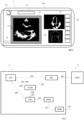
- An example of such a device 1 is illustrated in the figure 2 .
- the device 1 may comprise a tablet, a smartphone, a computer, or any other device comprising at least one display and a processor associated with a memory.
- the invention also relates to a system 2 comprising a device 1 according to the invention and a SECH ultrasound probe connected to said device 1.
- the method comprises a step 100 of receiving ultrasound images 11 in real time. Said ultrasound images are received by a receiver REC of the device.
- the receiver REC may comprise a buffer memory in which the images received 11 by the receiver REC are temporarily stored before their transmission.
- the received ultrasound images 11 are displayed in real time, preferably on an AFF display of the device.
- the received ultrasound images can therefore be transmitted to a display for their display in real time.
- the device includes an AFF display.
- the AFF display may include a screen such as the screen of a monitor, a touch pad or a smartphone.
- the AFF display is connected to the REC receiver and/or to the CLASS classifier and/or to the processor.
- the AFF display is configured to display in real time the ultrasound images received by the REC receiver.
- the AFF display advantageously allows the operator to have real-time feedback on the ultrasound images that he is currently acquiring.
- the AFF display is configured to display indicators or additional data that will be described later in this description.
- the AFF display displays an image 1 comprising an ultrasound image received 11 by the REC receiver.
- the ultrasound image displayed is the last ultrasound image received by the REC receiver.
- the AFF display is thus configured to display in real time the images captured by the SECH ultrasound probe connected to the device according to the invention.
- the method comprises a step 200 of classifying the ultrasound images 11 received by the receiver REC in real time.
- the classification is carried out by a classifier CLASS of the device.
- the classification of an ultrasound image comprises the association of said ultrasound image with a class 14, preferably a class from a group of predefined classes.
- the classification further comprises generating 300 a quality indicator 13 based on said class associated with the ultrasound image.
- the quality indicator may be representative of a class or a predefined set of classes.
- the classification step further comprises generating a confidence value.
- the confidence value may represent the confidence rate of the classification performed.
- the confidence value is generated by the CLASS classifier.
- the classification step is preferably executed by a CLASS classifier of the device.
- classifier we mean an algorithmic function executed by a processor or a calculator associated with a memory.
- the CLASS classifier is configured to receive images received by the REC receiver.
- the classifier is configured to classify in real time at least part of the images received by the receiver REC.
- the classifier is configured to associate a class with an ultrasound image.
- the classifier generates a quality indicator 13 as a function of the associated class.
- the quality indicator 13 is preferentially associated with said classified ultrasound image.
- the device of the image processing means for processing the received images before providing them to the classifier CLASS may comprise image filters or contrast functions.
- the classifier CLASS is configured to classify an ultrasound image of the heart among a plurality of classes comprising, on the one hand, classes each representing a particular view of the heart and on the other hand, a class representing a view of insufficient quality.
- an image when classified into a class representing a particular view of the heart, this image is considered to be of sufficient quality to advantageously allow the clinician to perform predefined measurements from these images.
- cardiologists use well-identified particular views for their diagnoses.
- each of these views can be characterized by the presence of one or more specific parts of the heart.
- the view of a parasternal short axis section is characterized by the presence on the image of the section of the left ventricle and the right ventricle. From this view, the cardiologist can calculate the shortening fraction and calculate pulmonary pressures.
- the class representing a view of insufficient quality represents ultrasound views of the heart not belonging to one of the specific views of the other classes or specific ultrasound views listed above, but whose image quality does not allow the necessary measurements related to this specific view to be carried out.
- the CLASS classifier is implemented using a learning function trained from supervised and/or automatic learning.
- the learning function preferably comprises a neural network.
- the learning function is preferably trained by a series of labeled ultrasound images of the heart.
- the learning function was configured from a learning process comprising submitting to the classifier a plurality of ultrasound images each associated with a label.
- the classifier was trained with ultrasound images of a specific view of the heart of sufficient quality, each image being associated with a label representing said specific view.
- the classifier was also trained with ultrasound images of views of the heart other than those cited above or not having sufficient quality allowing the cardiologist to perform measurements from these images, each of these images each being associated with a quality label representing a view of insufficient quality.
- the labels may also comprise information characteristic of a shot of an image of an organ comprising a characteristic viewing angle and/or size, of an image quality, of the presence of a specific portion of an image of the heart, of the presence of a contour of measurable anatomical shape.
- labels include the name of the particular view of the heart or a name associated with a poor quality view.
- the CLASS classifier may be executed in a processor itself associated with a memory.
- the CLASS classifier may be recorded in a computer-readable medium such as a memory associated with said processor.
- the step of classifying an ultrasound image includes generating a matching score for each class.
- the matching score may comprise a probability that the ultrasound image belongs to each class.
- the method then comprises selecting the class with the highest score.
- the confidence value is generated from said matching score of the class associated with said ultrasound image.
- the CLASS classifier classifies 100% of the images received in real time.
- the CLASS classifier is configured to classify only a rate of received images.
- the classifier is then configured to classify a portion of the ultrasound images received in real time. This embodiment is particularly advantageous when the classification rate is lower than the image acquisition rate (in frames per unit time).
- classifying an ultrasound image includes classifying the last received ultrasound image. When said image has been classified, the method again takes the last received ultrasound image. Thus, it is possible that some ultrasound images are not classified.
- this embodiment advantageously allows ultrasound images to be classified in real time, regardless of the speed of the classifier or the frequency of reception of the ultrasound images.
- the method according to the invention comprises the generation and display in real time on the AFF display of the generated quality indicator 13.
- the displayed quality indicator 13 corresponds to the last generated quality indicator 13.
- this quality indicator 13 is generated by the classifier CLASS.
- a classifier is configured to, when it receives an ultrasound image of the heart, classify said image into one of the classes mentioned above. The method then comprises the generation of a quality indicator representative of the classification of this image. Said quality indicator can be associated with said classified ultrasound image.
- the quality indicator 13 may be generated by a remote processor connected to the AFF display and receiving information from the classifier.
- the display of the quality indicator 13 may comprise the display of a colored indicator whose color depends on the class associated with the classified ultrasound image.
- the color of the indicator may be representative of a single class.
- the color of the colored indicator may take two distinct colors, a first color representative of a class group including the classes representing a particular view of the heart such as those stated above, and a second color representative of a class representing a view of insufficient quality.
- the classifier is also configured to, when receiving an ultrasound image of the heart, generate a confidence value 21.
- the confidence value 21 may be representative of a level of certainty that the image has been well classified.
- the quality indicator may comprise said confidence value.
- the confidence indicator 21 is preferentially displayed by the AFF display.
- the quality indicator 13 associated with the classified image may include said confidence indicator.
- a confidence indicator representative of the confidence value is generated and displayed in real time.
- the displayed quality indicator 13 comprises a colored frame, which preferably extends around the displayed ultrasound image. The operator can then advantageously visualize whether the image he is taking is indeed the one expected without having to look away from the image.
- the confidence indicator can be displayed in the form of a numerical value as illustrated in the figure 1 .
- the quality indicator 13 comprises a colored indicator.
- the color of the colored indicator is representative of the classification of the last ultrasound image. At least one of the dimensions of said colored indicator is a function of the confidence value.
- quality indicator 13 is a bar whose length varies according to the confidence value and whose color depends on the class associated with the last ultrasound image.
- Such an indicator advantageously allows the operator to visualize whether the image he is taking is of sufficient quality and also allows him to visualize without looking away whether the indicator is degraded or improved depending on the movement of the SECH ultrasound probe.
- the advantage of such an indicator is to allow the operator, when he slightly moves the probe, to visualize whether this movement increases or reduces the confidence value, and consequently increases or reduces his chances of obtaining a validated image sequence described below.
- the method comprises the automatic recording 400 of a sequence 20 of images in a memory MEM.
- This sequence 20 is automatically recorded when a sequence of a predefined number of received or classified images comprises a rate of images associated with the same class 14 greater than a predefined rate.
- said class is a class representing a particular view of the heart.
- image sequence we mean a series of ultrasound images that follow one another in the chronological order of their acquisition. We will therefore also use the term “video sequence” to designate such an image sequence.
- a sufficiently long video comprises, for example, a majority of ultrasound images classified in the same class representing a particular view of the heart
- the video sequence is automatically recorded in a memory.
- This automatic recording advantageously automatically generates videos of a particular view of the heart which can be analyzed by a cardiologist without validation by the operator.
- the predefined number of images received or classified can be understood as a predefined minimum duration, as long as the image acquisition frequency of the SECH ultrasound probe is constant.
- the advantage of this threshold is to ensure that the video sequence is long enough to be analyzed by a cardiologist.
- the predefined number of images can be replaced by a number of images received or classified.
- the predefined number can be set to correspond to a predefined number of cardiac cycles of the subject's heart.
- the rate of images associated with the same class in said video sequence greater than a predefined rate advantageously allows automatic recording despite a negligible number of images not having sufficient quality in said sequence. Indeed, this negligible number of images in a video sequence can come from a classification error, or from noise linked to a particular image. This tolerance advantageously consists of a good compromise between the ease of automatic generation of a sequence and the quality of said sequence.
- the predefined rate is at least greater than 50%. In another execution mode, the predefined rate may be set by the operator, for example by a user interface of the device.
- the automatic recording step comprises buffering the continuously received ultrasound images as well as the generated quality indices associated with said images. In this way, when a video sequence meeting the criteria set forth above is detected, the images belonging to this sequence can be transferred from the buffer memory to another memory, and/or grouped into a single file to generate a video sequence.
- the method comprises a step of generating and displaying in real time a time indicator 12.
- the time indicator 12 displayed is representative of the number of images remaining to be received or classified to reach a sequence of a predefined number of received images comprising a rate of images associated with the same class greater than a predefined rate.
- This indicator advantageously allows the operator to visualize the time remaining to validate a video sequence of a particular view of the heart.
- This indicator advantageously allows the operator to visualize the remaining time during which it must maintain an ultrasound image of sufficient quality, said quality being displayed to it by the quality indicator 13 and/or the confidence indicator.
- the time indicator 12 may comprise a numerical value such as a countdown.
- the time indicator 12 may comprise a gauge filling or emptying depending on the time remaining to validate a video sequence.
- the method comprises, when a first image is classified into a class representing a particular view of the heart, automatically generating and displaying the time indicator 12.
- the method comprises displaying in real time the received ultrasound images, the quality indicator 13, and optionally the time indicator 12. Such a display is described below with reference to the figure 2 .
- the image displayed by the AFF display includes the ultrasound image 11 received in real time or taken in real time by the SECH ultrasound probe, the quality indicator 13 and the time indicator 12.
- the method according to the invention comprises the selection of a first class from among the classes representing a particular view of the heart such as one of the views mentioned above.
- the selection of the first class generates the display on the AFF display of a first pre-recorded ultrasound image 15 of the heart representing such a view.
- the label 17 which may include the name of said particular view is also displayed on the AFF display.
- the selection of the first class also generates the display of a first setpoint image 14.
- the first setpoint image 14 is preferably pre-recorded.
- the setpoint image illustrates a position and/or orientation setpoint for the SECH ultrasound probe on a patient to obtain said view corresponding to the selected class.
- the operator is thus advantageously guided and assisted in taking such a view. He can thus, on the same AFF display, view an example of the view he must take, the position and orientation of the probe to achieve it.
- the operator can visualize in real time whether the view that is acquired is of sufficiently good quality by the quality indicator 13, and can advantageously visualize the influence of the movements that he makes on the quality of the acquired image thanks to the confidence indicator.
- the time indicator 12 representing the time during which he must maintain the probe to acquire images of sufficient quality.
- the operator is also assured, by viewing the time indicator 12, that a first sequence 20 will be recorded while maintaining his position.
- the method may comprise the automatic selection of a second class from among the classes representing a particular view of the heart such as one of the views mentioned above.
- the selection of said second class automatically generates the display of a second pre-recorded reference image and the display of a second pre-recorded ultrasound image illustrating an ultrasound view.
- the method further comprises generating and displaying a progress indicator 18.
- the progress indicator may be representative of the number of sequences that have been recorded.
- the progress indicator is representative of the number of classes representing a particular view of the heart for which a sequence 20 of images was generated and/or recorded automatically according to the method of the invention.
- the method comprises displaying the recorded sequence and further comprises a second manual validation by the operator after displaying said sequence.
- the device according to the invention comprises software and hardware means for implementing the method as described above.
- the device comprises a receiver REC.
- the receiver REC is intended to be connected to an SECH ultrasound probe so as to continuously and in real time receive ultrasound images 11 acquired by said SECH ultrasound probe.
- the REC receiver can be connected to the SECH ultrasound probe by a wired connection or a wireless connection, for example by a Bluetooth connection or a WI-FI connection or any other data exchange protocol known to those skilled in the art.
- the REC receiver may comprise or be associated with one or more memories for temporarily storing the received images.
- the REC receiver is directly or indirectly connected to the AFF display to transmit the acquired ultrasound images 11 to the AFF display.
- the device further comprises means for implementing a classifier CLASS as described above.
- the classifier is configured to receive the ultrasound images 11, associate a class 14 with the ultrasound images in real time, and generate the quality indicator 13 in real time.
- the classifier is directly or indirectly connected to the AFF display to transmit the generated indicators to the AFF display.
- the classifier is implemented by remote electronic equipment, such as a remote server.
- the device comprises an interface for exchanging data with the remote equipment in order to transmit data and retrieve the result of the processed, i.e. classified, data.
- the device of the invention can be interpreted as being the system comprising on the one hand the local device described in the present application and the remote means making it possible to implement the classification function.
- the device further comprises at least one processor or a CALC calculator associated with a memory to execute at least part of the steps of the method according to the invention.
- said processor can be configured to execute the steps of classification 200, generation of display of the quality indicator 300 and of the time indicator 500 and/or automatic recording 300 of the sequence 20 of images.
- Said processor can be connected to the AFF display to transmit the time indicator 12 to the AFF display.
- the device 1 may also comprise a plurality of processors, each associated with one or more memories, and configured to together execute such steps.
- the processor(s) may be remote and connected to the display by a data network.
- the device further comprises a memory for storing or recording the sequences generated by the method according to the invention.
- the device further comprises an EMM transmitter connected to said MEM memory for transmitting said sequences recorded on said MEM memory to a data network.
- the AFF display may comprise means for receiving the various information 11, 13, 21, 12 received by the various REC, CLASS, PROC means of the device to generate the final image to be displayed.
Landscapes
- Health & Medical Sciences (AREA)
- Life Sciences & Earth Sciences (AREA)
- Engineering & Computer Science (AREA)
- General Health & Medical Sciences (AREA)
- Medical Informatics (AREA)
- Physics & Mathematics (AREA)
- Nuclear Medicine, Radiotherapy & Molecular Imaging (AREA)
- Radiology & Medical Imaging (AREA)
- Veterinary Medicine (AREA)
- Public Health (AREA)
- Animal Behavior & Ethology (AREA)
- Surgery (AREA)
- Biophysics (AREA)
- Pathology (AREA)
- Biomedical Technology (AREA)
- Heart & Thoracic Surgery (AREA)
- Molecular Biology (AREA)
- Computer Vision & Pattern Recognition (AREA)
- Theoretical Computer Science (AREA)
- General Physics & Mathematics (AREA)
- Evolutionary Computation (AREA)
- Cardiology (AREA)
- Artificial Intelligence (AREA)
- Computing Systems (AREA)
- Databases & Information Systems (AREA)
- Software Systems (AREA)
- Multimedia (AREA)
- Physiology (AREA)
- Quality & Reliability (AREA)
- Ultra Sonic Daignosis Equipment (AREA)
Claims (15)
- Vorrichtung (1) zur Unterstützung bei der Erstellung einer Ultraschallbildsequenz, die so ausgelegt ist, dass sie in Echtzeit Ultraschallbilder (11) empfängt, die von einer an dieser Vorrichtung (1) angeschlossenen Ultraschallsonde (SECH) erfasst werden, und umfassend:▪ einen ersten Rechner, der einem ersten Speicher zugeordnet ist, um einen Klassifizierer (CLASS) zu implementieren, der so ausgelegt ist, dass er die empfangenen Ultraschallbilder (11) in Echtzeit klassifiziert und jedem Bild eine Klasse (14) zuordnet, um einen Qualitätsindikator (13) in Abhängigkeit von der zugeordneten Klasse (14) zu generieren;▪ eine Anzeigevorrichtung (AFF), um in Echtzeit den zuletzt generierten Qualitätsindikator (13) anzuzeigen;▪ einen zweiten Rechner (CALC), der einem zweiten Speicher zugeordnet und so ausgelegt ist, dass er die folgenden Schritte ausführt:▪ wenn eine Sequenz einer vordefinierten Anzahl empfangener Bilder eine Bildrate umfasst, die einer gleichen Klasse zugeordnet ist, die größer ist als eine vordefinierte Rate, das automatische Speichern der Bildsequenz (20) in einem dritten Speicher (MEM); und▪ das Generieren und das Anzeigen auf der Anzeigevorrichtung in Echtzeit eines Zeitindikators (12), der die Anzahl der verbleibenden Bilder darstellt, die empfangen werden müssen, um die Sequenz einer vordefinierten Anzahl empfangener Bilder zu erreichen, die eine Bildrate umfasst, die einer gleichen Klasse zugeordnet ist, die größer ist als eine vordefinierten Rate.
- Vorrichtung nach Anspruch 1, wobei der Klassifizierer (CLASS) ausgelegt ist, um ein Bild zu klassifizieren in:▪ eine Vielzahl von Klassen, die jeweils eine bestimmte Ansicht des Herzorgans repräsentieren; und▪ mindestens eine Klasse, die eine Ansicht in unzureichender Qualität darstellt.
- Vorrichtung nach Anspruch 2, wobei der Klassifizierer (CLASS) mittels einer Lernfunktion implementiert wird, die auf der Basis eines überwachten maschinellen Lernens konfiguriert ist.
- Vorrichtung nach vorhergehendem Anspruch, wobei der Klassifizierer (CLASS) mittels eines Neuronennetzwerks implementiert wird, das von einer Reihe gekennzeichneter Ultraschallbilder des Herzens trainiert wird, wobei die Kennzeichnungen eine charakteristische Information aufweisen:• einer Aufnahme eines Bildes eines Organs, die einen charakteristischen Blickwinkel und/oder eine charakteristische Größe aufweist;• einer Bildqualität;• des Vorhandenseins eines Teils eines Bildes des Herzens; und/oder• des Vorhandenseins einer Kontur in einer messbaren anatomischen Form.
- Vorrichtung (1) nach einem der vorhergehenden Ansprüche, wobei der angezeigte Qualitätsindikator (13) einen farbigen Indikator umfasst, dessen Farbe von der Klasse abhängt, die dem zuletzt klassifizierten Bild zugeordnet ist.
- Vorrichtung (1) nach Anspruch 5, wobei wenigstens eine der Abmessungen des angezeigten farbigen Indikators vom Vertrauenswert abhängt.
- Vorrichtung (1) nach einem der vorhergehenden Ansprüche, wobei der angezeigte Zeitindikator (12) eine Messlehre umfasst, deren Füllung vom Wert des Zeitindikators abhängt.
- Vorrichtung (1) nach einem der vorhergehenden Ansprüche, wobei die Anzeigevorrichtung zum Anzeigen der empfangenen Ultraschallbilder (11) in Echtzeit ausgebildet ist.
- System (2), das eine Vorrichtung (1) nach einem der Ansprüche 1 bis 8 und eine Ultraschallsonde (SECH) umfasst, die an diese Vorrichtung (1) angeschlossen werden kann, um die von der Sonde aufgenommenen Ultraschallbilder an die Vorrichtung zu übertragen.
- Verfahren (1000) zur Unterstützung des Generierens einer computergestützten Ultraschallbildsequenz, umfassend:▪ das Empfangen (100) von Ultraschallbildern (11) von einer Ultraschallsonde (SEC);▪ das Klassifizieren (200) in Echtzeit der empfangenen Ultraschallbilder (11) und das Zuordnen einer Klasse zu jedem Bild;▪ das Generieren und Anzeigen (300) eines Qualitätsindikators (13) in Abhängigkeit von dieser Klasse (14), die dem klassifizierten Ultraschallbild zugeordnet wurde;▪ wenn eine Sequenz einer vordefinierten Anzahl empfangener Bilder eine Bildrate umfasst, die einer gleichen Klasse zugeordnet ist, die größer ist als eine vordefinierte Rate, das automatische Speichern (400) der Bildsequenz (20) in einem Speicher (MEM);▪ das Generieren und das Anzeigen (500) in Echtzeit eines Zeitindikators (12), der die Anzahl der verbleibenden Bilder darstellt, die empfangen werden müssen, um die Sequenz einer vordefinierten Anzahl empfangener Bilder zu erreichen, die eine Bildrate umfasst, die einer gleichen Klasse zugeordnet ist, die größer ist als eine vordefinierten Rate.
- Verfahren nach vorhergehendem Anspruch, wobei ein vordefinierter Bruchteil der empfangenen Bilder klassifiziert wird, wobei der vordefinierte Bruchteil über eine Benutzerschnittstelle steuerbar ist.
- Verfahren nach einem der Ansprüche 10 oder 11, wobei die empfangenen Ultraschallbilder (11) kontinuierlich angezeigt werden.
- Verfahren nach einem der Ansprüche 10 bis 12, wobei das Verfahren ferner die Auswahl einer ersten Klasse umfasst, die eine erste besondere Ansicht des Herzens darstellt, wobei die Auswahl automatisch generiert:▪ die Anzeige eines ersten voraufgezeichneten Bildes, das einem Bild der ersten Ultraschallansicht des Herzens der ersten ausgewählten Klasse entspricht, und/oder▪ die Anzeige eines ersten voraufgezeichneten Sollwerts für die Position der Ultraschallsonde an einem Patienten zur Erfassung der ersten besonderen Ansicht des Herzens.
- Rechnerprogrammprodukt, das Anweisungen umfasst, die eine Vorrichtung anleiten, die Schritte des Verfahrens nach einem der Ansprüche 10 bis 13 auszuführen.
- Rechnerlesbares Medium, auf dem das Rechnerprogramm nach Anspruch 14 gespeichert ist.
Applications Claiming Priority (2)
| Application Number | Priority Date | Filing Date | Title |
|---|---|---|---|
| FR2105597A FR3123198B1 (fr) | 2021-05-28 | 2021-05-28 | Dispositif pour l’acquisition d’une sequence d’images echographiques du cœur et procede associe |
| PCT/EP2022/064109 WO2022248500A1 (fr) | 2021-05-28 | 2022-05-24 | Dispositif pour l'acquisition d'une sequence d'images echographiques et procede associe |
Publications (3)
| Publication Number | Publication Date |
|---|---|
| EP4344405A1 EP4344405A1 (de) | 2024-04-03 |
| EP4344405C0 EP4344405C0 (de) | 2024-10-16 |
| EP4344405B1 true EP4344405B1 (de) | 2024-10-16 |
Family
ID=76807805
Family Applications (1)
| Application Number | Title | Priority Date | Filing Date |
|---|---|---|---|
| EP22728640.8A Active EP4344405B1 (de) | 2021-05-28 | 2022-05-24 | Vorrichtung zur erfassung einer sequenz von echografischen bildern und zugehöriges verfahren |
Country Status (7)
| Country | Link |
|---|---|
| US (1) | US12251261B2 (de) |
| EP (1) | EP4344405B1 (de) |
| JP (1) | JP2024521848A (de) |
| KR (1) | KR20240013183A (de) |
| BR (1) | BR112023023837A2 (de) |
| FR (1) | FR3123198B1 (de) |
| WO (1) | WO2022248500A1 (de) |
Families Citing this family (1)
| Publication number | Priority date | Publication date | Assignee | Title |
|---|---|---|---|---|
| CN117998024B (zh) * | 2024-04-07 | 2024-05-31 | 中国医学科学院阜外医院 | 一种心脏辅助检测用超声影像传输方法 |
Family Cites Families (4)
| Publication number | Priority date | Publication date | Assignee | Title |
|---|---|---|---|---|
| CN112957074A (zh) * | 2016-03-09 | 2021-06-15 | 安科诺思公司 | 利用人工智能网络的超声图像识别系统和方法 |
| US10628932B2 (en) * | 2017-10-27 | 2020-04-21 | Butterfly Network, Inc. | Quality indicators for collection of and automated measurement on ultrasound images |
| US20200178934A1 (en) * | 2018-12-10 | 2020-06-11 | General Electric Company | Ultrasound imaging system and method for displaying a target object quality level |
| US20200245976A1 (en) * | 2019-01-31 | 2020-08-06 | Bay Labs, Inc. | Retrospective image saving for ultrasound diagnostics |
-
2021
- 2021-05-28 FR FR2105597A patent/FR3123198B1/fr active Active
-
2022
- 2022-05-24 JP JP2023573354A patent/JP2024521848A/ja active Pending
- 2022-05-24 WO PCT/EP2022/064109 patent/WO2022248500A1/fr not_active Ceased
- 2022-05-24 US US18/560,302 patent/US12251261B2/en active Active
- 2022-05-24 KR KR1020237044440A patent/KR20240013183A/ko active Pending
- 2022-05-24 BR BR112023023837A patent/BR112023023837A2/pt not_active Application Discontinuation
- 2022-05-24 EP EP22728640.8A patent/EP4344405B1/de active Active
Also Published As
| Publication number | Publication date |
|---|---|
| BR112023023837A2 (pt) | 2024-02-06 |
| EP4344405A1 (de) | 2024-04-03 |
| FR3123198B1 (fr) | 2023-05-19 |
| KR20240013183A (ko) | 2024-01-30 |
| FR3123198A1 (fr) | 2022-12-02 |
| US12251261B2 (en) | 2025-03-18 |
| EP4344405C0 (de) | 2024-10-16 |
| US20240225589A1 (en) | 2024-07-11 |
| JP2024521848A (ja) | 2024-06-04 |
| WO2022248500A1 (fr) | 2022-12-01 |
Similar Documents
| Publication | Publication Date | Title |
|---|---|---|
| US11207055B2 (en) | Ultrasound Cardiac Doppler study automation | |
| US11129591B2 (en) | Echocardiographic image analysis | |
| US20200214679A1 (en) | Methods and apparatuses for receiving feedback from users regarding automatic calculations performed on ultrasound data | |
| EP2129296B1 (de) | Vorrichtung zur messung der viskoelastizität biologischer gewebe und verfahren zur verwendung dieser vorrichtung | |
| TW201923776A (zh) | 超音波影像上的自動化測量及用於自動化測量的收集的品質指示器 | |
| EP3691535A1 (de) | System und verfahren zur fusion von ultraschall mit zusätzlichen signalen | |
| FR2986145A1 (fr) | Procede et systeme pour surveiller une matrice de transducteurs dans un systeme d'echographie | |
| AU2024251739A1 (en) | Three-dimensional characterization of atherosclerotic plaques using two-dimensional ultrasound images | |
| US20190076127A1 (en) | Method and system for automatically selecting ultrasound image loops from a continuously captured stress echocardiogram based on assigned image view types and image characteristic metrics | |
| WO2022248500A1 (fr) | Dispositif pour l'acquisition d'une sequence d'images echographiques et procede associe | |
| EP4007528B1 (de) | Kardiale vorrichtung | |
| EP3294121B1 (de) | Verfahren und vorrichtung zur bestimmung der charakteristischen parameter einer kardiovaskulären aktivität | |
| WO2025141151A1 (fr) | Procede mis en oeuvre par ordinateur d'estimation de la fraction de flux de reserve coronarienne, systeme | |
| US20250125039A1 (en) | Natural language based image comparison system | |
| WO2026047118A1 (fr) | Dispositif pour l'acquisition de donnees echographiques doppler et procede associe | |
| FR3136152A1 (fr) | Dispositif pour l’assistance au guidage pour acquerir une image echographique et procede associe | |
| KR102794462B1 (ko) | 심장 신호 분할 방법 및 이를 이용한 심장 신호 분할용 디바이스 | |
| EP4740174A1 (de) | Unterstützung beim nachweis von herzamyloidose mittels künstlicher intelligenz | |
| EP4712867A1 (de) | Vorrichtung und system zur tomographie der lunge und zugehöriges tomographieverfahren | |
| Vignon-Clementel | Écoulement intraventriculaire en échocardiographie Doppler avec réseaux de neurones fondés sur la physique | |
| EP4346616A1 (de) | Tragbares abbildungssystem |
Legal Events
| Date | Code | Title | Description |
|---|---|---|---|
| STAA | Information on the status of an ep patent application or granted ep patent |
Free format text: STATUS: UNKNOWN |
|
| STAA | Information on the status of an ep patent application or granted ep patent |
Free format text: STATUS: THE INTERNATIONAL PUBLICATION HAS BEEN MADE |
|
| PUAI | Public reference made under article 153(3) epc to a published international application that has entered the european phase |
Free format text: ORIGINAL CODE: 0009012 |
|
| STAA | Information on the status of an ep patent application or granted ep patent |
Free format text: STATUS: REQUEST FOR EXAMINATION WAS MADE |
|
| 17P | Request for examination filed |
Effective date: 20231227 |
|
| AK | Designated contracting states |
Kind code of ref document: A1 Designated state(s): AL AT BE BG CH CY CZ DE DK EE ES FI FR GB GR HR HU IE IS IT LI LT LU LV MC MK MT NL NO PL PT RO RS SE SI SK SM TR |
|
| GRAP | Despatch of communication of intention to grant a patent |
Free format text: ORIGINAL CODE: EPIDOSNIGR1 |
|
| STAA | Information on the status of an ep patent application or granted ep patent |
Free format text: STATUS: GRANT OF PATENT IS INTENDED |
|
| DAV | Request for validation of the european patent (deleted) | ||
| DAX | Request for extension of the european patent (deleted) | ||
| INTG | Intention to grant announced |
Effective date: 20240430 |
|
| GRAS | Grant fee paid |
Free format text: ORIGINAL CODE: EPIDOSNIGR3 |
|
| GRAA | (expected) grant |
Free format text: ORIGINAL CODE: 0009210 |
|
| STAA | Information on the status of an ep patent application or granted ep patent |
Free format text: STATUS: THE PATENT HAS BEEN GRANTED |
|
| RAP3 | Party data changed (applicant data changed or rights of an application transferred) |
Owner name: DESKI |
|
| AK | Designated contracting states |
Kind code of ref document: B1 Designated state(s): AL AT BE BG CH CY CZ DE DK EE ES FI FR GB GR HR HU IE IS IT LI LT LU LV MC MK MT NL NO PL PT RO RS SE SI SK SM TR |
|
| REG | Reference to a national code |
Ref country code: GB Ref legal event code: FG4D Free format text: NOT ENGLISH |
|
| REG | Reference to a national code |
Ref country code: CH Ref legal event code: EP Ref country code: DE Ref legal event code: R096 Ref document number: 602022006900 Country of ref document: DE |
|
| REG | Reference to a national code |
Ref country code: IE Ref legal event code: FG4D Free format text: LANGUAGE OF EP DOCUMENT: FRENCH |
|
| U01 | Request for unitary effect filed |
Effective date: 20241115 |
|
| U07 | Unitary effect registered |
Designated state(s): AT BE BG DE DK EE FI FR IT LT LU LV MT NL PT RO SE SI Effective date: 20241125 |
|
| PG25 | Lapsed in a contracting state [announced via postgrant information from national office to epo] |
Ref country code: HR Free format text: LAPSE BECAUSE OF FAILURE TO SUBMIT A TRANSLATION OF THE DESCRIPTION OR TO PAY THE FEE WITHIN THE PRESCRIBED TIME-LIMIT Effective date: 20241016 Ref country code: IS Free format text: LAPSE BECAUSE OF FAILURE TO SUBMIT A TRANSLATION OF THE DESCRIPTION OR TO PAY THE FEE WITHIN THE PRESCRIBED TIME-LIMIT Effective date: 20250216 |
|
| PG25 | Lapsed in a contracting state [announced via postgrant information from national office to epo] |
Ref country code: ES Free format text: LAPSE BECAUSE OF FAILURE TO SUBMIT A TRANSLATION OF THE DESCRIPTION OR TO PAY THE FEE WITHIN THE PRESCRIBED TIME-LIMIT Effective date: 20241016 |
|
| PG25 | Lapsed in a contracting state [announced via postgrant information from national office to epo] |
Ref country code: NO Free format text: LAPSE BECAUSE OF FAILURE TO SUBMIT A TRANSLATION OF THE DESCRIPTION OR TO PAY THE FEE WITHIN THE PRESCRIBED TIME-LIMIT Effective date: 20250116 |
|
| PG25 | Lapsed in a contracting state [announced via postgrant information from national office to epo] |
Ref country code: GR Free format text: LAPSE BECAUSE OF FAILURE TO SUBMIT A TRANSLATION OF THE DESCRIPTION OR TO PAY THE FEE WITHIN THE PRESCRIBED TIME-LIMIT Effective date: 20250117 |
|
| PG25 | Lapsed in a contracting state [announced via postgrant information from national office to epo] |
Ref country code: PL Free format text: LAPSE BECAUSE OF FAILURE TO SUBMIT A TRANSLATION OF THE DESCRIPTION OR TO PAY THE FEE WITHIN THE PRESCRIBED TIME-LIMIT Effective date: 20241016 |
|
| PG25 | Lapsed in a contracting state [announced via postgrant information from national office to epo] |
Ref country code: RS Free format text: LAPSE BECAUSE OF FAILURE TO SUBMIT A TRANSLATION OF THE DESCRIPTION OR TO PAY THE FEE WITHIN THE PRESCRIBED TIME-LIMIT Effective date: 20250116 |
|
| U20 | Renewal fee for the european patent with unitary effect paid |
Year of fee payment: 4 Effective date: 20250528 |
|
| PG25 | Lapsed in a contracting state [announced via postgrant information from national office to epo] |
Ref country code: SM Free format text: LAPSE BECAUSE OF FAILURE TO SUBMIT A TRANSLATION OF THE DESCRIPTION OR TO PAY THE FEE WITHIN THE PRESCRIBED TIME-LIMIT Effective date: 20241016 |
|
| PGFP | Annual fee paid to national office [announced via postgrant information from national office to epo] |
Ref country code: CH Payment date: 20250627 Year of fee payment: 4 |
|
| PG25 | Lapsed in a contracting state [announced via postgrant information from national office to epo] |
Ref country code: SK Free format text: LAPSE BECAUSE OF FAILURE TO SUBMIT A TRANSLATION OF THE DESCRIPTION OR TO PAY THE FEE WITHIN THE PRESCRIBED TIME-LIMIT Effective date: 20241016 |
|
| PG25 | Lapsed in a contracting state [announced via postgrant information from national office to epo] |
Ref country code: CZ Free format text: LAPSE BECAUSE OF FAILURE TO SUBMIT A TRANSLATION OF THE DESCRIPTION OR TO PAY THE FEE WITHIN THE PRESCRIBED TIME-LIMIT Effective date: 20241016 |
|
| PLBE | No opposition filed within time limit |
Free format text: ORIGINAL CODE: 0009261 |
|
| STAA | Information on the status of an ep patent application or granted ep patent |
Free format text: STATUS: NO OPPOSITION FILED WITHIN TIME LIMIT |
|
| 26N | No opposition filed |
Effective date: 20250717 |
|
| PG25 | Lapsed in a contracting state [announced via postgrant information from national office to epo] |
Ref country code: MC Free format text: LAPSE BECAUSE OF FAILURE TO SUBMIT A TRANSLATION OF THE DESCRIPTION OR TO PAY THE FEE WITHIN THE PRESCRIBED TIME-LIMIT Effective date: 20241016 |
|
| PG25 | Lapsed in a contracting state [announced via postgrant information from national office to epo] |
Ref country code: IE Free format text: LAPSE BECAUSE OF NON-PAYMENT OF DUE FEES Effective date: 20250524 |