EP4275794A2 - Device for automated nested recombinase polymerase amplification - Google Patents
Device for automated nested recombinase polymerase amplification Download PDFInfo
- Publication number
- EP4275794A2 EP4275794A2 EP23168518.1A EP23168518A EP4275794A2 EP 4275794 A2 EP4275794 A2 EP 4275794A2 EP 23168518 A EP23168518 A EP 23168518A EP 4275794 A2 EP4275794 A2 EP 4275794A2
- Authority
- EP
- European Patent Office
- Prior art keywords
- sample
- nucleic acid
- amplification
- amplification product
- module
- Prior art date
- Legal status (The legal status is an assumption and is not a legal conclusion. Google has not performed a legal analysis and makes no representation as to the accuracy of the status listed.)
- Pending
Links
- 238000003199 nucleic acid amplification method Methods 0.000 title claims abstract description 297
- 230000003321 amplification Effects 0.000 title claims abstract description 296
- 102000018120 Recombinases Human genes 0.000 title abstract description 22
- 108010091086 Recombinases Proteins 0.000 title abstract description 22
- 150000007523 nucleic acids Chemical class 0.000 claims abstract description 188
- 102000039446 nucleic acids Human genes 0.000 claims abstract description 175
- 108020004707 nucleic acids Proteins 0.000 claims abstract description 175
- 102000040430 polynucleotide Human genes 0.000 claims abstract description 48
- 108091033319 polynucleotide Proteins 0.000 claims abstract description 48
- 239000002157 polynucleotide Substances 0.000 claims abstract description 48
- 238000001514 detection method Methods 0.000 claims description 103
- 238000000034 method Methods 0.000 claims description 70
- 108020004414 DNA Proteins 0.000 claims description 49
- 102000053602 DNA Human genes 0.000 claims description 36
- 244000037640 animal pathogen Species 0.000 claims description 20
- 108090000623 proteins and genes Proteins 0.000 claims description 19
- 101710163270 Nuclease Proteins 0.000 claims description 18
- 108020004682 Single-Stranded DNA Proteins 0.000 claims description 15
- 241001465754 Metazoa Species 0.000 claims description 13
- 208000037797 influenza A Diseases 0.000 claims description 12
- 241000700605 Viruses Species 0.000 claims description 11
- 241000712431 Influenza A virus Species 0.000 claims description 9
- 241000713196 Influenza B virus Species 0.000 claims description 9
- 108020005202 Viral DNA Proteins 0.000 claims description 9
- 208000037798 influenza B Diseases 0.000 claims description 9
- 101150076489 B gene Proteins 0.000 claims description 6
- 108091034117 Oligonucleotide Proteins 0.000 claims description 6
- 238000002372 labelling Methods 0.000 claims description 6
- 108091032973 (ribonucleotides)n+m Proteins 0.000 claims description 5
- 241000894006 Bacteria Species 0.000 claims description 5
- 108020005196 Mitochondrial DNA Proteins 0.000 claims description 5
- 206010036790 Productive cough Diseases 0.000 claims description 5
- 108020000999 Viral RNA Proteins 0.000 claims description 5
- 239000008280 blood Substances 0.000 claims description 5
- 210000004369 blood Anatomy 0.000 claims description 5
- 239000002299 complementary DNA Substances 0.000 claims description 5
- 241001493065 dsRNA viruses Species 0.000 claims description 5
- 210000003097 mucus Anatomy 0.000 claims description 5
- 239000013612 plasmid Substances 0.000 claims description 5
- 210000003296 saliva Anatomy 0.000 claims description 5
- 210000003802 sputum Anatomy 0.000 claims description 5
- 208000024794 sputum Diseases 0.000 claims description 5
- 210000001138 tear Anatomy 0.000 claims description 5
- 210000002700 urine Anatomy 0.000 claims description 5
- WJFKNYWRSNBZNX-UHFFFAOYSA-N 10H-phenothiazine Chemical compound C1=CC=C2NC3=CC=CC=C3SC2=C1 WJFKNYWRSNBZNX-UHFFFAOYSA-N 0.000 claims description 3
- TZMSYXZUNZXBOL-UHFFFAOYSA-N 10H-phenoxazine Chemical compound C1=CC=C2NC3=CC=CC=C3OC2=C1 TZMSYXZUNZXBOL-UHFFFAOYSA-N 0.000 claims description 3
- VEPOHXYIFQMVHW-XOZOLZJESA-N 2,3-dihydroxybutanedioic acid (2S,3S)-3,4-dimethyl-2-phenylmorpholine Chemical compound OC(C(O)C(O)=O)C(O)=O.C[C@H]1[C@@H](OCCN1C)c1ccccc1 VEPOHXYIFQMVHW-XOZOLZJESA-N 0.000 claims description 3
- PCNDJXKNXGMECE-UHFFFAOYSA-N Phenazine Natural products C1=CC=CC2=NC3=CC=CC=C3N=C21 PCNDJXKNXGMECE-UHFFFAOYSA-N 0.000 claims description 3
- KJTLSVCANCCWHF-UHFFFAOYSA-N Ruthenium Chemical compound [Ru] KJTLSVCANCCWHF-UHFFFAOYSA-N 0.000 claims description 3
- PYKYMHQGRFAEBM-UHFFFAOYSA-N anthraquinone Natural products CCC(=O)c1c(O)c2C(=O)C3C(C=CC=C3O)C(=O)c2cc1CC(=O)OC PYKYMHQGRFAEBM-UHFFFAOYSA-N 0.000 claims description 3
- 150000004056 anthraquinones Chemical class 0.000 claims description 3
- YAGKRVSRTSUGEY-UHFFFAOYSA-N ferricyanide Chemical compound [Fe+3].N#[C-].N#[C-].N#[C-].N#[C-].N#[C-].N#[C-] YAGKRVSRTSUGEY-UHFFFAOYSA-N 0.000 claims description 3
- KTWOOEGAPBSYNW-UHFFFAOYSA-N ferrocene Chemical compound [Fe+2].C=1C=C[CH-]C=1.C=1C=C[CH-]C=1 KTWOOEGAPBSYNW-UHFFFAOYSA-N 0.000 claims description 3
- 229910052762 osmium Inorganic materials 0.000 claims description 3
- SYQBFIAQOQZEGI-UHFFFAOYSA-N osmium atom Chemical compound [Os] SYQBFIAQOQZEGI-UHFFFAOYSA-N 0.000 claims description 3
- 229950000688 phenothiazine Drugs 0.000 claims description 3
- 229910052707 ruthenium Inorganic materials 0.000 claims description 3
- 108020001738 DNA Glycosylase Proteins 0.000 claims 1
- 102000028381 DNA glycosylase Human genes 0.000 claims 1
- 238000003556 assay Methods 0.000 abstract description 9
- 239000000523 sample Substances 0.000 description 206
- 238000006243 chemical reaction Methods 0.000 description 102
- 239000003153 chemical reaction reagent Substances 0.000 description 100
- 239000000047 product Substances 0.000 description 87
- 238000012546 transfer Methods 0.000 description 68
- 239000013615 primer Substances 0.000 description 42
- 239000007788 liquid Substances 0.000 description 34
- 238000010517 secondary reaction Methods 0.000 description 30
- 239000012530 fluid Substances 0.000 description 24
- 238000002156 mixing Methods 0.000 description 23
- 230000003287 optical effect Effects 0.000 description 18
- 239000007787 solid Substances 0.000 description 17
- 239000000203 mixture Substances 0.000 description 16
- 239000011541 reaction mixture Substances 0.000 description 15
- 108091028043 Nucleic acid sequence Proteins 0.000 description 13
- 239000002609 medium Substances 0.000 description 13
- 235000018102 proteins Nutrition 0.000 description 12
- 102000004169 proteins and genes Human genes 0.000 description 12
- 230000005284 excitation Effects 0.000 description 10
- 238000005259 measurement Methods 0.000 description 10
- 241000588724 Escherichia coli Species 0.000 description 9
- 241000894007 species Species 0.000 description 9
- 239000003795 chemical substances by application Substances 0.000 description 8
- 239000002987 primer (paints) Substances 0.000 description 8
- 239000012634 fragment Substances 0.000 description 7
- UEGPKNKPLBYCNK-UHFFFAOYSA-L magnesium acetate Chemical compound [Mg+2].CC([O-])=O.CC([O-])=O UEGPKNKPLBYCNK-UHFFFAOYSA-L 0.000 description 7
- 229940069446 magnesium acetate Drugs 0.000 description 7
- 235000011285 magnesium acetate Nutrition 0.000 description 7
- 239000011654 magnesium acetate Substances 0.000 description 7
- 102000052510 DNA-Binding Proteins Human genes 0.000 description 6
- 108010014303 DNA-directed DNA polymerase Proteins 0.000 description 6
- 102000016928 DNA-directed DNA polymerase Human genes 0.000 description 6
- 230000008878 coupling Effects 0.000 description 6
- 238000010168 coupling process Methods 0.000 description 6
- 238000005859 coupling reaction Methods 0.000 description 6
- 230000008569 process Effects 0.000 description 6
- 108700020911 DNA-Binding Proteins Proteins 0.000 description 5
- 241000701553 Myoviridae Species 0.000 description 5
- 102000001218 Rec A Recombinases Human genes 0.000 description 5
- 108010055016 Rec A Recombinases Proteins 0.000 description 5
- 239000007795 chemical reaction product Substances 0.000 description 5
- 239000008188 pellet Substances 0.000 description 5
- 239000000376 reactant Substances 0.000 description 5
- 230000004044 response Effects 0.000 description 5
- 108700014590 single-stranded DNA binding proteins Proteins 0.000 description 5
- 208000035657 Abasia Diseases 0.000 description 4
- 241000499087 Acinetobacter virus 133 Species 0.000 description 4
- 241000632298 Aeromonas virus 25 Species 0.000 description 4
- 241000023635 Aeromonas virus 65 Species 0.000 description 4
- OKTJSMMVPCPJKN-UHFFFAOYSA-N Carbon Chemical compound [C] OKTJSMMVPCPJKN-UHFFFAOYSA-N 0.000 description 4
- 241000673307 Enterobacteria phage LZ2 Species 0.000 description 4
- FYYHWMGAXLPEAU-UHFFFAOYSA-N Magnesium Chemical compound [Mg] FYYHWMGAXLPEAU-UHFFFAOYSA-N 0.000 description 4
- 239000002202 Polyethylene glycol Substances 0.000 description 4
- 241000120979 Prochlorococcus phage P-SSM2 Species 0.000 description 4
- 241000688788 Synechococcus phage S-PM2 Species 0.000 description 4
- 241000210655 Vibrio phage nt-1 Species 0.000 description 4
- 230000008901 benefit Effects 0.000 description 4
- 239000000872 buffer Substances 0.000 description 4
- 229910052799 carbon Inorganic materials 0.000 description 4
- 239000003054 catalyst Substances 0.000 description 4
- 238000003776 cleavage reaction Methods 0.000 description 4
- 230000000295 complement effect Effects 0.000 description 4
- 206010022000 influenza Diseases 0.000 description 4
- 238000003780 insertion Methods 0.000 description 4
- 230000037431 insertion Effects 0.000 description 4
- 239000011777 magnesium Substances 0.000 description 4
- 229910052749 magnesium Inorganic materials 0.000 description 4
- 230000003278 mimic effect Effects 0.000 description 4
- 230000035772 mutation Effects 0.000 description 4
- 229920001223 polyethylene glycol Polymers 0.000 description 4
- 230000007017 scission Effects 0.000 description 4
- 230000003612 virological effect Effects 0.000 description 4
- 108010017826 DNA Polymerase I Proteins 0.000 description 3
- 102000004594 DNA Polymerase I Human genes 0.000 description 3
- 108060002716 Exonuclease Proteins 0.000 description 3
- 102000011931 Nucleoproteins Human genes 0.000 description 3
- 108010061100 Nucleoproteins Proteins 0.000 description 3
- BQCADISMDOOEFD-UHFFFAOYSA-N Silver Chemical compound [Ag] BQCADISMDOOEFD-UHFFFAOYSA-N 0.000 description 3
- 125000003275 alpha amino acid group Chemical group 0.000 description 3
- 239000007853 buffer solution Substances 0.000 description 3
- 150000001875 compounds Chemical class 0.000 description 3
- 239000004020 conductor Substances 0.000 description 3
- 238000000835 electrochemical detection Methods 0.000 description 3
- 238000005516 engineering process Methods 0.000 description 3
- 102000013165 exonuclease Human genes 0.000 description 3
- 239000011888 foil Substances 0.000 description 3
- 239000013642 negative control Substances 0.000 description 3
- 230000003647 oxidation Effects 0.000 description 3
- 238000007254 oxidation reaction Methods 0.000 description 3
- 230000037361 pathway Effects 0.000 description 3
- 229910052709 silver Inorganic materials 0.000 description 3
- 239000004332 silver Substances 0.000 description 3
- 108091093088 Amplicon Proteins 0.000 description 2
- 229920002307 Dextran Polymers 0.000 description 2
- 102000004190 Enzymes Human genes 0.000 description 2
- 108090000790 Enzymes Proteins 0.000 description 2
- 229920001917 Ficoll Polymers 0.000 description 2
- 239000004372 Polyvinyl alcohol Substances 0.000 description 2
- 238000011529 RT qPCR Methods 0.000 description 2
- 102000002490 Rad51 Recombinase Human genes 0.000 description 2
- 108010068097 Rad51 Recombinase Proteins 0.000 description 2
- MTCFGRXMJLQNBG-UHFFFAOYSA-N Serine Natural products OCC(N)C(O)=O MTCFGRXMJLQNBG-UHFFFAOYSA-N 0.000 description 2
- 101710126859 Single-stranded DNA-binding protein Proteins 0.000 description 2
- 235000001014 amino acid Nutrition 0.000 description 2
- 150000001413 amino acids Chemical class 0.000 description 2
- CKLJMWTZIZZHCS-REOHCLBHSA-N aspartic acid group Chemical group N[C@@H](CC(=O)O)C(=O)O CKLJMWTZIZZHCS-REOHCLBHSA-N 0.000 description 2
- 230000015572 biosynthetic process Effects 0.000 description 2
- 210000001124 body fluid Anatomy 0.000 description 2
- 239000010839 body fluid Substances 0.000 description 2
- 230000000694 effects Effects 0.000 description 2
- 239000007850 fluorescent dye Substances 0.000 description 2
- 125000000291 glutamic acid group Chemical group N[C@@H](CCC(O)=O)C(=O)* 0.000 description 2
- HNDVDQJCIGZPNO-UHFFFAOYSA-N histidine Natural products OC(=O)C(N)CC1=CN=CN1 HNDVDQJCIGZPNO-UHFFFAOYSA-N 0.000 description 2
- 125000000487 histidyl group Chemical group [H]N([H])C(C(=O)O*)C([H])([H])C1=C([H])N([H])C([H])=N1 0.000 description 2
- 230000000887 hydrating effect Effects 0.000 description 2
- 238000005286 illumination Methods 0.000 description 2
- 230000000977 initiatory effect Effects 0.000 description 2
- 239000000463 material Substances 0.000 description 2
- 239000002773 nucleotide Substances 0.000 description 2
- 125000003729 nucleotide group Chemical group 0.000 description 2
- 229920002113 octoxynol Polymers 0.000 description 2
- 230000002572 peristaltic effect Effects 0.000 description 2
- -1 poly(vinylpyrrolidone) Polymers 0.000 description 2
- 229920002451 polyvinyl alcohol Polymers 0.000 description 2
- 239000011148 porous material Substances 0.000 description 2
- 238000005086 pumping Methods 0.000 description 2
- 230000006798 recombination Effects 0.000 description 2
- 238000005215 recombination Methods 0.000 description 2
- 230000009467 reduction Effects 0.000 description 2
- 230000008439 repair process Effects 0.000 description 2
- 125000003607 serino group Chemical group [H]N([H])[C@]([H])(C(=O)[*])C(O[H])([H])[H] 0.000 description 2
- 239000000126 substance Substances 0.000 description 2
- 241000317507 Aeromonas virus Aeh1 Species 0.000 description 1
- 102100027211 Albumin Human genes 0.000 description 1
- 108010088751 Albumins Proteins 0.000 description 1
- 244000063299 Bacillus subtilis Species 0.000 description 1
- 235000014469 Bacillus subtilis Nutrition 0.000 description 1
- 241000581608 Burkholderia thailandensis Species 0.000 description 1
- 101000909256 Caldicellulosiruptor bescii (strain ATCC BAA-1888 / DSM 6725 / Z-1320) DNA polymerase I Proteins 0.000 description 1
- 108010063113 DNA Polymerase II Proteins 0.000 description 1
- 102000010567 DNA Polymerase II Human genes 0.000 description 1
- 108010071146 DNA Polymerase III Proteins 0.000 description 1
- 102000007528 DNA Polymerase III Human genes 0.000 description 1
- 108010001132 DNA Polymerase beta Proteins 0.000 description 1
- 102000001996 DNA Polymerase beta Human genes 0.000 description 1
- 239000003155 DNA primer Substances 0.000 description 1
- 102100033934 DNA repair protein RAD51 homolog 2 Human genes 0.000 description 1
- 102100034484 DNA repair protein RAD51 homolog 3 Human genes 0.000 description 1
- 102100034483 DNA repair protein RAD51 homolog 4 Human genes 0.000 description 1
- 102100027830 DNA repair protein XRCC2 Human genes 0.000 description 1
- 102100027829 DNA repair protein XRCC3 Human genes 0.000 description 1
- 230000004543 DNA replication Effects 0.000 description 1
- 230000006820 DNA synthesis Effects 0.000 description 1
- 108010063362 DNA-(Apurinic or Apyrimidinic Site) Lyase Proteins 0.000 description 1
- 102100035619 DNA-(apurinic or apyrimidinic site) lyase Human genes 0.000 description 1
- 101710116602 DNA-Binding protein G5P Proteins 0.000 description 1
- 230000004568 DNA-binding Effects 0.000 description 1
- 101100224482 Drosophila melanogaster PolE1 gene Proteins 0.000 description 1
- 102000004533 Endonucleases Human genes 0.000 description 1
- 108010042407 Endonucleases Proteins 0.000 description 1
- 101100443914 Enterobacteria phage T4 43 gene Proteins 0.000 description 1
- 101000939283 Escherichia coli (strain K12) Protein UmuC Proteins 0.000 description 1
- 101000939288 Escherichia coli (strain K12) Protein UmuD Proteins 0.000 description 1
- 241000193385 Geobacillus stearothermophilus Species 0.000 description 1
- 101001132271 Homo sapiens DNA repair protein RAD51 homolog 3 Proteins 0.000 description 1
- 101001132266 Homo sapiens DNA repair protein RAD51 homolog 4 Proteins 0.000 description 1
- 101000649306 Homo sapiens DNA repair protein XRCC2 Proteins 0.000 description 1
- 101000949825 Homo sapiens Meiotic recombination protein DMC1/LIM15 homolog Proteins 0.000 description 1
- 101001046894 Homo sapiens Protein HID1 Proteins 0.000 description 1
- 208000026350 Inborn Genetic disease Diseases 0.000 description 1
- 241000124008 Mammalia Species 0.000 description 1
- 102100035285 Meiotic recombination protein DMC1/LIM15 homolog Human genes 0.000 description 1
- 102000008300 Mutant Proteins Human genes 0.000 description 1
- 108010021466 Mutant Proteins Proteins 0.000 description 1
- WHNWPMSKXPGLAX-UHFFFAOYSA-N N-Vinyl-2-pyrrolidone Chemical compound C=CN1CCCC1=O WHNWPMSKXPGLAX-UHFFFAOYSA-N 0.000 description 1
- 108020005187 Oligonucleotide Probes Proteins 0.000 description 1
- 229920003171 Poly (ethylene oxide) Polymers 0.000 description 1
- 229920002534 Polyethylene Glycol 1450 Polymers 0.000 description 1
- 229920002538 Polyethylene Glycol 20000 Polymers 0.000 description 1
- 229920002560 Polyethylene Glycol 3000 Polymers 0.000 description 1
- 229920002594 Polyethylene Glycol 8000 Polymers 0.000 description 1
- 239000004793 Polystyrene Substances 0.000 description 1
- 241000864367 Prevotella pallens Species 0.000 description 1
- 241000157996 Pseudoalteromonas denitrificans Species 0.000 description 1
- 101000902592 Pyrococcus furiosus (strain ATCC 43587 / DSM 3638 / JCM 8422 / Vc1) DNA polymerase Proteins 0.000 description 1
- 101710018890 RAD51B Proteins 0.000 description 1
- 102000018780 Replication Protein A Human genes 0.000 description 1
- 101710162453 Replication factor A Proteins 0.000 description 1
- 101710176758 Replication protein A 70 kDa DNA-binding subunit Proteins 0.000 description 1
- 101710176276 SSB protein Proteins 0.000 description 1
- 241000191967 Staphylococcus aureus Species 0.000 description 1
- 101100117496 Sulfurisphaera ohwakuensis pol-alpha gene Proteins 0.000 description 1
- 108010074310 X-ray repair cross complementing protein 3 Proteins 0.000 description 1
- 238000010521 absorption reaction Methods 0.000 description 1
- 238000009825 accumulation Methods 0.000 description 1
- 238000004458 analytical method Methods 0.000 description 1
- 238000000149 argon plasma sintering Methods 0.000 description 1
- 238000002820 assay format Methods 0.000 description 1
- 230000033228 biological regulation Effects 0.000 description 1
- 108091092356 cellular DNA Proteins 0.000 description 1
- 230000008859 change Effects 0.000 description 1
- 230000000052 comparative effect Effects 0.000 description 1
- MTHSVFCYNBDYFN-UHFFFAOYSA-N diethylene glycol Chemical compound OCCOCCO MTHSVFCYNBDYFN-UHFFFAOYSA-N 0.000 description 1
- 238000001903 differential pulse voltammetry Methods 0.000 description 1
- 238000002848 electrochemical method Methods 0.000 description 1
- 230000005518 electrochemistry Effects 0.000 description 1
- 108010052305 exodeoxyribonuclease III Proteins 0.000 description 1
- 238000002474 experimental method Methods 0.000 description 1
- 230000002538 fungal effect Effects 0.000 description 1
- 208000016361 genetic disease Diseases 0.000 description 1
- 230000036541 health Effects 0.000 description 1
- 208000015181 infectious disease Diseases 0.000 description 1
- 238000004519 manufacturing process Methods 0.000 description 1
- 230000000873 masking effect Effects 0.000 description 1
- 238000012986 modification Methods 0.000 description 1
- 230000004048 modification Effects 0.000 description 1
- 238000001823 molecular biology technique Methods 0.000 description 1
- 239000002751 oligonucleotide probe Substances 0.000 description 1
- 244000052769 pathogen Species 0.000 description 1
- 239000013610 patient sample Substances 0.000 description 1
- 238000012123 point-of-care testing Methods 0.000 description 1
- 229920002223 polystyrene Polymers 0.000 description 1
- 229920000036 polyvinylpyrrolidone Polymers 0.000 description 1
- 239000001267 polyvinylpyrrolidone Substances 0.000 description 1
- 235000013855 polyvinylpyrrolidone Nutrition 0.000 description 1
- 238000007639 printing Methods 0.000 description 1
- 238000012545 processing Methods 0.000 description 1
- 101710197907 rDNA transcriptional regulator pol5 Proteins 0.000 description 1
- 230000035484 reaction time Effects 0.000 description 1
- 101150079601 recA gene Proteins 0.000 description 1
- 230000000284 resting effect Effects 0.000 description 1
- 238000007650 screen-printing Methods 0.000 description 1
- 230000035945 sensitivity Effects 0.000 description 1
- 230000000087 stabilizing effect Effects 0.000 description 1
- 239000006163 transport media Substances 0.000 description 1
- 238000011269 treatment regimen Methods 0.000 description 1
Images


Classifications
-
- C—CHEMISTRY; METALLURGY
- C12—BIOCHEMISTRY; BEER; SPIRITS; WINE; VINEGAR; MICROBIOLOGY; ENZYMOLOGY; MUTATION OR GENETIC ENGINEERING
- C12Q—MEASURING OR TESTING PROCESSES INVOLVING ENZYMES, NUCLEIC ACIDS OR MICROORGANISMS; COMPOSITIONS OR TEST PAPERS THEREFOR; PROCESSES OF PREPARING SUCH COMPOSITIONS; CONDITION-RESPONSIVE CONTROL IN MICROBIOLOGICAL OR ENZYMOLOGICAL PROCESSES
- C12Q1/00—Measuring or testing processes involving enzymes, nucleic acids or microorganisms; Compositions therefor; Processes of preparing such compositions
- C12Q1/70—Measuring or testing processes involving enzymes, nucleic acids or microorganisms; Compositions therefor; Processes of preparing such compositions involving virus or bacteriophage
- C12Q1/701—Specific hybridization probes
-
- C—CHEMISTRY; METALLURGY
- C12—BIOCHEMISTRY; BEER; SPIRITS; WINE; VINEGAR; MICROBIOLOGY; ENZYMOLOGY; MUTATION OR GENETIC ENGINEERING
- C12Q—MEASURING OR TESTING PROCESSES INVOLVING ENZYMES, NUCLEIC ACIDS OR MICROORGANISMS; COMPOSITIONS OR TEST PAPERS THEREFOR; PROCESSES OF PREPARING SUCH COMPOSITIONS; CONDITION-RESPONSIVE CONTROL IN MICROBIOLOGICAL OR ENZYMOLOGICAL PROCESSES
- C12Q1/00—Measuring or testing processes involving enzymes, nucleic acids or microorganisms; Compositions therefor; Processes of preparing such compositions
- C12Q1/68—Measuring or testing processes involving enzymes, nucleic acids or microorganisms; Compositions therefor; Processes of preparing such compositions involving nucleic acids
- C12Q1/6844—Nucleic acid amplification reactions
-
- B—PERFORMING OPERATIONS; TRANSPORTING
- B01—PHYSICAL OR CHEMICAL PROCESSES OR APPARATUS IN GENERAL
- B01L—CHEMICAL OR PHYSICAL LABORATORY APPARATUS FOR GENERAL USE
- B01L3/00—Containers or dishes for laboratory use, e.g. laboratory glassware; Droppers
- B01L3/50—Containers for the purpose of retaining a material to be analysed, e.g. test tubes
- B01L3/502—Containers for the purpose of retaining a material to be analysed, e.g. test tubes with fluid transport, e.g. in multi-compartment structures
- B01L3/5027—Containers for the purpose of retaining a material to be analysed, e.g. test tubes with fluid transport, e.g. in multi-compartment structures by integrated microfluidic structures, i.e. dimensions of channels and chambers are such that surface tension forces are important, e.g. lab-on-a-chip
- B01L3/502715—Containers for the purpose of retaining a material to be analysed, e.g. test tubes with fluid transport, e.g. in multi-compartment structures by integrated microfluidic structures, i.e. dimensions of channels and chambers are such that surface tension forces are important, e.g. lab-on-a-chip characterised by interfacing components, e.g. fluidic, electrical, optical or mechanical interfaces
-
- C—CHEMISTRY; METALLURGY
- C12—BIOCHEMISTRY; BEER; SPIRITS; WINE; VINEGAR; MICROBIOLOGY; ENZYMOLOGY; MUTATION OR GENETIC ENGINEERING
- C12Q—MEASURING OR TESTING PROCESSES INVOLVING ENZYMES, NUCLEIC ACIDS OR MICROORGANISMS; COMPOSITIONS OR TEST PAPERS THEREFOR; PROCESSES OF PREPARING SUCH COMPOSITIONS; CONDITION-RESPONSIVE CONTROL IN MICROBIOLOGICAL OR ENZYMOLOGICAL PROCESSES
- C12Q1/00—Measuring or testing processes involving enzymes, nucleic acids or microorganisms; Compositions therefor; Processes of preparing such compositions
- C12Q1/68—Measuring or testing processes involving enzymes, nucleic acids or microorganisms; Compositions therefor; Processes of preparing such compositions involving nucleic acids
- C12Q1/6806—Preparing nucleic acids for analysis, e.g. for polymerase chain reaction [PCR] assay
-
- C—CHEMISTRY; METALLURGY
- C12—BIOCHEMISTRY; BEER; SPIRITS; WINE; VINEGAR; MICROBIOLOGY; ENZYMOLOGY; MUTATION OR GENETIC ENGINEERING
- C12Q—MEASURING OR TESTING PROCESSES INVOLVING ENZYMES, NUCLEIC ACIDS OR MICROORGANISMS; COMPOSITIONS OR TEST PAPERS THEREFOR; PROCESSES OF PREPARING SUCH COMPOSITIONS; CONDITION-RESPONSIVE CONTROL IN MICROBIOLOGICAL OR ENZYMOLOGICAL PROCESSES
- C12Q1/00—Measuring or testing processes involving enzymes, nucleic acids or microorganisms; Compositions therefor; Processes of preparing such compositions
- C12Q1/68—Measuring or testing processes involving enzymes, nucleic acids or microorganisms; Compositions therefor; Processes of preparing such compositions involving nucleic acids
- C12Q1/6844—Nucleic acid amplification reactions
- C12Q1/686—Polymerase chain reaction [PCR]
-
- C—CHEMISTRY; METALLURGY
- C12—BIOCHEMISTRY; BEER; SPIRITS; WINE; VINEGAR; MICROBIOLOGY; ENZYMOLOGY; MUTATION OR GENETIC ENGINEERING
- C12Q—MEASURING OR TESTING PROCESSES INVOLVING ENZYMES, NUCLEIC ACIDS OR MICROORGANISMS; COMPOSITIONS OR TEST PAPERS THEREFOR; PROCESSES OF PREPARING SUCH COMPOSITIONS; CONDITION-RESPONSIVE CONTROL IN MICROBIOLOGICAL OR ENZYMOLOGICAL PROCESSES
- C12Q1/00—Measuring or testing processes involving enzymes, nucleic acids or microorganisms; Compositions therefor; Processes of preparing such compositions
- C12Q1/68—Measuring or testing processes involving enzymes, nucleic acids or microorganisms; Compositions therefor; Processes of preparing such compositions involving nucleic acids
- C12Q1/6876—Nucleic acid products used in the analysis of nucleic acids, e.g. primers or probes
- C12Q1/6888—Nucleic acid products used in the analysis of nucleic acids, e.g. primers or probes for detection or identification of organisms
- C12Q1/689—Nucleic acid products used in the analysis of nucleic acids, e.g. primers or probes for detection or identification of organisms for bacteria
-
- C—CHEMISTRY; METALLURGY
- C12—BIOCHEMISTRY; BEER; SPIRITS; WINE; VINEGAR; MICROBIOLOGY; ENZYMOLOGY; MUTATION OR GENETIC ENGINEERING
- C12Q—MEASURING OR TESTING PROCESSES INVOLVING ENZYMES, NUCLEIC ACIDS OR MICROORGANISMS; COMPOSITIONS OR TEST PAPERS THEREFOR; PROCESSES OF PREPARING SUCH COMPOSITIONS; CONDITION-RESPONSIVE CONTROL IN MICROBIOLOGICAL OR ENZYMOLOGICAL PROCESSES
- C12Q1/00—Measuring or testing processes involving enzymes, nucleic acids or microorganisms; Compositions therefor; Processes of preparing such compositions
- C12Q1/70—Measuring or testing processes involving enzymes, nucleic acids or microorganisms; Compositions therefor; Processes of preparing such compositions involving virus or bacteriophage
-
- G—PHYSICS
- G01—MEASURING; TESTING
- G01N—INVESTIGATING OR ANALYSING MATERIALS BY DETERMINING THEIR CHEMICAL OR PHYSICAL PROPERTIES
- G01N27/00—Investigating or analysing materials by the use of electric, electrochemical, or magnetic means
- G01N27/26—Investigating or analysing materials by the use of electric, electrochemical, or magnetic means by investigating electrochemical variables; by using electrolysis or electrophoresis
- G01N27/416—Systems
-
- B—PERFORMING OPERATIONS; TRANSPORTING
- B01—PHYSICAL OR CHEMICAL PROCESSES OR APPARATUS IN GENERAL
- B01L—CHEMICAL OR PHYSICAL LABORATORY APPARATUS FOR GENERAL USE
- B01L2200/00—Solutions for specific problems relating to chemical or physical laboratory apparatus
- B01L2200/04—Exchange or ejection of cartridges, containers or reservoirs
-
- B—PERFORMING OPERATIONS; TRANSPORTING
- B01—PHYSICAL OR CHEMICAL PROCESSES OR APPARATUS IN GENERAL
- B01L—CHEMICAL OR PHYSICAL LABORATORY APPARATUS FOR GENERAL USE
- B01L2200/00—Solutions for specific problems relating to chemical or physical laboratory apparatus
- B01L2200/10—Integrating sample preparation and analysis in single entity, e.g. lab-on-a-chip concept
-
- B—PERFORMING OPERATIONS; TRANSPORTING
- B01—PHYSICAL OR CHEMICAL PROCESSES OR APPARATUS IN GENERAL
- B01L—CHEMICAL OR PHYSICAL LABORATORY APPARATUS FOR GENERAL USE
- B01L2200/00—Solutions for specific problems relating to chemical or physical laboratory apparatus
- B01L2200/16—Reagents, handling or storing thereof
-
- B—PERFORMING OPERATIONS; TRANSPORTING
- B01—PHYSICAL OR CHEMICAL PROCESSES OR APPARATUS IN GENERAL
- B01L—CHEMICAL OR PHYSICAL LABORATORY APPARATUS FOR GENERAL USE
- B01L2300/00—Additional constructional details
- B01L2300/04—Closures and closing means
- B01L2300/041—Connecting closures to device or container
- B01L2300/043—Hinged closures
-
- B—PERFORMING OPERATIONS; TRANSPORTING
- B01—PHYSICAL OR CHEMICAL PROCESSES OR APPARATUS IN GENERAL
- B01L—CHEMICAL OR PHYSICAL LABORATORY APPARATUS FOR GENERAL USE
- B01L2300/00—Additional constructional details
- B01L2300/06—Auxiliary integrated devices, integrated components
- B01L2300/0627—Sensor or part of a sensor is integrated
- B01L2300/0645—Electrodes
-
- B—PERFORMING OPERATIONS; TRANSPORTING
- B01—PHYSICAL OR CHEMICAL PROCESSES OR APPARATUS IN GENERAL
- B01L—CHEMICAL OR PHYSICAL LABORATORY APPARATUS FOR GENERAL USE
- B01L2300/00—Additional constructional details
- B01L2300/06—Auxiliary integrated devices, integrated components
- B01L2300/0627—Sensor or part of a sensor is integrated
- B01L2300/0654—Lenses; Optical fibres
-
- B—PERFORMING OPERATIONS; TRANSPORTING
- B01—PHYSICAL OR CHEMICAL PROCESSES OR APPARATUS IN GENERAL
- B01L—CHEMICAL OR PHYSICAL LABORATORY APPARATUS FOR GENERAL USE
- B01L2300/00—Additional constructional details
- B01L2300/08—Geometry, shape and general structure
- B01L2300/0809—Geometry, shape and general structure rectangular shaped
- B01L2300/0816—Cards, e.g. flat sample carriers usually with flow in two horizontal directions
-
- B—PERFORMING OPERATIONS; TRANSPORTING
- B01—PHYSICAL OR CHEMICAL PROCESSES OR APPARATUS IN GENERAL
- B01L—CHEMICAL OR PHYSICAL LABORATORY APPARATUS FOR GENERAL USE
- B01L2300/00—Additional constructional details
- B01L2300/08—Geometry, shape and general structure
- B01L2300/0861—Configuration of multiple channels and/or chambers in a single devices
- B01L2300/0864—Configuration of multiple channels and/or chambers in a single devices comprising only one inlet and multiple receiving wells, e.g. for separation, splitting
-
- B—PERFORMING OPERATIONS; TRANSPORTING
- B01—PHYSICAL OR CHEMICAL PROCESSES OR APPARATUS IN GENERAL
- B01L—CHEMICAL OR PHYSICAL LABORATORY APPARATUS FOR GENERAL USE
- B01L2300/00—Additional constructional details
- B01L2300/08—Geometry, shape and general structure
- B01L2300/0887—Laminated structure
-
- B—PERFORMING OPERATIONS; TRANSPORTING
- B01—PHYSICAL OR CHEMICAL PROCESSES OR APPARATUS IN GENERAL
- B01L—CHEMICAL OR PHYSICAL LABORATORY APPARATUS FOR GENERAL USE
- B01L3/00—Containers or dishes for laboratory use, e.g. laboratory glassware; Droppers
- B01L3/50—Containers for the purpose of retaining a material to be analysed, e.g. test tubes
- B01L3/502—Containers for the purpose of retaining a material to be analysed, e.g. test tubes with fluid transport, e.g. in multi-compartment structures
- B01L3/5027—Containers for the purpose of retaining a material to be analysed, e.g. test tubes with fluid transport, e.g. in multi-compartment structures by integrated microfluidic structures, i.e. dimensions of channels and chambers are such that surface tension forces are important, e.g. lab-on-a-chip
-
- B—PERFORMING OPERATIONS; TRANSPORTING
- B01—PHYSICAL OR CHEMICAL PROCESSES OR APPARATUS IN GENERAL
- B01L—CHEMICAL OR PHYSICAL LABORATORY APPARATUS FOR GENERAL USE
- B01L7/00—Heating or cooling apparatus; Heat insulating devices
- B01L7/52—Heating or cooling apparatus; Heat insulating devices with provision for submitting samples to a predetermined sequence of different temperatures, e.g. for treating nucleic acid samples
-
- C—CHEMISTRY; METALLURGY
- C12—BIOCHEMISTRY; BEER; SPIRITS; WINE; VINEGAR; MICROBIOLOGY; ENZYMOLOGY; MUTATION OR GENETIC ENGINEERING
- C12Q—MEASURING OR TESTING PROCESSES INVOLVING ENZYMES, NUCLEIC ACIDS OR MICROORGANISMS; COMPOSITIONS OR TEST PAPERS THEREFOR; PROCESSES OF PREPARING SUCH COMPOSITIONS; CONDITION-RESPONSIVE CONTROL IN MICROBIOLOGICAL OR ENZYMOLOGICAL PROCESSES
- C12Q2521/00—Reaction characterised by the enzymatic activity
- C12Q2521/50—Other enzymatic activities
- C12Q2521/507—Recombinase
-
- C—CHEMISTRY; METALLURGY
- C12—BIOCHEMISTRY; BEER; SPIRITS; WINE; VINEGAR; MICROBIOLOGY; ENZYMOLOGY; MUTATION OR GENETIC ENGINEERING
- C12Q—MEASURING OR TESTING PROCESSES INVOLVING ENZYMES, NUCLEIC ACIDS OR MICROORGANISMS; COMPOSITIONS OR TEST PAPERS THEREFOR; PROCESSES OF PREPARING SUCH COMPOSITIONS; CONDITION-RESPONSIVE CONTROL IN MICROBIOLOGICAL OR ENZYMOLOGICAL PROCESSES
- C12Q2549/00—Reactions characterised by the features used to influence the efficiency or specificity
- C12Q2549/10—Reactions characterised by the features used to influence the efficiency or specificity the purpose being that of reducing false positive or false negative signals
- C12Q2549/119—Reactions characterised by the features used to influence the efficiency or specificity the purpose being that of reducing false positive or false negative signals using nested primers
-
- C—CHEMISTRY; METALLURGY
- C12—BIOCHEMISTRY; BEER; SPIRITS; WINE; VINEGAR; MICROBIOLOGY; ENZYMOLOGY; MUTATION OR GENETIC ENGINEERING
- C12Q—MEASURING OR TESTING PROCESSES INVOLVING ENZYMES, NUCLEIC ACIDS OR MICROORGANISMS; COMPOSITIONS OR TEST PAPERS THEREFOR; PROCESSES OF PREPARING SUCH COMPOSITIONS; CONDITION-RESPONSIVE CONTROL IN MICROBIOLOGICAL OR ENZYMOLOGICAL PROCESSES
- C12Q2565/00—Nucleic acid analysis characterised by mode or means of detection
- C12Q2565/60—Detection means characterised by use of a special device
- C12Q2565/629—Detection means characterised by use of a special device being a microfluidic device
Definitions
- This invention relates to a flu assay system, and more particularly to a system including a sample module, a microfluidic nucleic acid amplification device, and an analyzer to facilitate fully automated nested recombinase polymerase amplification (RPA) on a sample delivered to the nucleic acid amplification device via the sample module.
- RPA fully automated nested recombinase polymerase amplification
- Detection of trace levels of polynucleotide sequences can play a significant role in the detection of pathogens and genetic disease and with helping to tailor treatment regimens to particular infections or genotypes.
- Certain isothermal nucleic acid amplification methods are able to amplify target polynucleotide sequences from trace levels to very high and detectable levels within a matter of minutes.
- Such isothermal methods e.g., Recombinase Polymerase Amplification (RPA) or Nicking and Extension Amplification Reaction (NEAR), can allow users to detect a particular sequence in trace amounts, facilitating point-of-care testing and increasing the accessibility and speed of diagnostics.
- Nucleic acid amplification devices disclosed herein are constructed to include an array of microfluidic channels that interconnect primary and secondary reaction chambers to detection chambers. Integrated pump modules are also provided to permit selective movement of liquid through the device at appropriate times.
- a primary reaction chamber is provided, in which a first round of RPA occurs, which results in amplification of a target polynucleotide sequence of interest.
- sample liquid is combined with specific RPA primers and moved to a secondary reaction chamber.
- secondary amplification a sequence completely contained within the primary reaction product is amplified to form secondary reaction products; following which detection of the secondary reaction products is performed. Detection may be achieved using optical or electrochemical means.
- a product mixture from a first round of RPA may be separated into a plurality of streams and passed through reagent reservoirs, in which the product mixture is combined with the same or different RPA primers, before entering a plurality of secondary reaction chambers.
- a nucleic acid amplification device may be used to detect more than one target of interest (e.g., influenza A virus and influenza B virus).
- one of the secondary reaction chambers may be used as a control.
- a first general aspect includes providing a sample to a microfluidic device, and amplifying a target polynucleotide sequence in the sample.
- Amplifying the target polynucleotide sequence includes performing a first round of amplification on the sample to yield a first amplification product, and performing a second round of amplification on the first amplification product to yield a second amplification product.
- the second amplification product includes a smaller sequence completely contained within the first amplification product produced during the first round of amplification.
- Implementations of the first general aspect may include one or more of the following features.
- Some implementations include detecting the second amplification product.
- detecting the second amplification product may include labeling the second amplification product with a first oligonucleotide linked to a fluorophore and a quencher to yield a labeled second product, cleaving the quencher from the labeled second amplification product, and optically detecting a signal from the fluorophore, wherein a detectable signal is indicative of the presence of the second amplification product.
- Cleaving the quencher may be performed using a nuclease.
- the nuclease may target double-stranded DNA. In some cases, the nuclease is formamidopyrimine-DNA glycosylase.
- detecting the second amplification product includes labeling the second amplification product with a first oligonucleotide linked to a redox moiety to yield a labeled second amplification product, cleaving the redox moiety from the labeled second amplification product, and electrochemically detecting a signal from the cleaved redox moiety, wherein a detectable signal is indicative of the presence of the second amplification product.
- the redox moiety is typically selected from the group consisting of phenothiazine, a phenoxazine, a ferrocene, ferricyanide, ruthenium (III), osmium (II), an anthraquinone, a phenazine, and derivatives thereof. Cleaving the redox moiety may be performed using a nuclease.
- the nuclease may target double-stranded DNA. In some cases, the nuclease is formamidopyrimine-DNA glycosylase.
- Some implementations include performing a third round of amplification on the second amplification product to yield a third amplification product, and detecting the third amplification product, wherein the third amplification product includes a smaller sequence completely contained within the second amplification product produced during the second round of amplification.
- the sample may be obtained from an animal.
- the sample may be obtained from the blood, sputum, mucus, saliva, tears, or urine of the animal.
- the sample is obtained from a human.
- a target nucleic acid may include the target polynucleotide sequence.
- the target nucleic acid is obtained from an animal pathogen.
- the animal pathogen may be a single-stranded DNA virus, double-stranded DNA virus, or single-stranded RNA virus.
- the animal pathogen may be a bacterium.
- the target nucleic acid may be double-stranded DNA, single-stranded DNA, or RNA.
- the target nucleic acid is selected from the group consisting of genomic DNA, plasmid DNA, viral DNA, mitochondrial DNA, cDNA, synthetic double-stranded DNA and synthetic single-stranded DNA.
- the target nucleic acid may be viral DNA or viral RNA.
- the animal pathogen is an influenza A virus or an influenza B virus.
- two or more target polynucleotide sequences in the sample are amplified.
- a target polynucleotide sequence including an influenza A gene sequence and a target polynucleotide sequence including an influenza B gene sequence are amplified.
- two or more second amplification products are detected.
- a second amplification product including an influenza A gene sequence and a second amplification product including an influenza B gene sequence are detected.
- a diagnostic card in a second general aspect, includes a card body.
- the card body includes a primary reaction chamber, one or more secondary reaction chambers, a passage for supplying the sample fluid to the primary reaction chamber, one or more detection chambers in fluidic connection with the one or more secondary reaction chambers, and a detection module associated with each detection chamber.
- the primary reaction chamber is configured to carry out a first nucleic acid amplification on a sample fluid in the reaction chamber to form a first amplification product.
- Each secondary reaction chamber is configured to carry out a second nucleic amplification on the first amplification product to form second amplification products
- Implementations of the second general aspect may include one or more of the following features.
- the detection module is an optical module, such as a fluorescence detector.
- the fluorescence detector may include a single light pipe to direct illumination light to the one or more detection chambers, and discrete light pipes to receive reflected light from each detection chamber.
- the detection module is an electrode module.
- the detection module may include a series of conductive tracks terminating in electrodes for each detection chamber.
- the device may include additional conductive tracks and electrodes to detect position of liquid throughout the microfluidic card.
- the amplification includes a recombinase polymerase amplification (RPA) reaction.
- RPA recombinase polymerase amplification
- the diagnostic card includes mixing means, pumps, and connection ports for connecting to a sample module.
- the primary reaction chamber may be coupled to a heater.
- the primary reaction chamber may include a mixing means or be coupled to a mixing means.
- the primary reaction chamber includes a reagent.
- the reagent may include a RPA reagent.
- the RPA reagent may be freeze dried.
- each secondary reaction chamber includes a reagent.
- the reagent may include a RPA reagent.
- the RPA regent may be freeze dried.
- the sample fluid is a sample obtained from an animal.
- the sample may be obtained from the blood, sputum, mucus, saliva, tears, or urine of the animal.
- the sample fluid is a sample obtained from a human.
- the sample fluid may include a target nucleic acid.
- the target nucleic acid may be obtained from an animal pathogen.
- the animal pathogen may be a single-stranded DNA virus, double-stranded DNA virus, or single-stranded RNA virus.
- the animal pathogen is a bacterium.
- the target nucleic acid may be double-stranded DNA, single-stranded DNA, or RNA.
- the target nucleic acid is selected from the group consisting of genomic DNA, plasmid DNA, viral DNA, mitochondrial DNA, cDNA, synthetic double-stranded DNA and synthetic single-stranded DNA.
- the target nucleic acid may be viral DNA or viral RNA.
- the animal pathogen may be influenza A virus or influenza B virus.
- the second amplification products are produced 30 minutes or less, 15 minutes or less, 10 minutes or less, or 5 minutes or less after delivery of the sample fluid to the diagnostic card.
- the diagnostic card is typically disposable.
- the diagnostic card includes additional reaction chambers, each configured to carry out an additional round of nucleic acid amplification reactions to form additional amplified products, such that the amplification product from each successive n +1 round of amplification is a smaller sequence completely contained within the amplification product of the prior nth round.
- a third general aspect includes a reader configured to receive the diagnostic card of the second general aspect.
- the reader includes a detector configured to detect the presence of the second amplified products in the secondary reaction chambers.
- a fourth general aspect includes a nucleic acid amplification device.
- the nucleic acid amplification device includes a first reaction chamber fluidically coupled to a first inlet port and a first outlet port, second reaction chambers fluidically coupled to a second inlet port and a second outlet port, detection chambers, a first pump, a second pump, and a third pump.
- the first inlet port is fluidically coupled to the first reaction chamber via a first pump
- the first outlet port is fluidically coupled to the first reaction chamber.
- the first reaction chamber is fluidically coupled to the second reaction chambers via the second pump
- the second outlet port is fluidically coupled to the second reaction chambers.
- the second inlet port is fluidically coupled to the second reaction chambers via the third pump.
- Implementations of the fourth general aspect may include one or more of the following features.
- the nucleic acid amplification device is a microfluidic device.
- the first reaction chamber typically includes a reagent.
- the first reaction chamber includes a catalyst.
- the catalyst may include magnesium.
- the nucleic acid amplification device includes reagent reservoirs, and the second pump and the third pump are fluidically coupled to each second reaction chamber via a first reagent reservoir.
- the second pump and the third pump may be fluidically coupled to each second reaction chamber via a first reagent reservoir and a second reagent reservoir.
- the first reagent reservoir and the second reagent reservoir are in series.
- the first reagent reservoir may include oligomers.
- the second reagent reservoir may include magnesium.
- each second reaction chamber is a detection chamber.
- a portion of each detection chamber may be optically transparent.
- electrodes are coupled to each detection chamber. In one example, three electrodes are coupled to each detection chamber.
- the nucleic acid amplification device includes fluid detection regions.
- the first pump and the first reaction chamber may be coupled via a first detection region.
- the second pump and the second reaction chambers may be coupled via a second detection region.
- the third pump and the second reaction chambers may be coupled via a third detection region.
- the third pump and the first reaction chamber may be coupled via a fourth detection region.
- a portion of each detection region is optically transparent.
- a flow detection chamber may be coupled to each detection region.
- the nucleic acid amplification device includes a heater coupled to the first reaction chamber.
- the first reaction chamber may include a stirrer.
- the first pump is configured to provide a sample delivered to the nucleic acid amplification device via the first inlet port to the first reaction chamber.
- the second and third pumps may be configured to combine a reagent delivered to the nucleic acid amplification device body via the second inlet port with a product from the first reaction chamber to yield a reactant mixture.
- the second and third pumps may be configured to provide a portion of the reactant mixture to each of the second reaction chambers.
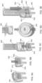
- FIGS. 1A-1D depict components of system 100 for conducting fully automated nested RPA on a sample provided to a microfluidic nucleic acid amplification device.
- FIG. 1A depicts sample module 102, which includes receiver module 104 and transfer module 106.
- FIG. 1B depicts microfluidic nucleic acid amplification device 110.
- sample module 102 and nucleic acid amplification device 108 are coupled to form nucleic acid amplification assembly 110.
- FIG. 1D depicts system 100, including nucleic acid amplification assembly 110 inserted in analyzer 112 for assessment of the presence of a target nucleic acid in a sample provided to nucleic acid amplification device 108 from sample module 102.
- System 100 is used to assess the presence of a target nucleic acid in a sample provided to receiver module 104 of sample module 102.
- Coupling sample module 102 and nucleic acid amplification device 108 creates fluidic pathways between the sample module and the nucleic acid amplification device, allowing delivery of a RPA reaction mixture to the nucleic acid amplification device.
- system 100 is used to assess the presence of two or more target nucleic acids in a sample.
- system 100 is used to assess the presence of influenza A virus and influenza B virus in a sample.
- sample module 102 and nucleic acid amplification device 108 are configured to perform three or more rounds of nested RPA.
- FIGS. 2A-2E depict an alternative workflow for system 100.
- FIG. 2A which nucleic acid amplification device 108 is inserted into analyzer 112.
- sample module 102 is advanced toward nucleic acid amplification device 108 in analyzer.
- Registration features on analyzer 112 interface constrain sample module 102 in two dimensions prior to coupling, allowing the sample module and nucleic acid amplification device 108 to be mated to form passageways that allow fluid to pass from the sample module to the nucleic acid amplification device and vice versa.
- FIG. 2C depicts nucleic acid amplification assembly 110 in analyzer 112.
- Coupling of sample module 102 to nucleic acid amplification device 108 may initiate the flow of reactants from the sample module to the nucleic acid amplification device, thereby initiating assessment of the presence of the target nucleic acid in the sample.
- registration features in analyzer 112 may be engaged to release nucleic acid amplification assembly 110.
- FIG. 2E depicts nucleic acid amplification assembly 110 after release from analyzer 112. Nucleic acid amplification assembly 110 may be disposed of after release from analyzer 112.
- FIGS. 3A and 3B depict perspective views of an embodiment of receiver module 104 of sample module 102.
- FIG. 3A depicts a perspective view of receiver module 104 with chambers 300 for receiving a sample, containing a reagent, or both.
- Receiver module 104 also includes registration features 302 for aligning the receiver module with transfer module 106.
- FIG. 3B depicts a perspective view opposite that of FIG. 3A , which depicts an exterior view of the bottoms 304 of chambers 300.
- FIGS. 4A and 4B depict perspective views of an embodiment of a transfer module 106 configured to mate with receiver module 104.
- FIG. 4A depicts a perspective view of transfer module 106 with chambers 400, each chamber having an inlet port 402 and an outlet port 404. Transfer module 106 also includes registration features 406 for aligning the transfer module with receiver module 104.
- FIG. 4B depicts a perspective view opposite that of FIG. 4A , which depicts an exterior view of the bottoms 408, as well as inlet ports 402 and outlet ports 404 of chambers 400.
- FIGS. 5A-5G depicts a workflow for providing a sample to sample module 500 having a coupled receiver module 502 and transfer module 504.
- sample module 500 may be provided in sealed pouch 506.
- Sealed pouch 506 may be a foil pouch.
- FIG. 5B depicts sample module 500 after removal from pouch 506, with hinge 508 opened to expose hermetic seals 510 and 512 on receiver module 502 and transfer module 504, respectively.
- seal 510 may be removed from receiver module 502 to expose sample chamber 514 and blank chamber 516.
- Sample chamber 514 and blank chamber 516 typically include a liquid medium, such as a buffer solution.
- a sample e.g., a body fluid
- device 518 e.g., a swab
- Blank chamber 516 may be covered with occluding element 520 to prevent insertion of a sample in the blank chamber.
- Gaskets 522 and 524 may be positioned about an exterior of sample chamber 514 and blank chamber 516, respectively, to promote seal formation between receiver module 502 and transfer module 504 after a sample has been deposited in sample chamber 514.
- Registration features 526 on receiver module 502 are configured to mate with corresponding registration features on transfer module 504.
- seal 512 may be removed from transfer module 504 to expose sample chamber 528 and blank chamber 530.
- Retaining elements 532 and 534 may be positioned in sample chamber 528 and blank chamber 530, respectively, to retain a solid reagent in the sample chamber, the blank chamber, or both.
- retaining element 532 retains a reagent pellet in sample chamber 528.
- the reagent pellet may include oligomers for a RPA reaction. In some cases, the pellet is a freeze dried pellet.
- Blank chamber 530 may be free of a solid reagent.
- Retaining elements 532 and 534 typically define openings, such as pores. In some cases, retaining elements 532 and 534 are frits.
- Frits may be selected to facilitate transfer of the fluid from receiver module 502 to transfer module 504.
- retaining elements 532 and 534 are hydrophilic frits.
- Transfer module 504 includes registration features 536 configured to mate with registration features 526 of receiver module 502.
- the transfer module may be rotated about hinge 508 and secured to receiver module 502, with retaining elements 532 and 534 retaining reagents present in sample chamber 528 and blank chamber 530, respectively.
- retaining elements 532 and 534 retaining reagents present in sample chamber 528 and blank chamber 530, respectively.
- FIG. 5G registration features 526 and 536 lockingly engage, gasket 522 seals sample chambers 514 and 528 together, and gasket 524 seals blank chambers 516 and 530 together.
- Registration features 526 and 536 may be configured to irreversibly seal receiver module 502 and transfer module 504 such that sample module 500 cannot be opened unintentionally.
- sample module 500 Prior to coupling sample module 500 to a nucleic acid amplification device, the sample module is inverted to cause movement of the liquid medium in receiver module 502 toward transfer module 504, thereby hydrating solid reagents in the transfer module to form hydrated reaction mixtures.
- freeze dried RPA reagents in the transfer module are hydrated to form a hydrated reaction mixture.
- FIGS. 6A-6H depict an alternative workflow for providing a sample to sample module 600 having a separate receiver module 602 and transfer module 604.
- receiver module 602 and transfer module 604 may each be provided in a separate sealed pouch 606, 606'.
- Sealed pouch 606 may be a foil pouch.
- FIG. 6B depicts transfer module 604 after removal from sealed pouch 606'. Transfer module 604 is sealed with seal 612.
- FIG. 6C depicts receiver module 602 removed from pouch 606. Receiver module 602 is sealed with seal 610. After removal of seal 610 from receiver module 602, as depicted in FIG. 6D , sample chamber 614 and blank chamber 616 are exposed. Sample chamber 614 and blank chamber 616 typically include a liquid medium, such as a buffer solution.
- a sample e.g., a body fluid
- device 618 e.g., a swab
- Blank chamber 616 may be covered with occluding element 620 to prevent insertion of a sample in the blank chamber.
- Gaskets 622 and 624 may be positioned about an exterior of sample chamber 614 and blank chamber 616, respectively, to promote seal formation between receiver module 602 and transfer module 604.
- Registration features 626 on receiver module 602 are configured to mate with corresponding registration features on transfer module 604.
- seal 612 may be removed from transfer module 604.
- Removing seal 612 from transfer module 604 exposes a sample chamber and a blank chamber (not shown).
- Retaining elements may be positioned in the sample chamber and blank chamber, respectively, to retain a solid reagent in the sample chamber, the blank chamber, or both.
- the solid reagent includes oligomers for a RPA reaction.
- the solid reagent is a freeze dried pellet.
- the blank chamber may be free of a solid reagent.
- the retaining elements typically define openings, such as pores.
- the retaining elements are frits. Frits may be selected to facilitate transfer of the fluid from receiver module 602 to transfer module 604.
- the retaining elements are hydrophilic frits.
- Transfer module 602 includes registration features 636 configured to mate with registration features 626 of receiver module 602.
- the transfer module may be inverted to align registration features 626 and 636. During this inversion, retaining elements in transfer module 604 retain reagents present in the sample chamber and blank chamber of the transfer module.
- registration features 626 and 636 lockingly engage, gasket 622 seals the sample chambers of the receiver and transfer modules together, and gasket 624 seals the blank chambers of the receiver and transfer modules together.
- Registration features 626 and 636 may be configured to irreversibly seal receiver module 602 and transfer module 604, as depicted in FIG. 6H , such that sample module 600 cannot be opened unintentionally.
- the sample module Prior to coupling sample module 600 to a nucleic acid amplification device, the sample module may be inverted to cause movement of the liquid medium in receiver module 602 toward transfer module 604, thereby hydrating solid reagents in the transfer module to form hydrated reaction mixtures.
- freeze dried RPA reagents in the transfer module are hydrated to form a hydrated reaction mixture.
- FIG. 7 is a perspective view of sample module 500.
- Transfer module 500 may be packaged with seal 700 covering the portion of the transfer module configured to couple to the nucleic acid amplification device.
- Seal 700 may be a foil seal that provides an opaque surface to cover openings of inlet ports 702 and 704 and outlet ports 706 and 708. Seal 700 may retain the hydrated reaction mixture in sample module 500 upon inversion. In some cases, seal 700 is removed from sample module 500, nucleic acid amplification device is coupled to the sample device, and the sample module 500 is first inverted after it is sealed to the nucleic acid amplification device.
- Inlet ports 702 and 704 and outlet ports 706 and 708 may have tapered ends (e.g., low profile luer connectors) configured to be inserted into a nucleic acid amplification device.
- gaskets 710, 712, 714, and 716 may be positioned on inlet ports 702, 704, 706, and 708, respectively, to form an air-tight seal with a nucleic acid amplification device.
- FIGS. 8A and 8B depict an alternative embodiment of a sample module.
- FIG. 8A is a perspective view of sample module 800 including receiver module 802 and transfer module 804.
- FIG. 8B is a perspective cross-sectional view of sample module 800.
- receiver module 802 defines sample chamber 806 having opening 808.
- Sample chamber 806 holds liquid medium 810.
- Liquid medium 810 may be a buffer solution.
- Receiver module 802 includes inlet port 812 and outlet port 814.
- Receiver module 802 also includes registration feature 816 configured to engage a registration feature of transfer module 804.
- Transfer module 804 includes housing 818 having extension 820 and defining opening 822 configured to accept sample chamber 806 of receiver module 802.
- Ram 824 is positioned in housing, with extension 820 positioned in arm 826 of ram. Arm 826 is positioned within spring 828, and the spring is held in a loaded position with release catch 830.
- Porous element 832 is positioned between ram 824 and opening 822. Porous element 832 contains a solid reagent (e.g., a freeze dried RPA reagent).
- Registration feature 834 is configured to engage with registration feature 816 of receiver module 802, and gasket 836 forms a seal between the receiver module and transfer module 804. As depicted, receiver module 802 is seated in opening 822 of transfer module 804.
- Registration features 816 and 834 lockingly engage to seal receiver module 802 and transfer module 804 via gasket 836. Registration features 816 and 834 may be configured to irreversibly seal receiver module 802 and transfer module 804 such that sample module 800 cannot be opened unintentionally.
- FIGS. 9A-9E depict a workflow for providing a sample to sample module 800.
- seal 900 is removed from receiver module 802.
- a sample is provided to liquid medium 810 in sample chamber 806 of receiver module 802 via opening 808.
- transfer module 804 is advanced toward receiver module 802 to lockingly engage registration features 816 and 834. After receiver module 802 is sealed to transfer module 804 via gasket 836, force may be applied to release catch 830 to release spring-loaded ram 824, as depicted in FIG. 9D .
- FIG. 9E depicts sealed sample module 800 with ram 824 resting in receiver module 802, having forced the solid reagent porous element 832 into liquid medium 810.
- Sealed sample module 800 may be coupled to a nucleic acid amplification device to assess the presence of a target nucleic acid in the sample provided to the receiver module 802.
- FIG. 10 depicts an exploded view of nucleic acid amplification device 1000 for optical detection.
- Nucleic acid amplification device 1000 is a laminated microfluidic device including top layer 1002, intermediate layer 1004, and base layer 1006.
- Base layer 1006 may include more than one component. As depicted, base layer 1006 include two components 1008 and 1010.
- Intermediate layer 1004 includes inlet ports 1012 and 1014 and outlet ports 1016 and 1018, which couple to outlet ports and inlet ports, respectively, of a sample module.
- Intermediate layer 1004 typically includes reagents, such as RPA reagents.
- primary reaction chamber 1020 includes a solid reagent 1022 (e.g., Mg 2+ in the form of magnesium acetate).
- Intermediate layer 1004 includes reagent reservoirs 1024 and 1026, which contain solid reagents 1028 and 1030.
- solid reagent 1028 includes dried (e.g., freeze dried) oligomers and solid reagent 1030 includes Mg 2+ (e.g., in the form of magnesium acetate).
- Secondary reaction chambers 1032 may also function as detection chambers, in which target nucleic acids are detected via optical signals by an analyzer. Secondary reaction chambers 1032 have an optically transparent covering, such that fluorescent signals generated when the fluorophore and quencher are separated via an exonuclease can be detected by optical sensors in the analyzer in which the nucleic acid amplification device is configured to be inserted. Registration features 1036 allow alignment of nucleic acid amplification device 1000 in an analyzer.
- Intermediate layer 1004 may also include flow detection chambers 1034, each having a transparent covering through which the presence of fluid is monitored optically by an analyzer to detect a flow of liquid.
- An analyzer configured to accept nucleic acid amplification device 1000 includes a light source directed toward each flow detection chamber configured. The analyzer is configured to detect (e.g., via light scattering) the presence of liquid in each flow detection chamber.
- Detection of liquid in a flow detection chamber may trigger various operations (e.g., initiation or cessation of pumping), and a controller in the analyzer may be configured to implement various parameters (e.g., pumping time, reaction time, mixing time, flow time) based on detection of a liquid in a flow detection chamber, such that reagents are provided in pre-determined volumes and allowed to react for pre-determined times.
- various parameters e.g., pumping time, reaction time, mixing time, flow time
- Nucleic acid amplification device 1000 may include additional features not depicted in FIG. 10 , such as pumps and microfluidic pathways.
- One or more of the pumps may be a peristaltic pump or a syringe pump.
- the pumps may selectively drive reagents from the sample module and the primary reaction chamber 1020 toward secondary reaction chambers 1032 based on elapsed time or flow of fluid through flow detection chambers detected by optical sensors in an optical analyzer, metering aliquots as needed.
- nucleic acid amplification device 1000 Operation of nucleic acid amplification device 1000 with a sample module is described with respect to FIG. 11 .
- outlet ports of the sample module are coupled to inlet ports 1012 and 1014 of the nucleic acid amplification device, and inlet ports of the sample module are coupled to outlet ports 1016 and 1018 of the nucleic acid amplification device.
- Reagents in the sample module flow into inlet ports 1012 and 1014 of nucleic acid amplification device 1000 via the outlet ports of the sample module, and fluid (e.g., gas, liquid, or both) displaced from the nucleic acid amplification module flows via outlet ports 1016 and 1018 of the nucleic acid amplification module into the inlet ports of the sample module.
- fluid e.g., gas, liquid, or both
- sample and buffer flow from the sample chamber of a receiver module to hydrate RPA reagents (e.g., dried oligomers) in the sample chamber of the transfer module, through the outlet port an into inlet port 1012.
- First pump 1040 advances this primary reaction mixture through first flow detection chamber 1042.
- Primary reaction chamber 1020 includes RPA reagent 1022 (e.g,. Mg 2+ in the form of magnesium acetate) and is coupled to a heater and a mixer.
- the mixer may be present as magnetic mixer 1048.
- first pump 1040 advances the product formed in primary reaction chamber 1020 to third flow detection chamber 1050. From third flow detection chamber 1050, air and a portion of the product of the primary RPA reaction from the primary reaction chamber flow toward the sample module via outlet port 1016.
- An aliquot of the product from primary reaction chamber 1020 is pulled from shunt 1052 by second pump 1054 and flows toward fourth flow detection chamber 1056.
- Third pump 1058 pulls reagents (e.g., buffer) for the secondary RPA reaction from the blank chamber of the transfer module via the outlet port of the transfer module into inlet port 1014 of nucleic acid amplification device 1000 and through fifth flow detection chamber 1060.
- Fourth flow detection chamber 1056 and fifth flow detection chamber 1060 meet in a Y junction 1062, mixing selected amounts of the product from the first RPA reaction with reagents for the secondary RPA reaction.
- This mixture is pumped by second pump 1054 and third pump 1058 through a first series of mixing elements 1064 and a second series of mixing elements 1066.
- each stream flows through first reagent reservoir 1024 with mixing cylinders 1072 configured to mix the reaction mixture with reagent 1028 (e.g., Mg 2+ in the form of magnesium acetate).
- reagent 1028 e.g., Mg 2+ in the form of magnesium acetate.
- second reagent reservoir 1026 containing reagent 1030.
- Reagent 1030 in second reagent reservoirs 1026 may be the same or different.
- at least two of reagents 1030 include different RPA primers for particular targets of interest, such as influenza A virus and influenza B virus.
- third pump 1058 drives the mixtures through mixing elements 1074 and into secondary reaction chambers 1032.
- Secondary amplification occurs in secondary reaction chambers 1032.
- Secondary reaction chambers 1032 may also function as detection chambers.
- nucleic acid amplification device 1000 secondary reaction chambers 1032 have an optically transparent covering, such that fluorescent signals generated when the fluorophore and quencher are separated via an exonuclease can be detected optically in an analyzer in which the nucleic acid amplification device is configured to be inserted, such as the analyzer described with respect to FIGS. 15-18 .
- FIG. 12 depicts an exploded view of nucleic acid amplification device 1200 for electrochemical detection.
- Nucleic acid amplification device 1200 is a laminated microfluidic device including sensor layer 1201, top layer 1202, intermediate layer 1204, and base layer 1006.
- Base layer 1206 may include more than one component. As depicted, base layer 1206 include two components 1208 and 1210.
- Intermediate layer 1204 includes inlet ports 1212 and 1214 and outlet ports 1216 and 1218, which couple to outlet ports and inlet ports, respectively, of a sample module.
- Intermediate layer 1204 typically includes reagents, such as RPA reagents.
- primary reaction chamber 1220 includes a solid reagent 1222 (e.g., Mg 2+ in the form of magnesium acetate).
- a stirrer 1223 may be embedded in solid reagent 1222. In one example, the stirrer is a magnetic puck.
- Secondary reaction chambers 1232 may also function as detection chambers, with openings 1225 in top layer 1202 allowing liquid in the reaction chambers to contact electrodes on an underside of sensor layer 1201.
- Intermediate layer 1204 may also include flow detection chambers 1234, in which the presence of fluid is monitored electrically by electrodes in sensor layer 1201 superimposed over openings in top layer 1202, such that liquid flowing through the flow detection chambers contacts the electrodes.
- Registration features 1236 allow alignment of nucleic acid amplification device 1200 in an analyzer.
- Nucleic acid amplification device 1200 may include additional features not depicted in FIG. 12 , such as pumps and microfluidic pathways.
- One or more of the pumps may be a peristaltic pump or a syringe pump.
- the pumps may selectively drive reagents from the sample module and the primary reaction chamber 1220 toward secondary reaction chambers 1232 based on elapsed time or flow of fluid through flow detection chambers detected by sensors in an electrical analyzer, metering aliquots as needed.
- nucleic acid amplification device 1200 Operation of nucleic acid amplification device 1200 with a sample module is described with respect to FIG. 13 , which depicts a top, see-through view of intermediate layer.
- outlet ports of the sample module are coupled to inlet ports 1212 and 1214 of the nucleic acid amplification device, and inlet ports of the sample module are coupled to outlet ports 1216 and 1218 of the nucleic acid amplification device.
- Reagents in the sample module flow into inlet ports 1212 and 1214 of nucleic acid amplification device 1200 via the outlet ports of the sample module, and fluid (e.g., gas, liquid, or both) displaced from the nucleic acid amplification module flows via outlet ports 1216 and 1218 of the nucleic acid amplification module into the inlet ports of the sample module.
- fluid e.g., gas, liquid, or both
- the sample and buffer flow from the sample chamber of a receiver module to hydrate RPA reagents (e.g., dried oligomers) in the sample chamber of the transfer module, through the outlet port an into inlet port 1212.
- First pump 1240 advances this primary reaction mixture through first flow detection chamber 1242 into the first pump, through mixing chamber 1244, to second flow detection chamber 1246, and into primary reaction chamber 1220.
- Primary reaction chamber 1220 includes RPA reagent 1222 (e.g., Mg 2+ in the form of magnesium acetate) and is coupled to a heater and a mixer. The mixer may be present as magnetic mixer 1248.
- first pump 1240 advances the product formed in primary reaction chamber 1220 to third flow detection chamber 1250. From third flow detection chamber 1250, air and a portion of the product of the primary RPA reaction from the primary reaction chamber flow toward the sample module via outlet port 1216.
- An aliquot of the product from primary reaction chamber 1220 is pulled from shunt 1252 by second pump 1254 and flows toward fourth flow detection chamber 1256.
- Third pump 1258 pulls reagents (e.g., buffer) for the secondary RPA reaction from the blank chamber of the transfer module via the outlet port of the transfer module into inlet port 1214 of nucleic acid amplification device 1200 and through fifth flow detection chamber 1260.
- Fourth flow detection chamber 1256 and fifth flow detection chamber 1260 meet in a Y junction 1262, mixing selected amounts of the product from the first RPA reaction with reagents for the secondary RPA reaction.
- This mixture is pumped by second pump 1254 and third pump 1258 through a first series of mixing elements 1264 and a second series of mixing elements 1266.
- each stream flows through first reagent reservoir 1224 with mixing cylinders 1272 configured to mix the reaction mixture with reagent 1228 (e.g., Mg 2+ in the form of magnesium acetate).
- reagent 1228 e.g., Mg 2+ in the form of magnesium acetate.
- second reagent reservoir 1226 containing reagent 1230.
- Reagent 1230 in second reagent reservoirs 1226 may be the same or different.
- at least two of reagents 1230 include different RPA primers for particular targets of interest, such as influenza A virus and influenza B virus.
- third pump 1258 drives the mixtures through mixing elements 1274 and into secondary reaction chambers 1232.
- Secondary amplification occurs in secondary reaction chambers 1232.
- Secondary reaction chambers 1232 may also function as detection chambers.
- liquid in secondary reaction chambers 1232 contacts electrodes on an underside of sensor layer 1201, such that electrons resulting from the oxidation of a redox active compound, such as described in U.S. Serial No. 62/300,242 , that has been cleaved from an RPA probe that is labelled with the redox active compound, are detected by the analyzer in which the nucleic acid amplification device is configured to be inserted.
- FIG. 14 depicts a top, see-through view of nucleic acid amplification device 1200 with electrodes in sensor layer 1201 superimposed over openings in top layer 1202 and intermediate layer 1204. Electrodes are positioned on an underside of sensor layer 1201 to contact liquid in flow sensor detectors 1246, 1250, 1256, and 1260 and reaction chambers 1232.
- sensing electrodes, as well as conductive tracks, which electrically couple the sensing electrodes to terminals which electrically communicate with an analyzer may be formed by disposing a first conductive layer on sensor layer. In another example, the first conductive layer may be disposed over a second conductive layer on the sensor layer.
- the electrodes may be electrically isolated by masking the conductive layers and disposing a dielectric layer over the exposed regions.
- the first conductive material includes carbon.
- the second conductive material includes silver.
- "disposing" includes printing methods, such as screen printing. When a silver layer is deposited beneath the carbon layer, the resulting conductive track typically has a lower resistance when compared with a conductive track formed using carbon alone. In both examples electrochemical measurements are performed on a carbon surface.
- Flow sensor detectors 1246 and 1250 are each electrically coupled to two liquid sense electrodes.
- liquid sense electrodes 1400 and 1402 are electrically coupled to wirings 1404 and 1406, which are electrically coupled to connections 1408 and 1410, respectively.
- Flow sensor detectors 1256 and 1260 are each electrically coupled to four liquid sense electrodes.
- liquid sense electrodes 1412 and 1414 are electrically coupled to wirings 1420 and 1422, which are electrically coupled to connections 1428 and 1430, respectively, and electrodes 1416 and 1418 are electrically coupled to wirings 1424 and 1426, which are electrically coupled to connections 1432 and 1434, respectively.
- Each detection chamber 1232 is coupled to three measurement electrodes, including reference electrode 1436, working electrode 1438, and counter electrode 1440, and each electrode is electrically coupled to connections via wirings.
- the wirings may be conductive traces including a conductive material (e.g., silver).
- the connections are configured to engage terminals in an analyzer.
- the liquid sense electrodes operate on principle of conductivity. That is, a voltage is applied across the terminals, and when fluid contacts the sense electrodes within the respective chambers, current passes through the liquid, and the analyzer detects a flow of current.
- a potential is applied between the counter electrode and the working electrode; the reference electrode acts to ensure the applied potential is as expected.
- a current flows proportional to the concentration of electroactive species in contact with the working electrode (effectively electrons received or donated depending on whether oxidation or reduction of the target species occurs at the specified potential).
- the potential is swept from one voltage to another and the resulting current recorded giving rise to peaks and or troughs as a result of the oxidation or reduction of the electroactive species.
- FIG. 15 depicts nucleic acid amplification system 1500, including nucleic acid amplification device 1502 inserted in analyzer 1504.
- Nucleic acid amplification device 1502 and analyzer may be configured for optical or electrochemical detection of RPA products.
- insertion of nucleic acid amplification device in analyzer initiates assessment of the presence of target nucleic acid in a sample provided to the nucleic acid amplification device.
- subsequent coupling of a sample module to nucleic acid amplification device initiates assessment of the presence of target nucleic acid in the sample.
- assessment of the presence of target nucleic acid in a sample provided to the nucleic acid amplification device is initiated by the user after insertion of the nucleic acid amplification device or assembly into the analyzer.
- nucleic acid amplification device 1502 and analyzer 1504 are configured for optical detection of RPA products.
- analyzer 1504 is configured to detect fluorescence from fluorescent probes coupled to RPA products in the detection chambers of nucleic acid amplification device 1502.
- Analyzer 1504 includes light sources, excitation light guides 1600 corresponding to each light source, emission light guides 1602 corresponding to each emission light guide, and a photodetector.
- the light sources are typically light emitting diodes (LEDs) selected to achieve a good match between the LED emission peak and absorption of the target fluorescent label.
- Analyzer 1504 incorporates a skew geometry to allow fluorescence measurement from multiple reaction cells using a single optical emission filter.
- FIG. 17 depicts an enlarged view of a portion of FIG. 16 .
- analyzer 1504 includes four light sources to allow fluorescence measurement from four detection chambers 1700 in nucleic acid amplification device 1502.
- Excitation light guides 1600 direct light from the light sources to detection chambers 1700
- emission light guides 1602 direct the fluorescent emission from the detection chambers to a common photodiode via the optical filter. Discrimination is provided between the four measurement channels by time division multiplexing of the four light sources.
- Each excitation light guide 1600 is configured to direct incident light from one of the light sources to a target in a plane such that the angle between the incident light and the plane is in a range between 30° and 60° (e.g., 40°), and each emission light guide 1602 is configured to direct emitted light from the target to the photodetector such that an angle between the emitted light is between 40° and 60° (e.g., 30°).
- Analyzer 1504 typically includes a first lens and a second lens corresponding to each light source, with each corresponding excitation light guide configured to collimate light transmitted through the first lens from its corresponding light source and, via total internal reflection, direct the collimated light at an angle toward the second lens.
- FIGS. 18A-18C depict skew geometry of analyzer 1504. Angles depicted in FIGS. 18A-18C are exemplary and are selected for ease of explanation, however, these angles may be altered in embodiments of analyzer 1500. As depicted in FIG. 18A , the rotation axes are oriented at 45° to a line connecting the centers of reaction chambers 1700. This configuration facilitates avoiding positional clashes between the excitation light guides and the emission light guides. FIG. 18B depicts the excitation optical axis at 30° to the detection chamber surface normal (rotated in the x plane about the y axis). FIG.
- 18C depicts the emission optical axis at 40° to the detection chamber surface normal (about an orthogonal axis of rotation, i.e., rotated in the yz plane about the x axis).
- the center of rotation is below the normal liquid surface position (e.g., from 0.1 to 1 mm below).
- Other combinations of skew angles are listed in Table 1 below.
- a maximum bend angle in the excitation light guides and the emission light guides is typically 45° or less. Table 1.
- Skew angles of excitation and emission light guides Emission light guide angle (°)
- Excitation light guide angle (°) 20 45 30 40 34 34 40 30 45 20
- Analyzer 1504 includes a controller operatively coupled to the light sources and the photodetector. The controller initiates production of incident light by the light sources and initiates collection of emitted light from the detection chambers.
- Analyzer 1504 typically includes a single photodetector and a single emission filter operatively positioned between the emission light guides and the photodetector: however, in some embodiments, one or more additional photodetectors, emission filters, or both may be present.
- RPA Recombinase Polymerase Amplification
- NEAR Nicking and Extension Amplification Reaction
- RPA employs enzymes, known as recombinases, which are capable of pairing oligonucleotide primers with homologous sequences in template double-stranded nucleic acid.
- RPA introduces a recombinase for inserting two primers with a template in duplex DNA, a single stranded DNA-binding protein for stabilizing the displaced strands of DNA and for preventing the primers from being displaced, and strand-displacing polymerase for extending primers bound to template DNA. In this way, DNA synthesis is directed to defined points in a template double-stranded nucleic acid.
- an exponential amplification reaction is initiated if the template nucleic acid is present.
- the reaction progresses rapidly and results in specific amplification of a sequence present within the template double-stranded nucleic acid from just a few copies of the template nucleic acid to detectable levels of the amplified products within minutes.
- RPA processes proceed under isothermal conditions under physiological temperatures (e.g., 37-42°C).
- RPA methods are disclosed, e.g., in US 7,270,981 ; US 7,399,590 ; US 7,666,598 ; US 7,435,561 ; US 2009/0029421 ; and WO 2010/141940 , all of which are incorporated herein by reference.
- RPA incorporates components of the cellular DNA replication and repair machinery, and establishes a 'dynamic' recombination environment having adequate rates of both recombinase loading and unloading that maintain high levels of recombination activity achieved in the presence of specific crowding agents.
- RPA has the advantage that it combines the sensitivity, specificity, and most other features of PCR, but without the need for thermocycling and with extraordinary speed and robustness to off-temperature set-up.
- RPA benefits from the potential employment of a wide variety of nucleic acid processing enzymes such as known repair endonucleases which have been untapped by other processes due at least in part to the need for thermostable equivalents, poor regulation without accessory proteins such as single-stranded DNA binding proteins, or a combination thereof.
- RPA includes the following steps: first, a recombinase agent is contacted with a first and a second nucleic acid primer to form a first and a second nucleoprotein primer. Second, the first and second nucleoprotein primers are contacted with a double stranded target sequence to form a first double stranded structure at a first portion of the first strand and form a double stranded structure at a second portion of the second strand, so the 3' ends of said first nucleic acid primer and said second nucleic acid primer are oriented towards each other on a given template DNA molecule.
- first and second nucleoprotein primers are extended by DNA polymerases to generate first and second double stranded nucleic acids, and first and second displaced strands of nucleic acid.
- the second and third steps are repeated until a desired degree of amplification is reached.
- This disclosure also provides for a method of performing nested RPA within a microfluidic cartridge or device.
- a nested RPA a first region of nucleic acid is amplified by RPA to form a first amplified region.
- a second region of nucleic acid that is completely within the first amplified region is amplified using RPA to form a second amplified region.
- This process may be repeated multiple times.
- a third region of nucleic acid, which is completely within the second region may be amplified from the second amplified region by RPA.
- the RPA reagents disclosed herein can contain a set of primers that amplify the target nucleic acid sequence.
- the primers can include sequences that are complementary to the target nucleic acid sequence or that differ from the target nucleic acid sequence at one or more positions.
- the amplification product of RPA with a primer that differs from the target nucleic acid sequence at one or more positions can differ from the target sequence at the one or more positions.
- the amplification product of the RPA reaction described herein can include a target cleavage sequence.
- the set of RPA primers can amplify the target nucleic acid sequence or introduce a sequence that differs from the target nucleic acid sequence at one or more positions. This introduced sequence can consist of a target cleavage sequence.
- the first primer can be complementary to the target nucleic acid sequence.
- the second primer can include a first portion that is complementary to the target nucleic acid sequence and a second portion that is different from the target nucleic acid sequence at one or more positions. When the two primers amplify the nucleic acid sequence the second primer incorporates the one or more different positions into the amplified products. This amplified region is different from the target nucleic acid sequence at the one or more positions and can consist of the target cleavage sequence.
- the RPA composition disclosed herein contains a recombinase, which may originate from prokaryotic, viral or eukaryotic origin.
- exemplary recombinases include RecA and UvsX (e.g., a RecA protein or UvsX protein obtained from any species), and fragments or mutants thereof, and combinations thereof.
- the RecA and UvsX proteins can be obtained from any species.
- RecA and UvsX fragments or mutant proteins can also be produced using the available RecA and UvsS protein and nucleic acids sequences, and molecular biology techniques (see, e.g., the mutant forms of UvsX described in U.S. Patent No. 8,071,308 ).
- Exemplary UvsX proteins include those derived from myoviridae phages, such as T4, T2, T6, Rb69, Aehl, KVP40, Acinetobacter phage 133, Aeromonas phage 65, cyanophage P-SSM2, cyanophage PSSM4, cyanophage S-PM2, Rb14, Rb32, Aeromonas phage 25, Vibrio phage nt-1, phi-1, Rb16, Rb43, Phage 31, phage 44RR2.8t, Rb49, phage Rb3, and phage LZ2.
- myoviridae phages such as T4, T2, T6, Rb69, Aehl, KVP40, Acinetobacter phage 133, Aeromonas phage 65, cyanophage P-SSM2, cyanophage PSSM4, cyanophage S-PM2, Rb14, Rb32, Aeromonas phage 25, Vibrio phage nt-1
- Additional exemplary recombinase proteins include archaebacterial RADA and RADB proteins and eukaryotic (e.g., plant, mammal, and fungal) Rad51 proteins (e.g., RAD51, RAD51B, RAD51C, RAD51D, DMC1, XRCC2, XRCC3, and recA) (see, e.g., Lin et al., Proc. Natl. Acad. Sci. U.S.A. 103:10328-10333, 2006 ).
- the recombinase may be a mutant or hybrid recombinase.
- the mutant UvsX is an Rb69 UvsX that includes at least one mutation in the Rb69 UvsX amino acid sequence, wherein the mutation is selected from the group consisting of (a) an amino acid which is not histidine at position 64, a serine at position 64, the addition of one or more glutamic acid residues at the C-terminus, the addition of one or more aspartic acid residues at the C-terminus, and a combination thereof.
- the mutant UvsX is a T6 UvsX having at least one mutation in the T6 UvsX amino acid sequence, wherein the mutation is selected from the group consisting of: (a) an amino acid which is not histidine at position 66; (b) a serine at position 66; (c) the addition of one or more glutamic acid residues at the C-terminus; (d) the addition of one or more aspartic acid residues at the C-terminus; and (e) a combination thereof.
- the hybrid protein may, for example, be an UvsX protein that includes at least one region that includes an amino acid sequence derived from a different UvsX species. The region may be, for example, the DNA-binding loop-2 region of UvsX.
- the DNA polymerase disclosed herein may be a eukaryotic or prokaryotic polymerase.
- eukaryotic polymerases include pol-alpha, pol-beta, pol-delta, pol-epsilon, and mutants or fragments thereof, or combinations thereof.
- prokaryotic polymerase include E. coli DNA polymerase I (e.g., Klenow fragment), bacteriophage T4 gp43 DNA polymerase, Bacillus stearothermophilus polymerase I large fragment, Phi-29 DNA polymerase, T7 DNA polymerase, Bacillus subtilis Pol I, Staphylococcus aureus Pol I, E. coli DNA polymerase I, E.
- the DNA polymerase lacks 3'-5' exonuclease activity.
- the DNA polymerase has strand-displacing properties, e.g., large fragments of prokaryotic polymerases of class pol I or pol V.
- one or more single-stranded DNA binding proteins can be used to stabilize nucleic acids during the various exchange reactions that are ongoing in the reaction.
- the one or more single-stranded DNA binding proteins can be derived or obtained from any species, e.g., from a prokaryotic, viral or eukaryotic species.
- Non-limiting exemplary single-stranded DNA binding proteins include E.
- coli SSB and those derived from myoviridae phages, such as T4, T2, T6, Rb69, Aehl, KVP40, Acinetobacter phage 133, Aeromonas phage 65, cyanophage P-SSM2, cyanophage PSSM4, cyanophage S-PM2, Rb14, Rb32, Aeromonas phage 25, Vibrio phage nt-1, phi-1, Rb16, Rb43, Phage 31, phage 44RR2.8t, Rb49, phage Rb3, and phage LZ2. Additional examples of single-stranded DNA binding proteins include A.
- the crowding agent may include one or more of polyethylene glycol, polyethylene oxide, polyvinyl alcohol, polystyrene, Ficoll, dextran, poly(vinylpyrrolidone) (PVP), Triton-X, and albumin.
- the crowding agent has a molecular weight of less than 200,000 daltons.
- the composition comprises a crowding agent selected from the group consisting of polyethylene glycol (PEG)(e.g., PEG1450, PEG3000, PEG8000, PEG10000, PEG14000, PEG15000, PEG20000, PEG250000, PEG30000, PEG35000, PEG40000, PEG compound with molecular weight between 15,000 and 20,000 daltons, or combinations thereof), dextran, polyvinyl alcohol, polyvinyl pyrrolidone, Triton-X, and Ficoll.
- PEG polyethylene glycol
- the crowding agent is present in the reaction mixture at a concentration between 1 to 15% by weight or by volume of the reaction mixture, e.g., between any two concentration values selected from 1.0%, 1.5%, 2.0%, 2.5%, 3.0%, 3.5%, 4.0%, 4.5%, 5.0%, 5.5%, 6.0%, 6.5%, 7.0%, 7.5%, 8.0%, 8.5%, 9.0%, 9.5%, 10.0%, 10.5%, 11.0%, 11.5%, 12.0%, 12.5%, 13.0%, 13.5%, 14.0% 14.5% and 15.0%.
- the recombinase loading protein may be of prokaryotic, viral or eukaryotic origin.
- Exemplary recombinase loading proteins include E . coli RecO, E. coli RecR, UvsY, and mutants or fragments thereof, or combinations thereof.
- Exemplary UvsY proteins include those derived from myoviridae phages, such as T4, T2, T6, Rb69, Aehl, KVP40, Acinetobacter phage 133, Aeromonas phage 65, cyanophage P-SSM2, cyanophage PSSM4, cyanophage S-PM2, Rb14, Rb32, Aeromonas phage 25, Vibrio phage nt-1, phi-1, Rb16, Rb43, Phage 31, phage 44RR2.8t, Rb49, phage Rb3, and phage LZ2.
- the recombinase loading agent may be derived from a myoviridae phage.
- the myoviridae phage may be, for example, T4, T2, T6, Rb69, Aeh1, KVP40, Acinetobacter phage 133, Aeromonas phage 65, cyanophage P-SSM2, cyanophage PSSM4, cyanophage S-PM2, Rb14, Rb32, Aeromonas phage 25, Vibrio phage nt-1, phi-1, Rb16, Rb43, Phage 31, phage 44RR2.8t, Rb49, phage Rb3, or phage LZ2.
- Amplification methods suitable for use in the present methods include amplification methods performed without subjecting the polynucleotides to a temperature sufficient to denature double stranded polynucleotides during the amplification.
- the amplification of the polynucleotides may be performed without subjecting the polynucleotides to a temperature in excess of about 90° C, about 80° C, about 70° C, or about 60° C during amplification.
- the amplification of the polynucleotides is performed without subjecting the polynucleotides to conditions sufficient to denature double stranded polynucleotides during the amplification.
- the amplification may be performed without subjecting the polynucleotides to physical, chemical, or thermal conditions sufficient to denature double stranded polynucleotides during amplification.
- Amplification methods suitable for use in the present methods include amplification methods performed without first subjecting the polynucleotides to a temperature sufficient to denature double stranded polynucleotides present in the sample.
- the amplification of the polynucleotides may be performed without first subjecting the polynucleotides to a temperature in excess of about 90° C, about 80° C, about 70° C, about 60° C, or about 55° C.
- the polynucleotides and/or amplicons thereof are detected without first subjecting the polynucleotides to such excess temperatures.
- the amplification of the polynucleotides is performed without first subjecting the polynucleotides to conditions sufficient to denature double stranded polynucleotides present in the sample.
- the amplification may be performed without first subjecting the polynucleotides to physical, chemical, or thermal conditions sufficient to denature double stranded polynucleotides present in the sample.
- Amplification methods suitable for use in the present methods include amplification methods performed in a total time (T) beginning with a step of combining the polynucleotides with reagents sufficient to perform the amplification and ending when amplification has proceeded by an amount sufficient to permit the qualitative or quantitative determination of the polynucleotides or amplicons thereof.
- the total time T may be about 45 minutes or less, about 30 minutes or less, about 20 minutes or less, or about 15 minutes or less.
- the amplification of the polynucleotides includes, for example, amplifying the polynucleotides by at least about 10 6 fold, at least about 10 7 fold, at least about 10 8 fold, at least about 10 9 fold, at least about 10 10 fold, at least about 10 11 fold, or at least about 10 12 fold. Such amplification may be performed within the time T.
- Amplification methods suitable for use in the present methods include "real time” or “quantitative” polynucleotide amplification methods known to the skilled artisan. Such methods detect the accumulation of polynucleotide amplification product after each amplification cycle in real time as the reaction progresses, allowing for the determination of amplification kinetics. Real time methods are quantitative because the time (e.g., number of cycles) to reach a specific threshold concentration of amplified products directly relates to the initial copy number of the target nucleotide. According to some embodiments, the amplification reaction is monitored by electrochemical detection using the oligonucleotide probes described herein.
- FIGS. 19A-19D represent results of analyses conducted using the method of performing nested RPA on a microfluidic card as described herein.
- the results demonstrate the capability of the nested RPA assay to distinguish between samples with different known targets.
- Samples were obtained from a commercial supplier of influenza positive and influenza negative sample materials. A series of measurements were performed using a total of ninety samples: thirty samples were known positive for influenza A (Inf A), ten samples were known positive for influenza B (Inf B) and fifty samples were known to be free of influenza A or B (negative). Each sample was applied to a single assay device, and a measurement was obtained from each of the four detection chambers on the assay card.
- Detection of RPA reagents with labelled probes is previously described and typically involves at least one probe with a detectable label for detection of amplified target, if present.
- a probe may include a fluor and a quencher, which are separated following cleavage by a nuclease when the probe hybridizes to a complementary polynucleotide sequence, if present in amplification reaction products. Similar results (not shown) were obtained with the ninety samples with respect to identification of whether a sample was influenza A positive, influenza B positive or a negative control, when the samples were tested using electrochemically labelled probes.
- Tables 2A-2D The combined results of both fluorescent and electrochemical probe measurements are depicted in Tables 2A-2D.
- Each Table includes comparative measurements made using a standard commercial qPCR assay that was performed by the supplier of the influenza sample materials. qPCR was performed at the point of sample acquisition by the commercial supplier of sample material, the results of which were used to classify the samples as positive or negative, as well as fluA or fluB; classified samples were stored suitably and were supplied in viral transport medium (VTM). There was no apparent influence of the VTM on the performance of RPA.
- VTM viral transport medium
- the RPA assay was developed to incorporate two different primer and probe sets that were directed to different nucleotide regions to maximise the likelihood of identifying samples positive for influenza A.
- the primers and probes used in the RPA assay as described herein are listed herein below as SEQ ID NOS. 1 to 21.
- the primary primer sequences were used to contact the whole sample in the first reaction chamber in order to perform primary amplification.
- the product of primary amplification was then used to contact the secondary primers and probes within the individual secondary reaction chambers to specifically amplify the respective target species using InfA PA, InfA PB2, InfB PA and IC to generate a signal whenever fluA or fluB is present in a patient sample.
- probes were used for making fluorescence measurements, these were designed to be cleaved by the nuclease Exonuclease III (Exo); probes used for electrochemistry were designed for use with the nuclease 8-oxoguanine DNA glycosylase (fpg). Examples of suitable electrochemical probes are described in co-pending application PCT/US2017/019446, filed 24 February 2017 and incorporated herein by reference in its entirety.
- FIG. 19A represents the results obtained when the 90 samples were exposed to the InfA PA RPA primers and probes. The results indicate that the InfA PA primers detected 24 out of 30 InfA positive samples; none of the InfB or negative samples showed a response with the InfA PA primers.
- FIG. 19B shows that all of the InfA samples provided a positive response when exposed to InfA PB2 primers and probes; neither InfB nor negative samples showed any response with the InfA PB2 primers and probes.
- FIG. 19C shows that all of the InfB samples provided a positive response when exposed to the InfB PA primers and probes; neither the InfA nor negative control samples gave any response.
- FIG. 19D indicates when negative control primers (Xcon) and probes were used none of the samples showed a positive signal.
- the dashed line represents a baseline threshold, which was determined as three standard deviations above the maximum negative result.
- the data show that the combined use of RPA primers and probes against InfA PA and InfA PB2 regions resulted in a 100% identification of influenza A positive samples, demonstrating the ability of the assay format described herein to successfully determine the presence of influrenza viruses with differing nucleotide sequences.
Landscapes
- Chemical & Material Sciences (AREA)
- Life Sciences & Earth Sciences (AREA)
- Organic Chemistry (AREA)
- Health & Medical Sciences (AREA)
- Proteomics, Peptides & Aminoacids (AREA)
- Zoology (AREA)
- Wood Science & Technology (AREA)
- Engineering & Computer Science (AREA)
- Analytical Chemistry (AREA)
- Immunology (AREA)
- General Health & Medical Sciences (AREA)
- Biochemistry (AREA)
- Physics & Mathematics (AREA)
- Molecular Biology (AREA)
- Biophysics (AREA)
- Microbiology (AREA)
- Genetics & Genomics (AREA)
- General Engineering & Computer Science (AREA)
- Bioinformatics & Cheminformatics (AREA)
- Biotechnology (AREA)
- Chemical Kinetics & Catalysis (AREA)
- Virology (AREA)
- General Physics & Mathematics (AREA)
- Electrochemistry (AREA)
- Pathology (AREA)
- Clinical Laboratory Science (AREA)
- Dispersion Chemistry (AREA)
- Hematology (AREA)
- Measuring Or Testing Involving Enzymes Or Micro-Organisms (AREA)
- Apparatus Associated With Microorganisms And Enzymes (AREA)
- Enzymes And Modification Thereof (AREA)
- Automatic Analysis And Handling Materials Therefor (AREA)
Abstract
Description
- This application claims the benefit of
U.S. Patent Application Serial No. 62/303,934 entitled "AUTOMATED NESTED RECOMBINASE POLYMERASE AMPLIFICATION" filed March 4, 2016 - This invention was made with government support under HHSO100201400011C awarded by the U.S. Department of Health and Human Services. The government has certain rights in the invention.
- This invention relates to a flu assay system, and more particularly to a system including a sample module, a microfluidic nucleic acid amplification device, and an analyzer to facilitate fully automated nested recombinase polymerase amplification (RPA) on a sample delivered to the nucleic acid amplification device via the sample module.
- Detection of trace levels of polynucleotide sequences can play a significant role in the detection of pathogens and genetic disease and with helping to tailor treatment regimens to particular infections or genotypes. Certain isothermal nucleic acid amplification methods are able to amplify target polynucleotide sequences from trace levels to very high and detectable levels within a matter of minutes. Such isothermal methods, e.g., Recombinase Polymerase Amplification (RPA) or Nicking and Extension Amplification Reaction (NEAR), can allow users to detect a particular sequence in trace amounts, facilitating point-of-care testing and increasing the accessibility and speed of diagnostics.
- Nucleic acid amplification devices disclosed herein are constructed to include an array of microfluidic channels that interconnect primary and secondary reaction chambers to detection chambers. Integrated pump modules are also provided to permit selective movement of liquid through the device at appropriate times. A primary reaction chamber is provided, in which a first round of RPA occurs, which results in amplification of a target polynucleotide sequence of interest. Following the first round of RPA, sample liquid is combined with specific RPA primers and moved to a secondary reaction chamber. During secondary amplification, a sequence completely contained within the primary reaction product is amplified to form secondary reaction products; following which detection of the secondary reaction products is performed. Detection may be achieved using optical or electrochemical means.
- A product mixture from a first round of RPA may be separated into a plurality of streams and passed through reagent reservoirs, in which the product mixture is combined with the same or different RPA primers, before entering a plurality of secondary reaction chambers. In this manner, a nucleic acid amplification device may be used to detect more than one target of interest (e.g., influenza A virus and influenza B virus). In some cases, one of the secondary reaction chambers may be used as a control.
- A first general aspect includes providing a sample to a microfluidic device, and amplifying a target polynucleotide sequence in the sample. Amplifying the target polynucleotide sequence includes performing a first round of amplification on the sample to yield a first amplification product, and performing a second round of amplification on the first amplification product to yield a second amplification product. The second amplification product includes a smaller sequence completely contained within the first amplification product produced during the first round of amplification.
- Implementations of the first general aspect may include one or more of the following features.
- Some implementations include detecting the second amplification product.
- In some embodiments, detecting the second amplification product may include labeling the second amplification product with a first oligonucleotide linked to a fluorophore and a quencher to yield a labeled second product, cleaving the quencher from the labeled second amplification product, and optically detecting a signal from the fluorophore, wherein a detectable signal is indicative of the presence of the second amplification product. Cleaving the quencher may be performed using a nuclease. The nuclease may target double-stranded DNA. In some cases, the nuclease is formamidopyrimine-DNA glycosylase.
- In some embodiments, detecting the second amplification product includes labeling the second amplification product with a first oligonucleotide linked to a redox moiety to yield a labeled second amplification product, cleaving the redox moiety from the labeled second amplification product, and electrochemically detecting a signal from the cleaved redox moiety, wherein a detectable signal is indicative of the presence of the second amplification product. The redox moiety is typically selected from the group consisting of phenothiazine, a phenoxazine, a ferrocene, ferricyanide, ruthenium (III), osmium (II), an anthraquinone, a phenazine, and derivatives thereof. Cleaving the redox moiety may be performed using a nuclease. The nuclease may target double-stranded DNA. In some cases, the nuclease is formamidopyrimine-DNA glycosylase.
- Some implementations include performing a third round of amplification on the second amplification product to yield a third amplification product, and detecting the third amplification product, wherein the third amplification product includes a smaller sequence completely contained within the second amplification product produced during the second round of amplification.
- The sample may be obtained from an animal. For instance, the sample may be obtained from the blood, sputum, mucus, saliva, tears, or urine of the animal. In some cases, the sample is obtained from a human.
- A target nucleic acid may include the target polynucleotide sequence. In some embodiments, the target nucleic acid is obtained from an animal pathogen. The animal pathogen may be a single-stranded DNA virus, double-stranded DNA virus, or single-stranded RNA virus. The animal pathogen may be a bacterium. The target nucleic acid may be double-stranded DNA, single-stranded DNA, or RNA. In some cases, the target nucleic acid is selected from the group consisting of genomic DNA, plasmid DNA, viral DNA, mitochondrial DNA, cDNA, synthetic double-stranded DNA and synthetic single-stranded DNA. The target nucleic acid may be viral DNA or viral RNA. In certain cases, the animal pathogen is an influenza A virus or an influenza B virus.
- In some implementations, two or more target polynucleotide sequences in the sample are amplified. In one example, a target polynucleotide sequence including an influenza A gene sequence and a target polynucleotide sequence including an influenza B gene sequence are amplified.
- In some implementations, two or more second amplification products are detected. In certain implementations, a second amplification product including an influenza A gene sequence and a second amplification product including an influenza B gene sequence are detected.
- In a second general aspect, a diagnostic card includes a card body. The card body includes a primary reaction chamber, one or more secondary reaction chambers, a passage for supplying the sample fluid to the primary reaction chamber, one or more detection chambers in fluidic connection with the one or more secondary reaction chambers, and a detection module associated with each detection chamber. The primary reaction chamber is configured to carry out a first nucleic acid amplification on a sample fluid in the reaction chamber to form a first amplification product. Each secondary reaction chamber is configured to carry out a second nucleic amplification on the first amplification product to form second amplification products
- Implementations of the second general aspect may include one or more of the following features.
- In some embodiments, the detection module is an optical module, such as a fluorescence detector. The fluorescence detector may include a single light pipe to direct illumination light to the one or more detection chambers, and discrete light pipes to receive reflected light from each detection chamber.
- In some embodiments, the detection module is an electrode module. The detection module may include a series of conductive tracks terminating in electrodes for each detection chamber. The device may include additional conductive tracks and electrodes to detect position of liquid throughout the microfluidic card.
- In some implementations, the amplification includes a recombinase polymerase amplification (RPA) reaction.
- In some implementations, the diagnostic card includes mixing means, pumps, and connection ports for connecting to a sample module. The primary reaction chamber may be coupled to a heater. The primary reaction chamber may include a mixing means or be coupled to a mixing means. In some cases, the primary reaction chamber includes a reagent. The reagent may include a RPA reagent. The RPA reagent may be freeze dried.
- In some implementations, each secondary reaction chamber includes a reagent. The reagent may include a RPA reagent. The RPA regent may be freeze dried.
- In some implementations, the sample fluid is a sample obtained from an animal. The sample may be obtained from the blood, sputum, mucus, saliva, tears, or urine of the animal. In some cases, the sample fluid is a sample obtained from a human. The sample fluid may include a target nucleic acid. The target nucleic acid may be obtained from an animal pathogen. The animal pathogen may be a single-stranded DNA virus, double-stranded DNA virus, or single-stranded RNA virus. In some cases, the animal pathogen is a bacterium. The target nucleic acid may be double-stranded DNA, single-stranded DNA, or RNA. In certain cases, the target nucleic acid is selected from the group consisting of genomic DNA, plasmid DNA, viral DNA, mitochondrial DNA, cDNA, synthetic double-stranded DNA and synthetic single-stranded DNA. The target nucleic acid may be viral DNA or viral RNA. The animal pathogen may be influenza A virus or influenza B virus.
- In some implementations, the second amplification products are produced 30 minutes or less, 15 minutes or less, 10 minutes or less, or 5 minutes or less after delivery of the sample fluid to the diagnostic card. The diagnostic card is typically disposable.
- In some implementations, the diagnostic card includes additional reaction chambers, each configured to carry out an additional round of nucleic acid amplification reactions to form additional amplified products, such that the amplification product from each successive n +1 round of amplification is a smaller sequence completely contained within the amplification product of the prior nth round.
- A third general aspect includes a reader configured to receive the diagnostic card of the second general aspect. The reader includes a detector configured to detect the presence of the second amplified products in the secondary reaction chambers.
- A fourth general aspect includes a nucleic acid amplification device. The nucleic acid amplification device includes a first reaction chamber fluidically coupled to a first inlet port and a first outlet port, second reaction chambers fluidically coupled to a second inlet port and a second outlet port, detection chambers, a first pump, a second pump, and a third pump. The first inlet port is fluidically coupled to the first reaction chamber via a first pump, and the first outlet port is fluidically coupled to the first reaction chamber. The first reaction chamber is fluidically coupled to the second reaction chambers via the second pump, and the second outlet port is fluidically coupled to the second reaction chambers. The second inlet port is fluidically coupled to the second reaction chambers via the third pump.
- Implementations of the fourth general aspect may include one or more of the following features.
- In some implementations, the nucleic acid amplification device is a microfluidic device. The first reaction chamber typically includes a reagent. In some cases, the first reaction chamber includes a catalyst. The catalyst may include magnesium.
- In some implementations, the nucleic acid amplification device includes reagent reservoirs, and the second pump and the third pump are fluidically coupled to each second reaction chamber via a first reagent reservoir. The second pump and the third pump may be fluidically coupled to each second reaction chamber via a first reagent reservoir and a second reagent reservoir. In some cases, the first reagent reservoir and the second reagent reservoir are in series. The first reagent reservoir may include oligomers. The second reagent reservoir may include magnesium.
- In some implementations, each second reaction chamber is a detection chamber. A portion of each detection chamber may be optically transparent. In some cases, electrodes are coupled to each detection chamber. In one example, three electrodes are coupled to each detection chamber.
- In some implementations, the nucleic acid amplification device includes fluid detection regions. The first pump and the first reaction chamber may be coupled via a first detection region. The second pump and the second reaction chambers may be coupled via a second detection region. The third pump and the second reaction chambers may be coupled via a third detection region. The third pump and the first reaction chamber may be coupled via a fourth detection region. In some cases, a portion of each detection region is optically transparent. A flow detection chamber may be coupled to each detection region.
- In some implementations, the nucleic acid amplification device includes a heater coupled to the first reaction chamber. The first reaction chamber may include a stirrer. In certain implementations, the first pump is configured to provide a sample delivered to the nucleic acid amplification device via the first inlet port to the first reaction chamber. The second and third pumps may be configured to combine a reagent delivered to the nucleic acid amplification device body via the second inlet port with a product from the first reaction chamber to yield a reactant mixture. The second and third pumps may be configured to provide a portion of the reactant mixture to each of the second reaction chambers.
- Other features and advantages of the invention will be apparent from the following detailed description and figures, and from the claims.
-
-
FIGS. 1A-1D depict components of a system for conducting fully automated nested RPA on a sample delivered to a nucleic acid amplification device via a sample module. -
FIGS. 2A-2E depicts an alternative workflow for the system depicted inFIG. 1 . -
FIGS. 3A and 3B depict perspective views of a receiver module portion of a sample module. -
FIGS. 4A and 4B depict perspective views of a transfer module portion of a sample module. -
FIGS. 5A-5G depict a workflow for providing a sample to a sample module having a coupled receiver module and transfer module. -
FIGS. 6A-6H depict a workflow for providing a sample to a sample module having a separate receiver module and transfer module. -
FIG. 7 depicts a perspective view of a hinged sample module. -
FIGS. 8A and 8B depict views of an alternative sample module. -
FIGS. 9A-9E depict a workflow for providing a sample to the sample module depicted inFIGS. 8A and 8B . -
FIG. 10 depicts an exploded view of a microfluidic nucleic acid amplification device for detection of optical probes. -
FIG. 11 depicts the working components of the microfluidic nucleic acid amplification device depicted inFIG. 10 . -
FIG. 12 depicts an exploded view of a microfluidic nucleic acid amplification device for detection of electrochemical probes. -
FIG. 13 depicts a top see-through view of intermediate layer of the microfluidic nucleic acid amplification device depicted inFIG. 12 . -
FIG. 14 depicts a top see-through view through sensor layer of the microfluidic nucleic acid amplification device depicted inFIG. 12 . -
FIG. 15 depicts a perspective view of a nucleic acid amplification device inserted in an analyzer. -
FIG. 16 depicts a see-through view of an optical analyzer. -
FIG. 17 depicts a detail view of light pipes in the optical analyzer depicted inFIG. 16 . -
FIGS. 18A-18C depict excitation and emission coordinate systems described with respect to the optical analyzer depicted inFIG. 16 . -
FIGS. 19A-19D depict the results of nested RPA amplification as described herein. -
FIGS. 1A-1D depict components ofsystem 100 for conducting fully automated nested RPA on a sample provided to a microfluidic nucleic acid amplification device.FIG. 1A depictssample module 102, which includesreceiver module 104 andtransfer module 106.FIG. 1B depicts microfluidic nucleicacid amplification device 110. As depicted inFIG. 1C ,sample module 102 and nucleicacid amplification device 108 are coupled to form nucleicacid amplification assembly 110.FIG. 1D depictssystem 100, including nucleicacid amplification assembly 110 inserted inanalyzer 112 for assessment of the presence of a target nucleic acid in a sample provided to nucleicacid amplification device 108 fromsample module 102. -
System 100 is used to assess the presence of a target nucleic acid in a sample provided toreceiver module 104 ofsample module 102.Receiver module 104 andtransfer module 106 ofsample module 102, as well as nucleicacid amplification device 108, contain reagents required to perform a first round of RPA, followed by subsequent second rounds of RPA to amplify the target nucleic acid, if present in the sample.Coupling sample module 102 and nucleicacid amplification device 108 creates fluidic pathways between the sample module and the nucleic acid amplification device, allowing delivery of a RPA reaction mixture to the nucleic acid amplification device. In some cases,system 100 is used to assess the presence of two or more target nucleic acids in a sample. In one example,system 100 is used to assess the presence of influenza A virus and influenza B virus in a sample. In certain cases,sample module 102 and nucleicacid amplification device 108 are configured to perform three or more rounds of nested RPA. -
FIGS. 2A-2E depict an alternative workflow forsystem 100. As depicted inFIG. 2A , which nucleicacid amplification device 108 is inserted intoanalyzer 112. InFIG. 2B ,sample module 102 is advanced toward nucleicacid amplification device 108 in analyzer. Registration features onanalyzer 112 interface constrainsample module 102 in two dimensions prior to coupling, allowing the sample module and nucleicacid amplification device 108 to be mated to form passageways that allow fluid to pass from the sample module to the nucleic acid amplification device and vice versa.FIG. 2C depicts nucleicacid amplification assembly 110 inanalyzer 112. Coupling ofsample module 102 to nucleicacid amplification device 108 may initiate the flow of reactants from the sample module to the nucleic acid amplification device, thereby initiating assessment of the presence of the target nucleic acid in the sample. Once the assessment is complete, as depicted inFIG. 2D , registration features inanalyzer 112 may be engaged to release nucleicacid amplification assembly 110.FIG. 2E depicts nucleicacid amplification assembly 110 after release fromanalyzer 112. Nucleicacid amplification assembly 110 may be disposed of after release fromanalyzer 112. -
FIGS. 3A and 3B depict perspective views of an embodiment ofreceiver module 104 ofsample module 102.FIG. 3A depicts a perspective view ofreceiver module 104 withchambers 300 for receiving a sample, containing a reagent, or both.Receiver module 104 also includes registration features 302 for aligning the receiver module withtransfer module 106.FIG. 3B depicts a perspective view opposite that ofFIG. 3A , which depicts an exterior view of thebottoms 304 ofchambers 300. -
FIGS. 4A and 4B depict perspective views of an embodiment of atransfer module 106 configured to mate withreceiver module 104.FIG. 4A depicts a perspective view oftransfer module 106 withchambers 400, each chamber having aninlet port 402 and anoutlet port 404.Transfer module 106 also includes registration features 406 for aligning the transfer module withreceiver module 104.FIG. 4B depicts a perspective view opposite that ofFIG. 4A , which depicts an exterior view of thebottoms 408, as well asinlet ports 402 andoutlet ports 404 ofchambers 400. -
FIGS. 5A-5G depicts a workflow for providing a sample to samplemodule 500 having a coupledreceiver module 502 andtransfer module 504. As depicted inFIG. 5A ,sample module 500 may be provided in sealedpouch 506.Sealed pouch 506 may be a foil pouch.FIG. 5B depictssample module 500 after removal frompouch 506, withhinge 508 opened to exposehermetic seals receiver module 502 andtransfer module 504, respectively. - As depicted in
FIG. 5C ,seal 510 may be removed fromreceiver module 502 to exposesample chamber 514 andblank chamber 516.Sample chamber 514 andblank chamber 516 typically include a liquid medium, such as a buffer solution. A sample (e.g., a body fluid) may be delivered to samplechamber 514 via device 518 (e.g., a swab), thereby introducing the sample to the liquid medium insample chamber 514.Blank chamber 516 may be covered with occludingelement 520 to prevent insertion of a sample in the blank chamber.Gaskets sample chamber 514 andblank chamber 516, respectively, to promote seal formation betweenreceiver module 502 andtransfer module 504 after a sample has been deposited insample chamber 514. Registration features 526 onreceiver module 502 are configured to mate with corresponding registration features ontransfer module 504. - As depicted in
FIGS. 5D and 5E ,seal 512 may be removed fromtransfer module 504 to exposesample chamber 528 andblank chamber 530. Retainingelements sample chamber 528 andblank chamber 530, respectively, to retain a solid reagent in the sample chamber, the blank chamber, or both. In one example, retainingelement 532 retains a reagent pellet insample chamber 528. The reagent pellet may include oligomers for a RPA reaction. In some cases, the pellet is a freeze dried pellet.Blank chamber 530 may be free of a solid reagent. Retainingelements elements receiver module 502 to transfermodule 504. In one example, retainingelements Transfer module 504 includes registration features 536 configured to mate with registration features 526 ofreceiver module 502. - After
seal 512 is removed fromtransfer module 504, as depicted inFIG. 5F , the transfer module may be rotated abouthinge 508 and secured toreceiver module 502, with retainingelements sample chamber 528 andblank chamber 530, respectively. Whenreceiver module 502 andtransfer module 504 are pressed together, as depicted inFIG. 5G , registration features 526 and 536 lockingly engage, gasket 522 seals samplechambers blank chambers sample module 502 is oriented as depicted withtransfer module 504 abovereceiver module 502, before inversion has occurred, the liquid medium insample chamber 514 andblank chamber 516 remains in the receiver module and does not flow towardsample chamber 528 andblank chamber 530, respectively, intransfer module 504. Registration features 526 and 536 may be configured to irreversibly sealreceiver module 502 andtransfer module 504 such thatsample module 500 cannot be opened unintentionally. - Prior to
coupling sample module 500 to a nucleic acid amplification device, the sample module is inverted to cause movement of the liquid medium inreceiver module 502 towardtransfer module 504, thereby hydrating solid reagents in the transfer module to form hydrated reaction mixtures. In one example, freeze dried RPA reagents in the transfer module are hydrated to form a hydrated reaction mixture. -
FIGS. 6A-6H depict an alternative workflow for providing a sample to samplemodule 600 having aseparate receiver module 602 andtransfer module 604. As depicted inFIG. 6A ,receiver module 602 andtransfer module 604 may each be provided in a separate sealedpouch 606, 606'.Sealed pouch 606 may be a foil pouch. -
FIG. 6B depictstransfer module 604 after removal from sealed pouch 606'.Transfer module 604 is sealed withseal 612.FIG. 6C depictsreceiver module 602 removed frompouch 606.Receiver module 602 is sealed with seal 610. After removal of seal 610 fromreceiver module 602, as depicted inFIG. 6D ,sample chamber 614 andblank chamber 616 are exposed.Sample chamber 614 andblank chamber 616 typically include a liquid medium, such as a buffer solution. A sample (e.g., a body fluid) may be delivered to samplechamber 614 via device 618 (e.g., a swab), thereby introducing the sample to the liquid medium in the sample chamber.Blank chamber 616 may be covered with occludingelement 620 to prevent insertion of a sample in the blank chamber.Gaskets sample chamber 614 andblank chamber 616, respectively, to promote seal formation betweenreceiver module 602 andtransfer module 604. Registration features 626 onreceiver module 602 are configured to mate with corresponding registration features ontransfer module 604. - As depicted in
FIG. 6E ,seal 612 may be removed fromtransfer module 604. Removingseal 612 fromtransfer module 604 exposes a sample chamber and a blank chamber (not shown). Retaining elements (not shown) may be positioned in the sample chamber and blank chamber, respectively, to retain a solid reagent in the sample chamber, the blank chamber, or both. In one example, the solid reagent includes oligomers for a RPA reaction. In some cases, the solid reagent is a freeze dried pellet. The blank chamber may be free of a solid reagent. The retaining elements typically define openings, such as pores. In some cases, the retaining elements are frits. Frits may be selected to facilitate transfer of the fluid fromreceiver module 602 to transfermodule 604. In one example, the retaining elements are hydrophilic frits.Transfer module 602 includes registration features 636 configured to mate with registration features 626 ofreceiver module 602. - After
seal 612 is removed fromtransfer module 604, as depicted inFIG. 6F , the transfer module may be inverted to align registration features 626 and 636. During this inversion, retaining elements intransfer module 604 retain reagents present in the sample chamber and blank chamber of the transfer module. Whenreceiver module 602 andtransfer module 604 are pressed together, as depicted inFIG. 6G , registration features 626 and 636 lockingly engage, gasket 622 seals the sample chambers of the receiver and transfer modules together, and gasket 624 seals the blank chambers of the receiver and transfer modules together. Withtransfer module 604 abovereceiver module 602 as depicted inFIG. 6G , the liquid medium insample chamber 614 andblank chamber 616 remains in the receiver module and does not flow toward the sample chamber and the blank chamber in the transfer module, respectively. Registration features 626 and 636 may be configured to irreversibly sealreceiver module 602 andtransfer module 604, as depicted inFIG. 6H , such thatsample module 600 cannot be opened unintentionally. - Prior to
coupling sample module 600 to a nucleic acid amplification device, the sample module may be inverted to cause movement of the liquid medium inreceiver module 602 towardtransfer module 604, thereby hydrating solid reagents in the transfer module to form hydrated reaction mixtures. In one example, freeze dried RPA reagents in the transfer module are hydrated to form a hydrated reaction mixture. -
FIG. 7 is a perspective view ofsample module 500.Transfer module 500 may be packaged withseal 700 covering the portion of the transfer module configured to couple to the nucleic acid amplification device.Seal 700 may be a foil seal that provides an opaque surface to cover openings ofinlet ports outlet ports Seal 700 may retain the hydrated reaction mixture insample module 500 upon inversion. In some cases,seal 700 is removed fromsample module 500, nucleic acid amplification device is coupled to the sample device, and thesample module 500 is first inverted after it is sealed to the nucleic acid amplification device.Inlet ports outlet ports gaskets inlet ports -
FIGS. 8A and 8B depict an alternative embodiment of a sample module.FIG. 8A is a perspective view ofsample module 800 includingreceiver module 802 andtransfer module 804.FIG. 8B is a perspective cross-sectional view ofsample module 800. As depicted inFIG. 8B ,receiver module 802 definessample chamber 806 havingopening 808.Sample chamber 806 holdsliquid medium 810.Liquid medium 810 may be a buffer solution.Receiver module 802 includesinlet port 812 andoutlet port 814.Receiver module 802 also includesregistration feature 816 configured to engage a registration feature oftransfer module 804. -
Transfer module 804 includeshousing 818 havingextension 820 and definingopening 822 configured to acceptsample chamber 806 ofreceiver module 802.Ram 824 is positioned in housing, withextension 820 positioned inarm 826 of ram.Arm 826 is positioned withinspring 828, and the spring is held in a loaded position withrelease catch 830.Porous element 832 is positioned betweenram 824 andopening 822.Porous element 832 contains a solid reagent (e.g., a freeze dried RPA reagent).Registration feature 834 is configured to engage withregistration feature 816 ofreceiver module 802, andgasket 836 forms a seal between the receiver module andtransfer module 804. As depicted,receiver module 802 is seated in opening 822 oftransfer module 804. Registration features 816 and 834 lockingly engage to sealreceiver module 802 andtransfer module 804 viagasket 836. Registration features 816 and 834 may be configured to irreversibly sealreceiver module 802 andtransfer module 804 such thatsample module 800 cannot be opened unintentionally. -
FIGS. 9A-9E depict a workflow for providing a sample to samplemodule 800. InFIG. 9A ,seal 900 is removed fromreceiver module 802. InFIG. 9B , a sample is provided to liquid medium 810 insample chamber 806 ofreceiver module 802 viaopening 808. InFIG. 9C ,transfer module 804 is advanced towardreceiver module 802 to lockingly engage registration features 816 and 834. Afterreceiver module 802 is sealed to transfermodule 804 viagasket 836, force may be applied to releasecatch 830 to release spring-loadedram 824, as depicted inFIG. 9D . Releasing spring-loadedram 824 advancesporous element 832 throughopening 822, such that the solid reagent in the porous element is hydrated inliquid medium 810 ofreceiver module 802.FIG. 9E depicts sealedsample module 800 withram 824 resting inreceiver module 802, having forced the solid reagentporous element 832 intoliquid medium 810.Sealed sample module 800 may be coupled to a nucleic acid amplification device to assess the presence of a target nucleic acid in the sample provided to thereceiver module 802. -
FIG. 10 depicts an exploded view of nucleicacid amplification device 1000 for optical detection. Nucleicacid amplification device 1000 is a laminated microfluidic device includingtop layer 1002,intermediate layer 1004, andbase layer 1006.Base layer 1006 may include more than one component. As depicted,base layer 1006 include twocomponents -
Intermediate layer 1004 includesinlet ports outlet ports Intermediate layer 1004 typically includes reagents, such as RPA reagents. As depicted inFIG. 10 ,primary reaction chamber 1020 includes a solid reagent 1022 (e.g., Mg2+ in the form of magnesium acetate).Intermediate layer 1004 includesreagent reservoirs solid reagents solid reagent 1028 includes dried (e.g., freeze dried) oligomers andsolid reagent 1030 includes Mg2+ (e.g., in the form of magnesium acetate).Secondary reaction chambers 1032 may also function as detection chambers, in which target nucleic acids are detected via optical signals by an analyzer.Secondary reaction chambers 1032 have an optically transparent covering, such that fluorescent signals generated when the fluorophore and quencher are separated via an exonuclease can be detected by optical sensors in the analyzer in which the nucleic acid amplification device is configured to be inserted. Registration features 1036 allow alignment of nucleicacid amplification device 1000 in an analyzer. -
Intermediate layer 1004 may also includeflow detection chambers 1034, each having a transparent covering through which the presence of fluid is monitored optically by an analyzer to detect a flow of liquid. An analyzer configured to accept nucleicacid amplification device 1000 includes a light source directed toward each flow detection chamber configured. The analyzer is configured to detect (e.g., via light scattering) the presence of liquid in each flow detection chamber. Detection of liquid in a flow detection chamber may trigger various operations (e.g., initiation or cessation of pumping), and a controller in the analyzer may be configured to implement various parameters (e.g., pumping time, reaction time, mixing time, flow time) based on detection of a liquid in a flow detection chamber, such that reagents are provided in pre-determined volumes and allowed to react for pre-determined times. - Nucleic
acid amplification device 1000 may include additional features not depicted inFIG. 10 , such as pumps and microfluidic pathways. One or more of the pumps may be a peristaltic pump or a syringe pump. The pumps may selectively drive reagents from the sample module and theprimary reaction chamber 1020 towardsecondary reaction chambers 1032 based on elapsed time or flow of fluid through flow detection chambers detected by optical sensors in an optical analyzer, metering aliquots as needed. - Operation of nucleic
acid amplification device 1000 with a sample module is described with respect toFIG. 11 . When a sample module is coupled to nucleicacid amplification device 1000, outlet ports of the sample module are coupled toinlet ports outlet ports inlet ports acid amplification device 1000 via the outlet ports of the sample module, and fluid (e.g., gas, liquid, or both) displaced from the nucleic acid amplification module flows viaoutlet ports - In more detail, the sample and buffer flow from the sample chamber of a receiver module to hydrate RPA reagents (e.g., dried oligomers) in the sample chamber of the transfer module, through the outlet port an into
inlet port 1012.First pump 1040 advances this primary reaction mixture through first flow detection chamber 1042. - From the flow detection chamber, the primary reaction mixture is drawn into the first pump, through mixing
chamber 1044, to secondflow detection chamber 1046, and intoprimary reaction chamber 1020.Primary reaction chamber 1020 includes RPA reagent 1022 (e.g,. Mg2+ in the form of magnesium acetate) and is coupled to a heater and a mixer. The mixer may be present asmagnetic mixer 1048. After a sufficient mixing time,first pump 1040 advances the product formed inprimary reaction chamber 1020 to thirdflow detection chamber 1050. From thirdflow detection chamber 1050, air and a portion of the product of the primary RPA reaction from the primary reaction chamber flow toward the sample module viaoutlet port 1016. - An aliquot of the product from
primary reaction chamber 1020 is pulled fromshunt 1052 bysecond pump 1054 and flows toward fourthflow detection chamber 1056.Third pump 1058 pulls reagents (e.g., buffer) for the secondary RPA reaction from the blank chamber of the transfer module via the outlet port of the transfer module intoinlet port 1014 of nucleicacid amplification device 1000 and through fifthflow detection chamber 1060. Fourthflow detection chamber 1056 and fifthflow detection chamber 1060 meet in aY junction 1062, mixing selected amounts of the product from the first RPA reaction with reagents for the secondary RPA reaction. This mixture is pumped bysecond pump 1054 andthird pump 1058 through a first series of mixingelements 1064 and a second series of mixingelements 1066. After passing through mixingelements 1066, the mixture is bifurcated atjunction 1068 and bifurcated again atjunction 1070 to yield four streams of the reaction mixture. Each stream flows throughfirst reagent reservoir 1024 with mixingcylinders 1072 configured to mix the reaction mixture with reagent 1028 (e.g., Mg2+ in the form of magnesium acetate). Fromfirst reagent reservoirs 1024, each mixture flows throughsecond reagent reservoir 1026 containingreagent 1030.Reagent 1030 insecond reagent reservoirs 1026 may be the same or different. In one example, at least two ofreagents 1030 include different RPA primers for particular targets of interest, such as influenza A virus and influenza B virus. - From
second reagent reservoirs 1026,third pump 1058 drives the mixtures through mixingelements 1074 and intosecondary reaction chambers 1032. Secondary amplification occurs insecondary reaction chambers 1032.Secondary reaction chambers 1032 may also function as detection chambers. In nucleicacid amplification device 1000,secondary reaction chambers 1032 have an optically transparent covering, such that fluorescent signals generated when the fluorophore and quencher are separated via an exonuclease can be detected optically in an analyzer in which the nucleic acid amplification device is configured to be inserted, such as the analyzer described with respect toFIGS. 15-18 . -
FIG. 12 depicts an exploded view of nucleicacid amplification device 1200 for electrochemical detection. Nucleicacid amplification device 1200 is a laminated microfluidic device includingsensor layer 1201,top layer 1202,intermediate layer 1204, andbase layer 1006.Base layer 1206 may include more than one component. As depicted,base layer 1206 include twocomponents -
Intermediate layer 1204 includesinlet ports outlet ports Intermediate layer 1204 typically includes reagents, such as RPA reagents. As depicted inFIG. 12 ,primary reaction chamber 1220 includes a solid reagent 1222 (e.g., Mg2+ in the form of magnesium acetate). Astirrer 1223 may be embedded insolid reagent 1222. In one example, the stirrer is a magnetic puck.Secondary reaction chambers 1232 may also function as detection chambers, withopenings 1225 intop layer 1202 allowing liquid in the reaction chambers to contact electrodes on an underside ofsensor layer 1201.Intermediate layer 1204 may also includeflow detection chambers 1234, in which the presence of fluid is monitored electrically by electrodes insensor layer 1201 superimposed over openings intop layer 1202, such that liquid flowing through the flow detection chambers contacts the electrodes. Registration features 1236 allow alignment of nucleicacid amplification device 1200 in an analyzer. - Nucleic
acid amplification device 1200 may include additional features not depicted inFIG. 12 , such as pumps and microfluidic pathways. One or more of the pumps may be a peristaltic pump or a syringe pump. The pumps may selectively drive reagents from the sample module and theprimary reaction chamber 1220 towardsecondary reaction chambers 1232 based on elapsed time or flow of fluid through flow detection chambers detected by sensors in an electrical analyzer, metering aliquots as needed. - Operation of nucleic
acid amplification device 1200 with a sample module is described with respect toFIG. 13 , which depicts a top, see-through view of intermediate layer. When a sample module is coupled to nucleicacid amplification device 1200, outlet ports of the sample module are coupled toinlet ports outlet ports inlet ports acid amplification device 1200 via the outlet ports of the sample module, and fluid (e.g., gas, liquid, or both) displaced from the nucleic acid amplification module flows viaoutlet ports - In more detail, the sample and buffer flow from the sample chamber of a receiver module to hydrate RPA reagents (e.g., dried oligomers) in the sample chamber of the transfer module, through the outlet port an into
inlet port 1212.First pump 1240 advances this primary reaction mixture through first flow detection chamber 1242 into the first pump, through mixingchamber 1244, to secondflow detection chamber 1246, and intoprimary reaction chamber 1220.Primary reaction chamber 1220 includes RPA reagent 1222 (e.g., Mg2+ in the form of magnesium acetate) and is coupled to a heater and a mixer. The mixer may be present asmagnetic mixer 1248. After a sufficient mixing time,first pump 1240 advances the product formed inprimary reaction chamber 1220 to thirdflow detection chamber 1250. From thirdflow detection chamber 1250, air and a portion of the product of the primary RPA reaction from the primary reaction chamber flow toward the sample module viaoutlet port 1216. - An aliquot of the product from
primary reaction chamber 1220 is pulled fromshunt 1252 bysecond pump 1254 and flows toward fourthflow detection chamber 1256. Third pump 1258 pulls reagents (e.g., buffer) for the secondary RPA reaction from the blank chamber of the transfer module via the outlet port of the transfer module intoinlet port 1214 of nucleicacid amplification device 1200 and through fifthflow detection chamber 1260. Fourthflow detection chamber 1256 and fifthflow detection chamber 1260 meet in aY junction 1262, mixing selected amounts of the product from the first RPA reaction with reagents for the secondary RPA reaction. This mixture is pumped bysecond pump 1254 and third pump 1258 through a first series of mixingelements 1264 and a second series of mixingelements 1266. After passing through mixingelements 1266, the mixture is bifurcated atjunction 1268 and bifurcated again atjunctions 1270 to yield four streams of the reaction mixture. Each stream flows throughfirst reagent reservoir 1224 with mixing cylinders 1272 configured to mix the reaction mixture with reagent 1228 (e.g., Mg2+ in the form of magnesium acetate). Fromfirst reagent reservoirs 1224, each mixture flows throughsecond reagent reservoir 1226 containingreagent 1230.Reagent 1230 insecond reagent reservoirs 1226 may be the same or different. In one example, at least two ofreagents 1230 include different RPA primers for particular targets of interest, such as influenza A virus and influenza B virus. - From
second reagent reservoirs 1226, third pump 1258 drives the mixtures through mixingelements 1274 and intosecondary reaction chambers 1232. Secondary amplification occurs insecondary reaction chambers 1232.Secondary reaction chambers 1232 may also function as detection chambers. In nucleicacid amplification device 1200, liquid insecondary reaction chambers 1232 contacts electrodes on an underside ofsensor layer 1201, such that electrons resulting from the oxidation of a redox active compound, such as described inU.S. Serial No. 62/300,242 -
FIG. 14 depicts a top, see-through view of nucleicacid amplification device 1200 with electrodes insensor layer 1201 superimposed over openings intop layer 1202 andintermediate layer 1204. Electrodes are positioned on an underside ofsensor layer 1201 to contact liquid inflow sensor detectors reaction chambers 1232. In one example, sensing electrodes, as well as conductive tracks, which electrically couple the sensing electrodes to terminals which electrically communicate with an analyzer may be formed by disposing a first conductive layer on sensor layer. In another example, the first conductive layer may be disposed over a second conductive layer on the sensor layer. The electrodes may be electrically isolated by masking the conductive layers and disposing a dielectric layer over the exposed regions. In one example, the first conductive material includes carbon. In another example, the second conductive material includes silver. As used herein, "disposing" includes printing methods, such as screen printing. When a silver layer is deposited beneath the carbon layer, the resulting conductive track typically has a lower resistance when compared with a conductive track formed using carbon alone. In both examples electrochemical measurements are performed on a carbon surface. -
Flow sensor detectors flow sensor detector 1246,liquid sense electrodes wirings connections Flow sensor detectors flow sensor detector 1260,liquid sense electrodes wirings connections electrodes wirings connections detection chamber 1232 is coupled to three measurement electrodes, including reference electrode 1436, working electrode 1438, and counter electrode 1440, and each electrode is electrically coupled to connections via wirings. The wirings may be conductive traces including a conductive material (e.g., silver). The connections are configured to engage terminals in an analyzer. - The liquid sense electrodes operate on principle of conductivity. That is, a voltage is applied across the terminals, and when fluid contacts the sense electrodes within the respective chambers, current passes through the liquid, and the analyzer detects a flow of current. For the measurement electrodes, a potential is applied between the counter electrode and the working electrode; the reference electrode acts to ensure the applied potential is as expected. When operated in an amperometric mode, a current flows proportional to the concentration of electroactive species in contact with the working electrode (effectively electrons received or donated depending on whether oxidation or reduction of the target species occurs at the specified potential). In the differential pulse voltammetry mode, the potential is swept from one voltage to another and the resulting current recorded giving rise to peaks and or troughs as a result of the oxidation or reduction of the electroactive species.
-
FIG. 15 depicts nucleicacid amplification system 1500, including nucleicacid amplification device 1502 inserted inanalyzer 1504. Nucleicacid amplification device 1502 and analyzer may be configured for optical or electrochemical detection of RPA products. In some cases, insertion of nucleic acid amplification device in analyzer initiates assessment of the presence of target nucleic acid in a sample provided to the nucleic acid amplification device. In other cases, subsequent coupling of a sample module to nucleic acid amplification device initiates assessment of the presence of target nucleic acid in the sample. In still other cases, assessment of the presence of target nucleic acid in a sample provided to the nucleic acid amplification device is initiated by the user after insertion of the nucleic acid amplification device or assembly into the analyzer. - As depicted in
FIG. 16 , nucleicacid amplification device 1502 andanalyzer 1504 are configured for optical detection of RPA products. In particular,analyzer 1504 is configured to detect fluorescence from fluorescent probes coupled to RPA products in the detection chambers of nucleicacid amplification device 1502.Analyzer 1504 includes light sources, excitation light guides 1600 corresponding to each light source, emission light guides 1602 corresponding to each emission light guide, and a photodetector. The light sources are typically light emitting diodes (LEDs) selected to achieve a good match between the LED emission peak and absorption of the target fluorescent label.Analyzer 1504 incorporates a skew geometry to allow fluorescence measurement from multiple reaction cells using a single optical emission filter. -
FIG. 17 depicts an enlarged view of a portion ofFIG. 16 . As depicted inFIG. 17 ,analyzer 1504 includes four light sources to allow fluorescence measurement from fourdetection chambers 1700 in nucleicacid amplification device 1502. Excitation light guides 1600 direct light from the light sources todetection chambers 1700, and emission light guides 1602 direct the fluorescent emission from the detection chambers to a common photodiode via the optical filter. Discrimination is provided between the four measurement channels by time division multiplexing of the four light sources. Eachexcitation light guide 1600 is configured to direct incident light from one of the light sources to a target in a plane such that the angle between the incident light and the plane is in a range between 30° and 60° (e.g., 40°), and eachemission light guide 1602 is configured to direct emitted light from the target to the photodetector such that an angle between the emitted light is between 40° and 60° (e.g., 30°).Analyzer 1504 typically includes a first lens and a second lens corresponding to each light source, with each corresponding excitation light guide configured to collimate light transmitted through the first lens from its corresponding light source and, via total internal reflection, direct the collimated light at an angle toward the second lens. -
FIGS. 18A-18C depict skew geometry ofanalyzer 1504. Angles depicted inFIGS. 18A-18C are exemplary and are selected for ease of explanation, however, these angles may be altered in embodiments ofanalyzer 1500. As depicted inFIG. 18A , the rotation axes are oriented at 45° to a line connecting the centers ofreaction chambers 1700. This configuration facilitates avoiding positional clashes between the excitation light guides and the emission light guides.FIG. 18B depicts the excitation optical axis at 30° to the detection chamber surface normal (rotated in the x plane about the y axis).FIG. 18C depicts the emission optical axis at 40° to the detection chamber surface normal (about an orthogonal axis of rotation, i.e., rotated in the yz plane about the x axis). The center of rotation is below the normal liquid surface position (e.g., from 0.1 to 1 mm below). Other combinations of skew angles are listed in Table 1 below. A maximum bend angle in the excitation light guides and the emission light guides is typically 45° or less.Table 1. Skew angles of excitation and emission light guides Emission light guide angle (°) Excitation light guide angle (°) 20 45 30 40 34 34 40 30 45 20 -
Analyzer 1504 includes a controller operatively coupled to the light sources and the photodetector. The controller initiates production of incident light by the light sources and initiates collection of emitted light from the detection chambers.Analyzer 1504 typically includes a single photodetector and a single emission filter operatively positioned between the emission light guides and the photodetector: however, in some embodiments, one or more additional photodetectors, emission filters, or both may be present. - While the devices and methods herein have been described as applications of Recombinase Polymerase Amplification (RPA) technology, other isothermal technologies for amplifying and detecting target nucleic acids may also be implemented in the devices described herein, for example Nicking and Extension Amplification Reaction (NEAR) technology. Methods of RPA amplification and detection of RPA amplification products, as described herein, are described in detail in
U.S. Patent Nos. 7,399,590 ;8,580,507 ;7,270,981 ;7,399,590 ;7,666,598 ;7,435,561 ;9,469,867 9,057,097 8,071,308 ;8,637,253 ; and8,062,850 . NEAR methods are described inU.S. Patent Application Publication Nos. 2009/0081670 and2009/0017453 . Each of the foregoing references is incorporated herein by reference in its entirety and considered part of the present disclosure. - As described here, RPA employs enzymes, known as recombinases, which are capable of pairing oligonucleotide primers with homologous sequences in template double-stranded nucleic acid. RPA introduces a recombinase for inserting two primers with a template in duplex DNA, a single stranded DNA-binding protein for stabilizing the displaced strands of DNA and for preventing the primers from being displaced, and strand-displacing polymerase for extending primers bound to template DNA. In this way, DNA synthesis is directed to defined points in a template double-stranded nucleic acid. Using two or more sequence-specific (e.g., gene-specific) primers, an exponential amplification reaction is initiated if the template nucleic acid is present. The reaction progresses rapidly and results in specific amplification of a sequence present within the template double-stranded nucleic acid from just a few copies of the template nucleic acid to detectable levels of the amplified products within minutes. RPA processes proceed under isothermal conditions under physiological temperatures (e.g., 37-42°C). RPA methods are disclosed, e.g., in
US 7,270,981 ;US 7,399,590 ;US 7,666,598 ;US 7,435,561 ;US 2009/0029421 ; andWO 2010/141940 , all of which are incorporated herein by reference. - RPA incorporates components of the cellular DNA replication and repair machinery, and establishes a 'dynamic' recombination environment having adequate rates of both recombinase loading and unloading that maintain high levels of recombination activity achieved in the presence of specific crowding agents. RPA has the advantage that it combines the sensitivity, specificity, and most other features of PCR, but without the need for thermocycling and with extraordinary speed and robustness to off-temperature set-up. RPA benefits from the potential employment of a wide variety of nucleic acid processing enzymes such as known repair endonucleases which have been untapped by other processes due at least in part to the need for thermostable equivalents, poor regulation without accessory proteins such as single-stranded DNA binding proteins, or a combination thereof.
- Briefly, RPA includes the following steps: first, a recombinase agent is contacted with a first and a second nucleic acid primer to form a first and a second nucleoprotein primer. Second, the first and second nucleoprotein primers are contacted with a double stranded target sequence to form a first double stranded structure at a first portion of the first strand and form a double stranded structure at a second portion of the second strand, so the 3' ends of said first nucleic acid primer and said second nucleic acid primer are oriented towards each other on a given template DNA molecule. Third, the 3' end of said first and second nucleoprotein primers are extended by DNA polymerases to generate first and second double stranded nucleic acids, and first and second displaced strands of nucleic acid. The second and third steps are repeated until a desired degree of amplification is reached.
- This disclosure also provides for a method of performing nested RPA within a microfluidic cartridge or device. In a nested RPA, a first region of nucleic acid is amplified by RPA to form a first amplified region. Then a second region of nucleic acid that is completely within the first amplified region is amplified using RPA to form a second amplified region. This process may be repeated multiple times. For example, a third region of nucleic acid, which is completely within the second region, may be amplified from the second amplified region by RPA.
- The RPA reagents disclosed herein can contain a set of primers that amplify the target nucleic acid sequence. The primers can include sequences that are complementary to the target nucleic acid sequence or that differ from the target nucleic acid sequence at one or more positions. As described herein, the amplification product of RPA with a primer that differs from the target nucleic acid sequence at one or more positions can differ from the target sequence at the one or more positions. The amplification product of the RPA reaction described herein can include a target cleavage sequence.
- The set of RPA primers can amplify the target nucleic acid sequence or introduce a sequence that differs from the target nucleic acid sequence at one or more positions. This introduced sequence can consist of a target cleavage sequence. The first primer can be complementary to the target nucleic acid sequence. The second primer can include a first portion that is complementary to the target nucleic acid sequence and a second portion that is different from the target nucleic acid sequence at one or more positions. When the two primers amplify the nucleic acid sequence the second primer incorporates the one or more different positions into the amplified products. This amplified region is different from the target nucleic acid sequence at the one or more positions and can consist of the target cleavage sequence.
- The RPA composition disclosed herein contains a recombinase, which may originate from prokaryotic, viral or eukaryotic origin. Exemplary recombinases include RecA and UvsX (e.g., a RecA protein or UvsX protein obtained from any species), and fragments or mutants thereof, and combinations thereof. The RecA and UvsX proteins can be obtained from any species. RecA and UvsX fragments or mutant proteins can also be produced using the available RecA and UvsS protein and nucleic acids sequences, and molecular biology techniques (see, e.g., the mutant forms of UvsX described in
U.S. Patent No. 8,071,308 ). Exemplary UvsX proteins include those derived from myoviridae phages, such as T4, T2, T6, Rb69, Aehl, KVP40, Acinetobacter phage 133, Aeromonas phage 65, cyanophage P-SSM2, cyanophage PSSM4, cyanophage S-PM2, Rb14, Rb32, Aeromonas phage 25, Vibrio phage nt-1, phi-1, Rb16, Rb43, Phage 31, phage 44RR2.8t, Rb49, phage Rb3, and phage LZ2. Additional exemplary recombinase proteins include archaebacterial RADA and RADB proteins and eukaryotic (e.g., plant, mammal, and fungal) Rad51 proteins (e.g., RAD51, RAD51B, RAD51C, RAD51D, DMC1, XRCC2, XRCC3, and recA) (see, e.g., Lin et al., Proc. Natl. Acad. Sci. U.S.A. 103:10328-10333, 2006). - In any process of this disclosure, the recombinase (e.g., UvsX) may be a mutant or hybrid recombinase. In some embodiments, the mutant UvsX is an Rb69 UvsX that includes at least one mutation in the Rb69 UvsX amino acid sequence, wherein the mutation is selected from the group consisting of (a) an amino acid which is not histidine at position 64, a serine at position 64, the addition of one or more glutamic acid residues at the C-terminus, the addition of one or more aspartic acid residues at the C-terminus, and a combination thereof. In other embodiments, the mutant UvsX is a T6 UvsX having at least one mutation in the T6 UvsX amino acid sequence, wherein the mutation is selected from the group consisting of: (a) an amino acid which is not histidine at position 66; (b) a serine at position 66; (c) the addition of one or more glutamic acid residues at the C-terminus; (d) the addition of one or more aspartic acid residues at the C-terminus; and (e) a combination thereof. Where a hybrid recombinase protein is used, the hybrid protein may, for example, be an UvsX protein that includes at least one region that includes an amino acid sequence derived from a different UvsX species. The region may be, for example, the DNA-binding loop-2 region of UvsX.
- The DNA polymerase disclosed herein may be a eukaryotic or prokaryotic polymerase. Examples of eukaryotic polymerases include pol-alpha, pol-beta, pol-delta, pol-epsilon, and mutants or fragments thereof, or combinations thereof. Examples of prokaryotic polymerase include E. coli DNA polymerase I (e.g., Klenow fragment), bacteriophage T4 gp43 DNA polymerase, Bacillus stearothermophilus polymerase I large fragment, Phi-29 DNA polymerase, T7 DNA polymerase, Bacillus subtilis Pol I, Staphylococcus aureus Pol I, E. coli DNA polymerase I, E. coli DNA polymerase II, E. coli DNA polymerase III, E. coli DNA polymerase IV, E. coli DNA polymerase V, and mutants or fragments thereof, or combinations thereof. In some embodiments, the DNA polymerase lacks 3'-5' exonuclease activity. In some embodiments, the DNA polymerase has strand-displacing properties, e.g., large fragments of prokaryotic polymerases of class pol I or pol V.
- Additionally, one or more single-stranded DNA binding proteins can be used to stabilize nucleic acids during the various exchange reactions that are ongoing in the reaction. The one or more single-stranded DNA binding proteins can be derived or obtained from any species, e.g., from a prokaryotic, viral or eukaryotic species. Non-limiting exemplary single-stranded DNA binding proteins include E. coli SSB and those derived from myoviridae phages, such as T4, T2, T6, Rb69, Aehl, KVP40, Acinetobacter phage 133, Aeromonas phage 65, cyanophage P-SSM2, cyanophage PSSM4, cyanophage S-PM2, Rb14, Rb32, Aeromonas phage 25, Vibrio phage nt-1, phi-1, Rb16, Rb43, Phage 31, phage 44RR2.8t, Rb49, phage Rb3, and phage LZ2. Additional examples of single-stranded DNA binding proteins include A. denitrificans Alide_2047, Burkholderia thailandensis BthaB_33951, Prevotella pallens HMPREF9144_0124, and eukaryotic single-stranded DNA binding protein replication protein A.
- Any of the RPA processes of this disclosure may be performed in the presence of a crowding agent. In some embodiments, the crowding agent may include one or more of polyethylene glycol, polyethylene oxide, polyvinyl alcohol, polystyrene, Ficoll, dextran, poly(vinylpyrrolidone) (PVP), Triton-X, and albumin. In some embodiments, the crowding agent has a molecular weight of less than 200,000 daltons. In some embodiments of any of the aspects described here, the composition comprises a crowding agent selected from the group consisting of polyethylene glycol (PEG)(e.g., PEG1450, PEG3000, PEG8000, PEG10000, PEG14000, PEG15000, PEG20000, PEG250000, PEG30000, PEG35000, PEG40000, PEG compound with molecular weight between 15,000 and 20,000 daltons, or combinations thereof), dextran, polyvinyl alcohol, polyvinyl pyrrolidone, Triton-X, and Ficoll. In some embodiments, the crowding agent is present in the reaction mixture at a concentration between 1 to 15% by weight or by volume of the reaction mixture, e.g., between any two concentration values selected from 1.0%, 1.5%, 2.0%, 2.5%, 3.0%, 3.5%, 4.0%, 4.5%, 5.0%, 5.5%, 6.0%, 6.5%, 7.0%, 7.5%, 8.0%, 8.5%, 9.0%, 9.5%, 10.0%, 10.5%, 11.0%, 11.5%, 12.0%, 12.5%, 13.0%, 13.5%, 14.0% 14.5% and 15.0%.
- If a recombinase loading protein is used, the recombinase loading protein may be of prokaryotic, viral or eukaryotic origin. Exemplary recombinase loading proteins include E. coli RecO, E. coli RecR, UvsY, and mutants or fragments thereof, or combinations thereof. Exemplary UvsY proteins include those derived from myoviridae phages, such as T4, T2, T6, Rb69, Aehl, KVP40, Acinetobacter phage 133, Aeromonas phage 65, cyanophage P-SSM2, cyanophage PSSM4, cyanophage S-PM2, Rb14, Rb32, Aeromonas phage 25, Vibrio phage nt-1, phi-1, Rb16, Rb43, Phage 31, phage 44RR2.8t, Rb49, phage Rb3, and phage LZ2. In any of the processes of this disclosure, the recombinase loading agent may be derived from a myoviridae phage. The myoviridae phage may be, for example, T4, T2, T6, Rb69, Aeh1, KVP40, Acinetobacter phage 133, Aeromonas phage 65, cyanophage P-SSM2, cyanophage PSSM4, cyanophage S-PM2, Rb14, Rb32, Aeromonas phage 25, Vibrio phage nt-1, phi-1, Rb16, Rb43, Phage 31, phage 44RR2.8t, Rb49, phage Rb3, or phage LZ2.
- Amplification methods suitable for use in the present methods include amplification methods performed without subjecting the polynucleotides to a temperature sufficient to denature double stranded polynucleotides during the amplification. For example, the amplification of the polynucleotides may be performed without subjecting the polynucleotides to a temperature in excess of about 90° C, about 80° C, about 70° C, or about 60° C during amplification. In embodiments, the amplification of the polynucleotides is performed without subjecting the polynucleotides to conditions sufficient to denature double stranded polynucleotides during the amplification. For example, the amplification may be performed without subjecting the polynucleotides to physical, chemical, or thermal conditions sufficient to denature double stranded polynucleotides during amplification.
- Amplification methods suitable for use in the present methods include amplification methods performed without first subjecting the polynucleotides to a temperature sufficient to denature double stranded polynucleotides present in the sample. For example, the amplification of the polynucleotides may be performed without first subjecting the polynucleotides to a temperature in excess of about 90° C, about 80° C, about 70° C, about 60° C, or about 55° C. In some embodiments, the polynucleotides and/or amplicons thereof are detected without first subjecting the polynucleotides to such excess temperatures. In some embodiments, the amplification of the polynucleotides is performed without first subjecting the polynucleotides to conditions sufficient to denature double stranded polynucleotides present in the sample. For example, the amplification may be performed without first subjecting the polynucleotides to physical, chemical, or thermal conditions sufficient to denature double stranded polynucleotides present in the sample.
- Amplification methods suitable for use in the present methods include amplification methods performed in a total time (T) beginning with a step of combining the polynucleotides with reagents sufficient to perform the amplification and ending when amplification has proceeded by an amount sufficient to permit the qualitative or quantitative determination of the polynucleotides or amplicons thereof. In any of such embodiments, the total time T may be about 45 minutes or less, about 30 minutes or less, about 20 minutes or less, or about 15 minutes or less.
- The amplification of the polynucleotides includes, for example, amplifying the polynucleotides by at least about 106 fold, at least about 107 fold, at least about 108 fold, at least about 109 fold, at least about 1010 fold, at least about 1011 fold, or at least about 1012 fold. Such amplification may be performed within the time T.
- Amplification methods suitable for use in the present methods include "real time" or "quantitative" polynucleotide amplification methods known to the skilled artisan. Such methods detect the accumulation of polynucleotide amplification product after each amplification cycle in real time as the reaction progresses, allowing for the determination of amplification kinetics. Real time methods are quantitative because the time (e.g., number of cycles) to reach a specific threshold concentration of amplified products directly relates to the initial copy number of the target nucleotide. According to some embodiments, the amplification reaction is monitored by electrochemical detection using the oligonucleotide probes described herein.
-
FIGS. 19A-19D represent results of analyses conducted using the method of performing nested RPA on a microfluidic card as described herein. The results demonstrate the capability of the nested RPA assay to distinguish between samples with different known targets. Samples were obtained from a commercial supplier of influenza positive and influenza negative sample materials. A series of measurements were performed using a total of ninety samples: thirty samples were known positive for influenza A (Inf A), ten samples were known positive for influenza B (Inf B) and fifty samples were known to be free of influenza A or B (negative). Each sample was applied to a single assay device, and a measurement was obtained from each of the four detection chambers on the assay card. - In each of the experiments shown in
FIGS. 19A-19D , detection of reaction products was performed using fluorescently labelled probes. Detection of RPA reagents with labelled probes is previously described and typically involves at least one probe with a detectable label for detection of amplified target, if present. A probe may include a fluor and a quencher, which are separated following cleavage by a nuclease when the probe hybridizes to a complementary polynucleotide sequence, if present in amplification reaction products. Similar results (not shown) were obtained with the ninety samples with respect to identification of whether a sample was influenza A positive, influenza B positive or a negative control, when the samples were tested using electrochemically labelled probes. - The combined results of both fluorescent and electrochemical probe measurements are depicted in Tables 2A-2D. Each Table includes comparative measurements made using a standard commercial qPCR assay that was performed by the supplier of the influenza sample materials. qPCR was performed at the point of sample acquisition by the commercial supplier of sample material, the results of which were used to classify the samples as positive or negative, as well as fluA or fluB; classified samples were stored suitably and were supplied in viral transport medium (VTM). There was no apparent influence of the VTM on the performance of RPA.


- Because the nucleic acid sequence of influenza A is known to frequently change from year to year, the RPA assay was developed to incorporate two different primer and probe sets that were directed to different nucleotide regions to maximise the likelihood of identifying samples positive for influenza A.
- The primers and probes used in the RPA assay as described herein are listed herein below as SEQ ID NOS. 1 to 21. During the first round of nested amplification the primary primer sequences were used to contact the whole sample in the first reaction chamber in order to perform primary amplification. The product of primary amplification was then used to contact the secondary primers and probes within the individual secondary reaction chambers to specifically amplify the respective target species using InfA PA, InfA PB2, InfB PA and IC to generate a signal whenever fluA or fluB is present in a patient sample. When probes were used for making fluorescence measurements, these were designed to be cleaved by the nuclease Exonuclease III (Exo); probes used for electrochemistry were designed for use with the nuclease 8-oxoguanine DNA glycosylase (fpg). Examples of suitable electrochemical probes are described in co-pending application
PCT/US2017/019446, filed 24 February 2017 and incorporated herein by reference in its entirety. - InfA[PA] primary amplification primers
- >FluAPAR111
TGCATGTGTGAGGAAGGAGTTGAACCAAG*A (SEQ ID NO. 1) - >FluAPAF523
AAATTGCTTCTCATTGTTCAGGCACTTAGGG∗A (SEQ ID NO. 2) - InfA[PB2] primary amplification primers
- >FluAPB2F201
GAACTGAGTAACCTTGCAAARGGGGAAAAGG∗C (SEQ ID NO. 3) - >FluAPB2F218
GAACTGAGTAACCTTGCAAAAGGGGAAAAAG∗C (SEQ ID NO. 4) - >FluAPB2R103
AYTAATTGATGGCCATCCGAATTCTTTTGGTCGCT∗G (SEQ ID NO. 5) - InfB[PA] primary amplification primers
- >FluBPAF44
AAGGATTGGCTGATGATTACTTTTGGAAAAAGAAA∗G (SEQ ID NO. 6) - >FluBPAR42
TAATTCAGCCTGAAGTTCTGTGAGTCTGCTTAG∗C (SEQ ID NO. 7) - Xcon primary amplification primers
- >XConF7
AATCATGAACCTCATGGCATCTTCCCTCGCCGC∗C (SEQ ID NO. 8) - >XConR6
ACAATGCAATCATATGCTTCTGCTATGTTAAGC∗G (SEQ ID NO. 9) - InfA[PA] secondary amplification primers
- >FLUPAF507ii
AACCTGGGACCTTTGATCTTGGGGGGCTATAT∗G (SEQ ID NO. 10) - >FLUAPAR106ii
ATGTGTTAGGAAGGAGTTGAACCAAGAAGCAT∗T (SEQ ID NO. 11) - InfA[PA] Exo probe
>FluAPAExoP12dFAM F= dT-FAM, H= THF (abasic site mimic), Q= dT-BHQ-1, 3'=block C3spacer
GAACCAAGATGCATTRAGCAAAACCCAGGGAFHAQTAATCAGGCACTC (SEQ ID NO. 12) - InfA[PB2] secondary amplification primers
- >FluAPB2F403
AATGTGCTAATYGGGCAAGGAGACGTGGTGTTG∗G (SEQ ID NO. 13) - >FluAPB2R703
GGCCATCCGAATTCTTTTGGTCGCTGTCTGG∗C (SEQ ID NO. 14) - InfA[PB2] Exo probe
>FluAPB2ExoP2 F= dT-FAM, H= THF (abasic site mimic), Q= dT-BHQ-1, 3'=block C3spacer
CGAATTCTTTTGGTCGCTGTCTGGCTGTCAGTAAGFHQGCTAGAGTCCCG (SEQ ID NO. 15) - InfB[PA] secondary amplification primers
- >MSFBPA_F6+1-2
GGAAAAAGAAAGAAAAGCTGGGAAATAGCATG∗G (SEQ ID NO. 16) - >MSFBPA_R6+1
GCTTAGCACTCTCCCTTTCCCTTCCTCATCCAAT∗G (SEQ ID NO. 17) - InfB[PA] Exo probe
>MSFBPAx1 F= dT-FAM, H= THF (abasic site mimic), Q= dT-BHQ-1, 3'=block C3spacer
ACTGATGATATTCAGCTACAATCAAGACFAHQCGTTAAGTAATGAA (SEQ ID NO. 18) - Xcon secondary amplification primers
- >XConR13
TTCCAGTCAGTCCTAGTCAGAAACGGTCCTTAGAC∗G (SEQ ID NO. 19) - >APOBEXTF
GCCAGGTTTATAGCACACTTGTCACCTA∗C (SEQ ID NO. 20) - Xcon Exo probe
>APOB1FAM F= dT-FAM, H= THF (abasic site mimic), Q= dT-BHQ-1, 3'=block C3spacer
GCCAGGTTTATAGCACACTTGTCACCTACAQTHCFGATTGGTGGACTCT (SEQ ID NO. 21) -
FIG. 19A represents the results obtained when the 90 samples were exposed to the InfA PA RPA primers and probes. The results indicate that the InfA PA primers detected 24 out of 30 InfA positive samples; none of the InfB or negative samples showed a response with the InfA PA primers.FIG. 19B shows that all of the InfA samples provided a positive response when exposed to InfA PB2 primers and probes; neither InfB nor negative samples showed any response with the InfA PB2 primers and probes.FIG. 19C shows that all of the InfB samples provided a positive response when exposed to the InfB PA primers and probes; neither the InfA nor negative control samples gave any response.FIG. 19D indicates when negative control primers (Xcon) and probes were used none of the samples showed a positive signal. - In each of
FIGS. 19A-19D , the dashed line represents a baseline threshold, which was determined as three standard deviations above the maximum negative result. The data show that the combined use of RPA primers and probes against InfA PA and InfA PB2 regions resulted in a 100% identification of influenza A positive samples, demonstrating the ability of the assay format described herein to successfully determine the presence of influrenza viruses with differing nucleotide sequences. - A number of embodiments of the invention have been described. Nevertheless, it will be understood that various modifications may be made without departing from the spirit and scope of the invention. Accordingly, other embodiments are within the scope of the following claims.
- Preferred embodiments of the present invention are described below and are referred to as embodiments E1 to E85.
- 1. A method comprising:
- providing a sample to a microfluidic device; and
- amplifying a target polynucleotide sequence in the sample, wherein the amplifying comprises:
- performing a first round of amplification on the sample to yield a first amplification product; and
- performing a second round of amplification on the first amplification product to yield a second amplification product, wherein the second amplification product comprises a smaller sequence completely contained within the first amplification product produced during the first round of amplification.
- 2. The method of E1, further comprising detecting the second amplification product.
- 3. The method of E2, wherein detection of the second amplification product comprises:
- labeling the second amplification product with a first oligonucleotide linked to a fluorophore and a quencher to yield a labeled second product;
- cleaving the quencher from the labeled second amplification product; and
- optically detecting a signal from the fluorophore, wherein a detectable signal is indicative of the presence of the second amplification product.
- 4. The method of E3, wherein cleaving the quencher is performed using a nuclease.
- 5. The method of E4, the nuclease targets double-stranded DNA.
- 6. The method of E5, wherein the nuclease is formamidopyrimine-DNA glycosylase.
- 7. The method of E2, wherein detection of the second amplification product comprises:
labeling the second amplification product with a first oligonucleotide linked to a redox moiety to yield a labeled second amplification product;
cleaving the redox moiety from the labeled second amplification product; and electrochemically detecting a signal from the cleaved redox moiety, wherein a detectable
signal is indicative of the presence of the second amplification product. - 8. The method of E7, wherein the redox moiety is selected from the group consisting of phenothiazine, a phenoxazine, a ferrocene, ferricyanide, ruthenium (III), osmium (II), an anthraquinone, a phenazine, and derivatives thereof.
- 9. The method of E7, wherein cleaving the redox moiety is performed using a nuclease.
- 10. The method of E9, the nuclease targets double-stranded DNA.
- 11. The method of E10, wherein the nuclease is formamidopyrimine-DNA glycosylase.
- 12. The method of E1, further comprising:
- performing a third round of amplification on the second amplification product to yield a third amplification product; and
- detecting the third amplification product, wherein the third amplification product comprises a smaller sequence completely contained within the second amplification product produced during the second round of amplification.
- 13. The method of E1, wherein the sample is obtained from an animal.
- 14. The method of E13, wherein the sample obtained from an animal is obtained from the blood, sputum, mucus, saliva, tears, or urine of the animal.
- 15. The method of E13, wherein the sample is obtained from a human.
- 16. The method of E1, wherein the target polynucleotide sequence is comprised in a target nucleic acid.
- 17. The method of E16, wherein the target nucleic acid is obtained from an animal pathogen.
- 18. The method of E17, wherein the animal pathogen is a single-stranded DNA virus, double-stranded DNA virus, or single-stranded RNA virus.
- 19. The method of E17, wherein the animal pathogen is a bacterium.
- 20. The method of E16, wherein the target nucleic acid is double-stranded DNA, single-stranded DNA, or RNA.
- 21. The method of E16, wherein the target nucleic acid is selected from the group consisting of genomic DNA, plasmid DNA, viral DNA, mitochondrial DNA, cDNA, synthetic double-stranded DNA and synthetic single-stranded DNA.
- 22. The method of E16, wherein the target nucleic acid is viral DNA or viral RNA.
- 23. The method of E17, wherein the animal pathogen is an influenza A virus or an influenza B virus.
- 24. The method of E1, wherein two or more target polynucleotide sequences in the sample are amplified.
- 25. The method of E24, wherein a target polynucleotide sequence comprising an influenza A gene sequence and a target polynucleotide sequence comprising an influenza B gene sequence are amplified.
- 26. The method of E2, wherein two or more second amplification products are detected.
- 27. The method of E26, wherein a second amplification product comprising an influenza A gene sequence and a second amplification product comprising an influenza B gene sequence are detected.
- 28. A diagnostic card comprising:
- a card body, comprising:
- (i) a primary reaction chamber, configured to carry out a first nucleic acid amplification on a sample fluid in the reaction chamber to form a first amplification product; and
- (ii) one or more secondary reaction chambers, each secondary reaction chamber configured to carry out a second nucleic amplification on the first amplification product to form second amplification products;
- a passage for supplying the sample fluid to the primary reaction chamber;
- one or more detection chambers in fluidic connection with the one or more secondary reaction chambers; and
- a detection module associated with each detection chamber.
- a card body, comprising:
- 29. The diagnostic card of E28, wherein the detection module is an optical module.
- 30. The diagnostic card of E28, wherein the detection module is a fluorescence detector.
- 31. The diagnostic card of E30, wherein the fluorescence detector comprises a single light pipe to direct illumination light to the one or more detection chambers, and discrete light pipes to receive reflected light from each detection chamber.
- 32. The diagnostic card of E28, wherein the detection module is an electrode module.
- 33. The diagnostic card of E32, wherein the detection module comprises series of conductive tracks terminating in electrodes for each detection chamber.
- 34. The diagnostic card of E32, wherein the device includes additional conductive tracks and electrodes to detect position of liquid throughout the microfluidic card.
- 35. The diagnostic card of E28, wherein the amplification comprises a recombinase polymerase amplification (RPA) reaction.
- 36. The diagnostic card of E28, further comprising mixing means, pumps, and connection ports for connecting to a sample module.
- 37. The diagnostic card of E28, wherein the primary reaction chamber is coupled to a heater.
- 38. The diagnostic card of E28, wherein the primary reaction chamber comprises a mixing means, or is coupled to a mixing means.
- 39. The diagnostic card of E28, wherein each secondary reaction chamber comprises a reagent.
- 40. The diagnostic card of E39, wherein the reagent comprises a RPA reagent.
- 41. The diagnostic card of E40, wherein the RPA regent is freeze dried.
- 42. The diagnostic card of E28, wherein the primary reaction chamber comprises a reagent.
- 43. The diagnostic card of E42, wherein the reagent comprises a RPA reagent.
- 44. The diagnostic card of E43, wherein the RPA reagent is freeze dried.
- 45. The diagnostic card of E28, wherein the sample fluid is a sample obtained from an animal.
- 46. The diagnostic card of E45, wherein the sample obtained from an animal is obtained from the blood, sputum, mucus, saliva, tears, or urine of the animal.
- 47. The diagnostic card of E45, wherein the sample fluid is a sample obtained from a human.
- 48. The diagnostic card of E28, wherein the sample fluid comprises a target nucleic acid.
- 49. The diagnostic card of E48, wherein the target nucleic acid is obtained from an animal pathogen.
- 50. The diagnostic card of E49, wherein the animal pathogen is a single-stranded DNA virus, double-stranded DNA virus, or single-stranded RNA virus.
- 51. The diagnostic card of E49, wherein the animal pathogen is a bacterium.
- 52. The diagnostic card of E48, wherein the target nucleic acid is double-stranded DNA, single-stranded DNA, or RNA.
- 53. The diagnostic card of E48, wherein the target nucleic acid is selected from the group consisting of genomic DNA, plasmid DNA, viral DNA, mitochondrial DNA, cDNA, synthetic double-stranded DNA and synthetic single-stranded DNA.
- 54. The diagnostic card of E48, wherein the target nucleic acid is viral DNA or viral RNA.
- 55. The diagnostic card of E49, wherein the animal pathogen is influenza A virus or influenza B virus.
- 56. The diagnostic card of E28, wherein the second amplification products are produced 30 minutes or less, 15 minutes or less, 10 minutes or less, or 5 minutes or less after delivery of the sample fluid to the diagnostic card.
- 57. The diagnostic card of E28, wherein the diagnostic card is disposable.
- 58. The diagnostic card of E28, further comprising additional reaction chambers, each configured to carry out an additional round of nucleic acid amplification reactions to form additional amplified products, such that the amplification product from each successive n +1 round of amplification is a smaller sequence completely contained within the amplification product of the prior nth round.
- 59. A reader configured to receive the diagnostic card of E28, the reader comprising a detector configured to detect the presence of the second amplified products in the secondary reaction chambers.
- 60. A nucleic acid amplification device comprising:
- a first reaction chamber fluidically coupled to a first inlet port and a first outlet port;
- second reaction chambers fluidically coupled to a second inlet port and a second outlet port;
- detection chambers;
- a first pump, wherein the first inlet port is fluidically coupled to the first reaction chamber via a first pump and the first outlet port is fluidically coupled to the first reaction chamber;
- a second pump, wherein the first reaction chamber is fluidically coupled to the second reaction chambers via the second pump and the second outlet port is fluidically coupled to the second reaction chambers;
- a third pump, wherein the second inlet port is fluidically coupled to the second reaction chambers via the third pump.
- 61. The nucleic acid amplification device of E60, wherein the nucleic acid amplification device is a microfluidic device.
- 62. The nucleic acid amplification device of E60, wherein the first reaction chamber comprises a reagent.
- 63. The nucleic acid amplification device of E62, wherein the first reaction chamber comprises a catalyst.
- 64. The nucleic acid amplification device of E63, wherein the catalyst comprises magnesium.
- 65. The nucleic acid amplification device of E60, further comprising reagent reservoirs, wherein the second pump and the third pump are fluidically coupled to each second reaction chamber via a first reagent reservoir.
- 66. The nucleic acid amplification device of E60, wherein the second pump and the third pump are fluidically coupled to each second reaction chamber via a first reagent reservoir and a second reagent reservoir.
- 67. The nucleic acid amplification device of E66, wherein the first reagent reservoir and the second reagent reservoir are in series.
- 68. The nucleic acid amplification device of E67, wherein the first reagent reservoir comprises oligomers.
- 69. The nucleic acid amplification device of claim 67, wherein the second reagent reservoir comprises magnesium.
- 70. The nucleic acid amplification device of E66, wherein each second reaction chamber is a detection chamber.
- 71. The nucleic acid amplification device of E70, wherein a portion of each detection chamber is optically transparent.
- 72. The nucleic acid amplification device of E70, further comprising electrodes coupled to each detection chamber.
- 73. The nucleic acid amplification device of E72, wherein three electrodes are coupled to each detection chamber.
- 74. The nucleic acid amplification device of E60, further comprising fluid detection regions.
- 75. The nucleic acid amplification device of E74, wherein the first pump and the first reaction chamber are coupled via a first detection region.
- 76. The nucleic acid amplification device of E74, wherein the second pump and the second reaction chambers are coupled via a second detection region.
- 77. The nucleic acid amplification device of E74, wherein the third pump and the second reaction chambers are coupled via a third detection region.
- 78. The nucleic acid amplification device of E74, wherein the third pump and the first reaction chamber are coupled via a fourth detection region.
- 79. The nucleic acid amplification device of E74, wherein a portion of each detection region is optically transparent.
- 80. The nucleic acid amplification device of E74, further comprising a flow detection chamber coupled to each detection region.
- 81. The nucleic acid amplification device of E60, further comprising a heater coupled to the first reaction chamber.
- 82. The nucleic acid amplification device of E60, wherein the first reaction chamber comprises a stirrer.
- 83. The nucleic acid amplification device of E60, wherein the first pump is configured to provide a sample delivered to the nucleic acid amplification device via the first inlet port to the first reaction chamber.
- 84. The nucleic acid amplification device of E60, wherein the second and third pumps are configured to combine a reagent delivered to the nucleic acid amplification device body via the second inlet port with a product from the first reaction chamber to yield a reactant mixture.
- 85. The nucleic acid amplification device of E84, wherein the second and third pumps are configured to provide a portion of the reactant mixture to each of the second reaction chambers.
Claims (15)
- A method comprising:providing a sample to a microfluidic device; andamplifying a target polynucleotide sequence in the sample, wherein the amplifying comprises:performing a first round of amplification on the sample to yield a first amplification product; andperforming a second round of amplification on the first amplification product to yield a second amplification product, wherein the second amplification product comprises a smaller sequence completely contained within the first amplification product produced during the first round of amplification.
- The method of claim 1, further comprising detecting the second amplification product.
- The method of claim 1 or claim 2, wherein detection of the second amplification product comprises:labeling the second amplification product with a first oligonucleotide linked to a fluorophore and a quencher to yield a labeled second product;cleaving the quencher from the labeled second amplification product; andoptically detecting a signal from the fluorophore, wherein a detectable signal is indicative of the presence of the second amplification product.
- The method of claim 1 or claim 2, wherein detection of the second amplification product comprises:labeling the second amplification product with a first oligonucleotide linked to a redox moiety to yield a labeled second amplification product;cleaving the redox moiety from the labeled second amplification product; andelectrochemically detecting a signal from the cleaved redox moiety, wherein a detectable signal is indicative of the presence of the second amplification product,optionally wherein the redox moiety is selected from the group consisting of phenothiazine, a phenoxazine, a ferrocene, ferricyanide, ruthenium (III), osmium (II), an anthraquinone, a phenazine, and derivatives thereof.
- The method of claim 3 or claim 4, wherein cleaving the quencher or cleaving the redox moiety is performed using a nuclease, optionally wherein the nuclease targets double-stranded DNA, further optionally wherein the nuclease is formamidopyrimine- DNA glycosylase.
- The method of any one of the preceding claims, further comprising:performing a third round of amplification on the second amplification product to yield a third amplification product; anddetecting the third amplification product, wherein the third amplification product comprises a smaller sequence completely contained within the second amplification product produced during the second round of amplification.
- The method of any one of the preceding claims, wherein the sample is obtained from an animal, optionally wherein the sample obtained from an animal is obtained from the blood, sputum, mucus, saliva, tears, or urine of the animal, further optionally wherein the sample is obtained from a human.
- The method of any one of the preceding claims, wherein the target polynucleotide sequence is comprised in a target nucleic acid.
- The method of claim 8, wherein the target nucleic acid is obtained from an animal pathogen, optionally wherein the animal pathogen is a single-stranded DNA virus, double-stranded DNA virus, or single-stranded RNA virus; or wherein the animal pathogen is a bacterium.
- The method of claim 8, wherein the target nucleic acid is double-stranded DNA, single-stranded DNA, or RNA, optionally wherein the target nucleic acid is selected from the group consisting of genomic DNA, plasmid DNA, viral DNA, mitochondrial DNA, cDNA, synthetic double-stranded DNA, synthetic single-stranded DNA and viral RNA.
- The method of claim 9, wherein the animal pathogen is an influenza A virus or an influenza B virus.
- The method of any one of the preceding claims, wherein two or more target polynucleotide sequences in the sample are amplified.
- The method of claim 12, wherein a target polynucleotide sequence comprising an influenza A gene sequence and a target polynucleotide sequence comprising an influenza B gene sequence are amplified.
- The method of any one of the preceding claims, wherein two or more second amplification products are detected.
- The method of claim 14, wherein a second amplification product comprising an influenza A gene sequence and a second amplification product comprising an influenza B gene sequence are detected.
Applications Claiming Priority (3)
| Application Number | Priority Date | Filing Date | Title |
|---|---|---|---|
| US201662303934P | 2016-03-04 | 2016-03-04 | |
| PCT/US2017/020782 WO2017152122A2 (en) | 2016-03-04 | 2017-03-03 | Automated nested recombinase polymerase amplification |
| EP17760946.8A EP3423599B1 (en) | 2016-03-04 | 2017-03-03 | Device for automated nested recombinase polymerase amplification |
Related Parent Applications (1)
| Application Number | Title | Priority Date | Filing Date |
|---|---|---|---|
| EP17760946.8A Division EP3423599B1 (en) | 2016-03-04 | 2017-03-03 | Device for automated nested recombinase polymerase amplification |
Publications (2)
| Publication Number | Publication Date |
|---|---|
| EP4275794A2 true EP4275794A2 (en) | 2023-11-15 |
| EP4275794A3 EP4275794A3 (en) | 2024-01-17 |
Family
ID=59744470
Family Applications (2)
| Application Number | Title | Priority Date | Filing Date |
|---|---|---|---|
| EP23168518.1A Pending EP4275794A3 (en) | 2016-03-04 | 2017-03-03 | Device for automated nested recombinase polymerase amplification |
| EP17760946.8A Active EP3423599B1 (en) | 2016-03-04 | 2017-03-03 | Device for automated nested recombinase polymerase amplification |
Family Applications After (1)
| Application Number | Title | Priority Date | Filing Date |
|---|---|---|---|
| EP17760946.8A Active EP3423599B1 (en) | 2016-03-04 | 2017-03-03 | Device for automated nested recombinase polymerase amplification |
Country Status (11)
| Country | Link |
|---|---|
| US (1) | US20200232050A1 (en) |
| EP (2) | EP4275794A3 (en) |
| JP (3) | JP2019508043A (en) |
| KR (2) | KR20220167397A (en) |
| CN (1) | CN109072289A (en) |
| AU (1) | AU2017228401B2 (en) |
| BR (1) | BR112018067739A2 (en) |
| CA (1) | CA3016582A1 (en) |
| ES (1) | ES2950618T3 (en) |
| MX (1) | MX2018010575A (en) |
| WO (1) | WO2017152122A2 (en) |
Families Citing this family (18)
| Publication number | Priority date | Publication date | Assignee | Title |
|---|---|---|---|---|
| US11299777B2 (en) | 2016-04-04 | 2022-04-12 | Nat Diagnostics, Inc. | Isothermal amplification components and processes |
| US9617587B1 (en) | 2016-04-04 | 2017-04-11 | Nat Diagnostics, Inc. | Isothermal amplification components and processes |
| USD834724S1 (en) * | 2016-10-14 | 2018-11-27 | Life Technologies Corporation | Illumination and imaging instrument |
| CA3054610A1 (en) | 2017-02-28 | 2018-09-07 | Alere San Diego, Inc. | Microfluidic devices and related methods |
| CN108103247A (en) * | 2018-02-01 | 2018-06-01 | 上海海洋大学 | Kit and its primer special and probe based on RPA technologies III type grass carp reovirus of detection |
| USD909605S1 (en) * | 2019-01-21 | 2021-02-02 | Life Technologies Corporation | Polymerase chain reaction instrument |
| CN111235316B (en) * | 2020-03-23 | 2022-11-25 | 中国检验检疫科学研究院 | Primer probe for identifying novel coronavirus and application of primer probe in triple fluorescence RPA |
| WO2021212523A1 (en) * | 2020-04-25 | 2021-10-28 | Huang Wanqiu | Primer pair, probe and kit for detecting sars-cov-2 by means of using nested rpa technology and use thereof |
| CN111621597A (en) * | 2020-05-09 | 2020-09-04 | 清华大学 | Virus recombinase-polymerase amplification detection method |
| WO2021226754A1 (en) * | 2020-05-09 | 2021-11-18 | 杭州梓晶生物有限公司 | Method for detecting viral recombinase polymerase amplification |
| US20210381067A1 (en) * | 2020-06-08 | 2021-12-09 | Ampliwise Inc. | Nucleic acid amplification and detection with attenuaiting probe |
| CN112481358A (en) * | 2020-11-11 | 2021-03-12 | 清华大学 | Nested recombinase-polymerase amplification method and application thereof |
| CN112501262A (en) * | 2020-12-15 | 2021-03-16 | 苏州点晶生物科技有限公司 | Nucleic acid amplification detection method and reagent based on dual multi-enzyme mediation |
| CN112760417B (en) * | 2021-01-20 | 2022-05-17 | 中国疾病预防控制中心病毒病预防控制所 | RAP gene detection kit, detection method and application and RAP virus detection kit |
| WO2022174377A1 (en) * | 2021-02-19 | 2022-08-25 | 清华大学 | Nested recombinase-polymerase amplification method and use thereof |
| CN112899404B (en) * | 2021-03-16 | 2022-12-27 | 广东省农业科学院动物卫生研究所 | Nested-RAA detection primer pair for mandarin fish frog iridovirus as well as application and detection method thereof |
| WO2023088952A1 (en) * | 2021-11-17 | 2023-05-25 | Lex diagnostics Ltd. | Devices, systems, and methods for nucleic acid amplification |
| WO2024064317A2 (en) * | 2022-09-23 | 2024-03-28 | Customarray Inc. | Reagent delivery system with fluidic device and sensor |
Citations (7)
| Publication number | Priority date | Publication date | Assignee | Title |
|---|---|---|---|---|
| US7270981B2 (en) | 2002-02-21 | 2007-09-18 | Asm Scientific, Inc. | Recombinase polymerase amplification |
| US7399590B2 (en) | 2002-02-21 | 2008-07-15 | Asm Scientific, Inc. | Recombinase polymerase amplification |
| US7435561B2 (en) | 2005-07-25 | 2008-10-14 | Asm Scientific, Inc. | Methods for multiplexing recombinase polymerase amplification |
| US20090017453A1 (en) | 2007-07-14 | 2009-01-15 | Maples Brian K | Nicking and extension amplification reaction for the exponential amplification of nucleic acids |
| US20090029421A1 (en) | 2006-05-04 | 2009-01-29 | Olaf Piepenburg | Recombinase polymerase amplification |
| WO2010141940A1 (en) | 2009-06-05 | 2010-12-09 | Alere San Diego, Inc. | Recombinase polymerase amplification reagents and kits |
| US9469867B2 (en) | 2009-05-20 | 2016-10-18 | Alere San Diego, Inc. | DNA glycosylase/lyase and AP endonuclease substrates |
Family Cites Families (15)
| Publication number | Priority date | Publication date | Assignee | Title |
|---|---|---|---|---|
| EP3540073B1 (en) * | 2004-06-01 | 2021-08-25 | Abbott Diagnostics Scarborough, Inc. | Recombinase polymerase amplification |
| EP1805318B1 (en) * | 2004-10-27 | 2014-09-03 | Cepheid | Closed-system multi-stage nucleic acid amplification reactions |
| EP1866434B1 (en) * | 2005-02-19 | 2013-06-05 | Avacta Group plc | Isothermal nucleic acid amplification |
| EP1856405A4 (en) * | 2005-03-09 | 2010-08-04 | Univ California | Microfluidic valve for liquids |
| CA3185510A1 (en) * | 2006-02-13 | 2007-08-23 | The Government Of The United States Of America As Represented By The Secretary Of The Department Of Health And Human Services, Centers For Disease Control And Prevention | Primers and probes for detection and discrimination of types and subtypes of influenza viruses |
| JP5254949B2 (en) * | 2006-03-15 | 2013-08-07 | マイクロニクス, インコーポレイテッド | Integrated nucleic acid assay |
| CN102203287B (en) * | 2008-08-26 | 2017-09-19 | 弗卢迪格姆公司 | Increase the assay method of sample and/or target flux |
| ES2541710T3 (en) * | 2009-11-03 | 2015-07-23 | Sva Statens Veterinärmedicinska Anstalt | Genotyping procedure |
| CA2790123A1 (en) * | 2010-02-15 | 2011-08-18 | Cascade Biosystems, Inc. | Methods and materials for detecting viral or microbial infections |
| US8809021B2 (en) * | 2011-04-07 | 2014-08-19 | Alere San Diego Inc. | Monitoring recombinase polymerase amplification mixtures |
| CA2911308C (en) * | 2013-05-07 | 2021-10-19 | Micronics, Inc. | Device for preparation and analysis of nucleic acids |
| GB2516675A (en) * | 2013-07-29 | 2015-02-04 | Atlas Genetics Ltd | A valve which depressurises, and a valve system |
| KR102402446B1 (en) * | 2013-08-28 | 2022-05-30 | 벡톤 디킨슨 앤드 컴퍼니 | Massively parallel single cell analysis |
| CN104195236B (en) * | 2014-08-13 | 2015-08-19 | 北京恩济和生物科技有限公司 | A kind of nest-type PRC digital quantitative method |
| CA3054610A1 (en) * | 2017-02-28 | 2018-09-07 | Alere San Diego, Inc. | Microfluidic devices and related methods |
-
2017
- 2017-03-03 AU AU2017228401A patent/AU2017228401B2/en active Active
- 2017-03-03 WO PCT/US2017/020782 patent/WO2017152122A2/en active Application Filing
- 2017-03-03 JP JP2018546635A patent/JP2019508043A/en active Pending
- 2017-03-03 EP EP23168518.1A patent/EP4275794A3/en active Pending
- 2017-03-03 BR BR112018067739A patent/BR112018067739A2/en not_active IP Right Cessation
- 2017-03-03 MX MX2018010575A patent/MX2018010575A/en unknown
- 2017-03-03 EP EP17760946.8A patent/EP3423599B1/en active Active
- 2017-03-03 KR KR1020227042058A patent/KR20220167397A/en not_active Application Discontinuation
- 2017-03-03 US US16/081,941 patent/US20200232050A1/en active Pending
- 2017-03-03 ES ES17760946T patent/ES2950618T3/en active Active
- 2017-03-03 CA CA3016582A patent/CA3016582A1/en active Pending
- 2017-03-03 CN CN201780022595.2A patent/CN109072289A/en active Pending
- 2017-03-03 KR KR1020187028595A patent/KR102473905B1/en active IP Right Grant
-
2021
- 2021-04-06 JP JP2021064771A patent/JP2021118695A/en active Pending
-
2023
- 2023-05-30 JP JP2023088617A patent/JP2023123447A/en active Pending
Patent Citations (14)
| Publication number | Priority date | Publication date | Assignee | Title |
|---|---|---|---|---|
| US7666598B2 (en) | 2002-02-21 | 2010-02-23 | Twistdx, Inc. | Recombinase polymerase amplification |
| US7399590B2 (en) | 2002-02-21 | 2008-07-15 | Asm Scientific, Inc. | Recombinase polymerase amplification |
| US7270981B2 (en) | 2002-02-21 | 2007-09-18 | Asm Scientific, Inc. | Recombinase polymerase amplification |
| US7435561B2 (en) | 2005-07-25 | 2008-10-14 | Asm Scientific, Inc. | Methods for multiplexing recombinase polymerase amplification |
| US8580507B2 (en) | 2005-07-25 | 2013-11-12 | Alere San Diego, Inc. | Methods for multiplexing recombinase polymerase amplification |
| US8062850B2 (en) | 2005-07-25 | 2011-11-22 | Alere San Diego, Inc. | Methods for multiplexing recombinase polymerase amplification |
| US8071308B2 (en) | 2006-05-04 | 2011-12-06 | Alere San Diego, Inc. | Recombinase polymerase amplification |
| US20090029421A1 (en) | 2006-05-04 | 2009-01-29 | Olaf Piepenburg | Recombinase polymerase amplification |
| US8637253B2 (en) | 2006-05-04 | 2014-01-28 | Alere San Diego, Inc. | Recombinase polymerase amplification |
| US20090081670A1 (en) | 2007-07-14 | 2009-03-26 | Ionian Technologies, Inc. | Nicking and extension amplification reaction for the exponential amplification of nucleic acids |
| US20090017453A1 (en) | 2007-07-14 | 2009-01-15 | Maples Brian K | Nicking and extension amplification reaction for the exponential amplification of nucleic acids |
| US9469867B2 (en) | 2009-05-20 | 2016-10-18 | Alere San Diego, Inc. | DNA glycosylase/lyase and AP endonuclease substrates |
| WO2010141940A1 (en) | 2009-06-05 | 2010-12-09 | Alere San Diego, Inc. | Recombinase polymerase amplification reagents and kits |
| US9057097B2 (en) | 2009-06-05 | 2015-06-16 | Alere San Diego Inc. | Recombinase polymerase amplification reagents and kits |
Non-Patent Citations (1)
| Title |
|---|
| LIN ET AL., PROC. NATL. ACAD. SCI. U.S.A., vol. 103, 2006, pages 10328 - 10333 |
Also Published As
| Publication number | Publication date |
|---|---|
| ES2950618T3 (en) | 2023-10-11 |
| BR112018067739A2 (en) | 2019-01-08 |
| AU2017228401B2 (en) | 2023-07-27 |
| EP3423599A2 (en) | 2019-01-09 |
| WO2017152122A3 (en) | 2017-10-19 |
| WO2017152122A2 (en) | 2017-09-08 |
| EP3423599A4 (en) | 2020-01-08 |
| JP2019508043A (en) | 2019-03-28 |
| KR20220167397A (en) | 2022-12-20 |
| JP2021118695A (en) | 2021-08-12 |
| AU2017228401A1 (en) | 2018-09-27 |
| EP3423599B1 (en) | 2023-04-19 |
| CN109072289A (en) | 2018-12-21 |
| KR102473905B1 (en) | 2022-12-06 |
| US20200232050A1 (en) | 2020-07-23 |
| EP4275794A3 (en) | 2024-01-17 |
| JP2023123447A (en) | 2023-09-05 |
| CA3016582A1 (en) | 2017-09-08 |
| MX2018010575A (en) | 2019-01-10 |
| KR20180133406A (en) | 2018-12-14 |
Similar Documents
| Publication | Publication Date | Title |
|---|---|---|
| EP3423599B1 (en) | Device for automated nested recombinase polymerase amplification | |
| JP6285478B2 (en) | Apparatus and method for integrated sample preparation, reaction, and detection | |
| WO2018057993A2 (en) | Rotational and microfluidic elements for a system | |
| AU2014224115B2 (en) | Apparatus and methods for integrated sample preparation, reaction and detection | |
| AU2017201579B2 (en) | Apparatus and methods for integrated sample preparation, reaction and detection |
Legal Events
| Date | Code | Title | Description |
|---|---|---|---|
| PUAI | Public reference made under article 153(3) epc to a published international application that has entered the european phase |
Free format text: ORIGINAL CODE: 0009012 |
|
| STAA | Information on the status of an ep patent application or granted ep patent |
Free format text: STATUS: THE APPLICATION HAS BEEN PUBLISHED |
|
| AC | Divisional application: reference to earlier application |
Ref document number: 3423599 Country of ref document: EP Kind code of ref document: P |
|
| AK | Designated contracting states |
Kind code of ref document: A2 Designated state(s): AL AT BE BG CH CY CZ DE DK EE ES FI FR GB GR HR HU IE IS IT LI LT LU LV MC MK MT NL NO PL PT RO RS SE SI SK SM TR |
|
| REG | Reference to a national code |
Ref country code: DE Ref legal event code: R079 Free format text: PREVIOUS MAIN CLASS: B01L0007000000 Ipc: C12Q0001684400 |
|
| PUAL | Search report despatched |
Free format text: ORIGINAL CODE: 0009013 |
|
| AK | Designated contracting states |
Kind code of ref document: A3 Designated state(s): AL AT BE BG CH CY CZ DE DK EE ES FI FR GB GR HR HU IE IS IT LI LT LU LV MC MK MT NL NO PL PT RO RS SE SI SK SM TR |
|
| RIC1 | Information provided on ipc code assigned before grant |
Ipc: B01L 7/00 20060101ALI20231212BHEP Ipc: C12Q 1/70 20060101ALI20231212BHEP Ipc: B01L 3/00 20060101ALI20231212BHEP Ipc: C12Q 1/6844 20180101AFI20231212BHEP |
|
| REG | Reference to a national code |
Ref country code: HK Ref legal event code: DE Ref document number: 40102799 Country of ref document: HK |
|
| STAA | Information on the status of an ep patent application or granted ep patent |
Free format text: STATUS: REQUEST FOR EXAMINATION WAS MADE |
|
| 17P | Request for examination filed |
Effective date: 20240717 |
|
| RBV | Designated contracting states (corrected) |
Designated state(s): AL AT BE BG CH CY CZ DE DK EE ES FI FR GB GR HR HU IE IS IT LI LT LU LV MC MK MT NL NO PL PT RO RS SE SI SK SM TR |