EP4269439A2 - Reducing systemic regulatory t cell levels or activity for treatment of disease and injury of the cns - Google Patents
Reducing systemic regulatory t cell levels or activity for treatment of disease and injury of the cns Download PDFInfo
- Publication number
- EP4269439A2 EP4269439A2 EP23184844.1A EP23184844A EP4269439A2 EP 4269439 A2 EP4269439 A2 EP 4269439A2 EP 23184844 A EP23184844 A EP 23184844A EP 4269439 A2 EP4269439 A2 EP 4269439A2
- Authority
- EP
- European Patent Office
- Prior art keywords
- antibody
- mice
- pharmaceutical composition
- treatment
- cells
- Prior art date
- Legal status (The legal status is an assumption and is not a legal conclusion. Google has not performed a legal analysis and makes no representation as to the accuracy of the status listed.)
- Pending
Links
- 238000011282 treatment Methods 0.000 title claims abstract description 285
- 230000009885 systemic effect Effects 0.000 title claims abstract description 136
- 208000037265 diseases, disorders, signs and symptoms Diseases 0.000 title claims abstract description 85
- 201000010099 disease Diseases 0.000 title claims abstract description 46
- 230000000694 effects Effects 0.000 title claims description 124
- 210000003289 regulatory T cell Anatomy 0.000 title claims description 51
- 208000014674 injury Diseases 0.000 title abstract description 33
- 230000006378 damage Effects 0.000 title abstract description 26
- 208000027418 Wounds and injury Diseases 0.000 title abstract description 24
- 239000008194 pharmaceutical composition Substances 0.000 claims abstract description 130
- 206010062016 Immunosuppression Diseases 0.000 claims abstract description 58
- 230000001506 immunosuppresive effect Effects 0.000 claims abstract description 58
- 230000009467 reduction Effects 0.000 claims abstract description 55
- 238000002560 therapeutic procedure Methods 0.000 claims abstract description 32
- 241000282414 Homo sapiens Species 0.000 claims description 70
- 210000002987 choroid plexus Anatomy 0.000 claims description 68
- 229940126547 T-cell immunoglobulin mucin-3 Drugs 0.000 claims description 48
- 230000003472 neutralizing effect Effects 0.000 claims description 48
- 210000000265 leukocyte Anatomy 0.000 claims description 47
- 210000003169 central nervous system Anatomy 0.000 claims description 44
- 210000001744 T-lymphocyte Anatomy 0.000 claims description 34
- 230000002829 reductive effect Effects 0.000 claims description 34
- 102000004127 Cytokines Human genes 0.000 claims description 32
- 108090000695 Cytokines Proteins 0.000 claims description 32
- 230000002490 cerebral effect Effects 0.000 claims description 27
- 208000034799 Tauopathies Diseases 0.000 claims description 26
- 230000001052 transient effect Effects 0.000 claims description 25
- 230000015654 memory Effects 0.000 claims description 24
- 230000003920 cognitive function Effects 0.000 claims description 23
- 230000000971 hippocampal effect Effects 0.000 claims description 22
- 210000002865 immune cell Anatomy 0.000 claims description 19
- 201000011240 Frontotemporal dementia Diseases 0.000 claims description 16
- 206010018341 Gliosis Diseases 0.000 claims description 16
- 230000007423 decrease Effects 0.000 claims description 16
- 230000013016 learning Effects 0.000 claims description 14
- 230000007115 recruitment Effects 0.000 claims description 14
- 230000001976 improved effect Effects 0.000 claims description 12
- 230000007387 gliosis Effects 0.000 claims description 11
- 230000008449 language Effects 0.000 claims description 11
- 210000004985 myeloid-derived suppressor cell Anatomy 0.000 claims description 11
- 208000017004 dementia pugilistica Diseases 0.000 claims description 9
- 210000003162 effector t lymphocyte Anatomy 0.000 claims description 9
- 206010012289 Dementia Diseases 0.000 claims description 8
- 210000004369 blood Anatomy 0.000 claims description 8
- 239000008280 blood Substances 0.000 claims description 8
- 210000001616 monocyte Anatomy 0.000 claims description 8
- 210000002682 neurofibrillary tangle Anatomy 0.000 claims description 7
- 230000000770 proinflammatory effect Effects 0.000 claims description 7
- 208000004051 Chronic Traumatic Encephalopathy Diseases 0.000 claims description 5
- 102000003814 Interleukin-10 Human genes 0.000 claims description 5
- 108090000174 Interleukin-10 Proteins 0.000 claims description 5
- 208000000609 Pick Disease of the Brain Diseases 0.000 claims description 5
- 230000003247 decreasing effect Effects 0.000 claims description 5
- 230000002518 glial effect Effects 0.000 claims description 5
- 210000002540 macrophage Anatomy 0.000 claims description 5
- 208000025698 brain inflammatory disease Diseases 0.000 claims description 3
- 206010014599 encephalitis Diseases 0.000 claims description 3
- 239000013543 active substance Substances 0.000 abstract description 107
- 208000035475 disorder Diseases 0.000 abstract description 29
- 241000699670 Mus sp. Species 0.000 description 234
- 238000011818 5xFAD mouse Methods 0.000 description 86
- 210000004556 brain Anatomy 0.000 description 85
- 238000000034 method Methods 0.000 description 83
- 101100519207 Mus musculus Pdcd1 gene Proteins 0.000 description 59
- 208000024827 Alzheimer disease Diseases 0.000 description 55
- 210000004027 cell Anatomy 0.000 description 47
- 239000007924 injection Substances 0.000 description 46
- 238000002347 injection Methods 0.000 description 46
- 239000000203 mixture Substances 0.000 description 37
- 238000012360 testing method Methods 0.000 description 35
- 102000037982 Immune checkpoint proteins Human genes 0.000 description 34
- 108091008036 Immune checkpoint proteins Proteins 0.000 description 34
- 102100024216 Programmed cell death 1 ligand 1 Human genes 0.000 description 34
- 108010074708 B7-H1 Antigen Proteins 0.000 description 33
- 241001465754 Metazoa Species 0.000 description 33
- 108010074328 Interferon-gamma Proteins 0.000 description 32
- 229940045513 CTLA4 antagonist Drugs 0.000 description 31
- 230000001225 therapeutic effect Effects 0.000 description 31
- 102100037850 Interferon gamma Human genes 0.000 description 30
- 241000699666 Mus <mouse, genus> Species 0.000 description 29
- 230000007170 pathology Effects 0.000 description 27
- 210000000987 immune system Anatomy 0.000 description 25
- IAZDPXIOMUYVGZ-UHFFFAOYSA-N Dimethylsulphoxide Chemical compound CS(C)=O IAZDPXIOMUYVGZ-UHFFFAOYSA-N 0.000 description 24
- 102000016607 Diphtheria Toxin Human genes 0.000 description 24
- 108010053187 Diphtheria Toxin Proteins 0.000 description 24
- 241000700159 Rattus Species 0.000 description 24
- 238000000684 flow cytometry Methods 0.000 description 24
- 210000001320 hippocampus Anatomy 0.000 description 24
- 238000002474 experimental method Methods 0.000 description 23
- 238000011529 RT qPCR Methods 0.000 description 22
- 239000005090 green fluorescent protein Substances 0.000 description 21
- 210000001519 tissue Anatomy 0.000 description 20
- 201000007737 Retinal degeneration Diseases 0.000 description 19
- 108020004999 messenger RNA Proteins 0.000 description 19
- 238000001543 one-way ANOVA Methods 0.000 description 19
- XLYOFNOQVPJJNP-UHFFFAOYSA-N water Substances O XLYOFNOQVPJJNP-UHFFFAOYSA-N 0.000 description 19
- 230000004258 retinal degeneration Effects 0.000 description 18
- 239000005557 antagonist Substances 0.000 description 17
- 239000003795 chemical substances by application Substances 0.000 description 17
- 230000003931 cognitive performance Effects 0.000 description 17
- 230000004770 neurodegeneration Effects 0.000 description 17
- 108090000623 proteins and genes Proteins 0.000 description 17
- 238000010186 staining Methods 0.000 description 17
- 239000003981 vehicle Substances 0.000 description 17
- 102000053171 Glial Fibrillary Acidic Human genes 0.000 description 16
- 238000000692 Student's t-test Methods 0.000 description 16
- 238000004458 analytical method Methods 0.000 description 16
- 150000001875 compounds Chemical class 0.000 description 16
- 230000006870 function Effects 0.000 description 16
- 230000001965 increasing effect Effects 0.000 description 16
- 108700005000 Glial Fibrillary Acidic Proteins 0.000 description 15
- 230000007792 alzheimer disease pathology Effects 0.000 description 15
- 210000001947 dentate gyrus Anatomy 0.000 description 15
- 238000007912 intraperitoneal administration Methods 0.000 description 15
- 230000004044 response Effects 0.000 description 15
- 239000003112 inhibitor Substances 0.000 description 14
- 230000002441 reversible effect Effects 0.000 description 14
- 108010072051 Glatiramer Acetate Proteins 0.000 description 13
- 208000015122 neurodegenerative disease Diseases 0.000 description 13
- 238000004445 quantitative analysis Methods 0.000 description 13
- FHEAIOHRHQGZPC-KIWGSFCNSA-N acetic acid;(2s)-2-amino-3-(4-hydroxyphenyl)propanoic acid;(2s)-2-aminopentanedioic acid;(2s)-2-aminopropanoic acid;(2s)-2,6-diaminohexanoic acid Chemical compound CC(O)=O.C[C@H](N)C(O)=O.NCCCC[C@H](N)C(O)=O.OC(=O)[C@@H](N)CCC(O)=O.OC(=O)[C@@H](N)CC1=CC=C(O)C=C1 FHEAIOHRHQGZPC-KIWGSFCNSA-N 0.000 description 12
- 239000012190 activator Substances 0.000 description 12
- 238000010171 animal model Methods 0.000 description 12
- 208000010877 cognitive disease Diseases 0.000 description 12
- 230000001149 cognitive effect Effects 0.000 description 12
- 239000012634 fragment Substances 0.000 description 12
- 229960003776 glatiramer acetate Drugs 0.000 description 12
- 230000005746 immune checkpoint blockade Effects 0.000 description 12
- 102000006639 indoleamine 2,3-dioxygenase Human genes 0.000 description 12
- 108020004201 indoleamine 2,3-dioxygenase Proteins 0.000 description 12
- 238000010172 mouse model Methods 0.000 description 12
- 230000037361 pathway Effects 0.000 description 12
- 101000801234 Homo sapiens Tumor necrosis factor receptor superfamily member 18 Proteins 0.000 description 11
- 208000023105 Huntington disease Diseases 0.000 description 11
- 102100033728 Tumor necrosis factor receptor superfamily member 18 Human genes 0.000 description 11
- 239000012636 effector Substances 0.000 description 11
- 230000008629 immune suppression Effects 0.000 description 11
- 239000003446 ligand Substances 0.000 description 11
- 230000007334 memory performance Effects 0.000 description 11
- 210000001525 retina Anatomy 0.000 description 11
- 239000000523 sample Substances 0.000 description 11
- 150000003384 small molecules Chemical class 0.000 description 11
- 102100039498 Cytotoxic T-lymphocyte protein 4 Human genes 0.000 description 10
- 206010061818 Disease progression Diseases 0.000 description 10
- 101000889276 Homo sapiens Cytotoxic T-lymphocyte protein 4 Proteins 0.000 description 10
- 238000012347 Morris Water Maze Methods 0.000 description 10
- 230000004913 activation Effects 0.000 description 10
- 230000005750 disease progression Effects 0.000 description 10
- 239000003814 drug Substances 0.000 description 10
- 230000028993 immune response Effects 0.000 description 10
- 230000006872 improvement Effects 0.000 description 10
- 208000028173 post-traumatic stress disease Diseases 0.000 description 10
- 230000035755 proliferation Effects 0.000 description 10
- 102000004169 proteins and genes Human genes 0.000 description 10
- 230000006886 spatial memory Effects 0.000 description 10
- 102000013498 tau Proteins Human genes 0.000 description 10
- 108010026424 tau Proteins Proteins 0.000 description 10
- 230000032258 transport Effects 0.000 description 10
- 102100035943 HERV-H LTR-associating protein 2 Human genes 0.000 description 9
- 101001021491 Homo sapiens HERV-H LTR-associating protein 2 Proteins 0.000 description 9
- 101000666896 Homo sapiens V-type immunoglobulin domain-containing suppressor of T-cell activation Proteins 0.000 description 9
- 108010064593 Intercellular Adhesion Molecule-1 Proteins 0.000 description 9
- 208000007400 Relapsing-Remitting Multiple Sclerosis Diseases 0.000 description 9
- 102100022153 Tumor necrosis factor receptor superfamily member 4 Human genes 0.000 description 9
- 101710165473 Tumor necrosis factor receptor superfamily member 4 Proteins 0.000 description 9
- 102100038282 V-type immunoglobulin domain-containing suppressor of T-cell activation Human genes 0.000 description 9
- 239000002671 adjuvant Substances 0.000 description 9
- 239000000556 agonist Substances 0.000 description 9
- 206010002026 amyotrophic lateral sclerosis Diseases 0.000 description 9
- 238000000540 analysis of variance Methods 0.000 description 9
- 239000000427 antigen Substances 0.000 description 9
- 230000006999 cognitive decline Effects 0.000 description 9
- 230000035772 mutation Effects 0.000 description 9
- 230000007788 spatial learning performance Effects 0.000 description 9
- 210000000952 spleen Anatomy 0.000 description 9
- 210000004988 splenocyte Anatomy 0.000 description 9
- 230000000638 stimulation Effects 0.000 description 9
- 230000008733 trauma Effects 0.000 description 9
- 102000013455 Amyloid beta-Peptides Human genes 0.000 description 8
- 108010090849 Amyloid beta-Peptides Proteins 0.000 description 8
- 102100027207 CD27 antigen Human genes 0.000 description 8
- 208000033810 Choroidal dystrophy Diseases 0.000 description 8
- 238000000729 Fisher's exact test Methods 0.000 description 8
- 101000914511 Homo sapiens CD27 antigen Proteins 0.000 description 8
- 101001137987 Homo sapiens Lymphocyte activation gene 3 protein Proteins 0.000 description 8
- 108060003951 Immunoglobulin Proteins 0.000 description 8
- 108010043610 KIR Receptors Proteins 0.000 description 8
- 102000002698 KIR Receptors Human genes 0.000 description 8
- 206010056715 Laurence-Moon-Bardet-Biedl syndrome Diseases 0.000 description 8
- 102100020862 Lymphocyte activation gene 3 protein Human genes 0.000 description 8
- 206010025412 Macular dystrophy congenital Diseases 0.000 description 8
- 208000036110 Neuroinflammatory disease Diseases 0.000 description 8
- 208000018737 Parkinson disease Diseases 0.000 description 8
- 208000037658 Parkinson-dementia complex of Guam Diseases 0.000 description 8
- 238000003639 Student–Newman–Keuls (SNK) method Methods 0.000 description 8
- 208000004622 abetalipoproteinemia Diseases 0.000 description 8
- 108091007433 antigens Proteins 0.000 description 8
- 102000036639 antigens Human genes 0.000 description 8
- 230000009286 beneficial effect Effects 0.000 description 8
- 210000001185 bone marrow Anatomy 0.000 description 8
- 210000003710 cerebral cortex Anatomy 0.000 description 8
- 102000018358 immunoglobulin Human genes 0.000 description 8
- 239000013642 negative control Substances 0.000 description 8
- 210000002569 neuron Anatomy 0.000 description 8
- 238000012758 nuclear staining Methods 0.000 description 8
- 238000013105 post hoc analysis Methods 0.000 description 8
- 230000031836 visual learning Effects 0.000 description 8
- 201000007790 vitelliform macular dystrophy Diseases 0.000 description 8
- 201000010371 vitreoretinal dystrophy Diseases 0.000 description 8
- 102100038078 CD276 antigen Human genes 0.000 description 7
- 208000011990 Corticobasal Degeneration Diseases 0.000 description 7
- 102100021317 Inducible T-cell costimulator Human genes 0.000 description 7
- 101710205775 Inducible T-cell costimulator Proteins 0.000 description 7
- 241000283973 Oryctolagus cuniculus Species 0.000 description 7
- -1 TCR-β Proteins 0.000 description 7
- 230000019771 cognition Effects 0.000 description 7
- 230000004069 differentiation Effects 0.000 description 7
- 230000001404 mediated effect Effects 0.000 description 7
- 210000000066 myeloid cell Anatomy 0.000 description 7
- 230000000750 progressive effect Effects 0.000 description 7
- 201000002212 progressive supranuclear palsy Diseases 0.000 description 7
- 238000011002 quantification Methods 0.000 description 7
- 230000002207 retinal effect Effects 0.000 description 7
- 230000003442 weekly effect Effects 0.000 description 7
- 108091023037 Aptamer Proteins 0.000 description 6
- 101000884279 Homo sapiens CD276 antigen Proteins 0.000 description 6
- 101000720704 Homo sapiens Neuronal migration protein doublecortin Proteins 0.000 description 6
- 101000643024 Homo sapiens Stimulator of interferon genes protein Proteins 0.000 description 6
- 102000015271 Intercellular Adhesion Molecule-1 Human genes 0.000 description 6
- 241000699660 Mus musculus Species 0.000 description 6
- 102100025929 Neuronal migration protein doublecortin Human genes 0.000 description 6
- 108010042215 OX40 Ligand Proteins 0.000 description 6
- 102000004473 OX40 Ligand Human genes 0.000 description 6
- 208000007014 Retinitis pigmentosa Diseases 0.000 description 6
- 102100035533 Stimulator of interferon genes protein Human genes 0.000 description 6
- SHGAZHPCJJPHSC-YCNIQYBTSA-N all-trans-retinoic acid Chemical compound OC(=O)\C=C(/C)\C=C\C=C(/C)\C=C\C1=C(C)CCCC1(C)C SHGAZHPCJJPHSC-YCNIQYBTSA-N 0.000 description 6
- 230000001363 autoimmune Effects 0.000 description 6
- 229910002092 carbon dioxide Inorganic materials 0.000 description 6
- 208000007118 chronic progressive multiple sclerosis Diseases 0.000 description 6
- 238000010166 immunofluorescence Methods 0.000 description 6
- 238000003364 immunohistochemistry Methods 0.000 description 6
- 238000012744 immunostaining Methods 0.000 description 6
- 230000002401 inhibitory effect Effects 0.000 description 6
- 230000007787 long-term memory Effects 0.000 description 6
- 208000002780 macular degeneration Diseases 0.000 description 6
- 239000003550 marker Substances 0.000 description 6
- 210000004980 monocyte derived macrophage Anatomy 0.000 description 6
- 230000001537 neural effect Effects 0.000 description 6
- 230000006576 neuronal survival Effects 0.000 description 6
- 239000013641 positive control Substances 0.000 description 6
- 238000002360 preparation method Methods 0.000 description 6
- 108090000765 processed proteins & peptides Proteins 0.000 description 6
- 102000005962 receptors Human genes 0.000 description 6
- 108020003175 receptors Proteins 0.000 description 6
- 229930002330 retinoic acid Natural products 0.000 description 6
- 230000003956 synaptic plasticity Effects 0.000 description 6
- 238000011830 transgenic mouse model Methods 0.000 description 6
- 108091032973 (ribonucleotides)n+m Proteins 0.000 description 5
- 238000010175 APPswe/PSEN1dE9 Methods 0.000 description 5
- 102000003952 Caspase 3 Human genes 0.000 description 5
- 108090000397 Caspase 3 Proteins 0.000 description 5
- 229940121878 P300 inhibitor Drugs 0.000 description 5
- 102100038929 V-set domain-containing T-cell activation inhibitor 1 Human genes 0.000 description 5
- 238000009825 accumulation Methods 0.000 description 5
- 208000037875 astrocytosis Diseases 0.000 description 5
- 230000007341 astrogliosis Effects 0.000 description 5
- 230000004071 biological effect Effects 0.000 description 5
- 230000000903 blocking effect Effects 0.000 description 5
- 230000008859 change Effects 0.000 description 5
- 230000007278 cognition impairment Effects 0.000 description 5
- 229940079593 drug Drugs 0.000 description 5
- 210000000981 epithelium Anatomy 0.000 description 5
- 238000013401 experimental design Methods 0.000 description 5
- 230000004438 eyesight Effects 0.000 description 5
- 230000005714 functional activity Effects 0.000 description 5
- 238000002991 immunohistochemical analysis Methods 0.000 description 5
- 230000008095 long lasting therapeutic effect Effects 0.000 description 5
- 210000004698 lymphocyte Anatomy 0.000 description 5
- 125000002496 methyl group Chemical group [H]C([H])([H])* 0.000 description 5
- 229960003301 nivolumab Drugs 0.000 description 5
- 239000000843 powder Substances 0.000 description 5
- 230000008569 process Effects 0.000 description 5
- 230000001681 protective effect Effects 0.000 description 5
- 230000008685 targeting Effects 0.000 description 5
- YBJHBAHKTGYVGT-ZKWXMUAHSA-N (+)-Biotin Chemical group N1C(=O)N[C@@H]2[C@H](CCCCC(=O)O)SC[C@@H]21 YBJHBAHKTGYVGT-ZKWXMUAHSA-N 0.000 description 4
- 201000001321 Bardet-Biedl syndrome Diseases 0.000 description 4
- 208000009278 Blue cone monochromatism Diseases 0.000 description 4
- 208000014644 Brain disease Diseases 0.000 description 4
- 239000002126 C01EB10 - Adenosine Substances 0.000 description 4
- 102100032912 CD44 antigen Human genes 0.000 description 4
- 102100025221 CD70 antigen Human genes 0.000 description 4
- 241000283707 Capra Species 0.000 description 4
- 208000016615 Central areolar choroidal dystrophy Diseases 0.000 description 4
- 201000001353 Doyne honeycomb retinal dystrophy Diseases 0.000 description 4
- 208000032274 Encephalopathy Diseases 0.000 description 4
- 208000037312 Familial drusen Diseases 0.000 description 4
- 208000002339 Frontotemporal Lobar Degeneration Diseases 0.000 description 4
- 102100031351 Galectin-9 Human genes 0.000 description 4
- 101710121810 Galectin-9 Proteins 0.000 description 4
- 201000004066 Ganglioglioma Diseases 0.000 description 4
- 208000012473 Goldmann-Favre syndrome Diseases 0.000 description 4
- 208000007698 Gyrate Atrophy Diseases 0.000 description 4
- 101000868273 Homo sapiens CD44 antigen Proteins 0.000 description 4
- 101000934356 Homo sapiens CD70 antigen Proteins 0.000 description 4
- 101000738771 Homo sapiens Receptor-type tyrosine-protein phosphatase C Proteins 0.000 description 4
- 101000831007 Homo sapiens T-cell immunoreceptor with Ig and ITIM domains Proteins 0.000 description 4
- 101000955999 Homo sapiens V-set domain-containing T-cell activation inhibitor 1 Proteins 0.000 description 4
- 206010048804 Kearns-Sayre syndrome Diseases 0.000 description 4
- 208000008972 Laurence-Moon syndrome Diseases 0.000 description 4
- 201000003533 Leber congenital amaurosis Diseases 0.000 description 4
- 208000035719 Maculopathy Diseases 0.000 description 4
- 206010028980 Neoplasm Diseases 0.000 description 4
- 208000013661 Oguchi disease Diseases 0.000 description 4
- 101150056612 PPIA gene Proteins 0.000 description 4
- 208000027089 Parkinsonian disease Diseases 0.000 description 4
- 206010034010 Parkinsonism Diseases 0.000 description 4
- 208000034247 Pattern dystrophy Diseases 0.000 description 4
- 208000036757 Postencephalitic parkinsonism Diseases 0.000 description 4
- 102100037422 Receptor-type tyrosine-protein phosphatase C Human genes 0.000 description 4
- 208000005587 Refsum Disease Diseases 0.000 description 4
- 208000027073 Stargardt disease Diseases 0.000 description 4
- 208000027077 Stickler syndrome Diseases 0.000 description 4
- 208000037065 Subacute sclerosing leukoencephalitis Diseases 0.000 description 4
- 206010042297 Subacute sclerosing panencephalitis Diseases 0.000 description 4
- 102100024834 T-cell immunoreceptor with Ig and ITIM domains Human genes 0.000 description 4
- XNBRWUQWSKXMPW-UHFFFAOYSA-N Tozadenant Chemical compound C1=2SC(NC(=O)N3CCC(C)(O)CC3)=NC=2C(OC)=CC=C1N1CCOCC1 XNBRWUQWSKXMPW-UHFFFAOYSA-N 0.000 description 4
- 208000026911 Tuberous sclerosis complex Diseases 0.000 description 4
- 108060008683 Tumor Necrosis Factor Receptor Proteins 0.000 description 4
- 208000014769 Usher Syndromes Diseases 0.000 description 4
- 229960005305 adenosine Drugs 0.000 description 4
- 208000030597 adult Refsum disease Diseases 0.000 description 4
- 208000013968 amyotrophic lateral sclerosis-parkinsonism-dementia complex Diseases 0.000 description 4
- 208000014450 amyotrophic lateral sclerosis-parkinsonism/dementia complex 1 Diseases 0.000 description 4
- 238000003556 assay Methods 0.000 description 4
- 201000007728 blue cone monochromacy Diseases 0.000 description 4
- 230000037396 body weight Effects 0.000 description 4
- 239000000969 carrier Substances 0.000 description 4
- 235000013339 cereals Nutrition 0.000 description 4
- 238000006243 chemical reaction Methods 0.000 description 4
- 208000003571 choroideremia Diseases 0.000 description 4
- 210000000349 chromosome Anatomy 0.000 description 4
- 201000006754 cone-rod dystrophy Diseases 0.000 description 4
- 230000002354 daily effect Effects 0.000 description 4
- 230000001419 dependent effect Effects 0.000 description 4
- 208000011325 dry age related macular degeneration Diseases 0.000 description 4
- 201000003441 enhanced S-cone syndrome Diseases 0.000 description 4
- 210000002919 epithelial cell Anatomy 0.000 description 4
- 201000005649 gangliocytoma Diseases 0.000 description 4
- 201000008361 ganglioneuroma Diseases 0.000 description 4
- 230000030279 gene silencing Effects 0.000 description 4
- 230000006951 hyperphosphorylation Effects 0.000 description 4
- 238000010212 intracellular staining Methods 0.000 description 4
- 210000003140 lateral ventricle Anatomy 0.000 description 4
- 230000007774 longterm Effects 0.000 description 4
- HQKMJHAJHXVSDF-UHFFFAOYSA-L magnesium stearate Chemical compound [Mg+2].CCCCCCCCCCCCCCCCCC([O-])=O.CCCCCCCCCCCCCCCCCC([O-])=O HQKMJHAJHXVSDF-UHFFFAOYSA-L 0.000 description 4
- 208000014380 ornithine aminotransferase deficiency Diseases 0.000 description 4
- 208000002593 pantothenate kinase-associated neurodegeneration Diseases 0.000 description 4
- 229960002621 pembrolizumab Drugs 0.000 description 4
- 230000002093 peripheral effect Effects 0.000 description 4
- 239000012071 phase Substances 0.000 description 4
- 239000000049 pigment Substances 0.000 description 4
- 229920000036 polyvinylpyrrolidone Polymers 0.000 description 4
- 235000013855 polyvinylpyrrolidone Nutrition 0.000 description 4
- 208000000170 postencephalitic Parkinson disease Diseases 0.000 description 4
- 238000004321 preservation Methods 0.000 description 4
- 230000001105 regulatory effect Effects 0.000 description 4
- 201000007714 retinoschisis Diseases 0.000 description 4
- 230000001953 sensory effect Effects 0.000 description 4
- 239000000243 solution Substances 0.000 description 4
- 230000009261 transgenic effect Effects 0.000 description 4
- 208000009999 tuberous sclerosis Diseases 0.000 description 4
- 102000003298 tumor necrosis factor receptor Human genes 0.000 description 4
- 230000000007 visual effect Effects 0.000 description 4
- ZADWXFSZEAPBJS-SNVBAGLBSA-N (2r)-2-amino-3-(1-methylindol-3-yl)propanoic acid Chemical compound C1=CC=C2N(C)C=C(C[C@@H](N)C(O)=O)C2=C1 ZADWXFSZEAPBJS-SNVBAGLBSA-N 0.000 description 3
- CVCLJVVBHYOXDC-IAZSKANUSA-N (2z)-2-[(5z)-5-[(3,5-dimethyl-1h-pyrrol-2-yl)methylidene]-4-methoxypyrrol-2-ylidene]indole Chemical compound COC1=C\C(=C/2N=C3C=CC=CC3=C\2)N\C1=C/C=1NC(C)=CC=1C CVCLJVVBHYOXDC-IAZSKANUSA-N 0.000 description 3
- 125000004169 (C1-C6) alkyl group Chemical group 0.000 description 3
- 229940124258 Adenosine A1 receptor antagonist Drugs 0.000 description 3
- 229940123053 Adenosine A2b receptor antagonist Drugs 0.000 description 3
- 108700022150 Designed Ankyrin Repeat Proteins Proteins 0.000 description 3
- 238000002965 ELISA Methods 0.000 description 3
- 241000283073 Equus caballus Species 0.000 description 3
- 208000010412 Glaucoma Diseases 0.000 description 3
- 102000003886 Glycoproteins Human genes 0.000 description 3
- 108090000288 Glycoproteins Proteins 0.000 description 3
- 102100038885 Histone acetyltransferase p300 Human genes 0.000 description 3
- 102100032742 Histone-lysine N-methyltransferase SETD2 Human genes 0.000 description 3
- 101000654725 Homo sapiens Histone-lysine N-methyltransferase SETD2 Proteins 0.000 description 3
- 101000891579 Homo sapiens Microtubule-associated protein tau Proteins 0.000 description 3
- 101000914484 Homo sapiens T-lymphocyte activation antigen CD80 Proteins 0.000 description 3
- 206010061218 Inflammation Diseases 0.000 description 3
- 102000013462 Interleukin-12 Human genes 0.000 description 3
- 108010065805 Interleukin-12 Proteins 0.000 description 3
- 208000010038 Ischemic Optic Neuropathy Diseases 0.000 description 3
- UTLPKQYUXOEJIL-UHFFFAOYSA-N LSM-3822 Chemical compound N1=CC=2C3=NC(C=4OC=CC=4)=NN3C(N)=NC=2N1CCC1=CC=CC=C1 UTLPKQYUXOEJIL-UHFFFAOYSA-N 0.000 description 3
- GUBGYTABKSRVRQ-QKKXKWKRSA-N Lactose Natural products OC[C@H]1O[C@@H](O[C@H]2[C@H](O)[C@@H](O)C(O)O[C@@H]2CO)[C@H](O)[C@@H](O)[C@H]1O GUBGYTABKSRVRQ-QKKXKWKRSA-N 0.000 description 3
- 208000009829 Lewy Body Disease Diseases 0.000 description 3
- 201000002832 Lewy body dementia Diseases 0.000 description 3
- 244000237986 Melia azadirachta Species 0.000 description 3
- 235000013500 Melia azadirachta Nutrition 0.000 description 3
- 102000009664 Microtubule-Associated Proteins Human genes 0.000 description 3
- 108010020004 Microtubule-Associated Proteins Proteins 0.000 description 3
- 101100407308 Mus musculus Pdcd1lg2 gene Proteins 0.000 description 3
- 206010030924 Optic ischaemic neuropathy Diseases 0.000 description 3
- 108700030875 Programmed Cell Death 1 Ligand 2 Proteins 0.000 description 3
- 102100024213 Programmed cell death 1 ligand 2 Human genes 0.000 description 3
- 208000006289 Rett Syndrome Diseases 0.000 description 3
- VYPSYNLAJGMNEJ-UHFFFAOYSA-N Silicium dioxide Chemical compound O=[Si]=O VYPSYNLAJGMNEJ-UHFFFAOYSA-N 0.000 description 3
- 208000013200 Stress disease Diseases 0.000 description 3
- 102000004874 Synaptophysin Human genes 0.000 description 3
- 108090001076 Synaptophysin Proteins 0.000 description 3
- 102100027222 T-lymphocyte activation antigen CD80 Human genes 0.000 description 3
- IQFYYKKMVGJFEH-XLPZGREQSA-N Thymidine Chemical compound O=C1NC(=O)C(C)=CN1[C@@H]1O[C@H](CO)[C@@H](O)C1 IQFYYKKMVGJFEH-XLPZGREQSA-N 0.000 description 3
- 108060008682 Tumor Necrosis Factor Proteins 0.000 description 3
- 102000000852 Tumor Necrosis Factor-alpha Human genes 0.000 description 3
- 206010046851 Uveitis Diseases 0.000 description 3
- 208000000208 Wet Macular Degeneration Diseases 0.000 description 3
- 239000002598 adenosine A1 receptor antagonist Substances 0.000 description 3
- 229940121359 adenosine receptor antagonist Drugs 0.000 description 3
- 201000007058 anterior ischemic optic neuropathy Diseases 0.000 description 3
- 230000003110 anti-inflammatory effect Effects 0.000 description 3
- 238000013459 approach Methods 0.000 description 3
- 230000006399 behavior Effects 0.000 description 3
- 238000009227 behaviour therapy Methods 0.000 description 3
- 230000036755 cellular response Effects 0.000 description 3
- 238000007596 consolidation process Methods 0.000 description 3
- 230000001054 cortical effect Effects 0.000 description 3
- 230000006735 deficit Effects 0.000 description 3
- 210000004443 dendritic cell Anatomy 0.000 description 3
- 239000002552 dosage form Substances 0.000 description 3
- 229950009791 durvalumab Drugs 0.000 description 3
- 230000001747 exhibiting effect Effects 0.000 description 3
- 239000003862 glucocorticoid Substances 0.000 description 3
- 230000009808 hippocampal neurogenesis Effects 0.000 description 3
- 230000005965 immune activity Effects 0.000 description 3
- 230000001900 immune effect Effects 0.000 description 3
- 230000001771 impaired effect Effects 0.000 description 3
- 230000006698 induction Effects 0.000 description 3
- 230000002757 inflammatory effect Effects 0.000 description 3
- 239000008101 lactose Substances 0.000 description 3
- 229960001375 lactose Drugs 0.000 description 3
- 210000001165 lymph node Anatomy 0.000 description 3
- 238000004519 manufacturing process Methods 0.000 description 3
- 102000006240 membrane receptors Human genes 0.000 description 3
- 210000003071 memory t lymphocyte Anatomy 0.000 description 3
- 230000004048 modification Effects 0.000 description 3
- 238000012986 modification Methods 0.000 description 3
- 238000012544 monitoring process Methods 0.000 description 3
- 230000003959 neuroinflammation Effects 0.000 description 3
- 230000003962 neuroinflammatory response Effects 0.000 description 3
- 230000007658 neurological function Effects 0.000 description 3
- 239000012188 paraffin wax Substances 0.000 description 3
- 239000000546 pharmaceutical excipient Substances 0.000 description 3
- 208000014670 posterior cortical atrophy Diseases 0.000 description 3
- 208000001282 primary progressive aphasia Diseases 0.000 description 3
- 206010063401 primary progressive multiple sclerosis Diseases 0.000 description 3
- 239000000296 purinergic P1 receptor antagonist Substances 0.000 description 3
- 229940044551 receptor antagonist Drugs 0.000 description 3
- 239000002464 receptor antagonist Substances 0.000 description 3
- 229960004641 rituximab Drugs 0.000 description 3
- 238000010825 rotarod performance test Methods 0.000 description 3
- 201000008628 secondary progressive multiple sclerosis Diseases 0.000 description 3
- 230000035882 stress Effects 0.000 description 3
- 238000007920 subcutaneous administration Methods 0.000 description 3
- 208000024891 symptom Diseases 0.000 description 3
- 239000003826 tablet Substances 0.000 description 3
- 210000000211 third ventricle Anatomy 0.000 description 3
- 238000012549 training Methods 0.000 description 3
- 238000007492 two-way ANOVA Methods 0.000 description 3
- HJTAZXHBEBIQQX-UHFFFAOYSA-N 1,5-bis(chloromethyl)naphthalene Chemical compound C1=CC=C2C(CCl)=CC=CC2=C1CCl HJTAZXHBEBIQQX-UHFFFAOYSA-N 0.000 description 2
- OVHCTHHFOHMNFV-UHFFFAOYSA-N 8-[4-[4-(4-chlorophenyl)piperazin-1-yl]sulfonylphenyl]-1-propyl-3,7-dihydropurine-2,6-dione Chemical compound N1C=2C(=O)N(CCC)C(=O)NC=2N=C1C(C=C1)=CC=C1S(=O)(=O)N(CC1)CCN1C1=CC=C(Cl)C=C1 OVHCTHHFOHMNFV-UHFFFAOYSA-N 0.000 description 2
- MSJODEOZODDVGW-UHFFFAOYSA-N 9-chloro-2-(2-furanyl)-[1,2,4]triazolo[1,5-c]quinazolin-5-amine Chemical compound N=1N2C(N)=NC3=CC=C(Cl)C=C3C2=NC=1C1=CC=CO1 MSJODEOZODDVGW-UHFFFAOYSA-N 0.000 description 2
- 108010042708 Acetylmuramyl-Alanyl-Isoglutamine Proteins 0.000 description 2
- 102000007471 Adenosine A2A receptor Human genes 0.000 description 2
- 108010085277 Adenosine A2A receptor Proteins 0.000 description 2
- 108700028369 Alleles Proteins 0.000 description 2
- 208000037259 Amyloid Plaque Diseases 0.000 description 2
- 201000006474 Brain Ischemia Diseases 0.000 description 2
- 229940126669 CCR4 antagonist Drugs 0.000 description 2
- 239000012275 CTLA-4 inhibitor Substances 0.000 description 2
- CURLTUGMZLYLDI-UHFFFAOYSA-N Carbon dioxide Chemical compound O=C=O CURLTUGMZLYLDI-UHFFFAOYSA-N 0.000 description 2
- 108010001857 Cell Surface Receptors Proteins 0.000 description 2
- 206010008120 Cerebral ischaemia Diseases 0.000 description 2
- 206010068051 Chimerism Diseases 0.000 description 2
- 208000018652 Closed Head injury Diseases 0.000 description 2
- 229920002261 Corn starch Polymers 0.000 description 2
- 108091008102 DNA aptamers Proteins 0.000 description 2
- KCXVZYZYPLLWCC-UHFFFAOYSA-N EDTA Chemical compound OC(=O)CN(CC(O)=O)CCN(CC(O)=O)CC(O)=O KCXVZYZYPLLWCC-UHFFFAOYSA-N 0.000 description 2
- 241000283074 Equus asinus Species 0.000 description 2
- 108010010803 Gelatin Proteins 0.000 description 2
- 208000016988 Hemorrhagic Stroke Diseases 0.000 description 2
- 241000282412 Homo Species 0.000 description 2
- 101000882390 Homo sapiens Histone acetyltransferase p300 Proteins 0.000 description 2
- 101001019455 Homo sapiens ICOS ligand Proteins 0.000 description 2
- 101001046686 Homo sapiens Integrin alpha-M Proteins 0.000 description 2
- 101000863873 Homo sapiens Tyrosine-protein phosphatase non-receptor type substrate 1 Proteins 0.000 description 2
- 102100034980 ICOS ligand Human genes 0.000 description 2
- 108010054477 Immunoglobulin Fab Fragments Proteins 0.000 description 2
- 102000001706 Immunoglobulin Fab Fragments Human genes 0.000 description 2
- 102000018071 Immunoglobulin Fc Fragments Human genes 0.000 description 2
- 108010091135 Immunoglobulin Fc Fragments Proteins 0.000 description 2
- 108010067060 Immunoglobulin Variable Region Proteins 0.000 description 2
- 102000017727 Immunoglobulin Variable Region Human genes 0.000 description 2
- 102100022338 Integrin alpha-M Human genes 0.000 description 2
- 102000008070 Interferon-gamma Human genes 0.000 description 2
- 108010050904 Interferons Proteins 0.000 description 2
- 102000014150 Interferons Human genes 0.000 description 2
- 208000032382 Ischaemic stroke Diseases 0.000 description 2
- OUYCCCASQSFEME-QMMMGPOBSA-N L-tyrosine Chemical compound OC(=O)[C@@H](N)CC1=CC=C(O)C=C1 OUYCCCASQSFEME-QMMMGPOBSA-N 0.000 description 2
- 239000002147 L01XE04 - Sunitinib Substances 0.000 description 2
- AEULVFLPCJOBCE-UHFFFAOYSA-N LSM-3027 Chemical compound C1=CC(OC)=CC=C1CCCN1C(N=C(N)N2C3=NC(=N2)C=2OC=CC=2)=C3C=N1 AEULVFLPCJOBCE-UHFFFAOYSA-N 0.000 description 2
- 235000019759 Maize starch Nutrition 0.000 description 2
- 229920000168 Microcrystalline cellulose Polymers 0.000 description 2
- 241001529936 Murinae Species 0.000 description 2
- 101000978776 Mus musculus Neurogenic locus notch homolog protein 1 Proteins 0.000 description 2
- 208000001140 Night Blindness Diseases 0.000 description 2
- 208000030768 Optic nerve injury Diseases 0.000 description 2
- 208000007964 Organophosphate Poisoning Diseases 0.000 description 2
- 239000012270 PD-1 inhibitor Substances 0.000 description 2
- 239000012668 PD-1-inhibitor Substances 0.000 description 2
- 229930040373 Paraformaldehyde Natural products 0.000 description 2
- 108010079855 Peptide Aptamers Proteins 0.000 description 2
- 108091008103 RNA aptamers Proteins 0.000 description 2
- 238000003559 RNA-seq method Methods 0.000 description 2
- 206010038933 Retinopathy of prematurity Diseases 0.000 description 2
- 108010003723 Single-Domain Antibodies Proteins 0.000 description 2
- PXIPVTKHYLBLMZ-UHFFFAOYSA-N Sodium azide Chemical compound [Na+].[N-]=[N+]=[N-] PXIPVTKHYLBLMZ-UHFFFAOYSA-N 0.000 description 2
- FAPWRFPIFSIZLT-UHFFFAOYSA-M Sodium chloride Chemical compound [Na+].[Cl-] FAPWRFPIFSIZLT-UHFFFAOYSA-M 0.000 description 2
- DBMJMQXJHONAFJ-UHFFFAOYSA-M Sodium laurylsulphate Chemical compound [Na+].CCCCCCCCCCCCOS([O-])(=O)=O DBMJMQXJHONAFJ-UHFFFAOYSA-M 0.000 description 2
- 239000004141 Sodium laurylsulphate Substances 0.000 description 2
- 229920002472 Starch Polymers 0.000 description 2
- 208000006011 Stroke Diseases 0.000 description 2
- 102000004887 Transforming Growth Factor beta Human genes 0.000 description 2
- 108090001012 Transforming Growth Factor beta Proteins 0.000 description 2
- 108700019146 Transgenes Proteins 0.000 description 2
- 208000030886 Traumatic Brain injury Diseases 0.000 description 2
- 239000013504 Triton X-100 Substances 0.000 description 2
- 229920004890 Triton X-100 Polymers 0.000 description 2
- 102100029948 Tyrosine-protein phosphatase non-receptor type substrate 1 Human genes 0.000 description 2
- 108091008107 XNA aptamers Proteins 0.000 description 2
- 239000004480 active ingredient Substances 0.000 description 2
- 238000007792 addition Methods 0.000 description 2
- 239000000443 aerosol Substances 0.000 description 2
- 108091008108 affimer Proteins 0.000 description 2
- 206010064930 age-related macular degeneration Diseases 0.000 description 2
- 230000004075 alteration Effects 0.000 description 2
- 230000003321 amplification Effects 0.000 description 2
- 230000006907 apoptotic process Effects 0.000 description 2
- GOLCXWYRSKYTSP-UHFFFAOYSA-N arsenic trioxide Inorganic materials O1[As]2O[As]1O2 GOLCXWYRSKYTSP-UHFFFAOYSA-N 0.000 description 2
- 229960003852 atezolizumab Drugs 0.000 description 2
- 229950002916 avelumab Drugs 0.000 description 2
- 210000003719 b-lymphocyte Anatomy 0.000 description 2
- 230000003542 behavioural effect Effects 0.000 description 2
- 239000011230 binding agent Substances 0.000 description 2
- 229960002685 biotin Drugs 0.000 description 2
- 235000020958 biotin Nutrition 0.000 description 2
- 239000011616 biotin Substances 0.000 description 2
- 210000005098 blood-cerebrospinal fluid barrier Anatomy 0.000 description 2
- 230000003925 brain function Effects 0.000 description 2
- 239000000872 buffer Substances 0.000 description 2
- 238000002619 cancer immunotherapy Methods 0.000 description 2
- 239000002775 capsule Substances 0.000 description 2
- 210000004970 cd4 cell Anatomy 0.000 description 2
- 230000003915 cell function Effects 0.000 description 2
- 208000015114 central nervous system disease Diseases 0.000 description 2
- 206010008118 cerebral infarction Diseases 0.000 description 2
- 230000001684 chronic effect Effects 0.000 description 2
- 230000007370 cognitive improvement Effects 0.000 description 2
- 239000002299 complementary DNA Substances 0.000 description 2
- 230000001276 controlling effect Effects 0.000 description 2
- 230000002596 correlated effect Effects 0.000 description 2
- 230000000139 costimulatory effect Effects 0.000 description 2
- 230000034994 death Effects 0.000 description 2
- 230000002950 deficient Effects 0.000 description 2
- 230000007850 degeneration Effects 0.000 description 2
- 238000012217 deletion Methods 0.000 description 2
- 230000037430 deletion Effects 0.000 description 2
- 230000006866 deterioration Effects 0.000 description 2
- 239000000539 dimer Substances 0.000 description 2
- 230000002222 downregulating effect Effects 0.000 description 2
- 230000003828 downregulation Effects 0.000 description 2
- 108010011867 ecallantide Proteins 0.000 description 2
- 239000000839 emulsion Substances 0.000 description 2
- 239000002158 endotoxin Substances 0.000 description 2
- 230000003203 everyday effect Effects 0.000 description 2
- 238000009472 formulation Methods 0.000 description 2
- 210000004055 fourth ventricle Anatomy 0.000 description 2
- 238000002599 functional magnetic resonance imaging Methods 0.000 description 2
- 229920000159 gelatin Polymers 0.000 description 2
- 239000008273 gelatin Substances 0.000 description 2
- 229940014259 gelatin Drugs 0.000 description 2
- 235000019322 gelatine Nutrition 0.000 description 2
- 235000011852 gelatine desserts Nutrition 0.000 description 2
- 229910052736 halogen Inorganic materials 0.000 description 2
- 210000003128 head Anatomy 0.000 description 2
- 238000000265 homogenisation Methods 0.000 description 2
- 238000003384 imaging method Methods 0.000 description 2
- 230000002519 immonomodulatory effect Effects 0.000 description 2
- 230000036039 immunity Effects 0.000 description 2
- 230000003053 immunization Effects 0.000 description 2
- 238000002649 immunization Methods 0.000 description 2
- 238000011532 immunohistochemical staining Methods 0.000 description 2
- 230000004957 immunoregulator effect Effects 0.000 description 2
- 238000001727 in vivo Methods 0.000 description 2
- 238000010348 incorporation Methods 0.000 description 2
- 230000004054 inflammatory process Effects 0.000 description 2
- 230000005764 inhibitory process Effects 0.000 description 2
- 108091008042 inhibitory receptors Proteins 0.000 description 2
- 238000011221 initial treatment Methods 0.000 description 2
- 102000006495 integrins Human genes 0.000 description 2
- 108010044426 integrins Proteins 0.000 description 2
- 230000003993 interaction Effects 0.000 description 2
- 229940079322 interferon Drugs 0.000 description 2
- 229960003130 interferon gamma Drugs 0.000 description 2
- 230000003834 intracellular effect Effects 0.000 description 2
- 208000020658 intracerebral hemorrhage Diseases 0.000 description 2
- 150000002500 ions Chemical class 0.000 description 2
- 229960005386 ipilimumab Drugs 0.000 description 2
- VBGWSQKGUZHFPS-VGMMZINCSA-N kalbitor Chemical compound C([C@H]1C(=O)N[C@@H](CC(N)=O)C(=O)N[C@H](C(N[C@@H](CC=2C=CC=CC=2)C(=O)N[C@H](C(=O)N[C@@H](CCCNC(N)=N)C(=O)N[C@@H](CCC(N)=O)C(=O)N[C@@H]2C(=O)N[C@@H](CCC(O)=O)C(=O)N[C@@H](CCC(O)=O)C(=O)N[C@@H](CC=3C=CC=CC=3)C(=O)N[C@H](C(=O)N[C@@H](CC=3C=CC(O)=CC=3)C(=O)NCC(=O)NCC(=O)N[C@H]3CSSC[C@H](NC(=O)[C@@H]4CCCN4C(=O)CNC(=O)[C@H](CC(O)=O)NC(=O)[C@H](CC(O)=O)NC(=O)[C@H](C)NC(=O)[C@H](CCCCN)NC(=O)[C@H](CC=4C=CC=CC=4)NC(=O)[C@H](C)NC(=O)[C@@H](NC(=O)[C@H](CC=4C=CC=CC=4)NC(=O)[C@H](CO)NC(=O)[C@H](CC=4NC=NC=4)NC(=O)[C@H](CCSC)NC(=O)[C@H](C)NC(=O)[C@@H](N)CCC(O)=O)CSSC[C@H](NC(=O)[C@H](CCSC)NC(=O)[C@H](CCCCN)NC(=O)[C@H](CCCCN)NC(=O)[C@@H](NC(=O)[C@H](CCC(O)=O)NC(=O)[C@H](CCC(O)=O)NC(=O)[C@H](CC(C)C)NC(=O)[C@H](CO)NC(=O)[C@H](CCC(O)=O)NC(=O)[C@H](CC=4C=CC=CC=4)NC(=O)[C@H](CCCNC(N)=N)NC(=O)[C@H](CC(N)=O)NC(=O)[C@H](CCC(N)=O)NC(=O)[C@H](CC(N)=O)NC(=O)CNC(=O)[C@H](CCC(O)=O)NC3=O)CSSC2)C(=O)N[C@@H]([C@H](C)O)C(=O)N[C@@H](CCCNC(N)=N)C(=O)N[C@@H](CC(O)=O)C(O)=O)C(=O)N[C@@H](CCCNC(N)=N)C(=O)N[C@@H](C)C(=O)N[C@@H](C)C(=O)N[C@@H](CC=2NC=NC=2)C(=O)N2CCC[C@H]2C(=O)N[C@@H](CCCNC(N)=N)C(=O)N[C@@H](CC=2C3=CC=CC=C3NC=2)C(=O)N[C@@H](CC=2C=CC=CC=2)C(=O)N1)[C@@H](C)CC)[C@H](C)O)=O)[C@@H](C)CC)C1=CC=CC=C1 VBGWSQKGUZHFPS-VGMMZINCSA-N 0.000 description 2
- 238000002372 labelling Methods 0.000 description 2
- 230000007786 learning performance Effects 0.000 description 2
- 230000000670 limiting effect Effects 0.000 description 2
- 229920006008 lipopolysaccharide Polymers 0.000 description 2
- 239000007788 liquid Substances 0.000 description 2
- 230000033001 locomotion Effects 0.000 description 2
- 230000005923 long-lasting effect Effects 0.000 description 2
- 239000000314 lubricant Substances 0.000 description 2
- 235000019359 magnesium stearate Nutrition 0.000 description 2
- 238000012423 maintenance Methods 0.000 description 2
- 239000000463 material Substances 0.000 description 2
- 238000005259 measurement Methods 0.000 description 2
- 230000007246 mechanism Effects 0.000 description 2
- 230000006996 mental state Effects 0.000 description 2
- 235000019813 microcrystalline cellulose Nutrition 0.000 description 2
- 229940016286 microcrystalline cellulose Drugs 0.000 description 2
- 239000008108 microcrystalline cellulose Substances 0.000 description 2
- 210000000274 microglia Anatomy 0.000 description 2
- 238000001000 micrograph Methods 0.000 description 2
- 235000013336 milk Nutrition 0.000 description 2
- 239000008267 milk Substances 0.000 description 2
- 210000004080 milk Anatomy 0.000 description 2
- 230000007659 motor function Effects 0.000 description 2
- BSOQXXWZTUDTEL-ZUYCGGNHSA-N muramyl dipeptide Chemical compound OC(=O)CC[C@H](C(N)=O)NC(=O)[C@H](C)NC(=O)[C@@H](C)O[C@H]1[C@H](O)[C@@H](CO)O[C@@H](O)[C@@H]1NC(C)=O BSOQXXWZTUDTEL-ZUYCGGNHSA-N 0.000 description 2
- 208000010125 myocardial infarction Diseases 0.000 description 2
- WYVBISCFCHREDA-UHFFFAOYSA-N n-cycloheptyl-6,7-dimethoxy-2-(4-piperidin-1-ylpiperidin-1-yl)quinazolin-4-amine Chemical compound C=12C=C(OC)C(OC)=CC2=NC(N2CCC(CC2)N2CCCCC2)=NC=1NC1CCCCCC1 WYVBISCFCHREDA-UHFFFAOYSA-N 0.000 description 2
- 230000004766 neurogenesis Effects 0.000 description 2
- 238000003199 nucleic acid amplification method Methods 0.000 description 2
- 238000011275 oncology therapy Methods 0.000 description 2
- 229920002866 paraformaldehyde Polymers 0.000 description 2
- 230000001575 pathological effect Effects 0.000 description 2
- 229940121655 pd-1 inhibitor Drugs 0.000 description 2
- 230000000149 penetrating effect Effects 0.000 description 2
- 210000003819 peripheral blood mononuclear cell Anatomy 0.000 description 2
- 230000008823 permeabilization Effects 0.000 description 2
- 239000000825 pharmaceutical preparation Substances 0.000 description 2
- 229950010773 pidilizumab Drugs 0.000 description 2
- 239000001267 polyvinylpyrrolidone Substances 0.000 description 2
- 238000010149 post-hoc-test Methods 0.000 description 2
- 239000003755 preservative agent Substances 0.000 description 2
- 102000004196 processed proteins & peptides Human genes 0.000 description 2
- 238000012545 processing Methods 0.000 description 2
- 230000004043 responsiveness Effects 0.000 description 2
- 150000003839 salts Chemical class 0.000 description 2
- 150000007949 saponins Chemical class 0.000 description 2
- 230000011218 segmentation Effects 0.000 description 2
- 230000035945 sensitivity Effects 0.000 description 2
- 210000002966 serum Anatomy 0.000 description 2
- 230000006403 short-term memory Effects 0.000 description 2
- 230000011664 signaling Effects 0.000 description 2
- 235000019333 sodium laurylsulphate Nutrition 0.000 description 2
- 208000020431 spinal cord injury Diseases 0.000 description 2
- 239000008107 starch Substances 0.000 description 2
- 229940032147 starch Drugs 0.000 description 2
- 235000019698 starch Nutrition 0.000 description 2
- 238000007619 statistical method Methods 0.000 description 2
- 239000000126 substance Substances 0.000 description 2
- WINHZLLDWRZWRT-ATVHPVEESA-N sunitinib Chemical compound CCN(CC)CCNC(=O)C1=C(C)NC(\C=C/2C3=CC(F)=CC=C3NC\2=O)=C1C WINHZLLDWRZWRT-ATVHPVEESA-N 0.000 description 2
- 229960001796 sunitinib Drugs 0.000 description 2
- 230000001629 suppression Effects 0.000 description 2
- 239000000375 suspending agent Substances 0.000 description 2
- 239000000725 suspension Substances 0.000 description 2
- 238000007910 systemic administration Methods 0.000 description 2
- 230000000472 traumatic effect Effects 0.000 description 2
- 238000011269 treatment regimen Methods 0.000 description 2
- 229960001727 tretinoin Drugs 0.000 description 2
- 229960004799 tryptophan Drugs 0.000 description 2
- OUYCCCASQSFEME-UHFFFAOYSA-N tyrosine Natural products OC(=O)C(N)CC1=CC=C(O)C=C1 OUYCCCASQSFEME-UHFFFAOYSA-N 0.000 description 2
- 230000002861 ventricular Effects 0.000 description 2
- 230000004382 visual function Effects 0.000 description 2
- PQEJXGNZBLONLG-XJDOXCRVSA-N (2r)-2-amino-3-[1-[3-[2-[4-[1,3-bis(2-methoxyethylcarbamoyloxy)propan-2-yloxy]butanoylamino]ethylamino]-3-oxopropyl]-2,5-dioxopyrrolidin-3-yl]sulfanylpropanoic acid Chemical compound COCCNC(=O)OCC(COC(=O)NCCOC)OCCCC(=O)NCCNC(=O)CCN1C(=O)CC(SC[C@H](N)C(O)=O)C1=O PQEJXGNZBLONLG-XJDOXCRVSA-N 0.000 description 1
- MZOFCQQQCNRIBI-VMXHOPILSA-N (3s)-4-[[(2s)-1-[[(2s)-1-[[(1s)-1-carboxy-2-hydroxyethyl]amino]-4-methyl-1-oxopentan-2-yl]amino]-5-(diaminomethylideneamino)-1-oxopentan-2-yl]amino]-3-[[2-[[(2s)-2,6-diaminohexanoyl]amino]acetyl]amino]-4-oxobutanoic acid Chemical compound OC[C@@H](C(O)=O)NC(=O)[C@H](CC(C)C)NC(=O)[C@H](CCCN=C(N)N)NC(=O)[C@H](CC(O)=O)NC(=O)CNC(=O)[C@@H](N)CCCCN MZOFCQQQCNRIBI-VMXHOPILSA-N 0.000 description 1
- MUVQOOPKPBEAJZ-VQHVLOKHSA-N (4e)-4-[(3-chloro-4-fluoroanilino)-nitrosomethylidene]-1,2,5-oxadiazol-3-amine Chemical compound NC1=NON\C1=C(N=O)/NC1=CC=C(F)C(Cl)=C1 MUVQOOPKPBEAJZ-VQHVLOKHSA-N 0.000 description 1
- DDMOUSALMHHKOS-UHFFFAOYSA-N 1,2-dichloro-1,1,2,2-tetrafluoroethane Chemical compound FC(F)(Cl)C(F)(F)Cl DDMOUSALMHHKOS-UHFFFAOYSA-N 0.000 description 1
- YTRRAUACYORZLX-UHFFFAOYSA-N 1-cyclohexyl-2-(5h-imidazo[5,1-a]isoindol-5-yl)ethanol Chemical compound C12=CC=CC=C2C2=CN=CN2C1CC(O)C1CCCCC1 YTRRAUACYORZLX-UHFFFAOYSA-N 0.000 description 1
- PLRACCBDVIHHLZ-UHFFFAOYSA-N 1-methyl-4-phenyl-1,2,3,6-tetrahydropyridine Chemical compound C1N(C)CCC(C=2C=CC=CC=2)=C1 PLRACCBDVIHHLZ-UHFFFAOYSA-N 0.000 description 1
- IIZPXYDJLKNOIY-JXPKJXOSSA-N 1-palmitoyl-2-arachidonoyl-sn-glycero-3-phosphocholine Chemical compound CCCCCCCCCCCCCCCC(=O)OC[C@H](COP([O-])(=O)OCC[N+](C)(C)C)OC(=O)CCC\C=C/C\C=C/C\C=C/C\C=C/CCCCC IIZPXYDJLKNOIY-JXPKJXOSSA-N 0.000 description 1
- QKNYBSVHEMOAJP-UHFFFAOYSA-N 2-amino-2-(hydroxymethyl)propane-1,3-diol;hydron;chloride Chemical compound Cl.OCC(N)(CO)CO QKNYBSVHEMOAJP-UHFFFAOYSA-N 0.000 description 1
- FSWYRLDCYUORIK-UHFFFAOYSA-N 2-chloro-4-[5-[[3-[2-(4-methylanilino)-2-oxoethyl]-2,4-dioxo-1,3-thiazolidin-5-ylidene]methyl]furan-2-yl]benzoic acid Chemical compound C1=CC(C)=CC=C1NC(=O)CN(C(=O)S1)C(=O)C1=CC1=CC=C(C=2C=C(Cl)C(C(O)=O)=CC=2)O1 FSWYRLDCYUORIK-UHFFFAOYSA-N 0.000 description 1
- HSTOKWSFWGCZMH-UHFFFAOYSA-N 3,3'-diaminobenzidine Chemical compound C1=C(N)C(N)=CC=C1C1=CC=C(N)C(N)=C1 HSTOKWSFWGCZMH-UHFFFAOYSA-N 0.000 description 1
- FWBHETKCLVMNFS-UHFFFAOYSA-N 4',6-Diamino-2-phenylindol Chemical compound C1=CC(C(=N)N)=CC=C1C1=CC2=CC=C(C(N)=N)C=C2N1 FWBHETKCLVMNFS-UHFFFAOYSA-N 0.000 description 1
- HEKJYZZSCQBJGB-UNOMPAQXSA-N 4-[(4z)-4-[[5-(4,5-dimethyl-2-nitrophenyl)furan-2-yl]methylidene]-3-methyl-5-oxopyrazol-1-yl]benzoic acid Chemical compound CC1=NN(C=2C=CC(=CC=2)C(O)=O)C(=O)\C1=C/C(O1)=CC=C1C1=CC(C)=C(C)C=C1[N+]([O-])=O HEKJYZZSCQBJGB-UNOMPAQXSA-N 0.000 description 1
- BECDGAOCROSGKF-UHFFFAOYSA-N 4-hydroxy-3-[[2-(3-iodophenyl)-1,3-benzoxazol-5-yl]iminomethyl]benzoic acid Chemical compound OC1=C(C=C(C(=O)O)C=C1)C=NC=1C=CC2=C(N=C(O2)C2=CC(=CC=C2)I)C1 BECDGAOCROSGKF-UHFFFAOYSA-N 0.000 description 1
- BPVHBBXCESDRKW-UHFFFAOYSA-N 5(6)-carboxyfluorescein Chemical compound C12=CC=C(O)C=C2OC2=CC(O)=CC=C2C21OC(=O)C1=CC(C(=O)O)=CC=C21.C12=CC=C(O)C=C2OC2=CC(O)=CC=C2C11OC(=O)C2=CC=C(C(=O)O)C=C21 BPVHBBXCESDRKW-UHFFFAOYSA-N 0.000 description 1
- FQFLPZZATVCNLA-UHFFFAOYSA-N 5-[[methyl(prop-2-ynyl)amino]methyl]quinolin-8-ol Chemical compound C1=CC=C2C(CN(CC#C)C)=CC=C(O)C2=N1 FQFLPZZATVCNLA-UHFFFAOYSA-N 0.000 description 1
- 108010049881 ABY-025 Proteins 0.000 description 1
- 229940123702 Adenosine A2a receptor antagonist Drugs 0.000 description 1
- 101150051188 Adora2a gene Proteins 0.000 description 1
- 239000012099 Alexa Fluor family Substances 0.000 description 1
- 235000019489 Almond oil Nutrition 0.000 description 1
- 102100035248 Alpha-(1,3)-fucosyltransferase 4 Human genes 0.000 description 1
- GUBGYTABKSRVRQ-XLOQQCSPSA-N Alpha-Lactose Chemical compound O[C@@H]1[C@@H](O)[C@@H](O)[C@@H](CO)O[C@H]1O[C@@H]1[C@@H](CO)O[C@H](O)[C@H](O)[C@H]1O GUBGYTABKSRVRQ-XLOQQCSPSA-N 0.000 description 1
- WSVLPVUVIUVCRA-KPKNDVKVSA-N Alpha-lactose monohydrate Chemical compound O.O[C@@H]1[C@@H](O)[C@@H](O)[C@@H](CO)O[C@H]1O[C@@H]1[C@@H](CO)O[C@H](O)[C@H](O)[C@H]1O WSVLPVUVIUVCRA-KPKNDVKVSA-N 0.000 description 1
- 208000000044 Amnesia Diseases 0.000 description 1
- 108010049777 Ankyrins Proteins 0.000 description 1
- 102000008102 Ankyrins Human genes 0.000 description 1
- 241000416162 Astragalus gummifer Species 0.000 description 1
- 108090001008 Avidin Proteins 0.000 description 1
- 102100029822 B- and T-lymphocyte attenuator Human genes 0.000 description 1
- 230000003844 B-cell-activation Effects 0.000 description 1
- 102100022005 B-lymphocyte antigen CD20 Human genes 0.000 description 1
- 101150035467 BDNF gene Proteins 0.000 description 1
- 229940125565 BMS-986016 Drugs 0.000 description 1
- 241000894006 Bacteria Species 0.000 description 1
- DWRXFEITVBNRMK-UHFFFAOYSA-N Beta-D-1-Arabinofuranosylthymine Natural products O=C1NC(=O)C(C)=CN1C1C(O)C(O)C(CO)O1 DWRXFEITVBNRMK-UHFFFAOYSA-N 0.000 description 1
- 201000004569 Blindness Diseases 0.000 description 1
- 238000010152 Bonferroni least significant difference Methods 0.000 description 1
- 102000004219 Brain-derived neurotrophic factor Human genes 0.000 description 1
- 108090000715 Brain-derived neurotrophic factor Proteins 0.000 description 1
- 101710185679 CD276 antigen Proteins 0.000 description 1
- 210000004366 CD4-positive T-lymphocyte Anatomy 0.000 description 1
- 101150013553 CD40 gene Proteins 0.000 description 1
- 210000001266 CD8-positive T-lymphocyte Anatomy 0.000 description 1
- 108010001282 CT-322 Proteins 0.000 description 1
- 101100454807 Caenorhabditis elegans lgg-1 gene Proteins 0.000 description 1
- UXVMQQNJUSDDNG-UHFFFAOYSA-L Calcium chloride Chemical compound [Cl-].[Cl-].[Ca+2] UXVMQQNJUSDDNG-UHFFFAOYSA-L 0.000 description 1
- 102000009410 Chemokine receptor Human genes 0.000 description 1
- 108050000299 Chemokine receptor Proteins 0.000 description 1
- 102000019034 Chemokines Human genes 0.000 description 1
- 108010012236 Chemokines Proteins 0.000 description 1
- 102100040836 Claudin-1 Human genes 0.000 description 1
- 108090000600 Claudin-1 Proteins 0.000 description 1
- 102000029816 Collagenase Human genes 0.000 description 1
- 108060005980 Collagenase Proteins 0.000 description 1
- 206010010904 Convulsion Diseases 0.000 description 1
- 102000015833 Cystatin Human genes 0.000 description 1
- 108020004414 DNA Proteins 0.000 description 1
- 206010012689 Diabetic retinopathy Diseases 0.000 description 1
- 239000004338 Dichlorodifluoromethane Substances 0.000 description 1
- 241000792859 Enema Species 0.000 description 1
- 108091006020 Fc-tagged proteins Proteins 0.000 description 1
- 102100037362 Fibronectin Human genes 0.000 description 1
- 108010067306 Fibronectins Proteins 0.000 description 1
- 241000192125 Firmicutes Species 0.000 description 1
- 238000012413 Fluorescence activated cell sorting analysis Methods 0.000 description 1
- 108010001498 Galectin 1 Proteins 0.000 description 1
- 102100021736 Galectin-1 Human genes 0.000 description 1
- 241000287828 Gallus gallus Species 0.000 description 1
- 102100032518 Gamma-crystallin B Human genes 0.000 description 1
- 101710092798 Gamma-crystallin B Proteins 0.000 description 1
- 206010052128 Glare Diseases 0.000 description 1
- 101710193519 Glial fibrillary acidic protein Proteins 0.000 description 1
- 108010043121 Green Fluorescent Proteins Proteins 0.000 description 1
- 102000004144 Green Fluorescent Proteins Human genes 0.000 description 1
- 102100028972 HLA class I histocompatibility antigen, A alpha chain Human genes 0.000 description 1
- 108010075704 HLA-A Antigens Proteins 0.000 description 1
- 102100034458 Hepatitis A virus cellular receptor 2 Human genes 0.000 description 1
- 229940122597 Histone acetyltransferase inhibitor Drugs 0.000 description 1
- 108090000246 Histone acetyltransferases Proteins 0.000 description 1
- 102000003893 Histone acetyltransferases Human genes 0.000 description 1
- 101001022185 Homo sapiens Alpha-(1,3)-fucosyltransferase 4 Proteins 0.000 description 1
- 101000823051 Homo sapiens Amyloid-beta precursor protein Proteins 0.000 description 1
- 101000864344 Homo sapiens B- and T-lymphocyte attenuator Proteins 0.000 description 1
- 101000897405 Homo sapiens B-lymphocyte antigen CD20 Proteins 0.000 description 1
- 101100166833 Homo sapiens CP gene Proteins 0.000 description 1
- 101001068133 Homo sapiens Hepatitis A virus cellular receptor 2 Proteins 0.000 description 1
- 101001030705 Homo sapiens Huntingtin Proteins 0.000 description 1
- 101000599852 Homo sapiens Intercellular adhesion molecule 1 Proteins 0.000 description 1
- 101000868279 Homo sapiens Leukocyte surface antigen CD47 Proteins 0.000 description 1
- 101000917858 Homo sapiens Low affinity immunoglobulin gamma Fc region receptor III-A Proteins 0.000 description 1
- 101000917839 Homo sapiens Low affinity immunoglobulin gamma Fc region receptor III-B Proteins 0.000 description 1
- 101000934372 Homo sapiens Macrosialin Proteins 0.000 description 1
- 101000950695 Homo sapiens Mitogen-activated protein kinase 8 Proteins 0.000 description 1
- 101000946889 Homo sapiens Monocyte differentiation antigen CD14 Proteins 0.000 description 1
- 101000934338 Homo sapiens Myeloid cell surface antigen CD33 Proteins 0.000 description 1
- 101001117317 Homo sapiens Programmed cell death 1 ligand 1 Proteins 0.000 description 1
- 101001092197 Homo sapiens RNA binding protein fox-1 homolog 3 Proteins 0.000 description 1
- 101000914514 Homo sapiens T-cell-specific surface glycoprotein CD28 Proteins 0.000 description 1
- 101000679851 Homo sapiens Tumor necrosis factor receptor superfamily member 4 Proteins 0.000 description 1
- 101000851370 Homo sapiens Tumor necrosis factor receptor superfamily member 9 Proteins 0.000 description 1
- 101150088952 IGF1 gene Proteins 0.000 description 1
- 108091008029 Immune checkpoint ligands Proteins 0.000 description 1
- 102000037977 Immune checkpoint ligands Human genes 0.000 description 1
- 102000037978 Immune checkpoint receptors Human genes 0.000 description 1
- 108091008028 Immune checkpoint receptors Proteins 0.000 description 1
- 108090000723 Insulin-Like Growth Factor I Proteins 0.000 description 1
- 102000004218 Insulin-Like Growth Factor I Human genes 0.000 description 1
- 102000003996 Interferon-beta Human genes 0.000 description 1
- 108090000467 Interferon-beta Proteins 0.000 description 1
- 102000014158 Interleukin-12 Subunit p40 Human genes 0.000 description 1
- 108010011429 Interleukin-12 Subunit p40 Proteins 0.000 description 1
- 108010055717 JNK Mitogen-Activated Protein Kinases Proteins 0.000 description 1
- 102000019145 JUN kinase activity proteins Human genes 0.000 description 1
- WHUUTDBJXJRKMK-VKHMYHEASA-N L-glutamic acid Chemical compound OC(=O)[C@@H](N)CCC(O)=O WHUUTDBJXJRKMK-VKHMYHEASA-N 0.000 description 1
- CIBIXJYFYPFMTN-UHFFFAOYSA-N LSM-1748 Chemical compound C1C(CC(C2)C3)CC3C12C1=NC(N(CCCO)C(=O)N(C2=O)CCCC)=C2N1 CIBIXJYFYPFMTN-UHFFFAOYSA-N 0.000 description 1
- 240000007472 Leucaena leucocephala Species 0.000 description 1
- 235000010643 Leucaena leucocephala Nutrition 0.000 description 1
- 102100032913 Leukocyte surface antigen CD47 Human genes 0.000 description 1
- 108050006654 Lipocalin Proteins 0.000 description 1
- 102000019298 Lipocalin Human genes 0.000 description 1
- 102100029185 Low affinity immunoglobulin gamma Fc region receptor III-B Human genes 0.000 description 1
- 108010026255 MP0112 Proteins 0.000 description 1
- 102100025136 Macrosialin Human genes 0.000 description 1
- 102000018697 Membrane Proteins Human genes 0.000 description 1
- 108010052285 Membrane Proteins Proteins 0.000 description 1
- 101150082854 Mertk gene Proteins 0.000 description 1
- 241000699673 Mesocricetus auratus Species 0.000 description 1
- 102000029749 Microtubule Human genes 0.000 description 1
- 108091022875 Microtubule Proteins 0.000 description 1
- 108091028066 Mir-126 Proteins 0.000 description 1
- 102100037808 Mitogen-activated protein kinase 8 Human genes 0.000 description 1
- 102100035877 Monocyte differentiation antigen CD14 Human genes 0.000 description 1
- 208000026072 Motor neurone disease Diseases 0.000 description 1
- 101100260568 Mus musculus Thy1 gene Proteins 0.000 description 1
- 101001135571 Mus musculus Tyrosine-protein phosphatase non-receptor type 2 Proteins 0.000 description 1
- 102100025243 Myeloid cell surface antigen CD33 Human genes 0.000 description 1
- WHNWPMSKXPGLAX-UHFFFAOYSA-N N-Vinyl-2-pyrrolidone Chemical compound C=CN1CCCC1=O WHNWPMSKXPGLAX-UHFFFAOYSA-N 0.000 description 1
- 239000004677 Nylon Substances 0.000 description 1
- CTQNGGLPUBDAKN-UHFFFAOYSA-N O-Xylene Chemical compound CC1=CC=CC=C1C CTQNGGLPUBDAKN-UHFFFAOYSA-N 0.000 description 1
- YGACXVRLDHEXKY-WXRXAMBDSA-N O[C@H](C[C@H]1c2c(cccc2F)-c2cncn12)[C@H]1CC[C@H](O)CC1 Chemical compound O[C@H](C[C@H]1c2c(cccc2F)-c2cncn12)[C@H]1CC[C@H](O)CC1 YGACXVRLDHEXKY-WXRXAMBDSA-N 0.000 description 1
- 108091034117 Oligonucleotide Proteins 0.000 description 1
- 238000010222 PCR analysis Methods 0.000 description 1
- 239000012271 PD-L1 inhibitor Substances 0.000 description 1
- 239000012272 PD-L2 inhibitor Substances 0.000 description 1
- 102100034539 Peptidyl-prolyl cis-trans isomerase A Human genes 0.000 description 1
- 101710111198 Peptidyl-prolyl cis-trans isomerase A Proteins 0.000 description 1
- 206010034960 Photophobia Diseases 0.000 description 1
- 206010062519 Poor quality sleep Diseases 0.000 description 1
- 102100022033 Presenilin-1 Human genes 0.000 description 1
- 102100040678 Programmed cell death protein 1 Human genes 0.000 description 1
- 101710089372 Programmed cell death protein 1 Proteins 0.000 description 1
- 108010029485 Protein Isoforms Proteins 0.000 description 1
- 102000001708 Protein Isoforms Human genes 0.000 description 1
- 102100035530 RNA binding protein fox-1 homolog 3 Human genes 0.000 description 1
- 206010038848 Retinal detachment Diseases 0.000 description 1
- 101800000645 Rhomboid-related protein 2, N-terminal fragment Proteins 0.000 description 1
- 102400000267 Rhomboid-related protein 2, N-terminal fragment Human genes 0.000 description 1
- 102000000395 SH3 domains Human genes 0.000 description 1
- 108050008861 SH3 domains Proteins 0.000 description 1
- 108700013394 SOD1 G93A Proteins 0.000 description 1
- 238000011831 SOD1-G93A transgenic mouse Methods 0.000 description 1
- 229920002125 Sokalan® Polymers 0.000 description 1
- 241000193996 Streptococcus pyogenes Species 0.000 description 1
- 229930006000 Sucrose Natural products 0.000 description 1
- CZMRCDWAGMRECN-UGDNZRGBSA-N Sucrose Chemical compound O[C@H]1[C@H](O)[C@@H](CO)O[C@@]1(CO)O[C@@H]1[C@H](O)[C@@H](O)[C@H](O)[C@@H](CO)O1 CZMRCDWAGMRECN-UGDNZRGBSA-N 0.000 description 1
- 241000205098 Sulfolobus acidocaldarius Species 0.000 description 1
- 101000844753 Sulfolobus acidocaldarius (strain ATCC 33909 / DSM 639 / JCM 8929 / NBRC 15157 / NCIMB 11770) DNA-binding protein 7d Proteins 0.000 description 1
- 108010021188 Superoxide Dismutase-1 Proteins 0.000 description 1
- 102000008221 Superoxide Dismutase-1 Human genes 0.000 description 1
- 108091008874 T cell receptors Proteins 0.000 description 1
- 102000016266 T-Cell Antigen Receptors Human genes 0.000 description 1
- 102100027213 T-cell-specific surface glycoprotein CD28 Human genes 0.000 description 1
- 239000012163 TRI reagent Substances 0.000 description 1
- 108010060818 Toll-Like Receptor 9 Proteins 0.000 description 1
- 102100033117 Toll-like receptor 9 Human genes 0.000 description 1
- 229920001615 Tragacanth Polymers 0.000 description 1
- 102100023935 Transmembrane glycoprotein NMB Human genes 0.000 description 1
- 206010044565 Tremor Diseases 0.000 description 1
- 239000007983 Tris buffer Substances 0.000 description 1
- 102100040245 Tumor necrosis factor receptor superfamily member 5 Human genes 0.000 description 1
- 102100036856 Tumor necrosis factor receptor superfamily member 9 Human genes 0.000 description 1
- 101800000716 Tumor necrosis factor, membrane form Proteins 0.000 description 1
- 206010045178 Tunnel vision Diseases 0.000 description 1
- 108090000848 Ubiquitin Proteins 0.000 description 1
- 102000044159 Ubiquitin Human genes 0.000 description 1
- 108010079206 V-Set Domain-Containing T-Cell Activation Inhibitor 1 Proteins 0.000 description 1
- 108010059993 Vancomycin Proteins 0.000 description 1
- 108010000134 Vascular Cell Adhesion Molecule-1 Proteins 0.000 description 1
- 241000251539 Vertebrata <Metazoa> Species 0.000 description 1
- 102000001763 Vesicular Glutamate Transport Proteins Human genes 0.000 description 1
- 108010040170 Vesicular Glutamate Transport Proteins Proteins 0.000 description 1
- ZMJPCIAEJKVKMQ-UHFFFAOYSA-M [4-[[4-[benzyl(methyl)amino]phenyl]-[4-(dimethylamino)phenyl]methylidene]cyclohexa-2,5-dien-1-ylidene]-dimethylazanium;chloride Chemical compound [Cl-].C1=CC(N(C)C)=CC=C1C(C=1C=CC(=CC=1)N(C)CC=1C=CC=CC=1)=C1C=CC(=[N+](C)C)C=C1 ZMJPCIAEJKVKMQ-UHFFFAOYSA-M 0.000 description 1
- PVNJLUVGTFULAE-UHFFFAOYSA-N [NH4+].[Cl-].[K] Chemical compound [NH4+].[Cl-].[K] PVNJLUVGTFULAE-UHFFFAOYSA-N 0.000 description 1
- 238000002679 ablation Methods 0.000 description 1
- 230000009471 action Effects 0.000 description 1
- 230000001154 acute effect Effects 0.000 description 1
- 230000037328 acute stress Effects 0.000 description 1
- 239000000654 additive Substances 0.000 description 1
- OLCWZBFDIYXLAA-IOSLPCCCSA-N adenosine 5'-methylenediphosphate Chemical compound C1=NC=2C(N)=NC=NC=2N1[C@@H]1O[C@H](COP(O)(=O)CP(O)(O)=O)[C@@H](O)[C@H]1O OLCWZBFDIYXLAA-IOSLPCCCSA-N 0.000 description 1
- 239000002467 adenosine A2a receptor antagonist Substances 0.000 description 1
- 230000002776 aggregation Effects 0.000 description 1
- 238000004220 aggregation Methods 0.000 description 1
- 230000032683 aging Effects 0.000 description 1
- 230000001270 agonistic effect Effects 0.000 description 1
- 235000010443 alginic acid Nutrition 0.000 description 1
- 239000000783 alginic acid Substances 0.000 description 1
- 229920000615 alginic acid Polymers 0.000 description 1
- 229960001126 alginic acid Drugs 0.000 description 1
- 150000004781 alginic acids Chemical class 0.000 description 1
- 239000008168 almond oil Substances 0.000 description 1
- WNROFYMDJYEPJX-UHFFFAOYSA-K aluminium hydroxide Chemical compound [OH-].[OH-].[OH-].[Al+3] WNROFYMDJYEPJX-UHFFFAOYSA-K 0.000 description 1
- 239000012491 analyte Substances 0.000 description 1
- 230000003042 antagnostic effect Effects 0.000 description 1
- 239000003242 anti bacterial agent Substances 0.000 description 1
- 230000003466 anti-cipated effect Effects 0.000 description 1
- 229940124599 anti-inflammatory drug Drugs 0.000 description 1
- 230000000692 anti-sense effect Effects 0.000 description 1
- 230000000259 anti-tumor effect Effects 0.000 description 1
- 230000006023 anti-tumor response Effects 0.000 description 1
- 229940088710 antibiotic agent Drugs 0.000 description 1
- 230000030741 antigen processing and presentation Effects 0.000 description 1
- 230000025378 antigen processing and presentation of exogenous antigen Effects 0.000 description 1
- 230000011013 antigen processing and presentation of exogenous peptide antigen Effects 0.000 description 1
- 230000031257 antigen processing and presentation of exogenous peptide antigen via MHC class II Effects 0.000 description 1
- 230000024306 antigen processing and presentation of peptide antigen Effects 0.000 description 1
- 230000024854 antigen processing and presentation of peptide antigen via MHC class II Effects 0.000 description 1
- 230000030463 antigen processing and presentation of peptide or polysaccharide antigen via MHC class II Effects 0.000 description 1
- 210000000612 antigen-presenting cell Anatomy 0.000 description 1
- 230000000890 antigenic effect Effects 0.000 description 1
- 230000006400 anxiety behaviour Effects 0.000 description 1
- 238000003782 apoptosis assay Methods 0.000 description 1
- 239000008346 aqueous phase Substances 0.000 description 1
- 239000008135 aqueous vehicle Substances 0.000 description 1
- 210000001367 artery Anatomy 0.000 description 1
- 230000005784 autoimmunity Effects 0.000 description 1
- 230000037424 autonomic function Effects 0.000 description 1
- 230000008335 axon cargo transport Effects 0.000 description 1
- 230000008901 benefit Effects 0.000 description 1
- IQFYYKKMVGJFEH-UHFFFAOYSA-N beta-L-thymidine Natural products O=C1NC(=O)C(C)=CN1C1OC(CO)C(O)C1 IQFYYKKMVGJFEH-UHFFFAOYSA-N 0.000 description 1
- 230000013629 beta-amyloid clearance Effects 0.000 description 1
- 230000008033 biological extinction Effects 0.000 description 1
- 230000033228 biological regulation Effects 0.000 description 1
- 230000015572 biosynthetic process Effects 0.000 description 1
- KQNZDYYTLMIZCT-KQPMLPITSA-N brefeldin A Chemical compound O[C@@H]1\C=C\C(=O)O[C@@H](C)CCC\C=C\[C@@H]2C[C@H](O)C[C@H]21 KQNZDYYTLMIZCT-KQPMLPITSA-N 0.000 description 1
- JUMGSHROWPPKFX-UHFFFAOYSA-N brefeldin-A Natural products CC1CCCC=CC2(C)CC(O)CC2(C)C(O)C=CC(=O)O1 JUMGSHROWPPKFX-UHFFFAOYSA-N 0.000 description 1
- 238000010804 cDNA synthesis Methods 0.000 description 1
- 239000001110 calcium chloride Substances 0.000 description 1
- 229910001628 calcium chloride Inorganic materials 0.000 description 1
- 235000011148 calcium chloride Nutrition 0.000 description 1
- FUFJGUQYACFECW-UHFFFAOYSA-L calcium hydrogenphosphate Chemical compound [Ca+2].OP([O-])([O-])=O FUFJGUQYACFECW-UHFFFAOYSA-L 0.000 description 1
- 239000001506 calcium phosphate Substances 0.000 description 1
- 229910000389 calcium phosphate Inorganic materials 0.000 description 1
- 235000011010 calcium phosphates Nutrition 0.000 description 1
- 238000004422 calculation algorithm Methods 0.000 description 1
- 238000004364 calculation method Methods 0.000 description 1
- 239000001569 carbon dioxide Substances 0.000 description 1
- 229960004424 carbon dioxide Drugs 0.000 description 1
- 230000001364 causal effect Effects 0.000 description 1
- 230000030833 cell death Effects 0.000 description 1
- 230000032823 cell division Effects 0.000 description 1
- 210000000170 cell membrane Anatomy 0.000 description 1
- 230000001413 cellular effect Effects 0.000 description 1
- 230000007969 cellular immunity Effects 0.000 description 1
- 239000001913 cellulose Substances 0.000 description 1
- 235000010980 cellulose Nutrition 0.000 description 1
- 229920002678 cellulose Polymers 0.000 description 1
- HGXSLPIXNPASGZ-UHFFFAOYSA-N chembl584991 Chemical compound NC1=NON=C1C(=NO)NC1=CC=C(F)C(Cl)=C1 HGXSLPIXNPASGZ-UHFFFAOYSA-N 0.000 description 1
- 239000003153 chemical reaction reagent Substances 0.000 description 1
- 230000037326 chronic stress Effects 0.000 description 1
- 230000008045 co-localization Effects 0.000 description 1
- 229940110456 cocoa butter Drugs 0.000 description 1
- 235000019868 cocoa butter Nutrition 0.000 description 1
- 230000003930 cognitive ability Effects 0.000 description 1
- 230000036992 cognitive tasks Effects 0.000 description 1
- 229960002424 collagenase Drugs 0.000 description 1
- 229940075614 colloidal silicon dioxide Drugs 0.000 description 1
- 238000004891 communication Methods 0.000 description 1
- 238000013270 controlled release Methods 0.000 description 1
- 230000036461 convulsion Effects 0.000 description 1
- 229940038717 copaxone Drugs 0.000 description 1
- 108050004038 cystatin Proteins 0.000 description 1
- 230000001086 cytosolic effect Effects 0.000 description 1
- 230000002939 deleterious effect Effects 0.000 description 1
- 210000001787 dendrite Anatomy 0.000 description 1
- 230000008021 deposition Effects 0.000 description 1
- 238000011161 development Methods 0.000 description 1
- 230000018109 developmental process Effects 0.000 description 1
- 235000019700 dicalcium phosphate Nutrition 0.000 description 1
- PXBRQCKWGAHEHS-UHFFFAOYSA-N dichlorodifluoromethane Chemical compound FC(F)(Cl)Cl PXBRQCKWGAHEHS-UHFFFAOYSA-N 0.000 description 1
- 235000019404 dichlorodifluoromethane Nutrition 0.000 description 1
- 229940042935 dichlorodifluoromethane Drugs 0.000 description 1
- 229940087091 dichlorotetrafluoroethane Drugs 0.000 description 1
- HPNMFZURTQLUMO-UHFFFAOYSA-N diethylamine Chemical compound CCNCC HPNMFZURTQLUMO-UHFFFAOYSA-N 0.000 description 1
- 230000029087 digestion Effects 0.000 description 1
- 239000003085 diluting agent Substances 0.000 description 1
- 239000001177 diphosphate Substances 0.000 description 1
- 206010013023 diphtheria Diseases 0.000 description 1
- 229940042399 direct acting antivirals protease inhibitors Drugs 0.000 description 1
- 239000007884 disintegrant Substances 0.000 description 1
- BFMYDTVEBKDAKJ-UHFFFAOYSA-L disodium;(2',7'-dibromo-3',6'-dioxido-3-oxospiro[2-benzofuran-1,9'-xanthene]-4'-yl)mercury;hydrate Chemical compound O.[Na+].[Na+].O1C(=O)C2=CC=CC=C2C21C1=CC(Br)=C([O-])C([Hg])=C1OC1=C2C=C(Br)C([O-])=C1 BFMYDTVEBKDAKJ-UHFFFAOYSA-L 0.000 description 1
- 239000002270 dispersing agent Substances 0.000 description 1
- 231100000673 dose–response relationship Toxicity 0.000 description 1
- 229940126534 drug product Drugs 0.000 description 1
- 230000009977 dual effect Effects 0.000 description 1
- 229960001174 ecallantide Drugs 0.000 description 1
- 230000001490 effect on brain Effects 0.000 description 1
- 230000002888 effect on disease Effects 0.000 description 1
- 239000003995 emulsifying agent Substances 0.000 description 1
- 210000002889 endothelial cell Anatomy 0.000 description 1
- 230000003511 endothelial effect Effects 0.000 description 1
- 239000007920 enema Substances 0.000 description 1
- 229940079360 enema for constipation Drugs 0.000 description 1
- 238000005516 engineering process Methods 0.000 description 1
- 230000002708 enhancing effect Effects 0.000 description 1
- 210000005081 epithelial layer Anatomy 0.000 description 1
- 210000004783 epithelial tight junction Anatomy 0.000 description 1
- 210000003743 erythrocyte Anatomy 0.000 description 1
- 150000002148 esters Chemical class 0.000 description 1
- BEFDCLMNVWHSGT-UHFFFAOYSA-N ethenylcyclopentane Chemical compound C=CC1CCCC1 BEFDCLMNVWHSGT-UHFFFAOYSA-N 0.000 description 1
- 125000001495 ethyl group Chemical group [H]C([H])([H])C([H])([H])* 0.000 description 1
- DEFVIWRASFVYLL-UHFFFAOYSA-N ethylene glycol bis(2-aminoethyl)tetraacetic acid Chemical compound OC(=O)CN(CC(O)=O)CCOCCOCCN(CC(O)=O)CC(O)=O DEFVIWRASFVYLL-UHFFFAOYSA-N 0.000 description 1
- 230000007717 exclusion Effects 0.000 description 1
- 239000003925 fat Substances 0.000 description 1
- 235000019197 fats Nutrition 0.000 description 1
- 239000000945 filler Substances 0.000 description 1
- 238000007667 floating Methods 0.000 description 1
- MHMNJMPURVTYEJ-UHFFFAOYSA-N fluorescein-5-isothiocyanate Chemical compound O1C(=O)C2=CC(N=C=S)=CC=C2C21C1=CC=C(O)C=C1OC1=CC(O)=CC=C21 MHMNJMPURVTYEJ-UHFFFAOYSA-N 0.000 description 1
- 108020001507 fusion proteins Proteins 0.000 description 1
- 102000037865 fusion proteins Human genes 0.000 description 1
- 239000007789 gas Substances 0.000 description 1
- SDUQYLNIPVEERB-QPPQHZFASA-N gemcitabine Chemical compound O=C1N=C(N)C=CN1[C@H]1C(F)(F)[C@H](O)[C@@H](CO)O1 SDUQYLNIPVEERB-QPPQHZFASA-N 0.000 description 1
- 229960005277 gemcitabine Drugs 0.000 description 1
- 238000003205 genotyping method Methods 0.000 description 1
- 230000004313 glare Effects 0.000 description 1
- 230000007277 glial cell activation Effects 0.000 description 1
- 210000005046 glial fibrillary acidic protein Anatomy 0.000 description 1
- 229930195712 glutamate Natural products 0.000 description 1
- 210000001362 glutamatergic neuron Anatomy 0.000 description 1
- 125000005456 glyceride group Chemical group 0.000 description 1
- 239000005337 ground glass Substances 0.000 description 1
- 230000001339 gustatory effect Effects 0.000 description 1
- 230000003394 haemopoietic effect Effects 0.000 description 1
- 125000005843 halogen group Chemical group 0.000 description 1
- 150000002367 halogens Chemical class 0.000 description 1
- 238000010842 high-capacity cDNA reverse transcription kit Methods 0.000 description 1
- 210000001661 hippocampal ca3 region Anatomy 0.000 description 1
- 210000004295 hippocampal neuron Anatomy 0.000 description 1
- 238000010562 histological examination Methods 0.000 description 1
- 230000013632 homeostatic process Effects 0.000 description 1
- 239000011539 homogenization buffer Substances 0.000 description 1
- 102000046783 human APP Human genes 0.000 description 1
- 102000043559 human ICAM1 Human genes 0.000 description 1
- 102000050320 human TNFRSF4 Human genes 0.000 description 1
- 229920003088 hydroxypropyl methyl cellulose Polymers 0.000 description 1
- 239000001866 hydroxypropyl methyl cellulose Substances 0.000 description 1
- 235000010979 hydroxypropyl methyl cellulose Nutrition 0.000 description 1
- UFVKGYZPFZQRLF-UHFFFAOYSA-N hydroxypropyl methyl cellulose Chemical compound OC1C(O)C(OC)OC(CO)C1OC1C(O)C(O)C(OC2C(C(O)C(OC3C(C(O)C(O)C(CO)O3)O)C(CO)O2)O)C(CO)O1 UFVKGYZPFZQRLF-UHFFFAOYSA-N 0.000 description 1
- 230000005934 immune activation Effects 0.000 description 1
- 230000008102 immune modulation Effects 0.000 description 1
- 229950009034 indoximod Drugs 0.000 description 1
- 230000001939 inductive effect Effects 0.000 description 1
- 230000003960 inflammatory cascade Effects 0.000 description 1
- 238000001802 infusion Methods 0.000 description 1
- 239000004615 ingredient Substances 0.000 description 1
- 238000003780 insertion Methods 0.000 description 1
- 230000037431 insertion Effects 0.000 description 1
- 229960001388 interferon-beta Drugs 0.000 description 1
- 238000007918 intramuscular administration Methods 0.000 description 1
- 239000007928 intraperitoneal injection Substances 0.000 description 1
- 238000007913 intrathecal administration Methods 0.000 description 1
- 238000001990 intravenous administration Methods 0.000 description 1
- PGHMRUGBZOYCAA-ADZNBVRBSA-N ionomycin Chemical compound O1[C@H](C[C@H](O)[C@H](C)[C@H](O)[C@H](C)/C=C/C[C@@H](C)C[C@@H](C)C(/O)=C/C(=O)[C@@H](C)C[C@@H](C)C[C@@H](CCC(O)=O)C)CC[C@@]1(C)[C@@H]1O[C@](C)([C@@H](C)O)CC1 PGHMRUGBZOYCAA-ADZNBVRBSA-N 0.000 description 1
- PGHMRUGBZOYCAA-UHFFFAOYSA-N ionomycin Natural products O1C(CC(O)C(C)C(O)C(C)C=CCC(C)CC(C)C(O)=CC(=O)C(C)CC(C)CC(CCC(O)=O)C)CCC1(C)C1OC(C)(C(C)O)CC1 PGHMRUGBZOYCAA-UHFFFAOYSA-N 0.000 description 1
- 235000014413 iron hydroxide Nutrition 0.000 description 1
- NCNCGGDMXMBVIA-UHFFFAOYSA-L iron(ii) hydroxide Chemical compound [OH-].[OH-].[Fe+2] NCNCGGDMXMBVIA-UHFFFAOYSA-L 0.000 description 1
- 229940018902 kalbitor Drugs 0.000 description 1
- 230000002147 killing effect Effects 0.000 description 1
- 229960001021 lactose monohydrate Drugs 0.000 description 1
- 230000002045 lasting effect Effects 0.000 description 1
- 239000000787 lecithin Substances 0.000 description 1
- 235000010445 lecithin Nutrition 0.000 description 1
- 229940067606 lecithin Drugs 0.000 description 1
- 230000003902 lesion Effects 0.000 description 1
- 231100000518 lethal Toxicity 0.000 description 1
- 230000001665 lethal effect Effects 0.000 description 1
- 208000013469 light sensitivity Diseases 0.000 description 1
- 239000002502 liposome Substances 0.000 description 1
- 229950011263 lirilumab Drugs 0.000 description 1
- 230000007108 local immune response Effects 0.000 description 1
- 231100000863 loss of memory Toxicity 0.000 description 1
- 208000018769 loss of vision Diseases 0.000 description 1
- 231100000864 loss of vision Toxicity 0.000 description 1
- 210000003141 lower extremity Anatomy 0.000 description 1
- 239000007937 lozenge Substances 0.000 description 1
- 239000006166 lysate Substances 0.000 description 1
- 230000002934 lysing effect Effects 0.000 description 1
- 230000014759 maintenance of location Effects 0.000 description 1
- 230000036210 malignancy Effects 0.000 description 1
- 238000004949 mass spectrometry Methods 0.000 description 1
- KJLLKLRVCJAFRY-UHFFFAOYSA-N mebutizide Chemical compound ClC1=C(S(N)(=O)=O)C=C2S(=O)(=O)NC(C(C)C(C)CC)NC2=C1 KJLLKLRVCJAFRY-UHFFFAOYSA-N 0.000 description 1
- 238000002844 melting Methods 0.000 description 1
- 230000008018 melting Effects 0.000 description 1
- 108020004084 membrane receptors Proteins 0.000 description 1
- 230000005056 memory consolidation Effects 0.000 description 1
- 230000003924 mental process Effects 0.000 description 1
- 235000010270 methyl p-hydroxybenzoate Nutrition 0.000 description 1
- 238000007431 microscopic evaluation Methods 0.000 description 1
- 210000004688 microtubule Anatomy 0.000 description 1
- 230000005012 migration Effects 0.000 description 1
- 238000013508 migration Methods 0.000 description 1
- 230000009456 molecular mechanism Effects 0.000 description 1
- 208000005264 motor neuron disease Diseases 0.000 description 1
- 229940035036 multi-peptide vaccine Drugs 0.000 description 1
- ICOKMZZMQHZKGX-UHFFFAOYSA-N n-cyclohexyl-2-phenoxy-7h-purin-6-amine;oxalic acid Chemical compound OC(=O)C(O)=O.C1CCCCC1NC1=NC(OC=2C=CC=CC=2)=NC2=C1NC=N2.C1CCCCC1NC1=NC(OC=2C=CC=CC=2)=NC2=C1NC=N2 ICOKMZZMQHZKGX-UHFFFAOYSA-N 0.000 description 1
- 239000006199 nebulizer Substances 0.000 description 1
- 210000004498 neuroglial cell Anatomy 0.000 description 1
- 230000002314 neuroinflammatory effect Effects 0.000 description 1
- 230000003961 neuronal insult Effects 0.000 description 1
- 230000007991 neuronal integrity Effects 0.000 description 1
- 230000007512 neuronal protection Effects 0.000 description 1
- 230000002981 neuropathic effect Effects 0.000 description 1
- 230000004112 neuroprotection Effects 0.000 description 1
- 230000003557 neuropsychological effect Effects 0.000 description 1
- 239000003076 neurotropic agent Substances 0.000 description 1
- 239000002687 nonaqueous vehicle Substances 0.000 description 1
- 229920001778 nylon Polymers 0.000 description 1
- 229950006584 obatoclax Drugs 0.000 description 1
- 210000003733 optic disk Anatomy 0.000 description 1
- NEGYEDYHPHMHGK-UHFFFAOYSA-N para-methoxyamphetamine Chemical compound COC1=CC=C(CC(C)N)C=C1 NEGYEDYHPHMHGK-UHFFFAOYSA-N 0.000 description 1
- 238000007911 parenteral administration Methods 0.000 description 1
- 229940121656 pd-l1 inhibitor Drugs 0.000 description 1
- 229940121654 pd-l2 inhibitor Drugs 0.000 description 1
- 229950004427 pegdinetanib Drugs 0.000 description 1
- 239000000137 peptide hydrolase inhibitor Substances 0.000 description 1
- 230000008447 perception Effects 0.000 description 1
- 210000005259 peripheral blood Anatomy 0.000 description 1
- 239000011886 peripheral blood Substances 0.000 description 1
- 230000005043 peripheral vision Effects 0.000 description 1
- 125000000286 phenylethyl group Chemical group [H]C1=C([H])C([H])=C(C([H])=C1[H])C([H])([H])C([H])([H])* 0.000 description 1
- 230000026731 phosphorylation Effects 0.000 description 1
- 238000006366 phosphorylation reaction Methods 0.000 description 1
- 210000000608 photoreceptor cell Anatomy 0.000 description 1
- 230000001766 physiological effect Effects 0.000 description 1
- 230000007505 plaque formation Effects 0.000 description 1
- 238000006116 polymerization reaction Methods 0.000 description 1
- 230000034190 positive regulation of NF-kappaB transcription factor activity Effects 0.000 description 1
- 238000002600 positron emission tomography Methods 0.000 description 1
- 229920001592 potato starch Polymers 0.000 description 1
- 229940069328 povidone Drugs 0.000 description 1
- 239000002243 precursor Substances 0.000 description 1
- 230000002028 premature Effects 0.000 description 1
- 230000002335 preservative effect Effects 0.000 description 1
- 230000003518 presynaptic effect Effects 0.000 description 1
- 210000001948 pro-b lymphocyte Anatomy 0.000 description 1
- 230000005522 programmed cell death Effects 0.000 description 1
- 230000001737 promoting effect Effects 0.000 description 1
- 239000003380 propellant Substances 0.000 description 1
- 235000010232 propyl p-hydroxybenzoate Nutrition 0.000 description 1
- QELSKZZBTMNZEB-UHFFFAOYSA-N propylparaben Chemical class CCCOC(=O)C1=CC=C(O)C=C1 QELSKZZBTMNZEB-UHFFFAOYSA-N 0.000 description 1
- 230000004224 protection Effects 0.000 description 1
- 238000000751 protein extraction Methods 0.000 description 1
- 238000000164 protein isolation Methods 0.000 description 1
- 238000000746 purification Methods 0.000 description 1
- 239000002510 pyrogen Substances 0.000 description 1
- 238000003753 real-time PCR Methods 0.000 description 1
- 230000016413 regulation of adaptive immune response Effects 0.000 description 1
- 230000024833 regulation of cytokine production Effects 0.000 description 1
- 230000022532 regulation of transcription, DNA-dependent Effects 0.000 description 1
- 230000003014 reinforcing effect Effects 0.000 description 1
- 230000008439 repair process Effects 0.000 description 1
- 238000011160 research Methods 0.000 description 1
- 230000029058 respiratory gaseous exchange Effects 0.000 description 1
- 230000000717 retained effect Effects 0.000 description 1
- 230000004264 retinal detachment Effects 0.000 description 1
- 150000004492 retinoid derivatives Chemical class 0.000 description 1
- 238000012502 risk assessment Methods 0.000 description 1
- 238000005070 sampling Methods 0.000 description 1
- 229930182490 saponin Natural products 0.000 description 1
- 235000017709 saponins Nutrition 0.000 description 1
- 210000005212 secondary lymphoid organ Anatomy 0.000 description 1
- 230000035939 shock Effects 0.000 description 1
- 239000000377 silicon dioxide Substances 0.000 description 1
- 238000002603 single-photon emission computed tomography Methods 0.000 description 1
- 239000011780 sodium chloride Substances 0.000 description 1
- 229940079832 sodium starch glycolate Drugs 0.000 description 1
- 239000008109 sodium starch glycolate Substances 0.000 description 1
- 229920003109 sodium starch glycolate Polymers 0.000 description 1
- 235000010199 sorbic acid Nutrition 0.000 description 1
- 239000004334 sorbic acid Substances 0.000 description 1
- 229940075582 sorbic acid Drugs 0.000 description 1
- 239000000600 sorbitol Substances 0.000 description 1
- 235000010356 sorbitol Nutrition 0.000 description 1
- 239000007921 spray Substances 0.000 description 1
- 239000003381 stabilizer Substances 0.000 description 1
- 230000000087 stabilizing effect Effects 0.000 description 1
- 229910001220 stainless steel Inorganic materials 0.000 description 1
- 239000010935 stainless steel Substances 0.000 description 1
- 239000000758 substrate Substances 0.000 description 1
- 239000005720 sucrose Substances 0.000 description 1
- 239000006228 supernatant Substances 0.000 description 1
- 239000000829 suppository Substances 0.000 description 1
- 239000002511 suppository base Substances 0.000 description 1
- 239000004094 surface-active agent Substances 0.000 description 1
- 210000000225 synapse Anatomy 0.000 description 1
- 230000007470 synaptic degeneration Effects 0.000 description 1
- 230000000946 synaptic effect Effects 0.000 description 1
- 230000007645 synaptic failure Effects 0.000 description 1
- 210000002504 synaptic vesicle Anatomy 0.000 description 1
- 238000003786 synthesis reaction Methods 0.000 description 1
- 239000006188 syrup Substances 0.000 description 1
- 235000020357 syrup Nutrition 0.000 description 1
- 239000000454 talc Substances 0.000 description 1
- 229910052623 talc Inorganic materials 0.000 description 1
- 229940126625 tavolimab Drugs 0.000 description 1
- 210000001550 testis Anatomy 0.000 description 1
- ZRKFYGHZFMAOKI-QMGMOQQFSA-N tgfbeta Chemical compound C([C@H](NC(=O)[C@H](C(C)C)NC(=O)CNC(=O)[C@H](CCC(O)=O)NC(=O)[C@H](CCCNC(N)=N)NC(=O)[C@H](CC(N)=O)NC(=O)[C@H](CC(C)C)NC(=O)[C@H]([C@@H](C)O)NC(=O)[C@H](CCC(O)=O)NC(=O)[C@H]([C@@H](C)O)NC(=O)[C@H](CC(C)C)NC(=O)CNC(=O)[C@H](C)NC(=O)[C@H](CO)NC(=O)[C@H](CCC(N)=O)NC(=O)[C@@H](NC(=O)[C@H](C)NC(=O)[C@H](C)NC(=O)[C@@H](NC(=O)[C@H](CC(C)C)NC(=O)[C@@H](N)CCSC)C(C)C)[C@@H](C)CC)C(=O)N[C@@H]([C@@H](C)O)C(=O)N[C@@H](C(C)C)C(=O)N[C@@H](CC=1C=CC=CC=1)C(=O)N[C@@H](C)C(=O)N1[C@@H](CCC1)C(=O)N[C@@H]([C@@H](C)O)C(=O)N[C@@H](CC(N)=O)C(=O)N[C@@H](CCC(O)=O)C(=O)N[C@@H](C)C(=O)N[C@@H](CC=1C=CC=CC=1)C(=O)N[C@@H](CCCNC(N)=N)C(=O)N[C@@H](C)C(=O)N[C@@H](CC(C)C)C(=O)N1[C@@H](CCC1)C(=O)N1[C@@H](CCC1)C(=O)N[C@@H](CCCNC(N)=N)C(=O)N[C@@H](CCC(O)=O)C(=O)N[C@@H](CCCNC(N)=N)C(=O)N[C@@H](CO)C(=O)N[C@@H](CCCNC(N)=N)C(=O)N[C@@H](CC(C)C)C(=O)N[C@@H](CC(C)C)C(O)=O)C1=CC=C(O)C=C1 ZRKFYGHZFMAOKI-QMGMOQQFSA-N 0.000 description 1
- 229940104230 thymidine Drugs 0.000 description 1
- 230000000699 topical effect Effects 0.000 description 1
- 239000003053 toxin Substances 0.000 description 1
- 231100000765 toxin Toxicity 0.000 description 1
- 108700012359 toxins Proteins 0.000 description 1
- 229950000564 tozadenant Drugs 0.000 description 1
- 238000013519 translation Methods 0.000 description 1
- 108091007466 transmembrane glycoproteins Proteins 0.000 description 1
- 238000002054 transplantation Methods 0.000 description 1
- 229950007217 tremelimumab Drugs 0.000 description 1
- QORWJWZARLRLPR-UHFFFAOYSA-H tricalcium bis(phosphate) Chemical compound [Ca+2].[Ca+2].[Ca+2].[O-]P([O-])([O-])=O.[O-]P([O-])([O-])=O QORWJWZARLRLPR-UHFFFAOYSA-H 0.000 description 1
- CYRMSUTZVYGINF-UHFFFAOYSA-N trichlorofluoromethane Chemical compound FC(Cl)(Cl)Cl CYRMSUTZVYGINF-UHFFFAOYSA-N 0.000 description 1
- 229940029284 trichlorofluoromethane Drugs 0.000 description 1
- LENZDBCJOHFCAS-UHFFFAOYSA-N tris Chemical compound OCC(N)(CO)CO LENZDBCJOHFCAS-UHFFFAOYSA-N 0.000 description 1
- 210000001364 upper extremity Anatomy 0.000 description 1
- 229960003165 vancomycin Drugs 0.000 description 1
- MYPYJXKWCTUITO-UHFFFAOYSA-N vancomycin Natural products O1C(C(=C2)Cl)=CC=C2C(O)C(C(NC(C2=CC(O)=CC(O)=C2C=2C(O)=CC=C3C=2)C(O)=O)=O)NC(=O)C3NC(=O)C2NC(=O)C(CC(N)=O)NC(=O)C(NC(=O)C(CC(C)C)NC)C(O)C(C=C3Cl)=CC=C3OC3=CC2=CC1=C3OC1OC(CO)C(O)C(O)C1OC1CC(C)(N)C(O)C(C)O1 MYPYJXKWCTUITO-UHFFFAOYSA-N 0.000 description 1
- MYPYJXKWCTUITO-LYRMYLQWSA-O vancomycin(1+) Chemical compound O([C@@H]1[C@@H](O)[C@H](O)[C@@H](CO)O[C@H]1OC1=C2C=C3C=C1OC1=CC=C(C=C1Cl)[C@@H](O)[C@H](C(N[C@@H](CC(N)=O)C(=O)N[C@H]3C(=O)N[C@H]1C(=O)N[C@H](C(N[C@@H](C3=CC(O)=CC(O)=C3C=3C(O)=CC=C1C=3)C([O-])=O)=O)[C@H](O)C1=CC=C(C(=C1)Cl)O2)=O)NC(=O)[C@@H](CC(C)C)[NH2+]C)[C@H]1C[C@](C)([NH3+])[C@H](O)[C@H](C)O1 MYPYJXKWCTUITO-LYRMYLQWSA-O 0.000 description 1
- 229950001067 varlilumab Drugs 0.000 description 1
- 235000015112 vegetable and seed oil Nutrition 0.000 description 1
- 239000008158 vegetable oil Substances 0.000 description 1
- 210000003462 vein Anatomy 0.000 description 1
- 230000004393 visual impairment Effects 0.000 description 1
- 238000012800 visualization Methods 0.000 description 1
- 239000000080 wetting agent Substances 0.000 description 1
- 239000008096 xylene Substances 0.000 description 1
- NWONKYPBYAMBJT-UHFFFAOYSA-L zinc sulfate Chemical compound [Zn+2].[O-]S([O-])(=O)=O NWONKYPBYAMBJT-UHFFFAOYSA-L 0.000 description 1
- 229960001763 zinc sulfate Drugs 0.000 description 1
- 229910000368 zinc sulfate Inorganic materials 0.000 description 1
Images


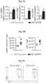


Classifications
-
- C—CHEMISTRY; METALLURGY
- C07—ORGANIC CHEMISTRY
- C07K—PEPTIDES
- C07K16/00—Immunoglobulins [IGs], e.g. monoclonal or polyclonal antibodies
- C07K16/18—Immunoglobulins [IGs], e.g. monoclonal or polyclonal antibodies against material from animals or humans
- C07K16/28—Immunoglobulins [IGs], e.g. monoclonal or polyclonal antibodies against material from animals or humans against receptors, cell surface antigens or cell surface determinants
- C07K16/2803—Immunoglobulins [IGs], e.g. monoclonal or polyclonal antibodies against material from animals or humans against receptors, cell surface antigens or cell surface determinants against the immunoglobulin superfamily
-
- A—HUMAN NECESSITIES
- A61—MEDICAL OR VETERINARY SCIENCE; HYGIENE
- A61P—SPECIFIC THERAPEUTIC ACTIVITY OF CHEMICAL COMPOUNDS OR MEDICINAL PREPARATIONS
- A61P25/00—Drugs for disorders of the nervous system
- A61P25/28—Drugs for disorders of the nervous system for treating neurodegenerative disorders of the central nervous system, e.g. nootropic agents, cognition enhancers, drugs for treating Alzheimer's disease or other forms of dementia
-
- A—HUMAN NECESSITIES
- A61—MEDICAL OR VETERINARY SCIENCE; HYGIENE
- A61K—PREPARATIONS FOR MEDICAL, DENTAL OR TOILETRY PURPOSES
- A61K39/00—Medicinal preparations containing antigens or antibodies
- A61K39/395—Antibodies; Immunoglobulins; Immune serum, e.g. antilymphocytic serum
-
- A—HUMAN NECESSITIES
- A61—MEDICAL OR VETERINARY SCIENCE; HYGIENE
- A61P—SPECIFIC THERAPEUTIC ACTIVITY OF CHEMICAL COMPOUNDS OR MEDICINAL PREPARATIONS
- A61P27/00—Drugs for disorders of the senses
-
- A—HUMAN NECESSITIES
- A61—MEDICAL OR VETERINARY SCIENCE; HYGIENE
- A61P—SPECIFIC THERAPEUTIC ACTIVITY OF CHEMICAL COMPOUNDS OR MEDICINAL PREPARATIONS
- A61P27/00—Drugs for disorders of the senses
- A61P27/02—Ophthalmic agents
-
- C—CHEMISTRY; METALLURGY
- C07—ORGANIC CHEMISTRY
- C07K—PEPTIDES
- C07K16/00—Immunoglobulins [IGs], e.g. monoclonal or polyclonal antibodies
- C07K16/18—Immunoglobulins [IGs], e.g. monoclonal or polyclonal antibodies against material from animals or humans
- C07K16/28—Immunoglobulins [IGs], e.g. monoclonal or polyclonal antibodies against material from animals or humans against receptors, cell surface antigens or cell surface determinants
- C07K16/2803—Immunoglobulins [IGs], e.g. monoclonal or polyclonal antibodies against material from animals or humans against receptors, cell surface antigens or cell surface determinants against the immunoglobulin superfamily
- C07K16/2818—Immunoglobulins [IGs], e.g. monoclonal or polyclonal antibodies against material from animals or humans against receptors, cell surface antigens or cell surface determinants against the immunoglobulin superfamily against CD28 or CD152
-
- C—CHEMISTRY; METALLURGY
- C07—ORGANIC CHEMISTRY
- C07K—PEPTIDES
- C07K16/00—Immunoglobulins [IGs], e.g. monoclonal or polyclonal antibodies
- C07K16/18—Immunoglobulins [IGs], e.g. monoclonal or polyclonal antibodies against material from animals or humans
- C07K16/28—Immunoglobulins [IGs], e.g. monoclonal or polyclonal antibodies against material from animals or humans against receptors, cell surface antigens or cell surface determinants
- C07K16/2803—Immunoglobulins [IGs], e.g. monoclonal or polyclonal antibodies against material from animals or humans against receptors, cell surface antigens or cell surface determinants against the immunoglobulin superfamily
- C07K16/2827—Immunoglobulins [IGs], e.g. monoclonal or polyclonal antibodies against material from animals or humans against receptors, cell surface antigens or cell surface determinants against the immunoglobulin superfamily against B7 molecules, e.g. CD80, CD86
-
- A—HUMAN NECESSITIES
- A61—MEDICAL OR VETERINARY SCIENCE; HYGIENE
- A61K—PREPARATIONS FOR MEDICAL, DENTAL OR TOILETRY PURPOSES
- A61K39/00—Medicinal preparations containing antigens or antibodies
- A61K2039/505—Medicinal preparations containing antigens or antibodies comprising antibodies
-
- A—HUMAN NECESSITIES
- A61—MEDICAL OR VETERINARY SCIENCE; HYGIENE
- A61K—PREPARATIONS FOR MEDICAL, DENTAL OR TOILETRY PURPOSES
- A61K39/00—Medicinal preparations containing antigens or antibodies
- A61K2039/505—Medicinal preparations containing antigens or antibodies comprising antibodies
- A61K2039/507—Comprising a combination of two or more separate antibodies
-
- A—HUMAN NECESSITIES
- A61—MEDICAL OR VETERINARY SCIENCE; HYGIENE
- A61K—PREPARATIONS FOR MEDICAL, DENTAL OR TOILETRY PURPOSES
- A61K39/00—Medicinal preparations containing antigens or antibodies
- A61K2039/545—Medicinal preparations containing antigens or antibodies characterised by the dose, timing or administration schedule
-
- C—CHEMISTRY; METALLURGY
- C07—ORGANIC CHEMISTRY
- C07K—PEPTIDES
- C07K2317/00—Immunoglobulins specific features
- C07K2317/70—Immunoglobulins specific features characterized by effect upon binding to a cell or to an antigen
- C07K2317/76—Antagonist effect on antigen, e.g. neutralization or inhibition of binding
Definitions
- the present invention relates in general to methods and compositions for treating disease, disorder, condition or injury of the Central Nervous System (CNS) by transiently reducing the level of systemic immunosuppression in the circulation.
- CNS Central Nervous System
- AD central nervous system
- a ⁇ amyloid-beta
- the present invention provides a pharmaceutical composition
- a pharmaceutical composition comprising an active agent that causes reduction of the level of systemic immunosuppression in an individual for use in treating a disease, disorder, condition or injury of the CNS that does not include the autoimmune neuroinflammatory disease, relapsing-remitting multiple sclerosis (RRMS), wherein said pharmaceutical composition is for administration by a dosage regimen comprising at least two courses of therapy, each course of therapy comprising in sequence a treatment session followed by an interval session of non-treatment.
- RRMS relapsing-remitting multiple sclerosis
- the present invention provides a method for treating a disease, disorder, condition or injury of the Central Nervous System (CNS) that does not include the autoimmune neuroinflammatory disease relapsing-remitting multiple sclerosis (RRMS), said method comprising administering to an individual in need thereof a pharmaceutical composition comprising an active agent that causes reduction of the level of systemic immunosuppression according to the present invention, wherein said pharmaceutical composition is administered by a dosage regime comprising at least two courses of therapy, each course of therapy comprising in sequence a treatment session followed by an interval session of a non-treatment period.
- CNS Central Nervous System
- RRMS autoimmune neuroinflammatory disease relapsing-remitting multiple sclerosis
- Figs. 1A-B depict the choroid plexus (CP) activity along disease progression in the 5XFAD transgenic mouse model of AD (AD-Tg).
- error bars represent mean ⁇ s.e.m.; *, P ⁇ 0.05; **, P ⁇ 0.01;***, P ⁇ 0.001.
- error bars represent mean ⁇ s.e.m.; *, P ⁇ 0.05; **, P ⁇ 0.01;***, P ⁇ 0.001.
- Fig. 4 shows gating strategy and representative flow cytometry plots of splenocytes from AD-Tg/Foxp3-DTR +/- mice, 1 day after the last injection of DTx. DTx was injected i.p. for 4 constitutive days, achieving ⁇ 99% depletion of Foxp3 + cells.
- Figs. 5A-G show the effects of transient depletion of Tregs in AD-Tg mice.
- AD-Tg/Foxp3-DTR + which express the DTR transgene
- AD-Tg/Foxp3-DTR non-DTR-expressing AD-Tg littermate
- B-D Flow cytometry analysis of the brain parenchyma (excluding the choroid plexus, which was separately excised) of 6-month old DTx-treated AD-Tg mice and controls, 3 weeks following the last DTx injection.
- Figs. 6A-E show the effect of transient depletion of Tregs on A ⁇ plaques learning/memory performance.
- A Representative microscopic images and
- B quantitative analysis of the brains of 5-month old DTx-treated AD-Tg/Foxp3-DTR + and AD-Tg/Foxp3-DTR - control mice, 3 weeks after the last DTx injection, immunostained for A ⁇ plaques and Hoechst nuclear staining (scale bar, 250 ⁇ m).
- Mean A ⁇ plaque area and numbers in the hippocampal dentate gyrus (DG) and the 5 th layer of the cerebral cortex were quantified (in 6 ⁇ m brain slices; n 5-6 per group; Student's t test).
- DG hippocampal dentate gyrus
- Figs. 8A-I show the therapeutic effect of administration of weekly Glatiramer acetate (GA) in AD-Tg mice.
- A Schematic representation of weekly-GA treatment regimen. Mice (5-month old) were s.c. injected with GA (100 ⁇ g), twice during the first week (on day 1 and 4), and once every week thereafter, for an overall period of 4 weeks. The mice were examined for cognitive performance, 1 week (MWM), 1 month (RAWM) and 2 months (RAWM, using different experimental spatial settings) after the last injection, and for hippocampal inflammation.
- MMM 1 week
- RAWM 1 month
- RAWM 2 months
- pro-inflammatory cytokines such as TNF- ⁇ , IL-1 ⁇ and IL-12p40
- C elevation of the anti-inflammatory cytokines IL-10 and TGF- ⁇
- D the neurotropic factors
- AD-Tg mice (5 months old) were treated with either weekly-GA or with vehicle (PBS), and compared to age-matched WT littermates in the MWM task at the age of 6m.
- FIGs. 9A-H show further therapeutic effects of administration of weekly-GA in AD-Tg mice.
- A-B shows 5XFAD AD-Tg mice that were treated with either weekly-GA, or vehicle (PBS), and were examined at the end of the 1 st week of the administration regimen (after a total of two GA injections).
- Flow cytometry analysis for CD4 + Foxp3 + splenocyte frequencies (A), and CP IFN- ⁇ -expressing immune cells (B; intracellularly stained and pre-gated on CD45), in treated 6-month old AD-Tg mice, compared to age-matched WT controls (n 4-6 per group; one-way ANOVA followed by Newman-Keuls post hoc analysis).
- FIG. 9D-E show representative images of brain sections from 6-month old AD-Tg/CX 3 CR1 GFP/+ BM chimeras following weekly-GA.
- CX 3 CR1 GFP cells were localized at the CP of the third ventricle (3V; i ), the adjacent ventricular spaces ( ii ), and the CP of the lateral ventricles (LV; iii ) in AD-Tg mice treated with weekly-GA (D; scale bar, 25 ⁇ m).
- CX 3 CR1 GFP cells are co-localized with the myeloid marker IBA-1 in brains of GA-treated AD-Tg/CX 3 CR1 GFP/+ mice in the vicinity of A ⁇ plaques, and co-expressing the myeloid marker, IBA-1 (scale bar, 25 ⁇ m).
- Figs. 9G-H show representative flow cytometry plots of cells isolated from the hippocampus of 4-month old WT, untreated AD-Tg, and AD-Tg mice, on the 2 nd week of the weekly-GA regimen.
- Figs. 10A-H depict the therapeutic effect of administration of a p300 inhibitor (C646) in AD-Tg mice.
- a p300 inhibitor C646
- aged mice (18 months) were treated with either p300i or vehicle (DMSO) for a period of 1 week, and examined a day after cessation of treatment.
- Representative flow cytometry plots showing elevation in the frequencies of CD4 + T cells expressing IFN- ⁇ in the spleen (A), and IFN- ⁇ -expressing immune cell numbers in the CP (B), following p300i treatment.
- Figs. 10A-H depict the therapeutic effect of administration of a p300 inhibitor (C646) in AD-Tg mice.
- DMSO vehicle
- 10C-E show representative microscopic images (C), and quantitative analysis, of A ⁇ plaque burden in the brains of 10-month old AD-Tg mice, which received either p300i or vehicle (DMSO) for a period of 1 week, and were subsequently examined after 3 additional weeks.
- error bars represent mean ⁇ s.e.m.; *, P ⁇ 0.05; **, P ⁇ 0.01;***, P ⁇ 0.001.
- Figs. 11 A-D show that PD-1 blockade augments percentage of IFN- ⁇ -producing CD4+ T-cells in the spleen, as well as IFN- ⁇ expression at the choroid plexus in AD-Tg mice.
- 10-month old AD-Tg mice were i.p. injected on day 1 and day 4 with 250ug of either anti-PD-1 or control IgG, and examined at days 7-10 for the effect on the systemic immune response and CP activity.
- C mRNA expression levels of ifn-g, measured by RT-qPCR in the CP of AD-Tg mice treated with anti-PD-1 when compared to IgG treated and untreated AD-Tg controls
- Figs. 12A-B show that PD-1 blockade mitigates cognitive decline in AD-Tg mice.
- 10-month old AD-Tg mice were i.p. injected on day 1 and day 4 with 250 ug of either anti-PD-1 or control IgG, and examined 1 or 2 months later for the effect on pathology with (A) showing performance of AD-Tg mice in the RAWM after 1 treatment session with anti-PD-1 or IgG control and (B) showing effect of single anti-PD-1 treatment session, or 2 sessions with a 1 month interval on performance.
- Single arrows indicate time points of treatment, and double arrows indicate time points of cognitive testing.
- Fig. 14 shows the effect of different dosing and frequency of administration of anti-PD-1 antibody on cognitive decline in AD-Tg mice and illustrates the dosage scheme and the effect of anti-PD-1 antibody treatment on spatial learning and memory performance using the radial arm water maze (RAWM) task at 7 months of age.
- Black arrows indicate time points of treatment, and illustrations indicate time points of cognitive testing.
- Figs. 15A-H show the effect of repeated administration of anti-PD-1 antibody on cognitive decline in AD-Tg mice.
- 5XFAD mice were treated with either PD-1-specific antibody or IgG control, starting at 3 months of age; treatment was continued once a month until the age of 5 months (total of three injections).
- Experimental design is presented in (A). Black arrows indicate time points of treatment, and illustrations indicate time points of cognitive testing.
- Fig. 16 shows the effect of a single administration of anti-TIM-3 antibody on cognitive decline in AD-Tg mice and illustrates the dosage scheme and the effect of anti-TIM-3 antibody treatment on spatial learning and memory performance using the radial arm water maze (RAWM) task at 7 months of age.
- Black arrows indicate time points of treatment, and illustrations indicate time points of cognitive testing.
- Figs. 17A-J show that PD-L1 blockade mitigates cognitive decline in AD-Tg mice.
- 5XFAD mice (7-months old) were treated with either PD-1-specific antibody, PD-L1-specific antibody or isotype matched control antibody (IgG).
- Experimental design is presented in (A). Black arrow indicates time point of treatment, and illustrations indicate time points of cognitive scoring using the RAWM.
- Figs. 18A-B show that PD-L1 expression increases at the CP with aging with (A) showing expression of PDL1 in the CP of young (left bar) and aged (right bar) mice, measured by RT-qPCR; and (B) showing immunohistochemical staining of epithelial expression of PD-L1 at the CP of young (left micrograph) and aged (right micrograph) mice. LV; lateral ventricle.
- Figs. 21A-E show that PD-1/PD-L1 axis blockade in DM-hTau mice enhances monocyte recruitment to the brain.
- A Flow cytometry gating strategy for splenocytes, and (B) quantitative analysis of FoxP3 + regulatory T cells, and (C) CD44 + CD62L -/low effector memory T (T EF ) cells, and (C) CD44 + CD62L -/low effector memory T (T EF ) cells, versus CD44 + CD62L high central memory T (Tcm) cells.
- D Flow cytometry gating strategy for brain CD45 low CD11b + and CD45 high CD11b + myeloid cells.
- E Brains from the same mice were excised and analyzed for the presence of CD45 high CD11b + infiltrating myeloid cells.
- Figs. 22A-G shows that PD-1/PD-L1 axis blockade in DM-hTau mice enhances monocyte recruitment to the brain.
- Male mice expressing the human-tau gene with two mutations K257T/P301S; double mutant, DM-hTAU) (average cohorts aged 8 months) were treated with anti-PD-1-specific antibody, anti-PD-L1-specific antibody, or isotype matched control antibody (IgG) (one i.p. injection of 0.5mg/mouse); experimental design is presented in (A). Black arrow indicates time point of treatment, and illustrations indicate time points of cognitive testing.
- C Representative heat-map plots of the time spent in the distinct arms of the three tested groups.
- Figs. 23A-G show that blocking PD-1/PD-L1 pathway reduces hyperphosphorylation in DM-tau mice.
- Immunostaining of Neurofibrillary tangles (NFTs) in brains of 8 month old DM-hTAU mice 1 month after treatment with anti-PD-1, anti-PD-L1, or isotype matched control antibody or with isotype matched control antibody (IgG) A-F.
- A, D Representative immunofluorescence images
- B, C, E, F quantitative analysis of AT-100 and AT-180.
- the effect of PD-L1 blockade on spatial memory was determined using the T maze task.
- Fig. 24A-B shows PD-1 blockade enhances hippocampal neurogenesis in 5XFAD mice with (A) showing parasagittal brain sections immunostained for neuronal marker-NeuN (in green), DCX (in red), and hoechst nuclear staining (in blue); and (B) showing a graph quantitating the staining in anti-PD-1 treated animals, IgG immune controls and aged-matched wild-type controls.
- Fig. 25A-B shows PD-1 blockade enhances hippocampal synaptic plasticity in 5XFAD mice with (A) showing parasagittal brain sections immunostained for VgluT1 (red); and (B) showing a graph quantitating the staining in anti-PD-1 treated animals, IgG immune controls and aged-matched wild-type controls.
- Fig. 26A-B shows PD-1 blockade reduces neuronal loss in the subiculum of 5XFAD mice with (A) showing parasagittal brain sections immunostained for neuronal marker-NeuN (in green); and (B) showing a graph quantitating the staining in anti-PD-1 treated animals, IgG immune controls and aged-matched wild-type controls.
- Immune checkpoint mechanisms which include cell-intrinsic downregulation of activated T cell responsiveness and effector function by inhibitory receptors, maintain systemic immune homeostasis and autoimmune tolerance (Joller et al, 2012; Pardoll, 2012).
- immune checkpoint blockade activates a cascade of immunological events that starts in the periphery and culminates in numerous activities inside the brain.
- an immune response increases the availability of IFN- ⁇ at the secondary lymphoid organs (lymph nodes, spleen, etc.) and circulating monocytes in the periphery.
- This immune response leads to the immunological activation of the brain's choroid plexus (CP), an epithelial layer at the brain ventricles, which forms the blood-cerebrospinal fluid-barrier (B-CSF-B), and serves as a selective gateway for leukocytes entering the CNS.
- CP brain's choroid plexus
- B-CSF-B blood-cerebrospinal fluid-barrier
- the effect of the blockade of inhibitory immune checkpoints on CP gateway activity for leukocyte is mediated by the IFN- ⁇ -induced expression of leukocyte trafficking molecules (adhesion molecules and chemokines) by the CP epithelium, which enables leukocyte trafficking.
- leukocyte trafficking molecules asdhesion molecules and chemokines
- This increased expression leads to the recruitment of monocyte-derived macrophages and immunoregulatory cells to diseased sites within the brain.
- this recruitment results in a comprehensive effect on brain function, including reduced of plaque burden, restored of immunological balance, resolved local inflammation, reduced gliosis, reduced synaptic loss, increased neurogenesis, increased neuronal protection and enhanced neuronal survival, collectively leading to neuroprotection and/or reduction in cognitive decline.
- Immune checkpoints are molecules in the immune system that either turn up a signal (costimulatory molecules) or turn down a signal.
- Four stimulatory checkpoint molecules are members of the tumor necrosis factor (TNF) receptor superfamily - CD27, CD40, OX40, GITR and CD137.
- TNF tumor necrosis factor
- Another two stimulatory checkpoint molecules belongs to the B7-CD28 superfamily - CD28 itself and ICOS.
- Many inhibitor checkpoint molecules are known, including, without limitation, A2aR, B7-H3, B7-H4, BTLA, CTLA-4, IDO, KIR, LAG-3, PD-1, TIM-3 and VISTA.
- the present invention provides a method for treating a disease, disorder, condition or injury of the Central Nervous System (CNS).
- the disclosed method for treating a disease, disorder, condition or injury of the Central Nervous System (CNS) does not include the autoimmune neuroinflammatory disease relapsing-remitting multiple sclerosis (RRMS).
- the disclose method comprising administering to an individual in need thereof an active agent that causes reduction of the level of systemic immunosuppression, wherein said active agent is administered by a dosage regime comprising at least two courses of therapy, each course of therapy comprising in sequence a treatment session followed by an interval session of non-treatment.
- the present invention is directed to an active agent that causes reduction of the level of systemic immunosuppression in an individual, or a pharmaceutical composition comprising the active agent, for use in treating a disease, disorder, condition or injury of the CNS that does not include the autoimmune neuroinflammatory disease, relapsing-remitting multiple sclerosis (RRMS), wherein said pharmaceutical composition is for administration by a dosage regimen comprising at least two courses of therapy, each course of therapy comprising in sequence a treatment session followed by an interval session of non-treatment.
- RRMS relapsing-remitting multiple sclerosis
- the dosage regimen is calibrated such that the level of systemic immunosuppression is transiently reduced.
- treating refers to means of obtaining a desired physiological effect.
- the effect may be therapeutic in terms of partially or completely curing a disease and/or symptoms attributed to the disease.
- the term refers to inhibiting the disease, i.e. arresting or slowing its development; or ameliorating the disease, i.e. causing regression of the disease.
- systemic presence of regulatory or effector T cells refers to the presence of the regulatory or effector T cells (as measured by their level or activity) in the circulating immune system, i.e. the blood, spleen and lymph nodes. It is a well-known fact in the field of immunology that the cell population profile in the spleen is reflected in the cell population profile in the blood (Zhao et al, 2007).
- the present treatment is applicable to both patients that show elevation of systemic immune suppression, as well as to patients that do not show such an elevation.
- the individual in need for the treatment according to the present invention has a certain level of peripheral immunosuppression, which is reflected by elevated frequencies or numbers of Tregs in the circulation, and/or their enhanced functional activity and/or a decrease in IFN ⁇ -producing leukocytes and/or decreased proliferation of leukocytes in response to stimulation.
- the elevation of frequencies or numbers of Tregs can be in total numbers or as percentage of the total CD4 cells. For example, it has been found in accordance with the present invention that an animal model of Alzheimer's disease has higher frequencies of Foxp3 out of CD4 cells as compared with wild-type mice.
- the method of the present invention that reduces the level or activity of systemic immunosuppression is effective in treating disease, disorder, condition or injury of the CNS that does not include the autoimmune neuroinflammatory disease RRMS.
- said systemic immune suppression can also involve additional immune cell types except of Tregs, such as myeloid-derived suppressor cells (MDSCs) (Gabrilovich & Nagaraj, 2009).
- MDSCs myeloid-derived suppressor cells
- the level of systemic immunosuppression may be detected by various methods that are well known to those of ordinary skill in the art.
- the level of Tregs may be measured by flow cytometry analysis of peripheral blood mononuclear cells or T lymphocytes, immunostained either for cellular surface markers or nuclear intracellular markers of Treg (Chen & Oppenheim, 2011), CD45, TCR- ⁇ , or CD4 markers of lymphocytes, and measuring the amount of antibody specifically bound to the cells.
- Tregs may be measured by various assays; For example the thymidine incorporation assay is being commonly used, in which suppression of anti-CD3 mAb stimulated proliferation of CD4 + CD25 - T cells (conventional T cells) is measured by [ 3 H]thymidine incorporation or by using CFSE (5-(and 6)-carboxyfluorescein diacetate succinimidyl ester, which is capable of entering the cells; cell division is measured as successive halving of the fluorescence intensity of CFSE).
- CFSE 5-(and 6)-carboxyfluorescein diacetate succinimidyl ester
- the number of IFN ⁇ -producing leukocytes or their activity or their proliferation capacity can easily be assessed by a skilled artisan using methods known in the art;
- the level of IFN ⁇ -producing leukocytes may be measured by flow cytometry analysis of peripheral blood mononuclear cells, following short ex-vivo stimulation and golgi-stop, and immunostaining by IFN ⁇ intracellular staining (using e.g., BD Biosciences Cytofix/cytoperm TM fixation/permeabilization kit), by collecting the condition media of these cells and quantifying the level of secreted cytokines using ELISA, or by comparing the ratio of different cytokines in the condition media, for example IL2/IL10, IL2/IL4, INF ⁇ /TGF ⁇ , etc.
- the levels of MDSCs in the human peripheral blood easily can be assessed by a skilled artisan, for example by using flow cytometry analysis of frequency of DR - /LIN - /CD11b+, DR - /LIN - /CD15+, DR - /LIN - /CD33+ and DR(-/low)/CD14+ cells, as described (Kotsakis et al, 2012).
- the peripheral/systemic immunosuppression may be considered elevated when the total number of Tregs in the circulation is higher than 10, 20, 30, 40, 50, 60, 70, 80, 90, or 100% or more than in a healthy control population, the percentage of Treg cells out of the total CD4+ cells is elevated by 10, 20, 30, 40, 50, 60, 70, 80, 90, or 100% or more than in a healthy control population, or the functional activity of Tregs is elevated by 10, 20, 30, 40, 50, 60, 70, 80, 90, or 100% or more than in a healthy control population.
- the peripheral/systemic immunosuppression may be considered elevated when the level of IFN ⁇ -producing leukocytes or their activity is reduced relative to that of a healthy control population by 10, 20, 30, 40, 50, 60, 70, 80, 90 or 100%; or the proliferation of leukocytes in response to stimulation is reduced relative to that of a healthy control population by 10, 20, 30, 40, 50, 60, 70, 80, 90 or 100%.
- An agent may be considered an agent that causes reduction of the level of systemic immunosuppression when, upon administration of the agent to an individual, the total number of Tregs in the circulation of this individual is reduced by 10, 20, 30, 40, 50, 60, 70, 80, 90 or 100% as compared with the level before administration of the agent, the percentage of Treg cells out of the total CD4+ cells drops by 10, 20, 30, 40, 50, 60, 70, 80, 90 or 100% relative to that of a healthy control population or the functional activity of Tregs is reduced by 10, 20, 30, 40, 50, 60, 70, 80, 90 or 100% as compared with the level before administration of the agent.
- an agent may be considered an agent that causes reduction of the level of systemic immunosuppression when, upon administration of the agent to an individual, the total number of IFN ⁇ -producing leukocytes or their activity is increased by 10, 20, 30, 40, 50, 60, 70, 80, 90, or 100% or more; or the proliferation of leukocytes in response to stimulation is increased relative to that of a healthy control population by 10, 20, 30, 40, 50, 60, 70, 80, 90 or 100% or more.
- the active agent causes reduction of the level of systemic immunosuppression by release of a restraint imposed on the immune system by one or more immune checkpoints, for example by blockade of the one or more immune checkpoints.
- the reduction of the level of systemic immunosuppression is associated with an increase in systemic presence or activity of IFN ⁇ -producing leukocytes.
- the active agent causes reduction of the level of systemic immunosuppression and thereby an increase in the systemic presence or activity of effector T cells.
- the reduction of the level of systemic immunosuppression is associated with an increase in systemic presence or activity of an IFN ⁇ cytokine.
- the reduction of the level of systemic immunosuppression is associated with a decrease in systemic presence or activity of regulatory T-cells.
- the reduction of the level of systemic immunosuppression is associated with a decrease in systemic presence or activity of an IL-10 cytokine.
- the reduction of the level of systemic immunosuppression is associated with a decrease in systemic presence or activity of myeloid-derived suppressor cells (MDSCs).
- MDSCs myeloid-derived suppressor cells
- the active agent causes reduction of the level of systemic immunosuppression and thereby an increase in the systemic presence or activity of effector T cells.
- the checkpoints that may be manipulated to release the systemic immunosuppression are referred to herein as a pair of an immune checkpoint receptor and its native ligand or either one of the two partners.
- PD-1 which has two known ligands is referred to herein as “ PD-L1” and “ PD-L2”, while B7H3, the ligand of which has not yet been identified, is referred to simply by "B7H3".
- the checkpoints that may be manipulated to release the systemic immunosuppression in accordance with the present invention include, without limitation, PD-1-PD-L1, PD-1-PD-L2, CD28-CD80, CD28-CD86, CTLA-4-CD80, CTLA-4-CD86, ICOS-B7RP1, B7H3, B7H4, B7H7, B7-CD28-like molecule, BTLA-HVEM, KIR-MHC class I or II, LAG3-MHC class I or II, CD137-CD137L, OX40-OX40L, CD27-CD70, CD40L-CD40, TIM3-GAL9, V-domain Ig suppressor of T cell activation (VISTA), STimulator of INterferon Genes (STING), T cell immunoglobulin and immunoreceptor tyrosine-based inhibitory motif domain (TIGIT), glucocorticoid-induced tumor necrosis factor receptor related protein (GITR), A2aR-
- the active agent that may be used according to the present invention may be an antibody.
- An antibody as disclosed herein can be a polyclonal antibody, a monoclonal antibody, a dimer, a multimer, a multispecific antibody, a human antibody, a humanized antibody, a recombinant antibody, a chimeric antibody, bi-functional antibody, a cell-associated antibody like an Ig receptor, a linear antibody, a diabody, a minibody or a nanobody, so long as the fragment exhibits the desired biological activity, and single chain derivatives of the same.
- An antibody can be a full-length immunoglobulin molecule comprising the VH and VL domains, as well as a light chain constant domain (CL) and heavy chain constant domains, CH1, CH2 and CH3, or an immunologically active fragment of a full-length immunoglobulin molecule, such as, e.g., a single domain antibody (sdAb), a single-chain variable fragment (scFv), a Fab fragment, a F(ab')2 fragment, a Fc fragment, a Fd fragment, a Fv fragment.
- sdAb single domain antibody
- scFv single-chain variable fragment
- An antibody can be derived from any vertebrate species (e.g., human, goat, horse, donkey, murine, rat, rabbit, or chicken), and can be of any type (e.g., IgG, IgE, IgM, IgD, and IgA), class (e.g., IgA, IgD, IgE, IgG, and IgM) or subclass (lgG1, IgG2, IgG3, IgG4, IgA1 and IgA2).
- an antibody disclosed herein may be an antagonist antibody, meaning an antibody that inhibits a biological activity or an antibody disclosed herein may be an agonist antibody, meaning an antibody that stimulates a biological activity.
- an antibody disclosed herein may be a neutralizing antibody, meaning an antibody that can block or neutralize a biological activity.
- a neutralizing antibody meaning an antibody that can block or neutralize a biological activity.
- An antibody disclosed herein may be, without limitation, an anti-PD-1, an anti-PD-L1, an anti-PD-L2, an anti-CTLA-4, an anti-CD80,an anti-CD86, an anti-B7RP1, an anti-B7-H3, an anti-B7-H4, an anti-B7-H7, an anti-BTLA, an anti-HVEM, an anti-CD-27, an anti-CD40, an anti-CD40L, an anti-CD70, an anti-CD80, an anti-CD86, an anti-CD137, an anti-CD137L, an anti-OX40, an anti-OX40L, an anti-TIM-3, an anti-Galectin9, an anti-KIR, an anti-LAG-3, an anti-ICOS, an anti-VISTA, an anti-STING, an anti-TIGIT, anti-GITR or any combination thereof.
- An antibody disclosed herein may be administered to a human at a dosage of for example about 0.1 mg/kg - 20 mg/kg, 0.1 mg/kg - 15 mg/kg, 0.1 mg/kg - 10 mg/kg, 0.1 mg/kg - 5 mg/kg, 0.2 mg/kg - 20 mg/kg, 0.2 mg/kg - 15 mg/kg, 0.2 mg/kg - 10 mg/kg, 0.2 mg/kg - 6 mg/kg, 0.2 mg/kg - 5 mg/kg, 0.3 mg/kg - 20 mg/kg, 0.3 mg/kg - 15 mg/kg, 0.3 mg/kg - 10 mg/kg, 0.3 mg/kg - 5 mg/kg, , 1 mg/kg - 20 mg/kg, 1 mg/kg - 15 mg/kg, 1 mg/kg - 10 mg/kg, 1 mg/kg - 5 mg/kg, 1.5 mg/kg - 20 mg/kg, 1.5 mg/kg - 15 mg/kg, 1.5 mg/kg - 10 mg/kg
- Programmed cell death protein 1 also known as PD-1 and CD279 (cluster of differentiation 279), is a cell surface receptor that belongs to the immunoglobulin superfamily and is expressed on T cells and pro-B cells.
- PD-1 binds two ligands, PD-L1 and PD-L2. Functioning as an immune checkpoint, PD-1 plays an important role in down regulating the immune system by preventing the activation of T-cells, which in turn reduces autoimmunity and promotes self-tolerance.
- the inhibitory effect of PD-1 is accomplished through a dual mechanism of promoting apoptosis (programmed cell death) in antigen specific T-cells in lymph nodes while simultaneously reducing apoptosis in regulatory T cells (suppressor T cells).
- compounds which inhibit PD-1 function such as PD-1 inhibitors, PD-L1 inhibitors and/or PD-L2 inhibitors, serve to activate the immune system.
- One class of PD-1 inhibitors includes antagonist or neutralizing anti-PD-1, anti-PD-L1 and anti-PD-L2 antibodies. Many antagonist or neutralizing anti-PD-1, anti-PD-L1 and anti-PD-L2 antibodies are known in the art.
- the anti-PD-1 antibody used in accordance with the present invention may be selected from those disclosed in Ohaegbulam et al. (Ohaegbulam et al, 2015), the entire contents of which being hereby incorporated herein by reference.
- human or humanized anti-PD-1 antibodies include, without limitation, CD279 (human anti-PD1 monoclonal antibody, Bio X Cell), MEDI0680 (AMP-514; humanized IgG4 anti-PD-1 monoclonal antibody; AstraZeneca), Nivolumab (BMS-936558; human IgG4 anti-PD1 monoclonal antibody; Bristol-Myers Squibb), Pembrolizumab (Lambrolizumab, MK-3475; humanized IgG4 anti-PD1 monoclonal antibody; Merck), Pidilizumab (CT-011; humanized IgG1 anti-PD1 monoclonal antibody; Medivation) and TSR-042 (humanized IgG4 anti-PD-1 monoclonal antibody; Tesaro).
- CD279 human anti-PD1 monoclonal antibody, Bio X Cell
- MEDI0680 AMP-514; humanized IgG4 anti-PD-1 monoclonal antibody; AstraZene
- human or humanized anti-PD-L1 antibodies include, without limitation, Avelumab (MSB0010718C; human IgG1 anti-PD-L1 monoclonal antibody; Merck-Serono), Atezolizumab (MPDL3280A, RG7446; human IgG anti-PD-L1 monoclonal antibody; Hoffmann-La Roche), BMS-936559 (MDX-1105; human IgG4 anti-PD-L1 monoclonal antibody; Bristol-Myers Squibb), Durvalumab (MEDI4736; humanized IgG1 anti-PD-L1 monoclonal antibody; AstraZeneca), KN035 (anti-PD-L1 monoclonal antibody; 3D Medicines) and LY3300054 (anti-PD-L1 monoclonal antibody; Eli Lilly).
- Avelumab MSB0010718C
- human IgG1 anti-PD-L1 monoclonal antibody Merck-Serono
- an anti-PD-1 antibody, an anti-PD-L1 antibody and/or an anti-PD-L2 antibody may be administered to a human at a dosage of for example about 0.1 mg/kg - 20 mg/kg, 0.1 mg/kg - 15 mg/kg, 0.1 mg/kg - 10 mg/kg, 0.1 mg/kg - 5 mg/kg, 0.2 mg/kg - 20 mg/kg, 0.2 mg/kg - 15 mg/kg, 0.2 mg/kg - 10 mg/kg, 0.2 mg/kg - 6 mg/kg, 0.2 mg/kg - 5 mg/kg, 0.3 mg/kg - 20 mg/kg, 0.3 mg/kg - 15 mg/kg, 0.3 mg/kg - 10 mg/kg, 0.3 mg/kg - 5 mg/kg, , 1 mg/kg
- Pidilizumab may be administered to a human at a dosage of 0.2-6 mg/kg or between 1.5-6 mg/kg; Pembrolizumab may be administered to a human at a dosage of 1-10 mg/kg; Nivolumab may be administered to a human at a dosage of 0.3-20 mg/kg, 0.3-10 mg/kg, 1-10 mg/kg or at 1 or 3 mg/kg; BMS-936559 may be administered to a human at a dosage of 0.3-10 mg/kg; Atezolizumab may be administered to a human at a dosage of 1-20 mg/kg; Durvalumab may be administered to a human at a dosage of 0.1-15 mg/kg; and Avelumab may be administered to a human at a dosage of 1-20 mg/kg.
- T-cell immunoglobulin and mucin domain-3 is a Th1-specific cell surface protein that acts as an immune checkpoint that inhibits lymphocyte activity by down regulating macrophage activation and playing an important role in CD8+ T cell exhaustion that takes place in chronic immune conditions.
- TIM-3 acts as a negative regulator of Th1/Tc1 function by triggering cell death upon interaction with its ligand, galectin-9 (Gal9).
- Gal9 galectin-9
- compounds which inhibit TIM-3 function such as TIM-3 inhibitors and/or Gal9 inhibitors, serve to activate the immune system.
- One class of TIM-3 inhibitors includes antagonist or neutralizing antibodies against TIM-3 and/or Gal-9. Many antagonist or neutralizing anti-TIM-3 and anti-Gal9 antibodies are known in the art.
- human or humanized anti-TIM-3 antibodies include, without limitation, AF2365 (human IgG anti-TIM-3 monoclonal antibody; R&D Systems), CD366 (human IgG1 anti-TIM-3 monoclonal antibody; BioLegend), F38-2E2 (human IgG1 anti-TIM-3 monoclonal antibody; R&D Systems), L3D (human IgG1 anti-TIM-3 monoclonal antibody; CN 102492038 B ), MAB2365 (human IgG2a anti-TIM-3 monoclonal antibody; R&D Systems), MAB23651 (human IgG1 anti-TIM-3 monoclonal antibody; R&D Systems) and TSR-022 (humanized IgG4 anti-TIM-3 monoclonal antibody; Tesaro).
- AF2365 human IgG anti-TIM-3 monoclonal antibody; R&D Systems
- CD366 human IgG1 anti-TIM-3 monoclonal antibody; BioLegend
- F38-2E2 human Ig
- an anti-TIM-3 antibody and/or an anti-Gal9 antibody may be administered to a human at a dosage of for example about 0.1 mg/kg - 20 mg/kg, 0.1 mg/kg - 15 mg/kg, 0.1 mg/kg - 10 mg/kg, 0.1 mg/kg - 5 mg/kg, 0.2 mg/kg - 20 mg/kg, 0.2 mg/kg - 15 mg/kg, 0.2 mg/kg - 10 mg/kg, 0.2 mg/kg - 6 mg/kg, 0.2 mg/kg - 5 mg/kg, 0.3 mg/kg - 20 mg/kg, 0.3 mg/kg - 15 mg/kg, 0.3 mg/kg - 10 mg/kg, 0.3 mg/kg - 5 mg/kg, , 1 mg/kg - 20 mg/kg, 1 mg/kg - 15 mg/kg, 1 mg/kg - 10 mg/kg, 1 mg/kg - 5 mg/kg, 1.5 mg/kg - 20 mg/kg, 1.5 mg/kg - 20 mg/kg
- CTLA-4 Cytotoxic T-lymphocyte-associated protein 4
- CD152 cluster of differentiation 152
- CTLA-4 is a protein receptor functioning as an immune checkpoint that downregulates immune responses.
- CTLA-4 is constitutively expressed in Tregs but only upregulated in conventional T cells after activation.
- CTLA-4 acts as an "off" switch when bound to CD80 or CD86 on the surface of antigen-presenting cells.
- compounds which inhibit CTLA-4 function such as CTLA-4 inhibitors, CD80 inhibitors and/or CD86 inhibitors, serve to activate the immune system.
- CTLA-4 inhibitors includes antagonist or neutralizing antibodies against CTLA-4, CD80 and/or CD86. Many antagonist or neutralizing anti-CTLA-4, anti-CD80 and anti-CD86 antibodies are known in the art.
- human or humanized anti-CTLA-4 antibodies include, without limitation, Ipilimumab (human IgG1 anti-CTLA-4 monoclonal antibody; Bristol-Myers Squibb) and Tremelimumab (human IgG2 anti-CTLA-4 monoclonal antibody; Pfizer).
- an anti-CTLA-4 antibody, an anti-CB80 antibody and/or an anti-CD86 antibody may be administered to a human at a dosage of for example about 0.1 mg/kg - 20 mg/kg, 0.1 mg/kg - 15 mg/kg, 0.1 mg/kg - 10 mg/kg, 0.1 mg/kg - 5 mg/kg, 0.2 mg/kg - 20 mg/kg, 0.2 mg/kg - 15 mg/kg, 0.2 mg/kg - 10 mg/kg, 0.2 mg/kg - 6 mg/kg, 0.2 mg/kg - 5 mg/kg, 0.3 mg/kg - 20 mg/kg, 0.3 mg/kg - 15 mg/kg, 0.3 mg/kg - 10 mg/kg, 0.3 mg/kg - 5 mg/kg, , 1 mg/kg - 20 mg/kg, 1 mg/kg - 15 mg/kg, 1 mg/kg - 10 mg/kg, 1 mg/kg - 5 mg/kg, 1.5 mg/kg - 20 mg/kg, 0.1
- KIRs Killer-cell immunoglobulin-like receptors
- MHC major histocompatibility
- KIRs are inhibitors of lymphocyte activity.
- compounds which inhibit KIR function such as KIR inhibitors, serve to activate the immune system.
- One class of KIR inhibitors includes antagonist or neutralizing antibodies against KIR. Many antagonist or neutralizing anti-KIR antibodies are known in the art.
- an anti-KIR antibody may be administered to a human at a dosage of for example about 0.1 mg/kg - 20 mg/kg, 0.1 mg/kg - 15 mg/kg, 0.1 mg/kg - 10 mg/kg, 0.1 mg/kg - 5 mg/kg, 0.2 mg/kg - 20 mg/kg, 0.2 mg/kg - 15 mg/kg, 0.2 mg/kg - 10 mg/kg, 0.2 mg/kg - 6 mg/kg, 0.2 mg/kg - 5 mg/kg, 0.3 mg/kg - 20 mg/kg, 0.3 mg/kg - 15 mg/kg, 0.3 mg/kg - 10 mg/kg, 0.3 mg/kg - 5 mg/kg, , 1 mg/kg - 20 mg/kg, 1 mg/kg - 15 mg/kg, 1 mg/kg - 15 mg/kg, 1 mg/kg - 20 mg/kg, 1 mg/kg - 15 mg/kg, , 1 mg/kg - 20 mg/kg, 1 mg/kg - 15
- Lymphocyte-activation gene 3 (LAG-3), also known as cluster of differentiation 223 (CD223), is a cell surface molecule with diverse biologic effects on T cell function.
- LAG-3 is an immune checkpoint that inhibits lymphocyte activity by suppressing an immune response by action to Tregs as well as direct effects on CD8+ T cells.
- compounds which inhibit LAG-3 function such as LAG-3 inhibitors, serve to activate the immune system.
- One class of LAG-3 inhibitors includes antagonist or neutralizing antibodies against LAG-3. Many antagonist or neutralizing anti-LAG-3 antibodies are known in the art. Examples of human or humanized anti-LAG-3 antibodies include, without limitation, BMS-986016 (human anti-LAG-3 monoclonal antibody; Bristol-Myers Squibb).
- an anti-LAG-3 antibody may be administered to a human at a dosage of for example about 0.1 mg/kg - 20 mg/kg, 0.1 mg/kg - 15 mg/kg, 0.1 mg/kg - 10 mg/kg, 0.1 mg/kg - 5 mg/kg, 0.2 mg/kg - 20 mg/kg, 0.2 mg/kg - 15 mg/kg, 0.2 mg/kg - 10 mg/kg, 0.2 mg/kg - 6 mg/kg, 0.2 mg/kg - 5 mg/kg, 0.3 mg/kg - 20 mg/kg, 0.3 mg/kg - 15 mg/kg, 0.3 mg/kg - 10 mg/kg, 0.3 mg/kg - 5 mg/kg, , 1 mg/kg - 20 mg/kg, 1 mg/kg - 15 mg/kg, 1 mg/kg - 10 mg/kg, 1 mg/kg - 5 mg/kg, 1.5 mg/kg - 20 mg/kg, 1.5 mg/kg - 15 mg/kg, 1.5 mg/kg - 20 mg
- OX40 also known as cluster of differentiation 134 (CD134)
- CD134 cluster of differentiation 134
- OX40L also known as cluster of differentiation 252 (CD252).
- OX40 activators and/or OX40L activators serve to activate the immune system.
- OX40 activators includes agonist antibodies against OX40 and OX40L. Many agonist antibodies against OX40 and/or OX40L are known in the art. Examples of human or humanized anti-OX40 antibodies include, without limitation, GSK3174998; humanized IgG1 anti-OX40 monoclonal antibody; GlaxoSmithKline), MEDI0562 (humanized anti-OX40 monoclonal antibody; Medlmmune) and MEDI6383 (human OX40 fusion protein; Medlmmune). Other anti-OX40 antibodies include, without limitation, MEDI6469 (9B12; murine anti-OX40 monoclonal antibody; Medlmmune).
- an anti-OX40 antibody and/or an anti-OX40L antibody may be administered to a human at a dosage of for example about 0.1 mg/kg - 20 mg/kg, 0.1 mg/kg - 15 mg/kg, 0.1 mg/kg - 10 mg/kg, 0.1 mg/kg - 5 mg/kg, 0.2 mg/kg - 20 mg/kg, 0.2 mg/kg - 15 mg/kg, 0.2 mg/kg - 10 mg/kg, 0.2 mg/kg - 6 mg/kg, 0.2 mg/kg - 5 mg/kg, 0.3 mg/kg - 20 mg/kg, 0.3 mg/kg - 15 mg/kg, 0.3 mg/kg - 10 mg/kg, 0.3 mg/kg - 5 mg/kg, , 1 mg/kg - 20 mg/kg, 1 mg/kg - 15 mg/kg, 1 mg/kg - 10 mg/kg, 1 mg/kg - 5 mg/kg, 1.5 mg/kg - 20 mg/kg, 1.5 mg/kg - 20 mg
- Anti-GITR antibodies target glucocorticoid-induced tumor necrosis factor receptor related protein (GITR), which is regularly expressed on the surface of regulatory T-cells (Tregs) and is expressed on the surface of effector T-cells after their activation.
- GITR antibodies block the interaction of GITR, found on multiple types of T cells, with its ligand, thereby inducing both the activation of tumor-antigen-specific T effector cells, as well as abrogating the suppression induced by inappropriately activated T regulatory cells.
- compounds which activate or stimulate GITR function such as GITR activators, serve to activate the immune system.
- One class of GITR activators includes agonist antibodies against GITR. Many agonist antibodies against GITR are known in the art.
- an anti-GITR antibody may be administered to a human at a dosage of for example about 0.1 mg/kg - 20 mg/kg, 0.1 mg/kg - 15 mg/kg, 0.1 mg/kg - 10 mg/kg, 0.1 mg/kg - 5 mg/kg, 0.2 mg/kg - 20 mg/kg, 0.2 mg/kg - 15 mg/kg, 0.2 mg/kg - 10 mg/kg, 0.2 mg/kg - 6 mg/kg, 0.2 mg/kg - 5 mg/kg, 0.3 mg/kg - 20 mg/kg, 0.3 mg/kg - 15 mg/kg, 0.3 mg/kg - 10 mg/kg, 0.3 mg/kg - 5 mg/kg, , 1 mg/kg - 20 mg/kg, 0.1 mg/kg - 20 mg/kg, 0.1 mg/kg - 15 mg/kg, 0.3 mg/kg - 10 mg/kg, 0.3 mg/kg - 5 mg/kg, , 1 mg/kg - 20 mg/kg, 0.1 mg
- CD27 is a member of the tumor necrosis factor receptor superfamily. CD27 activity is governed by the transient availability of its ligand, CD70, on lymphocytes and dendritic cells. Activation of CD27 plays a key role in regulating B-cell activation and immunoglobulin synthesis, supports antigen-specific expansion of naive T cells, is required for generation and long-term maintenance of T cell immunity and is a memory marker of B cells. CD27 transduces signals that lead to the activation of NF- ⁇ B and MAPK8/JNK. As such, compounds which activate or stimulate CD27 function, such as CD27 activators and/or CD70 activators, serve to activate the immune system.
- One class of CD27 activators includes agonist antibodies against CD27 and/or CD70.
- an anti-CD27 antibody and/or an anti-CD70 antibody may be administered to a human at a dosage of for example about 0.1 mg/kg - 20 mg/kg, 0.1 mg/kg - 15 mg/kg, 0.1 mg/kg - 10 mg/kg, 0.1 mg/kg - 5 mg/kg, 0.2 mg/kg - 20 mg/kg, 0.2 mg/kg - 15 mg/kg, 0.2 mg/kg - 10 mg/kg, 0.2 mg/kg - 6 mg/kg, 0.2 mg/kg - 5 mg/kg, 0.3 mg/kg - 20 mg/kg, 0.3 mg/kg - 15 mg/kg, 0.3 mg/kg - 10 mg/kg, 0.3 mg/kg - 5 mg/kg, 0.3 mg/kg - 20 mg/kg, 0.3 mg/kg - 15 mg/kg, 0.3 mg/kg - 10 mg/kg, 0.3 mg/kg - 5 mg/kg, 0.3 mg/kg - 20 mg/kg, 0.3 mg/kg - 15
- Inducible T-cell COStimulator also known as cluster of differentiation 278 (CD278), is a CD28-superfamily costimulatory cell-surface receptor that is expressed on activated T cells. It is an activator of T cell function.
- compounds which activate or stimulate ICOS function such as ICOS activators or B7RP1 activators, serve to activate the immune system.
- One class of ICOS activators includes agonist antibodies against ICOS and/or B7RP1. Many agonist anti-ICOS and anti-B7RP1 antibodies are known in the art.
- an anti-ICOS antibody and/or an anti-B7RP1 antibody may be administered to a human at a dosage of for example about 0.1 mg/kg - 20 mg/kg, 0.1 mg/kg - 15 mg/kg, 0.1 mg/kg - 10 mg/kg, 0.1 mg/kg - 5 mg/kg, 0.2 mg/kg - 20 mg/kg, 0.2 mg/kg - 15 mg/kg, 0.2 mg/kg - 10 mg/kg, 0.2 mg/kg - 6 mg/kg, 0.2 mg/kg - 5 mg/kg, 0.3 mg/kg - 20 mg/kg, 0.3 mg/kg - 15 mg/kg, 0.3 mg/kg - 10 mg/kg, 0.3 mg/kg - 5 mg/kg, , 1 mg/kg - 20 mg/kg, 1 mg/kg - 15 mg/kg, 1 mg/kg - 10 mg/kg, 1 mg/kg - 5 mg/kg, 1.5 mg/kg - 20 mg/kg, 1.5 mg/kg - 20 mg/
- combinations of antibodies may be used such as but not limited to: CT-011 in combination with Rituximab (trade names Rituxan, MabThera and Zytux) a chimeric monoclonal antibody against the protein CD20, for example, each at 3mg/kg; Nivolumab (for example 1mg/kg) in combination with Ipilimumab; for example at 3 mg/kg); or Nivolumab (e.g. 1-10 mg/kg) in combination with a an HLA-A*0201-restricted multipeptide vaccine (Weber et al, 2013).
- Rituximab trade names Rituxan, MabThera and Zytux
- Nivolumab for example 1mg/kg
- Ipilimumab for example at 3 mg/kg
- Nivolumab e.g. 1-10 mg/kg in combination with a an HLA-A*0201-restricted multipeptide vaccine
- the active agent that may be used according to the present invention may be an antibody mimetic.
- Antibody mimetics can specifically bind antigens like antibodies, but that are not structurally related to antibodies. They are usually artificial peptides or proteins with a molar mass of about 3 kDa to 20 kDa.
- an antibody mimetic disclosed herein may be an affibody molecule; an affilin; an affimer; an affitin; an alphabody; an anticalin; an avimer; a DARPin; a fynomer; a Kunitz domain peptide; or a monobody.
- Non-limiting examples of antibody mimetics are presented in Table 1. Table 1.
- Antibody mimetics Antibody mimetic Scaffold Molar mass
- Example drug Affibodies [1] Z domain of Protein A 6 kDa ABY-025 Affilins [2] Gamma-B crystallin 20 Da Ubiquitin 10 Da SPVF 2801 Affimers [3] Cystatin 12-14 kDa Affitins [4] Sac7d (from Sulfolobus acidocaldarius ) 7 kDa Alphabodies [5] Triple helix coiled coil 10 Da CMPX-1023 Anticalins [6] Lipocalins 20 Da Avimers [7] A domains of various membrane receptors 9-18 kDa DARPins [8] Ankyrin repeat motif 10-19 kDa MP0112 Fynomers [9] SH3 domain of Fyn 7 kDa Kunitz domain peptides [10] Kunitz domains of various protease inhibitors 6 kDa Ecallantide (Kalbit
- the active agent that may be used according to the present invention may be an aptamer.
- Aptamers are oligonucleotide or peptide molecules that bind to a specific target molecule.
- an aptamer disclosed herein may be a DNA aptamer, a RNA aptamer, a XNA aptamer or a peptide aptamer.
- an antibody disclosed herein may be in combination with an adjuvant.
- An adjuvant is any substance or mixture of substances that increases or diversifies the immune response.
- An adjuvant may serve to reduce the number of immunizations or the amount of antigen required for protective immunization.
- Non-limiting adjuvants include, e.g., liposomes, oily phases, including, without limitation, the Freund type of adjuvants, such as, e.g., Freund's complete adjuvant (FCA); Freund's incomplete adjuvant (FIA); sapogenin glycosides, such as, e.g., saponins; carbopol; N-acetylmuramyl-L-alanyl-D-isoglutamine (commonly known as muramyl dipeptide or "MDP"); and lipopolysaccharide (LPS).
- FCA Freund's complete adjuvant
- FIA Freund's incomplete adjuvant
- sapogenin glycosides such as, e.g., saponins; carbopol; N-acetylmuramyl-L-alanyl-D-isoglutamine (commonly known as muramyl dipeptide or "MDP"); and lipopolysaccharide (LPS
- inorganic salts include aluminum hydroxide, zinc sulfate, colloidal iron hydroxide, calcium phosphate or calcium chloride.
- an antibody disclosed herein may be combined with, e.g., an anti-CTLA-4 antibody in combination with an anti-OX40 antibody and a TLR9 ligand such as CpG (Marabelle et al, 2013).
- the active agent that may be used according to the present invention may be a small molecule.
- a small molecule disclosed herein may be: (a) a p300 inhibitor (Liu et al, 2013), such as gemcitabine (low dose) or C646 or analogs thereof, i.e.
- R 1 is selected from H, -CO 2 R 6 , -CONR 6 R 7 , -SOsH, or -SO 2 NR 6 R 7 ;
- R 2 is selected from H, -CO 2 R 6 , or halogen, preferably Cl;
- R 3 is selected from halogen, preferably F, -NO 2 , -CN, -CO 2 R 6 , preferably CO 2 CH 3 or CO 2 CH 2 CH 3 , or -CH 2 OH;
- R 4 and R 5 each independently is H or -C 1 -C 6 alkyl, preferably methyl;
- R 6 is H or -C 1 -C 6 alkyl, preferably H, methyl or ethyl; and
- R 7 is H or -C 1 -C 6 alkyl, preferably H or methyl;
- (c) a Polyoxometalate-1 (POM-1) (Ghiringhelli et al,
- the agent is a p300 inhibitor, which formulas are listed in Table 2, i.e. C646 (4-(4-((5-(4,5-dimethyl-2-nitrophenyl)furan-2-yl)methylene)-3-methyl-5-oxo-4,5-dihydro-1H-pyrazol-1-yl)benzoic acid), C146 (4-hydroxy-3-(((2-(3-iodophenyl)benzo[d]oxazol-5-yl)imino)methyl)benzoic acid) or C375 (2-chloro-4-(5-((2,4-dioxo-3-(2-oxo-2-(p-tolylamino)ethyl)thiazolidin-5-ylidene)methyl)furan-2-yl)benzoic acid).
- the p300 inhibitor is C646. Table 2* C646 C375 C146 *Based on Bowers et al. (2010)
- the adenosine receptor antagonist may be CGS15943 (9-Chloro-2-(2-furanyl)-[1,2,4]triazolo[1,5-c]quinazolin-5-amine); the adenosine A1 receptor antagonist may be PSB 36 (1-Butyl-8-(hexahydro-2,5-methanopentalen-3a(1H)-yl)-3,7-dihydro-3-(3-ydroxypropyl)-1H-purine-2,6-dione); the adenosine A2a receptor antagonist may be SCH58261 (5-Amino-7-(2-phenylethyl)-2-(2-furyl)-pyrazolo(4,3-e)-1,2,4-triazolo(1,5-c)pyrimidine), SYN115 (4-Hydroxy-N-[4-methoxy-7-(4-morpholinyl)-2-benzothiazolyl]-4-methyl
- the small molecule inhibitor of the indoleamine-2,3-dioxygenase pathway may be Indoximod (NSC-721782/NLG-9189 (1-Methyl-D-tryptophan), NewLink Genetics), INCB024360 ((4E)-4-[(3-chloro-4-fluoroanilino)-nitrosomethylidene]-1,2,5-oxadiazol-3-amine, Incyte) or NLG-919 (1-Cyclohexyl-2-(5H-imidazo[5,1-a]isoindol-5-yl)ethanol), NewLink Genetics).
- the HIF-1 regulator may be M30, (5-[N-methyl-N-propargylaminomethyl]-8-hydroxyquinoline) described in Zheng et al. (Zheng et al, 2015).
- an active agent that may be used according to the present invention may be any combination of an antibody disclosed herein and a small molecule disclosed herein. In aspects of this embodiment, an active agent may be any combination of antibody disclosed herein and small molecule disclosed herein.
- an active agent that may be used according to the present invention may be a protein selected from the group consisting of: (a) Neem leaf glycoprotein (NLGP; (Roy et al, 2013)); and/or (b) sCTLA-4 (soluble isoform of CTLA-4) (Ward et al, 2013).
- an active agent that may be used according to the present invention may be a silencing molecule.
- a silencing molecule is selected from the group consisting of miR-126 antisense (Qin et al, 2013) and anti-galectin-1 (Gal-1; (Dalotto-Moreno et al, 2013)).
- an active agent that may be used according to the present invention may be an OK-432 (lyophilized preparation of Streptococcus pyogenes) (Hirayama et al, 2013).
- an active agent that may be used according to the present invention may be a combination of IL-12 and anti-CTLA-4.
- the agent can be derived from a broad spectrum of antibiotics which targets gram-positive and gram-negative bacteria, and thereby facilitating immunomodulation of Tregs, e.g. vancomycin which targets gram-positive bacteria and has been shown to reduce Treg levels/activity (Brestoff & Artis, 2013; Smith et al, 2013).
- the active agent is administered by a dosage regime comprising at least two courses of therapy, each course of therapy comprising in sequence a treatment session followed by an interval session of non-treatment.
- treatment session is used interchangeably herein with the terms “treatment period” or “period of treatment” and refers to a session during which one or more active agents disclosed herein are administered to the individual being treated.
- a treatment session can be a single dosing event, or can be a multiple dosing regimen that occurs over a period of time.
- a treatment session results in a therapeutically effective amount of an active agent disclosed herein to be consistently maintained throughout the treatment session.
- non-treatment session is used interchangeably herein with the terms “non-treatment period”, “period of no treatment”, “interval session” or “interval session of non-treatment” and refers to a period of time during which no active agent disclosed herein is administered to the individual being treated.
- the cessation of active agent administration during the non-treatment session results in the reduction of an active agent disclosed herein to sub-therapeutic levels in the individual being treated.
- a “non-treatment session” is not the same event as a period of time that intervenes between a dosing event making up a multiple dosing regimen that occurs over a period of time during a treatment session. If administration of one or more active agents disclosed herein during a treatment session is a repeated administration, the non-treatment session is longer than the intervening period between these repeated administrations during the treatment session.
- the dosage regime may be determined in a number of ways.
- the level of immunosuppression may be calibrated to a desired level for each patient who is being treated (personalized medicine), by monitoring the level or activity of IFN- ⁇ -producing leukocytes or proliferation rate of leukocytes in response to stimulation individually, and adjusting the treatment session, the frequency of administration and the interval session empirically and personally as determined from the results of the monitoring.
- the treatment session may comprise administering the active agent or pharmaceutical composition to the individual and the treatment session is maintained at least until the systemic presence or level of IFN- ⁇ -producing leukocytes, or the rate of proliferation of leukocytes in response to stimulation rises above a reference, the administering is paused during the interval session, and the interval session is maintained as long as the level is above the reference, wherein the reference is selected from (a) the level of systemic presence or activity of IFN- ⁇ -producing leukocytes, or the rate of proliferation of leukocytes in response to stimulation, measured in the most recent blood sample obtained from said individual before said administering; or (b) the level of systemic presence or activity of IFN- ⁇ -producing leukocytes, or the rate of proliferation of leukocytes in response to stimulation, characteristic of a population of individuals afflicted with a disease, disorder, condition or injury of the CNS.
- the reference is selected from (a) the level of systemic presence or activity of IFN- ⁇ -producing leukocytes, or the rate of proliferation of
- the length of the treatment and non-treatment or interval sessions may be determined by physicians in clinical trials directed to a certain patient population and then applied consistently to this patient population, without the need for monitoring the level of immunosuppression on a personal basis.
- the treatment session comprises administering the active agent to the individual and the treatment session is maintained at least until the systemic presence of the active agent reaches therapeutic levels, the administering is paused during the interval session, and the interval session is maintained as long as the level is above about 95%, 90%, 80%, 70%, 60% or 50% of said therapeutic level.
- therapeutic level refers to generally accepted systemic levels of drugs used to block immune checkpoints in known therapies, such as cancer therapy (see above).
- the treatment session comprises administering the active agent to the individual and the treatment session is maintained at least until the systemic presence or activity of the active agent reaches a therapeutic level, at which point the administration is then stopped, and a non-treatment period is maintained as long as the systemic presence or activity of the active agent is maintained above a threshold therapeutic level.
- a threshold therapeutic level is a level that is, e.g. at least 10%, at least 20%, at least 30%, at least 40%, at least 50%, at least 60%, at least 70%, at least 80%, at least 90%, of the therapeutic level.
- therapeutic level refers to generally accepted systemic levels of drugs used to block immune checkpoints in known therapies, such as cancer therapy (see above).
- the active agent is an anti-PD-1 antibody, an anti-PD-L1 antibody, an anti-PD-L2 antibody, an anti-CTLA-4 antibody, an anti-B7RP1 antibody, an anti-B7-H3 antibody, an anti-B7-H4 antibody, an anti-B7-H7 antibody, an anti-BTLA antibody, an anti-HVEM antibody, an anti-CD-27 antibody, an anti-CD40 antibody, an anti-CD40L antibody, an anti-CD70 antibody, an anti-CD80 antibody, an anti-CD86 antibody, an anti-CD137 antibody, an anti-CD137L antibody, an anti-OX40 antibody, an anti-OX40L antibody, an anti-TIM-3 antibody, an anti-Galectin9 antibody, an anti-KIR antibody, an anti-LAG-3 antibody, an anti-ICOS antibody, an anti-VISTA antibody, an anti-STING, an anti-TIGIT, anti-GITR or any combination thereof.
- the treatment session comprises administering the active agent to the individual and the treatment session is maintained at least until the systemic presence or activity of the active agent reaches a therapeutic level, at which point the administration is then stopped, and a non-treatment period is maintained as long as a beneficial effect on cognition is maintained above the level before treatment commencement.
- a beneficial effect on cognition is maintained is one that shows an improvement of, e.g. at least 10%, at least 20%, at least 30%, at least 40%, at least 50%, at least 60%, at least 70%, at least 80%, at least 90%, above the cognition level before treatment commencement.
- the active agent is an anti-PD-1 antibody, an anti-PD-L1 antibody, an anti-PD-L2 antibody, an anti-CTLA-4 antibody, an anti-B7RP1 antibody, an anti-B7-H3 antibody, an anti-B7-H4 antibody, an anti-B7-H7 antibody, an anti-BTLA antibody, an anti-HVEM antibody, an anti-CD-27 antibody, an anti-CD40 antibody, an anti-CD40L antibody, an anti-CD70 antibody, an anti-CD80 antibody, an anti-CD86 antibody, an anti-CD137 antibody, an anti-CD137L antibody, an anti-OX40 antibody, an anti-OX40L antibody, an anti-TIM-3 antibody, an anti-Galectin9 antibody, an anti-KIR antibody, an anti-LAG-3 antibody, an anti-ICOS antibody, an anti-VISTA antibody, an anti-STING, an anti-TIGIT, anti-GITR or any combination thereof.
- the treatment session comprises administering the active agent to the individual and the treatment session is maintained at least until the systemic presence or activity of the active agent reaches a therapeutic level, at which point the administration is then stopped, and a non-treatment period is maintained as long as a beneficial effect on vision is maintained above the level before treatment commencement.
- a beneficial effect on vision is maintained is one that shows an improvement of, e.g. at least 10%, at least 20%, at least 30%, at least 40%, at least 50%, at least 60%, at least 70%, at least 80%, at least 90%, above the vision level before treatment commencement.
- the active agent is an anti-PD-1 antibody, an anti-PD-L1 antibody, an anti-PD-L2 antibody, an anti-CTLA-4 antibody, an anti-B7RP1 antibody, an anti-B7-H3 antibody, an anti-B7-H4 antibody, an anti-B7-H7 antibody, an anti-BTLA antibody, an anti-HVEM antibody, an anti-CD-27 antibody, an anti-CD40 antibody, an anti-CD40L antibody, an anti-CD70 antibody, an anti-CD80 antibody, an anti-CD86 antibody, an anti-CD137 antibody, an anti-CD137L antibody, an anti-OX40 antibody, an anti-OX40L antibody, an anti-TIM-3 antibody, an anti-Galectin9 antibody, an anti-KIR antibody, an anti-LAG-3 antibody, an anti-ICOS antibody, an anti-VISTA antibody, an anti-STING, an anti-TIGIT, anti-GITR or any combination thereof.
- the treatment session may be a single administration or it may comprise multiple administrations given during a prescribed period of time.
- a treatment session may be multiple administrations given in the course of between, e . g ., 1 day to four weeks, 2 days to four weeks, 3 days to four weeks, 4 days to four weeks, 5 days to four weeks, 6 days to four weeks, one week and four weeks, 10 days and four weeks, two weeks and four weeks, 17 days and four weeks or three weeks and four weeks.
- the treatment session may comprise two administrations both given within one week, such as, e . g ., the second administration given 1, 2, 3, 4, 5 or 6 days after the first administration.
- the treatment session may comprise three administrations all given within one week such as, e . g ., given 1, 2 or 3 days after the preceding administration.
- the treatment session may comprise three administrations all given within two week such as, e . g ., given 1, 2, 3, 4 or 5 days after the preceding administration.
- the treatment session may comprise four administrations all given within two week such as, e.g., given 1, 2, 3 or 4 days after the preceding administration.
- the treatment session may comprise four administrations all given within three week such as, e.g., given 1, 2, 3, 4, 5 or 6 days after the preceding administration.
- the treatment session may comprise five administrations all given within three week such as, e.g., given 1, 2, 3, 4 or 5 days after the preceding administration.
- the interval session of non-treatment may be between one week and six months, for example between 2 weeks to 4 weeks, 3 weeks to 4 weeks, 2 weeks to 6 weeks, 3 weeks to 6 weeks, 4 weeks to 6 weeks, 5 weeks to 6 weeks, 2 weeks to 2 months, 3 weeks to 2 months, 4 weeks to 2 months, 5 weeks to 2 months, 6 weeks to 2 months, 7 weeks to 2 months, 2 months to 3 months, 2 months to 4 months, 3 months to 4 months, 3 months to 5 months, 3 months to 5 months, 4 months to 5 months, 1 week to 6 months, 2 weeks to 6 months, 3 weeks to 6 months, 4 weeks to 6 months, 6 weeks to 6 months, 2 months to 6 months, 3 months to 6 months, 4 months to 6 months or 5 months to 6 months.
- the interval session of non-treatment may be 1 to 2 months in length, 1 to 3 months in length or 2 to 3 months in length.
- the administration of the active agent or pharmaceutical composition may be a single administration or repeated administration, for example the active agent or pharmaceutical composition may be administered only once and then immediately followed by an interval, or it may be administered daily, or once every two, three, four, five or six days, or once weekly, once every two weeks, once every three weeks or once every four weeks.
- These frequencies are applicable to any active agent, may be based on commonly used practices in the art, and may finally be determined by physicians in clinical trials.
- the frequency of the repeated administration in the treatment session could be adapted according to the nature of the active agent, wherein for example, a small molecule may be administered daily and an antibody may be administered once every 3 days.
- an agent when administered during a treatment session at a relatively low frequency, for example once per week during a treatment session of one month, or once per month during a treatment session of six months, this treatment session is followed by a non-treatment interval session, the length of which is longer than the period between the repeated administrations during the treatment session (i.e. longer than one week or one month, respectively, in this example).
- the pause of one week or one month between the administrations during the treatment session in this example is not considered an interval session.
- the lengths of the treatment session and the non-treatment or interval session may be adjusted to the frequency of the administration such that, for example, a frequency of administering the active agent once every 3 days may result in a treatment session of 6 or 9 days and an interval session that is commenced accordingly.
- the dosage regimen is determined by the length of the non-treatment interval, so that a single administration is followed by a non-treatment interval of 7, 8, 9, 10, 14, 18, 21, 24 or 28 days or longer before the next single-administration treatment session.
- the dosage regimen consists of single administrations interspersed with non-treatment intervals of non-treatment of 2, 3 or 4 weeks.
- the dosage regimen may consist of single administrations interspersed with non-treatment intervals of non-treatment of 2 to 4 weeks, 2 to 3 weeks or 3 to 4 weeks.
- the dosage regimen is determined by the length of the non-treatment interval, so that multiple administrations given within one week is followed by a non-treatment interval of 7, 10, 14, 18, 21, 24 or 28 days or longer before the next multiple-administration treatment session.
- the dosage regimen may consist of multiple administrations given within one week interspersed with non-treatment intervals of non-treatment of 2 or 3 or 4 weeks.
- the dosage regimen may consist of multiple administrations given within one week interspersed with intervals of non-treatment of 2 to 4 weeks, 2 to 3 weeks or 3 to 4 weeks.
- the dosage regimen may comprise multiple administrations given within two weeks followed by a non-treatment interval of 2 weeks, 3 weeks or 1, 2, 3 or 4 months or longer before the next multiple-administration treatment session.
- the dosage regimen may consist of multiple administrations given within two weeks interspersed with intervals of non-treatment of 1, 2, 3 or 4 months.
- the dosage regimen may consists of multiple administrations given within two week interspersed with intervals of non-treatment of 1 to 2 months, 1 to 3 months, 1 to 4 months, 2 to 3 months, 2 to 4 months or 3 to 4 months.
- the dosage regimen may comprise multiple administrations given within three week followed by 1, 2, 3, 4, 5 or 6 months or longer of non-treatment before the next multiple-administration treatment session.
- the dosage regimen may consist of multiple administrations given within three weeks interspersed with intervals of non-treatment of 1, 2, 3, 4, 5 or 6 months.
- the dosage regimen may consists of multiple administrations given within three weeks interspersed with intervals of non-treatment of 1 to 2 months, 1 to 3 months, 1 to 4 months, 1 to 5 months, 1 to 6 months, 2 to 3 months, 2 to 4 months, 2 to 5 months, 2 to 6 months, 3 to 4 months, 3 to 5 months, 3 to 6 months, 4 to 5 months, 4 to 6 months or 5 to 6 months.
- a flexible dosage regimen is envisioned that starts with a certain regimen and is replaced with another.
- treatment sessions each one including 2 single administrations 3 days apart, with an interval of for example 1 week between the treatment sessions, could be replaced when considered appropriate by a dosage regimen including treatment sessions of single administrations separated by for example 2, 3 or 4 weeks intervals.
- treatment sessions, each one including 2 single administrations 7 days apart, with an interval of for example 2 weeks between the treatment sessions could be replaced when considered appropriate by a dosage regimen including treatment sessions of single administrations separated by for example 2, 3, 4, 5 or 6 weeks intervals.
- treatment sessions, each one including 3 single administrations 3 days apart, with an interval of for example 2 weeks between the treatment sessions could be replaced when considered appropriate by a dosage regimen including treatment sessions of single administrations separated by for example 2, 3, 4, 5 or 6 weeks intervals.
- the dosage regimen i.e. the length of the treatment session and the interval session, is calibrated such that the reduction in the level of immunosuppression, for example as measured by a reduction in the level of systemic presence or activity of regulatory T cells or the increase in the level of systemic presence or activity of IFN- ⁇ producing leukocytes in the individual, is transient.
- the method, active agent or pharmaceutical composition according to the present invention may be for treating a disease, disorder or condition of the CNS that is a neurodegenerative disease, disorder or condition.
- the neurodegenerative disease, disorder or condition is Alzheimer's disease, amyotrophic lateral sclerosis, Parkinson's disease Huntington's disease, primary progressive multiple sclerosis; secondary progressive multiple sclerosis, corticobasal degeneration, Rett syndrome, a tauopathy, a retinal degeneration disorder; anterior ischemic optic neuropathy; glaucoma; uveitis; depression; trauma-associated stress or post-traumatic stress disorder, frontotemporal dementia, Lewy body dementias, mild cognitive impairments, posterior cortical atrophy, primary progressive aphasia or progressive supranuclear palsy.
- the condition of the CNS is aged-related dementia.
- the condition of the CNS is Alzheimer's disease, amyotrophic lateral sclerosis, Parkinson's disease Huntington's
- Tauopathies are a clinically, morphologically and biochemically heterogeneous class of neurodegenerative diseases characterized by a pathological aggregation of tau protein in neurofibrillary or gliofibrillary tangles in the human brain.
- Tau is a microtubule-associated protein (MAP) that binds to microtubules and promotes their polymerization. It plays an important role in maintaining axonal transport and neuronal integrity but has a physiological role in dendrites, and it is expressed at low levels in glial cells.
- a tauopathy tangles are formed by hyperphosphorylation of tau causing it to aggregate in an insoluble form.
- Non-limiting examples of tauopathies include Alzheimer's disease, argyrophilic grain disease, chronic traumatic encephalopathy, corticobasal degeneration, dementia pugilistica, frontotemporal dementia, frontotemporal lobar degeneration, Hallervorden-Spatz disease, Huntington's disease, ganglioglioma, gangliocytoma, globular glial tauopathy, lead encephalopathy, lipofuscinosis, Lytico-Bodig disease (Parkinson-dementia complex of Guam), meningioangiomatosis, Parkinsonism disease linked to chromosome 17, Pick's disease, primary age-related tauopathy (PART), formerly known as neurofibrillary tangle-only dementia (NFT-dementia), postencephalitic parkinsonism, progressive supranuclear palsy, subacute sclerosing panencephalitis and tuberous sclerosis.
- PART primary age-related tauopathy
- Retinal degeneration disorders are ones that result in the deterioration of the retina due to the death of photoreceptor cells.
- retinal degeneration There are several causes for retinal degeneration, including artery or vein occlusion, diabetic retinopathy, retrolental fibroplasia/retinopathy of prematurity, or disease (usually hereditary).
- Symptoms include, without limitation, impaired vision, night blindness, retinal detachment, light sensitivity, glare sensitivity, tunnel vision, loss of depth perception, loss of contrast, night blindness, loss of central vision, loss of peripheral vision and total loss of vision.
- Retinal degeneration disorders include, without limitation, Age-Related Macular Degeneration (wet and dry), Retinitis Pigmentosa, Choroideremia, Cone-Rod Retinal Dystrophy, Gyrate Atrophy, Juvenile Retinoschisis, Vitelliform Macular Dystrophy (Best's Disease), Abetalipoproteinemia (Bassen-Kornzweig Disease), Bardet-Biedl Syndrome, Blue Cone Monochromatism Disease, Dominant Drusen, Goldman-Favre Vitreoretinal Dystrophy (Enhanced S-cone Syndrome), Kearns-Sayre Syndrome, Laurence-Moon Syndrome, Leber's Congenital Amaurosis, Leber's Refsum disease, Oguchi Disease, Peripapillary (pericentral) Choroidal Dystrophy, Pigment Pattern Dystrophy, Sorsby Macular Dystrophy, Stargardt's Disease, Stickler's Syndrome, Usher Syndrome and Wagner'
- the method, active agent and pharmaceutical composition according to the present invention may further be for treating an injury of the CNS selected from spinal cord injury, closed head injury, blunt trauma, penetrating trauma, hemorrhagic stroke, ischemic stroke, cerebral ischemia, optic nerve injury, myocardial infarction, organophosphate poisoning and injury caused by tumor excision.
- an injury of the CNS selected from spinal cord injury, closed head injury, blunt trauma, penetrating trauma, hemorrhagic stroke, ischemic stroke, cerebral ischemia, optic nerve injury, myocardial infarction, organophosphate poisoning and injury caused by tumor excision.
- the treatment of the CNS
- the present invention improves the cognitive function in mice that emulates Alzheimer's disease.
- the method, active agent and pharmaceutical composition may be for use in improving CNS motor and/or cognitive function, for example for use in alleviating age-associated loss of cognitive function, which may occur in individuals free of a diagnosed disease, as well as in people suffering from neurodegenerative disease.
- the method, active agent and pharmaceutical composition may be for use in alleviating loss of cognitive function resulting from acute stress or traumatic episode.
- the cognitive function mentioned herein above may comprise learning, memory or both.
- mice that emulates Alzheimer's disease
- 5XFAD AD-Tg mice the improvement of cognitive function in mice that emulates Alzheimer's disease
- 5XFAD AD-Tg mice begin to display cerebral plaque pathology at the ages of 2.5 months and cognitive deficits at the ages of 5 months (Oakley et al, 2006).
- Example 2 the inventors describe the therapeutic effect in 5XFAD mice at 6 months of age, in Example 5 they characterize the therapeutic effect in 5XFAD mice at 11 and 12 months of age - an extremely progressive stage of amyloid beta plaque deposition and cognitive deficits in this model.
- Stage 1 - Mild/Early lasts 2-4 years
- Stage 2 - Moderate/Middle lasts 2-10 years
- Stage 3 - Severe/Late lasts 1-3+ years.
- CNS function refers, inter alia, to receiving and processing sensory information, thinking, learning, memorizing, perceiving, producing and understanding language, controlling motor function and auditory and visual responses, maintaining balance and equilibrium, movement coordination, the conduction of sensory information and controlling such autonomic functions as breathing, heart rate, and digestion.
- Cognition is formed in multiple areas of the brain such as hippocampus, cortex and other brain structures. However, it is assumed that long term memories are stored at least in part in the cortex and it is known that sensory information is acquired, consolidated and retrieved by a specific cortical structure, the gustatory cortex, which resides within the insular cortex.
- cognitive function may be measured by any know method, for example and without limitation, by the clinical global impression of change scale (CIBIC-plus scale); the Mini Mental State Exam (MMSE); the Neuropsychiatric Inventory (NPI); the Clinical Dementia Rating Scale (CDR); the Cambridge Neuropsychological Test Automated Battery (CANTAB) or the Sandoz Clinical Assessment-Geriatric (SCAG).
- Cognitive function may also be measured indirectly using imaging techniques such as Positron Emission Tomography (PET), functional magnetic resonance imaging (fMRI), Single Photon Emission Computed Tomography (SPECT), or any other imaging technique that allows one to measure brain function.
- An improvement of one or more of the processes affecting the cognition in a patient will signify an improvement of the cognitive function in said patient, thus in certain embodiments improving cognition comprises improving learning, plasticity, and/or long term memory.
- improving cognition comprises improving learning, plasticity, and/or long term memory.
- the term "learning” relates to acquiring or gaining new, or modifying and reinforcing, existing knowledge, behaviors, skills, values, or preferences.
- plasticity relates to synaptic plasticity, brain plasticity or neuroplasticity associated with the ability of the brain to change with learning, and to change the already acquired memory.
- One measurable parameter reflecting plasticity is memory extinction.
- Memory relates to the process in which information is encoded, stored, and retrieved. Memory has three distinguishable categories: sensory memory, short-term memory, and long-term memory.
- Long term memory is the ability to keep information for a long or unlimited period of time.
- Long term memory comprises two major divisions: explicit memory (declarative memory) and implicit memory (non-declarative memory).
- Long term memory is achieved by memory consolidation which is a category of processes that stabilize a memory trace after its initial acquisition. Consolidation is distinguished into two specific processes, synaptic consolidation, which occurs within the first few hours after learning, and system consolidation, where hippocampus-dependent memories become independent of the hippocampus over a period of weeks to years.
- the present invention provides methods for reducing A ⁇ -plaque burden in a patient diagnosed with Alzheimer's disease, comprising administering to said patient an active agent or pharmaceutical composition as defined herein above that causes reduction of the level of systemic immunosuppression by release of a restraint imposed on the immune system by one or more immune checkpoints.
- the present invention provides a method for reducing hippocampal gliosis in a patient diagnosed with Alzheimer's disease, comprising administering to said patient an active agent or pharmaceutical composition as defined herein above that causes reduction of the level of systemic immunosuppression by release of a restraint imposed on the immune system by one or more immune checkpoints.
- compositions for use in accordance with the present invention may be formulated in conventional manner using one or more physiologically acceptable carriers or excipients.
- the carrier(s) must be "acceptable” in the sense of being compatible with the other ingredients of the composition and not deleterious to the recipient thereof.
- Methods of administration include, but are not limited to, parenteral, e.g., intravenous, intraperitoneal, intramuscular, subcutaneous, mucosal (e.g., oral, intranasal, buccal, vaginal, rectal, intraocular), intrathecal, topical and intradermal routes. Administration can be systemic or local.
- parenteral e.g., intravenous, intraperitoneal, intramuscular, subcutaneous, mucosal (e.g., oral, intranasal, buccal, vaginal, rectal, intraocular), intrathecal, topical and intradermal routes.
- parenteral e.g., intravenous, intraperitoneal, intramuscular, subcutaneous, mucosal (e.g., oral, intranasal, buccal, vaginal, rectal, intraocular), intrathecal, topical and intradermal routes.
- mucosal e.g., oral, intranasal, buccal
- carrier refers to a diluent, adjuvant, excipient, or vehicle with which the active agent is administered.
- the carriers in the pharmaceutical composition may comprise a binder, such as microcrystalline cellulose, polyvinylpyrrolidone (polyvidone or povidone), gum tragacanth, gelatin, starch, lactose or lactose monohydrate; a disintegrating agent, such as alginic acid, maize starch and the like; a lubricant or surfactant, such as magnesium stearate, or sodium lauryl sulphate; and a glidant, such as colloidal silicon dioxide.
- a binder such as microcrystalline cellulose, polyvinylpyrrolidone (polyvidone or povidone), gum tragacanth, gelatin, starch, lactose or lactose monohydrate
- a disintegrating agent such as alginic acid, maize starch and the like
- a lubricant or surfactant such as
- the pharmaceutical preparation may be in liquid form, for example, solutions, syrups or suspensions, or may be presented as a drug product for reconstitution with water or other suitable vehicle before use.
- Such liquid preparations may be prepared by conventional means with pharmaceutically acceptable additives such as suspending agents (e.g., sorbitol syrup, cellulose derivatives or hydrogenated edible fats); emulsifying agents (e.g., lecithin or acacia); non-aqueous vehicles (e.g., almond oil, oily esters, or fractionated vegetable oils); and preservatives (e.g., methyl or propyl-p-hydroxybenzoates or sorbic acid).
- suspending agents e.g., sorbitol syrup, cellulose derivatives or hydrogenated edible fats
- emulsifying agents e.g., lecithin or acacia
- non-aqueous vehicles e.g., almond oil, oily esters, or fractionated vegetable oils
- preservatives e.g
- compositions may take the form of, for example, tablets or capsules prepared by conventional means with pharmaceutically acceptable excipients such as binding agents (e.g., pregelatinized maize starch, polyvinyl pyrrolidone or hydroxypropyl methylcellulose); fillers (e.g., lactose, microcrystalline cellulose or calcium hydrogen phosphate); lubricants (e.g., magnesium stearate, talc or silica); disintegrants (e.g., potato starch or sodium starch glycolate); or wetting agents (e.g., sodium lauryl sulphate).
- binding agents e.g., pregelatinized maize starch, polyvinyl pyrrolidone or hydroxypropyl methylcellulose
- fillers e.g., lactose, microcrystalline cellulose or calcium hydrogen phosphate
- lubricants e.g., magnesium stearate, talc or silica
- disintegrants e.g., potato starch
- Preparations for oral administration may be suitably formulated to give controlled release of the active compound.
- compositions may take the form of tablets or lozenges formulated in conventional manner.
- compositions may be formulated for parenteral administration by injection, e.g., by bolus injection or continuous infusion.
- Formulations for injection may be presented in unit dosage form, e.g., in ampoules or in multidose containers, with an added preservative.
- the compositions may take such forms as suspensions, solutions or emulsions in oily or aqueous vehicles, and may contain formulatory agents such as suspending, stabilizing and/or dispersing agents.
- the active ingredient may be in powder form for constitution with a suitable vehicle, e.g., sterile pyrogen free water, before use.
- compositions may also be formulated in rectal compositions such as suppositories or retention enemas, e.g., containing conventional suppository bases such as cocoa butter or other glycerides.
- compositions for use according to the present invention are conveniently delivered in the form of an aerosol spray presentation from pressurized packs or a nebulizer, with the use of a suitable propellant, e.g., dichlorodifluoromethane, trichlorofluoromethane, dichlorotetrafluoroethane, carbon dioxide or other suitable gas.
- a suitable propellant e.g., dichlorodifluoromethane, trichlorofluoromethane, dichlorotetrafluoroethane, carbon dioxide or other suitable gas.
- a suitable propellant e.g., dichlorodifluoromethane, trichlorofluoromethane, dichlorotetrafluoroethane, carbon dioxide or other suitable gas.
- a suitable propellant e.g., dichlorodifluoromethane, trichlorofluoromethane, dichlorotetrafluoroethane, carbon dioxide or
- the determination of the doses of the active ingredient to be used for human use is based on commonly used practices in the art, and will be finally determined by physicians in clinical trials.
- An expected approximate equivalent dose for administration to a human can be calculated based on the in vivo experimental evidence disclosed herein below, using known formulas (e.g. Reagan-Show et al. (2007) Dose translation from animal to human studies revisited. The FASEB Journal 22:659-661 ). According to this paradigm, the adult human equivalent dose (mg/kg body weight) equals a dose given to a mouse (mg/kg body weight) multiplied with 0.081.
- mice that co-overexpress familial AD mutant forms of human APP (the Swedish mutation, K670N/M671L; the Florida mutation, I716V; and the London mutation, V717I) and PS1 (M146L/L286V) transgenes under transcriptional control of the neuron-specific mouse Thy-1 promoter (Oakley et al, 2006), and AD double transgenic B6.Cg-Tg (APPswe, PSEN1dE9) 85Dbo/J mice (Borchelt et al, 1997) were purchased from The Jackson Laboratory. Genotyping was performed by PCR analysis of tail DNA, as previously described (Oakley et al, 2006).
- mice Heterozygous mutant cx 3 cr1 GFP/+ mice (Jung et al, 2000) (B6.129P- cx3cr1 tm1Litt /J, in which one of the CX 3 CR1 chemokine receptor alleles was replaced with a gene encoding GFP) were used as donors for BM chimeras.
- Foxp3.LuciDTR mice (Suffner et al, 2010) were bred with 5XFAD mice to enable conditional depletion of Foxp3 + Tregs. Animals were bred and maintained by the Animal Breeding Center of the Weizmann Institute of Science. All experiments detailed herein complied with the regulations formulated by the Institutional Animal Care and Use Committee (IACUC) of the Weizmann Institute of Science.
- RNA purification, cDNA synthesis, and quantitative real-time PCR analysis Total RNA of the hippocampal dentate gyrus (DG) was extracted with TRI Reagent (Molecular Research Center) and purified from the lysates using an RNeasy Kit (Qiagen). Total RNA of the choroid plexus was extracted using an RNA MicroPrep Kit (Zymo Research). mRNA (1 ⁇ g) was converted into cDNA using a High Capacity cDNA Reverse Transcription Kit (Applied Biosystems). The expression of specific mRNAs was assayed using fluorescence-based quantitative real-time PCR (RT-qPCR). RT-qPCR reactions were performed using Fast-SYBR PCR Master Mix (Applied Biosystems).
- Quantification reactions were performed in triplicate for each sample using the standard curve method.
- Peptidylprolyl isomerase A ( ppia ) was chosen as a reference (housekeeping) gene.
- the amplification cycles were 95°C for 5 s, 60°C for 20 s, and 72°C for 15 s.
- a melting curve was constructed to evaluate the specificity of the reaction.
- the cDNA was pre-amplified in 14 PCR cycles with non-random PCR primers, thereby increasing the sensitivity of the subsequent real-time PCR analysis, according to the manufacturer's protocol (PreAmp Master Mix Kit; Applied Biosystems).
- mRNA expression was determined using TaqMan RT-qPCR, according to the manufacturer's instructions (Applied Biosystems). All RT-qPCR reactions were performed and analyzed using StepOne software V2.2.2 (Applied Biosystems). The following TaqMan Assays-on-Demand TM probes were used: Mm02342430_g1 (ppia) and Mm01168134_m1 (ifn- ⁇ ).
- icam1 forward 5'-AGATCACATTCACGGTGCTGGCTA-3' (SEQ ID NO: 35) and reverse 5'-AGCTTTGGGATGGTAGCTGGAAGA-3' (SEQ ID NO: 36);
- cxcl10 forward 5'-AACTGCATCCATATCGATGAC-3' (SEQ ID NO: 39) and reverse 5'-GTGGCAATGATCTCAACAC-3' (SEQ ID NO: 40);
- mice were transcardially perfused with PBS prior to tissue excision and fixation.
- CP tissues were isolated under a dissecting microscope (Stemi DV4; Zeiss) from the lateral, third, and fourth ventricles of the brain.
- tissue were fixated with 2.5% paraformaldehyde (PFA) for 1 hour at 4°C, and subsequently transferred to PBS containing 0.05% sodium azide.
- PFA paraformaldehyde
- the dissected tissues were washed with PBS and blocked (20% horse serum, 0.3% Triton X-100, and PBS) for 1h at room temperature.
- mice anti-A ⁇ (1:300, Covance, #SIG-39320); rabbit anti-GFP (1:100, MBL, #598); rat anti-CD68 (1:300, eBioscience, #14-0681); rat anti-ICAM-1 (1:200, Abcam, #AB2213); goat anti-GFP (1:100, Abcam, #ab6658); rabbit anti-IBA-1 (1:300, Wako, #019-19741); goat anti-IL-10 (1:20, R&D systems, #AF519); rat anti-Foxp3 (1:20, eBioscience, #13-5773-80); rabbit anti-CD3 (1:500, Dako, #IS503);; mouse anti-ZO-1, mouse anti-E-Cahedrin, and rabbit anti-Claudin-1 (all 1:100, Invitrogen, #33-9100, #33-4000, #51-9000); rabbit anti-GFAP (1:200, Dako, #Z0334).
- Retreivagen Kit #550524, #550527; BD Pharmingen TM ).
- Paraffin embedded sections of human CP Human brain sections of young and aged postmortem non-CNS-disease individuals, as well as AD patients, were obtained from the Oxford Brain Bank (formerly known as the Thomas Willis Oxford Brain Collection (TWOBC)) with appropriate consent and Ethics Committee approval (TW220). The experiments involving these sections were approved by the Weizmann Institute of Science Bioethics Committee.
- Intracellular labeling of cytokines was done with BD Cytofix/Cytoperm TM Plus fixation/permeabilization kit (cat. no. 555028).
- Cytofix/Cytoperm TM Plus fixation/permeabilization kit cat. no. 555028
- eBioscience FoxP3 staining buffer set cat. no. 00-5523-00 was used.
- fluorochrome-labeled monoclonal antibodies were purchased from BD Pharmingen, BioLegend, R&D Systems, or eBiosciences, and used according to the manufacturers' protocols: PE or Alexa Fluor 450-conjugated anti-CD4; PE-conjugated anti-CD25; PerCP-Cy5.5-conjugated anti-CD45; FITC-conjugated anti-TCR ⁇ ; APC-conjugated anti-IFN- ⁇ ; APC-conjugated anti-FoxP3; Brilliant-violet-conjugated anti-CD45.
- Cells were analyzed on an LSRII cytometer (BD Biosciences) using FlowJo software. In each experiment, relevant negative control groups, positive controls, and single stained samples for each tissue were used to identify the populations of interest and to exclude other populations.
- BM chimeras were prepared as previously described (Shechter et al, 2009; Shechter et al, 2013). In brief, gender-matched recipient mice were subjected to lethal whole-body irradiation (950 rad) while shielding the head (Shechter et al, 2009). The mice were then injected intravenously with 5 ⁇ 10 6 BM cells from CX 3 CR1 GFP/+ donors. Mice were left for 8-10 weeks after BM transplantation to enable reconstitution of the hematopoietic lineage, prior to their use in experiments.
- the percentage of chimerism was determined by FACS analysis of blood samples according to percentages of GFP expressing cells out of circulating monocytes (CD11b + ). In this head-shielded model, an average of 60% chimerism was achieved, and CNS-infiltrating GFP + myeloid cells were verified to be CD45 high /CD11b high , representing monocyte-derived macrophages and not microglia (Shechter et al, 2013).
- the inter-trial interval for each mouse was 10 min. On day 5, the platform was removed, and mice were given a single trial lasting 60s without available escape. On days 6 and 7, the platform was placed in the quadrant opposite the original training quadrant, and the mouse was retrained for three sessions each day. Data were recorded using the EthoVision V7.1 automated tracking system (Noldus Information Technology). Statistical analysis was performed using analysis of variance (ANOVA) and the Bonferroni post-hoc test. All MWM testing was performed between 10 a.m. and 5 p.m. during the lights-off phase.
- the radial-arm water maze was used to test spatial learning and memory, as was previously described in detail (Alamed et al, 2006). Briefly, six stainless steel inserts were placed in the tank, forming six swim arms radiating from an open central area. The escape platform was located at the end of one arm (the goal arm), 1.5 cm below the water surface, in a pool 1.1 m in diameter. The water temperature was kept between 21-22°C. Water was made opaque with milk powder. Within the testing room, only distal visual shape and object cues were available to the mice to aid in location of the submerged platform. The goal arm location remained constant for a given mouse.
- mice were trained for 15 trials (spaced over 3 h), with trials alternating between a visible and hidden platform, and the last 4 trails with hidden platform only.
- mice were trained for 15 trials with the hidden platform. Entry into an incorrect arm, or failure to select an arm within 15 sec, was scored as an error. Spatial learning and memory were measured by counting the number of arm entry errors or the escape latency of the mice on each trial. Training data were analyzed as the mean errors or escape latency, for training blocks of three consecutive trials.
- GA administration Each mouse was subcutaneously (s.c.) injected with a total dose of 100 ⁇ g of GA (batch no. P53640; Teva Pharmaceutical Industries, Petah Tiqva, Israel) dissolved in 200 ⁇ l of PBS. Mice were either injected according to a weekly-GA regimen (Butovsky et al, 2006), or daily-GA administration ( Fig. 8 and Fig. 16 ). Mice were euthanized either 1 week after the last GA injection, or 1 month after treatment, as indicated for each experiment.
- ATRA treatment All-trans retinoic acid (ATRA) administration to mice was performed similarly to previously described (Walsh et al, 2014).
- ATRA (Sigma) was dissolved in DMSO and injected i.p. (8 mg kg -1 d -1 ) every other day over the course of 1 week. Vehicle-treated mice were similarly injected with DMSO.
- Soluble A ⁇ (sA ⁇ ) protein isolation and quantification Tissue homogenization and sA ⁇ protein extraction was performed as previously described (Schmidt et al, 2005). Briefly, cerebral brain parenchyma was dissected and snap-frozen and kept at -80°C until homogenization. Proteins were sequentially extracted from samples to obtain separate fractions containing proteins of differing solubility.
- Samples were homogenized in 10 volumes of ice-cold tissue homogenization buffer, containing 250mM of sucrose, 20mM of Tris base, 1mM of ethylenediaminetetraacetic acid (EDTA), and 1mM of ethylene glycol tetraacetic acid (pH 7.4), using a ground glass pestle in a Dounce homogenizer. After six strokes, the homogenate was mixed 1:1 with 0.4% diethylamine (DEA) in a 100-mM NaCl solution before an additional six strokes, and then centrifuged at 135,000g at 4°C for 45 min.
- DEA diethylamine
- the supernatant (DEA-soluble fraction containing extracellular and cytosolic proteins) was collected and neutralized with 10% of 0.5Mof Tris-HCl (pH 6.8).
- a ⁇ 1-40 and A ⁇ 1-42 were individually measured by enzyme-linked immunosorbent assay (ELISA) from the soluble fraction using commercially available kits (Biolegend; #SIG-38954 and #SIG-38956, respectively) according to the manufacturer instructions.
- a ⁇ plaque quantitation From each brain, 6 ⁇ m coronal slices were collected, and eight sections per mouse, from four different pre-determined depths throughout the region of interest (dentate gyrus or cerebral cortex) were immunostained. Histogram-based segmentation of positively stained pixels was performed using the Image-Pro Plus software (Media Cybernetics, Bethesda, MD, USA). The segmentation algorithm was manually applied to each image, in the dentate gyrus area or in the cortical layer V, and the percentage of the area occupied by total A ⁇ immunostaining was determined. Plaque numbers were quantified from the same 6 ⁇ m coronal brain slices, and are presented as average number of plaques per brain region. Prior to quantification, slices were coded to mask the identity of the experimental groups, and plaque burden was quantified by an observer blinded to the identity of the groups.
- Example 1 Choroid plexus (CP) gateway activity along disease progression in the mouse model of AD.
- AD-Tg 5XFAD transgenic mouse model of AD
- WT age-matched wild-type mice
- Example 2 The functional relationships between Treg-mediated systemic immune suppression, CP gateway activity, and AD pathology.
- Tregs Regulatory T cells
- Regulatory T cells play a pivotal role in suppressing systemic effector immune responses (Sakaguchi et al, 2008).
- Treg-mediated systemic immune suppression affects IFN- ⁇ availability at the CP, and therefore focused on the involvement of Tregs in AD pathology.
- Example 3 Weekly administration of Copolymer-1 reduces Treg-mediated systemic immune suppression, improves CP gateway activity, and mitigates AD pathology.
- Example 4 Interference with Treg activity using a small molecule histone acetyltransferase inhibitor.
- mice treated with p300i compared to vehicle (DMSO) treated controls, showed elevated levels of systemic IFN- ⁇ -expressing cells in the spleen ( Fig. 10A ), as well as in the CP ( Fig. 10B ).
- AD-Tg mice with either p300i or vehicle over the course of 1 week, and examined the animals 3 weeks later for cerebral A ⁇ plaque burden. Immunohistochemical analysis revealed a significant reduction in cerebral A ⁇ plaque load in the p300i treated AD-Tg mice ( Fig. 10C-E ). We also tested whether the effect on plaque pathology following one course of treatment would last beyond the 3 weeks, and if so, whether additional courses of treatment would contribute to a long-lasting effect. We therefore compared AD-Tg mice that received a single course of p300i treatment and were examined 2 month later, to an age-matched group that received two courses of treatments during this period, with a 1-month interval in between (schematically depicted in Fig. 10F ).
- Example 5 Therapeutic potential of PD-1 immune checkpoint blockade in Alzheimer's disease.
- AD-Tg mice 5XFAD AD transgenic mice
- AD-Tg mice at the age of 10 months, a time point at which cerebral pathology is advanced, were administrated with two intraperitoneal (i.p.) injections of either blocking antibodies directed at PD-1 (anti-PD-1) or IgG control antibodies, on days 1 and 4, and then examined on day 7.
- Flow cytometry analysis revealed that blockade of the PD-1 pathway resulted in elevated frequencies of IFN- ⁇ -producing CD4 + T splenocytes ( Fig. 11A , B ).
- RNA-sequencing of the CP showed an expression profile associated with response to IFN- ⁇ ( Fig. 11D and Table 3), and real-time quantitative PCR (RT-qPCR) verified elevated IFN- ⁇ mRNA levels at the CP, when compared to IgG-treated or untreated AD-Tg controls ( Fig. 11C ).
- RT-qPCR real-time quantitative PCR
- RAWM radial arm water maze
- AD-Tg mice One month following treatment (two i.p. injections with 3-day interval), anti-PD1 treated AD-Tg mice exhibited a significant improvement in cognitive function relative to IgG-treated or untreated age-matched controls, reaching cognitive levels similar to that of age-matched WT mice ( Fig. 12A ).
- Control groups included WT mice, untreated AD-Tg mice, and AD-Tg mice that received two sessions of IgG treatment.
- AD-Tg mice that received two sessions of anti-PD-1, at a 1-month interval displayed cognitive performance similar to that of WT mice, at the end of the 2-month timeframe ( Fig. 12B ).
- PD-1 blockade affected AD pathology as manifested by cerebral A ⁇ plaque load and gliosis.
- Brains of AD-Tg mice that received anti-PD-1 or IgG in either one or two sessions were examined by immunohistochemistry for A ⁇ and glial fibrillary acid protein (GFAP).
- GFAP glial fibrillary acid protein
- Aged matched wild-type (WT) mice were used as additional control group.
- Black arrows indicate time points of treatment, and illustrations indicate time points of cognitive testing. Repeated-measures were analyzed using two-way ANOVA and Dunnett post-test.
- mice Male 5XFAD AD transgenic mice were treated in a repeated treatment session, once a month, with either anti-PD-1-specific antibody (IgG2a anti-mouse PD-1) or IgG control (Rat IgG2a).
- the first injection was at the age of 3 months, the second at the age of 4 months, and the third at the age of 5 months. Dosage is indicated in the scheme of the experimental design ( Fig. 15A ).
- Aged matched wild-type (WT) mice were used as additional control group.
- Treatment effect on spatial learning and memory performance was evaluated using the radial arm water maze (RAWM) task, at two different time points - the age of 5 months ( Fig. 15B ), and the age of 6 months ( Fig. 15C ).
- RAWM radial arm water maze
- Black arrows indicate time points of treatment, and illustrations indicate time points of cognitive testing.
- FIG. 15B illustrates the decline in the IgG-treated mice between the ages of 5 and 6 months, while the anti-PD-1 antibody treated group retained learning capacity.
- the 5XFAD transgenic mouse is one of the few animal models of AD that exhibits significant neuronal loss, which becomes evident in these mice at the age of 6 months.
- Immunohistochemical analysis revealed a higher number of Neu-N + cells in the subiculum of 5XFAD mice treated with anti-PD-1 antibody, as compared to the IgG-treated group ( Fig. 15E , F ).
- mice treated with anti-PD-1 antibody showed a reduction in activated Caspase-3 immunoreactivity in Neu-N + neurons, as compared to the IgG-treated group ( Fig. 15G , H ), further substantiating the effect of the anti-PD-1 antibody treatment on neuronal survival.
- Example 6 Therapeutic potential of TIM-3 immune checkpoint blockade in Alzheimer's disease.
- TIM-3 blockade On AD pathology, we treated 6-month old female 5XFAD AD-Tg mice with either anti-TIM-3-specific antibody (anti-mouse TIM-3) or IgG control (Rat IgG2a antibody). Dosage is indicated in the scheme of the experimental design ( Fig. 16 ). The treatment was consistent of two i.p. injections of the antibody, 250 ⁇ g each, with a 3-day interval between injections. Aged matched wild-type (WT) mice were used as additional control group.
- Example 7 Therapeutic potential of PD-L1 immune checkpoint blockade in Alzheimer's disease, and comparison to anti-PD-1 treatment.
- PD-1 is an inhibitory receptor expressed by numerous immune cells, among which are effector CD4 T cells, while its ligand, PD-L1, is expressed by dendritic cells, epithelial cells and regulatory T cells.
- PD-L1 ligand expressed by dendritic cells, epithelial cells and regulatory T cells.
- Fig. 17A 6-month old 5XFAD mice were first treated with a single dose of anti PD-L1 antibody (0.1mg, 0.5mg, or 1.5mg per mouse), injected intraperitoneally. Mice were assessed 1 month later, at the age of 7 months, using RAWM.
- mice treated either with anti-PD-1 or anti-PD-L1 antibody we also observed a higher level of immunoreactivity specific to synaptophysin, a marker of pre-synaptic activity, indicating a better preservation of synapses ( Fig. 17H , I ).
- RT real-time(RT)-qPCR
- the levels of il-12p40 and il-10 in the hippocampi of the treated 5XFAD mice The results showed a bias towards anti-inflammatory activity in the hippocampi of mice treated with either anti-PD-1 or anti-PD-L1, as manifested by a reduced il-12p40 / il-10 ratio relative to IgG treated 5XFAD animals ( Fig. 17J ).
- PD-L1 is expressed by activated immune cells such as T cells, B cells, macrophages, dendritic cells and microglia, as well as by non-immune cells such as endothelial and epithelial cells.
- activated immune cells such as T cells, B cells, macrophages, dendritic cells and microglia
- non-immune cells such as endothelial and epithelial cells.
- the expression of PD-L1 by the CP epithelium might contribute to the down regulation of the trafficking of leukocytes to the CNS, by dampening the activity of IFN- ⁇ producing T-cells which express PD-1 upon communication with PD-L1-expressing epithelial cells within the CP.
- Immunohistochemical analysis shows that in aged mice the CP epithelium expressed significantly higher levels of PD-L1, compared to young mice ( Fig. 18 ).
- Example 8 Therapeutic potential of immune checkpoint blockade in Alzheimer's disease.
- AD-Tg mice are treated at ages between 6 to10-month old with one of the following anti-checkpoint antibodies: anti-ICOS, anti-B7RP1, anti-VISTA, anti-CD40, anti-CD40L, anti-CD80, anti-CD86, anti-B7-H3, anti-B7-H4, B7-H7, anti-BTLA, anti-HVEM, anti-CD137, anti-CD137L, anti-OX40L, anti-CD-27, anti-CD70, anti-STING, anti-TIGIT antibody or anti-GITR antibody.
- anti-ICOS anti-B7RP1, anti-VISTA
- anti-CD40 anti-CD40L
- anti-CD80 anti-CD86
- anti-B7-H3, anti-B7-H4, B7-H7, anti-BTLA, anti-HVEM anti-CD137, anti-CD137L, anti-OX40L, anti-CD-27, anti-CD70, anti-STING, anti-TIGIT antibody or anti
- mice are treated with anti-PD-1 antibody as positive control, IgG control as negative control or combinations of anti-PD1 and one of the other anti-checkpoint antibodies mentioned above.
- Treatment effect on spatial learning and memory performance using the radial arm water maze (RAWM) task, A ⁇ plaque burden by immunohistochemistry for A ⁇ and hippocampal astrogliosis by immunohistochemistry for glial fibrillary acid protein (GFAP) will be measured one month following treatment.
- RAWM radial arm water maze
- mice treated with the antibodies display significant cognitive improvement in comparison to IgG-treated and untreated AD-Tg mice as well as a significant reduction of cerebral plaque load.
- Example 9 Therapeutic potential of PD-1 in combination with CTLA-4 immune checkpoint blockade in Alzheimer's disease.
- AD Alzheimer's' disease
- Tg 5XFAD Alzheimer's' disease (AD) transgenic mice are injected i.p. with either 250 ⁇ g of anti-PD1 (RMP1-14; #BE0146; Bioxcell Lifesciences Pvt. LTD.) and 250pg anti-CTLA-4 (InVivoMAb anti-mCD152; #BE0131; Bioxcell Lifesciences Pvt. LTD.) or control IgG (IgG2a, #BE0089 or Polyclonal Syrian Hamster IgG, #BE0087; Bioxcell Lifesciences Pvt. LTD.) antibodies, on day 1 and day 4 of the experiment, and are examined 3 weeks after for their cognitive performance by radial arm water maze (RAWM) spatial learning and memory task, as described above.
- RAWM radial arm water maze
- mice receive an additional treatment session with an interval session of 3 weeks.
- Control groups are either treated with IgG or untreated, and all groups of mice are tested for their cognitive performance 3 weeks later.
- mice treated with the combination of antibodies display significant cognitive improvement in comparison to IgG-treated and untreated AD-Tg mice as well as a significant reduction of cerebral plaque load.
- Example 10 Therapeutic potential of immune checkpoint blockade approach in PTSD pathology.
- mice Severely stressful conditions or chronic stress can lead to posttraumatic stress disorder (PTSD) and depression.
- PTSD posttraumatic stress disorder
- mice We adopted a physiological PTSD-like animal model in which the mice exhibit hypervigilant behavior, impaired attention, increased risk assessment, and poor sleep (Lebow et al, 2012).
- mice are habituated for 10 days to a reverse day/night cycle, inflicted with two episodes of electrical shocks (the trauma and the trigger), referred to as a "PTSD induction”, and evaluated at different time points subsequent to trauma.
- mice Following the traumatic event mice are injected with said compound which blocks immune checkpoints.
- the mice are treated according to one of the following regimens:
- mice are treated with one of the following anti-checkpoint antibodies: anti-ICOS, anti-B7RP1 , anti-VISTA, anti-CD40, anti-CD40L, anti-CD80, anti-CD86, anti-B7-H3, anti-B7-H4, B7-H7, anti-BTLA, anti-HVEM, anti-CD137, anti-CD137L, anti-OX40L, anti-CD-27, anti-CD70, anti-STING, anti-GITR or anti-TIGIT antibody alone or in combination with an anti-CTLA-4 antibody.
- Some mice are treated with anti-PD-1 antibody as positive control, IgG control as negative control or combinations of anti-PD1 and one of the other anti-checkpoint antibodies mentioned above.
- mice receive an additional treatment session with an appropriate interval session.
- mice that receive the treatment do not display anxiety behavior associated with PTSD in this experimental model, as assessed by time spent exploring and risk assessing in dark/light maze or the other behavioral tasks described in (Lebow et al, 2012).
- Example 11 Therapeutic potential of immune checkpoint blockade approach in Parkinson's disease pathology.
- mice Parkinson disease (PD) transgenic (Tg) mice or the MPTP-induced mouse models of PD are used in these experiment.
- the mice are treated at the progressive stages of disease according to one of the following regimens:
- PD-Tg mice are treated with one of the following anti-checkpoint antibodies: anti-ICOS, anti-B7RP1, anti-VISTA, anti-CD40, anti-CD40L, anti-CD80, anti-CD86, anti-B7-H3, anti-B7-H4, B7-H7, anti-BTLA, anti-HVEM, anti-CD137, anti-CD137L, anti-OX40L, anti-CD-27, anti-CD70, anti-STING, anti-GITR or anti-TIGIT antibody alone or in combination with an anti-CTLA-4 antibody.
- Some mice are treated with anti-PD-1 antibody as positive control, IgG control as negative control or combinations of anti-PD1 and one of the other anti-checkpoint antibodies mentioned above.
- Motor neurological functions are evaluated using for example the rotarod performance test, which assesses the capacity of the mice to stay on a rotating rod.
- mice treated with one treatment session show significant improved motor performance, compared to IgG-treated or vehicle treated control group, or untreated group.
- PD-Tg mice which receive two courses of therapy, and examined after an appropriate interval session are expected to show a long-lasting therapeutic effect. To maintain this therapeutic effect mice are subjected to an active session of treatment with an appropriate interval session of non-treatment between each treatment session.
- Example 12 Therapeutic potential of PD-1 in combination with CTLA-4 immune checkpoint blockade in Huntington's disease pathology.
- the model used in these experiments may be the Huntington's disease (HD) R6/2 transgenic mice (Tg) test system.
- R6/2 transgenic mice over express the mutated human huntingtin gene that includes the insertion of multiple CAG repeats mice at the progressive stages of disease. These mice show progressive behavioral-motor deficits starting as early as 5-6 weeks of age, and leading to premature death at 10-13 weeks.
- the symptoms include low body weight, clasping, tremor and convulsions.
- mice are treated according to one of the following regimens when they are 45 days old:
- mice are treated with one of the following anti-checkpoint antibodies: anti-ICOS, anti-B7RP1 , anti-VISTA, anti-CD40, anti-CD40L, anti-CD80, anti-CD86, anti-B7-H3, anti-B7-H4, B7-H7, anti-BTLA, anti-HVEM, anti-CD137, anti-CD137L, anti-OX40L, anti-CD-27, anti-CD70, anti-STING, anti-GITR or anti-TIGIT antibody alone or in combination with an anti-CTLA-4 antibody.
- Some mice are treated with anti-PD-1 antibody as positive control, IgG control as negative control or combinations of anti-PD1 and one of the other anti-checkpoint antibodies mentioned above.
- Motor neurological functions are evaluated using for example the rotarod performance test, which assesses the capacity of the mice to stay on a rotating rod.
- HD-Tg mice treated with one treatment session show significant improved motor performance, compared to IgG-treated or vehicle treated control group, or untreated group.
- HD-Tg mice which receive which receive two courses of therapy, and examined after an appropriate interval session are expected to show a long-lasting therapeutic effect. To maintain this therapeutic effect mice are subjected to an active session of treatment with an appropriate interval session of non-treatment between each treatment session.
- Example 13 Therapeutic potential of immune checkpoint blockade approach in amyotrophic lateral sclerosis pathology.
- the model used in this experiment may be the transgenic mice overexpressing the defective human mutant SOD1 allele containing the Gly93 ⁇ Ala (G93A) gene (B6SJL-TgN (SOD1-G93A)1Gur (herein "ALS mice”).
- This model develop motor neuron disease and thus constitute an accepted animal model for testing ALS.
- mice are treated according to one of the following regimens when they are 75 days old:
- mice are treated with one of the following anti-checkpoint antibodies: anti-ICOS, anti-B7RP1 , anti-VISTA, anti-CD40, anti-CD40L, anti-CD80, anti-CD86, anti-B7-H3, anti-B7-H4, B7-H7, anti-BTLA, anti-HVEM, anti-CD137, anti-CD137L, anti-OX40L, anti-CD-27, anti-CD70, anti-STING, anti-GITR or anti-TIGIT antibody alone or in combination with an anti-CTLA-4 antibody.
- Some mice are treated with anti-PD-1 antibody as positive control, IgG control as negative control or combinations of anti-PD1 and one of the other anti-checkpoint antibodies mentioned above.
- mice are allowed to grasp and hold onto a vertical wire (2 mm in diameter) with a small loop at the lower end.
- a vertical wire allows mice to use both fore- and hindlimbs to grab onto the wire.
- the wire is maintained in a vertically oriented circular motion (the circle radius was 10 cm) at 24 rpm.
- the time that the mouse is able to hang onto the wire is recorded with a timer.
- mice treated with one treatment session show significant improved motor performance, compared to IgG-treated or vehicle treated control group, or untreated group.
- ALS mice which receive which receive two courses of therapy, and examined after an appropriate interval session are expected to show a long-lasting therapeutic effect.
- mice are subjected to an active session of treatment with an appropriate interval session of non-treatment between each treatment session.
- the study groups will include 1) untreated 5XFAD mice; 2) 5XFAD mice which receive 1 injection of 500 ⁇ g control anti-PD-1 (RMP1-14; #BE0146; Bioxcell Lifesciences Pvt. LTD.); 3) 5XFAD mice which receive 1 injection of 250 ⁇ g control anti-PD-1 (RMP1-14; #BE0146; Bioxcell Lifesciences Pvt.
- mice which receive 1 injection of 100 ⁇ g control anti-PD-1 (RMP1-14; #BE0146; Bioxcell Lifesciences Pvt. LTD.); and 5) 5XFAD mice which receive 1 injection of 500 ⁇ g control IgG (IgG2a; #BE0089; Bioxcell Lifesciences Pvt. LTD.). All of the mice are treated at the start of the experiment, and from each group mice are sacrificed and their brains are examined at intervals of 1 month, 2 months, and 3 months following the start of the treatment.
- mice treated with the anti-PD-1 antibodies display significant reduction in cerebral amyloid beta plaque load in comparison to untreated AD-Tg mice or to control IgG-treated mice.
- 5XFAD AD-Tg mice are injected with the drug at a dosage that will be determined according to the previous study results. Mice will be injected and their cognitive performance is monitored using the radial arm water maze learning and memory task during and after the study period. Histological examination of the brain for amyloid plaque burden is also performed.
- mice Different groups of mice are injected repeatedly with single injections (or double injections 3 days apart as described in Example 5) with 2, 3 or 4 weeks intervals of non-treatment (Table 4). The mice are monitored as described above at one, two or three months after the initial treatment. Table 4. Frequency of administration and timing of tests Week Frequency 0 1 2 3 4 5 6 7 8 9 10 11 12 2 x x x x x x 3 x x x x x Test T T T T T
- Example 15 Systemic administration of anti-PD-1 monoclonal antibody in RCS rats attenuates retinal degeneration.
- the aim of this experiment was to determine whether systemic administration of an anti-PD1 antibody attenuates the degeneration of the outer nuclear layer in an animal model of a retinal degeneration disease.
- RCS rats an accepted animal model of dry AMD and Retinitis Pigmentosa and other retinal degenerative diseases and conditions, carry a deletion mutation in the gene encoding the MerTK protein which leads to retinal degeneration and complete loss of sight by the age of three months.
- a significant and rapid deterioration in the thickness of the retinal outer nuclear layer (ONL) is observed starting at the age of 4 weeks. Preservation of ONL thickness in this model is thus considered in the scientific literature to be directly correlated to preservation of sight.
- Example 16 Local administration of anti-PD-1 monoclonal antibody directly into the vitreous in RCS rats attenuates retinal degeneration.
- the aim of this experiment was to determine whether local administration of an anti-PD1 antibody directly into the vitreous of the eye attenuates the degeneration of the outer nuclear layer in an animal model of a retinal degeneration disease.
- Example 17 Local PD-1 blockade through intravitreal injection of PD-1 or PD-L1 monoclonal antibodies attenuates retinal degeneration.
- Anti-PD-L1 monoclonal antibodies or anti-PD-1 monoclonal antibodies will be injected directly into the vitreous of RCS rats at 4 weeks of age. Throughout the 8 following weeks, assessment of visual function of the animals in response to visual stimuli will be taken. Importantly, only a single eye of each animal will receive the treatment while the contra-lateral eye will be left untreated and will serve as an additional control.
- Example 18 Targeting PD-1/PD-L1 pathway in a mouse model of Tau pathology enhances recruitment of monocyte-derived macrophages to the brain parenchyma.
- Example 19 Blockade of the PD-1/PD-L1 axis in a mouse model of Tau pathology mitigates cognitive deficits and cerebral pathology.
- T maze and Y maze tests which measure short-term spatial memory, are commonly used.
- the two groups that received either anti-PD-1 or anti-PD-L1 antibodies demonstrated increased preference for the novel arm in the T-maze assay as compared to the IgG-treated control group, 1 month after a single injection of the antibodies ( Fig. 22B , C ).
- both treatment groups exhibited improved cognitive performance in the Y-maze test, compared to the IgG-treated control group ( Fig. 22D ).
- both anti-PD-1 and anti-PD-L1 reduced the expression levels of the pro-inflammatory cytokine il-12p40 relative to the anti-inflammatory cytokine, il-10 in the hippocampus.
- both treatments resulted in reduced levels of the pro-inflammatory cytokines, tnf ⁇ and il-6 ( Fig . 22G ) .
- mice treated with 0.5 or 1.5 mg /mouse showed performance approaching that of WT mice; the anti-PD-L1 dose of 1.5 mg/mouse was slightly more effective, but the difference between the doses was not significnat ( Fig. 23G ).
- Example 20 PD-1 blockade enhances hippocampal neurogenesis in 5XFAD mice.
- Doublecortin is a microtubule-associated protein expressed by neuronal precursor cells and immature neurons. In the adult neuronal tissue DCX is used as a marker for neurogenesis since it is expressed almost solely by developing neurons.
- Aged matched wild-type (WT; n 9) mice were used as additional control group. Parasagittal brain sections from representative animals were prepared and the granular layer of the dentate gyrus was marked ( Fig. 24A ).
- Example 21 PD-1 blockade enhances hippocampal synaptic plasticity in 5XFAD mice.
- VGLUT1 Vesicular glutamate transporters 1 (VGLUT1), expressed by glutamatergic neurons, mediate glutamate uptake into synaptic vesicles and was shown to contribute to hippocampal synaptic plasticity and hippocampus-dependent spatial learning.
- Aged matched wild-type (WT; n 9) mice were used as additional control group. Parasagittal brain sections from representative animals were prepared and the Subiculum region was marked ( Fig. 25A ).
- Example 22 PD-1 blockade reduces neuronal loss in the Subiculum of 5XFAD mice.
- the 5XFAD transgenic mouse model is one of the few amyloid animal models that exhibits significant neuron loss, similar to AD progression in human patients. Neuronal loss in 5XFAD mice was characterized in the Subiculum and cortical Layer 5.
- Aged matched wild-type (WT; n 9) mice were used as additional control group. Parasagittal brain sections from representative animals were prepared and the Subiculum region was marked ( Fig. 26A ).
- the open-ended transitional term "comprising" (and equivalent open-ended transitional phrases thereof like including, containing and having) encompasses all the expressly recited elements, limitations, steps and/or features alone or in combination with unrecited subject matter; the named elements, limitations and/or features are essential, but other unnamed elements, limitations and/or features may be added and still form a construct within the scope of the claim.
- the meaning of the open-ended transitional phrase “comprising” is being defined as encompassing all the specifically recited elements, limitations, steps and/or features as well as any optional, additional unspecified ones.
- the meaning of the closed-ended transitional phrase “consisting of” is being defined as only including those elements, limitations, steps and/or features specifically recited in the claim whereas the meaning of the closed-ended transitional phrase “consisting essentially of” is being defined as only including those elements, limitations, steps and/or features specifically recited in the claim and those elements, limitations, steps and/or features that do not materially affect the basic and novel characteristic(s) of the claimed subject matter.
- the open-ended transitional phrase “comprising” includes within its meaning, as a limiting case, claimed subject matter specified by the closed-ended transitional phrases “consisting of” or “consisting essentially of.”
- claimed subject matter specified by the closed-ended transitional phrases “consisting of” or “consisting essentially of.”
- embodiments described herein or so claimed with the phrase “comprising” are expressly or inherently unambiguously described, enabled and supported herein for the phrases “consisting essentially of” and “consisting of.”
Abstract
Description
- The present invention relates in general to methods and compositions for treating disease, disorder, condition or injury of the Central Nervous System (CNS) by transiently reducing the level of systemic immunosuppression in the circulation.
- Most central nervous system (CNS) pathologies share a common neuroinflammatory component, which is part of disease progression, and contributes to disease escalation. Among these pathologies is Alzheimer's disease (AD), an age-related neurodegenerative disease characterized by progressive loss of memory and cognitive functions, in which accumulation of amyloid-beta (Aβ) peptide aggregates was suggested to play a key role in the inflammatory cascade within the CNS, eventually leading to neuronal damage and tissue destruction (Akiyama et al, 2000; Hardy & Selkoe, 2002; Vom Berg et al, 2012). Despite the chronic neuroinflammatory response in neurodegenerative diseases, clinical and pre-clinical studies over the past decade, investigating immunosuppression-based therapies in neurodegenerative diseases, have raised the question as to why anti-inflammatory drugs fall short (Breitner et al, 2009; Group et al, 2007; Wyss-Coray & Rogers, 2012). We provide a novel answer that overcomes the drawbacks of existing therapies of AD and similar diseases and injuries of the CNS; this method is based on our unique understanding of the role of the different components of systemic and central immune system in CNS maintenance and repair.
- In one aspect, the present invention provides a pharmaceutical composition comprising an active agent that causes reduction of the level of systemic immunosuppression in an individual for use in treating a disease, disorder, condition or injury of the CNS that does not include the autoimmune neuroinflammatory disease, relapsing-remitting multiple sclerosis (RRMS), wherein said pharmaceutical composition is for administration by a dosage regimen comprising at least two courses of therapy, each course of therapy comprising in sequence a treatment session followed by an interval session of non-treatment.
- In another aspect, the present invention provides a method for treating a disease, disorder, condition or injury of the Central Nervous System (CNS) that does not include the autoimmune neuroinflammatory disease relapsing-remitting multiple sclerosis (RRMS), said method comprising administering to an individual in need thereof a pharmaceutical composition comprising an active agent that causes reduction of the level of systemic immunosuppression according to the present invention, wherein said pharmaceutical composition is administered by a dosage regime comprising at least two courses of therapy, each course of therapy comprising in sequence a treatment session followed by an interval session of a non-treatment period.
-
Figs. 1A-B depict the choroid plexus (CP) activity along disease progression in the 5XFAD transgenic mouse model of AD (AD-Tg). (A) mRNA expression levels for the genes icam1, veam1, cxcl10 and cc/2, measured by RT-qPCR, in CPs isolated from 1, 2, 4 and 8-month old AD-Tg mice, shown as fold-change compared to age-matched WT controls (n=6-8 per group; Student's t test for each time point). (B) Representative microscopic images of CPs of 8-month old AD-Tg mice and age-matched WT controls, immunostained for the epithelial tight junction molecule Claudin-1, Hoechst nuclear staining, and the integrin lignad, ICAM-1 (scale bar, 50µm). In all panels, error bars represent mean ± s.e.m.; *, P < 0.05; **, P < 0.01;***, P < 0.001. -
Figs. 2A-C show (A) Quantification of ICAM-1 immunoreactivity in human postmortem CP of young and aged non-CNS diseased, and AD patients (n=5 per group; one-way ANOVA followed by Newman-Keuls post hoc analysis); (B) flow cytometry analysis for IFN-γ-expressing immune cells (intracellularly stained, and pre-gated on CD45) in CPs of 8-month old AD-Tg mice and age-matched WT controls. Shaded histogram represents isotype control (n=4-6 per group; Student's t test); and (C) mRNA expression levels of ifn-γ, measured by RT-qPCR, in CP tissues isolated from 4- and 8-month old AD-Tg mice, compared to age-matched WT controls (n=5-8 per group; Student's t test for each time point). In all panels, error bars represent mean ± s.e.m.; *, P < 0.05; **, P < 0.01;***, P < 0.001. -
Figs. 3A-B depict (A) representative flow cytometry plots of CD4+Foxp3+ splenocyte frequencies (pre-gated on TCRβ) in 8-month old AD-Tg and WT control mice; and (B) quantitative analysis of splenocytes from 1, 2, 4 and 8-month AD-Tg and WT control mice (n=6-8 per group; Student's t test for each time point). In all panels, error bars represent mean ± s.e.m.; *, P < 0.05; **, P < 0.01;***, P < 0.001. -
Fig. 4 shows gating strategy and representative flow cytometry plots of splenocytes from AD-Tg/Foxp3-DTR+/- mice, 1 day after the last injection of DTx. DTx was injected i.p. for 4 constitutive days, achieving ~99% depletion of Foxp3+ cells. -
Figs. 5A-G show the effects of transient depletion of Tregs in AD-Tg mice. (A) AD-Tg/Foxp3-DTR+ (which express the DTR transgene) and a non-DTR-expressing AD-Tg littermate (AD-Tg/Foxp3-DTR) control group were treated with DTx for 4 constitutive days. CP mRNA expression levels for the genes icam1, cxcl10 and cc/2, measured by RT-qPCR, in 6-month old DTx-treated AD-Tg mice, 1 day after the last DTx injection (n=6-8 per group; Student's t test). (B-D) Flow cytometry analysis of the brain parenchyma (excluding the choroid plexus, which was separately excised) of 6-month old DTx-treated AD-Tg mice and controls, 3 weeks following the last DTx injection. Quantitative flow cytometry analysis showing increased numbers of CD11bhigh/CD45high mo-MΦ and CD4+ T cells (B), and representative flow cytometry plots (C) and quantitative analysis (D) of CD4+Foxp3+ Treg frequencies, in the brain parenchyma of AD-Tg/Foxp3-DTR+ mice and AD-Tg/Foxp3-DTR- controls treated with DTx (n=3-7 per group; Student's t test). (E) mRNA expression levels of foxp3 and il10 in the brain parenchyma of 6-month old DTx-treated AD-Tg AD-Tg/Foxp3-DTR+ and AD-Tg/Foxp3-DTR- contros, 3 weeks after the last DTx injection (n=6-8 per group; Student's t test). (F) quantitative analysis of GFAP immunostaining, showing reduced astrogliosis in hippocampal sections from 6-month old DTx-treated AD-Tg/Foxp3-DTR+ and AD-Tg/Foxp3-DTR- control mice, 3 weeks following the last DTx injection (scale bar, 50µm; n=3-5 per group; Student's t test). (G) mRNA expression levels of il-12p40 and tnf-a in the brain parenchyma, 3 weeks following the last DTx injection (n=6-8 per group; Student's t test). In all panels, error bars represent mean ± s.e.m.; *, P < 0.05; **, P < 0.01;***, P < 0.001. -
Figs. 6A-E show the effect of transient depletion of Tregs on Aβ plaques learning/memory performance. (A) Representative microscopic images and (B) quantitative analysis of the brains of 5-month old DTx-treated AD-Tg/Foxp3-DTR+ and AD-Tg/Foxp3-DTR- control mice, 3 weeks after the last DTx injection, immunostained for Aβ plaques and Hoechst nuclear staining (scale bar, 250µm). Mean Aβ plaque area and numbers in the hippocampal dentate gyrus (DG) and the 5th layer of the cerebral cortex were quantified (in 6µm brain slices; n=5-6 per group; Student's t test).Figs. 6C-E ) show Morris water maze (MWM) test performance of 6-month old DTx-treated AD-Tg/Foxp3-DTR+ and control mice, 3 weeks after the last DTx injection. Following transient Treg depletion, AD-Tg mice showed better spatial learning/memory performance in the (C) acquisition, (D) probe and (E) reversal phases of the MWM, relative to AD-Tg controls (n=7-9 per group; two-way repeated measures ANOVA followed by Bonferroni post-hoc analysis for individual pair comparisons; *, P < 0.05 for overall acquisition, probe, and reversal). In all panels, error bars represent mean ± s.e.m.; *, P < 0.05; **, P < 0.01;***, P < 0.001. -
Fig. 7 shows mRNA expression levels of ifn-γ, measured by RT-qPCR, in CPs isolated from 6- and 12-month old APP/PS1 AD-Tg mice (a mouse model for Alzheimer's disease (see Materials and Methods)), compared to age-matched WT controls (n=5-8 per group; Student's t test). Error bars represent mean ± s.e.m.; *, P < 0.05. -
Figs. 8A-I show the therapeutic effect of administration of weekly Glatiramer acetate (GA) in AD-Tg mice. (A) Schematic representation of weekly-GA treatment regimen. Mice (5-month old) were s.c. injected with GA (100µg), twice during the first week (onday 1 and 4), and once every week thereafter, for an overall period of 4 weeks. The mice were examined for cognitive performance, 1 week (MWM), 1 month (RAWM) and 2 months (RAWM, using different experimental spatial settings) after the last injection, and for hippocampal inflammation.Figs. 8B-D show mRNA expression levels of genes in the hippocampus of untreated AD-Tg mice, and AD-Tg mice treated with weekly-GA, at the age of 6m, showing (B) reduced expression of pro-inflammatory cytokines such as TNF-α, IL-1β and IL-12p40, (C) elevation of the anti-inflammatory cytokines IL-10 and TGF-β, and of (D) the neurotropic factors, IGF-1 and BDNF, in weekly-GA treated mice (n=6-8 per group; Student's t test). InFigs. 8E-G , AD-Tg mice (5 months old) were treated with either weekly-GA or with vehicle (PBS), and compared to age-matched WT littermates in the MWM task at the age of 6m. Treated mice showed better spatial learning/memory performance in the acquisition (E), probe (F) and reversal (G) phases of the MWM, relative to controls (n=6-9 per group; two-way repeated measures ANOVA followed by Bonferroni post-hoc for individual pair comparisons; WT mice, black circles; AD-Tg controls, white circles; treated AD-Tg, grey circles).Figs. 8H-I show cognitive performance of the same mice in the RAWM task, 1 month (H) or 2 months (I) following the last GA injection (n=6-9 per group; two-way repeated measures ANOVA followed by Bonferroni post-hoc for individual pair comparisons). Data are representative of at least three independent experiments. In all panels, error bars represent mean ± s.e.m.; *, P < 0.05; **, P < 0.01;***, P < 0.001. -
Figs. 9A-H show further therapeutic effects of administration of weekly-GA in AD-Tg mice. A-B shows 5XFAD AD-Tg mice that were treated with either weekly-GA, or vehicle (PBS), and were examined at the end of the 1st week of the administration regimen (after a total of two GA injections). Flow cytometry analysis for CD4+Foxp3+ splenocyte frequencies (A), and CP IFN-γ-expressing immune cells (B; intracellularly stained and pre-gated on CD45), in treated 6-month old AD-Tg mice, compared to age-matched WT controls (n=4-6 per group; one-way ANOVA followed by Newman-Keuls post hoc analysis). (C) mRNA expression levels for the genes icam1, cxcl10 and cc/2, measured by RT-qPCR, in CPs of 4-month old AD-Tg mice, treated with either weekly-GA or vehicle, and examined either at the end of the 1st or 4th week of the weekly-GA regimen (n=6-8 per group; one-way ANOVA followed by Newman-Keuls post hoc analysis).Figs. 9D-E show representative images of brain sections from 6-month old AD-Tg/CX3CR1GFP/+ BM chimeras following weekly-GA. CX3CR1GFP cells were localized at the CP of the third ventricle (3V; i ), the adjacent ventricular spaces ( ii ), and the CP of the lateral ventricles (LV; iii ) in AD-Tg mice treated with weekly-GA (D; scale bar, 25µm). Representative orthogonal projections of confocal z-axis stacks, showing co-localization of GFP+ cells with the myeloid marker, CD68, in the CP of 7-month old AD-Tg/CX3CR1GFP/+ mice treated with weekly-GA, but not in control PBS-treated AD-Tg/CX3CR1GFP/+ mice (E; scale bar, 25µm). (F) CX3CR1GFP cells are co-localized with the myeloid marker IBA-1 in brains of GA-treated AD-Tg/CX3CR1GFP/+ mice in the vicinity of Aβ plaques, and co-expressing the myeloid marker, IBA-1 (scale bar, 25µm).Figs. 9G-H show representative flow cytometry plots of cells isolated from the hippocampus of 4-month old WT, untreated AD-Tg, and AD-Tg mice, on the 2nd week of the weekly-GA regimen. CD11bhigh/CD45high mo-MΦ were gated (G) and quantified (H; n=4-5 per group; one-way ANOVA followed by Newman-Keuls post hoc analysis). In all panels, error bars represent mean ± s.e.m.; *, P < 0.05; **, P < 0.01;***, P < 0.001. -
Figs. 10A-H depict the therapeutic effect of administration of a p300 inhibitor (C646) in AD-Tg mice. InFigs. 10A-B , aged mice (18 months) were treated with either p300i or vehicle (DMSO) for a period of 1 week, and examined a day after cessation of treatment. Representative flow cytometry plots showing elevation in the frequencies of CD4+ T cells expressing IFN-γ in the spleen (A), and IFN-γ-expressing immune cell numbers in the CP (B), following p300i treatment.Figs. 10C-E show representative microscopic images (C), and quantitative analysis, of Aβ plaque burden in the brains of 10-month old AD-Tg mice, which received either p300i or vehicle (DMSO) for a period of 1 week, and were subsequently examined after 3 additional weeks. Brains were immunostained for Aβ plaques and by Hoechst nuclear staining (n=5 per group; Scale bar, 250µm). Mean Aβ plaque area and plaque numbers were quantified in the hippocampal DG (D) and the 5th layer of the cerebral cortex (E) (in 6µm brain slices; n=5-6 per group; Student's t test). (F) Schematic representation of the p300i treatment (or DMSO as vehicle) administration regimen to the different groups of AD-Tg mice at the age of 7 months, in either 1 or 2 sessions.Figs. 10G-H show the change mean of Aβ plaque percentage coverage of the cerebral cortex (5th layer) (G), and the change in mean cerebral soluble Aβ1-40 and Aβ1-42 protein levels (H), relative to the untreated AD-Tg group (Aβ1-40 and Aβ1-42 mean level in untreated group, 90.5±11.2 and 63.8±6.8 pg/mg total portion, respectively; n=5-6 per group; one-way ANOVA followed by Newman-Keuls post hoc analysis). In all panels, error bars represent mean ± s.e.m.; *, P < 0.05; **, P < 0.01;***, P < 0.001. -
Figs. 11 A-D show that PD-1 blockade augments percentage of IFN-γ-producing CD4+ T-cells in the spleen, as well as IFN-γ expression at the choroid plexus in AD-Tg mice. 10-month old AD-Tg mice were i.p. injected onday 1 andday 4 with 250ug of either anti-PD-1 or control IgG, and examined at days 7-10 for the effect on the systemic immune response and CP activity. (A-B) Representative flow cytometry plots (A), and quantitative analysis (B), of CD4+IFN-γ+ splenocyte frequencies (intracellularly stained and pre-gated on CD45 and TCR-β), in anti-PD-1 or IgG treated AD-Tg mice, and untreated AD-Tg and WT controls (n=4-6 per group; one-way ANOVA followed by Newman-Keuls post hoc analysis; **, P < 0.01 between the indicted treated groups; error bars represent mean ± s.e.m.). (C) mRNA expression levels of ifn-g, measured by RT-qPCR in the CP of AD-Tg mice treated with anti-PD-1 when compared to IgG treated and untreated AD-Tg controls (D) GO annotation terms enriched in RNA-Seq in CPs of the same mice (n=3-5 per group; one-way ANOVA followed by Newman-Keuls post hoc analysis; *, P < 0.05) (gray scale corresponds to negative log-base 10 of P-value). -
Figs. 12A-B show that PD-1 blockade mitigates cognitive decline in AD-Tg mice. 10-month old AD-Tg mice were i.p. injected onday 1 andday 4 with 250 ug of either anti-PD-1 or control IgG, and examined 1 or 2 months later for the effect on pathology with (A) showing performance of AD-Tg mice in the RAWM after 1 treatment session with anti-PD-1 or IgG control and (B) showing effect of single anti-PD-1 treatment session, or 2 sessions with a 1 month interval on performance. Single arrows indicate time points of treatment, and double arrows indicate time points of cognitive testing. Cognitive performance of anti-PD-1 and IgG treated mice, compared to age-matched WT and untreated AD-Tg mice, assessed by the average number of errors per day in the RAWM learning and memory task (n=6-8 per group; two-way repeated measures ANOVA followed by Bonferroni post-hoc for individual pair comparisons). -
Figs. 13A-D depict representative microscopic images showing that PD-1 blockade mitigates AD pathology (A), and quantitative analyses (B, C, D), of Aβ plaque burden and astrogliosis in the brains of AD-Tg mice, which were treated at the age of 10-months with either anti-PD-1 (in 1 or 2 sessions, as depicted inFig. 12A-B ) or IgG control, and subsequently examined at the age of 12 months. Brains were immunostained for Aβ plaques (in red), GFAP (marking astrogliosis, in green), and by Hoechst nuclear staining (n=4-5 per group; Scale bar, 50 µm). Mean Aβ plaque area and plaque numbers were quantified in the hippocampal dentate gyrus (DG) and the 5th layer of the cerebral cortex, and GFAP immunoreactivity was measured in the hippocampus (in 6µm brain slices; n=5-6 per group; Student's t test). In all panels, error bars represent mean ± s.e.m.; *, P < 0.05; **, P < 0.01;***, P < 0.001. -
Fig. 14 shows the effect of different dosing and frequency of administration of anti-PD-1 antibody on cognitive decline in AD-Tg mice and illustrates the dosage scheme and the effect of anti-PD-1 antibody treatment on spatial learning and memory performance using the radial arm water maze (RAWM) task at 7 months of age. Black arrows indicate time points of treatment, and illustrations indicate time points of cognitive testing. -
Figs. 15A-H show the effect of repeated administration of anti-PD-1 antibody on cognitive decline in AD-Tg mice. 5XFAD mice were treated with either PD-1-specific antibody or IgG control, starting at 3 months of age; treatment was continued once a month until the age of 5 months (total of three injections). Experimental design is presented in (A). Black arrows indicate time points of treatment, and illustrations indicate time points of cognitive testing. (B) RAWM performance at the age of 5 months, of anti-PD-1-treated 5XFAD mice (n = 7), control antibody (IgG)-treated 5XFAD mice (n = 9), and wild-type (WT) (n = 8). (C) RAWM performance at the age of 6 months, of anti-PD-1-treated 5XFAD mice (n = 7), IgG-treated 5XFAD mice (n = 9), and wild-type (WT) (n = 8) controls. (Two-way repeated-measures ANOVA and Dunnett's post-hoc test for multiple comparisons between the the two 5XFAD treated groupos). (D) Comparison of the performance of anti-PD-1 and IgG-treated groups at 5 and 6 months; the values indicating the number of errors for each mouse are taken from the last measurement on the second day of the test. (E) Representative immunofluorescence images, and (F) quantitative analysis of Neu-N+ neurons in the subiculum of anti-PD-1-treated 5XFAD mice (n = 9), IgG-treated 5XFAD mice (n = 10), and wild-type (WT) (n = 6) controls. (G) Representative immunofluorescence images of hippocampal neurons exhibiting increased Caspase-3 activity in IgG-treated 5XFAD mice (n = 8) in comparison to the anti-PD-1-treated 5XFAD mice (n = 5). (H) Quantification of activated Caspase-3 Neu-N+ immunoreactive cells in the hippocampal CA3 region (one-way ANOVA and Fisher's exact test). Scale bars, 100 µm (e,g). Data are represented as mean ± s.e.m.; *P<0.05, **P<0.01, ***P<0.001 -
Fig. 16 shows the effect of a single administration of anti-TIM-3 antibody on cognitive decline in AD-Tg mice and illustrates the dosage scheme and the effect of anti-TIM-3 antibody treatment on spatial learning and memory performance using the radial arm water maze (RAWM) task at 7 months of age. Black arrows indicate time points of treatment, and illustrations indicate time points of cognitive testing. -
Figs. 17A-J show that PD-L1 blockade mitigates cognitive decline in AD-Tg mice. 5XFAD mice (7-months old) were treated with either PD-1-specific antibody, PD-L1-specific antibody or isotype matched control antibody (IgG). Experimental design is presented in (A). Black arrow indicates time point of treatment, and illustrations indicate time points of cognitive scoring using the RAWM. (B) RAWM performance of 5XFAD mice (male and female animals were used in equal proportions in all groups including IgG controls), treated with either 0.1mg/mouse (n=8), 0.5mg/mouse (n=9), or 1.5mg/mouse (n=9), single dose injections of anti-PD-L1-specific antibody, or with IgG control at 1.5mg/mouse (n=10). Age matched wild-type (WT) littermates were used as an additional control group (n=10). (C) Comparison of RAWM performance of 5XFAD mice treated with either 0.5mg of anti-PD-1-specific antibody (n = 14), or with with 0.5mg of anti-PD-L1-specific antibody (n = 7); IgG control antibody (n = 15), and WT controls (n = 19) were also tested (Two-way repeated-measures ANOVA and Dunnett's post-hoc test for multiple comparisons between each anti-PD-1 treated group and the IgG-treated group). Results shown are from two experiments that were pooled. (D) Representative immunofluorescence images of brains immunostained for Aβ (in red), GFAP (in green) and DAPI nuclear staining (Scale bars, 100 µm), and quantitative analyses (E, F) of Aβ in anti-PD-1-treated 5XFAD mice (n = 9), anti-PD-L1-treated 5XFAD mice (n = 10), and IgG-treated (n = 9) 5XFAD mice. (G) GFAP in anti-PD-1-treated (n = 8), anti-PD-L1-treated 5XFAD mice (n = 10), IgG-treated (n = 15) 5XFAD mice, and WT (n=6), assessed 1 month after treatment. Mean plaque area and plaque numbers were quantified (in 6-µm brain slices) in the dentate gyrus (DG) and in the cerebral cortex (layer V), and GFAP immunoreactivity was measured in the hippocampus (one-way ANOVA and Fisher's exact test). (H) Representative immunofluorescence images, and (I) quantitative analysis of synaptophysin, assessed 1 month after treatment, in the brains of anti-PD-1-treated 5XFAD mice (n = 6), anti-PD-L1-treated 5XFAD mice (n = 8), and IgG-treated 5XFAD mice (n = 8). Synaptophysin immunoreactivity was measured in the hippocampal DG and CA3 regions (one-way ANOVA and Fisher's exact test). (J) mRNA expression levels of il-12p40 relative to il-10 expression, measured by RT-qPCR, in hippocampal tissue isolated from5XFAD mice 1 month after treatment with IgG control (n = 5), anti-PD-1 (n = 5), or anti-PD-L1 (n = 5) (one-way ANOVA and Fisher's exact test). (B-C, E-G, I-J) Data are represented as mean ± s.e.m.; *P<0.05, **P<0.01, ***P<0.001 -
Figs. 18A-B show that PD-L1 expression increases at the CP with aging with (A) showing expression of PDL1 in the CP of young (left bar) and aged (right bar) mice, measured by RT-qPCR; and (B) showing immunohistochemical staining of epithelial expression of PD-L1 at the CP of young (left micrograph) and aged (right micrograph) mice. LV; lateral ventricle. -
Figs. 19A-B shows thickness plots of the outer nuclear layer (ONL) throughout the entire retina measured in individual eyes of RCS rats treated by intraperitoneal injection with anti-PD1 mAb (n=10 rats; 20 eyes), IgG (n=10 rats; 20 eyes) or untreated animals (n=4 rats; 8 eyes) through histological analysis based on H&E stain with (A) showing all animals and (B) showing responders only. Data is presented as mean±standard error values; *P<0.05; **P<0.01; ***P<0.001. -
Figs. 20A-B shows thickness plots of the outer nuclear layer (ONL) throughout the entire retina measured in individual eyes of RCS rats treated by intravitreal injection with anti-PD1 mAb (n=6) or IgG (n=5) or through histological analysis based on H&E stain with showing both eyes (the injected eye and the contralateral-non injected eye) of (A) anti-PD1 mAb treated animals; and (B) IgG treated animals. Data is presented as mean±standard error values; Significant differences were determined through student T-test per each individual sampling point and are marked by asterisks (*P<0.05). -
Figs. 21A-E show that PD-1/PD-L1 axis blockade in DM-hTau mice enhances monocyte recruitment to the brain. 10 month old DM-hTAU mice, which were treated with 0.5 mg of anti-PD-L1 (n=10) or IgG matched control antibody (IgG) (n=17), and age matched WT littermates (n=13) were examined 14 days following treatment using flow cytometry. (A) Flow cytometry gating strategy for splenocytes, and (B) quantitative analysis of FoxP3+ regulatory T cells, and (C) CD44+CD62L-/low effector memory T (TEF) cells, and (C) CD44+CD62L-/low effector memory T (TEF) cells, versus CD44+CD62Lhigh central memory T (Tcm) cells. (D) Flow cytometry gating strategy for brain CD45lowCD11b+ and CD45highCD11b+ myeloid cells. (E) Brains from the same mice were excised and analyzed for the presence of CD45highCD11b+ infiltrating myeloid cells. Quantitative analysis of brain CD45highCD11b+ cells, showing increased frequencies of infiltrating myeloid cells, 14 days following PD-L1 blockade (n=10) relative to IgG-treated DM-hTAU mice (n=16) and WT littermates (n=13). Results are pooled from two independent experiments. Data are shown as mean ± s.e.m.; * P < 0.05, **P < 0.01, ***P < 0.001. -
Figs. 22A-G shows that PD-1/PD-L1 axis blockade in DM-hTau mice enhances monocyte recruitment to the brain. Male mice expressing the human-tau gene with two mutations (K257T/P301S; double mutant, DM-hTAU) (average cohorts aged 8 months) were treated with anti-PD-1-specific antibody, anti-PD-L1-specific antibody, or isotype matched control antibody (IgG) (one i.p. injection of 0.5mg/mouse); experimental design is presented in (A). Black arrow indicates time point of treatment, and illustrations indicate time points of cognitive testing. (B) Effect of PD-1/PD-L1 blockade on spatial memory using T maze task. DM-hTAU mice treated with either anti-PD-1 (n = 10) or anti-PD-L1(n = 10) exhibited preference for the novel arm relative to IgG controls (n=16) (B-C). Age matched wild-type (WT) littermates (n=19) were used as an additional control group. Results are pooled from two independent experiments. (C) Representative heat-map plots of the time spent in the distinct arms of the three tested groups. (D) Y maze cognitive task performance of DM-hTAU mice treated with anti-PD-1 (n=6), anti-PD-L1 (n=4), or IgG isotype control (n=6) and age matched WT littermates (n=5). (E) Representative immunofluorescence images, and (F) quantitative analysis of GFAP immunoreactivity, assessed 1 month after treatment, in the hippocampus of DM-TAU + IgG (n=6), DM-hTAU + anti-PD-1 (n=6), DM-hTAU + anti-PD-L1 (n=4), and untreated WT littermates (n=5). GFAP immunoreactivity in the brains of DM-hTAU + IgG (n=11), DM-TAU + anti-PD-1 (n=11), DM-tau + anti-PD-L1 (n=4), and untreated WT littermates (n=5), versus IgG-treated controls (one-way ANOVA and Fisher's exact test). Scale bars, 100 µm. (G) mRNA expression levels of tnf-a; il-6 and of il-12p40 relative to il-10 expression, measured by RT-qPCR, in hippocampi isolated from DM-hTAU mice 1 month after treatment with IgG control (n=5), anti-PD-1 (n=6), or anti-PD-L1 (n=4) (one-way ANOVA and Fisher exact-test). (b, d, f-g) Data are represented as mean ± s.e.m.; * P < 0.05, **P < 0.01, ***P < 0.001. -
Figs. 23A-G show that blocking PD-1/PD-L1 pathway reduces hyperphosphorylation in DM-tau mice. Immunostaining of Neurofibrillary tangles (NFTs) in brains of 8 month old DM-hTAU mice 1 month after treatment with anti-PD-1, anti-PD-L1, or isotype matched control antibody or with isotype matched control antibody (IgG) (A-F). (A, D) Representative immunofluorescence images, and (B, C, E, F) quantitative analysis of AT-100 and AT-180. Immunoreactivity of AT-100 and AT-180 was measured in the hippocampal CA1 and CA3 regions of DM-hTAU + IgG (n=11), DM-hTAU + anti-PD-1 (n=10), DM-tau + anti-PD-L1 (n=4) (one-way ANOVA and Fisher's exact test). Scale bars, 100 µm. (B-C, E-F) Data are represented as mean ± s.e.m. (G) Male and female mice (equally distributed among all tested groups) expressing the human-tau gene with two mutations (K257T/P301S; double mutant, DM-hTAU) (average cohorts aged 9 months) were treated with either 0.1mg/mouse (n=7), 0.5mg/mouse (n=10), or 1.5mg/mouse (n=9), single dose injections of anti-PD-L1-specific antibody, or with IgG control at 1.5mg/mouse (n=10). The effect of PD-L1 blockade on spatial memory was determined using the T maze task. Age matched wild-type (WT) littermates were used as an additional control group (n=10). (one-way ANOVA and Fisher's exact test); * P < 0.05, **P < 0.01, ***P < 0.001. -
Fig. 24A-B shows PD-1 blockade enhances hippocampal neurogenesis in 5XFAD mice with (A) showing parasagittal brain sections immunostained for neuronal marker-NeuN (in green), DCX (in red), and hoechst nuclear staining (in blue); and (B) showing a graph quantitating the staining in anti-PD-1 treated animals, IgG immune controls and aged-matched wild-type controls. -
Fig. 25A-B shows PD-1 blockade enhances hippocampal synaptic plasticity in 5XFAD mice with (A) showing parasagittal brain sections immunostained for VgluT1 (red); and (B) showing a graph quantitating the staining in anti-PD-1 treated animals, IgG immune controls and aged-matched wild-type controls. -
Fig. 26A-B shows PD-1 blockade reduces neuronal loss in the subiculum of 5XFAD mice with (A) showing parasagittal brain sections immunostained for neuronal marker-NeuN (in green); and (B) showing a graph quantitating the staining in anti-PD-1 treated animals, IgG immune controls and aged-matched wild-type controls. - Immune checkpoint mechanisms, which include cell-intrinsic downregulation of activated T cell responsiveness and effector function by inhibitory receptors, maintain systemic immune homeostasis and autoimmune tolerance (Joller et al, 2012; Pardoll, 2012). In recent years, blockade of these immune checkpoints, such as the programmed death-1 (PD-1) pathway (Francisco et al, 2010), has demonstrated notable anti-tumor efficacy, highlighting the potential of unleashing the power of the immune system in fighting various malignancies Recently, it was shown (
WO 2015/136541; Baruch et al., 2016 ) that administration of anti-PD-1 antibodies to an animal model of Alzheimer's disease leads to clearance of Aβ, reversal of cognitive decline, and is associated with resolution of the neuroinflammatory response. Thus, systemic immunosuppression interferes with the ability to fight off AD pathology, and by releasing restrains on the systemic immune system, AD pathology could be mitigated. - Without wishing to be limited to any theory, immune checkpoint blockade activates a cascade of immunological events that starts in the periphery and culminates in numerous activities inside the brain. Initially, an immune response increases the availability of IFN-γ at the secondary lymphoid organs (lymph nodes, spleen, etc.) and circulating monocytes in the periphery. This immune response leads to the immunological activation of the brain's choroid plexus (CP), an epithelial layer at the brain ventricles, which forms the blood-cerebrospinal fluid-barrier (B-CSF-B), and serves as a selective gateway for leukocytes entering the CNS. The effect of the blockade of inhibitory immune checkpoints on CP gateway activity for leukocyte is mediated by the IFN-γ-induced expression of leukocyte trafficking molecules (adhesion molecules and chemokines) by the CP epithelium, which enables leukocyte trafficking. This increased expression leads to the recruitment of monocyte-derived macrophages and immunoregulatory cells to diseased sites within the brain. Importantly, this recruitment results in a comprehensive effect on brain function, including reduced of plaque burden, restored of immunological balance, resolved local inflammation, reduced gliosis, reduced synaptic loss, increased neurogenesis, increased neuronal protection and enhanced neuronal survival, collectively leading to neuroprotection and/or reduction in cognitive decline.
- Immune checkpoints are molecules in the immune system that either turn up a signal (costimulatory molecules) or turn down a signal. Four stimulatory checkpoint molecules are members of the tumor necrosis factor (TNF) receptor superfamily - CD27, CD40, OX40, GITR and CD137. Another two stimulatory checkpoint molecules belongs to the B7-CD28 superfamily - CD28 itself and ICOS. Many inhibitor checkpoint molecules are known, including, without limitation, A2aR, B7-H3, B7-H4, BTLA, CTLA-4, IDO, KIR, LAG-3, PD-1, TIM-3 and VISTA.
- The present invention provides a method for treating a disease, disorder, condition or injury of the Central Nervous System (CNS). In one embodiment, the disclosed method for treating a disease, disorder, condition or injury of the Central Nervous System (CNS) does not include the autoimmune neuroinflammatory disease relapsing-remitting multiple sclerosis (RRMS). The disclose method comprising administering to an individual in need thereof an active agent that causes reduction of the level of systemic immunosuppression, wherein said active agent is administered by a dosage regime comprising at least two courses of therapy, each course of therapy comprising in sequence a treatment session followed by an interval session of non-treatment.
- In another aspect, the present invention is directed to an active agent that causes reduction of the level of systemic immunosuppression in an individual, or a pharmaceutical composition comprising the active agent, for use in treating a disease, disorder, condition or injury of the CNS that does not include the autoimmune neuroinflammatory disease, relapsing-remitting multiple sclerosis (RRMS), wherein said pharmaceutical composition is for administration by a dosage regimen comprising at least two courses of therapy, each course of therapy comprising in sequence a treatment session followed by an interval session of non-treatment.
- In certain embodiments, the dosage regimen is calibrated such that the level of systemic immunosuppression is transiently reduced.
- The term "treating" as used herein refers to means of obtaining a desired physiological effect. The effect may be therapeutic in terms of partially or completely curing a disease and/or symptoms attributed to the disease. The term refers to inhibiting the disease, i.e. arresting or slowing its development; or ameliorating the disease, i.e. causing regression of the disease.
- The term "systemic presence" of regulatory or effector T cells as used herein refers to the presence of the regulatory or effector T cells (as measured by their level or activity) in the circulating immune system, i.e. the blood, spleen and lymph nodes. It is a well-known fact in the field of immunology that the cell population profile in the spleen is reflected in the cell population profile in the blood (Zhao et al, 2007).
- The present treatment is applicable to both patients that show elevation of systemic immune suppression, as well as to patients that do not show such an elevation. Sometimes the individual in need for the treatment according to the present invention has a certain level of peripheral immunosuppression, which is reflected by elevated frequencies or numbers of Tregs in the circulation, and/or their enhanced functional activity and/or a decrease in IFNγ-producing leukocytes and/or decreased proliferation of leukocytes in response to stimulation. The elevation of frequencies or numbers of Tregs can be in total numbers or as percentage of the total CD4 cells. For example, it has been found in accordance with the present invention that an animal model of Alzheimer's disease has higher frequencies of Foxp3 out of CD4 cells as compared with wild-type mice. However, even if the levels of systemic Treg cells is not elevated, their functional activity is not enhanced, the level of IFNγ-producing leukocytes is not reduced or the proliferation of leukocytes in response to stimulation is not decreased, in said individual, the method of the present invention that reduces the level or activity of systemic immunosuppression is effective in treating disease, disorder, condition or injury of the CNS that does not include the autoimmune neuroinflammatory disease RRMS. Importantly, said systemic immune suppression can also involve additional immune cell types except of Tregs, such as myeloid-derived suppressor cells (MDSCs) (Gabrilovich & Nagaraj, 2009).
- The level of systemic immunosuppression may be detected by various methods that are well known to those of ordinary skill in the art. For example, the level of Tregs may be measured by flow cytometry analysis of peripheral blood mononuclear cells or T lymphocytes, immunostained either for cellular surface markers or nuclear intracellular markers of Treg (Chen & Oppenheim, 2011), CD45, TCR-β, or CD4 markers of lymphocytes, and measuring the amount of antibody specifically bound to the cells. The functional activity of Tregs may be measured by various assays; For example the thymidine incorporation assay is being commonly used, in which suppression of anti-CD3 mAb stimulated proliferation of CD4+CD25- T cells (conventional T cells) is measured by [3H]thymidine incorporation or by using CFSE (5-(and 6)-carboxyfluorescein diacetate succinimidyl ester, which is capable of entering the cells; cell division is measured as successive halving of the fluorescence intensity of CFSE). The number of IFNγ-producing leukocytes or their activity or their proliferation capacity can easily be assessed by a skilled artisan using methods known in the art; For example, the level of IFNγ-producing leukocytes may be measured by flow cytometry analysis of peripheral blood mononuclear cells, following short ex-vivo stimulation and golgi-stop, and immunostaining by IFNγ intracellular staining (using e.g., BD Biosciences Cytofix/cytoperm™ fixation/permeabilization kit), by collecting the condition media of these cells and quantifying the level of secreted cytokines using ELISA, or by comparing the ratio of different cytokines in the condition media, for example IL2/IL10, IL2/IL4, INFγ/TGFβ, etc. The levels of MDSCs in the human peripheral blood easily can be assessed by a skilled artisan, for example by using flow cytometry analysis of frequency of DR-/LIN-/CD11b+, DR-/LIN-/CD15+, DR-/LIN-/CD33+ and DR(-/low)/CD14+ cells, as described (Kotsakis et al, 2012).
- In humans, the peripheral/systemic immunosuppression may be considered elevated when the total number of Tregs in the circulation is higher than 10, 20, 30, 40, 50, 60, 70, 80, 90, or 100% or more than in a healthy control population, the percentage of Treg cells out of the total CD4+ cells is elevated by 10, 20, 30, 40, 50, 60, 70, 80, 90, or 100% or more than in a healthy control population, or the functional activity of Tregs is elevated by 10, 20, 30, 40, 50, 60, 70, 80, 90, or 100% or more than in a healthy control population. Alternatively, the peripheral/systemic immunosuppression may be considered elevated when the level of IFNγ-producing leukocytes or their activity is reduced relative to that of a healthy control population by 10, 20, 30, 40, 50, 60, 70, 80, 90 or 100%; or the proliferation of leukocytes in response to stimulation is reduced relative to that of a healthy control population by 10, 20, 30, 40, 50, 60, 70, 80, 90 or 100%.
- An agent may be considered an agent that causes reduction of the level of systemic immunosuppression when, upon administration of the agent to an individual, the total number of Tregs in the circulation of this individual is reduced by 10, 20, 30, 40, 50, 60, 70, 80, 90 or 100% as compared with the level before administration of the agent, the percentage of Treg cells out of the total CD4+ cells drops by 10, 20, 30, 40, 50, 60, 70, 80, 90 or 100% relative to that of a healthy control population or the functional activity of Tregs is reduced by 10, 20, 30, 40, 50, 60, 70, 80, 90 or 100% as compared with the level before administration of the agent. Alternatively, an agent may be considered an agent that causes reduction of the level of systemic immunosuppression when, upon administration of the agent to an individual, the total number of IFNγ-producing leukocytes or their activity is increased by 10, 20, 30, 40, 50, 60, 70, 80, 90, or 100% or more; or the proliferation of leukocytes in response to stimulation is increased relative to that of a healthy control population by 10, 20, 30, 40, 50, 60, 70, 80, 90 or 100% or more.
- In certain embodiments, the active agent causes reduction of the level of systemic immunosuppression by release of a restraint imposed on the immune system by one or more immune checkpoints, for example by blockade of the one or more immune checkpoints.
- In certain embodiments, the reduction of the level of systemic immunosuppression is associated with an increase in systemic presence or activity of IFNγ-producing leukocytes.
- In certain embodiments, the active agent causes reduction of the level of systemic immunosuppression and thereby an increase in the systemic presence or activity of effector T cells.
- In certain embodiments, the reduction of the level of systemic immunosuppression is associated with an increase in systemic presence or activity of an IFNγ cytokine.
- In certain embodiments, the reduction of the level of systemic immunosuppression is associated with a decrease in systemic presence or activity of regulatory T-cells.
- In certain embodiments, the reduction of the level of systemic immunosuppression is associated with a decrease in systemic presence or activity of an IL-10 cytokine.
- In certain embodiments, the reduction of the level of systemic immunosuppression is associated with a decrease in systemic presence or activity of myeloid-derived suppressor cells (MDSCs).
- In certain embodiments, the active agent causes reduction of the level of systemic immunosuppression and thereby an increase in the systemic presence or activity of effector T cells.
- The checkpoints that may be manipulated to release the systemic immunosuppression are referred to herein as a pair of an immune checkpoint receptor and its native ligand or either one of the two partners. For example, PD-1, which has two known ligands is referred to herein as " PD-L1" and " PD-L2", while B7H3, the ligand of which has not yet been identified, is referred to simply by "B7H3". The checkpoints that may be manipulated to release the systemic immunosuppression in accordance with the present invention include, without limitation, PD-1-PD-L1, PD-1-PD-L2, CD28-CD80, CD28-CD86, CTLA-4-CD80, CTLA-4-CD86, ICOS-B7RP1, B7H3, B7H4, B7H7, B7-CD28-like molecule, BTLA-HVEM, KIR-MHC class I or II, LAG3-MHC class I or II, CD137-CD137L, OX40-OX40L, CD27-CD70, CD40L-CD40, TIM3-GAL9, V-domain Ig suppressor of T cell activation (VISTA), STimulator of INterferon Genes (STING), T cell immunoglobulin and immunoreceptor tyrosine-based inhibitory motif domain (TIGIT), glucocorticoid-induced tumor necrosis factor receptor related protein (GITR), A2aR-Adenosine and indoleamine-2,3-dioxygenase (IDO)-L-tryptophan.
- Agents capable of blocking immune checkpoints are known in the art (Colombo & Piconese, 2007) and these agents can be used in accordance with the present invention. Each one of the cited publications below, and Pardoll, 2012, is incorporated by reference as if fully disclosed herein.
- In certain embodiments, the active agent that may be used according to the present invention may be an antibody. An antibody as disclosed herein can be a polyclonal antibody, a monoclonal antibody, a dimer, a multimer, a multispecific antibody, a human antibody, a humanized antibody, a recombinant antibody, a chimeric antibody, bi-functional antibody, a cell-associated antibody like an Ig receptor, a linear antibody, a diabody, a minibody or a nanobody, so long as the fragment exhibits the desired biological activity, and single chain derivatives of the same. An antibody can be a full-length immunoglobulin molecule comprising the VH and VL domains, as well as a light chain constant domain (CL) and heavy chain constant domains, CH1, CH2 and CH3, or an immunologically active fragment of a full-length immunoglobulin molecule, such as, e.g., a single domain antibody (sdAb), a single-chain variable fragment (scFv), a Fab fragment, a F(ab')2 fragment, a Fc fragment, a Fd fragment, a Fv fragment. An antibody can be derived from any vertebrate species (e.g., human, goat, horse, donkey, murine, rat, rabbit, or chicken), and can be of any type (e.g., IgG, IgE, IgM, IgD, and IgA), class (e.g., IgA, IgD, IgE, IgG, and IgM) or subclass (lgG1, IgG2, IgG3, IgG4, IgA1 and IgA2). Functionally, an antibody disclosed herein may be an antagonist antibody, meaning an antibody that inhibits a biological activity or an antibody disclosed herein may be an agonist antibody, meaning an antibody that stimulates a biological activity. Similarly, an antibody disclosed herein may be a neutralizing antibody, meaning an antibody that can block or neutralize a biological activity. For general disclosure on the structure of naturally occurring antibodies, non-naturally occurring antibodies, and antigenic compound-binding fragments thereof, see, e.g., Pluckthun in The Pharmacology of Monoclonal Antibodies, vol. 113, Rosenburg and Moore eds., Springer-Verlag, New York, pp. 269-315 (1994); Borrabeck, Antibody Engineering, 2d ed. (Oxford University Press 1995), each of which is hereby incorporated by reference in its entirety.
- An antibody disclosed herein may be, without limitation, an anti-PD-1, an anti-PD-L1, an anti-PD-L2, an anti-CTLA-4, an anti-CD80,an anti-CD86, an anti-B7RP1, an anti-B7-H3, an anti-B7-H4, an anti-B7-H7, an anti-BTLA, an anti-HVEM, an anti-CD-27, an anti-CD40, an anti-CD40L, an anti-CD70, an anti-CD80, an anti-CD86, an anti-CD137, an anti-CD137L, an anti-OX40, an anti-OX40L, an anti-TIM-3, an anti-Galectin9, an anti-KIR, an anti-LAG-3, an anti-ICOS, an anti-VISTA, an anti-STING, an anti-TIGIT, anti-GITR or any combination thereof. An antibody disclosed herein may be administered to a human at a dosage of for example about 0.1 mg/kg - 20 mg/kg, 0.1 mg/kg - 15 mg/kg, 0.1 mg/kg - 10 mg/kg, 0.1 mg/kg - 5 mg/kg, 0.2 mg/kg - 20 mg/kg, 0.2 mg/kg - 15 mg/kg, 0.2 mg/kg - 10 mg/kg, 0.2 mg/kg - 6 mg/kg, 0.2 mg/kg - 5 mg/kg, 0.3 mg/kg - 20 mg/kg, 0.3 mg/kg - 15 mg/kg, 0.3 mg/kg - 10 mg/kg, 0.3 mg/kg - 5 mg/kg, , 1 mg/kg - 20 mg/kg, 1 mg/kg - 15 mg/kg, 1 mg/kg - 10 mg/kg, 1 mg/kg - 5 mg/kg, 1.5 mg/kg - 20 mg/kg, 1.5 mg/kg - 15 mg/kg, 1.5 mg/kg - 10 mg/kg, 1.5 mg/kg - 6 mg/kg or 1.5 mg/kg - 5 mg/kg.
- Programmed
cell death protein 1, also known as PD-1 and CD279 (cluster of differentiation 279), is a cell surface receptor that belongs to the immunoglobulin superfamily and is expressed on T cells and pro-B cells. PD-1 binds two ligands, PD-L1 and PD-L2. Functioning as an immune checkpoint, PD-1 plays an important role in down regulating the immune system by preventing the activation of T-cells, which in turn reduces autoimmunity and promotes self-tolerance. The inhibitory effect of PD-1 is accomplished through a dual mechanism of promoting apoptosis (programmed cell death) in antigen specific T-cells in lymph nodes while simultaneously reducing apoptosis in regulatory T cells (suppressor T cells). As such, compounds which inhibit PD-1 function, such as PD-1 inhibitors, PD-L1 inhibitors and/or PD-L2 inhibitors, serve to activate the immune system. One class of PD-1 inhibitors includes antagonist or neutralizing anti-PD-1, anti-PD-L1 and anti-PD-L2 antibodies. Many antagonist or neutralizing anti-PD-1, anti-PD-L1 and anti-PD-L2 antibodies are known in the art. For example, the anti-PD-1 antibody used in accordance with the present invention may be selected from those disclosed in Ohaegbulam et al. (Ohaegbulam et al, 2015), the entire contents of which being hereby incorporated herein by reference. Examples of human or humanized anti-PD-1 antibodies include, without limitation, CD279 (human anti-PD1 monoclonal antibody, Bio X Cell), MEDI0680 (AMP-514; humanized IgG4 anti-PD-1 monoclonal antibody; AstraZeneca), Nivolumab (BMS-936558; human IgG4 anti-PD1 monoclonal antibody; Bristol-Myers Squibb), Pembrolizumab (Lambrolizumab, MK-3475; humanized IgG4 anti-PD1 monoclonal antibody; Merck), Pidilizumab (CT-011; humanized IgG1 anti-PD1 monoclonal antibody; Medivation) and TSR-042 (humanized IgG4 anti-PD-1 monoclonal antibody; Tesaro). Examples of human or humanized anti-PD-L1 antibodies include, without limitation, Avelumab (MSB0010718C; human IgG1 anti-PD-L1 monoclonal antibody; Merck-Serono), Atezolizumab (MPDL3280A, RG7446; human IgG anti-PD-L1 monoclonal antibody; Hoffmann-La Roche), BMS-936559 (MDX-1105; human IgG4 anti-PD-L1 monoclonal antibody; Bristol-Myers Squibb), Durvalumab (MEDI4736; humanized IgG1 anti-PD-L1 monoclonal antibody; AstraZeneca), KN035 (anti-PD-L1 monoclonal antibody; 3D Medicines) and LY3300054 (anti-PD-L1 monoclonal antibody; Eli Lilly). Examples of human or humanized anti-PD-L2 antibodies include, without limitation, AMP-224 (IgG2a Fc fusion protein of PD-L2; AstraZeneca). In certain embodiments, an anti-PD-1 antibody, an anti-PD-L1 antibody and/or an anti-PD-L2 antibody may be administered to a human at a dosage of for example about 0.1 mg/kg - 20 mg/kg, 0.1 mg/kg - 15 mg/kg, 0.1 mg/kg - 10 mg/kg, 0.1 mg/kg - 5 mg/kg, 0.2 mg/kg - 20 mg/kg, 0.2 mg/kg - 15 mg/kg, 0.2 mg/kg - 10 mg/kg, 0.2 mg/kg - 6 mg/kg, 0.2 mg/kg - 5 mg/kg, 0.3 mg/kg - 20 mg/kg, 0.3 mg/kg - 15 mg/kg, 0.3 mg/kg - 10 mg/kg, 0.3 mg/kg - 5 mg/kg, , 1 mg/kg - 20 mg/kg, 1 mg/kg - 15 mg/kg, 1 mg/kg - 10 mg/kg, 1 mg/kg - 5 mg/kg, 1.5 mg/kg - 20 mg/kg, 1.5 mg/kg - 15 mg/kg, 1.5 mg/kg - 10 mg/kg, 1.5 mg/kg - 6 mg/kg or 1.5 mg/kg - 5 mg/kg. - In certain embodiments, Pidilizumab may be administered to a human at a dosage of 0.2-6 mg/kg or between 1.5-6 mg/kg; Pembrolizumab may be administered to a human at a dosage of 1-10 mg/kg; Nivolumab may be administered to a human at a dosage of 0.3-20 mg/kg, 0.3-10 mg/kg, 1-10 mg/kg or at 1 or 3 mg/kg; BMS-936559 may be administered to a human at a dosage of 0.3-10 mg/kg; Atezolizumab may be administered to a human at a dosage of 1-20 mg/kg; Durvalumab may be administered to a human at a dosage of 0.1-15 mg/kg; and Avelumab may be administered to a human at a dosage of 1-20 mg/kg.
- T-cell immunoglobulin and mucin domain-3 (TIM-3) is a Th1-specific cell surface protein that acts as an immune checkpoint that inhibits lymphocyte activity by down regulating macrophage activation and playing an important role in CD8+ T cell exhaustion that takes place in chronic immune conditions. TIM-3 acts as a negative regulator of Th1/Tc1 function by triggering cell death upon interaction with its ligand, galectin-9 (Gal9). As such, compounds which inhibit TIM-3 function, such as TIM-3 inhibitors and/or Gal9 inhibitors, serve to activate the immune system. One class of TIM-3 inhibitors includes antagonist or neutralizing antibodies against TIM-3 and/or Gal-9. Many antagonist or neutralizing anti-TIM-3 and anti-Gal9 antibodies are known in the art. Examples of human or humanized anti-TIM-3 antibodies include, without limitation, AF2365 (human IgG anti-TIM-3 monoclonal antibody; R&D Systems), CD366 (human IgG1 anti-TIM-3 monoclonal antibody; BioLegend), F38-2E2 (human IgG1 anti-TIM-3 monoclonal antibody; R&D Systems), L3D (human IgG1 anti-TIM-3 monoclonal antibody;
CN 102492038 B ), MAB2365 (human IgG2a anti-TIM-3 monoclonal antibody; R&D Systems), MAB23651 (human IgG1 anti-TIM-3 monoclonal antibody; R&D Systems) and TSR-022 (humanized IgG4 anti-TIM-3 monoclonal antibody; Tesaro). In certain embodiments, an anti-TIM-3 antibody and/or an anti-Gal9 antibody may be administered to a human at a dosage of for example about 0.1 mg/kg - 20 mg/kg, 0.1 mg/kg - 15 mg/kg, 0.1 mg/kg - 10 mg/kg, 0.1 mg/kg - 5 mg/kg, 0.2 mg/kg - 20 mg/kg, 0.2 mg/kg - 15 mg/kg, 0.2 mg/kg - 10 mg/kg, 0.2 mg/kg - 6 mg/kg, 0.2 mg/kg - 5 mg/kg, 0.3 mg/kg - 20 mg/kg, 0.3 mg/kg - 15 mg/kg, 0.3 mg/kg - 10 mg/kg, 0.3 mg/kg - 5 mg/kg, , 1 mg/kg - 20 mg/kg, 1 mg/kg - 15 mg/kg, 1 mg/kg - 10 mg/kg, 1 mg/kg - 5 mg/kg, 1.5 mg/kg - 20 mg/kg, 1.5 mg/kg - 15 mg/kg, 1.5 mg/kg - 10 mg/kg, 1.5 mg/kg - 6 mg/kg or 1.5 mg/kg - 5 mg/kg. - Cytotoxic T-lymphocyte-associated protein 4 (CTLA-4), also known as cluster of differentiation 152 (CD152), is a protein receptor functioning as an immune checkpoint that downregulates immune responses. CTLA-4 is constitutively expressed in Tregs but only upregulated in conventional T cells after activation. CTLA-4 acts as an "off" switch when bound to CD80 or CD86 on the surface of antigen-presenting cells. As such, compounds which inhibit CTLA-4 function, such as CTLA-4 inhibitors, CD80 inhibitors and/or CD86 inhibitors, serve to activate the immune system. One class of CTLA-4 inhibitors includes antagonist or neutralizing antibodies against CTLA-4, CD80 and/or CD86. Many antagonist or neutralizing anti-CTLA-4, anti-CD80 and anti-CD86 antibodies are known in the art. Examples of human or humanized anti-CTLA-4 antibodies include, without limitation, Ipilimumab (human IgG1 anti-CTLA-4 monoclonal antibody; Bristol-Myers Squibb) and Tremelimumab (human IgG2 anti-CTLA-4 monoclonal antibody; Pfizer). In certain embodiments, an anti-CTLA-4 antibody, an anti-CB80 antibody and/or an anti-CD86 antibody may be administered to a human at a dosage of for example about 0.1 mg/kg - 20 mg/kg, 0.1 mg/kg - 15 mg/kg, 0.1 mg/kg - 10 mg/kg, 0.1 mg/kg - 5 mg/kg, 0.2 mg/kg - 20 mg/kg, 0.2 mg/kg - 15 mg/kg, 0.2 mg/kg - 10 mg/kg, 0.2 mg/kg - 6 mg/kg, 0.2 mg/kg - 5 mg/kg, 0.3 mg/kg - 20 mg/kg, 0.3 mg/kg - 15 mg/kg, 0.3 mg/kg - 10 mg/kg, 0.3 mg/kg - 5 mg/kg, , 1 mg/kg - 20 mg/kg, 1 mg/kg - 15 mg/kg, 1 mg/kg - 10 mg/kg, 1 mg/kg - 5 mg/kg, 1.5 mg/kg - 20 mg/kg, 1.5 mg/kg - 15 mg/kg, 1.5 mg/kg - 10 mg/kg, 1.5 mg/kg - 6 mg/kg or 1.5 mg/kg - 5 mg/kg.
- Killer-cell immunoglobulin-like receptors (KIRs) are a family of type I transmembrane glycoproteins expressed on the plasma membrane of natural killer (NK) cells and a minority of T cells. They regulate the killing function of these cells by interacting with major histocompatibility (MHC) class I molecules, which are expressed on all nucleated cell types. Thus, KIRs are inhibitors of lymphocyte activity. As such, compounds which inhibit KIR function, such as KIR inhibitors, serve to activate the immune system. One class of KIR inhibitors includes antagonist or neutralizing antibodies against KIR. Many antagonist or neutralizing anti-KIR antibodies are known in the art. Examples of human or humanized anti-KIR antibodies include, without limitation, Lirilumab (BMS-986015; human anti-KIR monoclonal antibody; Bristol-Myers Squibb). In certain embodiments, an anti-KIR antibody may be administered to a human at a dosage of for example about 0.1 mg/kg - 20 mg/kg, 0.1 mg/kg - 15 mg/kg, 0.1 mg/kg - 10 mg/kg, 0.1 mg/kg - 5 mg/kg, 0.2 mg/kg - 20 mg/kg, 0.2 mg/kg - 15 mg/kg, 0.2 mg/kg - 10 mg/kg, 0.2 mg/kg - 6 mg/kg, 0.2 mg/kg - 5 mg/kg, 0.3 mg/kg - 20 mg/kg, 0.3 mg/kg - 15 mg/kg, 0.3 mg/kg - 10 mg/kg, 0.3 mg/kg - 5 mg/kg, , 1 mg/kg - 20 mg/kg, 1 mg/kg - 15 mg/kg, 1 mg/kg - 10 mg/kg, 1 mg/kg - 5 mg/kg, 1.5 mg/kg - 20 mg/kg, 1.5 mg/kg - 15 mg/kg, 1.5 mg/kg - 10 mg/kg, 1.5 mg/kg - 6 mg/kg or 1.5 mg/kg - 5 mg/kg.
- Lymphocyte-activation gene 3 (LAG-3), also known as cluster of differentiation 223 (CD223), is a cell surface molecule with diverse biologic effects on T cell function. LAG-3 is an immune checkpoint that inhibits lymphocyte activity by suppressing an immune response by action to Tregs as well as direct effects on CD8+ T cells. As such, compounds which inhibit LAG-3 function, such as LAG-3 inhibitors, serve to activate the immune system. One class of LAG-3 inhibitors includes antagonist or neutralizing antibodies against LAG-3. Many antagonist or neutralizing anti-LAG-3 antibodies are known in the art. Examples of human or humanized anti-LAG-3 antibodies include, without limitation, BMS-986016 (human anti-LAG-3 monoclonal antibody; Bristol-Myers Squibb). In certain embodiments, an anti-LAG-3 antibody may be administered to a human at a dosage of for example about 0.1 mg/kg - 20 mg/kg, 0.1 mg/kg - 15 mg/kg, 0.1 mg/kg - 10 mg/kg, 0.1 mg/kg - 5 mg/kg, 0.2 mg/kg - 20 mg/kg, 0.2 mg/kg - 15 mg/kg, 0.2 mg/kg - 10 mg/kg, 0.2 mg/kg - 6 mg/kg, 0.2 mg/kg - 5 mg/kg, 0.3 mg/kg - 20 mg/kg, 0.3 mg/kg - 15 mg/kg, 0.3 mg/kg - 10 mg/kg, 0.3 mg/kg - 5 mg/kg, , 1 mg/kg - 20 mg/kg, 1 mg/kg - 15 mg/kg, 1 mg/kg - 10 mg/kg, 1 mg/kg - 5 mg/kg, 1.5 mg/kg - 20 mg/kg, 1.5 mg/kg - 15 mg/kg, 1.5 mg/kg - 10 mg/kg, 1.5 mg/kg - 6 mg/kg or 1.5 mg/kg - 5 mg/kg.
- OX40, also known as cluster of differentiation 134 (CD134), is a member of the TNFR-superfamily of receptors. OX40 promotes the expansion of effector and memory T cells, however it is also noted for its ability to suppress the differentiation and activity of T-regulatory cells, and also for its regulation of cytokine production. Being transiently expressed after T-cell receptor engagement, OX40 is only upregulated on the most recently antigen-activated T cells within inflammatory lesions. Its ligand is OX40L, also known as cluster of differentiation 252 (CD252). As such, compounds which activate or stimulate OX40 function, such as OX40 activators and/or OX40L activators, serve to activate the immune system. One class of OX40 activators includes agonist antibodies against OX40 and OX40L. Many agonist antibodies against OX40 and/or OX40L are known in the art. Examples of human or humanized anti-OX40 antibodies include, without limitation, GSK3174998; humanized IgG1 anti-OX40 monoclonal antibody; GlaxoSmithKline), MEDI0562 (humanized anti-OX40 monoclonal antibody; Medlmmune) and MEDI6383 (human OX40 fusion protein; Medlmmune). Other anti-OX40 antibodies include, without limitation, MEDI6469 (9B12; murine anti-OX40 monoclonal antibody; Medlmmune). In certain embodiments, an anti-OX40 antibody and/or an anti-OX40L antibody may be administered to a human at a dosage of for example about 0.1 mg/kg - 20 mg/kg, 0.1 mg/kg - 15 mg/kg, 0.1 mg/kg - 10 mg/kg, 0.1 mg/kg - 5 mg/kg, 0.2 mg/kg - 20 mg/kg, 0.2 mg/kg - 15 mg/kg, 0.2 mg/kg - 10 mg/kg, 0.2 mg/kg - 6 mg/kg, 0.2 mg/kg - 5 mg/kg, 0.3 mg/kg - 20 mg/kg, 0.3 mg/kg - 15 mg/kg, 0.3 mg/kg - 10 mg/kg, 0.3 mg/kg - 5 mg/kg, , 1 mg/kg - 20 mg/kg, 1 mg/kg - 15 mg/kg, 1 mg/kg - 10 mg/kg, 1 mg/kg - 5 mg/kg, 1.5 mg/kg - 20 mg/kg, 1.5 mg/kg - 15 mg/kg, 1.5 mg/kg - 10 mg/kg, 1.5 mg/kg - 6 mg/kg or 1.5 mg/kg - 5 mg/kg.
- Anti-GITR antibodies target glucocorticoid-induced tumor necrosis factor receptor related protein (GITR), which is regularly expressed on the surface of regulatory T-cells (Tregs) and is expressed on the surface of effector T-cells after their activation. Anti-GITR antibodies block the interaction of GITR, found on multiple types of T cells, with its ligand, thereby inducing both the activation of tumor-antigen-specific T effector cells, as well as abrogating the suppression induced by inappropriately activated T regulatory cells. As such, compounds which activate or stimulate GITR function, such as GITR activators, serve to activate the immune system. One class of GITR activators includes agonist antibodies against GITR. Many agonist antibodies against GITR are known in the art. Examples of human or humanized anti-TIGR antibodies include, without limitation, GWN323 (humanized anti-GITR monoclonal antibody; Novartis) and TRX518 (humanized anti-GITR monoclonal antibody; GITR, Inc.). In certain embodiments, an anti-GITR antibody may be administered to a human at a dosage of for example about 0.1 mg/kg - 20 mg/kg, 0.1 mg/kg - 15 mg/kg, 0.1 mg/kg - 10 mg/kg, 0.1 mg/kg - 5 mg/kg, 0.2 mg/kg - 20 mg/kg, 0.2 mg/kg - 15 mg/kg, 0.2 mg/kg - 10 mg/kg, 0.2 mg/kg - 6 mg/kg, 0.2 mg/kg - 5 mg/kg, 0.3 mg/kg - 20 mg/kg, 0.3 mg/kg - 15 mg/kg, 0.3 mg/kg - 10 mg/kg, 0.3 mg/kg - 5 mg/kg, , 1 mg/kg - 20 mg/kg, 1 mg/kg - 15 mg/kg, 1 mg/kg - 10 mg/kg, 1 mg/kg - 5 mg/kg, 1.5 mg/kg - 20 mg/kg, 1.5 mg/kg - 15 mg/kg, 1.5 mg/kg - 10 mg/kg, 1.5 mg/kg - 6 mg/kg or 1.5 mg/kg - 5 mg/kg.
- CD27 is a member of the tumor necrosis factor receptor superfamily. CD27 activity is governed by the transient availability of its ligand, CD70, on lymphocytes and dendritic cells. Activation of CD27 plays a key role in regulating B-cell activation and immunoglobulin synthesis, supports antigen-specific expansion of naive T cells, is required for generation and long-term maintenance of T cell immunity and is a memory marker of B cells. CD27 transduces signals that lead to the activation of NF-κB and MAPK8/JNK. As such, compounds which activate or stimulate CD27 function, such as CD27 activators and/or CD70 activators, serve to activate the immune system. One class of CD27 activators includes agonist antibodies against CD27 and/or CD70. Many agonist antibodies against CD27 and/or CD70 are known in the art. Examples of human or humanized anti-CD27 antibodies include, without limitation, Varlilumab (CDX-1127; human anti-CD27 monoclonal antibody; Celldex Therapeutics). In certain embodiments, an anti-CD27 antibody and/or an anti-CD70 antibody may be administered to a human at a dosage of for example about 0.1 mg/kg - 20 mg/kg, 0.1 mg/kg - 15 mg/kg, 0.1 mg/kg - 10 mg/kg, 0.1 mg/kg - 5 mg/kg, 0.2 mg/kg - 20 mg/kg, 0.2 mg/kg - 15 mg/kg, 0.2 mg/kg - 10 mg/kg, 0.2 mg/kg - 6 mg/kg, 0.2 mg/kg - 5 mg/kg, 0.3 mg/kg - 20 mg/kg, 0.3 mg/kg - 15 mg/kg, 0.3 mg/kg - 10 mg/kg, 0.3 mg/kg - 5 mg/kg, , 1 mg/kg - 20 mg/kg, 1 mg/kg - 15 mg/kg, 1 mg/kg - 10 mg/kg, 1 mg/kg - 5 mg/kg, 1.5 mg/kg - 20 mg/kg, 1.5 mg/kg - 15 mg/kg, 1.5 mg/kg - 10 mg/kg, 1.5 mg/kg - 6 mg/kg or 1.5 mg/kg - 5 mg/kg.
- Inducible T-cell COStimulator (ICOS), also known as cluster of differentiation 278 (CD278), is a CD28-superfamily costimulatory cell-surface receptor that is expressed on activated T cells. It is an activator of T cell function. As such, compounds which activate or stimulate ICOS function, such as ICOS activators or B7RP1 activators, serve to activate the immune system. One class of ICOS activators includes agonist antibodies against ICOS and/or B7RP1. Many agonist anti-ICOS and anti-B7RP1 antibodies are known in the art. In certain embodiments, an anti-ICOS antibody and/or an anti-B7RP1 antibody may be administered to a human at a dosage of for example about 0.1 mg/kg - 20 mg/kg, 0.1 mg/kg - 15 mg/kg, 0.1 mg/kg - 10 mg/kg, 0.1 mg/kg - 5 mg/kg, 0.2 mg/kg - 20 mg/kg, 0.2 mg/kg - 15 mg/kg, 0.2 mg/kg - 10 mg/kg, 0.2 mg/kg - 6 mg/kg, 0.2 mg/kg - 5 mg/kg, 0.3 mg/kg - 20 mg/kg, 0.3 mg/kg - 15 mg/kg, 0.3 mg/kg - 10 mg/kg, 0.3 mg/kg - 5 mg/kg, , 1 mg/kg - 20 mg/kg, 1 mg/kg - 15 mg/kg, 1 mg/kg - 10 mg/kg, 1 mg/kg - 5 mg/kg, 1.5 mg/kg - 20 mg/kg, 1.5 mg/kg - 15 mg/kg, 1.5 mg/kg - 10 mg/kg, 1.5 mg/kg - 6 mg/kg or 1.5 mg/kg - 5 mg/kg.
- In certain embodiments, combinations of antibodies may be used such as but not limited to: CT-011 in combination with Rituximab (trade names Rituxan, MabThera and Zytux) a chimeric monoclonal antibody against the protein CD20, for example, each at 3mg/kg; Nivolumab (for example 1mg/kg) in combination with Ipilimumab; for example at 3 mg/kg); or Nivolumab (e.g. 1-10 mg/kg) in combination with a an HLA-A*0201-restricted multipeptide vaccine (Weber et al, 2013).
- In certain embodiments, the active agent that may be used according to the present invention may be an antibody mimetic. Antibody mimetics can specifically bind antigens like antibodies, but that are not structurally related to antibodies. They are usually artificial peptides or proteins with a molar mass of about 3 kDa to 20 kDa. In aspects of this embodiment, an antibody mimetic disclosed herein may be an affibody molecule; an affilin; an affimer; an affitin; an alphabody; an anticalin; an avimer; a DARPin; a fynomer; a Kunitz domain peptide; or a monobody. Non-limiting examples of antibody mimetics are presented in Table 1.
Table 1. Antibody mimetics Antibody mimetic Scaffold Molar mass Example drug Affibodies [1] Z domain of Protein A 6 kDa ABY-025 Affilins [2] Gamma- B crystallin 20 Da Ubiquitin 10 Da SPVF 2801 Affimers [3] Cystatin 12-14 kDa Affitins [4] Sac7d (from Sulfolobus acidocaldarius) 7 kDa Alphabodies [5] Triple helix coiled coil 10 Da CMPX-1023 Anticalins [6] Lipocalins 20 Da Avimers [7] A domains of various membrane receptors 9-18 kDa DARPins [8] Ankyrin repeat motif 10-19 kDa MP0112 Fynomers [9] SH3 domain of Fyn 7 kDa Kunitz domain peptides [10] Kunitz domains of various protease inhibitors 6 kDa Ecallantide (Kalbitor) Monobodies [11]10th type III domain of fibronectin 10 kDa Pegdinetanib (Angiocept) [1]Nygren PA (June 2008); [2] Ebersbach et al. (2007); [3] Johnson et al. (2012); [4] Krehenbrink et al. (2008); [5] Desmet et al. (2014); [6] Skerra A (2008); [7] Silverman et al. (2005); [8] Stumpp et al. (2008)' [9] Grabulovski et al. (2007); [10] Nixon et al. (2006); [11] Koide et al. (2007). - In certain embodiments, the active agent that may be used according to the present invention may be an aptamer. Aptamers are oligonucleotide or peptide molecules that bind to a specific target molecule. In aspects of this embodiment, an aptamer disclosed herein may be a DNA aptamer, a RNA aptamer, a XNA aptamer or a peptide aptamer.
- In certain embodiments, an antibody disclosed herein may be in combination with an adjuvant. An adjuvant is any substance or mixture of substances that increases or diversifies the immune response. An adjuvant may serve to reduce the number of immunizations or the amount of antigen required for protective immunization. Non-limiting adjuvants include, e.g., liposomes, oily phases, including, without limitation, the Freund type of adjuvants, such as, e.g., Freund's complete adjuvant (FCA); Freund's incomplete adjuvant (FIA); sapogenin glycosides, such as, e.g., saponins; carbopol; N-acetylmuramyl-L-alanyl-D-isoglutamine (commonly known as muramyl dipeptide or "MDP"); and lipopolysaccharide (LPS). Such adjuvants are generally used in the form of an emulsion with an aqueous phase, or, more commonly, with water-insoluble inorganic salts. These inorganic salts include aluminum hydroxide, zinc sulfate, colloidal iron hydroxide, calcium phosphate or calcium chloride. In aspects of this embodiment, an antibody disclosed herein may be combined with, e.g., an anti-CTLA-4 antibody in combination with an anti-OX40 antibody and a TLR9 ligand such as CpG (Marabelle et al, 2013).
- In certain embodiments, the active agent that may be used according to the present invention may be a small molecule. In aspects of this embodiment, a small molecule disclosed herein may be: (a) a p300 inhibitor (Liu et al, 2013), such as gemcitabine (low dose) or C646 or analogs thereof, i.e. a compound of the formula I:

- In certain embodiments, the agent is a p300 inhibitor, which formulas are listed in Table 2, i.e. C646 (4-(4-((5-(4,5-dimethyl-2-nitrophenyl)furan-2-yl)methylene)-3-methyl-5-oxo-4,5-dihydro-1H-pyrazol-1-yl)benzoic acid), C146 (4-hydroxy-3-(((2-(3-iodophenyl)benzo[d]oxazol-5-yl)imino)methyl)benzoic acid) or C375 (2-chloro-4-(5-((2,4-dioxo-3-(2-oxo-2-(p-tolylamino)ethyl)thiazolidin-5-ylidene)methyl)furan-2-yl)benzoic acid). In particular, the p300 inhibitor is C646.
Table 2* C646 C375 C146 


*Based on Bowers et al. (2010) - In certain embodiments, the adenosine receptor antagonist may be CGS15943 (9-Chloro-2-(2-furanyl)-[1,2,4]triazolo[1,5-c]quinazolin-5-amine); the adenosine A1 receptor antagonist may be PSB 36 (1-Butyl-8-(hexahydro-2,5-methanopentalen-3a(1H)-yl)-3,7-dihydro-3-(3-ydroxypropyl)-1H-purine-2,6-dione); the adenosine A2a receptor antagonist may be SCH58261 (5-Amino-7-(2-phenylethyl)-2-(2-furyl)-pyrazolo(4,3-e)-1,2,4-triazolo(1,5-c)pyrimidine), SYN115 (4-Hydroxy-N-[4-methoxy-7-(4-morpholinyl)-2-benzothiazolyl]-4-methyl-1-piperidinecarboxamide), FSPTP (also called SCH58261 (5-amino-7-[2-(4-fluorosulfonyl)phenylethyl]-2-(2-furyl)-pryazolo[4,3-ε]-1,2,4-triazolo[1,5-c]pyrimidine), SCH442416 (2-(2-Furanyl)-7-[3-(4-methoxyphenyl)propyl]-7H-pyrazolo[4,3-e][1,2,4]triazolo[1,5-c]pyrimidin-5-amine), or ZM241385 (also called tozadenant (4-Hydroxy-N-(4-methoxy-7-morpholinobenzo[d]thiazol-2-yl)-4-methylpiperidine-1-carboxamide); the adenosine A2b receptor antagonist may be PSB 603 (8-{4-[4-(4-chlorophenyl)piperazine-1-sulfonyl]phenyl}-1-propyl-2,3,6,7-tetrahydro-1H-purine-2,6-dione (Nakatsukasa et al, 2011)); and the A3 receptor antagonist may be MRS3777 (2-Phenoxy-6-(cyclohexylamino)purine hemioxalate).
- In certain embodiments, the small molecule inhibitor of the indoleamine-2,3-dioxygenase pathway may be Indoximod (NSC-721782/NLG-9189 (1-Methyl-D-tryptophan), NewLink Genetics), INCB024360 ((4E)-4-[(3-chloro-4-fluoroanilino)-nitrosomethylidene]-1,2,5-oxadiazol-3-amine, Incyte) or NLG-919 (1-Cyclohexyl-2-(5H-imidazo[5,1-a]isoindol-5-yl)ethanol), NewLink Genetics).
- The HIF-1 regulator may be M30, (5-[N-methyl-N-propargylaminomethyl]-8-hydroxyquinoline) described in Zheng et al. (Zheng et al, 2015).
- In certain embodiments, an active agent that may be used according to the present invention may be any combination of an antibody disclosed herein and a small molecule disclosed herein. In aspects of this embodiment, an active agent may be any combination of antibody disclosed herein and small molecule disclosed herein.
- In certain embodiments, an active agent that may be used according to the present invention may be a protein selected from the group consisting of: (a) Neem leaf glycoprotein (NLGP; (Roy et al, 2013)); and/or (b) sCTLA-4 (soluble isoform of CTLA-4) (Ward et al, 2013).
- In certain embodiments, an active agent that may be used according to the present invention may be a silencing molecule. In aspects of this embodiment, a silencing molecule is selected from the group consisting of miR-126 antisense (Qin et al, 2013) and anti-galectin-1 (Gal-1; (Dalotto-Moreno et al, 2013)).
- In certain embodiments, an active agent that may be used according to the present invention may be an OK-432 (lyophilized preparation of Streptococcus pyogenes) (Hirayama et al, 2013).
- In certain embodiments, an active agent that may be used according to the present invention may be a combination of IL-12 and anti-CTLA-4.
- In certain embodiments, the agent can be derived from a broad spectrum of antibiotics which targets gram-positive and gram-negative bacteria, and thereby facilitating immunomodulation of Tregs, e.g. vancomycin which targets gram-positive bacteria and has been shown to reduce Treg levels/activity (Brestoff & Artis, 2013; Smith et al, 2013).
- In certain embodiments, an active agent that may be used according to the present invention may be any combination of an antibody disclosed herein, an antibody mimetic disclosed herein, an aptamer disclosed herein, a small molecule disclosed herein, a Neem leaf glycoprotein disclosed herein, a sCTLA-4 disclosed herein, a silencing molecule disclosed herein, an OK-432 disclosed herein, and/or a combination of IL-12 and anti-CTLA-4 disclosed herein.
- As stated above, the active agent is administered by a dosage regime comprising at least two courses of therapy, each course of therapy comprising in sequence a treatment session followed by an interval session of non-treatment.
- The term "treatment session" is used interchangeably herein with the terms "treatment period" or "period of treatment" and refers to a session during which one or more active agents disclosed herein are administered to the individual being treated. As discussed in more detail below, a treatment session can be a single dosing event, or can be a multiple dosing regimen that occurs over a period of time. A treatment session results in a therapeutically effective amount of an active agent disclosed herein to be consistently maintained throughout the treatment session.
- The term "non-treatment session" is used interchangeably herein with the terms "non-treatment period", "period of no treatment", "interval session" or "interval session of non-treatment" and refers to a period of time during which no active agent disclosed herein is administered to the individual being treated. The cessation of active agent administration during the non-treatment session results in the reduction of an active agent disclosed herein to sub-therapeutic levels in the individual being treated. As disclosed herein, a "non-treatment session" is not the same event as a period of time that intervenes between a dosing event making up a multiple dosing regimen that occurs over a period of time during a treatment session. If administration of one or more active agents disclosed herein during a treatment session is a repeated administration, the non-treatment session is longer than the intervening period between these repeated administrations during the treatment session.
- The dosage regime may be determined in a number of ways. For example, the level of immunosuppression may be calibrated to a desired level for each patient who is being treated (personalized medicine), by monitoring the level or activity of IFN-γ-producing leukocytes or proliferation rate of leukocytes in response to stimulation individually, and adjusting the treatment session, the frequency of administration and the interval session empirically and personally as determined from the results of the monitoring.
- Thus, the treatment session may comprise administering the active agent or pharmaceutical composition to the individual and the treatment session is maintained at least until the systemic presence or level of IFN-γ-producing leukocytes, or the rate of proliferation of leukocytes in response to stimulation rises above a reference, the administering is paused during the interval session, and the interval session is maintained as long as the level is above the reference, wherein the reference is selected from (a) the level of systemic presence or activity of IFN-γ-producing leukocytes, or the rate of proliferation of leukocytes in response to stimulation, measured in the most recent blood sample obtained from said individual before said administering; or (b) the level of systemic presence or activity of IFN-γ-producing leukocytes, or the rate of proliferation of leukocytes in response to stimulation, characteristic of a population of individuals afflicted with a disease, disorder, condition or injury of the CNS.
- The length of the treatment and non-treatment or interval sessions may be determined by physicians in clinical trials directed to a certain patient population and then applied consistently to this patient population, without the need for monitoring the level of immunosuppression on a personal basis.
- In certain embodiments, the treatment session comprises administering the active agent to the individual and the treatment session is maintained at least until the systemic presence of the active agent reaches therapeutic levels, the administering is paused during the interval session, and the interval session is maintained as long as the level is above about 95%, 90%, 80%, 70%, 60% or 50% of said therapeutic level. The term "therapeutic level" as used herein refers to generally accepted systemic levels of drugs used to block immune checkpoints in known therapies, such as cancer therapy (see above).
- In certain embodiments, the treatment session comprises administering the active agent to the individual and the treatment session is maintained at least until the systemic presence or activity of the active agent reaches a therapeutic level, at which point the administration is then stopped, and a non-treatment period is maintained as long as the systemic presence or activity of the active agent is maintained above a threshold therapeutic level. In aspects of this embodiment, a threshold therapeutic level is a level that is, e.g. at least 10%, at least 20%, at least 30%, at least 40%, at least 50%, at least 60%, at least 70%, at least 80%, at least 90%, of the therapeutic level. The term "therapeutic level" as used herein refers to generally accepted systemic levels of drugs used to block immune checkpoints in known therapies, such as cancer therapy (see above). In aspects of this embodiment, the active agent is an anti-PD-1 antibody, an anti-PD-L1 antibody, an anti-PD-L2 antibody, an anti-CTLA-4 antibody, an anti-B7RP1 antibody, an anti-B7-H3 antibody, an anti-B7-H4 antibody, an anti-B7-H7 antibody, an anti-BTLA antibody, an anti-HVEM antibody, an anti-CD-27 antibody, an anti-CD40 antibody, an anti-CD40L antibody, an anti-CD70 antibody, an anti-CD80 antibody, an anti-CD86 antibody, an anti-CD137 antibody, an anti-CD137L antibody, an anti-OX40 antibody, an anti-OX40L antibody, an anti-TIM-3 antibody, an anti-Galectin9 antibody, an anti-KIR antibody, an anti-LAG-3 antibody, an anti-ICOS antibody, an anti-VISTA antibody, an anti-STING, an anti-TIGIT, anti-GITR or any combination thereof.
- In certain embodiments, the treatment session comprises administering the active agent to the individual and the treatment session is maintained at least until the systemic presence or activity of the active agent reaches a therapeutic level, at which point the administration is then stopped, and a non-treatment period is maintained as long as a beneficial effect on cognition is maintained above the level before treatment commencement. In aspects of this embodiment, a beneficial effect on cognition is maintained is one that shows an improvement of, e.g. at least 10%, at least 20%, at least 30%, at least 40%, at least 50%, at least 60%, at least 70%, at least 80%, at least 90%, above the cognition level before treatment commencement. In aspects of this embodiment, the active agent is an anti-PD-1 antibody, an anti-PD-L1 antibody, an anti-PD-L2 antibody, an anti-CTLA-4 antibody, an anti-B7RP1 antibody, an anti-B7-H3 antibody, an anti-B7-H4 antibody, an anti-B7-H7 antibody, an anti-BTLA antibody, an anti-HVEM antibody, an anti-CD-27 antibody, an anti-CD40 antibody, an anti-CD40L antibody, an anti-CD70 antibody, an anti-CD80 antibody, an anti-CD86 antibody, an anti-CD137 antibody, an anti-CD137L antibody, an anti-OX40 antibody, an anti-OX40L antibody, an anti-TIM-3 antibody, an anti-Galectin9 antibody, an anti-KIR antibody, an anti-LAG-3 antibody, an anti-ICOS antibody, an anti-VISTA antibody, an anti-STING, an anti-TIGIT, anti-GITR or any combination thereof.
- In certain embodiments, the treatment session comprises administering the active agent to the individual and the treatment session is maintained at least until the systemic presence or activity of the active agent reaches a therapeutic level, at which point the administration is then stopped, and a non-treatment period is maintained as long as a beneficial effect on vision is maintained above the level before treatment commencement. In aspects of this embodiment, a beneficial effect on vision is maintained is one that shows an improvement of, e.g. at least 10%, at least 20%, at least 30%, at least 40%, at least 50%, at least 60%, at least 70%, at least 80%, at least 90%, above the vision level before treatment commencement. In aspects of this embodiment, the active agent is an anti-PD-1 antibody, an anti-PD-L1 antibody, an anti-PD-L2 antibody, an anti-CTLA-4 antibody, an anti-B7RP1 antibody, an anti-B7-H3 antibody, an anti-B7-H4 antibody, an anti-B7-H7 antibody, an anti-BTLA antibody, an anti-HVEM antibody, an anti-CD-27 antibody, an anti-CD40 antibody, an anti-CD40L antibody, an anti-CD70 antibody, an anti-CD80 antibody, an anti-CD86 antibody, an anti-CD137 antibody, an anti-CD137L antibody, an anti-OX40 antibody, an anti-OX40L antibody, an anti-TIM-3 antibody, an anti-Galectin9 antibody, an anti-KIR antibody, an anti-LAG-3 antibody, an anti-ICOS antibody, an anti-VISTA antibody, an anti-STING, an anti-TIGIT, anti-GITR or any combination thereof.
- In certain embodiments, the treatment session may be a single administration or it may comprise multiple administrations given during a prescribed period of time. In aspects of this embodiment, a treatment session may be multiple administrations given in the course of between, e.g., 1 day to four weeks, 2 days to four weeks, 3 days to four weeks, 4 days to four weeks, 5 days to four weeks, 6 days to four weeks, one week and four weeks, 10 days and four weeks, two weeks and four weeks, 17 days and four weeks or three weeks and four weeks. For example, the treatment session may comprise two administrations both given within one week, such as, e.g., the second administration given 1, 2, 3, 4, 5 or 6 days after the first administration. As another example, the treatment session may comprise three administrations all given within one week such as, e.g., given 1, 2 or 3 days after the preceding administration. As another example, the treatment session may comprise three administrations all given within two week such as, e.g., given 1, 2, 3, 4 or 5 days after the preceding administration. As another example, the treatment session may comprise four administrations all given within two week such as, e.g., given 1, 2, 3 or 4 days after the preceding administration. As another example, the treatment session may comprise four administrations all given within three week such as, e.g., given 1, 2, 3, 4, 5 or 6 days after the preceding administration. As another example, the treatment session may comprise five administrations all given within three week such as, e.g., given 1, 2, 3, 4 or 5 days after the preceding administration.
- In certain embodiments, the interval session of non-treatment may be between one week and six months, for example between 2 weeks to 4 weeks, 3 weeks to 4 weeks, 2 weeks to 6 weeks, 3 weeks to 6 weeks, 4 weeks to 6 weeks, 5 weeks to 6 weeks, 2 weeks to 2 months, 3 weeks to 2 months, 4 weeks to 2 months, 5 weeks to 2 months, 6 weeks to 2 months, 7 weeks to 2 months, 2 months to 3 months, 2 months to 4 months, 3 months to 4 months, 3 months to 5 months, 3 months to 5 months, 4 months to 5 months, 1 week to 6 months, 2 weeks to 6 months, 3 weeks to 6 months, 4 weeks to 6 months, 6 weeks to 6 months, 2 months to 6 months, 3 months to 6 months, 4 months to 6 months or 5 months to 6 months. In certain embodiments, the interval session of non-treatment may be 1 to 2 months in length, 1 to 3 months in length or 2 to 3 months in length.
- In the treatments session, the administration of the active agent or pharmaceutical composition may be a single administration or repeated administration, for example the active agent or pharmaceutical composition may be administered only once and then immediately followed by an interval, or it may be administered daily, or once every two, three, four, five or six days, or once weekly, once every two weeks, once every three weeks or once every four weeks. These frequencies are applicable to any active agent, may be based on commonly used practices in the art, and may finally be determined by physicians in clinical trials. Alternatively, the frequency of the repeated administration in the treatment session could be adapted according to the nature of the active agent, wherein for example, a small molecule may be administered daily and an antibody may be administered once every 3 days. It should be understood that when an agent is administered during a treatment session at a relatively low frequency, for example once per week during a treatment session of one month, or once per month during a treatment session of six months, this treatment session is followed by a non-treatment interval session, the length of which is longer than the period between the repeated administrations during the treatment session (i.e. longer than one week or one month, respectively, in this example). The pause of one week or one month between the administrations during the treatment session in this example is not considered an interval session.
- The lengths of the treatment session and the non-treatment or interval session may be adjusted to the frequency of the administration such that, for example, a frequency of administering the active agent once every 3 days may result in a treatment session of 6 or 9 days and an interval session that is commenced accordingly.
- If the treatment session consists of a single administration, the dosage regimen is determined by the length of the non-treatment interval, so that a single administration is followed by a non-treatment interval of 7, 8, 9, 10, 14, 18, 21, 24 or 28 days or longer before the next single-administration treatment session. In particular, the dosage regimen consists of single administrations interspersed with non-treatment intervals of non-treatment of 2, 3 or 4 weeks. In addition, the dosage regimen may consist of single administrations interspersed with non-treatment intervals of non-treatment of 2 to 4 weeks, 2 to 3 weeks or 3 to 4 weeks.
- If the treatment session consists of a multiple administrations, the dosage regimen is determined by the length of the non-treatment interval, so that multiple administrations given within one week is followed by a non-treatment interval of 7, 10, 14, 18, 21, 24 or 28 days or longer before the next multiple-administration treatment session. In particular, the dosage regimen may consist of multiple administrations given within one week interspersed with non-treatment intervals of non-treatment of 2 or 3 or 4 weeks. In addition, the dosage regimen may consist of multiple administrations given within one week interspersed with intervals of non-treatment of 2 to 4 weeks, 2 to 3 weeks or 3 to 4 weeks.
- As another example, the dosage regimen may comprise multiple administrations given within two weeks followed by a non-treatment interval of 2 weeks, 3 weeks or 1, 2, 3 or 4 months or longer before the next multiple-administration treatment session. In particular, the dosage regimen may consist of multiple administrations given within two weeks interspersed with intervals of non-treatment of 1, 2, 3 or 4 months. In addition, the dosage regimen may consists of multiple administrations given within two week interspersed with intervals of non-treatment of 1 to 2 months, 1 to 3 months, 1 to 4 months, 2 to 3 months, 2 to 4 months or 3 to 4 months.
- As another example, the dosage regimen may comprise multiple administrations given within three week followed by 1, 2, 3, 4, 5 or 6 months or longer of non-treatment before the next multiple-administration treatment session. In particular, the dosage regimen may consist of multiple administrations given within three weeks interspersed with intervals of non-treatment of 1, 2, 3, 4, 5 or 6 months. In addition, the dosage regimen may consists of multiple administrations given within three weeks interspersed with intervals of non-treatment of 1 to 2 months, 1 to 3 months, 1 to 4 months, 1 to 5 months, 1 to 6 months, 2 to 3 months, 2 to 4 months, 2 to 5 months, 2 to 6 months, 3 to 4 months, 3 to 5 months, 3 to 6 months, 4 to 5 months, 4 to 6 months or 5 to 6 months.
- Of course, a flexible dosage regimen is envisioned that starts with a certain regimen and is replaced with another. For example, treatment sessions, each one including 2
single administrations 3 days apart, with an interval of for example 1 week between the treatment sessions, could be replaced when considered appropriate by a dosage regimen including treatment sessions of single administrations separated by for example 2, 3 or 4 weeks intervals. As another example, treatment sessions, each one including 2single administrations 7 days apart, with an interval of for example 2 weeks between the treatment sessions, could be replaced when considered appropriate by a dosage regimen including treatment sessions of single administrations separated by for example 2, 3, 4, 5 or 6 weeks intervals. As another example, treatment sessions, each one including 3single administrations 3 days apart, with an interval of for example 2 weeks between the treatment sessions, could be replaced when considered appropriate by a dosage regimen including treatment sessions of single administrations separated by for example 2, 3, 4, 5 or 6 weeks intervals. - In any case, the dosage regimen, i.e. the length of the treatment session and the interval session, is calibrated such that the reduction in the level of immunosuppression, for example as measured by a reduction in the level of systemic presence or activity of regulatory T cells or the increase in the level of systemic presence or activity of IFN-γ producing leukocytes in the individual, is transient.
- The method, active agent or pharmaceutical composition according to the present invention may be for treating a disease, disorder or condition of the CNS that is a neurodegenerative disease, disorder or condition. In aspects of this embodiment, the neurodegenerative disease, disorder or condition is Alzheimer's disease, amyotrophic lateral sclerosis, Parkinson's disease Huntington's disease, primary progressive multiple sclerosis; secondary progressive multiple sclerosis, corticobasal degeneration, Rett syndrome, a tauopathy, a retinal degeneration disorder; anterior ischemic optic neuropathy; glaucoma; uveitis; depression; trauma-associated stress or post-traumatic stress disorder, frontotemporal dementia, Lewy body dementias, mild cognitive impairments, posterior cortical atrophy, primary progressive aphasia or progressive supranuclear palsy. In certain embodiments, the condition of the CNS is aged-related dementia. In certain aspects of this embodiment, the condition of the CNS is Alzheimer's disease, amyotrophic lateral sclerosis, Parkinson's disease Huntington's disease.
- Tauopathies are a clinically, morphologically and biochemically heterogeneous class of neurodegenerative diseases characterized by a pathological aggregation of tau protein in neurofibrillary or gliofibrillary tangles in the human brain. Tau is a microtubule-associated protein (MAP) that binds to microtubules and promotes their polymerization. It plays an important role in maintaining axonal transport and neuronal integrity but has a physiological role in dendrites, and it is expressed at low levels in glial cells. In a tauopathy, tangles are formed by hyperphosphorylation of tau causing it to aggregate in an insoluble form. Non-limiting examples of tauopathies include Alzheimer's disease, argyrophilic grain disease, chronic traumatic encephalopathy, corticobasal degeneration, dementia pugilistica, frontotemporal dementia, frontotemporal lobar degeneration, Hallervorden-Spatz disease, Huntington's disease, ganglioglioma, gangliocytoma, globular glial tauopathy, lead encephalopathy, lipofuscinosis, Lytico-Bodig disease (Parkinson-dementia complex of Guam), meningioangiomatosis, Parkinsonism disease linked to chromosome 17, Pick's disease, primary age-related tauopathy (PART), formerly known as neurofibrillary tangle-only dementia (NFT-dementia), postencephalitic parkinsonism, progressive supranuclear palsy, subacute sclerosing panencephalitis and tuberous sclerosis.
- Retinal degeneration disorders are ones that result in the deterioration of the retina due to the death of photoreceptor cells. There are several causes for retinal degeneration, including artery or vein occlusion, diabetic retinopathy, retrolental fibroplasia/retinopathy of prematurity, or disease (usually hereditary). Symptoms include, without limitation, impaired vision, night blindness, retinal detachment, light sensitivity, glare sensitivity, tunnel vision, loss of depth perception, loss of contrast, night blindness, loss of central vision, loss of peripheral vision and total loss of vision. Retinal degeneration disorders include, without limitation, Age-Related Macular Degeneration (wet and dry), Retinitis Pigmentosa, Choroideremia, Cone-Rod Retinal Dystrophy, Gyrate Atrophy, Juvenile Retinoschisis, Vitelliform Macular Dystrophy (Best's Disease), Abetalipoproteinemia (Bassen-Kornzweig Disease), Bardet-Biedl Syndrome, Blue Cone Monochromatism Disease, Dominant Drusen, Goldman-Favre Vitreoretinal Dystrophy (Enhanced S-cone Syndrome), Kearns-Sayre Syndrome, Laurence-Moon Syndrome, Leber's Congenital Amaurosis, Leber's Refsum disease, Oguchi Disease, Peripapillary (pericentral) Choroidal Dystrophy, Pigment Pattern Dystrophy, Sorsby Macular Dystrophy, Stargardt's Disease, Stickler's Syndrome, Usher Syndrome and Wagner's Vitreoretinal Dystrophy.
- In certain embodiments, each one of the active agents described above that blocks one of the immune checkpoints selected from ICOS-B7RP1, V-domain Ig suppressor of T cell activation (VISTA), B7-CD28-like molecule, CD40L-CD40, CD28-CD80, CD28-CD86, B7H3, B7H4, B7H7, BTLA-HVEM, CD137-CD137L, OX40L, CD27-CD70, STING, TIGIT and A2aR-Adenosine and indoleamine-2,3-dioxygenase (IDO)-L-tryptophan, such as an antibody against one of the two partners of the immune checkpoint, is for use in treating either one of a neurodegenerative disease, disorder or condition selected from Alzheimer's disease, amyotrophic lateral sclerosis, Parkinson's disease Huntington's disease, primary progressive multiple sclerosis; secondary progressive multiple sclerosis, corticobasal degeneration, Rett syndrome, a retinal degeneration disorder selected from the group consisting of age-related macular degeneration and retinitis pigmentosa; anterior ischemic optic neuropathy; glaucoma; uveitis; depression; trauma-associated stress or post-traumatic stress disorder, frontotemporal dementia, Lewy body dementias, mild cognitive impairments, posterior cortical atrophy, primary progressive aphasia or progressive supranuclear palsy. The treatment of any one of these diseases comprising the use of any one of these active agents can be done according to the regiment described above.
- The method, active agent and pharmaceutical composition according to the present invention may further be for treating an injury of the CNS selected from spinal cord injury, closed head injury, blunt trauma, penetrating trauma, hemorrhagic stroke, ischemic stroke, cerebral ischemia, optic nerve injury, myocardial infarction, organophosphate poisoning and injury caused by tumor excision.
- In certain embodiments, each one of the active agents described above that blocks one of the immune checkpoints selected from ICOS-B7RP1, V-domain Ig suppressor of T cell activation (VISTA), B7-CD28-like molecule, CD40L-CD40, CD28-CD80, CD28-CD86, B7H3, B7H4, B7H7, BTLA-HVEM, CD137-CD137L, OX40L, CD27-CD70, STING, TIGIT and A2aR-Adenosine and indoleamine-2,3-dioxygenase (IDO)-L-tryptophan, such as an antibody against one of the two partners of the immune checkpoint, is for use in treating an injury of the CNS selected from spinal cord injury, closed head injury, blunt trauma, penetrating trauma, hemorrhagic stroke, ischemic stroke, cerebral ischemia, optic nerve injury, myocardial infarction, organophosphate poisoning and injury caused by tumor excision. The treatment of any one of these injuries comprising the use of any one of these active agents can be done according to the regiment described above.
- As stated above, the inventors have found that the present invention improves the cognitive function in mice that emulates Alzheimer's disease. Thus, the method, active agent and pharmaceutical composition may be for use in improving CNS motor and/or cognitive function, for example for use in alleviating age-associated loss of cognitive function, which may occur in individuals free of a diagnosed disease, as well as in people suffering from neurodegenerative disease. Furthermore, the method, active agent and pharmaceutical composition may be for use in alleviating loss of cognitive function resulting from acute stress or traumatic episode. The cognitive function mentioned herein above may comprise learning, memory or both.
- It should be emphasized, that the improvement of cognitive function in mice that emulates Alzheimer's disease (5XFAD AD-Tg mice) were observed and characterized by the inventors in various stages of disease manifestation; both early and late progressive stages of disease pathology could be mitigated by the treatment. 5XFAD AD-Tg mice begin to display cerebral plaque pathology at the ages of 2.5 months and cognitive deficits at the ages of 5 months (Oakley et al, 2006). Of note, while in Example 2 below the inventors describe the therapeutic effect in 5XFAD mice at 6 months of age, in Example 5 they characterize the therapeutic effect in 5XFAD mice at 11 and 12 months of age - an extremely progressive stage of amyloid beta plaque deposition and cognitive deficits in this model. It is therefore expected that the proposed invention would be of relevance to patients of different stages of disease progression, such as Stage 1 - Mild/Early (lasts 2-4 years); Stage 2 - Moderate/Middle (lasts 2-10 years); and Stage 3 - Severe/Late (lasts 1-3+ years).
- The term "CNS function" as used herein refers, inter alia, to receiving and processing sensory information, thinking, learning, memorizing, perceiving, producing and understanding language, controlling motor function and auditory and visual responses, maintaining balance and equilibrium, movement coordination, the conduction of sensory information and controlling such autonomic functions as breathing, heart rate, and digestion.
- The terms "cognition", "cognitive function" and "cognitive performance" are used herein interchangeably and are related to any mental process or state that involves but is not limited to learning, memory, creation of imagery, thinking, awareness, reasoning, spatial ability, speech and language skills, language acquisition and capacity for judgment attention. Cognition is formed in multiple areas of the brain such as hippocampus, cortex and other brain structures. However, it is assumed that long term memories are stored at least in part in the cortex and it is known that sensory information is acquired, consolidated and retrieved by a specific cortical structure, the gustatory cortex, which resides within the insular cortex.
- In humans, cognitive function may be measured by any know method, for example and without limitation, by the clinical global impression of change scale (CIBIC-plus scale); the Mini Mental State Exam (MMSE); the Neuropsychiatric Inventory (NPI); the Clinical Dementia Rating Scale (CDR); the Cambridge Neuropsychological Test Automated Battery (CANTAB) or the Sandoz Clinical Assessment-Geriatric (SCAG). Cognitive function may also be measured indirectly using imaging techniques such as Positron Emission Tomography (PET), functional magnetic resonance imaging (fMRI), Single Photon Emission Computed Tomography (SPECT), or any other imaging technique that allows one to measure brain function.
- An improvement of one or more of the processes affecting the cognition in a patient will signify an improvement of the cognitive function in said patient, thus in certain embodiments improving cognition comprises improving learning, plasticity, and/or long term memory. The terms "improving" and "enhancing" may be used interchangeably.
- The term "learning" relates to acquiring or gaining new, or modifying and reinforcing, existing knowledge, behaviors, skills, values, or preferences.
- The term "plasticity" relates to synaptic plasticity, brain plasticity or neuroplasticity associated with the ability of the brain to change with learning, and to change the already acquired memory. One measurable parameter reflecting plasticity is memory extinction.
- The term "memory" relates to the process in which information is encoded, stored, and retrieved. Memory has three distinguishable categories: sensory memory, short-term memory, and long-term memory.
- The term "long term memory" is the ability to keep information for a long or unlimited period of time. Long term memory comprises two major divisions: explicit memory (declarative memory) and implicit memory (non-declarative memory). Long term memory is achieved by memory consolidation which is a category of processes that stabilize a memory trace after its initial acquisition. Consolidation is distinguished into two specific processes, synaptic consolidation, which occurs within the first few hours after learning, and system consolidation, where hippocampus-dependent memories become independent of the hippocampus over a period of weeks to years.
- The embodiments above that describe different features of the pharmaceutical composition of the present invention are relevant also for the method of the invention, because the method employs the same pharmaceutical composition.
- In yet another aspect, the present invention provides methods for reducing Aβ-plaque burden in a patient diagnosed with Alzheimer's disease, comprising administering to said patient an active agent or pharmaceutical composition as defined herein above that causes reduction of the level of systemic immunosuppression by release of a restraint imposed on the immune system by one or more immune checkpoints.
- In still another aspect, the present invention provides a method for reducing hippocampal gliosis in a patient diagnosed with Alzheimer's disease, comprising administering to said patient an active agent or pharmaceutical composition as defined herein above that causes reduction of the level of systemic immunosuppression by release of a restraint imposed on the immune system by one or more immune checkpoints.
- Pharmaceutical compositions for use in accordance with the present invention may be formulated in conventional manner using one or more physiologically acceptable carriers or excipients. The carrier(s) must be "acceptable" in the sense of being compatible with the other ingredients of the composition and not deleterious to the recipient thereof.
- The following exemplification of carriers, modes of administration, dosage forms, etc., are listed as known possibilities from which the carriers, modes of administration, dosage forms, etc., may be selected for use with the present invention. Those of ordinary skill in the art will understand, however, that any given formulation and mode of administration selected should first be tested to determine that it achieves the desired results.
- Methods of administration include, but are not limited to, parenteral, e.g., intravenous, intraperitoneal, intramuscular, subcutaneous, mucosal (e.g., oral, intranasal, buccal, vaginal, rectal, intraocular), intrathecal, topical and intradermal routes. Administration can be systemic or local.
- The term "carrier" refers to a diluent, adjuvant, excipient, or vehicle with which the active agent is administered. The carriers in the pharmaceutical composition may comprise a binder, such as microcrystalline cellulose, polyvinylpyrrolidone (polyvidone or povidone), gum tragacanth, gelatin, starch, lactose or lactose monohydrate; a disintegrating agent, such as alginic acid, maize starch and the like; a lubricant or surfactant, such as magnesium stearate, or sodium lauryl sulphate; and a glidant, such as colloidal silicon dioxide.
- For oral administration, the pharmaceutical preparation may be in liquid form, for example, solutions, syrups or suspensions, or may be presented as a drug product for reconstitution with water or other suitable vehicle before use. Such liquid preparations may be prepared by conventional means with pharmaceutically acceptable additives such as suspending agents (e.g., sorbitol syrup, cellulose derivatives or hydrogenated edible fats); emulsifying agents (e.g., lecithin or acacia); non-aqueous vehicles (e.g., almond oil, oily esters, or fractionated vegetable oils); and preservatives (e.g., methyl or propyl-p-hydroxybenzoates or sorbic acid). The pharmaceutical compositions may take the form of, for example, tablets or capsules prepared by conventional means with pharmaceutically acceptable excipients such as binding agents (e.g., pregelatinized maize starch, polyvinyl pyrrolidone or hydroxypropyl methylcellulose); fillers (e.g., lactose, microcrystalline cellulose or calcium hydrogen phosphate); lubricants (e.g., magnesium stearate, talc or silica); disintegrants (e.g., potato starch or sodium starch glycolate); or wetting agents (e.g., sodium lauryl sulphate). The tablets may be coated by methods well-known in the art.
- Preparations for oral administration may be suitably formulated to give controlled release of the active compound.
- For buccal administration, the compositions may take the form of tablets or lozenges formulated in conventional manner.
- The compositions may be formulated for parenteral administration by injection, e.g., by bolus injection or continuous infusion. Formulations for injection may be presented in unit dosage form, e.g., in ampoules or in multidose containers, with an added preservative. The compositions may take such forms as suspensions, solutions or emulsions in oily or aqueous vehicles, and may contain formulatory agents such as suspending, stabilizing and/or dispersing agents. Alternatively, the active ingredient may be in powder form for constitution with a suitable vehicle, e.g., sterile pyrogen free water, before use.
- The compositions may also be formulated in rectal compositions such as suppositories or retention enemas, e.g., containing conventional suppository bases such as cocoa butter or other glycerides.
- For administration by inhalation, the compositions for use according to the present invention are conveniently delivered in the form of an aerosol spray presentation from pressurized packs or a nebulizer, with the use of a suitable propellant, e.g., dichlorodifluoromethane, trichlorofluoromethane, dichlorotetrafluoroethane, carbon dioxide or other suitable gas. In the case of a pressurized aerosol the dosage unit may be determined by providing a valve to deliver a metered amount. Capsules and cartridges of, e.g., gelatin, for use in an inhaler or insufflator may be formulated containing a powder mix of the compound and a suitable powder base such as lactose or starch.
- The determination of the doses of the active ingredient to be used for human use is based on commonly used practices in the art, and will be finally determined by physicians in clinical trials. An expected approximate equivalent dose for administration to a human can be calculated based on the in vivo experimental evidence disclosed herein below, using known formulas (e.g. Reagan-Show et al. (2007) Dose translation from animal to human studies revisited. The FASEB Journal 22:659-661). According to this paradigm, the adult human equivalent dose (mg/kg body weight) equals a dose given to a mouse (mg/kg body weight) multiplied with 0.081.
- Aspects/embodiments of the present specification can also be described as follows:
- 1. A method of treating a treating a disease, disorder, condition or injury of the Central Nervous System to an individual in need thereof, the method comprising administering to the individual a composition comprising an active agent that causes reduction of the level of systemic immunosuppression by release of a restraint imposed on the immune system by one or more immune checkpoints, wherein the composition is administered by a dosage regime comprising at least one course of therapy, each course of therapy comprising in sequence a treatment session where the composition is administered to the individual followed by a non-treatment session where the composition is not administered to the individual, wherein the non-treatment period is longer than the treatment session; wherein, if administration of the composition during the treatment session is a repeated administration, the non-treatment period is longer than the period between repeated administrations during the treatment session; wherein administration of the composition transiently reduces levels of systemic immunosuppression and increases choroid plexus gateway activity in facilitating selective recruitment of immune cells into the central nervous system, thereby treating the individual.
- 2. An active agent, or a pharmaceutical composition comprising the active agent, for use in treating a treating a disease, disorder, condition or injury of the Central Nervous System, wherein the active agent causes reduction of the level of systemic immunosuppression by release of a restraint imposed on the immune system by one or more immune checkpoints, wherein the active agent or pharmaceutica composition is administered by a dosage regime comprising at least one course of therapy, each course of therapy comprising in sequence a treatment session where the composition is administered to the individual followed by a non-treatment session where the composition is not administered to the individual, wherein the non-treatment period is longer than the treatment session; wherein, if administration of the composition during the treatment session is a repeated administration, the non-treatment period is longer than the period between repeated administrations during the treatment session; wherein administration of the composition transiently reduces levels of systemic immunosuppression and increases choroid plexus gateway activity in facilitating selective recruitment of immune cells into the central nervous system, thereby treating the individual.
- 3. Use of an active agent that causes reduction of the level of systemic immunosuppression by release of a restraint imposed on the immune system by one or more immune checkpoints in the treatment of a disease, disorder, condition or injury of the Central Nervous System.
- 4. Use of an active agent that causes reduction of the level of systemic immunosuppression by release of a restraint imposed on the immune system by one or more immune checkpoints in the manufacture of a medicament for the treatment of a disease, disorder, condition or injury of the Central Nervous System.
- 5. The method according to
embodiment 1, the active agent or pharmaceutical composition according toembodiment 2, or the use according toembodiments - 6. The method according to
embodiment 5, the active agent or pharmaceutical composition according toembodiments 5, or the use according toembodiment 5, wherein the antibody is a polyclonal antibody or a monoclonal antibody. - 7. The method according to
embodiments embodiments embodiments - 8. The method according to any one of embodiments 5-7, the active agent or pharmaceutical composition according to embodiments 5-7, or the use according to any one of embodiments 5-7, wherein the antibody is a human antibody or a humanized antibody.
- 9. The method according to any one of embodiments 5-8, the active agent or pharmaceutical composition according to embodiments 5-8, or the use according to any one of embodiments 5-8, wherein the antibody is an antagonistic antibody or an agonistic antibody.
- 10. The method according to any one of embodiments 5-9, the active agent or pharmaceutical composition according to embodiments 5-9, or the use according to any one of embodiments 5-9, wherein the antibody is a neutralizing antibody.
- 11. The method according to any one of embodiments 5-10, the active agent or pharmaceutical composition according to embodiments 5-10, or the use according to any one of embodiments 5-10, wherein the antibody is a full-length immunoglobulin molecule or an immunologically active fragment.
- 12. The method according to
embodiment 11, the active agent or pharmaceutical composition according toembodiments 11, or the use according toembodiment 11, wherein the immunologically active fragment is a single domain antibody (sdAb), a single-chain variable fragment (scFv), a Fab fragment, a F(ab')2 fragment, a Fc fragment, a Fd fragment, a Fv fragment. - 13. The method according to any one of embodiments 5-12, the active agent or pharmaceutical composition according to
embodiments - 14. The method according to
embodiment 5, the active agent or pharmaceutical composition according toembodiments 5, or the use according toembodiment 5, wherein the antibody mimetic is an affibody molecule, an affilin, an affimer, an affitin, an alphabody, an anticalin, an avimer, a DARPin, a fynomer, a Kunitz domain peptide, or a monobody. - 15. The method according to
embodiment 5, the active agent or pharmaceutical composition according toembodiments 5, or the use according toembodiment 4, wherein the aptamer is a DNA aptamer, a RNA aptamer, a XNA aptamer or a peptide aptamer. - 16. The method according to
embodiment 5, the active agent or pharmaceutical composition according toembodiments 5, or the use according toembodiment 5, wherein the small molecule is a p300 inhibitor, a Sunitinib, a Polyoxometalate-1, an α,β-methyteneadenosine 5'-diphosphate, an arsenic trioxide, a GX15-070, a retinoic acid antagonist, a CCR4 antagonist, an adenosine receptor antagonist, an adenosine A1 receptor antagonist; an adenosine A2a receptor, an adenosine A2b receptor antagonist, an A3 receptor antagonist, an antagonist of indoleamine-2,3-dioxygenase or an HIF-1 regulator. - 17. The method according to any one of embodiments 1-16, the active agent or pharmaceutical composition according to any one of embodiments 2-16, or the use according to any one of embodiments 3-16, wherein the administration of the composition during the treatment session is a single administration.
- 18. The method according to any one of embodiments 1-16, the active agent or pharmaceutical composition according to any one of embodiments 2-16, or the use according to any one of embodiments 3-16, wherein the administration of the composition during the treatment session is a repeated administration.
- 19. The method according to
embodiment 18, the active agent or pharmaceutical composition according toembodiments 18, or the use according toembodiment 18, wherein the repeated administration occurs once every day, once every two days, once every three days, once every four days, once every five days or once every six days. - 20. The method according to
embodiment 18, the active agent or pharmaceutical composition according toembodiments 18, or the use according toembodiment 18, wherein the repeated administration occurs once weekly or once every two weeks, once every three weeks or once every four weeks. - 21. The method according to any one of embodiments 1-20, the active agent or pharmaceutical composition according to any one of embodiments 2-20, or the use according to any one of embodiments 3-20, wherein the treatment session is from 1 day to four weeks.
- 22. The method according to embodiment 21, the active agent or pharmaceutical composition according to embodiments 21, or the use according to embodiment 21, wherein the treatment session is from 3 days to four weeks.
- 23. The method according to embodiment 22, the active agent or pharmaceutical composition according to embodiments 22, or the use according to embodiment 22, wherein the treatment session is from one week to four weeks.
- 24. The method according to any one of embodiments 1-23, the active agent or pharmaceutical composition according to any one of embodiments 2-23, or the use according to any one of embodiments 3-23, wherein the non-treatment period is from one week to six months.
- 25. The method according to embodiment 24, the active agent or pharmaceutical composition according to embodiments 24, or the use according to embodiment 24, wherein the non-treatment period is from two weeks to six months.
- 26. The method according to
embodiment 25, the active agent or pharmaceutical composition according toembodiments 25, or the use according toembodiment 25, wherein the non-treatment period is from three weeks to six months. - 27. The method according to embodiment 26, the active agent or pharmaceutical composition according to embodiments 26, or the use according to embodiment 26, wherein the non-treatment period is from one month to three months.
- 28. The method according to embodiment 27, the active agent or pharmaceutical composition according to embodiments 27, or the use according to embodiment 27, wherein the non-treatment period is from one month to two months.
- 29. The method according to any one of embodiments 1-28, the active agent or pharmaceutical composition according to any one of embodiments 2-28 or the use according to any one of embodiments 3-28, wherein the transient reduction in the level of systemic immunosuppression is associated with an increase in a systemic presence or activity of IFNγ-producing leukocytes and/or an increase in a systemic presence or activity of an IFNγ cytokine.
- 30. The method according to any one of embodiments 1-29, the active agent or pharmaceutical composition according to any one of embodiments 2-29, or the use according to any one of embodiments 3-29, wherein the transient reduction in the level of systemic immunosuppression is associated with an increase in a systemic presence or activity of effector T cells.
- 31. The method according to any one of embodiments 1-30, the active agent or pharmaceutical composition according to any one of embodiments 2-30, or the use according to any one of embodiments 3-30, wherein the transient reduction in the level of systemic immunosuppression is associated with a decrease in a systemic presence or activity of regulatory T cells and/or a decrease in a systemic presence of an IL-10 cytokine.
- 32. The method according to any one of embodiments 1-31, the active agent or pharmaceutical composition according to any one of embodiments 2-31, or the use according to any one of embodiments 3-31, wherein the transient reduction in the level of systemic immunosuppression is associated with a decrease in a systemic presence or myeloid-derived suppressor cells (MDSCs).
- 33. The method according to any one of embodiments 1-32, the active agent or pharmaceutical composition according to any one of embodiments 2-32, or the use according to any one of embodiments 3-32, wherein the transient reduction in the level of systemic immunosuppression occurs by release of a restraint imposed on the immune system by one or more immune checkpoints.
- 34. The method according to embodiment 33, the active agent or pharmaceutical composition according to embodiments 33, or the use according to embodiment 33, wherein administration of the composition blocks the one or more immune checkpoints, thereby causing the transient reduction in the level of systemic immunosuppression.
- 35. The method according to embodiment 34, the active agent or pharmaceutical composition according to embodiments 34, or the use according to embodiment 34, wherein the one or more immune checkpoints includes a PD-1-PD-L1, a PD-1-PD-L2, a CD28-CD80, a CD28-CD86, a CTLA-4-CD80, a CTLA-4-CD86, an ICOS-B7RP1, a B7H3, a B7H4, a B7H7, a B7-CD28-like molecule, a BTLA-HVEM, a KIR-MHC class I or Il,a LAG3-MHC class I or II, a CD137-CD137L, an OX40-OX40L, a CD27-CD70, a CD40L-CD40, a TIM3-GAL9, a V-domain Ig suppressor of T cell activation (VISTA), a STimulator of INterferon Genes (STING), a T cell immunoglobulin and immunoreceptor tyrosine-based inhibitory motif domain (TIGIT), a glucocorticoid-induced tumor necrosis factor receptor related protein (GITR), an A2aR-Adenosine or an indoleamine-2,3-dioxygenase (IDO)-L-tryptophan.
- 36. The method according to any one of embodiments 1-35, the active agent or pharmaceutical composition according to any one of embodiments 2-35, or the use according to any one of embodiments 3-35, wherein the administration of the composition during the treatment session is maintained at least until a systemic presence or activity of IFNγ-producing leukocytes and/or an IFNγ cytokine rises above a reference, at which point the administration is stopped, and the non-treatment period is maintained as long as the systemic presence or activity of IFNγ-producing leukocytes and/or an IFNγ cytokine is above the reference, wherein the reference includes a) a level of a systemic presence or activity of IFNγ-producing leukocytes and/or an IFNγ cytokine measured in the most recent blood sample obtained from the individual before the administering; or b) a level of a systemic presence or activity of IFNγ-producing leukocytes and/or an IFNγ cytokine characteristic of a population of individuals afflicted with the disease, disorder, condition or injury of the Central Nervous System.
- 37. The method according to any one of embodiments 1-36, the active agent or pharmaceutical composition according to any one of embodiments 2-36, or the use according to any one of embodiments 3-36, wherein a cerebral level of soluble amyloid beta peptide is reduced in the individual, a cerebral amyloid beta (Aβ) plaque burden is reduced or cleared in the individual, a hippocampal gliosis is reduced in the individual, a cerebral level of a pro-inflammatory cytokine is reduced in the individual, a brain inflammation is decreased in the individual and/or a cognitive function is improved in the individual.
- 38. The method according to embodiment 37, the active agent or pharmaceutical composition according to embodiments 37, or the use according to embodiment 37, wherein the improved cognitive function is learning, memory, creation of imagery, plasticity, thinking, awareness, reasoning, spatial ability, speech and language skills, language acquisition, capacity for judgment attention or any combination thereof.
- 39. The method according to any one of embodiments 1-38, the active agent or pharmaceutical composition according to any one of embodiments 2-38, or the use according to any one of embodiments 3-38, wherein the immune cells include monocytes, macrophages, or T cells.
- 40. The method according to
embodiment 39, the active agent or pharmaceutical composition according toembodiments 39, or the use according toembodiment 39, wherein the T cells include regulatory T cells. - 41. The method according to any one of embodiments 1-40, the active agent or pharmaceutical composition according to any one of embodiments 2-40, or the use according to any one of embodiments 3-40, wherein the disease, disorder, condition or injury of the Central Nervous System is Alzheimer's disease, amyotrophic lateral sclerosis, Parkinson's disease Huntington's disease, primary progressive multiple sclerosis, secondary progressive multiple sclerosis, corticobasal degeneration, Rett syndrome, anterior ischemic optic neuropathy, glaucoma, uveitis, depression, trauma-associated stress or post-traumatic stress disorder, frontotemporal dementia, Lewy body dementias, mild cognitive impairments, posterior cortical atrophy, primary progressive aphasia or progressive supranuclear palsy
- 42. The method according to any one of embodiments 1-40, the active agent or pharmaceutical composition according to any one of embodiments 2-40, or the use according to any one of embodiments 1-40, wherein the disease, disorder, condition or injury of the Central Nervous System is a tauopathy.
- 43. The method according to embodiment 42, the active agent or pharmaceutical composition according to embodiments 42, or the use according to embodiment 42, wherein the tauopathy is Alzheimer's disease, argyrophilic grain disease, chronic traumatic encephalopathy, corticobasal degeneration, dementia pugilistica, frontotemporal dementia, frontotemporal lobar degeneration, Hallervorden-Spatz disease, Huntington's disease, ganglioglioma, gangliocytoma, globular glial tauopathy, lead encephalopathy, lipofuscinosis, Lytico-Bodig disease (Parkinson-dementia complex of Guam), meningioangiomatosis, Parkinsonism disease linked to chromosome 17, Pick's disease, primary age-related tauopathy (PART), formerly known as neurofibrillary tangle-only dementia (NFT-dementia), postencephalitic parkinsonism, progressive supranuclear palsy, subacute sclerosing panencephalitis or tuberous sclerosis.
- 44. The method according to any one of embodiments 1-40, the active agent or pharmaceutical composition according to any one of embodiments 2-40, or the use according to any one of embodiments 3-40, wherein the disease, disorder, condition or injury of the Central Nervous System is a retinal degeneration disorder.
- 45. The method according to embodiment 44, the active agent or pharmaceutical composition according to embodiments 44, or the use according to embodiment 44, wherein the retinal degeneration disorder is wet Age-Related Macular Degeneration, dry Age-Related Macular Degeneration, Retinitis Pigmentosa, Choroideremia, Cone-Rod Retinal Dystrophy, Gyrate Atrophy, Juvenile Retinoschisis, Vitelliform Macular Dystrophy (Best's Disease), Abetalipoproteinemia (Bassen-Kornzweig Disease), Bardet-Biedl Syndrome, Blue Cone Monochromatism Disease, Dominant Drusen, Goldman-Favre Vitreoretinal Dystrophy (Enhanced S-cone Syndrome), Kearns-Sayre Syndrome, Laurence-Moon Syndrome, Leber's Congenital Amaurosis, Leber's Refsum disease, Oguchi Disease, Peripapillary (pericentral) Choroidal Dystrophy, Pigment Pattern Dystrophy, Sorsby Macular Dystrophy, Stargardt's Disease, Stickler's Syndrome, Usher Syndrome or Wagner's Vitreoretinal Dystrophy.
- Aspects/embodiments of the present specification can also be described as follows:
- 1'. A pharmaceutical composition comprising an anti-PD-1 antibody, an anti-PD-L1 antibody, an anti-TIM-3 antibody, or any combination thereof for use in treating a tauopathy, wherein the pharmaceutical composition is administered by a dosage regime comprising at least two courses of therapy, each course of therapy comprising in sequence a treatment session where the pharmaceutical composition is administered to the individual followed by a non-treatment period where the pharmaceutical composition is not administered to the individual, wherein the non-treatment period is longer than the treatment session; wherein, if administration of the pharmaceutical composition during the treatment session is a repeated administration, the non-treatment period is longer than the period between repeated administrations during the treatment session; and wherein administration of the pharmaceutical composition transiently reduces levels of systemic immunosuppression and increases choroid plexus gateway activity in facilitating selective recruitment of immune cells into the central nervous system, thereby treating the individual.
- 2'. A pharmaceutical composition comprising an anti-PD-1 antibody, an anti-PD-L1 antibody, an anti-TIM-3 antibody, or any combination thereof, for use in treating a retina degeneration disorder, wherein the pharmaceutical composition is administered by a dosage regime comprising at least two courses of therapy, each course of therapy comprising in sequence a treatment session where the pharmaceutical composition is administered to the individual followed by a non-treatment period where the pharmaceutical composition is not administered to the individual, wherein the non-treatment period is longer than the treatment session; wherein, if administration of the pharmaceutical composition during the treatment session is a repeated administration, the non-treatment period is longer than the period between repeated administrations during the treatment session; and wherein administration of the pharmaceutical composition transiently reduces levels of systemic immunosuppression and increases choroid plexus gateway activity in facilitating selective recruitment of immune cells into the central nervous system, thereby treating the individual.
- 3'. The pharmaceutical composition for the use according to Embodiment 1' or Embodiment 2', wherein the administration of the pharmaceutical composition during the treatment session is a single administration.
- 4'. The pharmaceutical composition for the use according to Embodiment 1' or Embodiment 2', wherein the administration of the pharmaceutical composition during the treatment session is a repeated administration.
- 5'. The pharmaceutical composition for the use according to Embodiment 4', wherein the repeated administration occurs once every day, once every two days, once every three days, once every four days, once every five days or once every six days.
- 6'. The pharmaceutical composition for the use according to Embodiment 4', wherein the repeated administration occurs once weekly or once every two weeks, once every three weeks or once every four weeks.
- 7'. The pharmaceutical composition for the use according to any one of Embodiments 1'-6', wherein the treatment session is from 1 day to four weeks.
- 8'. The pharmaceutical composition for the use according to Embodiment 7', wherein the treatment session is from 3 days to four weeks.
- 9'. The pharmaceutical composition for the use according to Embodiment 8', wherein the treatment session is from one week to four weeks.
- 10'. The pharmaceutical composition for the use according to any one of Embodiments 1'-6', wherein the non- treatment period is from one week to six months.
- 11'. The pharmaceutical composition for the use according to Embodiment 10', wherein the non-treatment period is from two weeks to six months.
- 12'. The pharmaceutical composition for the use according to Embodiment 11', wherein the non-treatment period is from three weeks to six months.
- 13'. The pharmaceutical composition for the use according to Embodiment 12', wherein the non-treatment period is from one month to three months.
- 14'. The pharmaceutical composition for the use according to Embodiment 13', wherein the non-treatment period is from one month to two months.
- 15'. The pharmaceutical composition for the use according to any one of Embodiments 1'-14', wherein the anti- PD-1 antibody is a neutralizing anti-PD-1 antibody, the anti-PD-L1 antibody is a neutralizing anti-PD- L1 antibody and/or the anti-TIM-3 antibody is a neutralizing anti-TIM-3 antibody.
- 16'. The pharmaceutical composition for the use according to any one of Embodiments 1'-14', wherein the anti- PD-1 antibody is a human neutralizing anti-PD-1 antibody or a humanized, neutralizing anti-PD-1 antibody.
- 17'. The pharmaceutical composition for the use according to any one of Embodiments 1'-15', wherein the anti- PD-L1 antibody is a human neutralizing anti-PD-L1 antibody or a humanized, neutralizing anti-PD-L1 antibody.
- 18'. The pharmaceutical composition for the use according to any one of Embodiments 1'-16', wherein the anti- TIM-3 antibody is a human neutralizing anti-TIM-3 antibody or a humanized, neutralizing anti-TIM-3 antibody.
- 19'. The pharmaceutical composition for the use according to any one of Embodiments 1'-18', wherein the transient reduction in the level of systemic immunosuppression is associated with an increase in a systemic presence or activity of IFNy-producing leukocytes and/or an increase in a systemic presence or activity of an IFNy cytokine.
- 20'. The pharmaceutical composition for the use according to any one of Embodiments 1'-19', wherein the transient reduction in the level of systemic immunosuppression is associated with an increase in a systemic presence or activity of effector T cells.
- 21'. The pharmaceutical composition for the use according to any one of Embodiments 1'-20', wherein the transient reduction in the level of systemic immunosuppression is associated with a decrease in a systemic presence or activity of regulatory T cells and/or a decrease in a systemic presence of an IL- 10 cytokine.
- 22'. The pharmaceutical composition for the use according to any one of Embodiments 1'-21', wherein the transient reduction in the level of systemic immunosuppression is associated with a decrease in a systemic presence or myeloid-derived suppressor cells (MDSCs).
- 23'. The pharmaceutical composition for the use according to any one of Embodiments 1'-22', wherein the transient reduction in the level of systemic immunosuppression occurs by release of a restraint imposed on the immune system by one or more immune checkpoints.
- 24'. The pharmaceutical composition for the use according to Embodiment 23', wherein administration of the pharmaceutical composition blocks the one or more immune checkpoints, thereby causing the transient reduction in the level of systemic immunosuppression.
- 25'. The pharmaceutical composition for the use according to Embodiment 24', wherein the one or more immune checkpoints includes a PD1-PD-L1 , a PD1-PD-L2, a TIM-3-Gal9 or any combination thereof.
- 26'. The pharmaceutical composition for the use according to any one of Embodiments 1'-25', wherein the administration of the pharmaceutical composition during the treatment session is maintained at least until a systemic presence or activity of IFNy-producing leukocytes and/or an IFNy cytokine rises above a reference, at which point the administration is stopped, and the non-treatment period is maintained as long as the systemic presence or activity of IFNy-producing leukocytes and/or an IFNy cytokine is above the reference,wherein the reference includes
- a) a level of a systemic presence or activity of IFNy-producing leukocytes and/or an IFNy cytokine measured in the most recent blood sample obtained from the individual before the administering; or
- b) a level of a systemic presence or activity of IFNy-producing leukocytes and/or an IFNy cytokine characteristic of a population of individuals afflicted with the tauopathy.
- 27'. The pharmaceutical composition for the use according to any one of Embodiments 1'-26', wherein a cerebral level of soluble amyloid beta peptide is reduced in the individual, a cerebral amyloid beta (Aβ) plaque burden is reduced or cleared in the individual, a hippocampal gliosis is reduced in the individual, a cerebral level of a pro-inflammatory cytokine is reduced in the individual, a brain inflammation is decreased in the individual and/or a cognitive function is improved in the individual.
- 28'. The pharmaceutical composition for the use according to Embodiment 27', wherein the improved cognitive function is learning, memory, creation of imagery, plasticity, thinking, awareness, reasoning, spatial ability, speech and language skills, language acquisition, capacity for judgment attention or any combination thereof.
- 29'. The pharmaceutical composition for the use according to any one of Embodiments 1'-28', wherein the immune cells include monocytes, macrophages, or T cells.
- 30'. The pharmaceutical composition for the use according to Embodiment 29', wherein the T cells include regulatory T cells.
- 31'. The pharmaceutical composition for the use according to any one of Embodiments 1'-30', wherein the tauopathy is Alzheimer's disease, argyrophilic grain disease, chronic traumatic encephalopathy, corticobasal degeneration, dementia pugilistica, frontotemporal dementia, frontotemporal lobar degeneration, Hallervorden-Spatz disease, Huntington's disease, ganglioglioma, gangliocytoma, globular glial tauopathy, lead encephalopathy, lipofuscinosis, Lytico-Bodig disease (Parkinson- dementia complex of Guam), meningioangiomatosis, Parkinsonism disease linked to chromosome 17, Pick's disease, primary age-related tauopathy (PART), formerly known as neurofibrillary tangle- only dementia (NFT-dementia), postencephalitic parkinsonism, progressive supranuclear palsy, subacute sclerosing panencephalitis or tuberous sclerosis.
- 32'. The pharmaceutical composition for the use according to any one of Embodiments 2'-30', wherein the retinal degeneration disorder is wet Age-Related Macular Degeneration, dry Age-Related Macular Degeneration, Retinitis Pigmentosa, Choroideremia, Cone-Rod Retinal Dystrophy, Gyrate Atrophy, Juvenile Retinoschisis, Vitelliform Macular Dystrophy (Best's Disease), Abetalipoproteinemia (Bassen-Kornzweig Disease), Bardet-Biedl Syndrome, Blue Cone Monochromatism Disease, Dominant Drusen, Goldman-Favre Vitreoretinal Dystrophy (Enhanced S-cone Syndrome), Kearns-Sayre Syndrome, Laurence-Moon Syndrome, Leber's Congenital Amaurosis, Leber's Refsum disease, Oguchi Disease, Peripapillary (pericentral) Choroidal Dystrophy, Pigment Pattern Dystrophy, Sorsby Macular Dystrophy, Stargardt's Disease, Stickler's Syndrome, Usher Syndrome or Wagner's Vitreoretinal Dystrophy.
- 33'. Use of an anti-PD-1 antibody, an anti-PD-L1 antibody, an anti-TIM-3 antibody, or any combination thereof in the treatment of a tauopathy.
- 34'. Use of an anti-PD-1 antibody, an anti-PD-L1 antibody, an anti-TIM-3 antibody, or any combination thereof in the manufacture of a medicament for the treatment of a tauopathy.
- 35'. The use according to Embodiment 33' or Embodiment 34', wherein the anti-PD-1 antibody is a neutralizing anti-PD-1 antibody, the anti-PD-L1 antibody is a neutralizing anti-PD-L1 antibody and/or the anti-TIM-3 antibody is a neutralizing anti-TIM-3 antibody.
- 36'. The use according to any one of Embodiments 33'-35', wherein the anti-PD-1 antibody is a human neutralizing anti-PD-1 antibody or a humanized, neutralizing anti-PD-1 antibody.
- 37'. The use according to any one of Embodiments 33'-36', wherein the anti-PD-L1 antibody is a human neutralizing anti-PD-L1 antibody or a humanized, neutralizing anti-PD-L1 antibody.
- 38'. The use according to any one of Embodiments 33'-37', wherein the anti-TIM-3 antibody is a human neutralizing anti-TIM-3 antibody or a humanized, neutralizing anti-TIM-3 antibody.
- 39'. The use according to any one of Embodiments 33'-38', wherein the tauopathy is Alzheimer's disease, argyrophilic grain disease, chronic traumatic encephalopathy, corticobasal degeneration, dementia pugilistica, frontotemporal dementia, frontotemporal lobar degeneration, Hallervorden-Spatz disease, Huntington's disease, ganglioglioma, gangliocytoma, globular glial tauopathy, lead encephalopathy, lipofuscinosis, Lytico-Bodig disease (Parkinson-dementia complex of Guam), meningioangiomatosis, Parkinsonism disease linked to chromosome 17, Pick's disease, primary age-related tauopathy (PART), formerly known as neurofibrillary tangle-only dementia (NFT-dementia), postencephalitic parkinsonism, progressive supranuclear palsy, subacute sclerosing panencephalitis or tuberous sclerosis.
- 40'. Use of an anti-PD-1 antibody, an anti-PD-L1 antibody, an anti-TIM-3 antibody, or any combination thereof in the treatment of a retinal degeneration disorder.
- 41'. Use of an anti-PD-1 antibody, an anti-PD-L1 antibody, an anti-TIM-3 antibody, or any combination thereof in the manufacture of a medicament for the treatment of a retinal degeneration disorder.
- 42'. The use according to Embodiment 40' or Embodiment 41', wherein the anti-PD-1 antibody is a neutralizing anti-PD-1 antibody, the anti-PD-L1 antibody is a neutralizing anti-PD-L1 antibody and/or the anti-TIM-3 antibody is a neutralizing anti-TIM-3 antibody.
- 43'. The use according to any one of Embodiments 40'-42', wherein the anti-PD-1 antibody is a human neutralizing anti-PD-1 antibody or a humanized, neutralizing anti-PD-1 antibody.
- 44'. The use according to any one of Embodiments 40'-43', wherein the anti-PD-L1 antibody is a human neutralizing anti-PD-L1 antibody or a humanized, neutralizing anti-PD-L1 antibody.
- 45'. The use according to any one of Embodiments 40'-44', wherein the anti-TIM-3 antibody is a human neutralizing anti-TIM-3 antibody or a humanized, neutralizing anti-TIM-3 antibody.
- 46'. The use according to any one of Embodiments 40'-45', wherein the retinal degeneration disorder is wet Age- Related Macular Degeneration, dry Age-Related Macular Degeneration, Retinitis Pigmentosa, Choroideremia, Cone-Rod Retinal Dystrophy, Gyrate Atrophy, Juvenile Retinoschisis, Vitelliform Macular Dystrophy (Best's Disease), Abetalipoproteinemia (Bassen-Kornzweig Disease), Bardet- Biedl Syndrome, Blue Cone Monochromatism Disease, Dominant Drusen, Goldman-Favre Vitreoretinal Dystrophy (Enhanced S-cone Syndrome), Kearns-Sayre Syndrome, Laurence-Moon Syndrome, Leber's Congenital Amaurosis, Leber's Refsum disease, Oguchi Disease, Peripapillary (pericentral) Choroidal Dystrophy, Pigment Pattern Dystrophy, Sorsby Macular Dystrophy, Stargardt's Disease, Stickler's Syndrome, Usher Syndrome or Wagner's Vitreoretinal Dystrophy.
- 47'. A method of treating a tauopathy to an individual in need thereof, the method comprising administering to the individual a composition comprising an anti-PD-1 antibody, an anti-PD-L1 antibody, an anti-TIM-3 antibody, or any combination thereof, wherein the composition is administered by a dosage regime comprising at least two courses of therapy, each course of therapy comprising in sequence a treatment session where the composition is administered to the individual followed by a non-treatment period where the composition is not administered to the individual, wherein the non-treatment period is longer than the treatment session; wherein, if administration of the composition during the treatment session is a repeated administration, the non-treatment period is longer than the period between repeated administrations during the treatment session; wherein administration of the composition transiently reduces levels of systemic immunosuppression and increases choroid plexus gateway activity in facilitating selective recruitment of immune cells into the central nervous system, thereby treating the individual.
- The following non-limiting examples are provided for illustrative purposes only in order to facilitate a more complete understanding of representative embodiments now contemplated. These examples should not be construed to limit any of the embodiments described in the present specification, including those pertaining to the active agents, pharmaceutical compositions, or methods and uses disclosed herein.
- Animals. 5XFAD transgenic mice (Tg6799) that co-overexpress familial AD mutant forms of human APP (the Swedish mutation, K670N/M671L; the Florida mutation, I716V; and the London mutation, V717I) and PS1 (M146L/L286V) transgenes under transcriptional control of the neuron-specific mouse Thy-1 promoter (Oakley et al, 2006), and AD double transgenic B6.Cg-Tg (APPswe, PSEN1dE9) 85Dbo/J mice (Borchelt et al, 1997) were purchased from The Jackson Laboratory. Genotyping was performed by PCR analysis of tail DNA, as previously described (Oakley et al, 2006). Heterozygous mutant cx3cr1 GFP/+ mice (Jung et al, 2000) (B6.129P-cx3cr1tm1Litt /J, in which one of the CX3CR1 chemokine receptor alleles was replaced with a gene encoding GFP) were used as donors for BM chimeras. Foxp3.LuciDTR mice (Suffner et al, 2010) were bred with 5XFAD mice to enable conditional depletion of Foxp3+ Tregs. Animals were bred and maintained by the Animal Breeding Center of the Weizmann Institute of Science. All experiments detailed herein complied with the regulations formulated by the Institutional Animal Care and Use Committee (IACUC) of the Weizmann Institute of Science.
- RNA purification, cDNA synthesis, and quantitative real-time PCR analysis. Total RNA of the hippocampal dentate gyrus (DG) was extracted with TRI Reagent (Molecular Research Center) and purified from the lysates using an RNeasy Kit (Qiagen). Total RNA of the choroid plexus was extracted using an RNA MicroPrep Kit (Zymo Research). mRNA (1 µg) was converted into cDNA using a High Capacity cDNA Reverse Transcription Kit (Applied Biosystems). The expression of specific mRNAs was assayed using fluorescence-based quantitative real-time PCR (RT-qPCR). RT-qPCR reactions were performed using Fast-SYBR PCR Master Mix (Applied Biosystems). Quantification reactions were performed in triplicate for each sample using the standard curve method. Peptidylprolyl isomerase A (ppia) was chosen as a reference (housekeeping) gene. The amplification cycles were 95°C for 5 s, 60°C for 20 s, and 72°C for 15 s. At the end of the assay, a melting curve was constructed to evaluate the specificity of the reaction. For ifn-γ and ppia gene analysis, the cDNA was pre-amplified in 14 PCR cycles with non-random PCR primers, thereby increasing the sensitivity of the subsequent real-time PCR analysis, according to the manufacturer's protocol (PreAmp Master Mix Kit; Applied Biosystems). mRNA expression was determined using TaqMan RT-qPCR, according to the manufacturer's instructions (Applied Biosystems). All RT-qPCR reactions were performed and analyzed using StepOne software V2.2.2 (Applied Biosystems). The following TaqMan Assays-on-Demand™ probes were used: Mm02342430_g1 (ppia) and Mm01168134_m1 (ifn-γ).
- For all other genes examined, the following primers were used:
- ppia forward 5'-AGCATACAGGTCCTGGCATCTTGT-3' (SEQ ID NO: 33) and reverse 5'-CAAAGACCACATGCTTGCCATCCA-3' (SEQ ID NO: 34);
- icam1 forward 5'-AGATCACATTCACGGTGCTGGCTA-3' (SEQ ID NO: 35) and reverse 5'-AGCTTTGGGATGGTAGCTGGAAGA-3' (SEQ ID NO: 36);
- vcam1 forward 5'-TGTGAAGGGATTAACGAGGCTGGA-3' (SEQ ID NO: 37) and reverse 5'-CCATGTTTCGGGCACATTTCCACA-3' (SEQ ID NO: 38);
- cxcl10 forward 5'-AACTGCATCCATATCGATGAC-3' (SEQ ID NO: 39) and reverse 5'-GTGGCAATGATCTCAACAC-3' (SEQ ID NO: 40);
- cc/2 forward 5'-CATCCACGTGTTGGCTCA-3' (SEQ ID NO: 41) and reverse 5'-GATCATCTTGCTGGTGAATGAGT-3' (SEQ ID NO: 42);
- tnf-y forward 5'-GCCTCTTCTCATTCCTGCTT-3' (SEQ ID NO: 43) reverse CTCCTCCACTTGGTGGTTTG-3' (SEQ ID NO: 44);
- il-1β forward 5'-CCAAAAGATGAAGGGCTGCTT-3' (SEQ ID NO: 45) and reverse 5'-TGCTGCTGCGAGATTTGAAG-3' (SEQ ID NO: 46);
- il-12p40 forward 5'-GAAGTTCAACATCAAGAGCA-3' (SEQ ID NO: 47) and reverse 5'-CATAGTCCCTTTGGTCCAG-3' (SEQ ID NO: 48);
- il-10 forward 5'-TGAATTCCCTGGGTGAGAAGCTGA-3' (SEQ ID NO: 49) and reverse 5'-TGGCCTTGTAGACACCTTGGTCTT-3' (SEQ ID NO: 50);
- tgfβ2 forward 5'-AATTGCTGCCTTCGCCCTCTTTAC-3' (SEQ ID NO: 51) and reverse 5'-TGTACAGGCTGAGGACTTTGGTGT-3' (SEQ ID NO: 52);
- igf-1 forward 5'-CCGGACCAGAGACCCTTTG (SEQ ID NO: 53) and reverse 5'-CCTGTGGGCTTGTTGAAGTAAAA-3' (SEQ ID NO: 54);
- bdnf forward 5'-GATGCTCAGCAGTCAAGTGCCTTT-3' (SEQ ID NO: 55) and reverse 5'-GACATGTTTGCGGCATCCAGGTAA-3' (SEQ ID NO: 56);
- Immunohistochemistry. Tissue processing and immunohistochemistry were performed on paraffin embedded sectioned mouse (6 µm thick) and human (10 µm thick) brains. For human ICAM-1 staining, primary mouse anti-ICAM (1:20 Abeam; ab2213) antibody was used. Slides were incubated for 10 min with 3% H2O2, and a secondary biotin-conjugated anti-mouse antibody was used, followed by biotin/avidin amplification with Vectastain ABC kit (Vector Laboratories). Subsequently, 3,3'-diaminobenzidine (DAB substrate) (Zytomed kit) was applied; slides were dehydrated and mounted with xylene-based mounting solution. For tissue stainings, mice were transcardially perfused with PBS prior to tissue excision and fixation. CP tissues were isolated under a dissecting microscope (Stemi DV4; Zeiss) from the lateral, third, and fourth ventricles of the brain. For whole mount CP staining, tissues were fixated with 2.5% paraformaldehyde (PFA) for 1 hour at 4°C, and subsequently transferred to PBS containing 0.05% sodium azide. Prior to staining, the dissected tissues were washed with PBS and blocked (20% horse serum, 0.3% Triton X-100, and PBS) for 1h at room temperature. Whole mount staining with primary antibodies (in PBS containing 2% horse serum and 0.3% Triton X-100), or secondary antibodies, was performed for 1h at room temperature. Each step was followed by three washes in PBS. The tissues were applied to slides, mounted with Immu-mount (9990402, from Thermo Scientific), and sealed with cover-slips. For staining of sectioned brains, two different tissue preparation protocols (paraffin embedded or microtomed free-floating sections) were applied, as previously described (Baruch et al, 2013; Kunis et al, 2013). The following primary antibodies were used: mouse anti-Aβ (1:300, Covance, #SIG-39320); rabbit anti-GFP (1:100, MBL, #598); rat anti-CD68 (1:300, eBioscience, #14-0681); rat anti-ICAM-1 (1:200, Abcam, #AB2213); goat anti-GFP (1:100, Abcam, #ab6658); rabbit anti-IBA-1 (1:300, Wako, #019-19741); goat anti-IL-10 (1:20, R&D systems, #AF519); rat anti-Foxp3 (1:20, eBioscience, #13-5773-80); rabbit anti-CD3 (1:500, Dako, #IS503);; mouse anti-ZO-1, mouse anti-E-Cahedrin, and rabbit anti-Claudin-1 (all 1:100, Invitrogen, #33-9100, #33-4000, #51-9000); rabbit anti-GFAP (1:200, Dako, #Z0334). Secondary antibodies included: Cy2/Cy3/Cy5-conjugated donkey anti-mouse/goat/rabbit/rat antibodies (1:200; all from Jackson Immunoresearch). The slides were exposed to Hoechst nuclear staining (1:4000; Invitrogen Probes) for 1 min. Two negative controls were routinely used in immunostaining procedures, staining with isotype control antibody followed by secondary antibody, or staining with secondary antibody alone. For Foxp3 intracellular staining, antigen retrieval from paraffin-embedded slides was performed using Retreivagen Kit (#550524, #550527; BD Pharmingen™). Microscopic analysis, was performed using a fluorescence microscope (E800; Nikon) or laser-scanning confocal microscope (Carl Zeiss, Inc.). The fluorescence microscope was equipped with a digital camera (DXM 1200F; Nikon), and with either a 20× NA 0.50 or 40× NA 0.75 objective lens (Plan Fluor; Nikon). The confocal microscope was equipped with LSM 510 laser scanning capacity (three lasers: Ar 488, HeNe 543, and HeNe 633). Recordings were made on postfixed tissues using acquisition software (NIS-Elements, F3 [Nikon] or LSM [Carl Zeiss, Inc.]). For quantification of staining intensity, total cell and background staining was measured using imaged software (NIH), and intensity of specific staining was calculated, as previously described (Burgess et al, 2010). Images were cropped, merged, and optimized using Photoshop CS6 13.0 (Adobe), and were arranged using Illustrator CS5 15.1 (Adobe).
- Paraffin embedded sections of human CP. Human brain sections of young and aged postmortem non-CNS-disease individuals, as well as AD patients, were obtained from the Oxford Brain Bank (formerly known as the Thomas Willis Oxford Brain Collection (TWOBC)) with appropriate consent and Ethics Committee approval (TW220). The experiments involving these sections were approved by the Weizmann Institute of Science Bioethics Committee.
- Flow cytometry, sample preparation and analysis. Mice were transcardially perfused with PBS, and tissues were treated as previously described (Baruch et al, 2013). Brains were dissected and the different brain regions were removed under a dissecting microscope (Stemi DV4; Zeiss) in PBS, and tissues were dissociated using the gentleMACS™ dissociator (Miltenyi Biotec). Choroid plexus tissues were isolated from the lateral, third and fourth ventricles of the brain, incubated at 37°C for 45 min in PBS (with Ca2+/Mg2+) containing 400 U/ml collagenase type IV (Worthington Biochemical Corporation), and then manually homogenized by pipetting. Spleens were mashed with the plunger of a syringe and treated with ACK (ammonium chloride potassium) lysing buffer to remove erythrocytes. In all cases, samples were stained according to the manufacturers' protocols. All samples were filtered through a 70µm nylon mesh, and blocked with anti-Fc CD16/32 (1:100; BD Biosciences). For intracellular staining of IFN-γ, the cells were incubated with para-methoxyamphetamine (10 ng/ml; Sigma-Aldrich) and ionomycin (250 ng/ml; Sigma-Aldrich) for 6h, and Brefeldin-A (10 µg/ml; Sigma-Aldrich) was added for the last 4h. Intracellular labeling of cytokines was done with BD Cytofix/Cytoperm™ Plus fixation/permeabilization kit (cat. no. 555028). For Treg staining, an eBioscience FoxP3 staining buffer set (cat. no. 00-5523-00) was used. The following fluorochrome-labeled monoclonal antibodies were purchased from BD Pharmingen, BioLegend, R&D Systems, or eBiosciences, and used according to the manufacturers' protocols: PE or Alexa Fluor 450-conjugated anti-CD4; PE-conjugated anti-CD25; PerCP-Cy5.5-conjugated anti-CD45; FITC-conjugated anti-TCRβ; APC-conjugated anti-IFN-γ; APC-conjugated anti-FoxP3; Brilliant-violet-conjugated anti-CD45. Cells were analyzed on an LSRII cytometer (BD Biosciences) using FlowJo software. In each experiment, relevant negative control groups, positive controls, and single stained samples for each tissue were used to identify the populations of interest and to exclude other populations.
- Preparation of BM chimeras. BM chimeras were prepared as previously described (Shechter et al, 2009; Shechter et al, 2013). In brief, gender-matched recipient mice were subjected to lethal whole-body irradiation (950 rad) while shielding the head (Shechter et al, 2009). The mice were then injected intravenously with 5×10⁶ BM cells from CX3CR1GFP/+ donors. Mice were left for 8-10 weeks after BM transplantation to enable reconstitution of the hematopoietic lineage, prior to their use in experiments. The percentage of chimerism was determined by FACS analysis of blood samples according to percentages of GFP expressing cells out of circulating monocytes (CD11b+). In this head-shielded model, an average of 60% chimerism was achieved, and CNS-infiltrating GFP+ myeloid cells were verified to be CD45high/CD11bhigh, representing monocyte-derived macrophages and not microglia (Shechter et al, 2013).
- Morris Water Maze. Mice were given three trials per day, for 4 consecutive days, to learn to find a hidden platform located 1.5 cm below the water surface in a pool (1.1 m in diameter). The water temperature was kept between 21-22°C. Water was made opaque with milk powder. Within the testing room, only distal visual shape and object cues were available to the mice to aid in location of the submerged platform. The escape latency, i.e., the time required to find and climb onto the platform, was recorded for up to 60 s. Each mouse was allowed to remain on the platform for 15 s and was then removed from the maze to its home cage. If the mouse did not find the platform within 60 s, it was manually placed on the platform and returned to its home cage after 15 s. The inter-trial interval for each mouse was 10 min. On
day 5, the platform was removed, and mice were given a single trial lasting 60s without available escape. Ondays - Radial Arm Water Maze. The radial-arm water maze (RAWM) was used to test spatial learning and memory, as was previously described in detail (Alamed et al, 2006). Briefly, six stainless steel inserts were placed in the tank, forming six swim arms radiating from an open central area. The escape platform was located at the end of one arm (the goal arm), 1.5 cm below the water surface, in a pool 1.1 m in diameter. The water temperature was kept between 21-22°C. Water was made opaque with milk powder. Within the testing room, only distal visual shape and object cues were available to the mice to aid in location of the submerged platform. The goal arm location remained constant for a given mouse. On
day 1, mice were trained for 15 trials (spaced over 3 h), with trials alternating between a visible and hidden platform, and the last 4 trails with hidden platform only. Onday 2, mice were trained for 15 trials with the hidden platform. Entry into an incorrect arm, or failure to select an arm within 15 sec, was scored as an error. Spatial learning and memory were measured by counting the number of arm entry errors or the escape latency of the mice on each trial. Training data were analyzed as the mean errors or escape latency, for training blocks of three consecutive trials. - GA administration. Each mouse was subcutaneously (s.c.) injected with a total dose of 100µg of GA (batch no. P53640; Teva Pharmaceutical Industries, Petah Tiqva, Israel) dissolved in 200µl of PBS. Mice were either injected according to a weekly-GA regimen (Butovsky et al, 2006), or daily-GA administration (
Fig. 8 andFig. 16 ). Mice were euthanized either 1 week after the last GA injection, or 1 month after treatment, as indicated for each experiment. - Conditional ablation of Treg. Diphtheria toxin (DTx; 8 ng/g body weight; Sigma) was injected intraperitoneally (i.p.) daily for 4 consecutive days to Foxp3.LuciDTR mice (Suffner et al, 2010). The efficiency of DTx was confirmed by flow cytometry analysis of immune cells in the blood and spleen, achieving almost complete (>99%) depletion of the GFP-expressing FoxP3+ CD4+ Treg cells (
Fig. 4 ). - P300 inhibition. Inhibition of p300 in mice was performed similarly to previously described (Liu et al, 2013). p300i (C646; Tocris Bioscience) was dissolved in DMSO and injected i.p. daily (8.9 mg kg-1 d-1, i.p.) for 1 week. Vehicle-treated mice were similarly injected with DMSO.
- ATRA treatment. All-trans retinoic acid (ATRA) administration to mice was performed similarly to previously described (Walsh et al, 2014). ATRA (Sigma) was dissolved in DMSO and injected i.p. (8 mg kg-1 d-1) every other day over the course of 1 week. Vehicle-treated mice were similarly injected with DMSO.
- Soluble Aβ (sAβ) protein isolation and quantification. Tissue homogenization and sAβ protein extraction was performed as previously described (Schmidt et al, 2005). Briefly, cerebral brain parenchyma was dissected and snap-frozen and kept at -80°C until homogenization. Proteins were sequentially extracted from samples to obtain separate fractions containing proteins of differing solubility. Samples were homogenized in 10 volumes of ice-cold tissue homogenization buffer, containing 250mM of sucrose, 20mM of Tris base, 1mM of ethylenediaminetetraacetic acid (EDTA), and 1mM of ethylene glycol tetraacetic acid (pH 7.4), using a ground glass pestle in a Dounce homogenizer. After six strokes, the homogenate was mixed 1:1 with 0.4% diethylamine (DEA) in a 100-mM NaCl solution before an additional six strokes, and then centrifuged at 135,000g at 4°C for 45 min. The supernatant (DEA-soluble fraction containing extracellular and cytosolic proteins) was collected and neutralized with 10% of 0.5Mof Tris-HCl (pH 6.8). Aβ1-40 and Aβ1-42 were individually measured by enzyme-linked immunosorbent assay (ELISA) from the soluble fraction using commercially available kits (Biolegend; #SIG-38954 and #SIG-38956, respectively) according to the manufacturer instructions.
- Aβ plaque quantitation. From each brain, 6 µm coronal slices were collected, and eight sections per mouse, from four different pre-determined depths throughout the region of interest (dentate gyrus or cerebral cortex) were immunostained. Histogram-based segmentation of positively stained pixels was performed using the Image-Pro Plus software (Media Cybernetics, Bethesda, MD, USA). The segmentation algorithm was manually applied to each image, in the dentate gyrus area or in the cortical layer V, and the percentage of the area occupied by total Aβ immunostaining was determined. Plaque numbers were quantified from the same 6 µm coronal brain slices, and are presented as average number of plaques per brain region. Prior to quantification, slices were coded to mask the identity of the experimental groups, and plaque burden was quantified by an observer blinded to the identity of the groups.
- Statistical analysis. The specific tests used to analyze each set of experiments are indicated in the figure legends. Data were analyzed using a two-tailed Student's t test to compare between two groups, one-way ANOVA was used to compare several groups, followed by the Newman-Keuls post-hoc procedure for pairwise comparison of groups after the null hypothesis was rejected (P < 0.05). Data from behavioral tests were analyzed using two-way repeated-measures ANOVA, and Bonferroni post-hoc procedure was used for follow-up pairwise comparison. Sample sizes were chosen with adequate statistical power based on the literature and past experience, and mice were allocated to experimental groups according to age, gender, and genotype. Investigators were blinded to the identity of the groups during experiments and outcome assessment. All inclusion and exclusion criteria were pre-established according to the IACUC guidelines. Results are presented as means ± s.e.m. In the graphs, y-axis error bars represent s.e.m. Statistical calculations were performed using the GraphPad Prism software (GraphPad Software, San Diego, CA).
- We first examined CP activity along disease progression in the 5XFAD transgenic mouse model of AD (AD-Tg); these mice co-express five mutations associated with familial AD and develop cerebral Aβ pathology and gliosis as early as 2 months of age (Oakley et al, 2006). We found that along the progressive stages of disease pathology, the CP of AD-Tg mice, compared to age-matched wild-type (WT) controls, expressed significantly lower levels of leukocyte homing and trafficking determinants, including icam1, veam1, exel10, and ccl2 (
Fig. 1A ), shown to be upregulated by the CP in response to acute CNS damage, and needed for transepithelial migration of leukocytes (Kunis et al, 2013; Shechter et al, 2013). Immunohistochemical staining for the integrin ligand, ICAM-1, confirmed its reduced expression by the CP epithelium of AD-Tg mice (Fig. 1b ). In addition, staining for ICAM-1 in human postmortem brains, showed its age-associated reduction in the CP epithelium, in line with our previous observations (Baruch et al, 2014), and quantitative assessment of this effect revealed further decline in AD patients compared to aged individuals without CNS disease (Fig. 2A ). Since the induction of leukocyte trafficking determinants by the CP is dependent on epithelial interferon (IFN)-γ signaling (Kunis et al, 2013), we next tested whether the observed effects could reflect loss of IFN-γ availability at the CP. Examining the CP of 5XFAD AD-Tg mice using flow cytometry intracellular staining, revealed significantly lower numbers of IFN-γ-producing cells in this compartment (Fig. 2B ), and quantitative real-time PCR (RT-qPCR) analysis confirmed lower mRNA expression levels of ifn-γ at the CP of AD-Tg mice compared to age-matched WT controls (Fig. 2C ). - Regulatory T cells (Tregs) play a pivotal role in suppressing systemic effector immune responses (Sakaguchi et al, 2008). We envisioned that Treg-mediated systemic immune suppression affects IFN-γ availability at the CP, and therefore focused on the involvement of Tregs in AD pathology. In line with previous reports of elevated Treg levels and suppressive activities in AD patients (Rosenkranz et al, 2007; Saresella et al, 2010; Torres et al, 2013), evaluating Foxp3+ Treg frequencies in splenocytes of 5XFAD AD-Tg mice, relative to their age-matched WT littermates, revealed their elevated levels along disease progression (
Fig. 3A, B ). To study the functional relationships between Treg-mediated systemic immune suppression, CP gateway activity, and AD pathology, we crossbred 5XFAD AD-Tg mice with Foxp3-diphtheria toxin receptor (DTR+) mice, enabling transient conditional in vivo depletion of Foxp3+ Tregs in AD-Tg/DTR+ mice by administration of diphtheria toxin (DTx) (Fig. 4 ). Transient depletion of Tregs resulted in elevated mRNA expression of leukocyte trafficking molecules by the CP of AD-Tg/DTR+ mice relative to DTx-treated AD-Tg/DTR- littermates (Fig. 5A ). Analysis of the long-term effect of the transient Treg depletion on the brain parenchyma (3 weeks later), revealed immune cell accumulation in the brain, including elevated numbers of CD45high/CD11bhigh myeloid cells, representing infiltrating mo-MΦ (Shechter et al, 2013), and CD4+ T cells (Fig. 5B ). In addition, the short and transient depletion of Tregs resulted in a marked enrichment of Foxp3+ Tregs among the CD4+ T cells that accumulated within the brain, as assessed by flow cytometry (Fig. 5C ,D ). RT-qPCR analysis of the hippocampus showed increased expression of foxp3 and il10 mRNA (Fig. 5E ). - We next examined whether the short-term depletion of Tregs, which was followed by accumulation of immunoregulatory cells in sites of brain pathology, led to a long-term effect on brain function. We observed reduction in hippocampal gliosis (
Fig. 5F ), and reduced mRNA expression levels of pro-inflammatory cytokines, such as il-12p40 and tnf-α (Fig. 5G ). Moreover, cerebral Aβ plaque burden in the hippocampal dentate gyrus, and the cerebral cortex (5th layer), two brain regions exhibiting robust Aβ plaque pathology in 5XFAD AD-Tg mice(Oakley et al, 2006), was reduced (Fig. 6A, B ). Evaluating the effect on cognitive function, using the Morris water maze (MWM) test, revealed a significant improvement in spatial learning and memory in AD-Tg/DTR+ mice following the Treg depletion, relative to DTx-treated AD-Tg/DTR aged matched mice, reaching performance similar to that of WT mice (Fig. 6C-E ). Taken together, these data demonstrated that transiently breaking Treg-mediated systemic immune suppression in AD-Tg mice resulted in accumulation of inflammation-resolving cells, including mo-MΦ and Tregs, in the brain, and was followed by resolution of the neuroinflammatory response, clearance of Aβ, and reversal of cognitive decline. - To further substantiate the causal nature of the inverse relationship between systemic immune suppression, CP function and AD pathology, we next made use of the immunomodulatory compound, Glatiramer acetate (GA; also known as Copolymer-1, or Copaxone®), which in a weekly administration regimen was found to have a therapeutic effect in the APP/PS1 mouse model of AD (Butovsky et al, 2006); this effect was functionally associated with mo-MΦ recruitment to cerebral sites of disease pathology (Butovsky et al, 2007). Here, we first examined whether the CP in APP/PS1 AD-Tg mice, similarly to our observation in 5XFAD AD-Tg mice, is also deficient with respect to IFN-γ expression levels. We found that in APP/PS1 AD-Tg mice, IFN-γ levels at the CP were reduced relative to age-matched WT controls (
Fig. 7 ). These results encouraged us to test whether the therapeutic effect of weekly-GA in APP/PS1 mice (Butovsky et al, 2006), could be reproduced in 5XFAD AD-Tg mice, and if so, whether it would affect systemic Tregs, and activation of the CP for mo-MΦ trafficking. We therefore treated 5XFAD AD-Tg mice with a weekly administration regimen of GA over a period of 4 weeks (henceforth, "weekly-GA"; schematically depicted inFig. 8A ). We found that 5XFAD AD-Tg mice treated with weekly-GA, showed reduced neuroinflammation (Fig. 8B-D ), and improved cognitive performance, which lasted up to 2 months after the treatment (Fig. 8E -I). Examining by flow cytometry the effect of weekly-GA on systemic immunity and on the CP, we found reduced splenocyte Foxp3+ Treg levels (Fig. 9A ), and an increase in IFN-γ-producing cells at the CP of the treated 5XFAD AD-Tg mice, reaching similar levels as those observed in WT controls (Fig. 9B ). The elevated level of IFN-γ-expressing cells at the CP in the weekly-GA treated mice, was accompanied by upregulated epithelial expression of leukocyte trafficking molecules (Fig. 9C ). - To detect infiltrating mo-MΦ entry to the CNS, we used 5XFAD AD-Tg/CX3CR1GFP/+ bone marrow (BM) chimeric mice (prepared using head protection), allowing the visualization of circulating (green fluorescent protein (GFP)+ labeled) myeloid cells (Shechter et al, 2009; Shechter et al, 2013). We found increased homing of GFP+ mo-MΦ to the CP and to the adjacent ventricular spaces following weekly-GA treatment, as compared to vehicle-treated AD-Tg/CX3CR1GFP/+ controls (
Fig. 9D-E ). Immunohistochemistry of the brain parenchyma revealed the presence of GFP+ mo-MΦ accumulation at sites of cerebral plaque formation (Fig. 9F ), and quantification of infiltrating myeloid cells, by flow cytometry analysis of the hippocampus in AD-Tg non-chimeric mice, showed increased numbers of CD11bhighCD45high-expressing cells (Fig. 9G, H ). Together, these results substantiated the functional linkage between mo-MΦ recruitment to sites of AD pathology, reduction of systemic Treg levels and IFN-γ-dependent activation of the CP. - The findings above, which suggested that Treg-mediated systemic immune suppression interferes with the ability to fight AD pathology, are reminiscent of the function attributed to Tregs in cancer immunotherapy, in which these cells hinder the ability of the immune system to mount an effective anti-tumor response (Bos & Rudensky, 2012; Nishikawa & Sakaguchi, 2010). Therefore, we considered that a treatment that directly interferes with Foxp3+ Treg cell activity might be advantageous in AD. We tested p300i (C646 (Bowers et al, 2010)), a nonpeptidic inhibitor of p300, a histone acetyltransferase that regulates Treg function (Liu et al, 2013); this inhibitor was shown to affect Treg suppressive activities while leaving protective T effector cell responses intact (Liu et al, 2013). We found that mice treated with p300i, compared to vehicle (DMSO) treated controls, showed elevated levels of systemic IFN-γ-expressing cells in the spleen (
Fig. 10A ), as well as in the CP (Fig. 10B ). We next treated AD-Tg mice with either p300i or vehicle over the course of 1 week, and examined theanimals 3 weeks later for cerebral Aβ plaque burden. Immunohistochemical analysis revealed a significant reduction in cerebral Aβ plaque load in the p300i treated AD-Tg mice (Fig. 10C-E ). We also tested whether the effect on plaque pathology following one course of treatment would last beyond the 3 weeks, and if so, whether additional courses of treatment would contribute to a long-lasting effect. We therefore compared AD-Tg mice that received a single course of p300i treatment and were examined 2 month later, to an age-matched group that received two courses of treatments during this period, with a 1-month interval in between (schematically depicted inFig. 10F ). We found that the reduction of cerebral plaque load was evident even two months after a single course of treatment, but was stronger in mice that received two courses of treatments with a 1-month interval in between (Fig. 10G ). Since impaired synaptic plasticity and memory in AD is associated with elevated cerebral levels of soluble Aβ1-40/Aβ1-42 (sAβ) levels (Shankar et al, 2008), we also measured sAβ levels following a single or repeated cycles of p300i treatment. Again, we found that both one and two courses (with an interval of 1 month in between) were effective in reducing cerebral sAβ, yet this effect was stronger following repeated courses with respect to the effect on sAβ1-42 (Fig. 10H ). These results indicated that while a single short-term course of treatment is effective, repeated courses of treatments would be advantageous to maintain a long-lasting therapeutic effect, similar to our observations following weekly-GA treatment. - We first tested whether targeting the PD-1 inhibitory pathway could affect IFN-γ- associated systemic immunity in 5XFAD AD transgenic (AD-Tg) mice, which co-expresses five mutations associated with familial AD (Oakley et al, 2006). AD-Tg mice at the age of 10 months, a time point at which cerebral pathology is advanced, were administrated with two intraperitoneal (i.p.) injections of either blocking antibodies directed at PD-1 (anti-PD-1) or IgG control antibodies, on
days day 7. Flow cytometry analysis revealed that blockade of the PD-1 pathway resulted in elevated frequencies of IFN-γ-producing CD4+ T splenocytes (Fig. 11A ,B ).Table 3. GO annotation, related to Figure 11 .GO term Description P-value FDR q-value GO:0034341 response to interferon-gamma 2.13E-14 2.30E-10 GO:0048002 antigen processing and presentation of peptide antigen 3.05E-10 1.65E-06 GO:0019886 antigen processing and presentation of exogenous peptide antigen via MHC class II 4.11E-10 1.48E-06 GO:0002478 antigen processing and presentation of exogenous peptide antigen 5.26E-10 1.42E-06 GO:0034097 response to cytokine 5.67E-10 1.22E-06 GO:0002504 antigen processing and presentation of peptide or polysaccharide antigen via MHC class II 1.04E-09 1.87E-06 GO:0002495 antigen processing and presentation of peptide antigen via MHC class II 1.04E-09 1.60E-06 GO:0019884 antigen processing and presentation of exogenous antigen 5.82E-09 7.86E-06 GO:0019882 antigen processing and presentation 1.43E-07 1.71E-04 GO:0035456 response to interferon-beta 6.67E-07 7.20E-04 GO:0006955 immune response 1.07E-06 1.05E-03 GO:0002819 regulation of adaptive immune response 1.92E-06 1.73E-03 GO:0071345 cellular response to cytokine stimulus 2.21E-06 1.84E-03 GO:0071346 cellular response to interferon-gamma 2.21E-06 1.71E-03 Gene ontology terms enriched in the CP of AD-Tg mice treated with anti-PD-1, when compared to IgG treated and untreated AD-Tg controls. Log 10 values of all RNA sequences of the CP were ranked according to their differential expression levels and analyzed. - We next examined whether this systemic immune response affected the CP activity. Genome wide RNA-sequencing of the CP (Not shown; the full analysis will be disclosed in a report by the present inventors having the title of Example 5 and it can be obtained from the inventors upon request) showed an expression profile associated with response to IFN-γ (
Fig. 11D and Table 3), and real-time quantitative PCR (RT-qPCR) verified elevated IFN-γ mRNA levels at the CP, when compared to IgG-treated or untreated AD-Tg controls (Fig. 11C ). These findings confirmed a systemic, and CP tissue-specific, IFN-γ immune response following PD-1 blockade, and encouraged us to next test the effect on disease pathology. - To examine the functional impact of PD-1 blockade on AD pathology, we treated 10-month old AD-Tg mice with either anti-PD-1 or IgG control antibodies, and evaluated the effect on spatial learning and memory performance, using the radial arm water maze (RAWM) task.
- One month following treatment (two i.p. injections with 3-day interval), anti-PD1 treated AD-Tg mice exhibited a significant improvement in cognitive function relative to IgG-treated or untreated age-matched controls, reaching cognitive levels similar to that of age-matched WT mice (
Fig. 12A ). We next tested whether the benefit of PD-1 blockade on cognitive performance in AD-Tg mice would last beyond 1 month, and whether additional therapeutic sessions would be advantageous. We treated AD-Tg mice with anti-PD-1 at the age of 10 months ("1 session") or at both 10 and 11 months of age ("2 sessions"), and examined the outcome on cognitive performance at the age of 12 months (schematically depicted inFig. 12B ). Control groups included WT mice, untreated AD-Tg mice, and AD-Tg mice that received two sessions of IgG treatment. We found that while a single session of anti-PD-1 administration had a beneficial effect on spatial learning andmemory 1 month following the treatment (Fig. 12A ), no significant effect could be detected in mice that received a single session of treatment and were tested 2 months later (Fig. 12B ). In contrast, AD-Tg mice that received two sessions of anti-PD-1, at a 1-month interval, displayed cognitive performance similar to that of WT mice, at the end of the 2-month timeframe (Fig. 12B ). - We examined whether PD-1 blockade affected AD pathology as manifested by cerebral Aβ plaque load and gliosis. Brains of AD-Tg mice that received anti-PD-1 or IgG in either one or two sessions were examined by immunohistochemistry for Aβ and glial fibrillary acid protein (GFAP). We found that cerebral Aβ plaque burden was reduced in the hippocampal dentate gyrus (
Fig. 13A ,B ), and the cerebral cortex (5th layer) (Fig. 13A ,C ), two brain regions exhibiting robust Aβ plaque pathology in 5XFAD mice (Oakley et al, 2006). The effect on Aβ clearance was evident following a single session of anti-PD-1 administration, and was more robust following two sessions. Quantitative analysis of GFAP immunostaining showed reduced hippocampal astrogliosis in both AD-Tg mice treated with 1 session, and those treated with 2 sessions of PD-1 blockade, relative to IgG-treated controls (Fig. 13A , D).To investigate the effect of dosage and frequency of administration, female 5XFAD AD transgenic mice (average cohorts age of 6 months) were treated with either anti-PD-1-specific antibody (IgG2a anti-mouse PD-1 or IgG control (Rat IgG2a). Anti-PD-1-treated mice received either 1 injection of 500ug of antibody onday 1 of the experiment, or two injections of 250ug with a 3-day interval between injections. Aged matched wild-type (WT) mice were used as additional control group. Treatment effect on spatial learning and memory performance of anti-PD-1-treated 5XFAD mice - one injection (n=7) or two injections (n=11), IgG2a-treated 5XFAD mice (n = 10), and WT (n = 14) controls was evaluated using the radial arm water maze (RAWM) task at the age of 7 months (Fig. 14 ). Black arrows indicate time points of treatment, and illustrations indicate time points of cognitive testing. Repeated-measures were analyzed using two-way ANOVA and Dunnett post-test. Error bars represent mean ± s.e.m.; *P < 0.05, **P < 0.01, ***P < 0.001, anti-PD-1-treated (1 injection) versus IgG-treated controls. One month following treatment (two i.p. injections with 3-day interval), anti-PD1 treated AD-Tg mice exhibited a significant improvement in cognitive function relative to IgG-treated or untreated age-matched controls, reaching cognitive levels similar to that of age-matched WT mice (Fig. 14 ). - Finally, male 5XFAD AD transgenic mice were treated in a repeated treatment session, once a month, with either anti-PD-1-specific antibody (IgG2a anti-mouse PD-1) or IgG control (Rat IgG2a). The first injection was at the age of 3 months, the second at the age of 4 months, and the third at the age of 5 months. Dosage is indicated in the scheme of the experimental design (
Fig. 15A ). Aged matched wild-type (WT) mice were used as additional control group. Treatment effect on spatial learning and memory performance was evaluated using the radial arm water maze (RAWM) task, at two different time points - the age of 5 months (Fig. 15B ), and the age of 6 months (Fig. 15C ). Black arrows indicate time points of treatment, and illustrations indicate time points of cognitive testing. RAWM performance of anti-PD-1-treated 5XFAD mice (n=7), IgG2a-treated 5XFAD mice (n = 9), and WT (n = 8) controls. Repeated-measures were analyzed using two-way ANOVA and Dunnett post-test. Error bars represent mean ± s.e.m.; *P < 0.05, **P < 0.01, ***P < 0.001, anti-PD-1-treated versus IgG-treated controls. At the age of 5 months, the control IgG-treated mice have not fully lost spatial learning/memory skills, and thus exhibited some learning on the last trial of the second day (Fig. 15B ), whereas at the age of 6 months, disease progression was observed, with a further decrease in functional performance (Fig. 15C,D). Figure 15D illustrates the decline in the IgG-treated mice between the ages of 5 and 6 months, while the anti-PD-1 antibody treated group retained learning capacity. These findings demonstrate that repeated sessions of treatment with PD-1 blockade, could not only reverse disease progression when given to 5XFAD mice at advanced stages of disease, but also delay disease onset when the treatment commences at an early age, prior to cognitive decline (Figs. 15B-D ). - In AD, neuronal loss and synaptic failure were reported to correlate most closely with impairment in spatial learning/memory skills. The 5XFAD transgenic mouse is one of the few animal models of AD that exhibits significant neuronal loss, which becomes evident in these mice at the age of 6 months. We therefore evaluated neuronal survival in the mice following the experiment described above (
Fig. 15A-D ), after the last behavioral test. We analyzed neuronal survival in the brains of these mice, focusing on the subiculum, previously used to demonstrate neuronal loss in this mouse model of AD. Immunohistochemical analysis revealed a higher number of Neu-N+ cells in the subiculum of 5XFAD mice treated with anti-PD-1 antibody, as compared to the IgG-treated group (Fig. 15E, F ). - The molecular mechanisms underlying neuronal loss in AD have been linked to neuronal Caspase-3 activation. We therefore assessed whether the rescue of neurons following the treatment with anti-PD-1 was associated with a reduction in the levels of activated Caspase-3 in Neu-N+ cells. We found that mice treated with anti-PD-1 antibody showed a reduction in activated Caspase-3 immunoreactivity in Neu-N+ neurons, as compared to the IgG-treated group (
Fig. 15G ,H ), further substantiating the effect of the anti-PD-1 antibody treatment on neuronal survival. - To examine the functional impact of TIM-3 blockade on AD pathology, we treated 6-month old female 5XFAD AD-Tg mice with either anti-TIM-3-specific antibody (anti-mouse TIM-3) or IgG control (Rat IgG2a antibody). Dosage is indicated in the scheme of the experimental design (
Fig. 16 ). The treatment was consistent of two i.p. injections of the antibody, 250 µg each, with a 3-day interval between injections. Aged matched wild-type (WT) mice were used as additional control group. Treatment effect on spatial learning and memory performance of anti-TIM-3-treated 5XFAD mice (n = 9), of IgG-treated (n = 6) 5XFAD mice and WT (n = 7) controls was evaluated using the radial arm water maze (RAWM) task at the age of 7 months (Fig. 16 ). Black arrows indicate time points of treatment, and illustrations indicate time points of cognitive testing. Repeated-measures were analyzed using two-way ANOVA and Dunnett post-test. Error bars represent mean ± s.e.m.; * P < 0.05, **P < 0.01, ***P < 0.001, anti-TIM-3-treated versus IgG-treated controls. One month following treatment, anti-TIM-3-treated AD-Tg mice exhibited a significant improvement in cognitive performance relative to IgG-treated 5XFAD mice, or aged-matched WT controls (Fig. 16 ). - PD-1 is an inhibitory receptor expressed by numerous immune cells, among which are effector CD4 T cells, while its ligand, PD-L1, is expressed by dendritic cells, epithelial cells and regulatory T cells. Thus, we asked whether blocking PD-L1 could have an effect similar to that of blocking PD-1 (
Fig. 17A ). To this end, 6-month old 5XFAD mice were first treated with a single dose of anti PD-L1 antibody (0.1mg, 0.5mg, or 1.5mg per mouse), injected intraperitoneally. Mice were assessed 1 month later, at the age of 7 months, using RAWM. While 0.1mg of anti-PD-L1 did not have any effect on cognitive performance compared to treatment with IgG isotype control, both 0.5mg and 1.5mg doses had a similar beneficial effect on RAWM task performance (Fig. 17B ). Next, we compared the effect of a single injection of 0.5mg of anti-PD-1 antibody to that of 0.5mg of anti-PD-L1 antibody on cognitive performance, and subsequently, on cerebral pathology (Fig. 17C-J ). Using the RAWM task, we found that anti-PD-L1 antibody treatment was as effective as anti-PD-1 antibody in improving functional cognitive outcome in 5XFAD mice (Fig. 17C ). Immunohistochemical analysis of the brains of these mice following the behavioral tests, revealed a similar reduction in plaque burden (measured by number of plaques and by the area covered by the plaques) in the hippocampus (HC) and cortex, and in gliosis (measured by Glial Fibrillary Acidic Protein (GFAP)-immunoreactivity) in both anti-PD-1 or anti-PD-L1 treatment groups, as compared to the IgG-treated group (Fig. 17D ). In the brains of mice treated either with anti-PD-1 or anti-PD-L1 antibody, we also observed a higher level of immunoreactivity specific to synaptophysin, a marker of pre-synaptic activity, indicating a better preservation of synapses (Fig. 17H ,I ). We analyzed by real-time(RT)-qPCR, the levels of il-12p40 and il-10 in the hippocampi of the treated 5XFAD mice. The results showed a bias towards anti-inflammatory activity in the hippocampi of mice treated with either anti-PD-1 or anti-PD-L1, as manifested by a reduced il-12p40 / il-10 ratio relative to IgG treated 5XFAD animals (Fig. 17J ). It should be noted that the isotype matched control antibodies for anti-PD-1 and anti-PD-L1, are IgG2a and IgG2b, respectively. However, in all our behavioral studies they gave similar results, and therefore these two control groups were combined in the final analysis presented here. - PD-L1 is expressed by activated immune cells such as T cells, B cells, macrophages, dendritic cells and microglia, as well as by non-immune cells such as endothelial and epithelial cells. Thus, we envisioned that the expression of PD-L1 by the CP epithelium might contribute to the down regulation of the trafficking of leukocytes to the CNS, by dampening the activity of IFN-γ producing T-cells which express PD-1 upon communication with PD-L1-expressing epithelial cells within the CP. Immunohistochemical analysis shows that in aged mice the CP epithelium expressed significantly higher levels of PD-L1, compared to young mice (
Fig. 18 ). - To teste whether blockade of immune checkpoints could attenuate AD pathology, AD-Tg mice are treated at ages between 6 to10-month old with one of the following anti-checkpoint antibodies: anti-ICOS, anti-B7RP1, anti-VISTA, anti-CD40, anti-CD40L, anti-CD80, anti-CD86, anti-B7-H3, anti-B7-H4, B7-H7, anti-BTLA, anti-HVEM, anti-CD137, anti-CD137L, anti-OX40L, anti-CD-27, anti-CD70, anti-STING, anti-TIGIT antibody or anti-GITR antibody. Some mice are treated with anti-PD-1 antibody as positive control, IgG control as negative control or combinations of anti-PD1 and one of the other anti-checkpoint antibodies mentioned above. Treatment effect on spatial learning and memory performance, using the radial arm water maze (RAWM) task, Aβ plaque burden by immunohistochemistry for Aβ and hippocampal astrogliosis by immunohistochemistry for glial fibrillary acid protein (GFAP) will be measured one month following treatment.
- It is expected that the mice treated with the antibodies display significant cognitive improvement in comparison to IgG-treated and untreated AD-Tg mice as well as a significant reduction of cerebral plaque load.
- At 10 months of age, 5XFAD Alzheimer's' disease (AD) transgenic (Tg) mice are injected i.p. with either 250µg of anti-PD1 (RMP1-14; #BE0146; Bioxcell Lifesciences Pvt. LTD.) and 250pg anti-CTLA-4 (InVivoMAb anti-mCD152; #BE0131; Bioxcell Lifesciences Pvt. LTD.) or control IgG (IgG2a, #BE0089 or Polyclonal Syrian Hamster IgG, #BE0087; Bioxcell Lifesciences Pvt. LTD.) antibodies, on
day 1 andday 4 of the experiment, and are examined 3 weeks after for their cognitive performance by radial arm water maze (RAWM) spatial learning and memory task, as described above. - Some mice receive an additional treatment session with an interval session of 3 weeks. Control groups are either treated with IgG or untreated, and all groups of mice are tested for their
cognitive performance 3 weeks later. - It is expected that the mice treated with the combination of antibodies display significant cognitive improvement in comparison to IgG-treated and untreated AD-Tg mice as well as a significant reduction of cerebral plaque load.
- Severely stressful conditions or chronic stress can lead to posttraumatic stress disorder (PTSD) and depression. We adopted a physiological PTSD-like animal model in which the mice exhibit hypervigilant behavior, impaired attention, increased risk assessment, and poor sleep (Lebow et al, 2012). In this experimental model of PTSD induction, mice are habituated for 10 days to a reverse day/night cycle, inflicted with two episodes of electrical shocks (the trauma and the trigger), referred to as a "PTSD induction", and evaluated at different time points subsequent to trauma. Following the traumatic event mice are injected with said compound which blocks immune checkpoints. The mice are treated according to one of the following regimens:
- The mice are treated with one of the following anti-checkpoint antibodies: anti-ICOS, anti-B7RP1 , anti-VISTA, anti-CD40, anti-CD40L, anti-CD80, anti-CD86, anti-B7-H3, anti-B7-H4, B7-H7, anti-BTLA, anti-HVEM, anti-CD137, anti-CD137L, anti-OX40L, anti-CD-27, anti-CD70, anti-STING, anti-GITR or anti-TIGIT antibody alone or in combination with an anti-CTLA-4 antibody. Some mice are treated with anti-PD-1 antibody as positive control, IgG control as negative control or combinations of anti-PD1 and one of the other anti-checkpoint antibodies mentioned above.
- Some mice receive an additional treatment session with an appropriate interval session.
- It is expected that mice that receive the treatment do not display anxiety behavior associated with PTSD in this experimental model, as assessed by time spent exploring and risk assessing in dark/light maze or the other behavioral tasks described in (Lebow et al, 2012).
- Parkinson disease (PD) transgenic (Tg) mice or the MPTP-induced mouse models of PD are used in these experiment. The mice are treated at the progressive stages of disease according to one of the following regimens:
- PD-Tg mice are treated with one of the following anti-checkpoint antibodies: anti-ICOS, anti-B7RP1, anti-VISTA, anti-CD40, anti-CD40L, anti-CD80, anti-CD86, anti-B7-H3, anti-B7-H4, B7-H7, anti-BTLA, anti-HVEM, anti-CD137, anti-CD137L, anti-OX40L, anti-CD-27, anti-CD70, anti-STING, anti-GITR or anti-TIGIT antibody alone or in combination with an anti-CTLA-4 antibody. Some mice are treated with anti-PD-1 antibody as positive control, IgG control as negative control or combinations of anti-PD1 and one of the other anti-checkpoint antibodies mentioned above.
- Motor neurological functions are evaluated using for example the rotarod performance test, which assesses the capacity of the mice to stay on a rotating rod.
- It is expected that PD-Tg mice treated with one treatment session show significant improved motor performance, compared to IgG-treated or vehicle treated control group, or untreated group. PD-Tg mice which receive two courses of therapy, and examined after an appropriate interval session are expected to show a long-lasting therapeutic effect. To maintain this therapeutic effect mice are subjected to an active session of treatment with an appropriate interval session of non-treatment between each treatment session.
- The model used in these experiments may be the Huntington's disease (HD) R6/2 transgenic mice (Tg) test system. R6/2 transgenic mice over express the mutated human huntingtin gene that includes the insertion of multiple CAG repeats mice at the progressive stages of disease. These mice show progressive behavioral-motor deficits starting as early as 5-6 weeks of age, and leading to premature death at 10-13 weeks. The symptoms include low body weight, clasping, tremor and convulsions.
- The mice are treated according to one of the following regimens when they are 45 days old:
- The mice are treated with one of the following anti-checkpoint antibodies: anti-ICOS, anti-B7RP1 , anti-VISTA, anti-CD40, anti-CD40L, anti-CD80, anti-CD86, anti-B7-H3, anti-B7-H4, B7-H7, anti-BTLA, anti-HVEM, anti-CD137, anti-CD137L, anti-OX40L, anti-CD-27, anti-CD70, anti-STING, anti-GITR or anti-TIGIT antibody alone or in combination with an anti-CTLA-4 antibody. Some mice are treated with anti-PD-1 antibody as positive control, IgG control as negative control or combinations of anti-PD1 and one of the other anti-checkpoint antibodies mentioned above.
- Motor neurological functions are evaluated using for example the rotarod performance test, which assesses the capacity of the mice to stay on a rotating rod.
- It is expected that HD-Tg mice treated with one treatment session show significant improved motor performance, compared to IgG-treated or vehicle treated control group, or untreated group. HD-Tg mice which receive which receive two courses of therapy, and examined after an appropriate interval session are expected to show a long-lasting therapeutic effect. To maintain this therapeutic effect mice are subjected to an active session of treatment with an appropriate interval session of non-treatment between each treatment session.
- The model used in this experiment may be the transgenic mice overexpressing the defective human mutant SOD1 allele containing the Gly93→Ala (G93A) gene (B6SJL-TgN (SOD1-G93A)1Gur (herein "ALS mice"). This model develop motor neuron disease and thus constitute an accepted animal model for testing ALS.
- The mice are treated according to one of the following regimens when they are 75 days old:
- The mice are treated with one of the following anti-checkpoint antibodies: anti-ICOS, anti-B7RP1 , anti-VISTA, anti-CD40, anti-CD40L, anti-CD80, anti-CD86, anti-B7-H3, anti-B7-H4, B7-H7, anti-BTLA, anti-HVEM, anti-CD137, anti-CD137L, anti-OX40L, anti-CD-27, anti-CD70, anti-STING, anti-GITR or anti-TIGIT antibody alone or in combination with an anti-CTLA-4 antibody. Some mice are treated with anti-PD-1 antibody as positive control, IgG control as negative control or combinations of anti-PD1 and one of the other anti-checkpoint antibodies mentioned above.
- Motor neurological functions are evaluated using for example the rotarod performance test, which assesses the capacity of the mice to stay on a rotating rod, or mice are allowed to grasp and hold onto a vertical wire (2 mm in diameter) with a small loop at the lower end. A vertical wire allows mice to use both fore- and hindlimbs to grab onto the wire. The wire is maintained in a vertically oriented circular motion (the circle radius was 10 cm) at 24 rpm. The time that the mouse is able to hang onto the wire is recorded with a timer.
- It is expected that ALS mice treated with one treatment session show significant improved motor performance, compared to IgG-treated or vehicle treated control group, or untreated group. ALS mice which receive which receive two courses of therapy, and examined after an appropriate interval session are expected to show a long-lasting therapeutic effect. To maintain this therapeutic effect mice are subjected to an active session of treatment with an appropriate interval session of non-treatment between each treatment session.
- We already showed that a single treatment session utilizing PD-1 blockade leads to a significant reduction in plaque burden and improved cognitive function that lasts for at least 2 months after the treatment, the last time point that was tested. Here we describe a dose response study using two additional dosages administered to 5XFAD AD transgenic mice. The readout will be amyloid plaque burden at one, two and three months post administration. The study groups will include 1) untreated 5XFAD mice; 2) 5XFAD mice which receive 1 injection of 500µg control anti-PD-1 (RMP1-14; #BE0146; Bioxcell Lifesciences Pvt. LTD.); 3) 5XFAD mice which receive 1 injection of 250µg control anti-PD-1 (RMP1-14; #BE0146; Bioxcell Lifesciences Pvt. LTD.); 4) 5XFAD mice which receive 1 injection of 100µg control anti-PD-1 (RMP1-14; #BE0146; Bioxcell Lifesciences Pvt. LTD.); and 5) 5XFAD mice which receive 1 injection of 500µg control IgG (IgG2a; #BE0089; Bioxcell Lifesciences Pvt. LTD.). All of the mice are treated at the start of the experiment, and from each group mice are sacrificed and their brains are examined at intervals of 1 month, 2 months, and 3 months following the start of the treatment.
- It is expected that the mice treated with the anti-PD-1 antibodies display significant reduction in cerebral amyloid beta plaque load in comparison to untreated AD-Tg mice or to control IgG-treated mice.
- An additional treatment session with anti-PD-1, a month after the initial treatment, was found by us to maintain the effect on cognitive performance improvement in 5XFAD AD-Tg mice (Example 5). These findings suggest that for long-term efficacy, repeated treatment sessions are needed. Here we describe a study using repeated injections for maintaining the long-lasting effect of the therapy.
- 5XFAD AD-Tg mice are injected with the drug at a dosage that will be determined according to the previous study results. Mice will be injected and their cognitive performance is monitored using the radial arm water maze learning and memory task during and after the study period. Histological examination of the brain for amyloid plaque burden is also performed.
- Different groups of mice are injected repeatedly with single injections (or
double injections 3 days apart as described in Example 5) with 2, 3 or 4 weeks intervals of non-treatment (Table 4). The mice are monitored as described above at one, two or three months after the initial treatment.Table 4. Frequency of administration and timing of tests Week Frequency 0 1 2 3 4 5 6 7 8 9 10 11 12 2 x x x x x 3 x x x x 4 x x x Test T T T - The aim of this experiment was to determine whether systemic administration of an anti-PD1 antibody attenuates the degeneration of the outer nuclear layer in an animal model of a retinal degeneration disease.
- RCS rats, an accepted animal model of dry AMD and Retinitis Pigmentosa and other retinal degenerative diseases and conditions, carry a deletion mutation in the gene encoding the MerTK protein which leads to retinal degeneration and complete loss of sight by the age of three months. A significant and rapid deterioration in the thickness of the retinal outer nuclear layer (ONL) is observed starting at the age of 4 weeks. Preservation of ONL thickness in this model is thus considered in the scientific literature to be directly correlated to preservation of sight.
- RCS rats were injected intraperitoneally (IP) at the age of 4 weeks with either anti-PD1 monoclonal antibodies (total of 760 µg per animal; n = 10) or suitable IgG control (IgG2a) at the same concentration (n = 10). An additional group of RCS rats were left untreated (n = 4). 2 weeks post-treatment, at the age of 6 weeks, the animals were sacrificed, their eyes excised and the thickness of the retinal ONL layer in each eye was determined through histological analysis using H&E stain. Treatment efficacy was determined by measuring the ONL thickness throughout the entire retinal length and horizontally plotting the data to generate a map that allows identification of treatment effects in any area of the retina.
- Analyzing the mean ONL thickness of all treated animals where each individual eye served as an independent data set, showed that at the age of 6 weeks (2 weeks after treatment), the ONL at the central area of the retina in close proximity to the optic nerve head was significantly thicker in anti-PD1 treated animals compared to both IgG-treated and untreated controls (
Fig. 19A ). - To evaluate the magnitude of the effect in treatment-responsive animals only, a threshold value for defining an animal as a 'positive responsive animal' was set as a value equals to 2 standard error values above the mean thickness value in the central retina of the IgG control group. Based on this set threshold value, 13 of the 20 analyzed eyes (65%) in the anti-PD1 group were characterized as positive-responders. In comparison, only 1 of 20 (5%) analyzed eye was characterized as a positive-responder in the IgG treatment group; a significant difference (Chi-square=15.82, P=0.00007). Looking only at the 13 anti-PD1 positive-responsive eyes of this treatment group showed that the thickness at the central retina area was 1.5-2 times double of that of the control eyes. Moreover, an even wider area of the central retina was significantly thicker compared to the control groups (
Fig. 19B ). Interestingly, although the antibody was administered systemically and is expected to reach and act on both eyes at a similar level, differences in responsiveness to the treatment between the two eyes of individual animals were observed in four of the anti-PD-1 treated animals. - The aim of this experiment was to determine whether local administration of an anti-PD1 antibody directly into the vitreous of the eye attenuates the degeneration of the outer nuclear layer in an animal model of a retinal degeneration disease.
- RCS rats at the age of 4 weeks were injected with either anti-PD1 monoclonal antibodies (n = 6) or suitable IgG control (50 µg per animal). Injection was performed directly into the vitreous of a single eye of each animal while the contralateral eye was left untreated. 2 weeks post-treatment, at the age of 6 weeks, the animals were sacrificed, their eyes excised and the thickness of the retinal ONL layer in each eye was determined through histological analysis using H&E stain. Treatment efficacy was determined by measuring the ONL thickness throughout the entire retinal length and horizontally plotting the data to generate a map that allows identification of treatment effects in any area of the retina.
- Analyzing the mean ONL thickness of the
animals 2 weeks after treatment showed that in the anti-PD1-treated group, the ONL was significantly thicker in several sections of the retina in the treated eye compared to the untreated contralateral eye (Fig. 20A ). No such effect was found in the IgG-treated animals (Fig. 20B ). - Anti-PD-L1 monoclonal antibodies or anti-PD-1 monoclonal antibodies, a combination of Anti-PD-L1 and anti-PD-1 monoclonal antibodies or suitable IgG fragments without the antigen-specific variable region, will be injected directly into the vitreous of RCS rats at 4 weeks of age. Throughout the 8 following weeks, assessment of visual function of the animals in response to visual stimuli will be taken. Importantly, only a single eye of each animal will receive the treatment while the contra-lateral eye will be left untreated and will serve as an additional control. In addition, throughout the experiment, designated groups of RCS rats from each treatment group will be subjected to analysis aiming at quantifying and qualifying the treatment effect on the retina in term of neuronal survival and local immune response, as well as the effect on the retinal epithelial cells in terms of their expression of leukocyte trafficking molecules and immune checkpoint ligands. Local blockade of the PD-1/PD-L1 pathway through intravitreal injection is expected to result in attenuation of retinal degeneration, immune modulation and preservation of visual function.
- Since treatment using immune checkpoint blockade directly targets systemic immune cells, we hypothesized that its efficacy would not be restricted to a specific neuropathological hallmark associated with AD. To test whether targeting PD-1/PD-L1 pathway treatment could be effective in other models of AD that simulate a distinct disease etiology, we used a mouse model of AD that expresses two mutations of the human-tau gene (K257T/P301S; double mutant, DM-hTAU) associated with Frontal-Temporal dementia. These mice develop neurofibrillary-tangle (NTFs) pathology, characteristic of a wide-range of tauopathies, including Alzheimer's-disease (AD) and other neurodegenerative diseases. Pathological features in these mice include cognitive deficits, neuroinflammation, glial cell activation, and phosphorylation of tau proteins within the brain.
- We previously demonstrated an inverse functional relationship between systemic immune suppression and AD pathology in 5XFAD mice. Moreover, it was found that in both 5XFAD and J20 mouse models of AD, disease progression is associated with loss of IFN-y availability at the CP. This reduction in IFN-y was accompanied by an elevation of FoxP3 regulatory T cells, in 5XFAD mice, and treatment with anti-PD-1 antibodies resulted in the restoration of IFN-γ signaling at the CP, and recruitment of monocyte-derived macrophages to the brain parenchyma (Baruch et al, 2016). Here, we first tested whether the administration of antibody directed against PD-L1 would affect levels of systemic effector memory T cells or regulatory cells in DM-hTAU mice, measured in the
spleen 2 weeks after antibody administration. We found that also in this mouse model, in the absence of treatment, there is a systemic elevation of FoxP3 regulatory T cells and reduced systemic levels of effector memory T cells (CD44+CD-62Llow), relative to age-matched wild type mice (Fig. 21A-C ). The administration of the anti-PD-L1 antibody did not reduce the levels of suppressor cells, but increased the level of effector T memory cells relative to IgG-treated mice (Fig. 21A-C ), as evaluated by flow cytometry analysis. We further analyzed the brains of the same mice to determine levels of monocyte-derived macrophages. We found a significant increase in monocyte-derived macrophages in the brain of DM-hTAU mice treated with anti-PD-L1 antibody relative to those treated with the IgG isotype control (Fig. 21D, E ). These findings support our hypothesis that peripheral immunosuppression develops in this mouse model, and that reducing immune suppression in the periphery facilitates the entry of disease-modifying leukocytes to the diseased brain. These results reinforce our contention that immune checkpoint blockade might be applicable to AD characterized by additional etiologies. - Our findings that targeting systemic immune checkpoint pathways enhances trafficking of monocytes to the brain parenchyma, as we observed in the 5XFAD mice, prompted us to test the effect of targeting PD-1 or PD-L1 on cognitive function in a Tau mouse model. We treated DM-hTAU mice at 8 months of age with anti-PD-1 or anti-PD-L1, using the same dose of 0.5mg, as was used in the 5XFAD mice (
Fig. 22A ); isotype matched antibodies directed to irrelevant antigens were used as negative controls (IgG-treated control). Age matched wild type mice were assessed as an additional control for normal learning/memory performance in the tests used in this study. In this mouse model of AD, T maze and Y maze tests, which measure short-term spatial memory, are commonly used. The two groups that received either anti-PD-1 or anti-PD-L1 antibodies demonstrated increased preference for the novel arm in the T-maze assay as compared to the IgG-treated control group, 1 month after a single injection of the antibodies (Fig. 22B, C ). In addition, both treatment groups exhibited improved cognitive performance in the Y-maze test, compared to the IgG-treated control group (Fig. 22D ). - The effect of the treatment on gliosis revealed reduction in GFAP immunoreactivity in the hippocampus of the anti- PD-1/PD-L1 treated groups (
Fig. 22E, F ). We further tested whether the beneficial effect of PD-1/PD-L1 axis blockade on gliosis might be associated with changes in the inflammatory cytokine milieu. To this end, we monitored levels of various cytokines using quantitative RT-qPCR to determine whether the inflammatory milieu of the CNS had been skewed as a reult of the treatment. We found that both anti-PD-1 and anti-PD-L1 reduced the expression levels of the pro-inflammatory cytokine il-12p40 relative to the anti-inflammatory cytokine, il-10 in the hippocampus. In addition, both treatments resulted in reduced levels of the pro-inflammatory cytokines, tnfα and il-6 (Fig. 22G ). - Neuroinflammation in animal models of Tau pathology was shown to enhance hyperphosphorylation and disease progression. We therefore tested the effect of anti-PD-1 and anti-PD-L1 on tau hyperphosphorylation. Immunohistochemical analysis of brain sections from DM-hTAU mice revealed reduced immunoreactivity of the AT-100 (Phospho-Tau Thr212, Ser214) and AT-180 (Phospho-Tau Thr231) epitopes, in the hippocampal CA1 and CA3 regions, following anti-PD-1 and anti-PD-L1 blockade, as compared to the IgG isotype control group (
Fig. 23A-F ). - Finally, because in cancer immunotherapy, anti-PD-L1 antibody is used at a higher dose than anti-PD-1, we tested whether increasing the dose of anti-PD-L1 antibody would have a superior effect. We therefore performed an additional study in which we tested the effect of various doses on short term memory of both female and male (equally distributed among the agroups) DM-hTAU mice at the age of 9 months. Mice were treated with a single injection of anti-PD-L1 at 0.1, 0.5, or 1.5 mg/mouse, relative to single injection of 1.5mg/mouse control antibody. WT littermate mice were used as a control for intact cognitive ability. Mice treated with 0.5 or 1.5 mg /mouse showed performance approaching that of WT mice; the anti-PD-L1 dose of 1.5 mg/mouse was slightly more effective, but the difference between the doses was not significnat (
Fig. 23G ). - Taken together, these results suggest that systemic immune activation modifies key processes in the brain that are associated with disease pathology in an animal model of tau, similar to the broad effect found in animal models of amyloid beta pathology.
- Doublecortin (DCX) is a microtubule-associated protein expressed by neuronal precursor cells and immature neurons. In the adult neuronal tissue DCX is used as a marker for neurogenesis since it is expressed almost solely by developing neurons. Female 5XFAD mice (average cohorts aged 6 months) were treated with either anti-PD-1-specific antibody (IgG2a anti-mouse PD-1; n=17), or IgG control (Rat IgG2a; n=7), and were sacrificed a month after. Aged matched wild-type (WT; n=9) mice were used as additional control group. Parasagittal brain sections from representative animals were prepared and the granular layer of the dentate gyrus was marked (
Fig. 24A ). Brain sections were immunostained for neuronal marker-NeuN (in green), DCX (in red), and hoechst nuclear staining (in blue). DCX+ cells were quantified in a double-blinded manner from 6m thick brain slices (Fig. 24B ). Repeated measures were analyzed using one-way ANOVA and Dunnett post-test. Error bars represent mean ± s.e.m.; * P < 0.05, **P < 0.01, ***P < 0.001. The results show that systemic blockade of PD-1/PD-L1 pathway triggers a protective immune activity which modulates the brain environment to support hippocampal neurogenesis, an effect on pathology which was repeatedly correlated before to a beneficial effect on behavior deficits and cognitive deficits.. - Vesicular glutamate transporters 1 (VGLUT1), expressed by glutamatergic neurons, mediate glutamate uptake into synaptic vesicles and was shown to contribute to hippocampal synaptic plasticity and hippocampus-dependent spatial learning. Female 5XFAD mice (average cohorts aged 6 months) were treated with either anti-PD-1-specific antibody (IgG2a anti-mouse PD-1; n=17), or IgG control (Rat IgG2a; n=7), and were sacrificed a month after. Aged matched wild-type (WT; n=9) mice were used as additional control group. Parasagittal brain sections from representative animals were prepared and the Subiculum region was marked (
Fig. 25A ). Brain sections were immunostained for VgluT1. Florescence intensity was quantified in a double-blinded manner using the imaged software from 6m thick brain slices (Fig. 25B ). Repeated measures were analyzed using one-way ANOVA and Dunnett post-test). Error bars represent mean ± s.e.m.; * P < 0.05, **P < 0.01, ***P < 0.001. The results show that systemic blockade of PD-1/PD-L1 pathway triggers a protective immune activity in the periphery and which modulates the brain environment to support synaptic plasticity and preserve cognitive function. - The 5XFAD transgenic mouse model is one of the few amyloid animal models that exhibits significant neuron loss, similar to AD progression in human patients. Neuronal loss in 5XFAD mice was characterized in the Subiculum and
cortical Layer 5. Female 5XFAD mice (average cohorts aged 6 months) were treated with either anti-PD-1-specific antibody (IgG2a anti-mouse PD-1; n=17), or IgG control (Rat IgG2a; n=7), and were sacrificed a month after. Aged matched wild-type (WT; n=9) mice were used as additional control group. Parasagittal brain sections from representative animals were prepared and the Subiculum region was marked (Fig. 26A ). Brain sections were immunostained for NeuN, labeling neurons (in green). Subiculum neurons were quantified in a double-blinded manner using the imaged software from 6m thick brain slices (Fig. 26B ). Repeated measures were analyzed using one-way ANOVA and Dunnett post-test). Error bars represent mean ± s.e.m.; * P < 0.05, **P < 0.01, ***P < 0.001. The results show that systemic blockade of PD-1/PD-L1 pathway triggers a protective immune activity which modulates the brain environment to become permissive for support neuronal survival and rescue, eventually contributing to better cognitive performance. - In closing, it is to be understood that although aspects of the present specification are highlighted by referring to specific embodiments, one skilled in the art will readily appreciate that these disclosed embodiments are only illustrative of the principles of the subject matter disclosed herein. Therefore, it should be understood that the disclosed subject matter is in no way limited to a particular compound, composition, article, apparatus, methodology, protocol, and/or reagent, etc., described herein, unless expressly stated as such. In addition, those of ordinary skill in the art will recognize that certain changes, modifications, permutations, alterations, additions, subtractions and sub-combinations thereof can be made in accordance with the teachings herein without departing from the spirit of the present specification. It is therefore intended that the following appended claims and claims hereafter introduced are interpreted to include all such changes, modifications, permutations, alterations, additions, subtractions and sub-combinations as are within their true spirit and scope.
- Certain embodiments of the present invention are described herein, including the best mode known to the inventors for carrying out the invention. Of course, variations on these described embodiments will become apparent to those of ordinary skill in the art upon reading the foregoing description. The inventor expects skilled artisans to employ such variations as appropriate, and the inventors intend for the present invention to be practiced otherwise than specifically described herein. Accordingly, this invention includes all modifications and equivalents of the subject matter recited in the claims appended hereto as permitted by applicable law. Moreover, any combination of the above-described embodiments in all possible variations thereof is encompassed by the invention unless otherwise indicated herein or otherwise clearly contradicted by context.
- Groupings of alternative embodiments, elements, or steps of the present invention are not to be construed as limitations. Each group member may be referred to and claimed individually or in any combination with other group members disclosed herein. It is anticipated that one or more members of a group may be included in, or deleted from, a group for reasons of convenience and/or patentability. When any such inclusion or deletion occurs, the specification is deemed to contain the group as modified thus fulfilling the written description of all Markush groups used in the appended claims.
- Unless otherwise indicated, all numbers expressing a characteristic, item, quantity, parameter, property, term, and so forth used in the present specification and claims are to be understood as being modified in all instances by the term "about." As used herein, the term "about" means that the characteristic, item, quantity, parameter, property, or term so qualified encompasses a range of plus or minus ten percent above and below the value of the stated characteristic, item, quantity, parameter, property, or term. Accordingly, unless indicated to the contrary, the numerical parameters set forth in the specification and attached claims are approximations that may vary. For instance, as mass spectrometry instruments can vary slightly in determining the mass of a given analyte, the term "about" in the context of the mass of an ion or the mass/charge ratio of an ion refers to +/-0.50 atomic mass unit. At the very least, and not as an attempt to limit the application of the doctrine of equivalents to the scope of the claims, each numerical indication should at least be construed in light of the number of reported significant digits and by applying ordinary rounding techniques.
- Use of the terms "may" or "can" in reference to an embodiment or aspect of an embodiment also carries with it the alternative meaning of "may not" or "cannot." As such, if the present specification discloses that an embodiment or an aspect of an embodiment may be or can be included as part of the inventive subject matter, then the negative limitation or exclusionary proviso is also explicitly meant, meaning that an embodiment or an aspect of an embodiment may not be or cannot be included as part of the inventive subject matter. In a similar manner, use of the term "optionally" in reference to an embodiment or aspect of an embodiment means that such embodiment or aspect of the embodiment may be included as part of the inventive subject matter or may not be included as part of the inventive subject matter. Whether such a negative limitation or exclusionary proviso applies will be based on whether the negative limitation or exclusionary proviso is recited in the claimed subject matter.
- Notwithstanding that the numerical ranges and values setting forth the broad scope of the invention are approximations, the numerical ranges and values set forth in the specific examples are reported as precisely as possible. Any numerical range or value, however, inherently contains certain errors necessarily resulting from the standard deviation found in their respective testing measurements. Recitation of numerical ranges of values herein is merely intended to serve as a shorthand method of referring individually to each separate numerical value falling within the range. Unless otherwise indicated herein, each individual value of a numerical range is incorporated into the present specification as if it were individually recited herein.
- The terms "a," "an," "the" and similar references used in the context of describing the present invention (especially in the context of the following claims) are to be construed to cover both the singular and the plural, unless otherwise indicated herein or clearly contradicted by context. Further, ordinal indicators - such as "first," "second," "third," etc. - for identified elements are used to distinguish between the elements, and do not indicate or imply a required or limited number of such elements, and do not indicate a particular position or order of such elements unless otherwise specifically stated. All methods described herein can be performed in any suitable order unless otherwise indicated herein or otherwise clearly contradicted by context. The use of any and all examples, or exemplary language (e.g., "such as") provided herein is intended merely to better illuminate the present invention and does not pose a limitation on the scope of the invention otherwise claimed. No language in the present specification should be construed as indicating any non-claimed element essential to the practice of the invention.
- When used in the claims, whether as filed or added per amendment, the open-ended transitional term "comprising" (and equivalent open-ended transitional phrases thereof like including, containing and having) encompasses all the expressly recited elements, limitations, steps and/or features alone or in combination with unrecited subject matter; the named elements, limitations and/or features are essential, but other unnamed elements, limitations and/or features may be added and still form a construct within the scope of the claim. Specific embodiments disclosed herein may be further limited in the claims using the closed-ended transitional phrases "consisting of" or "consisting essentially of" in lieu of or as an amended for "comprising." When used in the claims, whether as filed or added per amendment, the closed-ended transitional phrase "consisting of" excludes any element, limitation, step, or feature not expressly recited in the claims. The closed-ended transitional phrase "consisting essentially of" limits the scope of a claim to the expressly recited elements, limitations, steps and/or features and any other elements, limitations, steps and/or features that do not materially affect the basic and novel characteristic(s) of the claimed subject matter. Thus, the meaning of the open-ended transitional phrase "comprising" is being defined as encompassing all the specifically recited elements, limitations, steps and/or features as well as any optional, additional unspecified ones. The meaning of the closed-ended transitional phrase "consisting of" is being defined as only including those elements, limitations, steps and/or features specifically recited in the claim whereas the meaning of the closed-ended transitional phrase "consisting essentially of" is being defined as only including those elements, limitations, steps and/or features specifically recited in the claim and those elements, limitations, steps and/or features that do not materially affect the basic and novel characteristic(s) of the claimed subject matter. Therefore, the open-ended transitional phrase "comprising" (and equivalent open-ended transitional phrases thereof) includes within its meaning, as a limiting case, claimed subject matter specified by the closed-ended transitional phrases "consisting of" or "consisting essentially of." As such embodiments described herein or so claimed with the phrase "comprising" are expressly or inherently unambiguously described, enabled and supported herein for the phrases "consisting essentially of" and "consisting of."
- All patents, patent publications, and other publications referenced and identified in the present specification are individually and expressly incorporated herein by reference in their entirety for the purpose of describing and disclosing, for example, the compositions and methodologies described in such publications that might be used in connection with the present invention. These publications are provided solely for their disclosure prior to the filing date of the present application. Nothing in this regard should be construed as an admission that the inventors are not entitled to antedate such disclosure by virtue of prior invention or for any other reason. All statements as to the date or representation as to the contents of these documents is based on the information available to the applicants and does not constitute any admission as to the correctness of the dates or contents of these documents.
- Lastly, the terminology used herein is for the purpose of describing particular embodiments only, and is not intended to limit the scope of the present invention, which is defined solely by the claims. Accordingly, the present invention is not limited to that precisely as shown and described.
-
- Akiyama H, Barger S, Barnum S, Bradt B, Bauer J, Cole G M, Cooper N R, Eikelenboom P, Emmerling M, Fiebich B L, Finch C E, Frautschy S, Griffin WS, Hampel H, Hull M, Landreth G, Lue L, Mrak R, Mackenzie I R, McGeer P L, O'Banion M K, Pachter J, Pasinetti G, Plata-Salaman C, Rogers J, Rydel R, Shen Y, Streit W, Strohmeyer R, Tooyoma I, Van Muiswinkel F L, Veerhuis R, Walker D, Webster S, Wegrzyniak B, Wenk G, Wyss-Coray T (2000) Inflammation and Alzheimer's disease. Neurobiology of aging 21: 383-421
- Alamed J, Wilcock D M, Diamond D M, Gordon M N, Morgan D (2006) Two-day radial-arm water maze learning and memory task; robust resolution of amyloid-related memory deficits in transgenic mice. Nature protocols 1: 1671-1679
- Bai A, Lu N, Guo Y, Liu Z, Chen J, Peng Z (2009) All-trans retinoic acid down-regulates inflammatory responses by shifting the Treg/Th17 profile in human ulcerative and murine colitis. Journal of leukocyte biology 86: 959-969
- Baruch K, Deczkowska A, David E, Castellano J M, Miller O, Kertser A, Berkutzki T, Barnett-Itzhaki Z, Bezalel D, Wyss-Coray T, Amit I, Schwartz M (2014) Aging. Aging-induced type I interferon response at the choroid plexus negatively affects brain function. Science 346: 89-93
- Baruch K, Kertser A, Porat Z, Schwartz M (2015) Cerebral nitric oxide represses choroid plexus NFkappaB-dependent gateway activity for leukocyte trafficking. The EMBO journal
- Baruch K, Ron-Harel N, Gal H, Deczkowska A, Shifrut E, Ndifon W, Mirlas-Neisberg N, Cardon M, Vaknin I, Cahalon L, Berkutzki T, Mattson M P, Gomez-Pinilla F, Friedman N, Schwartz M (2013) CNS-specific immunity at the choroid plexus shifts toward destructive Th2 inflammation in brain aging. Proceedings of the National Academy of Sciences of the United States of America 110: 2264-2269
- Baruch, A. Deczkowska, N. Rosenzweig, A. Tsitsou-Kampeli, A. M. Sharif, O. Matcovitch-Natan, A. Kertser, E. David, I. Amit, M. Schwartz (2016) PD-1 immune checkpoint blockade reduces pathology and improves memory in mouse models of Alzheimer's disease, Nat. Med. 22, 135-137
- Borchelt D R, Ratovitski T, van Lare J, Lee M K, Gonzales V, Jenkins N A, Copeland N G, Price D L, Sisodia S S (1997) Accelerated amyloid deposition in the brains of transgenic mice coexpressing
- Bos P D, Rudensky A Y (2012) Treg cells in cancer: a case of multiple personality disorder. Science translational medicine 4: 164fs144
- Bowers E M, Yan G, Mukherjee C, Orry A, Wang L, Holbert M A, Crump N T, Hazzalin C A, Liszczak G, Yuan H, Larocca C, Saldanha SA, Abagyan R, Sun Y, Meyers D J, Marmorstein R, Mahadevan L C, Alani R M, Cole P A (2010) Virtual ligand screening of the p300/CBP histone acetyltransferase: identification of a selective small molecule inhibitor. Chemistry & biology 17: 471-482
- Breitner J C, Haneuse S J, Walker R, Dublin S, Crane P K, Gray S L, Larson E B (2009) Risk of dementia and AD with prior exposure to NSAIDs in an elderly community-based cohort. Neurology 72: 1899-1905
- Brestoff J R, Artis D (2013) Commensal bacteria at the interface of host metabolism and the immune system. Nature immunology 14: 676-684
- Burgess A, Vigneron S, Brioudes E, Labbe J C, Lorca T, Castro A (2010) Loss of human Greatwall results in G2 arrest and multiple mitotic defects due to deregulation of the cyclin B-Cdc2/PP2A balance. Proceedings of the National Academy of Sciences of the United States of America 107: 12564-12569
- Butovsky O, Koronyo-Hamaoui M, Kunis G, Ophir E, Landa G, Cohen H, Schwartz M (2006) Glatiramer acetate fights against Alzheimer's disease by inducing dendritic-like microglia expressing insulin-
- Butovsky O, Kunis G, Koronyo-Hamaoui M, Schwartz M (2007) Selective ablation of bone marrow-derived dendritic cells increases amyloid plaques in a mouse Alzheimer's disease model. The European journal of neuroscience 26: 413-416
- Chen X, Oppenheim J J (2011) Resolving the identity myth: key markers of functional CD4+FoxP3+ regulatory T cells. International immunopharmacology 11: 1489-1496
- Colombo M P, Piconese S (2007) Regulatory-T-cell inhibition versus depletion: the right choice in cancer immunotherapy. Nature reviews Cancer 7: 880-887
- Coyne G O, Gulley J L (2014) Adding fuel to the fire: Immunogenic intensification. Human vaccines & immunotherapeutics 10: 3306-3312
- Dalotto-Moreno T, Croci D O, Cerliani J P, Martinez-Allo V C, Dergan-Dylon S, Mendez-Huergo S P, Stupirski J C, Mazal D, Osinaga E, Toscano M A, Sundblad V, Rabinovich G A, Salatino M (2013) Targeting galectin-1 overcomes breast cancer-associated immunosuppression and prevents metastatic disease. Cancer research 73: 1107-1117
- Duraiswamy J, Freeman G J, Coukos G (2014) Dual blockade of PD-1 and CTLA-4 combined with tumor vaccine effectively restores T-cell rejection function in tumors-response. Cancer research 74: 633-634; discussion 635
- Francisco L M, Sage P T, Sharpe A H (2010) The PD-1 pathway in tolerance and autoimmunity. Immunological reviews 236: 219-242
- Gabrilovich D I, Nagaraj S (2009) Myeloid-derived suppressor cells as regulators of the immune system. Nature reviews Immunology 9: 162-174
- Galvin K C, Dyck L, Marshall N A, Stefanska A M, Walsh K P, Moran B, Higgins S C, Dungan L S, Mills K H (2013) Blocking retinoic acid receptor-alpha enhances the efficacy of a dendritic cell vaccine against tumours by suppressing the induction of regulatory T cells. Cancer immunology, immunotherapy: CII 62: 1273-1282
- Ghiringhelli F, Bruchard M, Chalmin F, Rebe C (2012) Production of adenosine by ectonucleotidases: a key factor in tumor immunoescape. Journal of biomedicine & biotechnology 2012: 473712
- Group A R, Lyketsos C G, Breitner J C, Green R C, Martin B K, Meinert C, Piantadosi S, Sabbagh M (2007) Naproxen and celecoxib do not prevent A D in early results from a randomized controlled trial. Neurology 68: 1800-1808
- Hardy J, Selkoe D J (2002) The amyloid hypothesis of Alzheimer's disease: progress and problems on the road to therapeutics. Science 297: 353-356
- He F, Balling R (2013) The role of regulatory T cells in neurodegenerative diseases. Wiley interdisciplinary reviews Systems biology and medicine 5: 153-180
- Hirayama M, Nishikawa H, Nagata Y, Tsuji T, Kato T, Kageyama S, Ueda S, Sugiyama D, Hori S, Sakaguchi S, Ritter G, Old L J, Gnjatic S, Shiku H (2013) Overcoming regulatory T-cell suppression by a lyophilized preparation of Streptococcus pyogenes. European journal of immunology 43: 989-1000
- Hong J, Li N, Zhang X, Zheng B, Zhang J Z (2005) Induction of CD4+CD25+ regulatory T cells by copolymer-I through activation of transcription factor Foxp3. Proceedings of the National Academy of Sciences of the United States of America 102: 6449-6454
- Joller N, Peters A, Anderson A C, Kuchroo V K (2012) Immune checkpoints in central nervous system autoimmunity. Immunological reviews 248: 122-139
- Ju Y, Shang X, Liu Z, Zhang J, Li Y, Shen Y, Liu Y, Liu C, Liu B, Xu L, Wang Y, Zhang B, Zou J (2014) The Tim-3/galectin-9 pathway involves in the homeostasis of hepatic Tregs in a mouse model of concanavalin A-induced hepatitis. Molecular immunology 58: 85-91
- Jung S, Aliberti J, Graemmel P, Sunshine M J, Kreutzberg G W, Sher A, Littman D R (2000) Analysis of fractalkine receptor CX(3)CR1 function by targeted deletion and green fluorescent protein reporter gene insertion. Molecular and cellular biology 20: 4106-4114
- Kim J M, Rasmussen J P, Rudensky A Y (2007) Regulatory T cells prevent catastrophic autoimmunity throughout the lifespan of mice. Nature immunology 8: 191-197
- Kim P S, Jochems C, Grenga I, Donahue R N, Tsang K Y, Gulley J L, Schlom J, Farsaci B (2014) Pan-Bcl-2 inhibitor, GX15-070 (obatoclax), decreases human T regulatory lymphocytes while preserving effector T lymphocytes: a rationale for its use in combination immunotherapy. Journal of immunology 192: 2622-2633
- Kotsakis A, Harasymczuk M, Schilling B, Georgoulias V, Argiris A, Whiteside T L (2012) Myeloid-derived suppressor cell measurements in fresh and cryopreserved blood samples. Journal of immunological methods 381: 14-22
- Kunis G, Baruch K, Miller O, Schwartz M (2015) Immunization with a Myelin-Derived Antigen Activates the Brain's Choroid Plexus for Recruitment of Immunoregulatory Cells to the CNS and Attenuates Disease Progression in a Mouse Model of ALS. The Journal of neuroscience: the official journal of the Society for Neuroscience 35: 6381-6393
- Kunis G, Baruch K, Rosenzweig N, Kertser A, Miller O, Berkutzki T, Schwartz M (2013) IFN-gamma-dependent activation of the brain's choroid plexus for CNS immune surveillance and repair. Brain: a journal of neurology 136: 3427-3440
- Lebow M, Neufeld-Cohen A, Kuperman Y, Tsoory M, Gil S, Chen A (2012) Susceptibility to PTSD-like behavior is mediated by corticotropin-releasing
- Lesokhin A M, Callahan M K, Postow M A, Wolchok J D (2015) On being less tolerant: enhanced cancer immunosurveillance enabled by targeting checkpoints and agonists of T cell activation. Science translational medicine 7: 280sr281
- Liu Y, Wang L, Predina J, Han R, Beier U H, Wang L C, Kapoor V, Bhatti T R, Akimova T, Singhal S, Brindle P K, Cole P A, Albelda S M, Hancock W W (2013) Inhibition of p300 impairs Foxp3(+) T regulatory cell function and promotes antitumor immunity. Nature medicine 19: 1173-1177
- Marabelle A, Kohrt H, Sagiv-Barfi I, Ajami B, Axtell R C, Zhou G, Rajapaksa R, Green M R, Torchia J, Brody J, Luong R, Rosenblum M D, Steinman L, Levitsky H I, Tse V, Levy R (2013) Depleting tumor-specific Tregs at a single site eradicates disseminated tumors. The Journal of clinical investigation 123: 2447-2463
- Mellman I, Coukos G, Dranoff G (2011) Cancer immunotherapy comes of age. Nature 480: 480-489
- Michaud J P, Bellavance M A, Prefontaine P, Rivest S (2013) Real-time in vivo imaging reveals the ability of monocytes to clear vascular amyloid beta. Cell reports 5: 646-653
- Nishikawa H, Sakaguchi S (2010) Regulatory T cells in tumor immunity. International journal of cancer Journal international du cancer 127: 759-767
- Oakley H, Cole S L, Logan S, Maus E, Shao P, Craft J, Guillozet-Bongaarts A, Ohno M, Disterhoft J, Van Eldik L, Berry R, Vassar R (2006) Intraneuronal beta-amyloid aggregates, neurodegeneration, and neuron loss in transgenic mice with five familial Alzheimer's disease mutations: potential factors in amyloid plaque formation. The Journal of neuroscience: the official journal of the Society for Neuroscience 26: 10129-10140
- Ohaegbulam K C, Assal A, Lazar-Molnar E, Yao Y, Zang X (2015) Human cancer immunotherapy with antibodies to the PD-1 and PD-L1 pathway. Trends in molecular medicine 21: 24-33
- Pardoll D M (2012) The blockade of immune checkpoints in cancer immunotherapy. Nature reviews Cancer 12: 252-264
- Peng W, Liu C, Xu C, Lou Y, Chen J, Yang Y, Yagita H, Overwijk W W, Lizee G, Radvanyi L, Hwu P (2012) PD-1 blockade enhances T-cell migration to tumors by elevating IFN-gamma inducible chemokines. Cancer research 72: 5209-5218
- Pere H, Montier Y, Bayry J, Quintin-Colonna F, Merillon N, Dransart E, Badoual C, Gey A, Ravel P, Marcheteau E, Batteux F, Sandoval F, Adotevi O, Chiu C, Garcia S, Tanchot C, Lone Y C, Ferreira L C, Nelson B H, Hanahan D, Fridman W H, Johannes L, Tartour E (2011) A CCR4 antagonist combined with vaccines induces antigen-specific CD8+ T cells and tumor immunity against self antigens. Blood 118: 4853-4862
- Postow M A, Callahan M K, Wolchok J D (2015) Immune Checkpoint Blockade in Cancer Therapy. Journal of clinical oncology: official journal of the American Society of Clinical Oncology
- Qin A, Wen Z, Zhou Y, Li Y, Li Y, Luo J, Ren T, Xu L (2013) MicroRNA-126 regulates the induction and function of CD4(+) Foxp3(+) regulatory T cells through PI3K/AKT pathway. Journal of cellular and molecular medicine 17: 252-264
- Rosenkranz D, Weyer S, Tolosa E, Gaenslen A, Berg D, Leyhe T, Gasser T, Stoltze L (2007) Higher frequency of regulatory T cells in the elderly and increased suppressive activity in neurodegeneration. Journal of neuroimmunology 188: 117-127
- Roy S, Barik S, Banerjee S, Bhuniya A, Pal S, Basu P, Biswas J, Goswami S, Chakraborty T, Bose A, Baral R (2013) Neem leaf glycoprotein overcomes
- Sakaguchi S, Yamaguchi T, Nomura T, Ono M (2008) Regulatory T cells and immune tolerance. Cell 133: 775-787
- Saresella M, Calabrese E, Marventano I, Piancone F, Gatti A, Calvo M G, Nemni R, Clerici M (2010) PD1 negative and PD1 positive CD4+ T regulatory cells in mild cognitive impairment and Alzheimer's disease. Journal of Alzheimer's disease JAD 21: 927-938
- Schmidt S D, Nixon R A, Mathews P M (2005) ELISA method for measurement of amyloid-beta levels. Methods in molecular biology 299: 279-297
- Schreiber R D, Old L J, Smyth M J (2011) Cancer immunoediting: integrating immunity's roles in cancer suppression and promotion. Science 331: 1565-1570
- Schwartz M, Baruch K (2014a) Breaking peripheral immune tolerance to CNS antigens in neurodegenerative diseases: boosting autoimmunity to fight-off chronic neuroinflammation. Journal of autoimmunity 54: 8-14
- Schwartz M, Baruch K (2014b) The resolution of neuroinflammation in neurodegeneration: leukocyte recruitment via the choroid plexus. The EMBO journal 33: 7-22
- Shankar G M, Li S, Mehta T H, Garcia-Munoz A, Shepardson N E, Smith I, Brett F M, Farrell M A, Rowan M J, Lemere C A, Regan C M, Walsh D M, Sabatini B L, Selkoe D J (2008) Amyloid-beta protein dimers isolated directly from Alzheimer's brains impair synaptic plasticity and memory. Nature medicine 14: 837-842
- Shechter R, London A, Varol C, Raposo C, Cusimano M, Yovel G, Rolls A, Mack M, Pluchino S, Martino G, Jung S, Schwartz M (2009) Infiltrating blood-derived macrophages are vital cells playing an anti-inflammatory role in recovery from spinal cord injury in mice. PLoS medicine 6: e1000113
- Shechter R, Miller O, Yovel G, Rosenzweig N, London A, Ruckh J, Kim K W, Klein E, Kalchenko V, Bendel P, Lira SA, Jung S, Schwartz M (2013) Recruitment of beneficial M2 macrophages to injured spinal cord is orchestrated by remote brain choroid plexus. Immunity 38: 555-569
- Shevchenko I, Karakhanova S, Soltek S, Link J, Bayry J, Werner J, Umansky V, Bazhin A V (2013) Low-dose gemcitabine depletes regulatory T cells and improves survival in the orthotopic Panc02 model of pancreatic cancer. International journal of cancer Journal international du cancer 133: 98-107
- Simpson T R, Li F, Montalvo-Ortiz W, Sepulveda M A, Bergerhoff K, Arce F, Roddie C, Henry J Y, Yagita H, Wolchok J D, Peggs K S, Ravetch J V, Allison J P, Quezada SA (2013) Fc-dependent depletion of tumor-infiltrating regulatory T cells co-defines the efficacy of anti-CTLA-4 therapy against melanoma. The Journal of experimental medicine 210: 1695-1710
- Smith P M, Howitt M R, Panikov N, Michaud M, Gallini C A, Bohlooly Y M, Glickman J N, Garrett W S (2013) The microbial metabolites, short-chain fatty acids, regulate colonic Treg cell homeostasis. Science 341: 569-573
- Suffner J, Hochweller K, Kuhnle M C, Li X, Kroczek R A, Garbi N, Hammerling G J (2010) Dendritic cells support homeostatic expansion of Foxp3+ regulatory T cells in Foxp3.LuciDTR mice. Journal of immunology 184: 1810-1820
- Terme M, Colussi O, Marcheteau E, Tanchot C, Tartour E, Taieb J (2012) Modulation of immunity by antiangiogenic molecules in cancer. Clinical & developmental immunology 2012: 492920
- Thomas-Schoemann A, Batteux F, Mongaret C, Nicco C, Chereau C, Annereau M, Dauphin A, Goldwasser F, Weill B, Lemare F, Alexandre J (2012) Arsenic trioxide exerts antitumor activity through regulatory T cell depletion mediated by oxidative stress in a murine model of colon cancer. Journal of immunology 189: 5171-5177
- Torres K C, Araujo Pereira P, Lima G S, Bozzi I C, Rezende V B, Bicalho M A, Moraes E N, Miranda D M, Romano-Silva M A (2013) Increased frequency of T cells expressing IL-10 in Alzheimer disease but not in late-onset depression patients. Progress in neuro-psychopharmacology & biological psychiatry 47: 40-45
- Vom Berg J, Prokop S, Miller K R, Obst J, Kalin R E, Lopategui-Cabezas I, Wegner A, Mair F, Schipke C G, Peters O, Winter Y, Becher B, Heppner F L (2012) Inhibition of IL-12/IL-23 signaling reduces Alzheimer's disease-like pathology and cognitive decline. Nature medicine 18: 1812-1819
- Voo K S, Boyer L, Harline M L, Vien L T, Facchinetti V, Arima K, Kwak L W, Liu Y J (2013) Antibodies targeting human OX40 expand effector T cells and block inducible and natural regulatory T cell function. Journal of immunology 191: 3641-3650
- Walsh J T, Zheng J, Smirnov I, Lorenz U, Tung K, Kipnis J (2014) Regulatory T cells in central nervous system injury: a double-edged sword. Journal of immunology 193: 5013-5022
- Ward F J, Dahal L N, Wijesekera S K, Abdul-Jawad S K, Kaewarpai T, Xu H, Vickers M A, Barker R N (2013) The soluble isoform of CTLA-4 as a regulator of T-cell responses. European journal of immunology 43: 1274-1285
- Weber J S, Kudchadkar R R, Yu B, Gallenstein D, Horak C E, Inzunza H D, Zhao X, Martinez A J, Wang W, Gibney G, Kroeger J, Eysmans C, Sarnaik A A, Chen Y A (2013) Safety, efficacy, and biomarkers of nivolumab with vaccine in ipilimumab-refractory or -naive melanoma. Journal of clinical oncology: official journal of the American Society of Clinical Oncology 31: 4311-4318
- Weber M S, Hohlfeld R, Zamvil S S (2007) Mechanism of action of glatiramer acetate in treatment of multiple sclerosis. Neurotherapeutics: the journal of the American Society for Experimental Neuro Therapeutics 4: 647-653
- Weiskopf K, Ring A M, Schnorr P J, Volkmer J P, Volkmer A K, Weissman I L, Garcia K C (2013) Improving macrophage responses to therapeutic antibodies by molecular engineering of SIRPalpha variants. Oncoimmunology 2: e25773
- Wyss-Coray T, Rogers J (2012) Inflammation in Alzheimer disease-a brief review of the basic science and clinical literature. Cold Spring Harbor perspectives in medicine 2: a006346
- Zeng J, See A P, Phallen J, Jackson C M, Belcaid Z, Ruzevick J, Durham N, Meyer C, Harris T J, Albesiano E, Pradilla G, Ford E, Wong J, Hammers H J, Mathios D, Tyler B, Brem H, Tran P T, Pardoll D, Drake C G, Lim M (2013) Anti-PD-1 blockade and stereotactic radiation produce long-term survival in mice with intracranial gliomas. International journal of radiation oncology, biology, physics 86: 343-349
- Zhao L, Sun L, Wang H, Ma H, Liu G, Zhao Y (2007) Changes of CD4+CD25+Foxp3+ regulatory T cells in aged Balb/c mice. Journal of leukocyte biology 81: 1386-1394
- Zheng H, Fridkin M, Youdim M (2015) New approaches to treating Alzheimer's disease. Perspectives in medicinal chemistry 7: 1-8
Claims (14)
- A pharmaceutical composition comprising an anti-PD-1 antibody, an anti-PD-L1 antibody, and/or an anti-TIM-3 antibody, for use in treating a tauopathy selected from chronic traumatic encephalopathy, primary age-related tauopathy (PART) formerly known as neurofibrillary tangle-only dementia (NFT-dementia), argyrophilic grain disease, Pick's disease, and globular glial tauopathy,wherein the pharmaceutical composition is administered by a dosage regime comprising at least two courses of therapy, each course of therapy comprising in sequence a treatment session where the pharmaceutical composition is administered to the individual followed by a non-treatment session where the pharmaceutical composition is not administered to the individual,wherein the treatment session is a single administration;the non-treatment session is longer than the treatment session and is between one week to six months; andwherein administration of the pharmaceutical composition transiently reduces levels of systemic immunosuppression and increases choroid plexus gateway activity in facilitating selective recruitment of immune cells into the central nervous system, thereby treating the individual.
- The pharmaceutical composition for the use according to Claim 1, wherein the anti-PD-1 antibody is a neutralizing anti-PD-1 antibody, the anti-PD-L1 antibody is a neutralizing anti-PD-L1 antibody and/or the anti-TIM-3 antibody is a neutralizing anti-TIM-3 antibody.
- The pharmaceutical composition for the use according to Claim 1 or 2, wherein the anti-PD-1 antibody is a human neutralizing anti-PD-1 antibody or a humanized, neutralizing anti-PD-1 antibody.
- The pharmaceutical composition for the use according to Claim 1 or 2, wherein the anti-PD-L1 antibody is a human neutralizing anti-PD-L1 antibody or a humanized, neutralizing anti-PD-L1 antibody.
- The pharmaceutical composition for the use according to Claim 1 or 2, wherein the anti-TIM-3 antibody is a human neutralizing anti-TIM-3 antibody or a humanized, neutralizing anti-TIM-3 antibody.
- The pharmaceutical composition for the use according to any one of Claims 1-5, wherein the transient reduction in the level of systemic immunosuppression is associated with an increase in a systemic presence or activity of IFNy-producing leukocytes and/or an increase in a systemic presence or activity of an IFNy cytokine.
- The pharmaceutical composition for the use according to any one of Claims 1-6, wherein the transient reduction in the level of systemic immunosuppression is associated with an increase in a systemic presence or activity of effector T cells.
- The pharmaceutical composition for the use according to any one of Claims 1-7, wherein the transient reduction in the level of systemic immunosuppression is associated with a decrease in a systemic presence or activity of regulatory T cells and/or a decrease in a systemic presence of an IL-10 cytokine.
- The pharmaceutical composition for the use according to any one of Claims 1-8, wherein the transient reduction in the level of systemic immunosuppression is associated with a decrease in a systemic presence or myeloid-derived suppressor cells (MDSCs).
- The pharmaceutical composition for the use according to any one of Claims 1-9, wherein the administration of the pharmaceutical composition during the treatment session is maintained at least until a systemic presence or activity of IFNy-producing leukocytes and/or an IFNy cytokine rises above a reference, at which point the administration is stopped, and the non-treatment period is maintained as long as the systemic presence or activity of IFNy-producing leukocytes and/or an IFNy cytokine is above the reference,
wherein the reference includesa) a level of a systemic presence or activity of IFNy-producing leukocytes and/or an IFNy cytokine measured in the most recent blood sample obtained from the individual before the administering; orb) a level of a systemic presence or activity of IFNy-producing leukocytes and/or an IFNy cytokine characteristic of a population of individuals afflicted with the tauopathy. - The pharmaceutical composition for the use according to any one of Claims 1-10, wherein a hippocampal gliosis is reduced in the individual, a cerebral level of a pro-inflammatory cytokine is reduced in the individual, a brain inflammation is decreased in the individual and/or a cognitive function is improved in the individual.
- The pharmaceutical composition for the use according to claim 11, wherein the improved cognitive function is learning, memory, creation of imagery, plasticity, thinking, awareness, reasoning, spatial ability, speech and language skills, language acquisition, capacity for judgment attention or any combination thereof.
- The pharmaceutical composition for the use according to any one of Claims 1-12, wherein the immune cells include monocytes, macrophages, or T cells.
- The pharmaceutical composition for the use according to 13, wherein the T cells include regulatory T cells.
Applications Claiming Priority (4)
| Application Number | Priority Date | Filing Date | Title |
|---|---|---|---|
| PCT/IB2016/001433 WO2017042633A2 (en) | 2015-09-10 | 2016-09-10 | Reducing systemic regulatory t cell levels or activity for treatment of disease and injury of the cns |
| US15/261,945 US10519237B2 (en) | 2014-03-12 | 2016-09-10 | Reducing systemic regulatory T cell levels or activity for treatment of disease and injury of the CNS |
| PCT/IL2017/051011 WO2018047178A1 (en) | 2016-09-10 | 2017-09-08 | Reducing systemic regulatory t cell levels or activity for treatment of disease and injury of the cns |
| EP17848290.7A EP3509636A4 (en) | 2016-09-10 | 2017-09-08 | Reducing systemic regulatory t cell levels or activity for treatment of disease and injury of the cns |
Related Parent Applications (1)
| Application Number | Title | Priority Date | Filing Date |
|---|---|---|---|
| EP17848290.7A Division EP3509636A4 (en) | 2016-09-10 | 2017-09-08 | Reducing systemic regulatory t cell levels or activity for treatment of disease and injury of the cns |
Publications (2)
| Publication Number | Publication Date |
|---|---|
| EP4269439A2 true EP4269439A2 (en) | 2023-11-01 |
| EP4269439A3 EP4269439A3 (en) | 2024-01-17 |
Family
ID=61561788
Family Applications (2)
| Application Number | Title | Priority Date | Filing Date |
|---|---|---|---|
| EP23184844.1A Pending EP4269439A3 (en) | 2016-09-10 | 2017-09-08 | Reducing systemic regulatory t cell levels or activity for treatment of disease and injury of the cns |
| EP17848290.7A Withdrawn EP3509636A4 (en) | 2016-09-10 | 2017-09-08 | Reducing systemic regulatory t cell levels or activity for treatment of disease and injury of the cns |
Family Applications After (1)
| Application Number | Title | Priority Date | Filing Date |
|---|---|---|---|
| EP17848290.7A Withdrawn EP3509636A4 (en) | 2016-09-10 | 2017-09-08 | Reducing systemic regulatory t cell levels or activity for treatment of disease and injury of the cns |
Country Status (9)
| Country | Link |
|---|---|
| EP (2) | EP4269439A3 (en) |
| JP (2) | JP7074662B2 (en) |
| KR (1) | KR20190050931A (en) |
| CN (2) | CN116492457A (en) |
| AU (1) | AU2017322656B2 (en) |
| CA (1) | CA2997578A1 (en) |
| IL (3) | IL300982B1 (en) |
| MX (1) | MX2018003028A (en) |
| WO (1) | WO2018047178A1 (en) |
Families Citing this family (10)
| Publication number | Priority date | Publication date | Assignee | Title |
|---|---|---|---|---|
| GB201403775D0 (en) | 2014-03-04 | 2014-04-16 | Kymab Ltd | Antibodies, uses & methods |
| US9567399B1 (en) | 2016-06-20 | 2017-02-14 | Kymab Limited | Antibodies and immunocytokines |
| KR20230044038A (en) | 2016-08-09 | 2023-03-31 | 키맵 리미티드 | Anti-icos antibodies |
| US11779604B2 (en) | 2016-11-03 | 2023-10-10 | Kymab Limited | Antibodies, combinations comprising antibodies, biomarkers, uses and methods |
| GB201709808D0 (en) | 2017-06-20 | 2017-08-02 | Kymab Ltd | Antibodies |
| WO2019122882A1 (en) | 2017-12-19 | 2019-06-27 | Kymab Limited | Bispecific antibody for icos and pd-l1 |
| WO2019227090A1 (en) * | 2018-05-25 | 2019-11-28 | Memorial Sloan Kettering Cancer Center | Diagnosis and treatment of immunotherapy-induced neurotoxicity |
| CN112638944A (en) | 2018-08-23 | 2021-04-09 | 西进公司 | anti-TIGIT antibody |
| CN111956799A (en) * | 2020-09-18 | 2020-11-20 | 生物抗素公司 | Application of anti-PD-1 antibody and/or PD-L1 antibody in preparation of medicine for treating Parkinson's disease |
| CN112569351B (en) * | 2020-12-16 | 2021-10-15 | 南通大学 | Application of CD44 antibody in preparing medicine for treating Parkinson's disease |
Citations (2)
| Publication number | Priority date | Publication date | Assignee | Title |
|---|---|---|---|---|
| CN102492038A (en) | 2011-12-09 | 2012-06-13 | 中国人民解放军军事医学科学院基础医学研究所 | Anti-human Tim-3 neutralized monoclonal antibody L3D and application thereof |
| WO2015136541A2 (en) | 2014-03-12 | 2015-09-17 | Yeda Research And Development Co. Ltd | Reducing systemic regulatory t cell levels or activity for treatment of disease and injury of the cns |
Family Cites Families (9)
| Publication number | Priority date | Publication date | Assignee | Title |
|---|---|---|---|---|
| US20050106586A1 (en) * | 2003-06-13 | 2005-05-19 | Mount Sinai Hospital | Detection of neurodegenerative diseases |
| CN1838950A (en) * | 2003-06-23 | 2006-09-27 | 神经化学(国际)有限公司 | Methods for treating protein aggregation disorders |
| DK1678194T3 (en) * | 2003-10-10 | 2013-10-07 | Alchemia Oncology Pty Ltd | MODULATING SYNTHESIS AND DEGRADING HYALURONANE IN THE TREATMENT OF DISEASE |
| WO2011110604A1 (en) * | 2010-03-11 | 2011-09-15 | Ucb Pharma, S.A. | Pd-1 antibody |
| US10183061B2 (en) * | 2013-06-25 | 2019-01-22 | Icm (Institut Du Cerveau Et De La Moelle Epiniere) | Boosting Treg cells for treating Alzheimer disease and related disorders |
| US9394365B1 (en) * | 2014-03-12 | 2016-07-19 | Yeda Research And Development Co., Ltd | Reducing systemic regulatory T cell levels or activity for treatment of alzheimer's disease |
| WO2017042633A2 (en) * | 2015-09-10 | 2017-03-16 | Yeda Research And Development Co. Ltd | Reducing systemic regulatory t cell levels or activity for treatment of disease and injury of the cns |
| TWI734975B (en) * | 2014-06-27 | 2021-08-01 | 美商C2N醫療診斷有限責任公司 | Humanized anti-tau antibodies |
| CN108137702B (en) * | 2015-08-28 | 2023-01-06 | 艾利妥 | anti-SIGLEC-7 antibodies and methods of use thereof |
-
2017
- 2017-09-08 MX MX2018003028A patent/MX2018003028A/en unknown
- 2017-09-08 CA CA2997578A patent/CA2997578A1/en active Pending
- 2017-09-08 CN CN202310632161.1A patent/CN116492457A/en active Pending
- 2017-09-08 JP JP2018512191A patent/JP7074662B2/en active Active
- 2017-09-08 KR KR1020187010034A patent/KR20190050931A/en not_active IP Right Cessation
- 2017-09-08 CN CN201780003867.4A patent/CN108348607B/en active Active
- 2017-09-08 EP EP23184844.1A patent/EP4269439A3/en active Pending
- 2017-09-08 WO PCT/IL2017/051011 patent/WO2018047178A1/en active Application Filing
- 2017-09-08 AU AU2017322656A patent/AU2017322656B2/en active Active
- 2017-09-08 EP EP17848290.7A patent/EP3509636A4/en not_active Withdrawn
- 2017-09-08 IL IL300982A patent/IL300982B1/en unknown
-
2018
- 2018-03-05 IL IL257885A patent/IL257885B/en unknown
-
2022
- 2022-03-29 IL IL291784A patent/IL291784B2/en unknown
- 2022-05-12 JP JP2022078512A patent/JP7451596B2/en active Active
Patent Citations (2)
| Publication number | Priority date | Publication date | Assignee | Title |
|---|---|---|---|---|
| CN102492038A (en) | 2011-12-09 | 2012-06-13 | 中国人民解放军军事医学科学院基础医学研究所 | Anti-human Tim-3 neutralized monoclonal antibody L3D and application thereof |
| WO2015136541A2 (en) | 2014-03-12 | 2015-09-17 | Yeda Research And Development Co. Ltd | Reducing systemic regulatory t cell levels or activity for treatment of disease and injury of the cns |
Non-Patent Citations (81)
| Title |
|---|
| "Pluckthun in The Pharmacology of Monoclonal Antibodies", vol. 113, 1994, SPRINGER-VERLAG, pages: 269 - 315 |
| AKIYAMA HBARGER SBARNUM SBRADT BBAUER JCOLE G MCOOPER N REIKELENBOOM PEMMERLING MFIEBICH B L: "Inflammation and Alzheimer's disease", NEUROBIOLOGY OF AGING, vol. 21, 2000, pages 383 - 421, XP002244292, DOI: 10.1016/S0197-4580(00)00124-X |
| ALAMED JWILCOCK D MDIAMOND D MGORDON M NMORGAN D: "Two-day radial-arm water maze learning and memory task; robust resolution of amyloid-related memory deficits in transgenic mice", NATURE PROTOCOLS, vol. 1, 2006, pages 1671 - 1679 |
| BAI ALU NGUO YLIU ZCHEN JPENG Z: "All-trans retinoic acid down-regulates inflammatory responses by shifting the Treg/Th17 profile in human ulcerative and murine colitis", JOURNAL OF LEUKOCYTE BIOLOGY, vol. 86, 2009, pages 959 - 969, XP055683612 |
| BARUCH KDECZKOWSKA ADAVID ECASTELLANO J MMILLER OKERTSER ABERKUTZKI TBARNETT-ITZHAKI ZBEZALEL DWYSS-CORAY T: "Aging-induced type I interferon response at the choroid plexus negatively affects brain function", SCIENCE, vol. 346, 2014, pages 89 - 93, XP055211098, DOI: 10.1126/science.1252945 |
| BARUCH KKERTSER APORAT ZSCHWARTZ M: "Cerebral nitric oxide represses choroid plexus NFkappaB-dependent gateway activity for leukocyte trafficking", THE EMBO JOURNAL, 2015 |
| BARUCH KRON-HAREL NGAL HDECZKOWSKA ASHIFRUT ENDIFON WMIRLAS-NEISBERG NCARDON MVAKNIN ICAHALON L: "CNS-specific immunity at the choroid plexus shifts toward destructive Th2 inflammation in brain aging", PROCEEDINGS OF THE NATIONAL ACADEMY OF SCIENCES OF THE UNITED STATES OF AMERICA, vol. 110, 2013, pages 2264 - 2269, XP055211100, DOI: 10.1073/pnas.1211270110 |
| BARUCH, A. DECZKOWSKAN. ROSENZWEIGA. TSITSOU-KAMPELIA. M. SHARIFO. MATCOVITCH-NATANA. KERTSERE. DAVIDI. AMITM. SCHWARTZ: "PD-1 immune checkpoint blockade reduces pathology and improves memory in mouse models of Alzheimer's disease", NAT. MED., vol. 22, 2016, pages 135 - 137, XP055395595, DOI: 10.1038/nm.4022 |
| BORCHELT D RRATOVITSKI TVAN LARE JLEE M KGONZALES VJENKINS N ACOPELAND N GPRICE D LSISODIA S S: "Accelerated amyloid deposition in the brains of transgenic mice coexpressing mutant presenilin 1 and amyloid precursor proteins", NEURON, vol. 19, 1997, pages 939 - 945, XP000978986, DOI: 10.1016/S0896-6273(00)80974-5 |
| BORRABECK: "Antibody Engineering", 1995, OXFORD UNIVERSITY PRESS |
| BOS P DRUDENSKY A Y: "Treg cells in cancer: a case of multiple personality disorder", SCIENCE TRANSLATIONAL MEDICINE, vol. 4, 2012, pages 164 - 144 |
| BOWERS E MYAN GMUKHERJEE CORRY AWANG LHOLBERT M ACRUMP N THAZZALIN C ALISZCZAK GYUAN H: "Virtual ligand screening of the p300/CBP histone acetyltransferase: identification of a selective small molecule inhibitor", CHEMISTRY & BIOLOGY, vol. 17, 2010, pages 471 - 482, XP055575681, DOI: 10.1016/j.chembiol.2010.03.006 |
| BREITNER J CHANEUSE S JWALKER RDUBLIN SCRANE P KGRAY S LLARSON E B: "Risk of dementia and AD with prior exposure to NSAIDs in an elderly community-based cohort", NEUROLOGY, vol. 72, 2009, pages 1899 - 1905 |
| BRESTOFF J RARTIS D: "Commensal bacteria at the interface of host metabolism and the immune system", NATURE IMMUNOLOGY, vol. 14, 2013, pages 676 - 684 |
| BURGESS AVIGNERON SBRIOUDES ELABBE J CLORCA TCASTRO A: "Loss of human Greatwall results in G2 arrest and multiple mitotic defects due to deregulation of the cyclin B-Cdc2/PP2A balance", PROCEEDINGS OF THE NATIONAL ACADEMY OF SCIENCES OF THE UNITED STATES OF AMERICA, vol. 107, 2010, pages 12564 - 12569 |
| BUTOVSKY OKORONYO-HAMAOUI MKUNIS GOPHIR ELANDA GCOHEN HSCHWARTZ M: "Glatiramer acetate fights against Alzheimer's disease by inducing dendritic-like microglia expressing insulin-like growth factor 1", PROCEEDINGS OF THE NATIONAL ACADEMY OF SCIENCES OF THE UNITED STATES OF AMERICA, vol. 103, 2006, pages 11784 - 11789, XP002677454, DOI: 10.1073/pnas.0604681103 |
| BUTOVSKY OKUNIS GKORONYO-HAMAOUI MSCHWARTZ M: "Selective ablation of bone marrow-derived dendritic cells increases amyloid plaques in a mouse Alzheimer's disease model", THE EUROPEAN JOURNAL OF NEUROSCIENCE, vol. 26, 2007, pages 413 - 416, XP055211364, DOI: 10.1111/j.1460-9568.2007.05652.x |
| CHEN XOPPENHEIM J J: "Resolving the identity myth: key markers of functional CD4+FoxP3+ regulatory T cells", INTERNATIONAL IMMUNOPHARMACOLOGY, vol. 11, 2011, pages 1489 - 1496, XP028297913, DOI: 10.1016/j.intimp.2011.05.018 |
| COLOMBO M PPICONESE S: "Regulatory-T-cell inhibition versus depletion: the right choice in cancer immunotherapy", NATURE REVIEWS CANCER, vol. 7, 2007, pages 880 - 887, XP055487120, DOI: 10.1038/nrc2250 |
| COYNE G OGULLEY J L: "Adding fuel to the fire: Immunogenic intensification", HUMAN VACCINES & IMMUNOTHERAPEUTICS, vol. 10, 2014, pages 3306 - 3312 |
| DALOTTO-MORENO T, CROCI D O, CERLIANI J P, MARTINEZ-ALLO V C, DERGAN-DYLON S, MENDEZ-HUERGO S P, STUPIRSKI J C, MAZAL D, OSINAGA E: "Targeting galectin-1 overcomes breast cancer-associated immunosuppression and prevents metastatic disease", CANCER RESEARCH, vol. 73, 2013, pages 1107 - 1117 |
| DURAISWAMY JFREEMAN G JCOUKOS G: "Dual blockade of PD-1 and CTLA-4 combined with tumor vaccine effectively restores T-cell rejection function in tumors-response", CANCER RESEARCH, vol. 74, 2014, pages 633 - 634 |
| FRANCISCO L MSAGE P TSHARPE A H: "The PD-1 pathway in tolerance and autoimmunity", IMMUNOLOGICAL REVIEWS, vol. 236, 2010, pages 219 - 242, XP002616816, DOI: 10.1111/j.1600-065X.2010.00923.x |
| GABRILOVICH D INAGARAJ S: "Myeloid-derived suppressor cells as regulators of the immune system", NATURE REVIEWS IMMUNOLOGY, vol. 9, 2009, pages 162 - 174, XP037923140, DOI: 10.1038/nri2506 |
| GALVIN K CDYCK LMARSHALL N ASTEFANSKA A MWALSH K PMORAN BHIGGINS S CDUNGAN L SMILLS K H: "Blocking retinoic acid receptor-alpha enhances the efficacy of a dendritic cell vaccine against tumours by suppressing the induction of regulatory T cells", CANCER IMMUNOLOGY, IMMUNOTHERAPY, vol. C1162, 2013, pages 1273 - 1282, XP055459674, DOI: 10.1007/s00262-013-1432-8 |
| GHIRINGHELLI FBRUCHARD MCHALMIN FREBE C: "Production of adenosine by ectonucleotidases: a key factor in tumor immunoescape", JOURNAL OF BIOMEDICINE & BIOTECHNOLOGY, 2012, pages 473712 |
| GROUP A RLYKETSOS C GBREITNER J CGREEN R CMARTIN B KMEINERT CPIANTADOSI SSABBAGH M: "Naproxen and celecoxib do not prevent A D in early results from a randomized controlled trial", NEUROLOGY, vol. 68, 2007, pages 1800 - 1808 |
| HARDY JSELKOE D J: "The amyloid hypothesis of Alzheimer's disease: progress and problems on the road to therapeutics", SCIENCE, vol. 297, 2002, pages 353 - 356 |
| HIRAYAMA MNISHIKAWA HNAGATA YTSUJI TKATO TKAGEYAMA SUEDA SSUGIYAMA DHORI SSAKAGUCHI S: "Overcoming regulatory T-cell suppression by a lyophilized preparation of Streptococcus pyogenes", EUROPEAN JOURNAL OF IMMUNOLOGY, vol. 43, 2013, pages 989 - 1000, XP071225814, DOI: 10.1002/eji.201242800 |
| HONG JLI NZHANG XZHENG BZHANG J Z: "Induction of CD4+CD25+ regulatory T cells by copolymer-I through activation of transcription factor Foxp3", PROCEEDINGS OF THE NATIONAL ACADEMY OF SCIENCES OF THE UNITED STATES OF AMERICA, vol. 102, 2005, pages 6449 - 6454, XP002442936, DOI: 10.1073/pnas.0502187102 |
| JOLLER NPETERS AANDERSON A CKUCHROO V K: "Immune checkpoints in central nervous system autoimmunity", IMMUNOLOGICAL REVIEWS, vol. 248, 2012, pages 122 - 139, XP055145273, DOI: 10.1111/j.1600-065X.2012.01136.x |
| JUNG SALIBERTI JGRAEMMEL PSUNSHINE M JKREUTZBERG G WSHER ALITTMAN D R: "Analysis of fractalkine receptor CX(3)CR1 function by targeted deletion and green fluorescent protein reporter gene insertion", MOLECULAR AND CELLULAR BIOLOGY, vol. 20, 2000, pages 4106 - 4114, XP002272343, DOI: 10.1128/MCB.20.11.4106-4114.2000 |
| KIM J MRASMUSSEN J PRUDENSKY A Y: "Regulatory T cells prevent catastrophic autoimmunity throughout the lifespan of mice", NATURE IMMUNOLOGY, vol. 8, 2007, pages 191 - 197 |
| KIM P SJOCHEMS CGRENGA IDONAHUE R NTSANG K YGULLEY J LSCHLOM JFARSACI B: "Pan-Bcl-2 inhibitor, GX15-070 (obatoclax), decreases human T regulatory lymphocytes while preserving effector T lymphocytes: a rationale for its use in combination immunotherapy", JOURNAL OF IMMUNOLOGY, vol. 192, 2014, pages 2622 - 2633 |
| KOTSAKIS AHARASYMCZUK MSCHILLING BGEORGOULIAS VARGIRIS AWHITESIDE T L: "Myeloid-derived suppressor cell measurements in fresh and cryopreserved blood samples", JOURNAL OF IMMUNOLOGICAL METHODS, vol. 381, 2012, pages 14 - 22, XP028509497, DOI: 10.1016/j.jim.2012.04.004 |
| KUNIS GBARUCH KMILLER OSCHWARTZ M: "Immunization with a Myelin-Derived Antigen Activates the Brain's Choroid Plexus for Recruitment of Immunoregulatory Cells to the CNS and Attenuates Disease Progression in a Mouse Model of ALS", THE JOURNAL OF NEUROSCIENCE: THE OFFICIAL JOURNAL OF THE SOCIETY FOR NEUROSCIENCE, vol. 35, 2015, pages 6381 - 6393, XP009186702, DOI: 10.1523/JNEUROSCI.3644-14.2015 |
| KUNIS GBARUCH KROSENZWEIG NKERTSER AMILLER OBERKUTZKI TSCHWARTZ M: "IFN-gamma-dependent activation of the brain's choroid plexus for CNS immune surveillance and repair", BRAIN: A JOURNAL OF NEUROLOGY, vol. 136, 2013, pages 3427 - 3440 |
| LEBOW MNEUFELD-COHEN AKUPERMAN YTSOORY MGIL SCHEN A: "Susceptibility to PTSD-like behavior is mediated by corticotropin-releasing factor receptor type 2 levels in the bed nucleus of the stria terminalis", J NEUROSCI, vol. 32, 2012, pages 6906 - 6916 |
| LESOKHIN A MCALLAHAN M KPOSTOW M AWOLCHOK J D: "On being less tolerant: enhanced cancer immunosurveillance enabled by targeting checkpoints and agonists of T cell activation", SCIENCE TRANSLATIONAL MEDICINE, vol. 7, 2015, pages 280 - 281 |
| LIU YWANG LPREDINA JHAN RBEIER U HWANG L CKAPOOR VBHATTI T RAKIMOVA TSINGHAL S: "Inhibition of p300 impairs Foxp3(+) T regulatory cell function and promotes antitumor immunity", NATURE MEDICINE, vol. 19, 2013, pages 1173 - 1177, XP055212950, DOI: 10.1038/nm.3286 |
| MARABELLE AKOHRT HSAGIV-BARFI IAJAMI BAXTELL R CZHOU GRAJAPAKSA RGREEN M RTORCHIA JBRODY J: "Depleting tumor-specific Tregs at a single site eradicates disseminated tumors", THE JOURNAL OF CLINICAL INVESTIGATION, vol. 123, 2013, pages 2447 - 2463, XP002759891, DOI: 10.1172/JCI64859 |
| MELLMAN ICOUKOS GDRANOFF G: "Cancer immunotherapy comes of age", NATURE, vol. 480, 2011, pages 480 - 489, XP037922858, DOI: 10.1038/nature10673 |
| MICHAUD J PBELLAVANCE M APREFONTAINE PRIVEST S: "Real-time in vivo imaging reveals the ability of monocytes to clear vascular amyloid beta", CELL REPORTS, vol. 5, 2013, pages 646 - 653, XP055759929, DOI: 10.1016/j.celrep.2013.10.010 |
| NISHIKAWA HSAKAGUCHI S: "Regulatory T cells in tumor immunity", INTERNATIONAL JOURNAL OF CANCER JOURNAL INTERNATIONAL DU CANCER, vol. 127, 2010, pages 759 - 767, XP071286006, DOI: 10.1002/ijc.25429 |
| OAKLEY HCOLE S LLOGAN SMAUS ESHAO PCRAFT JGUILLOZET-BONGAARTS AOHNO MDISTERHOFT JVAN ELDIK L: "Intraneuronal beta-amyloid aggregates, neurodegeneration, and neuron loss in transgenic mice with five familial Alzheimer's disease mutations: potential factors in amyloid plaque formation", THE JOURNAL OF NEUROSCIENCE: THE OFFICIAL JOURNAL OF THE SOCIETY FOR NEUROSCIENCE, vol. 26, 2006, pages 10129 - 10140, XP002688009, DOI: 10.1523/JNEUROSCI.1202-06.2006 |
| OHAEGBULAM K CASSAL ALAZAR-MOLNAR EYAO YZANG X: "Human cancer immunotherapy with antibodies to the PD-1 and PD-L1 pathway", TRENDS IN MOLECULAR MEDICINE, vol. 21, 2015, pages 24 - 33, XP055249717, DOI: 10.1016/j.molmed.2014.10.009 |
| P, STUPIRSKI J C, MAZAL D, OSINAGA E, TOSCANO M A, SUNDBLAD V, RABINOVICH G A, SALATINO M: "The Tim-3/galectin-9 pathway involves in the homeostasis of hepatic Tregs in a mouse model of concanavalin A-induced hepatitis", MOLECULAR IMMUNOLOGY, vol. 58, 2014, pages 85 - 91, XP028548619, DOI: 10.1016/j.molimm.2013.11.001 |
| PARDOLL D M: "The blockade of immune checkpoints in cancer immunotherapy", NATURE REVIEWS CANCER, vol. 12, 2012, pages 252 - 264, XP037114946, DOI: 10.1038/nrc3239 |
| PENG WLIU CXU CLOU YCHEN JYANG YYAGITA HOVERWIJK W WLIZEE GRADVANYI L: "PD-1 blockade enhances T-cell migration to tumors by elevating IFN-gamma inducible chemokines", CANCER RESEARCH, vol. 72, 2012, pages 5209 - 5218, XP002729473, DOI: 10.1158/0008-5472.CAN-12-1187 |
| PERE HMONTIER YBAYRY JQUINTIN-COLONNA FMERILLON NDRANSART EBADOUAL CGEY ARAVEL PMARCHETEAU E: "A CCR4 antagonist combined with vaccines induces antigen-specific CD8+ T cells and tumor immunity against self antigens", BLOOD, vol. 118, 2011, pages 4853 - 4862 |
| POSTOW M ACALLAHAN M KWOLCHOK J D: "Immune Checkpoint Blockade in Cancer Therapy", JOURNAL OF CLINICAL ONCOLOGY: OFFICIAL JOURNAL OF THE AMERICAN SOCIETY OF CLINICAL ONCOLOGY, 2015 |
| QIN AWEN ZZHOU YLI YLI YLUO JREN TXU L: "MicroRNA-126 regulates the induction and function of CD4(+) Foxp3(+) regulatory T cells through PI3K/AKT pathway", JOURNAL OF CELLULAR AND MOLECULAR MEDICINE, vol. 17, 2013, pages 252 - 264 |
| ROSENKRANZ DWEYER STOLOSA EGAENSLEN ABERG DLEYHE TGASSER TSTOLTZE L: "Higher frequency of regulatory T cells in the elderly and increased suppressive activity in neurodegeneration", JOURNAL OF NEUROIMMUNOLOGY, vol. 188, 2007, pages 117 - 127 |
| ROY SBARIK SBANERJEE SBHUNIYA APAL SBASU PBISWAS JGOSWAMI SCHAKRABORTY TBOSE A: "Neem leaf glycoprotein overcomes indoleamine 2,3 dioxygenase mediated tolerance in dendritic cells by attenuating hyperactive regulatory T cells in cervical cancer stage IIIB patients", HUMAN IMMUNOLOGY, vol. 74, 2013, pages 1015 - 1023 |
| SAKAGUCHI S, YAMAGUCHI T, NOMURA T, ONO M: "Regulatory T cells and immune tolerance", CELL, vol. 133, pages 775 - 787 |
| SARESELLA M, CALABRESE E, MARVENTANO I, PIANCONE F, GATTI A, CALVO M G, NEMNI R, CLERICI M: "PD1 negative and PD1 positive CD4+ T regulatory cells in mild cognitive impairment and Alzheimer's disease", JOURNAL OF ALZHEIMER'S DISEASE JAD, vol. 21, 2010, pages 927 - 938 |
| SCHMIDT S DNIXON R AMATHEWS P M: "ELISA method for measurement of amyloid-beta levels", METHODS IN MOLECULAR BIOLOGY, vol. 299, 2005, pages 279 - 297, XP055887411, DOI: 10.1385/1-59259-874-9:279 |
| SCHREIBER R DOLD L JSMYTH M J: "Cancer immunoediting: integrating immunity's roles in cancer suppression and promotion", SCIENCE, vol. 331, 2011, pages 1565 - 1570 |
| SCHWARTZ M, BARUCH K: " The resolution of neuroinflammation in neurodegeneration:leukocyte recruitment via the choroid plexus", THE EMBO JOURNAL, vol. 33, 2014, pages 7 - 22 |
| SCHWARTZ MBARUCH K: "Breaking peripheral immune tolerance to CNS antigens in neurodegenerative diseases: boosting autoimmunity to fight-off chronic neuroinflammation", JOURNAL OF AUTOIMMUNITY, vol. 54, 2014, pages 8 - 14, XP029081825, DOI: 10.1016/j.jaut.2014.08.002 |
| SHANKAR G MLI SMEHTA T HGARCIA-MUNOZ ASHEPARDSON N ESMITH IBRETT F MFARRELL M AROWAN M JLEMERE C A: "Amyloid-beta protein dimers isolated directly from Alzheimer's brains impair synaptic plasticity and memory", NATURE MEDICINE, vol. 14, 2008, pages 837 - 842 |
| SHECHTER RLONDON AVAROL CRAPOSO CCUSIMANO MYOVEL GROLLS AMACK MPLUCHINO SMARTINO G: "Infiltrating blood-derived macrophages are vital cells playing an anti-inflammatory role in recovery from spinal cord injury in mice", PLOS MEDICINE, vol. 6, 2009, pages 1000113 |
| SHECHTER RMILLER OYOVEL GROSENZWEIG NLONDON ARUCKH JKIM K WKLEIN EKALCHENKO VBENDEL P: "Recruitment of beneficial M2 macrophages to injured spinal cord is orchestrated by remote brain choroid plexus", IMMUNITY, vol. 38, 2013, pages 555 - 569 |
| SHEVCHENKO IKARAKHANOVA SSOLTEK SLINK JBAYRY JWERNER JUMANSKY VBAZHIN A V: "Low-dose gemcitabine depletes regulatory T cells and improves survival in the orthotopic Panc02 model of pancreatic cancer", INTERNATIONAL JOURNAL OF CANCER JOURNAL INTERNATIONAL DU CANCER, vol. 133, 2013, pages 98 - 107 |
| SIMPSON T RLI FMONTALVO-ORTIZ WSEPULVEDA M ABERGERHOFF KARCE FRODDIE CHENRY J YYAGITA HWOLCHOK J D: "Fc-dependent depletion of tumor-infiltrating regulatory T cells co-defines the efficacy of anti-CTLA-4 therapy against melanoma", THE JOURNAL OF EXPERIMENTAL MEDICINE, vol. 210, 2013, pages 1695 - 1710, XP009176006, DOI: 10.1084/jem.20130579 |
| SMITH P MHOWITT M RPANIKOV NMICHAUD MGALLINI C ABOHLOOLY Y MGLICKMAN J NGARRETT W S: "The microbial metabolites, short-chain fatty acids, regulate colonic Treg cell homeostasis", SCIENCE, vol. 341, 2013, pages 569 - 573, XP055247108, DOI: 10.1126/science.1241165 |
| SUFFNERJHOCHWELLER KKUHNLE M CLI XKROCZEK R AGARBI NHAMMERLING G J: "Dendritic cells support homeostatic expansion of Foxp3+ regulatory T cells in Foxp3.LuciDTR mice", JOURNAL OF IMMUNOLOGY, vol. 184, 2010, pages 1810 - 1820 |
| TERME MCOLUSSI OMARCHETEAU ETANCHOT CTARTOUR ETAIEB J: "Modulation of immunity by antiangiogenic molecules in cancer", CLINICAL & DEVELOPMENTAL IMMUNOLOGY, 2012, pages 492920 |
| THOMAS-SCHOEMANN ABATTEUX FMONGARET CNICCO CCHEREAU CANNEREAU MDAUPHIN AGOLDWASSER FWEILL BLEMARE F: "Arsenic trioxide exerts antitumor activity through regulatory T cell depletion mediated by oxidative stress in a murine model of colon cancer", JOURNAL OF IMMUNOLOGY, vol. 189, 2012, pages 5171 - 5177, XP055575852, DOI: 10.4049/jimmunol.1103094 |
| TORRES K CARAUJO PEREIRA PLIMA G SBOZZI I CREZENDE V BBICALHO M AMORAES E NMIRANDA D MROMANO-SILVA M A: "Increased frequency of T cells expressing IL-10 in Alzheimer disease but not in late-onset depression patients", PROGRESS IN NEURO-PSYCHOPHARMACOLOGY & BIOLOGICAL PSYCHIATRY, vol. 47, 2013, pages 40 - 45 |
| VOM BERG JPROKOP SMILLER K ROBST JKALIN R ELOPATEGUI-CABEZAS IWEGNER AMAIR F, SCHIPKE C GPETERS OWINTER Y: "Inhibition of IL-12/IL-23 signaling reduces Alzheimer's disease-like pathology and cognitive decline", NATURE MEDICINE, vol. 18, 2012, pages 1812 - 1819, XP055113403, DOI: 10.1038/nm.2965 |
| VOO K SBOYER LHARLINE M LVIEN L TFACCHINETTI VARIMA KKWAK L WLIU Y J: "Antibodies targeting human OX40 expand effector T cells and block inducible and natural regulatory T cell function", JOURNAL OF IMMUNOLOGY, vol. 191, 2013, pages 3641 - 3650, XP055194259, DOI: 10.4049/jimmunol.1202752 |
| WALSH J TZHENG JSMIRNOV ILORENZ UTUNG KKIPNIS J: "Regulatory T cells in central nervous system injury: a double-edged sword", JOURNAL OF IMMUNOLOGY, vol. 193, 2014, pages 5013 - 5022 |
| WARD F JDAHAL L NWIJESEKERA S KABDUL-JAWAD S KKAEWARPAI TXU HVICKERS M ABARKER R N: "The soluble isoform of CTLA-4 as a regulator of T-cell responses", EUROPEAN JOURNAL OF IMMUNOLOGY, vol. 43, 2013, pages 1274 - 1285 |
| WEBER J SKUDCHADKAR R RYU BGALLENSTEIN DHORAK C EINZUNZA H DZHAO XMARTINEZ A JWANG WGIBNEY G: "Safety, efficacy, and biomarkers of nivolumab with vaccine in ipilimumab-refractory or -naive melanoma", JOURNAL OF CLINICAL ONCOLOGY: OFFICIAL JOURNAL OF THE AMERICAN SOCIETY OF CLINICAL ONCOLOGY, vol. 31, 2013, pages 4311 - 4318, XP055259778, DOI: 10.1200/JCO.2013.51.4802 |
| WEBER M SHOHLFELD RZAMVIL S S: "Mechanism of action of glatiramer acetate in treatment of multiple sclerosis", NEUROTHERAPEUTICS: THE JOURNAL OF THE AMERICAN SOCIETY FOR EXPERIMENTAL NEURO THERAPEUTICS, vol. 4, 2007, pages 647 - 653 |
| WEISKOPF KRING A MSCHNORR P JVOLKMER J PVOLKMER A KWEISSMAN I LGARCIA K C: "Improving macrophage responses to therapeutic antibodies by molecular engineering of SIRPalpha variants", ONCOIMMUNOLOGY, vol. 2, 2013, pages e25773, XP055223796, DOI: 10.4161/onci.25773 |
| WYSS-CORAY TROGERS J: "Inflammation in Alzheimer disease-a brief review of the basic science and clinical literature", vol. 2, 2012, COLD SPRING HARBOR PERSPECTIVES IN MEDICINE, pages: a006346 |
| ZENG JSEE A PPHALLEN JJACKSON C MBELCAID ZRUZEVICK JDURHAM NMEYER CHARRIS T JALBESIANO E: "Anti-PD-1 blockade and stereotactic radiation produce long-term survival in mice with intracranial gliomas", INTERNATIONAL JOURNAL OF RADIATION ONCOLOGY, BIOLOGY, PHYSICS, vol. 86, 2013, pages 343 - 349, XP028531184, DOI: 10.1016/j.ijrobp.2012.12.025 |
| ZHAO LSUN LWANG HMA HLIU GZHAO Y: "Changes of CD4+CD25+Foxp3+ regulatory T cells in aged Balb/c mice", JOURNAL OF LEUKOCYTE BIOLOGY, vol. 81, 2007, pages 1386 - 1394 |
| ZHENG H, FRIDKIN M, YOUDIM M: "New approaches to treating Alzheimer's disease", PERSPECTIVES IN MEDICINAL CHEMISTRY, vol. 7, pages 1 - 8 |
Also Published As
| Publication number | Publication date |
|---|---|
| CN108348607B (en) | 2023-06-23 |
| JP7074662B2 (en) | 2022-05-24 |
| MX2018003028A (en) | 2018-05-28 |
| CN116492457A (en) | 2023-07-28 |
| CA2997578A1 (en) | 2018-03-10 |
| EP4269439A3 (en) | 2024-01-17 |
| IL291784B1 (en) | 2023-05-01 |
| AU2017322656A1 (en) | 2018-04-19 |
| AU2017322656B2 (en) | 2024-02-29 |
| EP3509636A1 (en) | 2019-07-17 |
| IL291784B2 (en) | 2023-09-01 |
| JP2019530635A (en) | 2019-10-24 |
| IL291784A (en) | 2022-06-01 |
| IL300982A (en) | 2023-04-01 |
| IL300982B1 (en) | 2024-04-01 |
| JP2022106983A (en) | 2022-07-20 |
| CN108348607A (en) | 2018-07-31 |
| IL257885A (en) | 2018-06-28 |
| JP7451596B2 (en) | 2024-03-18 |
| KR20190050931A (en) | 2019-05-14 |
| IL257885B (en) | 2022-05-01 |
| EP3509636A4 (en) | 2020-12-16 |
| WO2018047178A1 (en) | 2018-03-15 |
Similar Documents
| Publication | Publication Date | Title |
|---|---|---|
| US11884728B2 (en) | Reducing systemic regulatory T cell levels or activity for treatment of amyotrophic lateral sclerosis | |
| AU2017322656B2 (en) | Reducing systemic regulatory T cell levels or activity for treatment of disease and injury of the CNS | |
| US11492405B2 (en) | Reducing systemic regulatory t cell levels or activity for treatment of disease and injury of the CNS | |
| RU2815892C1 (en) | Reduced systemic levels or activity of regulatory t-cells for treating disease and central nervous system involvement |
Legal Events
| Date | Code | Title | Description |
|---|---|---|---|
| PUAI | Public reference made under article 153(3) epc to a published international application that has entered the european phase |
Free format text: ORIGINAL CODE: 0009012 |
|
| STAA | Information on the status of an ep patent application or granted ep patent |
Free format text: STATUS: THE APPLICATION HAS BEEN PUBLISHED |
|
| AC | Divisional application: reference to earlier application |
Ref document number: 3509636 Country of ref document: EP Kind code of ref document: P |
|
| AK | Designated contracting states |
Kind code of ref document: A2 Designated state(s): AL AT BE BG CH CY CZ DE DK EE ES FI FR GB GR HR HU IE IS IT LI LT LU LV MC MK MT NL NO PL PT RO RS SE SI SK SM TR |
|
| REG | Reference to a national code |
Ref country code: DE Ref legal event code: R079 Free format text: PREVIOUS MAIN CLASS: C07K0016280000 Ipc: A61K0039395000 |
|
| PUAL | Search report despatched |
Free format text: ORIGINAL CODE: 0009013 |
|
| AK | Designated contracting states |
Kind code of ref document: A3 Designated state(s): AL AT BE BG CH CY CZ DE DK EE ES FI FR GB GR HR HU IE IS IT LI LT LU LV MC MK MT NL NO PL PT RO RS SE SI SK SM TR |
|
| RIC1 | Information provided on ipc code assigned before grant |
Ipc: C07K 16/28 20060101ALI20231213BHEP Ipc: A61P 27/00 20060101ALI20231213BHEP Ipc: A61P 25/28 20060101ALI20231213BHEP Ipc: A61K 39/395 20060101AFI20231213BHEP |