EP4265740A2 - Méthodes de prédiction du risque de métastase dans un mélanome cutané - Google Patents
Méthodes de prédiction du risque de métastase dans un mélanome cutané Download PDFInfo
- Publication number
- EP4265740A2 EP4265740A2 EP23177369.8A EP23177369A EP4265740A2 EP 4265740 A2 EP4265740 A2 EP 4265740A2 EP 23177369 A EP23177369 A EP 23177369A EP 4265740 A2 EP4265740 A2 EP 4265740A2
- Authority
- EP
- European Patent Office
- Prior art keywords
- metastasis
- bap1
- risk
- class
- gene
- Prior art date
- Legal status (The legal status is an assumption and is not a legal conclusion. Google has not performed a legal analysis and makes no representation as to the accuracy of the status listed.)
- Granted
Links
Images


Classifications
-
- C—CHEMISTRY; METALLURGY
- C12—BIOCHEMISTRY; BEER; SPIRITS; WINE; VINEGAR; MICROBIOLOGY; ENZYMOLOGY; MUTATION OR GENETIC ENGINEERING
- C12Q—MEASURING OR TESTING PROCESSES INVOLVING ENZYMES, NUCLEIC ACIDS OR MICROORGANISMS; COMPOSITIONS OR TEST PAPERS THEREFOR; PROCESSES OF PREPARING SUCH COMPOSITIONS; CONDITION-RESPONSIVE CONTROL IN MICROBIOLOGICAL OR ENZYMOLOGICAL PROCESSES
- C12Q1/00—Measuring or testing processes involving enzymes, nucleic acids or microorganisms; Compositions therefor; Processes of preparing such compositions
- C12Q1/68—Measuring or testing processes involving enzymes, nucleic acids or microorganisms; Compositions therefor; Processes of preparing such compositions involving nucleic acids
- C12Q1/6876—Nucleic acid products used in the analysis of nucleic acids, e.g. primers or probes
- C12Q1/6883—Nucleic acid products used in the analysis of nucleic acids, e.g. primers or probes for diseases caused by alterations of genetic material
- C12Q1/6886—Nucleic acid products used in the analysis of nucleic acids, e.g. primers or probes for diseases caused by alterations of genetic material for cancer
-
- C—CHEMISTRY; METALLURGY
- C12—BIOCHEMISTRY; BEER; SPIRITS; WINE; VINEGAR; MICROBIOLOGY; ENZYMOLOGY; MUTATION OR GENETIC ENGINEERING
- C12Q—MEASURING OR TESTING PROCESSES INVOLVING ENZYMES, NUCLEIC ACIDS OR MICROORGANISMS; COMPOSITIONS OR TEST PAPERS THEREFOR; PROCESSES OF PREPARING SUCH COMPOSITIONS; CONDITION-RESPONSIVE CONTROL IN MICROBIOLOGICAL OR ENZYMOLOGICAL PROCESSES
- C12Q2600/00—Oligonucleotides characterized by their use
- C12Q2600/118—Prognosis of disease development
-
- C—CHEMISTRY; METALLURGY
- C12—BIOCHEMISTRY; BEER; SPIRITS; WINE; VINEGAR; MICROBIOLOGY; ENZYMOLOGY; MUTATION OR GENETIC ENGINEERING
- C12Q—MEASURING OR TESTING PROCESSES INVOLVING ENZYMES, NUCLEIC ACIDS OR MICROORGANISMS; COMPOSITIONS OR TEST PAPERS THEREFOR; PROCESSES OF PREPARING SUCH COMPOSITIONS; CONDITION-RESPONSIVE CONTROL IN MICROBIOLOGICAL OR ENZYMOLOGICAL PROCESSES
- C12Q2600/00—Oligonucleotides characterized by their use
- C12Q2600/158—Expression markers
Definitions
- CM Cutaneous melanoma
- CM tumors develop through a number of discreet stages during the progression from a benign melanocytic nevus to a malignant metastatic tumor.
- benign nevi present as thin, pigmented lesions.
- the lesions After the acquisition of key genetic mutations and the initiation of cytoarchitectural modifications leading to shallow invasion of the skin, the lesions begin growing radially, a process referred to as the radial growth phase.
- stromal invasion to deeper regions of the dermis occurs, marking the progression to the vertical growth phase.
- the vertical growth phase along with the genetic alterations that accompany this process, is thought to be the critical stepping stone in the development of metastatic melanoma.
- stage I CM tumors have a 5-year overall survival rate of 91-97%.
- TNM primary tumor
- N regional lymph nodes
- M distant metastases
- the TNM staging system is highly accurate for metastasis-free survival for stage 0, in situ , melanomas that do not invade the dermal layer (5-year survival of 99%), and stage IV melanomas (5-year survival ⁇ 10%), in which distant metastasis was detected at the time of primary diagnosis. Metastasis and short-term survival has been documented for subjects with stage I disease, with 5-10% of stage I tumors reporting metastatic activity despite successful surgical intervention (wide local excision with adequate clear margins). So, while the majority of patients with clinical stage I disease have a low chance of metastasis risk disease some patients will develop metastatic disease.
- stage II and stage III cases Prognosis for clinical stage II and stage III cases has poor accuracy as there is a large range within each stage and a larger overlap between the stages in 5-year survival rates.
- the 5-year survival rate for clinical stage II subjects is 53-82%, while the stage III 5-year survival is rate 22-68%.
- stage III tumors are the distinguishing hallmark between clinical stage II and stage III tumors, and stage I and stage III tumors, is the presence of localized metastasis of CM cells in the sentinel lymph node (SLN) following a SLN biopsy procedure.
- SSN sentinel lymph node
- the low sensitivity of SLN biopsy may relate to a direct hematogenous metastatic event versus an inaccurate SLN biopsy result.
- SLNB procedure it remains the single best prognostic factor for predicting metastatic risk for CM patients, and is included in multiple association guidelines for clinical management of CM disease.
- NCCN National Comprehensive Cancer Network
- CM staged at 1b (Breslow's thickness ⁇ 0.75mm but ⁇ 1.00 mm or presence of ⁇ 1 mitosis at any Breslow's thickness) are recommended to undergo SLN biopsy yet only 5% of SLN yield positivity. Meaning that of 20 patients with stage 1b melanomas who undergo SLN biopsy, 19 will be negative and exposed to a surgical complication of SLN biopsy. 12 Similarly, all pathologic stage II patients (Breslow's thickness >1.0mm) are recommended to undergo SLN biopsy yet only 18% will have a positive SLN. 12,13 In addition, a positive SLN biopsy results in recommendations for a complete regional lymph dissection which exposes patients to significant clinical complications, such as lymphedema, and has a low positivity rate.
- GEP gene expression profile
- RT-PCR real-time polymerase chain reaction
- the uveal melanoma gene signature separates cases into a low risk group that has greater than 95% metastasis free survival five years after diagnosis, and a high risk group with less than 20% metastasis free survival at the same time point.
- the signature has been extensively validated in the clinical setting, and has been shown to provide a significant improvement in prognostic accuracy compared to classification by TNM staging criteria 20,21 .
- CM metastatic risk could be assessed from primary tumor tissue
- development of an accurate molecular footprint, such as the gene expression profile assay encompassed by the invention disclosed herein, by which CM metastatic risk could be assessed from primary tumor tissue would be a significant advance forward for the field.
- Inaccurate prognosis for metastatic risk has profound effects upon patients including inappropriate exposure to over-treatment that includes enhanced surveillance, nodal surgery, and chemotherapy.
- Patients with inaccurate diagnoses are also at risk of under-treatment; that is, cancer cells are not seen in the sentinel lymph node although they are present and may have already spread to other regional lymph nodes or other parts of the body.
- a false-negative biopsy result gives the patient and the doctor a false sense of security about the extent of cancer in the patient's body.
- SLN biopsy exposes patients to significant clinical complications, such as lymphedema, and has a low positivity rate.
- the invention as disclosed herein is a method for predicting risk of metastasis, overall survival, or both, in a patient with a primary cutaneous melanoma tumor, the method comprising: (a) measuring gene expression levels of at least eight genes selected from the group consisting of BAP1_varA, BAP1_varB, MGP, SPP1, CXCL14, CLCA2, S100A8, BTG1, SAP130, ARG1, KRT6B, GJA, ID2, EIF1B, S100A9, CRABP2, KRT14, ROBO1, RBM23, TACSTD2, DSC1, SPRR1B, TRIM29, AQP3, TYRP1, PPL, LTA4H, and CST6, in a sample of the primary cutaneous melanoma tumor, wherein measuring gene expression levels of the at least eight genes comprises measurement of a level of fluorescence by a sequence detection system following RT-PCR of the at least eight genes; (b) determining a patient gene-expression profile signature comprising
- This disclosure provides a more objective method that more accurately predicts which melanoma tumors display aggressive metastatic activity and result in decreased patient overall survival.
- Development of an accurate molecular footprint, such as the gene expression profile assay encompassed by the invention disclosed herein, by which CM metastatic risk and patient overall survival could be assessed from primary tumor tissue would be a significant advance forward for the field leading to decreased loss of life, less patient suffering, more efficient treatments and use of resources.
- the invention as disclosed herein is a method for a method of treating cutaneous melanoma in a patient, the method comprising: (a) measuring gene expression levels of at least eight genes selected from the group consisting of BAP1_varA, BAP1_varB, MGP, SPP1, CXCL14, CLCA2, S100A8, BTG1, SAP130, ARG1, KRT6B, GJA, ID2, EIF1B, S100A9, CRABP2, KRT14, ROBO1, RBM23, TACSTD2, DSC1, SPRR1B, TRIM29, AQP3, TYRP1, PPL, LTA4H, and CST6 in a sample of a primary cutaneous melanoma tumor in the patient, wherein measuring gene expression levels of the at least eight genes comprises determining a level of fluorescence by a sequence detection system following RT-PCR of the at least eight genes; (b) determining a patient gene-expression profile signature comprising the gene expression levels of the at least eight genes;
- the invention as disclosed herein is a method of treating cutaneous melanoma in a patient, the method comprising: (a) measuring gene expression levels of at least eight genes selected from the group consisting of BAP1_varA, BAP1_varB, MGP, SPP1, CXCL14, CLCA2, S100A8, BTG1, SAP130, ARG1, KRT6B, GJA, ID2, EIF1B, S100A9, CRABP2, KRT14, ROBO1, RBM23, TACSTD2, DSC1, SPRR1B, TRIM29, AQP3, TYRP1, PPL, LTA4H, and CST6 in a sample of a primary cutaneous melanoma tumor in the patient, wherein measuring gene expression levels of the at least eight genes comprises determining a level of fluorescence by a sequence detection system following RT-PCR of the at least eight genes; (b) determining a patient gene-expression profile signature comprising the gene expression levels of the at least eight genes; (c)
- Methods well known to those skilled in the art can be used to construct genetic expression constructs and recombinant cells according to this invention. These methods include in vitro recombinant DNA techniques, synthetic techniques, in vivo recombination techniques, and polymerase chain reaction (PCR) techniques.
- PCR polymerase chain reaction
- nucleic acid means one or more nucleic acids.
- the term “substantially” is utilized herein to represent the inherent degree of uncertainty that can be attributed to any quantitative comparison, value, measurement, or other representation.
- the term “substantially” is also utilized herein to represent the degree by which a quantitative representation can vary from a stated reference without resulting in a change in the basic function of the subject matter at issue.
- nucleic acid can be used interchangeably to refer to nucleic acid comprising DNA, RNA, derivatives thereof, or combinations thereof.
- This 104-gene panel was subsequently compared to expression data sets reported in Scatolini et al., Jaeger, et al., Winnipenninckx et al., Haqq, et al., Smith et al., and Bittner et al . 22-24,26,27,29 Additionally, the expression data from Onken, et al., which reported 74 genes that were differentially regulated in metastatic and non-metastatic uveal melanoma tumors was compared to the 104-gene panel. 14
- the invention as disclosed herein is a method for predicting risk of metastasis, overall survival, or both, in a patient with a primary cutaneous melanoma tumor, the method comprising: (a) measuring gene expression levels of at least eight genes selected from the group consisting of BAP1_varA, BAP1_varB, MGP, SPP1, CXCL14, CLCA2, S100A8, BTG1, SAP130, ARG1, KRT6B, GJA, ID2, EIF1B, S100A9, CRABP2, KRT14, ROBO1, RBM23, TACSTD2, DSC1, SPRR1B, TRIM29, AQP3, TYRP1, PPL, LTA4H, and CST6, in a sample of the primary cutaneous melanoma tumor, wherein measuring gene expression levels of the at least eight genes comprises measurement of a level of fluorescence by a sequence detection system following RT-PCR of the at least eight genes; (b) determining a patient gene-expression profile signature comprising
- the risk of metastasis for the primary cutaneous melanoma tumor is classified from a low risk of metastasis to a high risk of metastasis (for example, the tumor has a graduated risk from low risk to high or high to low risk of metastasis).
- low risk of metastasis refers to 5-yr metastasis free survival rates of greater than 50%, 55%, 60%, 65%, 70%, 75%, 80%, 85%, 90%, 95% or more
- high risk of metastasis refers to a 5-yr metastasis free survival rates of less than 50%, 45%, 40%, 35%, 30%, 25%, 20%, 15%, 10%, 5% or less.
- class 1 indicates that the tumor is at a low risk of metastasis
- class 2 indicates that the tumor is at a high risk of metastasis
- class A indicates that the tumor is at a low risk of metastasis
- class B indicates that the tumor is at an intermediate risk of metastasis
- class C indicates that the tumor is at a high risk of metastasis.
- the methods described herein can comprise determining that the primary cutaneous melanoma tumor has an increased risk of metastasis or decreased overall survival by combining with TNM (Tumor-Node-Metastasis) status, clinical staging set by American Joint Committee on Cancer (AJCC) to stage the primary cutaneous melanoma tumor, or by combining with sentinel lymph node biopsy status, or all three.
- TNM Tumor-Node-Metastasis
- AJCC American Joint Committee on Cancer
- sentinel lymph node biopsy status or any combination of the four can be used in combination with the gene-expression signature of a primary cutaneous melanoma.
- a sentinel lymph node biopsy was performed in the patient from which the primary cutaneous melanoma tumor was separately obtained.
- the sentinel lymph node biopsy was negative.
- the invention as disclosed herein is a method for a method of treating cutaneous melanoma in a patient, the method comprising: (a) measuring gene expression levels of at least eight genes selected from the group consisting of BAP1_varA, BAP1_varB, MGP, SPP1, CXCL14, CLCA2, S100A8, BTG1, SAP130, ARG1, KRT6B, GJA, ID2, EIF1B, S100A9, CRABP2, KRT14, ROBO1, RBM23, TACSTD2, DSC1, SPRR1B, TRIM29, AQP3, TYRP1, PPL, LTA4H, and CST6 in a sample of a primary cutaneous melanoma tumor in the patient, wherein measuring gene expression levels of the at least eight genes comprises determining a level of fluorescence by a sequence detection system following RT-PCR of the at least eight genes; (b) determining a patient gene-expression profile signature comprising the gene expression levels of the at least eight genes;
- the invention as disclosed herein is a method of treating cutaneous melanoma in a patient, the method comprising: (a) measuring gene expression levels of at least eight genes selected from the group consisting of BAP1_varA, BAP1_varB, MGP, SPP1, CXCL14, CLCA2, S100A8, BTG1, SAP130, ARG1, KRT6B, GJA, ID2, EIF1B, S100A9, CRABP2, KRT14, ROBO1, RBM23, TACSTD2, DSC1, SPRR1B, TRIM29, AQP3, TYRP1, PPL, LTA4H, and CST6 in a sample of a primary cutaneous melanoma tumor in the patient, wherein measuring gene expression levels of the at least eight genes comprises determining a level of fluorescence by a sequence detection system following RT-PCR of the at least eight genes; (b) determining a patient gene-expression profile signature comprising the gene expression levels of the at least eight genes; (c)
- primary cutaneous melanoma tumor refers to any primary melanoma lesion, regardless of tumor thickness, in patients without clinical or histologic evidence of regional or distant metastatic disease and which may be obtained through a variety of sampling methods such as punch biopsy, shave biopsy, wide local excision, and other means of extracting RNA from the primary melanoma lesion.
- metastasis is defined as the recurrence or disease progression that may occur locally (such as local recurrence and in transit disease), regionally (such as nodal micrometastasis or macrometastasis), or distally (such as brain, lung and other tissues).
- Class 1 or class 2 of metastasis as defined herein includes low-risk (class 1) or high-risk (class 2) of metastasis according to any of the statistical methods disclosed herein.
- cutaneous melanoma metastasis as used herein includes sentinel lymph node metastasis, in transit metastasis, distant metastasis, and local recurrence.
- overall survival refers to the percentage of people in a study or treatment group who are still alive for a certain period of time after they were diagnosed with or started treatment for a disease, such as cancer.
- the overall survival rate is often stated as a five-year survival rate, which is the percentage of people in a study or treatment group who are alive five years after their diagnosis or the start of treatment.
- RNA includes mRNA transcripts, and/or specific spliced variants of mRNA.
- RNA product of the gene refers to RNA transcripts transcribed from the gene and/or specific spliced variants.
- protein it refers to proteins translated from the RNA transcripts transcribed from the gene.
- protein product of the gene refers to proteins translated from RNA products of the gene.
- a number of methods can be used to detect or quantify the level of RNA products of the gene or genes within a sample, including microarrays, RT-PCR (including quantitative RT-PCR), nuclease protection assays and Northern blot analyses.
- the assay uses the APPLIED BIOSYSTEMS TM HT7900 fast Real-Time PCR system.
- immunoassays such as Western blots, ELISA, and immunoprecipitation followed by SDS-PAGE and immunocytochemistry.
- RNA products of the biomarkers can be used to determine the expression of the genes.
- probes, primers, complementary nucleotide sequences or nucleotide sequences that hybridize to the RNA products can be used to detect protein products of the biomarkers.
- ligands or antibodies that specifically bind to the protein products can be used to detect protein products of the biomarkers.
- hybridize refers to the sequence specific non-covalent binding interaction with a complementary nucleic acid.
- the hybridization is under high stringency conditions. Appropriate stringency conditions which promote hybridization are known to those skilled in the art.
- probe refers to a nucleic acid sequence that will hybridize to a nucleic acid target sequence.
- the probe hybridizes to an RNA product of the gene or a nucleic acid sequence complementary thereof.
- the length of probe depends on the hybridizing conditions and the sequences of the probe and nucleic acid target sequence. In one embodiment, the probe is at least 8, 10, 15, 20, 25, 50, 75, 100, 150, 200, 250, 400, 500 or more nucleotides in length.
- a "sequence detection system” is any computational method in the art that can be used to analyze the results of a PCR reaction.
- gene expression can be analyzed using, e.g., direct DNA expression in microarray, Sanger sequencing analysis, Northern blot, the NANOSTRING ® technology, serial analysis of gene expression (SAGE), RNA-seq, tissue microarray, or protein expression with immunohistochemistry or western blot technique.
- the term “differentially expressed” or “differential expression” refers to a difference in the level of expression of the genes that can be assayed by measuring the level of expression of the products of the genes, such as the difference in level of messenger RNA transcript expressed or proteins expressed of the genes. In an embodiment, the difference is statistically significant.
- the term “difference in the level of expression” refers to an increase or decrease in the measurable expression level of a given gene as measured by the amount of messenger RNA transcript and/or the amount of protein in a sample as compared with the measurable expression level of a given gene in a control.
- the differential expression can be compared using the ratio of the level of expression of a given gene or genes as compared with the expression level of the given gene or genes of a control, wherein the ratio is not equal to 1.0.
- an RNA or protein is differentially expressed if the ratio of the level of expression in a first sample as compared with a second sample is greater than or less than 1.0.
- the differential expression is measured using p-value.
- a biomarker when using p-value, is identified as being differentially expressed as between a first sample and a second sample when the p-value is less than 0.1, preferably less than 0.05, more preferably less than 0.01, even more preferably less than 0.005, the most preferably less than 0.001.
- control and standard refer to a specific value that one can use to determine the value obtained from the sample.
- a dataset may be obtained from samples from a group of subjects known to have a cutaneous melanoma type or subtype.
- the expression data of the genes in the dataset can be used to create a control (standard) value that is used in testing samples from new subjects.
- the "control” or “standard” is a predetermined value for each gene or set of genes obtained from subjects with cutaneous melanoma subjects whose gene expression values and tumor types are known.
- gene-expression profile signature is any combination of genes, the measured messenger RNA transcript expression levels or direct DNA expression levels or immunohistochemistry levels of which can be used to distinguish between two biologically different corporal tissues and/or cells and/or cellular changes.
- gene-expression profile signature is comprised of the gene-expression levels of at least 28, 27, 26, 25, 24, 23, 22, 21, 20, 19, 18, 17, 16, 15, 14, 13, 12, 11, 10, 9, or 8 genes.
- the genes selected are: (a) KRT6B, GJA1, AQP3, TRIM29, TYRP1, RBM23, MGP and EIF1B; (b) SAP130, ARG1, KRT6B, EIF1B, S10A9, KRT14, ROBO1, RBM23, TRIM29, AQP3, TYRP1 and CST6; (c) GJA1, PPL, ROBO1, MGP, TRIM29, AQP3, RBM23, TACSTD2, TYRP1, KRT6B, EIF1B and DSC1; (d) CRABP2, TYRP1, PPL, EIF1B, SPRR1B, DSC1, GJA1, AQP3, MGP, RBM23, CLCA2 and TRIM29; (e) RBM23, TACSTD2, CRABP2, PPL, GJA1, SPP1, CXCL14, EIF1B, AQP3, MGP, LTA4H and KRT6B; (f)
- predictive training set means a cohort of CM tumors with known clinical metastatic outcome and known genetic expression profile, used to define/establish all other CM tumors, based upon the genetic expression profile of each, as a low-risk, class 1 tumor type or a high-risk, class 2 tumor type. Additionally, included in the predictive training set is the definition of "threshold points” points at which a classification of metastatic risk is determined, specific to each individual gene expression level.
- altered in a predictive manner means changes in genetic expression profile that predict metastatic risk or predict overall survival.
- Predictive modeling risk assessment can be measured as: 1) a binary outcome having risk of metastasis or overall survival that is classified as low risk (e.g., termed Class 1 herein) vs. high risk (e.g., termed Class 2 herein); and/or 2) a linear outcome based upon a probability score from 0 to 1 that reflects the correlation of the genetic expression profile of a cutaneous melanoma tumor with the genetic expression profile of the samples that comprise the training set used to predict risk outcome.
- a probability score for example, less than 0.5 reflects a tumor sample with a low risk of metastasis or death from disease, while a probability score, for example, greater than 0.5 reflects a tumor sample with a high risk of metastasis or death from disease.
- the increasing probability score from 0 to 1 reflects incrementally declining metastasis free survival, as illustrated for example in Figure 7 .
- cutaneous melanoma tumors that have a probability score from 0 to 0.299 exhibit 5-year metastasis free survival rates are 100%, and rates remain above 90% for subsets D (0.3-0.399) and E (0.4-0.499).
- tumor subsets F 0.5-0.599, 50% 5-yr MFS
- G 0.6-0.699, 45% 5-yr MFS
- H 0.7-0.799, 33% 5-yr MFS
- I 0.8-0.899, 25% 5-yr MFS
- J 0.9-1.0, 10% 5-yr MFS
- the TNM (Tumor-Node-Metastasis) status system is the most widely used cancer staging system among clinicians and is maintained by the American Joint Committee on Cancer (AJCC) and the International Union for Cancer Control (UICC). Cancer staging systems codify the extent of cancer to provide clinicians and patients with the means to quantify prognosis for individual patients and to compare groups of patients in clinical trials and who receive standard care around the world. Clinical staging includes microstaging of the primary melanoma and clinical/radiologic evaluation for metastases. By convention, clinical staging should be used after complete excision of the primary melanoma with clinical assessment for regional and distant metastasis. The AJCC and the UICC update the TNM status cancer staging system periodically.
- T-classification of melanoma of the skin T-CLASSIFICATION THICKNESS (mm) ULCERATION STATUS/MITOSES T1 ⁇ 1.0 a: w/o ulceration and mitosis ⁇ 1/mm 2 b: with ulceration or mitoses ⁇ 1/mm 2 T2 1.01-2.0 a: w/o ulceration b: with ulceration T3 2.01-4.0 a: w/o ulceration b: with ulceration T4 >4.0 a: w/o ulceration b: with ulceration
- sentinel lymph node biopsy refers is a procedure in which the first lymph node(s) (i.e. , the sentinel lymph node) to which cancer cells are most likely to spread from a primary tumor are identified, removed, and examined to determine whether cancer cells are present.
- a sentinel lymph node biopsy can be used to help determine the extent, or stage, of cancer in the body. During the SLNB procedure, multiple SLN's are biopsied. A negative SLNB result can suggest that cancer has not developed the ability to spread to nearby lymph nodes or other organs.
- a positive SLNB result indicates that cancer is present in the sentinel lymph node and may be present in other nearby lymph nodes (called regional lymph nodes) and, possibly, other organs.
- regional lymph nodes lymph nodes
- SLNB has a low yield with only 5% of patients with CM staged at 1b that undergo SLNB yielding a positive SLNB, and only 18% of stage II patients yielding a positive SLNB.
- SNLB can have adverse effects.
- the potential adverse effects of lymph node surgery include the following: risk of complications from general anesthesia, lymphedema, or tissue swelling; seroma, or the buildup of lymph fluid at the site of the surgery; numbness, tingling, low of motor function or pain at the site of the surgery; difficulty moving the affected body part; and infection.
- SLNB like other surgical procedures, can cause short-term pain, swelling, and bruising at the surgical site and increase the risk of infection.
- some patients may have skin or allergic reactions to the blue dye used in SLNB.
- Another clinically significant harm is a false-negative biopsy result-that is, cancer cells are not seen in the sentinel lymph node although they may be present in the lymphatic system - perhaps they haven't reached the SLNB, were biologically cleared from the SLNB and may be present regional lymph nodes, spread hematogenously, or other parts of the body.
- MSLT-1 study and the AJCC guidelines approximately twice as many patients who are SLNB negative will metastasize than those that are SLNB positive.
- an aggressive cancer treatment regimen is determined by a medical professional and can be specific to each patient.
- An aggressive cancer treatment regimen is defined by the National Comprehensive Cancer Network (NCCN), and has been defined in the NCCN Guidelines ® as including one or more of 1) intensified imaging (CT scan, PET/CT, MRI, chest X-ray), 2) discussion and/or offering of sentinel lymph node biopsy with subsequent partial or complete lymphadenectomy, 3) inclusion in ongoing clinical trials, and 4) therapeutic intervention with interferon alpha treatment and radiation to nodal basin.
- NCCN Guidelines for clinical practice are published in the National Comprehensive Cancer Network (NCCN Guidelines ® Melanoma version 2.2013 available on the World Wide Web at NCCN.org).
- Additional therapeutic options include, but are not limited to, injections of Bacille Calmette-Guerin (BCG) vaccine, interferon, or interleukin-2 (IL-2) directly into the melanoma; radiation therapy without chemotherapeutic intervention; or applying imiquimod (ALDARA ® ) cream.
- BCG Bacille Calmette-Guerin
- IL-2 interleukin-2
- ALDARA ® imiquimod
- melanomas on an arm or leg another option might be isolated limb perfusion (i.e ., infusing the limb with a heated solution of chemotherapy).
- targeted therapy such as immunotherapy (e.g ., ipilimumab/YERVOY ® ), chemotherapy (e.g ., dacarbazine/DTIC ® and temozolomide/TEMODAR ® ), or immunotherapy combined with chemotherapy (i.e ., biochemotherapy).
- immunotherapy e.g ., ipilimumab/YERVOY ®
- chemotherapy e.g ., dacarbazine/DTIC ® and temozolomide/TEMODAR ®
- immunotherapy i.e ., biochemotherapy
- treatment refers to a method of reducing the effects of a disease or condition or symptom of the disease or condition.
- treatment can refer to a 5%, 10%, 20%, 30%, 40%, 50%, 60%, 70%, 80%, 90%, or 100% reduction in the severity of an established disease or condition or symptom of the disease or condition.
- a method of treating a disease is considered to be a treatment if there is a 5% reduction in one or more symptoms of the disease in a subject as compared to a control.
- the reduction can be a 5%, 10%, 20%, 30%, 40%, 50%, 60%, 70%, 80%, 90%, 100% or any percent reduction between 5 and 100% as compared to native or control levels. It is understood that treatment does not necessarily refer to a cure or complete ablation of the disease, condition, or symptoms of the disease or condition.
- the cutaneous melanoma tumor is taken from a formalin-fixed, paraffin embedded wide local excision sample. In another embodiment, the cutaneous melanoma tumor is taken from formalin-fixed, paraffin embedded punch biopsy sample. In another embodiment, the cutaneous melanoma tumor is taken from formalin-fixed, paraffin embedded shave biopsy sample.
- analysis of genetic expression and determination of outcome is carried out using radial basis machine and/or partition tree analysis, logistic regression analysis (LRA), K-nearest neighbor, or other algorithmic approach.
- LRA logistic regression analysis
- K-nearest neighbor K-nearest neighbor
- JMP GENOMICS ® software provides an interface for utilizing each of the predictive modeling methods disclosed herein, and should not limit the claims to methods performed only with JMP GENOMICS ® software.
- FFPE Formalin fixed paraffin embedded
- RNA isolated from FFPE samples was converted to cDNA using the APPLIED BIOSYSTEMS TM High Capacity cDNA Reverse Transcription Kit (Life Technologies Corporation, Grand Island, NY).
- APPLIED BIOSYSTEMS TM High Capacity cDNA Reverse Transcription Kit (Life Technologies Corporation, Grand Island, NY).
- each cDNA sample underwent a 14-cycle pre-amplification step.
- Pre-amplified cDNA samples were diluted 20-fold in TE buffer.
- 50ul of each diluted sample was mixed with 50ul of 2X TAQMAN ® Gene Expression Master Mix, and the solution was loaded to a custom high throughput microfluidics gene card containing primers specific for 28 class discriminating genes and 3 endogenous control genes (HNRNPL, YKT2 and FXR1).
- HNRNPL, YKT2 and FXR1 endogenous control genes
- Mean Ct values were calculated for triplicate sample sets, and ⁇ C t values were calculated by subtracting the mean Ct of each discriminating gene from the geometric mean of the mean Ct values of all three endogenous control genes (HNRNPL, YKT2 and FXR1). ⁇ C t values were standardized according to the mean of the expression of all discriminant genes with a scale equivalent to the standard deviation. Three control genes were selected based upon analysis using geNorm. Various linear and non-linear predictive modeling methods, including radial basis machine, k-nearest neighbor, partition tree, logistic regression, discriminant analysis and distance scoring, were performed using JMP GENOMICS ® SAS-based software (JMP, Cary, NC). Kaplan-Meier curves reflecting metastasis free survival were also generated in JMP, and statistical significance was calculated according to the Log Rank method. Cox univariate and multivariate regression analysis was performed using WinSTAT for Microsoft Excel version 2012.1.
- Example 1 Cutaneous melanoma metastatic risk genetic signature and biomarker expression
- BAP1_varA BRCA1 associated protein-1 TPDS UCHL2, HUCEP-13, HUCEP-6, BAP_var1, BAP (a1) BAP1_varB BRCA1 associated protein-1 UCHL2, HUCEP-13, HUCEP-6, BAP_var2, BAP (a2) MGP matrix gamma-carboxyglutamic acid matrix Gla protein, gamma-carboxyglutamic acid, GIG36, MGLAP, NTI SPP1 secreted phosphoprotein 1 BNSP, BSPI, ETA-1, OPN, PSEC0156 CXCL14 chemokine (C-X-C motif) ligand 14 small inducible cytokine subfamily B member 14, UNQ240/PRO273, BMAC, BRAK, KEC, KS1, MIP-2g, MIP2G, NJAC, SCYB 14 CLCA2 chloride channel accessory 2 chloride channel regulator 2, chloride channel calcium activated 2, CACC, CACC3, CLCRG
- Example 2 Initial training set development studies and comparison to validation cohort
- a training set of 164 cutaneous melanoma samples was generated that could accurately predict the risk of metastasis based upon the 28 gene signature.
- the training set contained 15 stage 0 in situ melanomas, 61 stage I, 70 stage II, 17 stage III, and 1 stage IV melanomas.
- Metastatic risk was assessed using a radial basis machine predictive modeling algorithm, which reports class 1 (low risk of metastasis) or class 2 (high risk of metastasis).
- ⁇ Ct values generated from RT-PCR analysis of the training set cohort were standardized to the mean for each gene, with a scale equivalent to the standard deviation.
- Example 3 Analysis of 162 sample training set with multiple predictive modeling methods
- JMP GENOMICS ® software allows for analysis using linear and non-linear predictive modeling methods.
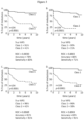
- partition tree, K-nearest neighbor, logistic regression, and discriminant analysis were performed ( Figure 2 and 3 ).
- Training set ROC, accuracy, sensitivity, specificity and K-M 5-year MFS for class 1 and class 2 cases were highly comparable to the RBM method.
- Highly accurate prediction of metastasis, and significantly different 5-year MFS between class 1 and class 2 cases was observed when using partition tree ( Figure 2A ), K-nearest neighbor ( Figure 2B ), logistic regression ( Figure 2C ), or discriminant analysis ( Figure 2D ).
- stage 0 in situ melanoma samples were removed from all stage 0 the training and validation cohorts, generating a new training set of 149 cases; or b) all stage 0 samples were included in the training set only, generating a training set comprised of 164 cases.
- the new cutaneous melanoma predictive training sets were used to train a 104 sample validation cohort.
- sensitivity is 85% for the 164 sample training set and 89% for the 104 sample validation set when the 28-gene signature incorporating all discriminant genes is used to predict metastatic risk. Smaller sets of genes were generated that had equal or increased sensitivity compared to the 28-gene signature (Table 3, bolded). However, the majority of gene sets that did not include all 28 genes were not able to produce the sensitivity thresholds required for use in a clinically feasible GEP test.
- Table 3 Sensitivity, or accuracy of predicting a metastatic event, achieved when using the 28-gene signature or smaller subsets of genes.
- Gene set # of variables Sensitivity training/validation sets (%) All discriminant genes 28 89/94 SPP1, CXCL14, BAP1 varB, CLCA2, S100A8, BTG1 6 79/89 TACSTD2, RBM23, PPL, S100A8, MGP, TYRP1 6 86/78 SAP130, ARG1, KRT6B, GJA1, EIF1B, ID2 6 81/81 CRABP2, KRT14, ROBO1, RBM23, TACSTD2, DSC1 6 70/81 ROBO1, CST6, BAP1 varB, ID2, SPRR1B, KRT6B 6 75/83 SPRR1B, AQP3, PPL, DSC1,TYRP1, TRIM29 6 81/81 KRT6B, GJA1, AQP3, TRIM29, TYRP1, RBM23, MGP,
- Example 6 28-gene signature for cutaneous melanoma predicts aggressive tumor progression to SLN, distant lymph nodes, in transit metastasis, distant metastasis, and local recurrence
- Training set Validation set total called class 2 total called class 2 SLN+ (stage III) samples 17 14 (82%) 12 11 (92%) documented met event 12 11 (92%) 11 10 (91%) no documented met event 5 3 (60%) 1 1 (100%) distant lymph node metastasis 6 (2 SLN+) 6 (100%) 7 (5 SLN+) 6 (86%) in transit metastasis 3 2 (66%) 4 4 (100%) local recurrence 9 4* (44%) 5** 3 (60%) *2/4 LR have known metastatic event (both called class 2) **1/5 LR is stage III (SLN+) - called class 2
- Example 7 Statistical comparison of GEP signature to common CM prognostic factors
- An important clinical application of the GEP test is to identify patients who are staged as low risk (clinical stage I and II) for metastasis, but who are at actual high risk for metastasis based upon the genomic signature of their CM.
- Example 8 28-gene signature for cutaneous melanoma predicts metastasis in superficial spreading and nodular growth patterns
- Probability of the metastatic risk prediction is based upon the coefficients of the 28 variables (genes) and reported as a value between 0 and 1. For the two-class analysis, cases with a probability score between 0 and 0.5 are designated low risk, class 1 cases. Conversely, cases with a probability score between 0.5 and 1.0 are classified as class 2, high risk.
- the linear output of the probability score can also be used directly to assess the risk associated with the genetic signature of particular CM tumor. As shown in Figure 7 , 5 year MFS of cases in the 104 sample validation cohort is strongly correlated with the probability of metastasis. Assessment of all cases with a probability of metastasis lower than 0.299 shows that none of those cases has had a documented metastatic event to date, and 5 year MFS is 100%.
- Example 10 Prediction of distant metastasis free survival (DMFS) and overall survival (OS) using the GEP prognostic signature.
- DMFS distant metastasis free survival
- OS overall survival
- MFS metastasis free survival
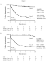
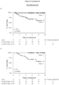
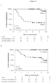
- Kaplan-Meier analysis was performed for compare MFS, DMFS and OS in two related validation sets, the first consisting of 104 CM cases that includes Stage I-IV melanomas ( Figure 8A-C ), and a second smaller subset of 78 CM cases that includes only Stage I and Stage II melanomas ( Figure 8D-F ).
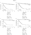
- DMFS was determined for the GEP signature, and the results were compared to Breslow thickness, ulceration and mitotic rate, three clinical T-factors commonly assessed to determine AJCC stage for each CM patient ( Figure 9 ).
- DMFS is grouped by GEP class assignment ( Figure 9A ), Breslow thickness of greater or less than 0.75mm ( Figure 9B ), presence or absence of ulceration ( Figure 9C ), or mitotic rate of greater or less than 1/mm 2 ( Figure 9D ).
- the comparison indicates that the GEP signature has prognostic accuracy better than, or comparable to, the clinical factors currently recommended for the clinical assessment of CM patient risk.
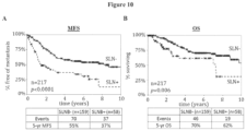
- Example 11 Comparison of prognostic GEP signature to sentinel lymph node biopsy for predicting regional and distant metastasis.
- the AJCC staging parameters of the SLNB positive and negative patients is summarized in Table 7.
- Figure 10 shows the KM curves for metastasis free survival (MFS) and overall survival (OS) for the SLNB negative and the SLNB positive patients.
- MFS metastasis free survival
- OS overall survival
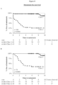
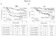
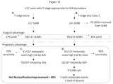
- Figure 12 illustrates that using GEP classification in combination with SLNB status further improves prognostication.
- the overall 5-year MFS and 5-year OS for Class 1 patients is 79% and 89%, respectively, and for SLNB negative patients is 55% and 70%, respectively.
- the 31% of patients with a GEP Class 1 and a negative SLNB result had an overall 5-year MFS and 5-year OS of 83% and 91%, respectively.
- the overall 5-year MFS and 5 year OS for GEP Class 2 patients is 34% and 54% respectively and for SLNB positive patients is 37% and 62%.
- the 23% of patients with a GEP Class 2 and a positive SLNB had an overall 5-year MFS and 5 year OS of 33% and 57%.
- the 5-year MFS and 5-year OS were 35% and 54%, respectively, similar to those patients who were Class 2 with a positive SLNB result. Only a small cohort of 9 patients (representing 4%) with a Class 1 GEP score, but positive SLNB result, was identified. The overall 5-year MFS and 5-year OS for this group was 53% and 78%, respectively. The respective p -values for the difference between the four combined GEP and SLNB groups was ⁇ 0.0001 for both MFS and OS analyses.
- the invention pertains also to the following:
Landscapes
- Chemical & Material Sciences (AREA)
- Life Sciences & Earth Sciences (AREA)
- Health & Medical Sciences (AREA)
- Organic Chemistry (AREA)
- Proteomics, Peptides & Aminoacids (AREA)
- Engineering & Computer Science (AREA)
- Immunology (AREA)
- Pathology (AREA)
- Analytical Chemistry (AREA)
- Zoology (AREA)
- Wood Science & Technology (AREA)
- Genetics & Genomics (AREA)
- Hospice & Palliative Care (AREA)
- Biochemistry (AREA)
- Microbiology (AREA)
- Molecular Biology (AREA)
- Biophysics (AREA)
- Physics & Mathematics (AREA)
- Oncology (AREA)
- Biotechnology (AREA)
- Bioinformatics & Cheminformatics (AREA)
- General Engineering & Computer Science (AREA)
- General Health & Medical Sciences (AREA)
- Measuring Or Testing Involving Enzymes Or Micro-Organisms (AREA)
- Investigating Or Analysing Biological Materials (AREA)
- User Interface Of Digital Computer (AREA)
- Steroid Compounds (AREA)
Applications Claiming Priority (4)
| Application Number | Priority Date | Filing Date | Title |
|---|---|---|---|
| US201361783755P | 2013-03-14 | 2013-03-14 | |
| EP14713968.7A EP2971085B1 (fr) | 2013-03-14 | 2014-02-28 | Méthodes de prédiction du risque de métastase dans un mélanome cutané |
| PCT/US2014/019326 WO2014158696A1 (fr) | 2013-03-14 | 2014-02-28 | Méthodes de prédiction du risque de métastase dans un mélanome cutané |
| EP18187484.3A EP3425061B1 (fr) | 2013-03-14 | 2014-02-28 | Méthodes de prédiction du risque de métastase dans un mélanome cutané |
Related Parent Applications (2)
| Application Number | Title | Priority Date | Filing Date |
|---|---|---|---|
| EP18187484.3A Division EP3425061B1 (fr) | 2013-03-14 | 2014-02-28 | Méthodes de prédiction du risque de métastase dans un mélanome cutané |
| EP14713968.7A Division EP2971085B1 (fr) | 2013-03-14 | 2014-02-28 | Méthodes de prédiction du risque de métastase dans un mélanome cutané |
Publications (3)
| Publication Number | Publication Date |
|---|---|
| EP4265740A2 true EP4265740A2 (fr) | 2023-10-25 |
| EP4265740A3 EP4265740A3 (fr) | 2024-04-24 |
| EP4265740B1 EP4265740B1 (fr) | 2026-04-08 |
Family
ID=50391379
Family Applications (3)
| Application Number | Title | Priority Date | Filing Date |
|---|---|---|---|
| EP18187484.3A Active EP3425061B1 (fr) | 2013-03-14 | 2014-02-28 | Méthodes de prédiction du risque de métastase dans un mélanome cutané |
| EP14713968.7A Active EP2971085B1 (fr) | 2013-03-14 | 2014-02-28 | Méthodes de prédiction du risque de métastase dans un mélanome cutané |
| EP23177369.8A Active EP4265740B1 (fr) | 2013-03-14 | 2014-02-28 | Méthodes de prédiction du risque de métastase dans un mélanome cutané |
Family Applications Before (2)
| Application Number | Title | Priority Date | Filing Date |
|---|---|---|---|
| EP18187484.3A Active EP3425061B1 (fr) | 2013-03-14 | 2014-02-28 | Méthodes de prédiction du risque de métastase dans un mélanome cutané |
| EP14713968.7A Active EP2971085B1 (fr) | 2013-03-14 | 2014-02-28 | Méthodes de prédiction du risque de métastase dans un mélanome cutané |
Country Status (9)
| Country | Link |
|---|---|
| US (7) | US20140272986A1 (fr) |
| EP (3) | EP3425061B1 (fr) |
| AU (3) | AU2014241995B2 (fr) |
| BR (1) | BR112015022977B1 (fr) |
| CA (1) | CA2904283A1 (fr) |
| ES (2) | ES2947946T3 (fr) |
| IL (1) | IL241086B (fr) |
| NZ (1) | NZ712575A (fr) |
| WO (1) | WO2014158696A1 (fr) |
Families Citing this family (7)
| Publication number | Priority date | Publication date | Assignee | Title |
|---|---|---|---|---|
| CA2904283A1 (fr) | 2013-03-14 | 2014-10-02 | Castle Biosciences, Inc. | Methodes de prediction du risque de metastase dans un melanome cutane |
| US20170329914A1 (en) * | 2016-05-11 | 2017-11-16 | International Business Machines Corporation | Predicting Personalized Cancer Metastasis Routes, Biological Mediators of Metastasis and Metastasis Blocking Therapies |
| WO2017210662A1 (fr) * | 2016-06-03 | 2017-12-07 | Castle Biosciences, Inc. | Procédés de prédiction du risque de récurrence et/ou de métastases dans le sarcome des tissus mous |
| US20180307741A1 (en) * | 2017-04-25 | 2018-10-25 | Intel Corporation | Filtering training data for simpler rbf models |
| US11767564B2 (en) | 2017-10-27 | 2023-09-26 | Board Of Regents, The University Of Texas System | Use of SDHA as a prognostic marker and therapeutic target in uveal melanoma |
| CN109485418B (zh) * | 2018-12-14 | 2021-07-20 | 华南理工大学 | 一种高效抗肿瘤铌酸钾钠基压电材料及其制备方法与应用 |
| CA3240376A1 (fr) * | 2021-12-08 | 2023-06-15 | Alexander MEVES | Evaluation et traitement d'un melanome |
Family Cites Families (13)
| Publication number | Priority date | Publication date | Assignee | Title |
|---|---|---|---|---|
| GB0504302D0 (en) * | 2005-03-02 | 2005-04-06 | Univ Dublin | Markers for melanoma |
| US20070082347A1 (en) * | 2005-06-08 | 2007-04-12 | Myriad Genetics, Incorporated | Gene variants and use thereof |
| PT2062049E (pt) * | 2006-09-06 | 2014-07-28 | Univ California | Diagnóstico molecular e classificação de melanoma maligno |
| WO2008031041A2 (fr) * | 2006-09-07 | 2008-03-13 | H. Lee Moffitt Cancer Center And Research Institute, Inc. | Signature de gène de mélanome |
| NZ555363A (en) | 2007-05-24 | 2009-11-27 | Pacific Edge Biotechnology Ltd | Prognosis prediction for melanoma cancer |
| CA2716357A1 (fr) * | 2008-03-05 | 2009-09-11 | The Regents Of The University Of California | Diagnostic moleculaire et classification de melanomes malins |
| WO2009131733A1 (fr) * | 2008-04-21 | 2009-10-29 | The Trustees Of Columbia University In The City Of New York | Amplification de gab2 dans un mélanome |
| EP2279416A4 (fr) * | 2008-04-22 | 2011-08-24 | Univ Washington | Procédé de prédiction du risque de métastase |
| AU2009262022A1 (en) * | 2008-06-26 | 2009-12-30 | Dana-Farber Cancer Institute, Inc. | Signatures and determinants associated with metastasis and methods of use thereof |
| WO2011039734A2 (fr) * | 2009-10-02 | 2011-04-07 | Enzo Medico | Utilisation de gènes impliqués dans l'indépendance d'ancrage pour l'optimisation du diagnostic et du traitement du cancer humain |
| EP2619321B1 (fr) * | 2010-09-20 | 2018-08-01 | Advanced Cell Diagnostics, Inc. | Marqueurs biologiques permettant de différencier un mélanome d'un nævus bénin sur la peau |
| WO2012125411A1 (fr) * | 2011-03-11 | 2012-09-20 | Metamark Genetics, Inc. | Procédés de prédiction du pronostic dans le cancer |
| CA2904283A1 (fr) | 2013-03-14 | 2014-10-02 | Castle Biosciences, Inc. | Methodes de prediction du risque de metastase dans un melanome cutane |
-
2014
- 2014-02-28 CA CA2904283A patent/CA2904283A1/fr active Pending
- 2014-02-28 US US14/193,378 patent/US20140272986A1/en not_active Abandoned
- 2014-02-28 ES ES18187484T patent/ES2947946T3/es active Active
- 2014-02-28 EP EP18187484.3A patent/EP3425061B1/fr active Active
- 2014-02-28 AU AU2014241995A patent/AU2014241995B2/en active Active
- 2014-02-28 WO PCT/US2014/019326 patent/WO2014158696A1/fr not_active Ceased
- 2014-02-28 EP EP14713968.7A patent/EP2971085B1/fr active Active
- 2014-02-28 BR BR112015022977-8A patent/BR112015022977B1/pt active IP Right Grant
- 2014-02-28 US US14/193,355 patent/US20140271545A1/en not_active Abandoned
- 2014-02-28 EP EP23177369.8A patent/EP4265740B1/fr active Active
- 2014-02-28 NZ NZ712575A patent/NZ712575A/en unknown
- 2014-02-28 ES ES14713968.7T patent/ES2690584T3/es active Active
-
2015
- 2015-09-03 IL IL241086A patent/IL241086B/en active IP Right Grant
-
2016
- 2016-03-19 US US15/075,133 patent/US10577660B2/en active Active
-
2020
- 2020-01-17 US US16/745,998 patent/US11434536B2/en active Active
- 2020-04-15 AU AU2020202536A patent/AU2020202536B2/en active Active
-
2022
- 2022-06-30 AU AU2022204690A patent/AU2022204690B2/en active Active
- 2022-08-08 US US17/883,223 patent/US12365948B2/en active Active
-
2025
- 2025-06-17 US US19/240,887 patent/US20250305065A1/en active Pending
- 2025-07-21 US US19/275,919 patent/US20260015676A1/en active Pending
Non-Patent Citations (30)
| Title |
|---|
| BALCH CMBUZAID ACSOONG SJ ET AL.: "Final version of the American Joint Committee on Cancer staging system for cutaneous melanoma", J CLIN ONCOL, vol. 19, 2001, pages 3635 - 48, XP009024807 |
| BALCH CMGERSHENWALD JESOONG SJ ET AL.: "Final version of 2009 AJCC melanoma staging and classification", J CLIN ONCOL, vol. 27, 2009, pages 6199 - 206 |
| BALCH CMGERSHENWALD JESOONG SJ ET AL.: "Multivariate analysis of prognostic factors among 2,313 patients with stage III melanoma: comparison of nodal micrometastases versus macrometastases", J CLIN ONCOL, vol. 28, 2010, pages 2452 - 9 |
| BALCH CMSOONG SJGERSHENWALD JE ET AL.: "Prognostic factors analysis of 17,600 melanoma patients: validation of the American Joint Committee on Cancer melanoma staging system", J CLIN ONCOL, vol. 19, 2001, pages 3622 - 34 |
| BITTNER MMELTZER PCHEN Y ET AL.: "Molecular classification of cutaneous malignant melanoma by gene expression profiling", NATURE, vol. 406, 2000, pages 536 - 40, XP037510086, DOI: 10.1038/35020115 |
| COIT DG, MELANOMA VERSION 2.2013: NATIONAL COMPREHENSIVE CANCER NETWORK, February 2013 (2013-02-01) |
| COLMAN HZHANG LSULMAN EP ET AL.: "A multigene predictor of outcome in glioblastoma", NEURO ONCOL, vol. 12, 2010, pages 49 - 57, XP055191030, DOI: 10.1093/neuonc/nop007 |
| EDGE SBCOMPTON CC: "The American Joint Committee on Cancer: the 7th edition of the AJCC cancer staging manual and the future of TNM", ANN SURG ONCOL, vol. 17, 2010, pages 1471 - 344 |
| FRANCIS PNAMLOS HMMULLER C ET AL.: "Diagnostic and prognostic gene expression signatures in 177 soft tissue sarcomas: hypoxia-induced transcription profile signifies metastatic potential", BMC GENOMICS, vol. 8, 2007, pages 73, XP021022381, DOI: 10.1186/1471-2164-8-73 |
| GORDON GJJENSEN RVHSIAO LL ET AL.: "Using gene expression ratios to predict outcome among patients with mesothelioma", J NATL CANCER INST, vol. 95, 2003, pages 598 - 605, XP055121820, DOI: 10.1093/jnci/95.8.598 |
| HAQQ CNOSRATI MSUDILOVSKY D ET AL.: "The gene expression signatures of melanoma progression", PROC NATL ACAD SCI U S A, vol. 102, 2005, pages 6092 - 7, XP002562661, DOI: 10.1073/pnas.0501564102 |
| JAEGER JKOCZAN DTHIESEN HJ ET AL.: "Gene expression signatures for tumor progression, tumor subtype, and tumor thickness in laser-microdissected melanoma tissues", CLIN CANCER RES, vol. 13, 2007, pages 806 - 15, XP055122141, DOI: 10.1158/1078-0432.CCR-06-1820 |
| KARIM RZSCOLYER RALI W ET AL.: "False negative sentinel lymph node biopsies in melanoma may result from deficiencies in nuclear medicine, surgery, or pathology", ANN SURG, vol. 247, 2008, pages 1003 - 10 |
| MANIATIS ET AL.: "CURRENT PROTOCOLS IN MOLECULAR BIOLOGY", 1989, GREENE PUBLISHING ASSOCIATES AND WILEY INTERSCIENCE |
| MAUERER AROESCH AHAFNER C ET AL.: "Identification of new genes associated with melanoma", EXP DERMATOL, vol. 20, 2011, pages 502 - 7, XP055122228, DOI: 10.1111/j.1600-0625.2011.01254.x |
| MORTON DL: "Overview and update of the phase III Multicenter Selective Lymphadenectomy Trials (MSLT-I and MSLT-II) in melanoma", CLIN EXP METASTASIS, vol. 29, 2012, pages 699 - 706, XP035131926, DOI: 10.1007/s10585-012-9503-3 |
| NICHOLL MBELASHOFF DTAKEUCHI HMORTON DLHOON DS: "Molecular upstaging based on paraffin-embedded sentinel lymph nodes: ten-year follow-up confirms prognostic utility in melanoma patients", ANN SURG, vol. 253, 2011, pages 116 - 22 |
| ONKEN MDWORLEY LACHAR DH ET AL.: "Collaborative Ocular Oncology Group Report Number 1: Prospective Validation of a Multi-Gene Prognostic Assay in Uveal Melanoma", OPHTHALMOLOGY, 2012 |
| ONKEN MDWORLEY LAEHLERS JPHARBOUR JW: "Gene expression profiling in uveal melanoma reveals two molecular classes and predicts metastatic death", CANCER RES, vol. 64, 2004, pages 7205 - 9, XP002638996, DOI: 10.1158/0008-5472.CAN-04-1750 |
| ONKEN MDWORLEY LATUSCAN MDHARBOUR JW: "An accurate, clinically feasible multi-gene expression assay for predicting metastasis in uveal melanoma", J MOL DIAGN, vol. 12, 2010, pages 461 - 8, XP055523610, DOI: 10.2353/jmoldx.2010.090220 |
| PAIK SSHAK STANG G ET AL.: "A multigene assay to predict recurrence of tamoxifen-treated, node-negative breast cancer", N ENGL J MED, vol. 351, 2004, pages 2817 - 26, XP002578486 |
| SATO T: "Locoregional management of hepatic metastasis from primary uveal melanoma", SEMIN ONCOL, vol. 37, 2010, pages 127 - 38 |
| SCATOLINI MGRAND MMGROSSO E ET AL.: "Altered molecular pathways in melanocytic lesions", INT J CANCER, vol. 126, 2010, pages 1869 - 81, XP071285742, DOI: 10.1002/ijc.24899 |
| SIEGEL RNAISHADHAM DJEMAL A: "Cancer statistics", CA CANCER J CLIN 2012, vol. 62, 2012, pages 10 - 29, XP055066773, DOI: 10.3322/caac.20138 |
| SMITH APHOEK KBECKER D: "Whole-genome expression profiling of the melanoma progression pathway reveals marked molecular differences between nevi/melanoma in situ and advanced-stage melanomas", CANCER BIOL THER, vol. 4, 2005, pages 1018 - 29, XP055086915, DOI: 10.4161/cbt.4.9.2165 |
| TAKEUCHI HMORTON DLKUO C ET AL.: "Prognostic significance of molecular upstaging of paraffin-embedded sentinel lymph nodes in melanoma patients", J CLIN ONCOL, vol. 22, 2004, pages 2671 - 80 |
| WEERARATNA ATBECKER DCARR KM ET AL.: "Generation and analysis of melanoma SAGE libraries: SAGE advice on the melanoma transcriptome", ONCOGENE, vol. 23, 2004, pages 2264 - 74, XP037739123, DOI: 10.1038/sj.onc.1207337 |
| WINNEPENNINCKX VLAZAR VMICHIELS S ET AL.: "Gene expression profiling of primary cutaneous melanoma and clinical outcome", J NATL CANCER INST, vol. 98, 2006, pages 472 - 82, XP002611616, DOI: 10.1093/JNCI/DJJ103 |
| YEE VSTHOMPSON JFMCKINNON JG ET AL.: "Outcome in 846 cutaneous melanoma patients from a single center after a negative sentinel node biopsy", ANN SURG ONCOL, vol. 12, 2005, pages 429 - 39, XP019369693 |
| ZETTERSTEN ESHAIKH LRAMIREZ RKASHANI-SABET M: "Prognostic factors in primary cutaneous melanoma", SURG CLIN NORTH AM, vol. 83, 2003, pages 61 - 75 |
Also Published As
Similar Documents
| Publication | Publication Date | Title |
|---|---|---|
| AU2020202536B2 (en) | Methods For Predicting Risk Of Metastasis In Cutaneous Melanoma | |
| RU2654587C2 (ru) | Способ предсказания рецидива рака молочной железы при эндокринном лечении | |
| EP2390370A2 (fr) | Procédé de prédiction d'une réponse à une tumeur chez un patient souffrant ou risquant de développer un cancer gynécologique récurrent vers un agent chimiothérapique | |
| EP3788167A1 (fr) | Procédés de diagnostic et de traitement de patients atteints d'un carcinome épidermoïde cutané | |
| CN113039289B (zh) | 预测黑色素瘤转移和患者预后的基因特征 | |
| EP4097260A1 (fr) | Méthodes de diagnostic et de traitement de patients atteints d'un carcinome épidermoïde cutané | |
| JP2012531925A (ja) | チボザニブ応答予測 | |
| Ferraz et al. | Liquid biopsy as a potential tool for diagnosis and clinical monitoring of cutaneous and uveal melanoma | |
| AU2021325154A1 (en) | Methods of diagnosing and treating patients with cutaneous squamous cell carcinoma | |
| EP2083087B1 (fr) | Procede de diagnostic d'un cancer de la langue | |
| US20220033882A1 (en) | Methods of diagnosing and treating patients with pigmented skin lesions |
Legal Events
| Date | Code | Title | Description |
|---|---|---|---|
| PUAI | Public reference made under article 153(3) epc to a published international application that has entered the european phase |
Free format text: ORIGINAL CODE: 0009012 |
|
| STAA | Information on the status of an ep patent application or granted ep patent |
Free format text: STATUS: THE APPLICATION HAS BEEN PUBLISHED |
|
| AC | Divisional application: reference to earlier application |
Ref document number: 2971085 Country of ref document: EP Kind code of ref document: P Ref document number: 3425061 Country of ref document: EP Kind code of ref document: P |
|
| AK | Designated contracting states |
Kind code of ref document: A2 Designated state(s): AL AT BE BG CH CY CZ DE DK EE ES FI FR GB GR HR HU IE IS IT LI LT LU LV MC MK MT NL NO PL PT RO RS SE SI SK SM TR |
|
| RIC1 | Information provided on ipc code assigned before grant |
Ipc: C12Q 1/6886 20180101AFI20231220BHEP |
|
| PUAL | Search report despatched |
Free format text: ORIGINAL CODE: 0009013 |
|
| AK | Designated contracting states |
Kind code of ref document: A3 Designated state(s): AL AT BE BG CH CY CZ DE DK EE ES FI FR GB GR HR HU IE IS IT LI LT LU LV MC MK MT NL NO PL PT RO RS SE SI SK SM TR |
|
| RIC1 | Information provided on ipc code assigned before grant |
Ipc: C12Q 1/6886 20180101AFI20240319BHEP |
|
| STAA | Information on the status of an ep patent application or granted ep patent |
Free format text: STATUS: REQUEST FOR EXAMINATION WAS MADE |
|
| 17P | Request for examination filed |
Effective date: 20241009 |
|
| RBV | Designated contracting states (corrected) |
Designated state(s): AL AT BE BG CH CY CZ DE DK EE ES FI FR GB GR HR HU IE IS IT LI LT LU LV MC MK MT NL NO PL PT RO RS SE SI SK SM TR |
|
| GRAP | Despatch of communication of intention to grant a patent |
Free format text: ORIGINAL CODE: EPIDOSNIGR1 |
|
| STAA | Information on the status of an ep patent application or granted ep patent |
Free format text: STATUS: GRANT OF PATENT IS INTENDED |
|
| INTG | Intention to grant announced |
Effective date: 20251104 |
|
| GRAS | Grant fee paid |
Free format text: ORIGINAL CODE: EPIDOSNIGR3 |
|
| GRAA | (expected) grant |
Free format text: ORIGINAL CODE: 0009210 |
|
| STAA | Information on the status of an ep patent application or granted ep patent |
Free format text: STATUS: THE PATENT HAS BEEN GRANTED |
|
| AC | Divisional application: reference to earlier application |
Ref document number: 3425061 Country of ref document: EP Kind code of ref document: P Ref document number: 2971085 Country of ref document: EP Kind code of ref document: P |
|
| AK | Designated contracting states |
Kind code of ref document: B1 Designated state(s): AL AT BE BG CH CY CZ DE DK EE ES FI FR GB GR HR HU IE IS IT LI LT LU LV MC MK MT NL NO PL PT RO RS SE SI SK SM TR |
|
| REG | Reference to a national code |
Ref country code: CH Ref legal event code: F10 Free format text: ST27 STATUS EVENT CODE: U-0-0-F10-F00 (AS PROVIDED BY THE NATIONAL OFFICE) Effective date: 20260408 Ref country code: GB Ref legal event code: FG4D |
|
| REG | Reference to a national code |
Ref country code: DE Ref legal event code: R096 Ref document number: 602014092951 Country of ref document: DE |