EP4265714A1 - Matrice extracellulaire dérivée de tissu décellularisé modifiée avec un dérivé de phénol et utilisation associée - Google Patents
Matrice extracellulaire dérivée de tissu décellularisé modifiée avec un dérivé de phénol et utilisation associée Download PDFInfo
- Publication number
- EP4265714A1 EP4265714A1 EP22742760.6A EP22742760A EP4265714A1 EP 4265714 A1 EP4265714 A1 EP 4265714A1 EP 22742760 A EP22742760 A EP 22742760A EP 4265714 A1 EP4265714 A1 EP 4265714A1
- Authority
- EP
- European Patent Office
- Prior art keywords
- hydrogel
- tissue
- composition
- extracellular matrix
- group
- Prior art date
- Legal status (The legal status is an assumption and is not a legal conclusion. Google has not performed a legal analysis and makes no representation as to the accuracy of the status listed.)
- Pending
Links
- 210000001519 tissue Anatomy 0.000 title claims abstract description 110
- 210000002744 extracellular matrix Anatomy 0.000 title claims abstract description 64
- 102000010834 Extracellular Matrix Proteins Human genes 0.000 title claims abstract description 63
- 108010037362 Extracellular Matrix Proteins Proteins 0.000 title claims abstract description 63
- ISWSIDIOOBJBQZ-UHFFFAOYSA-N phenol group Chemical group C1(=CC=CC=C1)O ISWSIDIOOBJBQZ-UHFFFAOYSA-N 0.000 title 1
- 239000000017 hydrogel Substances 0.000 claims abstract description 318
- 239000000203 mixture Substances 0.000 claims abstract description 39
- 150000002989 phenols Chemical class 0.000 claims abstract description 35
- 238000002513 implantation Methods 0.000 claims abstract description 17
- 230000017423 tissue regeneration Effects 0.000 claims abstract description 16
- 230000001737 promoting effect Effects 0.000 claims abstract description 15
- 238000012377 drug delivery Methods 0.000 claims abstract description 14
- 239000003106 tissue adhesive Substances 0.000 claims abstract description 10
- 230000024245 cell differentiation Effects 0.000 claims abstract description 9
- 238000004113 cell culture Methods 0.000 claims abstract description 8
- YCIMNLLNPGFGHC-UHFFFAOYSA-N catechol Chemical group OC1=CC=CC=C1O YCIMNLLNPGFGHC-UHFFFAOYSA-N 0.000 claims description 42
- 239000003814 drug Substances 0.000 claims description 34
- 229940079593 drug Drugs 0.000 claims description 34
- 210000000130 stem cell Anatomy 0.000 claims description 27
- 210000004027 cell Anatomy 0.000 claims description 26
- 238000000034 method Methods 0.000 claims description 22
- WQGWDDDVZFFDIG-UHFFFAOYSA-N pyrogallyl group Chemical group C1(=C(C(=CC=C1)O)O)O WQGWDDDVZFFDIG-UHFFFAOYSA-N 0.000 claims description 19
- 210000000988 bone and bone Anatomy 0.000 claims description 18
- 210000003491 skin Anatomy 0.000 claims description 17
- 230000001070 adhesive effect Effects 0.000 claims description 14
- 230000001590 oxidative effect Effects 0.000 claims description 14
- 102000004190 Enzymes Human genes 0.000 claims description 13
- 108090000790 Enzymes Proteins 0.000 claims description 13
- 239000007800 oxidant agent Substances 0.000 claims description 13
- VYFYYTLLBUKUHU-UHFFFAOYSA-N dopamine Chemical compound NCCC1=CC=C(O)C(O)=C1 VYFYYTLLBUKUHU-UHFFFAOYSA-N 0.000 claims description 12
- 238000007254 oxidation reaction Methods 0.000 claims description 10
- 230000008569 process Effects 0.000 claims description 10
- 230000003647 oxidation Effects 0.000 claims description 9
- CFFZDZCDUFSOFZ-UHFFFAOYSA-N 3,4-Dihydroxy-phenylacetic acid Chemical compound OC(=O)CC1=CC=C(O)C(O)=C1 CFFZDZCDUFSOFZ-UHFFFAOYSA-N 0.000 claims description 8
- RGCKGOZRHPZPFP-UHFFFAOYSA-N alizarin Chemical compound C1=CC=C2C(=O)C3=C(O)C(O)=CC=C3C(=O)C2=C1 RGCKGOZRHPZPFP-UHFFFAOYSA-N 0.000 claims description 8
- 239000002872 contrast media Substances 0.000 claims description 7
- 239000003242 anti bacterial agent Substances 0.000 claims description 6
- 229960003638 dopamine Drugs 0.000 claims description 6
- 229940079877 pyrogallol Drugs 0.000 claims description 6
- 239000000853 adhesive Substances 0.000 claims description 5
- 239000003102 growth factor Substances 0.000 claims description 5
- JWZZKOKVBUJMES-UHFFFAOYSA-N (+-)-Isoprenaline Chemical compound CC(C)NCC(O)C1=CC=C(O)C(O)=C1 JWZZKOKVBUJMES-UHFFFAOYSA-N 0.000 claims description 4
- BRRSNXCXLSVPFC-UHFFFAOYSA-N 2,3,4-Trihydroxybenzoic acid Chemical compound OC(=O)C1=CC=C(O)C(O)=C1O BRRSNXCXLSVPFC-UHFFFAOYSA-N 0.000 claims description 4
- RGZHEOWNTDJLAQ-UHFFFAOYSA-N 3,4,5-trihydroxybenzaldehyde Chemical compound OC1=CC(C=O)=CC(O)=C1O RGZHEOWNTDJLAQ-UHFFFAOYSA-N 0.000 claims description 4
- YQUVCSBJEUQKSH-UHFFFAOYSA-N 3,4-dihydroxybenzoic acid Chemical compound OC(=O)C1=CC=C(O)C(O)=C1 YQUVCSBJEUQKSH-UHFFFAOYSA-N 0.000 claims description 4
- XESZUVZBAMCAEJ-UHFFFAOYSA-N 4-tert-butylcatechol Chemical compound CC(C)(C)C1=CC=C(O)C(O)=C1 XESZUVZBAMCAEJ-UHFFFAOYSA-N 0.000 claims description 4
- WTDRDQBEARUVNC-UHFFFAOYSA-N L-Dopa Natural products OC(=O)C(N)CC1=CC=C(O)C(O)=C1 WTDRDQBEARUVNC-UHFFFAOYSA-N 0.000 claims description 4
- 150000001875 compounds Chemical class 0.000 claims description 4
- XMOCLSLCDHWDHP-IUODEOHRSA-N epi-Gallocatechin Chemical compound C1([C@H]2OC3=CC(O)=CC(O)=C3C[C@H]2O)=CC(O)=C(O)C(O)=C1 XMOCLSLCDHWDHP-IUODEOHRSA-N 0.000 claims description 4
- LNTHITQWFMADLM-UHFFFAOYSA-N gallic acid Chemical compound OC(=O)C1=CC(O)=C(O)C(O)=C1 LNTHITQWFMADLM-UHFFFAOYSA-N 0.000 claims description 4
- QAIPRVGONGVQAS-DUXPYHPUSA-N trans-caffeic acid Chemical compound OC(=O)\C=C\C1=CC=C(O)C(O)=C1 QAIPRVGONGVQAS-DUXPYHPUSA-N 0.000 claims description 4
- 102000004127 Cytokines Human genes 0.000 claims description 3
- 108090000695 Cytokines Proteins 0.000 claims description 3
- CTENFNNZBMHDDG-UHFFFAOYSA-N Dopamine hydrochloride Chemical compound Cl.NCCC1=CC=C(O)C(O)=C1 CTENFNNZBMHDDG-UHFFFAOYSA-N 0.000 claims description 3
- 230000003444 anaesthetic effect Effects 0.000 claims description 3
- 229940121363 anti-inflammatory agent Drugs 0.000 claims description 3
- 239000002260 anti-inflammatory agent Substances 0.000 claims description 3
- 239000002246 antineoplastic agent Substances 0.000 claims description 3
- 239000003443 antiviral agent Substances 0.000 claims description 3
- 230000003115 biocidal effect Effects 0.000 claims description 3
- 210000004204 blood vessel Anatomy 0.000 claims description 3
- 210000004556 brain Anatomy 0.000 claims description 3
- 210000000845 cartilage Anatomy 0.000 claims description 3
- 229960001149 dopamine hydrochloride Drugs 0.000 claims description 3
- 210000003238 esophagus Anatomy 0.000 claims description 3
- 230000003779 hair growth Effects 0.000 claims description 3
- 210000002216 heart Anatomy 0.000 claims description 3
- 210000002865 immune cell Anatomy 0.000 claims description 3
- 210000000936 intestine Anatomy 0.000 claims description 3
- 210000003734 kidney Anatomy 0.000 claims description 3
- 210000003041 ligament Anatomy 0.000 claims description 3
- 210000004185 liver Anatomy 0.000 claims description 3
- 210000004072 lung Anatomy 0.000 claims description 3
- 210000003205 muscle Anatomy 0.000 claims description 3
- 210000000496 pancreas Anatomy 0.000 claims description 3
- 108090000623 proteins and genes Proteins 0.000 claims description 3
- 102000004169 proteins and genes Human genes 0.000 claims description 3
- 210000003079 salivary gland Anatomy 0.000 claims description 3
- 210000000278 spinal cord Anatomy 0.000 claims description 3
- 210000002784 stomach Anatomy 0.000 claims description 3
- 210000002435 tendon Anatomy 0.000 claims description 3
- 230000001225 therapeutic effect Effects 0.000 claims description 3
- 210000001685 thyroid gland Anatomy 0.000 claims description 3
- 210000000515 tooth Anatomy 0.000 claims description 3
- 210000003932 urinary bladder Anatomy 0.000 claims description 3
- 210000004291 uterus Anatomy 0.000 claims description 3
- WMBWREPUVVBILR-WIYYLYMNSA-N (-)-Epigallocatechin-3-o-gallate Chemical compound O([C@@H]1CC2=C(O)C=C(C=C2O[C@@H]1C=1C=C(O)C(O)=C(O)C=1)O)C(=O)C1=CC(O)=C(O)C(O)=C1 WMBWREPUVVBILR-WIYYLYMNSA-N 0.000 claims description 2
- LSHVYAFMTMFKBA-TZIWHRDSSA-N (-)-epicatechin-3-O-gallate Chemical compound O([C@@H]1CC2=C(O)C=C(C=C2O[C@@H]1C=1C=C(O)C(O)=CC=1)O)C(=O)C1=CC(O)=C(O)C(O)=C1 LSHVYAFMTMFKBA-TZIWHRDSSA-N 0.000 claims description 2
- SFLSHLFXELFNJZ-QMMMGPOBSA-N (-)-norepinephrine Chemical compound NC[C@H](O)C1=CC=C(O)C(O)=C1 SFLSHLFXELFNJZ-QMMMGPOBSA-N 0.000 claims description 2
- ACEAELOMUCBPJP-UHFFFAOYSA-N (E)-3,4,5-trihydroxycinnamic acid Natural products OC(=O)C=CC1=CC(O)=C(O)C(O)=C1 ACEAELOMUCBPJP-UHFFFAOYSA-N 0.000 claims description 2
- UCTWMZQNUQWSLP-VIFPVBQESA-N (R)-adrenaline Chemical compound CNC[C@H](O)C1=CC=C(O)C(O)=C1 UCTWMZQNUQWSLP-VIFPVBQESA-N 0.000 claims description 2
- 229930182837 (R)-adrenaline Natural products 0.000 claims description 2
- RMTXUPIIESNLPW-UHFFFAOYSA-N 1,2-dihydroxy-3-(pentadeca-8,11-dienyl)benzene Natural products CCCC=CCC=CCCCCCCCC1=CC=CC(O)=C1O RMTXUPIIESNLPW-UHFFFAOYSA-N 0.000 claims description 2
- TUSDEZXZIZRFGC-UHFFFAOYSA-N 1-O-galloyl-3,6-(R)-HHDP-beta-D-glucose Natural products OC1C(O2)COC(=O)C3=CC(O)=C(O)C(O)=C3C3=C(O)C(O)=C(O)C=C3C(=O)OC1C(O)C2OC(=O)C1=CC(O)=C(O)C(O)=C1 TUSDEZXZIZRFGC-UHFFFAOYSA-N 0.000 claims description 2
- CRPNQSVBEWWHIJ-UHFFFAOYSA-N 2,3,4-trihydroxybenzaldehyde Chemical compound OC1=CC=C(C=O)C(O)=C1O CRPNQSVBEWWHIJ-UHFFFAOYSA-N 0.000 claims description 2
- RBQIPEJXQPQFJX-UHFFFAOYSA-N 3,4,5-trihydroxybenzamide Chemical compound NC(=O)C1=CC(O)=C(O)C(O)=C1 RBQIPEJXQPQFJX-UHFFFAOYSA-N 0.000 claims description 2
- QARRXYBJLBIVAK-UEMSJJPVSA-N 3-[(8e,11e)-pentadeca-8,11-dienyl]benzene-1,2-diol;3-[(8e,11e)-pentadeca-8,11,14-trienyl]benzene-1,2-diol;3-[(8e,11e,13e)-pentadeca-8,11,13-trienyl]benzene-1,2-diol;3-[(e)-pentadec-8-enyl]benzene-1,2-diol;3-pentadecylbenzene-1,2-diol Chemical compound CCCCCCCCCCCCCCCC1=CC=CC(O)=C1O.CCCCCC\C=C\CCCCCCCC1=CC=CC(O)=C1O.CCC\C=C\C\C=C\CCCCCCCC1=CC=CC(O)=C1O.C\C=C\C=C\C\C=C\CCCCCCCC1=CC=CC(O)=C1O.OC1=CC=CC(CCCCCCC\C=C\C\C=C\CC=C)=C1O QARRXYBJLBIVAK-UEMSJJPVSA-N 0.000 claims description 2
- IYROWZYPEIMDDN-UHFFFAOYSA-N 3-n-pentadec-8,11,13-trienyl catechol Natural products CC=CC=CCC=CCCCCCCCC1=CC=CC(O)=C1O IYROWZYPEIMDDN-UHFFFAOYSA-N 0.000 claims description 2
- LCAINUZZHIZKKS-UHFFFAOYSA-N 5-Hydroxydopamine Chemical compound NCCC1=CC(O)=C(O)C(O)=C1 LCAINUZZHIZKKS-UHFFFAOYSA-N 0.000 claims description 2
- NYUABOGYMWADSF-UHFFFAOYSA-N 5-methylbenzene-1,2,3-triol Chemical compound CC1=CC(O)=C(O)C(O)=C1 NYUABOGYMWADSF-UHFFFAOYSA-N 0.000 claims description 2
- HCNISNCKPIVZDX-UHFFFAOYSA-N 5-tert-butylbenzene-1,2,3-triol Chemical compound CC(C)(C)C1=CC(O)=C(O)C(O)=C1 HCNISNCKPIVZDX-UHFFFAOYSA-N 0.000 claims description 2
- LSHVYAFMTMFKBA-UHFFFAOYSA-N ECG Natural products C=1C=C(O)C(O)=CC=1C1OC2=CC(O)=CC(O)=C2CC1OC(=O)C1=CC(O)=C(O)C(O)=C1 LSHVYAFMTMFKBA-UHFFFAOYSA-N 0.000 claims description 2
- 239000001263 FEMA 3042 Substances 0.000 claims description 2
- WMBWREPUVVBILR-UHFFFAOYSA-N GCG Natural products C=1C(O)=C(O)C(O)=CC=1C1OC2=CC(O)=CC(O)=C2CC1OC(=O)C1=CC(O)=C(O)C(O)=C1 WMBWREPUVVBILR-UHFFFAOYSA-N 0.000 claims description 2
- WTDRDQBEARUVNC-LURJTMIESA-N L-DOPA Chemical compound OC(=O)[C@@H](N)CC1=CC=C(O)C(O)=C1 WTDRDQBEARUVNC-LURJTMIESA-N 0.000 claims description 2
- XMOCLSLCDHWDHP-UHFFFAOYSA-N L-Epigallocatechin Natural products OC1CC2=C(O)C=C(O)C=C2OC1C1=CC(O)=C(O)C(O)=C1 XMOCLSLCDHWDHP-UHFFFAOYSA-N 0.000 claims description 2
- LRBQNJMCXXYXIU-PPKXGCFTSA-N Penta-digallate-beta-D-glucose Natural products OC1=C(O)C(O)=CC(C(=O)OC=2C(=C(O)C=C(C=2)C(=O)OC[C@@H]2[C@H]([C@H](OC(=O)C=3C=C(OC(=O)C=4C=C(O)C(O)=C(O)C=4)C(O)=C(O)C=3)[C@@H](OC(=O)C=3C=C(OC(=O)C=4C=C(O)C(O)=C(O)C=4)C(O)=C(O)C=3)[C@H](OC(=O)C=3C=C(OC(=O)C=4C=C(O)C(O)=C(O)C=4)C(O)=C(O)C=3)O2)OC(=O)C=2C=C(OC(=O)C=3C=C(O)C(O)=C(O)C=3)C(O)=C(O)C=2)O)=C1 LRBQNJMCXXYXIU-PPKXGCFTSA-N 0.000 claims description 2
- 239000012190 activator Substances 0.000 claims description 2
- 210000004504 adult stem cell Anatomy 0.000 claims description 2
- 229940074360 caffeic acid Drugs 0.000 claims description 2
- 235000004883 caffeic acid Nutrition 0.000 claims description 2
- QAIPRVGONGVQAS-UHFFFAOYSA-N cis-caffeic acid Natural products OC(=O)C=CC1=CC=C(O)C(O)=C1 QAIPRVGONGVQAS-UHFFFAOYSA-N 0.000 claims description 2
- MHUWZNTUIIFHAS-CLFAGFIQSA-N dioleoyl phosphatidic acid Chemical compound CCCCCCCC\C=C/CCCCCCCC(=O)OCC(COP(O)(O)=O)OC(=O)CCCCCCC\C=C/CCCCCCCC MHUWZNTUIIFHAS-CLFAGFIQSA-N 0.000 claims description 2
- 210000001671 embryonic stem cell Anatomy 0.000 claims description 2
- DZYNKLUGCOSVKS-UHFFFAOYSA-N epigallocatechin Natural products OC1Cc2cc(O)cc(O)c2OC1c3cc(O)c(O)c(O)c3 DZYNKLUGCOSVKS-UHFFFAOYSA-N 0.000 claims description 2
- 229940030275 epigallocatechin gallate Drugs 0.000 claims description 2
- 229960005139 epinephrine Drugs 0.000 claims description 2
- 210000000604 fetal stem cell Anatomy 0.000 claims description 2
- 229940074391 gallic acid Drugs 0.000 claims description 2
- 235000004515 gallic acid Nutrition 0.000 claims description 2
- 210000004263 induced pluripotent stem cell Anatomy 0.000 claims description 2
- 229960001317 isoprenaline Drugs 0.000 claims description 2
- 229940039009 isoproterenol Drugs 0.000 claims description 2
- 229960004502 levodopa Drugs 0.000 claims description 2
- 229960002748 norepinephrine Drugs 0.000 claims description 2
- SFLSHLFXELFNJZ-UHFFFAOYSA-N norepinephrine Natural products NCC(O)C1=CC=C(O)C(O)=C1 SFLSHLFXELFNJZ-UHFFFAOYSA-N 0.000 claims description 2
- 108090000765 processed proteins & peptides Proteins 0.000 claims description 2
- 235000015523 tannic acid Nutrition 0.000 claims description 2
- 229920002258 tannic acid Polymers 0.000 claims description 2
- LRBQNJMCXXYXIU-NRMVVENXSA-N tannic acid Chemical compound OC1=C(O)C(O)=CC(C(=O)OC=2C(=C(O)C=C(C=2)C(=O)OC[C@@H]2[C@H]([C@H](OC(=O)C=3C=C(OC(=O)C=4C=C(O)C(O)=C(O)C=4)C(O)=C(O)C=3)[C@@H](OC(=O)C=3C=C(OC(=O)C=4C=C(O)C(O)=C(O)C=4)C(O)=C(O)C=3)[C@@H](OC(=O)C=3C=C(OC(=O)C=4C=C(O)C(O)=C(O)C=4)C(O)=C(O)C=3)O2)OC(=O)C=2C=C(OC(=O)C=3C=C(O)C(O)=C(O)C=3)C(O)=C(O)C=2)O)=C1 LRBQNJMCXXYXIU-NRMVVENXSA-N 0.000 claims description 2
- 229940033123 tannic acid Drugs 0.000 claims description 2
- DQTMTQZSOJMZSF-UHFFFAOYSA-N urushiol Natural products CCCCCCCCCCCCCCCC1=CC=CC(O)=C1O DQTMTQZSOJMZSF-UHFFFAOYSA-N 0.000 claims description 2
- 206010052428 Wound Diseases 0.000 description 45
- 208000027418 Wounds and injury Diseases 0.000 description 45
- 239000000243 solution Substances 0.000 description 41
- 102000009024 Epidermal Growth Factor Human genes 0.000 description 29
- 101710098940 Pro-epidermal growth factor Proteins 0.000 description 29
- 238000004132 cross linking Methods 0.000 description 23
- 241000699666 Mus <mouse, genus> Species 0.000 description 22
- 230000000704 physical effect Effects 0.000 description 20
- 238000012360 testing method Methods 0.000 description 19
- 238000004458 analytical method Methods 0.000 description 16
- 238000002360 preparation method Methods 0.000 description 15
- 238000010171 animal model Methods 0.000 description 14
- 238000010186 staining Methods 0.000 description 14
- 239000002953 phosphate buffered saline Substances 0.000 description 12
- XLYOFNOQVPJJNP-UHFFFAOYSA-N water Substances O XLYOFNOQVPJJNP-UHFFFAOYSA-N 0.000 description 12
- 102000004264 Osteopontin Human genes 0.000 description 11
- 108010081689 Osteopontin Proteins 0.000 description 11
- 238000012744 immunostaining Methods 0.000 description 11
- 229940088598 enzyme Drugs 0.000 description 10
- 102000008186 Collagen Human genes 0.000 description 9
- 108010035532 Collagen Proteins 0.000 description 9
- 230000017531 blood circulation Effects 0.000 description 9
- 229920001436 collagen Polymers 0.000 description 9
- 239000012153 distilled water Substances 0.000 description 9
- 239000000463 material Substances 0.000 description 9
- 229920002683 Glycosaminoglycan Polymers 0.000 description 8
- 239000007864 aqueous solution Substances 0.000 description 8
- 230000007547 defect Effects 0.000 description 8
- 239000008194 pharmaceutical composition Substances 0.000 description 8
- JQWHASGSAFIOCM-UHFFFAOYSA-M sodium periodate Chemical compound [Na+].[O-]I(=O)(=O)=O JQWHASGSAFIOCM-UHFFFAOYSA-M 0.000 description 8
- 102000012422 Collagen Type I Human genes 0.000 description 7
- 108010022452 Collagen Type I Proteins 0.000 description 7
- 230000015556 catabolic process Effects 0.000 description 7
- 238000006243 chemical reaction Methods 0.000 description 7
- 238000006731 degradation reaction Methods 0.000 description 7
- 239000000499 gel Substances 0.000 description 7
- 238000007490 hematoxylin and eosin (H&E) staining Methods 0.000 description 7
- 102000029816 Collagenase Human genes 0.000 description 6
- 108060005980 Collagenase Proteins 0.000 description 6
- LFQSCWFLJHTTHZ-UHFFFAOYSA-N Ethanol Chemical compound CCO LFQSCWFLJHTTHZ-UHFFFAOYSA-N 0.000 description 6
- HEMHJVSKTPXQMS-UHFFFAOYSA-M Sodium hydroxide Chemical compound [OH-].[Na+] HEMHJVSKTPXQMS-UHFFFAOYSA-M 0.000 description 6
- 229960002424 collagenase Drugs 0.000 description 6
- 230000002708 enhancing effect Effects 0.000 description 6
- 230000001965 increasing effect Effects 0.000 description 6
- 238000003860 storage Methods 0.000 description 6
- FWBHETKCLVMNFS-UHFFFAOYSA-N 4',6-Diamino-2-phenylindol Chemical compound C1=CC(C(=N)N)=CC=C1C1=CC2=CC=C(C(N)=N)C=C2N1 FWBHETKCLVMNFS-UHFFFAOYSA-N 0.000 description 5
- 108020004414 DNA Proteins 0.000 description 5
- VEXZGXHMUGYJMC-UHFFFAOYSA-N Hydrochloric acid Chemical compound Cl VEXZGXHMUGYJMC-UHFFFAOYSA-N 0.000 description 5
- 102000005789 Vascular Endothelial Growth Factors Human genes 0.000 description 5
- 108010019530 Vascular Endothelial Growth Factors Proteins 0.000 description 5
- 239000003795 chemical substances by application Substances 0.000 description 5
- 230000003247 decreasing effect Effects 0.000 description 5
- 230000000694 effects Effects 0.000 description 5
- 238000005538 encapsulation Methods 0.000 description 5
- 238000001879 gelation Methods 0.000 description 5
- 210000003780 hair follicle Anatomy 0.000 description 5
- 239000000546 pharmaceutical excipient Substances 0.000 description 5
- 230000008961 swelling Effects 0.000 description 5
- 210000003855 cell nucleus Anatomy 0.000 description 4
- 238000012512 characterization method Methods 0.000 description 4
- 230000003013 cytotoxicity Effects 0.000 description 4
- 231100000135 cytotoxicity Toxicity 0.000 description 4
- 239000001963 growth medium Substances 0.000 description 4
- 238000004519 manufacturing process Methods 0.000 description 4
- 239000013642 negative control Substances 0.000 description 4
- 230000004818 osteo-differentiation Effects 0.000 description 4
- 238000001338 self-assembly Methods 0.000 description 4
- 239000004055 small Interfering RNA Substances 0.000 description 4
- 239000012103 Alexa Fluor 488 Substances 0.000 description 3
- 239000012110 Alexa Fluor 594 Substances 0.000 description 3
- 102100023970 Keratin, type I cytoskeletal 10 Human genes 0.000 description 3
- 102100040445 Keratin, type I cytoskeletal 14 Human genes 0.000 description 3
- 108010065038 Keratin-10 Proteins 0.000 description 3
- 108010066321 Keratin-14 Proteins 0.000 description 3
- 102000004067 Osteocalcin Human genes 0.000 description 3
- 108090000573 Osteocalcin Proteins 0.000 description 3
- 229930040373 Paraformaldehyde Natural products 0.000 description 3
- DNIAPMSPPWPWGF-UHFFFAOYSA-N Propylene glycol Chemical compound CC(O)CO DNIAPMSPPWPWGF-UHFFFAOYSA-N 0.000 description 3
- 208000031737 Tissue Adhesions Diseases 0.000 description 3
- 230000002378 acidificating effect Effects 0.000 description 3
- 239000000654 additive Substances 0.000 description 3
- 239000012620 biological material Substances 0.000 description 3
- 230000003833 cell viability Effects 0.000 description 3
- KRKNYBCHXYNGOX-UHFFFAOYSA-N citric acid Chemical compound OC(=O)CC(O)(C(O)=O)CC(O)=O KRKNYBCHXYNGOX-UHFFFAOYSA-N 0.000 description 3
- 238000010586 diagram Methods 0.000 description 3
- 230000004069 differentiation Effects 0.000 description 3
- 239000002552 dosage form Substances 0.000 description 3
- IXCSERBJSXMMFS-UHFFFAOYSA-N hydrogen chloride Substances Cl.Cl IXCSERBJSXMMFS-UHFFFAOYSA-N 0.000 description 3
- 229910000041 hydrogen chloride Inorganic materials 0.000 description 3
- 238000003384 imaging method Methods 0.000 description 3
- 238000001727 in vivo Methods 0.000 description 3
- 239000007788 liquid Substances 0.000 description 3
- 108020004999 messenger RNA Proteins 0.000 description 3
- 239000012188 paraffin wax Substances 0.000 description 3
- 229920002866 paraformaldehyde Polymers 0.000 description 3
- 238000003753 real-time PCR Methods 0.000 description 3
- 229960005322 streptomycin Drugs 0.000 description 3
- 239000000725 suspension Substances 0.000 description 3
- WVTKBKWTSCPRNU-KYJUHHDHSA-N (+)-Tetrandrine Chemical compound C([C@H]1C=2C=C(C(=CC=2CCN1C)OC)O1)C(C=C2)=CC=C2OC(=C2)C(OC)=CC=C2C[C@@H]2N(C)CCC3=CC(OC)=C(OC)C1=C23 WVTKBKWTSCPRNU-KYJUHHDHSA-N 0.000 description 2
- 238000005160 1H NMR spectroscopy Methods 0.000 description 2
- CIWBSHSKHKDKBQ-JLAZNSOCSA-N Ascorbic acid Chemical compound OC[C@H](O)[C@H]1OC(=O)C(O)=C1O CIWBSHSKHKDKBQ-JLAZNSOCSA-N 0.000 description 2
- AOJJSUZBOXZQNB-TZSSRYMLSA-N Doxorubicin Chemical compound O([C@H]1C[C@@](O)(CC=2C(O)=C3C(=O)C=4C=CC=C(C=4C(=O)C3=C(O)C=21)OC)C(=O)CO)[C@H]1C[C@H](N)[C@H](O)[C@H](C)O1 AOJJSUZBOXZQNB-TZSSRYMLSA-N 0.000 description 2
- 239000006144 Dulbecco’s modified Eagle's medium Substances 0.000 description 2
- 238000008157 ELISA kit Methods 0.000 description 2
- 102400001368 Epidermal growth factor Human genes 0.000 description 2
- 101800003838 Epidermal growth factor Proteins 0.000 description 2
- 102000016359 Fibronectins Human genes 0.000 description 2
- 108010067306 Fibronectins Proteins 0.000 description 2
- WSFSSNUMVMOOMR-UHFFFAOYSA-N Formaldehyde Chemical compound O=C WSFSSNUMVMOOMR-UHFFFAOYSA-N 0.000 description 2
- DHCLVCXQIBBOPH-UHFFFAOYSA-N Glycerol 2-phosphate Chemical compound OCC(CO)OP(O)(O)=O DHCLVCXQIBBOPH-UHFFFAOYSA-N 0.000 description 2
- WZUVPPKBWHMQCE-UHFFFAOYSA-N Haematoxylin Chemical compound C12=CC(O)=C(O)C=C2CC2(O)C1C1=CC=C(O)C(O)=C1OC2 WZUVPPKBWHMQCE-UHFFFAOYSA-N 0.000 description 2
- XLYOFNOQVPJJNP-ZSJDYOACSA-N Heavy water Chemical compound [2H]O[2H] XLYOFNOQVPJJNP-ZSJDYOACSA-N 0.000 description 2
- 101000851176 Homo sapiens Pro-epidermal growth factor Proteins 0.000 description 2
- 101000808011 Homo sapiens Vascular endothelial growth factor A Proteins 0.000 description 2
- MHAJPDPJQMAIIY-UHFFFAOYSA-N Hydrogen peroxide Chemical compound OO MHAJPDPJQMAIIY-UHFFFAOYSA-N 0.000 description 2
- DGAQECJNVWCQMB-PUAWFVPOSA-M Ilexoside XXIX Chemical compound C[C@@H]1CC[C@@]2(CC[C@@]3(C(=CC[C@H]4[C@]3(CC[C@@H]5[C@@]4(CC[C@@H](C5(C)C)OS(=O)(=O)[O-])C)C)[C@@H]2[C@]1(C)O)C)C(=O)O[C@H]6[C@@H]([C@H]([C@@H]([C@H](O6)CO)O)O)O.[Na+] DGAQECJNVWCQMB-PUAWFVPOSA-M 0.000 description 2
- FBOZXECLQNJBKD-ZDUSSCGKSA-N L-methotrexate Chemical compound C=1N=C2N=C(N)N=C(N)C2=NC=1CN(C)C1=CC=C(C(=O)N[C@@H](CCC(O)=O)C(O)=O)C=C1 FBOZXECLQNJBKD-ZDUSSCGKSA-N 0.000 description 2
- 241000124008 Mammalia Species 0.000 description 2
- 241001465754 Metazoa Species 0.000 description 2
- 108700011259 MicroRNAs Proteins 0.000 description 2
- INVGWHRKADIJHF-UHFFFAOYSA-N Sanguinarin Chemical compound C1=C2OCOC2=CC2=C3[N+](C)=CC4=C(OCO5)C5=CC=C4C3=CC=C21 INVGWHRKADIJHF-UHFFFAOYSA-N 0.000 description 2
- 108091027967 Small hairpin RNA Proteins 0.000 description 2
- 108020004459 Small interfering RNA Proteins 0.000 description 2
- UIIMBOGNXHQVGW-UHFFFAOYSA-M Sodium bicarbonate Chemical compound [Na+].OC([O-])=O UIIMBOGNXHQVGW-UHFFFAOYSA-M 0.000 description 2
- QAOWNCQODCNURD-UHFFFAOYSA-L Sulfate Chemical compound [O-]S([O-])(=O)=O QAOWNCQODCNURD-UHFFFAOYSA-L 0.000 description 2
- DKGAVHZHDRPRBM-UHFFFAOYSA-N Tert-Butanol Chemical compound CC(C)(C)O DKGAVHZHDRPRBM-UHFFFAOYSA-N 0.000 description 2
- 108010073929 Vascular Endothelial Growth Factor A Proteins 0.000 description 2
- 230000002159 abnormal effect Effects 0.000 description 2
- 238000009825 accumulation Methods 0.000 description 2
- 230000000996 additive effect Effects 0.000 description 2
- 239000000443 aerosol Substances 0.000 description 2
- 238000001574 biopsy Methods 0.000 description 2
- 230000015572 biosynthetic process Effects 0.000 description 2
- 230000010478 bone regeneration Effects 0.000 description 2
- 239000001913 cellulose Substances 0.000 description 2
- 235000010980 cellulose Nutrition 0.000 description 2
- 229920002678 cellulose Polymers 0.000 description 2
- 229940096422 collagen type i Drugs 0.000 description 2
- VFLDPWHFBUODDF-FCXRPNKRSA-N curcumin Chemical compound C1=C(O)C(OC)=CC(\C=C\C(=O)CC(=O)\C=C\C=2C=C(OC)C(O)=CC=2)=C1 VFLDPWHFBUODDF-FCXRPNKRSA-N 0.000 description 2
- UREBDLICKHMUKA-CXSFZGCWSA-N dexamethasone Chemical compound C1CC2=CC(=O)C=C[C@]2(C)[C@]2(F)[C@@H]1[C@@H]1C[C@@H](C)[C@@](C(=O)CO)(O)[C@@]1(C)C[C@@H]2O UREBDLICKHMUKA-CXSFZGCWSA-N 0.000 description 2
- 229960003957 dexamethasone Drugs 0.000 description 2
- 239000003085 diluting agent Substances 0.000 description 2
- 201000010099 disease Diseases 0.000 description 2
- 208000037265 diseases, disorders, signs and symptoms Diseases 0.000 description 2
- AUZONCFQVSMFAP-UHFFFAOYSA-N disulfiram Chemical compound CCN(CC)C(=S)SSC(=S)N(CC)CC AUZONCFQVSMFAP-UHFFFAOYSA-N 0.000 description 2
- 239000003937 drug carrier Substances 0.000 description 2
- 230000002500 effect on skin Effects 0.000 description 2
- CTSPAMFJBXKSOY-UHFFFAOYSA-N ellipticine Chemical compound N1=CC=C2C(C)=C(NC=3C4=CC=CC=3)C4=C(C)C2=C1 CTSPAMFJBXKSOY-UHFFFAOYSA-N 0.000 description 2
- 239000000839 emulsion Substances 0.000 description 2
- 229940116977 epidermal growth factor Drugs 0.000 description 2
- 210000002615 epidermis Anatomy 0.000 description 2
- 125000001495 ethyl group Chemical group [H]C([H])([H])C([H])([H])* 0.000 description 2
- VJJPUSNTGOMMGY-MRVIYFEKSA-N etoposide Chemical compound COC1=C(O)C(OC)=CC([C@@H]2C3=CC=4OCOC=4C=C3[C@@H](O[C@H]3[C@@H]([C@@H](O)[C@@H]4O[C@H](C)OC[C@H]4O3)O)[C@@H]3[C@@H]2C(OC3)=O)=C1 VJJPUSNTGOMMGY-MRVIYFEKSA-N 0.000 description 2
- 229960005420 etoposide Drugs 0.000 description 2
- 239000003889 eye drop Substances 0.000 description 2
- 239000003885 eye ointment Substances 0.000 description 2
- 235000019197 fats Nutrition 0.000 description 2
- 239000012091 fetal bovine serum Substances 0.000 description 2
- -1 for example Substances 0.000 description 2
- 238000009472 formulation Methods 0.000 description 2
- 230000012010 growth Effects 0.000 description 2
- 230000036541 health Effects 0.000 description 2
- 102000058223 human VEGFA Human genes 0.000 description 2
- CGIGDMFJXJATDK-UHFFFAOYSA-N indomethacin Chemical compound CC1=C(CC(O)=O)C2=CC(OC)=CC=C2N1C(=O)C1=CC=C(Cl)C=C1 CGIGDMFJXJATDK-UHFFFAOYSA-N 0.000 description 2
- 230000001939 inductive effect Effects 0.000 description 2
- HQKMJHAJHXVSDF-UHFFFAOYSA-L magnesium stearate Chemical compound [Mg+2].CCCCCCCCCCCCCCCCCC([O-])=O.CCCCCCCCCCCCCCCCCC([O-])=O HQKMJHAJHXVSDF-UHFFFAOYSA-L 0.000 description 2
- 210000002901 mesenchymal stem cell Anatomy 0.000 description 2
- 229960000485 methotrexate Drugs 0.000 description 2
- 239000002679 microRNA Substances 0.000 description 2
- 210000004088 microvessel Anatomy 0.000 description 2
- GVUGOAYIVIDWIO-UFWWTJHBSA-N nepidermin Chemical compound C([C@@H](C(=O)N[C@@H]([C@@H](C)CC)C(=O)NCC(=O)N[C@@H](CCC(O)=O)C(=O)N[C@@H](CCCNC(N)=N)C(=O)N[C@@H](CS)C(=O)N[C@@H](CCC(N)=O)C(=O)N[C@@H](CC=1C=CC(O)=CC=1)C(=O)N[C@@H](CCCNC(N)=N)C(=O)N[C@@H](CC(O)=O)C(=O)N[C@@H](CC(C)C)C(=O)N[C@@H](CCCCN)C(=O)N[C@@H](CC=1C2=CC=CC=C2NC=1)C(=O)N[C@@H](CC=1C2=CC=CC=C2NC=1)C(=O)N[C@@H](CCC(O)=O)C(=O)N[C@@H](CC(C)C)C(=O)N[C@@H](CCCNC(N)=N)C(O)=O)NC(=O)CNC(=O)[C@@H](NC(=O)[C@@H](NC(=O)[C@H](CS)NC(=O)[C@H](CC(N)=O)NC(=O)[C@H](CS)NC(=O)[C@H](C)NC(=O)[C@H](CC=1C=CC(O)=CC=1)NC(=O)[C@H](CCCCN)NC(=O)[C@H](CC(O)=O)NC(=O)[C@H](CC(C)C)NC(=O)[C@H](C)NC(=O)[C@H](CCC(O)=O)NC(=O)[C@@H](NC(=O)[C@H](CC=1C=CC(O)=CC=1)NC(=O)[C@H](CCSC)NC(=O)[C@H](CS)NC(=O)[C@@H](NC(=O)CNC(=O)[C@H](CC(O)=O)NC(=O)[C@H](CC=1NC=NC=1)NC(=O)[C@H](CC(C)C)NC(=O)[C@H](CS)NC(=O)[C@H](CC=1C=CC(O)=CC=1)NC(=O)CNC(=O)[C@H](CC(O)=O)NC(=O)[C@H](CC=1NC=NC=1)NC(=O)[C@H](CO)NC(=O)[C@H](CC(C)C)NC(=O)[C@H]1N(CCC1)C(=O)[C@H](CS)NC(=O)[C@H](CCC(O)=O)NC(=O)[C@H](CO)NC(=O)[C@H](CC(O)=O)NC(=O)[C@H](CO)NC(=O)[C@@H](N)CC(N)=O)C(C)C)[C@@H](C)CC)C(C)C)C(C)C)C1=CC=C(O)C=C1 GVUGOAYIVIDWIO-UFWWTJHBSA-N 0.000 description 2
- 210000003463 organelle Anatomy 0.000 description 2
- 230000009818 osteogenic differentiation Effects 0.000 description 2
- 125000001997 phenyl group Chemical group [H]C1=C([H])C([H])=C(*)C([H])=C1[H] 0.000 description 2
- ZJAOAACCNHFJAH-UHFFFAOYSA-N phosphonoformic acid Chemical compound OC(=O)P(O)(O)=O ZJAOAACCNHFJAH-UHFFFAOYSA-N 0.000 description 2
- 229920000642 polymer Polymers 0.000 description 2
- 230000002829 reductive effect Effects 0.000 description 2
- 230000008929 regeneration Effects 0.000 description 2
- 238000011069 regeneration method Methods 0.000 description 2
- WVYADZUPLLSGPU-UHFFFAOYSA-N salsalate Chemical compound OC(=O)C1=CC=CC=C1OC(=O)C1=CC=CC=C1O WVYADZUPLLSGPU-UHFFFAOYSA-N 0.000 description 2
- 230000036560 skin regeneration Effects 0.000 description 2
- 239000011734 sodium Substances 0.000 description 2
- 229910052708 sodium Inorganic materials 0.000 description 2
- 229940083542 sodium Drugs 0.000 description 2
- UCSJYZPVAKXKNQ-HZYVHMACSA-N streptomycin Chemical compound CN[C@H]1[C@H](O)[C@@H](O)[C@H](CO)O[C@H]1O[C@@H]1[C@](C=O)(O)[C@H](C)O[C@H]1O[C@@H]1[C@@H](NC(N)=N)[C@H](O)[C@@H](NC(N)=N)[C@H](O)[C@H]1O UCSJYZPVAKXKNQ-HZYVHMACSA-N 0.000 description 2
- 239000000829 suppository Substances 0.000 description 2
- 239000006188 syrup Substances 0.000 description 2
- 235000020357 syrup Nutrition 0.000 description 2
- VBEQCZHXXJYVRD-GACYYNSASA-N uroanthelone Chemical compound C([C@@H](C(=O)N[C@H](C(=O)N[C@@H](CS)C(=O)N[C@@H](CC(N)=O)C(=O)N[C@@H](CS)C(=O)N[C@H](C(=O)N[C@@H]([C@@H](C)CC)C(=O)NCC(=O)N[C@@H](CC=1C=CC(O)=CC=1)C(=O)N[C@@H](CO)C(=O)NCC(=O)N[C@@H](CC(O)=O)C(=O)N[C@@H](CCCNC(N)=N)C(=O)N[C@@H](CS)C(=O)N[C@@H](CCC(N)=O)C(=O)N[C@@H]([C@@H](C)O)C(=O)N[C@@H](CCCNC(N)=N)C(=O)N[C@@H](CC(O)=O)C(=O)N[C@@H](CC(C)C)C(=O)N[C@@H](CCCNC(N)=N)C(=O)N[C@@H](CC=1C2=CC=CC=C2NC=1)C(=O)N[C@@H](CC=1C2=CC=CC=C2NC=1)C(=O)N[C@@H](CCC(O)=O)C(=O)N[C@@H](CC(C)C)C(=O)N[C@@H](CCCNC(N)=N)C(O)=O)C(C)C)[C@@H](C)O)NC(=O)[C@H](CO)NC(=O)[C@H](CC(O)=O)NC(=O)[C@H](CC(C)C)NC(=O)[C@H](CO)NC(=O)[C@H](CCC(O)=O)NC(=O)[C@@H](NC(=O)[C@H](CC=1NC=NC=1)NC(=O)[C@H](CCSC)NC(=O)[C@H](CS)NC(=O)[C@@H](NC(=O)CNC(=O)CNC(=O)[C@H](CC(N)=O)NC(=O)[C@H](CC(C)C)NC(=O)[C@H](CS)NC(=O)[C@H](CC=1C=CC(O)=CC=1)NC(=O)CNC(=O)[C@H](CC(O)=O)NC(=O)[C@H](CC=1C=CC(O)=CC=1)NC(=O)[C@H](CO)NC(=O)[C@H](CO)NC(=O)[C@H]1N(CCC1)C(=O)[C@H](CS)NC(=O)CNC(=O)[C@H]1N(CCC1)C(=O)[C@H](CC=1C=CC(O)=CC=1)NC(=O)[C@H](CO)NC(=O)[C@@H](N)CC(N)=O)C(C)C)[C@@H](C)CC)C1=CC=C(O)C=C1 VBEQCZHXXJYVRD-GACYYNSASA-N 0.000 description 2
- 239000000080 wetting agent Substances 0.000 description 2
- WEOHANUVLKERQI-UHFFFAOYSA-N (2,4-dioxoimidazolidin-1-yl)azanium;chloride Chemical compound Cl.NN1CC(=O)NC1=O WEOHANUVLKERQI-UHFFFAOYSA-N 0.000 description 1
- QDZOEBFLNHCSSF-PFFBOGFISA-N (2S)-2-[[(2R)-2-[[(2S)-1-[(2S)-6-amino-2-[[(2S)-1-[(2R)-2-amino-5-carbamimidamidopentanoyl]pyrrolidine-2-carbonyl]amino]hexanoyl]pyrrolidine-2-carbonyl]amino]-3-(1H-indol-3-yl)propanoyl]amino]-N-[(2R)-1-[[(2S)-1-[[(2R)-1-[[(2S)-1-[[(2S)-1-amino-4-methyl-1-oxopentan-2-yl]amino]-4-methyl-1-oxopentan-2-yl]amino]-3-(1H-indol-3-yl)-1-oxopropan-2-yl]amino]-1-oxo-3-phenylpropan-2-yl]amino]-3-(1H-indol-3-yl)-1-oxopropan-2-yl]pentanediamide Chemical compound C([C@@H](C(=O)N[C@H](CC=1C2=CC=CC=C2NC=1)C(=O)N[C@@H](CC(C)C)C(=O)N[C@@H](CC(C)C)C(N)=O)NC(=O)[C@@H](CC=1C2=CC=CC=C2NC=1)NC(=O)[C@H](CCC(N)=O)NC(=O)[C@@H](CC=1C2=CC=CC=C2NC=1)NC(=O)[C@H]1N(CCC1)C(=O)[C@H](CCCCN)NC(=O)[C@H]1N(CCC1)C(=O)[C@H](N)CCCNC(N)=N)C1=CC=CC=C1 QDZOEBFLNHCSSF-PFFBOGFISA-N 0.000 description 1
- KIUKXJAPPMFGSW-DNGZLQJQSA-N (2S,3S,4S,5R,6R)-6-[(2S,3R,4R,5S,6R)-3-Acetamido-2-[(2S,3S,4R,5R,6R)-6-[(2R,3R,4R,5S,6R)-3-acetamido-2,5-dihydroxy-6-(hydroxymethyl)oxan-4-yl]oxy-2-carboxy-4,5-dihydroxyoxan-3-yl]oxy-5-hydroxy-6-(hydroxymethyl)oxan-4-yl]oxy-3,4,5-trihydroxyoxane-2-carboxylic acid Chemical compound CC(=O)N[C@H]1[C@H](O)O[C@H](CO)[C@@H](O)[C@@H]1O[C@H]1[C@H](O)[C@@H](O)[C@H](O[C@H]2[C@@H]([C@@H](O[C@H]3[C@@H]([C@@H](O)[C@H](O)[C@H](O3)C(O)=O)O)[C@H](O)[C@@H](CO)O2)NC(C)=O)[C@@H](C(O)=O)O1 KIUKXJAPPMFGSW-DNGZLQJQSA-N 0.000 description 1
- RDJGLLICXDHJDY-NSHDSACASA-N (2s)-2-(3-phenoxyphenyl)propanoic acid Chemical compound OC(=O)[C@@H](C)C1=CC=CC(OC=2C=CC=CC=2)=C1 RDJGLLICXDHJDY-NSHDSACASA-N 0.000 description 1
- MINDHVHHQZYEEK-UHFFFAOYSA-N (E)-(2S,3R,4R,5S)-5-[(2S,3S,4S,5S)-2,3-epoxy-5-hydroxy-4-methylhexyl]tetrahydro-3,4-dihydroxy-(beta)-methyl-2H-pyran-2-crotonic acid ester with 9-hydroxynonanoic acid Natural products CC(O)C(C)C1OC1CC1C(O)C(O)C(CC(C)=CC(=O)OCCCCCCCCC(O)=O)OC1 MINDHVHHQZYEEK-UHFFFAOYSA-N 0.000 description 1
- LKJPYSCBVHEWIU-KRWDZBQOSA-N (R)-bicalutamide Chemical compound C([C@@](O)(C)C(=O)NC=1C=C(C(C#N)=CC=1)C(F)(F)F)S(=O)(=O)C1=CC=C(F)C=C1 LKJPYSCBVHEWIU-KRWDZBQOSA-N 0.000 description 1
- ZKMNUMMKYBVTFN-HNNXBMFYSA-N (S)-ropivacaine Chemical compound CCCN1CCCC[C@H]1C(=O)NC1=C(C)C=CC=C1C ZKMNUMMKYBVTFN-HNNXBMFYSA-N 0.000 description 1
- KPQZUUQMTUIKBP-UHFFFAOYSA-N 1-(2-methyl-5-nitro-1-imidazolyl)-2-propanol Chemical compound CC(O)CN1C(C)=NC=C1[N+]([O-])=O KPQZUUQMTUIKBP-UHFFFAOYSA-N 0.000 description 1
- 102100025573 1-alkyl-2-acetylglycerophosphocholine esterase Human genes 0.000 description 1
- LEBVLXFERQHONN-UHFFFAOYSA-N 1-butyl-N-(2,6-dimethylphenyl)piperidine-2-carboxamide Chemical compound CCCCN1CCCCC1C(=O)NC1=C(C)C=CC=C1C LEBVLXFERQHONN-UHFFFAOYSA-N 0.000 description 1
- VSNHCAURESNICA-NJFSPNSNSA-N 1-oxidanylurea Chemical compound N[14C](=O)NO VSNHCAURESNICA-NJFSPNSNSA-N 0.000 description 1
- IZXIZTKNFFYFOF-UHFFFAOYSA-N 2-Oxazolidone Chemical compound O=C1NCCO1 IZXIZTKNFFYFOF-UHFFFAOYSA-N 0.000 description 1
- NDMPLJNOPCLANR-UHFFFAOYSA-N 3,4-dihydroxy-15-(4-hydroxy-18-methoxycarbonyl-5,18-seco-ibogamin-18-yl)-16-methoxy-1-methyl-6,7-didehydro-aspidospermidine-3-carboxylic acid methyl ester Natural products C1C(CC)(O)CC(CC2(C(=O)OC)C=3C(=CC4=C(C56C(C(C(O)C7(CC)C=CCN(C67)CC5)(O)C(=O)OC)N4C)C=3)OC)CN1CCC1=C2NC2=CC=CC=C12 NDMPLJNOPCLANR-UHFFFAOYSA-N 0.000 description 1
- FPQQSJJWHUJYPU-UHFFFAOYSA-N 3-(dimethylamino)propyliminomethylidene-ethylazanium;chloride Chemical compound Cl.CCN=C=NCCCN(C)C FPQQSJJWHUJYPU-UHFFFAOYSA-N 0.000 description 1
- 238000010146 3D printing Methods 0.000 description 1
- AOJJSUZBOXZQNB-VTZDEGQISA-N 4'-epidoxorubicin Chemical compound O([C@H]1C[C@@](O)(CC=2C(O)=C3C(=O)C=4C=CC=C(C=4C(=O)C3=C(O)C=21)OC)C(=O)CO)[C@H]1C[C@H](N)[C@@H](O)[C@H](C)O1 AOJJSUZBOXZQNB-VTZDEGQISA-N 0.000 description 1
- NMUSYJAQQFHJEW-KVTDHHQDSA-N 5-azacytidine Chemical compound O=C1N=C(N)N=CN1[C@H]1[C@H](O)[C@H](O)[C@@H](CO)O1 NMUSYJAQQFHJEW-KVTDHHQDSA-N 0.000 description 1
- FHVDTGUDJYJELY-UHFFFAOYSA-N 6-{[2-carboxy-4,5-dihydroxy-6-(phosphanyloxy)oxan-3-yl]oxy}-4,5-dihydroxy-3-phosphanyloxane-2-carboxylic acid Chemical compound O1C(C(O)=O)C(P)C(O)C(O)C1OC1C(C(O)=O)OC(OP)C(O)C1O FHVDTGUDJYJELY-UHFFFAOYSA-N 0.000 description 1
- STQGQHZAVUOBTE-UHFFFAOYSA-N 7-Cyan-hept-2t-en-4,6-diinsaeure Natural products C1=2C(O)=C3C(=O)C=4C(OC)=CC=CC=4C(=O)C3=C(O)C=2CC(O)(C(C)=O)CC1OC1CC(N)C(O)C(C)O1 STQGQHZAVUOBTE-UHFFFAOYSA-N 0.000 description 1
- 108010078606 Adipokines Proteins 0.000 description 1
- 102000014777 Adipokines Human genes 0.000 description 1
- GUBGYTABKSRVRQ-XLOQQCSPSA-N Alpha-Lactose Chemical compound O[C@@H]1[C@@H](O)[C@@H](O)[C@@H](CO)O[C@H]1O[C@@H]1[C@@H](CO)O[C@H](O)[C@H](O)[C@H]1O GUBGYTABKSRVRQ-XLOQQCSPSA-N 0.000 description 1
- VHUUQVKOLVNVRT-UHFFFAOYSA-N Ammonium hydroxide Chemical compound [NH4+].[OH-] VHUUQVKOLVNVRT-UHFFFAOYSA-N 0.000 description 1
- 102000009840 Angiopoietins Human genes 0.000 description 1
- 108010009906 Angiopoietins Proteins 0.000 description 1
- QTGIAADRBBLJGA-UHFFFAOYSA-N Articaine Chemical compound CCCNC(C)C(=O)NC=1C(C)=CSC=1C(=O)OC QTGIAADRBBLJGA-UHFFFAOYSA-N 0.000 description 1
- 108010024976 Asparaginase Proteins 0.000 description 1
- BSYNRYMUTXBXSQ-UHFFFAOYSA-N Aspirin Chemical compound CC(=O)OC1=CC=CC=C1C(O)=O BSYNRYMUTXBXSQ-UHFFFAOYSA-N 0.000 description 1
- 108010001478 Bacitracin Proteins 0.000 description 1
- 108010006654 Bleomycin Proteins 0.000 description 1
- 102000007350 Bone Morphogenetic Proteins Human genes 0.000 description 1
- 108010007726 Bone Morphogenetic Proteins Proteins 0.000 description 1
- 108091003079 Bovine Serum Albumin Proteins 0.000 description 1
- 102000004219 Brain-derived neurotrophic factor Human genes 0.000 description 1
- 108090000715 Brain-derived neurotrophic factor Proteins 0.000 description 1
- KLWPJMFMVPTNCC-UHFFFAOYSA-N Camptothecin Natural products CCC1(O)C(=O)OCC2=C1C=C3C4Nc5ccccc5C=C4CN3C2=O KLWPJMFMVPTNCC-UHFFFAOYSA-N 0.000 description 1
- GAGWJHPBXLXJQN-UORFTKCHSA-N Capecitabine Chemical compound C1=C(F)C(NC(=O)OCCCCC)=NC(=O)N1[C@H]1[C@H](O)[C@H](O)[C@@H](C)O1 GAGWJHPBXLXJQN-UORFTKCHSA-N 0.000 description 1
- GAGWJHPBXLXJQN-UHFFFAOYSA-N Capecitabine Natural products C1=C(F)C(NC(=O)OCCCCC)=NC(=O)N1C1C(O)C(O)C(C)O1 GAGWJHPBXLXJQN-UHFFFAOYSA-N 0.000 description 1
- 102100025064 Cellular tumor antigen p53 Human genes 0.000 description 1
- PTHCMJGKKRQCBF-UHFFFAOYSA-N Cellulose, microcrystalline Chemical compound OC1C(O)C(OC)OC(CO)C1OC1C(O)C(O)C(OC)C(CO)O1 PTHCMJGKKRQCBF-UHFFFAOYSA-N 0.000 description 1
- 108010008951 Chemokine CXCL12 Proteins 0.000 description 1
- 102000019034 Chemokines Human genes 0.000 description 1
- 108010012236 Chemokines Proteins 0.000 description 1
- CMSMOCZEIVJLDB-UHFFFAOYSA-N Cyclophosphamide Chemical compound ClCCN(CCCl)P1(=O)NCCCO1 CMSMOCZEIVJLDB-UHFFFAOYSA-N 0.000 description 1
- UHDGCWIWMRVCDJ-CCXZUQQUSA-N Cytarabine Chemical compound O=C1N=C(N)C=CN1[C@H]1[C@@H](O)[C@H](O)[C@@H](CO)O1 UHDGCWIWMRVCDJ-CCXZUQQUSA-N 0.000 description 1
- 102000018832 Cytochromes Human genes 0.000 description 1
- 108010052832 Cytochromes Proteins 0.000 description 1
- FBPFZTCFMRRESA-FSIIMWSLSA-N D-Glucitol Natural products OC[C@H](O)[C@H](O)[C@@H](O)[C@H](O)CO FBPFZTCFMRRESA-FSIIMWSLSA-N 0.000 description 1
- FBPFZTCFMRRESA-KVTDHHQDSA-N D-Mannitol Chemical compound OC[C@@H](O)[C@@H](O)[C@H](O)[C@H](O)CO FBPFZTCFMRRESA-KVTDHHQDSA-N 0.000 description 1
- FBPFZTCFMRRESA-JGWLITMVSA-N D-glucitol Chemical compound OC[C@H](O)[C@@H](O)[C@H](O)[C@H](O)CO FBPFZTCFMRRESA-JGWLITMVSA-N 0.000 description 1
- 238000013382 DNA quantification Methods 0.000 description 1
- 108010092160 Dactinomycin Proteins 0.000 description 1
- WEAHRLBPCANXCN-UHFFFAOYSA-N Daunomycin Natural products CCC1(O)CC(OC2CC(N)C(O)C(C)O2)c3cc4C(=O)c5c(OC)cccc5C(=O)c4c(O)c3C1 WEAHRLBPCANXCN-UHFFFAOYSA-N 0.000 description 1
- LVGKNOAMLMIIKO-UHFFFAOYSA-N Elaidinsaeure-aethylester Natural products CCCCCCCCC=CCCCCCCCC(=O)OCC LVGKNOAMLMIIKO-UHFFFAOYSA-N 0.000 description 1
- HTIJFSOGRVMCQR-UHFFFAOYSA-N Epirubicin Natural products COc1cccc2C(=O)c3c(O)c4CC(O)(CC(OC5CC(N)C(=O)C(C)O5)c4c(O)c3C(=O)c12)C(=O)CO HTIJFSOGRVMCQR-UHFFFAOYSA-N 0.000 description 1
- 239000004386 Erythritol Substances 0.000 description 1
- UNXHWFMMPAWVPI-UHFFFAOYSA-N Erythritol Natural products OCC(O)C(O)CO UNXHWFMMPAWVPI-UHFFFAOYSA-N 0.000 description 1
- 102000003951 Erythropoietin Human genes 0.000 description 1
- 108090000394 Erythropoietin Proteins 0.000 description 1
- 108700039887 Essential Genes Proteins 0.000 description 1
- FCEXWTOTHXCQCQ-UHFFFAOYSA-N Ethoxydihydrosanguinarine Natural products C12=CC=C3OCOC3=C2C(OCC)N(C)C(C2=C3)=C1C=CC2=CC1=C3OCO1 FCEXWTOTHXCQCQ-UHFFFAOYSA-N 0.000 description 1
- LYCAIKOWRPUZTN-UHFFFAOYSA-N Ethylene glycol Chemical compound OCCO LYCAIKOWRPUZTN-UHFFFAOYSA-N 0.000 description 1
- TXDUTHBFYKGSAH-SFHVURJKSA-N Evodiamine Chemical compound C1=CC=C2N(C)[C@@H]3C(NC=4C5=CC=CC=4)=C5CCN3C(=O)C2=C1 TXDUTHBFYKGSAH-SFHVURJKSA-N 0.000 description 1
- HMXRXBIGGYUEAX-SFHVURJKSA-N Evodiamine Natural products CN1[C@H]2N(CCc3[nH]c4ccccc4c23)C(=O)c5ccccc15 HMXRXBIGGYUEAX-SFHVURJKSA-N 0.000 description 1
- 102000003971 Fibroblast Growth Factor 1 Human genes 0.000 description 1
- 108090000386 Fibroblast Growth Factor 1 Proteins 0.000 description 1
- 102000003974 Fibroblast growth factor 2 Human genes 0.000 description 1
- 108090000379 Fibroblast growth factor 2 Proteins 0.000 description 1
- 102000003972 Fibroblast growth factor 7 Human genes 0.000 description 1
- 108090000385 Fibroblast growth factor 7 Proteins 0.000 description 1
- GHASVSINZRGABV-UHFFFAOYSA-N Fluorouracil Chemical compound FC1=CNC(=O)NC1=O GHASVSINZRGABV-UHFFFAOYSA-N 0.000 description 1
- IECPWNUMDGFDKC-UHFFFAOYSA-N Fusicsaeure Natural products C12C(O)CC3C(=C(CCC=C(C)C)C(O)=O)C(OC(C)=O)CC3(C)C1(C)CCC1C2(C)CCC(O)C1C IECPWNUMDGFDKC-UHFFFAOYSA-N 0.000 description 1
- 108010010803 Gelatin Proteins 0.000 description 1
- 102000034615 Glial cell line-derived neurotrophic factor Human genes 0.000 description 1
- 108091010837 Glial cell line-derived neurotrophic factor Proteins 0.000 description 1
- WQZGKKKJIJFFOK-GASJEMHNSA-N Glucose Natural products OC[C@H]1OC(O)[C@H](O)[C@@H](O)[C@@H]1O WQZGKKKJIJFFOK-GASJEMHNSA-N 0.000 description 1
- 102000002068 Glycopeptides Human genes 0.000 description 1
- 108010015899 Glycopeptides Proteins 0.000 description 1
- BLCLNMBMMGCOAS-URPVMXJPSA-N Goserelin Chemical compound C([C@@H](C(=O)N[C@H](COC(C)(C)C)C(=O)N[C@@H](CC(C)C)C(=O)N[C@@H](CCCN=C(N)N)C(=O)N1[C@@H](CCC1)C(=O)NNC(N)=O)NC(=O)[C@H](CO)NC(=O)[C@H](CC=1C2=CC=CC=C2NC=1)NC(=O)[C@H](CC=1NC=NC=1)NC(=O)[C@H]1NC(=O)CC1)C1=CC=C(O)C=C1 BLCLNMBMMGCOAS-URPVMXJPSA-N 0.000 description 1
- 108010069236 Goserelin Proteins 0.000 description 1
- 108010017213 Granulocyte-Macrophage Colony-Stimulating Factor Proteins 0.000 description 1
- 102100039620 Granulocyte-macrophage colony-stimulating factor Human genes 0.000 description 1
- ZIXGXMMUKPLXBB-UHFFFAOYSA-N Guatambuinine Natural products N1C2=CC=CC=C2C2=C1C(C)=C1C=CN=C(C)C1=C2 ZIXGXMMUKPLXBB-UHFFFAOYSA-N 0.000 description 1
- 102000003745 Hepatocyte Growth Factor Human genes 0.000 description 1
- 108090000100 Hepatocyte Growth Factor Proteins 0.000 description 1
- 241000282412 Homo Species 0.000 description 1
- 101001066129 Homo sapiens Glyceraldehyde-3-phosphate dehydrogenase Proteins 0.000 description 1
- 101001086210 Homo sapiens Osteocalcin Proteins 0.000 description 1
- 101000613820 Homo sapiens Osteopontin Proteins 0.000 description 1
- 101000703512 Homo sapiens Sphingosine-1-phosphate phosphatase 1 Proteins 0.000 description 1
- UFHFLCQGNIYNRP-UHFFFAOYSA-N Hydrogen Chemical compound [H][H] UFHFLCQGNIYNRP-UHFFFAOYSA-N 0.000 description 1
- 102000016878 Hypoxia-Inducible Factor 1 Human genes 0.000 description 1
- 108010028501 Hypoxia-Inducible Factor 1 Proteins 0.000 description 1
- HEFNNWSXXWATRW-UHFFFAOYSA-N Ibuprofen Chemical compound CC(C)CC1=CC=C(C(C)C(O)=O)C=C1 HEFNNWSXXWATRW-UHFFFAOYSA-N 0.000 description 1
- XDXDZDZNSLXDNA-TZNDIEGXSA-N Idarubicin Chemical compound C1[C@H](N)[C@H](O)[C@H](C)O[C@H]1O[C@@H]1C2=C(O)C(C(=O)C3=CC=CC=C3C3=O)=C3C(O)=C2C[C@@](O)(C(C)=O)C1 XDXDZDZNSLXDNA-TZNDIEGXSA-N 0.000 description 1
- XDXDZDZNSLXDNA-UHFFFAOYSA-N Idarubicin Natural products C1C(N)C(O)C(C)OC1OC1C2=C(O)C(C(=O)C3=CC=CC=C3C3=O)=C3C(O)=C2CC(O)(C(C)=O)C1 XDXDZDZNSLXDNA-UHFFFAOYSA-N 0.000 description 1
- 206010061218 Inflammation Diseases 0.000 description 1
- 108090000723 Insulin-Like Growth Factor I Proteins 0.000 description 1
- 102000014150 Interferons Human genes 0.000 description 1
- 108010050904 Interferons Proteins 0.000 description 1
- 102000015696 Interleukins Human genes 0.000 description 1
- 108010063738 Interleukins Proteins 0.000 description 1
- ZSBXGIUJOOQZMP-UHFFFAOYSA-N Isomatrine Natural products C1CCC2CN3C(=O)CCCC3C3C2N1CCC3 ZSBXGIUJOOQZMP-UHFFFAOYSA-N 0.000 description 1
- 239000002211 L-ascorbic acid Substances 0.000 description 1
- 235000000069 L-ascorbic acid Nutrition 0.000 description 1
- 239000005411 L01XE02 - Gefitinib Substances 0.000 description 1
- 239000005551 L01XE03 - Erlotinib Substances 0.000 description 1
- 239000002147 L01XE04 - Sunitinib Substances 0.000 description 1
- GUBGYTABKSRVRQ-QKKXKWKRSA-N Lactose Natural products OC[C@H]1O[C@@H](O[C@H]2[C@H](O)[C@@H](O)C(O)O[C@@H]2CO)[C@H](O)[C@@H](O)[C@H]1O GUBGYTABKSRVRQ-QKKXKWKRSA-N 0.000 description 1
- 240000007472 Leucaena leucocephala Species 0.000 description 1
- 235000010643 Leucaena leucocephala Nutrition 0.000 description 1
- NNJVILVZKWQKPM-UHFFFAOYSA-N Lidocaine Chemical compound CCN(CC)CC(=O)NC1=C(C)C=CC=C1C NNJVILVZKWQKPM-UHFFFAOYSA-N 0.000 description 1
- 239000002616 MRI contrast agent Substances 0.000 description 1
- IPWKIXLWTCNBKN-UHFFFAOYSA-N Madelen Chemical compound CC1=NC=C([N+]([O-])=O)N1CC(O)CCl IPWKIXLWTCNBKN-UHFFFAOYSA-N 0.000 description 1
- 229930195725 Mannitol Natural products 0.000 description 1
- ZSBXGIUJOOQZMP-JLNYLFASSA-N Matrine Chemical compound C1CC[C@H]2CN3C(=O)CCC[C@@H]3[C@@H]3[C@H]2N1CCC3 ZSBXGIUJOOQZMP-JLNYLFASSA-N 0.000 description 1
- SBDNJUWAMKYJOX-UHFFFAOYSA-N Meclofenamic Acid Chemical compound CC1=CC=C(Cl)C(NC=2C(=CC=CC=2)C(O)=O)=C1Cl SBDNJUWAMKYJOX-UHFFFAOYSA-N 0.000 description 1
- ZRVUJXDFFKFLMG-UHFFFAOYSA-N Meloxicam Chemical compound OC=1C2=CC=CC=C2S(=O)(=O)N(C)C=1C(=O)NC1=NC=C(C)S1 ZRVUJXDFFKFLMG-UHFFFAOYSA-N 0.000 description 1
- 229920000168 Microcrystalline cellulose Polymers 0.000 description 1
- 229930192392 Mitomycin Natural products 0.000 description 1
- 241000699670 Mus sp. Species 0.000 description 1
- NWIBSHFKIJFRCO-WUDYKRTCSA-N Mytomycin Chemical compound C1N2C(C(C(C)=C(N)C3=O)=O)=C3[C@@H](COC(N)=O)[C@@]2(OC)[C@@H]2[C@H]1N2 NWIBSHFKIJFRCO-WUDYKRTCSA-N 0.000 description 1
- NQTADLQHYWFPDB-UHFFFAOYSA-N N-Hydroxysuccinimide Chemical compound ON1C(=O)CCC1=O NQTADLQHYWFPDB-UHFFFAOYSA-N 0.000 description 1
- ZDZOTLJHXYCWBA-VCVYQWHSSA-N N-debenzoyl-N-(tert-butoxycarbonyl)-10-deacetyltaxol Chemical compound O([C@H]1[C@H]2[C@@](C([C@H](O)C3=C(C)[C@@H](OC(=O)[C@H](O)[C@@H](NC(=O)OC(C)(C)C)C=4C=CC=CC=4)C[C@]1(O)C3(C)C)=O)(C)[C@@H](O)C[C@H]1OC[C@]12OC(=O)C)C(=O)C1=CC=CC=C1 ZDZOTLJHXYCWBA-VCVYQWHSSA-N 0.000 description 1
- 238000005481 NMR spectroscopy Methods 0.000 description 1
- BLXXJMDCKKHMKV-UHFFFAOYSA-N Nabumetone Chemical compound C1=C(CCC(C)=O)C=CC2=CC(OC)=CC=C21 BLXXJMDCKKHMKV-UHFFFAOYSA-N 0.000 description 1
- CMWTZPSULFXXJA-UHFFFAOYSA-N Naproxen Natural products C1=C(C(C)C(O)=O)C=CC2=CC(OC)=CC=C21 CMWTZPSULFXXJA-UHFFFAOYSA-N 0.000 description 1
- 108010025020 Nerve Growth Factor Proteins 0.000 description 1
- 102000015336 Nerve Growth Factor Human genes 0.000 description 1
- JZFPYUNJRRFVQU-UHFFFAOYSA-N Niflumic acid Chemical compound OC(=O)C1=CC=CN=C1NC1=CC=CC(C(F)(F)F)=C1 JZFPYUNJRRFVQU-UHFFFAOYSA-N 0.000 description 1
- 229930012538 Paclitaxel Natural products 0.000 description 1
- 229930182555 Penicillin Natural products 0.000 description 1
- JGSARLDLIJGVTE-MBNYWOFBSA-N Penicillin G Chemical compound N([C@H]1[C@H]2SC([C@@H](N2C1=O)C(O)=O)(C)C)C(=O)CC1=CC=CC=C1 JGSARLDLIJGVTE-MBNYWOFBSA-N 0.000 description 1
- 102000057297 Pepsin A Human genes 0.000 description 1
- 108090000284 Pepsin A Proteins 0.000 description 1
- 102000003992 Peroxidases Human genes 0.000 description 1
- 108010038512 Platelet-Derived Growth Factor Proteins 0.000 description 1
- 102000010780 Platelet-Derived Growth Factor Human genes 0.000 description 1
- 239000002202 Polyethylene glycol Substances 0.000 description 1
- 108010040201 Polymyxins Proteins 0.000 description 1
- 239000004793 Polystyrene Substances 0.000 description 1
- IWUCXVSUMQZMFG-AFCXAGJDSA-N Ribavirin Chemical compound N1=C(C(=O)N)N=CN1[C@H]1[C@H](O)[C@H](O)[C@@H](CO)O1 IWUCXVSUMQZMFG-AFCXAGJDSA-N 0.000 description 1
- SUYXJDLXGFPMCQ-INIZCTEOSA-N SJ000287331 Natural products CC1=c2cnccc2=C(C)C2=Nc3ccccc3[C@H]12 SUYXJDLXGFPMCQ-INIZCTEOSA-N 0.000 description 1
- 102000013275 Somatomedins Human genes 0.000 description 1
- 229920002472 Starch Polymers 0.000 description 1
- 108010034396 Streptogramins Proteins 0.000 description 1
- 102100021669 Stromal cell-derived factor 1 Human genes 0.000 description 1
- 102400000096 Substance P Human genes 0.000 description 1
- 101800003906 Substance P Proteins 0.000 description 1
- 229930006000 Sucrose Natural products 0.000 description 1
- CZMRCDWAGMRECN-UGDNZRGBSA-N Sucrose Chemical compound O[C@H]1[C@H](O)[C@@H](CO)O[C@@]1(CO)O[C@@H]1[C@H](O)[C@@H](O)[C@H](O)[C@@H](CO)O1 CZMRCDWAGMRECN-UGDNZRGBSA-N 0.000 description 1
- 229940123237 Taxane Drugs 0.000 description 1
- 239000004098 Tetracycline Substances 0.000 description 1
- 244000299461 Theobroma cacao Species 0.000 description 1
- 235000005764 Theobroma cacao ssp. cacao Nutrition 0.000 description 1
- 235000005767 Theobroma cacao ssp. sphaerocarpum Nutrition 0.000 description 1
- HJLSLZFTEKNLFI-UHFFFAOYSA-N Tinidazole Chemical compound CCS(=O)(=O)CCN1C(C)=NC=C1[N+]([O-])=O HJLSLZFTEKNLFI-UHFFFAOYSA-N 0.000 description 1
- 108010009583 Transforming Growth Factors Proteins 0.000 description 1
- 102000009618 Transforming Growth Factors Human genes 0.000 description 1
- 239000013504 Triton X-100 Substances 0.000 description 1
- 229920004890 Triton X-100 Polymers 0.000 description 1
- OIRDTQYFTABQOQ-UHTZMRCNSA-N Vidarabine Chemical compound C1=NC=2C(N)=NC=NC=2N1[C@@H]1O[C@H](CO)[C@@H](O)[C@@H]1O OIRDTQYFTABQOQ-UHTZMRCNSA-N 0.000 description 1
- JXLYSJRDGCGARV-WWYNWVTFSA-N Vinblastine Natural products O=C(O[C@H]1[C@](O)(C(=O)OC)[C@@H]2N(C)c3c(cc(c(OC)c3)[C@]3(C(=O)OC)c4[nH]c5c(c4CCN4C[C@](O)(CC)C[C@H](C3)C4)cccc5)[C@@]32[C@H]2[C@@]1(CC)C=CCN2CC3)C JXLYSJRDGCGARV-WWYNWVTFSA-N 0.000 description 1
- 229940122803 Vinca alkaloid Drugs 0.000 description 1
- TVXBFESIOXBWNM-UHFFFAOYSA-N Xylitol Natural products OCCC(O)C(O)C(O)CCO TVXBFESIOXBWNM-UHFFFAOYSA-N 0.000 description 1
- JRMSLDWZFJZLAS-UHFFFAOYSA-M [7-(dimethylamino)-1,9-dimethylphenothiazin-3-ylidene]-dimethylazanium;chloride Chemical compound [Cl-].CC1=CC(N(C)C)=CC2=[S+]C3=CC(N(C)C)=CC(C)=C3N=C21 JRMSLDWZFJZLAS-UHFFFAOYSA-M 0.000 description 1
- HPPONSCISKROOD-OYLNGHKZSA-N acetic acid;(2s)-n-[(2s)-1-[[(2s)-1-[[(2s)-1-[[(2s)-1-[[(2r)-1-[[(2s)-1-[[(2s)-1-[(2s)-2-[(2-amino-2-oxoethyl)carbamoyl]pyrrolidin-1-yl]-5-(diaminomethylideneamino)-1-oxopentan-2-yl]amino]-4-methyl-1-oxopentan-2-yl]amino]-3-(1h-indol-3-yl)-1-oxopropan-2-y Chemical compound CC(O)=O.C([C@@H](C(=O)N[C@H](CC=1C2=CC=CC=C2NC=1)C(=O)N[C@@H](CC(C)C)C(=O)N[C@@H](CCCN=C(N)N)C(=O)N1[C@@H](CCC1)C(=O)NCC(N)=O)NC(=O)[C@H](CO)NC(=O)[C@H](CC=1C2=CC=CC=C2NC=1)NC(=O)[C@H](CC=1NC=NC=1)NC(=O)[C@H]1NC(=O)CC1)C1=CC=C(O)C=C1 HPPONSCISKROOD-OYLNGHKZSA-N 0.000 description 1
- 229960001138 acetylsalicylic acid Drugs 0.000 description 1
- 229930183665 actinomycin Natural products 0.000 description 1
- 210000001789 adipocyte Anatomy 0.000 description 1
- 239000000478 adipokine Substances 0.000 description 1
- 229940072056 alginate Drugs 0.000 description 1
- 235000010443 alginic acid Nutrition 0.000 description 1
- 229920000615 alginic acid Polymers 0.000 description 1
- 229930013930 alkaloid Natural products 0.000 description 1
- 229940100198 alkylating agent Drugs 0.000 description 1
- 239000002168 alkylating agent Substances 0.000 description 1
- 229960003805 amantadine Drugs 0.000 description 1
- DKNWSYNQZKUICI-UHFFFAOYSA-N amantadine Chemical compound C1C(C2)CC3CC2CC1(N)C3 DKNWSYNQZKUICI-UHFFFAOYSA-N 0.000 description 1
- 229940126575 aminoglycoside Drugs 0.000 description 1
- 239000000908 ammonium hydroxide Substances 0.000 description 1
- XCPGHVQEEXUHNC-UHFFFAOYSA-N amsacrine Chemical compound COC1=CC(NS(C)(=O)=O)=CC=C1NC1=C(C=CC=C2)C2=NC2=CC=CC=C12 XCPGHVQEEXUHNC-UHFFFAOYSA-N 0.000 description 1
- 229960001220 amsacrine Drugs 0.000 description 1
- 229940045799 anthracyclines and related substance Drugs 0.000 description 1
- 230000000340 anti-metabolite Effects 0.000 description 1
- 230000000259 anti-tumor effect Effects 0.000 description 1
- 229940088710 antibiotic agent Drugs 0.000 description 1
- 229940100197 antimetabolite Drugs 0.000 description 1
- 239000002256 antimetabolite Substances 0.000 description 1
- 229960003831 articaine Drugs 0.000 description 1
- 229960005070 ascorbic acid Drugs 0.000 description 1
- 229960003852 atezolizumab Drugs 0.000 description 1
- 229960002756 azacitidine Drugs 0.000 description 1
- 229960002170 azathioprine Drugs 0.000 description 1
- LMEKQMALGUDUQG-UHFFFAOYSA-N azathioprine Chemical compound CN1C=NC([N+]([O-])=O)=C1SC1=NC=NC2=C1NC=N2 LMEKQMALGUDUQG-UHFFFAOYSA-N 0.000 description 1
- 229960003071 bacitracin Drugs 0.000 description 1
- 229930184125 bacitracin Natural products 0.000 description 1
- CLKOFPXJLQSYAH-ABRJDSQDSA-N bacitracin A Chemical compound C1SC([C@@H](N)[C@@H](C)CC)=N[C@@H]1C(=O)N[C@@H](CC(C)C)C(=O)N[C@H](CCC(O)=O)C(=O)N[C@@H]([C@@H](C)CC)C(=O)N[C@@H]1C(=O)N[C@H](CCCN)C(=O)N[C@@H]([C@@H](C)CC)C(=O)N[C@H](CC=2C=CC=CC=2)C(=O)N[C@@H](CC=2N=CNC=2)C(=O)N[C@H](CC(O)=O)C(=O)N[C@@H](CC(N)=O)C(=O)NCCCC1 CLKOFPXJLQSYAH-ABRJDSQDSA-N 0.000 description 1
- 230000008901 benefit Effects 0.000 description 1
- YBHILYKTIRIUTE-UHFFFAOYSA-N berberine Chemical compound C1=C2CC[N+]3=CC4=C(OC)C(OC)=CC=C4C=C3C2=CC2=C1OCO2 YBHILYKTIRIUTE-UHFFFAOYSA-N 0.000 description 1
- 229940093265 berberine Drugs 0.000 description 1
- QISXPYZVZJBNDM-UHFFFAOYSA-N berberine Natural products COc1ccc2C=C3N(Cc2c1OC)C=Cc4cc5OCOc5cc34 QISXPYZVZJBNDM-UHFFFAOYSA-N 0.000 description 1
- WQZGKKKJIJFFOK-VFUOTHLCSA-N beta-D-glucose Chemical compound OC[C@H]1O[C@@H](O)[C@H](O)[C@@H](O)[C@@H]1O WQZGKKKJIJFFOK-VFUOTHLCSA-N 0.000 description 1
- 229960000397 bevacizumab Drugs 0.000 description 1
- 229960000997 bicalutamide Drugs 0.000 description 1
- 238000006065 biodegradation reaction Methods 0.000 description 1
- 229960001561 bleomycin Drugs 0.000 description 1
- OYVAGSVQBOHSSS-UAPAGMARSA-O bleomycin A2 Chemical compound N([C@H](C(=O)N[C@H](C)[C@@H](O)[C@H](C)C(=O)N[C@@H]([C@H](O)C)C(=O)NCCC=1SC=C(N=1)C=1SC=C(N=1)C(=O)NCCC[S+](C)C)[C@@H](O[C@H]1[C@H]([C@@H](O)[C@H](O)[C@H](CO)O1)O[C@@H]1[C@H]([C@@H](OC(N)=O)[C@H](O)[C@@H](CO)O1)O)C=1N=CNC=1)C(=O)C1=NC([C@H](CC(N)=O)NC[C@H](N)C(N)=O)=NC(N)=C1C OYVAGSVQBOHSSS-UAPAGMARSA-O 0.000 description 1
- 229960003008 blinatumomab Drugs 0.000 description 1
- 210000000601 blood cell Anatomy 0.000 description 1
- 239000007767 bonding agent Substances 0.000 description 1
- 210000002449 bone cell Anatomy 0.000 description 1
- 229940112869 bone morphogenetic protein Drugs 0.000 description 1
- 229960001467 bortezomib Drugs 0.000 description 1
- GXJABQQUPOEUTA-RDJZCZTQSA-N bortezomib Chemical compound C([C@@H](C(=O)N[C@@H](CC(C)C)B(O)O)NC(=O)C=1N=CC=NC=1)C1=CC=CC=C1 GXJABQQUPOEUTA-RDJZCZTQSA-N 0.000 description 1
- 229940077737 brain-derived neurotrophic factor Drugs 0.000 description 1
- 229960003150 bupivacaine Drugs 0.000 description 1
- 235000014121 butter Nutrition 0.000 description 1
- 229960001573 cabazitaxel Drugs 0.000 description 1
- BMQGVNUXMIRLCK-OAGWZNDDSA-N cabazitaxel Chemical compound O([C@H]1[C@@H]2[C@]3(OC(C)=O)CO[C@@H]3C[C@@H]([C@]2(C(=O)[C@H](OC)C2=C(C)[C@@H](OC(=O)[C@H](O)[C@@H](NC(=O)OC(C)(C)C)C=3C=CC=CC=3)C[C@]1(O)C2(C)C)C)OC)C(=O)C1=CC=CC=C1 BMQGVNUXMIRLCK-OAGWZNDDSA-N 0.000 description 1
- 235000001046 cacaotero Nutrition 0.000 description 1
- 239000001506 calcium phosphate Substances 0.000 description 1
- 229910000389 calcium phosphate Inorganic materials 0.000 description 1
- 235000011010 calcium phosphates Nutrition 0.000 description 1
- 239000000378 calcium silicate Substances 0.000 description 1
- 229910052918 calcium silicate Inorganic materials 0.000 description 1
- 235000012241 calcium silicate Nutrition 0.000 description 1
- OYACROKNLOSFPA-UHFFFAOYSA-N calcium;dioxido(oxo)silane Chemical compound [Ca+2].[O-][Si]([O-])=O OYACROKNLOSFPA-UHFFFAOYSA-N 0.000 description 1
- 229940127093 camptothecin Drugs 0.000 description 1
- VSJKWCGYPAHWDS-FQEVSTJZSA-N camptothecin Chemical compound C1=CC=C2C=C(CN3C4=CC5=C(C3=O)COC(=O)[C@]5(O)CC)C4=NC2=C1 VSJKWCGYPAHWDS-FQEVSTJZSA-N 0.000 description 1
- 229960004117 capecitabine Drugs 0.000 description 1
- 239000002775 capsule Substances 0.000 description 1
- 229960004562 carboplatin Drugs 0.000 description 1
- 190000008236 carboplatin Chemical compound 0.000 description 1
- 239000000969 carrier Substances 0.000 description 1
- 229960005395 cetuximab Drugs 0.000 description 1
- JCKYGMPEJWAADB-UHFFFAOYSA-N chlorambucil Chemical compound OC(=O)CCCC1=CC=C(N(CCCl)CCCl)C=C1 JCKYGMPEJWAADB-UHFFFAOYSA-N 0.000 description 1
- 229960004630 chlorambucil Drugs 0.000 description 1
- 229960005091 chloramphenicol Drugs 0.000 description 1
- WIIZWVCIJKGZOK-RKDXNWHRSA-N chloramphenicol Chemical compound ClC(Cl)C(=O)N[C@H](CO)[C@H](O)C1=CC=C([N+]([O-])=O)C=C1 WIIZWVCIJKGZOK-RKDXNWHRSA-N 0.000 description 1
- 229960002023 chloroprocaine Drugs 0.000 description 1
- VDANGULDQQJODZ-UHFFFAOYSA-N chloroprocaine Chemical compound CCN(CC)CCOC(=O)C1=CC=C(N)C=C1Cl VDANGULDQQJODZ-UHFFFAOYSA-N 0.000 description 1
- 210000001612 chondrocyte Anatomy 0.000 description 1
- DQLATGHUWYMOKM-UHFFFAOYSA-L cisplatin Chemical compound N[Pt](N)(Cl)Cl DQLATGHUWYMOKM-UHFFFAOYSA-L 0.000 description 1
- 229960004316 cisplatin Drugs 0.000 description 1
- 229960002227 clindamycin Drugs 0.000 description 1
- KDLRVYVGXIQJDK-AWPVFWJPSA-N clindamycin Chemical compound CN1C[C@H](CCC)C[C@H]1C(=O)N[C@H]([C@H](C)Cl)[C@@H]1[C@H](O)[C@H](O)[C@@H](O)[C@@H](SC)O1 KDLRVYVGXIQJDK-AWPVFWJPSA-N 0.000 description 1
- 238000004891 communication Methods 0.000 description 1
- 238000012790 confirmation Methods 0.000 description 1
- 238000007796 conventional method Methods 0.000 description 1
- 239000006071 cream Substances 0.000 description 1
- 238000012258 culturing Methods 0.000 description 1
- 229940109262 curcumin Drugs 0.000 description 1
- 235000012754 curcumin Nutrition 0.000 description 1
- 239000004148 curcumin Substances 0.000 description 1
- 229960004397 cyclophosphamide Drugs 0.000 description 1
- 229960000684 cytarabine Drugs 0.000 description 1
- STQGQHZAVUOBTE-VGBVRHCVSA-N daunorubicin Chemical compound O([C@H]1C[C@@](O)(CC=2C(O)=C3C(=O)C=4C=CC=C(C=4C(=O)C3=C(O)C=21)OC)C(C)=O)[C@H]1C[C@H](N)[C@H](O)[C@H](C)O1 STQGQHZAVUOBTE-VGBVRHCVSA-N 0.000 description 1
- 238000001514 detection method Methods 0.000 description 1
- 229960003428 dexibuprofen Drugs 0.000 description 1
- HEFNNWSXXWATRW-JTQLQIEISA-N dexibuprofen Chemical compound CC(C)CC1=CC=C([C@H](C)C(O)=O)C=C1 HEFNNWSXXWATRW-JTQLQIEISA-N 0.000 description 1
- 229960002783 dexketoprofen Drugs 0.000 description 1
- DKYWVDODHFEZIM-NSHDSACASA-N dexketoprofen Chemical compound OC(=O)[C@@H](C)C1=CC=CC(C(=O)C=2C=CC=CC=2)=C1 DKYWVDODHFEZIM-NSHDSACASA-N 0.000 description 1
- 239000008121 dextrose Substances 0.000 description 1
- 229960001259 diclofenac Drugs 0.000 description 1
- DCOPUUMXTXDBNB-UHFFFAOYSA-N diclofenac Chemical compound OC(=O)CC1=CC=CC=C1NC1=C(Cl)C=CC=C1Cl DCOPUUMXTXDBNB-UHFFFAOYSA-N 0.000 description 1
- VFLDPWHFBUODDF-UHFFFAOYSA-N diferuloylmethane Natural products C1=C(O)C(OC)=CC(C=CC(=O)CC(=O)C=CC=2C=C(OC)C(O)=CC=2)=C1 VFLDPWHFBUODDF-UHFFFAOYSA-N 0.000 description 1
- HUPFGZXOMWLGNK-UHFFFAOYSA-N diflunisal Chemical compound C1=C(O)C(C(=O)O)=CC(C=2C(=CC(F)=CC=2)F)=C1 HUPFGZXOMWLGNK-UHFFFAOYSA-N 0.000 description 1
- 229960000616 diflunisal Drugs 0.000 description 1
- 238000010790 dilution Methods 0.000 description 1
- 239000012895 dilution Substances 0.000 description 1
- LOKCTEFSRHRXRJ-UHFFFAOYSA-I dipotassium trisodium dihydrogen phosphate hydrogen phosphate dichloride Chemical compound P(=O)(O)(O)[O-].[K+].P(=O)(O)([O-])[O-].[Na+].[Na+].[Cl-].[K+].[Cl-].[Na+] LOKCTEFSRHRXRJ-UHFFFAOYSA-I 0.000 description 1
- VSJKWCGYPAHWDS-UHFFFAOYSA-N dl-camptothecin Natural products C1=CC=C2C=C(CN3C4=CC5=C(C3=O)COC(=O)C5(O)CC)C4=NC2=C1 VSJKWCGYPAHWDS-UHFFFAOYSA-N 0.000 description 1
- 239000003534 dna topoisomerase inhibitor Substances 0.000 description 1
- 229960003668 docetaxel Drugs 0.000 description 1
- 229950005454 doxifluridine Drugs 0.000 description 1
- ZWAOHEXOSAUJHY-ZIYNGMLESA-N doxifluridine Chemical compound O[C@@H]1[C@H](O)[C@@H](C)O[C@H]1N1C(=O)NC(=O)C(F)=C1 ZWAOHEXOSAUJHY-ZIYNGMLESA-N 0.000 description 1
- 229960004679 doxorubicin Drugs 0.000 description 1
- OEHFRZLKGRKFAS-UHFFFAOYSA-N droxicam Chemical compound C12=CC=CC=C2S(=O)(=O)N(C)C(C2=O)=C1OC(=O)N2C1=CC=CC=N1 OEHFRZLKGRKFAS-UHFFFAOYSA-N 0.000 description 1
- 229960001850 droxicam Drugs 0.000 description 1
- 229960004199 dutasteride Drugs 0.000 description 1
- JWJOTENAMICLJG-QWBYCMEYSA-N dutasteride Chemical compound O=C([C@H]1CC[C@H]2[C@H]3[C@@H]([C@]4(C=CC(=O)N[C@@H]4CC3)C)CC[C@@]21C)NC1=CC(C(F)(F)F)=CC=C1C(F)(F)F JWJOTENAMICLJG-QWBYCMEYSA-N 0.000 description 1
- 230000004821 effect on bone Effects 0.000 description 1
- 229920001971 elastomer Polymers 0.000 description 1
- 210000002889 endothelial cell Anatomy 0.000 description 1
- 238000005516 engineering process Methods 0.000 description 1
- YQGOJNYOYNNSMM-UHFFFAOYSA-N eosin Chemical compound [Na+].OC(=O)C1=CC=CC=C1C1=C2C=C(Br)C(=O)C(Br)=C2OC2=C(Br)C(O)=C(Br)C=C21 YQGOJNYOYNNSMM-UHFFFAOYSA-N 0.000 description 1
- 229960001904 epirubicin Drugs 0.000 description 1
- 210000002919 epithelial cell Anatomy 0.000 description 1
- 229960001433 erlotinib Drugs 0.000 description 1
- AAKJLRGGTJKAMG-UHFFFAOYSA-N erlotinib Chemical compound C=12C=C(OCCOC)C(OCCOC)=CC2=NC=NC=1NC1=CC=CC(C#C)=C1 AAKJLRGGTJKAMG-UHFFFAOYSA-N 0.000 description 1
- UNXHWFMMPAWVPI-ZXZARUISSA-N erythritol Chemical compound OC[C@H](O)[C@H](O)CO UNXHWFMMPAWVPI-ZXZARUISSA-N 0.000 description 1
- 235000019414 erythritol Nutrition 0.000 description 1
- 229940009714 erythritol Drugs 0.000 description 1
- 229940105423 erythropoietin Drugs 0.000 description 1
- 150000002148 esters Chemical class 0.000 description 1
- LVGKNOAMLMIIKO-QXMHVHEDSA-N ethyl oleate Chemical compound CCCCCCCC\C=C/CCCCCCCC(=O)OCC LVGKNOAMLMIIKO-QXMHVHEDSA-N 0.000 description 1
- 229940093471 ethyl oleate Drugs 0.000 description 1
- 229960005293 etodolac Drugs 0.000 description 1
- XFBVBWWRPKNWHW-UHFFFAOYSA-N etodolac Chemical compound C1COC(CC)(CC(O)=O)C2=N[C]3C(CC)=CC=CC3=C21 XFBVBWWRPKNWHW-UHFFFAOYSA-N 0.000 description 1
- 230000029142 excretion Effects 0.000 description 1
- 229960004396 famciclovir Drugs 0.000 description 1
- GGXKWVWZWMLJEH-UHFFFAOYSA-N famcyclovir Chemical compound N1=C(N)N=C2N(CCC(COC(=O)C)COC(C)=O)C=NC2=C1 GGXKWVWZWMLJEH-UHFFFAOYSA-N 0.000 description 1
- 229960001419 fenoprofen Drugs 0.000 description 1
- 239000000945 filler Substances 0.000 description 1
- 239000012467 final product Substances 0.000 description 1
- 229960004039 finasteride Drugs 0.000 description 1
- DBEPLOCGEIEOCV-WSBQPABSSA-N finasteride Chemical compound N([C@@H]1CC2)C(=O)C=C[C@]1(C)[C@@H]1[C@@H]2[C@@H]2CC[C@H](C(=O)NC(C)(C)C)[C@@]2(C)CC1 DBEPLOCGEIEOCV-WSBQPABSSA-N 0.000 description 1
- 239000000796 flavoring agent Substances 0.000 description 1
- 229960004369 flufenamic acid Drugs 0.000 description 1
- LPEPZBJOKDYZAD-UHFFFAOYSA-N flufenamic acid Chemical compound OC(=O)C1=CC=CC=C1NC1=CC=CC(C(F)(F)F)=C1 LPEPZBJOKDYZAD-UHFFFAOYSA-N 0.000 description 1
- 229960002949 fluorouracil Drugs 0.000 description 1
- 229960002390 flurbiprofen Drugs 0.000 description 1
- SYTBZMRGLBWNTM-UHFFFAOYSA-N flurbiprofen Chemical compound FC1=CC(C(C(O)=O)C)=CC=C1C1=CC=CC=C1 SYTBZMRGLBWNTM-UHFFFAOYSA-N 0.000 description 1
- 229960002074 flutamide Drugs 0.000 description 1
- MKXKFYHWDHIYRV-UHFFFAOYSA-N flutamide Chemical compound CC(C)C(=O)NC1=CC=C([N+]([O-])=O)C(C(F)(F)F)=C1 MKXKFYHWDHIYRV-UHFFFAOYSA-N 0.000 description 1
- 235000013355 food flavoring agent Nutrition 0.000 description 1
- 235000003599 food sweetener Nutrition 0.000 description 1
- 229960005102 foscarnet Drugs 0.000 description 1
- 229960004675 fusidic acid Drugs 0.000 description 1
- IECPWNUMDGFDKC-MZJAQBGESA-N fusidic acid Chemical compound O[C@@H]([C@@H]12)C[C@H]3\C(=C(/CCC=C(C)C)C(O)=O)[C@@H](OC(C)=O)C[C@]3(C)[C@@]2(C)CC[C@@H]2[C@]1(C)CC[C@@H](O)[C@H]2C IECPWNUMDGFDKC-MZJAQBGESA-N 0.000 description 1
- 229960002963 ganciclovir Drugs 0.000 description 1
- IRSCQMHQWWYFCW-UHFFFAOYSA-N ganciclovir Chemical compound O=C1NC(N)=NC2=C1N=CN2COC(CO)CO IRSCQMHQWWYFCW-UHFFFAOYSA-N 0.000 description 1
- 229960002584 gefitinib Drugs 0.000 description 1
- XGALLCVXEZPNRQ-UHFFFAOYSA-N gefitinib Chemical compound C=12C=C(OCCCN3CCOCC3)C(OC)=CC2=NC=NC=1NC1=CC=C(F)C(Cl)=C1 XGALLCVXEZPNRQ-UHFFFAOYSA-N 0.000 description 1
- 239000008273 gelatin Substances 0.000 description 1
- 229920000159 gelatin Polymers 0.000 description 1
- 235000019322 gelatine Nutrition 0.000 description 1
- 235000011852 gelatine desserts Nutrition 0.000 description 1
- 229960005277 gemcitabine Drugs 0.000 description 1
- SDUQYLNIPVEERB-QPPQHZFASA-N gemcitabine Chemical compound O=C1N=C(N)C=CN1[C@H]1C(F)(F)[C@H](O)[C@@H](CO)O1 SDUQYLNIPVEERB-QPPQHZFASA-N 0.000 description 1
- 230000002068 genetic effect Effects 0.000 description 1
- 150000004676 glycans Chemical class 0.000 description 1
- 229960002913 goserelin Drugs 0.000 description 1
- 239000008187 granular material Substances 0.000 description 1
- 239000001046 green dye Substances 0.000 description 1
- 210000003958 hematopoietic stem cell Anatomy 0.000 description 1
- 210000003494 hepatocyte Anatomy 0.000 description 1
- 229940088597 hormone Drugs 0.000 description 1
- 239000005556 hormone Substances 0.000 description 1
- 102000055149 human BGLAP Human genes 0.000 description 1
- 102000047486 human GAPDH Human genes 0.000 description 1
- 102000051312 human SPP1 Human genes 0.000 description 1
- 229920002674 hyaluronan Polymers 0.000 description 1
- 229960003160 hyaluronic acid Drugs 0.000 description 1
- 239000001257 hydrogen Substances 0.000 description 1
- 229910052739 hydrogen Inorganic materials 0.000 description 1
- 229960001680 ibuprofen Drugs 0.000 description 1
- 229960000908 idarubicin Drugs 0.000 description 1
- 229960003685 imatinib mesylate Drugs 0.000 description 1
- YLMAHDNUQAMNNX-UHFFFAOYSA-N imatinib methanesulfonate Chemical compound CS(O)(=O)=O.C1CN(C)CCN1CC1=CC=C(C(=O)NC=2C=C(NC=3N=C(C=CN=3)C=3C=NC=CC=3)C(C)=CC=2)C=C1 YLMAHDNUQAMNNX-UHFFFAOYSA-N 0.000 description 1
- 230000028993 immune response Effects 0.000 description 1
- 229960000905 indomethacin Drugs 0.000 description 1
- 230000006698 induction Effects 0.000 description 1
- 230000004054 inflammatory process Effects 0.000 description 1
- 229940079322 interferon Drugs 0.000 description 1
- 102000007236 involucrin Human genes 0.000 description 1
- 108010033564 involucrin Proteins 0.000 description 1
- 229960005386 ipilimumab Drugs 0.000 description 1
- 229960004768 irinotecan Drugs 0.000 description 1
- UWKQSNNFCGGAFS-XIFFEERXSA-N irinotecan Chemical compound C1=C2C(CC)=C3CN(C(C4=C([C@@](C(=O)OC4)(O)CC)C=4)=O)C=4C3=NC2=CC=C1OC(=O)N(CC1)CCC1N1CCCCC1 UWKQSNNFCGGAFS-XIFFEERXSA-N 0.000 description 1
- 230000002427 irreversible effect Effects 0.000 description 1
- YYUAYBYLJSNDCX-UHFFFAOYSA-N isoxicam Chemical compound OC=1C2=CC=CC=C2S(=O)(=O)N(C)C=1C(=O)NC=1C=C(C)ON=1 YYUAYBYLJSNDCX-UHFFFAOYSA-N 0.000 description 1
- 229950002252 isoxicam Drugs 0.000 description 1
- DKYWVDODHFEZIM-UHFFFAOYSA-N ketoprofen Chemical compound OC(=O)C(C)C1=CC=CC(C(=O)C=2C=CC=CC=2)=C1 DKYWVDODHFEZIM-UHFFFAOYSA-N 0.000 description 1
- 229960000991 ketoprofen Drugs 0.000 description 1
- 229960004752 ketorolac Drugs 0.000 description 1
- OZWKMVRBQXNZKK-UHFFFAOYSA-N ketorolac Chemical compound OC(=O)C1CCN2C1=CC=C2C(=O)C1=CC=CC=C1 OZWKMVRBQXNZKK-UHFFFAOYSA-N 0.000 description 1
- 239000008101 lactose Substances 0.000 description 1
- 229960004288 levobupivacaine Drugs 0.000 description 1
- LEBVLXFERQHONN-INIZCTEOSA-N levobupivacaine Chemical compound CCCCN1CCCC[C@H]1C(=O)NC1=C(C)C=CC=C1C LEBVLXFERQHONN-INIZCTEOSA-N 0.000 description 1
- 229960004194 lidocaine Drugs 0.000 description 1
- 229940041028 lincosamides Drugs 0.000 description 1
- 239000000865 liniment Substances 0.000 description 1
- 229940040145 liniment Drugs 0.000 description 1
- 229940057995 liquid paraffin Drugs 0.000 description 1
- 244000144972 livestock Species 0.000 description 1
- 230000007774 longterm Effects 0.000 description 1
- 229960002202 lornoxicam Drugs 0.000 description 1
- OXROWJKCGCOJDO-JLHYYAGUSA-N lornoxicam Chemical compound O=C1C=2SC(Cl)=CC=2S(=O)(=O)N(C)\C1=C(\O)NC1=CC=CC=N1 OXROWJKCGCOJDO-JLHYYAGUSA-N 0.000 description 1
- 239000006210 lotion Substances 0.000 description 1
- 229960002373 loxoprofen Drugs 0.000 description 1
- YMBXTVYHTMGZDW-UHFFFAOYSA-N loxoprofen Chemical compound C1=CC(C(C(O)=O)C)=CC=C1CC1C(=O)CCC1 YMBXTVYHTMGZDW-UHFFFAOYSA-N 0.000 description 1
- 229960003511 macrogol Drugs 0.000 description 1
- 239000003120 macrolide antibiotic agent Substances 0.000 description 1
- 229940041033 macrolides Drugs 0.000 description 1
- 235000019359 magnesium stearate Nutrition 0.000 description 1
- 238000012423 maintenance Methods 0.000 description 1
- 239000000845 maltitol Substances 0.000 description 1
- 235000010449 maltitol Nutrition 0.000 description 1
- VQHSOMBJVWLPSR-WUJBLJFYSA-N maltitol Chemical compound OC[C@H](O)[C@@H](O)[C@@H]([C@H](O)CO)O[C@H]1O[C@H](CO)[C@@H](O)[C@H](O)[C@H]1O VQHSOMBJVWLPSR-WUJBLJFYSA-N 0.000 description 1
- 229940035436 maltitol Drugs 0.000 description 1
- 239000000594 mannitol Substances 0.000 description 1
- 235000010355 mannitol Nutrition 0.000 description 1
- 229930014456 matrine Natural products 0.000 description 1
- HAWPXGHAZFHHAD-UHFFFAOYSA-N mechlorethamine Chemical compound ClCCN(C)CCCl HAWPXGHAZFHHAD-UHFFFAOYSA-N 0.000 description 1
- 229960004961 mechlorethamine Drugs 0.000 description 1
- 229960003803 meclofenamic acid Drugs 0.000 description 1
- 239000012567 medical material Substances 0.000 description 1
- 239000002609 medium Substances 0.000 description 1
- 229960003464 mefenamic acid Drugs 0.000 description 1
- HYYBABOKPJLUIN-UHFFFAOYSA-N mefenamic acid Chemical compound CC1=CC=CC(NC=2C(=CC=CC=2)C(O)=O)=C1C HYYBABOKPJLUIN-UHFFFAOYSA-N 0.000 description 1
- 229960004296 megestrol acetate Drugs 0.000 description 1
- RQZAXGRLVPAYTJ-GQFGMJRRSA-N megestrol acetate Chemical compound C1=C(C)C2=CC(=O)CC[C@]2(C)[C@@H]2[C@@H]1[C@@H]1CC[C@@](C(C)=O)(OC(=O)C)[C@@]1(C)CC2 RQZAXGRLVPAYTJ-GQFGMJRRSA-N 0.000 description 1
- 229960001929 meloxicam Drugs 0.000 description 1
- SGDBTWWWUNNDEQ-LBPRGKRZSA-N melphalan Chemical compound OC(=O)[C@@H](N)CC1=CC=C(N(CCCl)CCCl)C=C1 SGDBTWWWUNNDEQ-LBPRGKRZSA-N 0.000 description 1
- 229960001924 melphalan Drugs 0.000 description 1
- 238000005374 membrane filtration Methods 0.000 description 1
- 229960002409 mepivacaine Drugs 0.000 description 1
- INWLQCZOYSRPNW-UHFFFAOYSA-N mepivacaine Chemical compound CN1CCCCC1C(=O)NC1=C(C)C=CC=C1C INWLQCZOYSRPNW-UHFFFAOYSA-N 0.000 description 1
- GLVAUDGFNGKCSF-UHFFFAOYSA-N mercaptopurine Chemical compound S=C1NC=NC2=C1NC=N2 GLVAUDGFNGKCSF-UHFFFAOYSA-N 0.000 description 1
- 229960001428 mercaptopurine Drugs 0.000 description 1
- HEBKCHPVOIAQTA-UHFFFAOYSA-N meso ribitol Natural products OCC(O)C(O)C(O)CO HEBKCHPVOIAQTA-UHFFFAOYSA-N 0.000 description 1
- 210000005033 mesothelial cell Anatomy 0.000 description 1
- 229920000609 methyl cellulose Polymers 0.000 description 1
- 239000001923 methylcellulose Substances 0.000 description 1
- 235000010981 methylcellulose Nutrition 0.000 description 1
- LXCFILQKKLGQFO-UHFFFAOYSA-N methylparaben Chemical compound COC(=O)C1=CC=C(O)C=C1 LXCFILQKKLGQFO-UHFFFAOYSA-N 0.000 description 1
- 229960000282 metronidazole Drugs 0.000 description 1
- VAOCPAMSLUNLGC-UHFFFAOYSA-N metronidazole Chemical compound CC1=NC=C([N+]([O-])=O)N1CCO VAOCPAMSLUNLGC-UHFFFAOYSA-N 0.000 description 1
- 235000019813 microcrystalline cellulose Nutrition 0.000 description 1
- 239000008108 microcrystalline cellulose Substances 0.000 description 1
- 229940016286 microcrystalline cellulose Drugs 0.000 description 1
- 239000002480 mineral oil Substances 0.000 description 1
- 235000010446 mineral oil Nutrition 0.000 description 1
- 229960004857 mitomycin Drugs 0.000 description 1
- 229960001156 mitoxantrone Drugs 0.000 description 1
- KKZJGLLVHKMTCM-UHFFFAOYSA-N mitoxantrone Chemical compound O=C1C2=C(O)C=CC(O)=C2C(=O)C2=C1C(NCCNCCO)=CC=C2NCCNCCO KKZJGLLVHKMTCM-UHFFFAOYSA-N 0.000 description 1
- 238000012986 modification Methods 0.000 description 1
- 230000004048 modification Effects 0.000 description 1
- 238000010172 mouse model Methods 0.000 description 1
- 229960003128 mupirocin Drugs 0.000 description 1
- 229930187697 mupirocin Natural products 0.000 description 1
- DDHVILIIHBIMQU-YJGQQKNPSA-L mupirocin calcium hydrate Chemical compound O.O.[Ca+2].C[C@H](O)[C@H](C)[C@@H]1O[C@H]1C[C@@H]1[C@@H](O)[C@@H](O)[C@H](C\C(C)=C\C(=O)OCCCCCCCCC([O-])=O)OC1.C[C@H](O)[C@H](C)[C@@H]1O[C@H]1C[C@@H]1[C@@H](O)[C@@H](O)[C@H](C\C(C)=C\C(=O)OCCCCCCCCC([O-])=O)OC1 DDHVILIIHBIMQU-YJGQQKNPSA-L 0.000 description 1
- 210000000663 muscle cell Anatomy 0.000 description 1
- 229960004270 nabumetone Drugs 0.000 description 1
- 239000002121 nanofiber Substances 0.000 description 1
- 229960002009 naproxen Drugs 0.000 description 1
- CMWTZPSULFXXJA-VIFPVBQESA-N naproxen Chemical compound C1=C([C@H](C)C(O)=O)C=CC2=CC(OC)=CC=C21 CMWTZPSULFXXJA-VIFPVBQESA-N 0.000 description 1
- 229940053128 nerve growth factor Drugs 0.000 description 1
- 210000002569 neuron Anatomy 0.000 description 1
- 229960000916 niflumic acid Drugs 0.000 description 1
- OSTGTTZJOCZWJG-UHFFFAOYSA-N nitrosourea Chemical compound NC(=O)N=NO OSTGTTZJOCZWJG-UHFFFAOYSA-N 0.000 description 1
- 229960003301 nivolumab Drugs 0.000 description 1
- 239000000041 non-steroidal anti-inflammatory agent Substances 0.000 description 1
- 229940021182 non-steroidal anti-inflammatory drug Drugs 0.000 description 1
- 238000000655 nuclear magnetic resonance spectrum Methods 0.000 description 1
- 239000002777 nucleoside Substances 0.000 description 1
- 150000003833 nucleoside derivatives Chemical class 0.000 description 1
- 239000002674 ointment Substances 0.000 description 1
- 235000008390 olive oil Nutrition 0.000 description 1
- 239000004006 olive oil Substances 0.000 description 1
- 230000003287 optical effect Effects 0.000 description 1
- 229960002313 ornidazole Drugs 0.000 description 1
- VSZGPKBBMSAYNT-RRFJBIMHSA-N oseltamivir Chemical compound CCOC(=O)C1=C[C@@H](OC(CC)CC)[C@H](NC(C)=O)[C@@H](N)C1 VSZGPKBBMSAYNT-RRFJBIMHSA-N 0.000 description 1
- 229960003752 oseltamivir Drugs 0.000 description 1
- 210000004409 osteocyte Anatomy 0.000 description 1
- 229960001756 oxaliplatin Drugs 0.000 description 1
- DWAFYCQODLXJNR-BNTLRKBRSA-L oxaliplatin Chemical compound O1C(=O)C(=O)O[Pt]11N[C@@H]2CCCC[C@H]2N1 DWAFYCQODLXJNR-BNTLRKBRSA-L 0.000 description 1
- 229960002739 oxaprozin Drugs 0.000 description 1
- OFPXSFXSNFPTHF-UHFFFAOYSA-N oxaprozin Chemical compound O1C(CCC(=O)O)=NC(C=2C=CC=CC=2)=C1C1=CC=CC=C1 OFPXSFXSNFPTHF-UHFFFAOYSA-N 0.000 description 1
- DIVDFFZHCJEHGG-UHFFFAOYSA-N oxidopamine Chemical compound NCCC1=CC(O)=C(O)C=C1O DIVDFFZHCJEHGG-UHFFFAOYSA-N 0.000 description 1
- 229960001592 paclitaxel Drugs 0.000 description 1
- 238000007911 parenteral administration Methods 0.000 description 1
- 239000006072 paste Substances 0.000 description 1
- 239000001814 pectin Substances 0.000 description 1
- 235000010987 pectin Nutrition 0.000 description 1
- 229920001277 pectin Polymers 0.000 description 1
- 229960002621 pembrolizumab Drugs 0.000 description 1
- 229940049954 penicillin Drugs 0.000 description 1
- 229940111202 pepsin Drugs 0.000 description 1
- 108040007629 peroxidase activity proteins Proteins 0.000 description 1
- 239000006187 pill Substances 0.000 description 1
- WVWHRXVVAYXKDE-UHFFFAOYSA-N piperine Natural products O=C(C=CC=Cc1ccc2OCOc2c1)C3CCCCN3 WVWHRXVVAYXKDE-UHFFFAOYSA-N 0.000 description 1
- MXXWOMGUGJBKIW-YPCIICBESA-N piperine Chemical compound C=1C=C2OCOC2=CC=1/C=C/C=C/C(=O)N1CCCCC1 MXXWOMGUGJBKIW-YPCIICBESA-N 0.000 description 1
- 229940075559 piperine Drugs 0.000 description 1
- 235000019100 piperine Nutrition 0.000 description 1
- 229960002702 piroxicam Drugs 0.000 description 1
- QYSPLQLAKJAUJT-UHFFFAOYSA-N piroxicam Chemical compound OC=1C2=CC=CC=C2S(=O)(=O)N(C)C=1C(=O)NC1=CC=CC=N1 QYSPLQLAKJAUJT-UHFFFAOYSA-N 0.000 description 1
- 239000013612 plasmid Substances 0.000 description 1
- 239000011505 plaster Substances 0.000 description 1
- 229920001223 polyethylene glycol Polymers 0.000 description 1
- 229920001282 polysaccharide Polymers 0.000 description 1
- 239000005017 polysaccharide Substances 0.000 description 1
- 229920000136 polysorbate Polymers 0.000 description 1
- 229920002223 polystyrene Polymers 0.000 description 1
- 229920000036 polyvinylpyrrolidone Polymers 0.000 description 1
- 239000001267 polyvinylpyrrolidone Substances 0.000 description 1
- 235000013855 polyvinylpyrrolidone Nutrition 0.000 description 1
- OXCMYAYHXIHQOA-UHFFFAOYSA-N potassium;[2-butyl-5-chloro-3-[[4-[2-(1,2,4-triaza-3-azanidacyclopenta-1,4-dien-5-yl)phenyl]phenyl]methyl]imidazol-4-yl]methanol Chemical compound [K+].CCCCC1=NC(Cl)=C(CO)N1CC1=CC=C(C=2C(=CC=CC=2)C2=N[N-]N=N2)C=C1 OXCMYAYHXIHQOA-UHFFFAOYSA-N 0.000 description 1
- 239000000843 powder Substances 0.000 description 1
- 239000003755 preservative agent Substances 0.000 description 1
- 230000002335 preservative effect Effects 0.000 description 1
- 229960004919 procaine Drugs 0.000 description 1
- MFDFERRIHVXMIY-UHFFFAOYSA-N procaine Chemical compound CCN(CC)CCOC(=O)C1=CC=C(N)C=C1 MFDFERRIHVXMIY-UHFFFAOYSA-N 0.000 description 1
- 239000000047 product Substances 0.000 description 1
- 230000002035 prolonged effect Effects 0.000 description 1
- QELSKZZBTMNZEB-UHFFFAOYSA-N propylparaben Chemical compound CCCOC(=O)C1=CC=C(O)C=C1 QELSKZZBTMNZEB-UHFFFAOYSA-N 0.000 description 1
- 229960003415 propylparaben Drugs 0.000 description 1
- 150000007660 quinolones Chemical class 0.000 description 1
- WVTKBKWTSCPRNU-UHFFFAOYSA-N rac-Tetrandrin Natural products O1C(C(=CC=2CCN3C)OC)=CC=2C3CC(C=C2)=CC=C2OC(=C2)C(OC)=CC=C2CC2N(C)CCC3=CC(OC)=C(OC)C1=C23 WVTKBKWTSCPRNU-UHFFFAOYSA-N 0.000 description 1
- 239000001044 red dye Substances 0.000 description 1
- 230000001105 regulatory effect Effects 0.000 description 1
- 230000002441 reversible effect Effects 0.000 description 1
- 229960000329 ribavirin Drugs 0.000 description 1
- HZCAHMRRMINHDJ-DBRKOABJSA-N ribavirin Natural products O[C@@H]1[C@H](O)[C@@H](CO)O[C@H]1N1N=CN=C1 HZCAHMRRMINHDJ-DBRKOABJSA-N 0.000 description 1
- 229960004641 rituximab Drugs 0.000 description 1
- 229960001549 ropivacaine Drugs 0.000 description 1
- 229960000953 salsalate Drugs 0.000 description 1
- 229940084560 sanguinarine Drugs 0.000 description 1
- YZRQUTZNTDAYPJ-UHFFFAOYSA-N sanguinarine pseudobase Natural products C1=C2OCOC2=CC2=C3N(C)C(O)C4=C(OCO5)C5=CC=C4C3=CC=C21 YZRQUTZNTDAYPJ-UHFFFAOYSA-N 0.000 description 1
- 229960004076 secnidazole Drugs 0.000 description 1
- 230000035945 sensitivity Effects 0.000 description 1
- 238000000926 separation method Methods 0.000 description 1
- 210000004927 skin cell Anatomy 0.000 description 1
- 229910000030 sodium bicarbonate Inorganic materials 0.000 description 1
- 235000017557 sodium bicarbonate Nutrition 0.000 description 1
- 239000007901 soft capsule Substances 0.000 description 1
- 239000007787 solid Substances 0.000 description 1
- 239000000600 sorbitol Substances 0.000 description 1
- 235000010356 sorbitol Nutrition 0.000 description 1
- 238000005507 spraying Methods 0.000 description 1
- 239000008107 starch Substances 0.000 description 1
- 235000019698 starch Nutrition 0.000 description 1
- 238000003756 stirring Methods 0.000 description 1
- 210000000434 stratum corneum Anatomy 0.000 description 1
- 239000005720 sucrose Substances 0.000 description 1
- 229960000894 sulindac Drugs 0.000 description 1
- MLKXDPUZXIRXEP-MFOYZWKCSA-N sulindac Chemical compound CC1=C(CC(O)=O)C2=CC(F)=CC=C2\C1=C/C1=CC=C(S(C)=O)C=C1 MLKXDPUZXIRXEP-MFOYZWKCSA-N 0.000 description 1
- 229960001796 sunitinib Drugs 0.000 description 1
- WINHZLLDWRZWRT-ATVHPVEESA-N sunitinib Chemical compound CCN(CC)CCNC(=O)C1=C(C)NC(\C=C/2C3=CC(F)=CC=C3NC\2=O)=C1C WINHZLLDWRZWRT-ATVHPVEESA-N 0.000 description 1
- 239000004094 surface-active agent Substances 0.000 description 1
- 238000013269 sustained drug release Methods 0.000 description 1
- 239000003765 sweetening agent Substances 0.000 description 1
- 238000003786 synthesis reaction Methods 0.000 description 1
- 239000003826 tablet Substances 0.000 description 1
- 239000000454 talc Substances 0.000 description 1
- 229910052623 talc Inorganic materials 0.000 description 1
- 235000012222 talc Nutrition 0.000 description 1
- RCINICONZNJXQF-MZXODVADSA-N taxol Chemical compound O([C@@H]1[C@@]2(C[C@@H](C(C)=C(C2(C)C)[C@H](C([C@]2(C)[C@@H](O)C[C@H]3OC[C@]3([C@H]21)OC(C)=O)=O)OC(=O)C)OC(=O)[C@H](O)[C@@H](NC(=O)C=1C=CC=CC=1)C=1C=CC=CC=1)O)C(=O)C1=CC=CC=C1 RCINICONZNJXQF-MZXODVADSA-N 0.000 description 1
- NRUKOCRGYNPUPR-QBPJDGROSA-N teniposide Chemical compound COC1=C(O)C(OC)=CC([C@@H]2C3=CC=4OCOC=4C=C3[C@@H](O[C@H]3[C@@H]([C@@H](O)[C@@H]4O[C@@H](OC[C@H]4O3)C=3SC=CC=3)O)[C@@H]3[C@@H]2C(OC3)=O)=C1 NRUKOCRGYNPUPR-QBPJDGROSA-N 0.000 description 1
- 229960001278 teniposide Drugs 0.000 description 1
- 229960002871 tenoxicam Drugs 0.000 description 1
- WZWYJBNHTWCXIM-UHFFFAOYSA-N tenoxicam Chemical compound O=C1C=2SC=CC=2S(=O)(=O)N(C)C1=C(O)NC1=CC=CC=N1 WZWYJBNHTWCXIM-UHFFFAOYSA-N 0.000 description 1
- MODVSQKJJIBWPZ-VLLPJHQWSA-N tesetaxel Chemical compound O([C@H]1[C@@H]2[C@]3(OC(C)=O)CO[C@@H]3CC[C@@]2(C)[C@H]2[C@@H](C3=C(C)[C@@H](OC(=O)[C@H](O)[C@@H](NC(=O)OC(C)(C)C)C=4C(=CC=CN=4)F)C[C@]1(O)C3(C)C)O[C@H](O2)CN(C)C)C(=O)C1=CC=CC=C1 MODVSQKJJIBWPZ-VLLPJHQWSA-N 0.000 description 1
- 229950009016 tesetaxel Drugs 0.000 description 1
- 238000010998 test method Methods 0.000 description 1
- 229960002372 tetracaine Drugs 0.000 description 1
- GKCBAIGFKIBETG-UHFFFAOYSA-N tetracaine Chemical compound CCCCNC1=CC=C(C(=O)OCCN(C)C)C=C1 GKCBAIGFKIBETG-UHFFFAOYSA-N 0.000 description 1
- 235000019364 tetracycline Nutrition 0.000 description 1
- 150000003522 tetracyclines Chemical class 0.000 description 1
- 229940040944 tetracyclines Drugs 0.000 description 1
- ZCTJIMXXSXQXRI-UHFFFAOYSA-N thaliblastine Natural products CN1CCC2=CC(OC)=C(OC)C3=C2C1CC1=C3C=C(OC)C(OC2=C(CC3C4=CC(OC)=C(OC)C=C4CCN3C)C=C(C(=C2)OC)OC)=C1 ZCTJIMXXSXQXRI-UHFFFAOYSA-N 0.000 description 1
- ZCTJIMXXSXQXRI-KYJUHHDHSA-N thalicarpine Chemical compound CN1CCC2=CC(OC)=C(OC)C3=C2[C@@H]1CC1=C3C=C(OC)C(OC2=C(C[C@H]3C4=CC(OC)=C(OC)C=C4CCN3C)C=C(C(=C2)OC)OC)=C1 ZCTJIMXXSXQXRI-KYJUHHDHSA-N 0.000 description 1
- 229960005053 tinidazole Drugs 0.000 description 1
- 229960002905 tolfenamic acid Drugs 0.000 description 1
- YEZNLOUZAIOMLT-UHFFFAOYSA-N tolfenamic acid Chemical compound CC1=C(Cl)C=CC=C1NC1=CC=CC=C1C(O)=O YEZNLOUZAIOMLT-UHFFFAOYSA-N 0.000 description 1
- 229960001017 tolmetin Drugs 0.000 description 1
- UPSPUYADGBWSHF-UHFFFAOYSA-N tolmetin Chemical compound C1=CC(C)=CC=C1C(=O)C1=CC=C(CC(O)=O)N1C UPSPUYADGBWSHF-UHFFFAOYSA-N 0.000 description 1
- 238000011200 topical administration Methods 0.000 description 1
- 229940044693 topoisomerase inhibitor Drugs 0.000 description 1
- 229960000303 topotecan Drugs 0.000 description 1
- UCFGDBYHRUNTLO-QHCPKHFHSA-N topotecan Chemical compound C1=C(O)C(CN(C)C)=C2C=C(CN3C4=CC5=C(C3=O)COC(=O)[C@]5(O)CC)C4=NC2=C1 UCFGDBYHRUNTLO-QHCPKHFHSA-N 0.000 description 1
- 229960000575 trastuzumab Drugs 0.000 description 1
- QORWJWZARLRLPR-UHFFFAOYSA-H tricalcium bis(phosphate) Chemical compound [Ca+2].[Ca+2].[Ca+2].[O-]P([O-])([O-])=O.[O-]P([O-])([O-])=O QORWJWZARLRLPR-UHFFFAOYSA-H 0.000 description 1
- 229960001082 trimethoprim Drugs 0.000 description 1
- IEDVJHCEMCRBQM-UHFFFAOYSA-N trimethoprim Chemical compound COC1=C(OC)C(OC)=CC(CC=2C(=NC(N)=NC=2)N)=C1 IEDVJHCEMCRBQM-UHFFFAOYSA-N 0.000 description 1
- 229960000434 triptorelin acetate Drugs 0.000 description 1
- 235000015112 vegetable and seed oil Nutrition 0.000 description 1
- 239000008158 vegetable oil Substances 0.000 description 1
- 230000035899 viability Effects 0.000 description 1
- 229960003636 vidarabine Drugs 0.000 description 1
- 229960003048 vinblastine Drugs 0.000 description 1
- JXLYSJRDGCGARV-XQKSVPLYSA-N vincaleukoblastine Chemical compound C([C@@H](C[C@]1(C(=O)OC)C=2C(=CC3=C([C@]45[C@H]([C@@]([C@H](OC(C)=O)[C@]6(CC)C=CCN([C@H]56)CC4)(O)C(=O)OC)N3C)C=2)OC)C[C@@](C2)(O)CC)N2CCC2=C1NC1=CC=CC=C21 JXLYSJRDGCGARV-XQKSVPLYSA-N 0.000 description 1
- 229960004528 vincristine Drugs 0.000 description 1
- OGWKCGZFUXNPDA-XQKSVPLYSA-N vincristine Chemical compound C([N@]1C[C@@H](C[C@]2(C(=O)OC)C=3C(=CC4=C([C@]56[C@H]([C@@]([C@H](OC(C)=O)[C@]7(CC)C=CCN([C@H]67)CC5)(O)C(=O)OC)N4C=O)C=3)OC)C[C@@](C1)(O)CC)CC1=C2NC2=CC=CC=C12 OGWKCGZFUXNPDA-XQKSVPLYSA-N 0.000 description 1
- OGWKCGZFUXNPDA-UHFFFAOYSA-N vincristine Natural products C1C(CC)(O)CC(CC2(C(=O)OC)C=3C(=CC4=C(C56C(C(C(OC(C)=O)C7(CC)C=CCN(C67)CC5)(O)C(=O)OC)N4C=O)C=3)OC)CN1CCC1=C2NC2=CC=CC=C12 OGWKCGZFUXNPDA-UHFFFAOYSA-N 0.000 description 1
- 229960004355 vindesine Drugs 0.000 description 1
- UGGWPQSBPIFKDZ-KOTLKJBCSA-N vindesine Chemical compound C([C@@H](C[C@]1(C(=O)OC)C=2C(=CC3=C([C@]45[C@H]([C@@]([C@H](O)[C@]6(CC)C=CCN([C@H]56)CC4)(O)C(N)=O)N3C)C=2)OC)C[C@@](C2)(O)CC)N2CCC2=C1N=C1[C]2C=CC=C1 UGGWPQSBPIFKDZ-KOTLKJBCSA-N 0.000 description 1
- 229960002066 vinorelbine Drugs 0.000 description 1
- GBABOYUKABKIAF-GHYRFKGUSA-N vinorelbine Chemical compound C1N(CC=2C3=CC=CC=C3NC=22)CC(CC)=C[C@H]1C[C@]2(C(=O)OC)C1=CC([C@]23[C@H]([C@]([C@H](OC(C)=O)[C@]4(CC)C=CCN([C@H]34)CC2)(O)C(=O)OC)N2C)=C2C=C1OC GBABOYUKABKIAF-GHYRFKGUSA-N 0.000 description 1
- 238000005406 washing Methods 0.000 description 1
- 239000000811 xylitol Substances 0.000 description 1
- 235000010447 xylitol Nutrition 0.000 description 1
- HEBKCHPVOIAQTA-SCDXWVJYSA-N xylitol Chemical compound OC[C@H](O)[C@@H](O)[C@H](O)CO HEBKCHPVOIAQTA-SCDXWVJYSA-N 0.000 description 1
- 229960002675 xylitol Drugs 0.000 description 1
- 229960001028 zanamivir Drugs 0.000 description 1
- ARAIBEBZBOPLMB-UFGQHTETSA-N zanamivir Chemical compound CC(=O)N[C@@H]1[C@@H](N=C(N)N)C=C(C(O)=O)O[C@H]1[C@H](O)[C@H](O)CO ARAIBEBZBOPLMB-UFGQHTETSA-N 0.000 description 1
- 150000003952 β-lactams Chemical class 0.000 description 1
Images
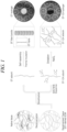
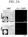

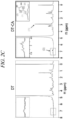

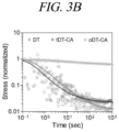


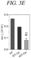


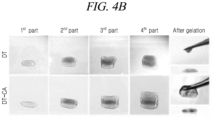


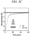


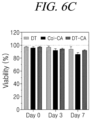
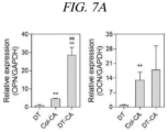


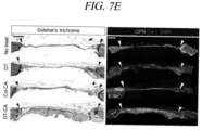
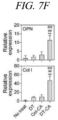

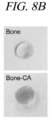
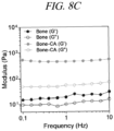
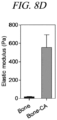
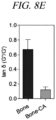
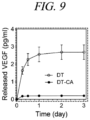


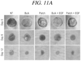
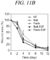
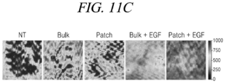


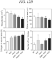

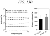
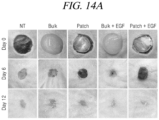
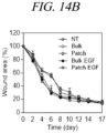


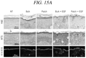

Classifications
-
- C—CHEMISTRY; METALLURGY
- C12—BIOCHEMISTRY; BEER; SPIRITS; WINE; VINEGAR; MICROBIOLOGY; ENZYMOLOGY; MUTATION OR GENETIC ENGINEERING
- C12N—MICROORGANISMS OR ENZYMES; COMPOSITIONS THEREOF; PROPAGATING, PRESERVING, OR MAINTAINING MICROORGANISMS; MUTATION OR GENETIC ENGINEERING; CULTURE MEDIA
- C12N5/00—Undifferentiated human, animal or plant cells, e.g. cell lines; Tissues; Cultivation or maintenance thereof; Culture media therefor
- C12N5/0012—Cell encapsulation
-
- C—CHEMISTRY; METALLURGY
- C12—BIOCHEMISTRY; BEER; SPIRITS; WINE; VINEGAR; MICROBIOLOGY; ENZYMOLOGY; MUTATION OR GENETIC ENGINEERING
- C12N—MICROORGANISMS OR ENZYMES; COMPOSITIONS THEREOF; PROPAGATING, PRESERVING, OR MAINTAINING MICROORGANISMS; MUTATION OR GENETIC ENGINEERING; CULTURE MEDIA
- C12N5/00—Undifferentiated human, animal or plant cells, e.g. cell lines; Tissues; Cultivation or maintenance thereof; Culture media therefor
- C12N5/06—Animal cells or tissues; Human cells or tissues
- C12N5/0602—Vertebrate cells
- C12N5/0625—Epidermal cells, skin cells; Cells of the oral mucosa
- C12N5/0629—Keratinocytes; Whole skin
-
- A—HUMAN NECESSITIES
- A61—MEDICAL OR VETERINARY SCIENCE; HYGIENE
- A61K—PREPARATIONS FOR MEDICAL, DENTAL OR TOILETRY PURPOSES
- A61K35/00—Medicinal preparations containing materials or reaction products thereof with undetermined constitution
- A61K35/12—Materials from mammals; Compositions comprising non-specified tissues or cells; Compositions comprising non-embryonic stem cells; Genetically modified cells
- A61K35/36—Skin; Hair; Nails; Sebaceous glands; Cerumen; Epidermis; Epithelial cells; Keratinocytes; Langerhans cells; Ectodermal cells
-
- A—HUMAN NECESSITIES
- A61—MEDICAL OR VETERINARY SCIENCE; HYGIENE
- A61K—PREPARATIONS FOR MEDICAL, DENTAL OR TOILETRY PURPOSES
- A61K45/00—Medicinal preparations containing active ingredients not provided for in groups A61K31/00 - A61K41/00
- A61K45/06—Mixtures of active ingredients without chemical characterisation, e.g. antiphlogistics and cardiaca
-
- A—HUMAN NECESSITIES
- A61—MEDICAL OR VETERINARY SCIENCE; HYGIENE
- A61K—PREPARATIONS FOR MEDICAL, DENTAL OR TOILETRY PURPOSES
- A61K47/00—Medicinal preparations characterised by the non-active ingredients used, e.g. carriers or inert additives; Targeting or modifying agents chemically bound to the active ingredient
- A61K47/46—Ingredients of undetermined constitution or reaction products thereof, e.g. skin, bone, milk, cotton fibre, eggshell, oxgall or plant extracts
-
- A—HUMAN NECESSITIES
- A61—MEDICAL OR VETERINARY SCIENCE; HYGIENE
- A61K—PREPARATIONS FOR MEDICAL, DENTAL OR TOILETRY PURPOSES
- A61K9/00—Medicinal preparations characterised by special physical form
- A61K9/06—Ointments; Bases therefor; Other semi-solid forms, e.g. creams, sticks, gels
-
- A—HUMAN NECESSITIES
- A61—MEDICAL OR VETERINARY SCIENCE; HYGIENE
- A61K—PREPARATIONS FOR MEDICAL, DENTAL OR TOILETRY PURPOSES
- A61K9/00—Medicinal preparations characterised by special physical form
- A61K9/70—Web, sheet or filament bases ; Films; Fibres of the matrix type containing drug
-
- A—HUMAN NECESSITIES
- A61—MEDICAL OR VETERINARY SCIENCE; HYGIENE
- A61L—METHODS OR APPARATUS FOR STERILISING MATERIALS OR OBJECTS IN GENERAL; DISINFECTION, STERILISATION OR DEODORISATION OF AIR; CHEMICAL ASPECTS OF BANDAGES, DRESSINGS, ABSORBENT PADS OR SURGICAL ARTICLES; MATERIALS FOR BANDAGES, DRESSINGS, ABSORBENT PADS OR SURGICAL ARTICLES
- A61L27/00—Materials for grafts or prostheses or for coating grafts or prostheses
- A61L27/36—Materials for grafts or prostheses or for coating grafts or prostheses containing ingredients of undetermined constitution or reaction products thereof, e.g. transplant tissue, natural bone, extracellular matrix
- A61L27/3604—Materials for grafts or prostheses or for coating grafts or prostheses containing ingredients of undetermined constitution or reaction products thereof, e.g. transplant tissue, natural bone, extracellular matrix characterised by the human or animal origin of the biological material, e.g. hair, fascia, fish scales, silk, shellac, pericardium, pleura, renal tissue, amniotic membrane, parenchymal tissue, fetal tissue, muscle tissue, fat tissue, enamel
- A61L27/3633—Extracellular matrix [ECM]
-
- A—HUMAN NECESSITIES
- A61—MEDICAL OR VETERINARY SCIENCE; HYGIENE
- A61L—METHODS OR APPARATUS FOR STERILISING MATERIALS OR OBJECTS IN GENERAL; DISINFECTION, STERILISATION OR DEODORISATION OF AIR; CHEMICAL ASPECTS OF BANDAGES, DRESSINGS, ABSORBENT PADS OR SURGICAL ARTICLES; MATERIALS FOR BANDAGES, DRESSINGS, ABSORBENT PADS OR SURGICAL ARTICLES
- A61L27/00—Materials for grafts or prostheses or for coating grafts or prostheses
- A61L27/36—Materials for grafts or prostheses or for coating grafts or prostheses containing ingredients of undetermined constitution or reaction products thereof, e.g. transplant tissue, natural bone, extracellular matrix
- A61L27/3683—Materials for grafts or prostheses or for coating grafts or prostheses containing ingredients of undetermined constitution or reaction products thereof, e.g. transplant tissue, natural bone, extracellular matrix subjected to a specific treatment prior to implantation, e.g. decellularising, demineralising, grinding, cellular disruption/non-collagenous protein removal, anti-calcification, crosslinking, supercritical fluid extraction, enzyme treatment
-
- A—HUMAN NECESSITIES
- A61—MEDICAL OR VETERINARY SCIENCE; HYGIENE
- A61L—METHODS OR APPARATUS FOR STERILISING MATERIALS OR OBJECTS IN GENERAL; DISINFECTION, STERILISATION OR DEODORISATION OF AIR; CHEMICAL ASPECTS OF BANDAGES, DRESSINGS, ABSORBENT PADS OR SURGICAL ARTICLES; MATERIALS FOR BANDAGES, DRESSINGS, ABSORBENT PADS OR SURGICAL ARTICLES
- A61L27/00—Materials for grafts or prostheses or for coating grafts or prostheses
- A61L27/36—Materials for grafts or prostheses or for coating grafts or prostheses containing ingredients of undetermined constitution or reaction products thereof, e.g. transplant tissue, natural bone, extracellular matrix
- A61L27/38—Materials for grafts or prostheses or for coating grafts or prostheses containing ingredients of undetermined constitution or reaction products thereof, e.g. transplant tissue, natural bone, extracellular matrix containing added animal cells
- A61L27/3804—Materials for grafts or prostheses or for coating grafts or prostheses containing ingredients of undetermined constitution or reaction products thereof, e.g. transplant tissue, natural bone, extracellular matrix containing added animal cells characterised by specific cells or progenitors thereof, e.g. fibroblasts, connective tissue cells, kidney cells
- A61L27/3834—Cells able to produce different cell types, e.g. hematopoietic stem cells, mesenchymal stem cells, marrow stromal cells, embryonic stem cells
-
- A—HUMAN NECESSITIES
- A61—MEDICAL OR VETERINARY SCIENCE; HYGIENE
- A61L—METHODS OR APPARATUS FOR STERILISING MATERIALS OR OBJECTS IN GENERAL; DISINFECTION, STERILISATION OR DEODORISATION OF AIR; CHEMICAL ASPECTS OF BANDAGES, DRESSINGS, ABSORBENT PADS OR SURGICAL ARTICLES; MATERIALS FOR BANDAGES, DRESSINGS, ABSORBENT PADS OR SURGICAL ARTICLES
- A61L27/00—Materials for grafts or prostheses or for coating grafts or prostheses
- A61L27/50—Materials characterised by their function or physical properties, e.g. injectable or lubricating compositions, shape-memory materials, surface modified materials
- A61L27/52—Hydrogels or hydrocolloids
-
- C—CHEMISTRY; METALLURGY
- C12—BIOCHEMISTRY; BEER; SPIRITS; WINE; VINEGAR; MICROBIOLOGY; ENZYMOLOGY; MUTATION OR GENETIC ENGINEERING
- C12N—MICROORGANISMS OR ENZYMES; COMPOSITIONS THEREOF; PROPAGATING, PRESERVING, OR MAINTAINING MICROORGANISMS; MUTATION OR GENETIC ENGINEERING; CULTURE MEDIA
- C12N5/00—Undifferentiated human, animal or plant cells, e.g. cell lines; Tissues; Cultivation or maintenance thereof; Culture media therefor
- C12N5/06—Animal cells or tissues; Human cells or tissues
- C12N5/0602—Vertebrate cells
- C12N5/0652—Cells of skeletal and connective tissues; Mesenchyme
- C12N5/0662—Stem cells
- C12N5/0667—Adipose-derived stem cells [ADSC]; Adipose stromal stem cells
-
- C—CHEMISTRY; METALLURGY
- C12—BIOCHEMISTRY; BEER; SPIRITS; WINE; VINEGAR; MICROBIOLOGY; ENZYMOLOGY; MUTATION OR GENETIC ENGINEERING
- C12N—MICROORGANISMS OR ENZYMES; COMPOSITIONS THEREOF; PROPAGATING, PRESERVING, OR MAINTAINING MICROORGANISMS; MUTATION OR GENETIC ENGINEERING; CULTURE MEDIA
- C12N2502/00—Coculture with; Conditioned medium produced by
- C12N2502/09—Coculture with; Conditioned medium produced by epidermal cells, skin cells, oral mucosa cells
-
- C—CHEMISTRY; METALLURGY
- C12—BIOCHEMISTRY; BEER; SPIRITS; WINE; VINEGAR; MICROBIOLOGY; ENZYMOLOGY; MUTATION OR GENETIC ENGINEERING
- C12N—MICROORGANISMS OR ENZYMES; COMPOSITIONS THEREOF; PROPAGATING, PRESERVING, OR MAINTAINING MICROORGANISMS; MUTATION OR GENETIC ENGINEERING; CULTURE MEDIA
- C12N2513/00—3D culture
-
- C—CHEMISTRY; METALLURGY
- C12—BIOCHEMISTRY; BEER; SPIRITS; WINE; VINEGAR; MICROBIOLOGY; ENZYMOLOGY; MUTATION OR GENETIC ENGINEERING
- C12N—MICROORGANISMS OR ENZYMES; COMPOSITIONS THEREOF; PROPAGATING, PRESERVING, OR MAINTAINING MICROORGANISMS; MUTATION OR GENETIC ENGINEERING; CULTURE MEDIA
- C12N2533/00—Supports or coatings for cell culture, characterised by material
- C12N2533/30—Synthetic polymers
-
- C—CHEMISTRY; METALLURGY
- C12—BIOCHEMISTRY; BEER; SPIRITS; WINE; VINEGAR; MICROBIOLOGY; ENZYMOLOGY; MUTATION OR GENETIC ENGINEERING
- C12N—MICROORGANISMS OR ENZYMES; COMPOSITIONS THEREOF; PROPAGATING, PRESERVING, OR MAINTAINING MICROORGANISMS; MUTATION OR GENETIC ENGINEERING; CULTURE MEDIA
- C12N2533/00—Supports or coatings for cell culture, characterised by material
- C12N2533/50—Proteins
- C12N2533/54—Collagen; Gelatin
-
- C—CHEMISTRY; METALLURGY
- C12—BIOCHEMISTRY; BEER; SPIRITS; WINE; VINEGAR; MICROBIOLOGY; ENZYMOLOGY; MUTATION OR GENETIC ENGINEERING
- C12N—MICROORGANISMS OR ENZYMES; COMPOSITIONS THEREOF; PROPAGATING, PRESERVING, OR MAINTAINING MICROORGANISMS; MUTATION OR GENETIC ENGINEERING; CULTURE MEDIA
- C12N2533/00—Supports or coatings for cell culture, characterised by material
- C12N2533/90—Substrates of biological origin, e.g. extracellular matrix, decellularised tissue
-
- C—CHEMISTRY; METALLURGY
- C12—BIOCHEMISTRY; BEER; SPIRITS; WINE; VINEGAR; MICROBIOLOGY; ENZYMOLOGY; MUTATION OR GENETIC ENGINEERING
- C12N—MICROORGANISMS OR ENZYMES; COMPOSITIONS THEREOF; PROPAGATING, PRESERVING, OR MAINTAINING MICROORGANISMS; MUTATION OR GENETIC ENGINEERING; CULTURE MEDIA
- C12N2537/00—Supports and/or coatings for cell culture characterised by physical or chemical treatment
- C12N2537/10—Cross-linking
Definitions
- the present disclosure relates to a hydrogel including a decellularized tissue-derived extracellular matrix functionalized with a phenol derivative, uses thereof, and a method of preparing the same.
- Decellularization can minimize immune response by removing cells from tissue and mimicking a tissue-specific microenvironment with various extracellular matrices, polysaccharides and specific proteins and thus has attracted a lot of attention in the field of tissue engineering ( Falguni Pati et al., Nature Communications, 5:3935, 2014 ; Yuehe Fu et al., J. Cell. Mol. Med., 20(4):740-749, 2016 ).
- a tissue-derived extracellular matrix obtained by decellularization can be made into a solution and then used for surface modification and hydrogel production. Thus, it can also be used as a biomaterial for cell culture, implantation and tissue regeneration.
- tissue-derived extracellular matrix-based hydrogels are crosslinked by self-assembly. Therefore, they have disadvantages such as requiring a long time to be formed and weak physical properties and thus have limitations in being grafted onto the latest medical engineering technologies such as long-term cell culture and 3D printing. Accordingly, there is a need to develop a tissue-derived extracellular matrix-based hydrogel which has enhanced physical properties and various tissue engineering applications while maintaining tissue-specific functionality.
- the present disclosure is conceived to provide a hydrogel including a decellularized tissue-derived extracellular matrix functionalized with a phenol derivative.
- the present disclosure is also conceived to provide a composition for cell culture including the hydrogel.
- the present disclosure is also conceived to provide a composition for promoting cell differentiation including the hydrogel.
- the present disclosure is also conceived to provide a composition for drug delivery including the hydrogel.
- the present disclosure is also conceived to provide a composition for promoting tissue regeneration including the hydrogel.
- the present disclosure is also conceived to provide a composition for tissue implantation including the hydrogel.
- the present disclosure is also conceived to provide a method of preparing a hydrogel based on a decellularized tissue-derived extracellular matrix functionalized with a phenol derivative, including a process of functionalizing a decellularized tissue-derived extracellular matrix with a phenol derivative.
- the present disclosure provides a hydrogel including a decellularized tissue-derived extracellular matrix functionalized with a phenol derivative.
- composition for cell culture including the hydrogel.
- the present disclosure provides a composition for promoting cell differentiation including the hydrogel.
- composition for drug delivery including the hydrogel.
- the present disclosure provides a composition for promoting tissue regeneration including the hydrogel.
- composition for tissue implantation including the hydrogel.
- the present disclosure provides a method of preparing a hydrogel based on a decellularized tissue-derived extracellular matrix functionalized with a phenol derivative, including a process of functionalizing a decellularized tissue-derived extracellular matrix with a phenol derivative.
- a hydrogel according to the present disclosure has improved physical properties and structural stability and thus can form a stable three-dimensional structure for a long time without the use of an additional polymer scaffold.
- the hydrogel has high biocompatibility and low cytotoxicity and thus can culture stem cells and promote differentiation of stem cells and tissue regeneration.
- the hydrogel or hydrogel patch can encapsulate a drug therein and thus enables continuous drug delivery.
- the hydrogel can promote tissue regeneration and can be used for tissue implantation. Thus, it can be usefully used in the field of tissue engineering.
- the present disclosure relates to a hydrogel including a decellularized tissue-derived extracellular matrix functionalized with a phenol derivative.
- the phenol derivative may be a catechol group derived from a catechol-based compound selected from the group consisting of catechol, 4-tert-butylcatechol (TBC), urushiol, alizarin, dopamine, dopamine hydrochloride, 3,4-dihydroxyphenylalanine (DOPA), caffeic acid, norepinephrine, epinephrine, 3,4-dihydroxyphenylacetic acid (DOPAC), isoprenaline, isoproterenol and 3,4-dihydroxybenzoic acid; or a pyrogallol group derived from a pyrogallol-based compound selected from the group consisting of pyrogallol, 5-hydroxydopamine, tannic acid, gallic acid, epigallocatechin, epicatechin gallate, epigallocatechin gallate, 2,3,4-trihydroxybenzaldehyde, 2,3,4-trihydroxybenzoic acid, 3,4,5-tri
- the tissue may be selected from the group consisting of liver, heart, kidney, muscle, stomach, intestine, lung, bone, cartilage, blood vessel, bladder, skin, brain, fat, thyroid gland, salivary gland, esophagus, pancreas, spinal cord, ligament, tendon, tooth and uterus, but is not limited thereto.
- 90% or more of cells may be removed from the decellularized tissue-derived extracellular matrix. Desirably, 95% or more of cells may be removed from the decellularized tissue-derived extracellular matrix. More desirably, 98% or more of cells may be removed from the decellularized tissue-derived extracellular matrix. More desirably, 99% or more of cells may be removed from the decellularized tissue-derived extracellular matrix.
- the hydrogel in the decellularized tissue-derived extracellular matrix functionalized with a phenol derivative, the hydrogel may be crosslinked through oxidation of the functionalized phenol derivative.
- the oxidation may be carried out by addition of an oxidizer or an enzyme.
- the oxidizer may be sodium periodate (NaIO 4 ) and hydrogen peroxide (H 2 O 2 ), and the enzyme may be a peroxidase, but is not limited thereto.
- the oxidation may be carried out by a body enzyme without addition of an oxidizer or an enzyme.
- a body enzyme without addition of an oxidizer or an enzyme.
- the hydrogel When the hydrogel is functionalized with a phenol derivative with stronger oxidative activity, for example, pyrogallol, it may be crosslinked through oxidation with a body enzyme without using an additional oxidizer or enzyme.
- the hydrogel may have a porous structure.
- the hydrogel may have an elastic modulus of from 100 Pa to 1500 Pa at 1 Hz.
- the hydrogel may have an elastic modulus of from 200 Pa to 1500 Pa, from 300 Pa to 1500 Pa, from 400 Pa to 1500 Pa, from 500 Pa to 1500 Pa, from 600 Pa to 1500 Pa, from 700 Pa to 1500 Pa, from 800 Pa to 1500 Pa and from 900 Pa to 1500 Pa at 1 Hz, but is not limited thereto.
- the hydrogel may have a tissue adhesive property or may be biodegradable.
- composition for cell culture including a hydrogel based on a decellularized tissue-derived extracellular matrix functionalized with a phenol derivative.
- the culture may be a three-dimensional culture.
- the cells may be stem cells, hematopoietic stem cells, hepatic cells, fibrous cells, epithelial cells, mesothelial cells, endothelial cells, muscle cells, nerve cells, immune cells, adipocytes, chondrocytes, bone cells, blood cells or skin cells, and can be applied to all cells capable of growth in the hydrogel of the present disclosure regardless of the cell type.
- composition for promoting cell differentiation including a hydrogel based on a decellularized tissue-derived extracellular matrix functionalized with a phenol derivative.
- the cell may be a stem cell, and the stem cell may be selected from the group consisting of embryonic stem cells, fetal stem cells, induced pluripotent stem cells and adult stem cells.
- composition for drug delivery including a hydrogel based on a decellularized tissue-derived extracellular matrix functionalized with a phenol derivative.
- the drug may be encapsulated or loaded in the hydrogel, and may be selected from the group consisting of an immune cell activator, an anticancer agent, a therapeutic antibody, an antibiotic, an antibacterial agent, an antiviral agent, an anti-inflammatory agent, a contrast agent, a protein drug, a growth factor, a cytokine, a peptide drug, a hair growth agent, an anesthetic and combinations thereof, but is not limited thereto.
- the anticancer agent may be selected from the group consisting of anthracyclines such as doxorubicin, daunomycin, epirubicin, idarubicin, etc.; taxanes such as paclitaxel, docetaxel, cabazitaxel, tesetaxel, etc.; alkaloids such as curcumin, camptothecin, berberine, evodiamine, matrine, piperine, sanguinarine, tetrandrine, thalicarpine, ellipticine, etc.; vinca alkaloids vinblastine, vincristine, vindesine, vinorelbine, etc.; platinums such as cisplatin, carboplatin, oxaliplatin, etc.; antimetabolites such as 5-fluorouracil, capecitabine, methotrexate, gemcitabine, etc.; topoisomerase inhibitors such as irinotecan, topotecan, etoposide, teni
- the therapeutic antibody may be selected from the group consisting of trastuzumab, rituximab, bevacizumab, cetuximab, bortezomib, erlotinib, gefitinib, imatinib mesylate, sunitinib, pembrolizumab, nivolumab, atezolizumab, ipilimumab and blinatumomab, but is not limited thereto.
- the antibiotic may be selected from the group consisting of ⁇ -lactams, aminoglycosides, macrolides, tetracyclines, glycopeptides, lincosamides, quinolones, chloramphenicol, sulfa drugs, trimethoprim, polymyxin, bacitracin, mupirocin, fusidic acid, streptogramin and oxazolidinone, but is not limited thereto.
- the antibacterial agent may be selected from the group consisting of metronidazole, secnidazole, ornidazole, tinidazole, clindamycin, sodium polystyrene sulfate and sodium cellulose sulfate, but is not limited thereto.
- the antiviral agent may be selected from the group consisting of acyclobyl, vidarabine, ganciclovir, foscarnet, famciclovir, ribavirin, amantadine, zanamivir and oseltamivir, but is not limited thereto.
- the anti-inflammatory agent may be selected from the group consisting of NSAIDs such as ibuprofen, dexibuprofen, naproxen, fenoprofen, ketoprofen, dexketoprofen, flurbiprofen, oxaprozin, loxoprofen, indomethacin, tolmetin, sulindac, etodolac, ketorolac, diclofenac, nabumetone, piroxicam, meloxicam, tenoxicam, droxicam, lornoxicam, isoxicam, mefenamic acid, meclofenamic acid, flufenamic acid, tolfenamic acid, niflumic acid, aspirin, diflunisal, salsalate and dexamethasone, but is not limited thereto.
- NSAIDs such as ibuprofen, dexibuprofen, naproxen, fenoprofen,
- the contrast agent may be selected from the group consisting of a PET contrast agent, a CT contrast agent, an MRI contrast agent, an optical image contrast agent and a US contrast agent, but is not limited thereto.
- the growth factor may be selected from the group consisting of a vascular endothelial growth factor, an epidermal growth factor, a keratinocyte growth factor, a growth and differentiation factor, a hepatocyte growth factor, a platelet-derived growth factor, a transforming growth factor, angiopoietin, erythropoietin, a bone morphogenetic protein, an insulin-like growth factor, an acidic and basic fibroblast growth factor, a granulocyte-macrophage colony-stimulating factor, a brain-derived neurotrophic factor, a glial cell-derived neurotrophic factor, a nerve growth factor, a stromal cell-derived factor-1, a substance P and a hypoxia-inducible factor-1, but is not limited thereto.
- the cytokine may be selected from the group consisting of interleukin, lymphokine, monokine, chemokine, interferon and adipokine, but is not limited thereto.
- the hair growth agent may be selected from the group consisting of finasteride or dutasteride, but is not limited thereto.
- the anesthetic may be selected from the group consisting of ropivacaine, bupivacaine, chloroprocaine, lidocaine, mepivacaine, procaine, tetracaine, levobupivacaine and articaine, but is not limited thereto.
- the composition may be in the form of an adhesive patch or a film.
- composition for promoting tissue regeneration or for tissue implantation including a hydrogel based on a decellularized tissue-derived extracellular matrix functionalized with a phenol derivative.
- the tissue may be selected from the group consisting of liver, heart, kidney, muscle, stomach, intestine, lung, bone, cartilage, blood vessel, bladder, skin, brain, fat, thyroid gland, salivary gland, esophagus, pancreas, spinal cord, ligament, tendon, tooth and uterus, but is not limited thereto.
- the composition may be in the form of an adhesive patch or a film, and the drug may be encapsulated or loaded in the hydrogel.
- the present disclosure provides a method of preparing a hydrogel based on a decellularized tissue-derived extracellular matrix functionalized with a phenol derivative, including a process of functionalizing a decellularized tissue-derived extracellular matrix with a phenol derivative and a process of oxidizing the functionalized phenol derivative.
- the oxidation may be carried out by addition of an oxidizer or an enzyme, or may be carried out by a body enzyme without addition of an oxidizer or an enzyme.
- the pharmaceutical composition according to the present disclosure may be prepared into a pharmaceutical dosage form by a well-known method in the art, so that an active component of the composition may be provided via a fast, suspended or prolonged release, after being administered into a mammal.
- the pharmaceutical composition according to the present disclosure may further contain a pharmaceutically acceptable carrier, to the extent that this carrier does not inhibit a function of the active component.
- the pharmaceutically acceptable carrier may include commonly-used carriers, such as pectin, hyaluronic acid, collagen, lactose, dextrose, sucrose, sorbitol, mannitol, xylitol, erythritol, maltitol, starch, acacia rubber, alginate, gelatin, calcium phosphate, calcium silicate, cellulose, methyl cellulose, microcrystalline cellulose, polyvinyl pyrrolidone, water, methyl hydroxybenzoate, propyl hydroxybenzoate, talc, magnesium stearate, and mineral oil, but is not limited thereto.
- the pharmaceutical composition of the present disclosure may further include a diluent or an excipient, such as fillers, weighting agents, bonding agents, wetting agents, disintegrating agents and surfactants, and other pharmaceutically acceptable additives.
- the pharmaceutical composition according to the present disclosure may be administered in a pharmaceutically effective amount.
- pharmaceutically effective amount refers to an amount sufficient to prevent or treat a disease at a reasonable benefit/risk ratio applicable to a medical treatment.
- the effective amount of the pharmaceutical composition of the present disclosure may be determined by a person with ordinary skill in the art according to various factors such as a formulation method, a patient's condition and weight, the patient's gender, age and degree of disease, a drug form, an administration route and period, an excretion rate, reaction sensitivity, etc.
- the effective amount may vary depending on a route of treatment, a use of excipients and a possibility of being used with other drugs, as recognized by a person with ordinary skill in the art.
- the pharmaceutical composition of the present disclosure may be administered to mammals such as mice, livestock, humans, etc. through various routes.
- the pharmaceutical composition of the present disclosure can be administered orally or parenterally (for example, applied or injected intravenously, subcutaneously or intraperitoneally).
- a solid preparation for oral administration may include powder, granule, tablet, capsule, soft capsule, pill, etc.
- a liquid preparation for oral administration may include suspension, liquid for internal use, emulsion, syrup, aerosol, etc., but may also include various excipients, for example, wetting agent, sweetener, flavoring agent and preservative in addition to generally used simple diluents such as water and liquid paraffin.
- a preparation for parenteral administration may be used by being formulated into a dosage form of external preparation and sterilized injectable preparation such as sterilized aqueous solution, liquid, water-insoluble excipient, suspension, emulsion, eye drop, eye ointment, syrup, suppository and aerosol according to respective conventional methods.
- the pharmaceutical composition may be used in the form of cream, gel, patch, spraying agent, ointment, plaster, lotion, liniment, eye ointment, eye drop, paste, or cataplasm, but is not limited thereto.
- a preparation for topical administration may be an anhydrous or aqueous form depending on a clinical prescription.
- propylene glycol polyethylene glycol
- vegetable oil such as olive oil
- Base materials of the suppository may include witepsol, macrogol, tween 61, cacao butter, laurinum and glycerogelatin.
- the porcine skin tissue was cut into pieces of 3 ⁇ 3 ⁇ 3 mm 3 and then washed with distilled water for 12 to 16 hours. Thereafter, decellularization was performed using the following solutions sequentially: 1% Triton X-100 (Wako, Osaka, Japan) supplemented with 0.1% ammonium hydroxide (Sigma, St. Louis, MO, USA) for 3 days; distilled water (DW) for 2 days; 1% penicillin-streptomycin (Thermo Fisher Scientific, Waltham, MA, USA) for 2 hours; and distilled water (DW) for 2 hours.
- the solutions used for decellularization and washing were replaced with fresh solutions twice a day. All processes were performed using a 4°C shaker (180 rpm).
- the decellularized tissue (hereinafter, referred to as "DT") was lyophilized and cold-stored until used.
- the decellularized tissue was made into a solution with stirring for 48 hours using a pepsin solution (4 mg/ml in 0.02 M hydrochloric acid, Sigma) (10 mg/ml).
- the decellularized tissue made into a solution was diluted to a concentration of 1 mg/ml using 0.01 M HCl, and the pH of the solution was adjusted to 4.5 using 1 M NaOH (Sigma). Thereafter, 1-(3-dimethylaminopropyl)-3-ethylcarbodiimide hydrochloride (EDC, Thermo Fisher Scientific) and N-hydroxysuccinimide (NHS, Sigma) were added to the solution to start the reaction.
- EDC 1-(3-dimethylaminopropyl)-3-ethylcarbodiimide hydrochloride
- NHS N-hydroxysuccinimide
- the molecular weight of DT was assumed to be 200 kg/mol, and the ratio of DT:EDC:NHS was adjusted to 1:5:5 (molar ratio). After the solution was allowed to react for 30 minutes, dopamine hydrochloride (Sigma) or 5-hydroxydopamine hydrochloride (Sigma) was added to DT at a ratio of 1: 5 (dopamine: DT, molar ratio) and then, the reaction was carried out overnight at pH 4.5.
- Dopamine (or hydroxydopamine), which did not participate in the reaction, was removed sequentially in acidic PBS (pH 3.5) and acidic distilled water (pH 3.5) by using a Cellu Sep with a 6 to 8 KDa cutoff (Membrane Filtration Products Inc., Seguin, TX, USA).
- the final product (hereinafter, referred to as "DT-CA”) was lyophilized and stored at -20°C until used.
- Collagen type I in the tissue was immunostained with anti-Col I primary antibodies (1:200 dilution, Calbiochem, San Diego, CA, USA) and Alexa 594-conjugated secondary antibodies (Invitrogen, Carlsbad, CA, USA), and the cell nuclei were stained with 4',6-diamidino-2-phenylindole (DAPI) (Vector Laboratories, Burlingame, CA, USA). The stained tissue was photographed with a confocal laser scanning microscope (LSM 880, Carl Zeiss).
- a test was performed to analyze the amount of DNA and glycosaminoglycan (GAG) before and after decellularization. Briefly, the amount of DNA remaining in the lyophilized tissue was quantified with a DNA detection kit (Bioneer, Daejeon, Korea) and the amount of glycosaminoglycan (GAG) was quantified with 1,9-dimethyl methylene blue (Sigma).
- DT decellularized tissue-derived extracellular matrix
- DT-CA decellularized tissue-derived extracellular matrix functionalized with a catechol group
- the NMR spectrum of the decellularized tissue-derived extracellular matrix functionalized with a catechol group showed the presence of an ethyl proton (1) and a phenyl proton (2) between the catechol group and the DT backbone, which confirms that the decellularized tissue was well functionalized with a catechol group ( FIG. 2C ).
- a hydrogel was prepared by temperature crosslinking or oxidative crosslinking. Briefly, for preparation of a hydrogel (hereinafter, referred to as "tDT-CA hydrogel") by self-assembly, which is a temperature crosslinking method, a DT (20 mg/ml in 0.02 M HCl) solution and a DT-CA (20 mg/ml in distilled water) solution were prepared at a final concentration of 10 mg/ml by using 10 ⁇ PBS (10% final concentration, Sigma) and distilled water. Immediately after the solution was adjusted to a final pH of 7.4 with 0.5 N NaOH, it was gelated at 37°C for 30 minutes.
- oDT-CA hydrogel For production of a hydrogel (hereinafter, referred to as "oDT-CA hydrogel") by oxidative crosslinking, DT-CA was dissolved in PBS and then, an oxidation reaction was performed by adding an oxidizer (sodium periodate, NaIO 4 , Sigma). The final concentration of DT-CA in the DT-CA solution was 10 mg/ml, and the oxidizer was added in a ratio of 2 : 1 (NaIO 4 : dopamine, molar ratio) with respect to dopamine.
- oxidizer sodium periodate, NaIO 4 , Sigma
- DT hydrogel decellularized tissue-derived extracellular matrix-based hydrogel
- the hydrogel may have various internal structures depending on the crosslinking method.
- the tDT-CA hydrogel which was prepared by temperature crosslinking had an internal structure in the form of nanofibers similar to that of the DT hydrogel which was not functionalized with a catechol group, whereas the oDT-CA hydrogel which was prepared by oxidative crosslinking had a porous internal structure ( FIG. 3A ).
- a storage modulus (G') and a loss modulus (G") of the hydrogel were measured within the range of 0.1 Hz to 10 Hz in a frequency sweep mode (1% strain condition). The average storage modulus at 1 Hz was taken as an elastic modulus.
- G' and G" were measured in a time sweep mode (10% strain, 1 Hz frequency) and the time for which G' and G" crossed over was taken as gelation time.
- the gelation rate of the hydrogel through self-assembly was measured by placing a pre-gel solution on a base plate set at 4°C and then increasing the temperature to 37°C.
- a relaxation stress over time was measured (10% shear strain).
- the relaxation test confirmed that the oDT-CA hydrogel composed of covalent bonds/ irreversible bonds was physically more stable than the DT hydrogel or tDT-CA hydrogel composed of ionic bonds/reversible bonds ( FIG. 3B ). Also, it was confirmed that the oDT-CA hydrogel (1055.1 ⁇ 82.3 Pa) increased in physical properties (modulus) by about 10 times compared with the DT hydrogel or tDT-CA hydrogel (119.2 ⁇ 14.2 Pa for the tDT-CA hydrogel) ( FIG. 3C to FIG. 3E ).
- Example 5 Analysis of structural stability and tissue adhesion of DT-CA hydrogel
- each gelated hydrogel was immersed in a PBS or collagenase solution (0.1 mg/ml, Sigma) and then cultured at 37°C while changes in weight were measured.
- Wt represents the weight of the hydrogel at a specific time
- Wi represents the weight of the hydrogel before the start of culture.
- tissue adhesive property of the DT-CA hydrogel was measured by placing each pre-gel solution in the mouse skin (fixed to the plate with a strong adhesive), sufficiently gelating the pre-gel solution by each method, and then measuring adhesion in a tack-separation mode (at a rate of 10 ⁇ m/s).
- the DT hydrogel has a normal force close to 0, whereas the DT-CA hydrogel has a normal force of about 3 N.
- the DT-CA hydrogel has a tissue adhesive property which does not appear in the DT hydrogel ( FIG. 5C and FIG. 5D ).
- the decellularized tissue-derived extracellular matrix-based hydrogel functionalized with a catechol group (DT-CA hydrogel) was improved in structural stability compared with the decellularized tissue-derived extracellular matrix-based hydrogel (DT hydrogel) which was not functionalized with a catechol group and could form a stable three-dimensional structure without an additional polymer scaffold.
- the DT-CA hydrogel had a tissue adhesive property which is very important for stable engraftment and maintenance after implantation of a biomaterial. Thus, it can be suitable as a biomaterial for tissue regeneration.
- Example 6 Analysis of structural stability and biocompatibility of DT-CA hydrogel upon cell encapsulation
- hADSCs human adipose-derived stem cells
- ATCC American Type Culture Collection, Rockville, MD, USA
- MSC mesenchymal stem cell growth medium
- the hADSCs were mixed with each pre-gel solution at a concentration of 1 ⁇ 10 7 cells/ml and gelated by self-assembly or oxidation.
- the DT hydrogel with encapsulated stem cells was shown to rapidly shrink over time.
- the hydrogel shrank to almost 20% or less on day 21, the end of the test ( FIG. 6A ).
- the DT-CA hydrogel with encapsulated stem cells hardly shrank even on day 21, the end date of the test ( FIG. 6A ).
- the encapsulated cells were stained with a live/dead viability/cytotoxicity kit (Invitrogen) to measure the cell viability under a fluorescence microscope.
- a collagen type I extracellular matrix-based hydrogel functionalized with a catechol group hereinafter, referred to as "Col-CA hydrogel" using collagen type I (Col I), which is an extracellular matrix widely used in tissue engineering, was used.
- Example 7 Analysis of efficacy of DT-CA hydrogel for enhancing stem cell differentiation
- osteodifferentiation was induced in hADSC cells in the hydrogel by using the following culture medium for inducing osteodifferentiation: DMEM medium (Dulbecco's Modified Eagle's Medium, Gibco, Gaithersburg, MD, USA) containing 100 nM dexamethasone (Sigma); 50 ⁇ g/ml L-ascorbic acid (Sigma); 10 mM ⁇ -glycerophosphate (Sigma); 10% (v/v) FBS (fetal bovine serum, Gibco); 3.7 g/L sodium bicarbonate (Sigma); and 1% (v/v) penicillin/streptomycin (Gibco). After differentiation was induced for 3 weeks, cell differentiation patterns were checked through immunostaining and real-time polymerase chain reaction (real time-PCR).
- the real time-PCR confirmed the expression levels of OPN (human osteopontin, Hs00959010_m1) and OCN (human osteocalcin, Hs01587814_g1), which are osteocyte-specific markers, and GAPDH (human glyceraldehyde-3-phosphate dehydrogenase, Hs02758991_g1) was used as a housekeeping gene.
- the hydrogel was fixed with 4% paraformaldehyde (Sigma) for 2 days and then staining was performed with primary antibodies, such as anti-Osteopontin (1:100, Santa Cruz Biotechnology Inc., Santa Cruz, CA, USA), anti-Fibronectin (1:100, Abcam, Cambridge, UK), and secondary antibodies, such as Alexa-Fluor 488-conjugated secondary antibody and Alexa-Fluor 594-conjugated secondary antibody (Invitrogen), and the cell nuclei were stained with DAPI (Vector Laboratories). The stained cells were photographed with a confocal laser scanning microscope (LSM 880, Carl Zeiss).
- the differentiated cells were stained with an Alizarin red solution (Sigma) for 5 minutes, and the excess solution was sufficiently removed with PBS, followed by photographing with a microscope.
- Alizarin red solution Sigma
- the stem cells in the DT-CA hydrogel showed a significantly higher expression level of OPN mRNA than the stem cells in the DT hydrogel or Col-CA hydrogel, and showed a much higher expression level of OPN mRNA than the stem cells in the DT hydrogel, which confirms that the DT-CA hydrogel has the effect of significantly enhancing osteodifferentiation ( FIG. 7A ). Also, it was further confirmed through immunostaining for OPN and Alizarin red staining that osteodifferentiation was more significantly enhanced in the stem cells in the DT-CA hydrogel than in the stem cells in the Col-CA hydrogel ( FIG. 7B and FIG. 7C ).
- the decellularized tissue-derived extracellular matrix-based hydrogel functionalized with a catechol group has the effect of significantly enhancing or promoting stem cell differentiation compared with the decellularized tissue-derived extracellular matrix-based hydrogel (DT hydrogel) that was not functionalized with a catechol group.
- Example 8 Analysis of efficacy of DT-CA hydrogel for enhancing tissue regeneration
- the calvaria defect animal model was prepared by removing 4 mm in size from the mouse calvaria. After 5 ⁇ 10 5 cells were encapsulated in each hydrogel (50 ⁇ L), the hydrogel was implanted to the defect site ( FIG. 7D ). In particular, the DT-CA hydrogel was implanted in the defect site immediately after pre-gel was mixed with an oxidizer, and then gelated for 10 minutes to maintain a tissue adhesive property.
- the calvaria was collected, fixed in 4% paraformaldehyde (Sigma) and treated with a decalcifying solution (Sigma) at room temperature for 4 hours. Thereafter, the tissue was fixed in paraffin and cut to a thickness of 6 ⁇ m to prepare a sample.
- Collagen formation in the tissue was confirmed by Goldner's Trichrome staining. Further, staining was performed with primary antibodies, such as anti-OPN (1:100, Santa Cruz Biotechnology Inc.) anti-Col I (1:200, Calbiochem), and secondary antibodies, such as Alexa-Fluor 488-conjugated secondary antibody and Alexa-Fluor 594-conjugated secondary antibody (Invitrogen), and the cell nuclei were stained with DAPI (Vector Laboratories). The stained tissue was photographed with a confocal laser scanning microscope (LSM 880, Carl Zeiss). As for the degree of bone regeneration in the bone defect site, the expression levels of OPN and Col I were quantified with ImageJ software (National Institutes of Health, Bethesda, MD, USA) based on the photographed images.
- primary antibodies such as anti-OPN (1:100, Santa Cruz Biotechnology Inc.) anti-Col I (1:200, Calbiochem
- secondary antibodies such as Alexa-Fluor 488-conju
- the porcine bone tissue was cultured in a 6% hydrogen peroxide solution (Sigma) for 3 days and then lyophilized and cut into pieces of 4 ⁇ 4 ⁇ 4 mm 3 or less. Thereafter, decellularization was performed using the following solutions: 0.5 N hydrogen chloride (Sigma) for 3 days; 70% ethanol overnight; 0.6 N citric acid (Sigma) for 3 days; distilled water overnight; and phosphate-buffered saline (PBS, Sigma) supplemented with 1% penicillin-streptomycin (Sigma) overnight.
- PBS phosphate-buffered saline
- a decellularized tissue-derived extracellular matrix functionalized with a phenol derivative (hereinafter, referred to as "Bone-CA”) was prepared from the decellularized bone tissue by the method as in Example 2, and a decellularized bone tissue-derived extracellular matrix-based hydrogel functionalized with a phenol derivative (catechol group) (hereinafter, referred to as "Bone-CA hydrogel”) was prepared by oxidative crosslinking as in Example 4.
- a bone tissue-derived extracellular matrix-based hydrogel (hereinafter, referred to as "Bone hydrogel”) prepared by temperature crosslinking was used.
- the decellularized porcine bone tissue appeared white ( FIG. 8A ), and it was confirmed that the hydrogel was well formed by oxidative crosslinking ( FIG. 8B ). Further, it was confirmed that the Bone-CA hydrogel prepared by oxidative crosslinking increased in physical properties by about 10 times compared with the Bone hydrogel prepared by temperature crosslinking, and also greatly increased in elasticity ( FIG. 8C to FIG. 8E ).
- hydrogel functionalized with a catechol group and prepared by oxidative crosslinking showed improved physical properties even in decellularized bone tissue.
- VEGF vascular endothelial growth factor
- the decellularized tissue-derived extracellular matrix functionalized with a catechol group can more stably maintain the drug encapsulated in the hydrogel and can continuously release the drug for a longer time than the decellularized tissue-derived extracellular matrix-based hydrogel (DT hydrogel) which was not functionalized with a catechol group, which confirms that the DT-CA hydrogel can be used for drug delivery.
- a patch was manufactured using the DT-CA hydrogel and a test was performed to analyze its characteristics. Briefly, 200 ⁇ L of a DT-CA solution (10 mg/ml) was injected into a specific mold and then lyophilized. For crosslinking of the DT-CA hydrogel patch, the DT-CA hydrogel patch was placed on the desired site and then, an oxidizer (4.5 mg/ml NaIO 4 ) was sprayed thereto.
- a oxidizer 4.5 mg/ml NaIO 4
- a test was performed to check a drug release pattern of the DT-CA hydrogel patch with the encapsulated drug.
- human EGF epidermal growth factor, Peprotech, Rocky Hill, NJ, USA
- PBS or collagenase A solution (2 mg/ml, Sigma).
- the amount of drug released was measured by replacing the culture medium at each given time.
- the amount of EGF in the solution was quantified with a human EGF ELISA kit (R&D Systems).
- the DT-CA hydrogel patch has enhanced physical properties and thus can effectively encapsulate a drug and can continuously release the drug for a long time by in vivo biodegradation of the hydrogel, which confirms that the DT-CA hydrogel patch can be used for drug delivery.
- Example 12 Analysis of efficacy of DT-CA hydrogel patch for treating wound or promoting tissue regeneration
- a test was performed to analyze the efficacy of the DT-CA hydrogel patch for wound treatment in a wound animal model. Briefly, a biopsy punch was used to induce a circular wound having a diameter of 8 mm in the dorsal skin tissue of a mouse (SKH1-hr, 6 weeks old, male, OrientBio, Seongnam, Korea). Then, the wound of the mouse was treated with a solution-based DT-CA hydrogel (Bulk) or DT-CA hydrogel patch (Patch), and also treated with a DT-CA hydrogel (Bulk+EGF) or DT-CA hydrogel patch (Patch+EGF) with encapsulated EGF (1 ⁇ g/mouse) to verify the efficacy for EGF delivery.
- Bulk solution-based DT-CA hydrogel
- Patch DT-CA hydrogel patch
- encapsulated EGF (1 ⁇ g/mouse
- the condition and treatment aspect of the mouse were checked for 12 days after preparation of the wound animal model and material implantation, and the blood flow at the wound site of the mouse was measured with a laser Doppler imaging device (Moor Instruments, Devon, UK) on day 12 after material implantation.
- a laser Doppler imaging device (Moor Instruments, Devon, UK) on day 12 after material implantation.
- early microvascularization occurred vigorously, but as the tissue organelles were formed in the dermal layer over time, the number of microvessels was relatively decreased.
- the efficacy for wound treatment was inferred by analyzing the blood flow in the skin surface.
- the size of the wound was most effectively reduced in the group treated with the DT-CA hydrogel patch or the DT-CA hydrogel patch with the encapsulated drug (Patch+EGF) ( FIG. 11A and FIG. 11B ). Further, it was confirmed from laser Doppler imaging analysis that the blood flow was significantly decreased in the group treated with the DT-CA hydrogel patch with the encapsulated drug (Patch+EGF) ( FIG. 11C and FIG. 11D ). Thus, it was confirmed that the DT-CA hydrogel patch with the encapsulated drug has the most remarkable effect on wound treatment.
- a histological analysis was performed to the wound site of the wound animal model. Briefly, the skin tissue was collected on day 12 after material implantation, fixed in 10% formalin (Sigma) and then fixed in paraffin to prepare 6- ⁇ m tissue slices, followed by each of H&E staining and Masson's trichrome staining (Sigma) for staining ECM and collagen in the tissue.
- immunostaining was performed with primary antibodies, such as anti-Keratin 14 (Abcam) and anti-Involucrin (Abcam), and secondary antibodies, such as Alexa-Fluor 488-conjugated secondary antibody and Alexa-Fluor 594-conjugated secondary antibody (Invitrogen), and the cell nuclei were stained with DAPI (Vector Laboratories) to check regeneration of the basal layer and the stratum corneum of the skin tissue.
- DAPI Vector Laboratories
- the stained tissue was photographed with a confocal laser scanning microscope (LSM 880, Carl Zeiss).
- LSM 880 Carl Zeiss
- the DT-CA hydrogel patch has improved physical properties and tissue adhesive property and thus can be stably attached to the wound site, and, thus, the DT-CA hydrogel patch can protect the wound site from external stimuli. Also, it was confirmed that the DT-CA hydrogel patch can be used for effective drug delivery through drug encapsulation and thus can be used for promoting tissue regeneration or for tissue implantation.
- a decellularized tissue-derived extracellular matrix functionalized with a pyrogallol group (PG), which has stronger oxidativity, instead of a catechol group among phenol derivatives was prepared in the same manner as in the above Example. Further, a hydrogel (hereinafter, referred to as "DT-PG hydrogel”) was prepared by oxidative crosslinking from the decellularized tissue-derived extracellular matrix functionalized with a pyrogallol group in the same manner as in the above Example.
- PG hydrogel a hydrogel
- Example 14 Analysis of efficacy of DT-PG hydrogel patch for treating wound or promoting tissue regeneration
- the applicability of the DT-PG hydrogel patch was checked as a medical material for promoting skin regeneration in a wound animal model. Briefly, a biopsy punch was used to induce a circular wound having a diameter of 8 mm in the dorsal skin tissue of a mouse and then, the wound of the mouse was treated with a solution-based DT-PG hydrogel (Bulk) or DT-PG hydrogel patch (Patch). Also, EGF (1 ⁇ g/mouse) was delivered to promote skin tissue regeneration and thus to induce the DT-PG material to improve treatment efficacy through sustained drug release.
- a biopsy punch was used to induce a circular wound having a diameter of 8 mm in the dorsal skin tissue of a mouse and then, the wound of the mouse was treated with a solution-based DT-PG hydrogel (Bulk) or DT-PG hydrogel patch (Patch).
- EGF (1 ⁇ g/mouse) was delivered to promote skin tissue regeneration and thus to induce the DT-PG material to improve treatment eff
- the groups did not show a significant difference in the size of the visually observed wound until day 17 after application of the patch ( FIG. 14A and FIG. 14B ).
- FIG. 14C and FIG. 14D it was confirmed from laser Doppler imaging analysis on day 17 that the blood flow was significantly decreased in the group treated with the DT-PG hydrogel patch with the encapsulated drug (Patch+EGF) ( FIG. 14C and FIG. 14D ).
- the blood flow was greatly decreased in the group treated with the DT-PG hydrogel patch with the encapsulated drug (Patch+EGF), and, thus, the size of the wound was most significantly reduced.
- the DT-PG hydrogel patch used in the present disclosure has improved physical properties and tissue adhesive property compared with the conventional hydrogel formulation and thus can be stably attached to the diseased site, and, thus, the DT-PG hydrogel patch can protect the wound site from external stimuli and can induce effective drug delivery.
- a tissue site where the wound was induced was collected on day 17 after induction of the model and application of the hydrogel, followed by histological analysis.
- H&E staining and Masson's trichrome staining that in the DT-PG hydrogel patch group with the encapsulated drug (Patch+EGF), the abnormal skin tissue had the smallest area with the greatest collagen accumulation area and the increase in number of newly formed hair follicles.
- the Keratin 10-positive epidermal layer had the greatest thickness (the tissue has a smaller thickness when damaged or inflamed) ( FIG. 15A and FIG. 15B ).
- the DT-PG patch system can induce effective tissue regeneration through stable drug delivery in vivo.
Landscapes
- Health & Medical Sciences (AREA)
- Life Sciences & Earth Sciences (AREA)
- Chemical & Material Sciences (AREA)
- Engineering & Computer Science (AREA)
- Biomedical Technology (AREA)
- General Health & Medical Sciences (AREA)
- Veterinary Medicine (AREA)
- Public Health (AREA)
- Medicinal Chemistry (AREA)
- Epidemiology (AREA)
- Animal Behavior & Ethology (AREA)
- Zoology (AREA)
- Bioinformatics & Cheminformatics (AREA)
- Biotechnology (AREA)
- Dermatology (AREA)
- Cell Biology (AREA)
- Wood Science & Technology (AREA)
- Organic Chemistry (AREA)
- Genetics & Genomics (AREA)
- Pharmacology & Pharmacy (AREA)
- Botany (AREA)
- Transplantation (AREA)
- Oral & Maxillofacial Surgery (AREA)
- Developmental Biology & Embryology (AREA)
- Chemical Kinetics & Catalysis (AREA)
- Biochemistry (AREA)
- Microbiology (AREA)
- General Engineering & Computer Science (AREA)
- Urology & Nephrology (AREA)
- Molecular Biology (AREA)
- Biophysics (AREA)
- Dispersion Chemistry (AREA)
- Rheumatology (AREA)
- Hematology (AREA)
- Immunology (AREA)
- Virology (AREA)
- Materials For Medical Uses (AREA)
- Compounds Of Unknown Constitution (AREA)
- Pharmaceuticals Containing Other Organic And Inorganic Compounds (AREA)
- Micro-Organisms Or Cultivation Processes Thereof (AREA)
Applications Claiming Priority (2)
| Application Number | Priority Date | Filing Date | Title |
|---|---|---|---|
| KR1020210008205A KR102602244B1 (ko) | 2021-01-20 | 2021-01-20 | 페놀 유도체가 수식된 탈세포화된 조직 유래 세포외기질 및 이의 용도 |
| PCT/KR2022/000510 WO2022158784A1 (fr) | 2021-01-20 | 2022-01-12 | Matrice extracellulaire dérivée de tissu décellularisé modifiée avec un dérivé de phénol et utilisation associée |
Publications (2)
| Publication Number | Publication Date |
|---|---|
| EP4265714A1 true EP4265714A1 (fr) | 2023-10-25 |
| EP4265714A4 EP4265714A4 (fr) | 2024-07-10 |
Family
ID=82549863
Family Applications (1)
| Application Number | Title | Priority Date | Filing Date |
|---|---|---|---|
| EP22742760.6A Pending EP4265714A4 (fr) | 2021-01-20 | 2022-01-12 | Matrice extracellulaire dérivée de tissu décellularisé modifiée avec un dérivé de phénol et utilisation associée |
Country Status (5)
| Country | Link |
|---|---|
| US (1) | US20230355518A1 (fr) |
| EP (1) | EP4265714A4 (fr) |
| KR (1) | KR102602244B1 (fr) |
| CN (1) | CN116783282A (fr) |
| WO (1) | WO2022158784A1 (fr) |
Families Citing this family (4)
| Publication number | Priority date | Publication date | Assignee | Title |
|---|---|---|---|---|
| EP4299083B1 (fr) * | 2021-03-31 | 2025-12-31 | Cellartgen Inc. | Dérivé de matrice extracellulaire dérivé de tissu, modifié par un dérivé de phénol pour la construction de tissu artificiel |
| KR20250127571A (ko) * | 2024-02-19 | 2025-08-26 | 서울대학교산학협력단 | 치료용 인자 전달을 위한 페인팅 가능한 하이드로겔 시스템 개발 |
| CN118725586B (zh) * | 2024-07-11 | 2025-04-01 | 遵义医科大学珠海校区 | 一种多功能导电有机水凝胶及其制备方法和应用 |
| CN118641745B (zh) * | 2024-08-13 | 2024-10-22 | 浙江大学 | 一种基于厚切技术的全组织三维结构脱细胞基质制备方法 |
Family Cites Families (7)
| Publication number | Priority date | Publication date | Assignee | Title |
|---|---|---|---|---|
| US20190117839A1 (en) * | 2016-03-14 | 2019-04-25 | Wisconsin Alumni Research Foundation | Functionalization of plant tissues for human cell expansion |
| US20190282731A1 (en) * | 2016-11-23 | 2019-09-19 | Trustees Of Tufts College | Enzymatically Crosslinked Compositions |
| EP3578573A4 (fr) * | 2017-02-02 | 2020-11-18 | Amtixbio Co., Ltd. | Hydrogel utilisant en tant que substrat un derivé d'acide hyaluronique modifié par un groupe gallol, et utilisation associée |
| KR102368895B1 (ko) * | 2017-08-23 | 2022-03-02 | 코오롱인더스트리 주식회사 | 접착제 조성물, 이를 포함하는 접착제 및 이의 제조방법 |
| US12152228B2 (en) * | 2017-12-27 | 2024-11-26 | Sekisui Chemical Co., Ltd. | Scaffolding material for cell cultures and cell culture method using same |
| IL307979B2 (en) * | 2018-05-09 | 2025-08-01 | Univ Johns Hopkins | Nanofiber-hydrogel composites for enhanced soft tissue replacement and regeneration |
| KR102102153B1 (ko) * | 2018-08-30 | 2020-04-27 | 연세대학교 산학협력단 | 탈세포 조직 기반 세포 봉입 및 배양용 비드, 및 이의 용도 |
-
2021
- 2021-01-20 KR KR1020210008205A patent/KR102602244B1/ko active Active
-
2022
- 2022-01-12 CN CN202280010609.XA patent/CN116783282A/zh active Pending
- 2022-01-12 EP EP22742760.6A patent/EP4265714A4/fr active Pending
- 2022-01-12 WO PCT/KR2022/000510 patent/WO2022158784A1/fr not_active Ceased
-
2023
- 2023-07-20 US US18/355,798 patent/US20230355518A1/en active Pending
Also Published As
| Publication number | Publication date |
|---|---|
| WO2022158784A1 (fr) | 2022-07-28 |
| US20230355518A1 (en) | 2023-11-09 |
| KR102602244B1 (ko) | 2023-11-16 |
| CN116783282A (zh) | 2023-09-19 |
| EP4265714A4 (fr) | 2024-07-10 |
| KR20220105472A (ko) | 2022-07-27 |
Similar Documents
| Publication | Publication Date | Title |
|---|---|---|
| EP4265714A1 (fr) | Matrice extracellulaire dérivée de tissu décellularisé modifiée avec un dérivé de phénol et utilisation associée | |
| Lee et al. | Mechanically-reinforced and highly adhesive decellularized tissue-derived hydrogel for efficient tissue repair | |
| Zhang et al. | In situ-formed adhesive hyaluronic acid hydrogel with prolonged amnion-derived conditioned medium release for diabetic wound repair | |
| Lu et al. | Exosomes loaded a smart bilayer-hydrogel scaffold with ROS-scavenging and macrophage-reprogramming properties for repairing cartilage defect | |
| US12569451B2 (en) | Bio-inspired tissue-adhesive hydrogel patch and uses thereof | |
| EP4088757B1 (fr) | Hydrogel de sulfate de chondroïtine fonctionnalisé par un dérivé de phénol et son utilisation | |
| Ge et al. | Combination therapy using multifunctional dissolvable hyaluronic acid microneedles for oral ulcers | |
| Alheib et al. | Injectable laminin-biofunctionalized gellan gum hydrogels loaded with myoblasts for skeletal muscle regeneration | |
| Yao et al. | Chitosan-based thermosensitive composite hydrogel enhances the therapeutic efficacy of human umbilical cord MSC in TBI rat model | |
| Li et al. | Zinc-induced photocrosslinked konjac glucomannan/glycyrrhizic acid hydrogel promotes skin wound healing in diabetic mice through immune regulation | |
| Lin et al. | Cytokine loaded layer-by-layer ultrathin matrices to deliver single dermal papilla cells for spot-by-spot hair follicle regeneration | |
| Zhang et al. | pH-and glucose-responsive antioxidant hydrogel promotes diabetic wound healing | |
| Wan et al. | BMSCs laden injectable amino-diethoxypropane modified alginate-chitosan hydrogel for hyaline cartilage reconstruction | |
| Xiao et al. | Injectable hydrogel loaded with 4-octyl itaconate enhances cartilage regeneration by regulating macrophage polarization | |
| WO2015187098A1 (fr) | Matière et structures destinées à l'adminisatration de cellules | |
| Wang et al. | Gene liposome nanocomplex-loaded dermal substitute promotes diabetic chronic wound healing and angiogenesis in rat | |
| Yuan et al. | Chitosan-taurine nanoparticles cross-linked carboxymethyl chitosan hydrogels facilitate both acute and chronic diabetic wound healing | |
| EP3928804A2 (fr) | Hydrogel comprenant de l'acide hyaluronique modifié au moyen de sérotonine et son utilisation | |
| Cai et al. | Identification of the CXCL12–CXCR4/CXCR7 axis as a potential therapeutic target for immunomodulating macrophage polarization and foreign body response to implanted biomaterials | |
| Hassan et al. | Injectable self‐oxygenating cardio‐protective and tissue adhesive silk‐based hydrogel for alleviating ischemia after Mi injury | |
| JP2016073671A (ja) | 複合化された重合体材料およびその使用 | |
| Pan et al. | Plasmid containing VEGF-165 and ANG-1 dual genes packaged with fibroin-modified PEI to promote the regeneration of vascular network and dermal tissue | |
| Gao et al. | HAMA-SBMA hydrogel with anti-inflammatory properties delivers cartilage organoids, boosting cartilage regeneration | |
| Lee et al. | PCL-fibrin-alginate hydrogel based cell co-culture system for improving angiogenesis and immune modulation in limb ischemia | |
| KR102750261B1 (ko) | 페놀 유도체가 수식된 콘드로이틴 설페이트 하이드로젤 및 이의 용도 |
Legal Events
| Date | Code | Title | Description |
|---|---|---|---|
| STAA | Information on the status of an ep patent application or granted ep patent |
Free format text: STATUS: THE INTERNATIONAL PUBLICATION HAS BEEN MADE |
|
| PUAI | Public reference made under article 153(3) epc to a published international application that has entered the european phase |
Free format text: ORIGINAL CODE: 0009012 |
|
| STAA | Information on the status of an ep patent application or granted ep patent |
Free format text: STATUS: REQUEST FOR EXAMINATION WAS MADE |
|
| 17P | Request for examination filed |
Effective date: 20230720 |
|
| AK | Designated contracting states |
Kind code of ref document: A1 Designated state(s): AL AT BE BG CH CY CZ DE DK EE ES FI FR GB GR HR HU IE IS IT LI LT LU LV MC MK MT NL NO PL PT RO RS SE SI SK SM TR |
|
| DAV | Request for validation of the european patent (deleted) | ||
| DAX | Request for extension of the european patent (deleted) | ||
| A4 | Supplementary search report drawn up and despatched |
Effective date: 20240607 |
|
| RIC1 | Information provided on ipc code assigned before grant |
Ipc: A61L 27/38 20060101ALI20240603BHEP Ipc: A61L 27/36 20060101ALI20240603BHEP Ipc: A61L 27/52 20060101ALI20240603BHEP Ipc: A61K 9/70 20060101ALI20240603BHEP Ipc: A61K 9/06 20060101ALI20240603BHEP Ipc: A61K 47/46 20060101ALI20240603BHEP Ipc: C12N 5/071 20100101AFI20240603BHEP |