EP4246451A1 - Method for modelling a joint - Google Patents
Method for modelling a joint Download PDFInfo
- Publication number
- EP4246451A1 EP4246451A1 EP22161849.9A EP22161849A EP4246451A1 EP 4246451 A1 EP4246451 A1 EP 4246451A1 EP 22161849 A EP22161849 A EP 22161849A EP 4246451 A1 EP4246451 A1 EP 4246451A1
- Authority
- EP
- European Patent Office
- Prior art keywords
- anatomical structure
- image data
- pose
- medical image
- data
- Prior art date
- Legal status (The legal status is an assumption and is not a legal conclusion. Google has not performed a legal analysis and makes no representation as to the accuracy of the status listed.)
- Pending
Links
Images


Classifications
-
- G—PHYSICS
- G06—COMPUTING; CALCULATING OR COUNTING
- G06T—IMAGE DATA PROCESSING OR GENERATION, IN GENERAL
- G06T17/00—Three dimensional [3D] modelling, e.g. data description of 3D objects
-
- G—PHYSICS
- G06—COMPUTING; CALCULATING OR COUNTING
- G06T—IMAGE DATA PROCESSING OR GENERATION, IN GENERAL
- G06T13/00—Animation
- G06T13/20—3D [Three Dimensional] animation
- G06T13/40—3D [Three Dimensional] animation of characters, e.g. humans, animals or virtual beings
-
- G—PHYSICS
- G06—COMPUTING; CALCULATING OR COUNTING
- G06T—IMAGE DATA PROCESSING OR GENERATION, IN GENERAL
- G06T7/00—Image analysis
- G06T7/30—Determination of transform parameters for the alignment of images, i.e. image registration
- G06T7/33—Determination of transform parameters for the alignment of images, i.e. image registration using feature-based methods
- G06T7/344—Determination of transform parameters for the alignment of images, i.e. image registration using feature-based methods involving models
-
- G—PHYSICS
- G16—INFORMATION AND COMMUNICATION TECHNOLOGY [ICT] SPECIALLY ADAPTED FOR SPECIFIC APPLICATION FIELDS
- G16H—HEALTHCARE INFORMATICS, i.e. INFORMATION AND COMMUNICATION TECHNOLOGY [ICT] SPECIALLY ADAPTED FOR THE HANDLING OR PROCESSING OF MEDICAL OR HEALTHCARE DATA
- G16H30/00—ICT specially adapted for the handling or processing of medical images
- G16H30/40—ICT specially adapted for the handling or processing of medical images for processing medical images, e.g. editing
-
- G—PHYSICS
- G16—INFORMATION AND COMMUNICATION TECHNOLOGY [ICT] SPECIALLY ADAPTED FOR SPECIFIC APPLICATION FIELDS
- G16H—HEALTHCARE INFORMATICS, i.e. INFORMATION AND COMMUNICATION TECHNOLOGY [ICT] SPECIALLY ADAPTED FOR THE HANDLING OR PROCESSING OF MEDICAL OR HEALTHCARE DATA
- G16H50/00—ICT specially adapted for medical diagnosis, medical simulation or medical data mining; ICT specially adapted for detecting, monitoring or modelling epidemics or pandemics
- G16H50/50—ICT specially adapted for medical diagnosis, medical simulation or medical data mining; ICT specially adapted for detecting, monitoring or modelling epidemics or pandemics for simulation or modelling of medical disorders
-
- G—PHYSICS
- G06—COMPUTING; CALCULATING OR COUNTING
- G06T—IMAGE DATA PROCESSING OR GENERATION, IN GENERAL
- G06T2207/00—Indexing scheme for image analysis or image enhancement
- G06T2207/10—Image acquisition modality
- G06T2207/10072—Tomographic images
-
- G—PHYSICS
- G06—COMPUTING; CALCULATING OR COUNTING
- G06T—IMAGE DATA PROCESSING OR GENERATION, IN GENERAL
- G06T2207/00—Indexing scheme for image analysis or image enhancement
- G06T2207/30—Subject of image; Context of image processing
- G06T2207/30004—Biomedical image processing
- G06T2207/30008—Bone
-
- G—PHYSICS
- G06—COMPUTING; CALCULATING OR COUNTING
- G06T—IMAGE DATA PROCESSING OR GENERATION, IN GENERAL
- G06T2210/00—Indexing scheme for image generation or computer graphics
- G06T2210/41—Medical
Definitions
- the invention provides a computer-implemented method for modelling a joint, a data processing system, a system comprising the data processing system, a computer program product, and a computer readable medium.
- articulated models e.g., representative of postures such as flexion of a joint or inhale/exhale for the thorax.
- 3D joint models may be employed for quality assessment, preparing for diagnosis, and/or treatment planning.
- certain diagnostic questions require a specific image acquisition whereby the joint flexion is within a specific range, since deviation from this flexion might cause certain anatomical features of diagnostic interest to be obscured within projection images.
- Articulated models may be used to evaluate the positioning of the subject based upon just the image data and, once they are fitted to available data, also allow for judging joint dynamics and (ab)normality of movement range.
- Regular imaging sessions like routine exams of a patient do usually not allow for significant workflow changes, particularly if they are time consuming, e.g., imaging a joint in multiple poses using the imaging modality used for the imaging session.
- Clinical workflows typically do not permit the addition of steps that slow down routine scheduling and throughput, such as performing dedicated measurements for different poses during examination. For example, externally measuring the flexion for multiple images obtained by the imaging modality of the workflow at different poses and recording it to be stored with the image, so as to co-register and sort them according to flexion angle and, based thereon, build a model is difficult to incorporated into the routine workflows.
- the invention provides a method for modelling a joint, a data processing system, a system comprising the data processing system, a computer program product, and a computer readable medium according to the independent claims.
- the method for modelling a joint comprises processing first 3D surface image data of a body part in a first pose by means of an automatic pose retrieval method so as to obtain first articulation parameters representative of the first pose, wherein the body part comprises a first anatomical structure and a second anatomical structure connected by a joint.
- the method for modelling a joint further comprises performing image registration between first medical image data of the body part, the first medical image data acquired simultaneously with the first 3D surface image data and depicting the first anatomical structure and the second anatomical structure, and second medical image data of the body part in a second pose.
- the image registration is performed individually for each of the first anatomical structure and the second anatomical structure.
- the method for modelling a joint further comprises determining transformation data representing one or more first transformations required to register the first anatomical structure in the first medical image data to the first anatomical structure in the second medical image data and/or one or more second transformations required to register the second anatomical structure in the first medical image data to the second anatomical structure in the second medical image data.
- the method for modelling a joint further comprises creating and/or updating a statistical articulated joint model based at least on the transformation data, the first articulation parameters, and second articulation parameters representative of the second pose.
- the method of the present disclosure allows to acquire arbitrary poses without the need for additional recording information. For example, no user observations regarding the articulation pose of a joint for a given acquisition are required and pose data does not have to be measured/tracked manually by the radiographer. This is particularly helpful when using measurement devices for measuring pose parameters would be a burden for the imaging modality, e.g. within a CT bore due to space restrictions or in an MRI scanner due to the use of metal not being possible.
- the proposed approach does not require interruption of imaging workflow, it allows for including images from clinical routine workflows to the data used for building the articulated model.
- a large and diverse dataset can go into building the model.
- the present disclosure allows, as one example, for the development of articulated joint models, where the model is created using MR scans of a joint in which the joint has been positioned a priori in one or more known poses or articulations.
- a potential application of the articulated model is for skeletal X-ray image quality assessment.
- the claim entails creating and/or updating the articulated model.
- the method may entail using any articulated model, which may be empirical, semi-empirical, or theoretical, as a starting point and apply the steps of the present disclosure, particularly repeatedly for one or more subjects and/or one or more imaging sessions of the same subject or different subjects, to improve the model. Improving the model may concern different aspects. For example, the model may become less generic and more closely represent reality, particularly statistically over a wide range of subjects. Moreover, the precision of the model may be improved, e.g., by allowing to meaningfully increase the size of the data set. Moreover, the range of the model may be extended with respect to extreme poses towards the end of the movement spectrum, e.g., poses representative of a very strong flexion and/or poses representative of a hyperextension of a joint.
- joint model is used synonymously with the term “statistical articulated joint model” for the sake of readability.
- a joint is a connection movably connecting bones in the body.
- the body part may, for example, be an arm, a wrist, a shoulder, an ankle, a leg, a hip, the foot, the hand, or any other portions of the body comprising a joint.
- anatomical structure refers to a sub-surface part of the body.
- An anatomical structure may, for example, be a bone, a muscle, a cartilage, a ligament, or the like. It is noted that different anatomical structures may be rigid to different degrees, such the movement of some anatomical structures may have more degrees of freedom than that of other anatomical structures.
- a bone is essentially rigid and non-compressible, such that its movement can generally be represented by translations and rotations.
- Sinews or cartilage may be elastically deformed, e.g., lengthened, contracted, or compressed, so their movement may also include, particularly elastic, deformation and scaling.
- a pose is a certain relative arrangement of a set of anatomical structures.
- a pose may be defined by a relative arrangement of two bones, e.g., one or more angles describing their relative arrangement in one or more degrees of freedom.
- the pose can be represented by articulation parameters.
- Articulation parameters may be any parameterization of relative arrangement of the anatomical structures.
- articulation parameters may comprise flexion angles.
- first and second pose are different, e.g., they differ in at least one articulation parameter.
- first pose and the second pose may differ in that they have a different flexion angle.
- Automatic pose retrieval may be any method known in the art, e.g., from gaming applications, that determines the pose of body parts based on 3D surface image data.
- the 3D information allows for resolving directional ambiguities that might not be resolved otherwise.
- second articulation parameters representative of the second pose are used.
- the second articulation parameters may be data derived from second 3D surface image data of the body part obtained using automatic pose retrieval.
- second articulation parameters may be articulation parameters derived from the second medical image data and/or image data obtained using a different imaging modality and/or obtained by direct pose measurements.
- 3D surface image data is data that provides depth information.
- 3D surface image data may be obtained, for example, by a 3D surface scanner, a stereoscopic camera, and/or an RGBD camera.
- Medical image data refers to image data depicting sub-surface structures of the body, e.g., image data obtained using ionizing radiation.
- the medical image data may, for example, comprise CT image data, MRI data, or X-ray image data.
- Medical image data may comprise 2D and/or 3D image data.
- 3D image data provides depth information of the imaged structure.
- 2D image data may, for example, depict a projection of the imaged structure.
- the first 3D surface image data and the first medical image data are acquired simultaneously. This may include that, during an imaging session, first 3D surface image data is acquired continuously over a period of time including the time or times when the first medical image data is acquired. 3D surface image data, particularly from said period of time may also be employed for building the model if there is not corresponding medical image data, e.g., when it was obtained in the time between capturing the medical images. For example, such image data may be used for improving automatic pose retrieval accuracy or for improving accuracy when building the joint model, e.g., by inferring keypoints of the anatomical structure from the surface image data, as explained in detail further below, or by aiding in removing ambiguities.
- Image registration may be performed using registration methods known in the art, including 2D/3D registration, wherein 2D image data is registered with 3D image data and/or a mesh, and 3D/3D registration, wherein 3D image data is registered with 3D image data and/or a mesh.
- image registration is performed individually for each anatomical structure. This means that the first and the second anatomical structure are independently registered. The image registration may nonetheless be performed in parallel for the first and second anatomical structure.
- Transformations according to the present disclosure may include rotations and/or translations.
- transformations may include deformations and scaling, e.g., in case the anatomical structure is non-rigid and/or compressible.
- An articulated joint model may be a model that is representative of the movement of the joint, particularly the anatomical structures of the joint, between different articulations.
- the articulated joint model may be a 2D model, which models the joint in a projection, or a 3D model having depth information of the joint, particularly of the anatomical structures.
- the process of creating and/or updating the model involves 3D data of some kind so as to allow avoid ambiguities.
- a statistical model is representative of data obtained from multiple imaging sessions, particularly, of more than one subject.
- the same joint can be imaged for multiple subjects and the model can be built so as to average out differences between different subjects, e.g., size and shape differences, differences in alignment of the anatomical structures or the like.
- the statistical model may also be selectively based on data from a plurality of subjects sharing certain characteristics, as will be described in detail below.
- Creating a statistical articulated joint model may entail starting with no model of the overall joint, for example starting with individual models of the first and second anatomical structures, and/or starting with a rigid model of the joint and/or a non-statistical model of the joint (articulated or non-articulated) to obtain a statistical articulated joint model.
- Updating a statistical articulated joint model may include modifying a current version of a statistical articulated joint model to account for additional information, in particular, to account for the transformation data, the first articulation parameters, and second articulation parameters representative of the second pose.
- creating and/or updating the statistical articulated joint model based on transformation data, the first articulation parameters, and second articulation parameters representative of the second pose may be seen as taking said data and parameters into account for modelling the joint, optionally together with pre-existing data for modelling the joint.
- the statistical articulated joint model may be based on transformation data, first articulation parameters, and second articulation parameters from image data acquired in multiple imaging sessions of a subject and/or from one or more imaging sessions of each of a plurality of subjects.
- An imaging session may be a routine examination and may, in particular, not comprise changing the routine workflow, particularly may not comprise acquiring medical image data in addition to the ones that are part of the routine examination and may not comprise performing manual measurement of pose.
- creating and/or updating the statistical articulated joint model may comprise determining a mean over a randomly selected subset of the population and/or determining a mean over a subset of the population sharing a type of misalignment of the joint.
- a statistical model that averages out subject-specific characteristics can be provided.
- the statistical articulated joint model may be a 3D model.
- a 3D surface of each of the anatomical structures of the joint may be provided, particularly over the entire range of the model. This allows for a particularly detailed study of the anatomical structures and their interactions.
- the method of the present disclosure may comprise creating, for each of a plurality of poses, including the first pose P 1 and the second pose P 2 , a constellation model C( ⁇ 0 , ..., ⁇ n ) representative of an arrangement of the first anatomical structure and an arrangement of the second anatomical structure as a function of the articulation parameters representative of the respective pose, particularly, as a function of a flexion angle ⁇ i of the joint, so as to obtain a plurality of constellation models.
- the constellation model as such may be used as input data for creating and/or updating the statistical articulated joint model.
- the arrangement may be determined with respect to any suitable common reference.
- creating and/or updating the statistical articulated joint model may be based on the plurality of constellation models, in particular, may comprise combining the plurality constellation models.
- the constellation model may be used for sorting medical images in image space for creating the joint model.
- the constellation model may be used together with interpolation so as to obtain an articulated model that comprises poses for which no image data is available.
- the image registration may comprise applying a registration algorithm registering image data, e.g., based on landmarks in the image data, and/or meshes obtained by a segmentation algorithm, e.g., based on one or more vertices of a mesh.
- the method of the present disclosure may comprise, prior to the image registration, applying a segmentation algorithm segmenting the first anatomical structure and/or the second anatomical structure to obtain meshes. Any known segmentation and/or image registration methods may be applied. Anatomical atlas data may be employed.
- the method of the present disclosure may comprise using the first articulation parameters and the second articulation parameters for a pre-registration.
- the first and second articulation parameters may be used for performing transformations that roughly match the anatomical structures prior to carrying out an image registration algorithm. This may reduce resources required for the image registration and may also resolve ambiguities that the image registration cannot resolve.
- the first anatomical structure may be used as a reference anatomical structure and the statistical articulated joint model may be representative of an arrangement of the second anatomical structure relative to the first anatomical structure.
- flexion angles may be expressed relative to the reference anatomical structure.
- the reference anatomical structure may be one of one or more bones of the joint.
- the statistical articulated joint model may be a model representative of one or more selected movement types and the method may comprise filtering data for one or more selected poses, in particular for one or more selected articulation parameters, representative of the one or more selected movement types, such that only data representative of the one or more selected movement types is used for creating and/or updating the statistical articulated joint model.
- An articulated joint model that is representative of the selected movement type(s) may be advantageous for a detailed characterization of a joint. It may also reduce unnecessary complexity where only one movement type is relevant for a given application.
- a movement type may be a rotation and/or bending in only a first plane or bending only in a second plane.
- a movement can be represented by a collection of poses that are part of the movement.
- a movement type may, for example, be represented by only a subset of all possible poses of a joint.
- a movement type may be represented only by poses having the same flexion angle in one direction and differing in the flexion angle in another direction.
- the articulation parameters representative of the selected movement type may be the articulation parameters corresponding to the selected poses representative of the selected movement type.
- Filtering data for the selected poses may entail that only data corresponding to the selected poses, e.g., image data and/or meshes, are used as input for creating the articulated joint model.
- the method of the present disclosure may comprise anatomical structure-wise registering of keypoints inferred from 3D surface image data of the body part to transform a reference segmentation of the first anatomical structure and/or of the second anatomical structure from the first pose P 1 to a third pose P 3 and creating and/or updating the statistical articulated joint model based on articulation parameters of the third pose P3 and the transformed reference segmentation and/or corresponding transformation data.
- bones or other anatomical structures may have an impact on the surface of the subject, e.g., may be discernable through the skin and/or tissue.
- protrusions may be caused on the surface.
- Points where an anatomical structure is discernable on the surface may be detected in the surface image data. The position of the detected points may then allow for estimating where one or more keypoints, e.g., on a mesh, of the anatomical structure are, particularly based on 3D image data and medical image data in a given pose optionally supplemented by general anatomical data.
- detecting the points where the anatomical structure is discernable on the surface in 3D image data allows for inferring the positions of the keypoints.
- a transformation of the keypoints from one pose to the other pose can be determined and the mesh can be transformed as well based on the transformation of the keypoints.
- the method of the present disclosure may comprise determining, in the first 3D surface image data of the body part, one or more keypoints of the surface of the body part in the first 3D surface image data corresponding to keypoints of the first anatomical structure and/or the second anatomical structure in the first medical image data.
- a keypoint of the surface may be a point on the surface of a subject where part of the anatomical structure is discernable, e.g., by creating a protrusion on the surface.
- the corresponding keypoint of the anatomical structure may be a point of the surface of the part of the anatomical structure that is discernable on the surface, e.g., that causes a protrusion.
- the method of the present disclosure may further comprise determining a position of each of the one or more keypoints of the surface of the body part and a position of each of the one or more keypoints of the first anatomical structure and/or second anatomical structure in the first pose P 1 , for example by means of a/the reference segmentation of the first anatomical structure and/or a/the reference segmentation of the second anatomical structure.
- the method of the present disclosure may further comprise processing third 3D surface image data of the body part arranged in a third pose P 3 to identify a position of each of the one or more keypoints of the surface of the body part in the third pose P 3 . It is to be understood that the third pose may be different from second pose and different from the first pose.
- the method of the present disclosure may further comprise, based on the position of each of the one or more keypoints of the surface of the body part in the third pose P 3 , the position of each of the one or more keypoints of the surface of the body part in the first pose P 1 , and the position of each of the one or more keypoints of the first anatomical structure and/or the second anatomical structure in the first pose P 1 , determining a position of each of the corresponding keypoints of the first anatomical structure and/or the second anatomical structure in the third pose P 3 .
- the method of the present disclosure may further comprise determining keypoint transformation data representing one or more third transformations required to match each of the one or more keypoints of the first anatomical structure and/or the second anatomical structure in the first pose P 1 with each of the keypoints of the first anatomical structure and/or the second anatomical structure in the third pose P 3 .
- the method of the present disclosure may further comprise creating and/or updating the statistical articulated joint model based on the keypoint transformation data and third articulation parameters representative of the third pose P 3 .
- surface image data for poses for which no medical image data is available may be used as input for the articulated model.
- This allows for an even more extensive set of input data for the model, which also increases accuracy of the model.
- the method of the present disclosure may comprise a step of checking for potential collisions of the reference segmentation of the first anatomical structure and the reference segmentation of the second anatomical structure when transforming from the first pose P 1 to the third pose P 3 and restricting movement of the anatomical structures accordingly.
- the method of the present disclosure may comprise simultaneously acquiring the first 3D surface image data and the first medical image data. That is, the method may comprise the steps of data acquisition of data used for creating and/or updating the model. This may be done by respective 3D surface imaging means and medical imaging means, e.g., as described above.
- the invention also provides a data processing system to carry out and/or control any of the method steps of the present disclosure.
- the data processing system may be configured to carry out one or more, in particular all, of the computing steps of the present disclosure and/or to control acquiring the 3D surface image data and/or the medical image data, for example, by controlling a 3D surface imaging system configured to acquire the 3D surface image data and/or an imaging system configured to acquire the medical image data.
- the data processing system may comprise one or more processors, in particular one or more computing devices and/or a distributed network of computing devices comprising the one or more processors, and the one or more processors may be configured to carry out and/or control the steps of the method of the present disclosure.
- the invention also provides a system comprising the data processing system of the present disclosure and further comprising a 3D surface imaging system configured to acquire the 3D surface image data, including the first 3D surface image data and/or the second 3D surface image data and/or the third 3D surface image data.
- the system also comprises an imaging system configured to acquire medical image data, including the first medical image data and/or the second medical image data, in particular a CT imaging system and/or an MRT imaging system and/or an X-ray imaging system.
- the system may be configured to perform any of the methods of the present disclosure.
- the invention also provides a computer program product comprising instructions which, when the program is executed by a computer, cause the computer to carry out and/or control any of the method steps described in the present disclosure.
- the invention also provides a computer readable medium comprising instructions which, when executed by a computer, cause the computer to carry out and/or control any of the method steps described in the present disclosure.
- the present disclosure also provides a use of the statistical articulated joint model for at least one of quality assurance for measurement data, properly placing/arranging a subject, properly measuring a subject, simulating and/or visualizing medical imaging arrangements, examining subject-specific articulation, deviations, and limitations.
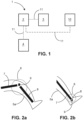
- Fig. 1 illustrates a schematic representation of a system according to the present disclosure.
- the system 1 comprises a data processing system 2 according to the present disclosure.
- the system may also comprise, as shown in the present example, a 3D surface imaging system 3, for example a depth camera, and an imaging system 4 configured to acquire 2D and/or 3D medical image data, for example, an MRI or CT scanner or an X-ray imaging device.
- the data processing system 2 may comprise one or more processors, in particular one or more computing devices and/or a distributed network of computing devices comprising the one or more processors.
- the system 1 is configured to carry out the method of the present disclosure, for example the method as specified in the method claims or as described below in the context of Figs. 3 and 4 .
- the data processing system may be configured to carry out and/or control the method of the present disclosure, for example the method as specified in the method claims or as described below.
- the data processing system may be configured to process first 3D surface image data of a body part in a first pose by means of an automatic pose retrieval method so as to obtain first articulation parameters representative of the first pose.
- Exemplary body parts are shown in Figs. 2a and 2b and described in the context thereof, e.g., a leg and an ankle respectively.
- the body part comprises a first anatomical structure and a second anatomical structure connected by a joint, for example the bones and joints described in the context of Figs. 2a and 2b .
- the system may further be configured to perform image registration between first medical image data of the body part and second medical image data of the body part in a second pose.
- the first medical image data e.g., CT, MRI, or X-ray image data
- the system is configured to perform the image registration individually for each of the first anatomical structure and the second anatomical structure.
- the system may further configured to determine transformation data representing one or more first transformations required to register the first anatomical structure in the first medical image data to the first anatomical structure in the second medical image data and/or one or more second transformations required to register the second anatomical structure in the first medical image data to the second anatomical structure in the second medical image data and to create and/or updating a statistical articulated joint model based at least on the transformation data, the first articulation parameters, and second articulation parameters representative of the second pose. If an articulated joint model is updated, the system may be configured to access a current statistical articulated joint model stored, e.g. locally or remotely, and update the accessed model. The system may then store the created and/or updated model, e.g., locally or remotely.
- the 3D surface imaging system 3 may be configured to acquire the 3D surface image data used in the method claims or as described below.
- the medical imaging system may be configured to acquire medical image data, for example, CT, MRI, or X-ray image data as used in the method claims or as described below.
- the data processing system in addition to being configured to carry out the steps described above, may be configured to control image acquisition by means of the 3D surface imaging system and/or the medical imaging systems so as to obtain the respective image data.
- the processing system may be configured to retrieve previously stored image data and/or models, e.g., stored locally and/or remotely at one or more data storage devices 10.
- the data processing system may connect to the imaging system(s) and/or data storage system(s) where the image data and/or models are stored via data connections 11, e.g., wireless and/or wired data connections.
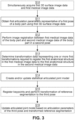
- Figs. 2a and 2b are schematic, not-to scale representations of two different types of joints.
- the body part 5 is the lower limb of a human subject and the knee is shown as an example for the joint 6.
- the first anatomical structure 7 may be a femur and the second anatomical structure 8 may be a tibia.
- the patella is shown as a third anatomical structure 9.
- the body part is the ankle of a human subject and the ankle joint is shown as an example for the joint 6.
- the tibia, talus bone, and calcaneus are shown as examples for the first, second and third anatomical structures 7 to 9. It is noted that the knee has only one degree of freedom and, accordingly, there is only one flexion angle for each pose.
- the ankle joint has two independent degrees of freedom and, accordingly, there are two flexion angles for each pose.
- a constellation model may be provided by the system and method of the present disclosure, which is a collection of the flexion angles for each of a plurality of poses.
- the constellation model may then be used to obtain the articulated joint model.
- FIG. 3 a flowchart illustrates an exemplary method for modelling a joint according to the present disclosure.
- step S11 first 3D surface image data of a body part in a first pose is processed by means of an automatic pose retrieval method so as to obtain first articulation parameters representative of the first pose.
- the body part comprises a first anatomical structure and a second anatomical structure connected by a joint.
- step S12 image registration between first medical image data of the body part and second medical image data of the body part in a second pose is performed.
- the first medical image data is image data acquired simultaneously with the first 3D surface image data and depicting the first anatomical structure and the second anatomical structure.
- the image registration is performed individually for each of the first anatomical structure and the second anatomical structure.
- the medical image data may be 3D medical image data, for example 3D CT or MRI image data. Accordingly, a 2D/3D registering of medical image data onto a mesh for obtaining 3D information may not be required in this case.
- step S13 transformation data representing one or more first transformations required to register the first anatomical structure in the first medical image data to the first anatomical structure in the second medical image data and/or one or more second transformations required to register the second anatomical structure in the first medical image data to the second anatomical structure in the second medical image data is determined.
- a statistical articulated joint model is created and/or updated based at least on the transformation data, the first articulation parameters, and second articulation parameters representative of the second pose.
- step S15 an anatomical structure-wise, e.g., bone-wise, registration of keypoints of anatomical structures in the medical image data that may be inferred from 3D surface image data of the body part may be performed.
- the shape of the surface of the body part may be indicative of the shape and position of a bone or other anatomical structure underneath the surface, such that detecting such shapes in the 3D surface image can be employed to estimate the position of keypoints of the respective anatomical structure in the medical image data.
- a reference segmentation of the first anatomical structure and/or of the second anatomical structure from the first pose to a third pose is transformed employing the estimated position of the keypoints.
- step S16 the articulated joint model is updated based on articulation parameters of the third pose and the transformed reference segmentation and/or corresponding transformation data. It is noted that steps S15 and S16 may be performed after steps S11 to S14 or afterwards. If they are performed prior to steps S11 to S14, they may be used for creating a joint model, which may then be updated in step S14.
- the optional steps S15 and S16 allow for incorporating information gained from 3D surface image data into the joint model even if no corresponding medical image data is available. This may be particularly useful when 3D surface image data within one imaging session are obtained at a higher rate than the medical image data.
- the method may optionally comprise the step S10 of simultaneously acquiring the 3D surface image data and the medical image data.
- the image data may also at least in part be retrieved image data.
- the medical images comprise 2D medical images. Most of the steps may, for example, be the same as in Fig. 3 and described above.
- an additional step S11a is performed, wherein a 2D/3D registering of the 2D first and second medical image data is performed prior to the registration step S12.
- the 2D medical image data may be registered onto a common mesh representative of a 3D shape of the surface of the respective anatomical structure, as will be described in more detail below.
- the registration S12 and the transformation step S13 may be carried out making use of the 2D/3D registered medical image data.
- the above method may be applied where medical imaging modalities that depict anatomical structures, like bones, in a 2D projection are used, for example regular X-ray imaging systems.
- 2D medical image data can be employed in the method according to the present disclosure for creating and/or updating the articulated joint model by performing a 2D/3D registering of the first and second 2D medical image data onto a common mesh topology. Thereby, the 2D image data can be accurately registered and incorporated into an articulated joint model.
- the 2D image data in particular, can also be employed to create and/or update a 3D articulated joint model by performing the 2D/3D registration.
- the mesh may be obtained from a pre-existing 3D model of each of the anatomical structures, for example a pre-existing 3D articulated model of the joint and/or a pre-existing rigid model of the joint or of the individual anatomical structures.
- the same mesh when repeating the 2D/3D registration for additional image data to be incorporated into the joint model.
- the joint model is a statistical model that may incorporate data from several different subjects, a mean mesh for these subjects may be employed, which may be updated with each new additional medical image data set.
- An initial mesh may be created manually or semi-automatically or automatically.
- meshes for 2D/3D registration of anatomical structures can be performed using known method known in the art.
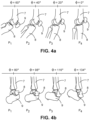
- Figs. 4a and 4b illustrate different joints, in case of Fig. 4a the knee and in case of Fig. 4b the ankle, in different poses P1 to P4, respectively.
- flexion angle ⁇ is indicated for each pose.
- the knee has only one degree of freedom and, accordingly, there is only one flexion angle for each pose.
- the ankle joint has two independent degrees of freedom and, accordingly, there are two flexion angles for each pose.
- flexion of the upper ankle joint i.e., dorsi- and plantarflexion
- the flexion angle for the lower ankle joint i.e. the inversion or eversion angle
- Figs. 5a and 5b show examples for a 3D articulated model of a knee.
- Fig. 5c shows a mesh, i.e., a segmented view of a knee, that may be used for different purposes for creating and/or updating the joint model, as described above.
- Fig. 6 shows another exemplary flow chart for a method according to the present disclosure.
- a medical imaging modality and a depth camera are used to simultaneously record medical image and depth image of the subject. This information is processed in a dedicated processing pipeline as shown in Fig. 6 in order to correctly co-register the medical images and generate or refine an articulated model.
- the building blocks of an exemplary method are:
- Output A constellation model C( ⁇ 0 , ..., ⁇ n ) describing the position of model components for a given pose.
- the registration in steps 4 and 5 may be based on the segmented bones, too, or both on the segmented bones and the estimated pose. In both cases, the depth-signal based pose estimation can be used for measuring the articulation parameters and thus for sorting the cases. Alternatively, the articulation parameters can be derived from the rotation degrees of freedom of the registration transformation (encoding rotations with respect to a reference pose).
- the depth-based pose estimation can be used to filter a dataset for specific articulations, e.g. for selecting cases in which only the flexion-angle of the ankle joint is varied while the inversion/eversion angle is binned to a small, fixed range.
- the bone segmentation of some reference case can also be used for constructing the articulated bone model.
- these reference segmentations e.g. in form of meshes
- these reference segmentations can be transformed to other poses by bone-wise registering the keypoints inferred from the depth signal.
- an algorithm could check for potential collisions of the bone meshes and may then restrict the movement of the bones accordingly.
Abstract
Disclosed is a method for modelling a joint, the method comprising, processing first 3D surface image data of a body part in a first pose by means of an automatic pose retrieval method so as to obtain first articulation parameters representative of the first pose, wherein the body part comprises a first anatomical structure and a second anatomical structure connected by a joint; performing image registration between first medical image data of the body part, the first medical image data acquired simultaneously with the first 3D surface image data and depicting the first anatomical structure and the second anatomical structure, and second medical image data of the body part in a second pose, wherein the image registration is performed individually for each of the first anatomical structure and the second anatomical structure; determining transformation data representing one or more first transformations required to register the first anatomical structure in the first medical image data to the first anatomical structure in the second medical image data and/or one or more second transformations required to register the second anatomical structure in the first medical image data to the second anatomical structure in the second medical image data; and creating and/or updating a statistical articulated joint model based at least on the transformation data, the first articulation parameters, and second articulation parameters representative of the second pose.
Description
- The invention provides a computer-implemented method for modelling a joint, a data processing system, a system comprising the data processing system, a computer program product, and a computer readable medium.
- Many recent developments in medical image analysis and processing demand dynamic anatomical models, also referred to as articulated models, e.g., representative of postures such as flexion of a joint or inhale/exhale for the thorax.
- For example, 3D joint models may be employed for quality assessment, preparing for diagnosis, and/or treatment planning. In particular, certain diagnostic questions require a specific image acquisition whereby the joint flexion is within a specific range, since deviation from this flexion might cause certain anatomical features of diagnostic interest to be obscured within projection images.
- Articulated models may be used to evaluate the positioning of the subject based upon just the image data and, once they are fitted to available data, also allow for judging joint dynamics and (ab)normality of movement range.
- Building those articulated models over a wide patient population is tedious and time-consuming.
- Moreover, building them requires the use of imaging modalities, e.g., using ionizing radiation, that may be harmful to a patient. Accordingly, volunteer scans outside of regular imaging sessions, like routine exams of a patient, may not be available at all in sufficient number and also pose ethical concerns.
- Regular imaging sessions, like routine exams of a patient do usually not allow for significant workflow changes, particularly if they are time consuming, e.g., imaging a joint in multiple poses using the imaging modality used for the imaging session. Clinical workflows typically do not permit the addition of steps that slow down routine scheduling and throughput, such as performing dedicated measurements for different poses during examination. For example, externally measuring the flexion for multiple images obtained by the imaging modality of the workflow at different poses and recording it to be stored with the image, so as to co-register and sort them according to flexion angle and, based thereon, build a model is difficult to incorporated into the routine workflows.
- Accordingly, it may be difficult to build a complete model with adequate precision.
- It is an object of the present invention to allow for improved modelling of a joint.
- The invention provides a method for modelling a joint, a data processing system, a system comprising the data processing system, a computer program product, and a computer readable medium according to the independent claims.
- The method for modelling a joint according to the present disclosure comprises processing first 3D surface image data of a body part in a first pose by means of an automatic pose retrieval method so as to obtain first articulation parameters representative of the first pose, wherein the body part comprises a first anatomical structure and a second anatomical structure connected by a joint.
- The method for modelling a joint according to the present disclosure further comprises performing image registration between first medical image data of the body part, the first medical image data acquired simultaneously with the first 3D surface image data and depicting the first anatomical structure and the second anatomical structure, and second medical image data of the body part in a second pose. The image registration is performed individually for each of the first anatomical structure and the second anatomical structure.
- The method for modelling a joint according to the present disclosure further comprises determining transformation data representing one or more first transformations required to register the first anatomical structure in the first medical image data to the first anatomical structure in the second medical image data and/or one or more second transformations required to register the second anatomical structure in the first medical image data to the second anatomical structure in the second medical image data.
- The method for modelling a joint according to the present disclosure further comprises creating and/or updating a statistical articulated joint model based at least on the transformation data, the first articulation parameters, and second articulation parameters representative of the second pose.
- In other words, the limitations of known methods can be addressed by relying on additional sensor data (in addition to data from the imaging modality) providing depth information, for example from a surface scanner or range camera or other 3D camera system, e.g., a RGBD camera, to record pose information during examination with the imaging modality. This enables to relate pose and image from the imaging modality obtained in a routine workflow. Based on the relation, an articulated model can be built and/or within a standard workflow of a radiology department.
- Particularly the method of the present disclosure allows to acquire arbitrary poses without the need for additional recording information. For example, no user observations regarding the articulation pose of a joint for a given acquisition are required and pose data does not have to be measured/tracked manually by the radiographer. This is particularly helpful when using measurement devices for measuring pose parameters would be a burden for the imaging modality, e.g. within a CT bore due to space restrictions or in an MRI scanner due to the use of metal not being possible.
- Thus, since the proposed approach does not require interruption of imaging workflow, it allows for including images from clinical routine workflows to the data used for building the articulated model. Thus, a large and diverse dataset can go into building the model.
- The present disclosure allows, as one example, for the development of articulated joint models, where the model is created using MR scans of a joint in which the joint has been positioned a priori in one or more known poses or articulations.
- A potential application of the articulated model is for skeletal X-ray image quality assessment.
- As can be seen from the above, the claim entails creating and/or updating the articulated model. For example, the method may entail using any articulated model, which may be empirical, semi-empirical, or theoretical, as a starting point and apply the steps of the present disclosure, particularly repeatedly for one or more subjects and/or one or more imaging sessions of the same subject or different subjects, to improve the model. Improving the model may concern different aspects. For example, the model may become less generic and more closely represent reality, particularly statistically over a wide range of subjects. Moreover, the precision of the model may be improved, e.g., by allowing to meaningfully increase the size of the data set. Moreover, the range of the model may be extended with respect to extreme poses towards the end of the movement spectrum, e.g., poses representative of a very strong flexion and/or poses representative of a hyperextension of a joint.
- In the present disclosure, excluding the description of the prior art, the term "joint model" is used synonymously with the term "statistical articulated joint model" for the sake of readability.
- A joint is a connection movably connecting bones in the body.
- The body part may, for example, be an arm, a wrist, a shoulder, an ankle, a leg, a hip, the foot, the hand, or any other portions of the body comprising a joint.
- In the present disclosure, the term "anatomical structure" refers to a sub-surface part of the body. An anatomical structure may, for example, be a bone, a muscle, a cartilage, a ligament, or the like. It is noted that different anatomical structures may be rigid to different degrees, such the movement of some anatomical structures may have more degrees of freedom than that of other anatomical structures. For example, a bone is essentially rigid and non-compressible, such that its movement can generally be represented by translations and rotations. Sinews or cartilage may be elastically deformed, e.g., lengthened, contracted, or compressed, so their movement may also include, particularly elastic, deformation and scaling.
- A pose is a certain relative arrangement of a set of anatomical structures. For example, a pose may be defined by a relative arrangement of two bones, e.g., one or more angles describing their relative arrangement in one or more degrees of freedom.
- The pose can be represented by articulation parameters. Articulation parameters may be any parameterization of relative arrangement of the anatomical structures. For example, articulation parameters may comprise flexion angles.
- It is to be understood that the first and second pose are different, e.g., they differ in at least one articulation parameter. For example, the first pose and the second pose may differ in that they have a different flexion angle.
- Automatic pose retrieval may be any method known in the art, e.g., from gaming applications, that determines the pose of body parts based on 3D surface image data. The 3D information allows for resolving directional ambiguities that might not be resolved otherwise.
- It is noted that according to
claim 1, second articulation parameters representative of the second pose are used. The second articulation parameters may be data derived from second 3D surface image data of the body part obtained using automatic pose retrieval. Alternatively, second articulation parameters may be articulation parameters derived from the second medical image data and/or image data obtained using a different imaging modality and/or obtained by direct pose measurements. - 3D surface image data is data that provides depth information. For example, 3D surface image data may be obtained, for example, by a 3D surface scanner, a stereoscopic camera, and/or an RGBD camera.
- Medical image data, in the present disclosure, refers to image data depicting sub-surface structures of the body, e.g., image data obtained using ionizing radiation. The medical image data may, for example, comprise CT image data, MRI data, or X-ray image data. Medical image data may comprise 2D and/or 3D image data. 3D image data provides depth information of the imaged structure. 2D image data may, for example, depict a projection of the imaged structure.
- According to the present disclosure, the first 3D surface image data and the first medical image data are acquired simultaneously. This may include that, during an imaging session, first 3D surface image data is acquired continuously over a period of time including the time or times when the first medical image data is acquired. 3D surface image data, particularly from said period of time may also be employed for building the model if there is not corresponding medical image data, e.g., when it was obtained in the time between capturing the medical images. For example, such image data may be used for improving automatic pose retrieval accuracy or for improving accuracy when building the joint model, e.g., by inferring keypoints of the anatomical structure from the surface image data, as explained in detail further below, or by aiding in removing ambiguities.
- Image registration may be performed using registration methods known in the art, including 2D/3D registration, wherein 2D image data is registered with 3D image data and/or a mesh, and 3D/3D registration, wherein 3D image data is registered with 3D image data and/or a mesh.
- According to the present disclosure, image registration is performed individually for each anatomical structure. This means that the first and the second anatomical structure are independently registered. The image registration may nonetheless be performed in parallel for the first and second anatomical structure.
- Transformations according to the present disclosure may include rotations and/or translations. In addition, transformations may include deformations and scaling, e.g., in case the anatomical structure is non-rigid and/or compressible.
- An articulated joint model may be a model that is representative of the movement of the joint, particularly the anatomical structures of the joint, between different articulations.
- The articulated joint model may be a 2D model, which models the joint in a projection, or a 3D model having depth information of the joint, particularly of the anatomical structures. For the sake of completeness, even if the articulated joint model is a 2D model, the process of creating and/or updating the model involves 3D data of some kind so as to allow avoid ambiguities.
- A statistical model is representative of data obtained from multiple imaging sessions, particularly, of more than one subject. As an example, the same joint can be imaged for multiple subjects and the model can be built so as to average out differences between different subjects, e.g., size and shape differences, differences in alignment of the anatomical structures or the like. The statistical model may also be selectively based on data from a plurality of subjects sharing certain characteristics, as will be described in detail below.
- Creating a statistical articulated joint model may entail starting with no model of the overall joint, for example starting with individual models of the first and second anatomical structures, and/or starting with a rigid model of the joint and/or a non-statistical model of the joint (articulated or non-articulated) to obtain a statistical articulated joint model.
- Updating a statistical articulated joint model may include modifying a current version of a statistical articulated joint model to account for additional information, in particular, to account for the transformation data, the first articulation parameters, and second articulation parameters representative of the second pose.
- Accordingly, creating and/or updating the statistical articulated joint model based on transformation data, the first articulation parameters, and second articulation parameters representative of the second pose, may be seen as taking said data and parameters into account for modelling the joint, optionally together with pre-existing data for modelling the joint.
- According to the present disclosure, the statistical articulated joint model may be based on transformation data, first articulation parameters, and second articulation parameters from image data acquired in multiple imaging sessions of a subject and/or from one or more imaging sessions of each of a plurality of subjects.
- An imaging session may be a routine examination and may, in particular, not comprise changing the routine workflow, particularly may not comprise acquiring medical image data in addition to the ones that are part of the routine examination and may not comprise performing manual measurement of pose. By allowing to combine data from different imaging sessions of the same or different subjects, a large data set and, accordingly, accurate statistics can be performed, thereby allowing for a more accurate model.
- Alternatively or in addition, according to the present disclosure, creating and/or updating the statistical articulated joint model may comprise determining a mean over a randomly selected subset of the population and/or determining a mean over a subset of the population sharing a type of misalignment of the joint.
- When using data acquired from a plurality of, particularly randomly selected, subjects, a statistical model that averages out subject-specific characteristics can be provided.
- When only data acquired for the subset of population sharing a type of misaligned is used for the statistical model, this may allow determining characteristics of the joint associated with the misalignment. This may help better address the needs brought about by the misalignment and yet still have large data set compared to only a single subject.
- According to the present disclosure, the statistical articulated joint model may be a 3D model. For example, a 3D surface of each of the anatomical structures of the joint may be provided, particularly over the entire range of the model. This allows for a particularly detailed study of the anatomical structures and their interactions.
- The method of the present disclosure may comprise creating, for each of a plurality of poses, including the first pose P1 and the second pose P2, a constellation model C(φ0, ..., φn) representative of an arrangement of the first anatomical structure and an arrangement of the second anatomical structure as a function of the articulation parameters representative of the respective pose, particularly, as a function of a flexion angle φi of the joint, so as to obtain a plurality of constellation models. The constellation model as such may be used as input data for creating and/or updating the statistical articulated joint model. The arrangement may be determined with respect to any suitable common reference.
- According to the present disclosure, creating and/or updating the statistical articulated joint model may be based on the plurality of constellation models, in particular, may comprise combining the plurality constellation models. For example, the constellation model may be used for sorting medical images in image space for creating the joint model. Moreover, the constellation model may be used together with interpolation so as to obtain an articulated model that comprises poses for which no image data is available.
- According to the present disclosure, the image registration may comprise applying a registration algorithm registering image data, e.g., based on landmarks in the image data, and/or meshes obtained by a segmentation algorithm, e.g., based on one or more vertices of a mesh. In particular, the method of the present disclosure may comprise, prior to the image registration, applying a segmentation algorithm segmenting the first anatomical structure and/or the second anatomical structure to obtain meshes. Any known segmentation and/or image registration methods may be applied. Anatomical atlas data may be employed.
- The method of the present disclosure may comprise using the first articulation parameters and the second articulation parameters for a pre-registration.
- For example, the first and second articulation parameters may be used for performing transformations that roughly match the anatomical structures prior to carrying out an image registration algorithm. This may reduce resources required for the image registration and may also resolve ambiguities that the image registration cannot resolve.
- According to the present disclosure, the first anatomical structure may be used as a reference anatomical structure and the statistical articulated joint model may be representative of an arrangement of the second anatomical structure relative to the first anatomical structure.
- For example, flexion angles may be expressed relative to the reference anatomical structure. The reference anatomical structure may be one of one or more bones of the joint.
- According to the present disclosure, the statistical articulated joint model may be a model representative of one or more selected movement types and the method may comprise filtering data for one or more selected poses, in particular for one or more selected articulation parameters, representative of the one or more selected movement types, such that only data representative of the one or more selected movement types is used for creating and/or updating the statistical articulated joint model.
- An articulated joint model that is representative of the selected movement type(s) may be advantageous for a detailed characterization of a joint. It may also reduce unnecessary complexity where only one movement type is relevant for a given application.
- For example, a movement type may be a rotation and/or bending in only a first plane or bending only in a second plane. A movement can be represented by a collection of poses that are part of the movement. A movement type may, for example, be represented by only a subset of all possible poses of a joint. For example, a movement type may be represented only by poses having the same flexion angle in one direction and differing in the flexion angle in another direction. Accordingly, the articulation parameters representative of the selected movement type may be the articulation parameters corresponding to the selected poses representative of the selected movement type.
- Filtering data for the selected poses may entail that only data corresponding to the selected poses, e.g., image data and/or meshes, are used as input for creating the articulated joint model.
- The method of the present disclosure may comprise anatomical structure-wise registering of keypoints inferred from 3D surface image data of the body part to transform a reference segmentation of the first anatomical structure and/or of the second anatomical structure from the first pose P1 to a third pose P3 and creating and/or updating the statistical articulated joint model based on articulation parameters of the third pose P3 and the transformed reference segmentation and/or corresponding transformation data.
- As an example, bones or other anatomical structures may have an impact on the surface of the subject, e.g., may be discernable through the skin and/or tissue. For example, protrusions may be caused on the surface. Points where an anatomical structure is discernable on the surface may be detected in the surface image data. The position of the detected points may then allow for estimating where one or more keypoints, e.g., on a mesh, of the anatomical structure are, particularly based on 3D image data and medical image data in a given pose optionally supplemented by general anatomical data. Based thereon, for other poses where no medical image data is available, detecting the points where the anatomical structure is discernable on the surface in 3D image data allows for inferring the positions of the keypoints. Based thereon, a transformation of the keypoints from one pose to the other pose can be determined and the mesh can be transformed as well based on the transformation of the keypoints. Thereby, additional data of the anatomical structure can be obtained even for poses where only surface image data is available. An example is described in more detail below.
- The method of the present disclosure may comprise determining, in the first 3D surface image data of the body part, one or more keypoints of the surface of the body part in the first 3D surface image data corresponding to keypoints of the first anatomical structure and/or the second anatomical structure in the first medical image data.
- A keypoint of the surface may be a point on the surface of a subject where part of the anatomical structure is discernable, e.g., by creating a protrusion on the surface. The corresponding keypoint of the anatomical structure may be a point of the surface of the part of the anatomical structure that is discernable on the surface, e.g., that causes a protrusion.
- The method of the present disclosure may further comprise determining a position of each of the one or more keypoints of the surface of the body part and a position of each of the one or more keypoints of the first anatomical structure and/or second anatomical structure in the first pose P1, for example by means of a/the reference segmentation of the first anatomical structure and/or a/the reference segmentation of the second anatomical structure.
- The method of the present disclosure may further comprise processing third 3D surface image data of the body part arranged in a third pose P3 to identify a position of each of the one or more keypoints of the surface of the body part in the third pose P3. It is to be understood that the third pose may be different from second pose and different from the first pose.
- The method of the present disclosure may further comprise, based on the position of each of the one or more keypoints of the surface of the body part in the third pose P3, the position of each of the one or more keypoints of the surface of the body part in the first pose P1, and the position of each of the one or more keypoints of the first anatomical structure and/or the second anatomical structure in the first pose P1, determining a position of each of the corresponding keypoints of the first anatomical structure and/or the second anatomical structure in the third pose P3.
- The method of the present disclosure may further comprise determining keypoint transformation data representing one or more third transformations required to match each of the one or more keypoints of the first anatomical structure and/or the second anatomical structure in the first pose P1 with each of the keypoints of the first anatomical structure and/or the second anatomical structure in the third pose P3.
- The method of the present disclosure may further comprise creating and/or updating the statistical articulated joint model based on the keypoint transformation data and third articulation parameters representative of the third pose P3.
- Thus, surface image data for poses for which no medical image data is available, e.g., the third pose, may be used as input for the articulated model. This allows for an even more extensive set of input data for the model, which also increases accuracy of the model.
- The method of the present disclosure may comprise a step of checking for potential collisions of the reference segmentation of the first anatomical structure and the reference segmentation of the second anatomical structure when transforming from the first pose P1 to the third pose P3 and restricting movement of the anatomical structures accordingly.
- This allows for avoiding extrapolation errors and/or other modelling errors. Specifically, where real anatomical structures do not collide, parts of the articulated model that represent a collision would be inaccurate and can therefore be discarded.
- The method of the present disclosure may comprise simultaneously acquiring the first 3D surface image data and the first medical image data. That is, the method may comprise the steps of data acquisition of data used for creating and/or updating the model. This may be done by respective 3D surface imaging means and medical imaging means, e.g., as described above.
- The invention also provides a data processing system to carry out and/or control any of the method steps of the present disclosure. In particular, the data processing system may be configured to carry out one or more, in particular all, of the computing steps of the present disclosure and/or to control acquiring the 3D surface image data and/or the medical image data, for example, by controlling a 3D surface imaging system configured to acquire the 3D surface image data and/or an imaging system configured to acquire the medical image data.
- In particular, the data processing system may comprise one or more processors, in particular one or more computing devices and/or a distributed network of computing devices comprising the one or more processors, and the one or more processors may be configured to carry out and/or control the steps of the method of the present disclosure.
- The invention also provides a system comprising the data processing system of the present disclosure and further comprising a 3D surface imaging system configured to acquire the 3D surface image data, including the first 3D surface image data and/or the second 3D surface image data and/or the third 3D surface image data. The system also comprises an imaging system configured to acquire medical image data, including the first medical image data and/or the second medical image data, in particular a CT imaging system and/or an MRT imaging system and/or an X-ray imaging system. The system may be configured to perform any of the methods of the present disclosure.
- The invention also provides a computer program product comprising instructions which, when the program is executed by a computer, cause the computer to carry out and/or control any of the method steps described in the present disclosure.
- The invention also provides a computer readable medium comprising instructions which, when executed by a computer, cause the computer to carry out and/or control any of the method steps described in the present disclosure.
- The present disclosure also provides a use of the statistical articulated joint model for at least one of quality assurance for measurement data, properly placing/arranging a subject, properly measuring a subject, simulating and/or visualizing medical imaging arrangements, examining subject-specific articulation, deviations, and limitations.
- The features and advantages outlined in the context of the method for modelling a joint similarly apply to the data processing system, the system comprising the data processing system, the computer program product, and the computer readable medium of the present disclosure.
- Further features, examples, and advantages will become apparent from the detailed description making reference to the accompanying drawings.
-
-
Fig. 1 shows a schematic illustration of a system according to the present disclosure; -
Figs. 2a and 2b shows schematic, not-to-scale representations of two different types of joints; -
Fig. 3 shows a flow diagram representative of a method according to the present disclosure; -
Figs. 4a and 4b show different views of different types of joints; -
Figs 5a to 5c show examples for a 3D articulated model and a segmented view of a knee; and -
Fig. 6 shows another exemplary flow chart for a method according to the present disclosure. -
Fig. 1 illustrates a schematic representation of a system according to the present disclosure. - The
system 1 comprises adata processing system 2 according to the present disclosure. The system may also comprise, as shown in the present example, a 3Dsurface imaging system 3, for example a depth camera, and animaging system 4 configured to acquire 2D and/or 3D medical image data, for example, an MRI or CT scanner or an X-ray imaging device. Thedata processing system 2 may comprise one or more processors, in particular one or more computing devices and/or a distributed network of computing devices comprising the one or more processors. - The
system 1 is configured to carry out the method of the present disclosure, for example the method as specified in the method claims or as described below in the context ofFigs. 3 and4 . In particular, the data processing system may be configured to carry out and/or control the method of the present disclosure, for example the method as specified in the method claims or as described below. - Specifically, the data processing system may be configured to process first 3D surface image data of a body part in a first pose by means of an automatic pose retrieval method so as to obtain first articulation parameters representative of the first pose. Exemplary body parts are shown in
Figs. 2a and 2b and described in the context thereof, e.g., a leg and an ankle respectively. The body part comprises a first anatomical structure and a second anatomical structure connected by a joint, for example the bones and joints described in the context ofFigs. 2a and 2b . The system may further be configured to perform image registration between first medical image data of the body part and second medical image data of the body part in a second pose. The first medical image data, e.g., CT, MRI, or X-ray image data, is image data acquired simultaneously with the first 3D surface image data and depicts the first anatomical structure and the second anatomical structure. The system is configured to perform the image registration individually for each of the first anatomical structure and the second anatomical structure. The system may further configured to determine transformation data representing one or more first transformations required to register the first anatomical structure in the first medical image data to the first anatomical structure in the second medical image data and/or one or more second transformations required to register the second anatomical structure in the first medical image data to the second anatomical structure in the second medical image data and to create and/or updating a statistical articulated joint model based at least on the transformation data, the first articulation parameters, and second articulation parameters representative of the second pose. If an articulated joint model is updated, the system may be configured to access a current statistical articulated joint model stored, e.g. locally or remotely, and update the accessed model. The system may then store the created and/or updated model, e.g., locally or remotely. - The 3D
surface imaging system 3 may be configured to acquire the 3D surface image data used in the method claims or as described below. The medical imaging system may be configured to acquire medical image data, for example, CT, MRI, or X-ray image data as used in the method claims or as described below. - The data processing system, in addition to being configured to carry out the steps described above, may be configured to control image acquisition by means of the 3D surface imaging system and/or the medical imaging systems so as to obtain the respective image data. Alternatively or in addition, the processing system may be configured to retrieve previously stored image data and/or models, e.g., stored locally and/or remotely at one or more
data storage devices 10. The data processing system may connect to the imaging system(s) and/or data storage system(s) where the image data and/or models are stored viadata connections 11, e.g., wireless and/or wired data connections. -
Figs. 2a and 2b are schematic, not-to scale representations of two different types of joints. - In
Fig. 2a , as an example, the body part 5 is the lower limb of a human subject and the knee is shown as an example for the joint 6. The firstanatomical structure 7 may be a femur and the secondanatomical structure 8 may be a tibia. Moreover, the patella is shown as a thirdanatomical structure 9. - In
Fig. 2b , as an example, the body part is the ankle of a human subject and the ankle joint is shown as an example for the joint 6. The tibia, talus bone, and calcaneus are shown as examples for the first, second and thirdanatomical structures 7 to 9. It is noted that the knee has only one degree of freedom and, accordingly, there is only one flexion angle for each pose. The ankle joint has two independent degrees of freedom and, accordingly, there are two flexion angles for each pose. - As described above in the general part of the description, a constellation model may be provided by the system and method of the present disclosure, which is a collection of the flexion angles for each of a plurality of poses. The constellation model may then be used to obtain the articulated joint model.
- In
Fig. 3 , a flowchart illustrates an exemplary method for modelling a joint according to the present disclosure. - In step S11, first 3D surface image data of a body part in a first pose is processed by means of an automatic pose retrieval method so as to obtain first articulation parameters representative of the first pose. The body part comprises a first anatomical structure and a second anatomical structure connected by a joint.
- In step S12, image registration between first medical image data of the body part and second medical image data of the body part in a second pose is performed. The first medical image data is image data acquired simultaneously with the first 3D surface image data and depicting the first anatomical structure and the second anatomical structure. The image registration is performed individually for each of the first anatomical structure and the second anatomical structure.
- In the present example, the medical image data may be 3D medical image data, for example 3D CT or MRI image data. Accordingly, a 2D/3D registering of medical image data onto a mesh for obtaining 3D information may not be required in this case.
- In step S13, transformation data representing one or more first transformations required to register the first anatomical structure in the first medical image data to the first anatomical structure in the second medical image data and/or one or more second transformations required to register the second anatomical structure in the first medical image data to the second anatomical structure in the second medical image data is determined.
- In step S14, a statistical articulated joint model is created and/or updated based at least on the transformation data, the first articulation parameters, and second articulation parameters representative of the second pose.
- In
Fig. 3 , optional steps S15 and S16 are shown. In step S15, an anatomical structure-wise, e.g., bone-wise, registration of keypoints of anatomical structures in the medical image data that may be inferred from 3D surface image data of the body part may be performed. For example, the shape of the surface of the body part may be indicative of the shape and position of a bone or other anatomical structure underneath the surface, such that detecting such shapes in the 3D surface image can be employed to estimate the position of keypoints of the respective anatomical structure in the medical image data. A reference segmentation of the first anatomical structure and/or of the second anatomical structure from the first pose to a third pose is transformed employing the estimated position of the keypoints. - In step S16, the articulated joint model is updated based on articulation parameters of the third pose and the transformed reference segmentation and/or corresponding transformation data. It is noted that steps S15 and S16 may be performed after steps S11 to S14 or afterwards. If they are performed prior to steps S11 to S14, they may be used for creating a joint model, which may then be updated in step S14.
- The optional steps S15 and S16 allow for incorporating information gained from 3D surface image data into the joint model even if no corresponding medical image data is available. This may be particularly useful when 3D surface image data within one imaging session are obtained at a higher rate than the medical image data.
- It is noted that the method may optionally comprise the step S10 of simultaneously acquiring the 3D surface image data and the medical image data. However, alternatively, the image data may also at least in part be retrieved image data.
- In the following, an exemplary method for modelling a joint according to the present disclosure will be described, wherein the medical images comprise 2D medical images. Most of the steps may, for example, be the same as in
Fig. 3 and described above. - However, an additional step S11a is performed, wherein a 2D/3D registering of the 2D first and second medical image data is performed prior to the registration step S12. For example, the 2D medical image data may be registered onto a common mesh representative of a 3D shape of the surface of the respective anatomical structure, as will be described in more detail below.
- The registration S12 and the transformation step S13 may be carried out making use of the 2D/3D registered medical image data.
- As an example, the above method may be applied where medical imaging modalities that depict anatomical structures, like bones, in a 2D projection are used, for example regular X-ray imaging systems. As mentioned above, 2D medical image data can be employed in the method according to the present disclosure for creating and/or updating the articulated joint model by performing a 2D/3D registering of the first and second 2D medical image data onto a common mesh topology. Thereby, the 2D image data can be accurately registered and incorporated into an articulated joint model. The 2D image data, in particular, can also be employed to create and/or update a 3D articulated joint model by performing the 2D/3D registration.
- The mesh may be obtained from a pre-existing 3D model of each of the anatomical structures, for example a pre-existing 3D articulated model of the joint and/or a pre-existing rigid model of the joint or of the individual anatomical structures.
- It is preferable to use the same mesh when repeating the 2D/3D registration for additional image data to be incorporated into the joint model. As an example, since the joint model is a statistical model that may incorporate data from several different subjects, a mean mesh for these subjects may be employed, which may be updated with each new additional medical image data set. An initial mesh may be created manually or semi-automatically or automatically.
- The use of meshes for 2D/3D registration of anatomical structures can be performed using known method known in the art.
-
Figs. 4a and 4b illustrate different joints, in case ofFig. 4a the knee and in case ofFig. 4b the ankle, in different poses P1 to P4, respectively. - In
Figs. 4a and 4b , the flexion angle φ is indicated for each pose. The knee has only one degree of freedom and, accordingly, there is only one flexion angle for each pose. The ankle joint has two independent degrees of freedom and, accordingly, there are two flexion angles for each pose. However only flexion of the upper ankle joint, i.e., dorsi- and plantarflexion, is shown for simplicity, i.e., the flexion angle for the lower ankle joint, i.e. the inversion or eversion angle, remains constant. -
Figs. 5a and 5b show examples for a 3D articulated model of a knee.Fig. 5c shows a mesh, i.e., a segmented view of a knee, that may be used for different purposes for creating and/or updating the joint model, as described above. -
Fig. 6 shows another exemplary flow chart for a method according to the present disclosure. A medical imaging modality and a depth camera are used to simultaneously record medical image and depth image of the subject. This information is processed in a dedicated processing pipeline as shown inFig. 6 in order to correctly co-register the medical images and generate or refine an articulated model. - The building blocks of an exemplary method are:
- A 3D camera or range camera recording the subject during image formation.
- A pose retrieval algorithm, e.g. known from computer games, for the joint of interest, especially deriving the flexion angle(s) φ 0..n .
- A segmentation algorithm for segmenting the bone(s) within the image.
- A registration algorithm registering images or meshes according to a prior (reference) bone using the measured poses of the respective bone.
- A registration algorithm registering all other bones using the measured poses of the respective bones.
- A translation/rotation transformation describing the relative movement of each bone.
- Output: A constellation model C(φ 0, ..., φn ) describing the position of model components for a given pose.
- The above describes a fully automatic joint model construction from (depth-camera signal, medical image) pairs.
- The registration in
steps 4 and 5 may be based on the segmented bones, too, or both on the segmented bones and the estimated pose. In both cases, the depth-signal based pose estimation can be used for measuring the articulation parameters and thus for sorting the cases. Alternatively, the articulation parameters can be derived from the rotation degrees of freedom of the registration transformation (encoding rotations with respect to a reference pose). - The depth-based pose estimation can be used to filter a dataset for specific articulations, e.g. for selecting cases in which only the flexion-angle of the ankle joint is varied while the inversion/eversion angle is binned to a small, fixed range.
- Instead of segmenting the bones in every image of the training cohort, the bone segmentation of some reference case can also be used for constructing the articulated bone model. In this case, these reference segmentations (e.g. in form of meshes) can be transformed to other poses by bone-wise registering the keypoints inferred from the depth signal. When transforming this pose to a different pose, an algorithm could check for potential collisions of the bone meshes and may then restrict the movement of the bones accordingly.
- While the invention has been illustrated and described in detail in the drawings and foregoing description, such illustration and description are to be considered exemplary and not restrictive. The invention is not limited to the disclosed embodiments. In view of the foregoing description and drawings it will be evident to a person skilled in the art that various modifications may be made within the scope of the invention, as defined by the claims.
-
-
System 1 -
Data processing system 2 - 3D
surface imaging system 3 -
Imaging system 4 - Body part 5
- Joint 6
- First
anatomical structure 7 - Second anatomical structure 8f
- Third
anatomical structure 9 -
Data storage device 10 -
Data connections 11 - First pose P1
- Second pose P2
- Third pose P3
- Fourth pose P4
- Flexion angles φ
Claims (15)
- A computer-implemented method for modelling a joint (6), the method comprisingprocessing (S 11) first 3D surface image data of a body part (5) in a first pose (P1) by means of an automatic pose retrieval method so as to obtain first articulation parameters representative of the first pose (P1), wherein the body part (5) comprises a first anatomical structure (7) and a second anatomical structure (8) connected by a joint (6);performing (S12) image registration between first medical image data of the body part (5), the first medical image data acquired simultaneously with the first 3D surface image data and depicting the first anatomical structure (7) and the second anatomical structure (8), and second medical image data of the body part (5) in a second pose (P2), wherein the image registration is performed individually for each of the first anatomical structure (7) and the second anatomical structure (8);determining (S13) transformation data representing one or more first transformations required to register the first anatomical structure (7) in the first medical image data to the first anatomical structure (7) in the second medical image data and/or one or more second transformations required to register the second anatomical structure (8) in the first medical image data to the second anatomical structure (8) in the second medical image data; andcreating and/or updating (S14) a statistical articulated joint model based at least on the transformation data, the first articulation parameters, and second articulation parameters representative of the second pose (P2).
- The method of claim 1,wherein the statistical articulated joint model is based on transformation data, first articulation parameters, and second articulation parameters from image data acquired in multiple imaging sessions of a subject and/or from one or more imaging sessions of each of a plurality of subjects, and/orwherein creating and/or updating the statistical articulated joint model comprises determining a mean over a randomly selected subset of the population and/or determining a mean over a subset of the population sharing a type of misalignment of the joint (6).
- The method of claim 1 or 2, wherein the statistical articulated joint model is a 3D model.
- The method of any of the preceding claims, comprising creating, for each of a plurality of poses, including the first pose (P1) and the second pose (P2), a constellation model C(φ0, ..., φn) representative of an arrangement of the first anatomical structure (7) and an arrangement of the second anatomical structure (8) as a function of the articulation parameters representative of the respective pose, particularly, as a function of a flexion angle (φi) of the joint (6), so as to obtain a plurality of constellation models,
wherein creating and/or updating the statistical articulated joint model is based on the plurality of constellation models, in particular, comprises combining the plurality constellation models. - The method of any of the preceding claims,wherein the image registration comprises applying a registration algorithm registering image data and/or meshes obtained by a segmentation algorithm, in particular, the method comprising, prior to the image registration, applying a segmentation algorithm segmenting the first anatomical structure (7) and/or the second anatomical structure (8) to obtain meshes, and/orwherein the method comprises using the first articulation parameters and the second articulation parameters for a pre-registration.
- The method of any of the preceding claims, wherein the first anatomical structure (7) is used as a reference anatomical structure and the statistical articulated joint model is representative of an arrangement of the second anatomical structure (8) relative to the first anatomical structure (7).
- The method of any of the preceding claims, wherein the statistical articulated joint model is a model representative of one or more selected movement types and the method comprises filtering data for one or more selected poses, in particular for one or more selected articulation parameters, representative of the one or more selected movement types, such that only data representative of the one or more selected movement types is used for creating and/or updating the statistical articulated joint model.
- The method of any of the preceding claims, further comprisinganatomical structure-wise registering (S15) of keypoints inferred from 3D surface image data of the body part (5) to transform a reference segmentation of the first anatomical structure (7) and/or of the second anatomical structure (8) from the first pose (P1) to a third pose (P3); andcreating and/or updating (S16) the statistical articulated joint model based on articulation parameters of the third pose (P3) and the transformed reference segmentation and/or corresponding transformation data.
- The method of any of the preceding claims, comprisingdetermining, in the first 3D surface image data of the body part (5), one or more keypoints of the surface of the body part (5) in the first 3D surface image data corresponding to keypoints of the first anatomical structure (7) and/or the second anatomical structure (8) in the first medical image data;determining a position of each of the one or more keypoints of the surface of the body part (5) and a position of each of the one or more keypoints of the first anatomical structure (7) and/or second anatomical structure (8) in the first pose (P1), for example by means of a/the reference segmentation of the first anatomical structure (7) and/or a/the reference segmentation of the second anatomical structure (8);processing third 3D surface image data of the body part (5) arranged in a third pose (P3) to identify a position of each of the one or more keypoints of the surface of the body part (5) in the third pose (P3);based on the position of each of the one or more keypoints of the surface of the body part (5) in the third pose (P3), the position of each of the one or more keypoints of the surface of the body part (5) in the first pose (P1), and the position of each of the one or more keypoints of the first anatomical structure (7) and/or the second anatomical structure (8) in the first pose (P1), determining a position of each of the corresponding keypoints of the first anatomical structure (7) and/or the second anatomical structure (8) in the third pose (P3);determining keypoint transformation data representing one or more third transformations required to match each of the one or more keypoints of the first anatomical structure (7) and/or the second anatomical structure (8) in the first pose (P1) with each of the keypoints of the first anatomical structure (7) and/or the second anatomical structure (8) in the third pose (P3); andcreating and/or updating (S15) the statistical articulated joint model based on the keypoint transformation data and third articulation parameters representative of the third pose (P3).
- The method of claim 8 or claim 9, comprising a step of checking for potential collisions of the reference segmentation of the first anatomical structure (7) and the reference segmentation of the second anatomical structure (8) when transforming from the first pose (P1) to the third pose (P3) and restricting movement of the anatomical structures accordingly.
- The method of any of the preceding claims comprising simultaneously acquiring (S10) the first 3D surface image data and the first medical image data.
- A data processing system (2) configured to carry out the method steps of any one of claims 1 to 10 and/or carry out and/or control the method steps of claim 11.
- A system (1) comprising the data processing system (2) of claim 12 and further comprising:a 3D surface imaging system (3) configured to acquire 3D surface image data, including the first 3D surface image data and/or the second 3D surface image data and/or the third 3D surface image data; andan imaging system (4) configured to acquire medical image data, including the first medical image data and/or the second medical image data, in particular a CT imaging system and/or an MRT imaging system and/or an X-ray imaging system,in particular, wherein the system (1) is configured to carry out the method of any one of claims 1 to 11.
- A computer program product comprising instructions which, when the program is executed by a computer, cause the computer to carry out the method of any one of claims 1 to 10 and/or carry out and/or control the method steps of claim 11.
- A computer readable medium comprising instructions which, when executed by a computer, cause the computer to carry out the method of any one of claims 1 to 10 and/or carry out and/or control the method steps of claim 11.
Priority Applications (2)
| Application Number | Priority Date | Filing Date | Title |
|---|---|---|---|
| EP22161849.9A EP4246451A1 (en) | 2022-03-14 | 2022-03-14 | Method for modelling a joint |
| PCT/EP2023/055098 WO2023174681A1 (en) | 2022-03-14 | 2023-03-01 | Method for modelling a joint |
Applications Claiming Priority (1)
| Application Number | Priority Date | Filing Date | Title |
|---|---|---|---|
| EP22161849.9A EP4246451A1 (en) | 2022-03-14 | 2022-03-14 | Method for modelling a joint |
Publications (1)
| Publication Number | Publication Date |
|---|---|
| EP4246451A1 true EP4246451A1 (en) | 2023-09-20 |
Family
ID=80739024
Family Applications (1)
| Application Number | Title | Priority Date | Filing Date |
|---|---|---|---|
| EP22161849.9A Pending EP4246451A1 (en) | 2022-03-14 | 2022-03-14 | Method for modelling a joint |
Country Status (2)
| Country | Link |
|---|---|
| EP (1) | EP4246451A1 (en) |
| WO (1) | WO2023174681A1 (en) |
Citations (1)
| Publication number | Priority date | Publication date | Assignee | Title |
|---|---|---|---|---|
| WO2015089118A1 (en) * | 2013-12-09 | 2015-06-18 | Mahfouz Mohamed R | Bone reconstruction and orthopedic implants |
-
2022
- 2022-03-14 EP EP22161849.9A patent/EP4246451A1/en active Pending
-
2023
- 2023-03-01 WO PCT/EP2023/055098 patent/WO2023174681A1/en unknown
Patent Citations (1)
| Publication number | Priority date | Publication date | Assignee | Title |
|---|---|---|---|---|
| WO2015089118A1 (en) * | 2013-12-09 | 2015-06-18 | Mahfouz Mohamed R | Bone reconstruction and orthopedic implants |
Non-Patent Citations (5)
| Title |
|---|
| CHEN XIN ET AL: "Automatic Generation of Statistical Pose and Shape Models for Articulated Joints", IEEE TRANSACTIONS ON MEDICAL IMAGING, IEEE, USA, vol. 33, no. 2, 1 February 2014 (2014-02-01), pages 372 - 383, XP011538599, ISSN: 0278-0062, [retrieved on 20140130], DOI: 10.1109/TMI.2013.2285503 * |
| CONSTANTINESCU MIHAELA A M ET AL: "Constrained Statistical Modelling of Knee Flexion From Multi-Pose Magnetic Resonance Imaging", IEEE TRANSACTIONS ON MEDICAL IMAGING, IEEE, USA, vol. 35, no. 7, 1 July 2016 (2016-07-01), pages 1686 - 1695, XP011615416, ISSN: 0278-0062, [retrieved on 20160629], DOI: 10.1109/TMI.2016.2524587 * |
| FLEUTE M ET AL: "NONRIGID 3-D/2-D REGISTRATION OF IMAGES USING STATISTICAL MODELS", ELECTRONIC PUBLISHING, ARTISTIC IMAGING, AND DIGITAL TYPOGRAPHY; [LECTURE NOTES IN COMPUTER SCIENCE , ISSN 0302-9743], SPRINGER VERLAG, DE, 19 September 1999 (1999-09-19), pages 138 - 147, XP008010626, ISBN: 978-3-540-24128-7, DOI: 10.1007/10704282_15 * |
| HANSIS EBERHARD ET AL: "Landmark constellation models for medical image content identification and localization", INTERNATIONAL JOURNAL OF COMPUTER ASSISTED RADIOLOGY AND SURGERY, SPRINGER, DE, vol. 11, no. 7, 11 December 2015 (2015-12-11), pages 1285 - 1295, XP035990794, ISSN: 1861-6410, [retrieved on 20151211], DOI: 10.1007/S11548-015-1328-5 * |
| MASCHINO E ET AL: "Joint registration and averaging of multiple 3D anatomical surface models", COMPUTER VISION AND IMAGE UNDERSTANDING, ACADEMIC PRESS, US, vol. 101, no. 1, 1 January 2006 (2006-01-01), pages 16 - 30, XP024917626, ISSN: 1077-3142, [retrieved on 20060101], DOI: 10.1016/J.CVIU.2005.06.004 * |
Also Published As
| Publication number | Publication date |
|---|---|
| WO2023174681A1 (en) | 2023-09-21 |
Similar Documents
| Publication | Publication Date | Title |
|---|---|---|
| EP3586787B1 (en) | Image matching device and image matching method | |
| Quijano et al. | Three-dimensional reconstruction of the lower limb from biplanar calibrated radiographs | |
| Sarkalkan et al. | Statistical shape and appearance models of bones | |
| JP2020175184A (en) | System and method for reconstruction of 3d anatomical image from 2d anatomical image | |
| JP2021154168A (en) | Surgical navigation of the hip using fluoroscopy and tracking sensors | |
| US8965075B2 (en) | System and method for predicting future fractures | |
| US20200058098A1 (en) | Image processing apparatus, image processing method, and image processing program | |
| US20210012492A1 (en) | Systems and methods for obtaining 3-d images from x-ray information for deformed elongate bones | |
| JP2003144454A (en) | Joint operation support information computing method, joint operation support information computing program, and joint operation support information computing system | |
| US20060216681A1 (en) | Method and system for characterization of knee joint morphology | |
| Zhang et al. | 3-D reconstruction of the spine from biplanar radiographs based on contour matching using the hough transform | |
| JP2013528880A (en) | A method to determine joint bone deformity using motion patterns | |
| Taghizadeh et al. | Biomechanical role of bone anisotropy estimated on clinical CT scans by image registration | |
| Bartels et al. | Computed tomography-based joint locations affect calculation of joint moments during gait when compared to scaling approaches | |
| CN109350059B (en) | Combined steering engine and landmark engine for elbow auto-alignment | |
| Bermejo et al. | Genetic algorithms for skull-face overlay including mandible articulation | |
| EP4246451A1 (en) | Method for modelling a joint | |
| JP6967983B2 (en) | Image processing equipment, image processing methods, and programs | |
| Bousigues et al. | 3D reconstruction of the scapula from biplanar X-rays for pose estimation and morphological analysis | |
| CN110459298A (en) | For finding out the method and apparatus, diagnostic terminal and imaging system of end value | |
| WO2022229816A1 (en) | 3d reconstruction of anatomical images | |
| EP3910597A1 (en) | Body representations | |
| Moura et al. | A flexible approach for the calibration of biplanar radiography of the spine on conventional radiological systems | |
| Mihcin | Methodology on co-registration of MRI and optoelectronic motion capture marker sets: in-vivo wrist case study | |
| Zeng et al. | Low‐dose three‐dimensional reconstruction of the femur with unit free‐form deformation |
Legal Events
| Date | Code | Title | Description |
|---|---|---|---|
| PUAI | Public reference made under article 153(3) epc to a published international application that has entered the european phase |
Free format text: ORIGINAL CODE: 0009012 |
|
| STAA | Information on the status of an ep patent application or granted ep patent |
Free format text: STATUS: THE APPLICATION HAS BEEN PUBLISHED |
|
| AK | Designated contracting states |
Kind code of ref document: A1 Designated state(s): AL AT BE BG CH CY CZ DE DK EE ES FI FR GB GR HR HU IE IS IT LI LT LU LV MC MK MT NL NO PL PT RO RS SE SI SK SM TR |