EP4245836A1 - Cell culture system and method of detecting proliferative properties of cells - Google Patents
Cell culture system and method of detecting proliferative properties of cells Download PDFInfo
- Publication number
- EP4245836A1 EP4245836A1 EP21891935.5A EP21891935A EP4245836A1 EP 4245836 A1 EP4245836 A1 EP 4245836A1 EP 21891935 A EP21891935 A EP 21891935A EP 4245836 A1 EP4245836 A1 EP 4245836A1
- Authority
- EP
- European Patent Office
- Prior art keywords
- cells
- culture
- oxygen concentration
- culture vessel
- cell
- Prior art date
- Legal status (The legal status is an assumption and is not a legal conclusion. Google has not performed a legal analysis and makes no representation as to the accuracy of the status listed.)
- Pending
Links
Images




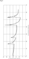

Classifications
-
- C—CHEMISTRY; METALLURGY
- C12—BIOCHEMISTRY; BEER; SPIRITS; WINE; VINEGAR; MICROBIOLOGY; ENZYMOLOGY; MUTATION OR GENETIC ENGINEERING
- C12M—APPARATUS FOR ENZYMOLOGY OR MICROBIOLOGY; APPARATUS FOR CULTURING MICROORGANISMS FOR PRODUCING BIOMASS, FOR GROWING CELLS OR FOR OBTAINING FERMENTATION OR METABOLIC PRODUCTS, i.e. BIOREACTORS OR FERMENTERS
- C12M23/00—Constructional details, e.g. recesses, hinges
- C12M23/24—Gas permeable parts
-
- C—CHEMISTRY; METALLURGY
- C12—BIOCHEMISTRY; BEER; SPIRITS; WINE; VINEGAR; MICROBIOLOGY; ENZYMOLOGY; MUTATION OR GENETIC ENGINEERING
- C12M—APPARATUS FOR ENZYMOLOGY OR MICROBIOLOGY; APPARATUS FOR CULTURING MICROORGANISMS FOR PRODUCING BIOMASS, FOR GROWING CELLS OR FOR OBTAINING FERMENTATION OR METABOLIC PRODUCTS, i.e. BIOREACTORS OR FERMENTERS
- C12M41/00—Means for regulation, monitoring, measurement or control, e.g. flow regulation
- C12M41/30—Means for regulation, monitoring, measurement or control, e.g. flow regulation of concentration
- C12M41/34—Means for regulation, monitoring, measurement or control, e.g. flow regulation of concentration of gas
-
- C—CHEMISTRY; METALLURGY
- C12—BIOCHEMISTRY; BEER; SPIRITS; WINE; VINEGAR; MICROBIOLOGY; ENZYMOLOGY; MUTATION OR GENETIC ENGINEERING
- C12M—APPARATUS FOR ENZYMOLOGY OR MICROBIOLOGY; APPARATUS FOR CULTURING MICROORGANISMS FOR PRODUCING BIOMASS, FOR GROWING CELLS OR FOR OBTAINING FERMENTATION OR METABOLIC PRODUCTS, i.e. BIOREACTORS OR FERMENTERS
- C12M41/00—Means for regulation, monitoring, measurement or control, e.g. flow regulation
- C12M41/30—Means for regulation, monitoring, measurement or control, e.g. flow regulation of concentration
- C12M41/36—Means for regulation, monitoring, measurement or control, e.g. flow regulation of concentration of biomass, e.g. colony counters or by turbidity measurements
Definitions
- the present invention relates to a cell culture technique, and especially relates to a cell culture system that detects a proliferative property of cells when the cells are statically cultured with a culture vessel formed of a gas permeable member.
- the cells are cultured in large quantities, it is important to confirm that the culture is properly performed by detecting the proliferative property of the cells. Additionally, for the detection of the proliferative property of cells, for example, it is preferred to allow confirming the number of cells in the culture vessel at desired timings during the culture.
- the method (1) has a problem that the risk of contamination increases because a part of the culture vessel is required to be opened for sampling the medium. Additionally, there is a problem that when adherent cells are cultured, it is impossible to detach only a part of the cells during the culture, and the number of cells cannot be counted at a desired timing.
- the method (2) has a problem that when floating cells are cultured, while the measurement can be performed when the culture density is low, the measurement becomes difficult because of the lamination of the cells when the cell density increases after the middle of the culture. Additionally, there is also a problem that since this method requires the microscope, the camera, and an image processing device, the mechanism of the cell culture system is complicated.
- a sealed state is required for avoiding the move of a gas from the inside to the outside of the culture vessel, and the entering of a gas into the culture vessel from outside.
- Patent Document 1 discloses a method for measuring a decreased amount of dissolved oxygen in a sample solution containing microorganisms in a state where the sample solution is sealed within a container, this measuring method needs to be performed using a culture vessel without gas permeability.
- Patent Document 2 discloses a cell culture device that operates an amount of temporal variation of oxygen amount relating to the number of cells in a culture vessel using an oxygen amount measured by oxygen amount measurement means that measures an oxygen amount dissolved in a medium in the culture vessel.
- this cell culture device uses a sealing container that avoids entering of a gas into a culture vessel from outside, and requires measuring a consumption of dissolved oxygen in a culture medium by changing a culture condition, for example, replacing a space on the culture medium inside the culture vessel with nitrogen to interrupt dissolution of oxygen into the culture medium, for obtaining a measurable state from an ordinary culture state.
- the present invention has been made in consideration of the above-described circumstances, and it is an object of the present invention to provide a cell culture system capable of detecting a proliferative property of cells in a culture vessel at a desired timing during a culture when the cells are statically cultured in the culture vessel formed of a gas permeable member, and a method for detecting the proliferative property of cells.
- a cell culture system of the present invention is a cell culture system for statically culturing cells with a culture vessel.
- the culture vessel is at least partially formed of a gas permeable member.
- the cell culture system includes an oxygen concentration sensor and a proliferation detection unit.
- the oxygen concentration sensor measures an oxygen concentration in a vicinity of a culture surface inside the culture vessel.
- the proliferation detection unit detects a proliferative property of the cells based on an oxygen permeability of the gas permeable member, a measurement value of the oxygen concentration sensor, an oxygen concentration around the culture vessel, and an oxygen consumption per cell of the cells.
- the proliferation detection unit preferably uses an estimated value of an oxygen concentration inside the culture vessel at a desired time point as the measurement value of the oxygen concentration sensor.
- the estimated value of the oxygen concentration inside the culture vessel at a desired time point is preferably obtained based on a ratio between a slope of a change in rise peak value of the oxygen concentration when an additional medium is added in the culture vessel multiple times and a slope of a change of the oxygen concentration when the culture vessel is stabilized.
- the oxygen concentration around the culture vessel is preferably an oxygen concentration inside an incubator in which the culture vessel is housed, or an atmospheric oxygen concentration.
- the oxygen concentration sensor is preferably a fluorescent type.
- the oxygen concentration sensor is preferably secured to the culture surface inside the culture vessel using a perforated resin member.
- a method for detecting a proliferative property of cells of the present invention is a method for detecting a proliferative property of cells in a cell culture in which the cells are statically cultured with a culture vessel.
- the culture vessel is at least partially formed of a gas permeable member.
- the method includes: measuring an oxygen concentration in a vicinity of a culture surface inside the culture vessel by an oxygen concentration sensor disposed on the culture surface inside the culture vessel; and calculating a count of the cells per unit area based on an oxygen permeability of the gas permeable member, a measurement value of the oxygen concentration sensor, an oxygen concentration around the culture vessel, and an oxygen consumption per cell of the cells.
- the present invention can provide the cell culture system capable of detecting the proliferative property of the cells in the culture vessel at a desired timing during the culture when the cells are statically cultured in the culture vessel formed of the gas permeable member, and the method for detecting the proliferative property of the cells.
- a cell culture system of this embodiment is a cell culture system for statically culturing cells with a culture vessel at least partially formed of a gas permeable member, and includes an oxygen concentration sensor that measures an oxygen concentration in a vicinity of a culture surface inside the culture vessel, and a proliferation detection unit that detects a proliferative property of cells based on an oxygen permeability of the gas permeable member, a measurement value of the oxygen concentration sensor, an oxygen concentration around the culture vessel, and an oxygen consumption per cell of the cells.
- the cell culture system of this embodiment can have, for example, a configuration as illustrated in FIG. 1 .
- a culture vessel 20 is housed in an incubator 10, and placed on a placement table 11.
- the culture vessel 20 includes two ports, and a medium supply container 40 housed in a cooler 30 is coupled to one port by a tube.
- a waste liquid container 50 inside the incubator 10 is coupled by a tube.
- liquid transfer means is disposed.
- a pump such as a peristaltic pump and a syringe pump, capable of transferring a liquid at a low speed with high accuracy is preferred to be used.
- a control device 60 controls the liquid transfer means between the culture vessel 20 and the medium supply container 40 at a predetermined timing, thus transferring a medium from the medium supply container 40 to the culture vessel 20.
- the control device 60 controls the liquid transfer means between the culture vessel 20 and the waste liquid container 50 at a predetermined timing, thus transferring the medium from the culture vessel 20 to the waste liquid container 50.
- a gas concentration inside the incubator 10 is controlled, and thus a gas concentration around the culture vessel 20 can be adjusted.
- the oxygen cylinder O and the carbon dioxide cylinder C are each connected to the incubator 10 via a valve, switching operations of the respective valves are controlled by the control device 60, and the gas supply to the incubator 10 from each gas cylinder is controlled, thus allowing the adjustment of oxygen concentration inside the incubator 10.
- the incubator 10 for example, a CO2 incubator is usable.
- the placement table 11 inside the incubator 10 one made of a perforated metal or the like can be disposed to be used, and the culture vessel 20 is placed on the placement table 11 and housed in a closed manner.
- the placement table 11 may be omitted.
- the incubator 10 may be omitted.
- the culture vessel 20 is a cell culture vessel of a closed system that is at least partially formed of a gas permeable member and has a gas permeability, and for example, one formed by heat-sealing peripheral edge portions of two rectangular gas permeable films can be used.
- the shape of the culture vessel 20 is formed to be inclined with respect to the ports in the vicinities of the ports, and this allows easily flowing the medium to the ports.
- the shape of the culture vessel 20 may be a rectangular shape such as an elongated rectangle.
- the number of the ports included in the culture vessel 20 is not limited to two, and may be one or three or more.
- a fluorescent oxygen concentration sensor 21 is disposed on a culture surface inside the culture vessel 20.
- a fluorescent light emitting/receiving unit 22 is disposed to be able to emit and receive a fluorescent light to the fluorescent oxygen concentration sensor 21.
- the fluorescent light emitting/receiving unit 22 is, for example, disposed under the placement table 11, and the fluorescent light emitting/receiving unit 22 can emit and receive a fluorescent light to the fluorescent oxygen concentration sensor 21 via the placement table 11 when it is transparent, or via a through hole provided to the placement table 11.
- the fluorescent light emitting/receiving unit 22 can be disposed to be in contact with the lower side of the culture vessel 20.
- the oxygen concentration inside the culture vessel 20 can be measured.
- the concentration of dissolved oxygen in the medium inside the culture vessel 20 largely differs depending on the position. That is, in the medium in the vicinity of the culture surface on which cells are present (upper surface of a lower surface gas permeable film 202), since the cells actively consume oxygen, oxygen is rapidly consumed, and gradually becomes 0% as the cell density increases.
- the oxygen concentration is slow in decrease from the initial state (for example, about 21% in atmosphere) when cells are not present in the vicinity.
- the oxygen concentration is approximately middle (for example, 10%) of them.
- the fluorescent oxygen concentration sensor 21 on the culture surface inside the culture vessel 20
- the oxygen concentration inside the culture vessel 20 based on the oxygen consumption by the cells can be appropriately measured.
- the oxygen consumption by the cells can be appropriately calculated.
- the oxygen concentration inside the culture vessel 20 is continuously 0% to about 1%, even when the measurement of the oxygen concentration by the fluorescent oxygen concentration sensor 21 is difficult, the oxygen consumption by the cells can be appropriately estimated.
- the dissolved gas in the medium inside the culture vessel is dissolved at a concentration that equilibrates with a partial pressure around the culture vessel.
- the concentration of oxygen dissolved in the medium placed in the incubator is about 21% the same as the concentration in atmosphere when the gas concentration inside the incubator is not controlled.
- the consumption of the dissolved oxygen inside the culture vessel by the cells decreases the dissolved oxygen of the medium around the cells, and the oxygen gas permeates the film and flows into the culture vessel so as to keep the equilibrium with the oxygen concentration outside the culture vessel.
- the gas permeability (speed) of the film is determined depending on the gas partial pressure difference between both sides of the film, affinity between the film and the gas, the diffusivity of gas molecules moving into the film, and the like.
- a fluorescent substance When a light is applied to a fluorescent element of the fluorescent oxygen concentration sensor, a fluorescent substance absorbs the light and becomes in an excited state, and then returns to a ground state while emitting a fluorescent light. At this time, when oxygen is present around the fluorescent substance, the excitation energy is lost, and the fluorescence emission intensity decreases. Accordingly, the more oxygen is, the more the fluorescence emission intensity decreases.
- the fluorescent light emitting/receiving unit measures the oxygen amount based on the change of the emission intensity due to the number of oxygen molecules contacting the fluorescent element when oxygen dissolved in the medium contacts the fluorescent element.
- a resin film or the like can be preferably used, and for example, a polyolefin-based resin such as polyethylene and polypropylene is usable.
- a polyolefin-based resin such as polyethylene and polypropylene
- polyethylene, a copolymer of ethylene and ⁇ -olefin, a copolymer of ethylene and vinyl acetate, an ionomer using: a copolymer of ethylene and an acrylic acid or ethylene and a methacrylic acid; and a metal ion, and the like can be listed.
- a polyolefin, a styrene-based elastomer, a polyester-based thermoplastic elastomer, and the like can be used.
- a soft vinyl chloride resin a polybutadiene resin, an ethylene-vinyl acetate copolymer, a chlorinated polyethylene resin, a polyurethane-based thermoplastic elastomer, a polyester-based thermoplastic elastomer, a silicone-based thermoplastic elastomer, a styrene-based elastomer, such as styrene-butadiene-styrene (SBS), styrene-isoprene-styrene (SIS), styrene-ethylene-butylene-styrene (SEBS), and styrene-ethylene-propylene-styrene (SEPS), a polyolefin resin, a fluorine-based resin, and the like may be used.
- SBS styrene-butadiene-styrene
- SIS styrene-isoprene-styren
- the gas permeable member used for at least a part of the culture vessel 20 among the above-described materials, especially, using a thermoplastic resin excellent in gas permeation performance is preferable, and a polyolefin-based resin, for example, polyethylene, such as LLDPE, or polypropylene can be preferably used.
- the gas permeable member is preferably a transparent material.
- a fluorescent oxygen concentration sensor 21a is secured to a culture surface (in the example of the drawing, upper surface of a lower surface gas permeable film 202a) inside a culture vessel 20a using a perforated resin member 23a.
- a fluorescent oxygen concentration sensor 21a is interposed between the perforated resin member 23a and the culture surface, and a welding region H of the perforated resin member 23a is welded to the culture surface, thereby allowing the securing.
- a polyolefin-based resin such as polyethylene, for example, LLDPE, and polypropylene is usable.
- polyethylene a copolymer of ethylene and ⁇ -olefin, a copolymer of ethylene and vinyl acetate, an ionomer using: a copolymer of ethylene and an acrylic acid or ethylene and a methacrylic acid; and a metal ion, and the like can be listed.
- a mesh material can be appropriately used.
- a porous material such as a sponge can be used.
- the specific mesh material and porous material are not especially limited, one of a similar material having the melting point close to that of the material of the culture vessel 20a is preferable, and for example, a polyethylene mesh material (NBC Meshtec Inc., Item Number PE 200 me, or the like) and a polyethylene porous membrane (TEIJIN LIMITED, miraim (Trade name) large pore ⁇ 3 ⁇ m) can be appropriately used.
- a polyethylene mesh material NBC Meshtec Inc., Item Number PE 200 me, or the like
- a polyethylene porous membrane TIJIN LIMITED, miraim (Trade name) large pore ⁇ 3 ⁇ m
- the removal and the move of the fluorescent oxygen concentration sensor 21a can be avoided when the medium is injected into the culture vessel 20a and the medium is discharged from the culture vessel 20a.
- the cells cultured using the culture vessel 20 are not especially limited, and the cells may be floating cells, such as lymphocytes or dendritic cells, cultured by floating cells in a culture fluid, or may be adherent cells, such as induced pluripotent stem cells (iPS cells), neural stem cells, embryonic stem cells (ES cells), mesenchymal stem cells, hepatic cells, islet cells, cardiac muscle cells, corneal endothelial cells, and lymph corpuscles in an activation process, cultured by making cells adhere to a culture surface in the culture vessel.
- iPS cells induced pluripotent stem cells
- ES cells embryonic stem cells
- mesenchymal stem cells hepatic cells
- islet cells hepatic cells
- cardiac muscle cells hepatic cells
- corneal endothelial cells hepatic cells
- lymph corpuscles in an activation process
- the cells cultured using the culture vessel 20 may be formed in an aggregate such as a spheroid and an organoid, and the cell culture system and the method for detecting the proliferative property of cells of this embodiment are applied to the culture of an aggregate.
- control device 60 in the cell culture system of this embodiment will be described in detail.
- the control device 60 (control unit) includes, as illustrated in FIG. 1 , an input/output unit 61, a fluorescent sensor input/output unit 62, a controller 63, an operation unit 64, and a power supply unit 65.
- the input/output unit 61 is connected to the pump disposed between the culture vessel 20 and the medium supply container 40 and the pump disposed between the culture vessel 20 and the waste liquid container 50, and controls the operations of these pumps based on input information from the controller 63. Accordingly, the medium is supplied to the culture vessel 20 from the medium supply container 40, and the medium is discharged from the culture vessel 20 to the waste liquid container 50.
- the input/output unit 61 is connected to a gas sensor 12 of the incubator 10, the valve disposed between the oxygen cylinder O and the incubator 10, and the valve disposed between the carbon dioxide cylinder C and the incubator 10, and transmits input information from the gas sensor 12 to the controller 63.
- the input/output unit 61 controls the operations of these valves based on the input information from the controller 63. Accordingly, the oxygen gas can be supplied from the oxygen cylinder O to the inside of the incubator 10, and the carbon dioxide gas can be supplied from the carbon dioxide cylinder C to the inside of the incubator 10, thus allowing the control of the gas concentration inside the incubator 10 to a desired level.
- the fluorescent sensor input/output unit 62 is connected to the fluorescent light emitting/receiving unit 22, controls the fluorescent light emission of the fluorescent light emitting/receiving unit 22 based on input information from the controller 63, and transmits information based on the light reception from the fluorescent oxygen concentration sensor 21 to the controller 63. Accordingly, the oxygen concentration in the vicinity of the culture surface inside the culture vessel 20 can be measured, and the controller 63 can use this oxygen concentration for detecting the proliferative property of cells.
- the controller 63 is configured of a PLC (programmable logic controller) or the like, can program and store a desired control content in advance, and can control the operations of the respective units based on it.
- PLC programmable logic controller
- the controller 63 transmits information for controlling the pumps and the valves to the input/output unit 61 at a predetermined timing. Additionally, based on any kinds of information including the input information from the fluorescent sensor input/output unit 62 and time information, information for controlling the pumps and the valves can be transmitted to the input/output unit 61.
- the controller 63 includes a proliferation detection unit 631 that detects the proliferative property of cells inside the culture vessel 20.
- the proliferation detection unit 631 detects the proliferative property of cells based on the oxygen permeability of the gas permeable member, the measurement value of the oxygen concentration sensor, the oxygen concentration around the culture vessel, and the oxygen consumption per cell of the cells. Specifically, the proliferation detection unit 631 can calculate the count of cells per unit area as the proliferative property of cells.
- the oxygen permeability of the gas permeable member and the oxygen consumption per cell of the cells are calculated in advance corresponding to the usage, and stored in the proliferation detection unit 631 or the like.
- the oxygen permeability Prior to the determination of the oxygen permeability (mg/(cm 2 ⁇ hr ⁇ atm)), an actual culture configuration is determined first. That is, the oxygen permeability can be calculated as follows after determining the container size, the amount of the medium to be filled, the position of disposing the fluorescent sensor, and whether to use a pressing jig or not.
- a culture is performed using a culture bag used in Test 1 described later, which is formed by boning two polyethylene films having a thickness of 110 ⁇ m and has a bottom surface area of 50 cm 2 , as the culture vessel 20 in a state where the fluorescent oxygen concentration sensor 21 is secured to the approximately center of the inner surface of this culture bag, and the culture vessel 20 is interposed between pressing jigs and pressurized.
- the bottom surface of the pressing jig is a transparent resin plate, and the fluorescent light emitting/receiving unit 22 is secured to a position corresponding to the fluorescent oxygen concentration sensor 21 in the resin plate.
- the medium is warmed to 37°C as a culture temperature, and injected into the culture vessel 20 together with the cells.
- the cells two kinds of PBMC (Peripheral Blood Mononuclear Cells) and Jurkat (lymphocyte cell line) were used, and the change amount of oxygen concentration was measured twice for each, thus calculating the oxygen permeability as follows and obtaining the average value.
- PBMC Peripheral Blood Mononuclear Cells
- Jurkat lymphocyte cell line
- PBMC As the medium for PBMC, one in which 2% of FBS was added to AlyS505N-7 (manufactured by Cell Science & Technology Institute, Inc.) was used, and 35 ml of the medium was filled in the culture bag together with the cells.
- AlyS505N-7 As the medium for Jurkat, one in which 2% of FBS was added to AlyS505N-0 (manufactured by Cell Science & Technology Institute, Inc.) was used, and 35 ml of the medium was filled in the culture bag together with the cells.
- the cells sank after approximately two hours from filling PBMC in the above-described culture bag with the cell filling amount per unit area of 2. 1e6 cells/cm 2 , and the oxygen concentration stabilized. The oxygen concentration at that time was 6%.
- the oxygen permeability G can be calculated as follows.
- the oxygen concentration around the culture bag was controlled to 30%. Approximately two hours later, the cells sank, and the oxygen concentration stabilized. The oxygen concentration at that time was 2%.
- the oxygen permeability G can be calculated as follows.
- the cells sink after approximately two hours from filling Jurkat in the above-described culture bag with the cell filling amount per unit area of 1.1e6 cells/cm 2 , and the oxygen concentration stabilizes.
- the oxygen concentration at that time was 8%.
- the oxygen permeability G can be calculated as follows.
- the oxygen concentration around the culture bag was controlled to 30%. Approximately two hours later, the cells sank, and the oxygen concentration stabilized. The oxygen concentration at that time was 5%.
- the oxygen permeability G can be calculated as follows.
- the oxygen permeability G of the culture bag used under the above-described conditions was determined as follows.
- the oxygen consumption per cell of the cells (mg/(hr ⁇ cells)) can be calculated as follows.
- the cells and the medium are injected into a sealed vessel that does not permeate a gas in a state where the count of cells is obtained in advance.
- Air bubbles are discharged using a syringe or the like so as to obtain a state where air bubbles are not mixed in the sealed vessel as much as possible.
- the dissolved oxygen amount is calculated from the volume of the sealed vessel.
- the medium warmed to 37°C was filled in the sealed vessel having the volume of 10 ml.
- oxygen of 6.86e-2 mg is dissolved (6.86e-2 mg/l@37°C (saturated dissolved oxygen)).
- the oxygen concentration change in the sealed vessel during the culture is measured, thus calculating the decrease in concentration of the dissolved oxygen per hour.
- the measurement value of the oxygen concentration sensor is the oxygen concentration in the vicinity of the culture surface inside the culture vessel 20 measured by the fluorescent oxygen concentration sensor 21, and input from the fluorescent light emitting/receiving unit 22 to the proliferation detection unit 631 via the fluorescent sensor input/output unit 62.
- the oxygen concentration around the culture vessel is the oxygen concentration inside the incubator 10 in which the culture vessel 20 is housed.
- the oxygen concentration around the culture vessel is the atmospheric oxygen concentration (about 21%). This is set in advance by the controller 63.
- the proliferation detection unit 631 can calculate the count of cells per unit area as the proliferative property of cells by a formula (1) below.
- C G ⁇ M / 100 ⁇ D / 100 / S
- the count of cells per unit area calculated by the cell culture system of this embodiment is not one obtained by strictly measuring the actual count of cells, but one obtained by roughly estimating the count of cells based on the culture environment of the culture vessel 20.
- the proliferation detection unit 631 uses an estimated value of the oxygen concentration inside the culture vessel 20 at a desired time point obtained based on a ratio between a slope of a change in rise peak value of the oxygen concentration when an additional medium is added in the culture vessel 20 multiple times and a slope of a change of the oxygen concentration when the culture vessel 20 is stabilized as a measurement value D of the oxygen concentration sensor in the above-described formula (1).
- the count of cells per unit area inside the culture vessel 20 can be roughly estimated.
- the count of cells per unit area can be calculated based on a formula (2) below.
- C G ⁇ M / 100 ⁇ D ′ / 100 / S
- the oxygen concentration at the rise peak of the oxygen concentration when the additional medium is added is influenced by the temperature of the medium to be added. Therefore, it is preferred to add the medium while holding the constant temperature.
- the operation unit 64 includes a display unit such as a touch panel, and transmits input information by a user to the controller 63 to execute setting of PLC and the like.
- the operation unit 64 displays input information from the controller 63.
- the power supply unit 65 (stabilized power source or the like) supplies electricity to the respective units in the control device 60.
- control device 60 can have a configuration further including a relaying unit (relay) and a circuit breaking unit (breaker).
- a relaying unit relay
- breaker circuit breaking unit
- a part of or the whole of the configuration, for example, the controller 63 and the operation unit 64, of the control device 60 may be achieved by a microcomputer or a computer.
- a method for detecting a proliferative property of cells of this embodiment is a method for detecting a proliferative property of cells in a cell culture statically culturing the cells by a culture vessel at least partially formed of a gas permeable member, and includes measuring an oxygen concentration in a vicinity of a culture surface inside the culture vessel by an oxygen concentration sensor disposed on the culture surface inside the culture vessel, and calculating a count of cells per unit area based on an oxygen permeability of the gas permeable member, a measurement value of the oxygen concentration sensor, an oxygen concentration around the culture vessel, and an oxygen consumption per cell of the cells.
- the count of cells per unit area can be calculated by the formula (1) below.
- C G ⁇ M / 100 ⁇ D / 100 / S
- an estimated value of the oxygen concentration inside the culture vessel 20 at a desired time point obtained based on a ratio between a slope of a change in rise peak value of the oxygen concentration when an additional medium is added in the culture vessel 20 multiple times and a slope of a change of the oxygen concentration when the culture vessel 20 is stabilized is used as a measurement value D of the oxygen concentration sensor in the above-described formula (1).
- the proliferative property of the cells inside the culture vessel at a desired timing during the culture can be detected, and the count of cells per unit area can be calculated as the proliferative property of the cells. Accordingly, this embodiment allows confirming whether the culture is properly performed or not in culturing cells in a large amount.
- Test 1 Jurkat (lymphocyte cell line) was cultured to detect the proliferative property. In this test, the culture was continued without interruption during the test period, and the estimated value of the cell density was compared with the calculated value of the count of cells per unit area of this embodiment.
- PBMC Peripheral Blood Mononuclear Cells
- Test 3 PBMC were cultured, the culture was interrupted at a predetermined timing to count the actual count of cells, and the obtained actually measured value of the cell density was compared with the calculated value of the count of cells per unit area of this embodiment.
- Test 4 iPS cells were cultured, the actual count of cells was counted after the end of the culture, and the obtained actually measured value of the cell density was compared with the calculated value of the count of cells per unit area of this embodiment.
- Test 5 is one conducted in a period following the culture of Test 3, and includes a period in which the cell density inside the culture vessel becomes high and the measurement value of the oxygen concentration in the vicinity of the culture surface is less than 1%.
- the actual count of cells was counted after the end of the culture, and the obtained actually measured value of the cell density was compared with the calculated value of the count of cells per unit area of this embodiment.
- a culture bag (manufactured by Toyo Seikan Group Holdings, Ltd.) made of a linear low-density polyethylene having a thickness of 110 ⁇ m was prepared.
- the culture bag had an outer shape size of 120 mm ⁇ 65 mm, and a bottom surface area of the culture space of 48 cm 2 .
- This culture bag was formed by heat-sealing peripheral edge portions of two flat films.
- This culture bag had the oxygen permeability of 1.4e-2 (mg/(cm 2 ⁇ hr ⁇ atm)).
- the culture was performed in a state where this culture bag was interposed between pressing jigs and pressurized.
- the bottom surface of the pressing jig was a transparent resin plate, and the fluorescent light emitting/receiving unit was secured to a position corresponding to the fluorescent oxygen concentration sensor in the resin plate.
- the fluorescent light emitting/receiving unit was attached to the fluorescent sensor input/output unit of the control device, and as the fluorescent sensor input/output unit, OXY-4mini (TAITEC CORPORATION) was used.
- the used fluorescent oxygen concentration sensor, fluorescent light emitting/receiving unit, and fluorescent sensor input/output unit are similar in the following tests.
- Jurkat cell line derived from human leukemia T cells
- the cell density count of cells per unit area
- the medium one in which 2% of FBS was added to AlyS505N-0 (manufactured by Cell Science & Technology Institute, Inc.) was used, and 35 ml of the medium was filled in the culture bag together with the cells.
- a multi-gas incubator capable of high oxygen culture PLC Corporation, model number MCO-5M-PJ was used, and an automatic liquid transfer mechanism was installed in the incubator.
- a medium supply bag in which the medium was filled was stored in a cooler at 10°C, and connected to the culture bag by a tube.
- a waste liquid bag was connected to the culture bag by a tube.
- the medium was transferred by controlling a tube pump. The medium was warmed while passing through inside the incubator at 37°C, and became about 30°C at the point of entering the culture bag.
- the control of the tube pump and the control of the oxygen concentration of the incubator were performed by the controller of the control device.
- the proliferation detection unit of the controller was configured by a computer.
- the medium deteriorated by the culture was discharged from the culture bag, and a new medium was filled.
- the replacement of the medium was performed at a predetermined timing after the start of the culture.
- the medium discharge amount was 25 ml in 35 ml.
- the liquid transfer rate was 2 ml/minute.
- the oxygen concentration of the incubator was increased at designated timings.
- the change from 21% to 25% was made at a timing after 23 hours from the start of the culture.
- the change from 25% to 30% was made at a timing after 32 hours from the start of the culture.
- the change from 30% to 35% was made at a timing after 50 hours from the start of the culture.
- the change from 35% to 40% was made at a timing after 53 hours from the start of the culture.
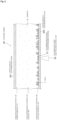
- FIG. 4 illustrates a graph indicating the measurement result of the oxygen concentration by the fluorescent oxygen concentration sensor secured to the culture bag in the cell culture of Test 1.
- the estimated value of the cell density was compared with the calculated value of the count of cells per unit area of this embodiment after 10, 40, and 60 hours from the start of the culture.
- the calculated value of the count of cells per unit area in the cell culture system and the method for detecting the proliferative property of cells of this embodiment is, for after 10, 40, and 60 hours, calculated as follows.
- the oxygen permeability of the gas permeable member (mg/(cm 2 ⁇ hr ⁇ atm)) was 1.4e-2.
- the oxygen consumption of one cell per unit time (mg/(hr ⁇ cells)) was 1.74e-9.
- the oxygen concentration (%) around the culture vessel was 21%, and the measurement value (%) of the oxygen concentration sensor was 5%.
- a count C of cells per unit area (cells/cm 2 ) after 10 hours is as follows.
- the cell density estimated value after 10 hours was, as described above, 1.40e6 cells/cm 2 .
- the oxygen concentration (%) around the culture vessel was 30%, and the measurement value (%) of the oxygen concentration sensor was 3%.
- the count C of cells per unit area (cells/cm 2 ) after 40 hours is as follows.
- the cell density estimated value after 40 hours was, as described above, 2.13e6 cells/cm 2 .
- the oxygen concentration (%) around the culture vessel was 40%, and the measurement value (%) of the oxygen concentration sensor was 2%.
- the count C of cells per unit area (cells/cm 2 ) after 60 hours is as follows.
- the cell density estimated value after 60 hours was, as described above, 2.82e6 cells/cm 2 .
- This culture bag had the oxygen permeability of 1.4e-2 (mg/(cm 2 ⁇ hr ⁇ atm)).
- the culture was performed in a state where this culture bag was interposed between pressing jigs and pressurized.
- the bottom surface of the pressing jig was a transparent resin plate, and the fluorescent light emitting/receiving unit was secured to a position corresponding to the fluorescent oxygen concentration sensor in the resin plate.
- PBMC Peripheral Blood Mononuclear Cells
- the PBMC Prior to seeding in the culture bag, the PBMC were activated for three days using a flask that was coated with an anti-CD3 antibody and immobilized, and then, the PBMC were seeded in the culture bag with the cell density of 1.6e6 cells/cm 2 .
- the medium one in which 2% of FBS was added to AlyS505N-7 (manufactured by Cell Science & Technology Institute, Inc.) was used, and 35 ml of the medium was filled in the culture bag together with the cells.
- the oxygen concentration was not measured for three days after the seeding in the culture bag, and the oxygen concentration inside the culture bag was measured for about 60 hr in the next three days.
- a multi-gas incubator capable of high oxygen culture PLC Corporation, model number MCO-5M-PJ was used, and an automatic liquid transfer mechanism was installed in the incubator.
- a medium supply bag in which the medium was filled was stored in a cooler at 10°C, and connected to the culture bag by a tube.
- a waste liquid bag was connected to the culture bag by a tube.
- the medium was transferred by controlling a tube pump.
- the medium deteriorated by the culture was discharged from the culture bag, and a new medium was filled.
- the replacement of the medium was performed at a predetermined timing after the start of the culture.
- the medium discharge amount was 25 ml in 35 ml.
- the liquid transfer rate was 2 ml/minute.
- the cells were uniformly floated in the medium by stirring inside the culture bag, the medium was extracted from the culture bag by about 0.5 ml using a syringe, and the cells were stained by trypan blue, thus measuring the cell density using a hemocytometer.
- the cell density after 61 hours from the measurement start was 2.34e6 cells/cm 2 .
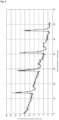
- FIG. 5 illustrates a graph indicating the measurement result of the oxygen concentration by the fluorescent oxygen concentration sensor secured to the culture bag in the cell culture of Test 2.
- the calculated value of the count of cells per unit area in the cell culture system and the method for detecting the proliferative property of cells of this embodiment is, for after 61 hours, calculated as follows.
- the oxygen permeability of the gas permeable member (mg/(cm 2 ⁇ hr ⁇ atm)) was 1.4e-2.
- the oxygen consumption of one cell per unit time (mg/(hr ⁇ cells)) was 1.00e-9.
- the oxygen concentration (%) around the culture vessel was 21%, and the measurement value (%) of the oxygen concentration sensor was 1.2%.
- the count C of cells per unit area (cells/cm 2 ) is as follows.
- This culture bag had the oxygen permeability of 1.4e-2 (mg/(cm 2 ⁇ hr ⁇ atm)).
- the culture was performed in a state where this culture bag was interposed between pressing jigs and pressurized.
- the bottom surface of the pressing jig was a transparent resin plate, and the fluorescent light emitting/receiving unit was secured to a position corresponding to the fluorescent oxygen concentration sensor in the resin plate.
- PBMC Peripheral Blood Mononuclear Cells
- the PBMC Prior to seeding in the culture bag, the PBMC were activated for three days using a flask that was coated with an anti-CD3 antibody and immobilized, and then, the PBMC were seeded in the culture bag with the cell density of 1.06e6 cells/cm 2 .
- the medium one in which 2% of FBS was added to AlyS505N-7 (manufactured by Cell Science & Technology Institute, Inc.) was used, and 35 ml of the medium was filled in the culture bag together with the cells.
- the oxygen concentration was not measured for four days after the seeding in the culture bag, and the oxygen concentration inside the culture bag was measured for about 65 hr in the next three days.
- Test 3 different from Test 2, for measuring the count of cells, the automatic liquid transfer was stopped, the cells were uniformly floated in the medium by stirring inside the culture bag, the medium was extracted from the culture bag by about 0.1 ml using a syringe, and the cells were stained by trypan blue, thus measuring the cell density using a hemocytometer. The number of extracted cells was extremely small, and does not affect the cell density of the whole.
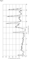
- FIG. 6 and FIG. 7 illustrate graphs indicating the measurement result of the oxygen concentration by the fluorescent oxygen concentration sensor secured to the culture bag in the cell culture of Test 3.
- the graph of FIG. 6 indicates the measurement result of the oxygen concentration up to the first measurement of the count of cells
- the graph of FIG. 7 indicates the measurement result of the oxygen concentration up to the second measurement of the count of cells.
- the change from 21% to 30% was made at a timing after 4 hours from the measurement start.
- the change from 30% to 40% was made at a timing after 23 hours from the start of the measurement.
- the change from 40% to 50% was made at a timing after 6 hours from the start of the measurement.
- the cells were uniformly floated in the medium by stirring inside the culture bag, the medium was extracted from the culture bag by about 0.1 ml, and the cells were stained by trypan blue, thus measuring the cell density using a hemocytometer.
- the cell density after 42 hours from the measurement start was 4.04e6 cells/cm 2 .
- the cells were uniformly floated in the medium by stirring inside the culture bag, the medium was extracted from the culture bag by about 0.1 ml, and the cells were stained by trypan blue, thus measuring the cell density using a hemocytometer.
- the cell density after 22.5 hours from the measurement start was 5.2e6 cells/cm 2 .
- the calculated value of the count of cells per unit area in the cell culture system and the method for detecting the proliferative property of cells of this embodiment is calculated as follows.
- the oxygen permeability of the gas permeable member (mg/(cm 2 ⁇ hr ⁇ atm)) was 1.4e-2.
- the oxygen consumption of one cell per unit time (mg/(hr ⁇ cells)) was 1.00e-9.
- the oxygen concentration (%) around the culture vessel was 40%, and the measurement value (%) of the oxygen concentration sensor was 7%.
- the count C of cells per unit area (cells/cm 2 ) is as follows.
- the oxygen concentration (%) around the culture vessel was 50%, and the measurement value (%) of the oxygen concentration sensor was 12%.
- the count C of cells per unit area (cells/cm 2 ) is as follows.
- a culture bag (manufactured by Toyo Seikan Group Holdings, Ltd.) made of a linear low-density polyethylene having a thickness of 110 ⁇ m was prepared.
- the culture bag had an outer shape size of 120 mm ⁇ 65 mm, and a bottom surface area of the culture space of 48cm 2 .
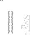
- this culture bag is provided with a plurality of protrusions inside, and a plurality of small protrusions formed on an outer surface.
- this culture bag was formed by heat-sealing peripheral edge portions of two films in which a plurality of approximately triangular prisms, provided with a pitch of 0.11 mm, a height of 0.19 mm, and an angle of 75°, were processed to be arranged in parallel in a mountain range pattern without intervals.
- the thickness of the thinnest portion (thin portion) of the film is 25 ⁇ m.
- This culture bag had the oxygen permeability of 8.0e-2 (mg/(cm 2 ⁇ hr ⁇ atm)). The oxygen permeability of this culture bag was calculated similarly to the method described with the example of the culture bag used in Test 1.
- the culture was performed without using the pressing jigs.
- the fluorescent light emitting/receiving unit was disposed at a position corresponding to the fluorescent oxygen concentration sensor in the lower side of the culture bag.
- iPS cells (cell line 1231A3, Center for iPS Cell Research and Application, Kyoto University) were seeded with the cell density of 1.7e4 cells/cm 2 .
- both of the upper surface and the lower surface inside the culture bag are used as culture surfaces, and this cell density includes both surfaces.
- StemFit(R) (AK-02N, Ajinomoto Healthy Supply Co., INC.) was used, and 35 ml of the medium was filled in the culture bag together with the cells.
- the incubator a multi-gas incubator capable of high oxygen culture (PHC Corporation, model number MCO-5M-PJ) was used. In this test, the automatic liquid transfer mechanism was not used, and the replacement of the medium was manually performed.
- a medium supply bag in which the medium was filled was stored in a cooler at 10°C, and connected to the culture bag by a tube.
- a waste liquid bag was connected to the culture bag by a tube.
- the medium was transferred by controlling a tube pump. The medium was warmed while passing through inside the incubator at 37°C, and became about 30°C at the point of entering the culture bag.
- the medium deteriorated by the culture was discharged from the culture bag, and a new medium was filled.
- the replacement of the medium was performed at a predetermined timing after the start of the culture.
- the medium discharge amount was 35 ml in 35 ml, and the liquid transfer rate was 2 ml/minute.
- the oxygen concentration inside the culture bag temporarily increased, and the peak was indicated in a graph described later.
- the cells were detached from the culture bag using a cell detachment solution (Thermo Fisher Scientific K.K., TrypLE). Then, the medium was filled in the culture bag, the detached cells were suspended in the medium, the whole amount of the medium was collected from the culture bag using a syringe, and the cells were stained by trypan blue, thus measuring the cell density using a hemocytometer. As a result, the cell density was 3.20e6 cells/cm 2 .
- FIG. 9 illustrates a graph indicating the measurement result of the oxygen concentration by the fluorescent oxygen concentration sensor secured to the culture bag in the cell culture of Test 4.
- the calculated value of the count of cells per unit area in the cell culture system and the method for detecting the proliferative property of cells of this embodiment is calculated as follows.
- the oxygen permeability of the gas permeable member (mg/(cm 2 ⁇ hr ⁇ atm)) was 8.0e-2.
- the oxygen consumption of one cell per unit time (mg/(hr ⁇ cells)) was 2.34e-9.
- the oxygen concentration (%) around the culture vessel was 21%, and the measurement value (%) of the oxygen concentration sensor was 12%.
- the count C of cells per unit area is as follows.
- the actually measured value of the cell density was, as described above, 3.20e6 cells/cm 2 .
- FIG. 10 illustrates a graph indicating the measurement result of the oxygen concentration by the fluorescent oxygen concentration sensor secured to the culture bag in the cell culture of Test 5.
- the cells were uniformly floated in the medium by stirring inside the culture bag, the medium was extracted from the culture bag by about 0.1 ml, and the cells were stained by trypan blue, thus measuring the cell density using a hemocytometer.
- the cell density after 56 hours from the measurement start was 1.04e7 cells/cm 2 .
- the cell density inside the culture bag is very high.
- the oxygen concentration inside the culture bag is less than 1%.
- the count of cells per unit area is calculated by the following method.
- a slope PT of the change in rise peak value of the oxygen concentration when the additional medium is added in the culture bag multiple times, and a slope QT of the change of the oxygen concentration when the culture bag is stabilized are calculated, and the ratio (QT/PT) is calculated.
- the ratio (QT/PT) is 2.9.
- the peak immediately before Q4 (56, less than 1) after 56 hours from the measurement start is P4 (55.1, 22.3), and the peak immediately before Q3 (31.1, 2.3) at which the oxygen concentration can be measured is P3 (29.5, 32.7).
- the oxygen concentration at Q4 changes from Q3 based on the above-described ratio (QT/PT) relative to the change amount of the oxygen concentration from P3 to P4.
- an oxygen concentration D' to be estimated can be calculated as follows.
- D ′ oxygen concentration at Q 3 ⁇ change amount of oxygen concentration from P 3 to P 4 ⁇ QT / PT
- the count of cells per unit area can be calculated based on a formula (2) below.
- C G ⁇ M / 100 ⁇ D ′ / 100 / S
- the count of cells per unit area can be calculated as follows.
- the oxygen permeability of the gas permeable member (mg/(cm 2 ⁇ hr ⁇ atm)) was 1.4e-2.
- the oxygen consumption of one cell per unit time (mg/(hr ⁇ cells)) was 1.00e-9.
- the actually measured value of the cell density was, as described above, 1.04e7 cells/cm 2 .
- the culture vessel is not limited to the exemplified one, and one appropriately changed corresponding to the purpose of the culture, for example, one including one port or three ports, and one provided with a plurality of wells on the culture surface, can be used.
- the present invention can be appropriately used, for example, when cells are cultured in a large amount with a high density using a cell culture bag.
Landscapes
- Chemical & Material Sciences (AREA)
- Health & Medical Sciences (AREA)
- Wood Science & Technology (AREA)
- Zoology (AREA)
- Engineering & Computer Science (AREA)
- Bioinformatics & Cheminformatics (AREA)
- Organic Chemistry (AREA)
- Life Sciences & Earth Sciences (AREA)
- Biotechnology (AREA)
- Sustainable Development (AREA)
- Microbiology (AREA)
- Biochemistry (AREA)
- General Engineering & Computer Science (AREA)
- General Health & Medical Sciences (AREA)
- Genetics & Genomics (AREA)
- Biomedical Technology (AREA)
- Analytical Chemistry (AREA)
- Clinical Laboratory Science (AREA)
- Apparatus Associated With Microorganisms And Enzymes (AREA)
- Measuring Or Testing Involving Enzymes Or Micro-Organisms (AREA)
Abstract
Description
- The present invention relates to a cell culture technique, and especially relates to a cell culture system that detects a proliferative property of cells when the cells are statically cultured with a culture vessel formed of a gas permeable member.
- Recently, in the production of pharmaceutical medicines and in the fields of gene therapy, regeneration medicine, immunotherapy, and the like, it is desired that cells and tissues are efficiently cultured in a large amount under an artificial environment.
- In such a situation, it has been proposed that cells are automatically cultured in large quantities in a closed system using a bag-shaped cell culture vessel formed of a gas permeable member.
- When the cells are cultured in large quantities, it is important to confirm that the culture is properly performed by detecting the proliferative property of the cells. Additionally, for the detection of the proliferative property of cells, for example, it is preferred to allow confirming the number of cells in the culture vessel at desired timings during the culture.
- As a method for confirming the number of cells in the culture vessel, the following three methods can be presented.
- (1) A part of a medium inside the culture vessel is extracted (sampling), and the number of cells is measured using a hemocytometer or the like.
- (2) A photograph of cells in the culture vessel is taken by a microscope and a camera, and image processing is performed on the obtained photograph to count the number of cells.
- (3) The number of cells is counted based on the decreased amount of dissolved oxygen in the culture vessel.
-
- Patent Document 1:
JP-A-S63-15150 - Patent Document 2:
JP-A-H6-121667 - However, the method (1) has a problem that the risk of contamination increases because a part of the culture vessel is required to be opened for sampling the medium. Additionally, there is a problem that when adherent cells are cultured, it is impossible to detach only a part of the cells during the culture, and the number of cells cannot be counted at a desired timing.
- The method (2) has a problem that when floating cells are cultured, while the measurement can be performed when the culture density is low, the measurement becomes difficult because of the lamination of the cells when the cell density increases after the middle of the culture. Additionally, there is also a problem that since this method requires the microscope, the camera, and an image processing device, the mechanism of the cell culture system is complicated.
- Furthermore, in the method (3), since it is necessary to measure the decreased amount of the dissolved oxygen in the culture vessel, a sealed state is required for avoiding the move of a gas from the inside to the outside of the culture vessel, and the entering of a gas into the culture vessel from outside. Alternatively, when there is a space on a culture medium inside the culture vessel, it is necessary to interrupt dissolution of oxygen into the culture medium by replacing the space with nitrogen, and measure the consumption quantity of the dissolved oxygen in the culture medium by the cells.
- However, there is a problem that when cells are cultured using a culture vessel formed of a gas permeable member, since it is impossible to detect the density change of only the dissolved oxygen consumed by the cells in the culture medium, and it is necessary to stir inside the culture vessel for the measurement, static culture cannot be performed.
- Here, while Patent Document 1 discloses a method for measuring a decreased amount of dissolved oxygen in a sample solution containing microorganisms in a state where the sample solution is sealed within a container, this measuring method needs to be performed using a culture vessel without gas permeability.
-
Patent Document 2 discloses a cell culture device that operates an amount of temporal variation of oxygen amount relating to the number of cells in a culture vessel using an oxygen amount measured by oxygen amount measurement means that measures an oxygen amount dissolved in a medium in the culture vessel. However, this cell culture device uses a sealing container that avoids entering of a gas into a culture vessel from outside, and requires measuring a consumption of dissolved oxygen in a culture medium by changing a culture condition, for example, replacing a space on the culture medium inside the culture vessel with nitrogen to interrupt dissolution of oxygen into the culture medium, for obtaining a measurable state from an ordinary culture state. - The inventions disclosed in these patent documents cannot be appropriately used when cells are statically cultured with a culture vessel formed of a gas permeable member.
- The present invention has been made in consideration of the above-described circumstances, and it is an object of the present invention to provide a cell culture system capable of detecting a proliferative property of cells in a culture vessel at a desired timing during a culture when the cells are statically cultured in the culture vessel formed of a gas permeable member, and a method for detecting the proliferative property of cells.
- To achieve the above-described object, a cell culture system of the present invention is a cell culture system for statically culturing cells with a culture vessel.
- The culture vessel is at least partially formed of a gas permeable member. The cell culture system includes an oxygen concentration sensor and a proliferation detection unit. The oxygen concentration sensor measures an oxygen concentration in a vicinity of a culture surface inside the culture vessel. The proliferation detection unit detects a proliferative property of the cells based on an oxygen permeability of the gas permeable member, a measurement value of the oxygen concentration sensor, an oxygen concentration around the culture vessel, and an oxygen consumption per cell of the cells.
- In the cell culture system of the present invention, the proliferation detection unit preferably calculates a count of the cells per unit area as the proliferative property of the cells by a formula (1) below,

- C:
- count of cells per unit area (cells/cm2)
- G:
- oxygen permeability of gas permeable member (mg/(cm2·hr·atm))
- M:
- oxygen concentration around culture vessel (%)
- D:
- measurement value of oxygen concentration sensor (%)
- S:
- oxygen consumption of one cell per unit time (mg/(hr cells)).
- In the cell culture system of the present invention, the proliferation detection unit preferably uses an estimated value of an oxygen concentration inside the culture vessel at a desired time point as the measurement value of the oxygen concentration sensor. The estimated value of the oxygen concentration inside the culture vessel at a desired time point is preferably obtained based on a ratio between a slope of a change in rise peak value of the oxygen concentration when an additional medium is added in the culture vessel multiple times and a slope of a change of the oxygen concentration when the culture vessel is stabilized.
- In the cell culture system of the present invention, the oxygen concentration around the culture vessel is preferably an oxygen concentration inside an incubator in which the culture vessel is housed, or an atmospheric oxygen concentration.
- In the cell culture system of the present invention, the oxygen concentration sensor is preferably a fluorescent type.
- In the cell culture system of the present invention, the oxygen concentration sensor is preferably secured to the culture surface inside the culture vessel using a perforated resin member.
- A method for detecting a proliferative property of cells of the present invention is a method for detecting a proliferative property of cells in a cell culture in which the cells are statically cultured with a culture vessel. The culture vessel is at least partially formed of a gas permeable member. The method includes: measuring an oxygen concentration in a vicinity of a culture surface inside the culture vessel by an oxygen concentration sensor disposed on the culture surface inside the culture vessel; and calculating a count of the cells per unit area based on an oxygen permeability of the gas permeable member, a measurement value of the oxygen concentration sensor, an oxygen concentration around the culture vessel, and an oxygen consumption per cell of the cells.
- The present invention can provide the cell culture system capable of detecting the proliferative property of the cells in the culture vessel at a desired timing during the culture when the cells are statically cultured in the culture vessel formed of the gas permeable member, and the method for detecting the proliferative property of the cells.
-
-
FIG. 1 is a schematic diagram illustrating a configuration of a cell culture system according to an embodiment of the present invention. -
FIG. 2 is an explanatory view illustrating a distribution of oxygen concentration inside a culture vessel in the cell culture system according to the embodiment of the present invention. -
FIG. 3 is an explanatory view illustrating a secured state of an oxygen concentration sensor inside the culture vessel in the cell culture system according to the embodiment of the present invention. -
FIG. 4 is a drawing illustrating a graph indicating a measurement result of an oxygen concentration by a fluorescent oxygen concentration sensor secured to a culture bag in a cell culture of Test 1. -
FIG. 5 is drawing illustrating a graph indicating a measurement result of the oxygen concentration by the fluorescent oxygen concentration sensor secured to the culture bag in a cell culture ofTest 2. -
FIG. 6 is a drawing illustrating a graph (1) indicating a measurement result of the oxygen concentration by the fluorescent oxygen concentration sensor secured to the culture bag in a cell culture of Test 3. -
FIG. 7 is a drawing illustrating a graph (2) indicating a measurement result of the oxygen concentration by the fluorescent oxygen concentration sensor secured to the culture bag in the cell culture of Test 3. -
FIG. 8 is a schematic diagram illustrating a part of a configuration of a culture bag used in a cell culture ofTest 4 in the cell culture system and the method for detecting proliferative property of cells according to the embodiment. -
FIG. 9 is a drawing illustrating a graph indicating a measurement result of the oxygen concentration by the fluorescent oxygen concentration sensor secured to the culture bag in the cell culture ofTest 4. -
FIG. 10 is a drawing illustrating a graph indicating a measurement result of the oxygen concentration by the fluorescent oxygen concentration sensor secured to the culture bag in a cell culture ofTest 5. - The following describes embodiments of a cell culture system and a method for detecting a proliferative property of cells of the present invention in detail. However, the present invention is not limited to the specific contents of the embodiments and examples below.
- A cell culture system of this embodiment is a cell culture system for statically culturing cells with a culture vessel at least partially formed of a gas permeable member, and includes an oxygen concentration sensor that measures an oxygen concentration in a vicinity of a culture surface inside the culture vessel, and a proliferation detection unit that detects a proliferative property of cells based on an oxygen permeability of the gas permeable member, a measurement value of the oxygen concentration sensor, an oxygen concentration around the culture vessel, and an oxygen consumption per cell of the cells.
- Specifically, the cell culture system of this embodiment can have, for example, a configuration as illustrated in
FIG. 1 . - First, a schematic configuration of the cell culture system of this embodiment will be described.
- In the cell culture system of this embodiment, a
culture vessel 20 is housed in anincubator 10, and placed on a placement table 11. Theculture vessel 20 includes two ports, and amedium supply container 40 housed in a cooler 30 is coupled to one port by a tube. To the other port of theculture vessel 20, awaste liquid container 50 inside theincubator 10 is coupled by a tube. - At the tube coupling the
culture vessel 20 to themedium supply container 40 and the tube coupling theculture vessel 20 to thewaste liquid container 50, liquid transfer means is disposed. As the liquid transfer means, for example, a pump, such as a peristaltic pump and a syringe pump, capable of transferring a liquid at a low speed with high accuracy is preferred to be used. - Then, a
control device 60 controls the liquid transfer means between theculture vessel 20 and themedium supply container 40 at a predetermined timing, thus transferring a medium from themedium supply container 40 to theculture vessel 20. Thecontrol device 60 controls the liquid transfer means between theculture vessel 20 and thewaste liquid container 50 at a predetermined timing, thus transferring the medium from theculture vessel 20 to thewaste liquid container 50. - By supplying an oxygen gas and a carbon dioxide gas from an oxygen cylinder O (oxygen supply device) and a carbon dioxide cylinder C (carbon dioxide supply device) to the
incubator 10, respectively, a gas concentration inside theincubator 10 is controlled, and thus a gas concentration around theculture vessel 20 can be adjusted. - That is, the oxygen cylinder O and the carbon dioxide cylinder C are each connected to the
incubator 10 via a valve, switching operations of the respective valves are controlled by thecontrol device 60, and the gas supply to theincubator 10 from each gas cylinder is controlled, thus allowing the adjustment of oxygen concentration inside theincubator 10. - As the
incubator 10, for example, a CO2 incubator is usable. As the placement table 11 inside theincubator 10, one made of a perforated metal or the like can be disposed to be used, and theculture vessel 20 is placed on the placement table 11 and housed in a closed manner. The placement table 11 may be omitted. - Since the cell culture system of this embodiment is applicable to a case where cells are cultured without using the
incubator 10, theincubator 10 may be omitted. - Next, the
culture vessel 20 in the cell culture system of this embodiment will be described in detail. - The
culture vessel 20 is a cell culture vessel of a closed system that is at least partially formed of a gas permeable member and has a gas permeability, and for example, one formed by heat-sealing peripheral edge portions of two rectangular gas permeable films can be used. - The shape of the
culture vessel 20 is formed to be inclined with respect to the ports in the vicinities of the ports, and this allows easily flowing the medium to the ports. The shape of theculture vessel 20 may be a rectangular shape such as an elongated rectangle. The number of the ports included in theculture vessel 20 is not limited to two, and may be one or three or more. - In the cell culture system of this embodiment, a fluorescent
oxygen concentration sensor 21 is disposed on a culture surface inside theculture vessel 20. In theincubator 10, a fluorescent light emitting/receivingunit 22 is disposed to be able to emit and receive a fluorescent light to the fluorescentoxygen concentration sensor 21. - The fluorescent light emitting/receiving
unit 22 is, for example, disposed under the placement table 11, and the fluorescent light emitting/receivingunit 22 can emit and receive a fluorescent light to the fluorescentoxygen concentration sensor 21 via the placement table 11 when it is transparent, or via a through hole provided to the placement table 11. The fluorescent light emitting/receivingunit 22 can be disposed to be in contact with the lower side of theculture vessel 20. - By controlling the fluorescent light emitting/receiving
unit 22 by thecontrol device 60, the oxygen concentration inside theculture vessel 20 can be measured. - As illustrated in
FIG. 2 , the concentration of dissolved oxygen in the medium inside theculture vessel 20 largely differs depending on the position. That is, in the medium in the vicinity of the culture surface on which cells are present (upper surface of a lower surface gas permeable film 202), since the cells actively consume oxygen, oxygen is rapidly consumed, and gradually becomes 0% as the cell density increases. In contrast, while depending on the liquid thickness inside theculture vessel 20, for example, when the liquid thickness is 10 mm or more, in the medium in the vicinity of an upper surface (lower surface of an upper surface gas permeable film 201) in the opposite side of the culture surface inside theculture vessel 20, the oxygen concentration is slow in decrease from the initial state (for example, about 21% in atmosphere) when cells are not present in the vicinity. In the medium near the center of theculture vessel 20, the oxygen concentration is approximately middle (for example, 10%) of them. - Accordingly, in the cell culture system of this embodiment, by disposing the fluorescent
oxygen concentration sensor 21 on the culture surface inside theculture vessel 20, the oxygen concentration inside theculture vessel 20 based on the oxygen consumption by the cells can be appropriately measured. - Additionally, as described later, in the cell culture system of this embodiment, by taking the oxygen permeability of the
culture vessel 20 and the oxygen concentration around theculture vessel 20 into consideration, the oxygen consumption by the cells can be appropriately calculated. - Furthermore, according to the cell culture system of this embodiment, since the oxygen concentration inside the
culture vessel 20 is continuously 0% to about 1%, even when the measurement of the oxygen concentration by the fluorescentoxygen concentration sensor 21 is difficult, the oxygen consumption by the cells can be appropriately estimated. - Here, a mechanism of gas permeation in the gas permeable film will be described.
- The dissolved gas in the medium inside the culture vessel is dissolved at a concentration that equilibrates with a partial pressure around the culture vessel. The concentration of oxygen dissolved in the medium placed in the incubator is about 21% the same as the concentration in atmosphere when the gas concentration inside the incubator is not controlled. The consumption of the dissolved oxygen inside the culture vessel by the cells decreases the dissolved oxygen of the medium around the cells, and the oxygen gas permeates the film and flows into the culture vessel so as to keep the equilibrium with the oxygen concentration outside the culture vessel.
- The gas permeability (speed) of the film is determined depending on the gas partial pressure difference between both sides of the film, affinity between the film and the gas, the diffusivity of gas molecules moving into the film, and the like.
- Next, a principle of measuring the oxygen concentration by the fluorescent oxygen concentration sensor will be described.
- When a light is applied to a fluorescent element of the fluorescent oxygen concentration sensor, a fluorescent substance absorbs the light and becomes in an excited state, and then returns to a ground state while emitting a fluorescent light. At this time, when oxygen is present around the fluorescent substance, the excitation energy is lost, and the fluorescence emission intensity decreases. Accordingly, the more oxygen is, the more the fluorescence emission intensity decreases. The fluorescent light emitting/receiving unit measures the oxygen amount based on the change of the emission intensity due to the number of oxygen molecules contacting the fluorescent element when oxygen dissolved in the medium contacts the fluorescent element.
- As a material of the
culture vessel 20, a resin film or the like can be preferably used, and for example, a polyolefin-based resin such as polyethylene and polypropylene is usable. For example, polyethylene, a copolymer of ethylene and α-olefin, a copolymer of ethylene and vinyl acetate, an ionomer using: a copolymer of ethylene and an acrylic acid or ethylene and a methacrylic acid; and a metal ion, and the like can be listed. Further, a polyolefin, a styrene-based elastomer, a polyester-based thermoplastic elastomer, and the like can be used. Furthermore, a soft vinyl chloride resin, a polybutadiene resin, an ethylene-vinyl acetate copolymer, a chlorinated polyethylene resin, a polyurethane-based thermoplastic elastomer, a polyester-based thermoplastic elastomer, a silicone-based thermoplastic elastomer, a styrene-based elastomer, such as styrene-butadiene-styrene (SBS), styrene-isoprene-styrene (SIS), styrene-ethylene-butylene-styrene (SEBS), and styrene-ethylene-propylene-styrene (SEPS), a polyolefin resin, a fluorine-based resin, and the like may be used. - As the gas permeable member used for at least a part of the
culture vessel 20, among the above-described materials, especially, using a thermoplastic resin excellent in gas permeation performance is preferable, and a polyolefin-based resin, for example, polyethylene, such as LLDPE, or polypropylene can be preferably used. The gas permeable member is preferably a transparent material. - As illustrated in
FIG. 3 , as a modification of the cell culture system of this embodiment, it is preferred that a fluorescentoxygen concentration sensor 21a is secured to a culture surface (in the example of the drawing, upper surface of a lower surface gaspermeable film 202a) inside aculture vessel 20a using aperforated resin member 23a. At this time, a fluorescentoxygen concentration sensor 21a is interposed between theperforated resin member 23a and the culture surface, and a welding region H of theperforated resin member 23a is welded to the culture surface, thereby allowing the securing. - As a material of the
perforated resin member 23a, a polyolefin-based resin such as polyethylene, for example, LLDPE, and polypropylene is usable. For example, polyethylene, a copolymer of ethylene and α-olefin, a copolymer of ethylene and vinyl acetate, an ionomer using: a copolymer of ethylene and an acrylic acid or ethylene and a methacrylic acid; and a metal ion, and the like can be listed. - Since the
perforated resin member 23a brings the fluorescentoxygen concentration sensor 21a into contact with the medium while securing the fluorescentoxygen concentration sensor 21a to the culture surface, a mesh material can be appropriately used. As theperforated resin member 23a, for example, a porous material such as a sponge can be used. - While the specific mesh material and porous material are not especially limited, one of a similar material having the melting point close to that of the material of the
culture vessel 20a is preferable, and for example, a polyethylene mesh material (NBC Meshtec Inc., Item Number PE 200 me, or the like) and a polyethylene porous membrane (TEIJIN LIMITED, miraim (Trade name) large pore ≤ 3 µm) can be appropriately used. - In the cell culture system of this embodiment, with the configuration in which the fluorescent
oxygen concentration sensor 21a is secured using the perforatedresin member 23a, the removal and the move of the fluorescentoxygen concentration sensor 21a can be avoided when the medium is injected into theculture vessel 20a and the medium is discharged from theculture vessel 20a. - The cells cultured using the
culture vessel 20 are not especially limited, and the cells may be floating cells, such as lymphocytes or dendritic cells, cultured by floating cells in a culture fluid, or may be adherent cells, such as induced pluripotent stem cells (iPS cells), neural stem cells, embryonic stem cells (ES cells), mesenchymal stem cells, hepatic cells, islet cells, cardiac muscle cells, corneal endothelial cells, and lymph corpuscles in an activation process, cultured by making cells adhere to a culture surface in the culture vessel. - The cells cultured using the
culture vessel 20 may be formed in an aggregate such as a spheroid and an organoid, and the cell culture system and the method for detecting the proliferative property of cells of this embodiment are applied to the culture of an aggregate. - Next, the
control device 60 in the cell culture system of this embodiment will be described in detail. - The control device 60 (control unit) includes, as illustrated in
FIG. 1 , an input/output unit 61, a fluorescent sensor input/output unit 62, acontroller 63, anoperation unit 64, and apower supply unit 65. - The input/
output unit 61 is connected to the pump disposed between theculture vessel 20 and themedium supply container 40 and the pump disposed between theculture vessel 20 and thewaste liquid container 50, and controls the operations of these pumps based on input information from thecontroller 63. Accordingly, the medium is supplied to theculture vessel 20 from themedium supply container 40, and the medium is discharged from theculture vessel 20 to thewaste liquid container 50. - The input/
output unit 61 is connected to agas sensor 12 of theincubator 10, the valve disposed between the oxygen cylinder O and theincubator 10, and the valve disposed between the carbon dioxide cylinder C and theincubator 10, and transmits input information from thegas sensor 12 to thecontroller 63. The input/output unit 61 controls the operations of these valves based on the input information from thecontroller 63. Accordingly, the oxygen gas can be supplied from the oxygen cylinder O to the inside of theincubator 10, and the carbon dioxide gas can be supplied from the carbon dioxide cylinder C to the inside of theincubator 10, thus allowing the control of the gas concentration inside theincubator 10 to a desired level. - The fluorescent sensor input/
output unit 62 is connected to the fluorescent light emitting/receivingunit 22, controls the fluorescent light emission of the fluorescent light emitting/receivingunit 22 based on input information from thecontroller 63, and transmits information based on the light reception from the fluorescentoxygen concentration sensor 21 to thecontroller 63. Accordingly, the oxygen concentration in the vicinity of the culture surface inside theculture vessel 20 can be measured, and thecontroller 63 can use this oxygen concentration for detecting the proliferative property of cells. - The
controller 63 is configured of a PLC (programmable logic controller) or the like, can program and store a desired control content in advance, and can control the operations of the respective units based on it. - That is, the
controller 63 transmits information for controlling the pumps and the valves to the input/output unit 61 at a predetermined timing. Additionally, based on any kinds of information including the input information from the fluorescent sensor input/output unit 62 and time information, information for controlling the pumps and the valves can be transmitted to the input/output unit 61. - The
controller 63 includes aproliferation detection unit 631 that detects the proliferative property of cells inside theculture vessel 20. - The
proliferation detection unit 631 detects the proliferative property of cells based on the oxygen permeability of the gas permeable member, the measurement value of the oxygen concentration sensor, the oxygen concentration around the culture vessel, and the oxygen consumption per cell of the cells. Specifically, theproliferation detection unit 631 can calculate the count of cells per unit area as the proliferative property of cells. - The oxygen permeability of the gas permeable member and the oxygen consumption per cell of the cells are calculated in advance corresponding to the usage, and stored in the
proliferation detection unit 631 or the like. - As described later, as a result of calculating the oxygen consumption per cell of PBMC, it was 1.0e-9 mg/(hr cells). As a result of calculating the oxygen consumption per cell of Jurkat (lymphocyte cell line), it was 1.74e-9 mg/(hr·cells).
- Prior to the determination of the oxygen permeability (mg/(cm2·hr·atm)), an actual culture configuration is determined first. That is, the oxygen permeability can be calculated as follows after determining the container size, the amount of the medium to be filled, the position of disposing the fluorescent sensor, and whether to use a pressing jig or not.
- For example, it is assumed that a culture is performed using a culture bag used in Test 1 described later, which is formed by boning two polyethylene films having a thickness of 110 µm and has a bottom surface area of 50 cm2, as the
culture vessel 20 in a state where the fluorescentoxygen concentration sensor 21 is secured to the approximately center of the inner surface of this culture bag, and theculture vessel 20 is interposed between pressing jigs and pressurized. The bottom surface of the pressing jig is a transparent resin plate, and the fluorescent light emitting/receivingunit 22 is secured to a position corresponding to the fluorescentoxygen concentration sensor 21 in the resin plate. - Next, the medium is warmed to 37°C as a culture temperature, and injected into the
culture vessel 20 together with the cells. As the cells, two kinds of PBMC (Peripheral Blood Mononuclear Cells) and Jurkat (lymphocyte cell line) were used, and the change amount of oxygen concentration was measured twice for each, thus calculating the oxygen permeability as follows and obtaining the average value. - As the medium for PBMC, one in which 2% of FBS was added to AlyS505N-7 (manufactured by Cell Science & Technology Institute, Inc.) was used, and 35 ml of the medium was filled in the culture bag together with the cells. As the medium for Jurkat, one in which 2% of FBS was added to AlyS505N-0 (manufactured by Cell Science & Technology Institute, Inc.) was used, and 35 ml of the medium was filled in the culture bag together with the cells.
- The cells sank after approximately two hours from filling PBMC in the above-described culture bag with the cell filling amount per unit area of 2. 1e6 cells/cm2, and the oxygen concentration stabilized. The oxygen concentration at that time was 6%.
- In this case, the oxygen permeability G can be calculated as follows.

- After filling PBMC in the above-described culture bag with the cell filling amount per unit area of 4.0e6 cells/cm2, the oxygen concentration around the culture bag was controlled to 30%. Approximately two hours later, the cells sank, and the oxygen concentration stabilized. The oxygen concentration at that time was 2%.
- In this case, the oxygen permeability G can be calculated as follows.
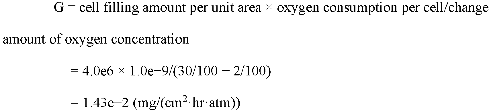
- The cells sink after approximately two hours from filling Jurkat in the above-described culture bag with the cell filling amount per unit area of 1.1e6 cells/cm2, and the oxygen concentration stabilizes. The oxygen concentration at that time was 8%.
- In this case, the oxygen permeability G can be calculated as follows.

- After filling Jurkat in the above-described culture bag with the cell filling amount per unit area of 1.9e6 cells/cm2, the oxygen concentration around the culture bag was controlled to 30%. Approximately two hours later, the cells sank, and the oxygen concentration stabilized. The oxygen concentration at that time was 5%.
- In this case, the oxygen permeability G can be calculated as follows.

- From the above-described results, the oxygen permeability G of the culture bag used under the above-described conditions was determined as follows.

- The oxygen consumption per cell of the cells (mg/(hr·cells)) can be calculated as follows.
- First, the cells and the medium are injected into a sealed vessel that does not permeate a gas in a state where the count of cells is obtained in advance. Air bubbles are discharged using a syringe or the like so as to obtain a state where air bubbles are not mixed in the sealed vessel as much as possible. The dissolved oxygen amount is calculated from the volume of the sealed vessel.
- Specifically, the medium warmed to 37°C was filled in the sealed vessel having the volume of 10 ml. In the medium of 10 ml at 37°C (atmospheric oxygen concentration about 21%), oxygen of 6.86e-2 mg is dissolved (6.86e-2 mg/l@37°C (saturated dissolved oxygen)). The oxygen concentration change in the sealed vessel during the culture is measured, thus calculating the decrease in concentration of the dissolved oxygen per hour.
- When PBMC is injected into the sealed vessel of 10 ml with the density of 2e6 cells/ml of the cell filling amount per unit area, 2e7 cells float in the vessel. At that time, the decrease of the oxygen concentration was 0.1 %/minute, and 6% per hour.
- Since the decrease by 6% relative to 21% of the saturated dissolved oxygen corresponds to the decrease by 6/21 = 0.29, the oxygen consumption per unit time is as follows.

- Accordingly, the oxygen consumption S per cell (mg/(hr·cells)) of PBMC was calculated as follows.

- With the similar method, the oxygen consumption S per cell (mg/(hr·cells)) of Jurkat was calculated as follows.

- The measurement value of the oxygen concentration sensor is the oxygen concentration in the vicinity of the culture surface inside the
culture vessel 20 measured by the fluorescentoxygen concentration sensor 21, and input from the fluorescent light emitting/receivingunit 22 to theproliferation detection unit 631 via the fluorescent sensor input/output unit 62. - The oxygen concentration around the culture vessel is the oxygen concentration inside the
incubator 10 in which theculture vessel 20 is housed. In a state where the oxygen concentration inside theincubator 10 is not controlled, or when theincubator 10 is not used, the oxygen concentration around the culture vessel is the atmospheric oxygen concentration (about 21%). This is set in advance by thecontroller 63. - Here, in the static culture, the amount of oxygen permeating the culture vessel matches and is balanced with the amount of oxygen consumed by the cells, and therefore, a formula below is satisfied.

- G:
- oxygen permeability of gas permeable member (mg/(cm2·hr·atm))
- M:
- oxygen concentration around culture vessel (%)
- D:
- measurement value of oxygen concentration sensor (%)
- C:
- count of cells per unit area (cells/cm2)
- S:
- oxygen consumption of one cell per unit time (mg/(hr - cells))
- Accordingly, in the cell culture system of this embodiment, the
proliferation detection unit 631 can calculate the count of cells per unit area as the proliferative property of cells by a formula (1) below.
- C:
- count of cells per unit area (cells/cm2)
- G:
- oxygen permeability of gas permeable member (mg/(cm2·hr·atm))
- M:
- oxygen concentration around culture vessel (%)
- D:
- measurement value of oxygen concentration sensor (%)
- S:
- oxygen consumption of one cell per unit time (mg/(hr·cells))
- The count of cells per unit area calculated by the cell culture system of this embodiment is not one obtained by strictly measuring the actual count of cells, but one obtained by roughly estimating the count of cells based on the culture environment of the
culture vessel 20. - In culturing cells in a large amount, since it is important to confirm whether the culture is properly performed or not, roughly estimating the count of cells inside the culture vessel is considerably beneficial.
- In the cell culture system of this embodiment, it is also preferred that the
proliferation detection unit 631 uses an estimated value of the oxygen concentration inside theculture vessel 20 at a desired time point obtained based on a ratio between a slope of a change in rise peak value of the oxygen concentration when an additional medium is added in theculture vessel 20 multiple times and a slope of a change of the oxygen concentration when theculture vessel 20 is stabilized as a measurement value D of the oxygen concentration sensor in the above-described formula (1). - That is, as described later in
Test 5, when the cell density inside theculture vessel 20 becomes high because of the progress of the cell culture, the oxygen concentration in the vicinity of the culture surface inside theculture vessel 20 becomes less than 1% even when the oxygen concentration around theculture vessel 20 is increased to be high, and the measurement limit of the fluorescentoxygen concentration sensor 21 is exceeded, thus failing in the measurement with accuracy. - According to the cell culture system of this embodiment, even in such a case, by the above-described method, the count of cells per unit area inside the
culture vessel 20 can be roughly estimated. - That is, by using an estimated value D' of the oxygen concentration inside the culture bag at a desired time point obtained based on a ratio between a slope of a change in rise peak value of the oxygen concentration when an additional medium is added in the culture bag multiple times and a slope of a change of the oxygen concentration when the culture bag is stabilized as the measurement value of the oxygen concentration sensor, the count of cells per unit area can be calculated based on a formula (2) below.

- The oxygen concentration at the rise peak of the oxygen concentration when the additional medium is added is influenced by the temperature of the medium to be added. Therefore, it is preferred to add the medium while holding the constant temperature.
- The method for calculating the estimated value D' will be described in detail in
Test 5. - The
operation unit 64 includes a display unit such as a touch panel, and transmits input information by a user to thecontroller 63 to execute setting of PLC and the like. Theoperation unit 64 displays input information from thecontroller 63. - The power supply unit 65 (stabilized power source or the like) supplies electricity to the respective units in the
control device 60. - Although not illustrated, the
control device 60 can have a configuration further including a relaying unit (relay) and a circuit breaking unit (breaker). A part of or the whole of the configuration, for example, thecontroller 63 and theoperation unit 64, of thecontrol device 60 may be achieved by a microcomputer or a computer. - A method for detecting a proliferative property of cells of this embodiment is a method for detecting a proliferative property of cells in a cell culture statically culturing the cells by a culture vessel at least partially formed of a gas permeable member, and includes measuring an oxygen concentration in a vicinity of a culture surface inside the culture vessel by an oxygen concentration sensor disposed on the culture surface inside the culture vessel, and calculating a count of cells per unit area based on an oxygen permeability of the gas permeable member, a measurement value of the oxygen concentration sensor, an oxygen concentration around the culture vessel, and an oxygen consumption per cell of the cells.
- In the method for detecting the proliferative property of cells of this embodiment, the count of cells per unit area can be calculated by the formula (1) below.

- C:
- count of cells per unit area (cells/cm2)
- G:
- oxygen permeability of gas permeable member (mg/(cm2·hr·atm))
- M:
- oxygen concentration around culture vessel (%)
- D:
- measurement value of oxygen concentration sensor (%)
- S:
- oxygen consumption of one cell per unit time (mg/(hr·cells))
- In the method for detecting the proliferative property of cells of this embodiment, it is also preferred that an estimated value of the oxygen concentration inside the
culture vessel 20 at a desired time point obtained based on a ratio between a slope of a change in rise peak value of the oxygen concentration when an additional medium is added in theculture vessel 20 multiple times and a slope of a change of the oxygen concentration when theculture vessel 20 is stabilized is used as a measurement value D of the oxygen concentration sensor in the above-described formula (1). - As described above, according to the cell culture system and the method for detecting the proliferative property of cells of this embodiment, in the case where the cells are statically cultured with the culture vessel formed of the gas permeable member, the proliferative property of the cells inside the culture vessel at a desired timing during the culture can be detected, and the count of cells per unit area can be calculated as the proliferative property of the cells. Accordingly, this embodiment allows confirming whether the culture is properly performed or not in culturing cells in a large amount. Examples
- The following describes tests conducted for confirming the effects of the cell culture system and the method for detecting the proliferative property of cells according to the embodiments of the present invention.
- In Test 1, Jurkat (lymphocyte cell line) was cultured to detect the proliferative property. In this test, the culture was continued without interruption during the test period, and the estimated value of the cell density was compared with the calculated value of the count of cells per unit area of this embodiment.
- In
Test 2, PBMC (Peripheral Blood Mononuclear Cells) were cultured, and the actually measured value of the cell density after the culture was compared with the calculated value of the count of cells per unit area of this embodiment. - In Test 3, PBMC were cultured, the culture was interrupted at a predetermined timing to count the actual count of cells, and the obtained actually measured value of the cell density was compared with the calculated value of the count of cells per unit area of this embodiment.
- In
Test 4, iPS cells were cultured, the actual count of cells was counted after the end of the culture, and the obtained actually measured value of the cell density was compared with the calculated value of the count of cells per unit area of this embodiment. -
Test 5 is one conducted in a period following the culture of Test 3, and includes a period in which the cell density inside the culture vessel becomes high and the measurement value of the oxygen concentration in the vicinity of the culture surface is less than 1%. The actual count of cells was counted after the end of the culture, and the obtained actually measured value of the cell density was compared with the calculated value of the count of cells per unit area of this embodiment. - As the culture bag, a culture bag (manufactured by Toyo Seikan Group Holdings, Ltd.) made of a linear low-density polyethylene having a thickness of 110 µm was prepared. The culture bag had an outer shape size of 120 mm × 65 mm, and a bottom surface area of the culture space of 48 cm2. This culture bag was formed by heat-sealing peripheral edge portions of two flat films. This culture bag had the oxygen permeability of 1.4e-2 (mg/(cm2·hr·atm)).
- In the approximately center of the inner surface of this culture bag, a fluorescent oxygen concentration sensor (sensor chip PSt3 type, PreSens) was secured.
- The culture was performed in a state where this culture bag was interposed between pressing jigs and pressurized. The bottom surface of the pressing jig was a transparent resin plate, and the fluorescent light emitting/receiving unit was secured to a position corresponding to the fluorescent oxygen concentration sensor in the resin plate. The fluorescent light emitting/receiving unit was attached to the fluorescent sensor input/output unit of the control device, and as the fluorescent sensor input/output unit, OXY-4mini (TAITEC CORPORATION) was used.
- The used fluorescent oxygen concentration sensor, fluorescent light emitting/receiving unit, and fluorescent sensor input/output unit are similar in the following tests.
- As the cells, Jurkat (cell line derived from human leukemia T cells) were seeded with the cell density (count of cells per unit area) of 1.20e6 cells/cm2.
- As the medium, one in which 2% of FBS was added to AlyS505N-0 (manufactured by Cell Science & Technology Institute, Inc.) was used, and 35 ml of the medium was filled in the culture bag together with the cells.
- As the incubator, a multi-gas incubator capable of high oxygen culture (PHC Corporation, model number MCO-5M-PJ) was used, and an automatic liquid transfer mechanism was installed in the incubator.
- A medium supply bag in which the medium was filled was stored in a cooler at 10°C, and connected to the culture bag by a tube. A waste liquid bag was connected to the culture bag by a tube. The medium was transferred by controlling a tube pump. The medium was warmed while passing through inside the incubator at 37°C, and became about 30°C at the point of entering the culture bag.
- The control of the tube pump and the control of the oxygen concentration of the incubator were performed by the controller of the control device. In each test, the proliferation detection unit of the controller was configured by a computer.
- Then, by automatically replacing a part of the medium at a designated time, the medium deteriorated by the culture was discharged from the culture bag, and a new medium was filled.
- Specifically, the replacement of the medium was performed at a predetermined timing after the start of the culture. At this time, the medium discharge amount was 25 ml in 35 ml. The liquid transfer rate was 2 ml/minute. When a new medium was added by the replacement of the medium, the oxygen concentration inside the culture bag temporarily increased, and the peak was indicated in a graph described later.
- The oxygen concentration of the incubator was increased at designated timings.
- Specifically, the change from 21% to 25% was made at a timing after 23 hours from the start of the culture. The change from 25% to 30% was made at a timing after 32 hours from the start of the culture. The change from 30% to 35% was made at a timing after 50 hours from the start of the culture. The change from 35% to 40% was made at a timing after 53 hours from the start of the culture.
-
FIG. 4 illustrates a graph indicating the measurement result of the oxygen concentration by the fluorescent oxygen concentration sensor secured to the culture bag in the cell culture of Test 1. - Based on this measurement result, the estimated value of the cell density was compared with the calculated value of the count of cells per unit area of this embodiment after 10, 40, and 60 hours from the start of the culture.
- Specifically, the proliferation speed of Jurkat cells is about 1.4 times/24hr, and relatively stable. Therefore, based on the cell density at the seeding (1.20e6 cells/cm2), the cell densities after 24, 48 hours from the start of the culture were estimated as 1.68e6 cells/cm2 (= 1.20e6 × 1.4), 2.35e6 cells/cm2 (= 1.68e6 × 1.4), respectively, and based on them, the estimated values of the cell density after 10, 40, and 60 hours were calculated as follows.



- The calculated value of the count of cells per unit area in the cell culture system and the method for detecting the proliferative property of cells of this embodiment is, for after 10, 40, and 60 hours, calculated as follows. First, the oxygen permeability of the gas permeable member (mg/(cm2·hr·atm)) was 1.4e-2. The oxygen consumption of one cell per unit time (mg/(hr·cells)) was 1.74e-9.
- After 10 hours, the oxygen concentration (%) around the culture vessel was 21%, and the measurement value (%) of the oxygen concentration sensor was 5%.
- Accordingly, a count C of cells per unit area (cells/cm2) after 10 hours is as follows.

- In contrast, the cell density estimated value after 10 hours was, as described above, 1.40e6 cells/cm2.
- After 40 hours, the oxygen concentration (%) around the culture vessel was 30%, and the measurement value (%) of the oxygen concentration sensor was 3%.
- Accordingly, the count C of cells per unit area (cells/cm2) after 40 hours is as follows.

- In contrast, the cell density estimated value after 40 hours was, as described above, 2.13e6 cells/cm2.
- Furthermore, after 60 hours, the oxygen concentration (%) around the culture vessel was 40%, and the measurement value (%) of the oxygen concentration sensor was 2%.
- Accordingly, the count C of cells per unit area (cells/cm2) after 60 hours is as follows.

- In contrast, the cell density estimated value after 60 hours was, as described above, 2.82e6 cells/cm2.
- As described above, it was seen that according to the cell culture system and the method for detecting the proliferative property of cells of this embodiment, when the cells are statically cultured with the culture vessel formed of the gas permeable member, the count of cells per unit area at a desired timing during the culture can be roughly calculated.
- As the culture bag, the same one as Test 1 was prepared. This culture bag had the oxygen permeability of 1.4e-2 (mg/(cm2·hr·atm)).
- In the approximately center of the inner surface of this culture bag, a fluorescent oxygen concentration sensor was secured.
- The culture was performed in a state where this culture bag was interposed between pressing jigs and pressurized. The bottom surface of the pressing jig was a transparent resin plate, and the fluorescent light emitting/receiving unit was secured to a position corresponding to the fluorescent oxygen concentration sensor in the resin plate.
- As the cells, PBMC (Peripheral Blood Mononuclear Cells) were used. Prior to seeding in the culture bag, the PBMC were activated for three days using a flask that was coated with an anti-CD3 antibody and immobilized, and then, the PBMC were seeded in the culture bag with the cell density of 1.6e6 cells/cm2.
- As the medium, one in which 2% of FBS was added to AlyS505N-7 (manufactured by Cell Science & Technology Institute, Inc.) was used, and 35 ml of the medium was filled in the culture bag together with the cells.
- The oxygen concentration was not measured for three days after the seeding in the culture bag, and the oxygen concentration inside the culture bag was measured for about 60 hr in the next three days.
- As the incubator, a multi-gas incubator capable of high oxygen culture (PHC Corporation, model number MCO-5M-PJ) was used, and an automatic liquid transfer mechanism was installed in the incubator.
- A medium supply bag in which the medium was filled was stored in a cooler at 10°C, and connected to the culture bag by a tube. A waste liquid bag was connected to the culture bag by a tube. The medium was transferred by controlling a tube pump.
- Then, by automatically replacing a part of the medium at a designated time, the medium deteriorated by the culture was discharged from the culture bag, and a new medium was filled.
- Specifically, the replacement of the medium was performed at a predetermined timing after the start of the culture. At this time, the medium discharge amount was 25 ml in 35 ml. The liquid transfer rate was 2 ml/minute. When a new medium was added by the replacement of the medium, the oxygen concentration inside the culture bag temporarily increased, and the peak was indicated in a graph described later.
- In this test, the oxygen concentration of the
incubator 10 was not increased, and the culture with high oxygen was not performed. - Then, after 61 hours from the measurement start, the cells were uniformly floated in the medium by stirring inside the culture bag, the medium was extracted from the culture bag by about 0.5 ml using a syringe, and the cells were stained by trypan blue, thus measuring the cell density using a hemocytometer. As a result, the cell density after 61 hours from the measurement start was 2.34e6 cells/cm2.
-
FIG. 5 illustrates a graph indicating the measurement result of the oxygen concentration by the fluorescent oxygen concentration sensor secured to the culture bag in the cell culture ofTest 2. - The calculated value of the count of cells per unit area in the cell culture system and the method for detecting the proliferative property of cells of this embodiment is, for after 61 hours, calculated as follows.
- First, the oxygen permeability of the gas permeable member (mg/(cm2·hr·atm)) was 1.4e-2. The oxygen consumption of one cell per unit time (mg/(hr·cells)) was 1.00e-9.
- The oxygen concentration (%) around the culture vessel was 21%, and the measurement value (%) of the oxygen concentration sensor was 1.2%.
- Accordingly, the count C of cells per unit area (cells/cm2) is as follows.

- In contrast, the actually measured value of the cell density was, as described above, 2.34e6 cells/cm2.
- That is, also by this test, it was seen that according to the cell culture system and the method for detecting the proliferative property of cells of this embodiment, when the cells are statically cultured with the culture vessel formed of the gas permeable member, the count of cells per unit area can be roughly calculated.
- As the culture bag, the same one as Test 1 was prepared. This culture bag had the oxygen permeability of 1.4e-2 (mg/(cm2·hr·atm)).
- In the approximately center of the inner surface of this culture bag, a fluorescent oxygen concentration sensor was secured.
- The culture was performed in a state where this culture bag was interposed between pressing jigs and pressurized. The bottom surface of the pressing jig was a transparent resin plate, and the fluorescent light emitting/receiving unit was secured to a position corresponding to the fluorescent oxygen concentration sensor in the resin plate.
- As the cells, PBMC (Peripheral Blood Mononuclear Cells) were used. Prior to seeding in the culture bag, the PBMC were activated for three days using a flask that was coated with an anti-CD3 antibody and immobilized, and then, the PBMC were seeded in the culture bag with the cell density of 1.06e6 cells/cm2.
- As the medium, one in which 2% of FBS was added to AlyS505N-7 (manufactured by Cell Science & Technology Institute, Inc.) was used, and 35 ml of the medium was filled in the culture bag together with the cells.
- The oxygen concentration was not measured for four days after the seeding in the culture bag, and the oxygen concentration inside the culture bag was measured for about 65 hr in the next three days.
- However, in Test 3, different from
Test 2, for measuring the count of cells, the automatic liquid transfer was stopped, the cells were uniformly floated in the medium by stirring inside the culture bag, the medium was extracted from the culture bag by about 0.1 ml using a syringe, and the cells were stained by trypan blue, thus measuring the cell density using a hemocytometer. The number of extracted cells was extremely small, and does not affect the cell density of the whole. - The use of the incubator and the replacement of the medium were performed similarly to Test 1.
-
FIG. 6 andFIG. 7 illustrate graphs indicating the measurement result of the oxygen concentration by the fluorescent oxygen concentration sensor secured to the culture bag in the cell culture of Test 3. The graph ofFIG. 6 indicates the measurement result of the oxygen concentration up to the first measurement of the count of cells, and the graph ofFIG. 7 indicates the measurement result of the oxygen concentration up to the second measurement of the count of cells. - In this test, the oxygen concentration of the
incubator 10 was increased at designated timings. - Specifically, in the graph of
FIG. 6 , the change from 21% to 30% was made at a timing after 4 hours from the measurement start. The change from 30% to 40% was made at a timing after 23 hours from the start of the measurement. Further, in the graph ofFIG. 7 , the change from 40% to 50% was made at a timing after 6 hours from the start of the measurement. - Then, in the graph of
FIG. 6 , after 42 hours from the measurement start, the cells were uniformly floated in the medium by stirring inside the culture bag, the medium was extracted from the culture bag by about 0.1 ml, and the cells were stained by trypan blue, thus measuring the cell density using a hemocytometer. As a result, the cell density after 42 hours from the measurement start was 4.04e6 cells/cm2. - Then, in the graph of
FIG. 7 , after 22.5 hours from the measurement start, the cells were uniformly floated in the medium by stirring inside the culture bag, the medium was extracted from the culture bag by about 0.1 ml, and the cells were stained by trypan blue, thus measuring the cell density using a hemocytometer. As a result, the cell density after 22.5 hours from the measurement start was 5.2e6 cells/cm2. - The calculated value of the count of cells per unit area in the cell culture system and the method for detecting the proliferative property of cells of this embodiment is calculated as follows.
- First, the oxygen permeability of the gas permeable member (mg/(cm2·hr·atm)) was 1.4e-2. The oxygen consumption of one cell per unit time (mg/(hr·cells)) was 1.00e-9.
- After 42 hours from the measurement start in the graph of
FIG. 6 , the oxygen concentration (%) around the culture vessel was 40%, and the measurement value (%) of the oxygen concentration sensor was 7%. - Accordingly, the count C of cells per unit area (cells/cm2) is as follows.

- In contrast, the actually measured value of the cell density was, as described above, 4.04e6 cells/cm2.
- After 22.5 hours from the measurement start in the graph of
FIG. 7 , the oxygen concentration (%) around the culture vessel was 50%, and the measurement value (%) of the oxygen concentration sensor was 12%. - Accordingly, the count C of cells per unit area (cells/cm2) is as follows.

- In contrast, the actually measured value of the cell density was, as described above, 5.2e6 cells/cm2.
- That is, also by this test, it was seen that according to the cell culture system and the method for detecting the proliferative property of cells of this embodiment, when the cells are statically cultured with the culture vessel formed of the gas permeable member, the count of cells per unit area can be roughly calculated.
- As the culture bag, a culture bag (manufactured by Toyo Seikan Group Holdings, Ltd.) made of a linear low-density polyethylene having a thickness of 110 µm was prepared. The culture bag had an outer shape size of 120 mm × 65 mm, and a bottom surface area of the culture space of 48cm2.
- As illustrated in
FIG. 8 , this culture bag is provided with a plurality of protrusions inside, and a plurality of small protrusions formed on an outer surface. Specifically, this culture bag was formed by heat-sealing peripheral edge portions of two films in which a plurality of approximately triangular prisms, provided with a pitch of 0.11 mm, a height of 0.19 mm, and an angle of 75°, were processed to be arranged in parallel in a mountain range pattern without intervals. The thickness of the thinnest portion (thin portion) of the film is 25 µm. This culture bag had the oxygen permeability of 8.0e-2 (mg/(cm2·hr·atm)). The oxygen permeability of this culture bag was calculated similarly to the method described with the example of the culture bag used in Test 1. - In the approximately center of the inner surface of this culture bag, a fluorescent oxygen concentration sensor was secured.
- In this test, the culture was performed without using the pressing jigs. The fluorescent light emitting/receiving unit was disposed at a position corresponding to the fluorescent oxygen concentration sensor in the lower side of the culture bag.
- As the cells, iPS cells (cell line 1231A3, Center for iPS Cell Research and Application, Kyoto University) were seeded with the cell density of 1.7e4 cells/cm2. In this test, both of the upper surface and the lower surface inside the culture bag are used as culture surfaces, and this cell density includes both surfaces.
- As the medium, StemFit(R) (AK-02N, Ajinomoto Healthy Supply Co., INC.) was used, and 35 ml of the medium was filled in the culture bag together with the cells.
- As the incubator, a multi-gas incubator capable of high oxygen culture (PHC Corporation, model number MCO-5M-PJ) was used. In this test, the automatic liquid transfer mechanism was not used, and the replacement of the medium was manually performed.
- A medium supply bag in which the medium was filled was stored in a cooler at 10°C, and connected to the culture bag by a tube. A waste liquid bag was connected to the culture bag by a tube. The medium was transferred by controlling a tube pump. The medium was warmed while passing through inside the incubator at 37°C, and became about 30°C at the point of entering the culture bag.
- Then, by replacing the whole amount of the medium at a designated time, the medium deteriorated by the culture was discharged from the culture bag, and a new medium was filled.
- Specifically, the replacement of the medium was performed at a predetermined timing after the start of the culture. At this time, the medium discharge amount was 35 ml in 35 ml, and the liquid transfer rate was 2 ml/minute. When a new medium was added by the replacement of the medium, the oxygen concentration inside the culture bag temporarily increased, and the peak was indicated in a graph described later.
- In this test, the oxygen concentration of the
incubator 10 was not increased, and the culture with high oxygen was not performed. - Then, after the medium was removed from the culture bag after 63 hours from the measurement start, the cells were detached from the culture bag using a cell detachment solution (Thermo Fisher Scientific K.K., TrypLE). Then, the medium was filled in the culture bag, the detached cells were suspended in the medium, the whole amount of the medium was collected from the culture bag using a syringe, and the cells were stained by trypan blue, thus measuring the cell density using a hemocytometer. As a result, the cell density was 3.20e6 cells/cm2.
-
FIG. 9 illustrates a graph indicating the measurement result of the oxygen concentration by the fluorescent oxygen concentration sensor secured to the culture bag in the cell culture ofTest 4. - The calculated value of the count of cells per unit area in the cell culture system and the method for detecting the proliferative property of cells of this embodiment is calculated as follows.
- First, the oxygen permeability of the gas permeable member (mg/(cm2·hr·atm)) was 8.0e-2. The oxygen consumption of one cell per unit time (mg/(hr·cells)) was 2.34e-9.
- The oxygen concentration (%) around the culture vessel was 21%, and the measurement value (%) of the oxygen concentration sensor was 12%.
- Accordingly, the count C of cells per unit area (cells/cm2) is as follows.

- In contrast, the actually measured value of the cell density was, as described above, 3.20e6 cells/cm2.
- That is, also by this test, it was seen that according to the cell culture system and the method for detecting the proliferative property of cells of this embodiment, when the cells are statically cultured with the culture vessel formed of the gas permeable member, the count of cells per unit area can be roughly calculated.
- This test was sequentially conducted after Test 3, and the oxygen concentration inside the culture bag was measured for about 60 hr in three days subsequent to the oxygen concentration measurement in Test 3.
-
FIG. 10 illustrates a graph indicating the measurement result of the oxygen concentration by the fluorescent oxygen concentration sensor secured to the culture bag in the cell culture ofTest 5. - After 56 hours from the measurement start, the cells were uniformly floated in the medium by stirring inside the culture bag, the medium was extracted from the culture bag by about 0.1 ml, and the cells were stained by trypan blue, thus measuring the cell density using a hemocytometer. As a result, the cell density after 56 hours from the measurement start was 1.04e7 cells/cm2.
- In this test, as illustrated in
FIG. 10 , from 25 hours after the measurement start, the cell density inside the culture bag is very high. Especially, at the point of 56 hours after the measurement start, the oxygen concentration inside the culture bag is less than 1%. - In such a case, in the cell culture system and the method for detecting the proliferative property of cells of this embodiment, the count of cells per unit area is calculated by the following method.
- First, in a culture period before the cell density inside the culture bag becomes very high, a slope PT of the change in rise peak value of the oxygen concentration when the additional medium is added in the culture bag multiple times, and a slope QT of the change of the oxygen concentration when the culture bag is stabilized are calculated, and the ratio (QT/PT) is calculated.
- Specifically, in
FIG. 10 , the slope PT of the change in rise peak value between P1 (5.5, 36.3) and P2 (20.1, 34.1) is -15% (= (34.1 - 36.3)/(20.1 - 5.5) × 100), the slope QT of the change of the oxygen concentration between Q1 (7.7, 9.3) and Q2 (22.9, 2.6) is -44% (= (2.6 - 9.3)/(22.9 - 7.7) × 100), and the ratio (QT/PT) is 2.9. - Next, the peak immediately before Q4 (56, less than 1) after 56 hours from the measurement start is P4 (55.1, 22.3), and the peak immediately before Q3 (31.1, 2.3) at which the oxygen concentration can be measured is P3 (29.5, 32.7).
- It can be estimated that the oxygen concentration at Q4 changes from Q3 based on the above-described ratio (QT/PT) relative to the change amount of the oxygen concentration from P3 to P4.
- Accordingly, an oxygen concentration D' to be estimated can be calculated as follows.

- That is, by using the estimated value D' of the oxygen concentration inside the culture bag at a desired time point obtained based on the ratio between the slope of the change in rise peak value of the oxygen concentration when the additional medium is added in the culture bag multiple times and the slope of the change of the oxygen concentration when the culture bag is stabilized as the measurement value of the oxygen concentration sensor, the count of cells per unit area can be calculated based on a formula (2) below.

- Accordingly, according to the cell culture system and the method for detecting the proliferative property of cells of this embodiment, the count of cells per unit area can be calculated as follows.
- The oxygen permeability of the gas permeable member (mg/(cm2·hr·atm)) was 1.4e-2. The oxygen consumption of one cell per unit time (mg/(hr·cells)) was 1.00e-9.


- That is, also by this test, it was seen that according to the cell culture system and the method for detecting the proliferative property of cells of this embodiment, when the cells are statically cultured with the culture vessel formed of the gas permeable member, the count of cells per unit area can be roughly calculated.
- The present invention is not limited to the above-described embodiments and examples, and, needless to say, various modifications can be made within the scope of the present invention. For example, the culture vessel is not limited to the exemplified one, and one appropriately changed corresponding to the purpose of the culture, for example, one including one port or three ports, and one provided with a plurality of wells on the culture surface, can be used.
- The present invention can be appropriately used, for example, when cells are cultured in a large amount with a high density using a cell culture bag.
- The entire contents of documents mentioned in this specification and the specification of the Japanese patent application to which this application claims priority under the Paris Convention are entirely incorporated by reference herein.
-
- 10
- Incubator
- 11
- Placement table
- 12
- Gas sensor
- 20, 20a
- Culture vessel
- 201, 201a
- Upper surface gas permeable film
- 202, 202a
- Lower surface gas permeable film
- 21, 21a
- Fluorescent oxygen concentration sensor
- 22, 22a
- Fluorescent light emitting/receiving unit
- 23a
- Perforated resin member
- 30
- Cooler
- 40
- Medium supply container
- 50
- Waste liquid container
- 60
- Control device
- 61
- Input/output unit
- 62
- Fluorescent sensor input/output unit
- 63
- Controller
- 631
- Proliferation detection unit
- 64
- Operation unit
- 65
- Power supply unit
- H
- Welding region
- i
- Cell
- L
- Medium
- O
- Oxygen cylinder
- C
- Carbon dioxide cylinder
Claims (8)
- A cell culture system for statically culturing cells with a culture vessel, the culture vessel being at least partially formed of a gas permeable member, the cell culture system comprising:an oxygen concentration sensor that measures an oxygen concentration in a vicinity of a culture surface inside the culture vessel; anda proliferation detection unit that detects a proliferative property of the cells based on an oxygen permeability of the gas permeable member, a measurement value of the oxygen concentration sensor, an oxygen concentration around the culture vessel, and an oxygen consumption per cell of the cells.
- The cell culture system according to claim 1, wherein
the proliferation detection unit calculates a count of the cells per unit area as the proliferative property of the cells by a formula (1) below, C: count of cells per unit area (cells/cm2)G: oxygen permeability of gas permeable member (mg/(cm2·hr·atm))M: oxygen concentration around culture vessel (%)D: measurement value of oxygen concentration sensor (%)S: oxygen consumption of one cell per unit time (mg/(hr cells)).
 - The cell culture system according to claim 1 or 2, wherein
the proliferation detection unit uses an estimated value of an oxygen concentration inside the culture vessel at a desired time point as the measurement value of the oxygen concentration sensor, and the estimated value of the oxygen concentration inside the culture vessel at a desired time point is obtained based on a ratio between a slope of a change in rise peak value of the oxygen concentration when an additional medium is added in the culture vessel multiple times and a slope of a change of the oxygen concentration when the culture vessel is stabilized. - The cell culture system according to any one of claims 1 to 3, wherein
the oxygen concentration around the culture vessel is an oxygen concentration inside an incubator in which the culture vessel is housed, or an atmospheric oxygen concentration. - The cell culture system according to any one of claims 1 to 4, wherein
the oxygen concentration sensor is a fluorescent type. - The cell culture system according to any one of claims 1 to 5, wherein
the oxygen concentration sensor is secured to the culture surface inside the culture vessel using a perforated resin member. - The cell culture system according to any one of claims 1 to 6, wherein
the culture vessel and the perforated resin member are made of any material selected from polyethylene, polypropylene, a copolymer of ethylene and α-olefin, a copolymer of ethylene and vinyl acetate, and an ionomer using: a copolymer of ethylene and an acrylic acid or ethylene and a methacrylic acid; and a metal ion. - A method for detecting a proliferative property of cells in a cell culture in which the cells are statically cultured with a culture vessel, the culture vessel being at least partially formed of a gas permeable member, the method comprising:measuring an oxygen concentration in a vicinity of a culture surface inside the culture vessel by an oxygen concentration sensor disposed on the culture surface inside the culture vessel; andcalculating a count of the cells per unit area based on an oxygen permeability of the gas permeable member, a measurement value of the oxygen concentration sensor, an oxygen concentration around the culture vessel, and an oxygen consumption per cell of the cells.
Applications Claiming Priority (2)
| Application Number | Priority Date | Filing Date | Title |
|---|---|---|---|
| JP2020189108A JP7729031B2 (en) | 2020-11-13 | 2020-11-13 | Cell culture system and method for detecting cell proliferation |
| PCT/JP2021/041461 WO2022102690A1 (en) | 2020-11-13 | 2021-11-11 | Cell culture system and method of detecting proliferative properties of cells |
Publications (2)
| Publication Number | Publication Date |
|---|---|
| EP4245836A1 true EP4245836A1 (en) | 2023-09-20 |
| EP4245836A4 EP4245836A4 (en) | 2025-01-29 |
Family
ID=81601284
Family Applications (1)
| Application Number | Title | Priority Date | Filing Date |
|---|---|---|---|
| EP21891935.5A Pending EP4245836A4 (en) | 2020-11-13 | 2021-11-11 | CELL CULTURE SYSTEM AND METHOD FOR DETECTING PROLIFERATIVE PROPERTIES OF CELLS |
Country Status (4)
| Country | Link |
|---|---|
| US (1) | US20240076603A1 (en) |
| EP (1) | EP4245836A4 (en) |
| JP (1) | JP7729031B2 (en) |
| WO (1) | WO2022102690A1 (en) |
Families Citing this family (1)
| Publication number | Priority date | Publication date | Assignee | Title |
|---|---|---|---|---|
| WO2024242088A1 (en) * | 2023-05-23 | 2024-11-28 | 三井化学株式会社 | Multilayered film, vessel, method for producing multilayered film, and method for incubating cells |
Family Cites Families (5)
| Publication number | Priority date | Publication date | Assignee | Title |
|---|---|---|---|---|
| JPS6315150A (en) * | 1986-07-08 | 1988-01-22 | Terumo Corp | Method and apparatus for measuring viable cell number |
| JPH06121667A (en) * | 1992-10-13 | 1994-05-06 | Shimadzu Corp | Cell culture device |
| CN100385232C (en) | 1998-08-26 | 2008-04-30 | 医药及科学传感器公司 | Optical-based sensing device |
| EP2850175B1 (en) * | 2012-05-18 | 2020-12-16 | Wilson Wolf Manufacturing Corporation | Improved methods of cell culture for adoptive cell therapy |
| WO2018052834A1 (en) | 2016-09-14 | 2018-03-22 | Yale University | Self regulating bioreactor apparatus and methods |
-
2020
- 2020-11-13 JP JP2020189108A patent/JP7729031B2/en active Active
-
2021
- 2021-11-11 EP EP21891935.5A patent/EP4245836A4/en active Pending
- 2021-11-11 WO PCT/JP2021/041461 patent/WO2022102690A1/en not_active Ceased
-
2023
- 2023-04-27 US US18/307,968 patent/US20240076603A1/en active Pending
Also Published As
| Publication number | Publication date |
|---|---|
| EP4245836A4 (en) | 2025-01-29 |
| WO2022102690A1 (en) | 2022-05-19 |
| JP2022078431A (en) | 2022-05-25 |
| US20240076603A1 (en) | 2024-03-07 |
| CN116323902A (en) | 2023-06-23 |
| JP7729031B2 (en) | 2025-08-26 |
Similar Documents
| Publication | Publication Date | Title |
|---|---|---|
| EP2625264B1 (en) | Methods and systems of growing and harvesting cells in a hollow fiber bioreactor system with control conditions | |
| EP2772533B1 (en) | Cell culture method, cell culture device, method for counting subject matters to be counted in container and device for counting | |
| EP2623587B1 (en) | Apparatus and method for automated replacement of culture medium and performing toxicity assays on live cells | |
| EP3184623A1 (en) | Cell culture bag, cell culture device, and cell culture container | |
| Wu et al. | A simple and versatile microfluidic cell density gradient generator for quantum dot cytotoxicity assay | |
| DK1144586T3 (en) | Method, container and device for monitoring metabolic activity in cell cultures in liquid media | |
| US20240076603A1 (en) | Cell culture system and method for detecting proliferative property of cells | |
| TWI760783B (en) | Biological detection cartridge and method for performing the same | |
| US20170211027A1 (en) | Method and system for suspension culture | |
| Barmaki et al. | A microfluidic oxygen sink to create a targeted cellular hypoxic microenvironment under ambient atmospheric conditions | |
| RU2587628C1 (en) | Device and method for automated maintenance of concentration of dissolved gases in culture medium in microfluid system | |
| JP2025179174A (en) | An end-to-end platform for human pluripotent stem cell manufacturing | |
| CN116323902B (en) | Cell culture system and method for detecting proliferation of cells | |
| CN102639987A (en) | Observation cell arrangement | |
| Li et al. | Closed loop bioreactor system for the ex vivo expansion of human T cells | |
| ES2837632T3 (en) | Use of gas void fraction measurement in closed-loop control of a fermentation procedure | |
| EP3978596A1 (en) | Device for providing a liquid medium with a controlled flow rate and with a controlled dissolved gas concentration | |
| US20240124821A1 (en) | Cell culture method, cell culture kit, and cell culture system | |
| JP2021185877A (en) | Cell culture container, cell culture method and evaluation method of cell growth state | |
| WO2024030965A2 (en) | Reinforced gas-permeable container | |
| JP5519994B2 (en) | Cell culture device and cell culture method | |
| Dornhof et al. | Oxygen and lactate monitoring in 3d breast cancer organoid culture with sensor-integrated microfluidic platform | |
| JP2022026843A (en) | Cell culture system, method for producing cell, and gas concentration control method | |
| KR100824357B1 (en) | Small bioreactor | |
| WO2008143722A2 (en) | Test device, method and kit for phage detection |
Legal Events
| Date | Code | Title | Description |
|---|---|---|---|
| STAA | Information on the status of an ep patent application or granted ep patent |
Free format text: STATUS: THE INTERNATIONAL PUBLICATION HAS BEEN MADE |
|
| PUAI | Public reference made under article 153(3) epc to a published international application that has entered the european phase |
Free format text: ORIGINAL CODE: 0009012 |
|
| STAA | Information on the status of an ep patent application or granted ep patent |
Free format text: STATUS: REQUEST FOR EXAMINATION WAS MADE |
|
| 17P | Request for examination filed |
Effective date: 20230531 |
|
| AK | Designated contracting states |
Kind code of ref document: A1 Designated state(s): AL AT BE BG CH CY CZ DE DK EE ES FI FR GB GR HR HU IE IS IT LI LT LU LV MC MK MT NL NO PL PT RO RS SE SI SK SM TR |
|
| DAV | Request for validation of the european patent (deleted) | ||
| DAX | Request for extension of the european patent (deleted) | ||
| A4 | Supplementary search report drawn up and despatched |
Effective date: 20250108 |
|
| RIC1 | Information provided on ipc code assigned before grant |
Ipc: C12Q 1/02 20060101ALI20241223BHEP Ipc: C12M 3/00 20060101ALI20241223BHEP Ipc: C12M 1/34 20060101ALI20241223BHEP Ipc: C12M 1/04 20060101ALI20241223BHEP Ipc: C12M 1/00 20060101AFI20241223BHEP |