EP4241789A2 - Conjugué protéine-médicament et procédé de conjugaison spécifique à un site - Google Patents
Conjugué protéine-médicament et procédé de conjugaison spécifique à un site Download PDFInfo
- Publication number
- EP4241789A2 EP4241789A2 EP21731365.9A EP21731365A EP4241789A2 EP 4241789 A2 EP4241789 A2 EP 4241789A2 EP 21731365 A EP21731365 A EP 21731365A EP 4241789 A2 EP4241789 A2 EP 4241789A2
- Authority
- EP
- European Patent Office
- Prior art keywords
- protein
- antigen
- drug conjugate
- linker peptide
- cys1
- Prior art date
- Legal status (The legal status is an assumption and is not a legal conclusion. Google has not performed a legal analysis and makes no representation as to the accuracy of the status listed.)
- Pending
Links
- 239000001990 protein-drug conjugate Substances 0.000 title claims abstract description 186
- 238000000034 method Methods 0.000 title claims description 113
- 230000001268 conjugating effect Effects 0.000 title 1
- 108090000765 processed proteins & peptides Proteins 0.000 claims abstract description 512
- 230000027455 binding Effects 0.000 claims abstract description 406
- 239000000427 antigen Substances 0.000 claims abstract description 390
- 108091007433 antigens Proteins 0.000 claims abstract description 390
- 102000036639 antigens Human genes 0.000 claims abstract description 390
- 239000012634 fragment Substances 0.000 claims abstract description 369
- 239000003814 drug Substances 0.000 claims abstract description 239
- 229940079593 drug Drugs 0.000 claims abstract description 239
- 239000000562 conjugate Substances 0.000 claims abstract description 184
- 102000004196 processed proteins & peptides Human genes 0.000 claims abstract description 138
- 102000025171 antigen binding proteins Human genes 0.000 claims abstract description 108
- 108091000831 antigen binding proteins Proteins 0.000 claims abstract description 108
- 235000018417 cysteine Nutrition 0.000 claims abstract description 92
- XUJNEKJLAYXESH-UHFFFAOYSA-N cysteine Natural products SCC(N)C(O)=O XUJNEKJLAYXESH-UHFFFAOYSA-N 0.000 claims abstract description 80
- 238000002360 preparation method Methods 0.000 claims abstract description 43
- 206010028980 Neoplasm Diseases 0.000 claims abstract description 33
- 201000010099 disease Diseases 0.000 claims abstract description 16
- 208000037265 diseases, disorders, signs and symptoms Diseases 0.000 claims abstract description 16
- 238000005859 coupling reaction Methods 0.000 claims description 253
- 238000010168 coupling process Methods 0.000 claims description 240
- 230000008878 coupling Effects 0.000 claims description 238
- 150000001413 amino acids Chemical class 0.000 claims description 168
- 229920001184 polypeptide Polymers 0.000 claims description 112
- 229940049595 antibody-drug conjugate Drugs 0.000 claims description 98
- 108090000623 proteins and genes Proteins 0.000 claims description 81
- -1 LewisY Proteins 0.000 claims description 57
- 238000000746 purification Methods 0.000 claims description 55
- 238000006243 chemical reaction Methods 0.000 claims description 52
- 230000009467 reduction Effects 0.000 claims description 51
- 125000000524 functional group Chemical group 0.000 claims description 50
- 235000001014 amino acid Nutrition 0.000 claims description 41
- NLMBVBUNULOTNS-HOKPPMCLSA-N [4-[[(2s)-5-(carbamoylamino)-2-[[(2s)-2-[6-(2,5-dioxopyrrol-1-yl)hexanoylamino]-3-methylbutanoyl]amino]pentanoyl]amino]phenyl]methyl n-[(2s)-1-[[(2s)-1-[[(3r,4s,5s)-1-[(2s)-2-[(1r,2r)-3-[[(1s,2r)-1-hydroxy-1-phenylpropan-2-yl]amino]-1-methoxy-2-methyl-3-o Chemical compound C1([C@H](O)[C@@H](C)NC(=O)[C@H](C)[C@@H](OC)[C@@H]2CCCN2C(=O)C[C@H]([C@H]([C@@H](C)CC)N(C)C(=O)[C@@H](NC(=O)[C@H](C(C)C)N(C)C(=O)OCC=2C=CC(NC(=O)[C@H](CCCNC(N)=O)NC(=O)[C@@H](NC(=O)CCCCCN3C(C=CC3=O)=O)C(C)C)=CC=2)C(C)C)OC)=CC=CC=C1 NLMBVBUNULOTNS-HOKPPMCLSA-N 0.000 claims description 40
- 230000035484 reaction time Effects 0.000 claims description 35
- 239000003638 chemical reducing agent Substances 0.000 claims description 34
- 125000003396 thiol group Chemical group [H]S* 0.000 claims description 32
- BGFTWECWAICPDG-UHFFFAOYSA-N 2-[bis(4-chlorophenyl)methyl]-4-n-[3-[bis(4-chlorophenyl)methyl]-4-(dimethylamino)phenyl]-1-n,1-n-dimethylbenzene-1,4-diamine Chemical compound C1=C(C(C=2C=CC(Cl)=CC=2)C=2C=CC(Cl)=CC=2)C(N(C)C)=CC=C1NC(C=1)=CC=C(N(C)C)C=1C(C=1C=CC(Cl)=CC=1)C1=CC=C(Cl)C=C1 BGFTWECWAICPDG-UHFFFAOYSA-N 0.000 claims description 30
- 102000006942 B-Cell Maturation Antigen Human genes 0.000 claims description 30
- 108010008014 B-Cell Maturation Antigen Proteins 0.000 claims description 30
- 239000011535 reaction buffer Substances 0.000 claims description 26
- 239000000611 antibody drug conjugate Substances 0.000 claims description 25
- 239000008194 pharmaceutical composition Substances 0.000 claims description 25
- PZBFGYYEXUXCOF-UHFFFAOYSA-N TCEP Chemical compound OC(=O)CCP(CCC(O)=O)CCC(O)=O PZBFGYYEXUXCOF-UHFFFAOYSA-N 0.000 claims description 23
- 102100033579 Trophoblast glycoprotein Human genes 0.000 claims description 21
- 101000801433 Homo sapiens Trophoblast glycoprotein Proteins 0.000 claims description 20
- 101001103039 Homo sapiens Inactive tyrosine-protein kinase transmembrane receptor ROR1 Proteins 0.000 claims description 17
- 108010091135 Immunoglobulin Fc Fragments Proteins 0.000 claims description 17
- 102000018071 Immunoglobulin Fc Fragments Human genes 0.000 claims description 17
- 229940124823 proteolysis targeting chimeric molecule Drugs 0.000 claims description 17
- 102000005962 receptors Human genes 0.000 claims description 17
- 108020003175 receptors Proteins 0.000 claims description 17
- 239000004365 Protease Substances 0.000 claims description 15
- 102100039615 Inactive tyrosine-protein kinase transmembrane receptor ROR1 Human genes 0.000 claims description 14
- 101001103036 Homo sapiens Nuclear receptor ROR-alpha Proteins 0.000 claims description 13
- 108700022150 Designed Ankyrin Repeat Proteins Proteins 0.000 claims description 12
- 238000001514 detection method Methods 0.000 claims description 12
- 230000004048 modification Effects 0.000 claims description 12
- 238000012986 modification Methods 0.000 claims description 12
- 108091034117 Oligonucleotide Proteins 0.000 claims description 10
- 239000003053 toxin Substances 0.000 claims description 9
- 231100000765 toxin Toxicity 0.000 claims description 9
- 108700012359 toxins Proteins 0.000 claims description 9
- 102000005720 Glutathione transferase Human genes 0.000 claims description 8
- 108010070675 Glutathione transferase Proteins 0.000 claims description 8
- 238000002560 therapeutic procedure Methods 0.000 claims description 8
- 108010067306 Fibronectins Proteins 0.000 claims description 7
- 102000019298 Lipocalin Human genes 0.000 claims description 7
- 108050006654 Lipocalin Proteins 0.000 claims description 7
- 102100032187 Androgen receptor Human genes 0.000 claims description 6
- 102100026094 C-type lectin domain family 12 member A Human genes 0.000 claims description 6
- 102100038083 Endosialin Human genes 0.000 claims description 6
- 101000884275 Homo sapiens Endosialin Proteins 0.000 claims description 6
- 101000932478 Homo sapiens Receptor-type tyrosine-protein kinase FLT3 Proteins 0.000 claims description 6
- 206010058467 Lung neoplasm malignant Diseases 0.000 claims description 6
- 102100021852 Neuronal cell adhesion molecule Human genes 0.000 claims description 6
- 101710130688 Neuronal cell adhesion molecule Proteins 0.000 claims description 6
- 102100020718 Receptor-type tyrosine-protein kinase FLT3 Human genes 0.000 claims description 6
- JLCPHMBAVCMARE-UHFFFAOYSA-N [3-[[3-[[3-[[3-[[3-[[3-[[3-[[3-[[3-[[3-[[3-[[5-(2-amino-6-oxo-1H-purin-9-yl)-3-[[3-[[3-[[3-[[3-[[3-[[5-(2-amino-6-oxo-1H-purin-9-yl)-3-[[5-(2-amino-6-oxo-1H-purin-9-yl)-3-hydroxyoxolan-2-yl]methoxy-hydroxyphosphoryl]oxyoxolan-2-yl]methoxy-hydroxyphosphoryl]oxy-5-(5-methyl-2,4-dioxopyrimidin-1-yl)oxolan-2-yl]methoxy-hydroxyphosphoryl]oxy-5-(6-aminopurin-9-yl)oxolan-2-yl]methoxy-hydroxyphosphoryl]oxy-5-(6-aminopurin-9-yl)oxolan-2-yl]methoxy-hydroxyphosphoryl]oxy-5-(6-aminopurin-9-yl)oxolan-2-yl]methoxy-hydroxyphosphoryl]oxy-5-(6-aminopurin-9-yl)oxolan-2-yl]methoxy-hydroxyphosphoryl]oxyoxolan-2-yl]methoxy-hydroxyphosphoryl]oxy-5-(5-methyl-2,4-dioxopyrimidin-1-yl)oxolan-2-yl]methoxy-hydroxyphosphoryl]oxy-5-(4-amino-2-oxopyrimidin-1-yl)oxolan-2-yl]methoxy-hydroxyphosphoryl]oxy-5-(5-methyl-2,4-dioxopyrimidin-1-yl)oxolan-2-yl]methoxy-hydroxyphosphoryl]oxy-5-(5-methyl-2,4-dioxopyrimidin-1-yl)oxolan-2-yl]methoxy-hydroxyphosphoryl]oxy-5-(6-aminopurin-9-yl)oxolan-2-yl]methoxy-hydroxyphosphoryl]oxy-5-(6-aminopurin-9-yl)oxolan-2-yl]methoxy-hydroxyphosphoryl]oxy-5-(4-amino-2-oxopyrimidin-1-yl)oxolan-2-yl]methoxy-hydroxyphosphoryl]oxy-5-(4-amino-2-oxopyrimidin-1-yl)oxolan-2-yl]methoxy-hydroxyphosphoryl]oxy-5-(4-amino-2-oxopyrimidin-1-yl)oxolan-2-yl]methoxy-hydroxyphosphoryl]oxy-5-(6-aminopurin-9-yl)oxolan-2-yl]methoxy-hydroxyphosphoryl]oxy-5-(4-amino-2-oxopyrimidin-1-yl)oxolan-2-yl]methyl [5-(6-aminopurin-9-yl)-2-(hydroxymethyl)oxolan-3-yl] hydrogen phosphate Polymers Cc1cn(C2CC(OP(O)(=O)OCC3OC(CC3OP(O)(=O)OCC3OC(CC3O)n3cnc4c3nc(N)[nH]c4=O)n3cnc4c3nc(N)[nH]c4=O)C(COP(O)(=O)OC3CC(OC3COP(O)(=O)OC3CC(OC3COP(O)(=O)OC3CC(OC3COP(O)(=O)OC3CC(OC3COP(O)(=O)OC3CC(OC3COP(O)(=O)OC3CC(OC3COP(O)(=O)OC3CC(OC3COP(O)(=O)OC3CC(OC3COP(O)(=O)OC3CC(OC3COP(O)(=O)OC3CC(OC3COP(O)(=O)OC3CC(OC3COP(O)(=O)OC3CC(OC3COP(O)(=O)OC3CC(OC3COP(O)(=O)OC3CC(OC3COP(O)(=O)OC3CC(OC3COP(O)(=O)OC3CC(OC3COP(O)(=O)OC3CC(OC3CO)n3cnc4c(N)ncnc34)n3ccc(N)nc3=O)n3cnc4c(N)ncnc34)n3ccc(N)nc3=O)n3ccc(N)nc3=O)n3ccc(N)nc3=O)n3cnc4c(N)ncnc34)n3cnc4c(N)ncnc34)n3cc(C)c(=O)[nH]c3=O)n3cc(C)c(=O)[nH]c3=O)n3ccc(N)nc3=O)n3cc(C)c(=O)[nH]c3=O)n3cnc4c3nc(N)[nH]c4=O)n3cnc4c(N)ncnc34)n3cnc4c(N)ncnc34)n3cnc4c(N)ncnc34)n3cnc4c(N)ncnc34)O2)c(=O)[nH]c1=O JLCPHMBAVCMARE-UHFFFAOYSA-N 0.000 claims description 6
- 108010080146 androgen receptors Proteins 0.000 claims description 6
- 239000003242 anti bacterial agent Substances 0.000 claims description 6
- 239000003446 ligand Substances 0.000 claims description 6
- 201000005202 lung cancer Diseases 0.000 claims description 6
- 208000020816 lung neoplasm Diseases 0.000 claims description 6
- 206010006187 Breast cancer Diseases 0.000 claims description 5
- 208000026310 Breast neoplasm Diseases 0.000 claims description 5
- 206010033128 Ovarian cancer Diseases 0.000 claims description 5
- 206010061535 Ovarian neoplasm Diseases 0.000 claims description 5
- 206010061902 Pancreatic neoplasm Diseases 0.000 claims description 5
- 208000015486 malignant pancreatic neoplasm Diseases 0.000 claims description 5
- 201000002528 pancreatic cancer Diseases 0.000 claims description 5
- 208000008443 pancreatic carcinoma Diseases 0.000 claims description 5
- 206010005003 Bladder cancer Diseases 0.000 claims description 4
- 208000008839 Kidney Neoplasms Diseases 0.000 claims description 4
- 102000003735 Mesothelin Human genes 0.000 claims description 4
- 108090000015 Mesothelin Proteins 0.000 claims description 4
- 208000034578 Multiple myelomas Diseases 0.000 claims description 4
- 108091005804 Peptidases Proteins 0.000 claims description 4
- 206010035226 Plasma cell myeloma Diseases 0.000 claims description 4
- 206010038389 Renal cancer Diseases 0.000 claims description 4
- 102100037486 Reverse transcriptase/ribonuclease H Human genes 0.000 claims description 4
- 208000007097 Urinary Bladder Neoplasms Diseases 0.000 claims description 4
- 239000002253 acid Substances 0.000 claims description 4
- 201000010536 head and neck cancer Diseases 0.000 claims description 4
- 208000014829 head and neck neoplasm Diseases 0.000 claims description 4
- 201000010982 kidney cancer Diseases 0.000 claims description 4
- 201000007270 liver cancer Diseases 0.000 claims description 4
- 208000014018 liver neoplasm Diseases 0.000 claims description 4
- 201000001441 melanoma Diseases 0.000 claims description 4
- 201000005112 urinary bladder cancer Diseases 0.000 claims description 4
- 102100040079 A-kinase anchor protein 4 Human genes 0.000 claims description 3
- 101710109924 A-kinase anchor protein 4 Proteins 0.000 claims description 3
- 102100031585 ADP-ribosyl cyclase/cyclic ADP-ribose hydrolase 1 Human genes 0.000 claims description 3
- 102000017918 ADRB3 Human genes 0.000 claims description 3
- 108060003355 ADRB3 Proteins 0.000 claims description 3
- 102100026402 Adhesion G protein-coupled receptor E2 Human genes 0.000 claims description 3
- 102100026423 Adhesion G protein-coupled receptor E5 Human genes 0.000 claims description 3
- 102100031836 Adhesion G-protein coupled receptor G2 Human genes 0.000 claims description 3
- 102100023003 Ankyrin repeat domain-containing protein 30A Human genes 0.000 claims description 3
- 102100025218 B-cell differentiation antigen CD72 Human genes 0.000 claims description 3
- 102100038080 B-cell receptor CD22 Human genes 0.000 claims description 3
- 102100024222 B-lymphocyte antigen CD19 Human genes 0.000 claims description 3
- 102100022005 B-lymphocyte antigen CD20 Human genes 0.000 claims description 3
- 102100027522 Baculoviral IAP repeat-containing protein 7 Human genes 0.000 claims description 3
- 102100037086 Bone marrow stromal antigen 2 Human genes 0.000 claims description 3
- 101710188619 C-type lectin domain family 12 member A Proteins 0.000 claims description 3
- 108700012439 CA9 Proteins 0.000 claims description 3
- 102100038078 CD276 antigen Human genes 0.000 claims description 3
- 108010058905 CD44v6 antigen Proteins 0.000 claims description 3
- 102100029390 CMRF35-like molecule 1 Human genes 0.000 claims description 3
- 102100024152 Cadherin-17 Human genes 0.000 claims description 3
- 101710196881 Cadherin-17 Proteins 0.000 claims description 3
- 102100024423 Carbonic anhydrase 9 Human genes 0.000 claims description 3
- 101710178046 Chorismate synthase 1 Proteins 0.000 claims description 3
- 102100038449 Claudin-6 Human genes 0.000 claims description 3
- 206010009944 Colon cancer Diseases 0.000 claims description 3
- 108010060385 Cyclin B1 Proteins 0.000 claims description 3
- 101710152695 Cysteine synthase 1 Proteins 0.000 claims description 3
- 102100027417 Cytochrome P450 1B1 Human genes 0.000 claims description 3
- 101100481408 Danio rerio tie2 gene Proteins 0.000 claims description 3
- 102000012804 EPCAM Human genes 0.000 claims description 3
- 101150084967 EPCAM gene Proteins 0.000 claims description 3
- 206010014733 Endometrial cancer Diseases 0.000 claims description 3
- 206010014759 Endometrial neoplasm Diseases 0.000 claims description 3
- 108010055196 EphA2 Receptor Proteins 0.000 claims description 3
- 108010055334 EphB2 Receptor Proteins 0.000 claims description 3
- 102100030340 Ephrin type-A receptor 2 Human genes 0.000 claims description 3
- 102100031968 Ephrin type-B receptor 2 Human genes 0.000 claims description 3
- 208000000461 Esophageal Neoplasms Diseases 0.000 claims description 3
- 102100031507 Fc receptor-like protein 5 Human genes 0.000 claims description 3
- 101150032879 Fcrl5 gene Proteins 0.000 claims description 3
- 102100035139 Folate receptor alpha Human genes 0.000 claims description 3
- 102100035144 Folate receptor beta Human genes 0.000 claims description 3
- 102000003817 Fos-related antigen 1 Human genes 0.000 claims description 3
- 108090000123 Fos-related antigen 1 Proteins 0.000 claims description 3
- 102100036939 G-protein coupled receptor 20 Human genes 0.000 claims description 3
- 102100021197 G-protein coupled receptor family C group 5 member D Human genes 0.000 claims description 3
- 102100032340 G2/mitotic-specific cyclin-B1 Human genes 0.000 claims description 3
- 208000022072 Gallbladder Neoplasms Diseases 0.000 claims description 3
- 101710088083 Glomulin Proteins 0.000 claims description 3
- 102100041003 Glutamate carboxypeptidase 2 Human genes 0.000 claims description 3
- 102100032530 Glypican-3 Human genes 0.000 claims description 3
- 102100022662 Guanylyl cyclase C Human genes 0.000 claims description 3
- 108010007712 Hepatitis A Virus Cellular Receptor 1 Proteins 0.000 claims description 3
- 102100034459 Hepatitis A virus cellular receptor 1 Human genes 0.000 claims description 3
- 101000777636 Homo sapiens ADP-ribosyl cyclase/cyclic ADP-ribose hydrolase 1 Proteins 0.000 claims description 3
- 101000718211 Homo sapiens Adhesion G protein-coupled receptor E2 Proteins 0.000 claims description 3
- 101000718243 Homo sapiens Adhesion G protein-coupled receptor E5 Proteins 0.000 claims description 3
- 101000775058 Homo sapiens Adhesion G-protein coupled receptor G2 Proteins 0.000 claims description 3
- 101000757191 Homo sapiens Ankyrin repeat domain-containing protein 30A Proteins 0.000 claims description 3
- 101000934359 Homo sapiens B-cell differentiation antigen CD72 Proteins 0.000 claims description 3
- 101000884305 Homo sapiens B-cell receptor CD22 Proteins 0.000 claims description 3
- 101000980825 Homo sapiens B-lymphocyte antigen CD19 Proteins 0.000 claims description 3
- 101000897405 Homo sapiens B-lymphocyte antigen CD20 Proteins 0.000 claims description 3
- 101000936083 Homo sapiens Baculoviral IAP repeat-containing protein 7 Proteins 0.000 claims description 3
- 101000740785 Homo sapiens Bone marrow stromal antigen 2 Proteins 0.000 claims description 3
- 101000912622 Homo sapiens C-type lectin domain family 12 member A Proteins 0.000 claims description 3
- 101000884279 Homo sapiens CD276 antigen Proteins 0.000 claims description 3
- 101000990055 Homo sapiens CMRF35-like molecule 1 Proteins 0.000 claims description 3
- 101000914324 Homo sapiens Carcinoembryonic antigen-related cell adhesion molecule 5 Proteins 0.000 claims description 3
- 101000914321 Homo sapiens Carcinoembryonic antigen-related cell adhesion molecule 7 Proteins 0.000 claims description 3
- 101000882898 Homo sapiens Claudin-6 Proteins 0.000 claims description 3
- 101000725164 Homo sapiens Cytochrome P450 1B1 Proteins 0.000 claims description 3
- 101001023230 Homo sapiens Folate receptor alpha Proteins 0.000 claims description 3
- 101001023204 Homo sapiens Folate receptor beta Proteins 0.000 claims description 3
- 101001071355 Homo sapiens G-protein coupled receptor 20 Proteins 0.000 claims description 3
- 101001040713 Homo sapiens G-protein coupled receptor family C group 5 member D Proteins 0.000 claims description 3
- 101000892862 Homo sapiens Glutamate carboxypeptidase 2 Proteins 0.000 claims description 3
- 101001014668 Homo sapiens Glypican-3 Proteins 0.000 claims description 3
- 101000899808 Homo sapiens Guanylyl cyclase C Proteins 0.000 claims description 3
- 101000878602 Homo sapiens Immunoglobulin alpha Fc receptor Proteins 0.000 claims description 3
- 101000840267 Homo sapiens Immunoglobulin lambda-like polypeptide 1 Proteins 0.000 claims description 3
- 101000998120 Homo sapiens Interleukin-3 receptor subunit alpha Proteins 0.000 claims description 3
- 101000984197 Homo sapiens Leukocyte immunoglobulin-like receptor subfamily A member 2 Proteins 0.000 claims description 3
- 101001138062 Homo sapiens Leukocyte-associated immunoglobulin-like receptor 1 Proteins 0.000 claims description 3
- 101001039199 Homo sapiens Low-density lipoprotein receptor-related protein 6 Proteins 0.000 claims description 3
- 101000956606 Homo sapiens Ly6/PLAUR domain-containing protein 8 Proteins 0.000 claims description 3
- 101001065550 Homo sapiens Lymphocyte antigen 6K Proteins 0.000 claims description 3
- 101001018034 Homo sapiens Lymphocyte antigen 75 Proteins 0.000 claims description 3
- 101001133056 Homo sapiens Mucin-1 Proteins 0.000 claims description 3
- 101000934338 Homo sapiens Myeloid cell surface antigen CD33 Proteins 0.000 claims description 3
- 101001109501 Homo sapiens NKG2-D type II integral membrane protein Proteins 0.000 claims description 3
- 101001051490 Homo sapiens Neural cell adhesion molecule L1 Proteins 0.000 claims description 3
- 101000721757 Homo sapiens Olfactory receptor 51E2 Proteins 0.000 claims description 3
- 101000613490 Homo sapiens Paired box protein Pax-3 Proteins 0.000 claims description 3
- 101000601724 Homo sapiens Paired box protein Pax-5 Proteins 0.000 claims description 3
- 101000589399 Homo sapiens Pannexin-3 Proteins 0.000 claims description 3
- 101000691463 Homo sapiens Placenta-specific protein 1 Proteins 0.000 claims description 3
- 101001064779 Homo sapiens Plexin domain-containing protein 2 Proteins 0.000 claims description 3
- 101000617725 Homo sapiens Pregnancy-specific beta-1-glycoprotein 2 Proteins 0.000 claims description 3
- 101001136592 Homo sapiens Prostate stem cell antigen Proteins 0.000 claims description 3
- 101001136981 Homo sapiens Proteasome subunit beta type-9 Proteins 0.000 claims description 3
- 101000880770 Homo sapiens Protein SSX2 Proteins 0.000 claims description 3
- 101001012157 Homo sapiens Receptor tyrosine-protein kinase erbB-2 Proteins 0.000 claims description 3
- 101000835984 Homo sapiens SLIT and NTRK-like protein 6 Proteins 0.000 claims description 3
- 101000884271 Homo sapiens Signal transducer CD24 Proteins 0.000 claims description 3
- 101000824971 Homo sapiens Sperm surface protein Sp17 Proteins 0.000 claims description 3
- 101000873927 Homo sapiens Squamous cell carcinoma antigen recognized by T-cells 3 Proteins 0.000 claims description 3
- 101000714168 Homo sapiens Testisin Proteins 0.000 claims description 3
- 101000772267 Homo sapiens Thyrotropin receptor Proteins 0.000 claims description 3
- 101000894428 Homo sapiens Transcriptional repressor CTCFL Proteins 0.000 claims description 3
- 101000904724 Homo sapiens Transmembrane glycoprotein NMB Proteins 0.000 claims description 3
- 101000638154 Homo sapiens Transmembrane protease serine 2 Proteins 0.000 claims description 3
- 101000851376 Homo sapiens Tumor necrosis factor receptor superfamily member 8 Proteins 0.000 claims description 3
- 101001047681 Homo sapiens Tyrosine-protein kinase Lck Proteins 0.000 claims description 3
- 101000808105 Homo sapiens Uroplakin-2 Proteins 0.000 claims description 3
- 101000955999 Homo sapiens V-set domain-containing T-cell activation inhibitor 1 Proteins 0.000 claims description 3
- 101000851007 Homo sapiens Vascular endothelial growth factor receptor 2 Proteins 0.000 claims description 3
- 101000814512 Homo sapiens X antigen family member 1 Proteins 0.000 claims description 3
- 102100038005 Immunoglobulin alpha Fc receptor Human genes 0.000 claims description 3
- 102100029616 Immunoglobulin lambda-like polypeptide 1 Human genes 0.000 claims description 3
- 102100033493 Interleukin-3 receptor subunit alpha Human genes 0.000 claims description 3
- 102100025586 Leukocyte immunoglobulin-like receptor subfamily A member 2 Human genes 0.000 claims description 3
- 102100020943 Leukocyte-associated immunoglobulin-like receptor 1 Human genes 0.000 claims description 3
- 102100040704 Low-density lipoprotein receptor-related protein 6 Human genes 0.000 claims description 3
- 102100038491 Ly6/PLAUR domain-containing protein 8 Human genes 0.000 claims description 3
- 102100032129 Lymphocyte antigen 6K Human genes 0.000 claims description 3
- 102100033486 Lymphocyte antigen 75 Human genes 0.000 claims description 3
- 108700012912 MYCN Proteins 0.000 claims description 3
- 101150022024 MYCN gene Proteins 0.000 claims description 3
- 206010027406 Mesothelioma Diseases 0.000 claims description 3
- 102100034256 Mucin-1 Human genes 0.000 claims description 3
- 101100481410 Mus musculus Tek gene Proteins 0.000 claims description 3
- 102100025243 Myeloid cell surface antigen CD33 Human genes 0.000 claims description 3
- 108700026495 N-Myc Proto-Oncogene Proteins 0.000 claims description 3
- 102100022680 NKG2-D type II integral membrane protein Human genes 0.000 claims description 3
- 102100024964 Neural cell adhesion molecule L1 Human genes 0.000 claims description 3
- 206010030155 Oesophageal carcinoma Diseases 0.000 claims description 3
- 102100025128 Olfactory receptor 51E2 Human genes 0.000 claims description 3
- 102100040891 Paired box protein Pax-3 Human genes 0.000 claims description 3
- 102100037504 Paired box protein Pax-5 Human genes 0.000 claims description 3
- 102100032364 Pannexin-3 Human genes 0.000 claims description 3
- 102100026181 Placenta-specific protein 1 Human genes 0.000 claims description 3
- 108010051742 Platelet-Derived Growth Factor beta Receptor Proteins 0.000 claims description 3
- 102100026547 Platelet-derived growth factor receptor beta Human genes 0.000 claims description 3
- 102100031889 Plexin domain-containing protein 2 Human genes 0.000 claims description 3
- 102100022019 Pregnancy-specific beta-1-glycoprotein 2 Human genes 0.000 claims description 3
- 102100023832 Prolyl endopeptidase FAP Human genes 0.000 claims description 3
- 102100036735 Prostate stem cell antigen Human genes 0.000 claims description 3
- 102100035764 Proteasome subunit beta type-9 Human genes 0.000 claims description 3
- 102100032831 Protein ITPRID2 Human genes 0.000 claims description 3
- 102100037686 Protein SSX2 Human genes 0.000 claims description 3
- 102100030086 Receptor tyrosine-protein kinase erbB-2 Human genes 0.000 claims description 3
- 101710100969 Receptor tyrosine-protein kinase erbB-3 Proteins 0.000 claims description 3
- 102100029986 Receptor tyrosine-protein kinase erbB-3 Human genes 0.000 claims description 3
- 108091006576 SLC34A2 Proteins 0.000 claims description 3
- 108091006938 SLC39A6 Proteins 0.000 claims description 3
- 102100025504 SLIT and NTRK-like protein 6 Human genes 0.000 claims description 3
- 102100038081 Signal transducer CD24 Human genes 0.000 claims description 3
- 102100038437 Sodium-dependent phosphate transport protein 2B Human genes 0.000 claims description 3
- 102100022441 Sperm surface protein Sp17 Human genes 0.000 claims description 3
- 102100035748 Squamous cell carcinoma antigen recognized by T-cells 3 Human genes 0.000 claims description 3
- 208000005718 Stomach Neoplasms Diseases 0.000 claims description 3
- 101150057140 TACSTD1 gene Proteins 0.000 claims description 3
- 108700012457 TACSTD2 Proteins 0.000 claims description 3
- 102100036494 Testisin Human genes 0.000 claims description 3
- 208000000728 Thymus Neoplasms Diseases 0.000 claims description 3
- 102100029337 Thyrotropin receptor Human genes 0.000 claims description 3
- 102100021393 Transcriptional repressor CTCFL Human genes 0.000 claims description 3
- 102100023935 Transmembrane glycoprotein NMB Human genes 0.000 claims description 3
- 102100031989 Transmembrane protease serine 2 Human genes 0.000 claims description 3
- 102100036857 Tumor necrosis factor receptor superfamily member 8 Human genes 0.000 claims description 3
- 102100027212 Tumor-associated calcium signal transducer 2 Human genes 0.000 claims description 3
- 102000003425 Tyrosinase Human genes 0.000 claims description 3
- 108060008724 Tyrosinase Proteins 0.000 claims description 3
- 102100024036 Tyrosine-protein kinase Lck Human genes 0.000 claims description 3
- 102100038851 Uroplakin-2 Human genes 0.000 claims description 3
- 102100038929 V-set domain-containing T-cell activation inhibitor 1 Human genes 0.000 claims description 3
- 102100033177 Vascular endothelial growth factor receptor 2 Human genes 0.000 claims description 3
- 102100039490 X antigen family member 1 Human genes 0.000 claims description 3
- 102100023144 Zinc transporter ZIP6 Human genes 0.000 claims description 3
- 230000003115 biocidal effect Effects 0.000 claims description 3
- 238000002512 chemotherapy Methods 0.000 claims description 3
- 108010087914 epidermal growth factor receptor VIII Proteins 0.000 claims description 3
- 102000052116 epidermal growth factor receptor activity proteins Human genes 0.000 claims description 3
- 108700015053 epidermal growth factor receptor activity proteins Proteins 0.000 claims description 3
- 201000004101 esophageal cancer Diseases 0.000 claims description 3
- 125000002446 fucosyl group Chemical group C1([C@@H](O)[C@H](O)[C@H](O)[C@@H](O1)C)* 0.000 claims description 3
- 230000004927 fusion Effects 0.000 claims description 3
- 201000010175 gallbladder cancer Diseases 0.000 claims description 3
- 206010017758 gastric cancer Diseases 0.000 claims description 3
- 108091070501 miRNA Proteins 0.000 claims description 3
- 239000002679 microRNA Substances 0.000 claims description 3
- YOHYSYJDKVYCJI-UHFFFAOYSA-N n-[3-[[6-[3-(trifluoromethyl)anilino]pyrimidin-4-yl]amino]phenyl]cyclopropanecarboxamide Chemical compound FC(F)(F)C1=CC=CC(NC=2N=CN=C(NC=3C=C(NC(=O)C4CC4)C=CC=3)C=2)=C1 YOHYSYJDKVYCJI-UHFFFAOYSA-N 0.000 claims description 3
- 229920001481 poly(stearyl methacrylate) Polymers 0.000 claims description 3
- 238000001959 radiotherapy Methods 0.000 claims description 3
- 230000000717 retained effect Effects 0.000 claims description 3
- 201000011549 stomach cancer Diseases 0.000 claims description 3
- 201000009377 thymus cancer Diseases 0.000 claims description 3
- 206010004593 Bile duct cancer Diseases 0.000 claims description 2
- 208000001333 Colorectal Neoplasms Diseases 0.000 claims description 2
- 206010025323 Lymphomas Diseases 0.000 claims description 2
- 208000026900 bile duct neoplasm Diseases 0.000 claims description 2
- 208000006990 cholangiocarcinoma Diseases 0.000 claims description 2
- 239000003937 drug carrier Substances 0.000 claims description 2
- 239000002955 immunomodulating agent Substances 0.000 claims description 2
- 230000002584 immunomodulator Effects 0.000 claims description 2
- 229940121354 immunomodulator Drugs 0.000 claims description 2
- 108010069196 Neural Cell Adhesion Molecules Proteins 0.000 claims 2
- 102100027347 Neural cell adhesion molecule 1 Human genes 0.000 claims 2
- 102100037362 Fibronectin Human genes 0.000 claims 1
- 102000055056 N-Myc Proto-Oncogene Human genes 0.000 claims 1
- XUJNEKJLAYXESH-REOHCLBHSA-N L-Cysteine Chemical compound SC[C@H](N)C(O)=O XUJNEKJLAYXESH-REOHCLBHSA-N 0.000 description 105
- 239000000047 product Substances 0.000 description 94
- 210000004027 cell Anatomy 0.000 description 70
- 102000004169 proteins and genes Human genes 0.000 description 68
- 238000004458 analytical method Methods 0.000 description 64
- 235000018102 proteins Nutrition 0.000 description 54
- 230000002829 reductive effect Effects 0.000 description 52
- 238000006722 reduction reaction Methods 0.000 description 43
- 239000000523 sample Substances 0.000 description 43
- 241000699666 Mus <mouse, genus> Species 0.000 description 40
- 238000005516 engineering process Methods 0.000 description 33
- 150000001875 compounds Chemical class 0.000 description 32
- 238000004895 liquid chromatography mass spectrometry Methods 0.000 description 32
- 239000000872 buffer Substances 0.000 description 31
- 238000004191 hydrophobic interaction chromatography Methods 0.000 description 26
- 230000035772 mutation Effects 0.000 description 23
- 102000007079 Peptide Fragments Human genes 0.000 description 19
- 108010033276 Peptide Fragments Proteins 0.000 description 19
- 239000013598 vector Substances 0.000 description 19
- 101000889276 Homo sapiens Cytotoxic T-lymphocyte protein 4 Proteins 0.000 description 18
- 238000000113 differential scanning calorimetry Methods 0.000 description 15
- VHJLVAABSRFDPM-QWWZWVQMSA-N dithiothreitol Chemical compound SC[C@@H](O)[C@H](O)CS VHJLVAABSRFDPM-QWWZWVQMSA-N 0.000 description 15
- 238000013365 molecular weight analysis method Methods 0.000 description 15
- 102100039498 Cytotoxic T-lymphocyte protein 4 Human genes 0.000 description 14
- 238000004128 high performance liquid chromatography Methods 0.000 description 14
- 230000003053 immunization Effects 0.000 description 14
- 238000002649 immunization Methods 0.000 description 14
- 238000004949 mass spectrometry Methods 0.000 description 14
- 102000039446 nucleic acids Human genes 0.000 description 14
- 108020004707 nucleic acids Proteins 0.000 description 14
- 150000007523 nucleic acids Chemical class 0.000 description 14
- 230000008685 targeting Effects 0.000 description 14
- 125000000151 cysteine group Chemical class N[C@@H](CS)C(=O)* 0.000 description 13
- 238000012545 processing Methods 0.000 description 13
- WEVYAHXRMPXWCK-UHFFFAOYSA-N Acetonitrile Chemical compound CC#N WEVYAHXRMPXWCK-UHFFFAOYSA-N 0.000 description 12
- 241000699670 Mus sp. Species 0.000 description 12
- 239000000539 dimer Substances 0.000 description 12
- 238000002474 experimental method Methods 0.000 description 12
- 230000014759 maintenance of location Effects 0.000 description 12
- 239000013612 plasmid Substances 0.000 description 12
- 238000004704 ultra performance liquid chromatography Methods 0.000 description 12
- 101000576802 Homo sapiens Mesothelin Proteins 0.000 description 11
- 102100025096 Mesothelin Human genes 0.000 description 11
- 108090000526 Papain Proteins 0.000 description 11
- 102100029948 Tyrosine-protein phosphatase non-receptor type substrate 1 Human genes 0.000 description 11
- 238000004811 liquid chromatography Methods 0.000 description 11
- 229940055729 papain Drugs 0.000 description 11
- 235000019834 papain Nutrition 0.000 description 11
- 238000003776 cleavage reaction Methods 0.000 description 10
- 230000021615 conjugation Effects 0.000 description 10
- 238000002844 melting Methods 0.000 description 10
- 230000008018 melting Effects 0.000 description 10
- 238000004007 reversed phase HPLC Methods 0.000 description 10
- 230000007017 scission Effects 0.000 description 10
- 239000003643 water by type Substances 0.000 description 10
- 101000863873 Homo sapiens Tyrosine-protein phosphatase non-receptor type substrate 1 Proteins 0.000 description 9
- 230000003013 cytotoxicity Effects 0.000 description 9
- 231100000135 cytotoxicity Toxicity 0.000 description 9
- 102000037865 fusion proteins Human genes 0.000 description 9
- 108020001507 fusion proteins Proteins 0.000 description 9
- RWSXRVCMGQZWBV-WDSKDSINSA-N glutathione Chemical compound OC(=O)[C@@H](N)CCC(=O)N[C@@H](CS)C(=O)NCC(O)=O RWSXRVCMGQZWBV-WDSKDSINSA-N 0.000 description 9
- 238000003752 polymerase chain reaction Methods 0.000 description 9
- 101000868279 Homo sapiens Leukocyte surface antigen CD47 Proteins 0.000 description 8
- 102100032913 Leukocyte surface antigen CD47 Human genes 0.000 description 8
- 108010059604 Neuronal Cell Adhesion Molecules Proteins 0.000 description 8
- 102000005608 Neuronal Cell Adhesion Molecules Human genes 0.000 description 8
- BDAGIHXWWSANSR-UHFFFAOYSA-N methanoic acid Natural products OC=O BDAGIHXWWSANSR-UHFFFAOYSA-N 0.000 description 8
- 102100033400 4F2 cell-surface antigen heavy chain Human genes 0.000 description 7
- 102000014914 Carrier Proteins Human genes 0.000 description 7
- DHMQDGOQFOQNFH-UHFFFAOYSA-N Glycine Chemical compound NCC(O)=O DHMQDGOQFOQNFH-UHFFFAOYSA-N 0.000 description 7
- 101000800023 Homo sapiens 4F2 cell-surface antigen heavy chain Proteins 0.000 description 7
- 241000282567 Macaca fascicularis Species 0.000 description 7
- IEDXPSOJFSVCKU-HOKPPMCLSA-N [4-[[(2S)-5-(carbamoylamino)-2-[[(2S)-2-[6-(2,5-dioxopyrrolidin-1-yl)hexanoylamino]-3-methylbutanoyl]amino]pentanoyl]amino]phenyl]methyl N-[(2S)-1-[[(2S)-1-[[(3R,4S,5S)-1-[(2S)-2-[(1R,2R)-3-[[(1S,2R)-1-hydroxy-1-phenylpropan-2-yl]amino]-1-methoxy-2-methyl-3-oxopropyl]pyrrolidin-1-yl]-3-methoxy-5-methyl-1-oxoheptan-4-yl]-methylamino]-3-methyl-1-oxobutan-2-yl]amino]-3-methyl-1-oxobutan-2-yl]-N-methylcarbamate Chemical compound CC[C@H](C)[C@@H]([C@@H](CC(=O)N1CCC[C@H]1[C@H](OC)[C@@H](C)C(=O)N[C@H](C)[C@@H](O)c1ccccc1)OC)N(C)C(=O)[C@@H](NC(=O)[C@H](C(C)C)N(C)C(=O)OCc1ccc(NC(=O)[C@H](CCCNC(N)=O)NC(=O)[C@@H](NC(=O)CCCCCN2C(=O)CCC2=O)C(C)C)cc1)C(C)C IEDXPSOJFSVCKU-HOKPPMCLSA-N 0.000 description 7
- 125000000539 amino acid group Chemical group 0.000 description 7
- 230000000890 antigenic effect Effects 0.000 description 7
- 108091008324 binding proteins Proteins 0.000 description 7
- 238000004587 chromatography analysis Methods 0.000 description 7
- 230000000694 effects Effects 0.000 description 7
- 102000016359 Fibronectins Human genes 0.000 description 6
- 101000801255 Homo sapiens Tumor necrosis factor receptor superfamily member 17 Proteins 0.000 description 6
- KFZMGEQAYNKOFK-UHFFFAOYSA-N Isopropanol Chemical compound CC(C)O KFZMGEQAYNKOFK-UHFFFAOYSA-N 0.000 description 6
- 230000002776 aggregation Effects 0.000 description 6
- 238000004220 aggregation Methods 0.000 description 6
- 238000012435 analytical chromatography Methods 0.000 description 6
- 230000015572 biosynthetic process Effects 0.000 description 6
- 239000003795 chemical substances by application Substances 0.000 description 6
- 231100000433 cytotoxic Toxicity 0.000 description 6
- 230000001472 cytotoxic effect Effects 0.000 description 6
- 230000000593 degrading effect Effects 0.000 description 6
- 239000012535 impurity Substances 0.000 description 6
- 230000003993 interaction Effects 0.000 description 6
- 239000000203 mixture Substances 0.000 description 6
- 230000008569 process Effects 0.000 description 6
- 238000010405 reoxidation reaction Methods 0.000 description 6
- 238000003998 size exclusion chromatography high performance liquid chromatography Methods 0.000 description 6
- 239000006228 supernatant Substances 0.000 description 6
- 238000011830 transgenic mouse model Methods 0.000 description 6
- 108091005625 BRD4 Proteins 0.000 description 5
- 102100029895 Bromodomain-containing protein 4 Human genes 0.000 description 5
- 108010021064 CTLA-4 Antigen Proteins 0.000 description 5
- 102000008203 CTLA-4 Antigen Human genes 0.000 description 5
- 108010031186 Glycoside Hydrolases Proteins 0.000 description 5
- 102000005744 Glycoside Hydrolases Human genes 0.000 description 5
- 108091028043 Nucleic acid sequence Proteins 0.000 description 5
- 108010008281 Recombinant Fusion Proteins Proteins 0.000 description 5
- 102000007056 Recombinant Fusion Proteins Human genes 0.000 description 5
- 210000003719 b-lymphocyte Anatomy 0.000 description 5
- 150000001720 carbohydrates Chemical group 0.000 description 5
- 238000002022 differential scanning fluorescence spectroscopy Methods 0.000 description 5
- 238000002296 dynamic light scattering Methods 0.000 description 5
- 239000013604 expression vector Substances 0.000 description 5
- 235000003969 glutathione Nutrition 0.000 description 5
- 102000046935 human TNFRSF17 Human genes 0.000 description 5
- 238000012932 thermodynamic analysis Methods 0.000 description 5
- XLYOFNOQVPJJNP-UHFFFAOYSA-N water Substances O XLYOFNOQVPJJNP-UHFFFAOYSA-N 0.000 description 5
- NFGXHKASABOEEW-UHFFFAOYSA-N 1-methylethyl 11-methoxy-3,7,11-trimethyl-2,4-dodecadienoate Chemical compound COC(C)(C)CCCC(C)CC=CC(C)=CC(=O)OC(C)C NFGXHKASABOEEW-UHFFFAOYSA-N 0.000 description 4
- OSWFIVFLDKOXQC-UHFFFAOYSA-N 4-(3-methoxyphenyl)aniline Chemical compound COC1=CC=CC(C=2C=CC(N)=CC=2)=C1 OSWFIVFLDKOXQC-UHFFFAOYSA-N 0.000 description 4
- 241000282832 Camelidae Species 0.000 description 4
- SBJKKFFYIZUCET-JLAZNSOCSA-N Dehydro-L-ascorbic acid Chemical compound OC[C@H](O)[C@H]1OC(=O)C(=O)C1=O SBJKKFFYIZUCET-JLAZNSOCSA-N 0.000 description 4
- SBJKKFFYIZUCET-UHFFFAOYSA-N Dehydroascorbic acid Natural products OCC(O)C1OC(=O)C(=O)C1=O SBJKKFFYIZUCET-UHFFFAOYSA-N 0.000 description 4
- 108010024636 Glutathione Proteins 0.000 description 4
- 102000000447 Peptide-N4-(N-acetyl-beta-glucosaminyl) Asparagine Amidase Human genes 0.000 description 4
- 108010055817 Peptide-N4-(N-acetyl-beta-glucosaminyl) Asparagine Amidase Proteins 0.000 description 4
- 108091008874 T cell receptors Proteins 0.000 description 4
- 102000016266 T-Cell Antigen Receptors Human genes 0.000 description 4
- 239000003153 chemical reaction reagent Substances 0.000 description 4
- 235000020960 dehydroascorbic acid Nutrition 0.000 description 4
- 239000011615 dehydroascorbic acid Substances 0.000 description 4
- 238000012217 deletion Methods 0.000 description 4
- 230000037430 deletion Effects 0.000 description 4
- 210000003527 eukaryotic cell Anatomy 0.000 description 4
- 235000019253 formic acid Nutrition 0.000 description 4
- 229960003180 glutathione Drugs 0.000 description 4
- 102000043321 human CTLA4 Human genes 0.000 description 4
- 230000002209 hydrophobic effect Effects 0.000 description 4
- 230000002519 immonomodulatory effect Effects 0.000 description 4
- 238000002347 injection Methods 0.000 description 4
- 239000007924 injection Substances 0.000 description 4
- 238000003780 insertion Methods 0.000 description 4
- 230000037431 insertion Effects 0.000 description 4
- PGLTVOMIXTUURA-UHFFFAOYSA-N iodoacetamide Chemical compound NC(=O)CI PGLTVOMIXTUURA-UHFFFAOYSA-N 0.000 description 4
- 238000001819 mass spectrum Methods 0.000 description 4
- 238000005457 optimization Methods 0.000 description 4
- 210000004180 plasmocyte Anatomy 0.000 description 4
- 230000017854 proteolysis Effects 0.000 description 4
- 238000012216 screening Methods 0.000 description 4
- 210000002966 serum Anatomy 0.000 description 4
- 238000006467 substitution reaction Methods 0.000 description 4
- WROMPOXWARCANT-UHFFFAOYSA-N tfa trifluoroacetic acid Chemical compound OC(=O)C(F)(F)F.OC(=O)C(F)(F)F WROMPOXWARCANT-UHFFFAOYSA-N 0.000 description 4
- 229960000575 trastuzumab Drugs 0.000 description 4
- 239000004471 Glycine Substances 0.000 description 3
- 241000282412 Homo Species 0.000 description 3
- 108060003951 Immunoglobulin Proteins 0.000 description 3
- AYFVYJQAPQTCCC-GBXIJSLDSA-N L-threonine Chemical compound C[C@@H](O)[C@H](N)C(O)=O AYFVYJQAPQTCCC-GBXIJSLDSA-N 0.000 description 3
- PEEHTFAAVSWFBL-UHFFFAOYSA-N Maleimide Chemical compound O=C1NC(=O)C=C1 PEEHTFAAVSWFBL-UHFFFAOYSA-N 0.000 description 3
- 241001465754 Metazoa Species 0.000 description 3
- 241000699660 Mus musculus Species 0.000 description 3
- 229940123384 Toll-like receptor (TLR) agonist Drugs 0.000 description 3
- 102100033726 Tumor necrosis factor receptor superfamily member 17 Human genes 0.000 description 3
- 239000002671 adjuvant Substances 0.000 description 3
- 229940088710 antibiotic agent Drugs 0.000 description 3
- 238000004422 calculation algorithm Methods 0.000 description 3
- 230000000295 complement effect Effects 0.000 description 3
- 239000013078 crystal Substances 0.000 description 3
- 239000013613 expression plasmid Substances 0.000 description 3
- 210000004408 hybridoma Anatomy 0.000 description 3
- 230000002163 immunogen Effects 0.000 description 3
- 230000005847 immunogenicity Effects 0.000 description 3
- 102000018358 immunoglobulin Human genes 0.000 description 3
- 239000004615 ingredient Substances 0.000 description 3
- 210000003292 kidney cell Anatomy 0.000 description 3
- 239000002075 main ingredient Substances 0.000 description 3
- ANZJBCHSOXCCRQ-FKUXLPTCSA-N mertansine Chemical compound CO[C@@H]([C@@]1(O)C[C@H](OC(=O)N1)[C@@H](C)[C@@H]1O[C@@]1(C)[C@@H](OC(=O)[C@H](C)N(C)C(=O)CCS)CC(=O)N1C)\C=C\C=C(C)\CC2=CC(OC)=C(Cl)C1=C2 ANZJBCHSOXCCRQ-FKUXLPTCSA-N 0.000 description 3
- 229960005558 mertansine Drugs 0.000 description 3
- 239000000178 monomer Substances 0.000 description 3
- 239000007800 oxidant agent Substances 0.000 description 3
- 230000001590 oxidative effect Effects 0.000 description 3
- 229920000642 polymer Polymers 0.000 description 3
- 210000001236 prokaryotic cell Anatomy 0.000 description 3
- 235000019419 proteases Nutrition 0.000 description 3
- 238000011160 research Methods 0.000 description 3
- 239000000243 solution Substances 0.000 description 3
- 241000894007 species Species 0.000 description 3
- 238000001370 static light scattering Methods 0.000 description 3
- 239000000126 substance Substances 0.000 description 3
- 239000003970 toll like receptor agonist Substances 0.000 description 3
- AGGWFDNPHKLBBV-YUMQZZPRSA-N (2s)-2-[[(2s)-2-amino-3-methylbutanoyl]amino]-5-(carbamoylamino)pentanoic acid Chemical compound CC(C)[C@H](N)C(=O)N[C@H](C(O)=O)CCCNC(N)=O AGGWFDNPHKLBBV-YUMQZZPRSA-N 0.000 description 2
- ZXSBHXZKWRIEIA-JTQLQIEISA-N (2s)-3-(4-acetylphenyl)-2-azaniumylpropanoate Chemical group CC(=O)C1=CC=C(C[C@H](N)C(O)=O)C=C1 ZXSBHXZKWRIEIA-JTQLQIEISA-N 0.000 description 2
- 239000012623 DNA damaging agent Substances 0.000 description 2
- 102100026662 Delta and Notch-like epidermal growth factor-related receptor Human genes 0.000 description 2
- BWGNESOTFCXPMA-UHFFFAOYSA-N Dihydrogen disulfide Chemical compound SS BWGNESOTFCXPMA-UHFFFAOYSA-N 0.000 description 2
- 102000004190 Enzymes Human genes 0.000 description 2
- 108090000790 Enzymes Proteins 0.000 description 2
- 102100030124 N-myc proto-oncogene protein Human genes 0.000 description 2
- 208000015914 Non-Hodgkin lymphomas Diseases 0.000 description 2
- 239000012124 Opti-MEM Substances 0.000 description 2
- 108020004511 Recombinant DNA Proteins 0.000 description 2
- VYPSYNLAJGMNEJ-UHFFFAOYSA-N Silicium dioxide Chemical compound O=[Si]=O VYPSYNLAJGMNEJ-UHFFFAOYSA-N 0.000 description 2
- AYFVYJQAPQTCCC-UHFFFAOYSA-N Threonine Natural products CC(O)C(N)C(O)=O AYFVYJQAPQTCCC-UHFFFAOYSA-N 0.000 description 2
- 239000004473 Threonine Substances 0.000 description 2
- 229940122429 Tubulin inhibitor Drugs 0.000 description 2
- 101710187885 Tumor necrosis factor receptor superfamily member 17 Proteins 0.000 description 2
- 241001416177 Vicugna pacos Species 0.000 description 2
- 238000007792 addition Methods 0.000 description 2
- 238000007259 addition reaction Methods 0.000 description 2
- 230000029936 alkylation Effects 0.000 description 2
- 238000005804 alkylation reaction Methods 0.000 description 2
- BFNBIHQBYMNNAN-UHFFFAOYSA-N ammonium sulfate Chemical compound N.N.OS(O)(=O)=O BFNBIHQBYMNNAN-UHFFFAOYSA-N 0.000 description 2
- 229910052921 ammonium sulfate Inorganic materials 0.000 description 2
- 235000011130 ammonium sulphate Nutrition 0.000 description 2
- 230000003321 amplification Effects 0.000 description 2
- 238000003556 assay Methods 0.000 description 2
- 125000000852 azido group Chemical group *N=[N+]=[N-] 0.000 description 2
- 230000008901 benefit Effects 0.000 description 2
- 239000004305 biphenyl Substances 0.000 description 2
- 210000004978 chinese hamster ovary cell Anatomy 0.000 description 2
- 229960002173 citrulline Drugs 0.000 description 2
- 238000011033 desalting Methods 0.000 description 2
- 238000011161 development Methods 0.000 description 2
- 230000018109 developmental process Effects 0.000 description 2
- 238000010586 diagram Methods 0.000 description 2
- USIUVYZYUHIAEV-UHFFFAOYSA-N diphenyl ether Chemical compound C=1C=CC=CC=1OC1=CC=CC=C1 USIUVYZYUHIAEV-UHFFFAOYSA-N 0.000 description 2
- 238000009826 distribution Methods 0.000 description 2
- 239000000975 dye Substances 0.000 description 2
- 230000006862 enzymatic digestion Effects 0.000 description 2
- 229940088598 enzyme Drugs 0.000 description 2
- 150000002148 esters Chemical class 0.000 description 2
- 239000012467 final product Substances 0.000 description 2
- 238000001943 fluorescence-activated cell sorting Methods 0.000 description 2
- 230000006870 function Effects 0.000 description 2
- 102000049583 human ROR1 Human genes 0.000 description 2
- 230000007062 hydrolysis Effects 0.000 description 2
- 238000006460 hydrolysis reaction Methods 0.000 description 2
- 229940072221 immunoglobulins Drugs 0.000 description 2
- 238000000338 in vitro Methods 0.000 description 2
- 150000002500 ions Chemical class 0.000 description 2
- 238000002955 isolation Methods 0.000 description 2
- 230000002147 killing effect Effects 0.000 description 2
- 208000032839 leukemia Diseases 0.000 description 2
- 238000011068 loading method Methods 0.000 description 2
- 210000004962 mammalian cell Anatomy 0.000 description 2
- 239000002609 medium Substances 0.000 description 2
- 239000012528 membrane Substances 0.000 description 2
- 238000002156 mixing Methods 0.000 description 2
- 231100001083 no cytotoxicity Toxicity 0.000 description 2
- 238000003199 nucleic acid amplification method Methods 0.000 description 2
- 239000002245 particle Substances 0.000 description 2
- 238000002823 phage display Methods 0.000 description 2
- 239000008363 phosphate buffer Substances 0.000 description 2
- 125000002924 primary amino group Chemical group [H]N([H])* 0.000 description 2
- 238000011165 process development Methods 0.000 description 2
- YUOCYTRGANSSRY-UHFFFAOYSA-N pyrrolo[2,3-i][1,2]benzodiazepine Chemical compound C1=CN=NC2=C3C=CN=C3C=CC2=C1 YUOCYTRGANSSRY-UHFFFAOYSA-N 0.000 description 2
- 108091008146 restriction endonucleases Proteins 0.000 description 2
- 238000000926 separation method Methods 0.000 description 2
- 229940126586 small molecule drug Drugs 0.000 description 2
- 150000003384 small molecules Chemical class 0.000 description 2
- 238000002415 sodium dodecyl sulfate polyacrylamide gel electrophoresis Methods 0.000 description 2
- 230000009870 specific binding Effects 0.000 description 2
- 210000004989 spleen cell Anatomy 0.000 description 2
- PXQLVRUNWNTZOS-UHFFFAOYSA-N sulfanyl Chemical compound [SH] PXQLVRUNWNTZOS-UHFFFAOYSA-N 0.000 description 2
- 238000003786 synthesis reaction Methods 0.000 description 2
- 238000012360 testing method Methods 0.000 description 2
- 238000013518 transcription Methods 0.000 description 2
- 238000001890 transfection Methods 0.000 description 2
- 238000003146 transient transfection Methods 0.000 description 2
- 230000014616 translation Effects 0.000 description 2
- 238000001195 ultra high performance liquid chromatography Methods 0.000 description 2
- MFRNYXJJRJQHNW-DEMKXPNLSA-N (2s)-2-[[(2r,3r)-3-methoxy-3-[(2s)-1-[(3r,4s,5s)-3-methoxy-5-methyl-4-[methyl-[(2s)-3-methyl-2-[[(2s)-3-methyl-2-(methylamino)butanoyl]amino]butanoyl]amino]heptanoyl]pyrrolidin-2-yl]-2-methylpropanoyl]amino]-3-phenylpropanoic acid Chemical compound CN[C@@H](C(C)C)C(=O)N[C@@H](C(C)C)C(=O)N(C)[C@@H]([C@@H](C)CC)[C@H](OC)CC(=O)N1CCC[C@H]1[C@H](OC)[C@@H](C)C(=O)N[C@H](C(O)=O)CC1=CC=CC=C1 MFRNYXJJRJQHNW-DEMKXPNLSA-N 0.000 description 1
- 108091032973 (ribonucleotides)n+m Proteins 0.000 description 1
- SLMHHOVQRSSRCV-UHFFFAOYSA-N 2,3-dibromopyridine Chemical compound BrC1=CC=CN=C1Br SLMHHOVQRSSRCV-UHFFFAOYSA-N 0.000 description 1
- QKNYBSVHEMOAJP-UHFFFAOYSA-N 2-amino-2-(hydroxymethyl)propane-1,3-diol;hydron;chloride Chemical compound Cl.OCC(N)(CO)CO QKNYBSVHEMOAJP-UHFFFAOYSA-N 0.000 description 1
- AFNOHTDETQTADW-IANFNVNHSA-N 2-azido-n-[(3r,4r,5r,6r)-2,4,5-trihydroxy-6-(hydroxymethyl)oxan-3-yl]acetamide Chemical compound OC[C@H]1OC(O)[C@H](NC(=O)CN=[N+]=[N-])[C@@H](O)[C@H]1O AFNOHTDETQTADW-IANFNVNHSA-N 0.000 description 1
- BIKSKRPHKQWJCW-UHFFFAOYSA-N 3,4-dibromopyrrole-2,5-dione Chemical compound BrC1=C(Br)C(=O)NC1=O BIKSKRPHKQWJCW-UHFFFAOYSA-N 0.000 description 1
- 101710118399 50S ribosomal protein L24, chloroplastic Proteins 0.000 description 1
- FVFVNNKYKYZTJU-UHFFFAOYSA-N 6-chloro-1,3,5-triazine-2,4-diamine Chemical group NC1=NC(N)=NC(Cl)=N1 FVFVNNKYKYZTJU-UHFFFAOYSA-N 0.000 description 1
- 108700001691 ALX148 Proteins 0.000 description 1
- 229940125979 ALX148 Drugs 0.000 description 1
- 102000052866 Amino Acyl-tRNA Synthetases Human genes 0.000 description 1
- 108700028939 Amino Acyl-tRNA Synthetases Proteins 0.000 description 1
- 208000023275 Autoimmune disease Diseases 0.000 description 1
- 108010058590 CD47 Antigen Proteins 0.000 description 1
- 102000006355 CD47 Antigen Human genes 0.000 description 1
- 206010008342 Cervix carcinoma Diseases 0.000 description 1
- 108091026890 Coding region Proteins 0.000 description 1
- 108020004705 Codon Proteins 0.000 description 1
- 241000699802 Cricetulus griseus Species 0.000 description 1
- 230000004568 DNA-binding Effects 0.000 description 1
- 102000004163 DNA-directed RNA polymerases Human genes 0.000 description 1
- 108090000626 DNA-directed RNA polymerases Proteins 0.000 description 1
- 238000002965 ELISA Methods 0.000 description 1
- 241000196324 Embryophyta Species 0.000 description 1
- 241000588724 Escherichia coli Species 0.000 description 1
- 101710182386 Fibroblast growth factor receptor 1 Proteins 0.000 description 1
- 102000030902 Galactosyltransferase Human genes 0.000 description 1
- 108060003306 Galactosyltransferase Proteins 0.000 description 1
- 108700028146 Genetic Enhancer Elements Proteins 0.000 description 1
- 108700039691 Genetic Promoter Regions Proteins 0.000 description 1
- 101001066129 Homo sapiens Glyceraldehyde-3-phosphate dehydrogenase Proteins 0.000 description 1
- 101000878605 Homo sapiens Low affinity immunoglobulin epsilon Fc receptor Proteins 0.000 description 1
- 102000008394 Immunoglobulin Fragments Human genes 0.000 description 1
- 108010021625 Immunoglobulin Fragments Proteins 0.000 description 1
- 102000017727 Immunoglobulin Variable Region Human genes 0.000 description 1
- 108010067060 Immunoglobulin Variable Region Proteins 0.000 description 1
- 108091092195 Intron Proteins 0.000 description 1
- HKXLAGBDJVHRQG-YFKPBYRVSA-N L-lysinamide Chemical compound NCCCC[C@H](N)C(N)=O HKXLAGBDJVHRQG-YFKPBYRVSA-N 0.000 description 1
- 101710098610 Leukocyte surface antigen CD47 Proteins 0.000 description 1
- 102100038007 Low affinity immunoglobulin epsilon Fc receptor Human genes 0.000 description 1
- KDXKERNSBIXSRK-UHFFFAOYSA-N Lysine Natural products NCCCCC(N)C(O)=O KDXKERNSBIXSRK-UHFFFAOYSA-N 0.000 description 1
- 208000025205 Mantle-Cell Lymphoma Diseases 0.000 description 1
- 108010019160 Pancreatin Proteins 0.000 description 1
- 108090000279 Peptidyltransferases Proteins 0.000 description 1
- 101150036449 SIRPA gene Proteins 0.000 description 1
- 229940044665 STING agonist Drugs 0.000 description 1
- 238000012300 Sequence Analysis Methods 0.000 description 1
- MTCFGRXMJLQNBG-UHFFFAOYSA-N Serine Natural products OCC(N)C(O)=O MTCFGRXMJLQNBG-UHFFFAOYSA-N 0.000 description 1
- 101710183280 Topoisomerase Proteins 0.000 description 1
- 101710190034 Trophoblast glycoprotein Proteins 0.000 description 1
- 108090000631 Trypsin Proteins 0.000 description 1
- 102000004142 Trypsin Human genes 0.000 description 1
- 102000004243 Tubulin Human genes 0.000 description 1
- 108090000704 Tubulin Proteins 0.000 description 1
- 108060008682 Tumor Necrosis Factor Proteins 0.000 description 1
- 102000000852 Tumor Necrosis Factor-alpha Human genes 0.000 description 1
- 208000006105 Uterine Cervical Neoplasms Diseases 0.000 description 1
- 238000005411 Van der Waals force Methods 0.000 description 1
- 241000700605 Viruses Species 0.000 description 1
- 239000012190 activator Substances 0.000 description 1
- 150000001263 acyl chlorides Chemical class 0.000 description 1
- 238000012436 analytical size exclusion chromatography Methods 0.000 description 1
- 150000008064 anhydrides Chemical class 0.000 description 1
- 230000001580 bacterial effect Effects 0.000 description 1
- 238000002869 basic local alignment search tool Methods 0.000 description 1
- 239000006227 byproduct Substances 0.000 description 1
- 229930195731 calicheamicin Natural products 0.000 description 1
- 201000011510 cancer Diseases 0.000 description 1
- 125000002915 carbonyl group Chemical group [*:2]C([*:1])=O 0.000 description 1
- 230000015556 catabolic process Effects 0.000 description 1
- 230000003833 cell viability Effects 0.000 description 1
- 201000010881 cervical cancer Diseases 0.000 description 1
- 230000008859 change Effects 0.000 description 1
- 238000012412 chemical coupling Methods 0.000 description 1
- 239000007795 chemical reaction product Substances 0.000 description 1
- 230000007012 clinical effect Effects 0.000 description 1
- 238000010367 cloning Methods 0.000 description 1
- 208000029742 colonic neoplasm Diseases 0.000 description 1
- 239000002299 complementary DNA Substances 0.000 description 1
- 230000001276 controlling effect Effects 0.000 description 1
- 238000007796 conventional method Methods 0.000 description 1
- 210000004748 cultured cell Anatomy 0.000 description 1
- 210000000805 cytoplasm Anatomy 0.000 description 1
- 230000001085 cytostatic effect Effects 0.000 description 1
- 238000007405 data analysis Methods 0.000 description 1
- 230000003247 decreasing effect Effects 0.000 description 1
- 238000006731 degradation reaction Methods 0.000 description 1
- 238000004925 denaturation Methods 0.000 description 1
- 230000036425 denaturation Effects 0.000 description 1
- 102000038379 digestive enzymes Human genes 0.000 description 1
- 108091007734 digestive enzymes Proteins 0.000 description 1
- 239000006185 dispersion Substances 0.000 description 1
- 239000003118 drug derivative Substances 0.000 description 1
- 238000004520 electroporation Methods 0.000 description 1
- 230000009881 electrostatic interaction Effects 0.000 description 1
- 239000003623 enhancer Substances 0.000 description 1
- 238000000684 flow cytometry Methods 0.000 description 1
- 238000002866 fluorescence resonance energy transfer Methods 0.000 description 1
- 238000009472 formulation Methods 0.000 description 1
- 238000005194 fractionation Methods 0.000 description 1
- 230000002538 fungal effect Effects 0.000 description 1
- 238000001502 gel electrophoresis Methods 0.000 description 1
- 238000007429 general method Methods 0.000 description 1
- 238000010353 genetic engineering Methods 0.000 description 1
- 108020004445 glyceraldehyde-3-phosphate dehydrogenase Proteins 0.000 description 1
- 102000006602 glyceraldehyde-3-phosphate dehydrogenase Human genes 0.000 description 1
- 230000005484 gravity Effects 0.000 description 1
- 210000004013 groin Anatomy 0.000 description 1
- 230000036541 health Effects 0.000 description 1
- 238000010438 heat treatment Methods 0.000 description 1
- 201000005787 hematologic cancer Diseases 0.000 description 1
- 230000002489 hematologic effect Effects 0.000 description 1
- 102000044459 human CD47 Human genes 0.000 description 1
- 102000047486 human GAPDH Human genes 0.000 description 1
- 229910052739 hydrogen Inorganic materials 0.000 description 1
- 239000001257 hydrogen Substances 0.000 description 1
- 230000001900 immune effect Effects 0.000 description 1
- 230000001571 immunoadjuvant effect Effects 0.000 description 1
- 229940127121 immunoconjugate Drugs 0.000 description 1
- 230000016784 immunoglobulin production Effects 0.000 description 1
- 239000000568 immunological adjuvant Substances 0.000 description 1
- 238000001727 in vivo Methods 0.000 description 1
- 230000001939 inductive effect Effects 0.000 description 1
- 230000002401 inhibitory effect Effects 0.000 description 1
- 230000005764 inhibitory process Effects 0.000 description 1
- 230000010354 integration Effects 0.000 description 1
- 239000012948 isocyanate Substances 0.000 description 1
- 150000002513 isocyanates Chemical class 0.000 description 1
- 150000002540 isothiocyanates Chemical class 0.000 description 1
- 239000007788 liquid Substances 0.000 description 1
- 239000012516 mab select resin Substances 0.000 description 1
- 238000004519 manufacturing process Methods 0.000 description 1
- 239000003550 marker Substances 0.000 description 1
- 230000007246 mechanism Effects 0.000 description 1
- 230000001404 mediated effect Effects 0.000 description 1
- 108020004999 messenger RNA Proteins 0.000 description 1
- 239000011259 mixed solution Substances 0.000 description 1
- 239000003607 modifier Substances 0.000 description 1
- 238000001823 molecular biology technique Methods 0.000 description 1
- 108010093470 monomethyl auristatin E Proteins 0.000 description 1
- OWIUPIRUAQMTTK-UHFFFAOYSA-M n-aminocarbamate Chemical compound NNC([O-])=O OWIUPIRUAQMTTK-UHFFFAOYSA-M 0.000 description 1
- 229950005751 ocrelizumab Drugs 0.000 description 1
- 229940043515 other immunoglobulins in atc Drugs 0.000 description 1
- 201000004228 ovarian endometrial cancer Diseases 0.000 description 1
- 210000001672 ovary Anatomy 0.000 description 1
- 150000002923 oximes Chemical class 0.000 description 1
- 229940055695 pancreatin Drugs 0.000 description 1
- 230000035515 penetration Effects 0.000 description 1
- 229960002087 pertuzumab Drugs 0.000 description 1
- LFGREXWGYUGZLY-UHFFFAOYSA-N phosphoryl Chemical group [P]=O LFGREXWGYUGZLY-UHFFFAOYSA-N 0.000 description 1
- 102000040430 polynucleotide Human genes 0.000 description 1
- 108091033319 polynucleotide Proteins 0.000 description 1
- 239000002157 polynucleotide Substances 0.000 description 1
- 238000007781 pre-processing Methods 0.000 description 1
- 239000012460 protein solution Substances 0.000 description 1
- 238000001243 protein synthesis Methods 0.000 description 1
- 239000012521 purified sample Substances 0.000 description 1
- 238000005215 recombination Methods 0.000 description 1
- 230000006798 recombination Effects 0.000 description 1
- 230000001105 regulatory effect Effects 0.000 description 1
- 229960004641 rituximab Drugs 0.000 description 1
- 238000012163 sequencing technique Methods 0.000 description 1
- 238000002741 site-directed mutagenesis Methods 0.000 description 1
- 238000001542 size-exclusion chromatography Methods 0.000 description 1
- 201000002314 small intestine cancer Diseases 0.000 description 1
- 108090000250 sortase A Proteins 0.000 description 1
- 230000003393 splenic effect Effects 0.000 description 1
- 230000001954 sterilising effect Effects 0.000 description 1
- 238000004659 sterilization and disinfection Methods 0.000 description 1
- 150000003431 steroids Chemical class 0.000 description 1
- 238000003860 storage Methods 0.000 description 1
- 125000000472 sulfonyl group Chemical group *S(*)(=O)=O 0.000 description 1
- YBBRCQOCSYXUOC-UHFFFAOYSA-N sulfuryl dichloride Chemical compound ClS(Cl)(=O)=O YBBRCQOCSYXUOC-UHFFFAOYSA-N 0.000 description 1
- 239000013595 supernatant sample Substances 0.000 description 1
- 229940124597 therapeutic agent Drugs 0.000 description 1
- 230000001225 therapeutic effect Effects 0.000 description 1
- SRVJKTDHMYAMHA-WUXMJOGZSA-N thioacetazone Chemical compound CC(=O)NC1=CC=C(\C=N\NC(N)=S)C=C1 SRVJKTDHMYAMHA-WUXMJOGZSA-N 0.000 description 1
- 210000001519 tissue Anatomy 0.000 description 1
- 230000035897 transcription Effects 0.000 description 1
- 230000005026 transcription initiation Effects 0.000 description 1
- 238000013519 translation Methods 0.000 description 1
- 230000014621 translational initiation Effects 0.000 description 1
- 238000011282 treatment Methods 0.000 description 1
- PYHOFAHZHOBVGV-UHFFFAOYSA-N triazane Chemical compound NNN PYHOFAHZHOBVGV-UHFFFAOYSA-N 0.000 description 1
- 239000012588 trypsin Substances 0.000 description 1
- 210000004881 tumor cell Anatomy 0.000 description 1
- 125000001493 tyrosinyl group Chemical class [H]OC1=C([H])C([H])=C(C([H])=C1[H])C([H])([H])C([H])(N([H])[H])C(*)=O 0.000 description 1
- 238000000108 ultra-filtration Methods 0.000 description 1
- 108020005087 unfolded proteins Proteins 0.000 description 1
- 238000011144 upstream manufacturing Methods 0.000 description 1
- 210000005253 yeast cell Anatomy 0.000 description 1
Images


Classifications
-
- A—HUMAN NECESSITIES
- A61—MEDICAL OR VETERINARY SCIENCE; HYGIENE
- A61K—PREPARATIONS FOR MEDICAL, DENTAL OR TOILETRY PURPOSES
- A61K47/00—Medicinal preparations characterised by the non-active ingredients used, e.g. carriers or inert additives; Targeting or modifying agents chemically bound to the active ingredient
- A61K47/50—Medicinal preparations characterised by the non-active ingredients used, e.g. carriers or inert additives; Targeting or modifying agents chemically bound to the active ingredient the non-active ingredient being chemically bound to the active ingredient, e.g. polymer-drug conjugates
- A61K47/51—Medicinal preparations characterised by the non-active ingredients used, e.g. carriers or inert additives; Targeting or modifying agents chemically bound to the active ingredient the non-active ingredient being chemically bound to the active ingredient, e.g. polymer-drug conjugates the non-active ingredient being a modifying agent
- A61K47/68—Medicinal preparations characterised by the non-active ingredients used, e.g. carriers or inert additives; Targeting or modifying agents chemically bound to the active ingredient the non-active ingredient being chemically bound to the active ingredient, e.g. polymer-drug conjugates the non-active ingredient being a modifying agent the modifying agent being an antibody, an immunoglobulin or a fragment thereof, e.g. an Fc-fragment
- A61K47/6889—Conjugates wherein the antibody being the modifying agent and wherein the linker, binder or spacer confers particular properties to the conjugates, e.g. peptidic enzyme-labile linkers or acid-labile linkers, providing for an acid-labile immuno conjugate wherein the drug may be released from its antibody conjugated part in an acidic, e.g. tumoural or environment
-
- A—HUMAN NECESSITIES
- A61—MEDICAL OR VETERINARY SCIENCE; HYGIENE
- A61K—PREPARATIONS FOR MEDICAL, DENTAL OR TOILETRY PURPOSES
- A61K47/00—Medicinal preparations characterised by the non-active ingredients used, e.g. carriers or inert additives; Targeting or modifying agents chemically bound to the active ingredient
- A61K47/50—Medicinal preparations characterised by the non-active ingredients used, e.g. carriers or inert additives; Targeting or modifying agents chemically bound to the active ingredient the non-active ingredient being chemically bound to the active ingredient, e.g. polymer-drug conjugates
- A61K47/51—Medicinal preparations characterised by the non-active ingredients used, e.g. carriers or inert additives; Targeting or modifying agents chemically bound to the active ingredient the non-active ingredient being chemically bound to the active ingredient, e.g. polymer-drug conjugates the non-active ingredient being a modifying agent
- A61K47/68—Medicinal preparations characterised by the non-active ingredients used, e.g. carriers or inert additives; Targeting or modifying agents chemically bound to the active ingredient the non-active ingredient being chemically bound to the active ingredient, e.g. polymer-drug conjugates the non-active ingredient being a modifying agent the modifying agent being an antibody, an immunoglobulin or a fragment thereof, e.g. an Fc-fragment
- A61K47/6801—Drug-antibody or immunoglobulin conjugates defined by the pharmacologically or therapeutically active agent
- A61K47/6803—Drugs conjugated to an antibody or immunoglobulin, e.g. cisplatin-antibody conjugates
- A61K47/68031—Drugs conjugated to an antibody or immunoglobulin, e.g. cisplatin-antibody conjugates the drug being an auristatin
-
- A—HUMAN NECESSITIES
- A61—MEDICAL OR VETERINARY SCIENCE; HYGIENE
- A61K—PREPARATIONS FOR MEDICAL, DENTAL OR TOILETRY PURPOSES
- A61K47/00—Medicinal preparations characterised by the non-active ingredients used, e.g. carriers or inert additives; Targeting or modifying agents chemically bound to the active ingredient
- A61K47/50—Medicinal preparations characterised by the non-active ingredients used, e.g. carriers or inert additives; Targeting or modifying agents chemically bound to the active ingredient the non-active ingredient being chemically bound to the active ingredient, e.g. polymer-drug conjugates
- A61K47/51—Medicinal preparations characterised by the non-active ingredients used, e.g. carriers or inert additives; Targeting or modifying agents chemically bound to the active ingredient the non-active ingredient being chemically bound to the active ingredient, e.g. polymer-drug conjugates the non-active ingredient being a modifying agent
- A61K47/62—Medicinal preparations characterised by the non-active ingredients used, e.g. carriers or inert additives; Targeting or modifying agents chemically bound to the active ingredient the non-active ingredient being chemically bound to the active ingredient, e.g. polymer-drug conjugates the non-active ingredient being a modifying agent the modifying agent being a protein, peptide or polyamino acid
- A61K47/65—Peptidic linkers, binders or spacers, e.g. peptidic enzyme-labile linkers
-
- A—HUMAN NECESSITIES
- A61—MEDICAL OR VETERINARY SCIENCE; HYGIENE
- A61K—PREPARATIONS FOR MEDICAL, DENTAL OR TOILETRY PURPOSES
- A61K47/00—Medicinal preparations characterised by the non-active ingredients used, e.g. carriers or inert additives; Targeting or modifying agents chemically bound to the active ingredient
- A61K47/50—Medicinal preparations characterised by the non-active ingredients used, e.g. carriers or inert additives; Targeting or modifying agents chemically bound to the active ingredient the non-active ingredient being chemically bound to the active ingredient, e.g. polymer-drug conjugates
- A61K47/51—Medicinal preparations characterised by the non-active ingredients used, e.g. carriers or inert additives; Targeting or modifying agents chemically bound to the active ingredient the non-active ingredient being chemically bound to the active ingredient, e.g. polymer-drug conjugates the non-active ingredient being a modifying agent
- A61K47/68—Medicinal preparations characterised by the non-active ingredients used, e.g. carriers or inert additives; Targeting or modifying agents chemically bound to the active ingredient the non-active ingredient being chemically bound to the active ingredient, e.g. polymer-drug conjugates the non-active ingredient being a modifying agent the modifying agent being an antibody, an immunoglobulin or a fragment thereof, e.g. an Fc-fragment
- A61K47/6835—Medicinal preparations characterised by the non-active ingredients used, e.g. carriers or inert additives; Targeting or modifying agents chemically bound to the active ingredient the non-active ingredient being chemically bound to the active ingredient, e.g. polymer-drug conjugates the non-active ingredient being a modifying agent the modifying agent being an antibody, an immunoglobulin or a fragment thereof, e.g. an Fc-fragment the modifying agent being an antibody or an immunoglobulin bearing at least one antigen-binding site
- A61K47/6851—Medicinal preparations characterised by the non-active ingredients used, e.g. carriers or inert additives; Targeting or modifying agents chemically bound to the active ingredient the non-active ingredient being chemically bound to the active ingredient, e.g. polymer-drug conjugates the non-active ingredient being a modifying agent the modifying agent being an antibody, an immunoglobulin or a fragment thereof, e.g. an Fc-fragment the modifying agent being an antibody or an immunoglobulin bearing at least one antigen-binding site the antibody targeting a determinant of a tumour cell
-
- A—HUMAN NECESSITIES
- A61—MEDICAL OR VETERINARY SCIENCE; HYGIENE
- A61P—SPECIFIC THERAPEUTIC ACTIVITY OF CHEMICAL COMPOUNDS OR MEDICINAL PREPARATIONS
- A61P35/00—Antineoplastic agents
Definitions
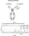
- the present invention generally relates to the field of biomedicine, and in particular to a protein-drug conjugate and a method for preparing the same.
- Antibody-drug conjugate is produced by the coupling of an antibody targeting a tumor antigen with highly-cytotoxic small molecule chemical drug through a linker.
- ADC Antibody-drug conjugate
- ADC technology has also been applied in non-tumor fields; for example, the AbbVie company conjugates steroids to anti-TNF ⁇ antibodies for the treatment of multiple TNF ⁇ -mediated autoimmune diseases.
- Coupling methods and linker structures are crucial for the stability of ADC drugs.
- the cysteine coupling technology is a representative of early chemical coupling methods; it utilizes four natural interchain disulfide bonds of IgG1 antibodies as active sulfhydryls for coupling.
- this technology has a controllable drug/antibody ratio (DAR), but still produces a mixture with a DAR distributed from 0 to 8.
- DAR drug/antibody ratio
- the third-generation ADC technology Due to the uneven components of the coupling products produced by such technologies, their stability and pharmaceutical properties have significant shortcomings.
- the third-generation ADC technology has improved the stability and pharmacokinetic performance of antibody-drug conjugates by obtaining stable DAR through site-specific conjugation between drugs and antibodies.
- the representative coupling technologies for the third-generation ADC mainly include ThioMab, ThioBridge, introduction of unnatural amino acids, transpeptidaton, N-saccharide chain coupling, etc.
- ThioMab technology was first developed by the Genentech company.
- cysteine coupling technology Based on cysteine coupling technology, two or more cysteines are introduced as coupling sites at specific sites in antibodies, except for natural disulfide bonds, to produce site-specific and highly-homogeneous antibody-drug conjugate with a DAR of 2; however, due to the fact that the mutations introduced are not naturally occurring amino acid sequences, there is a potential risk of molecular stability and immunogenicity.
- ThioBridge technology is to reduce the disulfide bond of the monoclonal antibody itself.
- the monoclonal antibody can be re-bridged, and a product with a main component with a DAR of 4 is obtained, but this technology still has the risk of disulfide bond mismatch.
- Unnatural amino acid coupling technology is to use a special tRNA synthetase encoding unnatural amino acids, which enables cells to synthesize various antibodies with p -acetylphenylalanine residues, and by utilizing the coupling reaction between the carbonyls of p -acetylphenylalanine residues and the linker with alkoxylamine groups, a product with a main component with a DAR of 2 is obtained; due to the need for the use of modified host cells to produce unnatural amino acids, the obvious drawbacks of this technology are the low yield and high cost of antibody production.
- transpeptidase Sortase A specifically recognizes this sequence and hydrolyzes a peptide bond between threonine and glycine, and then, a conjugate comprising glycine is connected to threonine; however, due to additional peptide sequences are introduced in a coupling product generated by this technology, there is a potential risk of immunogenicity.
- N-saccharide chain coupling technology such as GlycoConnect technology
- GlycoConnect technology is to use endoglycosidase Endo S2 to trim a saccharide chain of Asn297 (Eu numbering) located in an antibody CH2 region, then use the mutant galactosyl transferase GalT (Y289L) and N-azidoacetyl galactosamine (GalNAz) to introduce azido, and couple the azido with a small molecule drug by using click chemistry to produce stable and uniform antibody-drug conjugate with a DAR of 2; due to the need for the use of special mutant enzymes, the process development cost of this technology is relatively high.
- the present application provides a site-specific conjugation method for a protein-drug conjugate and a protein-drug conjugate prepared by the method.
- the present application has one or more of the following properties: 1) a coupling product with a uniform drug/antibody ratio (DAR) or a uniform conjugate/antibody ratio (CAR) can be produced through site-specific conjugation; 2) compared to other site-specific coupling technologies such as THIOMAB technology, natural hinge region amino acid sequences can be utilized without introducing amino acid mutations or additional peptide sequences and changing saccharide chain structures on Fc; 3) the length or site-specific amino acid types of the natural hinge region amino acid sequences can be optimized to improve the expression quantity and uniformity of a product; 4) there is no need for particularly complex coupling technologies or processes; 5) the site-specific conjugation method has fewer byproducts and strong developability; 6) antigen-binding fragments can be extended to various forms such as fully human VH domain, camelidae VHH domain, single-chain antibody s
- the present application provides a protein-drug conjugate, comprising an antigen-binding protein moiety and a drug conjugate moiety, the antigen-binding protein moiety comprising one or more antigen-binding fragments, and at least two linker peptides directly or indirectly connected with the antigen-binding fragment; the at least two linker peptides comprising a first linker peptide and a second linker peptide, wherein the first linker peptide comprises a first cysteine Cys1 and a second cysteine Cys2, and the drug conjugate moiety is coupled to the first linker peptide through the Cys1; and the second linker peptide comprises a first cysteine Cys1 and a second cysteine Cys2, and the drug conjugate moiety is coupled to the second linker peptide through the Cys1.
- the Cys2 of the first linker peptide is connected to the Cys2 of the second linker peptide through a disulfide bond.
- the one or more antigen-binding fragments do not comprise a functional group capable of affecting coupling of a drug conjugate with the first linker peptide and/or the second linker peptide at the Cys1, or a conjugate formed by the functional group and other drug conjugates, and the affection is manifested in significantly reducing a coupling probability of the drug conjugate at a Cys1 site.
- the one or more antigen-binding fragments do not comprise a functional group capable of forming a disulfide bond with the Cys1 of the first linker peptide and/or the Cys1 of the second linker peptide, or a conjugate formed by the functional group and other drug conjugates.
- the one or more antigen-binding fragments do not comprise a cysteine capable of forming a disulfide bond with the Cys1 of the first linker peptide and/or the Cys1 of the second linker peptide, or a conjugate formed by the cysteine and other drug conjugates.
- amino acid sequences of the first linker peptide and the second linker peptide can be identical or different.
- the first linker peptide and/or the second linker peptide comprise/comprises an amino acid sequence set forth in Cys1-(X)n-Cys2 from an N-terminus to a C-terminus, wherein n is an integer greater than or equal to 3, and X is any amino acid other than cysteine.
- the first linker peptide and/or the second linker peptide are/is derived from an antibody hinge region sequence or a derivative sequence thereof.
- the derivative sequence is obtained by modifying the antibody hinge region sequence, and the modification does not involve changes in Cys1 and Cys2 in the antibody hinge region.
- the modification is adjusting a type and number of amino acids between Cys1 and Cys2 in the antibody hinge region sequence, and/or adding flexible linker amino acids before Cys1.
- the linker peptide is derived from a human IgG hinge region.
- the antibody hinge region is a human IgG1 hinge region.
- the linker peptide is the human IgG1 hinge region
- sequences of the first linker peptide and the second linker peptide are each independently set forth in SEQ ID NO: 73
- the Cys1 in the linker peptides is Cys-220 according to EU numbering in the human IgG1 hinge region
- the Cys2 is Cys-226 according to EU numbering in the human IgG1 hinge region.
- the first linker peptide and the second linker peptide can be each independently derived from a derivative sequence of the human IgG1 hinge region, and an amino acid sequence thereof is set forth in any one of SEQ ID NOs: 74-105 and 145-146; the Cys1 in the first linker peptide is Cys corresponding to Cys-220 in the human IgG1 hinge region, and the Cys2 in the first linker peptide is Cys corresponding to Cys-226 in the human IgG1 hinge region; the Cys1 in the second linker peptide is Cys corresponding to Cys-220 in the human IgG1 hinge region, and the Cys2 in the second linker peptide is Cys corresponding to Cys-226 in the human IgG1 hinge region.
- the linker peptide is a mouse IgG2c hinge region
- sequences of the first linker peptide and the second linker peptide are each independently set forth in SEQ ID NO: 147
- the Cys1 in the linker peptides is a first cysteine in a sequence of the mouse IgG2c hinge region
- the Cys2 is a second cysteine in a sequence thereof.
- the plurality of antigen-binding fragments comprised by the antigen-binding protein moiety of the protein-drug conjugate are connected to the same linker peptide or respectively connected to the different linker peptides.
- the plurality of antigen-binding fragments can be partially or completely identical or different from each other.
- the plurality of antigen-binding fragments comprise a first antigen-binding fragment and a second antigen-binding fragment.
- the first antigen-binding fragment and the second antigen-binding fragment can be identical or different.
- the first antigen-binding fragment and the second antigen-binding fragment bind to the same target and have the same amino acid sequence; for example, the first antigen-binding fragment and the second antigen-binding fragment bind to the same target but have different amino acid sequences; for example, the first antigen-binding fragment and the second antigen-binding fragment bind to different targets.
- the plurality of antigen-binding fragments comprise a first antigen-binding fragment, a second antigen-binding fragment, a third antigen-binding fragment and a fourth antigen-binding fragment.
- the first antigen-binding fragment, the second antigen-binding fragment, the third antigen-binding fragment and the fourth antigen-binding fragment can be partially or completely identical or different from each other.
- the first antigen-binding fragment and the second antigen-binding fragment bind to the same target and have the same amino acid sequence; for example, the first antigen-binding fragment and the second antigen-binding fragment bind to the same target but have different amino acid sequences; for example, the first antigen-binding fragment and the second antigen-binding fragment bind to different targets.
- the third antigen-binding fragment and the fourth antigen-binding fragment bind to the same target and have the same amino acid sequence; for example, the third antigen-binding fragment and the fourth antigen-binding fragment bind to the same target but have different amino acid sequences; for example, the third antigen-binding fragment and the fourth antigen-binding fragment bind to different targets.
- the first antigen-binding fragment, the second antigen-binding fragment, the third antigen-binding fragment and the fourth antigen-binding fragment bind to different targets.
- the one or more antigen-binding fragments are capable of each independently being derived protein structures such as a VHH domain, a VH, a VL, an Fab, an ScFv, a receptor protein soluble extracellular region, a ligandin, a lipocalin, a neuronal cell adhesion molecule NCAM, a fibronectin and/or a designed ankyrin repeat protein DARPin.
- derived protein structures such as a VHH domain, a VH, a VL, an Fab, an ScFv, a receptor protein soluble extracellular region, a ligandin, a lipocalin, a neuronal cell adhesion molecule NCAM, a fibronectin and/or a designed ankyrin repeat protein DARPin.
- the one or more antigen-binding fragments are capable of specifically binding to a tumor antigen or a non-tumor antigen.
- the tumor antigen comprises CD19, BCMA, TSHR, CD171, CS-1, CLL-1, GD3, Tn Ag, FLT3, CD38, CD123, CD44v6, B7H3, B7H4, KIT, IL-13Ra2, IL-11Ra, PSCA, PSMA, PRSS21, VEGFR2, LewisY, CD24, PDGFR-beta, SSEA-4, MUC1, EGFR, NCAM, CAIX, LMP2, EphA2, fucosyl GM1, sLe, GM3, TGS5, HMWMAA, GD2, FOLR1, FOLR2, TEM1/CD248, TEM7R, CLDN6, CLDN18.2, GPRC5D, CXORF61, CD97, CD179a, ALK, polysialic acid, PLAC1, GloboH, NY-BR-1, UPK2, HAVCR1, ADRB3, PANX3, GPR20, LY6K, OR51E2, TAARP, WT1, ETV6
- the first antigen-binding fragment and/or the second antigen-binding fragment each independently comprise HCDR1, HCDR2, and HCDR3, and the antigen-binding fragments comprise amino acid sequences selected from any one of (1) HCDR1: SEQ ID NO: 7, HCDR2: SEQ ID NO: 22, HCDR3: SEQ ID NO: 38; (2) HCDR1: SEQ ID NO: 10, HCDR2: SEQ ID NO: 25, HCDR3: SEQ ID NO: 41; (3) HCDR1: SEQ ID NO: 11, HCDR2: SEQ ID NO: 26, HCDR3: SEQ ID NO: 42; (4) HCDR1: SEQ ID NO: 13, HCDR2: SEQ ID NO: 28, HCDR3: SEQ ID NO: 44; (5) HCDR1: SEQ ID NO: 14, HCDR2: SEQ ID NO: 29, HCDR3: SEQ ID NO: 42; (6) HCDR1: SEQ ID NO: 9, HCDR2: SEQ ID NO:
- the antigen-binding fragment comprises a VH
- the VH comprises an amino acid sequence set forth in any one of SEQ ID NOs: 55-59, 61 and 62.
- the first antigen-binding fragment and/or the second antigen-binding fragment each independently comprise HCDR1, HCDR2, HCDR3, LCDR1, LCDR2 and LCDR3, and the HCDR1, HCDR2, HCDR3, LCDR1, LCDR2 and LCDR3 respectively and sequentially comprise amino acid sequences set forth in SEQ ID NO: 12, SEQ ID NO: 27, SEQ ID NO: 43, SEQ ID NO: 49, SEQ ID NO: 51 and SEQ ID NO: 53.
- the first antigen-binding fragment and/or the second antigen-binding fragment each independently comprise a VH and a VL
- the VL comprises an amino acid sequence set forth in SEQ ID NO: 63
- the VH comprises an amino acid sequence set forth in SEQ ID NO: 60.
- the first antigen-binding fragment and/or the second antigen-binding fragment each independently comprise one pair of VH and VL and/or another type of VH
- the pair of VH and VL respectively comprises amino acid sequences set forth in SEQ ID NO: 60 and SEQ ID NO: 63
- the another type of VH comprises an amino acid sequence set forth in SEQ ID NO: 62.
- the pair of VH and VL can form scFv.
- the first antigen-binding fragment and/or the second antigen-binding fragment each independently comprise a VH
- the VH comprises an amino acid sequence set forth in SEQ ID NO: 62
- the third antigen-binding fragment and/or the fourth antigen-binding fragment each independently comprise one pair of VH and VL, and the pair of VH and VL respectively comprises amino acid sequences set forth in SEQ ID NO: 130 and SEQ ID NO: 131.
- the third antigen-binding fragment and/or the fourth antigen-binding fragment can comprise an Fab.
- the antigen-binding protein moiety in the protein-drug conjugate can further comprise a pairing unit moiety enabling the first linker peptide and the second linker peptide to be paired
- the pairing unit can comprise at least a first pairing subunit and a second pairing subunit
- the first pairing subunit can interact with the second pairing subunit through a covalent bond or a non-covalent bond.
- the pairing unit is an Fc fragment or a mutant Fc fragment.
- an N-terminus of the first pairing subunit is connected to a C-terminus of the first linker peptide
- an N-terminus of the second pairing subunit is connected to a C-terminus of the second linker peptide; or the N-terminus of the first pairing subunit is connected to the C-terminus of the second linker peptide, and the N-terminus of the second pairing subunit is connected to the C-terminus of the first linker peptide.
- the protein-drug conjugate comprises an amino acid sequence set forth in any one of SEQ ID NOs: 64-71, 106-129 and 148-149.
- the drug conjugate moiety of the protein-drug conjugate comprises two drug conjugates respectively connected to the first linker peptide and the second linker peptide, and the drug conjugate comprises a loaded drug and optionally a linker.
- the drug conjugate in the protein-drug conjugate comprises at least one loaded drug.
- the loaded drug can comprise, but is not limited to, a small molecule compound, a toxin molecule, an antibiotic, an oligonucleotide, a proteolysis targeting chimera (PROTAC), an affinity ligand, a fluorescently or nuclide-labeled group, a polypeptide, an immunomodulator, etc.
- the linker comprises a cleavable linker or a non-cleavable linker, wherein the cleavable linker comprises a protease cuttable linker.
- the drug conjugate comprises mc-vc-PAB-MMAE (as shown in 0).
- the drug conjugate comprises PROTAC, such as a BRD4 protein degrading agent (as shown in 0).
- the protein-drug conjugate is an antibody-drug conjugate (ADC).
- ADC antibody-drug conjugate
- the present application provides a method for preparing the protein-drug conjugate of the present application, comprising covalently binding the drug conjugate with a reactive sulfhydryl of the Cys1 on the first linker peptide and/or the second linker peptide.
- the present application provides a method for preparing a protein-drug conjugate.
- the protein-drug conjugate comprises an antigen-binding protein moiety and a drug conjugate moiety;
- the antigen-binding protein moiety comprises one or more antigen-binding fragments, and at least two linker peptides directly or indirectly connected with the antigen-binding fragment;
- the two linker peptides comprise a first linker peptide and a second linker peptide, wherein the first linker peptide comprises a first cysteine Cys1 and a second cysteine Cys2, and the second linker peptide comprises a first cysteine Cys 1 and a second cysteine Cys2;
- the method comprises binding the drug conjugate moiety with the first linker peptide through the Cys1 of the first linker peptide, and/or binding the drug conjugate moiety with the second linker peptide through the Cys1 of the second linker peptide.
- the one or more antigen-binding fragments do not comprise a functional group capable of affecting coupling of a drug conjugate with the first linker peptide and/or the second linker peptide at the Cys1, or a conjugate formed by the functional group and other drug conjugates, and the affection is manifested in significantly reducing a coupling probability of the drug conjugate at a Cys1 site.
- the one or more antigen-binding fragments in the method do not comprise a functional group capable of forming a disulfide bond with the Cys1 of the first linker peptide and/or the Cys1 of the second linker peptide, or a conjugate formed by the functional group and other drug conjugates.
- the one or more antigen-binding fragments do not comprise a cysteine capable of forming a disulfide bond with the Cys1 of the first linker peptide and/or the Cys1 of the second linker peptide, or a conjugate formed by the cysteine and other drug conjugates.
- amino acid sequences of the first linker peptide and the second linker peptide in the method can be identical or different.
- the first linker peptide and/or the second linker peptide in the method comprise/comprises an amino acid sequence set forth in Cysl-(X)n-Cys2 from an N-terminus to a C-terminus, wherein n is an integer greater than or equal to 3, and X is any amino acid other than cysteine.
- the first linker peptide and/or the second linker peptide are/is derived from an antibody hinge region sequence or a derivative sequence thereof.
- the derivative sequence in the method is obtained by modifying the antibody hinge region sequence, and the modification does not involve changes in Cys1 and Cys2 in the antibody hinge region.
- the modification is adjusting a type number of amino acids between Cys1 and Cys2 in the antibody hinge region sequence, and/or adding flexible linker amino acids before Cys1.
- the linker peptide in the method is derived from a human IgG hinge region, preferably a human IgG1 hinge region.
- the first linker peptide in the method is the human IgG1 hinge region
- a sequence of the first linker peptide is set forth in SEQ ID NO: 73
- the Cys1 in the first linker peptide is Cys-220 according to EU numbering in the human IgG1 hinge region
- the Cys2 is Cys-226 according to EU numbering in the human IgG1 hinge region.
- the second linker peptide in the method is the human IgG1 hinge region
- a sequence of the second linker peptide is set forth in SEQ ID NO: 73
- the Cys1 in the second linker peptide is Cys-220 according to EU numbering in the human IgG1 hinge region
- the Cys2 is Cys-226 according to EU numbering in the human IgG1 hinge region.
- the first linker peptide in the method can be derived from a derivative sequence of the human IgG1 hinge region, an amino acid sequence thereof is a sequence as set forth in any one of SEQ ID NOs: 74-105 and 145-146, the Cys1 in the first linker peptide is Cys corresponding to Cys-220 in the human IgG1 hinge region, and the Cys2 in the linker peptide is Cys corresponding to Cys-226 in the human IgG1 hinge region.
- the second linker peptide in the method can be derived from a derivative sequence of the human IgG1 hinge region, an amino acid sequence thereof is a sequence as set forth in any one of SEQ ID NOs: 74-105 and 145-146, the Cys1 in the second linker peptide is Cys corresponding to Cys-220 in the human IgG1 hinge region, and the Cys2 in the linker peptide is Cys corresponding to Cys-226 in the human IgG1 hinge region.
- the first linker peptide in the method is a mouse IgG2c hinge region, the amino acid sequence thereof is set forth in SEQ ID NO: 147, the Cys1 in the first linker peptide is a first cysteine in the sequence thereof, and the Cys2 is a second cysteine in the sequence thereof.
- the second linker peptide in the method is a mouse IgG2c hinge region, the amino acid sequence thereof is set forth in SEQ ID NO: 147, the Cys1 in the second linker peptide is a first cysteine in the sequence thereof, and the Cys2 is a second cysteine in the sequence thereof.
- the method comprises steps of reduction, coupling, and optionally purification.
- the reduction comprises reducing a disulfide bond between the Cys1 of the first linker peptide and the Cys1 of the second linker peptide under a condition of a reducing agent.
- the reduction comprises using DTT and/or TCEP as a reducing agent; in certain embodiments, the reducing agent is TCEP.
- an amount of usage of the reducing agent is 1 to 50 molar multiples; in certain embodiments, the amount of usage of the reducing agent is 1 to 6 molar multiples; in certain embodiments, the amount of usage of the reducing agent is 1.5 to 3 molar multiples.
- reaction time for the reduction is 1 to 17 hours; in certain embodiments, the reaction time for the reduction is 1 to 3 hours; in certain embodiments, the reaction time for the reduction is 1.5 to 2 hours.
- a reaction temperature for the reduction is 0 °C to 40 °C; in certain embodiments, the reaction temperature for the reduction is 0 °C or room temperature (25 °C to 30 °C) or 37 °C.
- a pH of a reaction buffer for the reduction is 4.0 to 9.0; in certain embodiments, the pH of the reaction buffer for the reduction is 5.0 to 8.0; in certain embodiments, the pH is 5.0 to 7.0; in certain embodiments, the pH is 5.0 to 6.0.
- the coupling comprises covalently binding the drug conjugate with a reactive sulfhydryl of the Cys1 on the first linker peptide and/or the second linker peptide.
- the drug conjugate comprises mc-vc-PAB-MMAE or PROTAC or a CpG oligonucleotide.
- an amount of usage of the drug conjugate is 1 to 50 molar multiples; in certain embodiments, the amount of usage of the drug conjugate is 1 to 10 molar multiples; in certain embodiments, the amount of usage of the drug conjugate is 3 to 7 molar multiples.
- the drug conjugate is mc-vc-PAB-MMAE, and an amount of usage of the mc-vc-PAB-MMAE is 1 to 50 molar multiples; in certain embodiments, the amount of usage of the mc-vc-PAB-MMAE is 3 to 18 molar multiples; in certain embodiments, the amount of usage of the mc-vc-PAB-MMAE is 3 to 7 molar multiples.
- an amount of usage of the drug conjugate PROTAC is 10 to 15 molar multiples.
- reaction time for the coupling is 0.5 to 10 hours; in certain embodiments, the reaction time for the coupling is 0.5 to 3 hours; in certain embodiments, the reaction time for the coupling is 0.5 to 1 hours.
- a reaction temperature for the coupling is 0 °C to 40 °C; in certain embodiments, the reaction temperature for the coupling is room temperature (25 °C to 30 °C).
- pH of a reaction buffer for the coupling is 4.0 to 9.0; in certain embodiments, the pH of the reaction buffer for the coupling is 5.0 to 8.0; in certain embodiments, the pH is 5.0 to 7.0; in certain embodiments, the pH is 7.0 to 8.0; in certain embodiments, the pH is 5.0 to 6.0.
- a purity of the protein-drug conjugate is greater than 80%.
- the purity of the protein-drug conjugate is greater than 82%, greater than 85%, greater than 88% or greater than 90%.
- the present application provides a pharmaceutical composition
- a pharmaceutical composition comprising the protein-drug conjugate and a pharmaceutically acceptable carrier.
- the present application provides a method for preventing and/or treating diseases, comprising administering the protein-drug conjugate and/or the pharmaceutical composition, wherein the protein-drug conjugate and/or the pharmaceutical composition are optionally in combination with other therapies or drugs.
- the present application provides use of the protein-drug conjugate and/or the pharmaceutical composition in preparation of a drug, wherein the drug is used for treating tumors or other diseases.
- the present application provides use of the protein-drug conjugate, in combination with other therapies or drugs, in preparation of a drug, wherein the drug is used for treating tumors or other diseases.
- the other therapies or drugs are selected from: chemotherapy, radiotherapy, miRNA and oligonucleotides.
- the tumor is selected from any one or more of the following groups: lymphoma, multiple myeloma, breast cancer, ovarian cancer, kidney cancer, endometrial cancer, melanoma, pancreatic cancer, lung cancer, stomach cancer, liver cancer, mesothelioma, esophageal cancer, head and neck cancer, bile duct cancer, gallbladder cancer, bladder cancer, thymus cancer and colorectal cancer.
- the present application provides a method for diagnosing a specific disease, comprising using the protein-drug conjugate and/or the pharmaceutical composition.
- the present application provides a method for detecting a specific target, comprising using the protein-drug conjugate and/or the pharmaceutical composition, wherein preferably the detection is for non-disease diagnostic purposes.
- the present application provides a kit for detection, comprising the protein-drug conjugate and/or the pharmaceutical composition, and optionally instructions for use.
- the present application provides an administration device, comprising the protein-drug conjugate and/or the pharmaceutical composition, and a device for administering the protein-drug conjugate and/or the pharmaceutical composition.
- the Cys-220 in the absence of a light chain, the Cys-220 (Eu-numbered) in the human IgG1 heavy chain hinge region has the opportunity to form a disulfide bond between heavy chains, however, the disulfide bond is not as stable as other disulfide bonds, and only a weaker reducing agent is needed to separate the disulfide bond of the Cys-220 from other disulfide bonds, so that the disulfide bond of the Cys-220 can be effectively cleaved while the other disulfide bonds remain intact. Furthermore, the Cys-220 becomes almost the only free cysteine residue that acts as a site-specific conjugation site for covalent binding with drug conjugates.
- the hinge region derivative sequence formed by adjusting the type, the number of amino acids between Cys-220 and Cys-226 in the human IgG1 natural hinge region or by adding a flexible linker peptide before Cys-220, can also serve as a linker peptide to achieve site-specific conjugation corresponding to a Cys-220 site in the natural hinge region.
- the basic four-chain antibody unit is a heterotetramer protein, which is composed of two identical light chains (L) and two identical heavy chains (H).
- L light chains
- H heavy chains
- each L chain is connected to the H chain through a covalent disulfide bond
- two H chains are connected to each other through one or more disulfide bonds.
- the number of disulfide bonds depends on the isotype of the H chain.
- Each H and L chain also has regularly-spaced intrachain disulfide bonds.
- Each H chain has a variable domain (VH) at an amino terminus, followed by three (for each ⁇ and ⁇ chain) or four (for ⁇ and ⁇ isotype) constant domain (CH).
- Each L chain has a variable domain (VL) at an amino terminus, followed by one constant domain (CL).
- Human IgG has four subtypes: IgG1, IgG2, IgG3 and IgG4.
- the L chain of human or mouse antibodies is further divided into ⁇ chain and ⁇ chain, and accordingly, its variable domain is divided into Y ⁇ and V ⁇ , and its constant domain is divided into C ⁇ and C ⁇ .
- binding protein or "antigen-binding protein” generally refers to a protein comprising an antigen-binding moiety, and optionally a scaffold or framework moiety that allows the antigen-binding moiety to adopt a conformation that facilitates the binding of the antigen-binding protein to the antigen.
- the "binding protein” or “antigen-binding protein” may typically comprise an antibody light chain variable region (VL) or an antibody heavy chain variable region (VH), or both.
- VL antibody light chain variable region
- VH antibody heavy chain variable region
- the VH and VL regions can be further divided into hypervariable regions termed complementary determining regions (CDRs), and relatively conserved framework regions (FRs). Each VH and VL can be composed of three CDRs and four FRs.
- the VH and VL comprise binding sites that interact with antigens.
- the three CDRs of VH are denoted as HCDR1, HCDR2 and HCDR3, respectively, and may also be denoted as VH CDR1, VH CDR2 and VH CDR3, respectively; and the three CDRs of VL are denoted as LCDR1, LCDR2 and LCDR3, respectively, and may also be denoted as VL CDR1, VL CDR2 and VL CDR3, respectively.
- antigen-binding proteins examples include, but are not limited to, antibodies, antigen-binding fragments (Fab, Fab', F(ab) 2 , Fv fragment, F(ab') 2 , scFv, di-scFv and/or dAb), immunoconjugates, multispecific antibodies (e.g., bispecific antibodies), antibody fragments, antibody derivatives, antibody analogs, receptor protein soluble extracellular regions, ligandin or other forms of fusion proteins, as long as they exhibit the desired antigen-binding activity.
- Fab antigen-binding fragments
- the CDRs of an antibody can be defined in the art using a variety of methods, such as the Kabat scheme based on sequence variability (see Kabat et al., Sequences of Proteins of Immunological Interest, Fifth Edition, National Institutes of Health (U.S.), Bethesda, Maryland (1991 )), and the Chothia scheme based on the location of the structural loop regions (see J Mol Biol 273: 927-948, 1997 ).
- the Combined scheme comprising the Kabat scheme and the Chothia scheme can also be used to determine the amino acid residues in a variable domain sequence.
- the Combined scheme combines the Kabat scheme with the Chothia scheme to obtain a larger range.
- CDR complementary determining region
- Laa-Lbb can refer to an amino acid sequence from position aa (the Chothia scheme) to position bb (the Chothia scheme) beginning at the N-terminus of the light chain of the antibody; and Haa-Hbb can refer to an amino acid sequence from position aa (the Chothia scheme) to position bb (the Chothia scheme) beginning at the N-terminus of the heavy chain of the antibody.
- L24-L34 can refer to the amino acid sequence from position 24 to position 34 according to the Chothia scheme beginning at the N-terminus of the light chain of the antibody
- H26-H32 can refer to the amino acid sequence from position 26 to position 32 according to the Chothia scheme beginning at the N-terminus of the heavy chain of the antibody. It should be known to those skilled in the art that there are positions where insertion sites are present in numbering CDRs with the Chothia scheme (see http://bioinf.org.uk/abs/).
- the term "monoclonal antibody” generally refers to an antibody obtained from a population of substantially homogeneous cells, that is, the antibodies in the population are identical except for a small amount of natural mutations that may exist.
- Monoclonal antibodies are generally highly specific for a single antigenic site. Moreover, unlike conventional polyclonal antibody formulations (which generally have different antibodies directed against different determinants), each monoclonal antibody is directed against a single determinant on the antigen.
- the advantage of monoclonal antibodies lies in that they can be synthesized through hybridoma culture and are not contaminated by other immunoglobulins.
- monoclonal represents the characteristics of the antibody obtained from a population of substantially homogeneous antibodies, but is not to be construed as requiring production of the antibody by any particular method.
- monoclonal antibodies used according to the present invention can be prepared in hybridoma cells or can be prepared by the recombinant DNA method.
- the term "fully human antibody” generally refers to an antibody that is expressed by a genetically engineered antibody gene-deleted animal into which the entire gene that encodes an antibody in human is transferred. All parts of the antibody (including variable and constant regions of the antibody) are composed of amino acid sequences of human origin.
- the fully human antibody can greatly reduce the immune side effects caused in the human body by the heterologous antibody. Methods for obtaining fully human antibodies in the art can include phage display, transgenic mice, and the like.
- the term "specific binding to” generally refers to that an antigen-binding protein binds to an antigenic epitope, and that the binding requires some complementarity between the antigen-binding protein and the epitope. According to this definition, an antibody is said to "specifically bind to” an antigen when the antibody more easily binds to an epitope via its antigen-binding protein than binds to a random, unrelated epitope.
- Epitope refers to a specific atomic group (e.g., saccharide side chains, phosphoryl, sulfonyl) or an amino acid on an antigen that binds to an antigen-binding protein (e.g., an antibody).
- a specific atomic group e.g., saccharide side chains, phosphoryl, sulfonyl
- an amino acid on an antigen that binds to an antigen-binding protein e.g., an antibody
- the term "Fab” generally refers to the portion of a conventional antibody (e.g., IgG) that binds to an antigen, including the heavy chain variable region VH, the light chain variable region VL, the heavy chain constant region domain CH1 and the light chain constant region CL of the antibody.
- a conventional antibody e.g., IgG
- the C-terminus of VH is linked to the N-terminus of CH1 to form a heavy chain Fd fragment
- the C-terminus of VL is linked to the N-terminus of CL to form a light chain
- the C-terminus of CH1 is further linked to the hinge region and other constant region domains of the heavy chain to form a heavy chain.
- “Fab” also refers to a variant structure of the Fab.
- the C-terminus of VH is linked to the N-terminus of CL to form one polypeptide chain
- the C-terminus of VL is linked to the N-terminus of CH1 to form the other polypeptide chain, in which case an Fab (cross VH/VL) structure is formed
- CH1 of the Fab is not linked to the hinge region, but rather the C-terminus of CL is linked to the hinge region of the heavy chain, in which case a Fab (cross Fd/LC) structure is formed.
- VH generally refers to the heavy chain variable region VH domain of an antibody.
- VH may be a heavy chain variable region VH of a conventional antibody (H2L2 structure) from humans or other animals.
- VH may also be a heavy chain variable region VHH of a heavy-chain antibody (HCAb structure) from animals such as camelidae.
- HCAb structure a heavy chain variable region VH of a fully-human heavy-chain antibody (HCAb structure) produced by a Harbour HCAb transgenic mouse.
- antigen-binding fragment generally refers to any protein functional region that can specifically bind to an antigen.
- antigen-binding fragment may be "Fab”.
- antigen-binding fragment may also be "VH”.
- antiigen-binding fragment may also be a single chain antibody scFv.
- antigen-binding fragment may further be other antigen-binding forms (e.g., a receptor protein soluble extracellular region, a ligandin, a lipocalin, a neuronal cell adhesion molecule (NCAM), a fibronectin, a designed ankyrin repeat protein (DARPin) and other derived protein structures).
- antigen-binding forms e.g., a receptor protein soluble extracellular region, a ligandin, a lipocalin, a neuronal cell adhesion molecule (NCAM), a fibronectin, a designed ankyrin repeat protein (DARPin) and other derived protein structures.
- the term "pairing unit” generally refers to a group of structures comprising at least two subunits that can be paired with each other.
- the paired subunits can bind with each other, and the binding requires complementarity between the subunits; covalent bond or non-covalent bond interaction can occur between the paired subunits; the non-covalent bond interaction can include van der Waals force, hydrogen bond, hydrophobic interaction, electrostatic interaction, dipole interaction, etc.
- “Pairing unit” may be a set of naturally occurring or modified polypeptide chains in the form of homologous or heterologous multimers; for example, “pairing unit” may be a dimer form of antibody Fc fragments or variants thereof; “pairing unit” may be ⁇ chain and ⁇ chain of CD79; “pairing unit” may be ⁇ chain and ⁇ chain of CD8; “pairing unit” may be ⁇ chain and ⁇ chain of a T-cell receptor (TCR).
- TCR T-cell receptor
- Fc fragment generally refers to the fragment crystallizable (Fc) of the antibody, that is, comprising a polypeptide chain composed of heavy chain constant domains CH2 and CH3.
- Fc fragment crystallizable
- an Fc fragment may be in the form of a natural dimer; in other embodiments, an Fc fragment may be in the form of a modified monomer.
- CTLA4 generally refers to the cytotoxic T lymphocyte-associated antigen-4 (also known as CD152), a functional variant thereof and/or a functional fragment thereof.
- the sequence of CTLA4 is known in the art.
- the sequence of an exemplary full-length human CTLA4 can be found under Uniprot accession No. P16410; and the sequence of an exemplary full-length cynomolgus monkey CTLA4 can be found under Uniprot accession No. G7PL88.
- Drugs directed against CTLA4 may be applied to treat melanoma, lung cancer, colon cancer, liver cancer, head and neck cancer, kidney cancer, breast cancer, pancreatic cancer, bladder cancer and other tumors.
- BCMA tumor necrosis factor receptor superfamily member 17 (also known as CD269, B cell maturation protein), a functional variant thereof and/or a functional fragment thereof.
- the sequence of BCMA is known in the art. For example, the sequence of an exemplary full-length human BCMA can be found under Uniprot accession No. Q02223; and the sequence of an exemplary full-length cynomolgus monkey BCMA can be found under NCBI accession No. XP_005591343. Drugs directed against BCMA may be applied to treat multiple myeloma and other hematological malignant tumors.
- MSLN generally refers to a mesothelin molecule (also known as MPF), a functional variant thereof and/or a functional fragment thereof.
- MPF mesothelin molecule
- the sequence of MSLN is known in the art.
- the sequence of an exemplary full-length human MSLN can be found under Uniprot accession No. Q13421; and the sequence of an exemplary full-length cynomolgus monkey MSLN can be found under NCBI accession No. XP_005590873.
- Drugs directed against MSLN may be applied to treat mesothelioma, lung cancer, pancreatic cancer, breast cancer, ovarian cancer, endometrial cancer and other tumors.
- 5T4 generally refers to a trophoblast glycoprotein (also known as TPBG), a functional variant thereof and/or a functional fragment thereof.
- TPBG trophoblast glycoprotein
- the sequence of 5T4 is known in the art.
- the sequence of an exemplary full-length human 5T4 can be found under Uniprot accession No. Q13641; and the sequence of an exemplary full-length cynomolgus monkey 5T4 can be found under Uniprot accession No. Q4R8Y9.
- Drugs directed against 5T4 may be applied to treat thymus cancer, lung cancer, esophageal cancer, stomach cancer, small intestine cancer, pancreatic cancer, liver cancer, gallbladder cancer, kidney cancer, bladder cancer, ovarian cancer, endometrial cancer, cervical cancer and other tumors.
- ROR1 generally refers to an inactivated tyrosine protein kinase transmembrane receptor ROR1 (also known as NTRKR1), a functional variant thereof and/or a functional fragment thereof.
- the sequence of ROR1 is known in the art. For example, the sequence of an exemplary full-length human ROR1 can be found under Uniprot accession No. Q01973; and the sequence of an exemplary full-length cynomolgus monkey ROR1 can be found under NCBI accession No. XP_015290264.
- Drugs directed against ROR1 may be applied to treat leukemia, non-Hodgkin lymphoma, mantle cell lymphoma, breast cancer, lung cancer, ovarian cancer and other tumors.
- CD47 generally refers to a leukocyte surface antigen CD47, a functional variant thereof and/or a functional fragment thereof.
- the sequence of CD47 is known in the art.
- the sequence of an exemplaryfull-length human CD47 can be found under Uniprot accession No. Q08722.
- Drugs directed against CD47 may be applied to treat leukemia, non-Hodgkin lymphoma, multiple myeloma, melanoma, head and neck cancer and other tumors.
- SIRP ⁇ generally refers to a signal regulatory protein ⁇ (also known as BIT, MFR, MYD1, PTPNS1, SHPS1 or SIRP), a functional variant thereof and/or a functional fragment thereof.
- SIRP ⁇ is a receptor for CD47.
- the sequence of SIRP ⁇ is known in the art. For example, the sequence of an exemplaryfull-length human SIRP ⁇ can be found under Uniprot accession No. P78324.
- protein-drug conjugate generally refers to a class of drug molecules formed by binding a drug conjugate to an antigen-binding protein through a covalent bond.
- the drug conjugate comprises at least one loaded drug.
- a variety of protein-drug conjugates and a preparation method thereof are known in the art.
- the protein-drug conjugate may be an antibody-drug conjugate (ADC).
- exemplary protein-drug conjugates can include, but are not limited to, trastuzumab, ocrelizumab, pertuzumab, rituximab and other antibodies.
- Loaded drug modules can be covalently attached to antibodies via linker units to form antibody-drug conjugates to achieve targeted therapeutic effects.
- protein-drug conjugate may comprise an antigen-binding fragment, a linker peptide and a drug conjugate.
- the drug conjugate of "protein-drug conjugate” may be mc-vc-PAB-MMAE (also known as VcMMAE).
- drug conjugate generally refers to a class of aggregations of atoms and groups with specific structures that can bind to other molecules by covalent bonds through specific functional groups; it comprises at least one loaded drug, and optionally comprises a linker.
- loaded drugs a variety of linkers or linker members are known in the art.
- the drug conjugate may be mc-vc-PAB-MMAE (also known as VcMMAE).
- loaded drug generally refers to the forms of a class of small molecule compounds or toxins or other drug molecules with pharmaceutical activity, which can be, but are not limited to, small molecule compounds, toxin molecules, antibiotics, oligonucleotides, proteolysis targeting chimeras (PROTACs), affinity ligands, fluorescent groups, nuclide groups, polypeptides, immunomodulatory molecules, etc.
- a variety of loaded drugs are known in the art, such as monomethyl auristatin E (“MMAE”), mertansine (“DM1”), and toll-like receptor agonist molecules.
- linker peptide or “peptide fragment” generally refers to a part of a polypeptide chain in the "protein-drug conjugate” in the present application.
- the linker peptide comprises at least one cysteine as a site for covalent binding with the drug conjugate.
- the linker peptide may comprise a first linker peptide and/or a second linker peptide, and amino acid sequences of the first linker peptide and the second linker peptide may be identical or different.
- linker or “linker unit” generally refers to a chemical functional module that can be used to connect one or more loaded drugs to the antigen-binding protein to form “protein-drug conjugate".
- the linker may comprise one or more linker members.
- linker members are known in the art, such as maleimidocaproyl ("MC"), valine-citrulline (“val-cit” or “vc”) and p -aminobenzyloxycarbonyl ("PAB”).
- the “linker” may include a polypeptide linker composed of amino acids, but the "linker” is different from the aforementioned "linker peptide”.
- the aforementioned "linker peptide” is a part of the antigen-binding protein.
- conjugate generally refers to the aggregation of atoms and groups formed by covalent binding between the "drug conjugate” in the present application and a specific functional group.
- the conjugate may be formed by the reaction of the drug conjugate with sulfhydryl.
- the conjugate may be an aggregation of stable atoms/groups formed by the addition reaction between the active maleimide reaction group of mc-vc-PAB-MMAE and the sulfhydryl.
- DAR Drug-Antibody Ratio
- the drug loading of a current protein-drug conjugate is generally 0 to 8 loaded drug molecules (D0 to D8)/antibody.
- D0 is also known as "naked antibody”.
- the HIC-HPLC analysis method is a commonly used analysis method for determining the DAR and drug loading distribution of a protein-drug conjugate.
- CAR Conjugate/Antigen binding protein Ratio
- one drug conjugate comprises at least one loaded drug; in some embodiments, the prior art enable one drug conjugate moiety to only comprise one loaded drug, in which case the values of "CAR” and “DAR” may be equivalent; in other embodiments, the prior art can enable one drug conjugate moiety to connect a plurality of loaded drugs through linkers (see Kumara et al., Bioorganic & Medicinal Chemistry Letters (2016), 28, 3617-3621 ), in which case the value of "DAR” may be several times that of "CAR”; for example, when one drug conjugate comprises two loaded drugs, the value of DAR may be twice that of CAR.
- the drug conjugate in the present application is linked by using the cysteine Cys-220 (Eu numbering) in the natural sequence (SEQ ID NO: 73) of the human IgG1 antibody heavy chain hinge region as a coupling site, so that a protein-drug conjugate with a uniform CAR value can be generated.
- the value of CAR and the value of DAR may be equivalent, or the value of DAR may be used to refer to the number of drug conjugates connected to one antigen-binding protein; in particular, a protein-drug conjugate with a uniform DAR value can be generated using the method; for example, the DAR of the protein-drug conjugate is 2.
- the term "derived from” generally refers to derivative amino acid or nucleotide sequences obtained from parental amino acid or nucleotide sequences. It generally refers to the structural similarity between the parental sequence and the derivative sequence, without implying or containing limitations on the process or source of derivative sequences derived from parental sequences. Therefore, when “derived from” is used to discuss proteins or polynucleotides, their physical origins are not considered. In certain cases, “derived from” can refer to the derivative sequence being a part of the unmodified parental sequence. For example, in specific embodiments of the present application, Cys1 and Cys2 may be a part of the unmodified natural antibody hinge region sequence.
- the present application provides the following embodiments:
- the present application provides an antigen-binding protein.
- the antigen-binding protein comprises at least two linker peptides, and one or more antigen-binding fragments.
- the two linker peptides are respectively a first linker peptide and a second linker peptide; and amino acid sequences of the first linker peptide and the second linker peptide may be identical or different.
- the first linker peptide can at least comprise a first cysteine Cys1 and a second cysteine Cys2, and the second linker peptide can at least comprise a first cysteine Cys1 and a second cysteine Cys2.
- a disulfide bond in the antigen-binding protein of the present application, can be formed between the Cys1 of the first linker peptide and the Cys1 of the second linker peptide.
- a disulfide bond in the antigen-binding protein of the present application, can be formed between the Cys2 of the first linker peptide and the Cys2 of the second linker peptide.
- the "disulfide bond Cys1-Cys1" is weaker than the "disulfide bond Cys2-Cys2".
- the disulfide bond Cys1-Cys1 is more easily cleaved by a reducing agent compared to the disulfide bond Cys2-Cys2.
- the Cys1 and the Cys2 of the first linker peptide in the antigen-binding protein can be separated by at least 3 (such as 4, 5, 6, 7, 8, 9, 10, 11, 12, 13, 14, 15 or more) amino acids other than cysteine.
- the Cys1 and the Cys2 of the second linker peptide in the antigen-binding protein can be separated by at least 3 (such as 4, 5, 6, 7, 8, 9, 10, 11, 12, 13, 14, 15 or more) amino acids other than cysteine.
- the first linker peptide can comprise an amino acid sequence set forth in Cys1-(X)n-Cys2 from the N-terminus to the C-terminus, wherein n can be an integer greater than or equal to 3 (for example, n may be 3, n may be 4, n may be 5, n may be 6, n may be 7, n may be 8, n may be 9, n may be 10, n may be 11, n may be 12, n may be 13, n may be 14, n may be 15, or n may be an integer greater than 15); and the X refers to any amino acid other than Cys.
- the second linker peptide can comprise an amino acid sequence set forth in Cys1-(X)n-Cys2 from the N-terminus to the C-terminus, wherein n can be an integer greater than or equal to 3 (for example, n may be 3, n may be 4, n may be 5, n may be 6, n may be 7, n may be 8, n may be 9, n may be 10, n may be 11, n may be 12, n may be 13, n may be 14, n may be 15, or n may be an integer greater than 15); and the X refers to any amino acid other than Cys.
- the linker peptide in the antigen-binding protein may be a naturally occurring amino acid sequence comprising at least two cysteines (Cys1 and Cys2), or a derivative sequence obtained by modifying "naturally occurring amino acid sequence comprising at least two cysteines".
- the "modifying" does not involve changes in the Cys1 and the Cys2 in the naturally occurring amino acid sequence; for example, the modification is adjusting the type or number of amino acids between the Cys1 and the Cys2 in the naturally occurring amino acid sequence, and/or adding flexible linker peptides (such as GSGS and GGGGS) before Cys1.
- the first linker peptide or the second linker peptide can be derived from a human IgG hinge region (SEQ ID NO: 73), and the Cys1 is a Cys-220 according to EU numbering in the human IgG hinge region.
- the first linker peptide can be a human IgG1 hinge region, a sequence of the linker peptide is set forth in SEQ ID NO: 73, the Cys1 in the linker peptide is Cys-220 according to EU numbering in the natural IgG1 hinge region, and the Cys2 is Cys-226 according to EU numbering in the natural IgG1 hinge region.
- the first linker peptide can be a derivative sequence derived from the human IgG1 hinge region, an amino acid sequence thereof is a sequence as set forth in any one of SEQ ID NOs: 74-105 and 145-146, the Cys1 in the linker peptide is Cys corresponding to Cys-220 in the human IgG1 hinge region, and the Cys2 in the linker peptide is Cys corresponding to Cys-226 in the human IgG1 hinge region.
- the second linker peptide can be a human IgG1 hinge region, a sequence of the linker peptide is set forth in SEQ ID NO: 73, the Cys1 in the linker peptide is Cys-220 according to EU numbering in the natural IgG1 hinge region, and the Cys2 is Cys-226 according to EU numbering in the natural IgG1 hinge region.
- the second linker peptide can be a derivative sequence derived from the human IgG1 hinge region, an amino acid sequence thereof is a sequence as set forth in any one of SEQ ID NOs: 74-105 and 145-146, the Cys1 in the linker peptide is Cys corresponding to Cys-220 in the human IgG1 hinge region, and the Cys2 in the linker peptide is Cys corresponding to Cys-226 in the human IgG1 hinge region.
- the first linker peptide or the second linker peptide can be a mouse IgG2c hinge region (SEQ ID NO: 147), the Cys1 is a first cysteine in the sequence thereof, and the Cys2 is a second cysteine in the sequence thereof.
- the "more antigen-binding fragments" in the “one or more antigen-binding fragments” may be partially or completely identical or all different; the “identical” refers to the amino acid sequences of two or more antigen-binding fragments being identical, the “different” refers to the amino acid sequences of two or more antigen-binding fragments being different.
- Two or more antigen-binding fragments can identify different antigens or epitopes, or two or more antigen-binding proteins can identify the identical antigen or identical epitope, but with different sequences and structures.
- the antigen-binding protein can comprise at least two the antigen-binding fragments.
- the antigen-binding protein can comprise a first antigen-binding fragment and a second antigen-binding fragment (a structure as shown in 0(A)), amino acid sequences of the first antigen-binding fragment and the second antigen-binding fragment may be identical or different, and targets of the first antigen-binding fragment and the second antigen-binding fragment may be identical or different.
- the antigen-binding protein can further comprise a third antigen-binding fragment and/or a fourth antigen-binding fragment (a structure as shown in 0(B)); the first, second, third and fourth antigen-binding fragments can be partially or completely identical or different.
- the one or more antigen-binding fragments can be connected to the same linker peptide or to different linker peptides.
- the antigen-binding fragment can be located at the N-terminus or C-terminus of the linker peptide.
- the antigen-binding protein comprises a first linker peptide and a second linker peptide, comprising a first antigen-binding fragment and a second antigen-binding fragment.
- the antigen-binding protein can comprise the following structures:
- the first antigen-binding fragment and/or the second antigen-binding fragment do/does not comprise a functional group capable of affecting the coupling of drug conjugates and linker peptides at Cys1.
- the first antigen-binding fragment and/or the second antigen-binding fragment do/does not comprise a functional group capable of forming a covalent bond, such as a disulfide bond, with side chain sulfhydryl of the Cys1 of the first linker peptide and/or the second linker peptide.
- the first antigen-binding fragment and/or the second antigen-binding fragment do/does not comprise a light chain or light chain fragment comprising a constant region Cys-214 (Eu numbering).
- the antigen-binding protein can further comprise a third antigen-binding fragment and/or a fourth antigen-binding fragment.
- the third antigen-binding fragment and/or fourth antigen-binding fragment further comprised are/is further connected to the N-terminus or C-terminus of the antigen-binding protein in (a) or (b) or (c) or (d) described above:
- the C-terminus of the third antigen-binding fragment can be directly or indirectly connected to the N-terminus of the first antigen-binding fragment, and the C-terminus of the first antigen-binding fragment can be connected to the N-terminus of the first linker peptide;
- the C-terminus of the fourth antigen-binding fragment can be directly or indirectly connected to the N-terminus of the second antigen-binding fragment, and the C-terminus of the second antigen-binding fragment can be connected to the N-terminus of the second linker peptide, with a structure as shown in 0(
- the third antigen-binding fragment and/or the fourth antigen-binding fragment can comprise any protein functional region that can specifically bind to the antigen, which can be "Fab", or "VH", or a single chain antibody scFv; or other antigen binding forms (such as receptor protein soluble extracellular regions, ligandins, lipocalins, neuronal cell adhesion molecules (NCAMs), fibronectins, designed ankyrin repeat proteins (DARPins) and other derived protein structures).
- the antigen-binding protein can further comprise a pairing unit enabling the first linker peptide and the second linker peptide to be paired, the pairing unit can comprise a first pairing subunit and a second pairing subunit, and the first pairing subunit can interact with the second pairing subunit through a covalent bond or a non-covalent bond.
- pairing unit may be in a dimer form of an antibody Fc fragment or a variant thereof.
- pairing unit may be in an asymmetric dimer form.
- the first pairing subunit is an Fc fragment monomer with a "knob” mutation (T366W)
- the second pairing subunit is an Fc fragment monomer with a "hole” mutation (T366S/L368A/Y407V).
- pairing unit may be in an asymmetric dimer form.
- the first pairing subunit is an ⁇ chain of CD8, and the second pairing subunit is a ⁇ chain of CD8.
- pairing unit may be in an asymmetric dimer form.
- the first pairing subunit is an ⁇ chain of CD79
- the second pairing subunit is a ⁇ chain of CD79.
- "pairing unit” may be in an asymmetric dimer form.
- the first pairing subunit is an ⁇ chain of a T-cell receptor
- the second pairing subunit is a ⁇ chain of a T-cell receptor.
- an N-terminus of the first pairing subunit can be connected to a C-terminus of the first linker peptide, and an N-terminus of the second pairing subunit is connected to a C-terminus of the second linker peptide; or the N-terminus of the first pairing subunit can be connected to the C-terminus of the second linker peptide, and the N-terminus of the second pairing subunit is connected to the C-terminus of the first linker peptide.
- the present application provides an antigen-binding protein with a structure as shown in 0(A), having the following structural features:
- the present application further provides an antigen-binding protein with a structure as shown in 0(B), having the following structural features:
- the antigen-binding protein of the present application can target a specific antigen.
- the antigen-binding protein can comprise one or more antigen-binding fragments, which can bind to a specific antigens on a target cell.
- the specific antigen on the target cell can be a tumor antigen.
- the tumor antigen can be CD19, BCMA, TSHR, CD171, CS-1, CLL-1, GD3, Tn Ag, FLT3, CD38, CD123, CD44v6, B7H3, B7H4, KIT, IL-13Ra2, IL-11Ra, PSCA, PSMA, PRSS21, VEGFR2, LewisY, CD24, PDGFR-beta, SSEA-4, MUC1, EGFR, NCAM, CAIX, LMP2, EphA2, fucosyl GM1, sLe, GM3, TGS5, HMWMAA, GD2, FOLR1, FOLR2, TEM1/CD248, TEM7R, CLDN6, CLDN18.2, GPRC5D, CXORF61, CD97, CD179a, ALK, poly
- the tumor antigen may be MSLN; for example, the tumor antigen may be BCMA; for example, the tumor antigen may be 5T4; for example, the tumor antigen may be ROR1.
- the isolated antigen-binding protein can comprise an antibody or an antigen-binding fragment thereof.
- the antigen-binding fragment can comprise an antibody heavy chain variable region VH; for another example, the antigen-binding fragment can comprise a single chain antibody variable region scFv; for another example, the antigen-binding fragment can comprise a receptor protein soluble extracellular region fusion protein.
- the antigen-binding protein can comprise an antibody targeting CTLA4 or an antigen-binding fragment thereof, an antibody targeting MSLN or an antigen-binding fragment thereof, an antibody targeting BCMA or an antigen-binding fragment thereof, an antibody targeting 5T4 or an antigen-binding fragment thereof, an antibody targeting ROR1 or an antigen-binding fragment thereof and/or a soluble fusion protein derived from SIRP ⁇ extracellular region targeting CD47.
- the CTLA4 antigen-binding fragment can comprise a heavy chain variable region; the heavy chain variable region VH comprises HCDR1, HCDR2 and HCDR3, which are amino acid sequences set forth in SEQ ID NOs: 7, 22 and 38, respectively.
- the amino acid sequences of the listed CDRs are shown according to the Chothia scheme.
- the heavy chain variable region VH can comprise an amino acid sequence set forth in SEQ ID NO: 55.
- the CTLA4 antigen-binding protein can comprise a polypeptide chain, as an amino acid sequence set forth in SEQ ID NO: 64.
- the CTLA4 antigen-binding fragment can comprise a heavy chain variable region; the heavy chain variable region VH can comprise HCDR1, HCDR2 and HCDR3, which are amino acid sequences set forth in SEQ ID NOs: 8, 23 and 39, respectively.
- the amino acid sequences of the listed CDRs are shown according to the Chothia scheme.
- the heavy chain variable region VH can comprise an amino acid sequence set forth in SEQ ID NO: 56.
- the CTLA4 antigen-binding protein can comprise a polypeptide chain, as an amino acid sequence set forth in SEQ ID NO: 65.
- the MSLN antigen-binding fragment can comprise a heavy chain variable region; the heavy chain variable region VH can comprise HCDR1, HCDR2 and HCDR3, which are amino acid sequences set forth in SEQ ID NOs: 10, 25 and 41, respectively.
- the amino acid sequences of the listed CDRs are shown according to the Chothia scheme.
- the heavy chain variable region VH can comprise an amino acid sequence set forth in SEQ ID NO: 58.
- the MSLN antigen-binding protein can comprise a polypeptide chain, as an amino acid sequence set forth in SEQ ID NO: 67.
- the 5T4 antigen-binding fragment can comprise a heavy chain variable region; the heavy chain variable region VH can comprise HCDR1, HCDR2 and HCDR3, which are amino acid sequences set forth in SEQ ID NOs: 13, 28 and 44, respectively.
- the amino acid sequences of the listed CDRs are shown according to the Chothia scheme.
- the heavy chain variable region VH can comprise an amino acid sequence set forth in SEQ ID NO: 61.
- the 5T4 antigen-binding protein can comprise a polypeptide chain, as an amino acid sequence set forth in SEQ ID NO: 69.
- the BCMA antigen-binding fragment can comprise a heavy chain variable region; the heavy chain variable region VH can comprise HCDR1, HCDR2 and HCDR3, which are amino acid sequences set forth in SEQ ID NOs: 9, 24 and 40, respectively.
- the amino acid sequences of the listed CDRs are shown according to the Chothia scheme.
- the heavy chain variable region VH can comprise an amino acid sequence set forth in SEQ ID NO: 57.
- the BCMA antigen-binding protein can comprise a polypeptide chain, as an amino acid sequence set forth in SEQ ID NO: 66.
- the BCMA antigen-binding fragment can comprise a heavy chain variable region; the heavy chain variable region VH can comprise HCDR1, HCDR2 and HCDR3, which are amino acid sequences set forth in SEQ ID NOs: 11, 26 and 42, respectively.
- the amino acid sequences of the listed CDRs are shown according to the Chothia scheme.
- the heavy chain variable region VH can comprise an amino acid sequence set forth in SEQ ID NO: 59.
- the BCMA antigen-binding protein can comprise a polypeptide chain, as an amino acid sequence set forth in SEQ ID NO: 68.
- the BCMA antigen-binding fragment can comprise a heavy chain variable region; the heavy chain variable region VH can comprise HCDR1, HCDR2 and HCDR3, which are amino acid sequences set forth in SEQ ID NOs: 14, 29 and 42, respectively.
- the amino acid sequences of the listed CDRs are shown according to the Chothia scheme.
- the heavy chain variable region VH can comprise an amino acid sequence set forth in SEQ ID NO: 62.
- the BCMA antigen-binding protein can comprise a polypeptide chain, as an amino acid sequence set forth in SEQ ID NO: 70.
- the BCMA antigen-binding protein can comprise a polypeptide chain, as an amino acid sequence set forth in any one of SEQ ID NOs: 106-129 and 148.
- the ROR1 antigen-binding fragment can comprise a light chain variable region and a heavy chain variable region;
- the light chain variable region VL can comprise LCDR1, LCDR2 and LCDR3, which are amino acid sequences set forth in SEQ ID NOs: 49, 51 and 53, respectively;
- the heavy chain variable region VH can comprise HCDR1, HCDR2 and HCDR3, which are amino acid sequences set forth in SEQ ID NOs: 12, 27 and 43, respectively.
- the amino acid sequences of the listed CDRs are shown according to the Chothia scheme.
- the light chain variable region VL can comprise an amino acid sequence set forth in SEQ ID NO: 63
- the heavy chain variable region VH can comprise an amino acid sequence set forth in SEQ ID NO: 60.
- the ROR1 antigen-binding protein can comprise a polypeptide chain, as an amino acid sequence set forth in SEQ ID NO: 71.
- the CD47 antigen-binding protein can comprise a polypeptide chain, as an amino acid sequence set forth in SEQ ID NO: 149.
- the antigen-binding protein can further comprise heavy chain constant regions CH2 and CH3 as well as a hinge region, a sequence of the heavy chain constant regions (comprising a hinge region) comprises an amino acid sequence set forth in SEQ ID NO: 72, and a sequence of the hinge region is preferably a human IgG1 antibody heavy chain hinge region sequence (SEQ ID NO: 73).
- the antigen-binding protein can further have a symmetrical structure as shown in 0, comprising four antigen-binding fragments and four polypeptide chains;
- the antigen-binding protein comprises an antigen-binding fragment A and an antigen-binding fragment B; the antigen-binding fragment A and the antigen-binding fragment B target different antigens or antigenic epitopes;
- the antigen-binding fragment A is an Fab structure, and the antigen-binding fragment B is a VH structure; in the antigen-binding protein, the number of the antigen-binding fragment A is two, and the number of the antigen-binding fragment B is two;
- the antigen-binding protein sequentially comprises the antigen-binding fragment A, the antigen-binding fragment B and Fc from the N-terminus to the C-terminus, wherein the antigen-binding fragment A is directly connected to the antigen-binding fragment B, and the antigen-binding fragment B is connected to the Fc through a linker
- the antigen-binding protein has four polypeptide chains, which comprise a first polypeptide chain, a second polypeptide chain, a third polypeptide chain and a fourth polypeptide chain; the first polypeptide chain and the second polypeptide chain have the same amino acid sequence, referred to as "long chain”; the third polypeptide chain and the fourth polypeptide chain have the same amino acid sequence, referred to as "short chain”; the short chain sequentially comprises VH_A-CH1 from the N-terminus to the C-terminus, and the long chain sequentially comprises VL_A-CL-VH_B-L-CH2-CH3 from the N-terminus to the C-terminus; wherein VH_ A and VL_ A are the heavy chain variable region and the light chain variable region of the antigen-binding fragment A respectively, VH_B is the heavy chain variable region of the antigen-binding fragment B, and the L is a linker peptide.
- the linker peptide has an amino acid sequence set forth in SEQ ID NO: 73.
- the antigen-binding fragment A comprises a light chain variable region and a heavy chain variable region
- the light chain variable region VL comprises an amino acid sequence set forth in SEQ ID NO: 131
- the heavy chain variable region VH comprises an amino acid sequence set forth in SEQ ID NO: 130
- the antigen-binding fragment B comprises a heavy chain variable region
- the heavy chain variable region VH comprises an amino acid sequence set forth in SEQ ID NO: 62.
- the antigen-binding protein comprises two same “short chains” and two same “long chains” (0), the "short chains” comprise an amino acid sequence set forth in SEQ ID NO: 134, and the "long chains” comprise an amino acid sequence set forth in SEQ ID NO: 135.
- the CDRs may comprise mutations based on the defined sequences.
- the mutation is an insertion, deletion or substitution of 3, 2 or 1 amino acids on the basis of the amino acid sequences of the VH CDR1, the VH CDR2, the VH CDR3, the VL CDR1, the VL CDR2 and the VL CDR3.
- amino acid mutation in the context like "insertion, deletion or substitution of 3, 2 or 1 amino acids” refers to a mutation of an amino acid in the sequence of a variant as compared to the sequence of an original amino acid, including the insertion, deletion or substitution of amino acids on the basis of the original amino acid sequence.
- the mutations to the CDRs may comprise 3, 2 or 1 amino acid mutations, and that the same or different numbers of amino acid residues can be optionally selected for the mutations to those CDRs, e.g., 1 amino acid mutation to CDR1, and no amino acid mutation to CDR2 and CDR3.
- the VH and VL or the polypeptide chain may comprise mutations based on the defined sequences.
- the mutation is a deletion, substitution or addition of one or more amino acid residues on the defined amino acid sequence, and the amino acid sequence with the mutation has at least 85% sequence homology to the defined amino acid sequence and maintains or improves the binding activity of the antibody or the antigen-binding fragment thereof and the binding protein, wherein the at least 85% sequence homology is preferably at least 90% sequence homology, more preferably at least 95% sequence homology, and most preferably at least 99% sequence homology.
- the homology generally refers to similarity, likeness or association between two or more sequences.
- the "percent sequence homology" can be calculated by the following steps: comparing two sequences to be aligned in a comparison window; determining the number of positions at which nucleic acid bases (e.g., A, T, C and G) or amino acid residues (e.g., Ala, Pro, Ser, Thr, Gly, Val, Leu, Ile, Phe, Tyr, Trp, Lys, Arg, His, Asp, Glu, Asn, Gln, Cys and Met) are identical in the two sequences to give the number of matched positions; dividing the number of matched positions by the total number of positions in the comparison window (i.e., the window size); and multiplying the result by 100 to give a percent sequence homology.
- nucleic acid bases e.g., A, T, C and G
- amino acid residues e.g., Ala, Pro, Ser, Thr, Gly, Val
- Alignment for determining the percent sequence homology can be achieved in a variety of ways known in the art, for example, using publicly available computer software such as BLAST, BLAST-2, ALIGN or Megalign (DNASTAR) software. Those skilled in the art can determine suitable parameters for alignment of the sequences, including any algorithms necessary to achieve optimal alignment in a full-length sequence range or target sequence region being compared. The homology can also be determined by the following methods: FASTAand BLAST. For description of the FASTA algorithm, see W. R. Pearson and D. J. Lipman, "Improved Tools for Biological Sequence Comparison", Proc. Natl. Acad. Sci. USA, 85: 2444-2448, 1988 ; and D. J. Lipman and W. R.
- the protein-drug conjugate in the present application can comprise at least one drug conjugate; the drug conjugate can be linked to a protein moiety in the protein-drug conjugate in a covalent binding form.
- the drug conjugate can comprise at least one loaded drug.
- the loaded drug may comprise an active drug ingredient and/or a labeled molecule.
- the loaded drug is the form of a class of small molecule compounds or toxins or other drug molecules with pharmaceutical activity, which can be, but are not limited to, small molecule compounds, toxin molecules, antibiotics, oligonucleotides, proteolysis targeting chimeras (PROTACs), affinity ligands, fluorescent groups, nuclide groups, polypeptides, immunomodulatory molecules (e.g., toll-like receptor agonist molecules, STING agonist molecules), etc.
- a variety of loaded drugs are known in the art.
- the small molecule compounds generally refer to a class of substances having strong cytotoxicity.
- Exemplary small molecule compounds can exert such cytotoxic and cytostatic effects by mechanisms including, but not limited to, tubulin binding, DNA binding, inhibition of RNA polymerase, protein synthesis or inhibiting topoisomerase.
- the small molecule compounds may be a tubulin inhibitor; for example, the tubulin inhibitor may be maytansines (such as DM1 or DM4), auristatins (such as MMAE or MMAF), and the like.
- the small molecule compound may be a DNA damaging agent; for example, the DNA damaging agent may be calicheamicins, pyrrolobenzodiazepine (PBD), and the like.
- proteolysis targeting chimeras are a class of compounds that can cause the degradation of a target protein by inducing polyubiquitination of the target protein; for example, the PROTAC may be a BET protein degrading agent.
- the drug conjugate may further comprise at least one linker.
- the linker may comprise a cleavable linker or a non-cleavable linker.
- the linker is used to link one or more loaded drugs to antigen-binding proteins.
- the cleavable linker may be a "cleavable" linker favoring the release of drugs.
- the cleavable linker may include, but is not limited to, an acid-sensitive linker, a protease-sensitive linker, a light-sensitive linker or a disulfide-containing linker.
- the linker can have a functional group capable of reacting with free cysteine present on the antigen-binding protein to form a covalent bond.
- reactive functional groups may include, but are not limited to, maleimide, haloacetamide, ⁇ -haloacetyl, activated esters such as succinimidyl ester, 4-nitrophenyl ester, pentafluorophenyl ester, tetrafluorophenyl ester, anhydride, acyl chloride, sulfonyl chloride, isocyanate and isothiocyanate.
- the linker can have a functional group capable of reacting with an electrophilic group present on the antigen-binding protein.
- reactive functional groups may include, but are not limited to, hydrazide, oxime, amino, hydrazine, thiosemicarbazone, hydrazine carboxylate and arylhydrazide.
- the linker may comprise one or more linker members.
- linker members are known in the art, such as maleimidocaproyl (“MC”), maleimidopropionyl (“MP”), valine-citrulline (“val-cit” or “vc”) and p -aminobenzyloxycarbonyl (“PAB”).
- the drug conjugate is mc-vc-PAB-MMAE (also known as VcMMAE, with a structure as shown in 0, CAS No.: 646502-53-6 ), which comprises a linker mc-vc-PAB and a loaded drug MMAE.
- the drug conjugate may further be a BRD4 protein degrading agent (with a structure as shown in 0), which comprises a PROTAC GNE-987 targeting BET and a linker comprising a disulfide.
- BRD4 protein degrading agent (with a structure as shown in 0), which comprises a PROTAC GNE-987 targeting BET and a linker comprising a disulfide.
- the present application provides a protein-drug conjugate.
- the protein-drug conjugate comprises at least one antigen-binding protein moiety and at least one drug conjugate moiety.
- the antigen-binding protein moiety is an antigen-binding protein that targets a specific antigen and comprises one or more antigen-binding fragments and at least two linker peptides, as described in previous "antigen-binding protein".
- the drug conjugate comprises at least one loaded drug.
- the loaded drug can comprise, but is not limited to, a small molecule compound, a toxin molecule, an antibiotic, an oligonucleotide, a proteolysis targeting chimera (PROTAC), an affinity ligand, a fluorescently-labeled groups, a nuclide-labeled group, a polypeptide, an immunomodulatory molecule (e.g., a toll-like receptor agonist), etc.
- the drug conjugate may further comprise at least one linker.
- the loaded drug can be coupled to the antigen-binding protein in a covalent bond binding manner through the linker; in the present application, the loaded drug can be coupled to the linker peptide in the antigen-binding protein in a covalent bond binding manner through the linker.
- the drug conjugate can be linked to the antigen-binding protein through the Cys1 of the first linker peptide and/or the Cys1 of the second linker peptide, thereby producing the protein-drug conjugate.
- covalent binding with the drug conjugate at Cys1 is achieved by cleaving the disulfide bond between the Cys1 of the first linker peptide and the second linker peptide of the antigen-binding protein to form a reactive sulfhydryl functional group.
- the one or more antigen-binding fragments do not comprise a functional group capable of affecting the coupling of the drug conjugate and the linker peptide at Cys1; for example, they do not comprise a functional group capable of forming a covalent bond with side chain sulfhydryl of the Cys1 in the linker peptide; for example, they do not comprise a functional group capable of forming a disulfide bond with side chain sulfhydryl of the Cys1 in the linker peptide.
- the linkage between the drug conjugate and the antigen-binding protein mainly occurs at the Cys1 site of the first linker peptide and/or the second linker peptide, and almost does not occur at other cysteine sites (such as the Cys2 site).
- the probability of binding the Cys1 of the linker peptide to the drug conjugate is at least 50%, may also be at least 55%, may also be at least 60%, may also be at least 65%, may also be at least 70%, may also be at least 75%, may also be at least 80%, may also be at least 85%, may also be at least 90%, may also be at least 95%, or may also be at least 99%.
- the probability of binding the Cys2 of the linker peptide to the drug conjugate is at most 50%, may also be at most 45%, may also be at most 40%, may also be at most 35%, may also be at most 30%, may also be at most 25%, may also be at most 20%, may also be at most 15%, may also be at most 10%, or may also be at most 5%.
- the drug conjugate in the present application is linked by using the Cys1 in the first linker peptide and/or the second linker peptide as a coupling site, so that a protein-drug conjugate with a uniform DAR value can be generated.
- the drug conjugate in the present application is linked by using the cysteine Cys-220 (Eu numbering) in the natural sequence (SEQ ID NO: 73) of the human IgG1 antibody heavy chain hinge region as a coupling site, so that a protein-drug conjugate with a uniform DAR value can be generated.
- the protein-drug conjugate may have a structure as shown in 0 or 0.
- the protein-drug conjugate may be an antibody-drug conjugate (ADC).
- ADC antibody-drug conjugate
- the present application further provides a protein-drug conjugate with a structure shown in 0 (A) obtained by drug coupling with an antigen-binding protein with a structure shown in 0 (A), having the following structural features:
- the present application further provides a protein-drug conjugate with a structure shown in 0 (B) obtained by drug coupling with an antigen-binding protein with a structure shown in 0 (B), having the following structural features:
- the present application further provides a protein-drug conjugate.
- the drug conjugate may include a first polypeptide chain and a second polypeptide chain.
- the first polypeptide chain may sequentially include a first antigen-binding fragment, a first linker peptide, and a first pairing subunit from an N-terminus to a C-terminus.
- the second polypeptide chain may sequentially include a second antigen-binding fragment, a second linker peptide, and a second pairing subunit from an N-terminus to a C-terminus.
- the first pairing subunit and the second pairing subunit can interact to form a dimer.
- the first polypeptide chain may sequentially include VH of the first antigen-binding fragment, VL of the first antigen-binding fragment, the first linker peptide (such as the human IgG1 hinge region or mouse IgG2c hinge region or derivative sequence), CH 2 and CH 3 from the N-terminus to the C-terminus, and the drug conjugate is connected to the first polypeptide chain through the Cys1 (such as Cys220 (EU numbering) of the IgG hinge region) of the first linker peptide.
- the first linker peptide such as the human IgG1 hinge region or mouse IgG2c hinge region or derivative sequence
- CH 2 and CH 3 from the N-terminus to the C-terminus
- the drug conjugate is connected to the first polypeptide chain through the Cys1 (such as Cys220 (EU numbering) of the IgG hinge region) of the first linker peptide.
- the second polypeptide chain may sequentially include VH of the second antigen-binding fragment, VL of the second antigen-binding fragment, the second linker peptide (such as the human IgG1 hinge region or mouse IgG2c hinge region or derivative sequence), CH 2 and CH 3 from the N-terminus to the C-terminus, and the drug conjugate is connected to the second polypeptide chain through the Cys1 (such as Cys220 (EU numbering) of the IgG hinge region) of the second linker peptide.
- the second linker peptide such as the human IgG1 hinge region or mouse IgG2c hinge region or derivative sequence
- CH 2 and CH 3 from the N-terminus to the C-terminus
- the drug conjugate is connected to the second polypeptide chain through the Cys1 (such as Cys220 (EU numbering) of the IgG hinge region) of the second linker peptide.
- the first polypeptide chain may sequentially include VH of the first antigen-binding fragment, the first linker peptide (such as the human IgG1 hinge region or mouse IgG2c hinge region or derivative sequence), CH 2 and CH 3 from the N-terminus to the C-terminus, and the drug conjugate is connected to the first polypeptide chain through the Cys1 (such as Cys220 (EU numbering) of the IgG hinge region) of the first linker peptide.
- the first linker peptide such as the human IgG1 hinge region or mouse IgG2c hinge region or derivative sequence
- CH 2 and CH 3 from the N-terminus to the C-terminus
- the drug conjugate is connected to the first polypeptide chain through the Cys1 (such as Cys220 (EU numbering) of the IgG hinge region) of the first linker peptide.
- the second polypeptide chain may sequentially include VH of the second antigen-binding fragment, the second linker peptide (such as the human IgG1 hinge region or mouse IgG2c hinge region or derivative sequence), CH 2 and CH 3 from the N-terminus to the C-terminus, and the drug conjugate is connected to the second polypeptide chain through the Cys1 (such as Cys220 (EU numbering) of the IgG hinge region) of the second linker peptide.
- the second linker peptide such as the human IgG1 hinge region or mouse IgG2c hinge region or derivative sequence
- CH 2 and CH 3 from the N-terminus to the C-terminus
- the drug conjugate is connected to the second polypeptide chain through the Cys1 (such as Cys220 (EU numbering) of the IgG hinge region) of the second linker peptide.
- the first polypeptide chain may sequentially include a receptor protein soluble extracellular region of the first antigen-binding fragment, the first linker peptide (such as the human IgG1 hinge region or mouse IgG2c hinge region or derivative sequence), CH 2 and CH 3 from the N-terminus to the C-terminus, and the drug conjugate is connected to the first polypeptide chain through the Cys1 (such as Cys220 (EU numbering) of the IgG hinge region) of the first linker peptide.
- the first linker peptide such as the human IgG1 hinge region or mouse IgG2c hinge region or derivative sequence
- CH 2 and CH 3 from the N-terminus to the C-terminus
- the drug conjugate is connected to the first polypeptide chain through the Cys1 (such as Cys220 (EU numbering) of the IgG hinge region) of the first linker peptide.
- the second polypeptide chain may sequentially include a receptor protein soluble extracellular region of the second antigen-binding fragment, the second linker peptide (such as the human IgG1 hinge region or mouse IgG2c hinge region or derivative sequence), CH 2 and CH 3 from the N-terminus to the C-terminus, and the drug conjugate is connected to the second polypeptide chain through the Cys1 (such as Cys220 (EU numbering) of the IgG hinge region) of the second linker peptide.
- the second linker peptide such as the human IgG1 hinge region or mouse IgG2c hinge region or derivative sequence
- CH 2 and CH 3 from the N-terminus to the C-terminus
- the drug conjugate is connected to the second polypeptide chain through the Cys1 (such as Cys220 (EU numbering) of the IgG hinge region) of the second linker peptide.
- the protein-drug conjugate may include a first polypeptide chain, a second polypeptide chain, a third polypeptide chain and a fourth polypeptide chain.
- the first polypeptide chain may sequentially include a third antigen-binding fragment, a first antigen-binding fragment, a first linker peptide, and a first pairing subunit from an N-terminus to a C-terminus.
- the second polypeptide chain may sequentially include a fourth antigen-binding fragment, a second antigen-binding fragment, a second linker peptide, and a second pairing subunit from an N-terminus to a C-terminus.
- the first pairing subunit and the second pairing subunit can interact to form a dimer.
- the third polypeptide chain may include a third antigen-binding fragment.
- the fourth polypeptide chain may include a fourth antigen-binding fragment.
- the first polypeptide chain may sequentially include VL of the third antigen-binding fragment, CL of the third antigen-binding fragment, VH of the first antigen-binding fragment, the first linker peptide (such as the human IgG1 hinge region or mouse IgG2c hinge region or derivative sequence), CH 2 and CH 3 from the N-terminus to the C-terminus, and the drug conjugate is connected to the first polypeptide chain through the Cys1 (such as Cys220 (EU numbering) of the IgG hinge region) of the first linker peptide.
- the first linker peptide such as the human IgG1 hinge region or mouse IgG2c hinge region or derivative sequence
- CH 2 and CH 3 from the N-terminus to the C-terminus
- the drug conjugate is connected to the first polypeptide chain through the Cys1 (such as Cys220 (EU numbering) of the IgG hinge region) of the first linker peptide.
- the second polypeptide chain may sequentially include VL of the fourth antigen-binding fragment, CL of the fourth antigen-binding fragment, VH of the second antigen-binding fragment, the second linker peptide (such as the human IgG1 hinge region or mouse IgG2c hinge region or derivative sequence), CH 2 and CH 3 from the N-terminus to the C-terminus, and the drug conjugate is connected to the second polypeptide chain through the Cys1 (such as Cys220 (EU numbering) of the IgG hinge region) of the second linker peptide.
- the second linker peptide such as the human IgG1 hinge region or mouse IgG2c hinge region or derivative sequence
- CH 2 and CH 3 from the N-terminus to the C-terminus
- the drug conjugate is connected to the second polypeptide chain through the Cys1 (such as Cys220 (EU numbering) of the IgG hinge region) of the second linker peptide.
- the third polypeptide chain may sequentially include VH of the third antigen-binding fragment and CH 1 of the third antigen-binding fragment from an N-terminus to a C-terminus.
- the fourth polypeptide chain may sequentially include VH of the fourth antigen-binding fragment and CH 1 of the fourth antigen-binding fragment from an N-terminus to a C-terminus.
- the third antigen-binding fragment and/or the fourth antigen-binding fragment may be Fab.
- the present application provides one or more nucleic acid molecules that can encode the protein-drug conjugate described herein.
- each of the one or more nucleic acid molecules can encode the complete protein-drug conjugate, or a part thereof (such as one or more of HCDR1-3, LCDR1-3, VL, VH, polypeptide chains or heavy chains).
- the nucleic acid molecule described herein may be isolated. For example, it may be produced or synthesized by the following methods: (i) in vitro amplification, such as amplification by polymerase chain reaction (PCR); (ii) cloning and recombination; (iii) purification, such as separation by enzymatic digestion and gel electrophoresis fractionation; or (iv) synthesis, such as chemical synthesis.
- in vitro amplification such as amplification by polymerase chain reaction (PCR)
- cloning and recombination cloning and recombination
- purification such as separation by enzymatic digestion and gel electrophoresis fractionation
- synthesis such as chemical synthesis.
- the isolated nucleic acid may be a nucleic acid molecule prepared by recombinant DNA techniques.
- the present application provides one or more vectors including the one or more nucleic acid molecules described herein.
- Each vector may include the one or more nucleic acid molecules.
- the vector may further include other genes, such as marker genes that allow selection of the vector in an appropriate host cell and under appropriate conditions.
- the vector may further include expression control elements that allow proper expression of the coding region in an appropriate host.
- control elements are well known to those skilled in the art and may include, for example, promoters, ribosome binding sites, enhancers and other control elements for regulating gene transcription or mRNA translation.
- the expression control sequence is a regulable element.
- the specific structure of the expression control sequence may vary depending on the function of the species or cell type, but generally includes a 5' non-transcribed sequence and 5' and 3' non-transcribed sequences involved in transcription and translation initiation, respectively, such as TATA boxes, capping sequences, CAAT sequences, etc.
- the 5' non-transcribed expression control sequence may include a promoter region, which may include a promoter sequence for transcribing and controlling functional linkage of nucleic acids.
- the expression control sequence may further include an enhancer sequence or an upstream activator sequence.
- appropriate promoters may include, for example, promoters for SP6, T3 and T7 polymerases, human U6RNA promoters, CMV promoters, and artificial hybrid promoters thereof (such as CMV), wherein a portion of the promoter can fuse with a portion of other cell protein (such as human GAPDH, glyceraldehyde-3-phosphate dehydrogenase) gene promoters, and may or may not include additional introns.
- the one or more nucleic acid molecules in the present application can be operably connected to the expression control element.
- the vector may include, for example, a plasmid, a cosmid, a virus, a phage or other vectors commonly used in, for example, genetic engineering.
- the vector is an expression vector.
- the expression vector may include a eukaryotic cell expression vector and/or a prokaryotic cell expression vector.
- the present application provides a host cell, which may include the one or more nucleic acid molecules and/or the one or more vectors described herein.
- a host cell may include the one or more nucleic acid molecules and/or the one or more vectors described herein.
- each type of or each host cell may include one type of or one nucleic acid molecule or vector described herein.
- each type of or each host cell may include multiple (e.g., two or more) or multiple types (e.g., two or more) of nucleic acid molecules or vectors described herein.
- the vector described herein can be introduced into the host cell described herein, e.g., a prokaryotic cell (for example, bacterial cell), mammalian eukaryotic cell or other eukaryotic cells, such as a plant-derived cell or a fungal or yeast cell.
- the vector described herein can be introduced into the host cell described herein based on methods known in the art, such as electroporation, lipofectine transfection and lipofectamin transfection.
- the host cell may be COS, CHO, NSO, sf9, sf21, DH5a, BL21 (DE3) or TG1.
- the prokaryotic cell may be selected from E. coli cells such as TG1 and BL21.
- the eukaryotic cell may be selected from CHO cell, CHO-K1 cell, CHOZN cell, CHO-S cell, ExpiCHO-S cell, NS/0 cell, HEK293 cell, HEK293-T cell, HEK293-F cell or HEK293-6E cell.
- the present application further provides a method for preparing the protein-drug conjugate, including covalently binding the drug conjugate with a specific cysteine (e.g., Cys1 of the first linker peptide and/or Cys1 of the second linker peptide) of the linker peptide, to obtain the protein-drug conjugate described herein.
- the preparation method may be a process of drug coupling based on reactive sulfhydryl functional groups of free cysteines at specific sites, the preparation method can produce a protein-drug conjugate with coupling site specificity and homogeneity.
- the preparation method may include a combination of one or more steps consisting of multiple implementation steps such as “reduction”, “reoxidation”, “coupling” and “purification”.
- the preparation method may include steps such as “reduction”, “coupling” and “purification”; for another example, the preparation method may include steps such as “reduction” and “coupling”.
- the implementation steps may include one or more reaction processes. During the reaction process, reaction conditions such as a reaction temperature, reaction time, a type and pH of a reaction buffer, a type and an amount of usage of a reducing agent, a type and an amount of usage of a conjugate molecule, etc., may have a significant impact on the results of the preparation method, for example on the efficiency of drug coupling and the quality of final product.
- the reaction process may further include combinations of different reaction condition parameters; for example, the reaction process of reducing a disulfide bond can be combined with multiple parameter conditions such as different types of reducing agents, different amounts of usage of reducing agents, different reduction reaction temperatures, different reaction buffers, different pH, and different reaction time.
- the step of "reduction" of the preparation method may use dithiothreitol (DTT) or Tris(2-carboxyethyl)phosphine (TCEP) as a reducing agent.
- DTT dithiothreitol
- TCEP Tris(2-carboxyethyl)phosphine
- the amount of usage of the reducing agent is about 1 to 50 molar multiples, for example, about 1 to 30 molar multiples, about 1 to 20 molar multiples, about 1 to 10 molar multiples, about 1 to 7 molar multiples, about 1 to 6 molar multiples, about 1 to 5 molar multiples, about 1.5 to 6 molar multiples, or about 1.5 to 5 molar multiples.
- the reaction time is about 1 to 17 hours, for example, about 1 to 10 hours, about 1 to 7 hours, about 1 to 5 hours, about 1 to 4 hours, about 1 to 3 hours, or about 1.5 to 2 hours.
- the reaction temperature is about 0 °C to 40 °C, for example, 0 °C to 37 °C, about 0 °C to 30 °C, about 20 °C to 37 °C, about 20 °C to 30 °C, or room temperature (about 25 °C to 30 °C).
- the reaction temperature is 0 °C, room temperature (25 °C to 30 °C) or 37 °C.
- the pH of the reaction buffer is about 4.0 to 9.0, about 5.0 to 8.0, or about 5.0 to 7.0.
- the step of "reduction" of the preparation method may use TCEP as a reducing agent.
- the amount of usage of the reducing agent is 1 to 7 molar multiples, the reaction time is 1 to 3 hours, the reaction temperature is 23 °C to 30 °C, and the pH of the reaction buffer is 5.0 to 7.0.
- the step of "reduction" of the preparation method may use dithiothreitol (DTT) or Tris(2-carboxyethyl)phosphine (TCEP) as a reducing agent.
- DTT dithiothreitol
- TCEP Tris(2-carboxyethyl)phosphine
- the amount of usage of the reducing agent is 1 to 50 molar multiples, the reaction time is 1 to 17 hours, the reaction temperature is 0 °C to 40 °C, and the pH of the reaction buffer is 5.0 to 8.0.
- the reducing agent is TCEP.
- the amount of usage of the TCEP may be 1 to 7 molar multiples, the reaction time may be 1 to 3 hours, the pH of the reaction buffer is 5.0 to 7.0, and the reaction temperature is 0 °C or the room temperature (25 °C to 30 °C) or 37 °C. More preferably, the amount of usage of the reducing agent TCEP may be 1.5 to 6 molar multiples, the reaction time may be 1.5 to 2 hours, the pH of the reaction buffer is 5.0 to 6.0, and the reaction temperature is 0 °C or the room temperature (25 °C to 30 °C) or 37 °C.
- the step of "coupling" of the preparation method may use mc-vc-PAB-MMAE (0) as a drug conjugate.
- the step of "coupling" of the preparation method may use a BRD4 protein degrading agent (0) as a drug conjugate.
- the amount of usage of the drug conjugate is about 1 to 50 molar multiples, for example, about 1 to 30 molar multiples, about 1 to 20 molar multiples, about 10 to 15 molar multiples, about 1 to 10 molar multiples, about 1 to 7 molar multiples, or about 3 to 7 molar multiples.
- the reaction time is about 0.5 to 10 hours, for example, about 0.5 to 10 hours, about 0.5 to 3 hours, about 0.5 to 2 hours, about 1 to 2 hours, or about 0.5 to 1 hour.
- the reaction temperature is about 0 °C to 40 °C, for example, 0 °C to 37 °C, about 0 °C to 30 °C, about 20 °C to 37 °C, about 20 °C to 30 °C, or room temperature (about 25 °C to 30 °C).
- the reaction temperature is the room temperature (25 °C to 30 °C).
- the pH of the reaction buffer is about 4.0 to 9.0, about 5.0 to 8.0, or about 5.0 to 7.0.
- the step of "coupling" of the preparation method may use mc-vc-PAB-MMAE (0) as a drug conjugate.
- the amount of usage of the drug conjugate is 3 to 7 molar multiples, the reaction time is 0.5 to 2 hours, the reaction temperature is 23 °C to 30 °C, and the pH of the reaction buffer is 5.0 to 7.0.
- the step of "coupling" of the preparation method may use mc-vc-PAB-MMAE (0) as a drug conjugate.
- the amount of usage of the drug conjugate is 1 to 50 molar multiples
- the reaction time is 0.5 to 10 hours
- the reaction temperature is 0 °C to 40 °C
- the pH of the reaction buffer is 5.0 to 8.0.
- the amount of usage of the mc-vc-PAB-MMAE may be 3 to 18 molar multiples
- the reaction time may be 0.5 to 3 hours
- the pH of the reaction buffer is 5.0 to 7.0
- the reaction temperature is the room temperature (25 °C to 30 °C).
- the amount of usage of the mc-vc-PAB-MMAE may be 3 to 7 molar multiples
- the reaction time may be 0.5 hour
- the pH of the reaction buffer is 5.0 to 6.0
- the reaction temperature is the room temperature (25 °C to 30 °C).
- the step of "coupling" of the preparation method may use a BRD4 protein degrading agent (0) as a drug conjugate.
- the amount of usage of the drug conjugate is 10 to 15 molar multiples, the reaction time is 1 to 3 hours, the reaction temperature is 23 °C to 30 °C, and the pH of the reaction buffer is 7.0 to 9.0.
- the preparation method may further provide a combination of steps including "reduction” and "coupling", as well as a combination of reaction condition parameters.
- a preferred combination of the reaction condition parameters may be as follows: a reaction buffer is a buffer with pH of 6.0 containing 20 mM His-HCl, the amount of usage of the reducing agent TCEP is 1.5 to 6.0 molar multiples, the reduction reaction time is 1.5 to 2.0 hours, the reduction reaction temperature is room temperature (25 °C to 30 °C), a conjugate is mc-vc-PAB-MMAE, the amount of usage of the conjugate is 3.0 to 7.0 molar multiples, the coupling reaction time is 0.5 hour, and the coupling reaction temperature is the room temperature (25 °C to 30 °C).
- the preparation method may further provide a step of "purification".
- HIC hydrophobic interaction chromatography
- the preparation method may further provide a combination of steps including "reduction” and “coupling”, as well as a combination of reaction condition parameters; under the combination of the reaction steps and reaction condition parameters, the disulfide bond of the first cysteine (Cys1) of the linker peptide can be effectively cleaved while the other disulfide bonds remain intact, so that the Cys1 becomes almost the only free cysteine residue and serves as a specific coupling site.
- the present application provides a pharmaceutical composition, which may include the protein-drug conjugate and a pharmaceutically acceptable vector.
- the pharmaceutical composition may further include other antigen-binding proteins or therapeutic agents.
- the present application provides use of the protein-drug conjugate and/or the pharmaceutical composition in preparation of a drug for diagnosing, preventing and/or treating diseases.
- the drug can be used for treating tumors or other diseases.
- the present application provides use of the protein-drug conjugate, in combination with other therapies or drugs, in preparation of a drug, wherein the drug can be used for treating tumors or other diseases.
- the other therapies or drugs are selected from: chemotherapy, radiotherapy, miRNA, and oligonucleotides.
- the present application provides a method for detecting a specific antigen in vitro or in vivo, which may include detection by using the protein-drug conjugate and/or the pharmaceutical composition. In certain applicable cases, the method may be for non-diagnostic purposes.
- the present application provides an administration device for administering the protein-drug conjugate or the pharmaceutical composition.
- the present invention is further illustrated by the following examples, which are not intended to limit the present invention.
- the examples do not include detailed descriptions of conventional methods, such as those methods for constructing vectors and plasmids, methods for inserting genes encoding proteins into such vectors and plasmids, or methods for introducing plasmids into host cells. Such methods are well known to those of ordinary skill in the art and are described in numerous publications. Experimental procedures without specified conditions in the following examples are performed in accordance with conventional procedures and conditions, or in accordance with instructions.
- the Harbour HCAb mouse (Harbour Antibodies BV, WO2010/109165A2 ) is a transgenic mouse carrying an immune repertoire of human immunoglobulins, capable of producing heavy chain-only antibodies that are only half the size of conventional IgG antibodies.
- the antibodies produced by the mouse have human antibody heavy chain variable domains and mouse constant domains.
- VH sequence of a heavy-chain antibody targeting a specific target is obtained, conventional molecular biology methods are used to fuse and express the VH sequence and the human IgG antibody heavy chain Fc sequence (preferably a sequence containing CH2 and CH3 regions of the human IgG1 hinge region set forth in SEQ ID NO: 72) to obtain a fully human recombinant HCAb antibody molecule.
- the Harbour H2L2 mouse (Harbour Antibodies BV) is a transgenic mouse carrying an immune repertoire of human immunoglobulins that produces antibodies with intact human antibody variable domains and rat constant domains.
- An antibody targeting a specific target is screened by using hybridoma technique, single B cell sorting technique, or other techniques.
- the VL and VH sequences of the antibody were fused to the corresponding human ⁇ light chain constant region and IgG1 heavy chain constant region sequences and expressed to obtain recombinant fully human antibody molecules.
- the human VH gene was amplified from plasma cells using conventional molecular biology techniques, and the amplified human VH gene fragments were constructed into mammalian cell expression plasmid pCAG vectors encoding the sequence of the heavy chain Fc region of the human IgG1 antibody.
- Mammalian host cells e.g., human embryonic kidney cell HEK293
- Positive HCAb antibodies were identified by testing the supernatant with HCAb antibodies for binding to recombinant human CTLA4 proteins by ELISA.
- HCAb antibodies were further identified, and several candidate HCAb antibody molecules were preferentially selected according to parameters such as the binding ability to human CTLA4, the binding ability to cynomolgus monkey CTLA4, and ability to inhibit the binding of CTLA4 to B7-1.
- the candidate HCAb antibody molecules were then subjected to sequence analysis and optimization to obtain several variant sequences.
- the VH sequence of the HCAb antibody and the human IgG1 heavy chain hinge region and Fc sequence were fused and expressed to obtain fully human recombinant HCAb antibody molecules.
- the recombinant fully human anti-CTLA4 HCAb antibodies are listed in Table 0-1.
- each mouse received an immunization with an immunogenic reagent prepared by mixing 25 ⁇ g of antigenic protein with Sigma Adjuvant System adjuvant (Sigma, #S6322).
- the interval between rounds of booster immunization was at least two weeks. In general, there are no more than five rounds of booster immunizations.
- the immunization was performed at days 0, 14, 28, 42, 56 and 70; and the antibody titer in serum of mice was determined at days 49 and 77.
- the last round of booster immunization was performed at a dose of 25 ⁇ g of antigenic protein per mouse 5 days before the isolation of Harbour HCAb mouse splenic B cells.
- the amplified VH gene fragment was constructed into a mammalian cell expression plasmid pCAG vector encoding the sequence of the heavy chain Fc domain of the human IgG1 antibody.
- Mammalian host cells such as human embryonic kidney cell HEK293 were transfected with the plasmid for expression, and the expressed HCAb antibody supernatant was screened with recombinant human BCMA-Fc, Avitag recombinant protein (ACRO Biosystems, #BC7-H82F0) through Mirrorball (SPT Labtech, mirrorball ® fluorescence cytometer). The obtained positive monoclonal antibody supernatant was further identified by flow cytometry FACS.
- the binding ability of the antibody supernatant to cells such as an HEK293T cell strain HEK293T/hBCMA (Beijing KYinno, KC-0233) highly expressing human BCMA, an HEK293T cell strain HEK293T/cyno BCMA (Beijing KYinno, KC-0979) highly expressing cynomolgus monkey BCMA, and a cell line NCI-H929 (ATCC, CRL-9068) highly expressing human BCMA was tested by FACS.
- Several positive candidate HCAb antibody molecules were obtained by multiple rounds of screening.
- the nucleotide sequences encoding the variable domains of the antibody molecules were obtained through conventional sequencing means.
- the VH sequence of the HCAb antibody and the human IgG1 heavy chain hinge region and Fc sequence were fused and expressed to obtain fully human recombinant HCAb antibody molecules.
- the CDR regions of the variable region VH of the anti-BCMAHCAb antibody PR001046 were subjected to two rounds of site-directed mutagenesis to obtain mutants with improved binding affinity for BCMA, e.g., PR001046_R2_4G10 (i.e., PR004433).
- the recombinant fully human anti-BCMAHCAb antibodies are listed in Table 0-1.
- a single chain variable region fragment scFv was prepared from the VH and VL of 1015M2-H4 by using overlap PCR; then, the scFv and a sequence containing a restriction endonuclease BamHI cleavage site (corresponding to the amino acid sequence GGGAS) as well as a human IgG heavy chain Fc (including a human IgG1 hinge region as well as CH 2 and CH 3 regions, as a sequence set forth in SEQ ID NO: 72) were fused and expressed to obtain an anti-ROR1 antigen-binding protein molecule PR002129 with an scFv-Fc dimer structure.
- Anti-ROR1 antigen-binding proteins are listed in Table 0-1.
- a general method for preparing antibodies in mammalian host cells e.g., human embryonic kidney cell HEK293 or Chinese hamster ovary CHO cells and cells derived therefrom
- mammalian host cells e.g., human embryonic kidney cell HEK293 or Chinese hamster ovary CHO cells and cells derived therefrom
- This method is applicable to an antibody of interest comprising an Fc region.
- the antibody of interest may consist of one or more protein polypeptide chains, and may be derived from one or more expression plasmids.
- the amino acid sequences of the polypeptide chains of the antibody were converted into nucleotide sequences by codon optimization.
- the encoding nucleotide sequences were synthesized and cloned into expression vectors compatible with the host cell.
- the mammalian host cells were transfected simultaneously with plasmids encoding the polypeptide chains of the antibody in a specific ratio, and the recombinant protein with correct folding and assembly of polypeptide chains could be obtained by the conventional recombinant protein expression and purification techniques.
- FreeStyle TM 293-F cells (Thermo, #R79007) were expanded in FreeStyle TM F17 Expression Medium (Thermo, #A1383504).
- the cells were adjusted to a concentration of (6-8) ⁇ 10 5 cells/mL, and cultured in a shaker at 37 °C with 8% CO 2 for 24 h to make a concentration of 1.2 ⁇ 10 6 cells/mL. 30 mL of cultured cells were taken. Plasmids encoding the polypeptide chains of the antibody were mixed in a certain ratio, and a total of 30 ⁇ g of the plasmids (the ratio of the plasmids to cells was 1 ⁇ g: 1 mL) were dissolved in 1.5 mL of Opti-MEM reduced serum medium (Thermo, #31985088). The resulting mixture was filtered through a 0.22 ⁇ m filter membrane for sterilization.
- Opti-MEM 1.5 mL of Opti-MEM was dissolved in 120 ⁇ L of 1 mg/mL PEI (Polysciences, #23966-2), and the mixture was left to stand for 5 min. PEI was slowly added to the plasmid, and the mixture was incubated at room temperature for 10 min. The mixed solution of plasmid and PEI was slowly added dropwise while shaking the culture flask, and the cells were cultured in a shaker at 37 °C with 8% CO 2 for 5 days. Cell viability was measured after 5 days. The culture was collected and centrifuged at 3,300 g for 10 min, and then the supernatant was collected and centrifuged at high speed to remove impurities.
- PEI Polysciences, #23966-2
- a gravity column (Bio-Rad, #7311550) containing MabSelect TM (GE Healthcare, #71-5020-91) was equilibrated with a PBS buffer (pH 7.4) and rinsed with 2-5 column volumes of PBS.
- the column was loaded with the supernatant sample, and rinsed with 5-10 column volumes of PBS buffer, followed by 0.1 M glycine at pH 3.5 to elute the target protein.
- the eluate was adjusted to neutrality with Tris-HCl at pH 8.0, and concentrated and buffer exchanged into PBS buffer or a buffer with other ingredients with an ultrafiltration tube (Millipore, #UFC901024) to obtain a purified solution of the recombinant protein.
- the purified protein solution was determined for concentration using NanoDrop (Thermo, NanoDrop TM One), subpackaged and stored for later use.
- the antigen-binding proteins used in the present application are summarized in Table 0-1, and the sequence numbers SEQ ID NOs of the corresponding polypeptide chains and the amino acid sequences in the CDR region are listed in Table 0-2, Table 0-3, Table 0-4 and Table 0-5. Table 0-1. Some exemplary antigen-binding proteins used in the present application.
- analytical size-exclusion chromatography was used to analyze the protein sample for purity and polymer form.
- An analytical chromatography column TSKgel G3000SWxl (Tosoh Bioscience, #08541, 5 ⁇ m, 7.8 mm ⁇ 30 cm) was connected to a high performance liquid chromatography HPLC (Agilent Technologies, Agilent 1260 Infinity II) and equilibrated with a PBS buffer at room temperature for at least 1 h.
- a proper amount of the protein sample (at least 10 ⁇ g) was filtered through a 0.22 ⁇ m filter membrane and then injected into the system, and an HPLC program was set: the sample was passed through the chromatography column with PBS buffer at a flow rate of 1.0 mL/min for a maximum of 25 min. The detection wavelength was 280 nm. Operation was performed at room temperature. After being recorded, the chromatogram was integrated using ChemStation software and relevant data were calculated. An analysis was generated, with the retention time of the components with different molecular sizes in the sample reported.
- Example 2.2 Analysis of protein purity, hydrophobicity and coupling product components by HIC-HPLC (hydrophobic interaction chromatography-high performance liquid chromatography)
- analytical hydrophobic interaction chromatography HIC was used to analyze the protein sample for purity and hydrophobicity.
- An analytical chromatography column TSKge1 Buty1-NPR (Tosoh Bioscience, #14947, 4.6 mm ⁇ 3.5 cm, 2.5 ⁇ m) was connected to a high performance liquid chromatography (HPLC, model: Agilent Technologies, Agilent 1260 Infinity II) and equilibrated with a PBS buffer at room temperature for at least 1 h.
- the program for HPLC was set: a linear gradient from 100% buffer A(25 mM phosphate buffer, 1.5 M ammonium sulfate ((NH4)2SO4), pH 7.0-7.2) to 100% buffer B (20 mM phosphate buffer, 20% isopropyl alcohol (IPA), pH 7.0-7.2) over about 22 min; flow rate: 0.7 mL/min; protein sample concentration: 1 mg/mL; and injection volume: 20 ⁇ L.
- the detection wavelength was 280 nm. Operation was performed at room temperature. After being recorded, the chromatogram was integrated using ChemStation software and relevant data were calculated. An analysis was generated, with the retention time of the components with different molecular sizes in the sample reported; the number of coupling molecules carried by different components of the coupling product could be inferred by different retention time.
- RP reversed phase high performance liquid chromatography
- an analytical chromatography column Zorbax RRHD 300-Diphenyl (Agilent Technologies, #858750-944, 2.1 ⁇ 100 mm, 1.8 ⁇ m) was connected to a high performance liquid chromatography (HPLC, model: Agilent Technologies, Agilent 1260 Infinity II) and the system was equilibrated in accordance with instructions.
- HPLC high performance liquid chromatography
- the program for HPLC was set: a linear gradient from 100% buffer A (1% trifluoroacetic acid TFA dissolved in water) to 100% buffer B (1% trifluoroacetic acid TFA dissolved in acetonitrile) over 60 min; flow rate: 0.35 mL/min; and operation temperature: 50 °C.
- the detection wavelength was 280 nm.
- the chromatogram was integrated using ChemStation software and relevant data were calculated. An analysis was generated, with the retention time of the components with different molecular sizes in the sample reported.
- an analytical chromatography column PLRP-S 1000A (Agilent Technologies, #PL1912-1502, 2.1 ⁇ 50 mm, 5.0 ⁇ m) was connected to a high performance liquid chromatography (HPLC, model: Agilent Technologies, Agilent 1260 Infinity II) and the system was equilibrated in accordance with instructions.
- HPLC high performance liquid chromatography
- the program for HPLC was set: a linear gradient from 100% buffer A (0.05% trifluoroacetic acid TFA dissolved in water) to 100% buffer B (0.05% trifluoroacetic acid TFA dissolved in acetonitrile) over 40 min; flow rate: 0.25 mL/min; and operation temperature: 60 °C.
- the detection wavelength was 280 nm. After being recorded, the chromatogram was integrated using ChemStation software and relevant data were calculated. An analysis was generated, with the retention time of the components with different molecular sizes in the sample reported. Then, the number of coupling molecules carried by different components of the coupling product could be inferred by different retention time.
- liquid chromatography-mass spectrometry was used to analyze the protein sample for molecular weight.
- LC-MS liquid chromatography-mass spectrometry
- MS mass spectrometer
- determination of the molecular weight of a protein sample required desaccharification on the sample to remove modifications such as saccharide chains, for example, the sample was processed with glycosidase PNGase F (NEB, #P0705L) at 37 °C for at least 4 h.
- glycosidase PNGase F N-(2-aminoethyl)
- the protein samples to be tested were subjected to desaccharification by the glycosidase PNGase F.
- determination of the reduced molecular weight of a protein sample required reduction on the sample, for example, the sample was processed with 10 nM dithiothreitol (DTT) at 37 °C for 10 min.
- DTT dithiothreitol
- a chromatography column bioZen 3.6 ⁇ m Intact C4 (Phenomenex, #00F-4767-AN, 2.1 ⁇ 150 mm, 3.6 ⁇ m) was connected to an ultra-high performance liquid chromatography (UPLC, model: Agilent Technologies, Agilent 1290 UPLC) and the system was equilibrated in accordance with instructions.
- Buffer A was 0.1% formic acid FA dissolved in water
- buffer B was 0.1% formic acid FA dissolved in acetonitrile.
- Injection about 1 ⁇ g; flow rate: 0.3 mL/min; and operation temperature: 80 °C.
- buffer A and buffer B were mixed within 12 min to form a linear gradient: using 10-60% B for the first 8 min and 80% B for the last 4 min.
- buffer A and buffer B were mixed within 9 min to form a linear gradient: using 15-60% B for the first 6 min and 80% B for the last 3 min.
- a chromatography column ACQUITY UPLC Protein BEH C4 (Waters, # 186004495, 2.1 ⁇ 50 mm, 1.7 ⁇ m, 300 ⁇ ) was connected to an ultra-high performance liquid chromatography (UPLC, model: Waters Acquity UPLC) and the system was equilibrated in accordance with instructions.
- Buffer A was 0.1% formic acid FA dissolved in water
- buffer B was 0.1% formic acid FA dissolved in acetonitrile.
- the detection wavelength was 214 nm.
- buffer A and buffer B were mixed within 20 min to form a linear gradient: using 25-90% B for the first 14 min and 25% B for the last 6 min.
- buffer A and buffer B were mixed within 8 min to form a linear gradient: using 5-95% B for the first 4 min and 95 -5% B for the last 4 min.
- the model of a mass spectrometer used was AB Sceix X500B, and the operating mode thereof was TOF-MS; when reduced samples were tested, a scanning range was 600-4000 m/z; when non-reduced samples were tested, a scanning range was 900-6000 m/z.
- Original mass spectrums were deconvolved by Sceix OS software to obtain molecular weight maps.
- the model of a mass spectrometer used was Waters Xevo G2-XS Q-TOF, and a molecular weight scanning range thereof was 500-4000 Da.
- Original mass spectrums were deconvolved by Water MaxEnt or Agilent MassHunter BioConfirm software.
- the combination set of different LC and MS conditions was used to achieve LC-MS combination for determining the molecular weight of protein samples.
- the combination of conditions used was (chromatography column: ACQUITY UPLC Protein BEH C4; LC instrument: Waters Acquity UPLC; MS instrument: Waters Xevo G2-XS Q-TOF).
- the combination of conditions used was (chromatography column: bioZen 3.6 ⁇ m Intact C4; LC instrument: Agilent 1290 UPLC; MS instrument: AB Sceix X500B).
- the combination of conditions used was (chromatography column: Agilent PLRP-S 1000A; LC instrument: Agilent 1290 UPLC; MS instrument: Waters Xevo G2-XS Q-TOF).
- Unchained Labs is a multifunctional one-stop protein stability analysis platform that characterizes protein stability by total fluorescence, static light scattering (SLS) and dynamic light scattering (DLS) assay methods.
- the parameters of melting temperature (Tm), aggregation temperature (Tagg) and particle size (diameter) can be obtained simultaneously for the same group of samples.
- Tm melting temperature
- Tagg aggregation temperature
- particle size particle size
- an "Tm & Tagg with optional DLS" application of Uncle was selected for the determination.
- 9 ⁇ L of sample was added to a Uni tube, and the temperature was set to gradually increase from 25 °C to 95 °C at a gradient of 0.3 °C/min.
- Four acquisitions of data were performed at the beginning and end of DLS assay, each for 5 s.
- the Tm value of each sample was calculated according to a barycentric mean (BCM) formula using the Uncle analysis software, the Tagg value was calculated from a curve (aggregation curve) of fluorescence intensity of SLS at the wavelength of 266 nm or 473 nm, and the particle size and dispersion of the samples were calculated from DLS related functions.
- BCM barycentric mean
- Malvern VP-capillary DSC instrument was used to analyze the protein sample for melting temperature (Tm) by differential scanning calorimetry (DSC).
- Samples were diluted with a buffer to a detection concentration (0.5 mg/mL) and placed in a DSC sample cell, and the buffer was placed in a reference cell.
- Identical isothermal heating control was conducted on the DSC sample cell and the reference cell, with a scanning temperature range of 25-100 °C, a scanning speed of 1 °C/min, and an equilibrium time of 3 min before scanning, and each sample to be tested was analyzed once.
- a peak plot composed of the heat compensating for the temperature difference in the sample pool is the thermodynamic curve of samples, and the peak temperature of the curve is the Tm value of samples.
- Protein samples containing multiple domains may exhibit multiple peaks in their curves due to different stability in different domains.
- DSF Differential scanning fluorimetry
- a human IgG1 antibody is a tetramer structure containing two identical heavy chains and two identical light chains. Two interchain disulfide bonds are formed between the two heavy chains, and an interchain disulfide bond is formed between one heavy chain and the corresponding light chain; several sulfhydryls formed after the natural interchain disulfide bonds of these antibodies are reduced become available cysteine coupling sites.
- 0 (B) shows a human IgG1 hinge region sequence (SEQ ID NO: 73), which contains three cysteines, with corresponding sites Cys-220, Cys-226 and Cys-229 according to Eu numbering, respectively; the Cys-226 and Cys-229 in the two heavy chain hinge regions each form a disulfide bond between heavy chains, while the Cys-220 forms a disulfide bond between the heavy chain and the light chain with Cys-214 near the C' terminus of the light chain.
- 0 shows a structure of a heavy-chain antibody HCAb with a human IgG1 antibody hinge region sequence and a difference in a hinge region disulfide bond structure between human IgG1 and HCAb. Due to the absence of light chains, Cys-220 located in the hinge region is clearly unable to form a disulfide bond with the light chain Cys-214.
- Cys-220 There are two possibilities for the state of Cys-220 as shown in 0: (I) Cys-220 does not form a disulfide bond between heavy chains, and its side chain sulfhydryl (-SH) will form a disulfide bond with a sulfhydryl-containing molecule (such as glutathione) in the environment (such as cytoplasm); (II) like Cys-226 or Cys-229, Cys-220 forms an interchain disulfide bond between two heavy chains, so three disulfide bonds will be formed in the hinge region.
- a sulfhydryl-containing molecule such as glutathione
- Cys-220 does not form a disulfide bond between heavy chains (hypothesis 1), but instead its side chain sulfhydryl forms a disulfide bond with other sulfhydryl-containing molecules in the environment, the non-reduced molecular weight (i.e. complete molecular weight) of the HCAb will be greater than the theoretical molecular weight at the time of forming the disulfide bond between heavy chains (hypothesis 2).
- the theoretical non-reduced molecular weight of PR000020 is 77,640 Da; according to hypothesis 1, its non-reduced molecular weight may be 77,878 Da (with an increase of 2 cysteines (+119 Da ⁇ 2)) or 78,250 Da (with an increase of 2 glutathione (+305 Da ⁇ 2)).
- HCAb PR000020 protein was desaccharified with glycosidase PNGase F, and its non-reduced molecular weight was determined by the method (LC instrument: Waters Acquity UPLC; MS instrument: Waters Xevo G2-XS Q-TOF) in 0, with a result of 77,643 Da (see 0 for deconvolution molecular weight map), which was almost consistent with the predicted molecular weight according to hypothesis 2. From this, it could be inferred that Cys-220 formed the disulfide bond between heavy chains. Table 0-1.
- Cys-220 does not form a disulfide bond between heavy chains, but forms a disulfide bond with a sulfhydryl-containing molecule (such as glutathione) in cells. Cys-220 forms a disulfide bond between heavy chains.
- Reduced molecular weight (Da) 38,823 38,823 38,826 Non-reduced molecular weight (Da) 77,878 ⁇ 78,250 77,640 77,643
- Papain can specifically hydrolyze a peptide bond between His-224 and Thr-225 in a hinge region (see Endo, S., & Arata, Y. (1985).). Purified HCAb PR000020 protein was cleaved with Papain.
- PR002129 is a dimer structure containing heavy chain Fc, with each polypeptide chain (heavy chain) including a single chain variable region fragment scFv, a linker peptide sequence and a human IgG1 heavy chain Fc sequence; the scFv is derived from the VH and VL sequences of anti-ROR1 antibody 1015M2-H4, and the linker peptide sequence includes a human IgG1 hinge region sequence and a GGGAS sequence (to introduce appropriate restriction endonuclease sites).
- Cys-220 there are also two possibilities for the state of Cys-220 as shown in 0 (A): the formation or non-formation of an interchain disulfide bond. If Cys-220 does not form a disulfide bond between heavy chains, its side chain sulfhydryl (-SH) will form a disulfide bond with a sulfhydryl-containing molecule (such as cysteine or glutathione) in the environment, so that the actual molecular weight thereof is significantly greater than the theoretical molecular weight.
- a sulfhydryl-containing molecule such as cysteine or glutathione
- Purified PR002129 protein was desaccharified with glycosidase PNGase F, and its non-reduced molecular weight was determined by the method (LC instrument: Agilent 1290 UPLC; MS instrument: AB Sceix X500B) in 0 (0 (B) shows the deconvolution molecular weight map), with a result of 104,510 Da.
- LC instrument Agilent 1290 UPLC
- MS instrument AB Sceix X500B
- Cys-220 does not link other sulfhydryl-containing molecules, so Cys-220 forms a disulfide bond between heavy chains. Table 0-2.
- Hinge region sequences of different human IgG isotypes are known, and hinge region structures and disulfide bonds have also been fully studied, for example, human IgG1 forms an interchain disulfide bond by the last Cys of a light chain (i.e., Cys-214 encoded by Eu) and the fifth Cys of a heavy chain (i.e., Cys-220 encoded by Eu), while IgG2, IgG3 and IgG4 form an interchain disulfide bond by the last Cys of the light chain and the third Cys of the heavy chain (see Liu, H. et al., mAbs (2012), 4(1), 17-23 ).
- the inventor of the present application further analyzed the crystal structure of IgG1 (PDB accession No. 1HZH), and found that Cys-220 forms a stable disulfide bond with the light chain Cys-214, and the positions of C ⁇ atoms of Cys-220 in the two heavy chain hinge regions are relatively far apart, and it is inferred from the IgG1 structure that it is difficult to form a disulfide bond between the two Cys-220.
- the previous research has verified the fact that Cys-220 forms a disulfide bond between heavy chains in structures lacking light chains.
- Cys-220 can form a disulfide bond between heavy chains like Cys-226 or Cys-229, but its disulfide bond stability may be weaker than that of Cys-226 or Cys-229. Therefore, optimization can enable the disulfide bond of Cys-220 to be specifically cleaved while keeping other disulfide bonds unchanged; in this way, Cys-220 may provide an anchor point for site-specific conjugation of cysteine.
- the inventor of the present application further studied the IgG antibody hinge region sequences and disulfide bond structures (0) of different species, and found that there are significant differences in hinge region sequences among humans, mice and alpacas, with different hinge region sequence lengths and different numbers of Cys contained (0 (A)).
- the inventor analyzed the disulfide bond structure in the hinge region using the known IgG full-length antibody crystal structures, for example, 0 (B) shows a human IgG1 hinge region (PDB accession No. 1HZH); 0 (C) shows a human IgG4 hinge region (PDB accession No. 5DK3); 0 (D) shows a mouse IgG1 hinge region (PDB accession No.
- 0 (E) shows a mouse IgG2a hinge region (PDB accession No. 1IGT).
- PDB accession No. 1IGT mouse IgG2a hinge region
- the human IgG1 forms a disulfide bond with the light chain by using Cys-220 (Eu numbering) as a first Cys in the hinge region
- the mouse IgG1 forms a disulfide bond with the light chain by using Cys-235 (numbered by residue) as a first Cys in the hinge region; however, the difference is that in the hinge region of human IgG1, the first Cys (Cys-220, Eu numbering) and the second Cys (Cys-226, Eu numbering) are separated by 5 other types of amino acid residues
- the first Cys (Cys-235, numbered by residue) and the second Cys (Cys-237, numbered by residue) are separated by 1 other type of amino acid residue.
- the Cys-220 of the human IgG1 heavy chain can form weaker disulfide bonds between heavy chains when lacking the light chain, which may be related to the distance between Cys-220 and Cys-226 (i.e., the distance between the first Cys and the second Cys); the first Cys and the second Cys may need to be separated by a certain number of other types of amino acid residues in order to make the two Cys form different disulfide bonds with stability (or bond energy) differed.
- the spacing between two Cys and the variant linker peptide sequence derived from the hinge region sequence were further studied, as well as the use of drug coupling using the first Cys.
- This example is to study the influence of the disulfide bond of Cys-220 on molecular structural stability.
- several HCAb molecules were selected to prepare their C220S mutant derived molecules (by mutating the Cys-220 in the hinge region into serine Ser to disrupt the formation of its disulfide bond), or the method in 0 is used to prepare a drug-derivative conjugate (ADC); the Uncle method in 0 or the DSC method in 0 was then used to determine the melting temperature (Tm) of molecules to characterize its thermostability.
- ADC drug-derivative conjugate
- Tm melting temperature
- the HCAb molecule is from Table 0-1.
- 0 shows DSC thermodynamic analysis curve graphs of HCAb PR000184 and the derived molecule thereof; 0 shows DSC thermodynamic analysis curve graphs of HCAb PR000453 and the derived molecule thereof; 0 shows Uncle thermodynamic analysis curve graphs of HCAb PR004432 and the derived molecule thereof; the results are summarized in Table 0-3, in which the minimum melting temperature (Tm1) for each sample is listed. It can be seen from this that C220S mutation has no obvious effect on the thermostability of molecular structures; moreover, the coupling of Cys-220 does not cause significant changes in thermostability. Table 0-3. Melting temperatures of HCAb and derived molecules thereof, Tm 1 being the minimum melting temperature. Target VH clone No.
- the drug coupling process based on cysteine residues includes a combination of one or more steps consisting of different reaction processes such as “reduction”, “reoxidation”, and “coupling”.
- reaction conditions such as a reaction temperature, reaction time, a type and pH of a reaction buffer, a type and an amount of usage of a reducing agent, a type and an amount of usage of a conjugate molecule, etc.
- reaction conditions include combinations of different parameters, and in this example, only representative methods are listed; those skilled in the art can also establish a similar coupling reaction process by combining different parameter conditions and through experimental exploration according to the method taught in this example.
- an oxidant dehydroascorbic acid may further be tried to be used for sample processing.
- a drug conjugate tried to be used was mc-vc-PAB-MMAE (also known as VcMMAE, with a molecular structure shown in 0, with a molecular weight of 1315.78 Da; Manufacturer: Levena Biopharma, Art. No.: SET0201), and the compound included an active maleimide reaction group that could undergo addition reaction with sulfhydryl to form a stable thioether bond.
- hydrophobic interaction was used to separate respective components in a coupling product.
- a sample obtained by coupling was purified by AKTA pure chromatography system (Cytiva Life Sciences, AKTA pure) using ToyoScreen Phenyl-600M hydrophobic interaction chromatography column (Tosoh Bioscience, #21892). Within 30 column volumes, a hydrophobic phase was linearly increased from 0% to 100%, during which each group of peaks was collected separately. The collected components were subjected to HIC-HPLC detection (the method in 0), followed by merging, concentration, and liquid exchange to obtain coupling products.
- This example studied the preparation of an antibody-drug conjugate with uniform DAR values using cysteine coupling to Cys-220 site by anti-CTLA4 HCAb antibody PR000020 (SEQ ID NO: 64) and mc-vc-PAB-MMAE.
- the method in 0 was used to produce and purify HCAb PR000020 recombinant protein; then, the method in 0 was used to couple the purified HCAb PR000020 under multiple combination conditions of different reduction, reoxidation and coupling conditions (Table 0-1), to try to obtain preferred reaction conditions.
- a reducing agent used was Tris(2-carboxyethyl)phosphine (TCEP) or dithiothreitol (DTT); an oxidant was dehydroascorbic acid (DHAA); a drug conjugate was mc-vc-PAB-MMAE (Manufacturer: Levena Biopharma, Art. No.: SET0201).
- HCAb PR000020 was coupled using 6 groups of conditions in parallel, and trastuzumab (the 7th group of experiment) was used as the IgG1 control.
- the pH of reduction or coupling reaction was 6.5 to 7.0, and the temperature was room temperature (25 °C to 30 °C).
- the components of coupling products in each group of experiments were analyzed by the HIC-HPLC analysis method described in 0, and listed in Table 0-2. Table 0-1.
- the optimized coupling reaction conditions preliminarily determined are as follows: TCEP with moderate reduction capacity is selected as a reducing agent, the amount of usage of the reducing agent is 1.5 to 2.5 molar multiples, the temperature of the reduction reaction is 25 °C to 30 °C, the pH for reaction is 6.5 to 7.0, and the reaction time is 3 h; the amount of usage of the drug conjugate mc-vc-PAB-MMAE used for coupling is 7 to 10 molar multiples, the temperature thereof is 25 °C to 30 °C, the pH thereof is 6.5 to 7.0, and the reaction time thereof is 1 to 2 h.
- HIC-HPLC method described in 0 was used to analyze the above samples.
- 0 and the table below respectively shows HIC-HPLC analysis results of HCAb PR000020 and a coupling product PR000020-ADC thereof: (A) before HCAb coupling; (B) after coupling and before purification; (C) after coupling and one-step HIC purification.
- the RP-HPLC method (using an analytical chromatography column Zorbax RRHD 300-Diphenyl) in 0 was used to analyze the above samples.
- 0 and the table below respectively shows RP-HPLC analysis results of HCAb PR000020 and a coupling product PR000020-ADC thereof: (A) before HCAb coupling; (B) after coupling and before purification; (C) after coupling and one-step HIC purification.
- the method (LC instrument: Waters Acquity UPLC; MS instrument: Waters Xevo G2-XS Q-TOF) in 0 was used to analyze the molecular weight of the sample after purification of the coupling product PR000020-ADC. Before LC-MS analysis, the sample to be tested was desaccharified. 0 respectively shows mass spectrometry deconvolution processing maps of molecular weight analysis of a coupling product PR000020-ADC: (A) non-reduced samples; (B) reduced samples.
- LC-MS was used to analyze ADC coupling sites.
- the purified samples of uncoupled HCAb PR000020 and the coupling product PR000020-ADC were reduced using dithiothreitol (DTT); then, the active sulfhydryl of cysteine was blocked with iodoacetamide (IAM); next, the sample was cleaved with a trypsin digestive enzyme; the peptide fragment of an enzyme cleavage product was analyzed by LC-MS.
- DTT dithiothreitol
- IAM iodoacetamide
- Cys reacts with mc-vc-PAB-MMAE to form a stable thioether bond, then the Cys site cannot form IAM modification through the aforementioned reduction alkylation method.
- Cys on each heavy chain of HCAb PR000020 (numbered in accordance with the order positions of amino acid sequences, respectively, C23, C96, C124, C130, C133, C165, C225, C271, C329), where C124 corresponds to 220 according to Eu numbering.
- Cys-IAM The list of peptide fragments containing IAM-modified Cys (Cys-IAM) identified in coupling samples is listed in 0 (B), and these peptide fragments are labeled with a gray background on the amino acid sequence of PR000020-ADC in 0 (A). It can be seen from this that in the PR000020-ADC samples, C124 (i.e., Cys-220) does not undergo IAM modification. When using Peaks Studio for library search, the possibility of a drug conjugate (mc-vc-PAB-MMAE, with a mass difference of 1315.78 Da) occurring on Cys was set as variable modification.
- the library search results show that only the peptide fragment PHGSDIWGQGTMVTVSSEPKSC#DK (# represents a modification site) containing C124 (i.e., Cys-220) is identified to be modified (i.e., drug coupling) in the coupling samples.
- This example was to study the activity of HCAb antibody PR000020 and a coupling product PR000020-ADC thereof binding to cells highly expressing CTLA4.
- the binding ability of the antibody molecules to an HEK293T cell strain HEK293/hCTLA4 (constructed by Harbour BioMed) highly expressing human CTLA4 and other cells was determined by flow cytometry FACS. Specifically, the HEK293/hCTLA4 cells were digested and resuspended in a DMEM medium, with the cell density adjusted to 1 ⁇ 10 6 cells/mL. HEK293 cells not expressing CTLA4 were used as a negative control. Thereafter, the cells were seeded in a 96-well V-bottom plate (Corning, #3894) at 100 ⁇ L/well and centrifuged at 4 °C for 5 min, and the supernatant was discarded.
- the serially diluted antibody molecules were added to the 96-well plate at 100 ⁇ L/well, and the mixtures were well mixed.
- the antibody molecules could be serially diluted 3-fold from the highest final concentration of 500 nM to obtain a total of 12 concentrations.
- the cells were incubated at 4 °C for 1 h away from light. Then, the cells in each well were rinsed twice with 100 ⁇ L of pre-cooled FACS buffer (PBS buffer containing 0.5% BSA) and centrifuged at 500 g at 4 °C for 5 min, and then the supernatant was discarded.
- pre-cooled FACS buffer PBS buffer containing 0.5% BSA
- a fluorescent secondary antibody Alexa Fluor 488 anti-human IgG Fc, Biolegend, #409322, diluted in a 1:1000 ratio
- a fluorescent secondary antibody Alexa Fluor 488 anti-human IgG Fc, Biolegend, #409322, diluted in a 1:1000 ratio
- Fluorescence signal values were read using a BD FACS CANTOII flow cytometer, and the data were processed and analyzed using FlowJo v10 (FlowJo, LLC) software. The data were processed and analyzed by plotting using GraphPad Prism 8 software, and binding curves of the antibodies to target cells, EC 50 values and other parameters were obtained through four-parameter nonlinear fitting.
- both the antibody PR000020 and the purified coupling product PR000020-ADC thereof have strong specific binding activity to the HEK293/hCTLA4 cells.
- This example studied the preparation of an antibody-drug conjugate with a uniform DAR value using cysteine coupling to Cys-220 site by an anti-MSLN HCAb antibody PR000759 (SEQ ID NO: 67) and mc-vc-PAB-MMAE.
- the purified HCAb PR000759 recombinant protein was coupled using 5 groups of conditions in parallel; TCEP was selected as a reducing agent, and the selected drug conjugate was mc-vc-PAB-MMAE.
- the amount of usage of the drug conjugate used for coupling was 5 to 18 molar multiples, and the reaction time was 1 to 2 h.
- the pH of reduction or coupling reaction was 6.5 to 7.0, and the temperature was room temperature (25 °C to 30 °C).
- the SEC-HPLC method described in 0 and the HIC-HPLC method described in 0 were used to analyze the above samples.
- 0 and Table 0-2 show SEC-HPLC analysis results of samples before HCAb PR000759 recombinant protein coupling, with a purity as high as 95%.
- the method described in 0 was used to analyze the molecular weights of HCAb PR000759 and the sample after purification of PR000759-ADC. Before LC-MS analysis, the sample to be tested was desaccharified. 0 shows mass spectrometry deconvolution processing maps of molecular weight analysis: (A) non-reduced samples of PR000759; (B) non-reduced samples ofPR000759-ADC; (C) reduced samples of PR000759-ADC. According to the process of the molecular weight analysis method described in 0, the molecular weight of the components coupled with different numbers of compounds and the contents thereof in the samples were calculated (see the table below). Table 0-4 shows actually determined molecular weights of HCAb PR000759.
- the heavy chain coupled with 1 compound (+1 D) is a main ingredient, and the average DAR value of PR000759-ADC product is calculated to be 1.98. Table 0-4.
- LC-MS was used to analyze ADC coupling sites.
- the first step was to preprocess a sample PR000759-ADC to be tested.
- DTT dithiothreitol
- IAA indole-3-acetic acid
- the second step was to analyze the sample to be tested.
- the sample was identified by LC-MS/MS after trypsin cleavage, and library search was performed using software Peaks Studio (Bioinformatics Solutions Inc.). The parameters were set as follows: trypsin cleavage; allowable fragment ion mass error: 0.05 Da; allowable parent ion mass error: 10 ppm; maximum number of missed cleavages: 2; variable modifications: Carbamidomethylation 57.02, Oxidation (M) 15.99, Deamidation (NQ) 0.98, Pyroglutamate formation (N-term E) -18.01, ADC drug 1315.78.
- This example studied the preparation of an antibody-drug conjugate with a uniform DAR value using cysteine coupling to Cys-220 site by an anti-BCMAHCAb antibody PR001046 (SEQ ID NO: 68) and mc-vc-PAB-MMAE.
- the purified HCAb PR001046 recombinant protein was coupled using 6 groups of conditions in parallel; TCEP was selected as a reducing agent, and the selected drug conjugate was mc-vc-PAB-MMAE.
- the amount of usage of the drug conjugate used for coupling was 5 to 8 molar multiples, and the reaction time was 1 to 3 h.
- the pH of reduction or coupling reaction was 6.5 to 7.0, and the temperature was room temperature (25 °C to 30 °C).
- the SEC-HPLC method described in 0 and the HIC-HPLC method described in 0 were used to analyze the above samples.
- 0 and Table 0-2 show SEC-HPLC analysis results of samples before HCAb PR001046 recombinant protein coupling, with a purity as high as 98%.
- the method described in 0 was used to analyze the molecular weights of HCAb PR001046 and the sample after purification of PR001046-ADC. Before LC-MS analysis, the sample to be tested was desaccharified. 0 shows mass spectrometry deconvolution processing maps of molecular weight analysis: (A) non-reduced samples ofPR001046; (B) non-reduced samples ofPR001046-ADC; (C) reduced samples of PR001046-ADC. According to the process of the molecular weight analysis method described in 0, the molecular weight of the components coupled with different numbers of compounds and the contents thereof in the samples were calculated (see the table below). Table 0-4 shows actually determined molecular weights of HCAb PR001046.
- the heavy chain coupled with 1 compound (+1 D) is a main ingredient, and the average DAR value of PR001046-ADC product is calculated to be 2.20. Table 0-4.
- This example was to study the activity of HCAb antibody PR001046 and a coupling product PR001046-ADC thereof binding to cells highly expressing BCMA.
- the binding ability of the antibody molecules to an HEK293T cell strain HEK293T-hBCMA (Beijing KYinno, KC-0233) highly expressing human BCMA was determined by flow cytometry FACS. Specifically, the cells were digested and resuspended in a DMEM complete medium, and the cell density was adjusted to 1 ⁇ 10 6 cells/mL. The cells were seeded in a 96-well V-bottom plate (Corning, #3894) at 100 ⁇ L/well, followed by the addition of test antibodies diluted in a 3-fold gradient at a concentration that was 2 times the final concentration, each at 100 ⁇ L/well. The cells were incubated at 4 °C for 1 h away from light.
- the cells in each well were rinsed twice with 100 ⁇ L of pre-cooled PBS, and centrifuged at 500 g at 4 °C for 5 min, and then the supernatant was discarded. Thereafter, a fluorescent secondary antibody (Alexa Fluor 488-conjugated AffiniPure Goat Anti-Human IgG, Fcy Fragment Specific, Jackson ImmunoResearch Inc., #109-545-098, diluted in a 1:500 ratio) was added at 100 ⁇ L/well, and the plate was incubated at 4 °C for 30 min away from light.
- a fluorescent secondary antibody Alexa Fluor 488-conjugated AffiniPure Goat Anti-Human IgG, Fcy Fragment Specific, Jackson ImmunoResearch Inc., #109-545-098, diluted in a 1:500 ratio
- the cells in each well were then rinsed twice with 100 ⁇ L of pre-cooled PBS and centrifuged at 500 g at 4 °C for 5 min, and then the supernatant was discarded. Finally, the cells in each well were resuspended in 200 ⁇ L of pre-cooled PBS. Fluorescence signal values were read using a BD FACS CANTOII flow cytometer, and the data were processed and analyzed using FlowJo v10 (FlowJo, LLC) software.
- the data were processed and analyzed by plotting using GraphPad Prism 8 software, and binding curves of the antibodies to target cells, EC 50 values and other parameters were obtained through four-parameter nonlinear fitting.
- both the antibody PR001046 and the purified coupling product PR001046-ADC thereof have strong specific binding activity to the HEK293T-hBCMA cells.
- This example was to study the specific cell killing activity of the anti-BCMA antibody coupling product PR001046-ADC against a cell line NCI-H929 (ATCC, CRL-9068) highly expressing human BCMA, without killing activity against SNU-16 cells (ATCC, CRL-5974) not expressing human BCMA.
- NCI-H929 cells and SNU-16 cells were seeded in 96-well plates (Perkin Elmer, #6005225) respectively, with the following seeded cell numbers: 10000 cells/50 ⁇ L/well (NCI-H929), 5000 cells/50 ⁇ L/well (SNU-16), 10000 NCI-H929 cells and 5000 SNU-16 cells/100 ⁇ L/well mixture (NCI-H929 + SNU-16).
- PR001046-ADC with concentrations of 0.1 ⁇ g/mL and above can specifically kill the BCMA-positive cells NCI-H929 and the mixed cells of NCI-H929 and SNU-16, but cannot kill the BCMA-negative cells SNU-16.
- uncoupled PR001046 or mc-vc-PAB-MMAE compounds cannot effectively kill the cells.
- This example studied the preparation of an antibody-drug conjugate with a uniform DAR value using cysteine coupling to Cys-220 site by an anti-5T4 HCAb antibody PR004432 (SEQ ID NO: 69) and mc-vc-PAB-MMAE.
- the purified HCAb PR004432 recombinant protein was coupled using 2 groups of conditions in parallel; TCEP was selected as a reducing agent, with the amount of usage of the TCEP of 4 molar multiples, and the selected drug conjugate was mc-vc-PAB-MMAE.
- the amount of usage of the drug conjugate used for coupling was 12 molar multiples, and the reaction time was 1 to 2 h.
- the pH of reduction or coupling reaction was 6.5 to 7.0, and the temperature was room temperature (25 °C to 30 °C).
- the SEC-HPLC method described in 0 and the RP-HPLC method (using an analytical chromatography column PLRP-S 1000A) described in 0 were used to analyze the above samples.
- 0 and Table 0-2 show SEC-HPLC analysis results of samples before HCAb PR004432 recombinant protein coupling, with a purity as high as 98%.
- the method (LC instrument: Agilent 1290 UPLC; MS instrument: AB Sceix X500B) described in 0 was used to analyze the molecular weight of the sample after purification of PR004432-ADC. Before LC-MS analysis, the sample to be tested was desaccharified. 0 shows mass spectrometry deconvolution processing maps of molecular weight analysis: (A) non-reduced samples of PR004432-ADC; (B) reduced samples of PR004432-ADC. According to the process of the molecular weight analysis method described in 0, the molecular weight of the components coupled with different numbers of compounds and the contents thereof in the samples were calculated (see the table below).
- the heavy chain coupled with 1 compound (+1 D) is a main ingredient (accounting for 96.5%), and the average DAR value of PR004432-ADC product is calculated to be 1.93. Table 0-4.
- the method described in 0 was used to analyze coupling sites of PR004432-ADC.
- the PR004432-ADC sample was identified by LC-MS/MS after trypsin cleavage, and library search was performed using software BioPharmView (SCIEX) to obtain the data.
- the parameters for the library search were set as follows: trypsin cleavage; allowable fragment ion mass error: 0.05 Da; allowable parent ion mass error: 10 ppm; maximum number of missed cleavages: 1; variable modifications: Carbamidomethylation 57.02, Oxidation (M) 15.99, Deamidation (NQ) 0.98, mc-vc-PAB-MMAE 1315.78.
- the relevant target peptide fragments with compound coupling modifications (molecular weight difference of 1315.78) were found and the corresponding secondary maps were checked for further confirmation.
- This example was to study the activity of HCAb antibody PR004432 and a coupling product PR004432-ADC thereof binding to cells highly expressing BCMA.
- the binding ability of the antibody molecules to a human breast cancer cell strain HCC1954 (ATCC, CRL-2338) highly expressing human 5T4 was determined by flow cytometry FACS. Specifically, the cell density was adjusted to 1 ⁇ 10 6 cells/mL. The cells were seeded in a 96-well V-bottom plate (Corning, #3894) at 100 ⁇ L/well, followed by the addition of test antibodies diluted in a 3-fold gradient at a concentration that was 2 times the final concentration, each at 100 ⁇ L/well. The cells were incubated at 4 °C for 1 h away from light.
- the cells in each well were rinsed twice with 100 ⁇ L of pre-cooled PBS, and centrifuged at 500 g at 4 °C for 5 min, and then the supernatant was discarded. Thereafter, a fluorescent secondary antibody (Alexa Fluor 488-conjugated AffiniPure Goat Anti-Human IgG, Fcy Fragment Specific, Jackson ImmunoResearch Inc., #109-545-098, diluted in a 1:500 ratio) was added at 100 ⁇ L/well, and the plate was incubated at 4 °C for 30 min away from light.
- a fluorescent secondary antibody Alexa Fluor 488-conjugated AffiniPure Goat Anti-Human IgG, Fcy Fragment Specific, Jackson ImmunoResearch Inc., #109-545-098, diluted in a 1:500 ratio
- the cells in each well were then rinsed twice with 100 ⁇ L of pre-cooled PBS and centrifuged at 500 g at 4 °C for 5 min, and then the supernatant was discarded. Finally, the cells in each well were resuspended in 200 ⁇ L of pre-cooled PBS. Fluorescence signal values were read using a BD FACS CANTOII flow cytometer, and the data were processed and analyzed using FlowJo v10 (FlowJo, LLC) software.
- the data were processed and analyzed by plotting using GraphPad Prism 8 software, and binding curves of the antibodies to target cells, EC 50 values and other parameters were obtained through four-parameter nonlinear fitting.
- both the antibody PR004432 and the purified coupling product PR004432-ADC thereof have strong specific binding activity to the HCC1954 cells.
- This example was to study the cell killing activity of the anti-5T4 antibody coupling product PR004432-ADC against a human breast cancer cell strain HCC1954 (ATCC, CRL-2338) highly expressing human 5T4.
- HCC1954 cells were seeded in a 96-well plate (Perkin Elmer, #6005225) at 4000 cells/50 ⁇ L/well and incubated overnight at 37 °C with 5% CO 2 . Then, a test sample serially pre-diluted was added at 50 ⁇ L/well.
- PR004432, PR004432-ADC and PR004433-ADC were diluted at a 3-fold concentration gradient from the highest final concentration of 30 nM, and mc-vc-PAB-MMAE was diluted at a 3-fold concentration gradient from the highest final concentration of 60 nM.
- a well without the test sample was set as a control well.
- the data were processed and analyzed by plotting using GraphPad Prism 8 software, and binding curves of the antibodies to target cells, EC 50 values and other parameters were obtained through four-parameter nonlinear fitting.
- PR004432-ADC shows the cytotoxic killing of the anti-5T4 HCAb PR004432 and the coupling product PR004432-ADC thereof, the coupling product PR004433-ADC of anti-BCMA HCAb PR004433, the uncoupled cytotoxic compound mc-vc-PAB-MMAE and other test samples against the HCC1954 cells highly expressing human 5T4.
- PR004432-ADC Only PR004432-ADC can produce effective and specific cytotoxic killing against HCC1954, and exhibit antibody concentration dependence; mc-vc-PAB-MMAE only has a killing effect at the highest concentration (60 nM), while other molecules cannot produce effective killing against the cells.
- This example studied the preparation of an antibody-drug conjugate with a uniform DAR value using cysteine coupling to Cys-220 site by an anti-BCMAHCAb antibody PR004433 (SEQ ID NO: 70) and mc-vc-PAB-MMAE.
- the purified HCAb PR004433 recombinant protein was coupled using 6 groups of conditions in parallel; TCEP was selected as a reducing agent, with the amount of usage of the TCEP of 1.7 to 2.1 molar multiples, and the selected drug conjugate was mc-vc-PAB-MMAE.
- the amount of usage of the drug conjugate used for coupling was 7 molar multiples, and the reaction time was 2 h.
- the pH of reduction or coupling reaction was 6.5 to 7.0, and the temperature was room temperature (25 °C to 30 °C).
- the SEC-HPLC method described in 0 and the HIC-HPLC method described in 0 were used to analyze the above samples.
- 0 and Table 0-2 show SEC-HPLC analysis results of samples before HCAb PR004433 recombinant protein coupling, with a purity as high as 98%.
- HIC-HPLC analysis results of HCAb PR004433 (lot.# HSP314-02 K20200330-0409PA0409) Retention time (min) Peak area% Peak instructions 7.07 0.11 7.77 99.89 HCAb main peak Table 0-5.
- the method (LC instrument: Agilent 1290 UPLC; MS instrument: AB Sceix X500B) described in 0 was used to analyze the molecular weight of the sample after purification of PR004433-ADC. Before LC-MS analysis, the sample to be tested was desaccharified. 0 shows mass spectrometry deconvolution processing maps of molecular weight analysis: (A) non-reduced samples of PR004433-ADC; (B) reduced samples of PR004433-ADC. According to the process of the molecular weight analysis method described in 0, the molecular weight of the components coupled with different numbers of compounds and the contents thereof in the samples were calculated (see the table below).
- the heavy chain coupled with 1 compound (+1 D) is a main ingredient (accounting for 90.5%), and the average DAR value of PR004433-ADC product is calculated to be 2.02. Table 0-7.
- 0 shows analysis of compound coupling sites of PR004433-ADC by using LC-MS peptide maps, including 3 screened peptide fragments with coupling sites and corresponding coupling site coverage rates. All peptide fragments containing Cys sites and undergoing coupling modifications were analyzed, and the secondary map corresponding to each peptide fragment was verified one by one to confirm that the coupling modifications they undergone were highly reliable. For example, the peptide fragment containing SC (+ 1315.78) DK accounted for 98%, indicating that 98% of the Cys-220 site was coupled with a compound. Similarly, only 4% of the Cys-226 or Cys-229 site was coupled.
- the method described in 0 was used to study the binding ability of an HCAb antibody PR004433 or a coupling product PR004433-ADC thereof to an HEK293T cell strain HEK293T-hBCMA (Beijing KYinno, KC-0233) highly expressing human BCMA, a cell line NCI-H929 (ATCC, CRL-9068) highly expressing human BCMA, or a cell line SNU-16 (ATCC, CRL-5974) not expressing human BCMA and other cells.
- HEK293T cell strain HEK293T-hBCMA Beijing KYinno, KC-0233
- NCI-H929 ATCC, CRL-9068
- SNU-16 ATCC, CRL-5974
- both PR004433 and PR004433-ADC can have a strong specific binding to the HEK293T-hBCMA cells (0A) and NCI-H929 cells (0B); moreover, PR004433 cannot bind to the BCMA-negative cells SNU-16 (0(C)).
- This example was to study the specific cell killing activity of the anti-BCMA antibody coupling product PR004433-ADC against an HEK293T cell strain HEK293T-hBCMA (Beijing KYinno, KC-0233) highly expressing human BCMA and a tumor cell line NCI-H929 (ATCC, CRL-9068) highly expressing human BCMA, without killing activity against HEK293T cells (ATCC, CRL-11268) and SNU-16 (ATCC, CRL-5974) not expressing human BCMA.
- the HEK293T-hBCMA, NCI-H929, HEK293T and SNU-16 cells were seeded in 96-well plates (Perkin Elmer, #6005225) respectively, with the following seeded cell number: 2500 cells/50 ⁇ L/well (HEK293T-hBCMA), 5000 cells/50 ⁇ L/well (NCI-H929), 2500 cells/50 ⁇ L/well (HEK293T), and 5000 cells/50 ⁇ L/well (SNU-16). Then, the plates were incubated overnight at 37 °C with 5% CO 2 . Then, a test sample serially pre-diluted was added at 50 ⁇ L/well.
- PR004433, PR004433-ADC and PR004432-ADC were diluted at a 3-fold concentration gradient from the highest final concentration of 10 nM, and mc-vc-PAB-MMAE was diluted at a 3-fold concentration gradient from the highest final concentration of 20 nM.
- the data were processed and analyzed by plotting using GraphPad Prism 8 software, and binding curves of the antibodies to target cells, EC 50 values and other parameters were obtained through four-parameter nonlinear fitting.
- 0 shows the cytotoxic killing of the anti-BCMA HCAb PR004433 and the coupling product PR004433-ADC thereof, the coupling product PR004432-ADC of anti-5T4 HCAb PR004432, the uncoupled cytotoxic compound mc-vc-PAB-MMAE and other test samples against the BCMA-positive cells (HEK293T-hBCMA and NCI-H929) and BCMA-negative cells (HEK293T and SNU-16).
- PR004433-ADC Only PR004433-ADC can produce effective and specific cytotoxic killing against the BCMA-positive cells HEK293T-hBCMA (0(A)) and NCI-H929 (0(C)), and exhibit antibody concentration dependence, without cytotoxicity against the BCMA-negative cells; while other molecules cannot produce effective killing against the cells. These results confirms that PR004433-ADC has the high specificity and high efficacy of target dependence.
- reaction conditions such as a reaction temperature, reaction time, a type and pH of a reaction buffer, a type and an amount of usage of a reducing agent, a type and an amount of usage of a conjugate molecule, etc.
- reaction conditions such as a reaction temperature, reaction time, a type and pH of a reaction buffer, a type and an amount of usage of a reducing agent, a type and an amount of usage of a conjugate molecule, etc.
- the concentration of PR004433 was 5 mg/mL; the reaction buffer was 20 mM His-HCl, with an adjustable pH; the reducing agent was TCEP, with a variable molar equivalent; the reduction reaction temperature and reaction time were variables; the drug conjugate molecule was mc-vc-PAB-MMAE (manufacturer: Levena Biopharma, Cat. No.: SET0201), with a variable molar equivalent; the coupling reaction temperature was room temperature, and the reaction time was 30 min.
- the 17 groups of experiments and respective reaction condition parameters were listed in Table 0-1.
- the coupling products were subjected to complete molecular weight analysis of the non-reduced samples using the method (LC instrument: Agilent 1290 UPLC; MS instrument: AB Sceix X500B; samples without desaccharification preprocessing) described in 0, and the molecular weights of the components coupled with different numbers of compounds and the contents in the samples thereof were calculated by using the total ion chromatogram (TIC) in the mass spectrum of each sample in combination with the molecular weight analysis process used in 0.
- TIC total ion chromatogram
- the method described in 0 was further used to analyze the coupling sites of the antibody-drug conjugate molecule.
- the results showed that 96.22% of the Cys-220 sites were coupled; similarly, only 6% of Cys-226 or Cys-229 sites were coupled. Therefore, the coupling product produced in Experiment #12 is a highly-homogeneous antibody-drug conjugate molecule that is site-specifically coupled to Cys-220.
- This example fully demonstrates that the inventors of the present application have made multiple attempts and optimizations to various reaction conditions throughout the coupling process, and have found a more optimized combination of reaction conditions. Under some combinations of reaction conditions, the disulfide bond of Cys-220 can be effectively cleaved while the other disulfide bonds remain intact, so that Cys-220 becomes almost the only free cysteine residue and serves as a specific coupling site.
- This example studied the number and type of amino acids by which the first Cys (Cys1) and the second Cys (Cys2) were separated in the linker peptide variant sequence generated based on the natural sequence (SEQ ID NO: 73) in the human IgG1 antibody heavy chain hinge region, as well as the effect on the generation of a coupling product with a uniform CAR value (a conjugate/antigen-binding protein ratio) by using the first Cys as a specific site-specific coupling site.
- antigen-binding proteins PR004433 variant sequence
- linker peptides were expressed and purified using the aforementioned method, and coupled with the drug conjugate mc-vc-PAB-MMAE to analyze the components and coupling sites of the coupling product.
- a series of different lengths of linker peptide sequences randomly produced were listed in Table 0-2.
- the hinge region sequence of HCAb PR004433 was replaced with the linker peptide sequence in Table 0-2 to produce a variety of antigen-binding protein variant sequences (Table 0-3) derived from PR004433.
- the linker peptides between the variable region and Fc constant region of these antigen-binding proteins have different lengths and amino acid compositions.
- linker peptide Number of amino acids by which Cys1 and Cys2 are separated Sequence of linker peptide SEQ ID NO hIgG1_hinge 5 EPKSCDKTHTCPPCP 73 Hinge3_5 3 EPKSCQPRCPPCP 82 Hinge3_6 3 EPKSCAGWCPPCP 83 Hinge3_18 3 EPKSCNVACPPCP 84 Hinge4_1 4 EPKSCTGKMCPPCP 85 Hinge4_6 4 EPKSCRDARCPPCP 86 Hinge4_15 4 EPKSCRGTLCPPCP 87 Hinge5_2 5 EPKSCQAESRCPPCP 88 Hinge5_12 5 EPKSCKDYGRCPPCP 89 Hinge5_13 5 EPKSCSDNHDCPPCP 90 Hinge6_4 6 EPKSCAPVRPWCPPCP 91 Hinge6_15 6 EPKSCSGPSTLCPPCP 92 Hinge6_18 6 EPKS
- the antigen-binding protein derived from PR004433 in Table 0-3 was expressed and purified according to the method described in 0, and the SEC-HPLC method described in 0 was used to analyze the purity and the DSF method described in 0 was used to determine the minimum melting temperature (Tm1).
- Tm1 minimum melting temperature
- This example further utilized the LC-MS molecular weight analysis method described in 0 to determine the state of Cys1 in different linker peptide variant sequences.
- variant sequences derived from PR004433 there were two possibilities for the state of Cys1 (or Cys-220) as shown in 0: the formation or no formation of an interchain disulfide bond. If Cys1 did not form a disulfide bond between heavy chains, its side chain sulfhydryl (-SH) would form a disulfide bond with a sulfhydryl-containing molecule (such as cysteine or glutathione) in the environment, so that the actual molecular weight thereof was significantly greater than the theoretical molecular weight.
- a sulfhydryl-containing molecule such as cysteine or glutathione
- Purified antigen-binding proteins were desaccharified with glycosidase PNGase F, and its complete (non-reduced) molecular weight was determined by the method (LC instrument: Agilent 1290 UPLC; MS instrument: AB Sceix X500B) described in 0 to be compared to the corresponding theoretical molecular weight. Table 0-5 shows that the theoretical and measured values of molecular weight are very consistent. Therefore, the Cys1 of the antigen-binding proteins does not link to other sulfhydryl-containing molecules.
- Cys1 when Cys1 and Cys2 are separated by 3 to 10 other amino acids, Cys1 can form a disulfide bond between heavy chains (i.e., a disulfide bond is formed between Cys1 of the first linker peptide and Cys1 of the second linker peptide).
- This example further utilized the method described in 0 to study the binding ability of the HCAb antibody PR004433 and a variant molecule thereof to NCI-H929 cells (ATCC, CRL-9068) highly expressing human BCMA.
- the binding ability of the PR004433 variant molecule to NCI-H929 is almost the same as that of its parent molecule PR004433, indicating that the change of the hinge region (linker peptide) sequence of PR004433 will not affect the binding of an antigen-binding fragment thereof to the target.
- This example further utilized the coupling method described in 0 to conduct coupling reaction on a variant molecule derived from PR004433 listed in Table 0-4, to produce different coupling reaction products by changing the amount of usage of a reducing agent TCEP based on the preferred reaction condition parameters obtained in 0.
- the concentration of variant molecules derived from PR004433 with different linker peptides was 5 mg/mL.
- a reaction buffer was a buffer with pH of 6.0 containing 20 mM His-HCl, the amount of usage of the reducing agent TCEP varied from 1.5 to 6.0 times molar equivalents, the reduction reaction was carried out at room temperature for 2 h, the amount of usage of a conjugate mc-vc-PAB-MMAE was 7.0 molar multiples, and the coupling reaction was carried out at room temperature for 0.5 h; the coupling product did not undergo the step of HIC purification. Furthermore, for the coupling product, the molecular weight analysis method used in 0 was also utilized to calculate the molecular weight of the components coupled with different numbers of compounds and the content thereof in the sample. The analysis results are listed in Table 0-6.
- Cys1 and Cys2 of the linker peptide were separated by at least 3 other amino acids
- Cys1 could form a disulfide bond between heavy chains (i.e., a disulfide bond was formed between Cys1 of the first linker peptide and Cys1 of the second linker peptide, called "Cys1-Cys1")
- Cys2 could also form a disulfide bond between heavy chains (i.e., a disulfide bond was formed between Cys2 of the first linker peptide and Cys2 of the second linker peptide, called “Cys2-Cys2").
- the stability of the disulfide bond Cys1-Cys1 could be weaker than that of other disulfide bonds.
- the disulfide bond Cys1-Cys1 can be effectively cleaved while the other disulfide bonds (such as Cys2-Cys2) remain intact, so that the Cys1 becomes almost the only free cysteine residue and serves as a specific coupling site.
- the protein-drug conjugate produced by using the antigen-binding protein containing the linker peptide has a coupling site at Cys1 and a CAR (or DAR) of about 2.
- This example studied the preparation of an antibody-drug conjugate using the coupling of the mouse IgG2c antibody heavy chain hinge region (SEQ ID NO: 147) and mc-vc-PAB-MMAE through cysteine.
- the mouse IgG2c hinge region has three cysteines, and the first cysteine (Cys1) and the second cysteine (Cys2) are separated by 5 other amino acid residues.
- HCAb PR004433 (SEQ ID NO: 70), the human IgG1 hinge region (SEQ ID NO: 73) was partially replaced with the mouse IgG2c antibody hinge region sequence (SEQ ID NO: 147); that is, the VH (SEQ ID NO: 62) of PR004433 was fused with the mouse IgG2c antibody hinge region (SEQ ID NO: 147) as well as the CH2 and CH3 sequences of the human IgG1 heavy chain constant region and expressed to obtain a new HCAb molecule PR006468 (SEQ ID NO: 148).
- PR006468 was expressed and purified according to the method described in 0, and the SEC-HPLC method described in 0 was used to analyze the purity. Then, the coupling method described in 0 was further used to conduct coupling reaction on the purified PR006468 recombinant protein.
- the concentration of PR006468 was 1.75 mg/mL.
- a reaction buffer was a buffer with pH of 6.0 containing 20 mM His-HCl, the amount of usage of the reducing agent TCEP was 7.0 times molar equivalents, the reduction reaction was carried out at 37 °C for 2 h, the amount of usage of a conjugate mc-vc-PAB-MMAE was 7.0 molar equivalents, and the coupling reaction was carried out at room temperature for 0.5 h. After the coupling was completed, the excess drug conjugates were removed from the product through a desalting column, and the method described in 0 was used to purify the coupling product PR006468-ADC.
- the method (LC instrument: Agilent 1290 UPLC; MS instrument: AB Sceix X500B) described in 0 was used to analyze the molecular weight of the sample after purification of PR006468-ADC. Before LC-MS analysis, the sample to be tested was desaccharified. 0 shows a mass spectrometry deconvolution processing map of non-reduced molecular weight analysis of PR006468-ADC. According to the process of the molecular weight analysis method described in 0, the molecular weight of the components coupled with different numbers of compounds and the contents thereof in the samples were calculated.
- 0 shows analysis of compound coupling sites of PR006468-ADC by using LC-MS peptide maps, including 3 screened peptide fragments with coupling sites and corresponding coupling site coverage rates. All peptide fragments containing Cys sites and undergoing coupling modifications were analyzed, and the secondary map corresponding to each peptide fragment was verified one by one to confirm that the coupling modifications they undergone were highly reliable. For example, the peptide fragment containing C (+ 1315.78) PPLK accounted for 90.8% (0 shows the secondary map of this peptide fragment), indicating that 90.8% of the Cys1 site in the mouse hinge region was coupled with a compound. Similarly, only 5.8% of the Cys2 site was coupled.
- mouse IgG2c hinge region sequence can also be used for site-specific cysteine coupling as described in the present invention.
- the method described in 0 was used to study the binding ability of antigen-binding proteins PR006468 (including a mouse IgG2c hinge region), PR004433 (including a human IgG1 hinge region) and coupling products PR006468-ADC and PR004433-ADC thereof to tumor cells NCI-H929 (ATCC, CRL-9068) highly expressing human BCMA.
- PR006468 and PR004433 have the similar binding ability. Accordingly, PR006468-ADC and PR004433-ADC also have the similar binding ability. This indicates that replacing the human IgG1 hinge region sequence in the antigen-binding protein with the mouse IgG2c hinge region sequence will not affect the target-binding ability of the antigen-binding proteins and the coupling products thereof.
- the method described in 0 was used to study the specific cell killing activity of antigen-binding proteins PR006468 (including a mouse IgG2c hinge region), PR004433 (including a human IgG1 hinge region) and coupling products PR006468-ADC and PR004433-ADC thereof against tumor cells NCI-H929 (ATCC, CRL-9068) highly expressing human BCMA.
- PR006468-ADC and PR004433-ADC both produce effective and specific cytotoxic killing against NCI-H929, and exhibit antibody concentration dependence, while other molecules cannot produce effective killing against the cells; and the target cell killing efficacy of PR006468-ADC and PR004433-ADC is almost identical. This also indicates that coupling products with mouse IgG2c hinge region sequences can also retain original functions.
- This example studied the preparation of an antibody-drug conjugate with a uniform DAR value using cysteine coupling to Cys-220 site by an anti-ROR1 antigen-binding protein PR002129 (SEQ ID NO: 71) and mc-vc-PAB-MMAE.
- the sequence of PR002129 was from 0 including a human IgG1 hinge region sequence.
- the antigen-binding protein was coupled.
- the purified scFv-Fc PR002129 recombinant protein was coupled under multiple groups of conditions in parallel.
- reaction conditions used in Experiment #20210202-2 were as follows: a reaction buffer was a buffer with pH of 6.0 containing 20 mM His-HCl, the amount of usage of the reducing agent TCEP was 2.3 times molar equivalents, the reduction reaction was carried out at room temperature for 2 h, the amount of usage of a conjugate mc-vc-PAB-MMAE was 7.0 molar equivalents, and the coupling reaction was carried out at room temperature for 0.5 h. Table 0-1.
- the HIC-HPLC method described in 0 was used to analyze the above samples.
- 0 and the table below show HIC-HPLC analysis results of a coupling product PR002129-ADC of PR002129 respectively: (A) after coupling and before purification (Table 0-2); (B) after coupling and one-step HIC purification (Table 0-3).
- LC instrument Agilent 1290 UPLC
- MS instrument AB Sceix X500B
- 0 shows mass spectrometry deconvolution processing maps of molecular weight analysis: (A) non-reduced molecular weights of samples before HIC purification; (B) reduced molecular weights of samples after HIC purification; (C) non-reduced molecular weights of samples after HIC purification.
- 0 shows analysis of compound coupling sites of PR002129-ADC by using LC-MS peptide maps, including 3 screened peptide fragments with coupling sites and corresponding coupling site coverage rates. All peptide fragments containing Cys sites and undergoing coupling
- This example was to study the activity of an antigen-binding protein PR002129 and a coupling product PR002129-ADC thereof binding to cells highly expressing ROR1.
- the binding ability of the antigen-binding protein to tumor cells PanC-1 (ATCC, CRL-1469) highly expressing human ROR1 was determined by flow cytometry FACS. Specifically, the cells were digested and resuspended in a DMEM complete medium, and the cell density was adjusted to 1 ⁇ 10 6 cells/mL. The cells were seeded in a 96-well V-bottom plate (Corning, #3894) at 100 ⁇ L/well, followed by the addition of test binding proteins diluted in a 3-fold gradient at a concentration that was 2 times the final concentration, each at 100 ⁇ L/well. The cells were incubated at 4 °C for 1 h away from light.
- the cells in each well were rinsed twice with 100 ⁇ L of pre-cooled PBS, and centrifuged at 500 g at 4 °C for 5 min, and then the supernatant was discarded. Thereafter, a fluorescent secondary antibody (Alexa Fluor 488-conjugated AffiniPure Goat Anti-Human IgG, Fcy Fragment Specific, Jackson ImmunoResearch Inc., #109-545-098, diluted in a 1:500 ratio) was added at 100 ⁇ L/well, and the plate was incubated at 4 °C for 30 min away from light.
- a fluorescent secondary antibody Alexa Fluor 488-conjugated AffiniPure Goat Anti-Human IgG, Fcy Fragment Specific, Jackson ImmunoResearch Inc., #109-545-098, diluted in a 1:500 ratio
- the cells in each well were then rinsed twice with 100 ⁇ L of pre-cooled PBS and centrifuged at 500 g at 4 °C for 5 min, and then the supernatant was discarded. Finally, the cells in each well were resuspended in 200 ⁇ L of pre-cooled PBS. Fluorescence signal values were read using a BD FACS CANTOII flow cytometer, and the data were processed and analyzed using FlowJo v10 (FlowJo, LLC) software.
- the data were processed and analyzed by plotting using GraphPad Prism 8 software, and binding curves of the antibodies to target cells, EC 50 values and other parameters were obtained through four-parameter nonlinear fitting.
- both PR002129 and the coupling product PR002129-ADC thereof have strong specific binding activity to the PanC-1 cells, with similar binding ability.
- the drug coupling method of the present application was applied to non-antibody fusion proteins, to conduct site-specific coupling on the fusion proteins using the first Cys of the linker peptide (such as Cys-220 of a human IgG1 hinge region).
- the first Cys of the linker peptide such as Cys-220 of a human IgG1 hinge region.
- ALX148 is a high-affinity variant fusion protein of human SIRPa ( Kauder SE et al., PLoS ONE (2016) 13(8): e0201832 ), and its sequence is disclosed in Patent US10829771B2 . Compared with the wild-type sequence of human SIRPa, the affinity of ALX148 to CD47 is increased by about 3000 times.
- the SIRPa variant partial sequence in ALX148 was fused with a sequence containing a human IgG1 hinge region and Fc (SEQ ID NO: 72) and expressed to obtain the SIRPa-Fc fusion protein PR006345 (SEQ ID NO: 149). Thereafter, coupling and analysis were conducted on PR006345.
- This example utilized the coupling method described in 0 to conduct coupling reaction on the SIRPa-Fc fusion protein PR006345, to produce different coupling reaction products by changing the amount of usage of a reducing agent TCEP based on the preferred reaction condition parameters obtained in 0.
- the concentration of PR006345 was 4.1 mg/mL
- a reaction buffer was a buffer with pH of 6.0 containing 20 mM His-HCl
- the amount of usage of the reducing agent TCEP varied from 1.5 to 3.0 times molar equivalents
- the reduction reaction was conducted at room temperature for 2 h
- the amount of usage of a conjugate mc-vc-PAB-MMAE was 7.0 molar multiples
- the coupling reaction was conducted at the room temperature for 0.5 h; the coupling product did not undergo the step of HIC purification.
- the molecular weight analysis method used in 0 was also used to calculate the molecular weight of the components coupled with different numbers of compounds and the contents thereof in the samples.
- the HIC-HPLC method described in 0 was used to analyze the above samples.
- the method (LC instrument: Agilent 1290 UPLC; MS instrument: AB Sceix X500B) described in 0 was used to analyze the molecular weight of the unpurified coupling product PR006345-ADC. Before LC-MS analysis, the sample to be tested was desaccharified. 0 shows a mass spectrometry deconvolution processing map of molecular weight analysis.
- 0 shows analysis of compound coupling sites of PR006345-ADC by using LC-MS peptide maps, including 3 screened peptide fragments with coupling sites and corresponding coupling site coverage rates. All peptide fragments containing Cys sites and undergoing coupling modifications were analyzed, and the secondary map corresponding to each peptide fragment was verified one by one to confirm that the coupling modifications they undergone were highly reliable. For example, the peptide fragment containing SC (+ 1315.78) DK accounted for 92.45%, indicating that 92.45% of the Cys-220 site was coupled with a compound.
- This example was to study the activity of SIRPa-Fc fusion protein PR006345 and a coupling product PR006345-ADC thereof binding to cells highly expressing CD47.
- the binding ability of the antigen-binding protein to CHOK1-hCD47 cells (GenScript, M00581) highly expressing human CD47 was determined by flow cytometry FACS. Specifically, the cells were digested and resuspended in a DMEM complete medium, and the cell density was adjusted to 1 ⁇ 10 6 cells/mL. The cells were seeded in a 96-well V-bottom plate (Corning, #3894) at 100 ⁇ L/well, followed by the addition of test binding proteins diluted in a 3-fold gradient at a concentration that was 2 times the final concentration, each at 100 ⁇ L/well. The cells were incubated at 4 °C for 1 h away from light.
- the cells in each well were rinsed twice with 100 ⁇ L of pre-cooled PBS, and centrifuged at 500 g at 4 °C for 5 min, and then the supernatant was discarded. Thereafter, a fluorescent secondary antibody (Alexa Fluor 488-conjugated AffiniPure Goat Anti-Human IgG, Fcy Fragment Specific, Jackson ImmunoResearch Inc., #109-545-098, diluted in a 1:500 ratio) was added at 100 ⁇ L/well, and the plate was incubated at 4 °C for 30 min away from light.
- a fluorescent secondary antibody Alexa Fluor 488-conjugated AffiniPure Goat Anti-Human IgG, Fcy Fragment Specific, Jackson ImmunoResearch Inc., #109-545-098, diluted in a 1:500 ratio
- the cells in each well were then rinsed twice with 100 ⁇ L of pre-cooled PBS and centrifuged at 500 g at 4 °C for 5 min, and then the supernatant was discarded. Finally, the cells in each well were resuspended in 200 ⁇ L of pre-cooled PBS. Fluorescence signal values were read using a BD FACS CANTOII flow cytometer, and the data were processed and analyzed using FlowJo v10 (FlowJo, LLC) software.
- the data were processed and analyzed by plotting using GraphPad Prism 8 software, and binding curves of the antibodies to target cells, EC 50 values and other parameters were obtained through four-parameter nonlinear fitting.
- both PR006345 and the coupling product PR006345-ADC thereof have strong specific binding activity to CD47, with similar binding ability.
- a tetravalent bispecific Fab-HCAb structure as shown in 0 was constructed using the antigen-binding fragment VH and antigen-binding fragment Fab, and the drug coupling method of the present application was applied to the structure to conduct site-specific coupling using the first Cys of the linker peptide (such as Cys-220 of a human IgG1 hinge region).
- the structure belongs to a bispecific symmetric structure as shown in 0(B), where one antigen-binding fragment is Fab and the other antigen-binding fragment is VH.
- the structure has two different polypeptide chains (called short and long chains)
- the short chain does not form a disulfide bond with the first Cys (Cys-220) on the linker peptide of the long chain. Therefore, under some combinations of reaction conditions, the disulfide bond of the Cys can be effectively cleaved while the other disulfide bonds remain intact, so that the Cys can serve as a site-specific coupling site.
- the VH sequence (SEQ ID NO: 62) of anti-BCMA HCAb PR004433 and the VH sequence (SEQ ID NO: 130) and VL sequence (SEQ ID NO: 131) of an anti-BCMA IgG antibody PR000892 were used to construct a bispecific (dual binding epitope) antigen-binding protein PR005744 with a tetravalent Fab-HCAb structure as shown in 0.
- the sequence of PR000892 is disclosed in Invention Patent CN111234020B .
- the sequence numbers for PR004433, PR000892, and PR005744 are listed in Table 0-1 and Table 0-2. Table 0-1. Sequence number table of monoclonal antibodies used in this example.
- the antigen-binding protein PR005744 was prepared using the method described in 0. Thereafter, its ability to bind to BCMA and its internalization ability on the NCI-H929 cells (ATCC, CRL-9068) highly expressing BCMA were tested.
- NCI-H929 (ATCC, CRL-9068) cells were seeded in a 96-well plate (Beyotime, # FT018) at 2 ⁇ 10 5 cells/well; then, 200 nM test antigen-binding proteins diluted in an FACS buffer was added; then, the plate was incubated at 4 °C for 1 h; thereafter, samples were taken and incubated at 37 °C for various periods of time (e.g., 30 min, 1 h, 2 h, and 4 h); then, the cells were centrifuged and resuspended, and incubated at 4 °C for 30 min after a fluorescent secondary antibody (Jackson ImmunoResearch Inc., #109-545-098) was added.
- a fluorescent secondary antibody Jackson ImmunoResearch Inc., #109-545-098
- fluorescence signal values were read using a flow cytometer, and the data were processed and analyzed using FlowJo v10 (FlowJo, LLC) software.
- the MFI of the fluorescence signal at 0 min (T0) of incubation at 37 °C was taken as a baseline, and the MFIs of samples at different incubation times were subtracted from the baseline of T0 and the relative reduction was calculated to reflect the efficiency of internalization of the antigen-binding proteins.
- the data were processed and analyzed by plotting using a GraphPad Prism 8 software.
- the bispecific antigen-binding protein PR005744 exhibited strong internalization; it allowed more than 60% of BCMA to be internalized within 30 min.
- a reaction buffer was 20 mM His-HCl with pH of 6.0; a reducing agent was TCEP, with variable molar equivalent; the reduction reaction was conducted at room temperature for 2 h, the amount of usage of a conjugate mc-vc-PAB-MMAE was 7.0 molar multiples, and the coupling reaction was conducted at the room temperature for 0.5 h.
- the coupling products were subjected to complete molecular weight analysis of the non-reduced samples using the method (LC instrument: Agilent 1290 UPLC; MS instrument: AB Sceix X500B) described in 0, and the molecular weights of the components coupled with different numbers of compounds and the contents in the samples thereof were calculated by using the total ion chromatogram or deconvolution map in the mass spectrum of each sample.
- the analysis results are listed in Table 0-3.
- a reaction buffer was a buffer with pH of 6.0 containing 20 mM His-HCl
- the amount of usage of the reducing agent TCEP was 3.5 molar multiples
- the reduction reaction was conducted at room temperature for 2 h
- the amount of usage of the conjugate mc-vc-PAB-MMAE was 7.0 molar multiples
- the coupling reaction was conducted at the room temperature for 0.5 h)
- 0 shows analysis of compound coupling sites of PR005744-ADC by using LC-MS peptide maps, including 3 screened peptide fragments with coupling sites and corresponding coupling site coverage rates. All peptide fragments containing Cys sites and undergoing coupling modifications were analyzed, and the secondary map corresponding to each peptide fragment was verified one by one to confirm that the coupling modifications they undergone were highly reliable. For example, the peptide fragment containing SC (+ 1315.78) DK accounted for 99.07%, indicating that almost all Cys-220 sites were coupled with compounds. This confirms that drug coupling specifically occurs at the first Cys (i.e., Cys-220) of the linker peptide in PR005744.
- the method described in 0 was used to study the binding ability of PR005744 and a coupling product PR005744-ADC thereof to tumor cells NCI-H929 (ATCC, CRL-9068) highly expressing human BCMA.
- both PR005744 and the coupling product PR005744-ADC thereof can specifically bind to the NCI-H929 cells, with similar binding ability.
- the method described in 0 was used to study the specific cell killing activity of PR005744 and a coupling product PR005744-ADC thereof against tumor cells NCI-H929 (ATCC, CRL-9068) highly expressing human BCMA.
- PR005744-ADC produces effective and specific cytotoxic killing against NCI-H929, and exhibits concentration dependence, while PR005744 cannot produce effective killing against the cells.
- This example studied the preparation of an antibody-drug conjugate using cysteine coupling to Cys-220 site by an anti-BCMAHCAb antibody PR004433 (SEQ ID NO: 70) and a proteolysis targeting chimera (PROTAC).
- the PROTACs are a class of compounds that can cause the degradation of a target protein by inducing polyubiquitination of the target protein.
- the mechanism of action of the PROTACs is very different from traditional small cytotoxic molecule compounds such as MMAE, and their molecular structures are more complex.
- the PROTACs have good efficacy against many targets, their tissue selectivity is relatively limited and it is difficult to distinguish different cell types.
- coupling the PROTACs to HER2-binding antibodies can enhance the selectivity of the PROTACs, so that they can only degrade targets in HER2-positive tumor cells ( Maneiro MA et al., ACS Chem Biol., 2020;15(6):1306-1312 ).
- the PROTAC used was a BRD4 protein degrading agent (with a structure as shown in 0), which comprises a PROTAC GNE-987 targeting BET and a linker comprising a disulfide.
- the PROTAC served as a drug conjugate, and the drug coupling method of the present application was used to conduct site-specific coupling on an HCAb antibody PR004433. This is a brand new attempt to extend the drug conjugate beyond traditional cytotoxic compounds such as MMAE, but to other loaded drug types; this indicates that the drug coupling method of the present application has universality.
- the coupling method described in 0 and further changed conditions were used to conduct coupling reaction on HCAb PR004433.
- the drug conjugate was a BRD4 protein degrading agent compound (with a structure as shown in 0 and molecular weight of 1306.58 Da; manufacturer: MedChemExpress, Cat. No.: HY-129938), which was abbreviated as PROTAC in this example.
- the concentration of PR004433 was 5.0 mg/mL
- a reaction buffer was a buffer with pH of 6.0 containing 20 mM His-HCl
- the amount of usage of a reducing agent TCEP was 3.0 molar equivalents
- the reduction reaction was conducted at room temperature for 2 h
- the product was replaced with a 30 kd ultrafiltration tube into a buffer with pH of 8.0 containing 50 mM Tris-HCl
- the amount of usage of the conjugate PROTAC was 10.0 to 15.0 molar equivalents
- the coupling reaction was conducted at the room temperature for 2 h
- the coupling product (PR004433-PROTAC) did not undergo the step of HIC purification.
- the methylsulfonyl (CH 3 SO 2 ) of the PROTAC molecule was cleaved, exposing a reactive sulfhydryl, and then forming a disulfide bond with the reactive sulfhydryl on the antibody. Therefore, coupling each PROTAC to the antibody increased the total molecular weight by 1226 Da.
- both PR004433 and the coupling product PR004433-PROTAC thereof can specifically bind to the NCI-H929 cells.
- the VH sequence (SEQ ID NO: 62) of anti-BCMA HCAb PR004433 and the scFv sequence (having VH set forth in SEQ ID NO: 60 and VL set forth in SEQ ID NO: 63) of an anti-ROR1 antibody PR002129 were used to construct a bivalent bispecific antigen-binding protein 2129/4433 with an asymmetric structure as shown in 0.
- the drug coupling method of the present application was applied to the structure, to conduct site-specific coupling using the first Cys of the linker peptide (such as Cys-220 of a human IgG1 hinge region).
- the structure belongs to the structure as shown in 0(A), but a first antigen-binding fragment and a second antigen-binding fragment thereof have different structures and amino acid sequences.
- the first antigen-binding fragment is a VH structure
- the second antigen-binding fragment is an scFv structure, so it is an asymmetric structure comprising two different polypeptide chains.
- the first polypeptide chain comprises a first linker peptide (containing a human IgG1 hinge region sequence)
- the second polypeptide chain comprises a second linker peptide (containing a human IgG1 hinge region sequence).
- the first Cys (Cys-220) on the first linker peptide and the first Cys (Cys-220) on the second linker peptide form a disulfide bond. Therefore, under some combinations of reaction conditions, the disulfide bond of the Cys-220 can be effectively cleaved while the other disulfide bonds remain intact, so that the Cys can serve as a site-specific coupling site.
- a plasmid encoding PR004433 polypeptide chain (SEQ ID NO: 70) and a plasmid encoding PR002129 polypeptide chain (SEQ ID NO: 71) were co-transfected, and expressed and undergone one-step affinity purification according to the method described in 0.
- the product had a PR004433 homodimer (4433/4433), a PR002129 homodimer (2129/2129), and PR004433 and PR002129 heterodimers (2129/4433, 0) formed by each chain.
- the heterodimer protein 2129/4433 was a target protein in this experiment.
- the product could be further separated using methods such as ion exchange chromatography or size-exclusion chromatography to obtain the purified heterodimer target protein 2129/4433.
- the method described in 0 was used to couple the purified heterodimer protein 2129/4433.
- concentration of 2129/4433 was 3.2 mg/mL
- a reaction buffer was a buffer with pH of 6.0 containing 20 mM His-HCl
- the amount of usage of the reducing agent TCEP was 2.1 to 2.5 molar equivalents
- the reduction reaction was carried out at room temperature for 2 h
- the amount of usage of a conjugate mc-vc-PAB-MMAE was 7.0 molar equivalents
- the coupling reaction was carried out at the room temperature for 0.5 h.
- the excess drug conjugates were removed from the product through a desalting column, to obtain a coupling product 2129/4433-ADC.
- the HIC method described in 0 was further used to purify the coupling product.
- TLR9 Toll-like receptor 9
- CpG oligonucleotide is a natural agonist ligand of TLR9, and the TLR9 can activate immune cells by binding to CpG.
- the CpG oligonucleotide was coupled to the antigen-binding protein in the aforementioned examples by cysteine through a linker containing a maleimide group or sulfhydryl to obtain a coupling product.
- anti-5T4 HCAb PR004432 and the CpG oligonucleotide CpG-1826 containing a 3'-terminus sulfhydryl modification were coupled to the Cys1 site (e.g., Cys-220) of the linker peptide using the method described in the present invention to prepare a drug conjugate PR004432-CpG; an anti-ROR1 binding protein PR002129 and CpG were coupled to the Cys-220 site to prepare a drug conjugate PR002129-CpG; a SIRPa-Fc fusion protein PR006345 and CpG were coupled to the Cys-220 site to prepare a drug conjugate PR006345-CpG.
- CpG drug conjugates specifically identify antigen targets on tumors, use Fc receptors on immune cells in tumor microenvironment to mediate internalization and activate TLR9 through CpG, and further activate immune cells, thereby improving the anti-tumor effect using multiple mechanisms.
Applications Claiming Priority (2)
| Application Number | Priority Date | Filing Date | Title |
|---|---|---|---|
| CN202011423832 | 2020-12-08 | ||
| PCT/CN2021/083024 WO2021115497A2 (fr) | 2020-12-08 | 2021-03-25 | Conjugué protéine-médicament et procédé de conjugaison spécifique à un site |
Publications (1)
| Publication Number | Publication Date |
|---|---|
| EP4241789A2 true EP4241789A2 (fr) | 2023-09-13 |
Family
ID=76330846
Family Applications (1)
| Application Number | Title | Priority Date | Filing Date |
|---|---|---|---|
| EP21731365.9A Pending EP4241789A2 (fr) | 2020-12-08 | 2021-03-25 | Conjugué protéine-médicament et procédé de conjugaison spécifique à un site |
Country Status (5)
| Country | Link |
|---|---|
| US (1) | US20240050583A1 (fr) |
| EP (1) | EP4241789A2 (fr) |
| CN (1) | CN113164621B (fr) |
| TW (1) | TW202222349A (fr) |
| WO (1) | WO2021115497A2 (fr) |
Families Citing this family (9)
| Publication number | Priority date | Publication date | Assignee | Title |
|---|---|---|---|---|
| US20230310605A1 (en) | 2021-10-28 | 2023-10-05 | Lyell Immunopharma, Inc. | Methods for culturing cells expressing ror1-binding protein |
| WO2023116802A1 (fr) * | 2021-12-23 | 2023-06-29 | 山东先声生物制药有限公司 | Nano-anticorps anti-gucy2c et son application |
| WO2023125619A1 (fr) * | 2021-12-28 | 2023-07-06 | 江苏恒瑞医药股份有限公司 | Anticorps anti-ror1, conjugué anticorps anti-ror1-médicament et leurs utilisations médicales |
| WO2023125289A1 (fr) * | 2021-12-31 | 2023-07-06 | 上海宏成药业有限公司 | Anticorps anti-pd-1 et ses utilisations |
| WO2023222108A1 (fr) * | 2022-05-20 | 2023-11-23 | 上海迈晋生物医药科技有限公司 | Procédé de préparation d'un conjugué anticorps-médicament |
| WO2024064952A1 (fr) | 2022-09-23 | 2024-03-28 | Lyell Immunopharma, Inc. | Procédés de culture de cellules déficientes en nr4a surexprimant c-jun |
| WO2024064958A1 (fr) | 2022-09-23 | 2024-03-28 | Lyell Immunopharma, Inc. | Procédés de culture de cellules déficientes en nr4a |
| CN115850449A (zh) * | 2022-09-29 | 2023-03-28 | 苏州智核生物医药科技有限公司 | 抗体和缀合物 |
| WO2024077174A1 (fr) | 2022-10-05 | 2024-04-11 | Lyell Immunopharma, Inc. | Procédés de culture de cellules déficientes en nr4a |
Family Cites Families (10)
| Publication number | Priority date | Publication date | Assignee | Title |
|---|---|---|---|---|
| GB0905023D0 (en) | 2009-03-24 | 2009-05-06 | Univ Erasmus Medical Ct | Binding molecules |
| CA2876365A1 (fr) * | 2012-06-19 | 2013-12-27 | Polytherics Limited | Nouveau procede de preparation de conjugues d'anticorps et nouveaux conjugues d'anticorps |
| CA2948082A1 (fr) * | 2014-05-09 | 2015-11-12 | Bayer Pharma Aktiengesellschaft | Procede de conjugaison ciblee de peptides et de proteines par pontage c2 par paires d'acides amines cysteine |
| US10829771B2 (en) | 2014-11-10 | 2020-11-10 | The Board Of Trustees Of The Leland Stanford Junior University | Compositions and methods for expressing polypeptides on the surface of cells |
| BR112017012042A2 (pt) * | 2014-12-08 | 2018-01-16 | Sorrento Therapeutics Inc | composição de conjugado anticorpo-droga e método para tratar câncer de mama |
| MA43348A (fr) * | 2015-10-01 | 2018-08-08 | Novo Nordisk As | Conjugués de protéines |
| CA3067311A1 (fr) * | 2017-06-20 | 2018-12-27 | Sorrento Therapeutics, Inc. | Conjugue medicament-anticorps anti-cd38 |
| CN110575547B (zh) * | 2018-06-07 | 2021-07-30 | 中国科学院上海药物研究所 | 靶向于tf的抗体-药物偶联物及其制法和用途 |
| AU2019341066B1 (en) * | 2019-08-07 | 2021-04-01 | Mabplex International Co., Ltd. | Antibody-drug conjugates and uses thereof |
| CN111234020B (zh) | 2020-01-23 | 2020-10-23 | 和铂医药(苏州)有限公司 | 一种bcma结合蛋白及其制备方法和应用 |
-
2021
- 2021-03-25 US US18/256,422 patent/US20240050583A1/en active Pending
- 2021-03-25 EP EP21731365.9A patent/EP4241789A2/fr active Pending
- 2021-03-25 WO PCT/CN2021/083024 patent/WO2021115497A2/fr unknown
- 2021-03-25 CN CN202180000978.6A patent/CN113164621B/zh active Active
- 2021-12-07 TW TW110145684A patent/TW202222349A/zh unknown
Also Published As
| Publication number | Publication date |
|---|---|
| CN113164621B (zh) | 2022-02-18 |
| US20240050583A1 (en) | 2024-02-15 |
| TW202222349A (zh) | 2022-06-16 |
| CN113164621A (zh) | 2021-07-23 |
| WO2021115497A3 (fr) | 2021-10-21 |
| WO2021115497A2 (fr) | 2021-06-17 |
Similar Documents
| Publication | Publication Date | Title |
|---|---|---|
| EP4241789A2 (fr) | Conjugué protéine-médicament et procédé de conjugaison spécifique à un site | |
| CN110240655B (zh) | 抗her2抗体及其缀合物 | |
| WO2021136483A1 (fr) | Anticorps anti-trop2, conjugués anticorps-médicament, et application de ceux-ci | |
| US20210061916A1 (en) | Anti-prlr antibody-drug conjugates (adc) and uses thereof | |
| CN114127117B (zh) | 用于偶联的多肽复合物及其应用 | |
| WO2022179039A1 (fr) | Anticorps anti-cd73 humain et son utilisation | |
| CN113045659B (zh) | 抗cd73人源化抗体 | |
| JP2021528973A (ja) | 抗steap1抗原結合タンパク質 | |
| US20200188525A1 (en) | Anti-egfr antibody drug conjugates (adc) and uses thereof | |
| JP2024510526A (ja) | システイン操作された抗体コンストラクト、コンジュゲート、及び使用方法 | |
| US20200297863A1 (en) | Anti-egfr antibody drug conjugates (adc) and uses thereof | |
| JP2022540975A (ja) | 胆道癌の治療のための、her2を標的とする二重特異性抗原結合構築物の使用方法 | |
| US20230295293A1 (en) | BINDING MOLECULES AGAINST FRa | |
| US20210292391A1 (en) | Modified binding polypeptides for optimized drug conjugation | |
| 진준영 | Development of a novel antibody-drug conjugate platform using a bispecific antibody | |
| CN115484989A (zh) | 含有抗ceacam5抗体缀合物、曲氟尿苷和三氟尿嘧啶的抗肿瘤组合 | |
| CN115427083A (zh) | 含有抗ceacam5抗体缀合物和西妥昔单抗的抗肿瘤组合 |
Legal Events
| Date | Code | Title | Description |
|---|---|---|---|
| STAA | Information on the status of an ep patent application or granted ep patent |
Free format text: STATUS: THE INTERNATIONAL PUBLICATION HAS BEEN MADE |
|
| PUAI | Public reference made under article 153(3) epc to a published international application that has entered the european phase |
Free format text: ORIGINAL CODE: 0009012 |
|
| STAA | Information on the status of an ep patent application or granted ep patent |
Free format text: STATUS: REQUEST FOR EXAMINATION WAS MADE |
|
| 17P | Request for examination filed |
Effective date: 20230607 |
|
| AK | Designated contracting states |
Kind code of ref document: A2 Designated state(s): AL AT BE BG CH CY CZ DE DK EE ES FI FR GB GR HR HU IE IS IT LI LT LU LV MC MK MT NL NO PL PT RO RS SE SI SK SM TR |
|
| DAV | Request for validation of the european patent (deleted) | ||
| DAX | Request for extension of the european patent (deleted) |