EP4239642A1 - Verfahren zur erzeugung von protokolldaten einer radiologischen bilddatenmessung - Google Patents
Verfahren zur erzeugung von protokolldaten einer radiologischen bilddatenmessung Download PDFInfo
- Publication number
- EP4239642A1 EP4239642A1 EP22159795.8A EP22159795A EP4239642A1 EP 4239642 A1 EP4239642 A1 EP 4239642A1 EP 22159795 A EP22159795 A EP 22159795A EP 4239642 A1 EP4239642 A1 EP 4239642A1
- Authority
- EP
- European Patent Office
- Prior art keywords
- medical data
- data
- structured
- generated
- user
- Prior art date
- Legal status (The legal status is an assumption and is not a legal conclusion. Google has not performed a legal analysis and makes no representation as to the accuracy of the status listed.)
- Pending
Links
Images




Classifications
-
- G—PHYSICS
- G16—INFORMATION AND COMMUNICATION TECHNOLOGY [ICT] SPECIALLY ADAPTED FOR SPECIFIC APPLICATION FIELDS
- G16H—HEALTHCARE INFORMATICS, i.e. INFORMATION AND COMMUNICATION TECHNOLOGY [ICT] SPECIALLY ADAPTED FOR THE HANDLING OR PROCESSING OF MEDICAL OR HEALTHCARE DATA
- G16H30/00—ICT specially adapted for the handling or processing of medical images
- G16H30/40—ICT specially adapted for the handling or processing of medical images for processing medical images, e.g. editing
-
- G—PHYSICS
- G16—INFORMATION AND COMMUNICATION TECHNOLOGY [ICT] SPECIALLY ADAPTED FOR SPECIFIC APPLICATION FIELDS
- G16H—HEALTHCARE INFORMATICS, i.e. INFORMATION AND COMMUNICATION TECHNOLOGY [ICT] SPECIALLY ADAPTED FOR THE HANDLING OR PROCESSING OF MEDICAL OR HEALTHCARE DATA
- G16H30/00—ICT specially adapted for the handling or processing of medical images
- G16H30/20—ICT specially adapted for the handling or processing of medical images for handling medical images, e.g. DICOM, HL7 or PACS
-
- G—PHYSICS
- G06—COMPUTING OR CALCULATING; COUNTING
- G06F—ELECTRIC DIGITAL DATA PROCESSING
- G06F40/00—Handling natural language data
- G06F40/30—Semantic analysis
-
- G—PHYSICS
- G16—INFORMATION AND COMMUNICATION TECHNOLOGY [ICT] SPECIALLY ADAPTED FOR SPECIFIC APPLICATION FIELDS
- G16H—HEALTHCARE INFORMATICS, i.e. INFORMATION AND COMMUNICATION TECHNOLOGY [ICT] SPECIALLY ADAPTED FOR THE HANDLING OR PROCESSING OF MEDICAL OR HEALTHCARE DATA
- G16H15/00—ICT specially adapted for medical reports, e.g. generation or transmission thereof
-
- G—PHYSICS
- G16—INFORMATION AND COMMUNICATION TECHNOLOGY [ICT] SPECIALLY ADAPTED FOR SPECIFIC APPLICATION FIELDS
- G16H—HEALTHCARE INFORMATICS, i.e. INFORMATION AND COMMUNICATION TECHNOLOGY [ICT] SPECIALLY ADAPTED FOR THE HANDLING OR PROCESSING OF MEDICAL OR HEALTHCARE DATA
- G16H50/00—ICT specially adapted for medical diagnosis, medical simulation or medical data mining; ICT specially adapted for detecting, monitoring or modelling epidemics or pandemics
- G16H50/20—ICT specially adapted for medical diagnosis, medical simulation or medical data mining; ICT specially adapted for detecting, monitoring or modelling epidemics or pandemics for computer-aided diagnosis, e.g. based on medical expert systems
-
- G—PHYSICS
- G06—COMPUTING OR CALCULATING; COUNTING
- G06F—ELECTRIC DIGITAL DATA PROCESSING
- G06F40/00—Handling natural language data
- G06F40/20—Natural language analysis
- G06F40/205—Parsing
-
- G—PHYSICS
- G06—COMPUTING OR CALCULATING; COUNTING
- G06F—ELECTRIC DIGITAL DATA PROCESSING
- G06F40/00—Handling natural language data
- G06F40/20—Natural language analysis
- G06F40/279—Recognition of textual entities
- G06F40/284—Lexical analysis, e.g. tokenisation or collocates
-
- G—PHYSICS
- G06—COMPUTING OR CALCULATING; COUNTING
- G06F—ELECTRIC DIGITAL DATA PROCESSING
- G06F40/00—Handling natural language data
- G06F40/20—Natural language analysis
- G06F40/279—Recognition of textual entities
- G06F40/289—Phrasal analysis, e.g. finite state techniques or chunking
- G06F40/295—Named entity recognition
Definitions
- the invention relates to a method for generating protocol data of a specific medical process.
- the invention also concerns a protocol data generation device. Further, the invention relates to a medical process system.
- Radiology reporting is typically done by a qualified medical professional via dictation and free-text entry in the native reporting language. Best practice as proposed by the Radiological Society of North America Radiology Reporting Committee includes that a radiologist includes the following sections as part of a radiology report: patient identifiers, imaging procedure description, imaging findings and summary information. This is disclosed in Kahn Jr, C.E. et al, 2009. Toward best practices in radiology reporting. Radiology, 252(3), pp. 852-856 .
- DICOM Structured Reporting objects (abbreviated as DICOM SR objects, DICOM is an acronym for an open standard for storing and exchanging information in medical image data management named "Digital Imaging and Communications in Medicine") with appropriately mapped concept groups.
- DICOM SR objects DICOM Structured Reporting objects
- the reports provided in the form of DICOM SR objects are typically static reports with often limited predefined measurements and are not updated automatically after a radiologist has finished reporting on a case. In case a radiologist re-measures a particular measurement or performs a new measurement, the DICOM SR object is not updated to reflect this.
- a DICOM SR object is typically a static object that is generated by an algorithm.
- Certain PACS (PACS is an acronym for Picture Archiving and Communication System) offer solutions to manually edit the DICOM SR objects.
- PACS Picture Archiving and Communication System
- Radiology reports Another aspect of radiology reports is concerned to a quality control of these reports by peer review.
- Continuous and systematic peer review is an essential means to assess clinical radiologist's performance of diagnostic accuracy and to ensure adherence to a practice of quality improvement within a radiology department.
- a tool such as RADPEER is used for peer review.
- RADPEER is a program that allows peer review to be performed during routine image interpretation. After submission of practice data by the radiologist, a responsible person, for example the group chairman or medical director, can access the reports online at any time.
- RADPEER is advocated in the United States and comprises various steps including case identification, assignment to a reviewing radiologist, peer review, committee review comparing findings from original interpreting radiologist, decision making and communication of findings followed by an improvement plan.
- the before-mentioned problem is solved by a method for generating protocol data of a specific medical process according to claim 1, by a protocol data generation device according to claim 12 and by a medical process system according to claim 13.
- the specific medical process comprises at least one measurement process, preferably a radiological imaging process.
- a specific medical process includes a medical treatment of a patient. Such a medical treatment includes actions of an expert for prevention, detection and treatment of diseases.
- Protocol data include information, for example measurement data, description data or protocol parameter, which characterize, for example describe or determine the specific medical process. These protocol data can include a report describing the specific medical process or control data, for example control parameter data, for predetermining the specific medical process and in particular the at least one measurement process.
- a preferred field of use of the method according to the invention is the use of protocol data for reporting and recording measurement data, in particular radiological imaging data, of a specific medical process.
- a measurement process is an experimental process for determining a specific value of a physical size as a multiple of a unit or a reference value as a measurement value.
- a measurement process includes all kinds of medical imaging processes, especially radiological imaging processes.
- a measurement process is performed using technical means, in particular measuring devices, most notably imaging devices, for generating measurement values.
- a first structured medical data object is generated based on user-generated electronic medical data.
- These user-generated electronic medical data are generated by a user, in particular an expert, who preferably documents or prepares the specific medical process.
- the first structured medical data object can be generated by extracting data elements from the user-generated electronic medical data.
- later described text-analysis methods including parsing processes and a method of determining concept items representing entities in the free-text and determining relationships between the found concept items can be used.
- Concept items include name/value pairs characterizing an identified entity.
- a second structured medical data object is generated by applying an automatic processing algorithm to automatically created electronic medical data.
- the automatically created electronic medical data refer to the specific medical process. "Automatically created” means that these data are not user-generated. In contrast thereto, they are generated by using an automatic process, in particular an imaging process or another kind of measurement process.
- An automatic processing algorithm is performed without intervention of a user.
- an automatic processing algorithm includes a model based on artificial intelligence.
- the automatic processing algorithm performs a task which is usually or alternatively carried out by an expert without intervention of an expert. Such a task can comprise an examination including an annotation and/or finding based on the automatically created electronic medical data.
- AI-based annotation and finding in image data is described in the following documents:
- the results of the automatic processing are transformed to the formal structure of the second structured medical data object. That can be achieved by organizing the results in a hierarchical fashion of concept items.
- the second structured medical data object comprises a predefined structure
- a particular template can be used for generating the predefined structure as later described.
- the predefined structure can include the structure of a hierarchical graph, in particular a DICOM SR object.
- the second structured medical data object can be generated based on a global graph structure and by reducing and refining the global graph structure, preferably by extracting a number of key features using computer aided detection or diagnostic tools as described later.
- the above-mentioned automatically created electronic medical data include measurement data. These measurement data are generated or recorded by a measuring system, for example by a radiological imaging system.
- a measuring system for example by a radiological imaging system.
- automatically created electronic medical data are directly available in an automatically processable form. In other words, the user-generated electronic medical data must first be brought into the form of a structured medical data object to be after that automatically processable. In contrast thereto, the automatically created electronic medical data are already in an automatically processable form.
- Structured medical data objects comprise medical data elements that are arranged according to defined structuring rules in a defined, predetermined structure.
- a structure can be the structure of an ontology or a data base structure, in particular a table.
- An ontology encompasses a linguistically composed and formally ordered representation of a set of concepts and the relationships between them in a domain, i.e. a specific subject area.
- the description of common properties in an ontology is defined as a concept.
- Concepts are also referred to as classes.
- Types represent object types in the ontology and represent the available types in the classes.
- Instances represent objects or entities in the ontology. They are generated using previously defined terms and are also referred to as individuals. Entities can be described by concept items, i.e. name/value-pairs.
- the user-generated electronic medical data are typically not generated in such a formal structure of a structured medical data object.
- the user-generated electronic medical data may include unprocessed measurement data and data generated by a human person, preferably free-text data. Therefore, these user-generated electronic medical data have to be brought into a formal structure by extraction and transformation.
- protocol data are generated.
- the generation of protocol data is implemented by comparing the first and second structured medical data objects to determine deviations, in particular contradictory differences or complements.
- Deviations include deviations, i.e. contradictory or non-contradictory differences between the first structured medical data object and the second structured medical data object.
- Complements refer to data differences between the first and the second structured medical data object that are not contradictory.
- the method according to the invention can also be applied to more than two structured medical data objects.
- two different second structured medical data objects are generated using the same or different automatic processing algorithms and both structured medical data objects are reconciled with the first structured medical data object.
- the proposed method according to the invention utilizes automatic extraction techniques for finding data elements in the user-generated electronic medical data which can be automatically processed and examined.
- extraction techniques can comprise Natural Language Processing to parse the user-generated electronic medical data, for example a free-text DICOM report. Then, the parsed data elements are mapped to standardized semantic concepts used for encoding the first structured medical data object along with the right encoding concept items and relationships between them.
- the first structured medical data object extracted from the user-generated documentation of the specific medical process i.e. the user-generated electronic medical data
- the step of comparison and contrasting can be implemented as a merging process.
- a merging process is a process of reconciling multiple changes made to different versions of a data set.
- the first and second structured medical data object may be interpreted as different versions concerned to the same content, i.e. the same specific medical process.
- the merged structured medical data objects form a final verified protocol data, preferably including a final verified third structured medical data object that can then be committed to an archive or a data file, for example to an electronic health record of the patient related to the specific medical process.
- the described workflow is preferably fully automated and preferably happens in the background as a particular case is being examined, preferably reported, and does not create additional burden to the user.
- the method according to the invention can be used for validating medical analysis data, in particular annotation data or finding data, generated by using an automatic processing algorithm.
- the validation is performed additionally based on user-generated data which may enjoy a higher level of trust than automatically generated data.
- the method according to the invention can also be used for validating user-generated data based on additional automatically generated medical analysis data, in particular finding data.
- Such a process can be named as an automatic peer review.
- the result is more trustable compared to the case which only includes one single data source for a medical analysis, in particular a finding or annotation process.
- the user is spared the uncomfortable feeling of being checked by another human expert.
- the method is accommodated for potential variations in reporting styles and a learningbased approach is used to decide if a discrepancy needs to be remarked upon by the user.
- the protocol data generation device comprises an extraction unit for generating a first structured medical data object based on user-generated electronic medical data, which user-generated electronic medical data are created by a user, preferably an expert, documenting a specific medical process comprising at least one measurement process.
- Part of the protocol data generation device is also a processing unit for generating a second structured medical data object by applying an automatic processing algorithm to automatically created electronic medical data, which electronic medical data refer to the specific medical process.
- the protocol data generation device comprises also a data generation unit for generating protocol data based on the first structured medical data object and on the second structured medical data object, wherein the first and second structured medical data object are compared to determine deviations.
- the protocol data generation device shares the advantages of the method for generating protocol data of a specific medical process according to the invention.
- the medical process system preferably a medical imaging system, according to the invention comprises a process unit for applying a specific medical process to a patient.
- a process unit applies a medical process, which includes at least one measurement process, to a patient.
- the measurement process includes a medical imaging process, i.e. a radiological process.
- the process unit includes a scan unit for recording raw data from the patient for performing a medical imaging process.
- the medical process system includes a documentation unit for generating automatically created electronic medical data documenting the specific medical process.
- the documentation unit includes means for processing measurement data of the specific medical process, particularly preferably, the means are used for generating medical image data.
- the documentation unit includes a reconstruction unit for generating imaging data based on raw data received from the process unit, in that special variant a scan unit.
- the medical process system includes a user interface for receiving user-generated electronic medical data created by a user documenting the specific medical process.
- the user-generated electronic medical data can include a medical report, preferably a radiological report in case image data are processed.
- the medical process system according to the invention also comprises a protocol data generation device according to the invention.
- the protocol data generation device generates a first structured medical data object based on the user-generated electronic medical data and a second structured medical data object based on the automatically created electronic medical data. The first and the second structured medical data object are compared to determine deviations between the first and second structured medical data object.
- the medical process system according to the invention shares the advantages of the method for generating protocol data of a specific medical process according to the invention.
- Some units or modules of the protocol data generation device according to the invention in particular the extraction unit, the processing unit and the data generation unit and some units or modules of the medical process system according to the invention, in particular the process unit, the documentation unit and parts of the user interface, can be completely or partially realized as software modules running on a processor of a respective computing system, e.g. of a control device of a medical process system, in particular a medical imaging system or a medical finding system.
- a realization largely in the form of software modules can have the advantage that applications already installed on an existing computing system can be updated with relatively little effort to install and run these units of the present application.
- the object of the invention is also achieved by a computer program product with a computer program that is directly loadable into the memory of a computing system.
- Such a computer program comprises program units to perform the steps of the inventive method, at least those steps that could be executed by a computer, especially steps of generating a first structured medical data object, generating a second structured medical data object and generating the protocol data, when the program is executed by the computing system.
- a computer program product can also comprise further parts such as a documentation and/or additional components, also hardware components such as a hardware key (dongle etc.) to facilitate access to the software.
- a largely software-based implementation has the advantage that medical process systems that have already been used, in particular medical imaging systems, can easily be retrofitted by a software update in order to work in the manner according to the invention.
- a computer readable medium such as a memory stick, a harddisk or other transportable or permanently-installed carrier can serve to transport and/or to store the executable parts of the computer program product so that these can be read from a processor unit of a computing system.
- a processor unit can comprise one or more microprocessors or their equivalents.
- a deviation detection reaction is performed if deviations, in particular contradictory differences and/or complements are determined.
- Such a deviation detection reaction is realized using technical means such as electronic means and/or otherwise physically effective means.
- an adaption to a detected deviation is enabled to influence, preferably to improve a medical workflow based on the generated protocol data.
- a deviation detection reaction comprises at least one of the following actions if deviations are determined:
- a user can be made aware by the deviation detection reaction, in particular by an alert and/or by amended protocol data about the detected deviations.
- the amended protocol data are displayed to the user and the user is such enabled to take note of the amendments or even to react to the amendments.
- Amendments can include replacements or added data.
- the added data include marks indicating deviations in the protocol data
- the user is even directed to the problematic passages in the protocol.
- the user even gets an explicit hint how to amend the protocol data for a correction in case the deviations comprise mistakes.
- the amended protocol data can be saved in an electronic health database, wherein they replace or complement an older version of the protocol data such that a corrected set of protocol data is provided for the user.
- user-generated results have a higher level of trustworthiness than automatically generated results.
- complements of the first structured medical data object which are not contradictory to corresponding data elements of the second structured medical data object are added to the corresponding data elements of the second structured medical data object.
- the first structured medical data object is complemented by the second structured medical data object also in case deviations between the first structured medical data object and the second structured medical data object are contradictory. Then the resulting protocol data are displayed to the user by marking the additional potentially contradictory data. Hence, the user is enabled to decide himself about an amendment of the first structured medical data object based on the received protocol data.
- the automatic validation is used to automatically improve the quality of protocol data or to facilitate such a correction or complementation of these protocol data by the user.
- the resulting protocol data representing the potentially contradictory data can be optionally converted into human readable clinical text, in particular a text of a radiology report.
- the conversation can be implemented using a rule based method using templates with named entities as placeholders.
- An alternative to rule based methods is to employ deep learning models such with a graph convolution encoder feeding to a text generation recurrent neural network-based decoder.
- a human expert can easily understand the results, i.e. the resulting protocol data without the knowledge of the definition of formalized data structures.
- the generated protocol data comprise a third structured medical data object.
- the protocol data can be processed automatically if intended.
- this information can be processed further by the usual data processing systems of a medical facility and can also be saved in the existing archives.
- the first and the second structured medical data object comprise the same data structure.
- the first and the second structured medical data object can be directly compared or merged with each other due to an analogue data structure without transforming data of the first structured medical data object into the form of the second structured medical data object or vice versa.
- the specific medical process comprises at least one of:
- Medical imaging is the technique and process of imaging the exterior or interior of a body for clinical analysis and medical intervention, as well as visual representation of the function of some organs or tissues.
- the user-generated electronic medical data document and analyse medical images and the automatically created electronic medical data include the medical images.
- the medical images are concurrently basis for the first structured medical data object and the second structured medical data object.
- a human-based and an automatic examination of medical images is combined, such that a higher trustworthiness of the examination is achieved compared to conventional examination of images by only one single instance, i.e. either by a human expert alone or by an automatic processing algorithm alone.
- An electrode based measurement on a patient uses electrode sensors for measurement of electric currents flowing through the patient due to physiological functions of the body of the patient.
- an electrode based measurement includes an electrocardiogram or an electroencephalogram.
- the user-generated electronic medical data include a description of the medical state of the patient by a doctor and the automatically created electronic medical data include the measurement date received from the electrode sensors.
- a lab-diagnostics examination based on a sample of a patient includes different types of medical diagnostics, as for example laboratory medicine, bacteriologic or parasitological diagnostics, virus diagnostics, human genetic lab-diagnostics, transfusion medicine lab-diagnostics and toxicological lab-diagnostics.
- the user-generated electronic medical data include a description of the diagnostic result of the patient generated by a doctor and the automatically created electronic medical data include the measurement values of the result of the lab-diagnostics examination.
- a clinical examination by a doctor can be directly documented by the doctor.
- these measurement data can also be directly processed by automatic processing algorithms.
- the documentation of the human expert can be supplemented by the result of an automatic processing algorithm.
- a documentation of a surgery can be performed directly by the surgeon and also based on stored monitoring data automatically recorded by a monitoring system in the operating room. These monitoring data can also be directly processed by automatic processing algorithms.
- the documentation of the human expert, i.e. the surgeon can be supplemented by the result of an automatic processing algorithm.
- a documentation of a clinical discharge process may be performed based on a free-text document and additionally based on automatically recorded measurement data which justify the discharge of a patient.
- a documentation of a discharge process by a human expert can be supplemented by the result of an automatic processing algorithm.
- a pathology examination may include user-generated electronic medical data, in particular a free-text documentation, and also automatically recorded measurement data.
- image data are recorded for a documentation of the status of a deceased person and for determining correct reasons for the death of the person.
- a documentation of a pathology examination by a human expert can be supplemented by the result of an automatic processing algorithm.
- a tumor-boarding offers an approach to treatment planning for malignancy in which a panel of physicians who are experts in various medical specialties, review and discuss a patient's medical condition and treatment options.
- the specialty of the participants of a tumor board depends on the respective disease.
- Regularly represented are oncologists, surgeons and other specialized physicians from different disciplines, for example surgery, dermatology, gynaecology, senology, urology, as well as radiologists, radiotherapists and pathologists.
- the resources for assessment and treatment planning may include user-generated electronic medical data, in particular free-text data and also automatically generated measurement based data, for example image data, which are used as basis for a finding. Such a finding itself can be used as basis for the assessment and treatment planning.
- the result of the assessment and treatment planning is also referred to as an interdisciplinary definition of treatment strategies and subsequent feedback on the course of the disease not only benefit the patients, but also have an effect on further education and training for the doctors involved.
- a therapy planning may include, additionally to user-generated electronic medical data, in particular free-text data, also automatically generated measurement based data, for example image data, which are used as basis for a finding.
- user-generated electronic medical data in particular free-text data
- measurement based data for example image data
- Such a finding itself can be used as basis for the therapy planning.
- a patient monitoring includes technical measurement and monitoring systems for monitoring vital bodily functions of a patient. Further, also user-generated free-text based information can be used for documenting the condition of the patient. Hence, also a patient monitoring can be improved by concurrently generating, analysing and comparing the results of the analysis of user-generated and automatically generated medical data.
- the process preferably comprises at least one of:
- Radiological imaging includes methods for imaging the inner parts of a human body.
- other ionizing radiation such as gamma rays or electrons
- X-ray imaging other imaging methods, such as sonography and magnetic resonance imaging, are also included in radiology, although no ionizing radiation is used in these methods.
- Oncological imaging plays a central role in the diagnosis and therapy of many tumor diseases. This is an elementary component in the initial diagnosis of solid tumors and guides the image-guided biopsy for histological confirmation. In all other stages of the disease, non-invasive radiological diagnostics allow tumor follow-up and the assessment of a response to therapy.
- Oncological imaging includes an automatic generation of measurement data and also user-generated information of medical data are used for the examination based on the oncological imaging and therefore oncological imaging belong to the application field of the method according to the invention.
- Dermatological imaging includes reflected light microscopy which has been established for many years for the diagnosis of skin tumors and pathogens firmly established because the higher larger details such as pigment distribution, blood vessels and even larger single cells are visible made. Sonography has evolved in the field proven in lymph node and vascular diagnostics, but high frequency ultrasound plays due to the insufficient resolution of dermal changes only a subordinate role. Dermatological imaging includes the automatic generation of measurement data. Also user-generated information of medical data are generated for an examination of these special type of imaging data and therefore they belong to the application field of the method according to the invention.
- OCT optical coherence tomography
- fluorescein angiography for venous occlusions is tried and tested, with e.g. the following information can be obtained: extent of the occlusion, degree of ischemia, presence and size of non-perfused areas, possible neovascularization formation and assessment of macular edema.
- All of the mentioned examinations comprising medical imaging can include the automatic generation of measurement data and also user-generated information of medical data and therefore belong to the application field of the method according to the invention.
- the radiological imaging comprises at least one of:
- Magnetic resonance imaging and computer tomography are well-known and widely used variants of radiological imaging and are therefore not described in detail at this point.
- OCT has been described above and is therefore not described again at this point.
- Photoacoustic tomography is a biomedical imaging modality based on the photoacoustic effect.
- Non-ionizing laser pulses are delivered into biological tissues and part of the energy will be absorbed and converted into heat, leading to transient thermoelastic expansion and thus wideband ultrasonic emission.
- PET is an acronym for positron emission tomography. PET is a functional imaging technique that uses radioactive substances known as radiotracers to visualize and measure changes in metabolic processes and in other physiological activities including blood flow, regional chemical composition and absorption.
- SPECT is an acronym for single-photon emission computed tomography.
- SPECT is a nuclear medicine tomographic imaging technique using gamma rays. It is very similar to conventional nuclear medicine planar imaging using a gamma camera, but is able to provide true 3D information. This information is typically presented as cross-sectional slices through the patient.
- Diffuse scattering tomography is a kind of diffuse optical imaging which is an imaging method using near-infrared spectroscopy or fluorescence-based methods. When used to create 3D volumetric models of the imaged material, diffuse optical imaging is referred to as diffuse optical tomography.
- the technique has many applications to neuroscience, sports medicine, wound monitoring and cancer detection.
- Ultrawide bandwidth tomography includes optoacoustic imaging.
- Broadband optoacoustic waves generated by biological tissues excited with nanosecond laser pulses carry information corresponding to a wide range of geometrical scales.
- the frequency content present in the signals generated during optoacoustic imaging is much larger compared to the frequency band captured by common ultrasonic detectors, the latter typically acting as bandpass filters.
- Ultrasound can also be used as separate means for generating medical images.
- An ultrasound image can be automatically analysed and in parallel analysed by an expert who generates user-generated electronic medical data.
- the user-generated electronic medical data comprise at least one of:
- the above-mentioned forms of electronic medical data are typically generated for a medical report, preferably a radiological report.
- a medical report can comprise free text, it can also comprise a combination of marked image data, annotations and free text.
- the free text can also be generated by voice, for example using speech-to-text-software, or instead of free text, also audio data can be directly generated by voice.
- these user-generated electronic medical data can be integrated in an automatic data processing and validation workflow without any restriction for the kind of generation of these data by the user.
- a mixture of audio data and video data i.e. audio-visually generated electronic medical data, can be generated by a user.
- the visual electronic medical data may include a medical image and some marks and annotations of the user.
- the audio data include a comment of the user corresponding to the marked annotations or a comment replacing annotations of the user.
- the user-generated electronic medical data include a radiological report of a free text.
- a radiological report is generated for evaluating, preferably annotating medical image data, for example X-ray image data or magnetic resonance image data.
- the automatically created electronic medical data include a medical image on which the radiological report is based.
- the automatic processing algorithm comprises an image processing algorithm which is applied to a medical image.
- Such an image processing algorithm can include an automatic extraction from the medical image or an automatic addition of information to the medical image.
- the image processing algorithm can include an automatic finding program which for example determines annotations referring to portions or locations of the electronic medical image data.
- the finding process of a user in particular a radiologist, is technically supported by an automatic finding process, wherein the results of the radiologists and the automatic analysis process are brought in an integrated and further automatically processable form without additional work of a human person.
- the structured medical data objects comprise a graph structure.
- a graph is in graph theory an abstract structure that represents a set of items together with the connections existing between these items.
- a graph includes vertices as the mentioned items and edges as the connections or relationships between these items.
- such a logical structure enables to map most data structures used in medical data application, in particular the above-mentioned structured medical data objects.
- the graph structure comprises at least one of:
- a hierarchical graph structure includes a direction and a source, i.e. a vertex without incoming edges and a plurality of sinks, i.e. vertices without outgoing edges.
- a hierarchical graph for example includes a tree with directed edges.
- a DICOM SR object includes a hierarchical graph structure.
- a cyclical graph structure includes a cycle, which is a nonempty trail in which only the first and last vertices are equal.
- a medical knowledge graph comprises a cyclical graph structure.
- a medical knowledge graph structure comprises a cyclical graph structure and includes information about a plurality of different entities related to medical data and relationships between them.
- the different entities include medical questions, a plurality of different modalities for generating measurement data from a patient and a plurality of different medical analysis data, in particular radiographic findings.
- a radiology knowledge graph includes in a preferred variant at least three different classes classifying the different entities.
- a first class includes different organs of a patient related to different medical questions
- a second class includes different modalities for recording image data from the organs
- a third class comprises different types of radiographic findings as different medical analysis data.
- a global medical knowledge graph structure is generated.
- the global medical knowledge graph is not yet specified to a specified medical process.
- a specific medical process for example a medical imaging of a special region of the body of a patient, is determined.
- a subgraph structure is determined which is adapted to the current application.
- the extraction and construction of a global medical knowledge graph can be implemented as follows: first, facts from an extensive medical data source including a plurality of medical cases are extracted.
- the extensive medical data source can be understood as a kind of training corpus.
- Such an extensive medical data source preferably includes a medical reports database, preferably a radiology reports database, or a database for electronic health records of patient summaries.
- Facts include named entities such as disease findings, anatomy locations, severity scale and measurement data and relationships between the named entities.
- the entities become part of the vertices of the global medical knowledge graph.
- the relationships are represented by edges of the global medical knowledge graph.
- facts from an extensive database including free text of a radiology report are extracted preferably using techniques for parsing the radiology report. Parsing techniques preferably include NLP techniques such as entity resolution and entity disambiguation.
- n-tuples are generated based on the extracted facts.
- N-tuples include the extracted entities and relationships between them.
- the generation of the n-tuples is implemented using methods of ontology learning.
- Ontology learning is the automatic or semi-automatic creation of ontologies.
- Ontology learning includes specific methods such as ontology extraction, formalization and refinement.
- Ontology extraction means that an ontology representing the training corpus is provided.
- Formalization and refinement are steps for providing additional constraints to the global medical knowledge graph. For example, contradictory or unrealistic entities or relationships are eliminated from the global medical knowledge graph.
- Formalization relies on domain specific knowledge available for e.g. through WebMD, or medical knowledge databases such as RadLex etc.
- WebMD is a kind of webbased lexicon pertaining to human health.
- RadLex includes a radiology lexicon comprising a comprehensive set of radiology terms for use in radiology reporting, decision support, data mining, data registries, education and research. Refinement is the process of checking for completeness and correctness of a global medical knowledge graph.
- a further step of constructing the global medical knowledge graph is performed by adding new links, i.e. relationships, between the entities and determining corresponding confidences using statistical relational learning, which identifies missing links, i.e. relationships between the entities.
- New links are generated as a part of learning from the training corpus. Confidence is the probability of a link between two entities. If two entities are strongly correlated, the confidence on the link is high.
- Statistical Relational Learning is a collective of tasks such as link prediction, i.e. telling if two entities are related, entity clustering, i.e. categorizing entities based on relationships, and entity resolution, etc.
- Statistical Relational Learning is usually implemented through relational machine learning models such as Probabilistic Graph Models (abbreviated by the acronym PGM), Markov Logic Networks, Bayesian Neural Networks and Relational Dependency Networks.
- PGM Probabilistic Graph Models
- Markov Logic Networks Markov Logic Networks
- Bayesian Neural Networks Bayesian Neural Networks
- Relational Dependency Networks Relational Dependency Networks
- Generic information extraction is referring to information extraction methods that are generalized and not domain specific.
- Open IE i.e. open information extraction
- Open IE is a task of generating a structured, machine-readable representation of the information in text, usually in the form of triples or n-tuple propositions.
- Open IE is described in Banko, Michele et al, (2007), "Open Information Extraction from the Web", Conference on Artificial Intelligence .
- DeepDive is a system to extract value from dark data.
- Dark data is the great mass of data buried in text, tables, figures and images, which lacks structure and so is essentially unprocessable by existing conventional software.
- DeepDive helps to bring dark data to light by creating structured data from unstructured information and integrating such data with an existing structured database. DeepDive is described in http://deepdive.stanford.edu/.
- IKE Interactive Knowledge Extraction
- the concept of a medical knowledge graph is applied to the use of a radiology knowledge graph which is related to medical imaging of the inside of a body of a patient.
- the construction of a first and a second radiology knowledge graph as a particular type of the first and the second structured medical data object is preferably performed based on a generic structure named global radiology knowledge graph.
- the generation of the global radiology knowledge graph is performed based on a generalized database related to a plurality of cases and patients.
- the step of extracting facts includes the collection of a large training corpus of radiology reports and parsing the radiology reports into relevant sections.
- a radiology report usually has multiple subtopics related to different sections that are reported by the radiologist such as description of patient's clinical condition or the technical quality of the examination. Parsing the radiology report is the process of extracting sub-text that is associated with these different sections.
- a relevant section can comprise: patient demographics, clinical history, comparison to prior exams, technique used, examination quality, differential diagnosis, conclusions, and recommendations. The membership to these sections is used to describe attributes of named entities in the extracted global radiology knowledge graph.
- Certain named entities could occur in multiple sections. So associating which section the named entity came from is important while comparing one radiology knowledge graph with another radiology knowledge graph. Attributes of named entities could include description of the named entity. For e.g. a lung nodule is a named entity and its attributes could be diameter, margin, spiculation shape etc.
- the steps of formulating n-tuples includes applying methods for knowledge graph extraction from structured or unstructured text using machine learning methods such as probabilistic graphic models, random walker methods such as Path Ranking Algorithm, ProPPR (which is an acronym for Programming with Personalized PageRank), embedding techniques such as matrix factorization tensor factorization and compositional neural models. This substep is used for establishing relationships between the entities in the global knowledge graph.
- the step of formulating n-tuples may also include the provision of a plurality of different sets of image data as a kind of training data that are used as basis for the construction of the global radiology knowledge graph.
- the construction includes the step of parsing image metadata for attributes such as modality, body part and protocol, to associate them as global entities.
- Meta data include data that provide information about other data, but not the content of the data, such as the text of a radiology report or the image data itself.
- an image illustrating an organ may also include additionally to the image itself information about the modality used for imaging the imaged body part and the protocol used for controlling the medical imaging.
- Path Ranking Algorithm The method of Path Ranking Algorithm is described in Lao et al, 2011, July, "Random walk inference and leaning in a large scale knowledge base", in proceedings of the 2011 conference on empirical methods in natural language processing (pp. 529-539 ) .
- ProPPR The method of ProPPR is described in Wang et al., 2013, October, "Programming with personalized pagerank: a locally groundable first-order probabilistic logic", in proceedings of the 22nd ACM international conference on Information & Knowledge Management (pp. 2129-2138 ).
- the step of formulating n-tuples may further include a refinement of an extracted global radiology knowledge graph using ontological knowledge of the domain, i.e. the special field, as encoded in an ontology schema such as MeSH, RadLex, UMLS.
- UMLS is an acronym for Unified Medical Language System.
- UMLS is a compendium of many controlled vocabularies in the biomedical sciences. It provides a mapping structure among these vocabularies and thus allows one to translate among the various terminology systems. It has the function of a comprehensive thesaurus and ontology of biomedical concepts. UMLS further provides facilities for natural language processing. UMLS consists of knowledge sources and a set of software tools.
- the step of constructing a global medical knowledge graph with new links may include a refinement of the radiology knowledge graph using ontological knowledge of the domain as encoded in ontology schema such as MeSH, RadLex, UMLS.
- ontology schema such as MeSH, RadLex, UMLS.
- An ontology scheme such as RadLex not just introduces entities such as radiological findings, body part etc., but also has domain knowledge creating relationships between them.
- An appropriate radiology knowledge graph is a radiology knowledge graph constructed for the task at hand. For example, for comparing radiology reports, we assume that the task of learning the radiology knowledge graph is done using the correct medical training corpus using the steps defined. Also, for radiology reports, one should not use a pathology knowledge graph.
- Such a global radiology knowledge graph can be used as generic template for an automatic peer review of a radiology report.
- the current case of use starts with receiving medical image data of a patient acquired from a modality.
- the medical image data are usually combined with corresponding image meta data.
- the image meta data comprise data related to medical image data.
- image meta data can be received via DICOM communication.
- the next step includes the generation of an appropriately selected subgraph structure based on the received image meta data. Since the exploration of a global radiology knowledge graph is difficult due to its large size and due to a lack of clinical relevance of a number of vertices to a current use case, a reduction of the size of the global radiology knowledge graph to a relevant subgraph is performed. For achieving the subgraph, the received image meta data are used to get information about attributes of relevant entities of the subgraph. In this way, relevant types of entities of the subgraph can be inferred and the remaining types of entities of the global radiology knowledge graph which are not inferred by the image meta data and seem to be therefore not relevant for the received medical image data are left out.
- a subgraph with a reduced graph structure compared to the global radiology knowledge graph is generated.
- the subgraph can be used as basis for a first subgraph related to the user-generated electronic medical data, in particular the radiology report and a second subgraph related to the automatically processed medical image data.
- the radiology report is also examined to extract relationships that have been reported as present or absent or not-reported.
- attributed entities can be taken from the radiology report.
- the extraction of relationships and attributed entities can be realized by tokenization approaches to identify key parts of speech, named entities such as anatomy, radiographic findings and differential diagnosis.
- Tokenizing, dependency parsing and mapping to the first subgraph by matching detected named entities and relationships, i.e. "present”, “absent”, “not-reported” or “uncertain”, into edge weights can be accomplished by using tools such as Stanford CoreNLP.
- Stanford CorNLP is described in Manning, C.D. et al., 2014, June, "The Stanford CoreNLP natural language processing toolkit", in Proceedings of 52nd annual meeting of the association for computational linguistics: system demonstrations, (pp. 55-60 ) .
- the second subgraph i.e. a knowledge graph generated by applying an automatic processing algorithm to the received medical image data
- relationships between the nodes i.e. vertices
- ML is an acronym for machine learning
- a number of key features are extracted using computer aided detection or diagnostic tools as edge weights, i.e. weights of relationships.
- the edges are connected based on different rules.
- Such a rule can include the order that if radiographic findings co-occur in the same anatomy, then the related nodes are linked with a bidirectional connection.
- relationship can also be established based on conditional probabilities estimated using AI-based methods for computer aided detection and diagnosis of particular radiographic findings in body regions.
- a comparison in particular a graph matching is performed to find all the elements that differ significantly between the first and second subgraph. It can be started by randomly parsing the second subgraph into smaller sub-subgraphs and trying to find a match to the first subgraph, preferably using the Hungarian algorithm or variants of it. This algorithm is described in https://en.wikipedia.org/wiki/hungarian-algorithm.
- the matching can also be accomplished using methods such as SimRank, similarity flooding, etc. If a match is found, then this is deemed as information already reported. If a match is not found, a deviation between the first and the second subgraph is detected and the deviation is added to a third subgraph, named difference graph. After a certain number of iterations, the difference graph tends toward isomorphism. Isomorphism means in that context that after some iterations there is no additional difference found, i.e. the difference graph does not change any more.
- the generated difference graph is converted into human readable clinical text, in particular a text of a radiology report, using a rule based method using templates with named entities as placeholders.
- Rule based methods is to employ deep learning models, for example using a graph convolution encoder feeding to a text generation recurrent neural network-based decoder.
- the method according to the invention can be deployed as a micro-service that is triggered in the backend as soon as a particular case is sent to a clinical workflow list, preferably a radiology workflow list.
- a difference report based on automatically generated, preferably AI-based peer reviewed data can be shown to the user who generated the radiology report.
- the difference report is either shown on a viewer, in particular a PACS/RIS window (RIS is an acronym for Radiology Information System), or as a pop-up message or before case-closeout as a confirmatory step.
- RIS Radiology Information System
- the automatically generated peer review report can be appended to the original radiology report, as a second opinion beyond the user-generated medical data generated by the user.
- the described variant can not only be employed to a radiology report, but also be employed to other user-generated text-based medical data assigned to medical image data.
- Structured medical data objects broadly used in medical data application preferably comprise a predefined structure.
- Those predefined structured medical data objects include the following types:
- a DICOM SR object is broadly used for the communication and management of medical imaging information and related data. DICOM is most commonly used for storing and transmitting medical images enabling the integration of medical imaging devices such as scanners, servers, workstations, printers, network hardware and image archiving and communications systems like PACS from multiple manufacturers.
- a DICOM SR object can be represented by a hierarchical graph structured as mentioned-above.
- the generation of a DICOM SR object based on automatically processed automatically created electronic medical data, in particular image data can be easily performed using a particular template such as CAD-SR or Measurements Report TID1500.
- the method according to the invention can also be applied to non-image attributes, e.g. values found in textual medical data like HL7 messages or FHIR resources concerning the order or the observations, for example order comments, procedure codes and descriptions, etc.
- non-image attributes e.g. values found in textual medical data like HL7 messages or FHIR resources concerning the order or the observations, for example order comments, procedure codes and descriptions, etc.
- This order is used in communication between the information systems involved in the fulfillment of a request directed to an imaging department, such as a Radiology Information System (RIS) and a Picture Archiving and Communication System (PACS).
- RIS Radiology Information System
- PES Picture Archiving and Communication System
- HL7 an acronym for Health Level Seven
- HL7 refers to a set of international standards for transfer of clinical and administrative data between software applications used by various healthcare providers. These standards focus on the application layer which is "layer 7" in the OSI model.
- the HL7 standards are produced by Health Level Seven International, an international standards organization and are adopted by other standards issuing bodies such as American National Standards Institute and International Organization for Standardization.
- FHIR is an acronym for "Fast healthcare Interoperability Resources” and includes a standard developed based on HL7. It describes formats and elements and an application programming interface for exchanging electronic health records. FHIRcast describes an application which synchronizes healthcare applications in real time to show the same clinical context to a common user.
- XML is an abbreviation of "Extensible Markup Language”.
- XML is a markup language and file format for storing, transmitting and reconstructing arbitrary data. It defines a set of rules for encoding documents in a format that is both humanreadable and machine-readable.
- the step of generating a first structured medical data object is preferably implemented by content extraction, comprising the steps of:
- the proposed invention utilizes parsing of text-based electronic medical data, preferably parsing a free text DICOM report, i.e. a special kind of radiological report.
- Natural Language Processing (with the corresponding acronym NLP) is used to parse the text-based electronic medical data, in particular a free text DICOM report, to map it to standardized semantic concepts, in particular concepts used for DICOM SR, encoding along with the right encoding concept items.
- NLP Natural Language Processing
- an encoding tree is constructed to map to a predefined standardized medical data object structure, in particular a DICOM SR object structure, with concept items and relationships between them.
- the structured medical data object in particular a DICOM SR object, generated by parsing from the text-based electronic medical data, in particular a radiological report and preferably a free text DICOM report, is then compared and contrasted with the automatically, preferably AI-generated structured medical data object, in particular a DICOM SR object. If there is a discrepancy found, the measurement of the automatic processing algorithm needs to be reconciled with human measurement.
- the structured medical data objects are then compared or merged to form a final human verified structured medical data object, in particular a DICOM SR object, that can then be optionally committed to an electronic health record of the patient.
- This workflow is fully automated and may happen in the background as a particular case is being reported. Hence, it does not create additional burden to the radiologist.
- the parsing step preferably includes at least one of:
- Clinical anatomical entities include the following types of clinical named entities: clinical finding, imaging modality, imaging observation, procedure and non-anatomical entities.
- the step of creating the first structured medical data object includes at least one of:
- Concept items including name/value pairs are determined using dictionary or rule-based methods, for example clinical linguistic-grammar methods.
- Typical concept items include name/value pairs, comprising text, numerics, code, image and spatial coordinates.
- semantic information can be extracted from the text-based electronic medical data for generating the first structured medical data object.
- an important variant of the method according to the invention realizes a "peer review" of user-generated medical electronic medical data, preferably a radiological report.
- the step of performing actions preferably includes generating a combined structured medical data object indicating deviations between the first and second structured medical data objects.
- the deviations may include contradictory concept items and/or relationships of the first and second structured medical object.
- the result of "peer review" is transferred into a structure which can be easily automatically processed further.
- text-based electronic medical data i.e. a peer-reviewed text-based data, preferably a peer reviewed radiological report
- the result is generated in an easily readable form.
- the user for example an expert, is provided with the peer-reviewed text-based electronic medical data, preferably the peer-reviewed radiological report.
- the user is enabled to correct his own text-based electronic medical data, preferably a radiological report, without any effort of another colleague and without the risk of a personal conflict between the user and a peer reviewing colleague.
- the actions comprise the step of archiving at least one of the automatically generated protocol data.
- the protocol data include a third structured medical data object
- the third structured medical data object is preferably archived.
- the third structured medical data object includes a DICOM SR object and the DICOM SR object is archived in an electronic health record of the patient.
- these text-based electronic medical data are transformed in a standardized data form.
- the protocol data are archived in a mostly compatible manner, such that a typical clinical data managing system is able to further process these generated data.
- the automatically peer-reviewed first structured medical data object may be archived.
- the peer-reviewed text-based electronic medical data are archived.
- the step of generating the protocol data includes a step of merging the first structured medical data object and the second structured medical data object.
- a topologically sorting of the objects, in particular the graphs G 1 and G 2 is performed.
- X 1 , X 2 are sets of features of vertices.
- V 1 , V 2 are sets of vertices and E 1 , E 2 are sets of edges, i.e. relationships.
- the function f can be realized with multiple approaches: Using heuristics, an object merge and edit, in particular a graph merge and edit, can be rule based. Such a rule can provide that user-generated results always supersede automatically generated results in times of conflict. If a conflict exists, automatically generated items are overwritten by user-generated concept items.
- a merge can be achieved using graph editing methods such as Bellman-Ford and Dijkstra's algorithm.
- a merge can be implemented using machine learning methods such as hierarchical clustering or graph neural networks.
- the protocol data generation device comprises a deviation detection reaction unit for performing a deviation reaction based on the generated protocol data generated based on detected deviations between the first structured medical data object and the second structured medical data object.
- the deviation detection reaction unit preferably includes technical means for implementing a deviation detection reaction, if deviations are determined.
- Such a technical means for deviation detection reaction includes preferably, at least one of the following means:
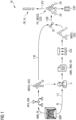
- FIG 1 shows a schematic view including symbols illustrating a method for generating protocol data of a specific medical process according to an embodiment of the invention
- FIG 2 shows a schematic view illustrating the step of generating a first structured medical data object
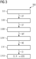
- FIG 3 shows a flow chart diagram illustrating details of the step of generating a first structured medical data object of the method for generating protocol data of a specific medical process according to the invention
- FIG 4 shows a schematic view illustrating the step of generating a second structured medical data object of the method for generating protocol data of a specific medical process according to the invention
- FIG 5 which is divided in FIG 5A and FIG 5B , shows a schematic view illustrating the step of generating a merged structured medical data object of the method for generating protocol data of a specific medical process according to the invention
- FIG 6 shows a schematic view on a protocol data generation device according to an embodiment of the invention
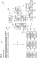
- FIG 7 shows a schematic view including symbols illustrating the method for generating protocol data of a specific medical process according to an embodiment of the invention, wherein peer reviewed radiological report data are generated
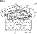
- FIG 8 shows a schematic view on a global radiology knowledge graph, representing the content of a radiological report
- FIG 9 shows a schematic view illustrating a reduction of a global radiology knowledge graph to two different subgraphs
- FIG 10 shows a flow chart diagram illustrating the method for generating protocol data of a specific medical process according to an embodiment of the invention, wherein a peer reviewed protocol is generated
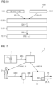
- FIG 11 shows a schematic view on a medical process system according to an embodiment of the invention.
- FIG 1 a scenario 10 illustrating the method for generating protocol data PD of a specific medical process SMP in an embodiment using DICOM SR objects ADS, UDS, CDS as special type of structured medical data objects MDO1, MDO2, MDO3 is illustrated.
- the method includes two different lines of action. A first line for generating a DICOM SR object UDS based on user-generated data represented by step 1.I and a second line for generating an AI-generated DICOM SR object ADS represented by step 1.II.
- the different DICOM SR objects UDS, ADS are merged to a composite result, i.e. to a final composite DICOM SR object CDS.
- the lower part is related to a semi-automatic generation of a DICOM SR object UDS based on user-generated data representing step 1.1 of the method according to the invention and the upper part is assigned to step 1.II, wherein a model based fully automatic generation of an AI-generated DICOM SR object ADS is performed.
- the common source for the different lines of action 1.1, 1.II is a radiology image RI of a patient.
- a radiology image RI represents a documentation of a specific medical process SMP, in this case a medical imaging process.
- the radiology image RI is analysed by a user U, in this special embodiment a radiologist.
- the user U usually dictates or transcribes an impression of the radiology image RI and in particular a description of annotations or segmentations and findings and a summary thereof on a PACS (PACS is an acronym for Picture Archiving and Communication System) using a dictation tool and generates a free-text radiology report RR in this manner.
- PACS Picture Archiving and Communication System
- the free-text radiology report RR is a special type of text-based electronic medical data TMD, which is a special type of user-generated medical data UMD.
- a concept extraction engine CEE For transferring that informal information of a free-text radiology report RR into a structured formalized data structure, a concept extraction engine CEE is used. Such a concept extraction engine CEE automatically extracts formalizable information, i.e. concept items and relationships, which are later discussed in detail, out of the free-text radiology report RR by parsing the free-text radiology report RR. The substep of parsing can be implemented using Natural Language Processing on the free-text radiology report RR. Then, the parsed information is assigned to the structure of a structured medical data object MDO1, in particular a DICOM SR object UDS generated based on user-generated data. Detailed steps of the concept extraction are described in context with FIG 3 and FIG 4 .
- the radiology image RI which is a particular type of automatically created electronic medical data AMD, is also processed based on an AI-model AIM.
- the AI-model AIM defines the rules of an automatic processing algorithm APA.
- the automatic processing algorithm APA is used to automatically generate a second structured medical data object MDO2, in particular an AI-generated DICOM SR object ADS by automatically processing image data of the radiology image RI as shown in the upper part of FIG 1 .
- the automatic processing algorithm APA includes an annotation and finding based on the radiology image RI.
- the AI-generated DICOM SR object ADS and the DICOM SR object UDS generated based on user-generated data are then merged using an SR update engine UE in step 1.III.
- the SR updating includes also an editing of conflicts between the AI-generated DICOM SR object ADS and the DICOM SR object UDS based on user-generated data.
- a final composite DICOM SR object CDS is generated based on the AI-generated DICOM SR object ADS and the DICOM SR object UDS generated based on user-generated data.
- the final composite DICOM SR object CDS is a special type of a third structured medical data object MDO3, which is in turn a special type of protocol data PD. If deviations DV between the AI-generated DICOM SR object ADS and the DICOM SR object UDS generated based on user-generated data exist, these deviations are received and marked in the final composite DICOM SR object CDS.
- the DICOM SR objects ADS, UDS, CDS can be represented as directed acyclic graph objects with vertices including concept items and edges representing the relationships between the concept items as later illustrated in detail in FIG 2 , FIG 4 and FIG 5 .
- the final composited graph or DICOM SR object CDS can be realized such that data of the DICOM SR object UDS generated based on user-generated data always supersedes data of the AI-generated DICOM SR object ADS in times of conflict. If a conflict exists, AI-generated concept items are overwritten by user-generated DICOM SR concept items.
- the graph vertices including the concept items and the edges including relationships between the concept items of the different AI-generated DICOM SR object ADS and the DICOM SR object UDS generated based on user-generated data are merged.
- more advanced graph linking approaches can be implemented such as hierarchical clustering, graph neural networks etc.
- the generated final composite DICOM SR object CDS is archived in an electronic health record EHR of the patient for archival purposes.
- step 1.IV an alert AL for the user U is generated as a special kind of deviation detection reaction DR based on the result of step 1.III.
- deviations DV are also displayed to the user U such that the user U can decide if it is reasonable to amend the user-generated DICOM SR object UDS and in particular the radiology report RR.
- FIG 2 the upper line of action of FIG 1 assigned to step 1.II, comprising the generation of an AI-generated DICOM SR object ADS using an AI-model AIM for processing a radiology image RI, is illustrated in an enlarged view 20.
- the AI generated DICOM SR object ADS comprises nodes or vertices including concept items CI1, CI2, ..., CI11 and edges including relationships R1, R2, R3, R4 between neighboured vertices.
- the AI-generated results are organized in a hierarchical fashion of concept items based on data types as for example coded nomenclature, numeric measurements, spatial and temporal ROI references, automated analysis results, procedure log, radiation report and based on data analysis techniques as for example key object selection etc.
- the AI-generated DICOM SR object ADS includes the following chains of information:
- FIG 3 a flowchart diagram 300 is shown, illustrating detailed steps of concept extraction from text-based medical data TMD, in particular a radiology report RR.
- Concept extraction is used in step 1.1 shown in FIG 1 .
- step 3.1 a clinical word-sense disambiguation for acronyms and abbreviations based on the text of the radiology report RR is performed using natural language processing techniques for generating disambiguated terms DT.
- step 3.II a semantic mapping to standardized terms ST is performed.
- semantically similar terms are mapped to a standardized terminology to transform the similar terms into a computer readable and searchable identifier.
- the most semantically closest standardized term is determined using available ontologies or privately defined dictionaries. Examples of useful tools are a privately curated ontology, for example, a private list of body parts or a standard ontology like RadLex, SNOMED-CT, LOINC or UMLS.
- SNOMED-CT Systematized Nomenclature of Medicine Clinical Terms and is currently one of the most comprehensive and important medical terminologies worldwide.
- LOINC Logical Observation Identifiers Names and Codes and represents a database and a universal standard for identifying medical laboratory observations.
- LOINC applies universal code names and identifiers to medical terminology related to electronic health records. The purpose is to assist in the electronic exchange and gathering of clinical results such as laboratory tests, clinical observations, outcomes management and research.
- step 3.III a machine translation MT of the text of the radiology report is performed. If the reporting language is not English, the incoming disambiguated and semantically mapped text of the radiology report RR needs to be translated into English language. For that purpose, neural machine translation tools and techniques typically built using sequence to sequence models can be used.
- step 3.IV clinical named entity recognition is performed.
- Step 3.IV is performed to locate and classify named entities NE mentioned in unstructured free text into pre-defined categories, also named concepts, such as anatomical entity, clinical finding, imaging modality, imaging observation, procedure, non-anatomical entities etc.
- named entity recognition for analysing a disambiguated free-text report, in particular a disambiguated imaging procedure description of chest x-ray images, named entity recognition groups and categorizes related words such that words assigned to the same group or category can be represented by the same concept item in the next step.
- step 3.V a mapping to concept items CI, i.e. pairs of a concept and a value, is performed.
- concept items CI for DICOM SR objects are generated from the radiology report RR. This is done using dictionary or rule-based methods or using clinical linguisticgrammar methods.
- the concept items CI in a DICOM SR object usually comprise text, numerics, code, image spatial coordinates etc. These concept items CI are shown in FIG 4 .
- the concept items CI are the vertices or nodes of a hierarchical acyclic graph, in particular representing a DICOM SR object UDS.
- step 3.VI relationships R between each concept item CI are established to construct a hierarchical tree structure as supported by the standard of DICOM SR.
- DICOM SR supports relationships R such as: “contains”, “has observation context”, “has concept modifier”, “has properties”, “has acquisition context”, “inferred from” and “selected from” and “finding location”.
- the method for extracting such a relationship in free text can be realized based on rule-based relationship extraction, for example implemented by parts-of-speech-tagging, word sequence patterns or by learning based methods including weakly supervised, supervised, distantly supervised or unsupervised approaches.
- FIG 4 shows an illustration 400 of a full pipeline of transforming an incoming free-text radiology report RR into a DICOM SR object UDS generated based on user-generated data.
- FIG 4 can be understood as a detailed description of the second part of step 1.1 in FIG 1 .
- the free-text radiology report RR is parsed to extract concept items, i.e. coded name value pairs CI1', CI4', ..., CI11' as described in context with FIG 3 .
- concept items i.e. coded name value pairs CI1', CI4', ..., CI11'
- relationships R1', R3', R4' between the concept items CI1', CI4', ..., CI11' are determined.
- the final representation of the DICOM SR object UDS is arranged in form of a hierarchical tree with the imaging procedure as the root vertex represented by the first concept item CI1'.
- FIG 5 which is divided in FIG 5A and FIG 5B , an illustration 50 of a merging process M between an AI-based generated DICOM SR object ADS (in FIG 5A , upper part, already shown in FIG 2 ) and a DICOM SR object UDS (in FIG 5A , lower part, already shown in FIG 4 ) generated based on user-generated data is depicted.
- the result of the merging process M is a final composite DICOM SR object CDS (depicted in FIG 5B ), for example usable for archival in patient records.
- the final composite DICOM SR object CDS is represented as a directed acyclic graph with vertices as the concept items and edges represented by relationships. A simple realization of such a graph merge and edit can be rule based.
- the concept item CI5' differs from the concept item CI5 and both CI5 and CI5' are received in the final composite DICOM SR object CDS, since they are complementary and not contradictory.
- CI7' differs from CI7 and CI7' replaces CI7 in the resulting final composite DICOM SR object CDS, since CI7 and CI7' are contradictory.
- CI9' differs from CI9 and CI9' replaces CI9 in the resulting final composite DICOM SR object CDS, since CI9 and CI9' are contradictory.
- CI10' differs from CI10 and CI10' replaces CI10 in the resulting final composite DICOM SR object CDS, since CI10 and CI10' are contradictory. If there is no contradiction between the concept items generated based on the user-generated data and AI-based generated concept items as it is the case for CI5 and CI5', both concept items are received in the resulting final composite DICOM SR object CDS. If there is a contradiction between a concept item generated based on the user-generated data and an AI-generated concept item as it is the case for CI7, CI9, CI10 and CI7', CI9' and CI10', the concept item generated based on user-generated data replaces the AI-generated concept item.
- results based on user-generated data always supersede AI-generated results in times of conflict.
- the AI-generated result is overwritten by human generated DICOM SR concept items, i.e. DICOM SR concept items generated based on user-generated data.
- the graph nodes, i.e. concept items, and the relationships are merged and both concept items or relationships are received in the resulting structured medical data object, i.e. in particular the final composite DICOM SR object CDS.
- more advanced graph linking approaches can also be implemented such as hierarchical clustering, graph neural networks, etc.
- FIG 6 a schematic view on a protocol data generation device 60 according to an embodiment of the invention is shown.
- the protocol data generation device 60 includes an extraction unit 61.
- the extraction unit 61 is arranged to generate a first structured medical data object MDO1, for example a DICOM SR object, based on user-generated electronic medical data UMD.
- the user-generated electronic medical data UMD may include text-based electronic medical data TMD and in particular a user-generated radiological report RR of a free text related to medical image data of a patient.
- the user-generated electronic medical data UMD may also include electronic medical data generated by voice VEMD or audio-visually generated electronic medical data AVEMD.
- Electronic medical data generated by voice VEMD include a radiology report generated by voice and audio-visually generated electronic medical data AVEMD include a video-based radiology report.
- Part of the protocol data generation device 60 is also a processing unit 62 for generating a second structured medical data object MDO2.
- the second structured medical data object MDO2 is generated by automatically determining findings and annotations based in the above-mentioned medical image data of a radiology image RI, which is a special type of automatically created electronic medical data AMD.
- the determination of findings and annotations is realized using an AI-based processing algorithm.
- the protocol data generation device 60 includes a data generation unit 63 for generating a merged third structured medical data object MDO3 and emits protocol data PD including a merged third structured medical data object MDO3.
- the merged third structured medical data object MDO3 is generated based on the first structured medical data object MDO1 and on the second structured medical data object MDO2.
- the merged third structured medical data object MDO3 includes information about deviations DV between the first and second structured medical data object MDO1, MDO2. Also complements CM, a special type of deviations DV, are indicated by the merged third structured medical data object MDO3.
- FIG 7 a schematic illustration of an AI-based explainable peer review of a radiology report RR which is generated by a user U based on a radiology image RI is shown.
- step 7.1 the incoming radiology report RR is parsed and mapped to a radiology knowledge graph KG representation.
- the radiology image RI is read by a multi-class classifier MC or an AI-based post-processing algorithm (not shown in FIG 7 ) that maps its results to a similar AI-generated radiology knowledge graph AI-KG.
- step 7.III the two radiology knowledge graphs KG, AI-KG are compared using a graph similarity network which outputs a similarity measure. If there is a difference dc between the compared radiology knowledge graphs KG, AI-KG, which is symbolized in FIG 7 with "y”, a difference graph GDV is generated. Further, graph explanation and visualization methods are used to highlight the difference. If there is no difference dc between the compared knowledge graphs KG, AI-KG, which is symbolized in FIG 7 with "n", the radiology report RR is signed off using a sign symbol RS.
- step 7.IV the difference graph GDV is converted to a human readable text using a report generation deep recurrent model. This is presented to the radiologist, i.e. the user U, as a peer-review report PRR for consideration prior to report sign off.
- the peer-review report PRR includes highlighted findings FDH with differences as depicted in the left sub portion of the field representing step 7.IV in FIG 7 .
- FIG 8 a schematic view on a representation of a radiology knowledge graph KG is shown.
- the entities are grouped according to the concepts of organs OG, modalities MD and the corresponding radiographic findings RF.
- different entities or concept items OG1, OG2, ..., OGk represent different types of organs OG.
- Different concept items MD1, MD2, ..., MDm represent different types of modalities MD and different concept items RF1, RF2, ..., RFn represent different types of radiology findings RF.
- the represented organs OG are shown in the following table: OG1 Chest OG2 Breast OGk-1 Brain OGk Shoulder.
- the represented modalities MD are shown in the following table: MD1 CT MD2 MRI MDm X-ray.
- the represented radiographic findings RF are shown in the following table: RF1 Lesion RF2 Phneumothorax RF3 Effusion RF4 Pneumonia RF5 Fracture RF6 Calcification RF7 Inflammation.
- the radiology knowledge graph KG shown in FIG 8 is implemented as a global radiology knowledge graph KG including all combinations of relations between organs OG, modalities MD and the corresponding radiographic findings RF.
- Such a global radiology knowledge graph KG is difficult to explore due to its large size and due to a lack of clinical relevance of a number of nodes to the case at hand.
- FIG 9 a reduction of a global radiology knowledge graph KG to relevant subgraphs KG-BR, KG-S is illustrated.
- a knowledge graph KG-BR representing a reduction to an X-ray image of a breast is illustrated and on the right side of FIG 9 , a knowledge graph KG-S representing a reduction to an MR-image of a shoulder is represented.
- MDm abbreviated by X-ray
- a lesion RF1, a calcification RF6 and an inflammation RF7 are linked with the mentioned organ OG2 and modality MDm.
- KG-S representing a reduction to an MR-image of a shoulder
- only the shoulder OGk is examined using an MR imaging system MD2 (abbreviated by MRI) and only the relevant findings, i.e. an effusion RF3, a fracture RF5, a calcification RF6 and an inflammation RF7 are linked with the mentioned organ OGk and modality MD2.
- the correct combination of attributes i.e. values and concept items inferable from image meta data, in particular from DICOM meta data, is used to extract the right semantically relevant subgraph KG-BR, KG-S from the global radiology knowledge graph KG.
- FIG 10 a flow chart diagram 1000 is shown, illustrating a special embodiment of the method for generating protocol data of a specific medical process according to the invention, wherein an AI-based peer review of a text-based radiology report RR is generated, also named AI-based peer review method.
- step 10.I based on an appropriately selected subgraph structure, the radiology report RR is parsed to extract relationships that have been reported as present or absent or not-reported.
- a dependency graph i.e. a radiology knowledge graph KG from a textual report
- tokenization approaches to identify key parts of speech, named entities such as anatomy, radiographic findings and differential diagnosis.
- Tokenizing, dependency parsing and mapping to the knowledge graph KG by matching detected named entities and relationships, i.e. "present”, “absent”, “not-reported” or “uncertain”, into edge weights can be accomplished by using tools such as Stanford CoreNLP.
- step 10.11 the generation of an AI-generated radiology knowledge graph AI-KG is implemented using computer aided detection tools.
- vertices are discovered and are connected based on relationships also named edges between the vertices. For example, a number of key features that can be extracted using computer aided detection or diagnostic tools are captured as edge weights for determining the relationships.
- the relationships are set based on different rules.
- a rule can include the order that if radiographic findings cooccur in the same anatomy, then those nodes are linked with a bidirectional connection.
- a relationship can also be established based on conditional probabilities estimated using AI-based methods for computer aided detection and diagnosis of particular radiographic findings in body regions of a patient.
- a graph matching to generate a difference graph is realized.
- the two input graphs KG, AI-KG are used for comparison.
- it can be performed graph matching to find all the elements that differ significantly between the two radiology knowledge graphs KG, AI-KG. It can be started by randomly parsing the AI-generated radiology knowledge graph AI-KG into smaller sub-subgraphs and trying to find a match of the sub-subraphs to the subgraph KG reported by the radiologist using the Hungarian algorithm or variants of it. This algorithm is described in https://en.wikipedia.org/wiki/hungarian-algorithm . This can be accomplished using methods such as SimRank, similarity flooding, etc. If a match is found, then the matched subsubgraph is deemed as information already reported. If a match is not found, this is added to the difference graph GDV.
- step 10.IV the difference graph GDV generated in step 10.III is converted into human readable clinical text, i.e. a peer review report PRR, using templates with named entities as placeholders.
- An alternative to rule based methods is to employ deep learning models such with a graph convolution encoder feeding to a text generation recurrent neural network-based decoder.
- step 10.V the peer review report PRR is displayed to the user.
- the explainable AI-based peer review method can be deployed as a micro-service that is triggered in the backend as soon as a particular case is sent to the radiology worklist.
- the medical process system 1 performs a specific medical process.
- the medical process system 1 includes a process unit 2 for applying the specific medical process to a patient P.
- the specific medical process also comprises a measurement process for generating measurement data MDA, for example raw data of an X-ray imaging process.
- the generated measurement data MDA are sent to a documentation unit 3 which is also a part of the medical process system 1.
- the documentation unit 3 for example a reconstruction unit, is arranged to generate automatically created electronic medical data AMD documenting the specific medical process, i.e. the medical imaging.
- the automatically created electronic medical data AMD include a radiology image RI.
- the medical process system 1 also includes a user interface 4 for receiving user-generated electronic medical data UMD created by a user U documenting the specific medical process.
- the user-generated electronic medical data UMD include user-generated annotations generated based on the radiology image RI by the user U.
- the medical process system 1 includes a protocol data generation device 60 according to an embodiment of the invention as it is described in context with FIG 6 .
- the protocol data generation device 60 generates protocol data PD based on the received user-generated electronic medical data UMD and the automatically created electronic medical data AMD. As illustrated in FIG 1 to FIG 5 and FIG 7 to FIG 9 , the protocol data generation device 60 generates as an intermediate step a first structured medical data object MDO1 and a second structured medical data object MDO2 (not shown in FIG 11 ) based on the received user-generated electronic medical data UMD and the automatically created electronic medical data AMD.
- the protocol data generation device 60 can optionally include a deviation detection reaction unit 64 for performing a deviation detection reaction DR, for example an alert AL, based on detected deviations between the first structured medical data object MDO1 and the second structured medical data object MDO2.
- a deviation detection reaction unit 64 for performing a deviation detection reaction DR, for example an alert AL, based on detected deviations between the first structured medical data object MDO1 and the second structured medical data object MDO2.
Landscapes
- Health & Medical Sciences (AREA)
- Engineering & Computer Science (AREA)
- General Health & Medical Sciences (AREA)
- Medical Informatics (AREA)
- Public Health (AREA)
- Epidemiology (AREA)
- Primary Health Care (AREA)
- Nuclear Medicine, Radiotherapy & Molecular Imaging (AREA)
- Radiology & Medical Imaging (AREA)
- Theoretical Computer Science (AREA)
- Biomedical Technology (AREA)
- Data Mining & Analysis (AREA)
- Pathology (AREA)
- Artificial Intelligence (AREA)
- Audiology, Speech & Language Pathology (AREA)
- Computational Linguistics (AREA)
- Physics & Mathematics (AREA)
- General Engineering & Computer Science (AREA)
- General Physics & Mathematics (AREA)
- Databases & Information Systems (AREA)
- Measuring And Recording Apparatus For Diagnosis (AREA)
- Medical Treatment And Welfare Office Work (AREA)
Priority Applications (3)
| Application Number | Priority Date | Filing Date | Title |
|---|---|---|---|
| EP22159795.8A EP4239642A1 (de) | 2022-03-02 | 2022-03-02 | Verfahren zur erzeugung von protokolldaten einer radiologischen bilddatenmessung |
| US18/175,799 US20230282337A1 (en) | 2022-03-02 | 2023-02-28 | Method for generating protocol data of a radiological image data measurement |
| CN202310183276.7A CN116705248A (zh) | 2022-03-02 | 2023-02-28 | 用于生成放射学图像数据测量的协议数据的方法 |
Applications Claiming Priority (1)
| Application Number | Priority Date | Filing Date | Title |
|---|---|---|---|
| EP22159795.8A EP4239642A1 (de) | 2022-03-02 | 2022-03-02 | Verfahren zur erzeugung von protokolldaten einer radiologischen bilddatenmessung |
Publications (1)
| Publication Number | Publication Date |
|---|---|
| EP4239642A1 true EP4239642A1 (de) | 2023-09-06 |
Family
ID=80625267
Family Applications (1)
| Application Number | Title | Priority Date | Filing Date |
|---|---|---|---|
| EP22159795.8A Pending EP4239642A1 (de) | 2022-03-02 | 2022-03-02 | Verfahren zur erzeugung von protokolldaten einer radiologischen bilddatenmessung |
Country Status (3)
| Country | Link |
|---|---|
| US (1) | US20230282337A1 (de) |
| EP (1) | EP4239642A1 (de) |
| CN (1) | CN116705248A (de) |
Families Citing this family (4)
| Publication number | Priority date | Publication date | Assignee | Title |
|---|---|---|---|---|
| EP4537351A1 (de) * | 2022-06-08 | 2025-04-16 | Smart Reporting GmbH | Verfahren und systeme zur erzeugung von texten medizinischer berichte |
| CN119700182A (zh) * | 2023-09-27 | 2025-03-28 | 通用电气精准医疗有限责任公司 | 超声扫描系统的扫描控制方法、装置和超声扫描系统 |
| DE102023211714A1 (de) * | 2023-11-24 | 2025-05-28 | Siemens Healthineers Ag | Verfahren und System zum Bereitstellen eines Datenelements aus einem Datenkorpus |
| WO2025117798A1 (en) * | 2023-11-30 | 2025-06-05 | Elevance Health, Inc. | Cognitive artificial intelligence platform for physicians |
Citations (2)
| Publication number | Priority date | Publication date | Assignee | Title |
|---|---|---|---|---|
| US20200043600A1 (en) * | 2018-08-02 | 2020-02-06 | Imedis Ai Ltd | Systems and methods for improved analysis and generation of medical imaging reports |
| US20200160993A1 (en) * | 2018-11-16 | 2020-05-21 | International Business Machines Corporation | Artificial Intelligence Based Alert System |
-
2022
- 2022-03-02 EP EP22159795.8A patent/EP4239642A1/de active Pending
-
2023
- 2023-02-28 US US18/175,799 patent/US20230282337A1/en active Pending
- 2023-02-28 CN CN202310183276.7A patent/CN116705248A/zh active Pending
Patent Citations (2)
| Publication number | Priority date | Publication date | Assignee | Title |
|---|---|---|---|---|
| US20200043600A1 (en) * | 2018-08-02 | 2020-02-06 | Imedis Ai Ltd | Systems and methods for improved analysis and generation of medical imaging reports |
| US20200160993A1 (en) * | 2018-11-16 | 2020-05-21 | International Business Machines Corporation | Artificial Intelligence Based Alert System |
Non-Patent Citations (29)
| Title |
|---|
| ATALAR, M.OZTOPRAK, B.GUMUS, C.EGILMEZ, H.BULUT, S.: "Classical Signs and Appearances in Pediatric Neuroradiology", EUROPEAN CONGRESS OF RADIOLOGY-ECR, March 2014 (2014-03-01) |
| BANERJEE, I.LING, Y.CHEN, M.C.HASAN, S.A.LANGLOTZ, C.P.MORADZADEH, N.CHAPMAN, B.AMRHEIN, T.MONG, D.RU-BIN, D.L.: "Comparative effectiveness of convolutional neural network (CNN) and recurrent neural network (RNN) architectures for radiology text report classification", ARTIFICIAL INTELLIGENCE IN MEDICINE, vol. 97, 2019, pages 79 - 88, XP085710385, DOI: 10.1016/j.artmed.2018.11.004 |
| BANKO, MICHELE ET AL.: "Open Information Extraction from the Web", CONFERENCE ON ARTIFICIAL INTELLIGENCE, 2007 |
| CALLI, E.SOGANCIOGLU, E.VAN GINNEKEN, B.VAN LEEUWEN, K.G.MURPHY, K.: "Deep learning for chest X-ray analysis: A survey", MEDICAL IMAGE ANALYSIS, vol. 72, 2021, pages 102125, XP086702554, DOI: 10.1016/j.media.2021.102125 |
| CHITICARIU, L.LI, Y.REISS, F.: "Rule-based information extraction is dead! long live rule-based information extraction systems!", PROCEEDINGS OF THE 2013 CONFERENCE ON EMPIRICAL METHODS IN NATURAL LANGUAGE PROCESSING, October 2013 (2013-10-01), pages 827 - 832 |
| DALVI, B. ET AL.: "IKE-an interactive tool for knowledge extraction", PROCEEDINGS OF THE 5TH WORKSHOP ON AUTOMATED KNOWLEDGE BASE CONSTRUCTION, June 2016 (2016-06-01), pages 12 - 17 |
| GRIFFITH, J.F., ROBINSON, P. AND PFIRRMANN, C.W.: "12th International Conference on Data Mining", December 2012, IEEE, article "Learning to refine an automatically extracted knowledge base using Markov logic", pages: 912 - 917 |
| HANSELL, D.M.BANKIER, A.A.MACMAHON, H.MCLOUD, T.C.MULLER, N.L.REMY, J.: "Fleischner Society: glossary of terms for thoracic imaging", RADIOLOGY, vol. 246, no. 3, 2008, pages 697 - 722 |
| HOU ET AL.: "Multi-Label Learning with Visual-Semantic Embedded Knowledge Graph for Diagnosis of Radiology Imaging", IEEE ACCESS, vol. 9, 2021, pages 15720 - 15730, XP011833066, DOI: 10.1109/ACCESS.2021.3052794 |
| HOU, B.KAISSIS, G.SUMMERS, R.M.KAINZ, B.: "International Conference on Medical Image Computing and Computer-Assisted Intervention", September 2021, SPRINGER, article "RATCHET: Medical Transformer for Chest X-ray Diagnosis and Reporting", pages: 293 - 303 |
| KAHN JR, C.E. ET AL.: "Toward best practices in radiology reporting", RADIOLOGY, vol. 252, no. 3, 2009, pages 852 - 856 |
| LAO ET AL.: "Random walk inference and leaning in a large scale knowledge base", PROCEEDINGS OF THE 2011 CONFERENCE ON EMPIRICAL METHODS IN NATURAL LANGUAGE PROCESSING, July 2011 (2011-07-01), pages 529 - 539 |
| LINDBERG, C.: "The Unified Medical Language System (UMLS) of the National Library of Medicine", JOURNAL (AMERICAN MEDICAL RECORD ASSOCIATION, vol. 61, no. 5, 1990, pages 40 - 42 |
| MAHAJAN ET AL.: "Deploying deep learning for quality control: An AI-assisted review of chest X-rays reported as normal in routine clinical practice", RADIOLOGICAL SOCIETY OF NORTH AMERICA 2019 SCIENTIFIC ASSEMBLY AND ANNUAL MEETING, 1 December 2019 (2019-12-01) |
| MANNING, C.D. ET AL.: "The Stanford CoreNLP natural language processing toolkit", PROCEEDINGS OF 52ND ANNUAL MEETING OF THE ASSOCIATION FOR COMPUTATIONAL LINGUISTICS: SYSTEM DEMONSTRATIONS, June 2014 (2014-06-01), pages 55 - 60, XP055678293, DOI: 10.3115/v1/P14-5010 |
| NIKLAUS, C. ET AL.: "A survey on open information extraction", ARXIV: 1806.05599, 2018 |
| PALMER, W., BANCROFT, L., BONAR, F., CHOI, J.A., COTTEN, A., GRIFFITH, J.F., ROBINSON, P., PFIRRMANN, C.W.: "Glossary of terms for musculoskeletal radiology", SKELETAL RADIOLOGY, vol. 49, no. 1, 2020, pages 1 - 33, XP037161938, DOI: 10.1007/s00256-020-03465-1 |
| POWEL, D.K.SILBERZWEIG, J.E.: "State of structured reporting in radiology, a survey", ACADEMIC RADIOLOGY, vol. 22, no. 2, 2015, pages 226 - 233 |
| PUJA ET AL.: "Large-Scale Knowledge Graph Identification Using PSL", AAAI FALL SYMPOSIA, 2013 |
| PYYSALO S.: "Encyclopedia of Systems Biology", 2013, SPRINGER, article "Text Parsing" |
| RADIOLOGY TECHNICAL COMMITTEE, IHE RADIOLOGY TECHNICAL FRAMEWORK SUPPLEMENT, AI RESULTS (AIR) 15 REVISION 1.1 - TRIAL IMPLEMENTATION, 16 July 2020 (2020-07-16), Retrieved from the Internet <URL:https://www.ihe.net/uploadedFiles/Documents/Radiology/IHERAD_Suppl_AIR.pdf> |
| RAO ET AL.: "Utility of artificial intelligence tool as a prospective radiology peer reviewer - Detection of unreported intracranial hemorrhage", ACADEMIC RADIOLOGY, vol. 28, no. 1, pages 85 - 93, XP086384712, DOI: 10.1016/j.acra.2020.01.035 |
| SUCHANEK ET AL.: "Advances in automated knowledge base construction", SIGMOD RECORDS JOURNAL, 2013 |
| TANG, Y.X.TANG, Y.B.PENG, Y.YAN, K.BAGHERI, M.REDD, B.A.BRANDON, C.J.LU, Z.HAN, M.XIAO, J.: "Automated abnormality classification of chest radiographs using deep convolutional neural networks", NPJ DIGITAL MEDICINE, vol. 3, no. 1, 2020, pages 1 - 8 |
| WALTL, B.BONCZEK, G.MATTHES, F.: "Rule-based information extraction: Advantages, limitations, and perspectives", JUSLETTER IT, February 2018 (2018-02-01) |
| WANG ET AL.: "Programming with personalized pagerank: a locally groundable first-order probabilistic logic", PROCEEDINGS OF THE 22ND ACM INTERNATIONAL CONFERENCE ON INFORMATION & KNOWLEDGE MANAGEMENT, October 2013 (2013-10-01), pages 2129 - 2138 |
| WANG, Y.WANG, L.RASTEGAR-MOJARAD, M.MOON, S.SHEN, F.AFZAL, N.LIU, S.ZENG, Y.MEHRABI, S.SOHN, S.: "Clinical information extraction applications: a literature review", JOURNAL OF BIOMEDICAL INFORMATICS, vol. 77, 2018, pages 34 - 49, XP085337350, DOI: 10.1016/j.jbi.2017.11.011 |
| WU, S.ROBERTS, K.DATTA, S.DU, J.JI, Z.SI, Y.SONI, S.WANG, Q.WEI, Q.XIANG, Y.: "Deep learning in clinical natural language processing: a methodical review", JOURNAL OF THE AMERICAN MEDICAL INFORMATICS ASSOCIATION, vol. 27, no. 3, 2020, pages 457 - 470 |
| ZHANG ET AL.: "When radiology report generation meets knowledge graph", PROCEEDINGS OF THE AAAI CONFERENCE ON ARTIFICIAL INTELLIGENCE, vol. 34, no. 07, April 2020 (2020-04-01), pages 12910 - 12917 |
Also Published As
| Publication number | Publication date |
|---|---|
| US20230282337A1 (en) | 2023-09-07 |
| CN116705248A (zh) | 2023-09-05 |
Similar Documents
| Publication | Publication Date | Title |
|---|---|---|
| US20250226068A1 (en) | Complex image data analysis using artificial intelligence and machine learning algorithms | |
| CN113243033B (zh) | 综合诊断系统和方法 | |
| JP5952835B2 (ja) | 撮像プロトコルの更新及び/又はリコメンダ | |
| JP6542664B2 (ja) | 患者情報を臨床基準にマッチングするシステム及び方法 | |
| US11232856B2 (en) | Methods for automatic cohort selection in epidemiologic studies and clinical trials | |
| US20230282337A1 (en) | Method for generating protocol data of a radiological image data measurement | |
| KR20240008838A (ko) | 인공 지능-보조 이미지 분석을 위한 시스템 및 방법 | |
| US11017033B2 (en) | Systems and methods for modeling free-text clinical documents into a hierarchical graph-like data structure based on semantic relationships among clinical concepts present in the documents | |
| RU2686627C1 (ru) | Автоматическое создание ориентированной на показатели продольной области для просмотра показателей пациента | |
| Liu et al. | A systematic review of deep learning-based research on radiology report generation | |
| US20030120458A1 (en) | Patient data mining | |
| US20180032676A1 (en) | Method and system for context-sensitive assessment of clinical findings | |
| Guo et al. | Automatic medical report generation: Methods and applications | |
| CN119742049A (zh) | 用于提供医学成像决策支持数据的方法 | |
| US20210217535A1 (en) | An apparatus and method for detecting an incidental finding | |
| Sharma et al. | XAI-based data visualization in multimodal medical data | |
| US12340894B2 (en) | Recommending at least one imaging protocol for scanning a patient | |
| Manimala et al. | Advancements in deep learning techniques for analyzing electronic medical records | |
| del Mar Roldán-García et al. | Towards an ontology-driven clinical experience sharing ecosystem: Demonstration with liver cases | |
| Xu et al. | Deep disease label-guided graph convolutional network for medical report generation | |
| Deshpande et al. | Ontology-based radiology teaching file summarization, coverage, and integration | |
| Subahi et al. | Fuzzy logic inference system for managing intensive care unit resources based on knowledge graph | |
| Ogungbe et al. | Design and implementation of diagnosis system for cardiomegaly from clinical chest X-ray reports | |
| Fonseca et al. | A Research Agenda on Pediatric Chest X-Ray: Is Deep Learning Still in Childhood? | |
| Strøm et al. | Machine learning performance metrics and diagnostic context in radiology |
Legal Events
| Date | Code | Title | Description |
|---|---|---|---|
| PUAI | Public reference made under article 153(3) epc to a published international application that has entered the european phase |
Free format text: ORIGINAL CODE: 0009012 |
|
| STAA | Information on the status of an ep patent application or granted ep patent |
Free format text: STATUS: REQUEST FOR EXAMINATION WAS MADE |
|
| 17P | Request for examination filed |
Effective date: 20230217 |
|
| AK | Designated contracting states |
Kind code of ref document: A1 Designated state(s): AL AT BE BG CH CY CZ DE DK EE ES FI FR GB GR HR HU IE IS IT LI LT LU LV MC MK MT NL NO PL PT RO RS SE SI SK SM TR |
|
| RAP1 | Party data changed (applicant data changed or rights of an application transferred) |
Owner name: SIEMENS HEALTHINEERS AG |
|
| STAA | Information on the status of an ep patent application or granted ep patent |
Free format text: STATUS: EXAMINATION IS IN PROGRESS |
|
| 17Q | First examination report despatched |
Effective date: 20240812 |