EP4239577B1 - Automatisches verfahren zum ausrichten eines dreidimensionalen dentalbibliotheksmodells auf dreidimensionale mundscandaten und computerlesbares medium mit programm zur durchführung des verfahrens - Google Patents
Automatisches verfahren zum ausrichten eines dreidimensionalen dentalbibliotheksmodells auf dreidimensionale mundscandaten und computerlesbares medium mit programm zur durchführung des verfahrens Download PDFInfo
- Publication number
- EP4239577B1 EP4239577B1 EP23159922.6A EP23159922A EP4239577B1 EP 4239577 B1 EP4239577 B1 EP 4239577B1 EP 23159922 A EP23159922 A EP 23159922A EP 4239577 B1 EP4239577 B1 EP 4239577B1
- Authority
- EP
- European Patent Office
- Prior art keywords
- scan data
- oral scan
- dental
- landmarks
- library model
- Prior art date
- Legal status (The legal status is an assumption and is not a legal conclusion. Google has not performed a legal analysis and makes no representation as to the accuracy of the status listed.)
- Active
Links
Images






Classifications
-
- G—PHYSICS
- G06—COMPUTING OR CALCULATING; COUNTING
- G06T—IMAGE DATA PROCESSING OR GENERATION, IN GENERAL
- G06T7/00—Image analysis
- G06T7/30—Determination of transform parameters for the alignment of images, i.e. image registration
- G06T7/33—Determination of transform parameters for the alignment of images, i.e. image registration using feature-based methods
-
- G—PHYSICS
- G06—COMPUTING OR CALCULATING; COUNTING
- G06T—IMAGE DATA PROCESSING OR GENERATION, IN GENERAL
- G06T7/00—Image analysis
- G06T7/30—Determination of transform parameters for the alignment of images, i.e. image registration
- G06T7/33—Determination of transform parameters for the alignment of images, i.e. image registration using feature-based methods
- G06T7/344—Determination of transform parameters for the alignment of images, i.e. image registration using feature-based methods involving models
-
- G—PHYSICS
- G06—COMPUTING OR CALCULATING; COUNTING
- G06T—IMAGE DATA PROCESSING OR GENERATION, IN GENERAL
- G06T2207/00—Indexing scheme for image analysis or image enhancement
- G06T2207/10—Image acquisition modality
- G06T2207/10028—Range image; Depth image; 3D point clouds
-
- G—PHYSICS
- G06—COMPUTING OR CALCULATING; COUNTING
- G06T—IMAGE DATA PROCESSING OR GENERATION, IN GENERAL
- G06T2207/00—Indexing scheme for image analysis or image enhancement
- G06T2207/20—Special algorithmic details
- G06T2207/20084—Artificial neural networks [ANN]
-
- G—PHYSICS
- G06—COMPUTING OR CALCULATING; COUNTING
- G06T—IMAGE DATA PROCESSING OR GENERATION, IN GENERAL
- G06T2207/00—Indexing scheme for image analysis or image enhancement
- G06T2207/30—Subject of image; Context of image processing
- G06T2207/30004—Biomedical image processing
- G06T2207/30036—Dental; Teeth
Definitions
- the present invention relates to an automated method for aligning a three dimensional (3D) dental library model to 3D oral scan data and a non-transitory computer-readable storage medium having stored thereon program instructions of the automated method for aligning the 3D dental library model to the 3D oral scan data. More particularly, the invention relates to an automated method for aligning a 3D dental library model to 3D oral scan data to reduce time and process of manufacturing prostheses, implants, braces and dental instruments and a non-transitory computer-readable storage medium having stored thereon program instructions of the automated method for aligning the 3D dental library model to the 3D oral scan data.
- 3D oral scan data may refer to data scanned by a 3D scanner for teeth and oral cavity, an impression model of the teeth and the oral cavity or a reconstruction model of the teeth and the oral cavity.
- dental treatment such as prosthetic treatment including in-ray, on-ray and crown, implant and orthodontics oral data of a patient may be obtained and may be used to design prostheses or implants and manufacture braces.
- a dental library model predesigned to some extent for each tooth type may be used.
- the dental library model may be aligned to the 3D oral scan data. If the process of aligning the dental library model to the 3D oral scan data is performed manually, a work fatigue of the dentist or dental technician may increase and an accuracy and a productivity of the result may decrease.
- the 3D oral scan data may refer to data scanned by a 3D scanner for teeth and oral cavity, an impression model of the teeth and the oral cavity or a reconstruction model of the teeth and the oral cavity.
- the 3D oral scan data may be mesh data including 3D vertexes and triangles or rectangles generated by connecting the 3D vertexes.
- the 3D oral scan data may be image data captured by the 3D scanner.
- a filename extension of the 3D oral scan data is not limited, and may be, for example, one of ply, obj and stl.
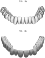
- FIGS. 2A and 2B represent examples of the 3D oral scan data.
- the 3D oral scan data of FIG. 2A may be oral scan data for a mandible and may include all teeth without missing teeth.
- the 3D oral scan data of FIG. 2B may be oral scan data for a maxilla and may include all teeth except for second premolar.
- the dental library model is a kind of sample tooth (a standard tooth) used to manufacture the prostheses, the implants and the braces and may have a typical tooth shape.
- the dental library model may have one sample tooth (the standard tooth) for each tooth number.
- the 3D oral scan data are captured by the 3D scanner so that the 3D oral scan data may have a low degree of completion of the mesh. When the degree of the completion of the mesh is low, a 3D printing may be inappropriate for manufacturing the prostheses, the implants and the braces. In contrast, the 3D dental library model may have a high degree of completion of the mesh.
- the automated method for aligning the 3D dental library model to the 3D oral scan data of the present embodiment may be operated by a computing apparatus.
- the operation (operation S300) of extracting the scan landmarks of the 3D oral scan data may be processed manually by the user or processed automatically through a deep learning.
- a case in which the operation (operation S300) of extracting the scan landmarks of the 3D oral scan data is processed automatically through a deep learning is explained later in detail referring to FIG. 12 .
- the library landmarks of the 3D dental library model may be pre-stored with the 3D dental library model.
- the operation (operation S500) of extracting the library landmarks may be loading the pre-stored library landmarks.
- the library landmarks may be extracted manually by the user or extracted automatically through a deep learning.
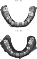
- FIG. 4A is a perspective view illustrating an example of first 3D oral scan data with tooth information.
- FIG. 4B is a perspective view illustrating an example of second 3D oral scan data with tooth information.
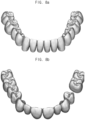
- FIG. 5A is a perspective view illustrating an example of teeth extracted from the first 3D oral scan data.
- FIG. 5B is a perspective view illustrating an example of teeth extracted from the second 3D oral scan data.
- the valid tooth of the 3D oral scan data is determined (operation S200). For example, a tooth type of the valid tooth of the 3D oral scan data may be determined (operation S200).
- a tooth type of the valid tooth of the 3D oral scan data may be determined (operation S200).
- the 3D dental library model is not loaded for the missing tooth. Therefore, the operation of determining the valid tooth of the 3D oral scan data may be preceded.
- individual tooth information of each tooth of the 3D oral scan data may be derived.
- the individual tooth information may include the tooth type (the tooth number), a location, a shape and a boundary between the tooth and gingiva.
- the individual tooth information may be a scalar value or a labeling value expressed on the 3D oral scan data.
- the teeth of the 3D oral scan data may be segmented so that the teeth segmentation data may be generated.
- FIG. 4A shows an example in which the individual tooth information is shaded on individual teeth of the first 3D oral scan data.
- FIG. 4B shows an example in which the individual tooth information is shaded on individual teeth of the second 3D oral scan data.
- FIG. 6A is a perspective view illustrating an example of the first 3D oral scan data with scan landmarks.
- FIG. 6B is a perspective view illustrating an example of the second 3D oral scan data with scan landmarks.
- FIG. 7A is a perspective view illustrating teeth segmentation data of the first 3D oral scan data with the scan landmarks.
- FIG. 7B is a perspective view illustrating teeth segmentation data of the second 3D oral scan data with the scan landmarks.
- FIGS. 1 to 7B the scan landmarks of the 3D oral scan data are extracted (operation S300).
- FIG. 6A shows an example of the first 3D oral scan data in which the scan landmarks are illustrated.
- FIG. 6B shows an example of the second 3D oral scan data in which the scan landmarks are illustrated.
- FIG. 7A shows an example of first teeth segmentation data in which the scan landmarks are illustrated.
- FIG. 7B shows an example of second teeth segmentation data in which the scan landmarks are illustrated.
- the scan landmarks may include at least three landmarks disposed in the 3D oral scan data.
- the scan landmarks may be disposed on an upper surface of the teeth, on a lateral surface of the teeth or inside the teeth.
- the scan landmarks may include a first landmark disposed at a first end of a first arch formed by the teeth of the 3D oral scan data, a second landmark disposed at a second end of the first arch and a third landmark disposed at a central point of the first arch.
- the scan landmarks may include a first landmark disposed at a last tooth in a first end in a horizontal direction of the 3D oral scan data, a second landmark disposed at a last tooth in a second end in the horizontal direction of the 3D oral scan data and a third landmark disposed at a center of two central incisors of the 3D oral scan data.
- FIG. 8A is a perspective view illustrating a first 3D dental library model corresponding to the first 3D oral scan data.
- FIG. 8B is a perspective view illustrating a second 3D dental library model corresponding to the second 3D oral scan data.
- the dental library model corresponding to the valid tooth of the 3D oral scan data is loaded (operation S400).
- the 3D oral scan data include all teeth
- the 3D dental library models of all teeth are loaded.
- the 3D dental library models of all teeth except for the missing tooth are loaded.
- FIG. 8A illustrates the first 3D dental library model corresponding to the first 3D oral scan data of FIG. 2A .
- FIG. 8B illustrates the second 3D dental library model corresponding to the second 3D oral scan data of FIG. 2B .
- the second 3D oral scan data of FIG. 2B does not include the second premolar so that the second 3D dental library model does not include a second premolar corresponding to the missing second premolar of the second 3D oral scan data.
- FIG. 9A is a perspective view illustrating the first 3D dental library model with library landmarks.
- FIG. 9B is a perspective view illustrating the second 3D dental library model with library landmarks.
- FIGS. 1 to 9B the library landmarks of the 3D dental library model are extracted (operation S500).
- FIG. 9A shows an example of the first 3D dental library model in which the library landmarks are illustrated.
- FIG. 9B shows an example of the second 3D dental library model in which the library landmarks are illustrated.
- the library landmarks may include a fourth landmark disposed at a first end of a second arch formed by the teeth of the 3D dental library model, a fifth landmark disposed at a second end of the second arch and a sixth landmark disposed at a central point of the second arch.
- the library landmarks may include a fourth landmark disposed at a last tooth in a first end in a horizontal direction of the 3D dental library model, a fifth landmark disposed at a last tooth in a second end in the horizontal direction of the 3D dental library model and a third landmark disposed at a center of two central incisors of the 3D dental library model.
- FIG. 10A is a perspective view illustrating the first 3D oral scan data and the first 3D dental library model which are automatically aligned.
- FIG. 10B is a perspective view illustrating the second 3D oral scan data and the second 3D dental library model which are automatically aligned.
- the 3D dental library model is initially aligned to the 3D oral scan data using the scan landmarks and the library landmarks (operation S600).
- the axes of the 3D dental library model and the axes of the 3D oral scan data may be aligned using the scan landmarks and the library landmarks.
- translation, rotation and scaling may be applied to the 3D dental library model.
- a size of the dental library model may be reduced as a whole.
- the size of the dental library model may be enlarged as a whole.
- the second arch formed by the teeth of the 3D dental library model may be matched with the first arch formed by the teeth of the 3D oral scan data.
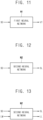
- the first neural network N1 may be used in the operation (operation S200) of determining the valid tooth of the 3D oral scan data and the second neural network N2 different from the first neural network N1 may be used in the operation (operation S300) of extracting the scan landmarks of the 3D oral scan data.
- the first neural network N1 and the second neural network may be artificial intelligence neural networks.
- the first neural network N1 and the second neural network are convolutional neural networks.
- An input of the first neural network N1 may be the 3D oral scan data SD and an output of the first neural network N1 may be the individual tooth information VT of the 3D oral scan data.
- the individual tooth information VT may be a scalar value or a labeling value expressed on the 3D oral scan data.
- the input of the first neural network N1 may be the 3D oral scan data SD and the output of the first neural network N1 may be the teeth segmentation data VT of the 3D oral scan data including segmented teeth of the 3D oral scan data.
- the operation (operation S300) of extracting the scan landmarks of the 3D oral scan data SD may include generating a two dimensional (2D) depth image based on the 3D oral scan data SD, extracting 2D coordinates of the scan landmarks SL from the 2D depth image using the second neural network N2 and inverse-projecting the 2D coordinates to the 3D oral scan data SD.
- the input of the second neural network N2 may be the 2D depth image generated based on the 3D oral scan data SD and the output of the second neural network N2 may be the 2D coordinates of the scan landmarks SL.
- FIG. 13 is a block diagram illustrating a second neural network N2 determining scan landmarks from the 3D oral scan data and library landmarks from the 3D dental library model.
- Inputs of the second neural network N2 may be the 3D oral scan data SD and the 3D dental library model LM and outputs of the second neural network N2 may be the 3D coordinates of the scan landmarks SL and the 3D coordinates of the library landmarks LL.
- the operation (operation S300) of extracting the scan landmarks of the 3D oral scan data SD may include generating a two dimensional (2D) depth image based on the 3D oral scan data SD, extracting 2D coordinates of the scan landmarks SL from the 2D depth image and inverse-projecting the 2D coordinates to the 3D oral scan data SD.
- the input of the second neural network N2 may be the 2D depth image generated based on the 3D oral scan data SD and the output of the second neural network N2 may be the 2D coordinates of the scan landmarks SL.
- the operation (operation S500) of extracting the library landmarks of the 3D dental library model LM may include generating a 2D depth image based on the 3D dental library model LM, extracting 2D coordinates of the library landmarks LL from the 2D depth image and inverse-projecting the 2D coordinates to the 3D dental library model LM.
- the input of the second neural network N2 may be the 2D depth image generated based on the 3D dental library model LM and the output of the second neural network N2 may be the 2D coordinates of the library landmarks LL.
- the process of aligning the 3D dental library model to the 3D oral scan data is performed automatically so that a work fatigue of the dentist or dental technician may decrease and an accuracy of the aligning result may increase.
Landscapes
- Engineering & Computer Science (AREA)
- Computer Vision & Pattern Recognition (AREA)
- Physics & Mathematics (AREA)
- General Physics & Mathematics (AREA)
- Theoretical Computer Science (AREA)
- Dental Tools And Instruments Or Auxiliary Dental Instruments (AREA)
Claims (13)
- Automatisiertes Verfahren zum Ausrichten eines 3D-Dentalbibliotheksmodells auf 3D-Mundscandaten, wobei das Verfahren umfasst:Bestimmen (S200) gültiger Zähne aus den 3D-Mundscandaten, wobei gültige Zähne Zähne sind, die nicht fehlen;Extrahieren (S300) von Scan-Landmarken aus den 3D-Mundscandaten;Laden (S400) eines Dentalbibliotheksmodells, das den gültigen Zähnen der 3D-Mundscandaten entspricht, wobei das Dentalbibliotheksmodell Standardzähne einer typischen Form umfasst;Extrahieren (S500) von Bibliotheks-Landmarken des 3D-Dentalbibliotheksmodells;Initialausrichten (S600) des 3D-Dentalbibliotheksmodells auf die 3D-Mundscandaten unter Verwendung der Scan-Landmarken und der Bibliotheks-Landmarken; undAbgleichen (S700) eines einzelnen Zahns des 3D-Dentalbibliotheksmodells und eines einzelnen Zahns der 3D-Mundscandaten.
- Verfahren nach Anspruch 1, wobei das Initialausrichten (S600) des 3D-Dentalbibliotheksmodells auf die 3D-Mundscandaten umfasst:
Abgleichen (S700) eines zweiten Bogens, der durch Zähne des 3D-Dentalbibliotheksmodells gebildet wird, mit einem ersten Bogen, der durch Zähne der 3D-Mundscandaten gebildet wird. - Verfahren nach Anspruch 1, wobei die Scan-Landmarken zumindest drei Landmarken beinhalten, die in den 3D-Mundscandaten angeordnet sind, und wobei die Bibliotheks-Landmarken zumindest drei Landmarken beinhalten, die in dem 3D-Dentalbibliotheksmodell angeordnet sind.
- Verfahren nach Anspruch 3, wobei die Scan-Landmarken eine erste Landmarke, die an einem ersten Ende eines ersten Bogens angeordnet ist, der durch Zähne der 3D-Mundscandaten gebildet wird, eine zweite Landmarke, die an einem zweiten Ende des ersten Bogens angeordnet ist, und eine dritte Landmarke, die an einem zentralen Punkt des ersten Bogens angeordnet ist, beinhalten, und
wobei die Bibliotheks-Landmarken eine vierte Landmarke, die an einem ersten Ende eines zweiten Bogens angeordnet ist, der durch Zähne des 3D-Dentalbibliotheksmodells gebildet wird, eine fünfte Landmarke, die an einem zweiten Ende des zweiten Bogens angeordnet ist, und eine sechste Landmarke, die an einem zentralen Punkt des zweiten Bogens angeordnet ist, beinhalten. - Verfahren nach Anspruch 3, wobei die Scan-Landmarken eine erste Landmarke, die an einem letzten Zahn an einem ersten Ende in horizontaler Richtung der 3D-Mundscandaten angeordnet ist, eine zweite Landmarke, die an einem letzten Zahn an einem zweiten Ende in horizontaler Richtung der 3D-Mundscandaten angeordnet ist, und eine dritte Landmarke, die im Zentrum der beiden mittleren Schneidezähne der 3D-Mundscandaten angeordnet ist, beinhalten, und
wobei die Bibliotheks-Landmarken eine vierte Landmarke, die an einem letzten Zahn an einem ersten Ende in horizontaler Richtung des 3D-Dentalbibliotheksmodells angeordnet ist, eine fünfte Landmarke, die an einem letzten Zahn an einem zweiten Ende in horizontaler Richtung des 3D-Dentalbibliotheksmodells angeordnet ist, und eine dritte Landmarke, die im Zentrum der beiden mittleren Schneidezähne des 3D-Dentalbibliotheksmodells angeordnet ist, beinhalten. - Verfahren nach Anspruch 1, wobei das Abgleichen (S700) des einzelnen Zahns des 3D-Dentalbibliotheksmodells und des einzelnen Zahns der 3D-Mundscandaten umfasst:
Abgleichen eines Zentrums des einzelnen Zahns des 3D-Dentalbibliotheksmodells mit einem Zentrum des einzelnen Zahns der 3D-Mundscandaten. - Verfahren nach Anspruch 6, wobei das Abgleichen (S700) des einzelnen Zahns des 3D-Dentalbibliotheksmodells und des einzelnen Zahns der 3D-Mundscandaten umfasst:
Drehen des einzelnen Zahns des 3D-Dentalbliotheksmodells und Anpassen einer Größe des einzelnen Zahns des 3D-Dentalbibliotheksmodells, um eine Differenz zwischen einem Winkel des einzelnen Zahns des 3D-Dentalbibliotheksmodells und einem Winkel des einzelnen Zahns der 3D-Mundscandaten und eine Differenz zwischen der Größe des einzelnen Zahns des 3D-Dentalbibliotheksmodells und einer Größe des einzelnen Zahns der 3D-Mundscandaten zu minimieren. - Verfahren nach Anspruch 1, wobei ein erstes neuronales Netzwerk bei der Bestimmung (S200) der gültigen Zähne der 3D-Mundscandaten verwendet wird, wobei eine Eingabe des ersten neuronalen Netzwerks die 3D-Mundscandaten ist und eine Ausgabe des ersten neuronalen Netzwerks individuelle Zahninformationen der 3D-Mundscandaten oder Zahnsegmentierungsdaten der 3D-Mundscandaten einschließlich segmentierter Zähne der 3D-Mundscandaten ist, undwobei ein zweites neuronales Netzwerk, das sich von dem ersten neuronalen Netzwerk unterscheidet, beim Extrahieren (S300) der Scan-Landmarken der 3D-Mundscandaten verwendet wird,wobei das erste neuronale Netzwerk (N1) und das zweite neuronale Netzwerk (N2) Convolutional Neural Netzworks (auf Deutsch etwa: faltende neuronale Netzwerke) sind.
- Verfahren nach Anspruch 8, wobei die individuellen Zahninformationen ein Skalarwert oder ein Kennzeichnungswert sind, der auf den 3D-Mundscandaten dargestellt ist.
- Verfahren nach Anspruch 8, wobei eine Eingabe des zweiten neuronalen Netzwerks die 3D-Mundscandaten ist und eine Ausgabe des zweiten neuronalen Netzwerks 3D-Koordinaten der Scan-Orientierungspunkte ist.
- Verfahren nach Anspruch 8, wobei das Extrahieren (S300) der Scan-Landmarken der 3D-Mundscandaten umfasst:Erzeugen eines 2D-Tiefenbildes basierend auf den 3D-Mundscandaten; Extrahieren von 2D-Koordinaten der Scan-Landmarken aus dem 2D-Tiefenbild unter Verwendung des zweiten neuronalen Netzes; undinverse Projektion der 2D-Koordinaten auf die 3D-Mundscandaten.
- Verfahren nach Anspruch 8, wobei das zweite neuronale Netzwerk beim Extrahieren (S500) der Bibliotheks-Landmarken des 3D-Dentalbibliotheksmodells verwendet wird.
- Nichtflüchtiges, computerlesbares Speichermedium, auf dem Programmanweisungen gespeichert sind, die, wenn sie von zumindest einem Hardwareprozessor ausgeführt werden, den Prozessor veranlassen, das Verfahren nach Anspruch 1 auszuführen.
Applications Claiming Priority (2)
| Application Number | Priority Date | Filing Date | Title |
|---|---|---|---|
| KR1020220028316A KR102522720B1 (ko) | 2022-03-04 | 2022-03-04 | 3차원 구강 스캔 데이터에 3차원 치아 라이브러리 모델을 자동 정렬하는 방법 및 이를 컴퓨터에서 실행시키기 위한 프로그램이 기록된 컴퓨터로 읽을 수 있는 기록 매체 |
| PCT/KR2022/003320 WO2023167356A1 (ko) | 2022-03-04 | 2022-03-08 | 3차원 구강 스캔 데이터에 3차원 치아 라이브러리 모델을 자동 정렬하는 방법 및 이를 컴퓨터에서 실행시키기 위한 프로그램이 기록된 컴퓨터로 읽을 수 있는 기록 매체 |
Publications (3)
| Publication Number | Publication Date |
|---|---|
| EP4239577A1 EP4239577A1 (de) | 2023-09-06 |
| EP4239577B1 true EP4239577B1 (de) | 2025-07-02 |
| EP4239577C0 EP4239577C0 (de) | 2025-07-02 |
Family
ID=85461729
Family Applications (1)
| Application Number | Title | Priority Date | Filing Date |
|---|---|---|---|
| EP23159922.6A Active EP4239577B1 (de) | 2022-03-04 | 2023-03-03 | Automatisches verfahren zum ausrichten eines dreidimensionalen dentalbibliotheksmodells auf dreidimensionale mundscandaten und computerlesbares medium mit programm zur durchführung des verfahrens |
Country Status (2)
| Country | Link |
|---|---|
| EP (1) | EP4239577B1 (de) |
| ES (1) | ES3037825T3 (de) |
Family Cites Families (1)
| Publication number | Priority date | Publication date | Assignee | Title |
|---|---|---|---|---|
| WO2021210966A1 (ko) * | 2020-04-16 | 2021-10-21 | 이마고웍스 주식회사 | 딥러닝을 이용한 3차원 의료 영상 데이터의 특징점 자동 검출 방법 및 장치, 치과용 3차원 데이터 위치 정렬 자동화 방법, 치과용 3차원 데이터 위치 정렬 자동화 방법, 치과용 3차원 스캔 데이터의 랜드마크 자동 검출 방법, 3차원 치과 ct 영상과 3차원 디지털 인상 모델의 정합 정확도 판단 방법 및 상기 방법들을 컴퓨터에서 실행시키기 위한 프로그램이 기록된 컴퓨터로 읽을 수 있는 기록 매체 |
-
2023
- 2023-03-03 ES ES23159922T patent/ES3037825T3/es active Active
- 2023-03-03 EP EP23159922.6A patent/EP4239577B1/de active Active
Also Published As
| Publication number | Publication date |
|---|---|
| ES3037825T3 (en) | 2025-10-07 |
| EP4239577A1 (de) | 2023-09-06 |
| EP4239577C0 (de) | 2025-07-02 |
Similar Documents
| Publication | Publication Date | Title |
|---|---|---|
| US12307693B2 (en) | Automated method for aligning three dimensional dental library model to three dimensional oral scan data and computer readable medium having program for performing the method | |
| JP7386215B2 (ja) | 歯列メッシュ矯正具除去のための方法および装置 | |
| EP4501275A1 (de) | Verfahren zum automatischen ausrichten eines dreidimensionalen einzelzahnmodells auf dreidimensionalen mundscandaten und computerlesbares aufzeichnungsmedium | |
| EP1500034B1 (de) | Verfahren und vorrichtung zum registrieren eines bekannten digitalen objekts auf ein gescanntes 3-d-modell | |
| US8135569B2 (en) | System and method for three-dimensional complete tooth modeling | |
| US11704819B2 (en) | Apparatus and method for aligning 3-dimensional data | |
| EP3108850A2 (de) | Erzeugung eines designs für ein zahnrestauratives produkt aus zahnbildern | |
| US20060127854A1 (en) | Image based dentition record digitization | |
| US20100167243A1 (en) | System and method for automatic construction of realistic looking tooth roots | |
| JP2018530372A (ja) | 歯科保存修復治療に用いるための歯の融通性のあるアーチモデルを作成する方法 | |
| CN105447908A (zh) | 基于口腔扫描数据和cbct数据的牙列模型生成方法 | |
| JP2002543918A (ja) | 歯観察システム | |
| JP2025525610A (ja) | 歯科修復自動化 | |
| Zhang et al. | Computer-aided design of dental inlay restoration based on dual-factor constrained deformation | |
| CN118475321A (zh) | 用于设计牙修复体的系统和方法 | |
| JP2025120478A (ja) | 3次元スキャンデータから補綴物を自動で生成する方法、及びこれをコンピュータで実行させるためのプログラムが記録されたコンピュータ読取り可能な記録媒体 | |
| US12586333B2 (en) | Automated method for generating prosthesis from three dimensional scan data, apparatus generating prosthesis from three dimensional scan data and computer readable medium having program for performing the method | |
| EP4239577A1 (de) | Automatisches verfahren zum ausrichten eines dreidimensionalen dentalbibliotheksmodells auf dreidimensionale mundscandaten und computerlesbares medium mit programm zur durchführung des verfahrens | |
| US12396831B2 (en) | Manufacturing of orthodontic devices based on standardized scan representations | |
| KR102916714B1 (ko) | 치아 라이브러리 모델을 이용한 3차원 구강 스캔 데이터의 치아 분리 방법 및 이를 컴퓨터에서 실행시키기 위한 프로그램이 기록된 컴퓨터로 읽을 수 있는 기록 매체 | |
| US20260130744A1 (en) | Automated method for aligning three dimensional single tooth model to three dimensional oral scan data and computer readable medium having program for performing the method | |
| KR102733517B1 (ko) | 임플란트 식립 가이드의 3차원 가이드 모델링 파일 생성 방법 | |
| JP2026040383A (ja) | 補綴物製作のためのユーザインタフェース提供方法、装置、及びこれをコンピュータで実行させるためのプログラムが記録されたコンピュータ読取り可能な記録媒体 |
Legal Events
| Date | Code | Title | Description |
|---|---|---|---|
| PUAI | Public reference made under article 153(3) epc to a published international application that has entered the european phase |
Free format text: ORIGINAL CODE: 0009012 |
|
| STAA | Information on the status of an ep patent application or granted ep patent |
Free format text: STATUS: REQUEST FOR EXAMINATION WAS MADE |
|
| 17P | Request for examination filed |
Effective date: 20230303 |
|
| AK | Designated contracting states |
Kind code of ref document: A1 Designated state(s): AL AT BE BG CH CY CZ DE DK EE ES FI FR GB GR HR HU IE IS IT LI LT LU LV MC ME MK MT NL NO PL PT RO RS SE SI SK SM TR |
|
| RBV | Designated contracting states (corrected) |
Designated state(s): AL AT BE BG CH CY CZ DE DK EE ES FI FR GB GR HR HU IE IS IT LI LT LU LV MC ME MK MT NL NO PL PT RO RS SE SI SK SM TR |
|
| GRAP | Despatch of communication of intention to grant a patent |
Free format text: ORIGINAL CODE: EPIDOSNIGR1 |
|
| STAA | Information on the status of an ep patent application or granted ep patent |
Free format text: STATUS: GRANT OF PATENT IS INTENDED |
|
| INTG | Intention to grant announced |
Effective date: 20250221 |
|
| GRAS | Grant fee paid |
Free format text: ORIGINAL CODE: EPIDOSNIGR3 |
|
| GRAA | (expected) grant |
Free format text: ORIGINAL CODE: 0009210 |
|
| STAA | Information on the status of an ep patent application or granted ep patent |
Free format text: STATUS: THE PATENT HAS BEEN GRANTED |
|
| AK | Designated contracting states |
Kind code of ref document: B1 Designated state(s): AL AT BE BG CH CY CZ DE DK EE ES FI FR GB GR HR HU IE IS IT LI LT LU LV MC ME MK MT NL NO PL PT RO RS SE SI SK SM TR |
|
| REG | Reference to a national code |
Ref country code: GB Ref legal event code: FG4D |
|
| REG | Reference to a national code |
Ref country code: CH Ref legal event code: EP |
|
| REG | Reference to a national code |
Ref country code: DE Ref legal event code: R096 Ref document number: 602023004387 Country of ref document: DE |
|
| REG | Reference to a national code |
Ref country code: IE Ref legal event code: FG4D |
|
| U01 | Request for unitary effect filed |
Effective date: 20250723 |
|
| U07 | Unitary effect registered |
Designated state(s): AT BE BG DE DK EE FI FR IT LT LU LV MT NL PT RO SE SI Effective date: 20250729 |
|
| REG | Reference to a national code |
Ref country code: ES Ref legal event code: FG2A Ref document number: 3037825 Country of ref document: ES Kind code of ref document: T3 Effective date: 20251007 |
|
| PG25 | Lapsed in a contracting state [announced via postgrant information from national office to epo] |
Ref country code: IS Free format text: LAPSE BECAUSE OF FAILURE TO SUBMIT A TRANSLATION OF THE DESCRIPTION OR TO PAY THE FEE WITHIN THE PRESCRIBED TIME-LIMIT Effective date: 20251102 |
|
| PG25 | Lapsed in a contracting state [announced via postgrant information from national office to epo] |
Ref country code: NO Free format text: LAPSE BECAUSE OF FAILURE TO SUBMIT A TRANSLATION OF THE DESCRIPTION OR TO PAY THE FEE WITHIN THE PRESCRIBED TIME-LIMIT Effective date: 20251002 |
|
| PG25 | Lapsed in a contracting state [announced via postgrant information from national office to epo] |
Ref country code: HR Free format text: LAPSE BECAUSE OF FAILURE TO SUBMIT A TRANSLATION OF THE DESCRIPTION OR TO PAY THE FEE WITHIN THE PRESCRIBED TIME-LIMIT Effective date: 20250702 |
|
| PG25 | Lapsed in a contracting state [announced via postgrant information from national office to epo] |
Ref country code: GR Free format text: LAPSE BECAUSE OF FAILURE TO SUBMIT A TRANSLATION OF THE DESCRIPTION OR TO PAY THE FEE WITHIN THE PRESCRIBED TIME-LIMIT Effective date: 20251003 |
|
| PG25 | Lapsed in a contracting state [announced via postgrant information from national office to epo] |
Ref country code: CZ Free format text: LAPSE BECAUSE OF FAILURE TO SUBMIT A TRANSLATION OF THE DESCRIPTION OR TO PAY THE FEE WITHIN THE PRESCRIBED TIME-LIMIT Effective date: 20250702 |
|
| PG25 | Lapsed in a contracting state [announced via postgrant information from national office to epo] |
Ref country code: PL Free format text: LAPSE BECAUSE OF FAILURE TO SUBMIT A TRANSLATION OF THE DESCRIPTION OR TO PAY THE FEE WITHIN THE PRESCRIBED TIME-LIMIT Effective date: 20250702 |
|
| PG25 | Lapsed in a contracting state [announced via postgrant information from national office to epo] |
Ref country code: RS Free format text: LAPSE BECAUSE OF FAILURE TO SUBMIT A TRANSLATION OF THE DESCRIPTION OR TO PAY THE FEE WITHIN THE PRESCRIBED TIME-LIMIT Effective date: 20251002 |
|
| U20 | Renewal fee for the european patent with unitary effect paid |
Year of fee payment: 4 Effective date: 20260224 |
|
| PG25 | Lapsed in a contracting state [announced via postgrant information from national office to epo] |
Ref country code: SM Free format text: LAPSE BECAUSE OF FAILURE TO SUBMIT A TRANSLATION OF THE DESCRIPTION OR TO PAY THE FEE WITHIN THE PRESCRIBED TIME-LIMIT Effective date: 20250702 |
|
| PG25 | Lapsed in a contracting state [announced via postgrant information from national office to epo] |
Ref country code: SK Free format text: LAPSE BECAUSE OF FAILURE TO SUBMIT A TRANSLATION OF THE DESCRIPTION OR TO PAY THE FEE WITHIN THE PRESCRIBED TIME-LIMIT Effective date: 20250702 |
|
| PLBE | No opposition filed within time limit |
Free format text: ORIGINAL CODE: 0009261 |
|
| STAA | Information on the status of an ep patent application or granted ep patent |
Free format text: STATUS: NO OPPOSITION FILED WITHIN TIME LIMIT |