EP4226840A1 - Medical image processing device and operation method for same - Google Patents
Medical image processing device and operation method for same Download PDFInfo
- Publication number
- EP4226840A1 EP4226840A1 EP21877348.9A EP21877348A EP4226840A1 EP 4226840 A1 EP4226840 A1 EP 4226840A1 EP 21877348 A EP21877348 A EP 21877348A EP 4226840 A1 EP4226840 A1 EP 4226840A1
- Authority
- EP
- European Patent Office
- Prior art keywords
- image
- designated
- category
- medical
- images
- Prior art date
- Legal status (The legal status is an assumption and is not a legal conclusion. Google has not performed a legal analysis and makes no representation as to the accuracy of the status listed.)
- Pending
Links
Images





Classifications
-
- G—PHYSICS
- G16—INFORMATION AND COMMUNICATION TECHNOLOGY [ICT] SPECIALLY ADAPTED FOR SPECIFIC APPLICATION FIELDS
- G16H—HEALTHCARE INFORMATICS, i.e. INFORMATION AND COMMUNICATION TECHNOLOGY [ICT] SPECIALLY ADAPTED FOR THE HANDLING OR PROCESSING OF MEDICAL OR HEALTHCARE DATA
- G16H30/00—ICT specially adapted for the handling or processing of medical images
- G16H30/40—ICT specially adapted for the handling or processing of medical images for processing medical images, e.g. editing
-
- G—PHYSICS
- G16—INFORMATION AND COMMUNICATION TECHNOLOGY [ICT] SPECIALLY ADAPTED FOR SPECIFIC APPLICATION FIELDS
- G16H—HEALTHCARE INFORMATICS, i.e. INFORMATION AND COMMUNICATION TECHNOLOGY [ICT] SPECIALLY ADAPTED FOR THE HANDLING OR PROCESSING OF MEDICAL OR HEALTHCARE DATA
- G16H30/00—ICT specially adapted for the handling or processing of medical images
-
- A—HUMAN NECESSITIES
- A61—MEDICAL OR VETERINARY SCIENCE; HYGIENE
- A61B—DIAGNOSIS; SURGERY; IDENTIFICATION
- A61B1/00—Instruments for performing medical examinations of the interior of cavities or tubes of the body by visual or photographical inspection, e.g. endoscopes; Illuminating arrangements therefor
- A61B1/00002—Operational features of endoscopes
- A61B1/00004—Operational features of endoscopes characterised by electronic signal processing
- A61B1/00009—Operational features of endoscopes characterised by electronic signal processing of image signals during a use of endoscope
- A61B1/000094—Operational features of endoscopes characterised by electronic signal processing of image signals during a use of endoscope extracting biological structures
-
- A—HUMAN NECESSITIES
- A61—MEDICAL OR VETERINARY SCIENCE; HYGIENE
- A61B—DIAGNOSIS; SURGERY; IDENTIFICATION
- A61B1/00—Instruments for performing medical examinations of the interior of cavities or tubes of the body by visual or photographical inspection, e.g. endoscopes; Illuminating arrangements therefor
- A61B1/00002—Operational features of endoscopes
- A61B1/00004—Operational features of endoscopes characterised by electronic signal processing
- A61B1/00009—Operational features of endoscopes characterised by electronic signal processing of image signals during a use of endoscope
- A61B1/000096—Operational features of endoscopes characterised by electronic signal processing of image signals during a use of endoscope using artificial intelligence
-
- A—HUMAN NECESSITIES
- A61—MEDICAL OR VETERINARY SCIENCE; HYGIENE
- A61B—DIAGNOSIS; SURGERY; IDENTIFICATION
- A61B1/00—Instruments for performing medical examinations of the interior of cavities or tubes of the body by visual or photographical inspection, e.g. endoscopes; Illuminating arrangements therefor
- A61B1/00002—Operational features of endoscopes
- A61B1/00039—Operational features of endoscopes provided with input arrangements for the user
- A61B1/0004—Operational features of endoscopes provided with input arrangements for the user for electronic operation
-
- A—HUMAN NECESSITIES
- A61—MEDICAL OR VETERINARY SCIENCE; HYGIENE
- A61B—DIAGNOSIS; SURGERY; IDENTIFICATION
- A61B1/00—Instruments for performing medical examinations of the interior of cavities or tubes of the body by visual or photographical inspection, e.g. endoscopes; Illuminating arrangements therefor
- A61B1/00002—Operational features of endoscopes
- A61B1/00043—Operational features of endoscopes provided with output arrangements
- A61B1/00045—Display arrangement
- A61B1/0005—Display arrangement combining images e.g. side-by-side, superimposed or tiled
-
- G—PHYSICS
- G06—COMPUTING OR CALCULATING; COUNTING
- G06V—IMAGE OR VIDEO RECOGNITION OR UNDERSTANDING
- G06V10/00—Arrangements for image or video recognition or understanding
- G06V10/20—Image preprocessing
- G06V10/25—Determination of region of interest [ROI] or a volume of interest [VOI]
-
- G—PHYSICS
- G06—COMPUTING OR CALCULATING; COUNTING
- G06V—IMAGE OR VIDEO RECOGNITION OR UNDERSTANDING
- G06V10/00—Arrangements for image or video recognition or understanding
- G06V10/70—Arrangements for image or video recognition or understanding using pattern recognition or machine learning
- G06V10/764—Arrangements for image or video recognition or understanding using pattern recognition or machine learning using classification, e.g. of video objects
-
- G—PHYSICS
- G16—INFORMATION AND COMMUNICATION TECHNOLOGY [ICT] SPECIALLY ADAPTED FOR SPECIFIC APPLICATION FIELDS
- G16H—HEALTHCARE INFORMATICS, i.e. INFORMATION AND COMMUNICATION TECHNOLOGY [ICT] SPECIALLY ADAPTED FOR THE HANDLING OR PROCESSING OF MEDICAL OR HEALTHCARE DATA
- G16H15/00—ICT specially adapted for medical reports, e.g. generation or transmission thereof
-
- G—PHYSICS
- G16—INFORMATION AND COMMUNICATION TECHNOLOGY [ICT] SPECIALLY ADAPTED FOR SPECIFIC APPLICATION FIELDS
- G16H—HEALTHCARE INFORMATICS, i.e. INFORMATION AND COMMUNICATION TECHNOLOGY [ICT] SPECIALLY ADAPTED FOR THE HANDLING OR PROCESSING OF MEDICAL OR HEALTHCARE DATA
- G16H30/00—ICT specially adapted for the handling or processing of medical images
- G16H30/20—ICT specially adapted for the handling or processing of medical images for handling medical images, e.g. DICOM, HL7 or PACS
-
- G—PHYSICS
- G16—INFORMATION AND COMMUNICATION TECHNOLOGY [ICT] SPECIALLY ADAPTED FOR SPECIFIC APPLICATION FIELDS
- G16H—HEALTHCARE INFORMATICS, i.e. INFORMATION AND COMMUNICATION TECHNOLOGY [ICT] SPECIALLY ADAPTED FOR THE HANDLING OR PROCESSING OF MEDICAL OR HEALTHCARE DATA
- G16H40/00—ICT specially adapted for the management or administration of healthcare resources or facilities; ICT specially adapted for the management or operation of medical equipment or devices
- G16H40/60—ICT specially adapted for the management or administration of healthcare resources or facilities; ICT specially adapted for the management or operation of medical equipment or devices for the operation of medical equipment or devices
- G16H40/63—ICT specially adapted for the management or administration of healthcare resources or facilities; ICT specially adapted for the management or operation of medical equipment or devices for the operation of medical equipment or devices for local operation
-
- G—PHYSICS
- G06—COMPUTING OR CALCULATING; COUNTING
- G06V—IMAGE OR VIDEO RECOGNITION OR UNDERSTANDING
- G06V10/00—Arrangements for image or video recognition or understanding
- G06V10/98—Detection or correction of errors, e.g. by rescanning the pattern or by human intervention; Evaluation of the quality of the acquired patterns
- G06V10/987—Detection or correction of errors, e.g. by rescanning the pattern or by human intervention; Evaluation of the quality of the acquired patterns with the intervention of an operator
-
- G—PHYSICS
- G06—COMPUTING OR CALCULATING; COUNTING
- G06V—IMAGE OR VIDEO RECOGNITION OR UNDERSTANDING
- G06V2201/00—Indexing scheme relating to image or video recognition or understanding
- G06V2201/03—Recognition of patterns in medical or anatomical images
Definitions
- the present invention relates to a medical image processing apparatus that acquires a report-attached image to be attached to a report, and an operation method thereof.
- a doctor diagnoses a patient while operating a device during an endoscope or an ultrasound examination. After diagnosis, a doctor such as an endoscopist performs a work of selecting an image to be used for a report from still images acquired in the examination.
- WO2009/008125A describes that, in a case where an image to be attached to a report is selected from images obtained by a capsule endoscope, a list is displayed as a digest image in order to facilitate selection of the image.
- the work of selecting an image to be used for a report after an examination is automated by artificial intelligence (AI), and a function of automatically selecting an image suitable for the report is installed in a report creation device, thereby reducing a work burden of a doctor who is a user.
- AI artificial intelligence
- the AI cannot appropriately select an image that the doctor thinks should be included in the report
- the image to be included in the report has a portion that depends on a user's preference such as an image of a specific light source mode, a specific composition, and an optimum image of a user's subjectivity, it is difficult to automatically select them uniformly by AI. Therefore, it has been required to reduce the burden of the report creation work while reflecting the user's preference.
- An object of the present invention is to provide a medical image processing apparatus and an operation method thereof that can reduce a burden of a report creation work while reflecting a user's preference for an image to be included in a report.
- a medical image processing apparatus comprising a processor, in which the processor is configured to: acquire a plurality of medical images; classify each of the plurality of medical images into a plurality of categories; display the plurality of medical images in a list on a first display screen of a display; receive a designation input of a user for the medical image displayed on the first display screen; determine a designated image group including designated images that are medical images with the designation input and a non-designated image group including non-designated images that are medical images without the designation input among the plurality of medical images; acquire selected images selected from the plurality of medical images based on a designated image category that is a category assigned to the designated image and a non-designated image category that is a category assigned to the non-designated image; and display the selected images in a list on a second display screen of the display.
- the selected image includes a first selected image selected from the non-designated image group based on a relationship between the designated image category and the non-designated image category. It is preferable that the relationship between the designated image category and the non-designated image category is configured such that the designated image category and the non-designated image category are different from each other. It is preferable that the selected image includes a second selected image in which all of the designated images included in the designated image group are selected.
- the processor is configured to: determine diagnostic usefulness of the medical image; and acquire the selected image based on the usefulness in addition to the designated image category and the non-designated image category. It is preferable that, on the second display screen, a display mode of an image corresponding to the designated image and a display mode of an image corresponding to the non-designated image among the selected images are different from each other.
- a display mode of each medical image is different depending on the category
- a display mode of the selected image is different depending on the category. It is preferable that, on the first display screen, a character or symbol information corresponding to the category is added to and displayed on each medical image, and/or, on the second display screen, a character or symbol information corresponding to the category is added to and displayed on each selected image.
- the processor is configured to: update the medical images displayed in the list on the first display screen based on the designated image category assigned to the designated image and the non-designated image category assigned to the non-designated image each time the designation input is performed; and receive the designation input for the updated medical images displayed in the list on the first display screen. It is preferable that the processor is configured to, in a case where the designation input is performed for a specific designated image category, perform the update such that a non-designated image to which the same non-designated image category as the specific designated image category is assigned is deleted from the first display screen, or a display mode is changed.
- the processor is configured to display designated image information, which is information about the designated image, on the display, and the designated image information includes at least information representing position information of a part corresponding to the designated image by characters or visually, or information representing the number of the designated images by characters or visually.
- the category is information about a part of a subject. It is preferable that the category is information about whether or not the image includes a region of interest. It is preferable that the category is information about whether or not the image includes a treatment tool or about a type of the treatment tool included. It is preferable that the category is information about a spraying state of a coloring agent or a dyeing agent in an image.
- an operation method of a medical image processing apparatus comprising a processor, in which the processor performs: a step of acquiring a plurality of medical images; a step of classifying each of the plurality of medical images into a plurality of categories; a step of displaying the plurality of medical images in a list on a first display screen of a display; a step of receiving a designation input of a user for the medical image displayed on the first display screen; a step of determining a designated image group including designated images that are medical images with the designation input and a non-designated image group including non-designated images that are medical images without the designation input among the plurality of medical images; a step of acquiring selected images selected from the plurality of medical images based on a designated image category that is a category assigned to the designated image and a non-designated image category that is a category assigned to the non-designated image; and a step of displaying the selected images in a list on a second display screen of the display
- a medical image processing apparatus 10 is connected to an endoscope system 11.
- the endoscope system 11 acquires a large amount of medical images 12 by using a light source device 11a, an endoscope 11b, and a processor device 11c.
- the light source device 11a supplies the endoscope 11b with illumination light for illuminating the inside of the body.
- the endoscope 11b acquires an endoscopic image of the inside of the body by irradiating the inside of the body with illumination light and receiving and imaging reflected light from the inside of the body.
- the processor device 11c performs image processing on the endoscopic image from the endoscope 11b and displays the endoscopic image on a display for an endoscope (not shown).
- the endoscope system 11 is connected to the medical image processing apparatus 10, and transmits the acquired endoscopic images as the medical images 12 to the medical image processing apparatus 10.
- the medical image processing apparatus 10 programs related to various types of processing are incorporated in a program memory (not shown).
- the medical image processing apparatus 10 is provided with a central control unit (not shown) configured by a processor.
- a central control unit configured by a processor.
- functions of an image acquisition unit 20, an image classification unit 21, a display control unit 22, an input receiving unit 23, an image group determination unit 24, a usefulness determination unit 25, an image selection unit 26, and a report creation device 27 are implemented.
- the medical image processing apparatus 10 is provided with a display 30 for displaying various images.
- the medical image processing apparatus 10 performs a process of selecting a report-attached image, which is a medical image to be attached to a report created by the report creation device 27, from the medical images obtained by the endoscope system 11.
- the image acquisition unit 20 acquires a plurality of medical images 12 from an endoscope system 11 and transmits the plurality of medical images 12 to the image classification unit 21.
- the image classification unit 21 extracts image information of the medical images 12 transmitted from the image acquisition unit 20 to acquire category information and calculate image quality information.
- the medical images 12 are classified into each category according to the acquired category information.
- the category to be classified is any one or a combination thereof, such as an imaging part, the presence or absence or type of a lesion which is one of the regions of interest, a usage state of a treatment tool, and a spraying state of a coloring agent.
- image quality information information such as brightness, noise, and a degree of blurring of the image is acquired and attached to the medical image 12.
- the image information is extracted using a trained convolutional neural network (CNN).
- the trained CNN is obtained by training using an image group including at least a plurality of medical images 12. It is preferable that the category information and the image quality information acquired at the time of imaging by the endoscope system 11 and attached to the medical image 12 have priority over the information extracted by the trained CNN.
- the display control unit 22 displays the plurality of medical images 12 in a list on a first display screen 30a of the display 30.
- the display control unit 22 displays the plurality of medical images 12 in a list in this way, the user can select the medical images 12. It is preferable that the medical images 12 displayed in a list on the first display screen 30a are categorized medical images 12 that have been classified into a plurality of categories by the image classification unit 21, or uncategorized medical images 12 that have not been classified into a plurality of categories by the image classification unit 21.
- the input receiving unit 23 receives at least a designation input of the user for the medical image 12 displayed on the display 30. Specifically, as shown in Fig. 3 , a cursor 32 that reflects a user operation such as a mouse operation is displayed on the display 30, and the user uses the cursor 32 to perform a designation input for the medical image 12. The designation input can be performed not only for one medical image but also for a plurality of images.
- the input receiving unit 23 receives a designation input designated by the cursor 32. After the designation input for the medical image is completed, a designation input completion icon 33 is operated using the cursor 32. Further, it is preferable that the medical image 12 with a designation input is displayed by adding a thick frame 13 to the outer edge or by adding a mark indicating the designation input thereto.
- the image group determination unit 24 determines a designated image group including designated images that are medical images with a designation input and a non-designated image group including non-designated images that are medical images without a designation input among a plurality of medical images acquired by the image acquisition unit 20. Specifically, as shown in Fig. 4 , the medical images 12 that have been designated and input at the time of completion of the designation input in which the input receiving unit 23 receives the instruction to complete the designation input are set as designated images 35, and a designated image group 36 including these designated images 35 is determined. In addition, the medical images 12 that have not been designated and input at the time of completion of the designation input are set as non-designated images 38, and a non-designated image group 39 including these non-designated images is determined.
- the usefulness determination unit 25 determines the diagnostic usefulness of the medical image 12 based on the classification result and the image quality information obtained by the image classification unit 21. The higher the diagnostic usefulness, the higher the likelihood that the image is suitable as a report-attached image.
- the evaluation of usefulness is represented by, for example, a graded evaluation. It can be evaluated that the medical image 12 having an item such as a lesion in the classification result of the category and having a low degree of blurring in the image quality evaluation is highly useful. In a case where there is no category information, the degree of blurring is large, the lightness is too high, or the lightness is too low, the usefulness is low.
- the medical image 12 for which the determination of usefulness has been completed is transmitted to the image selection unit 26. The usefulness is evaluated, and the evaluation threshold value can be freely set by the user. In addition to the medical image 12, the determination of usefulness may be performed on the designated image 35 or the non-designated image.
- the image selection unit 26 acquires selected images selected from the plurality of medical images 12 based on a designated image category that is a category assigned to the designated image 35 and a non-designated image category that is a category assigned to the non-designated image 38. More preferably, the image selection unit 26 may acquire the selected images based on the usefulness obtained by the usefulness determination unit 25 in addition to the designated image category and the non-designated image category. It is preferable that the number of selected images is set such that the sum of the number of designated images and the number of selected images is a specific number. Further, the number of selected images may be set to the number designated by a doctor. It is preferable that the setting of the number of images is received by the input receiving unit 23.
- selected images 40 acquired by the image selection unit 26 include a first selected image 42 selected from the non-designated image group 39 based on the relationship between the designated image category and the non-designated image category. It is preferable that the relationship between the designated image category and the non-designated image category is configured such that the designated image category and the non-designated image category are different from each other.
- examples of the first selected image 42 include "a non-designated image 38 of an imaging part different from the designated image 35", "a non-designated image 38 including no lesion in a case where the designated image 35 includes a lesion", "a non-designated image 38 in which the treatment tool is not used in a case where the designated image 35 is using the treatment tool", "a non-designated image 38 in which a coloring agent is not sprayed in a case where the designated image 35 is spraying a coloring agent", and the like.
- the selected images 40 include a second selected image 44 in which all of the designated images 35 included in the designated image group 36 are selected.
- the display control unit 22 displays the selected images 40 on a second display screen 30b of the display 30.
- the user checks the selected images 40 and determines that the selected images 40 are suitable as report-attached images to be used in the report creation device 27, the user operates an approval icon 46 displayed on the display 30 using the cursor 32.
- the approval icon 46 is operated, the selected images 40 are transmitted to the report creation device 27 as report-attached images.
- a disapproval icon 47 is operated. In this case, the display 30 switches to the first display screen 30a and switches to a state of receiving the designation input of the user.
- the user selects the second selected image 44, then automatically selects the first selected image 42, and sets the selected image 40 including the first selected image 42 and the second selected image 44 as the report-attached image to be used in the report creation device 27, thereby reducing the burden of the report creation work while reflecting the preference of the doctor who is the user.
- the display mode of the image corresponding to the designated image 35 and the display mode of the image corresponding to the non-designated image 38 among the selected images 40 are different from each other.
- a thick frame 51 is displayed on the outer edge of an image 50 corresponding to the designated image 35, while the thick frame 51 is not displayed on an image 52 corresponding to the non-designated image 38.
- the display mode of each medical image 12 is different depending, and it is preferable that, on the second display screen 30b, the display mode of each selected image 40 is different depending on the category.
- characters 54 corresponding to the categories are added to each selected image 40.
- the characters corresponding to the category include a part name, a lesion name, a light source used, and a presence or absence of treatment with a treatment tool.
- symbol information corresponding to the category may be added to and displayed on each selected image 40.
- the symbol information corresponding to the category includes information in which a part name, a lesion name, a light source used, a presence or absence of treatment with a treatment tool, and the like are symbolized.
- a part name a part name, a lesion name, a light source used, a presence or absence of treatment with a treatment tool, and the like are symbolized.
- the esophagus, stomach, and duodenum are designated as "areal”, “area2", and "area3" in the order of insertion.
- a character or symbol information corresponding to the category is added to and displayed on each medical image 12 through the same method as the second display screen 30b.
- the display mode of the medical image 12 or the selected image 40 corresponding to the category may be a mode in which characters or symbols are not used.
- the display position of the medical image 12 or the selected image 40 may be changed according to the category.
- the medical image or the selected image 40 is displayed in the row direction (vertical direction)
- the input receiving unit 23 receives the designation input of the user for the specific medical images 55 displayed in the list on the first display screen 30a.
- the specific condition includes at least one of the number of images included in the same category being a specific number or less, or the image being an image having high diagnostic usefulness.
- Medical images 56 other than the specific medical images 55 are excluded from the target of the designation input.
- the medical image 56 is either left displayed on a first display screen 50a (for example, grayed out) although the visibility is lower than that of the specific medical image 55 or completely excluded from the medical image 56 on the first display screen 50a (hidden).
- the report creation device 27 creates a report using a report creation screen 48 displayed on the display 30.
- patient information, user information, and the like that have been input in advance are automatically input thereto.
- the user inputs an opinion in an opinion input field 49 and creates a report.
- the image acquisition unit 20 acquires a plurality of medical images 12 from an endoscope system 11.
- the image classification unit 21 classifies the respective medical image 12 into a plurality of categories.
- the display control unit 22 displays the plurality of medical images in a list on the first display screen 30a of the display 30.
- the input receiving unit 23 receives the designation input of the user for the medical image displayed on the first display screen 30a. In a case where the designation input by the user is completed, the designation input completion icon 33 is operated.
- the image group determination unit 24 determines a designated image group 36 including designated images 35 that are medical images 12 with a designation input and a non-designated image group 39 including non-designated images 38 that are medical images 12 without a designation input among a plurality of medical images 12.
- the image selection unit 26 acquires the selected images 40 selected from the plurality of medical images 12 based on a designated image category that is a category assigned to the designated image 35 and a non-designated image category that is a category assigned to the non-designated image 38.
- the display control unit 22 displays the selected images 40 in a list on the second display screen 30b of the display 30.
- the approval icon 46 is operated.
- the selected images 40 are transmitted to the report creation device 27 as report-attached images.
- a disapproval icon 47 is operated. In this case, the display 30 switches to the first display screen 30a and switches to a state of receiving the designation input of the user.
- the medical images displayed in a list on the first display screen 30a are updated each time the designation input by the user is performed.
- the display control unit 22 updates the medical images displayed in a list on the first display screen 30a based on the designated image category and the non-designated image category each time the designation input is performed.
- the input receiving unit 23 receives the designation input of the user for the updated medical images displayed in the list on the first display screen 30a.
- the display control unit 22 performs the update such that a non-designated image X2 to which the same non-designated image category X as the specific designated image category X is assigned is deleted from the first display screen 30a (is hidden on the first display screen 30a).
- the number of images to be checked by the user is reduced, so that the work burden of the user can be reduced.
- examples of the image to be deleted from the first display screen 30a in response to the designation input by the user include, for example, "the non-designated image X2 in which the imaging part is the same as the designated image X1", “the non-designated image X2 in which the imaging part is close to the designated image X1", and "the non-designated image X2 during treatment or during coloring agent spraying in a case where the designated image X1 is an image during treatment with a treatment tool or an image during coloring agent spraying".
- the non-designated image X2 may be hidden on the first display screen 30a, or may be left displayed on the first display screen 30a (for example, grayed out) although the visibility is lower than that of the designated image X1.
- the display control unit 22 displays designated image information about the designated image on the display 30.
- the designated image information for example, it is preferable that the designated image information is information representing position information of a part corresponding to the designated image by characters or visually. In a case where the part corresponding to the designated image is the rectum which is a predetermined position of the large intestine, as shown in Fig.
- the designated image information is information representing the number of designated images 35 by characters or visually.
- the number of designated images 35 at the time of performing the designation input is five, as shown in Fig. 14 , it is preferable to display "4" as character information 59 on the display 30, or to display, on the display 30, a length 60a corresponding to "4" in a number counting bar 60 representing the number of designated images 35.
- processors for executing various kinds of processing, such as the image acquisition unit 20, the image classification unit 21, the display control unit 22, the input receiving unit 23, the image group determination unit 24, the usefulness determination unit 25, the image selection unit 26, and the report creation device 27, are various processors shown below.
- the various processors include a central processing unit (CPU) as a general-purpose processor functioning as various processing units by executing software (program), a programmable logic device (PLD) as a processor of which the circuit configuration can be changed after manufacturing such as a graphical processing unit (GPU) or a field programmable gate array (FPGA), a dedicated electrical circuit as a processor having a circuit configuration designed exclusively for executing various kinds of processing, and the like.
- CPU central processing unit
- PLD programmable logic device
- GPU graphical processing unit
- FPGA field programmable gate array
- One processing unit may be configured by one of various processors, or may be configured by a combination of the same or different kinds of two or more processors (for example, a combination of a plurality of FPGAs, a combination of a CPU and an FPGA, a combination of a CPU and a GPU, or the like).
- a plurality of processing units may be configured by one processor.
- configuring a plurality of processing units via one processor first, as represented by a computer, such as a client or a server, there is a form in which one processor is configured by a combination of one or more CPUs and software, and this processor functions as a plurality of processing units.
- SoC system on chip
- IC integrated circuit
- the hardware structure of these various processors is an electrical circuit (circuitry) in the form of a combination of circuit elements, such as semiconductor elements.
- the hardware structure of the storage unit is a storage device such as a hard disc drive (HDD) or a solid state drive (SSD).
Landscapes
- Health & Medical Sciences (AREA)
- Engineering & Computer Science (AREA)
- Life Sciences & Earth Sciences (AREA)
- Medical Informatics (AREA)
- General Health & Medical Sciences (AREA)
- Public Health (AREA)
- Surgery (AREA)
- Radiology & Medical Imaging (AREA)
- Nuclear Medicine, Radiotherapy & Molecular Imaging (AREA)
- Physics & Mathematics (AREA)
- Biomedical Technology (AREA)
- Epidemiology (AREA)
- Primary Health Care (AREA)
- Biophysics (AREA)
- Optics & Photonics (AREA)
- Veterinary Medicine (AREA)
- Animal Behavior & Ethology (AREA)
- Molecular Biology (AREA)
- Heart & Thoracic Surgery (AREA)
- Pathology (AREA)
- Theoretical Computer Science (AREA)
- General Physics & Mathematics (AREA)
- Multimedia (AREA)
- Evolutionary Computation (AREA)
- Artificial Intelligence (AREA)
- Signal Processing (AREA)
- Computer Vision & Pattern Recognition (AREA)
- Software Systems (AREA)
- Databases & Information Systems (AREA)
- Computing Systems (AREA)
- General Business, Economics & Management (AREA)
- Business, Economics & Management (AREA)
- Medical Treatment And Welfare Office Work (AREA)
Abstract
Description
- The present invention relates to a medical image processing apparatus that acquires a report-attached image to be attached to a report, and an operation method thereof.
- In the medical field, a doctor diagnoses a patient while operating a device during an endoscope or an ultrasound examination. After diagnosis, a doctor such as an endoscopist performs a work of selecting an image to be used for a report from still images acquired in the examination.
WO2009/008125A describes that, in a case where an image to be attached to a report is selected from images obtained by a capsule endoscope, a list is displayed as a digest image in order to facilitate selection of the image. - As described above, the work of selecting an image to be used for a report after an examination is automated by artificial intelligence (AI), and a function of automatically selecting an image suitable for the report is installed in a report creation device, thereby reducing a work burden of a doctor who is a user.
- However, in a case where the AI cannot appropriately select an image that the doctor thinks should be included in the report, there is a problem in that it takes time and effort to correct the automatically generated report, which does not lead to a reduction in the work burden. Since the image to be included in the report has a portion that depends on a user's preference such as an image of a specific light source mode, a specific composition, and an optimum image of a user's subjectivity, it is difficult to automatically select them uniformly by AI. Therefore, it has been required to reduce the burden of the report creation work while reflecting the user's preference.
- An object of the present invention is to provide a medical image processing apparatus and an operation method thereof that can reduce a burden of a report creation work while reflecting a user's preference for an image to be included in a report.
- According to an aspect of the present invention, there is provided a medical image processing apparatus comprising a processor, in which the processor is configured to: acquire a plurality of medical images; classify each of the plurality of medical images into a plurality of categories; display the plurality of medical images in a list on a first display screen of a display; receive a designation input of a user for the medical image displayed on the first display screen; determine a designated image group including designated images that are medical images with the designation input and a non-designated image group including non-designated images that are medical images without the designation input among the plurality of medical images; acquire selected images selected from the plurality of medical images based on a designated image category that is a category assigned to the designated image and a non-designated image category that is a category assigned to the non-designated image; and display the selected images in a list on a second display screen of the display.
- It is preferable that the selected image includes a first selected image selected from the non-designated image group based on a relationship between the designated image category and the non-designated image category. It is preferable that the relationship between the designated image category and the non-designated image category is configured such that the designated image category and the non-designated image category are different from each other. It is preferable that the selected image includes a second selected image in which all of the designated images included in the designated image group are selected.
- It is preferable that the processor is configured to: determine diagnostic usefulness of the medical image; and acquire the selected image based on the usefulness in addition to the designated image category and the non-designated image category. It is preferable that, on the second display screen, a display mode of an image corresponding to the designated image and a display mode of an image corresponding to the non-designated image among the selected images are different from each other.
- It is preferable that, on the first display screen, a display mode of each medical image is different depending on the category, and/or, on the second display screen, a display mode of the selected image is different depending on the category. It is preferable that, on the first display screen, a character or symbol information corresponding to the category is added to and displayed on each medical image, and/or, on the second display screen, a character or symbol information corresponding to the category is added to and displayed on each selected image.
- It is preferable that, on the first display screen, specific medical images satisfying a specific condition regarding a category and/or diagnostic usefulness among the plurality of medical images are displayed in a list.
- It is preferable that the processor is configured to: update the medical images displayed in the list on the first display screen based on the designated image category assigned to the designated image and the non-designated image category assigned to the non-designated image each time the designation input is performed; and receive the designation input for the updated medical images displayed in the list on the first display screen. It is preferable that the processor is configured to, in a case where the designation input is performed for a specific designated image category, perform the update such that a non-designated image to which the same non-designated image category as the specific designated image category is assigned is deleted from the first display screen, or a display mode is changed.
- It is preferable that the processor is configured to display designated image information, which is information about the designated image, on the display, and the designated image information includes at least information representing position information of a part corresponding to the designated image by characters or visually, or information representing the number of the designated images by characters or visually.
- It is preferable that the category is information about a part of a subject. It is preferable that the category is information about whether or not the image includes a region of interest. It is preferable that the category is information about whether or not the image includes a treatment tool or about a type of the treatment tool included. It is preferable that the category is information about a spraying state of a coloring agent or a dyeing agent in an image.
- According to another aspect of the present invention, there is provided an operation method of a medical image processing apparatus comprising a processor, in which the processor performs: a step of acquiring a plurality of medical images; a step of classifying each of the plurality of medical images into a plurality of categories; a step of displaying the plurality of medical images in a list on a first display screen of a display; a step of receiving a designation input of a user for the medical image displayed on the first display screen; a step of determining a designated image group including designated images that are medical images with the designation input and a non-designated image group including non-designated images that are medical images without the designation input among the plurality of medical images; a step of acquiring selected images selected from the plurality of medical images based on a designated image category that is a category assigned to the designated image and a non-designated image category that is a category assigned to the non-designated image; and a step of displaying the selected images in a list on a second display screen of the display.
- According to the aspects of the present invention, it is possible to reduce the burden of the report creation work while reflecting the user's preference for the image to be included in the report.
-
-
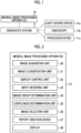
Fig. 1 is a schematic diagram showing a connection relationship between a medical image processing apparatus and an endoscope system. -
Fig. 2 is a block diagram showing a function of the medical image processing apparatus. -
Fig. 3 is an image diagram showing medical images displayed in a list on a first display screen. -
Fig. 4 is an image diagram showing a designated image and a non-designated image at the time of completion of a designation input. -
Fig. 5 is an explanatory diagram regarding acquisition of a selected image. -
Fig. 6 is an image diagram showing selected images displayed in a list on a second display screen. -
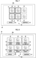
Fig. 7 is an image diagram showing that a display mode is different between an image corresponding to a designated image and an image corresponding to a non-designated image among the selected images. -
Fig. 8 is an image diagram showing characters related to a category assigned to each selected image. -
Fig. 9 is an image diagram showing specific medical images displayed in a list on the first display screen. -
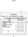
Fig. 10 is an image diagram showing a report creation screen. -
Fig. 11 is a flowchart showing a series of flows for selecting a report-attached image. -
Fig. 12 is an image diagram showing specific medical images displayed in a list on the first display screen. -
Fig. 13 is an image diagram showing position information of characters related to a designated image and a part corresponding to the designated image in a schematic diagram. -
Fig. 14 is an image diagram showing the number of designated images with characters and a number counting bar. - As shown in
Fig. 1 , a medicalimage processing apparatus 10 is connected to anendoscope system 11. Theendoscope system 11 acquires a large amount ofmedical images 12 by using alight source device 11a, anendoscope 11b, and aprocessor device 11c. Thelight source device 11a supplies theendoscope 11b with illumination light for illuminating the inside of the body. Theendoscope 11b acquires an endoscopic image of the inside of the body by irradiating the inside of the body with illumination light and receiving and imaging reflected light from the inside of the body. Theprocessor device 11c performs image processing on the endoscopic image from theendoscope 11b and displays the endoscopic image on a display for an endoscope (not shown). Theendoscope system 11 is connected to the medicalimage processing apparatus 10, and transmits the acquired endoscopic images as themedical images 12 to the medicalimage processing apparatus 10. - In the medical
image processing apparatus 10, programs related to various types of processing are incorporated in a program memory (not shown). The medicalimage processing apparatus 10 is provided with a central control unit (not shown) configured by a processor. By executing the program in the program memory by the central control unit, as shown inFig. 2 , functions of animage acquisition unit 20, an image classification unit 21, adisplay control unit 22, aninput receiving unit 23, an imagegroup determination unit 24, a usefulness determination unit 25, animage selection unit 26, and a report creation device 27 are implemented. Further, the medicalimage processing apparatus 10 is provided with adisplay 30 for displaying various images. - The medical
image processing apparatus 10 performs a process of selecting a report-attached image, which is a medical image to be attached to a report created by the report creation device 27, from the medical images obtained by theendoscope system 11. - The
image acquisition unit 20 acquires a plurality ofmedical images 12 from anendoscope system 11 and transmits the plurality ofmedical images 12 to the image classification unit 21. The image classification unit 21 extracts image information of themedical images 12 transmitted from theimage acquisition unit 20 to acquire category information and calculate image quality information. Themedical images 12 are classified into each category according to the acquired category information. - The category to be classified is any one or a combination thereof, such as an imaging part, the presence or absence or type of a lesion which is one of the regions of interest, a usage state of a treatment tool, and a spraying state of a coloring agent. In the image quality information, information such as brightness, noise, and a degree of blurring of the image is acquired and attached to the
medical image 12. The image information is extracted using a trained convolutional neural network (CNN). The trained CNN is obtained by training using an image group including at least a plurality ofmedical images 12. It is preferable that the category information and the image quality information acquired at the time of imaging by theendoscope system 11 and attached to themedical image 12 have priority over the information extracted by the trained CNN. - As shown in
Fig. 3 , thedisplay control unit 22 displays the plurality ofmedical images 12 in a list on afirst display screen 30a of thedisplay 30. By displaying the plurality ofmedical images 12 in a list in this way, the user can select themedical images 12. It is preferable that themedical images 12 displayed in a list on thefirst display screen 30a are categorizedmedical images 12 that have been classified into a plurality of categories by the image classification unit 21, or uncategorizedmedical images 12 that have not been classified into a plurality of categories by the image classification unit 21. - The
input receiving unit 23 receives at least a designation input of the user for themedical image 12 displayed on thedisplay 30. Specifically, as shown inFig. 3 , acursor 32 that reflects a user operation such as a mouse operation is displayed on thedisplay 30, and the user uses thecursor 32 to perform a designation input for themedical image 12. The designation input can be performed not only for one medical image but also for a plurality of images. Theinput receiving unit 23 receives a designation input designated by thecursor 32. After the designation input for the medical image is completed, a designationinput completion icon 33 is operated using thecursor 32. Further, it is preferable that themedical image 12 with a designation input is displayed by adding athick frame 13 to the outer edge or by adding a mark indicating the designation input thereto. - The image
group determination unit 24 determines a designated image group including designated images that are medical images with a designation input and a non-designated image group including non-designated images that are medical images without a designation input among a plurality of medical images acquired by theimage acquisition unit 20. Specifically, as shown inFig. 4 , themedical images 12 that have been designated and input at the time of completion of the designation input in which theinput receiving unit 23 receives the instruction to complete the designation input are set as designatedimages 35, and a designatedimage group 36 including these designatedimages 35 is determined. In addition, themedical images 12 that have not been designated and input at the time of completion of the designation input are set asnon-designated images 38, and anon-designated image group 39 including these non-designated images is determined. - The usefulness determination unit 25 determines the diagnostic usefulness of the
medical image 12 based on the classification result and the image quality information obtained by the image classification unit 21. The higher the diagnostic usefulness, the higher the likelihood that the image is suitable as a report-attached image. The evaluation of usefulness is represented by, for example, a graded evaluation. It can be evaluated that themedical image 12 having an item such as a lesion in the classification result of the category and having a low degree of blurring in the image quality evaluation is highly useful. In a case where there is no category information, the degree of blurring is large, the lightness is too high, or the lightness is too low, the usefulness is low. Themedical image 12 for which the determination of usefulness has been completed is transmitted to theimage selection unit 26. The usefulness is evaluated, and the evaluation threshold value can be freely set by the user. In addition to themedical image 12, the determination of usefulness may be performed on the designatedimage 35 or the non-designated image. - The
image selection unit 26 acquires selected images selected from the plurality ofmedical images 12 based on a designated image category that is a category assigned to the designatedimage 35 and a non-designated image category that is a category assigned to thenon-designated image 38. More preferably, theimage selection unit 26 may acquire the selected images based on the usefulness obtained by the usefulness determination unit 25 in addition to the designated image category and the non-designated image category. It is preferable that the number of selected images is set such that the sum of the number of designated images and the number of selected images is a specific number. Further, the number of selected images may be set to the number designated by a doctor. It is preferable that the setting of the number of images is received by theinput receiving unit 23. - Specifically, as shown in
Fig. 5 , it is preferable that selectedimages 40 acquired by theimage selection unit 26 include a first selectedimage 42 selected from thenon-designated image group 39 based on the relationship between the designated image category and the non-designated image category. It is preferable that the relationship between the designated image category and the non-designated image category is configured such that the designated image category and the non-designated image category are different from each other. In this case, examples of the first selectedimage 42 include "anon-designated image 38 of an imaging part different from the designatedimage 35", "anon-designated image 38 including no lesion in a case where the designatedimage 35 includes a lesion", "anon-designated image 38 in which the treatment tool is not used in a case where the designatedimage 35 is using the treatment tool", "anon-designated image 38 in which a coloring agent is not sprayed in a case where the designatedimage 35 is spraying a coloring agent", and the like. On the other hand, it is preferable that the selectedimages 40 include a second selectedimage 44 in which all of the designatedimages 35 included in the designatedimage group 36 are selected. - As shown in
Fig. 6 , thedisplay control unit 22 displays the selectedimages 40 on asecond display screen 30b of thedisplay 30. In a case where the user checks the selectedimages 40 and determines that the selectedimages 40 are suitable as report-attached images to be used in the report creation device 27, the user operates anapproval icon 46 displayed on thedisplay 30 using thecursor 32. In a case where theapproval icon 46 is operated, the selectedimages 40 are transmitted to the report creation device 27 as report-attached images. On the other hand, in a case where the selectedimages 40 are not approved as the report-attached images, adisapproval icon 47 is operated. In this case, thedisplay 30 switches to thefirst display screen 30a and switches to a state of receiving the designation input of the user. - As described above, before automatically selecting the report-attached image, the user selects the second selected
image 44, then automatically selects the first selectedimage 42, and sets the selectedimage 40 including the first selectedimage 42 and the second selectedimage 44 as the report-attached image to be used in the report creation device 27, thereby reducing the burden of the report creation work while reflecting the preference of the doctor who is the user. - It is preferable that, on the
second display screen 30b, the display mode of the image corresponding to the designatedimage 35 and the display mode of the image corresponding to thenon-designated image 38 among the selectedimages 40 are different from each other. For example, as shown inFig. 7 , there is a form in which athick frame 51 is displayed on the outer edge of animage 50 corresponding to the designatedimage 35, while thethick frame 51 is not displayed on animage 52 corresponding to thenon-designated image 38. - In addition, it is preferable that, on the category on the
first display screen 30a, the display mode of eachmedical image 12 is different depending, and it is preferable that, on thesecond display screen 30b, the display mode of each selectedimage 40 is different depending on the category. For example, as shown inFig. 8 , on thesecond display screen 30b,characters 54 corresponding to the categories are added to each selectedimage 40. The characters corresponding to the category include a part name, a lesion name, a light source used, and a presence or absence of treatment with a treatment tool. In addition, symbol information corresponding to the category may be added to and displayed on each selectedimage 40. The symbol information corresponding to the category includes information in which a part name, a lesion name, a light source used, a presence or absence of treatment with a treatment tool, and the like are symbolized. For example, in a case where an upper endoscope is used, it is conceivable that the esophagus, stomach, and duodenum are designated as "areal", "area2", and "area3" in the order of insertion. In addition, it is preferable that, on thefirst display screen 30a as well, a character or symbol information corresponding to the category is added to and displayed on eachmedical image 12 through the same method as thesecond display screen 30b. - In addition, as described above, the display mode of the
medical image 12 or the selectedimage 40 corresponding to the category may be a mode in which characters or symbols are not used. For example, on thefirst display screen 30a or thesecond display screen 30b, the display position of themedical image 12 or the selectedimage 40 may be changed according to the category. For example, in a case where the medical image or the selectedimage 40 is displayed in the row direction (vertical direction), it is preferable to display themedical image 12 or the selectedimage 40 of different categories in units of one or a plurality of rows. Accordingly, on thefirst display screen 30a, the user can easily select (designate and input) themedical image 12, and on thesecond display screen 30b, it is easy to grasp the category of the image group to be included in the report. - Note that, as shown in
Fig. 9 , it is preferable that, on thefirst display screen 30a, specificmedical images 55 satisfying a specific condition regarding a category and/or diagnostic usefulness among the plurality ofmedical images 12 are displayed in a list. Theinput receiving unit 23 receives the designation input of the user for the specificmedical images 55 displayed in the list on thefirst display screen 30a. As described above, by displaying only the specificmedical images 55 in a list instead of displaying all the medical images, it is possible to reduce the burden of the user due to the designation input. It is preferable that the specific condition includes at least one of the number of images included in the same category being a specific number or less, or the image being an image having high diagnostic usefulness.Medical images 56 other than the specificmedical images 55 are excluded from the target of the designation input. In this case, as a display mode of themedical image 56 other than the specificmedical image 55, preferably, themedical image 56 is either left displayed on a first display screen 50a (for example, grayed out) although the visibility is lower than that of the specificmedical image 55 or completely excluded from themedical image 56 on the first display screen 50a (hidden). - As shown in
Fig. 10 , the report creation device 27 creates a report using areport creation screen 48 displayed on thedisplay 30. In addition to the selectedimage 40 being automatically attached to thereport creation screen 48, patient information, user information, and the like that have been input in advance are automatically input thereto. The user inputs an opinion in anopinion input field 49 and creates a report. - Next, a series of flows of the process of selecting the report-attached image will be described with reference to the flowchart of
Fig. 11 . Theimage acquisition unit 20 acquires a plurality ofmedical images 12 from anendoscope system 11. The image classification unit 21 classifies the respectivemedical image 12 into a plurality of categories. Thedisplay control unit 22 displays the plurality of medical images in a list on thefirst display screen 30a of thedisplay 30. Theinput receiving unit 23 receives the designation input of the user for the medical image displayed on thefirst display screen 30a. In a case where the designation input by the user is completed, the designationinput completion icon 33 is operated. - In a case where the designation
input completion icon 33 is operated, the imagegroup determination unit 24 determines a designatedimage group 36 including designatedimages 35 that aremedical images 12 with a designation input and anon-designated image group 39 includingnon-designated images 38 that aremedical images 12 without a designation input among a plurality ofmedical images 12. Theimage selection unit 26 acquires the selectedimages 40 selected from the plurality ofmedical images 12 based on a designated image category that is a category assigned to the designatedimage 35 and a non-designated image category that is a category assigned to thenon-designated image 38. Thedisplay control unit 22 displays the selectedimages 40 in a list on thesecond display screen 30b of thedisplay 30. - In a case where the user approves the selected
images 40 displayed in the list as the report-attached images, theapproval icon 46 is operated. In a case where theapproval icon 46 is operated, the selectedimages 40 are transmitted to the report creation device 27 as report-attached images. On the other hand, in a case where the selectedimages 40 are not approved as the report-attached images, adisapproval icon 47 is operated. In this case, thedisplay 30 switches to thefirst display screen 30a and switches to a state of receiving the designation input of the user. - In a second embodiment, the medical images displayed in a list on the
first display screen 30a are updated each time the designation input by the user is performed. Specifically, thedisplay control unit 22 updates the medical images displayed in a list on thefirst display screen 30a based on the designated image category and the non-designated image category each time the designation input is performed. Theinput receiving unit 23 receives the designation input of the user for the updated medical images displayed in the list on thefirst display screen 30a. - For example, as shown in
Fig. 12 , in a case where a designation input is performed to a designated image X1 to which a specific designated image category X is assigned, thedisplay control unit 22 performs the update such that a non-designated image X2 to which the same non-designated image category X as the specific designated image category X is assigned is deleted from thefirst display screen 30a (is hidden on thefirst display screen 30a). Thereby, the number of images to be checked by the user is reduced, so that the work burden of the user can be reduced. Here, preferably, examples of the image to be deleted from thefirst display screen 30a in response to the designation input by the user include, for example, "the non-designated image X2 in which the imaging part is the same as the designated image X1", "the non-designated image X2 in which the imaging part is close to the designated image X1", and "the non-designated image X2 during treatment or during coloring agent spraying in a case where the designated image X1 is an image during treatment with a treatment tool or an image during coloring agent spraying". The non-designated image X2 may be hidden on thefirst display screen 30a, or may be left displayed on thefirst display screen 30a (for example, grayed out) although the visibility is lower than that of the designated image X1. - In a third embodiment, in order to make it easier for the user to understand the work status in a case where the designation input is performed, the
display control unit 22 displays designated image information about the designated image on thedisplay 30. As the designated image information, for example, it is preferable that the designated image information is information representing position information of a part corresponding to the designated image by characters or visually. In a case where the part corresponding to the designated image is the rectum which is a predetermined position of the large intestine, as shown inFig. 13 , it is preferable to display "rectum" on thedisplay 30 asposition information 62 of the characters, or to display, on thedisplay 30, apoint 58 indicating that the designation input has been made in a portion corresponding to "rectum" in a schematic diagram 57 of the large intestine. - In addition, as the designated image information, for example, it is preferable that the designated image information is information representing the number of designated
images 35 by characters or visually. In a case where the number of designatedimages 35 at the time of performing the designation input is five, as shown inFig. 14 , it is preferable to display "4" as character information 59 on thedisplay 30, or to display, on thedisplay 30, alength 60a corresponding to "4" in anumber counting bar 60 representing the number of designatedimages 35. - In the above embodiments, hardware structures of processing units for executing various kinds of processing, such as the
image acquisition unit 20, the image classification unit 21, thedisplay control unit 22, theinput receiving unit 23, the imagegroup determination unit 24, the usefulness determination unit 25, theimage selection unit 26, and the report creation device 27, are various processors shown below. The various processors include a central processing unit (CPU) as a general-purpose processor functioning as various processing units by executing software (program), a programmable logic device (PLD) as a processor of which the circuit configuration can be changed after manufacturing such as a graphical processing unit (GPU) or a field programmable gate array (FPGA), a dedicated electrical circuit as a processor having a circuit configuration designed exclusively for executing various kinds of processing, and the like. - One processing unit may be configured by one of various processors, or may be configured by a combination of the same or different kinds of two or more processors (for example, a combination of a plurality of FPGAs, a combination of a CPU and an FPGA, a combination of a CPU and a GPU, or the like). In addition, a plurality of processing units may be configured by one processor. As an example of configuring a plurality of processing units via one processor, first, as represented by a computer, such as a client or a server, there is a form in which one processor is configured by a combination of one or more CPUs and software, and this processor functions as a plurality of processing units. Second, as represented by a system on chip (SoC) or the like, there is a form of using a processor for realizing the function of the entire system including a plurality of processing units with one integrated circuit (IC) chip. Thus, various processing units are configured by using one or more of the above-described various processors as hardware structures.
- More specifically, the hardware structure of these various processors is an electrical circuit (circuitry) in the form of a combination of circuit elements, such as semiconductor elements. The hardware structure of the storage unit is a storage device such as a hard disc drive (HDD) or a solid state drive (SSD).
-
- 10: medical image processing apparatus
- 11: endoscope system
- 11a: light source device
- 11b: endoscope
- 11c: processor device
- 12: medical image
- 13: thick frame
- 20: image acquisition unit
- 21: image classification unit
- 22: display control unit
- 23: input receiving unit
- 24: image group determination unit
- 25: usefulness determination unit
- 26: image selection unit
- 27: report creation device
- 30: display
- 30a :first display screen
- 30b: second display screen
- 32: cursor
- 33: designation input completion icon
- 35: designated image
- 36: designated image group
- 38: non-designated image
- 39: non-designated image group
- 40: selected image
- 42: first selected image
- 44: second selected image
- 46: approval icon
- 47: disapproval icon
- 48: report creation screen
- 49: opinion input field
- 50, 52: image
- 51: thick frame
- 54: character
- 55: specific medical image
- 56: image other than specific medical image
- 57: schematic diagram of large intestine
- 58: point
- 59: character information
- 60: number counting bar
- 62: position information of character
- X1: designated image
- X2: non-designated image
Claims (17)
- A medical image processing apparatus comprising a processor,
wherein the processor is configured to:acquire a plurality of medical images;classify each of the plurality of medical images into a plurality of categories;display the plurality of medical images in a list on a first display screen of a display;receive a designation input of a user for the medical image displayed on the first display screen;determine a designated image group including designated images that are medical images with the designation input and a non-designated image group including non-designated images that are medical images without the designation input among the plurality of medical images;acquire selected images selected from the plurality of medical images based on a designated image category that is a category assigned to the designated image and a non-designated image category that is a category assigned to the non-designated image; anddisplay the selected images in a list on a second display screen of the display. - The medical image processing apparatus according to claim 1,
wherein the selected image includes a first selected image selected from the non-designated image group based on a relationship between the designated image category and the non-designated image category. - The medical image processing apparatus according to claim 2,
wherein the relationship between the designated image category and the non-designated image category is configured such that the designated image category and the non-designated image category are different from each other. - The medical image processing apparatus according to any one of claims 1 to 3,
wherein the selected image includes a second selected image in which all of the designated images included in the designated image group are selected. - The medical image processing apparatus according to any one of claims 1 to 4,
wherein the processor is configured to:determine diagnostic usefulness of the medical image; andacquire the selected image based on the usefulness in addition to the designated image category and the non-designated image category. - The medical image processing apparatus according to any one of claims 1 to 5,
wherein, on the second display screen, a display mode of an image corresponding to the designated image and a display mode of an image corresponding to the non-designated image among the selected images are different from each other. - The medical image processing apparatus according to any one of claims 1 to 6,
wherein, on the first display screen, a display mode of each medical image is different depending on the category, and/or, on the second display screen, a display mode of the selected image is different depending on the category. - The medical image processing apparatus according to claim 7,
wherein, on the first display screen, a character or symbol information corresponding to the category is added to and displayed on each medical image, and/or, on the second display screen, a character or symbol information corresponding to the category is added to and displayed on each selected image. - The medical image processing apparatus according to any one of claims 1 to 8,
wherein, on the first display screen, specific medical images satisfying a specific condition regarding a category and/or diagnostic usefulness among the plurality of medical images are displayed in a list. - The medical image processing apparatus according to any one of claims 1 to 9,
wherein the processor is configured to:update the medical images displayed in the list on the first display screen based on the designated image category assigned to the designated image and the non-designated image category assigned to the non-designated image each time the designation input is performed; andreceive the designation input for the updated medical images displayed in the list on the first display screen. - The medical image processing apparatus according to claim 10,
wherein the processor is configured to, in a case where the designation input is performed for a specific designated image category, perform the update such that a non-designated image to which the same non-designated image category as the specific designated image category is assigned is deleted from the first display screen, or a display mode is changed. - The medical image processing apparatus according to any one of claims 1 to 11,wherein the processor is configured to display designated image information, which is information about the designated image, on the display, andthe designated image information includes at least information representing position information of a part corresponding to the designated image by characters or visually, or information representing the number of the designated images by characters or visually.
- The medical image processing apparatus according to any one of claims 1 to 12,
wherein the category is information about a part of a subject. - The medical image processing apparatus according to any one of claims 1 to 13,
wherein the category is information about whether or not the image includes a region of interest. - The medical image processing apparatus according to any one of claims 1 to 14,
wherein the category is information about whether or not the image includes a treatment tool or about a type of the treatment tool included. - The medical image processing apparatus according to any one of claims 1 to 15,
wherein the category is information about a spraying state of a coloring agent or a dyeing agent in an image. - An operation method of a medical image processing apparatus comprising a processor, wherein the processor performs:a step of acquiring a plurality of medical images;a step of classifying each of the plurality of medical images into a plurality of categories;a step of displaying the plurality of medical images in a list on a first display screen of a display;a step of receiving a designation input of a user for the medical image displayed on the first display screen;a step of determining a designated image group including designated images that are medical images with the designation input and a non-designated image group including non-designated images that are medical images without the designation input among the plurality of medical images;a step of acquiring selected images selected from the plurality of medical images based on a designated image category that is a category assigned to the designated image and a non-designated image category that is a category assigned to the non-designated image; anda step of displaying the selected images in a list on a second display screen of the display.
Applications Claiming Priority (2)
| Application Number | Priority Date | Filing Date | Title |
|---|---|---|---|
| JP2020171382 | 2020-10-09 | ||
| PCT/JP2021/034377 WO2022075047A1 (en) | 2020-10-09 | 2021-09-17 | Medical image processing device and operation method for same |
Publications (2)
| Publication Number | Publication Date |
|---|---|
| EP4226840A1 true EP4226840A1 (en) | 2023-08-16 |
| EP4226840A4 EP4226840A4 (en) | 2024-03-20 |
Family
ID=81126485
Family Applications (1)
| Application Number | Title | Priority Date | Filing Date |
|---|---|---|---|
| EP21877348.9A Pending EP4226840A4 (en) | 2020-10-09 | 2021-09-17 | Medical image processing device and operation method for same |
Country Status (5)
| Country | Link |
|---|---|
| US (1) | US12555670B2 (en) |
| EP (1) | EP4226840A4 (en) |
| JP (1) | JP7702423B2 (en) |
| CN (1) | CN116325008B (en) |
| WO (1) | WO2022075047A1 (en) |
Families Citing this family (1)
| Publication number | Priority date | Publication date | Assignee | Title |
|---|---|---|---|---|
| CN114724669B (en) * | 2022-05-07 | 2023-08-22 | 丹阳慧创医疗设备有限公司 | Medical report generation method and device for near-infrared brain functional imaging device |
Family Cites Families (15)
| Publication number | Priority date | Publication date | Assignee | Title |
|---|---|---|---|---|
| JP2001024971A (en) * | 1999-07-06 | 2001-01-26 | Canon Inc | Image processing apparatus, image processing method, and memory medium |
| US8014576B2 (en) * | 2005-11-23 | 2011-09-06 | The Medipattern Corporation | Method and system of computer-aided quantitative and qualitative analysis of medical images |
| WO2008075508A1 (en) * | 2006-12-21 | 2008-06-26 | Konica Minolta Medical & Graphic, Inc. | Medical image system and first server |
| JP5305700B2 (en) * | 2007-04-25 | 2013-10-02 | 株式会社東芝 | Image diagnosis support system and image diagnosis support method |
| EP2174578A4 (en) | 2007-07-12 | 2013-07-03 | Olympus Medical Systems Corp | IMAGE PROCESSING DEVICE, ITS OPERATING METHOD AND PROGRAM |
| US20110032259A1 (en) * | 2009-06-09 | 2011-02-10 | Intromedic Co., Ltd. | Method of displaying images obtained from an in-vivo imaging device and apparatus using same |
| JP5486364B2 (en) * | 2009-09-17 | 2014-05-07 | 富士フイルム株式会社 | Interpretation report creation apparatus, method and program |
| CN104067277A (en) * | 2012-01-27 | 2014-09-24 | 皇家飞利浦有限公司 | medical selection system |
| JP6345487B2 (en) * | 2013-07-11 | 2018-06-20 | キヤノンメディカルシステムズ株式会社 | MEDICAL REPORT CREATION DEVICE AND MEDICAL IMAGE DIAGNOSIS DEVICE |
| JP6373773B2 (en) * | 2015-02-27 | 2018-08-15 | オリンパス株式会社 | Endoscope support system |
| US10929508B2 (en) * | 2015-04-30 | 2021-02-23 | Merge Healthcare Solutions Inc. | Database systems and interactive user interfaces for dynamic interaction with, and indications of, digital medical image data |
| US10579234B2 (en) * | 2016-09-09 | 2020-03-03 | Merge Healthcare Solutions Inc. | Systems and user interfaces for opportunistic presentation of functionality for increasing efficiencies of medical image review |
| US10803581B2 (en) * | 2017-11-06 | 2020-10-13 | Beijing Keya Medical Technology Co., Ltd. | System and method for generating and editing diagnosis reports based on medical images |
| KR20190074671A (en) * | 2017-12-20 | 2019-06-28 | (주)헬스허브 | Apparatus and method for supporting medical image display protocol |
| KR102034648B1 (en) * | 2018-05-16 | 2019-10-22 | 재단법인 아산사회복지재단 | Medical Image Management System, Method and Computer Readable Recording Medium |
-
2021
- 2021-09-17 WO PCT/JP2021/034377 patent/WO2022075047A1/en not_active Ceased
- 2021-09-17 CN CN202180069127.7A patent/CN116325008B/en active Active
- 2021-09-17 EP EP21877348.9A patent/EP4226840A4/en active Pending
- 2021-09-17 JP JP2022555341A patent/JP7702423B2/en active Active
-
2023
- 2023-04-07 US US18/297,432 patent/US12555670B2/en active Active
Also Published As
| Publication number | Publication date |
|---|---|
| JP7702423B2 (en) | 2025-07-03 |
| US20230245758A1 (en) | 2023-08-03 |
| CN116325008B (en) | 2026-02-24 |
| US12555670B2 (en) | 2026-02-17 |
| CN116325008A (en) | 2023-06-23 |
| JPWO2022075047A1 (en) | 2022-04-14 |
| WO2022075047A1 (en) | 2022-04-14 |
| EP4226840A4 (en) | 2024-03-20 |
Similar Documents
| Publication | Publication Date | Title |
|---|---|---|
| Waite et al. | A review of perceptual expertise in radiology-how it develops, how we can test it, and why humans still matter in the era of artificial intelligence | |
| JP7201404B2 (en) | MEDICAL IMAGE PROCESSING APPARATUS, MEDICAL IMAGE PROCESSING METHOD, AND PROGRAM | |
| US12579795B2 (en) | Diagnosis support system, diagnosis support method, and storage medium | |
| RU2451335C2 (en) | Image- and context-dependent anatomic applications for effective diagnostics | |
| EP3998038B1 (en) | Multiple bone density displaying method for establishing implant procedure plan, and image processing device therefor | |
| EP3649918B1 (en) | Medical image processing device, endoscope device, diagnostic support device, medical service support device and report generation support device | |
| EP3649917B1 (en) | Medical image processing device, endoscope device, diagnostic support device, medical service support device, and report generation support device | |
| JP7828486B2 (en) | Information processing device, information processing method, and information processing program | |
| CN104720835A (en) | Display device, image displaying method and computerized tomography apparatus | |
| JP4253055B2 (en) | Computer-aided diagnosis device | |
| US12555670B2 (en) | Medical image processing apparatus and operation method thereof | |
| US20240013524A1 (en) | Information processing apparatus, method of operating information processing apparatus, and program for operating information processing apparatus | |
| WO2022113800A1 (en) | Medical image display system, medical image display method, and program | |
| US11309073B2 (en) | Display control device, display control method, and display control program | |
| US12548155B2 (en) | Medical image processing apparatus and operation method thereof | |
| US11003342B1 (en) | Smart scrolling system | |
| EP4358022B1 (en) | Medical image diagnostic system, medical image diagnostic method, and program | |
| WO2022113798A1 (en) | Medical image display system, medical image display method, and program | |
| CN117836805A (en) | Coronary Artery Disease Analysis System | |
| JP4635077B2 (en) | Computer-aided diagnosis device | |
| JP7556970B2 (en) | Medical imaging device and method of operation thereof | |
| CN115205189B (en) | Image reading system and method for fundus images | |
| Castner | Gaze and visual scanpath features for data-driven expertise recognition in medical image inspection | |
| US20260065482A1 (en) | Medical information processing apparatus, storage medium, and method | |
| US12412326B2 (en) | Image processing apparatus and non-transitory computer readable medium for visualizing magnetic resonance data |
Legal Events
| Date | Code | Title | Description |
|---|---|---|---|
| STAA | Information on the status of an ep patent application or granted ep patent |
Free format text: STATUS: THE INTERNATIONAL PUBLICATION HAS BEEN MADE |
|
| PUAI | Public reference made under article 153(3) epc to a published international application that has entered the european phase |
Free format text: ORIGINAL CODE: 0009012 |
|
| STAA | Information on the status of an ep patent application or granted ep patent |
Free format text: STATUS: REQUEST FOR EXAMINATION WAS MADE |
|
| 17P | Request for examination filed |
Effective date: 20230409 |
|
| AK | Designated contracting states |
Kind code of ref document: A1 Designated state(s): AL AT BE BG CH CY CZ DE DK EE ES FI FR GB GR HR HU IE IS IT LI LT LU LV MC MK MT NL NO PL PT RO RS SE SI SK SM TR |
|
| DAV | Request for validation of the european patent (deleted) | ||
| DAX | Request for extension of the european patent (deleted) | ||
| A4 | Supplementary search report drawn up and despatched |
Effective date: 20240216 |
|
| RIC1 | Information provided on ipc code assigned before grant |
Ipc: G06V 10/98 20220101ALN20240212BHEP Ipc: G16H 40/63 20180101ALI20240212BHEP Ipc: G16H 30/40 20180101ALI20240212BHEP Ipc: G16H 30/20 20180101ALI20240212BHEP Ipc: G16H 15/00 20180101ALI20240212BHEP Ipc: A61B 1/00 20060101ALI20240212BHEP Ipc: A61B 5/00 20060101ALI20240212BHEP Ipc: G16H 30/00 20180101ALI20240212BHEP Ipc: A61B 1/045 20060101AFI20240212BHEP |