EP4186526A1 - Mittel zur umkehrung der resistenz gegen antikrebsmittel - Google Patents
Mittel zur umkehrung der resistenz gegen antikrebsmittel Download PDFInfo
- Publication number
- EP4186526A1 EP4186526A1 EP21820949.2A EP21820949A EP4186526A1 EP 4186526 A1 EP4186526 A1 EP 4186526A1 EP 21820949 A EP21820949 A EP 21820949A EP 4186526 A1 EP4186526 A1 EP 4186526A1
- Authority
- EP
- European Patent Office
- Prior art keywords
- antibody
- cancer
- anticancer drug
- cells
- pharmaceutical composition
- Prior art date
- Legal status (The legal status is an assumption and is not a legal conclusion. Google has not performed a legal analysis and makes no representation as to the accuracy of the status listed.)
- Pending
Links
- 239000002246 antineoplastic agent Substances 0.000 title claims abstract description 164
- 229940041181 antineoplastic drug Drugs 0.000 title claims abstract description 163
- 239000003795 chemical substances by application Substances 0.000 title claims abstract description 12
- 101000853000 Homo sapiens Interleukin-26 Proteins 0.000 claims abstract description 544
- 206010028980 Neoplasm Diseases 0.000 claims abstract description 182
- 201000011510 cancer Diseases 0.000 claims abstract description 125
- 239000003814 drug Substances 0.000 claims abstract description 75
- 229940079593 drug Drugs 0.000 claims abstract description 71
- 229940121647 egfr inhibitor Drugs 0.000 claims abstract description 69
- 239000008194 pharmaceutical composition Substances 0.000 claims abstract description 64
- 239000004480 active ingredient Substances 0.000 claims abstract description 17
- 208000026278 immune system disease Diseases 0.000 claims abstract description 4
- 230000003449 preventive effect Effects 0.000 claims abstract description 3
- 210000004027 cell Anatomy 0.000 claims description 321
- XGALLCVXEZPNRQ-UHFFFAOYSA-N gefitinib Chemical compound C=12C=C(OCCCN3CCOCC3)C(OC)=CC2=NC=NC=1NC1=CC=C(F)C(Cl)=C1 XGALLCVXEZPNRQ-UHFFFAOYSA-N 0.000 claims description 191
- 239000005411 L01XE02 - Gefitinib Substances 0.000 claims description 190
- 229960002584 gefitinib Drugs 0.000 claims description 190
- 208000003721 Triple Negative Breast Neoplasms Diseases 0.000 claims description 112
- 208000022679 triple-negative breast carcinoma Diseases 0.000 claims description 112
- 238000000034 method Methods 0.000 claims description 62
- 125000003275 alpha amino acid group Chemical group 0.000 claims description 61
- 230000027455 binding Effects 0.000 claims description 53
- 230000026731 phosphorylation Effects 0.000 claims description 51
- 238000006366 phosphorylation reaction Methods 0.000 claims description 51
- 230000002401 inhibitory effect Effects 0.000 claims description 49
- 230000003993 interaction Effects 0.000 claims description 34
- 239000000427 antigen Substances 0.000 claims description 32
- 108091007433 antigens Proteins 0.000 claims description 32
- 102000036639 antigens Human genes 0.000 claims description 32
- 206010006187 Breast cancer Diseases 0.000 claims description 30
- 208000026310 Breast neoplasm Diseases 0.000 claims description 30
- 230000035755 proliferation Effects 0.000 claims description 30
- 230000035945 sensitivity Effects 0.000 claims description 27
- 239000012634 fragment Substances 0.000 claims description 25
- 239000003112 inhibitor Substances 0.000 claims description 24
- 102000040430 polynucleotide Human genes 0.000 claims description 19
- 108091033319 polynucleotide Proteins 0.000 claims description 19
- 239000002157 polynucleotide Substances 0.000 claims description 19
- 108010047041 Complementarity Determining Regions Proteins 0.000 claims description 18
- 101001012157 Homo sapiens Receptor tyrosine-protein kinase erbB-2 Proteins 0.000 claims description 17
- 102100030086 Receptor tyrosine-protein kinase erbB-2 Human genes 0.000 claims description 17
- 102000001301 EGF receptor Human genes 0.000 claims description 16
- 239000013598 vector Substances 0.000 claims description 16
- 108060006698 EGF receptor Proteins 0.000 claims description 15
- 102000039446 nucleic acids Human genes 0.000 claims description 15
- 108020004707 nucleic acids Proteins 0.000 claims description 15
- 150000007523 nucleic acids Chemical class 0.000 claims description 15
- 102000015694 estrogen receptors Human genes 0.000 claims description 13
- 108010038795 estrogen receptors Proteins 0.000 claims description 13
- 210000004366 CD4-positive T-lymphocyte Anatomy 0.000 claims description 11
- 239000005551 L01XE03 - Erlotinib Substances 0.000 claims description 11
- 229960001433 erlotinib Drugs 0.000 claims description 11
- AAKJLRGGTJKAMG-UHFFFAOYSA-N erlotinib Chemical compound C=12C=C(OCCOC)C(OCCOC)=CC2=NC=NC=1NC1=CC=CC(C#C)=C1 AAKJLRGGTJKAMG-UHFFFAOYSA-N 0.000 claims description 11
- 210000002540 macrophage Anatomy 0.000 claims description 11
- 230000001747 exhibiting effect Effects 0.000 claims description 9
- 238000004519 manufacturing process Methods 0.000 claims description 9
- 238000012258 culturing Methods 0.000 claims description 8
- 238000002360 preparation method Methods 0.000 claims description 8
- 102000003998 progesterone receptors Human genes 0.000 claims description 8
- 108090000468 progesterone receptors Proteins 0.000 claims description 8
- 229940121358 tyrosine kinase inhibitor Drugs 0.000 claims description 6
- 210000004408 hybridoma Anatomy 0.000 claims description 5
- 230000003247 decreasing effect Effects 0.000 claims description 4
- 229940124597 therapeutic agent Drugs 0.000 claims description 4
- 239000005483 tyrosine kinase inhibitor Substances 0.000 claims description 4
- 230000004043 responsiveness Effects 0.000 claims description 3
- 229960001686 afatinib Drugs 0.000 claims description 2
- 150000004917 tyrosine kinase inhibitor derivatives Chemical class 0.000 claims 3
- BSPLGGCPNTZPIH-IPZCTEOASA-N (e)-n-[4-(3-chloro-4-fluoroanilino)-7-methoxyquinazolin-6-yl]-4-piperidin-1-ylbut-2-enamide;hydrate Chemical compound O.C=12C=C(NC(=O)\C=C\CN3CCCCC3)C(OC)=CC2=NC=NC=1NC1=CC=C(F)C(Cl)=C1 BSPLGGCPNTZPIH-IPZCTEOASA-N 0.000 claims 1
- USNRYVNRPYXCSP-JUGPPOIOSA-N afatinib dimaleate Chemical compound OC(=O)\C=C/C(O)=O.OC(=O)\C=C/C(O)=O.N1=CN=C2C=C(O[C@@H]3COCC3)C(NC(=O)/C=C/CN(C)C)=CC2=C1NC1=CC=C(F)C(Cl)=C1 USNRYVNRPYXCSP-JUGPPOIOSA-N 0.000 claims 1
- 229960001638 osimertinib mesylate Drugs 0.000 claims 1
- FUKSNUHSJBTCFJ-UHFFFAOYSA-N osimertinib mesylate Chemical compound CS(O)(=O)=O.COC1=CC(N(C)CCN(C)C)=C(NC(=O)C=C)C=C1NC1=NC=CC(C=2C3=CC=CC=C3N(C)C=2)=N1 FUKSNUHSJBTCFJ-UHFFFAOYSA-N 0.000 claims 1
- 241000282414 Homo sapiens Species 0.000 abstract description 102
- 241000699670 Mus sp. Species 0.000 abstract description 73
- 230000001629 suppression Effects 0.000 abstract description 28
- 108020003175 receptors Proteins 0.000 abstract description 19
- 230000037361 pathway Effects 0.000 abstract description 16
- 230000001225 therapeutic effect Effects 0.000 abstract description 15
- 241000282412 Homo Species 0.000 abstract description 8
- 108010055191 EphA3 Receptor Proteins 0.000 abstract description 7
- 102000021607 EphA3 Receptor Human genes 0.000 abstract description 7
- 230000003213 activating effect Effects 0.000 abstract description 6
- 230000009456 molecular mechanism Effects 0.000 abstract description 5
- 102100030324 Ephrin type-A receptor 3 Human genes 0.000 description 139
- 101710116735 Ephrin type-A receptor 3 Proteins 0.000 description 139
- 241000699666 Mus <mouse, genus> Species 0.000 description 90
- 238000011282 treatment Methods 0.000 description 89
- 238000002474 experimental method Methods 0.000 description 85
- 230000014509 gene expression Effects 0.000 description 65
- 230000004663 cell proliferation Effects 0.000 description 59
- 102100033810 RAC-alpha serine/threonine-protein kinase Human genes 0.000 description 53
- 102000019145 JUN kinase activity proteins Human genes 0.000 description 43
- 108090000623 proteins and genes Proteins 0.000 description 43
- 239000003981 vehicle Substances 0.000 description 40
- 238000007792 addition Methods 0.000 description 29
- 230000000694 effects Effects 0.000 description 29
- 230000004614 tumor growth Effects 0.000 description 22
- 238000000134 MTT assay Methods 0.000 description 20
- 231100000002 MTT assay Toxicity 0.000 description 20
- 230000003472 neutralizing effect Effects 0.000 description 20
- 235000001014 amino acid Nutrition 0.000 description 19
- 230000002829 reductive effect Effects 0.000 description 19
- 102000057485 human IL26 Human genes 0.000 description 18
- 230000005764 inhibitory process Effects 0.000 description 18
- 102000005962 receptors Human genes 0.000 description 18
- 239000012825 JNK inhibitor Substances 0.000 description 17
- 230000037396 body weight Effects 0.000 description 17
- 239000003197 protein kinase B inhibitor Substances 0.000 description 17
- 238000010186 staining Methods 0.000 description 17
- 241000283973 Oryctolagus cuniculus Species 0.000 description 16
- 150000001413 amino acids Chemical class 0.000 description 16
- 210000001519 tissue Anatomy 0.000 description 16
- 238000002054 transplantation Methods 0.000 description 16
- -1 ATF3 Proteins 0.000 description 15
- 241000283707 Capra Species 0.000 description 15
- 102100022706 Interleukin-20 receptor subunit alpha Human genes 0.000 description 15
- 101710174006 Interleukin-20 receptor subunit alpha Proteins 0.000 description 15
- 229940126638 Akt inhibitor Drugs 0.000 description 14
- 229940118135 JNK inhibitor Drugs 0.000 description 14
- 108020004999 messenger RNA Proteins 0.000 description 14
- 238000012360 testing method Methods 0.000 description 14
- 102100029145 DNA damage-inducible transcript 3 protein Human genes 0.000 description 13
- 238000002965 ELISA Methods 0.000 description 13
- 108010017324 STAT3 Transcription Factor Proteins 0.000 description 13
- 102100024040 Signal transducer and activator of transcription 3 Human genes 0.000 description 13
- 108010057666 Transcription Factor CHOP Proteins 0.000 description 13
- 230000004913 activation Effects 0.000 description 13
- 230000000259 anti-tumor effect Effects 0.000 description 13
- 238000003556 assay Methods 0.000 description 13
- 208000024908 graft versus host disease Diseases 0.000 description 13
- 239000000203 mixture Substances 0.000 description 13
- 102000004169 proteins and genes Human genes 0.000 description 13
- 230000000638 stimulation Effects 0.000 description 13
- 238000006467 substitution reaction Methods 0.000 description 13
- 238000001262 western blot Methods 0.000 description 13
- 108091032973 (ribonucleotides)n+m Proteins 0.000 description 12
- 102100020788 Interleukin-10 receptor subunit beta Human genes 0.000 description 12
- 101710199214 Interleukin-10 receptor subunit beta Proteins 0.000 description 12
- 108010076504 Protein Sorting Signals Proteins 0.000 description 12
- 125000000539 amino acid group Chemical group 0.000 description 12
- 201000010099 disease Diseases 0.000 description 12
- 208000037265 diseases, disorders, signs and symptoms Diseases 0.000 description 12
- 235000018102 proteins Nutrition 0.000 description 12
- 210000003171 tumor-infiltrating lymphocyte Anatomy 0.000 description 12
- 108060003951 Immunoglobulin Proteins 0.000 description 11
- 238000004458 analytical method Methods 0.000 description 11
- 239000000872 buffer Substances 0.000 description 11
- 239000002299 complementary DNA Substances 0.000 description 11
- 210000002472 endoplasmic reticulum Anatomy 0.000 description 11
- 102000018358 immunoglobulin Human genes 0.000 description 11
- 239000002953 phosphate buffered saline Substances 0.000 description 11
- 241000700159 Rattus Species 0.000 description 10
- 230000030609 dephosphorylation Effects 0.000 description 10
- 238000006209 dephosphorylation reaction Methods 0.000 description 10
- 239000007924 injection Substances 0.000 description 10
- 238000002347 injection Methods 0.000 description 10
- 238000011081 inoculation Methods 0.000 description 10
- 239000002904 solvent Substances 0.000 description 10
- 210000004881 tumor cell Anatomy 0.000 description 10
- 108010064593 Intercellular Adhesion Molecule-1 Proteins 0.000 description 9
- 102100037877 Intercellular adhesion molecule 1 Human genes 0.000 description 9
- 241000124008 Mammalia Species 0.000 description 9
- 102220497176 Small vasohibin-binding protein_T47D_mutation Human genes 0.000 description 9
- FAPWRFPIFSIZLT-UHFFFAOYSA-M Sodium chloride Chemical compound [Na+].[Cl-] FAPWRFPIFSIZLT-UHFFFAOYSA-M 0.000 description 9
- 238000002835 absorbance Methods 0.000 description 9
- 239000013604 expression vector Substances 0.000 description 9
- 238000001727 in vivo Methods 0.000 description 9
- 230000009467 reduction Effects 0.000 description 9
- FWBHETKCLVMNFS-UHFFFAOYSA-N 4',6-Diamino-2-phenylindol Chemical compound C1=CC(C(=N)N)=CC=C1C1=CC2=CC=C(C(N)=N)C=C2N1 FWBHETKCLVMNFS-UHFFFAOYSA-N 0.000 description 8
- WZUVPPKBWHMQCE-UHFFFAOYSA-N Haematoxylin Chemical compound C12=CC(O)=C(O)C=C2CC2(O)C1C1=CC=C(O)C(O)=C1OC2 WZUVPPKBWHMQCE-UHFFFAOYSA-N 0.000 description 8
- 230000000903 blocking effect Effects 0.000 description 8
- 238000001516 cell proliferation assay Methods 0.000 description 8
- 230000005754 cellular signaling Effects 0.000 description 8
- 238000011161 development Methods 0.000 description 8
- 230000007783 downstream signaling Effects 0.000 description 8
- 239000002773 nucleotide Substances 0.000 description 8
- 125000003729 nucleotide group Chemical group 0.000 description 8
- 239000000243 solution Substances 0.000 description 8
- 102100031968 Ephrin type-B receptor 2 Human genes 0.000 description 7
- 108020004459 Small interfering RNA Proteins 0.000 description 7
- 238000005516 engineering process Methods 0.000 description 7
- 238000011156 evaluation Methods 0.000 description 7
- 210000002865 immune cell Anatomy 0.000 description 7
- 238000003125 immunofluorescent labeling Methods 0.000 description 7
- 238000012744 immunostaining Methods 0.000 description 7
- 210000004969 inflammatory cell Anatomy 0.000 description 7
- 239000002609 medium Substances 0.000 description 7
- 238000003752 polymerase chain reaction Methods 0.000 description 7
- 239000000126 substance Substances 0.000 description 7
- 230000004083 survival effect Effects 0.000 description 7
- 238000002560 therapeutic procedure Methods 0.000 description 7
- 239000011534 wash buffer Substances 0.000 description 7
- 108091026890 Coding region Proteins 0.000 description 6
- 206010009944 Colon cancer Diseases 0.000 description 6
- 238000000018 DNA microarray Methods 0.000 description 6
- 102100033941 Ephrin-A5 Human genes 0.000 description 6
- 101000925251 Homo sapiens Ephrin-A5 Proteins 0.000 description 6
- 241001465754 Metazoa Species 0.000 description 6
- MUBZPKHOEPUJKR-UHFFFAOYSA-N Oxalic acid Chemical compound OC(=O)C(O)=O MUBZPKHOEPUJKR-UHFFFAOYSA-N 0.000 description 6
- 208000005718 Stomach Neoplasms Diseases 0.000 description 6
- 230000015572 biosynthetic process Effects 0.000 description 6
- 230000001684 chronic effect Effects 0.000 description 6
- 238000010586 diagram Methods 0.000 description 6
- 210000004700 fetal blood Anatomy 0.000 description 6
- 210000002950 fibroblast Anatomy 0.000 description 6
- 238000000684 flow cytometry Methods 0.000 description 6
- 206010017758 gastric cancer Diseases 0.000 description 6
- 230000002055 immunohistochemical effect Effects 0.000 description 6
- 230000008595 infiltration Effects 0.000 description 6
- 238000001764 infiltration Methods 0.000 description 6
- 230000002757 inflammatory effect Effects 0.000 description 6
- 239000003446 ligand Substances 0.000 description 6
- 210000005087 mononuclear cell Anatomy 0.000 description 6
- 201000011549 stomach cancer Diseases 0.000 description 6
- 108020004705 Codon Proteins 0.000 description 5
- 108010035532 Collagen Proteins 0.000 description 5
- 102000008186 Collagen Human genes 0.000 description 5
- 101000938351 Homo sapiens Ephrin type-A receptor 3 Proteins 0.000 description 5
- 102000004889 Interleukin-6 Human genes 0.000 description 5
- 108090001005 Interleukin-6 Proteins 0.000 description 5
- 201000004681 Psoriasis Diseases 0.000 description 5
- 208000000453 Skin Neoplasms Diseases 0.000 description 5
- 230000009471 action Effects 0.000 description 5
- 230000001093 anti-cancer Effects 0.000 description 5
- 229920001436 collagen Polymers 0.000 description 5
- 231100000673 dose–response relationship Toxicity 0.000 description 5
- 210000002889 endothelial cell Anatomy 0.000 description 5
- 239000003102 growth factor Substances 0.000 description 5
- 229940072221 immunoglobulins Drugs 0.000 description 5
- 238000011532 immunohistochemical staining Methods 0.000 description 5
- 230000004054 inflammatory process Effects 0.000 description 5
- 238000003780 insertion Methods 0.000 description 5
- 230000037431 insertion Effects 0.000 description 5
- 238000002372 labelling Methods 0.000 description 5
- 230000004807 localization Effects 0.000 description 5
- 210000004072 lung Anatomy 0.000 description 5
- 210000004379 membrane Anatomy 0.000 description 5
- 239000012528 membrane Substances 0.000 description 5
- 230000004048 modification Effects 0.000 description 5
- 238000012986 modification Methods 0.000 description 5
- 102000027426 receptor tyrosine kinases Human genes 0.000 description 5
- 108091008598 receptor tyrosine kinases Proteins 0.000 description 5
- 230000011664 signaling Effects 0.000 description 5
- 210000003491 skin Anatomy 0.000 description 5
- 201000000849 skin cancer Diseases 0.000 description 5
- 239000011780 sodium chloride Substances 0.000 description 5
- 239000012103 Alexa Fluor 488 Substances 0.000 description 4
- 102100039398 C-X-C motif chemokine 2 Human genes 0.000 description 4
- 208000001333 Colorectal Neoplasms Diseases 0.000 description 4
- 102100023580 Cyclic AMP-dependent transcription factor ATF-4 Human genes 0.000 description 4
- 102000004127 Cytokines Human genes 0.000 description 4
- 108090000695 Cytokines Proteins 0.000 description 4
- 102000050554 Eph Family Receptors Human genes 0.000 description 4
- 108091008815 Eph receptors Proteins 0.000 description 4
- 241000283074 Equus asinus Species 0.000 description 4
- 101000889128 Homo sapiens C-X-C motif chemokine 2 Proteins 0.000 description 4
- 101000905743 Homo sapiens Cyclic AMP-dependent transcription factor ATF-4 Proteins 0.000 description 4
- 101000988834 Homo sapiens Hypoxanthine-guanine phosphoribosyltransferase Proteins 0.000 description 4
- 102100029098 Hypoxanthine-guanine phosphoribosyltransferase Human genes 0.000 description 4
- 206010061218 Inflammation Diseases 0.000 description 4
- 108090001007 Interleukin-8 Proteins 0.000 description 4
- 102000004890 Interleukin-8 Human genes 0.000 description 4
- LRQKBLKVPFOOQJ-YFKPBYRVSA-N L-norleucine Chemical compound CCCC[C@H]([NH3+])C([O-])=O LRQKBLKVPFOOQJ-YFKPBYRVSA-N 0.000 description 4
- 239000012980 RPMI-1640 medium Substances 0.000 description 4
- 238000011529 RT qPCR Methods 0.000 description 4
- 240000004808 Saccharomyces cerevisiae Species 0.000 description 4
- 235000014680 Saccharomyces cerevisiae Nutrition 0.000 description 4
- PXIPVTKHYLBLMZ-UHFFFAOYSA-N Sodium azide Chemical compound [Na+].[N-]=[N+]=[N-] PXIPVTKHYLBLMZ-UHFFFAOYSA-N 0.000 description 4
- 210000001744 T-lymphocyte Anatomy 0.000 description 4
- 239000000654 additive Substances 0.000 description 4
- 210000000170 cell membrane Anatomy 0.000 description 4
- 208000017760 chronic graft versus host disease Diseases 0.000 description 4
- 230000008045 co-localization Effects 0.000 description 4
- 238000007417 hierarchical cluster analysis Methods 0.000 description 4
- 238000003364 immunohistochemistry Methods 0.000 description 4
- 238000000338 in vitro Methods 0.000 description 4
- 238000011534 incubation Methods 0.000 description 4
- 230000003902 lesion Effects 0.000 description 4
- 210000004185 liver Anatomy 0.000 description 4
- 230000007246 mechanism Effects 0.000 description 4
- 201000001441 melanoma Diseases 0.000 description 4
- 125000001360 methionine group Chemical group N[C@@H](CCSC)C(=O)* 0.000 description 4
- 238000001000 micrograph Methods 0.000 description 4
- 230000002438 mitochondrial effect Effects 0.000 description 4
- 210000001700 mitochondrial membrane Anatomy 0.000 description 4
- 238000010172 mouse model Methods 0.000 description 4
- 208000002154 non-small cell lung carcinoma Diseases 0.000 description 4
- 102000002574 p38 Mitogen-Activated Protein Kinases Human genes 0.000 description 4
- 239000013612 plasmid Substances 0.000 description 4
- 229920000136 polysorbate Polymers 0.000 description 4
- RXWNCPJZOCPEPQ-NVWDDTSBSA-N puromycin Chemical compound C1=CC(OC)=CC=C1C[C@H](N)C(=O)N[C@H]1[C@@H](O)[C@H](N2C3=NC=NC(=C3N=C2)N(C)C)O[C@@H]1CO RXWNCPJZOCPEPQ-NVWDDTSBSA-N 0.000 description 4
- 230000007115 recruitment Effects 0.000 description 4
- 230000019491 signal transduction Effects 0.000 description 4
- 230000004936 stimulating effect Effects 0.000 description 4
- 230000006354 stress signaling Effects 0.000 description 4
- 208000024891 symptom Diseases 0.000 description 4
- 208000011580 syndromic disease Diseases 0.000 description 4
- 230000008685 targeting Effects 0.000 description 4
- 238000011830 transgenic mouse model Methods 0.000 description 4
- 208000029729 tumor suppressor gene on chromosome 11 Diseases 0.000 description 4
- 210000003606 umbilical vein Anatomy 0.000 description 4
- 238000005406 washing Methods 0.000 description 4
- 208000023275 Autoimmune disease Diseases 0.000 description 3
- 206010004593 Bile duct cancer Diseases 0.000 description 3
- 208000003174 Brain Neoplasms Diseases 0.000 description 3
- 241000701022 Cytomegalovirus Species 0.000 description 3
- 108020004414 DNA Proteins 0.000 description 3
- AOJJSUZBOXZQNB-TZSSRYMLSA-N Doxorubicin Chemical compound O([C@H]1C[C@@](O)(CC=2C(O)=C3C(=O)C=4C=CC=C(C=4C(=O)C3=C(O)C=21)OC)C(=O)CO)[C@H]1C[C@H](N)[C@H](O)[C@H](C)O1 AOJJSUZBOXZQNB-TZSSRYMLSA-N 0.000 description 3
- 206010018338 Glioma Diseases 0.000 description 3
- 108010054477 Immunoglobulin Fab Fragments Proteins 0.000 description 3
- 102000001706 Immunoglobulin Fab Fragments Human genes 0.000 description 3
- 206010058467 Lung neoplasm malignant Diseases 0.000 description 3
- KDXKERNSBIXSRK-UHFFFAOYSA-N Lysine Natural products NCCCCC(N)C(O)=O KDXKERNSBIXSRK-UHFFFAOYSA-N 0.000 description 3
- 206010064912 Malignant transformation Diseases 0.000 description 3
- 101710151803 Mitochondrial intermediate peptidase 2 Proteins 0.000 description 3
- 125000000729 N-terminal amino-acid group Chemical group 0.000 description 3
- 206010061902 Pancreatic neoplasm Diseases 0.000 description 3
- DNIAPMSPPWPWGF-UHFFFAOYSA-N Propylene glycol Chemical compound CC(O)CO DNIAPMSPPWPWGF-UHFFFAOYSA-N 0.000 description 3
- 241000283984 Rodentia Species 0.000 description 3
- 206010039491 Sarcoma Diseases 0.000 description 3
- 206010040799 Skin atrophy Diseases 0.000 description 3
- 208000021712 Soft tissue sarcoma Diseases 0.000 description 3
- 238000010521 absorption reaction Methods 0.000 description 3
- 230000000692 anti-sense effect Effects 0.000 description 3
- 238000013459 approach Methods 0.000 description 3
- 230000002238 attenuated effect Effects 0.000 description 3
- 230000009286 beneficial effect Effects 0.000 description 3
- 230000004071 biological effect Effects 0.000 description 3
- 238000006243 chemical reaction Methods 0.000 description 3
- 239000000512 collagen gel Substances 0.000 description 3
- 230000001419 dependent effect Effects 0.000 description 3
- 239000000539 dimer Substances 0.000 description 3
- 238000010494 dissociation reaction Methods 0.000 description 3
- 230000005593 dissociations Effects 0.000 description 3
- 230000002708 enhancing effect Effects 0.000 description 3
- 238000007490 hematoxylin and eosin (H&E) staining Methods 0.000 description 3
- 206010073071 hepatocellular carcinoma Diseases 0.000 description 3
- 231100000844 hepatocellular carcinoma Toxicity 0.000 description 3
- 238000003119 immunoblot Methods 0.000 description 3
- 230000001939 inductive effect Effects 0.000 description 3
- 210000002510 keratinocyte Anatomy 0.000 description 3
- 229940043355 kinase inhibitor Drugs 0.000 description 3
- 210000002429 large intestine Anatomy 0.000 description 3
- 201000011061 large intestine cancer Diseases 0.000 description 3
- 201000005202 lung cancer Diseases 0.000 description 3
- 208000020816 lung neoplasm Diseases 0.000 description 3
- 210000004698 lymphocyte Anatomy 0.000 description 3
- 230000036212 malign transformation Effects 0.000 description 3
- 208000015486 malignant pancreatic neoplasm Diseases 0.000 description 3
- 230000001404 mediated effect Effects 0.000 description 3
- 230000027829 mitochondrial depolarization Effects 0.000 description 3
- 210000000822 natural killer cell Anatomy 0.000 description 3
- 210000004940 nucleus Anatomy 0.000 description 3
- 201000002528 pancreatic cancer Diseases 0.000 description 3
- 208000008443 pancreatic carcinoma Diseases 0.000 description 3
- 230000036961 partial effect Effects 0.000 description 3
- 239000003757 phosphotransferase inhibitor Substances 0.000 description 3
- 230000008488 polyadenylation Effects 0.000 description 3
- 208000005069 pulmonary fibrosis Diseases 0.000 description 3
- 208000016691 refractory malignant neoplasm Diseases 0.000 description 3
- 230000004044 response Effects 0.000 description 3
- 238000012163 sequencing technique Methods 0.000 description 3
- 210000002966 serum Anatomy 0.000 description 3
- 238000002415 sodium dodecyl sulfate polyacrylamide gel electrophoresis Methods 0.000 description 3
- 239000000758 substrate Substances 0.000 description 3
- 208000008732 thymoma Diseases 0.000 description 3
- 238000013518 transcription Methods 0.000 description 3
- 230000035897 transcription Effects 0.000 description 3
- 230000005760 tumorsuppression Effects 0.000 description 3
- YBJHBAHKTGYVGT-ZKWXMUAHSA-N (+)-Biotin Chemical compound N1C(=O)N[C@@H]2[C@H](CCCCC(=O)O)SC[C@@H]21 YBJHBAHKTGYVGT-ZKWXMUAHSA-N 0.000 description 2
- 102000040650 (ribonucleotides)n+m Human genes 0.000 description 2
- 239000012114 Alexa Fluor 647 Substances 0.000 description 2
- 108091023037 Aptamer Proteins 0.000 description 2
- RJUHZPRQRQLCFL-IMJSIDKUSA-N Asn-Asn Chemical compound NC(=O)C[C@H](N)C(=O)N[C@@H](CC(N)=O)C(O)=O RJUHZPRQRQLCFL-IMJSIDKUSA-N 0.000 description 2
- IJGRMHOSHXDMSA-UHFFFAOYSA-N Atomic nitrogen Chemical compound N#N IJGRMHOSHXDMSA-UHFFFAOYSA-N 0.000 description 2
- MLDQJTXFUGDVEO-UHFFFAOYSA-N BAY-43-9006 Chemical compound C1=NC(C(=O)NC)=CC(OC=2C=CC(NC(=O)NC=3C=C(C(Cl)=CC=3)C(F)(F)F)=CC=2)=C1 MLDQJTXFUGDVEO-UHFFFAOYSA-N 0.000 description 2
- 241000894006 Bacteria Species 0.000 description 2
- 206010005003 Bladder cancer Diseases 0.000 description 2
- 206010007953 Central nervous system lymphoma Diseases 0.000 description 2
- 206010008342 Cervix carcinoma Diseases 0.000 description 2
- 108010012236 Chemokines Proteins 0.000 description 2
- 102000019034 Chemokines Human genes 0.000 description 2
- UHDGCWIWMRVCDJ-CCXZUQQUSA-N Cytarabine Chemical compound O=C1N=C(N)C=CN1[C@H]1[C@@H](O)[C@H](O)[C@@H](CO)O1 UHDGCWIWMRVCDJ-CCXZUQQUSA-N 0.000 description 2
- 102000016928 DNA-directed DNA polymerase Human genes 0.000 description 2
- 108010014303 DNA-directed DNA polymerase Proteins 0.000 description 2
- ZBNZXTGUTAYRHI-UHFFFAOYSA-N Dasatinib Chemical compound C=1C(N2CCN(CCO)CC2)=NC(C)=NC=1NC(S1)=NC=C1C(=O)NC1=C(C)C=CC=C1Cl ZBNZXTGUTAYRHI-UHFFFAOYSA-N 0.000 description 2
- 206010012438 Dermatitis atopic Diseases 0.000 description 2
- 108090000204 Dipeptidase 1 Proteins 0.000 description 2
- 206010071975 EGFR gene mutation Diseases 0.000 description 2
- 241000196324 Embryophyta Species 0.000 description 2
- 206010014733 Endometrial cancer Diseases 0.000 description 2
- 206010014759 Endometrial neoplasm Diseases 0.000 description 2
- 102000004190 Enzymes Human genes 0.000 description 2
- 108090000790 Enzymes Proteins 0.000 description 2
- 241000588724 Escherichia coli Species 0.000 description 2
- 208000000461 Esophageal Neoplasms Diseases 0.000 description 2
- GHASVSINZRGABV-UHFFFAOYSA-N Fluorouracil Chemical compound FC1=CNC(=O)NC1=O GHASVSINZRGABV-UHFFFAOYSA-N 0.000 description 2
- WSFSSNUMVMOOMR-UHFFFAOYSA-N Formaldehyde Chemical compound O=C WSFSSNUMVMOOMR-UHFFFAOYSA-N 0.000 description 2
- 208000022072 Gallbladder Neoplasms Diseases 0.000 description 2
- 208000021309 Germ cell tumor Diseases 0.000 description 2
- 208000032612 Glial tumor Diseases 0.000 description 2
- KOSRFJWDECSPRO-WDSKDSINSA-N Glu-Glu Chemical compound OC(=O)CC[C@H](N)C(=O)N[C@@H](CCC(O)=O)C(O)=O KOSRFJWDECSPRO-WDSKDSINSA-N 0.000 description 2
- PEDCQBHIVMGVHV-UHFFFAOYSA-N Glycerine Chemical compound OCC(O)CO PEDCQBHIVMGVHV-UHFFFAOYSA-N 0.000 description 2
- 208000025309 Hair disease Diseases 0.000 description 2
- 101000687323 Homo sapiens Rabenosyn-5 Proteins 0.000 description 2
- 108010021625 Immunoglobulin Fragments Proteins 0.000 description 2
- 102000008394 Immunoglobulin Fragments Human genes 0.000 description 2
- OUYCCCASQSFEME-QMMMGPOBSA-N L-tyrosine Chemical compound OC(=O)[C@@H](N)CC1=CC=C(O)C=C1 OUYCCCASQSFEME-QMMMGPOBSA-N 0.000 description 2
- 239000005517 L01XE01 - Imatinib Substances 0.000 description 2
- 239000002147 L01XE04 - Sunitinib Substances 0.000 description 2
- 239000005511 L01XE05 - Sorafenib Substances 0.000 description 2
- 239000002067 L01XE06 - Dasatinib Substances 0.000 description 2
- 239000002136 L01XE07 - Lapatinib Substances 0.000 description 2
- 206010025323 Lymphomas Diseases 0.000 description 2
- 208000000172 Medulloblastoma Diseases 0.000 description 2
- 208000003445 Mouth Neoplasms Diseases 0.000 description 2
- 208000034578 Multiple myelomas Diseases 0.000 description 2
- 101100460719 Mus musculus Noto gene Proteins 0.000 description 2
- 201000003793 Myelodysplastic syndrome Diseases 0.000 description 2
- 201000007224 Myeloproliferative neoplasm Diseases 0.000 description 2
- NWIBSHFKIJFRCO-WUDYKRTCSA-N Mytomycin Chemical compound C1N2C(C(C(C)=C(N)C3=O)=O)=C3[C@@H](COC(N)=O)[C@@]2(OC)[C@@H]2[C@H]1N2 NWIBSHFKIJFRCO-WUDYKRTCSA-N 0.000 description 2
- 208000034176 Neoplasms, Germ Cell and Embryonal Diseases 0.000 description 2
- 108091028043 Nucleic acid sequence Proteins 0.000 description 2
- 206010030155 Oesophageal carcinoma Diseases 0.000 description 2
- 108700026244 Open Reading Frames Proteins 0.000 description 2
- 206010033128 Ovarian cancer Diseases 0.000 description 2
- 206010061535 Ovarian neoplasm Diseases 0.000 description 2
- 238000012408 PCR amplification Methods 0.000 description 2
- 101150044441 PECAM1 gene Proteins 0.000 description 2
- ISWSIDIOOBJBQZ-UHFFFAOYSA-N Phenol Chemical compound OC1=CC=CC=C1 ISWSIDIOOBJBQZ-UHFFFAOYSA-N 0.000 description 2
- 206010035226 Plasma cell myeloma Diseases 0.000 description 2
- 208000006664 Precursor Cell Lymphoblastic Leukemia-Lymphoma Diseases 0.000 description 2
- 102100033237 Pro-epidermal growth factor Human genes 0.000 description 2
- 206010060862 Prostate cancer Diseases 0.000 description 2
- 208000000236 Prostatic Neoplasms Diseases 0.000 description 2
- 102000004022 Protein-Tyrosine Kinases Human genes 0.000 description 2
- 108090000412 Protein-Tyrosine Kinases Proteins 0.000 description 2
- 102100024910 Rabenosyn-5 Human genes 0.000 description 2
- 208000015634 Rectal Neoplasms Diseases 0.000 description 2
- 201000000582 Retinoblastoma Diseases 0.000 description 2
- 108010044012 STAT1 Transcription Factor Proteins 0.000 description 2
- 102100029904 Signal transducer and activator of transcription 1-alpha/beta Human genes 0.000 description 2
- 206010059516 Skin toxicity Diseases 0.000 description 2
- 108091027967 Small hairpin RNA Proteins 0.000 description 2
- CDBYLPFSWZWCQE-UHFFFAOYSA-L Sodium Carbonate Chemical compound [Na+].[Na+].[O-]C([O-])=O CDBYLPFSWZWCQE-UHFFFAOYSA-L 0.000 description 2
- 108010090804 Streptavidin Proteins 0.000 description 2
- 239000012163 TRI reagent Substances 0.000 description 2
- NKANXQFJJICGDU-QPLCGJKRSA-N Tamoxifen Chemical compound C=1C=CC=CC=1C(/CC)=C(C=1C=CC(OCCN(C)C)=CC=1)/C1=CC=CC=C1 NKANXQFJJICGDU-QPLCGJKRSA-N 0.000 description 2
- IQFYYKKMVGJFEH-XLPZGREQSA-N Thymidine Chemical compound O=C1NC(=O)C(C)=CN1[C@@H]1O[C@H](CO)[C@@H](O)C1 IQFYYKKMVGJFEH-XLPZGREQSA-N 0.000 description 2
- ISAKRJDGNUQOIC-UHFFFAOYSA-N Uracil Chemical compound O=C1C=CNC(=O)N1 ISAKRJDGNUQOIC-UHFFFAOYSA-N 0.000 description 2
- 208000007097 Urinary Bladder Neoplasms Diseases 0.000 description 2
- 208000006105 Uterine Cervical Neoplasms Diseases 0.000 description 2
- RJURFGZVJUQBHK-UHFFFAOYSA-N actinomycin D Natural products CC1OC(=O)C(C(C)C)N(C)C(=O)CN(C)C(=O)C2CCCN2C(=O)C(C(C)C)NC(=O)C1NC(=O)C1=C(N)C(=O)C(C)=C2OC(C(C)=CC=C3C(=O)NC4C(=O)NC(C(N5CCCC5C(=O)N(C)CC(=O)N(C)C(C(C)C)C(=O)OC4C)=O)C(C)C)=C3N=C21 RJURFGZVJUQBHK-UHFFFAOYSA-N 0.000 description 2
- 230000001154 acute effect Effects 0.000 description 2
- 230000000996 additive effect Effects 0.000 description 2
- KOSRFJWDECSPRO-UHFFFAOYSA-N alpha-L-glutamyl-L-glutamic acid Natural products OC(=O)CCC(N)C(=O)NC(CCC(O)=O)C(O)=O KOSRFJWDECSPRO-UHFFFAOYSA-N 0.000 description 2
- 238000012197 amplification kit Methods 0.000 description 2
- 230000033115 angiogenesis Effects 0.000 description 2
- 230000010056 antibody-dependent cellular cytotoxicity Effects 0.000 description 2
- 230000006907 apoptotic process Effects 0.000 description 2
- CKLJMWTZIZZHCS-REOHCLBHSA-N aspartic acid group Chemical group N[C@@H](CC(=O)O)C(=O)O CKLJMWTZIZZHCS-REOHCLBHSA-N 0.000 description 2
- 201000008937 atopic dermatitis Diseases 0.000 description 2
- OHDRQQURAXLVGJ-HLVWOLMTSA-N azane;(2e)-3-ethyl-2-[(e)-(3-ethyl-6-sulfo-1,3-benzothiazol-2-ylidene)hydrazinylidene]-1,3-benzothiazole-6-sulfonic acid Chemical compound [NH4+].[NH4+].S/1C2=CC(S([O-])(=O)=O)=CC=C2N(CC)C\1=N/N=C1/SC2=CC(S([O-])(=O)=O)=CC=C2N1CC OHDRQQURAXLVGJ-HLVWOLMTSA-N 0.000 description 2
- 210000004204 blood vessel Anatomy 0.000 description 2
- 210000000621 bronchi Anatomy 0.000 description 2
- 210000003123 bronchiole Anatomy 0.000 description 2
- 238000010805 cDNA synthesis kit Methods 0.000 description 2
- 208000002458 carcinoid tumor Diseases 0.000 description 2
- 230000010261 cell growth Effects 0.000 description 2
- 201000010881 cervical cancer Diseases 0.000 description 2
- 230000008859 change Effects 0.000 description 2
- 239000003153 chemical reaction reagent Substances 0.000 description 2
- 210000004978 chinese hamster ovary cell Anatomy 0.000 description 2
- 208000006990 cholangiocarcinoma Diseases 0.000 description 2
- 230000037319 collagen production Effects 0.000 description 2
- 208000029742 colonic neoplasm Diseases 0.000 description 2
- 229940000425 combination drug Drugs 0.000 description 2
- 230000000295 complement effect Effects 0.000 description 2
- 150000001875 compounds Chemical class 0.000 description 2
- 238000010276 construction Methods 0.000 description 2
- 239000012228 culture supernatant Substances 0.000 description 2
- 229960000684 cytarabine Drugs 0.000 description 2
- 230000001086 cytosolic effect Effects 0.000 description 2
- 229960002448 dasatinib Drugs 0.000 description 2
- 230000008021 deposition Effects 0.000 description 2
- 238000013461 design Methods 0.000 description 2
- 230000004069 differentiation Effects 0.000 description 2
- 238000007865 diluting Methods 0.000 description 2
- 239000003085 diluting agent Substances 0.000 description 2
- LOKCTEFSRHRXRJ-UHFFFAOYSA-I dipotassium trisodium dihydrogen phosphate hydrogen phosphate dichloride Chemical compound P(=O)(O)(O)[O-].[K+].P(=O)(O)([O-])[O-].[Na+].[Na+].[Cl-].[K+].[Cl-].[Na+] LOKCTEFSRHRXRJ-UHFFFAOYSA-I 0.000 description 2
- 208000034653 disorder of pilosebaceous unit Diseases 0.000 description 2
- 229960004679 doxorubicin Drugs 0.000 description 2
- 210000003162 effector t lymphocyte Anatomy 0.000 description 2
- 239000000839 emulsion Substances 0.000 description 2
- 229940088598 enzyme Drugs 0.000 description 2
- 201000004101 esophageal cancer Diseases 0.000 description 2
- 230000005713 exacerbation Effects 0.000 description 2
- 201000008819 extrahepatic bile duct carcinoma Diseases 0.000 description 2
- 239000000835 fiber Substances 0.000 description 2
- 229960002949 fluorouracil Drugs 0.000 description 2
- 238000009472 formulation Methods 0.000 description 2
- 201000010175 gallbladder cancer Diseases 0.000 description 2
- 208000005017 glioblastoma Diseases 0.000 description 2
- 108010055341 glutamyl-glutamic acid Proteins 0.000 description 2
- 230000012010 growth Effects 0.000 description 2
- 201000010536 head and neck cancer Diseases 0.000 description 2
- 208000014829 head and neck neoplasm Diseases 0.000 description 2
- 239000000710 homodimer Substances 0.000 description 2
- 229960002411 imatinib Drugs 0.000 description 2
- KTUFNOKKBVMGRW-UHFFFAOYSA-N imatinib Chemical compound C1CN(C)CCN1CC1=CC=C(C(=O)NC=2C=C(NC=3N=C(C=CN=3)C=3C=NC=CC=3)C(C)=CC=2)C=C1 KTUFNOKKBVMGRW-UHFFFAOYSA-N 0.000 description 2
- 238000003018 immunoassay Methods 0.000 description 2
- 238000012760 immunocytochemical staining Methods 0.000 description 2
- 230000006698 induction Effects 0.000 description 2
- 230000003834 intracellular effect Effects 0.000 description 2
- 229960004891 lapatinib Drugs 0.000 description 2
- BCFGMOOMADDAQU-UHFFFAOYSA-N lapatinib Chemical compound O1C(CNCCS(=O)(=O)C)=CC=C1C1=CC=C(N=CN=C2NC=3C=C(Cl)C(OCC=4C=C(F)C=CC=4)=CC=3)C2=C1 BCFGMOOMADDAQU-UHFFFAOYSA-N 0.000 description 2
- 208000032839 leukemia Diseases 0.000 description 2
- 108020001756 ligand binding domains Proteins 0.000 description 2
- 208000012987 lip and oral cavity carcinoma Diseases 0.000 description 2
- 201000007270 liver cancer Diseases 0.000 description 2
- 208000014018 liver neoplasm Diseases 0.000 description 2
- 125000003588 lysine group Chemical group [H]N([H])C([H])([H])C([H])([H])C([H])([H])C([H])([H])C([H])(N([H])[H])C(*)=O 0.000 description 2
- 238000012423 maintenance Methods 0.000 description 2
- 230000003211 malignant effect Effects 0.000 description 2
- 239000000463 material Substances 0.000 description 2
- 108010082117 matrigel Proteins 0.000 description 2
- 230000028161 membrane depolarization Effects 0.000 description 2
- 208000037819 metastatic cancer Diseases 0.000 description 2
- 208000011575 metastatic malignant neoplasm Diseases 0.000 description 2
- 244000005700 microbiome Species 0.000 description 2
- 239000000178 monomer Substances 0.000 description 2
- 229940126619 mouse monoclonal antibody Drugs 0.000 description 2
- 230000035772 mutation Effects 0.000 description 2
- 201000005962 mycosis fungoides Diseases 0.000 description 2
- 230000010807 negative regulation of binding Effects 0.000 description 2
- 230000035407 negative regulation of cell proliferation Effects 0.000 description 2
- 238000006386 neutralization reaction Methods 0.000 description 2
- 210000000440 neutrophil Anatomy 0.000 description 2
- 238000001543 one-way ANOVA Methods 0.000 description 2
- 238000003305 oral gavage Methods 0.000 description 2
- 210000000056 organ Anatomy 0.000 description 2
- 235000006408 oxalic acid Nutrition 0.000 description 2
- 238000007911 parenteral administration Methods 0.000 description 2
- 230000007310 pathophysiology Effects 0.000 description 2
- 238000003068 pathway analysis Methods 0.000 description 2
- 230000000865 phosphorylative effect Effects 0.000 description 2
- 208000016800 primary central nervous system lymphoma Diseases 0.000 description 2
- 239000000047 product Substances 0.000 description 2
- 229950010131 puromycin Drugs 0.000 description 2
- 238000003753 real-time PCR Methods 0.000 description 2
- 206010038038 rectal cancer Diseases 0.000 description 2
- 201000001275 rectum cancer Diseases 0.000 description 2
- 230000001105 regulatory effect Effects 0.000 description 2
- 238000003757 reverse transcription PCR Methods 0.000 description 2
- HOZOZZFCZRXYEK-HNHWXVNLSA-M scopolamine butylbromide Chemical compound [Br-].C1([C@@H](CO)C(=O)OC2C[C@@H]3[N+]([C@H](C2)[C@@H]2[C@H]3O2)(C)CCCC)=CC=CC=C1 HOZOZZFCZRXYEK-HNHWXVNLSA-M 0.000 description 2
- 206010040882 skin lesion Diseases 0.000 description 2
- 231100000444 skin lesion Toxicity 0.000 description 2
- 231100000438 skin toxicity Toxicity 0.000 description 2
- 239000004055 small Interfering RNA Substances 0.000 description 2
- 230000000392 somatic effect Effects 0.000 description 2
- 229960003787 sorafenib Drugs 0.000 description 2
- 238000007920 subcutaneous administration Methods 0.000 description 2
- 229960001796 sunitinib Drugs 0.000 description 2
- WINHZLLDWRZWRT-ATVHPVEESA-N sunitinib Chemical compound CCN(CC)CCNC(=O)C1=C(C)NC(\C=C/2C3=CC(F)=CC=C3NC\2=O)=C1C WINHZLLDWRZWRT-ATVHPVEESA-N 0.000 description 2
- 238000002198 surface plasmon resonance spectroscopy Methods 0.000 description 2
- 238000013268 sustained release Methods 0.000 description 2
- 239000012730 sustained-release form Substances 0.000 description 2
- 125000001493 tyrosinyl group Chemical group [H]OC1=C([H])C([H])=C(C([H])=C1[H])C([H])([H])C([H])(N([H])[H])C(*)=O 0.000 description 2
- 201000005112 urinary bladder cancer Diseases 0.000 description 2
- 210000003556 vascular endothelial cell Anatomy 0.000 description 2
- 210000003462 vein Anatomy 0.000 description 2
- XLYOFNOQVPJJNP-UHFFFAOYSA-N water Substances O XLYOFNOQVPJJNP-UHFFFAOYSA-N 0.000 description 2
- ALBODLTZUXKBGZ-JUUVMNCLSA-N (2s)-2-amino-3-phenylpropanoic acid;(2s)-2,6-diaminohexanoic acid Chemical compound NCCCC[C@H](N)C(O)=O.OC(=O)[C@@H](N)CC1=CC=CC=C1 ALBODLTZUXKBGZ-JUUVMNCLSA-N 0.000 description 1
- DEQANNDTNATYII-OULOTJBUSA-N (4r,7s,10s,13r,16s,19r)-10-(4-aminobutyl)-19-[[(2r)-2-amino-3-phenylpropanoyl]amino]-16-benzyl-n-[(2r,3r)-1,3-dihydroxybutan-2-yl]-7-[(1r)-1-hydroxyethyl]-13-(1h-indol-3-ylmethyl)-6,9,12,15,18-pentaoxo-1,2-dithia-5,8,11,14,17-pentazacycloicosane-4-carboxa Chemical compound C([C@@H](N)C(=O)N[C@H]1CSSC[C@H](NC(=O)[C@H]([C@@H](C)O)NC(=O)[C@H](CCCCN)NC(=O)[C@@H](CC=2C3=CC=CC=C3NC=2)NC(=O)[C@H](CC=2C=CC=CC=2)NC1=O)C(=O)N[C@H](CO)[C@H](O)C)C1=CC=CC=C1 DEQANNDTNATYII-OULOTJBUSA-N 0.000 description 1
- FPVKHBSQESCIEP-UHFFFAOYSA-N (8S)-3-(2-deoxy-beta-D-erythro-pentofuranosyl)-3,6,7,8-tetrahydroimidazo[4,5-d][1,3]diazepin-8-ol Natural products C1C(O)C(CO)OC1N1C(NC=NCC2O)=C2N=C1 FPVKHBSQESCIEP-UHFFFAOYSA-N 0.000 description 1
- LKJPYSCBVHEWIU-KRWDZBQOSA-N (R)-bicalutamide Chemical compound C([C@@](O)(C)C(=O)NC=1C=C(C(C#N)=CC=1)C(F)(F)F)S(=O)(=O)C1=CC=C(F)C=C1 LKJPYSCBVHEWIU-KRWDZBQOSA-N 0.000 description 1
- BFPYWIDHMRZLRN-UHFFFAOYSA-N 17alpha-ethynyl estradiol Natural products OC1=CC=C2C3CCC(C)(C(CC4)(O)C#C)C4C3CCC2=C1 BFPYWIDHMRZLRN-UHFFFAOYSA-N 0.000 description 1
- GCKMFJBGXUYNAG-UHFFFAOYSA-N 17alpha-methyltestosterone Natural products C1CC2=CC(=O)CCC2(C)C2C1C1CCC(C)(O)C1(C)CC2 GCKMFJBGXUYNAG-UHFFFAOYSA-N 0.000 description 1
- CECDPVOEINSAQG-UHFFFAOYSA-N 2-(2-hydroxyphenyl)-4,5-dihydrothiazole-4-carboxylic acid Chemical compound OC(=O)C1CSC(C=2C(=CC=CC=2)O)=N1 CECDPVOEINSAQG-UHFFFAOYSA-N 0.000 description 1
- JKMHFZQWWAIEOD-UHFFFAOYSA-N 2-[4-(2-hydroxyethyl)piperazin-1-yl]ethanesulfonic acid Chemical compound OCC[NH+]1CCN(CCS([O-])(=O)=O)CC1 JKMHFZQWWAIEOD-UHFFFAOYSA-N 0.000 description 1
- RTQWWZBSTRGEAV-PKHIMPSTSA-N 2-[[(2s)-2-[bis(carboxymethyl)amino]-3-[4-(methylcarbamoylamino)phenyl]propyl]-[2-[bis(carboxymethyl)amino]propyl]amino]acetic acid Chemical compound CNC(=O)NC1=CC=C(C[C@@H](CN(CC(C)N(CC(O)=O)CC(O)=O)CC(O)=O)N(CC(O)=O)CC(O)=O)C=C1 RTQWWZBSTRGEAV-PKHIMPSTSA-N 0.000 description 1
- QKNYBSVHEMOAJP-UHFFFAOYSA-N 2-amino-2-(hydroxymethyl)propane-1,3-diol;hydron;chloride Chemical compound Cl.OCC(N)(CO)CO QKNYBSVHEMOAJP-UHFFFAOYSA-N 0.000 description 1
- NDMPLJNOPCLANR-UHFFFAOYSA-N 3,4-dihydroxy-15-(4-hydroxy-18-methoxycarbonyl-5,18-seco-ibogamin-18-yl)-16-methoxy-1-methyl-6,7-didehydro-aspidospermidine-3-carboxylic acid methyl ester Natural products C1C(CC)(O)CC(CC2(C(=O)OC)C=3C(=CC4=C(C56C(C(C(O)C7(CC)C=CCN(C67)CC5)(O)C(=O)OC)N4C)C=3)OC)CN1CCC1=C2NC2=CC=CC=C12 NDMPLJNOPCLANR-UHFFFAOYSA-N 0.000 description 1
- WEVYNIUIFUYDGI-UHFFFAOYSA-N 3-[6-[4-(trifluoromethoxy)anilino]-4-pyrimidinyl]benzamide Chemical compound NC(=O)C1=CC=CC(C=2N=CN=C(NC=3C=CC(OC(F)(F)F)=CC=3)C=2)=C1 WEVYNIUIFUYDGI-UHFFFAOYSA-N 0.000 description 1
- AOJJSUZBOXZQNB-VTZDEGQISA-N 4'-epidoxorubicin Chemical compound O([C@H]1C[C@@](O)(CC=2C(O)=C3C(=O)C=4C=CC=C(C=4C(=O)C3=C(O)C=21)OC)C(=O)CO)[C@H]1C[C@H](N)[C@@H](O)[C@H](C)O1 AOJJSUZBOXZQNB-VTZDEGQISA-N 0.000 description 1
- STQGQHZAVUOBTE-UHFFFAOYSA-N 7-Cyan-hept-2t-en-4,6-diinsaeure Natural products C1=2C(O)=C3C(=O)C=4C(OC)=CC=CC=4C(=O)C3=C(O)C=2CC(O)(C(C)=O)CC1OC1CC(N)C(O)C(C)O1 STQGQHZAVUOBTE-UHFFFAOYSA-N 0.000 description 1
- 208000030507 AIDS Diseases 0.000 description 1
- 208000002008 AIDS-Related Lymphoma Diseases 0.000 description 1
- 206010000234 Abortion spontaneous Diseases 0.000 description 1
- 206010069754 Acquired gene mutation Diseases 0.000 description 1
- 208000024893 Acute lymphoblastic leukemia Diseases 0.000 description 1
- 208000014697 Acute lymphocytic leukaemia Diseases 0.000 description 1
- 208000031261 Acute myeloid leukaemia Diseases 0.000 description 1
- 208000006468 Adrenal Cortex Neoplasms Diseases 0.000 description 1
- 229920000936 Agarose Polymers 0.000 description 1
- WQVFQXXBNHHPLX-ZKWXMUAHSA-N Ala-Ala-His Chemical compound C[C@H](N)C(=O)N[C@@H](C)C(=O)N[C@@H](Cc1cnc[nH]1)C(O)=O WQVFQXXBNHHPLX-ZKWXMUAHSA-N 0.000 description 1
- YYSWCHMLFJLLBJ-ZLUOBGJFSA-N Ala-Ala-Ser Chemical compound C[C@H](N)C(=O)N[C@@H](C)C(=O)N[C@@H](CO)C(O)=O YYSWCHMLFJLLBJ-ZLUOBGJFSA-N 0.000 description 1
- HJCMDXDYPOUFDY-WHFBIAKZSA-N Ala-Gln Chemical compound C[C@H](N)C(=O)N[C@H](C(O)=O)CCC(N)=O HJCMDXDYPOUFDY-WHFBIAKZSA-N 0.000 description 1
- 201000004384 Alopecia Diseases 0.000 description 1
- 206010061424 Anal cancer Diseases 0.000 description 1
- 229940122531 Anaplastic lymphoma kinase inhibitor Drugs 0.000 description 1
- 208000007860 Anus Neoplasms Diseases 0.000 description 1
- 241001550224 Apha Species 0.000 description 1
- 206010073360 Appendix cancer Diseases 0.000 description 1
- 101100067974 Arabidopsis thaliana POP2 gene Proteins 0.000 description 1
- OMLWNBVRVJYMBQ-YUMQZZPRSA-N Arg-Arg Chemical compound NC(N)=NCCC[C@H](N)C(=O)N[C@@H](CCCN=C(N)N)C(O)=O OMLWNBVRVJYMBQ-YUMQZZPRSA-N 0.000 description 1
- JSLGXODUIAFWCF-WDSKDSINSA-N Arg-Asn Chemical compound NC(N)=NCCC[C@H](N)C(=O)N[C@@H](CC(N)=O)C(O)=O JSLGXODUIAFWCF-WDSKDSINSA-N 0.000 description 1
- BFYIZQONLCFLEV-DAELLWKTSA-N Aromasine Chemical compound O=C1C=C[C@]2(C)[C@H]3CC[C@](C)(C(CC4)=O)[C@@H]4[C@@H]3CC(=C)C2=C1 BFYIZQONLCFLEV-DAELLWKTSA-N 0.000 description 1
- TWXZVVXRRRRSLT-IMJSIDKUSA-N Asn-Cys Chemical compound NC(=O)C[C@H](N)C(=O)N[C@@H](CS)C(O)=O TWXZVVXRRRRSLT-IMJSIDKUSA-N 0.000 description 1
- IQTUDDBANZYMAR-WDSKDSINSA-N Asn-Met Chemical compound CSCC[C@@H](C(O)=O)NC(=O)[C@@H](N)CC(N)=O IQTUDDBANZYMAR-WDSKDSINSA-N 0.000 description 1
- FRYULLIZUDQONW-IMJSIDKUSA-N Asp-Asp Chemical compound OC(=O)C[C@H](N)C(=O)N[C@@H](CC(O)=O)C(O)=O FRYULLIZUDQONW-IMJSIDKUSA-N 0.000 description 1
- 206010003571 Astrocytoma Diseases 0.000 description 1
- 206010060971 Astrocytoma malignant Diseases 0.000 description 1
- 206010003827 Autoimmune hepatitis Diseases 0.000 description 1
- 206010055128 Autoimmune neutropenia Diseases 0.000 description 1
- 206010069002 Autoimmune pancreatitis Diseases 0.000 description 1
- 208000010839 B-cell chronic lymphocytic leukemia Diseases 0.000 description 1
- 208000032791 BCR-ABL1 positive chronic myelogenous leukemia Diseases 0.000 description 1
- 244000063299 Bacillus subtilis Species 0.000 description 1
- 235000014469 Bacillus subtilis Nutrition 0.000 description 1
- 208000005440 Basal Cell Neoplasms Diseases 0.000 description 1
- 206010004146 Basal cell carcinoma Diseases 0.000 description 1
- VGGGPCQERPFHOB-MCIONIFRSA-N Bestatin Chemical compound CC(C)C[C@H](C(O)=O)NC(=O)[C@@H](O)[C@H](N)CC1=CC=CC=C1 VGGGPCQERPFHOB-MCIONIFRSA-N 0.000 description 1
- DWRXFEITVBNRMK-UHFFFAOYSA-N Beta-D-1-Arabinofuranosylthymine Natural products O=C1NC(=O)C(C)=CN1C1C(O)C(O)C(CO)O1 DWRXFEITVBNRMK-UHFFFAOYSA-N 0.000 description 1
- 208000008439 Biliary Liver Cirrhosis Diseases 0.000 description 1
- 208000033222 Biliary cirrhosis primary Diseases 0.000 description 1
- 108010006654 Bleomycin Proteins 0.000 description 1
- 206010005949 Bone cancer Diseases 0.000 description 1
- 208000018084 Bone neoplasm Diseases 0.000 description 1
- 241000283690 Bos taurus Species 0.000 description 1
- 206010006143 Brain stem glioma Diseases 0.000 description 1
- COVZYZSDYWQREU-UHFFFAOYSA-N Busulfan Chemical compound CS(=O)(=O)OCCCCOS(C)(=O)=O COVZYZSDYWQREU-UHFFFAOYSA-N 0.000 description 1
- 238000011740 C57BL/6 mouse Methods 0.000 description 1
- 229940045513 CTLA4 antagonist Drugs 0.000 description 1
- 241000282465 Canis Species 0.000 description 1
- GAGWJHPBXLXJQN-UORFTKCHSA-N Capecitabine Chemical compound C1=C(F)C(NC(=O)OCCCCC)=NC(=O)N1[C@H]1[C@H](O)[C@H](O)[C@@H](C)O1 GAGWJHPBXLXJQN-UORFTKCHSA-N 0.000 description 1
- GAGWJHPBXLXJQN-UHFFFAOYSA-N Capecitabine Natural products C1=C(F)C(NC(=O)OCCCCC)=NC(=O)N1C1C(O)C(O)C(C)O1 GAGWJHPBXLXJQN-UHFFFAOYSA-N 0.000 description 1
- 206010007275 Carcinoid tumour Diseases 0.000 description 1
- 206010007279 Carcinoid tumour of the gastrointestinal tract Diseases 0.000 description 1
- 201000009030 Carcinoma Diseases 0.000 description 1
- AOCCBINRVIKJHY-UHFFFAOYSA-N Carmofur Chemical compound CCCCCCNC(=O)N1C=C(F)C(=O)NC1=O AOCCBINRVIKJHY-UHFFFAOYSA-N 0.000 description 1
- DLGOEMSEDOSKAD-UHFFFAOYSA-N Carmustine Chemical compound ClCCNC(=O)N(N=O)CCCl DLGOEMSEDOSKAD-UHFFFAOYSA-N 0.000 description 1
- JWBOIMRXGHLCPP-UHFFFAOYSA-N Chloditan Chemical compound C=1C=CC=C(Cl)C=1C(C(Cl)Cl)C1=CC=C(Cl)C=C1 JWBOIMRXGHLCPP-UHFFFAOYSA-N 0.000 description 1
- 206010008609 Cholangitis sclerosing Diseases 0.000 description 1
- 208000006545 Chronic Obstructive Pulmonary Disease Diseases 0.000 description 1
- 208000010833 Chronic myeloid leukaemia Diseases 0.000 description 1
- PTOAARAWEBMLNO-KVQBGUIXSA-N Cladribine Chemical compound C1=NC=2C(N)=NC(Cl)=NC=2N1[C@H]1C[C@H](O)[C@@H](CO)O1 PTOAARAWEBMLNO-KVQBGUIXSA-N 0.000 description 1
- 206010009900 Colitis ulcerative Diseases 0.000 description 1
- 108020004635 Complementary DNA Proteins 0.000 description 1
- 208000011231 Crohn disease Diseases 0.000 description 1
- CMSMOCZEIVJLDB-UHFFFAOYSA-N Cyclophosphamide Chemical compound ClCCN(CCCl)P1(=O)NCCCO1 CMSMOCZEIVJLDB-UHFFFAOYSA-N 0.000 description 1
- 102100032218 Cytokine-inducible SH2-containing protein Human genes 0.000 description 1
- SHZGCJCMOBCMKK-UHFFFAOYSA-N D-mannomethylose Natural products CC1OC(O)C(O)C(O)C1O SHZGCJCMOBCMKK-UHFFFAOYSA-N 0.000 description 1
- 230000004544 DNA amplification Effects 0.000 description 1
- 108010092160 Dactinomycin Proteins 0.000 description 1
- 201000004624 Dermatitis Diseases 0.000 description 1
- 206010061818 Disease progression Diseases 0.000 description 1
- 241000255601 Drosophila melanogaster Species 0.000 description 1
- 206010059866 Drug resistance Diseases 0.000 description 1
- KCXVZYZYPLLWCC-UHFFFAOYSA-N EDTA Chemical compound OC(=O)CN(CC(O)=O)CCN(CC(O)=O)CC(O)=O KCXVZYZYPLLWCC-UHFFFAOYSA-N 0.000 description 1
- 101150039808 Egfr gene Proteins 0.000 description 1
- 208000001976 Endocrine Gland Neoplasms Diseases 0.000 description 1
- SAMRUMKYXPVKPA-VFKOLLTISA-N Enocitabine Chemical compound O=C1N=C(NC(=O)CCCCCCCCCCCCCCCCCCCCC)C=CN1[C@H]1[C@@H](O)[C@H](O)[C@@H](CO)O1 SAMRUMKYXPVKPA-VFKOLLTISA-N 0.000 description 1
- 206010014967 Ependymoma Diseases 0.000 description 1
- HTIJFSOGRVMCQR-UHFFFAOYSA-N Epirubicin Natural products COc1cccc2C(=O)c3c(O)c4CC(O)(CC(OC5CC(N)C(=O)C(C)O5)c4c(O)c3C(=O)c12)C(=O)CO HTIJFSOGRVMCQR-UHFFFAOYSA-N 0.000 description 1
- 241000283073 Equus caballus Species 0.000 description 1
- BFPYWIDHMRZLRN-SLHNCBLASA-N Ethinyl estradiol Chemical compound OC1=CC=C2[C@H]3CC[C@](C)([C@](CC4)(O)C#C)[C@@H]4[C@@H]3CCC2=C1 BFPYWIDHMRZLRN-SLHNCBLASA-N 0.000 description 1
- PIICEJLVQHRZGT-UHFFFAOYSA-N Ethylenediamine Chemical compound NCCN PIICEJLVQHRZGT-UHFFFAOYSA-N 0.000 description 1
- HKVAMNSJSFKALM-GKUWKFKPSA-N Everolimus Chemical compound C1C[C@@H](OCCO)[C@H](OC)C[C@@H]1C[C@@H](C)[C@H]1OC(=O)[C@@H]2CCCCN2C(=O)C(=O)[C@](O)(O2)[C@H](C)CC[C@H]2C[C@H](OC)/C(C)=C/C=C/C=C/[C@@H](C)C[C@@H](C)C(=O)[C@H](OC)[C@H](O)/C(C)=C/[C@@H](C)C(=O)C1 HKVAMNSJSFKALM-GKUWKFKPSA-N 0.000 description 1
- 208000006168 Ewing Sarcoma Diseases 0.000 description 1
- 108700024394 Exon Proteins 0.000 description 1
- 208000017259 Extragonadal germ cell tumor Diseases 0.000 description 1
- 108091008794 FGF receptors Proteins 0.000 description 1
- 208000004248 Familial Primary Pulmonary Hypertension Diseases 0.000 description 1
- 241000282324 Felis Species 0.000 description 1
- 229920001917 Ficoll Polymers 0.000 description 1
- PNNNRSAQSRJVSB-SLPGGIOYSA-N Fucose Natural products C[C@H](O)[C@@H](O)[C@H](O)[C@H](O)C=O PNNNRSAQSRJVSB-SLPGGIOYSA-N 0.000 description 1
- 102000013446 GTP Phosphohydrolases Human genes 0.000 description 1
- 108091006109 GTPases Proteins 0.000 description 1
- 206010064571 Gene mutation Diseases 0.000 description 1
- ZPLQIPFOCGIIHV-UHFFFAOYSA-N Gimeracil Chemical compound OC1=CC(=O)C(Cl)=CN1 ZPLQIPFOCGIIHV-UHFFFAOYSA-N 0.000 description 1
- 201000010915 Glioblastoma multiforme Diseases 0.000 description 1
- LOJYQMFIIJVETK-WDSKDSINSA-N Gln-Gln Chemical compound NC(=O)CC[C@H](N)C(=O)N[C@@H](CCC(N)=O)C(O)=O LOJYQMFIIJVETK-WDSKDSINSA-N 0.000 description 1
- JEFZIKRIDLHOIF-BYPYZUCNSA-N Gln-Gly Chemical compound NC(=O)CC[C@H](N)C(=O)NCC(O)=O JEFZIKRIDLHOIF-BYPYZUCNSA-N 0.000 description 1
- 206010018367 Glomerulonephritis chronic Diseases 0.000 description 1
- BLCLNMBMMGCOAS-URPVMXJPSA-N Goserelin Chemical compound C([C@@H](C(=O)N[C@H](COC(C)(C)C)C(=O)N[C@@H](CC(C)C)C(=O)N[C@@H](CCCN=C(N)N)C(=O)N1[C@@H](CCC1)C(=O)NNC(N)=O)NC(=O)[C@H](CO)NC(=O)[C@H](CC=1C2=CC=CC=C2NC=1)NC(=O)[C@H](CC=1NC=NC=1)NC(=O)[C@H]1NC(=O)CC1)C1=CC=C(O)C=C1 BLCLNMBMMGCOAS-URPVMXJPSA-N 0.000 description 1
- 108010069236 Goserelin Proteins 0.000 description 1
- 102100034221 Growth-regulated alpha protein Human genes 0.000 description 1
- 239000007995 HEPES buffer Substances 0.000 description 1
- 208000035186 Hemolytic Autoimmune Anemia Diseases 0.000 description 1
- 206010019799 Hepatitis viral Diseases 0.000 description 1
- 241000238631 Hexapoda Species 0.000 description 1
- 208000017604 Hodgkin disease Diseases 0.000 description 1
- 208000021519 Hodgkin lymphoma Diseases 0.000 description 1
- 208000010747 Hodgkins lymphoma Diseases 0.000 description 1
- 101000943420 Homo sapiens Cytokine-inducible SH2-containing protein Proteins 0.000 description 1
- 101000935548 Homo sapiens Cytoplasmic tyrosine-protein kinase BMX Proteins 0.000 description 1
- 101100118549 Homo sapiens EGFR gene Proteins 0.000 description 1
- 101000898701 Homo sapiens Ephrin type-A receptor 5 Proteins 0.000 description 1
- 101001069921 Homo sapiens Growth-regulated alpha protein Proteins 0.000 description 1
- 101000692455 Homo sapiens Platelet-derived growth factor receptor beta Proteins 0.000 description 1
- 101000932478 Homo sapiens Receptor-type tyrosine-protein kinase FLT3 Proteins 0.000 description 1
- 101000851018 Homo sapiens Vascular endothelial growth factor receptor 1 Proteins 0.000 description 1
- 108010001336 Horseradish Peroxidase Proteins 0.000 description 1
- 241000701024 Human betaherpesvirus 5 Species 0.000 description 1
- VSNHCAURESNICA-UHFFFAOYSA-N Hydroxyurea Chemical compound NC(=O)NO VSNHCAURESNICA-UHFFFAOYSA-N 0.000 description 1
- 206010020751 Hypersensitivity Diseases 0.000 description 1
- 206010021042 Hypopharyngeal cancer Diseases 0.000 description 1
- 206010056305 Hypopharyngeal neoplasm Diseases 0.000 description 1
- 102000018251 Hypoxanthine Phosphoribosyltransferase Human genes 0.000 description 1
- 108010091358 Hypoxanthine Phosphoribosyltransferase Proteins 0.000 description 1
- XQFRJNBWHJMXHO-RRKCRQDMSA-N IDUR Chemical compound C1[C@H](O)[C@@H](CO)O[C@H]1N1C(=O)NC(=O)C(I)=C1 XQFRJNBWHJMXHO-RRKCRQDMSA-N 0.000 description 1
- XDXDZDZNSLXDNA-TZNDIEGXSA-N Idarubicin Chemical compound C1[C@H](N)[C@H](O)[C@H](C)O[C@H]1O[C@@H]1C2=C(O)C(C(=O)C3=CC=CC=C3C3=O)=C3C(O)=C2C[C@@](O)(C(C)=O)C1 XDXDZDZNSLXDNA-TZNDIEGXSA-N 0.000 description 1
- XDXDZDZNSLXDNA-UHFFFAOYSA-N Idarubicin Natural products C1C(N)C(O)C(C)OC1OC1C2=C(O)C(C(=O)C3=CC=CC=C3C3=O)=C3C(O)=C2CC(O)(C(C)=O)C1 XDXDZDZNSLXDNA-UHFFFAOYSA-N 0.000 description 1
- 201000009794 Idiopathic Pulmonary Fibrosis Diseases 0.000 description 1
- 206010021245 Idiopathic thrombocytopenic purpura Diseases 0.000 description 1
- WMDZARSFSMZOQO-DRZSPHRISA-N Ile-Phe Chemical compound CC[C@H](C)[C@H](N)C(=O)N[C@H](C(O)=O)CC1=CC=CC=C1 WMDZARSFSMZOQO-DRZSPHRISA-N 0.000 description 1
- 229940076838 Immune checkpoint inhibitor Drugs 0.000 description 1
- 108010067060 Immunoglobulin Variable Region Proteins 0.000 description 1
- 102000017727 Immunoglobulin Variable Region Human genes 0.000 description 1
- 238000012404 In vitro experiment Methods 0.000 description 1
- 102000037984 Inhibitory immune checkpoint proteins Human genes 0.000 description 1
- 108091008026 Inhibitory immune checkpoint proteins Proteins 0.000 description 1
- 102000006992 Interferon-alpha Human genes 0.000 description 1
- 108010047761 Interferon-alpha Proteins 0.000 description 1
- 108090000467 Interferon-beta Proteins 0.000 description 1
- 102000003996 Interferon-beta Human genes 0.000 description 1
- 108010074328 Interferon-gamma Proteins 0.000 description 1
- 102000008070 Interferon-gamma Human genes 0.000 description 1
- 102000015696 Interleukins Human genes 0.000 description 1
- 108010063738 Interleukins Proteins 0.000 description 1
- 206010061252 Intraocular melanoma Diseases 0.000 description 1
- 108091092195 Intron Proteins 0.000 description 1
- 208000009164 Islet Cell Adenoma Diseases 0.000 description 1
- 229940122245 Janus kinase inhibitor Drugs 0.000 description 1
- 208000007766 Kaposi sarcoma Diseases 0.000 description 1
- 208000008839 Kidney Neoplasms Diseases 0.000 description 1
- FADYJNXDPBKVCA-UHFFFAOYSA-N L-Phenylalanyl-L-lysin Natural products NCCCCC(C(O)=O)NC(=O)C(N)CC1=CC=CC=C1 FADYJNXDPBKVCA-UHFFFAOYSA-N 0.000 description 1
- SHZGCJCMOBCMKK-DHVFOXMCSA-N L-fucopyranose Chemical compound C[C@@H]1OC(O)[C@@H](O)[C@H](O)[C@@H]1O SHZGCJCMOBCMKK-DHVFOXMCSA-N 0.000 description 1
- FBOZXECLQNJBKD-ZDUSSCGKSA-N L-methotrexate Chemical compound C=1N=C2N=C(N)N=C(N)C2=NC=1CN(C)C1=CC=C(C(=O)N[C@@H](CCC(O)=O)C(O)=O)C=C1 FBOZXECLQNJBKD-ZDUSSCGKSA-N 0.000 description 1
- 239000005536 L01XE08 - Nilotinib Substances 0.000 description 1
- 239000003798 L01XE11 - Pazopanib Substances 0.000 description 1
- 239000002118 L01XE12 - Vandetanib Substances 0.000 description 1
- 239000002145 L01XE14 - Bosutinib Substances 0.000 description 1
- 239000002146 L01XE16 - Crizotinib Substances 0.000 description 1
- 239000002144 L01XE18 - Ruxolitinib Substances 0.000 description 1
- 239000002177 L01XE27 - Ibrutinib Substances 0.000 description 1
- 206010023825 Laryngeal cancer Diseases 0.000 description 1
- 229920001491 Lentinan Polymers 0.000 description 1
- 241000880493 Leptailurus serval Species 0.000 description 1
- 108010000817 Leuprolide Proteins 0.000 description 1
- NNJVILVZKWQKPM-UHFFFAOYSA-N Lidocaine Chemical compound CCN(CC)CC(=O)NC1=C(C)C=CC=C1C NNJVILVZKWQKPM-UHFFFAOYSA-N 0.000 description 1
- 208000007433 Lymphatic Metastasis Diseases 0.000 description 1
- 208000031422 Lymphocytic Chronic B-Cell Leukemia Diseases 0.000 description 1
- 206010025312 Lymphoma AIDS related Diseases 0.000 description 1
- NVGBPTNZLWRQSY-UWVGGRQHSA-N Lys-Lys Chemical compound NCCCC[C@H](N)C(=O)N[C@H](C(O)=O)CCCCN NVGBPTNZLWRQSY-UWVGGRQHSA-N 0.000 description 1
- 239000012515 MabSelect SuRe Substances 0.000 description 1
- 206010025537 Malignant anorectal neoplasms Diseases 0.000 description 1
- 208000030070 Malignant epithelial tumor of ovary Diseases 0.000 description 1
- 208000032271 Malignant tumor of penis Diseases 0.000 description 1
- 102000018697 Membrane Proteins Human genes 0.000 description 1
- 108010052285 Membrane Proteins Proteins 0.000 description 1
- 206010027406 Mesothelioma Diseases 0.000 description 1
- 206010063569 Metastatic squamous cell carcinoma Diseases 0.000 description 1
- GCKMFJBGXUYNAG-HLXURNFRSA-N Methyltestosterone Chemical compound C1CC2=CC(=O)CC[C@]2(C)[C@@H]2[C@@H]1[C@@H]1CC[C@](C)(O)[C@@]1(C)CC2 GCKMFJBGXUYNAG-HLXURNFRSA-N 0.000 description 1
- 208000003250 Mixed connective tissue disease Diseases 0.000 description 1
- 101100339600 Mus musculus Hprt1 gene Proteins 0.000 description 1
- 208000033761 Myelogenous Chronic BCR-ABL Positive Leukemia Diseases 0.000 description 1
- 208000033776 Myeloid Acute Leukemia Diseases 0.000 description 1
- ZDZOTLJHXYCWBA-VCVYQWHSSA-N N-debenzoyl-N-(tert-butoxycarbonyl)-10-deacetyltaxol Chemical compound O([C@H]1[C@H]2[C@@](C([C@H](O)C3=C(C)[C@@H](OC(=O)[C@H](O)[C@@H](NC(=O)OC(C)(C)C)C=4C=CC=CC=4)C[C@]1(O)C3(C)C)=O)(C)[C@@H](O)C[C@H]1OC[C@]12OC(=O)C)C(=O)C1=CC=CC=C1 ZDZOTLJHXYCWBA-VCVYQWHSSA-N 0.000 description 1
- 206010028729 Nasal cavity cancer Diseases 0.000 description 1
- 208000001894 Nasopharyngeal Neoplasms Diseases 0.000 description 1
- 206010061306 Nasopharyngeal cancer Diseases 0.000 description 1
- 206010029260 Neuroblastoma Diseases 0.000 description 1
- 241000221960 Neurospora Species 0.000 description 1
- 208000015914 Non-Hodgkin lymphomas Diseases 0.000 description 1
- 238000000636 Northern blotting Methods 0.000 description 1
- 206010029888 Obliterative bronchiolitis Diseases 0.000 description 1
- 108010016076 Octreotide Proteins 0.000 description 1
- 108091034117 Oligonucleotide Proteins 0.000 description 1
- 108700020796 Oncogene Proteins 0.000 description 1
- 206010031096 Oropharyngeal cancer Diseases 0.000 description 1
- 206010057444 Oropharyngeal neoplasm Diseases 0.000 description 1
- 101710160107 Outer membrane protein A Proteins 0.000 description 1
- 208000007571 Ovarian Epithelial Carcinoma Diseases 0.000 description 1
- 206010061328 Ovarian epithelial cancer Diseases 0.000 description 1
- 102000038030 PI3Ks Human genes 0.000 description 1
- 108091007960 PI3Ks Proteins 0.000 description 1
- 229910019142 PO4 Inorganic materials 0.000 description 1
- 229930012538 Paclitaxel Natural products 0.000 description 1
- 108090000526 Papain Proteins 0.000 description 1
- 229930040373 Paraformaldehyde Natural products 0.000 description 1
- 208000000821 Parathyroid Neoplasms Diseases 0.000 description 1
- 208000031481 Pathologic Constriction Diseases 0.000 description 1
- 241001494479 Pecora Species 0.000 description 1
- 206010034277 Pemphigoid Diseases 0.000 description 1
- 241000721454 Pemphigus Species 0.000 description 1
- 208000002471 Penile Neoplasms Diseases 0.000 description 1
- 206010034299 Penile cancer Diseases 0.000 description 1
- 208000009565 Pharyngeal Neoplasms Diseases 0.000 description 1
- 206010034811 Pharyngeal cancer Diseases 0.000 description 1
- JMCOUWKXLXDERB-WMZOPIPTSA-N Phe-Trp Chemical compound C([C@H](N)C(=O)N[C@@H](CC=1C2=CC=CC=C2NC=1)C(O)=O)C1=CC=CC=C1 JMCOUWKXLXDERB-WMZOPIPTSA-N 0.000 description 1
- FSXRLASFHBWESK-HOTGVXAUSA-N Phe-Tyr Chemical compound C([C@H](N)C(=O)N[C@@H](CC=1C=CC(O)=CC=1)C(O)=O)C1=CC=CC=C1 FSXRLASFHBWESK-HOTGVXAUSA-N 0.000 description 1
- 206010034972 Photosensitivity reaction Diseases 0.000 description 1
- 206010050487 Pinealoblastoma Diseases 0.000 description 1
- 208000007641 Pinealoma Diseases 0.000 description 1
- KMSKQZKKOZQFFG-HSUXVGOQSA-N Pirarubicin Chemical compound O([C@H]1[C@@H](N)C[C@@H](O[C@H]1C)O[C@H]1C[C@@](O)(CC=2C(O)=C3C(=O)C=4C=CC=C(C=4C(=O)C3=C(O)C=21)OC)C(=O)CO)[C@H]1CCCCO1 KMSKQZKKOZQFFG-HSUXVGOQSA-N 0.000 description 1
- 208000007913 Pituitary Neoplasms Diseases 0.000 description 1
- 208000007452 Plasmacytoma Diseases 0.000 description 1
- 102100030485 Platelet-derived growth factor receptor alpha Human genes 0.000 description 1
- 101710148465 Platelet-derived growth factor receptor alpha Proteins 0.000 description 1
- 102100026547 Platelet-derived growth factor receptor beta Human genes 0.000 description 1
- IAPCTXZQXAVYNG-UHFFFAOYSA-M Potassium 2,6-dihydroxytriazinecarboxylate Chemical compound [K+].[O-]C(=O)C1=NC(=O)NC(=O)N1 IAPCTXZQXAVYNG-UHFFFAOYSA-M 0.000 description 1
- 208000012654 Primary biliary cholangitis Diseases 0.000 description 1
- 239000004365 Protease Substances 0.000 description 1
- 229940124158 Protease/peptidase inhibitor Drugs 0.000 description 1
- 102000002727 Protein Tyrosine Phosphatase Human genes 0.000 description 1
- 201000001263 Psoriatic Arthritis Diseases 0.000 description 1
- 208000036824 Psoriatic arthropathy Diseases 0.000 description 1
- 239000012083 RIPA buffer Substances 0.000 description 1
- 239000012979 RPMI medium Substances 0.000 description 1
- AHHFEZNOXOZZQA-ZEBDFXRSSA-N Ranimustine Chemical compound CO[C@H]1O[C@H](CNC(=O)N(CCCl)N=O)[C@@H](O)[C@H](O)[C@H]1O AHHFEZNOXOZZQA-ZEBDFXRSSA-N 0.000 description 1
- 101710100969 Receptor tyrosine-protein kinase erbB-3 Proteins 0.000 description 1
- 102100029986 Receptor tyrosine-protein kinase erbB-3 Human genes 0.000 description 1
- 102100029981 Receptor tyrosine-protein kinase erbB-4 Human genes 0.000 description 1
- 101710100963 Receptor tyrosine-protein kinase erbB-4 Proteins 0.000 description 1
- 102100020718 Receptor-type tyrosine-protein kinase FLT3 Human genes 0.000 description 1
- 108020004511 Recombinant DNA Proteins 0.000 description 1
- 206010038389 Renal cancer Diseases 0.000 description 1
- 101100123851 Saccharomyces cerevisiae (strain ATCC 204508 / S288c) HER1 gene Proteins 0.000 description 1
- 241000282695 Saimiri Species 0.000 description 1
- 208000004337 Salivary Gland Neoplasms Diseases 0.000 description 1
- 206010061934 Salivary gland cancer Diseases 0.000 description 1
- 206010039710 Scleroderma Diseases 0.000 description 1
- XZKQVQKUZMAADP-IMJSIDKUSA-N Ser-Ser Chemical compound OC[C@H](N)C(=O)N[C@@H](CO)C(O)=O XZKQVQKUZMAADP-IMJSIDKUSA-N 0.000 description 1
- DKGRNFUXVTYRAS-UBHSHLNASA-N Ser-Ser-Trp Chemical compound [H]N[C@@H](CO)C(=O)N[C@@H](CO)C(=O)N[C@@H](CC1=CNC2=C1C=CC=C2)C(O)=O DKGRNFUXVTYRAS-UBHSHLNASA-N 0.000 description 1
- ILVGMCVCQBJPSH-WDSKDSINSA-N Ser-Val Chemical compound CC(C)[C@@H](C(O)=O)NC(=O)[C@@H](N)CO ILVGMCVCQBJPSH-WDSKDSINSA-N 0.000 description 1
- MTCFGRXMJLQNBG-UHFFFAOYSA-N Serine Natural products OCC(N)C(O)=O MTCFGRXMJLQNBG-UHFFFAOYSA-N 0.000 description 1
- 208000021386 Sjogren Syndrome Diseases 0.000 description 1
- 206010041067 Small cell lung cancer Diseases 0.000 description 1
- OCOKWVBYZHBHLU-UHFFFAOYSA-N Sobuzoxane Chemical compound C1C(=O)N(COC(=O)OCC(C)C)C(=O)CN1CCN1CC(=O)N(COC(=O)OCC(C)C)C(=O)C1 OCOKWVBYZHBHLU-UHFFFAOYSA-N 0.000 description 1
- 238000002105 Southern blotting Methods 0.000 description 1
- ZSJLQEPLLKMAKR-UHFFFAOYSA-N Streptozotocin Natural products O=NN(C)C(=O)NC1C(O)OC(CO)C(O)C1O ZSJLQEPLLKMAKR-UHFFFAOYSA-N 0.000 description 1
- 238000000692 Student's t-test Methods 0.000 description 1
- 102000019259 Succinate Dehydrogenase Human genes 0.000 description 1
- 108010012901 Succinate Dehydrogenase Proteins 0.000 description 1
- LSNNMFCWUKXFEE-UHFFFAOYSA-N Sulfurous acid Chemical compound OS(O)=O LSNNMFCWUKXFEE-UHFFFAOYSA-N 0.000 description 1
- 208000031673 T-Cell Cutaneous Lymphoma Diseases 0.000 description 1
- 206010043189 Telangiectasia Diseases 0.000 description 1
- BPEGJWRSRHCHSN-UHFFFAOYSA-N Temozolomide Chemical compound O=C1N(C)N=NC2=C(C(N)=O)N=CN21 BPEGJWRSRHCHSN-UHFFFAOYSA-N 0.000 description 1
- 208000024313 Testicular Neoplasms Diseases 0.000 description 1
- 206010057644 Testis cancer Diseases 0.000 description 1
- 210000000447 Th1 cell Anatomy 0.000 description 1
- 210000000068 Th17 cell Anatomy 0.000 description 1
- FOCVUCIESVLUNU-UHFFFAOYSA-N Thiotepa Chemical compound C1CN1P(N1CC1)(=S)N1CC1 FOCVUCIESVLUNU-UHFFFAOYSA-N 0.000 description 1
- COYHRQWNJDJCNA-NUJDXYNKSA-N Thr-Thr-Thr Chemical compound C[C@@H](O)[C@H](N)C(=O)N[C@@H]([C@@H](C)O)C(=O)N[C@@H]([C@@H](C)O)C(O)=O COYHRQWNJDJCNA-NUJDXYNKSA-N 0.000 description 1
- 206010043515 Throat cancer Diseases 0.000 description 1
- 208000031981 Thrombocytopenic Idiopathic Purpura Diseases 0.000 description 1
- 201000009365 Thymic carcinoma Diseases 0.000 description 1
- 208000024770 Thyroid neoplasm Diseases 0.000 description 1
- 206010062129 Tongue neoplasm Diseases 0.000 description 1
- 206010052779 Transplant rejections Diseases 0.000 description 1
- 102000004243 Tubulin Human genes 0.000 description 1
- 108090000704 Tubulin Proteins 0.000 description 1
- 206010067584 Type 1 diabetes mellitus Diseases 0.000 description 1
- VNYDHJARLHNEGA-RYUDHWBXSA-N Tyr-Pro Chemical compound C([C@H](N)C(=O)N1[C@@H](CCC1)C(O)=O)C1=CC=C(O)C=C1 VNYDHJARLHNEGA-RYUDHWBXSA-N 0.000 description 1
- JAQGKXUEKGKTKX-HOTGVXAUSA-N Tyr-Tyr Chemical compound C([C@H](N)C(=O)N[C@@H](CC=1C=CC(O)=CC=1)C(O)=O)C1=CC=C(O)C=C1 JAQGKXUEKGKTKX-HOTGVXAUSA-N 0.000 description 1
- 201000006704 Ulcerative Colitis Diseases 0.000 description 1
- 206010046431 Urethral cancer Diseases 0.000 description 1
- 206010046458 Urethral neoplasms Diseases 0.000 description 1
- 208000002495 Uterine Neoplasms Diseases 0.000 description 1
- 201000005969 Uveal melanoma Diseases 0.000 description 1
- 101150117115 V gene Proteins 0.000 description 1
- 108091008605 VEGF receptors Proteins 0.000 description 1
- 102100033178 Vascular endothelial growth factor receptor 1 Human genes 0.000 description 1
- 102100033177 Vascular endothelial growth factor receptor 2 Human genes 0.000 description 1
- 206010047115 Vasculitis Diseases 0.000 description 1
- 241000251539 Vertebrata <Metazoa> Species 0.000 description 1
- JXLYSJRDGCGARV-WWYNWVTFSA-N Vinblastine Natural products O=C(O[C@H]1[C@](O)(C(=O)OC)[C@@H]2N(C)c3c(cc(c(OC)c3)[C@]3(C(=O)OC)c4[nH]c5c(c4CCN4C[C@](O)(CC)C[C@H](C3)C4)cccc5)[C@@]32[C@H]2[C@@]1(CC)C=CCN2CC3)C JXLYSJRDGCGARV-WWYNWVTFSA-N 0.000 description 1
- 206010047741 Vulval cancer Diseases 0.000 description 1
- 208000004354 Vulvar Neoplasms Diseases 0.000 description 1
- 208000033559 Waldenström macroglobulinemia Diseases 0.000 description 1
- 208000008383 Wilms tumor Diseases 0.000 description 1
- 229950001573 abemaciclib Drugs 0.000 description 1
- 238000002679 ablation Methods 0.000 description 1
- 230000002159 abnormal effect Effects 0.000 description 1
- 238000009825 accumulation Methods 0.000 description 1
- 230000021736 acetylation Effects 0.000 description 1
- 238000006640 acetylation reaction Methods 0.000 description 1
- USZYSDMBJDPRIF-SVEJIMAYSA-N aclacinomycin A Chemical compound O([C@H]1[C@@H](O)C[C@@H](O[C@H]1C)O[C@H]1[C@H](C[C@@H](O[C@H]1C)O[C@H]1C[C@]([C@@H](C2=CC=3C(=O)C4=CC=CC(O)=C4C(=O)C=3C(O)=C21)C(=O)OC)(O)CC)N(C)C)[C@H]1CCC(=O)[C@H](C)O1 USZYSDMBJDPRIF-SVEJIMAYSA-N 0.000 description 1
- 229960004176 aclarubicin Drugs 0.000 description 1
- RJURFGZVJUQBHK-IIXSONLDSA-N actinomycin D Chemical compound C[C@H]1OC(=O)[C@H](C(C)C)N(C)C(=O)CN(C)C(=O)[C@@H]2CCCN2C(=O)[C@@H](C(C)C)NC(=O)[C@H]1NC(=O)C1=C(N)C(=O)C(C)=C2OC(C(C)=CC=C3C(=O)N[C@@H]4C(=O)N[C@@H](C(N5CCC[C@H]5C(=O)N(C)CC(=O)N(C)[C@@H](C(C)C)C(=O)O[C@@H]4C)=O)C(C)C)=C3N=C21 RJURFGZVJUQBHK-IIXSONLDSA-N 0.000 description 1
- 208000024340 acute graft versus host disease Diseases 0.000 description 1
- ULXXDDBFHOBEHA-CWDCEQMOSA-N afatinib Chemical compound N1=CN=C2C=C(O[C@@H]3COCC3)C(NC(=O)/C=C/CN(C)C)=CC2=C1NC1=CC=C(F)C(Cl)=C1 ULXXDDBFHOBEHA-CWDCEQMOSA-N 0.000 description 1
- 108010044940 alanylglutamine Proteins 0.000 description 1
- 229960001611 alectinib Drugs 0.000 description 1
- KDGFLJKFZUIJMX-UHFFFAOYSA-N alectinib Chemical compound CCC1=CC=2C(=O)C(C3=CC=C(C=C3N3)C#N)=C3C(C)(C)C=2C=C1N(CC1)CCC1N1CCOCC1 KDGFLJKFZUIJMX-UHFFFAOYSA-N 0.000 description 1
- 229960000548 alemtuzumab Drugs 0.000 description 1
- 229930013930 alkaloid Natural products 0.000 description 1
- 229940100198 alkylating agent Drugs 0.000 description 1
- 239000002168 alkylating agent Substances 0.000 description 1
- SHGAZHPCJJPHSC-YCNIQYBTSA-N all-trans-retinoic acid Chemical compound OC(=O)\C=C(/C)\C=C\C=C(/C)\C=C\C1=C(C)CCCC1(C)C SHGAZHPCJJPHSC-YCNIQYBTSA-N 0.000 description 1
- 208000026935 allergic disease Diseases 0.000 description 1
- 230000007815 allergy Effects 0.000 description 1
- 230000004075 alteration Effects 0.000 description 1
- 239000003708 ampul Substances 0.000 description 1
- 229960002550 amrubicin Drugs 0.000 description 1
- VJZITPJGSQKZMX-XDPRQOKASA-N amrubicin Chemical compound O([C@H]1C[C@](CC2=C(O)C=3C(=O)C4=CC=CC=C4C(=O)C=3C(O)=C21)(N)C(=O)C)[C@H]1C[C@H](O)[C@H](O)CO1 VJZITPJGSQKZMX-XDPRQOKASA-N 0.000 description 1
- 229960002932 anastrozole Drugs 0.000 description 1
- YBBLVLTVTVSKRW-UHFFFAOYSA-N anastrozole Chemical compound N#CC(C)(C)C1=CC(C(C)(C#N)C)=CC(CN2N=CN=C2)=C1 YBBLVLTVTVSKRW-UHFFFAOYSA-N 0.000 description 1
- 238000010171 animal model Methods 0.000 description 1
- 239000003242 anti bacterial agent Substances 0.000 description 1
- 238000011123 anti-EGFR therapy Methods 0.000 description 1
- 230000000844 anti-bacterial effect Effects 0.000 description 1
- 230000000340 anti-metabolite Effects 0.000 description 1
- 229940088710 antibiotic agent Drugs 0.000 description 1
- 229940125644 antibody drug Drugs 0.000 description 1
- 210000000628 antibody-producing cell Anatomy 0.000 description 1
- 230000000890 antigenic effect Effects 0.000 description 1
- 229940100197 antimetabolite Drugs 0.000 description 1
- 239000002256 antimetabolite Substances 0.000 description 1
- 229940045985 antineoplastic platinum compound Drugs 0.000 description 1
- 201000011165 anus cancer Diseases 0.000 description 1
- 230000005756 apoptotic signaling Effects 0.000 description 1
- 208000021780 appendiceal neoplasm Diseases 0.000 description 1
- 108010068380 arginylarginine Proteins 0.000 description 1
- 238000003491 array Methods 0.000 description 1
- 125000003118 aryl group Chemical group 0.000 description 1
- 108010040443 aspartyl-aspartic acid Proteins 0.000 description 1
- 238000003149 assay kit Methods 0.000 description 1
- 201000000448 autoimmune hemolytic anemia Diseases 0.000 description 1
- 201000004981 autoimmune optic neuritis Diseases 0.000 description 1
- 201000003710 autoimmune thrombocytopenic purpura Diseases 0.000 description 1
- 229960003005 axitinib Drugs 0.000 description 1
- RITAVMQDGBJQJZ-FMIVXFBMSA-N axitinib Chemical compound CNC(=O)C1=CC=CC=C1SC1=CC=C(C(\C=C\C=2N=CC=CC=2)=NN2)C2=C1 RITAVMQDGBJQJZ-FMIVXFBMSA-N 0.000 description 1
- KLNFSAOEKUDMFA-UHFFFAOYSA-N azanide;2-hydroxyacetic acid;platinum(2+) Chemical compound [NH2-].[NH2-].[Pt+2].OCC(O)=O KLNFSAOEKUDMFA-UHFFFAOYSA-N 0.000 description 1
- VSRXQHXAPYXROS-UHFFFAOYSA-N azanide;cyclobutane-1,1-dicarboxylic acid;platinum(2+) Chemical compound [NH2-].[NH2-].[Pt+2].OC(=O)C1(C(O)=O)CCC1 VSRXQHXAPYXROS-UHFFFAOYSA-N 0.000 description 1
- WZSDNEJJUSYNSG-UHFFFAOYSA-N azocan-1-yl-(3,4,5-trimethoxyphenyl)methanone Chemical compound COC1=C(OC)C(OC)=CC(C(=O)N2CCCCCCC2)=C1 WZSDNEJJUSYNSG-UHFFFAOYSA-N 0.000 description 1
- 230000001580 bacterial effect Effects 0.000 description 1
- 244000052616 bacterial pathogen Species 0.000 description 1
- LFYJSSARVMHQJB-QIXNEVBVSA-N bakuchiol Chemical compound CC(C)=CCC[C@@](C)(C=C)\C=C\C1=CC=C(O)C=C1 LFYJSSARVMHQJB-QIXNEVBVSA-N 0.000 description 1
- YTKUWDBFDASYHO-UHFFFAOYSA-N bendamustine Chemical compound ClCCN(CCCl)C1=CC=C2N(C)C(CCCC(O)=O)=NC2=C1 YTKUWDBFDASYHO-UHFFFAOYSA-N 0.000 description 1
- 229960002707 bendamustine Drugs 0.000 description 1
- 208000036815 beta tubulin Diseases 0.000 description 1
- IQFYYKKMVGJFEH-UHFFFAOYSA-N beta-L-thymidine Natural products O=C1NC(=O)C(C)=CN1C1OC(CO)C(O)C1 IQFYYKKMVGJFEH-UHFFFAOYSA-N 0.000 description 1
- 102000006635 beta-lactamase Human genes 0.000 description 1
- 229960000397 bevacizumab Drugs 0.000 description 1
- 229960000997 bicalutamide Drugs 0.000 description 1
- 210000000013 bile duct Anatomy 0.000 description 1
- 208000026900 bile duct neoplasm Diseases 0.000 description 1
- 201000009036 biliary tract cancer Diseases 0.000 description 1
- 208000020790 biliary tract neoplasm Diseases 0.000 description 1
- 238000004166 bioassay Methods 0.000 description 1
- 229960002685 biotin Drugs 0.000 description 1
- 235000020958 biotin Nutrition 0.000 description 1
- 239000011616 biotin Substances 0.000 description 1
- 201000000053 blastoma Diseases 0.000 description 1
- 229960001561 bleomycin Drugs 0.000 description 1
- OYVAGSVQBOHSSS-UAPAGMARSA-O bleomycin A2 Chemical compound N([C@H](C(=O)N[C@H](C)[C@@H](O)[C@H](C)C(=O)N[C@@H]([C@H](O)C)C(=O)NCCC=1SC=C(N=1)C=1SC=C(N=1)C(=O)NCCC[S+](C)C)[C@@H](O[C@H]1[C@H]([C@@H](O)[C@H](O)[C@H](CO)O1)O[C@@H]1[C@H]([C@@H](OC(N)=O)[C@H](O)[C@@H](CO)O1)O)C=1N=CNC=1)C(=O)C1=NC([C@H](CC(N)=O)NC[C@H](N)C(N)=O)=NC(N)=C1C OYVAGSVQBOHSSS-UAPAGMARSA-O 0.000 description 1
- 210000000601 blood cell Anatomy 0.000 description 1
- 210000000988 bone and bone Anatomy 0.000 description 1
- 210000002798 bone marrow cell Anatomy 0.000 description 1
- 206010006007 bone sarcoma Diseases 0.000 description 1
- 229960001467 bortezomib Drugs 0.000 description 1
- GXJABQQUPOEUTA-RDJZCZTQSA-N bortezomib Chemical compound C([C@@H](C(=O)N[C@@H](CC(C)C)B(O)O)NC(=O)C=1N=CC=NC=1)C1=CC=CC=C1 GXJABQQUPOEUTA-RDJZCZTQSA-N 0.000 description 1
- 229960003736 bosutinib Drugs 0.000 description 1
- UBPYILGKFZZVDX-UHFFFAOYSA-N bosutinib Chemical compound C1=C(Cl)C(OC)=CC(NC=2C3=CC(OC)=C(OCCCN4CCN(C)CC4)C=C3N=CC=2C#N)=C1Cl UBPYILGKFZZVDX-UHFFFAOYSA-N 0.000 description 1
- 229960000455 brentuximab vedotin Drugs 0.000 description 1
- 201000003848 bronchiolitis obliterans Diseases 0.000 description 1
- 208000023367 bronchiolitis obliterans with obstructive pulmonary disease Diseases 0.000 description 1
- 201000002143 bronchus adenoma Diseases 0.000 description 1
- 239000006172 buffering agent Substances 0.000 description 1
- 229960002092 busulfan Drugs 0.000 description 1
- 230000036952 cancer formation Effects 0.000 description 1
- 239000012830 cancer therapeutic Substances 0.000 description 1
- 229940022399 cancer vaccine Drugs 0.000 description 1
- 238000009566 cancer vaccine Methods 0.000 description 1
- 229960004117 capecitabine Drugs 0.000 description 1
- 150000001720 carbohydrates Chemical class 0.000 description 1
- 235000014633 carbohydrates Nutrition 0.000 description 1
- 229960004562 carboplatin Drugs 0.000 description 1
- 125000003178 carboxy group Chemical group [H]OC(*)=O 0.000 description 1
- 229960003261 carmofur Drugs 0.000 description 1
- 229960005243 carmustine Drugs 0.000 description 1
- 239000000969 carrier Substances 0.000 description 1
- 238000004113 cell culture Methods 0.000 description 1
- 230000030833 cell death Effects 0.000 description 1
- 230000003833 cell viability Effects 0.000 description 1
- 201000007455 central nervous system cancer Diseases 0.000 description 1
- 238000005119 centrifugation Methods 0.000 description 1
- 201000007335 cerebellar astrocytoma Diseases 0.000 description 1
- 208000030239 cerebral astrocytoma Diseases 0.000 description 1
- 229960001602 ceritinib Drugs 0.000 description 1
- VERWOWGGCGHDQE-UHFFFAOYSA-N ceritinib Chemical compound CC=1C=C(NC=2N=C(NC=3C(=CC=CC=3)S(=O)(=O)C(C)C)C(Cl)=CN=2)C(OC(C)C)=CC=1C1CCNCC1 VERWOWGGCGHDQE-UHFFFAOYSA-N 0.000 description 1
- 229960005395 cetuximab Drugs 0.000 description 1
- 238000012512 characterization method Methods 0.000 description 1
- 201000004677 childhood cerebellar astrocytic neoplasm Diseases 0.000 description 1
- 201000008522 childhood cerebral astrocytoma Diseases 0.000 description 1
- 229960003996 chlormadinone Drugs 0.000 description 1
- VUHJZBBCZGVNDZ-TTYLFXKOSA-N chlormadinone Chemical compound C1=C(Cl)C2=CC(=O)CC[C@]2(C)[C@@H]2[C@@H]1[C@@H]1CC[C@@](C(=O)C)(O)[C@@]1(C)CC2 VUHJZBBCZGVNDZ-TTYLFXKOSA-N 0.000 description 1
- 210000000349 chromosome Anatomy 0.000 description 1
- 208000037976 chronic inflammation Diseases 0.000 description 1
- 208000037893 chronic inflammatory disorder Diseases 0.000 description 1
- 208000032852 chronic lymphocytic leukemia Diseases 0.000 description 1
- 208000019425 cirrhosis of liver Diseases 0.000 description 1
- 229960004316 cisplatin Drugs 0.000 description 1
- DQLATGHUWYMOKM-UHFFFAOYSA-L cisplatin Chemical compound N[Pt](N)(Cl)Cl DQLATGHUWYMOKM-UHFFFAOYSA-L 0.000 description 1
- 229960002436 cladribine Drugs 0.000 description 1
- 238000009643 clonogenic assay Methods 0.000 description 1
- 231100000096 clonogenic assay Toxicity 0.000 description 1
- 239000003086 colorant Substances 0.000 description 1
- 238000011284 combination treatment Methods 0.000 description 1
- 238000002591 computed tomography Methods 0.000 description 1
- 230000001276 controlling effect Effects 0.000 description 1
- 238000007796 conventional method Methods 0.000 description 1
- 229960005061 crizotinib Drugs 0.000 description 1
- KTEIFNKAUNYNJU-GFCCVEGCSA-N crizotinib Chemical compound O([C@H](C)C=1C(=C(F)C=CC=1Cl)Cl)C(C(=NC=1)N)=CC=1C(=C1)C=NN1C1CCNCC1 KTEIFNKAUNYNJU-GFCCVEGCSA-N 0.000 description 1
- 238000004132 cross linking Methods 0.000 description 1
- 210000004748 cultured cell Anatomy 0.000 description 1
- 230000001186 cumulative effect Effects 0.000 description 1
- 201000007241 cutaneous T cell lymphoma Diseases 0.000 description 1
- 229940043378 cyclin-dependent kinase inhibitor Drugs 0.000 description 1
- 229960004397 cyclophosphamide Drugs 0.000 description 1
- 235000018417 cysteine Nutrition 0.000 description 1
- XUJNEKJLAYXESH-UHFFFAOYSA-N cysteine Natural products SCC(N)C(O)=O XUJNEKJLAYXESH-UHFFFAOYSA-N 0.000 description 1
- 125000000151 cysteine group Chemical group N[C@@H](CS)C(=O)* 0.000 description 1
- 230000002559 cytogenic effect Effects 0.000 description 1
- 210000000805 cytoplasm Anatomy 0.000 description 1
- 210000004292 cytoskeleton Anatomy 0.000 description 1
- 210000001151 cytotoxic T lymphocyte Anatomy 0.000 description 1
- 229940127089 cytotoxic agent Drugs 0.000 description 1
- 231100000135 cytotoxicity Toxicity 0.000 description 1
- 230000003013 cytotoxicity Effects 0.000 description 1
- 229960002465 dabrafenib Drugs 0.000 description 1
- BFSMGDJOXZAERB-UHFFFAOYSA-N dabrafenib Chemical compound S1C(C(C)(C)C)=NC(C=2C(=C(NS(=O)(=O)C=3C(=CC=CC=3F)F)C=CC=2)F)=C1C1=CC=NC(N)=N1 BFSMGDJOXZAERB-UHFFFAOYSA-N 0.000 description 1
- 229950002205 dacomitinib Drugs 0.000 description 1
- LVXJQMNHJWSHET-AATRIKPKSA-N dacomitinib Chemical compound C=12C=C(NC(=O)\C=C\CN3CCCCC3)C(OC)=CC2=NC=NC=1NC1=CC=C(F)C(Cl)=C1 LVXJQMNHJWSHET-AATRIKPKSA-N 0.000 description 1
- 229960000640 dactinomycin Drugs 0.000 description 1
- 229960000975 daunorubicin Drugs 0.000 description 1
- STQGQHZAVUOBTE-VGBVRHCVSA-N daunorubicin Chemical compound O([C@H]1C[C@@](O)(CC=2C(O)=C3C(=O)C=4C=CC=C(C=4C(=O)C3=C(O)C=21)OC)C(C)=O)[C@H]1C[C@H](N)[C@H](O)[C@H](C)O1 STQGQHZAVUOBTE-VGBVRHCVSA-N 0.000 description 1
- 238000012217 deletion Methods 0.000 description 1
- 230000037430 deletion Effects 0.000 description 1
- 201000001981 dermatomyositis Diseases 0.000 description 1
- 238000001514 detection method Methods 0.000 description 1
- UREBDLICKHMUKA-CXSFZGCWSA-N dexamethasone Chemical compound C1CC2=CC(=O)C=C[C@]2(C)[C@]2(F)[C@@H]1[C@@H]1C[C@@H](C)[C@@](C(=O)CO)(O)[C@@]1(C)C[C@@H]2O UREBDLICKHMUKA-CXSFZGCWSA-N 0.000 description 1
- 229960003957 dexamethasone Drugs 0.000 description 1
- 238000000502 dialysis Methods 0.000 description 1
- 239000010432 diamond Substances 0.000 description 1
- NLORYLAYLIXTID-ISLYRVAYSA-N diethylstilbestrol diphosphate Chemical compound C=1C=C(OP(O)(O)=O)C=CC=1C(/CC)=C(\CC)C1=CC=C(OP(O)(O)=O)C=C1 NLORYLAYLIXTID-ISLYRVAYSA-N 0.000 description 1
- 230000029087 digestion Effects 0.000 description 1
- FSXRLASFHBWESK-UHFFFAOYSA-N dipeptide phenylalanyl-tyrosine Natural products C=1C=C(O)C=CC=1CC(C(O)=O)NC(=O)C(N)CC1=CC=CC=C1 FSXRLASFHBWESK-UHFFFAOYSA-N 0.000 description 1
- 230000005750 disease progression Effects 0.000 description 1
- BFMYDTVEBKDAKJ-UHFFFAOYSA-L disodium;(2',7'-dibromo-3',6'-dioxido-3-oxospiro[2-benzofuran-1,9'-xanthene]-4'-yl)mercury;hydrate Chemical compound O.[Na+].[Na+].O1C(=O)C2=CC=CC=C2C21C1=CC(Br)=C([O-])C([Hg])=C1OC1=C2C=C(Br)C([O-])=C1 BFMYDTVEBKDAKJ-UHFFFAOYSA-L 0.000 description 1
- 229960003668 docetaxel Drugs 0.000 description 1
- 239000002552 dosage form Substances 0.000 description 1
- 239000006196 drop Substances 0.000 description 1
- 239000003937 drug carrier Substances 0.000 description 1
- 230000009977 dual effect Effects 0.000 description 1
- 239000000975 dye Substances 0.000 description 1
- 230000000437 effect on angiogenesis Effects 0.000 description 1
- 230000002500 effect on skin Effects 0.000 description 1
- 238000001962 electrophoresis Methods 0.000 description 1
- 238000004520 electroporation Methods 0.000 description 1
- 229960004137 elotuzumab Drugs 0.000 description 1
- 239000012149 elution buffer Substances 0.000 description 1
- 201000008184 embryoma Diseases 0.000 description 1
- 230000002124 endocrine Effects 0.000 description 1
- 210000003890 endocrine cell Anatomy 0.000 description 1
- 201000011523 endocrine gland cancer Diseases 0.000 description 1
- 239000003623 enhancer Substances 0.000 description 1
- 229950011487 enocitabine Drugs 0.000 description 1
- 238000010201 enrichment analysis Methods 0.000 description 1
- YQGOJNYOYNNSMM-UHFFFAOYSA-N eosin Chemical compound [Na+].OC(=O)C1=CC=CC=C1C1=C2C=C(Br)C(=O)C(Br)=C2OC2=C(Br)C(O)=C(Br)C=C21 YQGOJNYOYNNSMM-UHFFFAOYSA-N 0.000 description 1
- 210000002615 epidermis Anatomy 0.000 description 1
- 229960001904 epirubicin Drugs 0.000 description 1
- 108700021358 erbB-1 Genes Proteins 0.000 description 1
- 229960003649 eribulin Drugs 0.000 description 1
- UFNVPOGXISZXJD-XJPMSQCNSA-N eribulin Chemical compound C([C@H]1CC[C@@H]2O[C@@H]3[C@H]4O[C@H]5C[C@](O[C@H]4[C@H]2O1)(O[C@@H]53)CC[C@@H]1O[C@H](C(C1)=C)CC1)C(=O)C[C@@H]2[C@@H](OC)[C@@H](C[C@H](O)CN)O[C@H]2C[C@@H]2C(=C)[C@H](C)C[C@H]1O2 UFNVPOGXISZXJD-XJPMSQCNSA-N 0.000 description 1
- 229960001842 estramustine Drugs 0.000 description 1
- FRPJXPJMRWBBIH-RBRWEJTLSA-N estramustine Chemical compound ClCCN(CCCl)C(=O)OC1=CC=C2[C@H]3CC[C@](C)([C@H](CC4)O)[C@@H]4[C@@H]3CCC2=C1 FRPJXPJMRWBBIH-RBRWEJTLSA-N 0.000 description 1
- 229960002568 ethinylestradiol Drugs 0.000 description 1
- 229960005420 etoposide Drugs 0.000 description 1
- VJJPUSNTGOMMGY-MRVIYFEKSA-N etoposide Chemical compound COC1=C(O)C(OC)=CC([C@@H]2C3=CC=4OCOC=4C=C3[C@@H](O[C@H]3[C@@H]([C@@H](O)[C@@H]4O[C@H](C)OC[C@H]4O3)O)[C@@H]3[C@@H]2C(OC3)=O)=C1 VJJPUSNTGOMMGY-MRVIYFEKSA-N 0.000 description 1
- 229960005167 everolimus Drugs 0.000 description 1
- 229960000255 exemestane Drugs 0.000 description 1
- 210000002907 exocrine cell Anatomy 0.000 description 1
- 210000001808 exosome Anatomy 0.000 description 1
- 238000010195 expression analysis Methods 0.000 description 1
- 208000024519 eye neoplasm Diseases 0.000 description 1
- 102000052178 fibroblast growth factor receptor activity proteins Human genes 0.000 description 1
- 238000001914 filtration Methods 0.000 description 1
- 238000007667 floating Methods 0.000 description 1
- 229960000390 fludarabine Drugs 0.000 description 1
- GIUYCYHIANZCFB-FJFJXFQQSA-N fludarabine phosphate Chemical compound C1=NC=2C(N)=NC(F)=NC=2N1[C@@H]1O[C@H](COP(O)(O)=O)[C@@H](O)[C@@H]1O GIUYCYHIANZCFB-FJFJXFQQSA-N 0.000 description 1
- MKXKFYHWDHIYRV-UHFFFAOYSA-N flutamide Chemical compound CC(C)C(=O)NC1=CC=C([N+]([O-])=O)C(C(F)(F)F)=C1 MKXKFYHWDHIYRV-UHFFFAOYSA-N 0.000 description 1
- 229960002074 flutamide Drugs 0.000 description 1
- 229960000297 fosfestrol Drugs 0.000 description 1
- 230000005714 functional activity Effects 0.000 description 1
- 230000004927 fusion Effects 0.000 description 1
- 108020001507 fusion proteins Proteins 0.000 description 1
- 102000037865 fusion proteins Human genes 0.000 description 1
- 210000000232 gallbladder Anatomy 0.000 description 1
- 201000011243 gastrointestinal stromal tumor Diseases 0.000 description 1
- 229960005277 gemcitabine Drugs 0.000 description 1
- SDUQYLNIPVEERB-QPPQHZFASA-N gemcitabine Chemical compound O=C1N=C(N)C=CN1[C@H]1C(F)(F)[C@H](O)[C@@H](CO)O1 SDUQYLNIPVEERB-QPPQHZFASA-N 0.000 description 1
- 229960000578 gemtuzumab Drugs 0.000 description 1
- 229960003297 gemtuzumab ozogamicin Drugs 0.000 description 1
- 230000002068 genetic effect Effects 0.000 description 1
- 201000007116 gestational trophoblastic neoplasm Diseases 0.000 description 1
- 229950009822 gimeracil Drugs 0.000 description 1
- 239000011521 glass Substances 0.000 description 1
- ZDXPYRJPNDTMRX-UHFFFAOYSA-N glutamine Natural products OC(=O)C(N)CCC(N)=O ZDXPYRJPNDTMRX-UHFFFAOYSA-N 0.000 description 1
- 108010078144 glutaminyl-glycine Proteins 0.000 description 1
- 235000011187 glycerol Nutrition 0.000 description 1
- 239000007973 glycine-HCl buffer Substances 0.000 description 1
- 230000013595 glycosylation Effects 0.000 description 1
- 238000006206 glycosylation reaction Methods 0.000 description 1
- 229960002913 goserelin Drugs 0.000 description 1
- 210000003714 granulocyte Anatomy 0.000 description 1
- 230000009036 growth inhibition Effects 0.000 description 1
- 210000003780 hair follicle Anatomy 0.000 description 1
- 208000024963 hair loss Diseases 0.000 description 1
- 230000003676 hair loss Effects 0.000 description 1
- 201000009277 hairy cell leukemia Diseases 0.000 description 1
- 208000019691 hematopoietic and lymphoid cell neoplasm Diseases 0.000 description 1
- 210000003494 hepatocyte Anatomy 0.000 description 1
- 208000029824 high grade glioma Diseases 0.000 description 1
- 210000003630 histaminocyte Anatomy 0.000 description 1
- 238000010562 histological examination Methods 0.000 description 1
- 229940125697 hormonal agent Drugs 0.000 description 1
- 210000005260 human cell Anatomy 0.000 description 1
- 210000004293 human mammary gland Anatomy 0.000 description 1
- 238000011365 human tumor colony assay Methods 0.000 description 1
- 229960001330 hydroxycarbamide Drugs 0.000 description 1
- 201000006866 hypopharynx cancer Diseases 0.000 description 1
- 230000002267 hypothalamic effect Effects 0.000 description 1
- 229960001001 ibritumomab tiuxetan Drugs 0.000 description 1
- 229960001507 ibrutinib Drugs 0.000 description 1
- XYFPWWZEPKGCCK-GOSISDBHSA-N ibrutinib Chemical compound C1=2C(N)=NC=NC=2N([C@H]2CN(CCC2)C(=O)C=C)N=C1C(C=C1)=CC=C1OC1=CC=CC=C1 XYFPWWZEPKGCCK-GOSISDBHSA-N 0.000 description 1
- 229960000908 idarubicin Drugs 0.000 description 1
- 208000016036 idiopathic nephrotic syndrome Diseases 0.000 description 1
- 229960001101 ifosfamide Drugs 0.000 description 1
- HOMGKSMUEGBAAB-UHFFFAOYSA-N ifosfamide Chemical compound ClCCNP1(=O)OCCCN1CCCl HOMGKSMUEGBAAB-UHFFFAOYSA-N 0.000 description 1
- 238000010191 image analysis Methods 0.000 description 1
- 238000003384 imaging method Methods 0.000 description 1
- 230000005931 immune cell recruitment Effects 0.000 description 1
- 210000000987 immune system Anatomy 0.000 description 1
- 239000012274 immune-checkpoint protein inhibitor Substances 0.000 description 1
- 238000010185 immunofluorescence analysis Methods 0.000 description 1
- 230000016784 immunoglobulin production Effects 0.000 description 1
- 238000012151 immunohistochemical method Methods 0.000 description 1
- 239000000367 immunologic factor Substances 0.000 description 1
- 230000001976 improved effect Effects 0.000 description 1
- 230000006872 improvement Effects 0.000 description 1
- 238000000126 in silico method Methods 0.000 description 1
- 238000007901 in situ hybridization Methods 0.000 description 1
- 238000010348 incorporation Methods 0.000 description 1
- 230000008611 intercellular interaction Effects 0.000 description 1
- 229960003130 interferon gamma Drugs 0.000 description 1
- 229960001388 interferon-beta Drugs 0.000 description 1
- 208000036971 interstitial lung disease 2 Diseases 0.000 description 1
- 208000014899 intrahepatic bile duct cancer Diseases 0.000 description 1
- 239000007928 intraperitoneal injection Substances 0.000 description 1
- 229960004768 irinotecan Drugs 0.000 description 1
- UWKQSNNFCGGAFS-XIFFEERXSA-N irinotecan Chemical compound C1=C2C(CC)=C3CN(C(C4=C([C@@](C(=O)OC4)(O)CC)C=4)=O)C=4C3=NC2=CC=C1OC(=O)N(CC1)CCC1N1CCCCC1 UWKQSNNFCGGAFS-XIFFEERXSA-N 0.000 description 1
- 210000004153 islets of langerhan Anatomy 0.000 description 1
- 201000010982 kidney cancer Diseases 0.000 description 1
- 230000002147 killing effect Effects 0.000 description 1
- 238000012004 kinetic exclusion assay Methods 0.000 description 1
- 206010023841 laryngeal neoplasm Diseases 0.000 description 1
- 229940115286 lentinan Drugs 0.000 description 1
- 229960003784 lenvatinib Drugs 0.000 description 1
- WOSKHXYHFSIKNG-UHFFFAOYSA-N lenvatinib Chemical compound C=12C=C(C(N)=O)C(OC)=CC2=NC=CC=1OC(C=C1Cl)=CC=C1NC(=O)NC1CC1 WOSKHXYHFSIKNG-UHFFFAOYSA-N 0.000 description 1
- 229960003881 letrozole Drugs 0.000 description 1
- HPJKCIUCZWXJDR-UHFFFAOYSA-N letrozole Chemical compound C1=CC(C#N)=CC=C1C(N1N=CN=C1)C1=CC=C(C#N)C=C1 HPJKCIUCZWXJDR-UHFFFAOYSA-N 0.000 description 1
- 210000000265 leukocyte Anatomy 0.000 description 1
- GFIJNRVAKGFPGQ-LIJARHBVSA-N leuprolide Chemical compound CCNC(=O)[C@@H]1CCCN1C(=O)[C@H](CCCNC(N)=N)NC(=O)[C@H](CC(C)C)NC(=O)[C@@H](CC(C)C)NC(=O)[C@@H](NC(=O)[C@H](CO)NC(=O)[C@H](CC=1C2=CC=CC=C2NC=1)NC(=O)[C@H](CC=1N=CNC=1)NC(=O)[C@H]1NC(=O)CC1)CC1=CC=C(O)C=C1 GFIJNRVAKGFPGQ-LIJARHBVSA-N 0.000 description 1
- 229960004338 leuprorelin Drugs 0.000 description 1
- 229960004194 lidocaine Drugs 0.000 description 1
- 150000002632 lipids Chemical class 0.000 description 1
- 239000007788 liquid Substances 0.000 description 1
- 229950001290 lorlatinib Drugs 0.000 description 1
- IIXWYSCJSQVBQM-LLVKDONJSA-N lorlatinib Chemical compound N=1N(C)C(C#N)=C2C=1CN(C)C(=O)C1=CC=C(F)C=C1[C@@H](C)OC1=CC2=CN=C1N IIXWYSCJSQVBQM-LLVKDONJSA-N 0.000 description 1
- 208000003747 lymphoid leukemia Diseases 0.000 description 1
- 208000019420 lymphoid neoplasm Diseases 0.000 description 1
- 239000006166 lysate Substances 0.000 description 1
- 230000002934 lysing effect Effects 0.000 description 1
- 108010054155 lysyllysine Proteins 0.000 description 1
- 230000002101 lytic effect Effects 0.000 description 1
- 201000000564 macroglobulinemia Diseases 0.000 description 1
- 230000036210 malignancy Effects 0.000 description 1
- 208000030883 malignant astrocytoma Diseases 0.000 description 1
- 201000011614 malignant glioma Diseases 0.000 description 1
- 208000006178 malignant mesothelioma Diseases 0.000 description 1
- 208000026045 malignant tumor of parathyroid gland Diseases 0.000 description 1
- 210000004962 mammalian cell Anatomy 0.000 description 1
- 238000013507 mapping Methods 0.000 description 1
- 238000005259 measurement Methods 0.000 description 1
- 229960004616 medroxyprogesterone Drugs 0.000 description 1
- FRQMUZJSZHZSGN-HBNHAYAOSA-N medroxyprogesterone Chemical compound C([C@@]12C)CC(=O)C=C1[C@@H](C)C[C@@H]1[C@@H]2CC[C@]2(C)[C@@](O)(C(C)=O)CC[C@H]21 FRQMUZJSZHZSGN-HBNHAYAOSA-N 0.000 description 1
- 229960001924 melphalan Drugs 0.000 description 1
- SGDBTWWWUNNDEQ-LBPRGKRZSA-N melphalan Chemical compound OC(=O)[C@@H](N)CC1=CC=C(N(CCCl)CCCl)C=C1 SGDBTWWWUNNDEQ-LBPRGKRZSA-N 0.000 description 1
- GLVAUDGFNGKCSF-UHFFFAOYSA-N mercaptopurine Chemical compound S=C1NC=NC2=C1NC=N2 GLVAUDGFNGKCSF-UHFFFAOYSA-N 0.000 description 1
- 229960001428 mercaptopurine Drugs 0.000 description 1
- 210000000716 merkel cell Anatomy 0.000 description 1
- 229960000485 methotrexate Drugs 0.000 description 1
- 229960001566 methyltestosterone Drugs 0.000 description 1
- 238000002493 microarray Methods 0.000 description 1
- 238000010208 microarray analysis Methods 0.000 description 1
- 231100000782 microtubule inhibitor Toxicity 0.000 description 1
- 208000015994 miscarriage Diseases 0.000 description 1
- 239000002829 mitogen activated protein kinase inhibitor Substances 0.000 description 1
- 229960004857 mitomycin Drugs 0.000 description 1
- 229960000350 mitotane Drugs 0.000 description 1
- 229960001156 mitoxantrone Drugs 0.000 description 1
- KKZJGLLVHKMTCM-UHFFFAOYSA-N mitoxantrone Chemical compound O=C1C2=C(O)C=CC(O)=C2C(=O)C2=C1C(NCCNCCO)=CC=C2NCCNCCO KKZJGLLVHKMTCM-UHFFFAOYSA-N 0.000 description 1
- 239000003607 modifier Substances 0.000 description 1
- 238000011242 molecular targeted therapy Methods 0.000 description 1
- 210000001616 monocyte Anatomy 0.000 description 1
- 238000002703 mutagenesis Methods 0.000 description 1
- 231100000350 mutagenesis Toxicity 0.000 description 1
- UZWDCWONPYILKI-UHFFFAOYSA-N n-[5-[(4-ethylpiperazin-1-yl)methyl]pyridin-2-yl]-5-fluoro-4-(7-fluoro-2-methyl-3-propan-2-ylbenzimidazol-5-yl)pyrimidin-2-amine Chemical compound C1CN(CC)CCN1CC(C=N1)=CC=C1NC1=NC=C(F)C(C=2C=C3N(C(C)C)C(C)=NC3=C(F)C=2)=N1 UZWDCWONPYILKI-UHFFFAOYSA-N 0.000 description 1
- 239000007923 nasal drop Substances 0.000 description 1
- 229940100662 nasal drops Drugs 0.000 description 1
- 229950007221 nedaplatin Drugs 0.000 description 1
- 239000013642 negative control Substances 0.000 description 1
- IXOXBSCIXZEQEQ-UHTZMRCNSA-N nelarabine Chemical compound C1=NC=2C(OC)=NC(N)=NC=2N1[C@@H]1O[C@H](CO)[C@@H](O)[C@@H]1O IXOXBSCIXZEQEQ-UHTZMRCNSA-N 0.000 description 1
- 229960000801 nelarabine Drugs 0.000 description 1
- 201000009925 nephrosclerosis Diseases 0.000 description 1
- 210000000653 nervous system Anatomy 0.000 description 1
- 210000002569 neuron Anatomy 0.000 description 1
- 230000007935 neutral effect Effects 0.000 description 1
- PGSADBUBUOPOJS-UHFFFAOYSA-N neutral red Chemical compound Cl.C1=C(C)C(N)=CC2=NC3=CC(N(C)C)=CC=C3N=C21 PGSADBUBUOPOJS-UHFFFAOYSA-N 0.000 description 1
- 229960001346 nilotinib Drugs 0.000 description 1
- HHZIURLSWUIHRB-UHFFFAOYSA-N nilotinib Chemical compound C1=NC(C)=CN1C1=CC(NC(=O)C=2C=C(NC=3N=C(C=CN=3)C=3C=NC=CC=3)C(C)=CC=2)=CC(C(F)(F)F)=C1 HHZIURLSWUIHRB-UHFFFAOYSA-N 0.000 description 1
- 229960001420 nimustine Drugs 0.000 description 1
- VFEDRRNHLBGPNN-UHFFFAOYSA-N nimustine Chemical compound CC1=NC=C(CNC(=O)N(CCCl)N=O)C(N)=N1 VFEDRRNHLBGPNN-UHFFFAOYSA-N 0.000 description 1
- 229960004378 nintedanib Drugs 0.000 description 1
- XZXHXSATPCNXJR-ZIADKAODSA-N nintedanib Chemical compound O=C1NC2=CC(C(=O)OC)=CC=C2\C1=C(C=1C=CC=CC=1)\NC(C=C1)=CC=C1N(C)C(=O)CN1CCN(C)CC1 XZXHXSATPCNXJR-ZIADKAODSA-N 0.000 description 1
- 229910052757 nitrogen Inorganic materials 0.000 description 1
- 229960003301 nivolumab Drugs 0.000 description 1
- 231100000252 nontoxic Toxicity 0.000 description 1
- 230000003000 nontoxic effect Effects 0.000 description 1
- 238000010899 nucleation Methods 0.000 description 1
- 229960003347 obinutuzumab Drugs 0.000 description 1
- 229960002700 octreotide Drugs 0.000 description 1
- 201000008106 ocular cancer Diseases 0.000 description 1
- 201000002575 ocular melanoma Diseases 0.000 description 1
- 239000002674 ointment Substances 0.000 description 1
- 230000003287 optical effect Effects 0.000 description 1
- 238000005457 optimization Methods 0.000 description 1
- 201000005443 oral cavity cancer Diseases 0.000 description 1
- 201000006958 oropharynx cancer Diseases 0.000 description 1
- 229960003278 osimertinib Drugs 0.000 description 1
- DUYJMQONPNNFPI-UHFFFAOYSA-N osimertinib Chemical compound COC1=CC(N(C)CCN(C)C)=C(NC(=O)C=C)C=C1NC1=NC=CC(C=2C3=CC=CC=C3N(C)C=2)=N1 DUYJMQONPNNFPI-UHFFFAOYSA-N 0.000 description 1
- 201000008968 osteosarcoma Diseases 0.000 description 1
- 229950000193 oteracil Drugs 0.000 description 1
- 230000002611 ovarian Effects 0.000 description 1
- 208000021284 ovarian germ cell tumor Diseases 0.000 description 1
- 229960001756 oxaliplatin Drugs 0.000 description 1
- DWAFYCQODLXJNR-BNTLRKBRSA-L oxaliplatin Chemical compound O1C(=O)C(=O)O[Pt]11N[C@@H]2CCCC[C@H]2N1 DWAFYCQODLXJNR-BNTLRKBRSA-L 0.000 description 1
- 230000001590 oxidative effect Effects 0.000 description 1
- 229960001592 paclitaxel Drugs 0.000 description 1
- 229960004390 palbociclib Drugs 0.000 description 1
- AHJRHEGDXFFMBM-UHFFFAOYSA-N palbociclib Chemical compound N1=C2N(C3CCCC3)C(=O)C(C(=O)C)=C(C)C2=CN=C1NC(N=C1)=CC=C1N1CCNCC1 AHJRHEGDXFFMBM-UHFFFAOYSA-N 0.000 description 1
- 238000002559 palpation Methods 0.000 description 1
- 229960001972 panitumumab Drugs 0.000 description 1
- 229940055729 papain Drugs 0.000 description 1
- 235000019834 papain Nutrition 0.000 description 1
- 239000012188 paraffin wax Substances 0.000 description 1
- 229920002866 paraformaldehyde Polymers 0.000 description 1
- 230000001717 pathogenic effect Effects 0.000 description 1
- 230000001575 pathological effect Effects 0.000 description 1
- 229960000639 pazopanib Drugs 0.000 description 1
- CUIHSIWYWATEQL-UHFFFAOYSA-N pazopanib Chemical compound C1=CC2=C(C)N(C)N=C2C=C1N(C)C(N=1)=CC=NC=1NC1=CC=C(C)C(S(N)(=O)=O)=C1 CUIHSIWYWATEQL-UHFFFAOYSA-N 0.000 description 1
- 229960002621 pembrolizumab Drugs 0.000 description 1
- 229960005079 pemetrexed Drugs 0.000 description 1
- QOFFJEBXNKRSPX-ZDUSSCGKSA-N pemetrexed Chemical compound C1=N[C]2NC(N)=NC(=O)C2=C1CCC1=CC=C(C(=O)N[C@@H](CCC(O)=O)C(O)=O)C=C1 QOFFJEBXNKRSPX-ZDUSSCGKSA-N 0.000 description 1
- 229960002340 pentostatin Drugs 0.000 description 1
- FPVKHBSQESCIEP-JQCXWYLXSA-N pentostatin Chemical compound C1[C@H](O)[C@@H](CO)O[C@H]1N1C(N=CNC[C@H]2O)=C2N=C1 FPVKHBSQESCIEP-JQCXWYLXSA-N 0.000 description 1
- 239000000137 peptide hydrolase inhibitor Substances 0.000 description 1
- 229960002087 pertuzumab Drugs 0.000 description 1
- 239000000546 pharmaceutical excipient Substances 0.000 description 1
- 229940124531 pharmaceutical excipient Drugs 0.000 description 1
- 108010083476 phenylalanyltryptophan Proteins 0.000 description 1
- NBIIXXVUZAFLBC-UHFFFAOYSA-K phosphate Chemical compound [O-]P([O-])([O-])=O NBIIXXVUZAFLBC-UHFFFAOYSA-K 0.000 description 1
- 239000010452 phosphate Substances 0.000 description 1
- 230000036211 photosensitivity Effects 0.000 description 1
- 230000004962 physiological condition Effects 0.000 description 1
- 201000003113 pineoblastoma Diseases 0.000 description 1
- 229960001221 pirarubicin Drugs 0.000 description 1
- 208000010916 pituitary tumor Diseases 0.000 description 1
- 239000013600 plasmid vector Substances 0.000 description 1
- BASFCYQUMIYNBI-UHFFFAOYSA-N platinum Substances [Pt] BASFCYQUMIYNBI-UHFFFAOYSA-N 0.000 description 1
- 150000003058 platinum compounds Chemical class 0.000 description 1
- 210000004224 pleura Anatomy 0.000 description 1
- 229920000642 polymer Polymers 0.000 description 1
- 208000005987 polymyositis Diseases 0.000 description 1
- 238000010837 poor prognosis Methods 0.000 description 1
- 239000011148 porous material Substances 0.000 description 1
- 239000013641 positive control Substances 0.000 description 1
- 230000004481 post-translational protein modification Effects 0.000 description 1
- 239000002243 precursor Substances 0.000 description 1
- 229940071643 prefilled syringe Drugs 0.000 description 1
- 239000003755 preservative agent Substances 0.000 description 1
- 125000002924 primary amino group Chemical group [H]N([H])* 0.000 description 1
- 208000025638 primary cutaneous T-cell non-Hodgkin lymphoma Diseases 0.000 description 1
- 201000000742 primary sclerosing cholangitis Diseases 0.000 description 1
- 108090000765 processed proteins & peptides Proteins 0.000 description 1
- 230000002035 prolonged effect Effects 0.000 description 1
- 230000001737 promoting effect Effects 0.000 description 1
- 238000002818 protein evolution Methods 0.000 description 1
- 108020000494 protein-tyrosine phosphatase Proteins 0.000 description 1
- 238000000746 purification Methods 0.000 description 1
- 108010045647 puromycin N-acetyltransferase Proteins 0.000 description 1
- 238000010791 quenching Methods 0.000 description 1
- 230000000171 quenching effect Effects 0.000 description 1
- 230000005855 radiation Effects 0.000 description 1
- 108010077182 raf Kinases Proteins 0.000 description 1
- 102000009929 raf Kinases Human genes 0.000 description 1
- 229960002633 ramucirumab Drugs 0.000 description 1
- 229960002185 ranimustine Drugs 0.000 description 1
- 238000010188 recombinant method Methods 0.000 description 1
- 208000030859 renal pelvis/ureter urothelial carcinoma Diseases 0.000 description 1
- 230000008521 reorganization Effects 0.000 description 1
- 230000010076 replication Effects 0.000 description 1
- 238000002271 resection Methods 0.000 description 1
- 230000016914 response to endoplasmic reticulum stress Effects 0.000 description 1
- 230000003938 response to stress Effects 0.000 description 1
- 108091008146 restriction endonucleases Proteins 0.000 description 1
- 230000002441 reversible effect Effects 0.000 description 1
- 238000012552 review Methods 0.000 description 1
- 201000009410 rhabdomyosarcoma Diseases 0.000 description 1
- 206010039073 rheumatoid arthritis Diseases 0.000 description 1
- 229960004641 rituximab Drugs 0.000 description 1
- 239000012146 running buffer Substances 0.000 description 1
- HFNKQEVNSGCOJV-OAHLLOKOSA-N ruxolitinib Chemical compound C1([C@@H](CC#N)N2N=CC(=C2)C=2C=3C=CNC=3N=CN=2)CCCC1 HFNKQEVNSGCOJV-OAHLLOKOSA-N 0.000 description 1
- 229960000215 ruxolitinib Drugs 0.000 description 1
- 238000003118 sandwich ELISA Methods 0.000 description 1
- 210000004116 schwann cell Anatomy 0.000 description 1
- 238000002821 scintillation proximity assay Methods 0.000 description 1
- 208000010157 sclerosing cholangitis Diseases 0.000 description 1
- 125000003607 serino group Chemical group [H]N([H])[C@]([H])(C(=O)[*])C(O[H])([H])[H] 0.000 description 1
- 108091006024 signal transducing proteins Proteins 0.000 description 1
- 102000034285 signal transducing proteins Human genes 0.000 description 1
- 230000007781 signaling event Effects 0.000 description 1
- 102000035025 signaling receptors Human genes 0.000 description 1
- 108091005475 signaling receptors Proteins 0.000 description 1
- 208000037968 sinus cancer Diseases 0.000 description 1
- 235000020183 skimmed milk Nutrition 0.000 description 1
- 208000000587 small cell lung carcinoma Diseases 0.000 description 1
- 201000002314 small intestine cancer Diseases 0.000 description 1
- 229950010372 sobuzoxane Drugs 0.000 description 1
- 229910000029 sodium carbonate Inorganic materials 0.000 description 1
- VSIVTUIKYVGDCX-UHFFFAOYSA-M sodium;4-[2-(2-methoxy-4-nitrophenyl)-3-(4-nitrophenyl)tetrazol-2-ium-5-yl]benzene-1,3-disulfonate Chemical compound [Na+].COC1=CC([N+]([O-])=O)=CC=C1[N+]1=NC(C=2C(=CC(=CC=2)S([O-])(=O)=O)S([O-])(=O)=O)=NN1C1=CC=C([N+]([O-])=O)C=C1 VSIVTUIKYVGDCX-UHFFFAOYSA-M 0.000 description 1
- 230000037439 somatic mutation Effects 0.000 description 1
- 208000000995 spontaneous abortion Diseases 0.000 description 1
- 239000007921 spray Substances 0.000 description 1
- 206010041823 squamous cell carcinoma Diseases 0.000 description 1
- 239000003381 stabilizer Substances 0.000 description 1
- 238000003153 stable transfection Methods 0.000 description 1
- 230000036262 stenosis Effects 0.000 description 1
- 208000037804 stenosis Diseases 0.000 description 1
- 238000003860 storage Methods 0.000 description 1
- 229960001052 streptozocin Drugs 0.000 description 1
- ZSJLQEPLLKMAKR-GKHCUFPYSA-N streptozocin Chemical compound O=NN(C)C(=O)N[C@H]1[C@@H](O)O[C@H](CO)[C@@H](O)[C@@H]1O ZSJLQEPLLKMAKR-GKHCUFPYSA-N 0.000 description 1
- 230000009211 stress pathway Effects 0.000 description 1
- 235000000346 sugar Nutrition 0.000 description 1
- 150000008163 sugars Chemical class 0.000 description 1
- 239000000829 suppository Substances 0.000 description 1
- 201000008205 supratentorial primitive neuroectodermal tumor Diseases 0.000 description 1
- 210000002437 synoviocyte Anatomy 0.000 description 1
- 201000000596 systemic lupus erythematosus Diseases 0.000 description 1
- 229950010130 tamibarotene Drugs 0.000 description 1
- MUTNCGKQJGXKEM-UHFFFAOYSA-N tamibarotene Chemical compound C=1C=C2C(C)(C)CCC(C)(C)C2=CC=1NC(=O)C1=CC=C(C(O)=O)C=C1 MUTNCGKQJGXKEM-UHFFFAOYSA-N 0.000 description 1
- 229960001603 tamoxifen Drugs 0.000 description 1
- RCINICONZNJXQF-MZXODVADSA-N taxol Chemical compound O([C@@H]1[C@@]2(C[C@@H](C(C)=C(C2(C)C)[C@H](C([C@]2(C)[C@@H](O)C[C@H]3OC[C@]3([C@H]21)OC(C)=O)=O)OC(=O)C)OC(=O)[C@H](O)[C@@H](NC(=O)C=1C=CC=CC=1)C=1C=CC=CC=1)O)C(=O)C1=CC=CC=C1 RCINICONZNJXQF-MZXODVADSA-N 0.000 description 1
- 208000009056 telangiectasis Diseases 0.000 description 1
- 229960004964 temozolomide Drugs 0.000 description 1
- 201000003120 testicular cancer Diseases 0.000 description 1
- 230000008719 thickening Effects 0.000 description 1
- 229960001196 thiotepa Drugs 0.000 description 1
- 229940104230 thymidine Drugs 0.000 description 1
- 201000002510 thyroid cancer Diseases 0.000 description 1
- 230000009772 tissue formation Effects 0.000 description 1
- 201000006134 tongue cancer Diseases 0.000 description 1
- UCFGDBYHRUNTLO-QHCPKHFHSA-N topotecan Chemical compound C1=C(O)C(CN(C)C)=C2C=C(CN3C4=CC5=C(C3=O)COC(=O)[C@]5(O)CC)C4=NC2=C1 UCFGDBYHRUNTLO-QHCPKHFHSA-N 0.000 description 1
- 229960000303 topotecan Drugs 0.000 description 1
- XFCLJVABOIYOMF-QPLCGJKRSA-N toremifene Chemical compound C1=CC(OCCN(C)C)=CC=C1C(\C=1C=CC=CC=1)=C(\CCCl)C1=CC=CC=C1 XFCLJVABOIYOMF-QPLCGJKRSA-N 0.000 description 1
- 229960005026 toremifene Drugs 0.000 description 1
- 229960004066 trametinib Drugs 0.000 description 1
- LIRYPHYGHXZJBZ-UHFFFAOYSA-N trametinib Chemical compound CC(=O)NC1=CC=CC(N2C(N(C3CC3)C(=O)C3=C(NC=4C(=CC(I)=CC=4)F)N(C)C(=O)C(C)=C32)=O)=C1 LIRYPHYGHXZJBZ-UHFFFAOYSA-N 0.000 description 1
- 238000001890 transfection Methods 0.000 description 1
- 238000012546 transfer Methods 0.000 description 1
- 230000009261 transgenic effect Effects 0.000 description 1
- 102000057702 transmembrane signaling receptor Human genes 0.000 description 1
- 108700011013 transmembrane signaling receptor Proteins 0.000 description 1
- 229960000575 trastuzumab Drugs 0.000 description 1
- 229960001727 tretinoin Drugs 0.000 description 1
- 210000005102 tumor initiating cell Anatomy 0.000 description 1
- 230000001875 tumorinhibitory effect Effects 0.000 description 1
- OUYCCCASQSFEME-UHFFFAOYSA-N tyrosine Natural products OC(=O)C(N)CC1=CC=C(O)C=C1 OUYCCCASQSFEME-UHFFFAOYSA-N 0.000 description 1
- 108010020532 tyrosyl-proline Proteins 0.000 description 1
- 108010003137 tyrosyltyrosine Proteins 0.000 description 1
- 229950009811 ubenimex Drugs 0.000 description 1
- 238000002604 ultrasonography Methods 0.000 description 1
- 238000011870 unpaired t-test Methods 0.000 description 1
- 229940035893 uracil Drugs 0.000 description 1
- 230000002485 urinary effect Effects 0.000 description 1
- 206010046766 uterine cancer Diseases 0.000 description 1
- 208000037965 uterine sarcoma Diseases 0.000 description 1
- 206010046885 vaginal cancer Diseases 0.000 description 1
- 208000013139 vaginal neoplasm Diseases 0.000 description 1
- 229960000241 vandetanib Drugs 0.000 description 1
- UHTHHESEBZOYNR-UHFFFAOYSA-N vandetanib Chemical compound COC1=CC(C(/N=CN2)=N/C=3C(=CC(Br)=CC=3)F)=C2C=C1OCC1CCN(C)CC1 UHTHHESEBZOYNR-UHFFFAOYSA-N 0.000 description 1
- 229960003862 vemurafenib Drugs 0.000 description 1
- GPXBXXGIAQBQNI-UHFFFAOYSA-N vemurafenib Chemical compound CCCS(=O)(=O)NC1=CC=C(F)C(C(=O)C=2C3=CC(=CN=C3NC=2)C=2C=CC(Cl)=CC=2)=C1F GPXBXXGIAQBQNI-UHFFFAOYSA-N 0.000 description 1
- 229960003048 vinblastine Drugs 0.000 description 1
- JXLYSJRDGCGARV-XQKSVPLYSA-N vincaleukoblastine Chemical compound C([C@@H](C[C@]1(C(=O)OC)C=2C(=CC3=C([C@]45[C@H]([C@@]([C@H](OC(C)=O)[C@]6(CC)C=CCN([C@H]56)CC4)(O)C(=O)OC)N3C)C=2)OC)C[C@@](C2)(O)CC)N2CCC2=C1NC1=CC=CC=C21 JXLYSJRDGCGARV-XQKSVPLYSA-N 0.000 description 1
- 229960004528 vincristine Drugs 0.000 description 1
- OGWKCGZFUXNPDA-XQKSVPLYSA-N vincristine Chemical compound C([N@]1C[C@@H](C[C@]2(C(=O)OC)C=3C(=CC4=C([C@]56[C@H]([C@@]([C@H](OC(C)=O)[C@]7(CC)C=CCN([C@H]67)CC5)(O)C(=O)OC)N4C=O)C=3)OC)C[C@@](C1)(O)CC)CC1=C2NC2=CC=CC=C12 OGWKCGZFUXNPDA-XQKSVPLYSA-N 0.000 description 1
- OGWKCGZFUXNPDA-UHFFFAOYSA-N vincristine Natural products C1C(CC)(O)CC(CC2(C(=O)OC)C=3C(=CC4=C(C56C(C(C(OC(C)=O)C7(CC)C=CCN(C67)CC5)(O)C(=O)OC)N4C=O)C=3)OC)CN1CCC1=C2NC2=CC=CC=C12 OGWKCGZFUXNPDA-UHFFFAOYSA-N 0.000 description 1
- 229960004355 vindesine Drugs 0.000 description 1
- UGGWPQSBPIFKDZ-KOTLKJBCSA-N vindesine Chemical compound C([C@@H](C[C@]1(C(=O)OC)C=2C(=CC3=C([C@]45[C@H]([C@@]([C@H](O)[C@]6(CC)C=CCN([C@H]56)CC4)(O)C(N)=O)N3C)C=2)OC)C[C@@](C2)(O)CC)N2CCC2=C1N=C1[C]2C=CC=C1 UGGWPQSBPIFKDZ-KOTLKJBCSA-N 0.000 description 1
- 229960002066 vinorelbine Drugs 0.000 description 1
- GBABOYUKABKIAF-GHYRFKGUSA-N vinorelbine Chemical compound C1N(CC=2C3=CC=CC=C3NC=22)CC(CC)=C[C@H]1C[C@]2(C(=O)OC)C1=CC([C@]23[C@H]([C@]([C@H](OC(C)=O)[C@]4(CC)C=CCN([C@H]34)CC2)(O)C(=O)OC)N2C)=C2C=C1OC GBABOYUKABKIAF-GHYRFKGUSA-N 0.000 description 1
- 201000001862 viral hepatitis Diseases 0.000 description 1
- 239000013603 viral vector Substances 0.000 description 1
- 210000000239 visual pathway Anatomy 0.000 description 1
- 230000004400 visual pathway Effects 0.000 description 1
- 201000005102 vulva cancer Diseases 0.000 description 1
- 230000003313 weakening effect Effects 0.000 description 1
- 230000004580 weight loss Effects 0.000 description 1
- 239000000080 wetting agent Substances 0.000 description 1
- QDLHCMPXEPAAMD-QAIWCSMKSA-N wortmannin Chemical compound C1([C@]2(C)C3=C(C4=O)OC=C3C(=O)O[C@@H]2COC)=C4[C@@H]2CCC(=O)[C@@]2(C)C[C@H]1OC(C)=O QDLHCMPXEPAAMD-QAIWCSMKSA-N 0.000 description 1
- QDLHCMPXEPAAMD-UHFFFAOYSA-N wortmannin Natural products COCC1OC(=O)C2=COC(C3=O)=C2C1(C)C1=C3C2CCC(=O)C2(C)CC1OC(C)=O QDLHCMPXEPAAMD-UHFFFAOYSA-N 0.000 description 1
Images
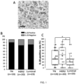

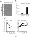
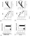


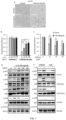

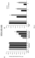
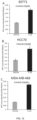

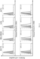
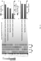
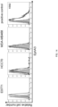
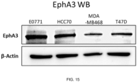
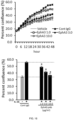
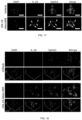
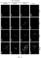


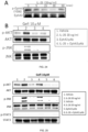

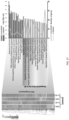
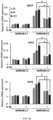

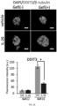

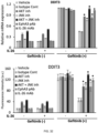
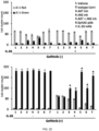
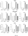


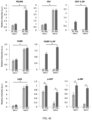


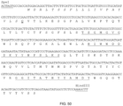
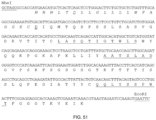
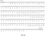
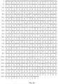


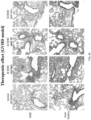
Classifications
-
- A—HUMAN NECESSITIES
- A61—MEDICAL OR VETERINARY SCIENCE; HYGIENE
- A61K—PREPARATIONS FOR MEDICAL, DENTAL OR TOILETRY PURPOSES
- A61K45/00—Medicinal preparations containing active ingredients not provided for in groups A61K31/00 - A61K41/00
- A61K45/06—Mixtures of active ingredients without chemical characterisation, e.g. antiphlogistics and cardiaca
-
- A—HUMAN NECESSITIES
- A61—MEDICAL OR VETERINARY SCIENCE; HYGIENE
- A61K—PREPARATIONS FOR MEDICAL, DENTAL OR TOILETRY PURPOSES
- A61K31/00—Medicinal preparations containing organic active ingredients
- A61K31/33—Heterocyclic compounds
- A61K31/395—Heterocyclic compounds having nitrogen as a ring hetero atom, e.g. guanethidine or rifamycins
- A61K31/41—Heterocyclic compounds having nitrogen as a ring hetero atom, e.g. guanethidine or rifamycins having five-membered rings with two or more ring hetero atoms, at least one of which being nitrogen, e.g. tetrazole
- A61K31/415—1,2-Diazoles
- A61K31/416—1,2-Diazoles condensed with carbocyclic ring systems, e.g. indazole
-
- A—HUMAN NECESSITIES
- A61—MEDICAL OR VETERINARY SCIENCE; HYGIENE
- A61K—PREPARATIONS FOR MEDICAL, DENTAL OR TOILETRY PURPOSES
- A61K31/00—Medicinal preparations containing organic active ingredients
- A61K31/33—Heterocyclic compounds
- A61K31/395—Heterocyclic compounds having nitrogen as a ring hetero atom, e.g. guanethidine or rifamycins
- A61K31/495—Heterocyclic compounds having nitrogen as a ring hetero atom, e.g. guanethidine or rifamycins having six-membered rings with two or more nitrogen atoms as the only ring heteroatoms, e.g. piperazine or tetrazines
- A61K31/505—Pyrimidines; Hydrogenated pyrimidines, e.g. trimethoprim
- A61K31/517—Pyrimidines; Hydrogenated pyrimidines, e.g. trimethoprim ortho- or peri-condensed with carbocyclic ring systems, e.g. quinazoline, perimidine
-
- A—HUMAN NECESSITIES
- A61—MEDICAL OR VETERINARY SCIENCE; HYGIENE
- A61K—PREPARATIONS FOR MEDICAL, DENTAL OR TOILETRY PURPOSES
- A61K31/00—Medicinal preparations containing organic active ingredients
- A61K31/33—Heterocyclic compounds
- A61K31/395—Heterocyclic compounds having nitrogen as a ring hetero atom, e.g. guanethidine or rifamycins
- A61K31/535—Heterocyclic compounds having nitrogen as a ring hetero atom, e.g. guanethidine or rifamycins having six-membered rings with at least one nitrogen and one oxygen as the ring hetero atoms, e.g. 1,2-oxazines
- A61K31/5375—1,4-Oxazines, e.g. morpholine
- A61K31/5377—1,4-Oxazines, e.g. morpholine not condensed and containing further heterocyclic rings, e.g. timolol
-
- A—HUMAN NECESSITIES
- A61—MEDICAL OR VETERINARY SCIENCE; HYGIENE
- A61K—PREPARATIONS FOR MEDICAL, DENTAL OR TOILETRY PURPOSES
- A61K31/00—Medicinal preparations containing organic active ingredients
- A61K31/56—Compounds containing cyclopenta[a]hydrophenanthrene ring systems; Derivatives thereof, e.g. steroids
- A61K31/58—Compounds containing cyclopenta[a]hydrophenanthrene ring systems; Derivatives thereof, e.g. steroids containing heterocyclic rings, e.g. danazol, stanozolol, pancuronium or digitogenin
- A61K31/585—Compounds containing cyclopenta[a]hydrophenanthrene ring systems; Derivatives thereof, e.g. steroids containing heterocyclic rings, e.g. danazol, stanozolol, pancuronium or digitogenin containing lactone rings, e.g. oxandrolone, bufalin
-
- A—HUMAN NECESSITIES
- A61—MEDICAL OR VETERINARY SCIENCE; HYGIENE
- A61K—PREPARATIONS FOR MEDICAL, DENTAL OR TOILETRY PURPOSES
- A61K39/00—Medicinal preparations containing antigens or antibodies
- A61K39/395—Antibodies; Immunoglobulins; Immune serum, e.g. antilymphocytic serum
- A61K39/39533—Antibodies; Immunoglobulins; Immune serum, e.g. antilymphocytic serum against materials from animals
- A61K39/3955—Antibodies; Immunoglobulins; Immune serum, e.g. antilymphocytic serum against materials from animals against proteinaceous materials, e.g. enzymes, hormones, lymphokines
-
- A—HUMAN NECESSITIES
- A61—MEDICAL OR VETERINARY SCIENCE; HYGIENE
- A61P—SPECIFIC THERAPEUTIC ACTIVITY OF CHEMICAL COMPOUNDS OR MEDICINAL PREPARATIONS
- A61P15/00—Drugs for genital or sexual disorders; Contraceptives
-
- A—HUMAN NECESSITIES
- A61—MEDICAL OR VETERINARY SCIENCE; HYGIENE
- A61P—SPECIFIC THERAPEUTIC ACTIVITY OF CHEMICAL COMPOUNDS OR MEDICINAL PREPARATIONS
- A61P35/00—Antineoplastic agents
-
- C—CHEMISTRY; METALLURGY
- C07—ORGANIC CHEMISTRY
- C07K—PEPTIDES
- C07K14/00—Peptides having more than 20 amino acids; Gastrins; Somatostatins; Melanotropins; Derivatives thereof
- C07K14/435—Peptides having more than 20 amino acids; Gastrins; Somatostatins; Melanotropins; Derivatives thereof from animals; from humans
- C07K14/52—Cytokines; Lymphokines; Interferons
- C07K14/54—Interleukins [IL]
-
- C—CHEMISTRY; METALLURGY
- C07—ORGANIC CHEMISTRY
- C07K—PEPTIDES
- C07K14/00—Peptides having more than 20 amino acids; Gastrins; Somatostatins; Melanotropins; Derivatives thereof
- C07K14/435—Peptides having more than 20 amino acids; Gastrins; Somatostatins; Melanotropins; Derivatives thereof from animals; from humans
- C07K14/705—Receptors; Cell surface antigens; Cell surface determinants
- C07K14/71—Receptors; Cell surface antigens; Cell surface determinants for growth factors; for growth regulators
-
- C—CHEMISTRY; METALLURGY
- C07—ORGANIC CHEMISTRY
- C07K—PEPTIDES
- C07K16/00—Immunoglobulins [IGs], e.g. monoclonal or polyclonal antibodies
- C07K16/18—Immunoglobulins [IGs], e.g. monoclonal or polyclonal antibodies against material from animals or humans
- C07K16/24—Immunoglobulins [IGs], e.g. monoclonal or polyclonal antibodies against material from animals or humans against cytokines, lymphokines or interferons
- C07K16/244—Interleukins [IL]
-
- C—CHEMISTRY; METALLURGY
- C07—ORGANIC CHEMISTRY
- C07K—PEPTIDES
- C07K16/00—Immunoglobulins [IGs], e.g. monoclonal or polyclonal antibodies
- C07K16/18—Immunoglobulins [IGs], e.g. monoclonal or polyclonal antibodies against material from animals or humans
- C07K16/28—Immunoglobulins [IGs], e.g. monoclonal or polyclonal antibodies against material from animals or humans against receptors, cell surface antigens or cell surface determinants
- C07K16/2866—Immunoglobulins [IGs], e.g. monoclonal or polyclonal antibodies against material from animals or humans against receptors, cell surface antigens or cell surface determinants against receptors for cytokines, lymphokines, interferons
-
- C—CHEMISTRY; METALLURGY
- C12—BIOCHEMISTRY; BEER; SPIRITS; WINE; VINEGAR; MICROBIOLOGY; ENZYMOLOGY; MUTATION OR GENETIC ENGINEERING
- C12N—MICROORGANISMS OR ENZYMES; COMPOSITIONS THEREOF; PROPAGATING, PRESERVING, OR MAINTAINING MICROORGANISMS; MUTATION OR GENETIC ENGINEERING; CULTURE MEDIA
- C12N15/00—Mutation or genetic engineering; DNA or RNA concerning genetic engineering, vectors, e.g. plasmids, or their isolation, preparation or purification; Use of hosts therefor
- C12N15/09—Recombinant DNA-technology
- C12N15/11—DNA or RNA fragments; Modified forms thereof; Non-coding nucleic acids having a biological activity
- C12N15/113—Non-coding nucleic acids modulating the expression of genes, e.g. antisense oligonucleotides; Antisense DNA or RNA; Triplex- forming oligonucleotides; Catalytic nucleic acids, e.g. ribozymes; Nucleic acids used in co-suppression or gene silencing
- C12N15/1138—Non-coding nucleic acids modulating the expression of genes, e.g. antisense oligonucleotides; Antisense DNA or RNA; Triplex- forming oligonucleotides; Catalytic nucleic acids, e.g. ribozymes; Nucleic acids used in co-suppression or gene silencing against receptors or cell surface proteins
-
- A—HUMAN NECESSITIES
- A61—MEDICAL OR VETERINARY SCIENCE; HYGIENE
- A61K—PREPARATIONS FOR MEDICAL, DENTAL OR TOILETRY PURPOSES
- A61K39/00—Medicinal preparations containing antigens or antibodies
- A61K2039/505—Medicinal preparations containing antigens or antibodies comprising antibodies
-
- C—CHEMISTRY; METALLURGY
- C07—ORGANIC CHEMISTRY
- C07K—PEPTIDES
- C07K2317/00—Immunoglobulins specific features
- C07K2317/20—Immunoglobulins specific features characterized by taxonomic origin
- C07K2317/24—Immunoglobulins specific features characterized by taxonomic origin containing regions, domains or residues from different species, e.g. chimeric, humanized or veneered
-
- C—CHEMISTRY; METALLURGY
- C07—ORGANIC CHEMISTRY
- C07K—PEPTIDES
- C07K2317/00—Immunoglobulins specific features
- C07K2317/70—Immunoglobulins specific features characterized by effect upon binding to a cell or to an antigen
- C07K2317/73—Inducing cell death, e.g. apoptosis, necrosis or inhibition of cell proliferation
-
- C—CHEMISTRY; METALLURGY
- C07—ORGANIC CHEMISTRY
- C07K—PEPTIDES
- C07K2317/00—Immunoglobulins specific features
- C07K2317/70—Immunoglobulins specific features characterized by effect upon binding to a cell or to an antigen
- C07K2317/76—Antagonist effect on antigen, e.g. neutralization or inhibition of binding
-
- C—CHEMISTRY; METALLURGY
- C12—BIOCHEMISTRY; BEER; SPIRITS; WINE; VINEGAR; MICROBIOLOGY; ENZYMOLOGY; MUTATION OR GENETIC ENGINEERING
- C12N—MICROORGANISMS OR ENZYMES; COMPOSITIONS THEREOF; PROPAGATING, PRESERVING, OR MAINTAINING MICROORGANISMS; MUTATION OR GENETIC ENGINEERING; CULTURE MEDIA
- C12N2310/00—Structure or type of the nucleic acid
- C12N2310/10—Type of nucleic acid
- C12N2310/14—Type of nucleic acid interfering nucleic acids [NA]
Definitions
- the present invention relates to a technical field of reversing resistance to anticancer drugs in cancer patients.
- IL-26 was first identified as AK155 in Herpes saimiri infected T-cells (Non-Patent Document 1).
- Human IL-26 a member of the IL-10 cytokine family, is produced mainly by Th1, Th17, or NK cells, and regulates T-cells, monocytes, NK cells, synoviocytes, fibroblasts, and bacterial pathogens in inflammatory lesions (Non-Patent Documents 2 to 6).
- IL-26 is composed of 171 amino acids with a calculated molecular weight of about 19 kDa, and exists as a 36 kDa homodimer in human serum (Non-Patent Document 1).
- Non-Patent Document 4 It has been previously reported by the present inventors that IL-26 produced and secreted by CD4+ effector T-cells activates fibroblasts for collagen production via its functional receptor IL-20RA/IL-10RB, and plays an important role in development of chronic graft-versus-host disease (cGVHD) pulmonary fibrosis (Non-Patent Document 4).
- cGVHD chronic graft-versus-host disease
- Non-Patent Document 1 Non-Patent Documents 5 and 7
- Other reports indicate that IL-26 activated keratinocytes in atopic dermatitis and was exacerbated by increased production of Th17 and Th2-related cytokines in keratinocytes (Non-Patent Document 8).
- IL-26 have also been reported to have antibacterial effects by stimulation of various immune cells and killing of bacteria by membrane pore formation (Non-Patent Document 4). Therefore, it has been suggested that IL-26 plays an important role as a mediator of local inflammation.
- Non-Patent Document 9 It has been reported that an expression level of IL-26 is a poor prognostic factor in hepatocellular carcinoma patients who have undergone surgical resection.
- Non-Patent Document 10 In pancreatic cancer, it has been reported that exosome miR-3607-3p secreted from NK cells suppressed the development of cancer and malignant transformation by targeting IL-26 (Non-Patent Document 11).
- Non-Patent Documents 13 and 14 Previous reports have shown that IL-26 binds to the cell-surface heterodimeric receptor IL-20RA/IL-10RB, leading to downstream signaling cascade events via STAT3 phosphorylation.
- EphA3 Human ephrin type-A receptor 3
- RTK Eph receptor tyrosine kinase
- a structure of EphA3 is well conserved between humans and mice, with about 96% homology and 88% similarity.
- EphA3 is composed of an N-terminal globular ligand-binding domain in the extracellular region, a regulatory juxtamembrane domain having two tyrosine residues, a tyrosine kinase domain, and a PDZ-binding motif in the intracellular cytoplasmic region (Non-Patent Document 15).
- EphA3 regulates cell-cell interactions and plays a role in development and tissue formation in living bodies (Non-Patent Document 16). EphA3 was first identified as a cell surface antigen of pre-B lymphoblastic leukemia cell line (Non-Patent Document 17) and was found to be an antigen of melanoma cells identified by lytic CD4+ T-cells (Non-Patent Document 18).
- Non-Patent Documents 15 and 19 membrane-bound Ephrin A5 induces clustering and tyrosine phosphorylation of EphA3, followed by activation of GTPase or ERK and dephosphorylation of AKT, leading to reorganization of cytoskeleton, cell retraction, and differentiation.
- Non-Patent Documents 15, 25, and 26 binding of unbound soluble ligand to EphA3 causes dephosphorylation of EphA3 by recruitment of protein tyrosine phosphatases and auto-inhibition of their RTK domains, leading to cell growth and proliferation.
- EphA3 is overexpressed in various tumor cells and is involved in maintenance of tumor initiating cells in glioblastoma and leukemia (Non-Patent Documents 16 and 20). Frequent somatic mutations of EphA3 as a receptor tyrosine kinase have been shown in various metastatic cancers (Non-Patent Document 27). Re-expression of wild-type EphA3 in a human lung cancer line including mutant EphA3 leads to a decrease in AKT phosphorylation and leads to tumor apoptosis and growth inhibition (Non-Patent Document 28). Furthermore, a decrease in EphA3 levels leads to suppression of cell proliferation in glioblastoma multiforme (Non-Patent Documents 29 and 30).
- Non-Patent Document 31 In breast cancer, it has been reported that an expression level of EphA3 varies depending on the stage, and EphA3 is highly expressed in lymph node metastasis but is not expressed in a primary tumor (Non-Patent Document 31). However, little is known about potential roles of EphA3 in pathophysiology of breast cancer, including breast cancer without expression of estrogen receptor (ER), progesterone receptor (PR), and human epidermal growth factor receptor 2 (HER2) (Non-Patent Document 32).
- ER estrogen receptor
- PR progesterone receptor
- HER2 human epidermal growth factor receptor 2
- TNBC triple negative breast cancer
- NSCLC non-small cell lung cancer
- CRC colorectal cancer
- Non-Patent Document 38 Anti-EGFR therapies such as EGFR tyrosine kinase inhibitors (EGFR-TKIs) have been successful in treating these cancers.
- EGFR-TKIs EGFR tyrosine kinase inhibitors
- EGFR-TKIs EGFR tyrosine kinase inhibitors
- EGFR-TKIs EGFR tyrosine kinase inhibitors
- Patent Document 1 Japanese Unexamined Patent Application Publication No. 2019 -99492
- IL-26 The precise molecular mechanism of action of IL-26 in cancer biology, including identification of functional receptors for IL-26 in cancer cells and downstream signaling phenomena thereof, has not yet been elucidated. Furthermore, due to a lack of IL-26 in mice and rats, a lack of a suitable mouse non-clinical model has prevented the understanding of the roles thereof in cancer biology of IL-26. The present inventors were able to investigate the role of IL-26 in EGFR-TKI-resistant TNBC by using hIL-26Tg mice.
- IL-26 which is not present in rodents but is present in humans, plays an important role in cell proliferation of EGFR-TKI-treated TNBC via activation of an EGFR bypass pathway.
- the interspecies difference in IL-26 expression has prevented development of suitable mouse models to assess unknown problems such as EGFR-TKI sensitivity and TNBC resistance.
- the present inventors have successfully defined a molecular mechanism involved in resistance via IL-26 to EGFR-TKI and established that IL-26 is an appropriate therapeutic target for TNBC. In other words, by revealing the mechanism involved in EGFR-TKI resistance via IL-26 in TNBC, the contradiction in the interspecies differences between the mouse model and human subjects observed in EGFR-TKI treatment of TNBC was resolved.
- the present inventors have found that exogenous IL-26 inhibits EGFR-TKI-induced suppression of cell proliferation and TNBC growth.
- the present inventors have found that blocking of IL-26 by anti-IL-26 monoclonal antibody (mAb) abolished IL-26 dependent EGFR-TKI resistance of TNBC.
- the present inventors have newly found EphA3 as a novel receptor for IL-26 of TNBC, and have found that its involvement in the induction of a downstream signaling cascade of AKT and JNK phosphorylation promotes cell proliferation even under EGFR-TKI treatment.
- Immunohistochemical assays of TNBC tissue samples have revealed that IL-26 is mainly expressed in TILs and that EphA3 is detected in tumor cells.
- TNBC cell lines used in the studies described in the present description did not express IL-20RA, suggesting that there may be IL-26 receptors other than IL-20RA/IL-10RB in TNBC cells.
- the present inventors have identified EphA3 as a new functional receptor for IL-26 in TNBC.
- exogenous soluble IL-26 induced dephosphorylation of EphA3 in TNBC and then phosphorylation of JNK and AKT, thereby inducing TNBC proliferation even under EGFR-TKI treatment conditions.
- EphA3 related molecular alterations were associated with IL-26 alone, IL-26 separate treatment did not alter TNBC tumor growth. Rather, the effect of IL-26 on TNBC tumor growth has been seen under conditions of EGFR-TKI exposure.
- TNBC tumor suppression has been achieved in the presence of EGFR-TKI agents after blocking the IL-26-EphA3 interaction.
- EGFR-TKI antitumor therapy a major side effect associated with EGFR-TKI antitumor therapy is skin toxicity ( Giovannini M et al., J Oncol 2009, 2009:849051 .). Inhibition of EGFR signaling by TKIs causes early differentiation and induces inflammation and apoptosis, skin atrophy, telangiectasia and photosensitivity in the dermal and epidermal regions ( Lacouture ME, Nat Rev Cancer 2006, 6(10): 803-812 .). The present inventors have recently shown that IL-26 plays an important role in angiogenesis and leukocyte recruitment.
- anti-IL-26 therapy in combination with EGFR-TKI may effectively suppress TNBC proliferation and reduce skin toxicity.
- the present invention has been achieved in a way of focusing on roles of IL-26 present in humans but not in mice by characterizing the molecular mechanisms involved in the interspecies differences in the efficacy of EGFR-TKI in TNBC.
- the present inventors have found that human TNBC are exposed to IL-26 in the tumor microenvironment (TME), activating EGFR-TKI bypass pathway, while suppression of IL-26 overcomes EGFR-TKI resistance in TNBC. This was further confirmed by a tumor-bearing mouse model utilizing human IL-26 transgenic (hIL-26Tg) mice.
- EphA3 as a new functional receptor for IL-26 of TNBC.
- the present invention demonstrates the importance of IL-26 for TNBC EGFR-TKI resistance, and provides a new cancer therapeutic strategy including inhibition of interaction between IL-26 and EphA3 and a combination of EGFR-targeted agents.
- the present invention contributes to the realization of treatment of EGFR-TKI-resistant cancer.
- the present invention relates to a pharmaceutical composition for use in combination with an anticancer drug for treating cancer in a patient, the pharmaceutical composition containing a drug that reverses resistance to an anticancer drug due to IL-26 as an active ingredient. Furthermore, the present invention relates to a method for producing a pharmaceutical composition for treating cancer, containing a drug that reverses resistance to an anticancer drug due to IL-26 as an active ingredient, for use in combination with an anticancer drug. In addition, the present invention also relates to a drug for reversing resistance to an anticancer drug due to IL-26 for use in a method for treating cancer in combination with a chemotherapeutic agent. Also, the present invention relates to a method for treating cancer in a patient, the method including administering an effective amount of an anticancer drug and a drug for reversing resistance to an anticancer drug due to IL-26 to a patient in need thereof.
- cancer and “tumor” are used interchangeably and mean cells that have undergone malignant transformation to be pathogenic for a host organism.
- Primary cancer cells that is, cells obtained from the vicinity of the site of malignant transformation
- the definition of cancer includes not only primary cancers but also any cancer derived from primary cancers, for example, metastatic cancers. Cancers that manifest as solid tumors are detectable based on a tumor mass and can be identified, for example, by CAT scan, MR imaging, X-ray, ultrasound, or palpation, and/or detection of expression of one or more cancer-specific antigens in a sample obtained from a patient.
- the cancers may be, for example, a hematopoietic tumor that is a tumor such as blood cells.
- the cancer in the present description include, but are not limited to, a lung cancer, a non-small cell lung cancer, a small cell lung cancer, a non-Hodgkin's lymphoma, an adrenocortical cancer, an AIDS-related cancer, an AIDS-related lymphoma, a pediatric cerebellar astrocytoma, a pediatric cerebral astrocytoma, a basal cell cancer, a skin cancer (non-melanoma), a biliary tract cancer, an extrahepatic bile duct cancer, an intrahepatic bile duct cancer, a bladder cancer, a cancer of bone or joint, an osteosarcoma and a malignant fibrohistiocytoma, a brain cancer, a brain tumor, a brain stem glioma, a cerebellar astrocytoma, a
- cancers or cancer cells are mammalian cancers or mammalian cancer cells, preferably human cancers or human cancer cells.
- the cancer in the present description may be HER2-negative, and includes, for example, HER2-negative, ER-positive, and PR-positive cancer and ER-negative, PR-negative, and HER2-negative cancer (triple negative cancer), and is preferably ER-negative, PR-negative, and HER2-negative breast cancer (triple negative breast cancer (TNBC)).
- TNBC triple negative breast cancer
- EphA3 is a 110 kDa protein consisting of 983 amino acids, also known as EK4, ETK, HEK, ETK1, HEK4, TYRO4, and belongs to the Ephrin receptor subfamily of the Eph receptor tyrosine kinase (RTK) family (NCBI Reference Sequence: NP_005224.2).
- RTK Eph receptor tyrosine kinase
- EphA3 is composed of an N-terminal globular ligand-binding domain in the extracellular region, a regulatory juxtaposed membrane domain having two tyrosine residues, a tyrosine kinase domain, and a PDZ-binding motif in the intracellular cytoplasmic region.
- Whether or not cancer cells express HER2, ER, PR, and EphA3 can be determined by an immunohistochemical (IHC) method, a fluorescence in situ hybridization (FISH) method, a CISH method, an SISH method, a Southern blot method, a PCR method, a Northern blot method, gene amplification by an RT-PCR method, an IHC method, a Western blot method, an ELISA method, a chemical fluorescence immunoassay method (CLIA method), or the like, using an antibody that recognizes these proteins and a nucleic acid complementary to DNA or RNA encoding these proteins.
- IHC immunohistochemical
- FISH fluorescence in situ hybridization
- the cancer to be treated or prevented according to the present invention may be a cancer having a low sensitivity to an anticancer drug or exhibiting resistance to the anticancer drug.
- the low sensitivity to the anticancer drug may mean that the cancer does not shrink or even proliferates when the anticancer drug is brought into contact with the cancer.
- showing resistance to an anticancer drug may mean that the disease progresses even when treatment with anticancer drug is performed, or the therapeutic effect is gradually weakened and the cancer recurs due to continuous treatment with anticancer drug.
- HTCA method human tumor clonogenic assay
- TIA method thymidine incorporation assay
- SDI method succinic dehydrogenase inhibition test
- CD-DST method Yuuki Takamura et al., Int. J. Cancer: 98, 450-455 (2002 )
- isolated floating cells obtained by enzymatically treating a tumor tissue are cultured in a culture vessel coated with a collagen gel; a mixture of the obtained tumor cells and a collagen solution is gelled and an anticancer drug is added; after serum-free culture, tumor living cells remaining in the collagen gel drop are stained by Neutral Red staining; mixed fibroblasts are erased on an image by image analysis; and the number of cells of only the remaining cancer cells is measured.
- the sensitivity to the anticancer drug is evaluated from a relative growth ratio between the anticancer drug added group and the non-added group.
- an anticancer drug is administered to the cancer patients, and the treatment progress is observed.
- a low therapeutic effect by the anticancer drug it can be determined that the cancer has a low sensitivity to the anticancer drug or exhibits resistance to the anticancer drug.
- IL-26 is a member of the IL-10 cytokine family identified as AK155, and is a molecule composed of 171 amino acids with a calculated molecular weight of about 19 kDa, and present as a 36 kDa homodimer in human serum (Human IL-26 precursor NCBI Reference Sequence: NP_060872.1).
- Whether or not the resistance to the anticancer drug is caused by IL-26 can be determined, for example, by performing the sensitivity or the resistance test for the anticancer drug described above using cancer cells of a mouse not expressing IL-26 in the presence or absence of IL-26 (under the condition of addition or non-addition of IL-26), and determining that the resistance to the anticancer drug is caused by IL-26 in a case where the antitumor effect due to the anticancer drug is further attenuated in the presence of IL-26 (under the addition condition) as compared with the case of absence of IL-26 (under the non-addition condition).
- the antitumor effect by the anticancer drug is determined in the presence or absence of an IL-26 inhibitor (siRNA molecules such as anti-IL-26 antibody or IL-26 dsRNA or IL-26 shRNA, and the like), and in a case where the antitumor effect by the anticancer drug is stronger in the presence of the IL-26 inhibitor (under the addition condition) than in the absence of the IL-26 inhibitor (under the non-addition condition), it can be determined that the resistance of the cells to the anticancer drug is caused by IL-26.
- an IL-26 inhibitor siRNA molecules such as anti-IL-26 antibody or IL-26 dsRNA or IL-26 shRNA, and the like
- a tumor-bearing model mouse transplanted with cancer is prepared using a human IL-26 transgenic mouse and a wild-type mouse (not expressing IL-26), the anticancer drug is administered to both mice to determine the antitumor effect, and in a case where the antitumor effect by the anticancer drug is low in the human IL-26 transgenic mouse, it can be determined that the resistance to the anticancer drug is caused by IL-26.
- the expression level of IL-26 in a cancer tissue or its periphery is measured, and in a case where the expression level of IL-26 is high, it can be determined that the resistance to the anticancer drug is caused by IL-26.
- the resistance to the anticancer drug is reversed by administering an IL-26 inhibitor such as the anti-IL-26 antibody, it can be determined that the resistance to the anticancer drug is caused by IL-26.
- the cancer cells used in these tests may be cancer cells whose resistance to the anticancer drug is already known.
- the resistance to an anticancer drug in the present description may be caused by the interaction between IL-26 and EphA3.
- the interaction between IL-26 and EphA3 refers to an action that contributes to resistance to an anticancer drug by coexistence of IL-26 and EphA3 in a cancer microenvironment, and IL-26 and EphA3 may be bound in the action mechanism thereof.
- Whether or not the resistance to an anticancer drug is caused by the interaction between IL-26 and EphA3 can be determined by examining whether or not the resistance to an anticancer drug due to IL-26 described above is reversed by inhibition of EphA3.
- the sensitivity or the resistance test to the anticancer drug described above is performed in the presence of IL-26 and in the presence or absence of an EphA3 inhibitor (such as an anti-EphA3 antibody) (under the condition of addition or non-addition of an anti-EphA3 antibody), and the antitumor effect of the anticancer drug reduced by IL-26 is restored by addition of the anti-EphA3 antibody, it can be determined that the resistance to the anticancer drug is caused by the interaction between IL-26 and EphA3.
- an EphA3 inhibitor such as an anti-EphA3 antibody
- the resistance to an anticancer drug in the present description may be caused by the phosphorylation of JNK and/or the phosphorylation of AKT due to IL-26.
- the phosphorylation of JNK and/or the phosphorylation of AKT may be caused by the interaction between IL-26 and EphA3.
- Whether or not the resistance to the anticancer drug is caused by the phosphorylation of JNK and/or the phosphorylation of AKT due to IL-26 can be determined by examining whether or not the resistance to the anticancer drug due to IL-26 described above is reversed by a phosphorylation inhibitor of JNK and/or a phosphorylation inhibitor of AKT.
- the resistance to the anticancer drug is caused by the interaction between IL-26 and the phosphorylation of JNK and/or the phosphorylation of AKT.
- the IL-26 may be produced by CD4+ T-cells or macrophages. Whether or not IL-26 is produced by CD4+ T-cells or macrophages can be determined, for example, by examining localization of CD4+ T-cells or macrophages and IL-26 in a tissue sample derived from the cancer patients. When the localization of CD4+ T-cells and IL-26 or macrophage and IL-26 matches, it can both be determined that IL-26 is produced by CD4+ T-cells or macrophages. The localization of IL-26 can be determined using a labeled anti-IL-26 antibody.
- the localization of CD4+ T-cells can be confirmed by immunostaining using a labeled anti-CD4 antibody.
- the localization of macrophages can be confirmed by immunostaining using a labeled anti-F4/80 antibody.
- By changing the color of a dye used for labeling a plurality of factors can be simultaneously labeled, and colocalization can be confirmed as overlapping of both colors.
- the "anticancer drug” means a drug effective for treating or preventing cancer, and includes an antibody drug and an artificially synthesized compound having an anticancer effect.
- the anticancer drug include nitrogen mustards such as cyclophosphamide, ifosfamide, melphalan, busulfan, and thiotepa; alkylating agents such as nitroureas such as nimustine, ranimustine, dacarbacin, procarbacin, temozolomide, carmustine, streptozotocin, and bendamustine; platinum compounds such as cisplatin, carboplatin, oxaliplatin, and nedaplatin; antimetabolites such as enocitabine, capecitabine, carmofur, cladribine, gemcitabine, cytarabine, cytarabine ocphosphate, decafur, decafur/uracil, decafur/gimeracil/oteracil
- the artificially synthesized compound having an anticancer effect is preferably a molecular target drug, and is preferably a kinase inhibitor.
- the kinase inhibitor include tyrosine kinase inhibitors such as epidermal growth factor receptor (EGFR, HER1, ErbB1), HER2 (ErbB2, EGFR2, ERBB2, CD340, and NEU), HER3 (ErbB3), HER4 (ErbB4), VEGFR (VEGFR1, 2, and 3), RET, PDGFR (PDGFR- ⁇ , ⁇ ), FGFR, KIT, c-KIT, Bcr-Abl, and FLT3; Raf kinase inhibitors (sorafenib, vemurafenib, dabrafenib, and the like); MEK inhibitors such as trametinib; CDK inhibitors (palbociclib, abemaciclib, and the like); anaplastic lymphoma kina
- Examples of the EGFR tyrosine kinase inhibitors include gefitinib, erlotinib, osimertinib, afatinib, dacomitinib, imatinib, dasatinib, bosutinib, vandetanib, sunitinib, axitinib, pazopanib, lenvatinib, lapatinib, nintedanib, nilotinib, and ibrutinib. gefitinib or erlotinib is preferable.
- the drug that reverses the resistance of IL-26 to the anticancer drug means a drug that can enhance the sensitivity of cancer cells to the anticancer drug by weakening or losing the resistance to the anticancer drug acquired by the cancer cells due to IL-26.
- it is an IL-26 inhibitor.
- the IL-26 inhibitor examples include siRNA molecules such as IL-26 dsRNA or shRNA, IL-26 antisense, anti-IL-26 antibodies, aptamers against IL-26, and the like.
- siRNA molecules such as IL-26 dsRNA or shRNA
- IL-26 antisense anti-IL-26 antibodies
- aptamers against IL-26 and the like.
- it is the anti-IL-26 antibody or the aptamer against IL-26, which is capable of inhibiting IL-26 in particular in a cancer microenvironment.
- a drug that reverses resistance to the anticancer drug due to IL-26 is a drug that inhibits or reduces interaction between EphA3 and IL-26 in one example.
- the drug that inhibits or reduces the interaction between EphA3 and IL-26 is not particularly limited as long as it is a substance capable of enhancing the sensitivity to the anticancer drug by inhibiting or reducing the interaction between IL-26 and EphA3, and examples thereof include an antibody that inhibits a binding between EphA3 and IL-26 and soluble EphA3. Therefore, the anti-IL-26 antibody described above may be an antibody that further inhibits the binding of IL-26 to EphA3.
- the anti-EphA3 antibody restores the anticancer effect of the anticancer drug attenuated by IL-26
- the anti-EphA3 antibody is also included in the drug that inhibits or reduces the interaction between EphA3 and IL-26.
- IL-26 dephosphorylates EphA3
- an agent that inhibits or reduces the interaction between EphA3 and IL-26 or the anti-IL-26 antibody may have a property of phosphorylating EphA3.
- a candidate drug for example, the anti-IL-26 antibodies
- the antitumor effect due to the anticancer drug in the presence or absence of a drug to be tested in the presence of IL-26 is determined using the cancer cells exhibiting resistance to the anticancer drug in the presence of IL-26 (under the condition of addition of IL-26), and in a case where the antitumor effect due to the anticancer drug is higher in the presence of the drug to be tested than in the absence of the drug to be tested, it can be determined that the drug to be tested reverses the resistance to the anticancer drug due to IL-26.
- the antitumor effect of the anticancer drug on the sample is determined in the presence or absence of the drug to be tested, and in a case where the antitumor effect of the anticancer drug is stronger in the presence of the drug to be tested than in the absence of the drug to be tested, it can be determined that the drug to be tested reverses the resistance of IL-26 to the anticancer drug.
- the resistance to the anticancer drug due to IL-26 is reversed when the antitumor effect is high in the mouse to which the drug to be tested is administered, compared with the mouse to which the drug to be tested is not administered by administering the anticancer drug under the conditions of administration and non-administration of the drug to be tested using a cancer-bearing model mouse in which the drug-resistant cancer is transplanted to the human IL-26 transgenic mouse.
- the anti-IL-26 antibody in the present description is a clone 20-3, 31-4, and 69-10 antibody produced from a hybridoma deposited under Accession Nos. NITE P-02577, NITE P-02578, NITE P-02579, or NITE P-02580, or an antibody having complementarity determining regions of a heavy chain and a light chain thereof, or an antibody having variable regions of a heavy chain and a light chain thereof.
- the anti-IL-26 antibody is an antibody including a heavy chain variable region (each of the first 19 amino acids is a signal sequence, and the mature protein may not contain a signal sequence), including an amino acid sequence described in SEQ ID NO:2 or 12 and a light chain variable region having an amino acid sequence described in SEQ ID NO:4 or 14 (the first 19 or 20 amino acids, respectively, are signal sequences, and the mature protein may not contain a signal sequence), respectively, or an antibody having complementarity determining regions of a heavy chain and a light chain thereof.
- a heavy chain variable region each of the first 19 amino acids is a signal sequence, and the mature protein may not contain a signal sequence
- the anti-IL-26 antibody contains both a heavy chain variable region having an amino acid sequence with at least 80%, 85%, 90%, 95%, 96%, 97%, 98%, or 99% identity to an amino acid sequence described in SEQ ID NO:2 or 12 and a light chain variable region having an amino acid sequence with at least 80%, 85%, 90%, 95%, 96%, 97%, 98%, or 99% identity to an amino acid sequence described in SEQ ID NO:4 or 14.
- the anti-IL-26 antibody may be an antibody having CDRH3 described in SEQ ID NO:7, and preferably may be an antibody having CDRH1, CDRH2, and CDRH3 with amino acid sequences described in SEQ ID NOs:5, 6, and 7, respectively.
- the antibody may further have the amino acid sequences described in SEQ ID NOs:8, 9, and 10 as CDRL1, CDRL2, and CDRL3.
- the anti-IL-26 antibody may be an antibody having CDRH3 described in SEQ ID NO:17, and preferably may be an antibody having CDRH1, CDRH2, and CDRH3 having amino acid sequences described in SEQ ID NOs:15, 16, and 17, respectively.
- the antibody may further have the amino acid sequences described in SEQ ID NOs:18, 19, and 20 as CDRL1, CDRL2, and CDRL3.
- the humanized anti-IL-26 antibody may be an antibody that binds to IL-26 or an antigen-binding fragment thereof.
- a heavy chain variable region (VH) has an amino acid sequence with 80% or more of identity to the amino acid sequence described in SEQ ID NO:25
- a light chain variable region (VL) has an amino acid sequence with 80% or more of identity to the amino acid sequence described in SEQ ID NO:30, or has an amino acid sequence with 80% or more of identity to the amino acid sequence described in SEQ ID NO:35.
- the humanized anti-IL-26 antibody is an antibody whose VH has the amino acid sequence described in SEQ ID NO:25 and whose VL has the amino acid sequence described in SEQ ID NO:30 or the amino acid sequence described in SEQ ID NO:35, or an antigen-binding fragment thereof.
- the humanized anti-IL-26 antibody may be an antibody or an antigen-binding fragment thereof.
- the heavy chain has the amino acid sequence described in SEQ ID NO:37 at positions 20 to 472 and the light chain has the amino acid sequence described in SEQ ID NO:39 at positions 21 to 234 or the amino acid sequence described in SEQ ID NO:41 at positions 21 to 234.
- the anti-IL-26 antibody and the anti-EphA3 antibody used in the present description specifically bind to IL-26 and EphA3, respectively.
- the anti-IL-26 antibody includes an antibody that binds to the same epitope as at least one antibody selected from the 20-3 antibody, the 31-4 antibody, and the 69-10 antibody that are mouse monoclonal antibodies.
- the anti-IL-26 antibody may be an antibody that inhibits the binding of at least one antibody selected from the 20-3 antibody, the 31-4 antibody, and the 69-10 antibody, which are mouse monoclonal antibodies, to IL-26 in a competition assay.
- binding affinity may be determined using a BIAcore biosensor, a KinExA biosensor, a scintillation proximity assay, ELISA, ORIGEN immunoassay (IGEN), fluorescence quenching, fluorescence transfer, and/or yeast display.
- the binding affinity may also be screened using appropriate bioassays.
- One way to determine the binding affinity of an antibody for IL-26 is to measure the affinity of a monofunctional Fab fragment of the antibody.
- antibodies e.g., IgG
- the affinity of an anti-IL-26 Fab fragment of a monoclonal antibody can be determined by a surface plasmon resonance (SPR) system (BIAcore 3000 (trademark), BIAcore, Piscaway, NJ) .
- SA chips streptavidin are used according to the supplier's instructions.
- Biotinylated IL-26 can be diluted in HBS-EP (100 mM HEPES pH 7.4, 150 mM NaCl, 3 mM EDTA, 0.005% P20) and injected onto the chip at a concentration of 0.005 mg/mL. Using variable flow times across individual chip channels, two ranges of antigen density are achieved: 10 to 20 response units (RU) for detailed kinetic testing and 500 to 600 RU for concentration. The mixture of Pierce elution buffer and 4M NaCl (2 : 1) efficiently removes bound Fab while preserving the activity of IL-26 on the chip for 200 or more injections. HBS-EP buffer can be used for all BIAcore assays as a running buffer.
- a step-wise diluted solution of purified Fab samples (0.1 to 10 ⁇ estimated KD) is injected at 100 ⁇ L/min for 2 min and a dissociation time up to 30 min is usually tolerated.
- Concentration of Fab protein can be determined by ELISA and/or SDS-PAGE electrophoresis using known concentrations (determined by amino acid analysis) of standard Fab.
- a binding rate (kon) and a dissociation rate (koff) of reaction kinetics are simultaneously obtained by fitting the data to a 1 : 1 Langmuir binding model (Lofas & Johnsson, 1990) using the BIA evaluation program.
- An equilibrium dissociation constant (KD) value is calculated as koff/kon.
- the competition assay can be used to determine whether two antibodies bind to the same epitope by recognizing the same or sterically overlapping epitope.
- the antigen is immobilized on a multi-well plate, and the performance of the unlabeled antibody to block the binding of the labeled antibody is measured.
- Common labels for such competition assays are radiolabels or enzyme labels.
- epitopes to which an antibody binds can be determined by using epitope mapping techniques known to those skilled in the art.
- whether or not an antibody to be tested competes with at least one antibody selected from 20-3 antibody, 31-4 antibody and 69-10 antibody in binding to IL-26 can be determined by contacting at least one antibody selected from labeled 20-3 antibody, 31-4 antibody and 69-10 antibody with an antigen (IL-26) together with the antibody to be tested.
- an antigen (IL-26) IL-26
- a control one obtained by contacting at least one antibody selected from 20-3 antibody, 31-4 antibody and 69-10 antibody labeled in the absence of the antibody to be tested with an antigen (IL-26) alone is used.
- a labeling amount in the presence of the antibody to be tested is smaller than the labeling amount in the absence of the antibody to be tested by comparing the labeling amount of at least one antibody selected from the labeled 20-3 antibody, 31-4 antibody and 69-10 antibody bound to the antigen in the presence or absence of the antibody to be tested, it can be determined that the antibody to be tested competes with the labeled antibody used.
- the "antibody” is an immunoglobulin molecule capable of specifically binding to a target such as a carbohydrate, polynucleotide, lipid, or antibody via at least one antigen recognition site located in a variable region of the immunoglobulin molecule.
- a target such as a carbohydrate, polynucleotide, lipid, or antibody via at least one antigen recognition site located in a variable region of the immunoglobulin molecule.
- the term antibody includes intact polyclonal or monoclonal antibodies as well as fragments thereof. It is known that a variable region (in particular, CDRs) of the antibody imparts a binding property, and it is widely known to those skilled in the art that even an antibody fragment that is not a complete antibody can use the binding property.
- fragment or "antigen-binding fragment” of an antibody means a protein or peptide containing a part (partial fragment) of an antibody and retaining the action (immunoreactivity/binding property) of the antibody on the antigen.
- immunoreactive fragments include F(ab') 2 , Fab', Fab, Fab 3 , single chain Fv (in the present description hereinafter, referred to as "scFv"), (tandem) bi-specific single chain Fv(sc(Fv) 2 ), single chain triple body, nanobody, divalent VHH, pentavalent VHH, minibody, (double chain) diabody, tandem diabody, bi-specific tribody, bi-specific bibody, dual affinity retargeting molecule (DART), triabody (or tribody), tetrabody (or [sc(Fv) 2 ] 2 or (scFv-SA) 4 ) disulfide-bonded Fv (in the present description hereinafter, referred to as "dsFv”), compact IgG, heavy chain antibody, or polymers thereof ( Nature Biotechnology, 29(1): 5-6 (2011 ); Maneesh Jain et al., TRENDS in Biotechnology, 25(7)(2007): 307
- the immunoreactive fragments may be any of mono-specific, bi-specific (bispecificity), tri-specific (trispecificity), and multi-specific (multispecificity).
- the fragment of the antibody may include a fragment having a length of at least about 10 amino acids, at least about 25 amino acids, at least about 50 amino acids, at least about 75 amino acids, or at least about 100 amino acids.
- Antibodies include any class of antibodies such as IgG, IgA, or IgM (or subclasses thereof) and need not be a particular class. Immunoglobulins are classified into different classes according to the antibody amino acid sequence of constant domains of the heavy chain. There are five main immunoglobulin classes: IgA, IgD, IgE, IgG, and IgM, some of which may be further subdivided into subclasses (isotypes), e.g., IgG1, IgG2, IgG3, IgG4, IgA1, and IgA2.
- the constant domains of the corresponding heavy chains of different classes of immunoglobulins are referred to as ⁇ , ⁇ , ⁇ , ⁇ , and ⁇ , respectively.
- Subunit structures and three-dimensional structures for different classes of immunoglobulins are well known.
- the antibody may be an IgG antibody, particularly an IgG1 antibody or an IgG2 antibody, or may be a human IgG antibody.
- the antibody in the present description can be a monoclonal antibody or a polyclonal antibody, and is preferably a monoclonal antibody.
- the term "monoclonal antibody” refers to an antibody obtained from a cell population that produces a substantially homogeneous antibody. In other words, the individual antibodies contained in the cell population are the same except for natural mutants that may be slightly present.
- the monoclonal antibodies are directed against a single antigenic site and are highly specific. Furthermore, in contrast to typical polyclonal antibodies containing different antigens targeting different determinants (epitopes), each monoclonal antibody targets a single determinant of the antigen.
- the modifier "monoclonal” indicates characteristics of the antibody as being obtained from a substantially homogeneous population of antibody-producing cells, and is not to be understood as requiring production of the antibody via a particular method.
- the monoclonal antibodies used according to the present invention may be prepared by recombinant DNA methods as described in US Patent No. 4,816,567 .
- the monoclonal antibodies can also be isolated, for example, from phage libraries generated using the techniques described in McCafferty et al., 1990, Nature, 348: 552-554 .
- the antibody in the present description can be chimeric antibodies, humanized antibodies, fully human antibodies, or animal antibodies adapted to any animal to be treated other than human, and is preferably a humanized antibody.
- a "humanized” antibody is an antibody or fragment thereof (Fv, Fab, Fab', F(ab') 2 , or other antigen-binding subsequences of antibodies, and the like) containing a minimal sequence derived from non-human immunoglobulin and a sequence derived from human.
- humanized antibodies are human immunoglobulins (recipient antibodies) in which residues from the complementarity determining region (CDR) of the receptor are replaced with residues from the CDRs of a non-human species (donor antibodies), such as mice, rats, or rabbits, having the desired specificity, affinity, and capacity.
- donor antibodies such as mice, rats, or rabbits
- Fv framework region (FR) residues of human immunoglobulins may be substituted by corresponding non-human-derived residues.
- the humanized antibodies include variable regions derived from non-human species (the donor antibodies), such as mice, rats, or rabbits, having the desired specificity, affinity, and/or capacity, although residues of one or more Fv framework regions and/or one or more CDR residues may be substituted by corresponding human residues (that is, residues derived from human antibody sequences).
- Humanized antibodies can contain residues that are not present in the transferred original CDR or framework sequences for further improvement and optimization of antibody performance.
- the humanized antibodies most preferably contain at least a portion of a constant region (Fc) or domain of a human immunoglobulin (generally a human immunoglobulin).
- Some humanized antibodies have modified Fc regions as described in WO 99/58572 .
- Some forms of humanized antibodies have one or more (for example, 1, 2, 3, 4, 5, 6) CDRs altered relative to the original antibody, which are also referred to as one or more CDRs "derived from" one or more CDRs of the original antibody.
- the antibodies contain one or more constant regions, e.g., human constant regions.
- the constant regions can be a constant region of the heavy chain and/or a constant region of the light chain.
- the antibodies may contain a constant region having at least about 80%, at least about 85%, at least about 90%, at least about 95%, at least about 98%, or 100% identity to a human constant region.
- the antibodies may contain an Fc region, e.g., a human Fc region.
- the antibodies may contain an Fc region having at least about 80%, at least about 85%, at least about 90%, at least about 95%, at least about 98%, or 100% identity to a human Fc region.
- the antibodies described in the present description may be modified, examples of such modifications include functionally equivalent antibodies and variants with enhanced or attenuated activity that do not significantly affect properties of the antibodies. Modifications of antibodies are routine manipulations in the art and need not be described in detail in the present description. Examples of modifications include conservative substitutions of amino acid residues, insertion, addition, or deletion of one or more amino acids that do not significantly degrade functional activity, or the use of chemical analogs.
- the insertion or addition of amino acid sequences includes addition of an amino acid to the amino and/or carboxyl terminus in length, and insertion within the sequence of a single or more amino acid residues. Examples of terminus insertions include antibodies with N-terminal methionyl residues or fused to epitope tags.
- Other insertion variants of antibody molecules include fusion of an enzyme or antibody that extends serum half-life of the antibody to the N-terminus or C-terminus of the antibody.
- a substituted variant at least one amino acid residue of the antibody sequence is removed, and a different residue is inserted at the position.
- the sites where the utmost effect is obtained by substitutional mutagenesis include CDRs, but FRs are also intended to be changed.
- the conservative substitutions are shown in Table 1. Where such substitutions result in altered biological activity, more substantial changes may be introduced to screen the products, designated as "exemplary substitutions" in Table 1, or as further described below with respect to amino acid classes.
- Substantial modifications in the biological properties of antibodies are achieved by selecting substitutions that significantly alter the effect of modification on (a) the structure of the antibody backbone in the substituted region, e.g., sheet or helix conformation, (b) charges or hydrophobicity of molecules at a target site, or (c) maintenance of volume of a side chain.
- the residues present in a natural state are classified into the following groups based on common side chain properties:
- Non-conservative substitutions are made by exchanging one member of these classes for another class. More conservative substitutions include exchanging one member of a class for another member of the same class.
- Amino acid substitutions in the variable region can alter binding affinity and/or specificity. For example, no more than 1 to 5 conservative amino acid substitutions are made within the CDR domain. For example, no more than 1 to 3 conservative amino acid substitutions are made within the CDR3 domain.
- the antibodies described in the present description also include glycosylated and aglycosylated antibodies as well as antibodies with other post-translational modifications such as, glycosylation, acetylation, and phosphorylation at different sugars.
- the antibodies are glycosylated at conserved positions in their constant regions ( Jefferis and Lund, 1997, Chem. Immunol. 65: 111-128 ; Wright and Morrison, 1997, TibTECH 15: 26-32 ). It has been reported that the absence of fucose in the Fc region of antibodies has an effect on antibody-dependent cellular cytotoxicity (ADCC).
- ADCC antibody-dependent cellular cytotoxicity
- the present invention relates to a pharmaceutical composition for improving the sensitivity of cancer cells to an anticancer drug, the pharmaceutical composition containing a drug that reverses resistance to the anticancer drug due to IL-26 as an active ingredient.
- the present invention relates to a method for producing a pharmaceutical composition for improving the sensitivity of cancer cells to the anticancer drug, containing a drug that reverses resistance to an anticancer drug due to IL-26 as an active ingredient.
- the present invention also relates to a drug for reversing resistance to the anticancer drug due to IL-26 for use in a method for improving the sensitivity of cancer cells to the anticancer drug.
- the present invention relates to a method for improving the sensitivity of cancer cells to an anticancer drug, the method including administering an effective amount of the anticancer drug and a drug for reversing resistance to the anticancer drug due to IL-26 to a patient in need thereof.
- the present invention relates to a pharmaceutical composition for suppressing proliferation of cancer cells, the pharmaceutical composition containing a drug that reverses resistance to the anticancer drug due to IL-26 as an active ingredient.
- the present invention relates to a method for producing a pharmaceutical composition for suppressing proliferation of cancer cells, containing a drug that reverses resistance to the anticancer drug due to IL-26 as an active ingredient.
- the present invention also relates to a drug for reversing resistance to the anticancer drug due to IL-26 for use in a method for suppressing proliferation of cancer cells.
- the present invention relates to a method for suppressing the proliferation of cancer cells, the method including administering an effective amount of anticancer drug and a drug for reversing the resistance to the anticancer drug due to IL-26 to a patient in need thereof.
- the pharmaceutical composition in the present description may contain a drug that reverses resistance to the anticancer drug due to IL-26 as the only active ingredient, or may contain other active ingredients.
- a drug that reverses resistance to the anticancer drug due to IL-26 may be administered as the only active ingredient, or may be administered together with other active ingredients.
- the "cancer cells” may be cancer cells having a low sensitivity to the anticancer drug or exhibiting resistance to the anticancer drug.
- the present invention relates to a nucleic acid molecule having a polynucleotide encoding an anti-IL-26 antibody or immunoreactive fragment thereof as described above.
- the nucleic acid molecule of the present invention may be a nucleic acid molecule having a polynucleotide encoding the amino acid described in SEQ ID NO:2 or 12 as VH and having a polynucleotide encoding the amino acid sequence described in SEQ ID NO:4 or 14 as VL.
- the nucleic acid molecule of the present invention is a nucleic acid molecule having a polynucleotide (for example, it has a base sequence described in SEQ ID NO:1) encoding the amino acid sequence described in SEQ ID NO:2 as VH and a polynucleotide (for example, it has a base sequence described in SEQ ID NO:3) encoding the amino acid sequence described in SEQ ID NO:4 as VL; a polynucleotide encoding the amino acid sequence described in SEQ ID NO:12 as VH (for example, it has a base sequence described in SEQ ID NO:11) and a polynucleotide encoding the amino acid sequence described in SEQ ID NO: 14 as VL (for example, it has a base sequence described in SEQ ID NO:13).
- a polynucleotide for example, it has a base sequence described in SEQ ID NO:1 encoding the amino acid sequence described in SEQ ID NO:2 as VH and
- the nucleic acid molecule of the present invention may be a nucleic acid molecule containing a polynucleotide encoding a VH having a base sequence described in SEQ ID NO:24 and a polynucleotide encoding a VL having a base sequence described in SEQ ID NO:29 or SEQ ID NO:34.
- the nucleic acid molecule of the present invention may be a nucleic acid molecule containing a polynucleotide encoding a heavy chain having a base sequence described in positions 58 to 1416 of SEQ ID NO:36 and a polynucleotide encoding a light chain having a base sequence described in positions 61 to 702 of SEQ ID NO:38 or positions 61 to 702 of SEQ ID NO:34.
- the present invention includes a vector having the nucleic acid molecule.
- a vector is not particularly limited as long as it is a vector that can be used for expression of an antibody, and can be selected according to a host using an appropriate viral vector or plasmid vector.
- the present invention relates to host cells containing the vector.
- the host cells are not particularly limited as long as it is a host cell that can be used for expression of the antibodies, and examples thereof include mammalian cells (mouse cells, rat cells, rabbit cells, human cells, and the like), yeast, and microorganisms (E. coli and the like).
- the "host cells” include an individual cell or cell culture that may or has already been a recipient of the vector that incorporates a polynucleotide insert.
- the host cells include progeny of a single host cell, and the progeny may not necessarily be identical (morphologically or in genomic DNA complement) to the original parent cell due to natural, accidental, or deliberate mutations.
- the host cells include cells transfected in vivo with the polynucleotides of the invention.
- treatment is an approach for obtaining beneficial or desired clinical results.
- beneficial or desired clinical results include, but are not limited to, alleviation of one or more symptoms, diminishment of extent of disease, stabilized (that is, it is not deteriorated) disease state, delay or slowing of disease progression, amelioration or palliation of disease state, and remission (partial or total), whether detectable or undetectable.
- the "treatment” may also mean prolonging life expectancy relative to expected life expectancy if not receiving treatment.
- the "effective amount” is an amount sufficient to achieve beneficial or desired clinical results, including clinical results.
- the effective amount can be administered in one or more administrations.
- the effective amount of a drug for reversing the resistance to the anticancer drug due to IL-26 or a drug for reversing the resistance to the anticancer drug due to IL-26 and an anticancer drug described in the present description is an amount sufficient to reverse resistance of the anticancer drug, delay the progression of a state related to tumor growth, or reduce a tumor size.
- the effective amount of the drug that reverses the resistance of the IL-26 to the anticancer drug and the other anticancer drug may vary or depend on other factors such as the patient medical history, and the drug that reverses the resistance of the IL-26 to the anticancer drug used and the type (and/or amount) of the anticancer drug used in combination, among others.
- compositions in the present description may optionally contain pharmaceutically acceptable additives.
- pharmaceutically acceptable additives When the “pharmaceutically acceptable additive" is combined with an active ingredient, the active ingredient is capable of maintaining biological activity, the “pharmaceutically acceptable additive” is non-reactive with the subject's immune system when delivered, and includes any material that is non-toxic to the subject. Examples include, but are not limited to, phosphate buffered saline, water, emulsions such as oil/water emulsions, and any standard pharmaceutical carrier such as various types of wetting agents. Preferred diluents for spray or parenteral administration are phosphate buffered saline or saline (0.9%).
- compositions containing such carriers are formulated by well-known conventional methods (see, e.g., Remington's Pharmaceutical Sciences, 18th edition, A. Gennaro ed., Mack Publishing Co., Easton, PA, 1990 and Remington, The Science and Practice of Pharmacy 20th Ed., Mack Publishing, 2000 ).
- the antibody is expressed in any organism or host cell derived from any organism, including but not limited to bacteria, yeast, plants, insects, and mammals.
- Specific types of cells include, but are not limited to, Drosophila melanogaster cells, Saccharomyces cerevisiae and other yeasts, E. coli, Bacillus subtilis, SF9 cells, HEK-293 cells, Neurospora, BHK cells, CHO cells, COS cells, Hela cells, fibroblasts, Schwann cell lines, immortalized mammalian bone marrow and lymphoid cell lines, Jurkat-cells, mast-cells and other endocrine and exocrine cells, and neuronal cells.
- GS glutamine synthase
- the present invention may be a complex in which a drug that reverses resistance to the anticancer drug due to IL-26 is bound to an anticancer drug.
- the present invention relates to a combination drug of a drug for reversing the resistance to an anticancer drug due to IL-26 and an anticancer drug. Therefore, the present invention is a pharmaceutical composition for treating cancer, including a pharmaceutical composition containing (i) an anticancer drug and (ii) a drug that reverses resistance to an anticancer drug due to IL-26.
- the drug that reverses resistance to the anticancer drug due to IL-26 and the anticancer drug can be contained in the same preparation or in separate preparations.
- the drug that reverses resistance to the anticancer drug due to IL-26 can be administered simultaneously with the anticancer drug, sequentially, after administration of the anticancer drug or before administration of the anticancer drug.
- the pharmaceutical composition that is a combination drug may contain only (i) an anticancer drug and (ii) a drug that reverses resistance to an anticancer drug due to IL-26, or may contain other active ingredients.
- the present invention relates to a kit for treating cancer, containing (i) an anticancer drug and (ii) a drug that reverses resistance to the anticancer drug due to IL-26.
- the kit can include an outer box, a container, a diluent, a turbid solution, and/or instructions on a preparation method and an administration method.
- kits different components among (i) the anticancer drug and (ii) the drug reversing the resistance to the anticancer drug due to the IL-26 may be packaged in separate containers and included in one kit, or only one component among (i) the anticancer drug and (ii) the drug reversing the resistance to the anticancer drug due to the IL-26 may be included in the kit, and another component may be provided separately from the kit.
- the pharmaceutical composition of the present invention is useful for various applications including a therapeutic method and a method for lysing malignant cells, but is not limited thereto.
- the present invention also provides a method for inhibiting the proliferation of cancer cells, wherein the inhibition of the proliferation of cancer cells contains any observable level of inhibition, including partial or complete inhibition of proliferation.
- the cell proliferation is inhibited by at least about 10%, at least about 25%, at least about 50%, at least about 75%, at least about 90%, at least about 95%, at least about 98%, or about 100%.
- This method may be performed in vitro or in vivo.
- the method for inhibiting proliferation of cancer cells includes contacting the cells with a pharmaceutical composition described in the present description. Generally, the cells are contacted with a sufficient amount of a pharmaceutical composition to inhibit the cell proliferation.
- the humanized anti-human IL-26 antibody or the antigen-binding fragment thereof of the present invention is useful as a therapeutic agent for various diseases caused by inhibiting the activity of human IL-26 and enhancing the IL-26 activity.
- diseases associated with IL-26 include refractory immune disorders and cancers.
- Specific target diseases include diseases selected from acute or chronic graft-versus-host disease, post-transplant rejection, autoimmune diseases, atopic dermatitis, cancers, idiopathic pulmonary hypertension, chronic obstructive pulmonary disease, idiopathic pulmonary fibrosis, bronchiolitis obliterans, viral hepatitis, liver cirrhosis, nephrosclerosis, chronic glomerulonephritis, and idiopathic nephrotic syndrome.
- examples of the autoimmune diseases include diseases selected from rheumatoid arthritis, systemic lupus erythematosus, psoriasis, psoriatic arthritis, polymyositis, dermatomyositis, scleroderma, Sjogren's syndrome, vasculitis syndrome, mixed connective tissue disease, autoimmune hepatitis, primary biliary cirrhosis, primary sclerosing cholangitis, autoimmune pancreatitis, ulcerative colitis, Crohn's disease, idiopathic thrombocytopenic purpura, autoimmune hemolytic anemia, autoimmune neutropenia, type I diabetes, pemphigus, pemphigoid, frequent miscarriage, causative diseases, and autoimmune optic neuritis.
- diseases selected from rheumatoid arthritis, systemic lupus erythematosus, psoriasis, psoriatic arthritis, polymyos
- examples of the cancers include head and neck cancer, esophageal cancer, gastric cancer, colon cancer, rectal cancer, liver cancer, gallbladder/bile duct cancer, pancreatic cancer, lung cancer, breast cancer, ovarian cancer, bladder cancer, prostate cancer, bone/soft part sarcoma, malignant lymphoma, leukemia, multiple myeloma, cervical cancer, skin cancer, brain tumor, and the like.
- the methods (including therapeutic methods) described in the present description may be made by a single direct injection at a single time point or multiple time points to a single site or multiple sites.
- the administration may also be administered to multiple sites at about the same time. Frequency of administration is determined and adjusted over a course of treatment, and is based on results desired to be achieved.
- sustained release formulations of the pharmaceutical compositions of the present invention may be suitable. Various formulations and devices for achieving sustained release are well known in the art.
- the subject to be treated includes mammals such as human, bovine, equine, canine, feline, porcine, and ovine, and is preferably human.
- the pharmaceutical composition of the present invention may adopt any oral or parenteral preparation as long as it is a preparation that can be administered to a patient.
- Examples of the composition for parenteral administration include injections, nasal drops, suppositories, patches, ointments, and the like. It is preferably an injection.
- Examples of dosage forms of the pharmaceutical composition of the present invention include a liquid preparation and a lyophilized preparation.
- additives including solubilizing agents such as propylene glycol, ethylenediamine, and the like; buffering agents such as phosphate; isotonizing agents such as sodium chloride, glycerin, and the like; stabilizers such as sulfite; preservatives such as phenol; and soothing agents such as lidocaine, can be added as necessary (refer to " Handbook of Pharmaceutical Excipients Fifth Edition" APhA Publications, Yakuji Nippo Limited ).
- examples of storage containers can include an ampoule, a vial, a prefilled syringe, a pen-type syringe cartridge, a drip bag, and the like.
- the present invention relates to a method for improving the responsiveness to the anticancer drug in cancer patients having a low sensitivity to the anticancer drug or exhibiting resistance to the anticancer drug, the method including inhibiting IL-26 in the cancer patients.
- the method may include inhibiting or reducing the interaction between EphA3 and IL-26.
- the present invention is a method for treating cancer in cancer patients having a low sensitivity to the anticancer drug or exhibiting resistance to the anticancer drug, the method including inhibiting IL-26 in the cancer patients and administering the anticancer drug to the patients.
- the method may include inhibiting or reducing the interaction between EphA3 and IL-26.
- the order of the inhibition of IL-26 and the administration of the anticancer drug in the cancer patients does not need to be this order, and either may precede.
- the anticancer drug may be administered after the inhibition of IL-26, IL-26 may be inhibited after the administration of the anticancer drug, or both may be performed simultaneously or sequentially.
- the inhibition of IL-26 in the cancer patients is preferably an inhibition of IL-26 in a microenvironment of the cancer to be treated.
- the inhibition of IL-26 is performed by administering a pharmaceutical composition containing a drug that reverses resistance to the anticancer drug due to IL-26 of the present invention as an active ingredient.
- composition of the present invention may be administered to a mammal by injection (for example, systemically, intravenously, intraperitoneally, subcutaneously, intramuscularly, intraportally, or into cancer tissues) or by other methods that ensure delivery to bloodstream in an effective form (for example, injection).
- injection for example, systemically, intravenously, intraperitoneally, subcutaneously, intramuscularly, intraportally, or into cancer tissues
- other methods that ensure delivery to bloodstream in an effective form (for example, injection).
- the effective dose and schedule for administering the pharmaceutical compositions of the present invention are empirically determined, and such methods of determination are within the common general knowledge in the art.
- the dose of the pharmaceutical composition of the present invention to be administered varies depending on, for example, a mammal inoculating the pharmaceutical composition of the present invention, a route of administration, a specific type of agent used, and other agents administered to the mammal.
- a typical dosage per day of the pharmaceutical composition of the present invention may range from about 1 ⁇ g/kg body weight to 100 mg/kg body weight or more per day, depending on the aforementioned factors.
- any of the dosages below may be used: at least about 50 mg/kg body weight; at least about 10 mg/kg body weight; at least about 3 mg/kg body weight; at least about 1 mg/kg body weight; at least about 750 ⁇ g/kg body weight; at least about 500 ⁇ g/kg body weight; at least about 250 ⁇ g/kg body weight; at least about 100 ⁇ g/kg body weight; at least about 50 ⁇ g/kg body weight; at least about 10 ⁇ g/kg body weight; a dosage of at least about 1 ⁇ g/kg body weight, or more, is administered.
- the pharmaceutical composition of the present invention may be further administered in combination with one or more therapeutic agents other than the anticancer drug in an effective amount to a mammal.
- the pharmaceutical compositions of the present invention may be administered sequentially or simultaneously with one or more other therapeutic agents. After administration of the pharmaceutical composition of the present invention to a mammal, physiological conditions of the mammal can be monitored in various ways well known to those skilled in the art.
- Mouse breast cancer E0771 cells were purchased from CH3 Biosystems (Amherst, NY).
- Human mammary gland cancer cell lines MDA-MB-468 human TNBC cell line
- HCC70 human TNBC cell line
- T47D ER/PR-positive, HER2-negative cell line
- the human large intestine cancer cell line COLO 205 was purchased from RIKEN BioResource Center (Tsukuba, Japan).
- Human umbilical vein endothelial cells HUVECs were purchased from LONZA (Walkersville, MD, USA) and cultured in EGM medium (LONZA).
- IL-26 dimers for cell stimulation were purchased from R&D Systems (Minneapolis, MN).
- the recombinant human IL-26 dimers were labeled with Alexa Fluor 488 using a protein labeling kit (Thermo Fisher Scientific, Waltham, MA) and used for immunofluorescence staining.
- Recombinant human and mouse EphA3 Fc chimeric proteins were purchased from R&D Systems.
- EGFR-TKI and the inhibitor used in this example are shown in Table 2.
- RNA oligonucleotide sequence not disclosed
- Qiagen Hilden, Germany
- EGFR-TKI and inhibitor list Product name The primary target of the inhibition ⁇ Conc Company Gefitinib (ZD1839) EGFR 5-40 ⁇ M Selleck Chemicals (Houston, TX, USA).
- Erlotinib CP358774) EGFR 30, 50 ⁇ M Selleck Chemicals wortmannin Akt Low: 0.375, Mid: 0.75, High: 1.5 ⁇ M Merck Millipore SP600125 JNK Low: 0.3, Mid: 1.0, High: 3.0 ⁇ M Tocris Biosystems
- clones 69-10, 20-3, 31-4, and 2-2 mAbs described in Hatano R et al., MAbs (2019) 1-15 , and JP 2009-99492 A were used.
- Hybridomas producing clones 69-10, 20-3, 31-4, and 2-2 mAbs have been deposited with National Institute of Technology and Evaluation Patent Microorganisms Depositary (NPMD) under Accession Nos. NITE P-02577, NITE P-02578, NITE P-02579, and NITE P-02580, respectively, in Kazusakamatari 2-5-8, Kisarazu-shi, Chiba, Japan on November 21, 2017.
- APC anti-mouse IL-10RB mAb (clone 547324) R&D Systems APC anti-human IL-20RA mAb (clone 173714) R&D Systems PE anti-human IL-10RB mAb (clone 90220) R&D Systems rabbit IgG.
- polyclonal - Isotype Control abeam APC rat IgG2a, Isotype Control (clone RTK2758) BioLegend APC mouse IgG1, Isotype Control (clone MOPC-21) BioLegend PE mouse IgG1, Isotype Control (clone MOPC-21) BioLegend PE anti-rabbit IgG BioLegend Immunohistochemistry rabbit anti-human IL-26 mAb (clone EPR22268-141) abcam rabbit anti-human EphA3 pAb Gene Tex (San Antonio. TX) HRP-Linked Goat anti-rabbit IgG abeam
- the human IL-26 transgenic mouse (hIL-26Tg mouse) from Thomas
- mice Provided by Dr. Aune, it was crossed with C57BL/6 mouse in an animal facility at Juntendo University. A female hIL-26Tg mouse of 8-12 weeks old and littermate control mice were used. NOD/Shi-scid, IL-2R ⁇ KO Jic (Cg-PrkdcscidI12rgtm1Sug/Jic) mice (in the present description hereinafter, NOG mice), ⁇ were purchased from Central Institute for Experimental Animals (Kawasaki, Japan). All mice used in this experiment were housed under specific sterile equipment in a microisolator cage. Animal experiments were performed in accordance with protocols approved by the Institutional Animal Care and Use Committee.
- Human and mouse breast cancer cells (1 ⁇ 10 4 ) were cultured in 10% FCS containing RPMI1640 medium on a 96-well flat bottom plate (Corning, Tewksbury, MA) at 37°C for 24 hours, and then stimulated with IL-26 for 48 hours in the presence or absence of EGFR-TKI (gefitinib or erlotinib) at various concentrations, a signal inhibitor, and a neutralizing antibody.
- HUVECs were seeded in a 96-well flat bottom plate at 5 ⁇ 10 3 cells/well in a 2% FBS-containing EGM medium in which growth factors were reduced by half, and cultured at 37°C for 12 hours.
- a recombinant human IL-26 dimer was adjusted to a final concentration of 10 ng/mL in a 2% FBS-containing EGM medium containing no growth factors, and added to the cells.
- the stimulation was started in the presence or absence of control human IgG and anti-human IL-26 antibodies, and the cells were cultured for 48 hours. Thereafter, a concentration of 20 ⁇ l or 1/10 of a 5 mg/ml MTT solution (Tetrazolium monosodium salt, WST-8) was added to each well, and the plate was allowed to stand for 1 hour.
- the absorbance at 450 nm/595 nm was measured using Microplate Reader (Bio-Rad, Hercules, CA) and data was analyzed with Microplate Manager 6 software (Bio-Rad).
- Human and mouse breast cancer cells (1 ⁇ 10 4 ) were cultured in RPMI1640 medium containing 10% FCS for 24 hours at 37°C on a 96-well flat bottom plate (Corning), and then stimulated with IL-26 (30 ng/ml) in the presence or absence of various concentrations of gefitinib and a neutralizing antibody.
- Cell proliferation was measured as cell confluence using IncuCyte ZOOM (Essen Bioscience, Ann Arbor, MI).
- E0771 cells and HCC70 cells in 100 mm dishes were stimulated with IL-26 (30 ng/ml) in the presence or absence of gefitinib (10 ⁇ M) and an anti-EphA3 polyclonal antibody (pAb) (10 ⁇ g/ml) for a specified period of time.
- IL-26 (30 ng/ml)
- pAb anti-EphA3 polyclonal antibody
- the cells were lysed with a 2% protease inhibitor mixture (Sigma-Aldrich) and/or RIPA buffer added with 1 ⁇ PhosSTOP (Roche Diagnostics, Tokyo, Japan), and a lysate was separated in a reduced state by SDS-PAGE (15 ⁇ g/lane).
- Immunoblots were performed using an anti-phosphorylated AKT antibody, an anti-phosphorylated ERK antibody, an anti-phosphorylated p38 antibody, an anti-phosphorylated JNK antibody, an anti-phosphorylated STAT3 antibody, and an anti-phosphorylated EphA3 antibody that recognize both human and mouse antigens.
- the membrane was immersed in a stripping buffer. After the stripping procedure, the membranes were re-probed with anti-pan-AKT, anti-pan-ERK, anti-pan-p38, anti-pan-JNK, anti-pan-STAT3, and anti-pan-EphA3 antibodies recognizing both human and mouse antigens. Images were taken using a C-Digit Blot scanner (MS Techno Systems, Inc., Japan).
- E0771 cells and HCC70 cells were washed with PBS containing 1% FBS and 0.1% sodium azide, and then stained with fluorochrome-labeled antibodies (anti-IL-20RA antibody and anti-IL-10RB antibody) at 4°C for 30 minutes.
- the cells were incubated with purified rabbit anti-human/mouse EphA3 antibody for 30 min at 4°C, followed by staining with PE-conjugated donkey anti-rabbit IgG (BioLegend) for 25 min at 4°C.
- the flow cytometry was performed on a two-laser FACSCalibur (BD Biosciences) and data was analyzed on FlowJo software (BD Biosciences).
- COLO205 was seeded at 5 ⁇ 10 4 cells/well in a 96-well flat bottom plate, and stimulated with recombinant human IL-26 monomer (20 ng/mL) in the presence or absence of control human IgG or anti-human IL-26 antibody. After 24 hours of culture, the cells were collected and washed with ice-cold PBS (FCM buffer) containing 1% FBS and 0.1% sodium azide.
- FCM buffer containing 1% FBS and 0.1% sodium azide.
- E0771 cells were treated with the exogenous IL-26 (30 ng/ml) for 6 or 24 hours.
- Total RNA was isolated and DNA microarray analysis was performed using 3D-Gene mouse mRNA oligo chip (Toray Industries, Inc., Japan).
- the data described in this patent application is provided at the NCBI Gene Expression Omnibus and can be accessed from accession number GSE147804 of the GEO series.
- HCC70 cells were treated with the exogenous IL-26 (30 ng/ml) in the presence or absence of gefitinib (40 ⁇ M) for 3 hours.
- Total RNA was subjected to DNA microarray analysis using 3D-Gene human mRNA oligo chip.
- the gene category portions identified by microarray Gene Ontology (GO) enrichment analysis were displayed as GO terms in descending order of correlation coefficient values.
- the monoclonal antibody (mAb) treatment was performed by diluting anti-IL-26 mAb (clone 69-10) ( Hatano R et al., MAbs (2019) 1-15 , and JP 2009 -99492 A ) or mouse IgG1 isotype control to 1 mg/ml with sterile PBS, and by intraperitoneal injection of 200 ⁇ l (200 ⁇ g) once a day twice a week starting on day 10 after E0771 cell injection. Mice were sacrificed until subcutaneous tumor size reached 1600 mm 3 .
- NOG mice On day -1, NOG mice were irradiated with a sublethal dose (100 cGy). On day 0, 5 ⁇ 10 6 human umbilical cord blood mononuclear cells were transplanted from the tail vein of NOG mice. Survival of the mice was assessed daily and body weight was measured twice a week.
- the control human IgG or anti-human IL-26 monoclonal antibody (the mouse antibody m69-10 and the humanized antibody h69-10A) was administered from day +28 of transplantation when clinical signs/symptoms of mild GVHD began to appear.
- the antibody solution was adjusted to a concentration of 1 mg/mL with sterile PBS, and 200 ⁇ L (200 ⁇ g) of the antibody solution per mouse was intraperitoneally injected twice a week. On day +56 after transplantation, the mice were sacrificed, and the skin, lung, liver, and large intestine were collected.
- NOG mice were dissected, and then the skin, lung, liver, and large intestine on the back were excised, fixed in 10% formalin solution (Wako) at room temperature, and embedded in paraffin. Thereafter, a section having a thickness of 5 ⁇ m was prepared and placed on a glass slide, and stained with hematoxylin and eosin (H&E). In addition, in order to confirm the proliferation of collagen fibers, Azan-Mallory staining was performed. Images of the optical microscope were taken with an Olympus BX41 microscope (OLYMPUS, Tokyo, Japan) connected with an Olympus digital camera DP25, and CellSens software was used.
- E0771 and HCC70 cells (1 ⁇ 10 5 ) were cultured for 24 hours in RPMI1640 medium with 10% FCS on Lab-Tek chamber slides (Thermo Fisher Scientific). The cultured cells were stimulated with Alexa Fluor 488 labeled IL-26 (30 ng/ml) in the presence or absence of recombinant human and mouse EphA3-Ig for 1 hour. HCC70 cells were stimulated with IL-26 (30 ng/ml) in the presence or absence of gefitinib (40 ⁇ M), a signal inhibitor, and a neutralizing antibody for 24 hours.
- Alexa Fluor 488 labeled IL-26 (30 ng/ml) in the presence or absence of recombinant human and mouse EphA3-Ig for 1 hour.
- HCC70 cells were stimulated with IL-26 (30 ng/ml) in the presence or absence of gefitinib (40 ⁇ M), a signal inhibitor, and a neutralizing antibody for 24 hours.
- Subcutaneous tumor samples obtained from mice were fixed in 4% paraformaldehyde (Thermo Fisher Scientific) and embedded in OCT compound (Tissue-Tek (registered trademark), Sakura Finetek, Japan). Cells and slides were immunostained with each antibody and observed using a Zeiss inverted microscope and Axiovision 2.0 program (Carl Zeiss, Germany). The fluorescence intensity was quantified with Image-J software (NIH). The images were captured using a 100 ⁇ 400x objective.
- Mitochondrial stability was evaluated using the mitochondrial membrane potential ( ⁇ m) assay kit (Cayman Chemical, Ann Arbor, MI, USA) utilizing JC-1 staining.
- HCC70 cells (1 ⁇ 10 4 cells) were stimulated with IL-26 (30 ng/ml) in the presence or absence of gefitinib (40 ⁇ M), a signal inhibitor, and a neutralizing antibody in 96-well flat bottom plate (Corning) for 24 hours. After stimulation, the cells were stained with JC-1 stain.
- ⁇ m was evaluated with a fluorescence microscope (Nikon, Japan) at wavelengths of 530 nm and 590 nm, respectively. ⁇ m depolarization showed green fluorescence. Stained cells were quantified using Image-J software (NIH). Images were captured using a 40x objective.
- E0771 cells, HCC70 cells, and MDA-MB-468 cells were cultured in RPMI1640 medium containing 10% FCS on a 6-well plate (Corning), and then stimulated with IL-26 (30 ng/ml) in the presence or absence of gefitinib for 3, 6, or 12 hours.
- the cells were harvested and total RNA was extracted.
- cDNA was synthesized using PrimeScript II first-strand cDNA synthesis kit (TaKaRa Bio, Japan) and oligo (dT) primers.
- the mRNA levels were measured by 7500 System SDS software (Applied Biosystems, Foster City, CA) and normalized to hypoxanthine phosphoribosyltransferase expression levels.
- Human TNBC tissue arrays were purchased from Abcam (ab214756, UK). Hatano R et al., PLoS One; Histological and immunohistochemical methods were performed according to 14(6): e0218330 (2019 ). Histological studies were performed at the Department of Mammology, Juntendo University School of Medicine (Tokyo, Japan) after approval by the Institutional Review Board at Juntendo University School of Medicine. A pathological examination was performed by two experienced pathologists. In the specimens subjected to IHC, the ER state and the PgR state were semi-quantitatively determined, and when 1% or more of the nuclei of the cancer cells showed staining, they were determined to be positive.
- HER2 was determined to be positive when 10% or more of the tumor cells showed strong staining of the entire cell membrane.
- Triple-negative (ER/PgR-HER2-) status was classified by IHC staining in a case where the sample was ER ( ⁇ 1%), PgR ( ⁇ 1%), and HER2 ( ⁇ 10%).
- Data was analyzed by two-tailed Student's t-test for two-group comparisons and one-way analysis of variance test using Tukey for multiple comparison tests. Data shows a mean value ⁇ standard deviation of triplicate samples of representative in vitro experiments or a mean value ⁇ standard error of three independent in vivo experiments. Significance was analyzed using MS Excel (Microsoft), and a value of p ⁇ 0.01 was considered significant and described in the drawings and the description of the figures.
- TILs tumor-infiltrating lymphocytes
- IL-26 expression in clinical specimens was assessed by immunohistochemical staining. The results are shown in FIG. 1 .
- IL-26 was clearly detected in TNBC TILs ( FIG. 1A ).
- FIG. 1B the proportion of IL-26 positive TILs was higher in TNBC and HER2 types than in luminal tumors ( FIG. 1C ).
- Example 2 Activation of EGFR-TKI bypass pathway in human and mouse TNBC with the exogenous IL-26
- IL-26 Since IL-26 is present in humans but not in rodents including mice, roles of IL-26 in human disease including cancers, has not yet been determined. The present inventors have previously shown an important role of IL-26 in pulmonary fibrosis or psoriasis models utilizing hIL-26Tg mice (Ohnuma K et al. (2015), supra; Itoh T et al. (2019), supra; Hatano R et al., MAbs 2019: 1-15 .; Ohnuma K et al. (2016), supra). IL-26 has been reported to play a role in gastric cancer and hepatocellular carcinoma using human samples (You W et al. (2013), supra; Xi ZF et al. (2019), supra; Xue T et al., J BUON 2019, 24(1): 215-220 ). However, the roles of IL-26 in the pathophysiology of breast cancer was unknown.
- mice TNBC cell line E0771 Using the mouse TNBC cell line E0771, the human TNBC cell line HCC70 ( Crosby EJ et al., Oncoimmunology 2018, 7 (5): e 1421891 .; Sameni M et al., Clin Cancer Res 2016, 22 (4): 923 -934 .) and MDA-MB468, and the human breast cancer cell line T47D, the effect of the exogenous IL-26 in combination with EGFR-TKI treatment on cell proliferation was evaluated.
- the suppression of cell proliferation of E0771 by gefitinib was significantly suppressed in a dose-dependent manner by an addition of the exogenous IL-26 ( FIGS. 5A and 5B ). Furthermore, the exogenous IL-26 significantly inhibited the suppression of gefitinib-induced cell proliferation even in the high dose of gefitinib ( FIG. 5C ). On the other hand, IL-26 treatment alone did not affect cell proliferation of E0771 ( FIGS. 2A, 2B and 2C ). In addition, four different anti-IL-26 neutralizing mAbs, clones 2-2, 20-3, 31-4, and 69-10, reversed the inhibitory effect of IL-26 on gefitinib-induced suppression of cell proliferation ( FIG. 5D ).
- the exogenous IL-26 also significantly suppressed the TNBC cell proliferation inhibitory effect by erlotinib, another EGFR-TKI, at a level similar to that seen with gefitinib ( FIG. 10 ). These results indicate that the exogenous IL-26 inhibits the proliferation inhibitory effect of EGFR-TKIs including gefitinib and erlotinib on TNBC. These findings also indicate that IL-26 induces increased the phosphorylation of AKT and JNK, thus activating the EGFR-TKI-associated bypass pathway, resulting in tumor growth of both mouse and human TNBC.
- cell proliferation assays were performed under anti-IL-26 mAb treatment.
- anti-IL-26 mAbs anti-IL-26 mAbs that recognize 4 different epitopes previously established (clone 2-2, 20-3, 31-4, and 69-10) were used (Hatano et al. (2019) supra).
- FIG. 11 three different anti-IL-26 mAbs, clones 20-3, 31-4 and 69-10, reversed the effect of IL-26 on gefitinib-induced suppression of cell proliferation.
- IL-26 has been reported to activate phosphorylation of STAT1 and STAT3 via IL-20RA and IL-10RB heterodimers, which function as IL-26 receptors, in various immune cells, fibroblast keratinocytes, and gastric cancer cells (Ohnuma et al., (2015) and You W et al., (2013) supra; and Fujii M et al., World J Gastroenterol 2017, 23(30): 5519-5529 .; and Hor S et al. (2004), supra).
- IL-26 stimulation did not induce STAT3 phosphorylation in E0771 cells, suggesting the presence of novel receptors/signaling pathways other than IL-20RA/IL-10RB-STAT3.
- a pathway analysis by DNA microarray of IL-26 treated E0771 cells was performed ( FIG. 13 ).
- EphA3 receptor was also detected in E0771, HCC70, MDA-MB468 and T47D ( FIG. 14 and FIG. 15 ).
- the present inventors investigated whether EphA3 mediates IL-26 stimulation of TNBC cells.
- the blocking of the EphA3 receptor by the anti-EphA3 receptor pAb significantly inhibited the proliferation of E0771 cells via IL-26 under EGFR-TKI treatment in a dose-dependent manner of the anti-EphA3 receptor pAb ( FIG. 16 ).
- Immunocytochemical analysis showed that the exogenous IL-26 colocalized with the cell surface EphA3 ( FIGS. 17 and 18 ). It was revealed that the colocalization by the EphA3/IL-26 interaction was inhibited by addition of soluble EphA3-Ig ( FIG. 19 ).
- EphA3 contains RTK activity and is converted into a downstream signaling pathway following the participation of its well-known ligand, Ephrin A5 ( Janes PW et al., Growth Factors (2014) 32(6): 176-189 . and Pasquale EB, Nat Rev Cancer (2010) 10(3): 165-180 ).
- Ephrin A5 Janes PW et al., Growth Factors (2014) 32(6): 176-189 . and Pasquale EB, Nat Rev Cancer (2010) 10(3): 165-180 .
- the present inventors have found that IL-26 induced dephosphorylation of EphA3 in the E0771 cell line ( FIG. 24A ). IL-26 also inhibited the suppression of AKT and JNK phosphorylation via gefitinib (lanes 1 to 2 in FIG. 24B ). Furthermore, this inhibition due to IL-26 was lost by anti-EphA3 receptor pAb (lane 4 in FIG.
- Example 6 Inhibition of EGFR- TKI-induced endoplasmic reticulum stress signaling pathway in TNBC cells by interaction of IL-26 with EphA3
- the expression levels of markers of endoplasmic reticulum stress signaling, DDIT3, ATF3, ATF4, in TNBC cells treated with IL-26 and gefitinib were analyzed.
- the mRNA expression levels of DDIT3, ATF3 and ATF4 were significantly increased by gefitinib treatment, whereas they were clearly reduced by treating IL-26 with gefitinib ( FIG. 28 ).
- the mRNA expression levels of DDIT3, ATF3 and ATF4 were significantly increased by gefitinib treatment and significantly decreased by IL-26 treatment with gefitinib ( FIG. 29 ).
- Immunocytochemical staining revealed a marked increase in expression of DDIT3 at the protein level by gefitinib treatment and a significant decrease in expression by IL-26 treatment with gefitinib ( FIG. 30 ).
- the gefitinib treatment significantly reduced mitochondrial activity
- IL-26 treatment with gefitinib significantly inhibited the reduction in mitochondrial activity ( FIG. 31 ).
- a decrease in the mRNA expression level of DDIT3 by IL-26 treatment with gefitinib was significantly inhibited by treatment with AKT inhibitor alone, JNK inhibitor alone, or EphA3 pAb, but this inhibition was more obvious by treatment with both AKT and JNK inhibitors and comparable to anti-IL-26 mAb treatment (upper figure in FIG.
- both the AKT and JNK inhibitors partially increased the expression level of DDIT3 and mitochondrial membrane depolarization in IL-26 treated HCC70 cells in the presence of gefitinib.
- the combination of the AKT inhibitor and the JNK inhibitor almost completely reversed the inhibitory effect of IL-26 on gefitinib-induced DDIT3 expression and the mitochondrial membrane depolarization to administration levels of gefitinib alone.
- Both anti-EphA3 pAb and anti-IL-26 mAb reversed the effect of IL-26 on HCC70 cells treated with gefitinib, and the inhibitory effect of anti-IL-26 mAb was stronger than that of anti-EphA315 pAb.
- gefitinib treatment significantly increased the mRNA expression levels of other endoplasmic reticulum stress-related cytokines/chemokines, including IL-6, IL-8 and CXCL2, in HCC70, MDA-MB-468 and E0771 cells, whereas IL-26 treatment with gefitinib significantly reduced the expression levels thereof ( FIG. 34 ).
- IL-26 reduced endoplasmic reticulum stress induced in TNBC by EGFR-TKI.
- Example 7 Action of IL-26 on promoting TNBC tumor growth in in vivo EGFR-TKI-treated mouse model
- an in vivo tumor growth assay of TNBC after gefitinib treatment was performed.
- a mouse syngeneic transplantation model of TNBC using hIL-26Tg mice was established. E0771 tumors grew in hIL-26Tg and its littermates control mice (tumor photographs in the upper panel of FIGS. 35A and 35B ) and tumor growth in control mice was suppressed after gefitinib treatment (tumor photographs in the upper panel of FIGS. 35A and 35B ).
- tumor suppression by gefitinib was significantly inhibited in hIL26-Tg mice (the following tumor photographs in the lower panels of FIG. 35A and FIG. 35B ).
- a tumor growth assay was performed by administering anti-IL-26 mAb to hIL-26Tg mice transplanted with TNBC.
- FIG. 36A and FIG. 36B tumor growth in TNBC transplanted into hIL-26Tg mice administered with anti-IL-26 mAb and gefitinib was significantly reduced compared to that seen with gefitinib alone ( FIG. 36A ).
- Treatment with anti-IL-26 mAb alone had no effect on tumor growth (not illustrated). From the above, these results strongly suggest that IL-26 brings about EGFR-TKI resistance of TNBC and that neutralization of IL-26 due to anti-IL-26 mAb effectively restores the tumor inhibitory effect of EGFR-TKI therapy.
- the present inventors have previously reported that CD4+ effector T-cells infiltrating inflammatory lesions secrete IL-26, resulting in exacerbation of cGVHD or psoriasis (Ohnuma K et al. (2015); Itoh T et al. (2019), supra).
- TME tumor microenvironment
- TNBC cells are exposed to CD4+ T-cells infiltrated with TME and IL-26 secreted by macrophages, and acquire resistance to EGFR-TKI therapy in association with the phosphorylation of AKT and JNK of ER stress response.
- Example 8 Expression of IL-26 in tumor infiltrating lymphocytes of clinical specimens of TNBC and expression of EphA3 in tumor cells
- FIG. 45 shows a schematic diagram of the inhibitory effect of IL-26 on tumor suppression via EGFR-TKI in TNBC cells.
- EGFR-TKI Through binding of ligands such as EGF, EGFR is activated by transautophosphorylation of receptors and recruitment of signaling proteins, leading to involvement of downstream signaling cascades including ERK, JNK, PI3K/AKT, which results in cell survival and proliferation ( Wee P et al., Cancers (Basel) (2017) 9(5 )).
- EGFR-TKI inactivates downstream signaling including ERK, JNK, and AKT by inhibiting EGFR phosphorylation, and inhibits EGF-mediated cell proliferation and survival ( Wee P et al.
- IL-26 binds to EphA3 on the cell surface and causes dephosphorylation of EphA3 while phosphorylating downstream factors AKT and JNK. This IL-26/EphA3 mediated EGFR-TKI bypass pathway recruitment was subsequently found to cause cell proliferation and tumor growth.
- IL-26 activated the EGFR-TKI-related bypass pathway and that the blocking of IL-26 overcomes EGFR-TKI resistance of TNBC.
- EphA3 is a new functional receptor for IL-26 in TNBC and that binding of IL-26 leads to cell proliferation signaling events that are located downstream of EphA3.
- mouse 69-10 hybridoma cells were grown in GIT medium (FUJIFILM Wako Pure Chemical Corporation, Osaka, Japan) with 5% BriClone (Boca Scientific, Westwood, MA) in a 37°C, 7.5% CO 2 incubator.
- GIT medium FJIFILM Wako Pure Chemical Corporation, Osaka, Japan
- BriClone BriClone
- Total RNA was extracted from approximately 10 7 hybridoma cells using the TRI Reagent (Molecular Research Center, Cincinnati, OH) following the supplier's protocol.
- cDNA primed with oligo dT was synthesized using the SMARTer RACE cDNA amplification kit (Clontech, Mountain View, CA) according to the Supplier's protocol.
- the 69-10 heavy chain and light chain variable region cDNAs were amplified by polymerase chain reaction (PCR) with Phusion DNA polymerase (Thermo Fisher Scientific, Waltham, MA) using a Universal Primer A Mix provided in the SMARTer RACE cDNA Amplification Kit as 3' primer and 5' primer that anneal to mouse gamma-1 and kappa chains, respectively.
- the sequence of the 3' primer for PCR amplification of the heavy chain variable region (VH) was 5'-GCCAGTGGATAGACAGATGG-3' (SEQ ID NO:46).
- VL light chain variable region
- SEQ ID NO:47 The sequence of the 3' primer for PCR amplification of the light chain variable region (VL) was 5'-GATGGATACAGTTGGTGCAGC-3' (SEQ ID NO:47). Amplified VH and VL cDNA was cloned into the pJet 1.2/blunt vector (Thermo Fisher Scientific) for sequencing. Several heavy chain and light chain clones were sequenced and unique sequences homologous to typical mouse heavy chain and light chain variable regions were identified.
- the gene encoding the mouse 69-10VH (Ch69-10VH) was designed as an exon containing a splice donor signal after the coding region, a SpeI site at the 5' end, and a HindIII site at the 3' end.
- the gene encoding the mouse 69-10VL (Ch69-10VL) was designed as an exon containing a splice donor signal after the coding region, a NheI site at the 5' end, and an EcoRI site at the 3' end. Codons rarely used in mammals were replaced by corresponding codons frequently used in the Ch69-10VH and VL genes. Furthermore, potential splice donor signals of the coding region were eliminated so as not to alter the amino acid sequence.
- Ch69-10VH and VL gene fragments were cloned into mammalian expression vectors carrying human gamma-1 and kappa constant regions for the production of a chimeric 69-10 IgG1/kappa antibody (Ch69-10-IgG1).
- a schematic diagram of the obtained expression vector is shown in FIG. 47 .
- the Human VH sequences homologous to the 69-10VH framework were retrieved in the GenBank database, and a VH sequence encoded by the human AY781820 cDNA (AY781820VH) (GenBank Accession No.) ( Dahlke, I. et al. (2006) J. Allergy Clin.Immunol.; 117: 1477-1483 ) was selected as an acceptor for humanization. There were no somatic hypermutations in the framework regions in AY781820VH. The CDR sequence of 69-10VH was first grafted at the corresponding position of AY781820VH.
- amino acid residues of AY781820VH were then replaced with the corresponding residues of mouse 69-10VH at framework positions 66 and 93.
- the amino acid sequences of the resulting humanized VH, Hu69-10VH1, and 69-10 and AY781820VH sequences are shown in FIG. 48 .
- the human V region encoded by the KU760971 cDNA (KU760971VL) (GenBank Accession No.) ( Jardine, J. G. et al., (2016) Science; 351: 1458-1463 ) was selected as an acceptor for humanization.
- KU760971VL had no somatic hypermutations in the framework regions.
- the CDR sequences of 69-10VL was transplanted into the corresponding location of KU760971VL.
- At framework amino acid residue position 70 the corresponding residue of mouse 69-10VL was used in humanization.
- the amino acid sequence of the resulting humanized VL (Hu69-10VL1) is shown in FIG. 49 together with the 69-10 and KU760971VL sequences.
- the gene encoding Hu69-10VH1 was designed as an exon containing a signal peptide, a SpeI site at the 5' end, a HindIII site at the 3' end, and a splice donor signal ( FIG. 50 ).
- the signal peptide sequence and splice donor signal of the Ch69-10VH gene were used for Hu69-10VH1.
- Each gene encoding Hu69-10VL1 and VL2 was similarly designed as an exon containing a signal peptide, a NheI site at the 5' end, an EcoRI site at the 3' end, and a splice donor signal ( FIGS. 51 and 52 ).
- the signal peptide sequence and splice donor signal of the Ch69-10VL gene were used for Hu69-10VL1 and VL2.
- pHu69-10A expresses Hu69-10A-IgG1/kappa antibody (Hu69-10A-IgG1).
- pHu69-10B expresses Hu69-10B IgG1/kappa antibody (Hu69-10B-IgG1).
- the expression vectors pCh69-10, pHu69-10A and pHu69-10B were individually introduced into chromosome of the Chinese hamster ovary cell line CHO-K1 (ATCC, Manassas, VA).
- CHO-K1 cells were grown in SFM4CHO medium (HyClone, Logan, UT) at 37°C in a 7.5% CO 2 incubator. Stable transfection into CHO-K1 cells was performed by electroporation. Prior to transfection, each expression vector was cut with FspI to be linear.
- Linear plasmid of 20 ⁇ g was transfected into to 2.5 ⁇ 10 6 cells, suspended in SFM4CHO medium, and the cells were appropriately diluted before seeding in 96-well plates. After 48 hours, 10 ⁇ g/ml of puromycin was added to isolate stable transfectants.
- ELISA plates were incubated for 1 hour at room temperature and washed with the wash buffer before adding 100 ⁇ l/well of 1/2,000 diluted HRP-conjugated goat anti-human kappa chain polyclonal antibody (Bethyl Laboratories, Montgomery, TX) to detect bound antibodies. After incubation at room temperature for 0.5 hours and washing with the washing buffer, 100 ⁇ l/well of ABTS substrate (Sigma-Aldrich, St. Louis, MO) was added for color development, and 100 ⁇ l/well of 2% oxalic acid was added to stop the color development. The absorbance was measured at 405 nm.
- CHO-K1 stable transfectants producing each of Ch69-10-IgG1 (clone 1H8), Hu69-10A-IgG1 (clone 1G10), and Hu69-10B-IgG1 (clone 1G5) at high levels were grown in SFM4CHO (500 ml) in roller bottles until cell viability became ⁇ 50%. After centrifugation and filtration, the culture supernatant was loaded onto a Protein A column (HiTrap MabSelect SuRe, GE Healthcare, Piscataway, NJ). The column was washed with PBS before the antibody was eluted with 0.1 M Glycine-HCl buffer (pH 3.0) containing 0.1 M NaCl.
- Ch69-10-IgG1, Hu69-10A-IgG1, and Hu69-10B-IgG1 antibodies, and the mouse 69-10 antibody were analyzed by SDS-PAGE. Analysis under reducing conditions showed that each of these antibodies was composed of a heavy chain with a molecular weight of about 50 kDa and a light chain with a molecular weight of about 25 kDa. Purity of each antibody appeared to be ⁇ 95%.
- the coding region of a gamma heavy chain was amplified by PCR using CMV2 and JNT098 as primers, or amplified by two separate PCR reactions using CMV2 and JNT082 as the first primer set and JNT097 and JNT098 as the second primer set together with Phusion DNA polymerase (Thermo Fisher Scientific). PCR fragments were gel-purified and sequenced using CMV2, JNT097, JNT098, and KT437 as primers. Similarly, the coding region of a kappa light chain was amplified using CMV2 and JNT026. Gel purified DNA fragments were sequenced using CMV2 and JNT026 as primers.
- the nucleotide sequences of the regions encoding the heavy chain and light chain of the obtained Ch69-10-IgG1, Hu69-10A-IgG1, and Hu69-10B-IgG1 completely corresponded to the amino acid sequences encoded by the pCh69-10 vector, the pHu69-10A vector, and the pHu69-10B vector.
- the nucleotide sequences of the heavy chains encoded by the pHu69-10A vector and the pHu69-10B vector are shown in FIG. 53
- the nucleotide sequence of the light chain encoded by the pHu69-10A vector is shown in FIG. 54
- the nucleotide sequence of the light chain encoded by the pHu69-10B vector is shown in FIG. 55 .
- the antigen binding of purified Ch69-10-IgG1, Hu69-10A-IgG1, and Hu69-10B-IgG1 was analyzed by ELISA.
- ELISA plates were coated overnight at 4°C with the addition of 0.5 ⁇ g/ml human IL-26 (recombinant human IL-26/AK155 monomer, Cat # 1375-IL/CF, R&D Systems, Minneapolis, MN) at 100 ⁇ l/well in 0.2 M sodium carbonate buffer (pH 9.4). Wells were washed with the wash buffer and blocked with the ELISA buffer.
- test antibody mouse 69-10, Ch69-10-IgG1, Hu69-10A-IgG1, or Hu69-10B-IgG1
- RPMI medium 100 ⁇ l/well of test antibody (mouse 69-10, Ch69-10-IgG1, Hu69-10A-IgG1, or Hu69-10B-IgG1) serially diluted 2-fold in RPMI medium starting at 5 ⁇ g/ml was added to measure a binding with IL-26.
- a 1/2,000-diluted HRP-conjugated goat anti-human kappa chain polyclonal antibody (Bethyl Laboratories) (for Ch69-10-IgG1, Hu69-10A-IgG1 and Hu69-10B-IgG1) or a 1/2,000-diluted HRP-labeled goat anti-mouse kappa chain polyclonal antibody (Bethyl Laboratories) (for mouse 69-10) in ELISA buffer was added at 100 ⁇ l/well to detect bound antibodies.
- EC50 values calculated using GraphPadPrism were 340 ng/ml for mouse 69-10, 299 ng/ml for Ch69-10-IgG1, 304 ng/ml for Hu69-10A-IgG1, and 396 ng/ml for Hu69-10B-IgG1.
- ICAM-1 on the cell membrane of COLO205 was enhanced by the exogenous IL-26 stimulation, and both the mouse anti-human IL-26 neutralizing mAb m69-10 and the humanized anti-IL-26 neutralizing mAb h69-10A and h69-10B partially inhibited the enhanced expression of ICAM-1 (* p ⁇ 0.01 compared to the corresponding control human IgG-added group).
- the goat anti-human IL-26 polyclonal antibody from R&D Systems almost completely inhibited the activity of IL-26.
- the present inventors have found that proliferation and tube formation of vascular endothelial cells are remarkably enhanced as a function of IL-26. Therefore, in addition to an evaluation system using COLO205 cells, the neutralizing activity of the humanized anti-IL-26 antibody was evaluated using human umbilical vein endothelial cells HUVECs. As shown in FIG. 58 , proliferation of HUVECs was promoted by IL-26 stimulation. In this evaluation system using HUVECs, the inhibitory effect of IL-26 in the monoclonal antibody 69-10 1 clone was more obviously observed than in the evaluation system using COLO205 cells, and it was shown that the level was not inferior to that of the commercially available goat anti-human IL-26 polyclonal antibody. At an antibody dose of 3 ⁇ g/mL, it was shown that the humanized antibodies h69-10A and h69-10B have a significantly higher inhibitory effect on IL-26 than the original mouse antibody m69-10 ( FIG. 58 ).
- the humanized anti-IL-26 antibody newly established by the present inventors has higher binding activity to IL-26 than the original mouse antibody m69-10, and the inhibitory effect of IL-26 is significantly higher than that of the original mouse antibody m69-10 in any test for evaluating the neutralizing activity of IL-26 using any of COLO205 cells, HUVEC, and HCC70 cells in vitro.
- the present inventors established a heterogeneous chronic GVHD model in which NOG mice, severe immunodeficient mice, were irradiated with sublethal doses of radiation, followed by transplantation of naive human umbilical cord blood mononuclear cells, in which most of the contained T-cells were not sensitized with antigen, and revealed that IL-26 enhances fibroblast collagen production and is crucial for lung fibrosis ( J Immunol. 2015; 194(8): 3697-712 ).
- a novel humanized anti-IL-26 monoclonal antibody h69-10A established by the present inventors was started from day 28 after transplantation when mild clinical signs/symptoms of GVHD such as hair disorder and weight loss began to be observed, and the therapeutic effect on chronic GVHD was examined.
- a histopathological evaluation of GVHD target organs was performed.
- the control human IgG was administered after umbilical cord blood mononuclear cell transplantation
- hair loss, skin atrophy, distortion of spine, decrease in momentum, and the like were observed from appearance at the eighth week of transplantation.
- skin histology inflammatory cell infiltration, thickening of epidermal layer, skin atrophy, fat reduction, and hair follicle falling were observed.
- lungs expansion of subepithelial fibrous tissue, inflammatory cell infiltration around bronchus, and blockage of bronchiole were observed.
- perivascular infiltration by inflammatory cells was observed in the tissue surrounding the bile duct (data not shown).
- novel humanized anti-IL-26 neutralizing monoclonal antibody established by the present inventors is also useful as a novel therapeutic approach for various chronic inflammatory diseases including chronic GVHD, autoimmune diseases, and cancers.
Landscapes
- Health & Medical Sciences (AREA)
- Chemical & Material Sciences (AREA)
- Life Sciences & Earth Sciences (AREA)
- General Health & Medical Sciences (AREA)
- Medicinal Chemistry (AREA)
- Organic Chemistry (AREA)
- Pharmacology & Pharmacy (AREA)
- Animal Behavior & Ethology (AREA)
- Public Health (AREA)
- Veterinary Medicine (AREA)
- Genetics & Genomics (AREA)
- Epidemiology (AREA)
- Molecular Biology (AREA)
- Engineering & Computer Science (AREA)
- Biophysics (AREA)
- Biochemistry (AREA)
- Immunology (AREA)
- Zoology (AREA)
- Bioinformatics & Cheminformatics (AREA)
- Proteomics, Peptides & Aminoacids (AREA)
- Nuclear Medicine, Radiotherapy & Molecular Imaging (AREA)
- Chemical Kinetics & Catalysis (AREA)
- General Chemical & Material Sciences (AREA)
- Biomedical Technology (AREA)
- Toxicology (AREA)
- Gastroenterology & Hepatology (AREA)
- Biotechnology (AREA)
- General Engineering & Computer Science (AREA)
- Endocrinology (AREA)
- Microbiology (AREA)
- Wood Science & Technology (AREA)
- Plant Pathology (AREA)
- Mycology (AREA)
- Reproductive Health (AREA)
- Physics & Mathematics (AREA)
- Cell Biology (AREA)
- Medicines Containing Antibodies Or Antigens For Use As Internal Diagnostic Agents (AREA)
- Medicines That Contain Protein Lipid Enzymes And Other Medicines (AREA)
- Peptides Or Proteins (AREA)
Applications Claiming Priority (3)
| Application Number | Priority Date | Filing Date | Title |
|---|---|---|---|
| JP2020099449 | 2020-06-08 | ||
| JP2021063781 | 2021-04-02 | ||
| PCT/JP2021/021576 WO2021251340A1 (ja) | 2020-06-08 | 2021-06-07 | 抗癌剤への抵抗性改善剤 |
Publications (2)
| Publication Number | Publication Date |
|---|---|
| EP4186526A1 true EP4186526A1 (de) | 2023-05-31 |
| EP4186526A4 EP4186526A4 (de) | 2024-09-25 |
Family
ID=78846268
Family Applications (1)
| Application Number | Title | Priority Date | Filing Date |
|---|---|---|---|
| EP21820949.2A Pending EP4186526A4 (de) | 2020-06-08 | 2021-06-07 | Mittel zur umkehrung der resistenz gegen antikrebsmittel |
Country Status (5)
| Country | Link |
|---|---|
| US (1) | US20230398116A1 (de) |
| EP (1) | EP4186526A4 (de) |
| JP (1) | JPWO2021251340A1 (de) |
| TW (1) | TW202214251A (de) |
| WO (1) | WO2021251340A1 (de) |
Family Cites Families (8)
| Publication number | Priority date | Publication date | Assignee | Title |
|---|---|---|---|---|
| US4816567A (en) | 1983-04-08 | 1989-03-28 | Genentech, Inc. | Recombinant immunoglobin preparations |
| GB9809951D0 (en) | 1998-05-08 | 1998-07-08 | Univ Cambridge Tech | Binding molecules |
| KR101032589B1 (ko) | 2002-01-17 | 2011-05-06 | 론자 바이올로직스 피엘씨 | 단백질을 생산 할 수 있으며 무글루타민 배지에서 성장할수 있는 글루타민-요구성 인간세포 |
| GB0208041D0 (en) | 2002-04-08 | 2002-05-22 | Lonza Biologics Plc | Method of culturing animal cells |
| GB0216648D0 (en) | 2002-07-18 | 2002-08-28 | Lonza Biologics Plc | Method of expressing recombinant protein in CHO cells |
| JP5147357B2 (ja) | 2007-10-19 | 2013-02-20 | コイト電工株式会社 | 道路照明装置 |
| JP6979342B2 (ja) * | 2017-11-28 | 2021-12-15 | 株式会社ファンケル | ソフトカプセル製剤 |
| JP7061750B2 (ja) * | 2017-12-01 | 2022-05-02 | 学校法人順天堂 | 抗ヒトil-26抗体 |
-
2021
- 2021-06-07 EP EP21820949.2A patent/EP4186526A4/de active Pending
- 2021-06-07 WO PCT/JP2021/021576 patent/WO2021251340A1/ja not_active Ceased
- 2021-06-07 US US18/009,083 patent/US20230398116A1/en active Pending
- 2021-06-07 JP JP2022530554A patent/JPWO2021251340A1/ja active Pending
- 2021-06-08 TW TW110120783A patent/TW202214251A/zh unknown
Also Published As
| Publication number | Publication date |
|---|---|
| US20230398116A1 (en) | 2023-12-14 |
| EP4186526A4 (de) | 2024-09-25 |
| WO2021251340A1 (ja) | 2021-12-16 |
| JPWO2021251340A1 (de) | 2021-12-16 |
| TW202214251A (zh) | 2022-04-16 |
Similar Documents
| Publication | Publication Date | Title |
|---|---|---|
| JP7797614B2 (ja) | Cd123結合性ポリペプチド及びその使用 | |
| RU2571224C2 (ru) | Гуманизированные антитела против axl | |
| JP2020180135A (ja) | 抗pd−l1抗体およびその使用 | |
| JP2021001176A (ja) | Pd−1軸結合拮抗薬及びil−17結合拮抗薬を使用したがんの治療方法 | |
| KR20130004568A (ko) | 항-전이성 요법에서 axl 신호전달의 저해 | |
| AU2020267349A1 (en) | CLEC12a-binding polypeptides and uses thereof | |
| KR20100057018A (ko) | 항epha2 항체 | |
| KR102644658B1 (ko) | 모노클로날 항체 항-fgfr4 | |
| US20240400681A1 (en) | Method for producing an anti-lag-3 antibody | |
| AU2020268827A1 (en) | CD33-binding polypeptides and uses thereof | |
| TW202300527A (zh) | 使用grem1拮抗劑治療疾病的方法 | |
| CN107278207B (zh) | 用于治疗转移性疾病的rankl特异性试剂 | |
| WO2022240833A1 (en) | Alpha 5 beta 1 integrin binding agents and uses thereof | |
| CN116194483A (zh) | 使用抗cd73抗体的癌症治疗方法 | |
| EP4186526A1 (de) | Mittel zur umkehrung der resistenz gegen antikrebsmittel | |
| WO2025230866A1 (en) | Anti-alpha5 integrin antibodies and uses thereof | |
| WO2024102980A1 (en) | Anti-alpha5 integrin antibodies and uses thereof | |
| KR20250039373A (ko) | 암 치료를 위한 결합 분자 | |
| EP3693012A1 (de) | Zusammensetzung zur abreicherung von zytotoxischen t-zellen | |
| HK40094175A (zh) | 使用grem1拮抗剂治疗疾病的方法 | |
| HK40026035A (en) | Composition for cytotoxic t cell depletion | |
| HK40004697A (en) | Antibody for treating autoimmune disease |
Legal Events
| Date | Code | Title | Description |
|---|---|---|---|
| STAA | Information on the status of an ep patent application or granted ep patent |
Free format text: STATUS: THE INTERNATIONAL PUBLICATION HAS BEEN MADE |
|
| PUAI | Public reference made under article 153(3) epc to a published international application that has entered the european phase |
Free format text: ORIGINAL CODE: 0009012 |
|
| STAA | Information on the status of an ep patent application or granted ep patent |
Free format text: STATUS: REQUEST FOR EXAMINATION WAS MADE |
|
| 17P | Request for examination filed |
Effective date: 20230105 |
|
| AK | Designated contracting states |
Kind code of ref document: A1 Designated state(s): AL AT BE BG CH CY CZ DE DK EE ES FI FR GB GR HR HU IE IS IT LI LT LU LV MC MK MT NL NO PL PT RO RS SE SI SK SM TR |
|
| DAV | Request for validation of the european patent (deleted) | ||
| DAX | Request for extension of the european patent (deleted) | ||
| REG | Reference to a national code |
Ref country code: DE Ref legal event code: R079 Free format text: PREVIOUS MAIN CLASS: A61K0045060000 Ipc: C07K0016240000 |
|
| A4 | Supplementary search report drawn up and despatched |
Effective date: 20240826 |
|
| RIC1 | Information provided on ipc code assigned before grant |
Ipc: C12N 15/113 20100101ALI20240820BHEP Ipc: C12N 15/63 20060101ALI20240820BHEP Ipc: C12N 15/13 20060101ALI20240820BHEP Ipc: C12N 5/10 20060101ALI20240820BHEP Ipc: C12N 1/21 20060101ALI20240820BHEP Ipc: C12N 1/19 20060101ALI20240820BHEP Ipc: C12N 1/15 20060101ALI20240820BHEP Ipc: C07K 16/40 20060101ALI20240820BHEP Ipc: C07K 16/30 20060101ALI20240820BHEP Ipc: A61P 43/00 20060101ALI20240820BHEP Ipc: A61P 37/02 20060101ALI20240820BHEP Ipc: A61P 35/00 20060101ALI20240820BHEP Ipc: A61P 15/00 20060101ALI20240820BHEP Ipc: A61K 39/395 20060101ALI20240820BHEP Ipc: A61K 31/5377 20060101ALI20240820BHEP Ipc: A61K 31/517 20060101ALI20240820BHEP Ipc: A61K 31/506 20060101ALI20240820BHEP Ipc: A61K 45/06 20060101ALI20240820BHEP Ipc: C07K 16/28 20060101ALI20240820BHEP Ipc: C07K 16/24 20060101AFI20240820BHEP |