EP4136446B1 - Verfahren zum nachweis von amyloidaggregaten im blut - Google Patents
Verfahren zum nachweis von amyloidaggregaten im blut Download PDFInfo
- Publication number
- EP4136446B1 EP4136446B1 EP21727436.4A EP21727436A EP4136446B1 EP 4136446 B1 EP4136446 B1 EP 4136446B1 EP 21727436 A EP21727436 A EP 21727436A EP 4136446 B1 EP4136446 B1 EP 4136446B1
- Authority
- EP
- European Patent Office
- Prior art keywords
- amyloid aggregates
- red blood
- layer
- blood cells
- blood sample
- Prior art date
- Legal status (The legal status is an assumption and is not a legal conclusion. Google has not performed a legal analysis and makes no representation as to the accuracy of the status listed.)
- Active
Links
Images


Classifications
-
- G—PHYSICS
- G01—MEASURING; TESTING
- G01N—INVESTIGATING OR ANALYSING MATERIALS BY DETERMINING THEIR CHEMICAL OR PHYSICAL PROPERTIES
- G01N33/00—Investigating or analysing materials by specific methods not covered by groups G01N1/00 - G01N31/00
- G01N33/48—Biological material, e.g. blood, urine; Haemocytometers
- G01N33/483—Physical analysis of biological material
- G01N33/487—Physical analysis of biological material of liquid biological material
- G01N33/48707—Physical analysis of biological material of liquid biological material by electrical means
- G01N33/48735—Investigating suspensions of cells, e.g. measuring microbe concentration
-
- G—PHYSICS
- G01—MEASURING; TESTING
- G01N—INVESTIGATING OR ANALYSING MATERIALS BY DETERMINING THEIR CHEMICAL OR PHYSICAL PROPERTIES
- G01N15/00—Investigating characteristics of particles; Investigating permeability, pore-volume or surface-area of porous materials
- G01N15/10—Investigating individual particles
- G01N15/1023—Microstructural devices for non-optical measurement
-
- G—PHYSICS
- G01—MEASURING; TESTING
- G01N—INVESTIGATING OR ANALYSING MATERIALS BY DETERMINING THEIR CHEMICAL OR PHYSICAL PROPERTIES
- G01N1/00—Sampling; Preparing specimens for investigation
- G01N1/28—Preparing specimens for investigation including physical details of (bio-)chemical methods covered elsewhere, e.g. G01N33/50, C12Q
- G01N1/36—Embedding or analogous mounting of samples
-
- G—PHYSICS
- G01—MEASURING; TESTING
- G01Q—SCANNING-PROBE TECHNIQUES OR APPARATUS; APPLICATIONS OF SCANNING-PROBE TECHNIQUES, e.g. SCANNING PROBE MICROSCOPY [SPM]
- G01Q60/00—Particular types of SPM [Scanning Probe Microscopy] or microscopes; Essential components thereof
- G01Q60/24—AFM [Atomic Force Microscopy] or apparatus therefor, e.g. AFM probes
-
- G—PHYSICS
- G06—COMPUTING OR CALCULATING; COUNTING
- G06T—IMAGE DATA PROCESSING OR GENERATION, IN GENERAL
- G06T7/00—Image analysis
- G06T7/0002—Inspection of images, e.g. flaw detection
- G06T7/0012—Biomedical image inspection
-
- G—PHYSICS
- G16—INFORMATION AND COMMUNICATION TECHNOLOGY [ICT] SPECIALLY ADAPTED FOR SPECIFIC APPLICATION FIELDS
- G16H—HEALTHCARE INFORMATICS, i.e. INFORMATION AND COMMUNICATION TECHNOLOGY [ICT] SPECIALLY ADAPTED FOR THE HANDLING OR PROCESSING OF MEDICAL OR HEALTHCARE DATA
- G16H10/00—ICT specially adapted for the handling or processing of patient-related medical or healthcare data
- G16H10/40—ICT specially adapted for the handling or processing of patient-related medical or healthcare data for data related to laboratory analysis, e.g. patient specimen analysis
-
- G—PHYSICS
- G16—INFORMATION AND COMMUNICATION TECHNOLOGY [ICT] SPECIALLY ADAPTED FOR SPECIFIC APPLICATION FIELDS
- G16H—HEALTHCARE INFORMATICS, i.e. INFORMATION AND COMMUNICATION TECHNOLOGY [ICT] SPECIALLY ADAPTED FOR THE HANDLING OR PROCESSING OF MEDICAL OR HEALTHCARE DATA
- G16H30/00—ICT specially adapted for the handling or processing of medical images
- G16H30/40—ICT specially adapted for the handling or processing of medical images for processing medical images, e.g. editing
-
- G—PHYSICS
- G01—MEASURING; TESTING
- G01N—INVESTIGATING OR ANALYSING MATERIALS BY DETERMINING THEIR CHEMICAL OR PHYSICAL PROPERTIES
- G01N1/00—Sampling; Preparing specimens for investigation
- G01N1/28—Preparing specimens for investigation including physical details of (bio-)chemical methods covered elsewhere, e.g. G01N33/50, C12Q
- G01N1/36—Embedding or analogous mounting of samples
- G01N2001/366—Moulds; Demoulding
-
- G—PHYSICS
- G01—MEASURING; TESTING
- G01N—INVESTIGATING OR ANALYSING MATERIALS BY DETERMINING THEIR CHEMICAL OR PHYSICAL PROPERTIES
- G01N15/00—Investigating characteristics of particles; Investigating permeability, pore-volume or surface-area of porous materials
- G01N15/01—Investigating characteristics of particles; Investigating permeability, pore-volume or surface-area of porous materials specially adapted for biological cells, e.g. blood cells
- G01N2015/012—Red blood cells
-
- G—PHYSICS
- G01—MEASURING; TESTING
- G01N—INVESTIGATING OR ANALYSING MATERIALS BY DETERMINING THEIR CHEMICAL OR PHYSICAL PROPERTIES
- G01N15/00—Investigating characteristics of particles; Investigating permeability, pore-volume or surface-area of porous materials
- G01N15/10—Investigating individual particles
- G01N2015/1006—Investigating individual particles for cytology
-
- G—PHYSICS
- G06—COMPUTING OR CALCULATING; COUNTING
- G06T—IMAGE DATA PROCESSING OR GENERATION, IN GENERAL
- G06T2207/00—Indexing scheme for image analysis or image enhancement
- G06T2207/10—Image acquisition modality
- G06T2207/10056—Microscopic image
-
- G—PHYSICS
- G06—COMPUTING OR CALCULATING; COUNTING
- G06T—IMAGE DATA PROCESSING OR GENERATION, IN GENERAL
- G06T2207/00—Indexing scheme for image analysis or image enhancement
- G06T2207/30—Subject of image; Context of image processing
- G06T2207/30004—Biomedical image processing
- G06T2207/30024—Cell structures in vitro; Tissue sections in vitro
Definitions
- the present invention pertains to a method for detecting amyloid aggregates in blood.
- Amyloids are a class of proteins and/or peptides whose excessive aggregation has been associated with a plurality of human diseases denoted as amyloidosis and neurodegenerative disorders such as Alzheimer's disease and Parkinson's disease.
- Pathogenic amyloids may be formed from former healthy proteins if they lose their normal structure and physiological functions.
- PET Positron emission tomography
- amyloid aggregates In addition to the physical techniques available to detect amyloid aggregates the medium of detection is also important.
- neurodegenerative diseases are attempted to being diagnosed in the cerebral spinal fluid by PET analysis of amyloid oligomers using immunoassays and solid state nanopores.
- immunoassays and nanopore based approaches the problem with immunoassays and nanopore based approaches is that it is not possible to unambiguously detect all forms of amyloid aggregates starting from early stages of monomers to fibrillar aggregates in the advanced phase.
- the document shows that treatment with beta amyloid peptide 1-42 (A ⁇ ) accelerates the occurrence of morphological and biochemical aging markers in human RBCs and influences the cell metabolism leading to intracellular ATP depletion.
- the morphological pattern of the overall shape changes of erythrocytes has been monitored using Atomic Force Microscopy (AFM) imaging and measuring the RBCs' plasma membrane roughness employed as a morphological parameter capable to provide information on the structure and integrity of the membrane-skeleton.
- AFM Atomic Force Microscopy
- the invention provides an advantageous method in accordance with independent claim 1 for detecting amyloid aggregates in blood.
- a method for detection of a neurodegenerative disease by detecting amyloid aggregates in blood comprises providing a blood sample.
- the blood sample comprises one or more red bloods cells, in particular a plurality of red blood cells.
- the method comprises performing an analysis of a surface layer of the blood sample by an atomic force microscope (AFM) to detect amyloid aggregates on a surface of the one or more red blood cells.
- the method further comprises imaging detected amyloid aggregates, thereby generating images of the detected amyloid aggregates, in particular height images and phase contrast images.
- the method further comprises using the images of the detected amyloid aggregates for the detection of the neurodegenerative disease.
- embodiments of the invention facilitate the direct detection of amyloid aggregates in blood using a non-invasive approach, namely by means of atomic force microcopy. Furthermore, with such an embodied method the detection of amyloid aggregates in blood may be performed in an efficient and reliable way.
- a surface layer of a blood sample is analyzed by imaging the surface by means of atomic force microscopy. The inventor of the present invention has discovered that such a surface analysis may already provide enough information to detect amyloid aggregates in the blood sample or in other words to detect the presence or absence of amyloid aggregates.
- the invention is used for the detection, in particular the early detection, of neurodegenerative diseases. According to embodiments, early stages of Alzheimers's disease and Parkinsons's disease may be detected.
- the method includes at first a step of detecting amyloid aggregates on the surface of the red blood cells. Once an amyloid aggregate has been detected, the detected amyloid aggregate is then imaged and analyzed in more detail.
- the analysis of the surface layer may encompass in particular quantifying the size, shape and/or the morphology of detected amyloid aggregates. This may be done according to embodiments on a nanometer scale.
- the method may comprise detecting and imaging detected amyloid aggregates with varying size and shapes.
- Protofibrils are structures with spherical particles arranged in a linear chain. Such information may be used for developing targeted therapeutics to reduce amyloid plaques in human brains.
- Embodiments of the invention allow in particular the detection of amyloid peptides including tau proteins and alpha-synucleins.
- Methods according to embodiments of the invention may be field deployed and may be faster when compared to currently available multi-step processes.
- the time period from the preparation of the blood sample until data extraction may be performed within approximately 30 to 45 minutes.
- Methods according to embodiments of the invention may include resolving one or more geometric parameters of detected amyloid aggregates.
- Such geometric parameters may be generally any suitable parameters of interest and in particular parameters that may be an early indicator for a neurodegenerative disease.
- the one or more geometric parameters may encompass the size, shape and/or morphology of detected amyloid aggregates.
- the step of resolving the one or more geometric parameters may involve a quantification of the one or more geometric parameters on a nanometer scale.
- the detected aggregates may encompass oligomeric aggregates (spherical particles) with a size span (maximum extension) between 3 nm to 15 nm.
- the detected aggregates may encompass single fibrils with a diameter from 6 to 15 nm (size derived from height profile) and a length from 200 nm to a few microns.
- methods according to embodiments of the invention may detect a full spectrum of size distribution of amyloid aggregates. This may include the detection and imaging of monomers, oligomers, protofibrils to fibrils.
- the overall size or in other words the maximum extension of the detected aggregates in one direction may range from 0.5 nm to several microns.
- the lateral resolution may be defined as the shortest distance between two points that can be differentiated by the observer or a camera system or in other words the smallest discernible feature that can be resolved by the AFM.
- the lateral resolution may also be denoted as spatial resolution.
- the step of resolving the one or more geometric parameters may involve quantifying the size distribution of detected amyloid aggregates with nanometer scale spatial resolution.
- methods according to embodiments of the invention include a detailed analysis and imaging of detected amyloid aggregates.
- the analysis targets and focuses on the detection and then the further detailed analysis/imaging of the amyloid aggregates as such and not on the resolution and imaging of the overall shape of red blood cells.
- the providing of the blood sample comprises passivating the blood sample with a passivation layer, wherein the passivation layer comprises a first material.
- the blood sample comprises the passivation layer during the performing of the analysis of the blood sample.
- Such a passivation layer facilitates the imaging and detection of the amyloid aggregates in the surface layer of the blood sample.
- the passivation layer may prevent or at least reduce ambient contaminations of the surface layer of the blood sample.
- the passivation layer may increase the imaging stability of the atomic force microscope.
- the passivation layer may be embodied as a liquid layer.
- the tip of the atomic force microscope immerses into the liquid layer and the surface of the blood sample underlying the liquid layer is analyzed.
- methods according to embodiments of the invention do not analyze the liquid layer which surrounds the red blood cells, in particular not the surface of the liquid layer, but they analyze directly the surface of the red blood cells which is below, behind or in other words underlies the liquid layer.
- the tip of the AFM touches directly the surface of the red blood cells while being stabilized by the surrounding liquid of the liquid layer.
- the liquid layer is configured to stabilize the tip movement during the analysis, thereby increasing the imaging stability of the atomic force microscope.
- such a liquid layer facilitates an immersion of the tip of the atomic force microscope into the liquid layer as well as a stabilization of the tip by the liquid surrounding the immersed tip. This further increases the image stability and facilitates to obtain very high-resolution images of the red blood cells without destroying the delicate cell membrane.
- such a thin liquid encapsulation of the blood sample by the liquid layer prevents the formation of a liquid bridge at the nanoscopic contact between the AFM tip and the surface of the blood sample, thereby improving the stability during AFM imaging.
- the liquid layer has a kinematic viscosity between 100 centistokes (cSt) and 1000 centistokes (cSt).
- cSt centistokes
- cSt centistokes
- the first material of the passivation layer is liquid silicone.
- the liquid silicone may be in particular embodied as silicone oil.
- silicone oil with the CAS-number 63148-62-9 and the linear formula [-Si(CH3) 2 O-] n may be used.
- the passivation layer in particular the layer of liquid silicone, may be applied by spray coating.
- the passivation layer has a thickness between 0.5 ⁇ m and 2 ⁇ m. Such dimensions provide advantageous imaging stability.
- the method further comprises steps of resolving edges of one or more red blood cells in the surface layer of the blood sample and performing one or more scans of the surface of the one or more red blood cells by operating a tip of the atomic force microscope at the one or more resolved red blood cells to detect amyloids which may have been aggregated on the surface of the red blood cells.
- Such a two-stage operation of the atomic force microscope facilitates an efficient imaging and detection of amyloid aggregates.
- the atomic force microscope is operated to detect and resolve the edges of the one or more red blood cells in the surface layer of the blood sample.
- the atomic force microscope is operated only at the detected and resolved red blood cells.
- the method further comprises steps of resolving edges of one or more red blood cells in the surface layer of the blood sample and performing an analysis of an edge layer of the one or more resolved red blood cells by operating the tip of the atomic force microscope at the edge layer of the one or more resolved red blood cells to detect amyloid aggregates in the edge layer of the red blood cells.
- the atomic force microscope is used to perform only or in particular an analysis of the edge layer of the red blood cells.
- Such an edge layer which may also be denoted as peripheral layer, is a surface layer which is situated at the edge of the red blood cells.
- the red blood cells have a biconcave structure, wherein the thickness of the red blood cells is higher at the edge than in the center.
- Red bloods cells have typically a lateral extension/diameter of 6.2 ⁇ m to 8.2 ⁇ m, a maximum thickness or height of 2 ⁇ m to 2.5 ⁇ m and a minimum thickness or height of 0.8 ⁇ m to 1 ⁇ m.
- the edge layer of the red blood cells Focusing on the edge layer of the red blood cells is counterintuitive, as it is much more difficult to operate the AFM at the edges of the red blood cells. More particularly, the tip-surface interactions are more unstable as the tip may slip from the interface due to height differences. Furthermore, the edge of red blood cells has a curvature whose sides cannot be resolved by the AFM tip. However, it may facilitate the early detection of amyloid aggregates. More particularly, the inventor of the present invention has discovered that at early stages of neurodegenerative diseases the amyloids aggregate at the edges of the red blood cell, but not or to a lower extent in the center.
- the edge layer may encompass a radial outer layer of the blood cells.
- the edge layer has a radial extension or thickness between 10% and 50% of the radius of the red blood cell, in particular between 20% and 30%.
- the edge layer may have a radial extension between 1.5 ⁇ m and 0.3 ⁇ m, in particular between 0.6 ⁇ m and 0.9 ⁇ m.
- the method further comprises performing a statistical analysis of height and phase (contrast) images to quantify the size, shape and/or the morphology of resolved amyloid aggregates.
- Such a post-processing of the height and phase contrast images may provide additional details of the detected amyloid aggregates.
- the method further comprises operating the atomic force microscope in peak force mode. This provides an advantageous resolution and detection of the amyloid aggregates.
- the atomic force microscope may also be operated in other modes, e.g. in traditional tapping or contact mode.
- the blood sample comprises or consists of dried blood. This facilitates an efficient and reliable sample preparation and handling. Dried blood samples may be fixed e.g. on a clean glass slide. Furthermore, a dried blood sample facilitates the application of the passivation layer. The dried blood sample may be in particular embodied as blood smear.
- fluid blood or purified red blood cells may be used.
- the atomic force measurements can be conducted according to embodiments on small blood samples encompassing quantities between 1 ⁇ L and 5 ⁇ L.
- the blood sample comprises human blood.
- Such an embodiment may be in particular used for the early detection of neurodegenerative human diseases such as Alzheimer.
- the blood of animals may be analyzed.
- amyloids or amyloid aggregates may denote according to embodiments of the invention amyloid peptides as well as amyloid proteins.
- the term shall encompass tau proteins and alpha-synucleins.
- amyloids or amyloid aggregates may encompass or may be defined as aggregates of proteins and/or peptides having a spherical and/or fibrillary morphology or fibrillar structure and comprising in particular oligomers from 3 nm - 15 nm in diameter and fibrils of 6 nm - 15 nm in diameter and a ⁇ -sheet secondary structure or in other words cross- ⁇ structure.
- fibrils may have an ability to be stained by particular dyes, such as Congo red.
- amyloid aggregates shall encompass oligomers, protofibrils as well as mature fibrils.
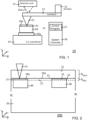
- FIG. 1 shows an exemplary block diagram of a measurement arrangement comprising an atomic force microscope 10 according to an embodiment of the invention.
- the atomic force microscope 10 is configured to perform an analysis of a blood sample 20 to detect amyloid aggregates in red blood cells of the blood sample 20. According to embodiments the atomic force microscope 10 may perform an automated and/or semi-automated imaging and detection of the amyloid aggregates in the blood sample 20.
- the atomic force microscope 10 comprises a tip 11, which may also be denoted as probe tip 11.
- the tip 11 is arranged on a free end of a cantilever 12.
- the apex of the tip 11 is preferably less than 5 nm and accordingly it may be in particular embodied as ultra-sharp tip.
- the cantilever 12 may be actuated by an actuator 13.
- the actuator 13 may be in particular arranged opposite to the free end of the cantilever 12.
- the actuator 13 may be embodied as piezoelectric actuator. According to embodiments, the actuator 13 may oscillate the cantilever 12, in particular the tip 11, in a vertical z-direction.
- the atomic force microscope 10 further comprises a x-y-z positioner unit 14 with a probe table 14a.
- the positioner unit 14 may move and position the probe table 14a in a horizontal plane corresponding to the x-direction and the y-direction as well as in a vertical z-direction.
- a blood sample 20 On the probe table 14a is arranged a blood sample 20.
- the blood sample 20 is covered by a passivation layer 21.
- the atomic force microscope 10 further comprises a detection unit 15 for detection of a deflection and motion of the cantilever 12.
- the detection unit 15 may encompass e.g. a laser unit for generating a laser beam 15a and an optical sensor for detecting the deflected laser beam 15a.
- the atomic force microscope 10 may comprise a system controller 16 which is configured to control the operation of the atomic force microscope 10.
- the system controller 16 may comprise a control program 17 comprising program code for the control and operation of the atomic force microscope 10.
- the control program 17 may comprise a computer program product for operating the atomic force microscope 10.
- the control program 17 may facilitate an automated/and or semi-automated analysis of the surface layer of the blood sample 20.
- the control program 17 may comprise in particular encoded artificial intelligence such as machine learning algorithms for pattern recognition for the detection of amyloid aggregates in blood samples.
- Fig. 2 shows an enlarged cross-sectional view or side view of the blood sample 20, the probe tip 11 and the passivation layer 21.
- the passivation layer 21 may be in particular embodied as a liquid layer.
- the liquid layer may preferably have a kinematic viscosity between 100 centistokes (cSt) and 1000 cSt.
- the passivation layer 21 comprises or consists of a first material, e.g. of liquid silicone.
- the passivation layer is preferably embodied as thin layer having a thickness or height h pass . between 0.5 ⁇ m and 2 ⁇ m. Such a thickness is ideal for immersing the tip 11 into the passivation layer 21 and for providing improved imaging stability.
- the scanning probe microscope 10 may perform an analysis of a surface layer 22 of the blood sample 20.
- the surface layer 22 which is subject to the detailed AFM analysis may have a thickness or height h slbs of e.g. 100 nm.
- the blood sample 20 comprises a plurality of red blood cells 30 (in this exemplary figure represented in a simplified manner by a rectangular cross section). Some of the red blood cells are arranged at the surface of the blood sample 20 and in particular at least partly in the surface layer 22 of the blood sample 20.
- the part of the red blood cell that is arranged in the surface layer 22 of blood sample 20 is in particular analyzed and imaged by the atomic force microscope 10 to detect amyloid aggregates and may be denoted as surface layer 33 of the red blood cells 30.
- the surface layer 33 of the red blood cells 30 which is subject to a detailed AFM analysis may have a thickness or height of e.g. 100 nm.
- FIG. 5 a flow chart of method steps of a method for imaging and detecting amyloid aggregates in blood will be explained.
- the method can be performed by the atomic force microscope 10 of FIG. 1 and hence in the following explanation of the method it will be referred to the components of FIGS. 1 and 2 .
- the blood sample 20 is provided.
- the blood sample 20 may be in particular a dried blood sample of a blood smear and may be fixed e.g. on a clean glass slide.
- the measurements and analysis of the dried blood sample 20 can be conducted on small blood sample quantities of e.g. 1 ⁇ L to 5 ⁇ L.
- the blood sample 20 may be a fluid blood sample or a blood sample comprising purified red blood cells.
- Purified red blood cells may have been derived from an original blood sample by centrifugation.
- the passivation layer 21 is applied to the blood sample 20.
- the blood sample 20 is coated with the passivation layer 21.
- the coating of the dried blood sample 20 may be performed using spray coating, e.g. by spray coating of liquid silicone with a viscosity range from 100 to 1000 cst.
- the imaging of the AFM starts. This includes to immerse the tip 11 into the passivation layer 21.
- the thin liquid encapsulation of the tip 11 by the passivation layer 21 prevents the formation of a liquid bridge at the nanoscopic contact between the tip 11 and the surface 20a of the blood sample 20. This improves the stability during the AFM imaging.
- the mode in which the AFM is operated ensures that the force between the blood sample 20 and the tip 11 is well controlled.
- the adjustable tip-sample forces can be modulated to ensure that height and phase contrast images can be extracted and overlayed.
- a force constant of 2 N/m and a loading force for best resolution between 0.5 and 1 nN may be used.
- the apex of the tip 11 is preferably less than 5 nm.
- the tip 11 may be oscillated vertically in the z-direction near its mechanical resonance frequency. As the tip 11 lightly taps the surface 20a of the blood sample 20, the amplitude of oscillation is reduced and the AFM 10 uses this change in amplitude in order to image the surface topography of the blood sample 20, thereby generating a height image.
- the motion of the tip 11 can be characterized by its relative phase with respect to a driving oscillator of the actuator 13. Such changes of the relative phase may generate phase images of the blood sample 20.
- the AFM 10 is operated in peak force mode. According to other embodiments, the AFM 10 may also be operated in tapping or contact mode.
- the (lateral) edges 31 of the red blood cells 30 which are situated in the surface layer 22 of the blood sample 20 are at first detected and resolved.
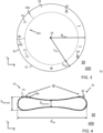
- FIG. 3 shows an enlarged 3-dimensional view 300 of a red blood cell 30.
- FIG. 4 shows a corresponding side view or cross sectional view 400 of the red blood cell 30.
- the red blood cell 30 comprises two types of amyloid aggregates, namely fibrils 41 and oligomers 42, which are collectively referred to as amyloid aggregates 40.
- the AFM 30 has resolved the amyloid aggregates 40 which are situated in the surface layer 33 of the red blood cell 30, in particular the lateral edges 31 of the red blood cells 30.
- the tip 11 is operated at the respective resolved red blood cell 30, in particular at the edges 31, in particular at an edge layer 32 of the respective red blood cell 30.
- This step 505 may encompass one or more scans or runs
- the edge layer 32 is a peripheral layer of the surface of the red bloods cells which is located at or close to the edge 31 of the red blood cells 30.
- the red blood cells 30 have a biconcave structure as illustrated in FIG. 4 .
- the red blood cells 30 have typically a size of 6.6 to 8.2 microns in diameter d rbc and a maximum thickness or height h rbcmax between 2 microns and 2.5 microns.
- the thickness or height h rbc in the z-direction of the red blood cells 30 is higher at the edges 31 than in the center 35 of the red blood cells 30.
- the red blood cells 30 have typically a minimum thickness or height h rbcmin between 0.8 microns and 1 micron in the center of the red blood cell.
- the tip 11 of the AFM 30 may be operated in particular at the peripheral area of the red blood cells which has a higher thickness in the z-direction.
- the tip 11 of the AFM may be operated in particular at the surface area of the red blood cells 30 which has a height h rbc of more than 1.5 ⁇ m, and more preferably a height h rbc of more than 1.7 ⁇ m.
- the edge layer 32 may be defined as the surface area of the red blood cell 30 which has a height h rbc of more than 1.5 ⁇ m, and more preferably a height h rbc of more than 1.7 ⁇ m.
- the edge layer 32 may also be defined according to embodiments as a radial outer layer of the red blood cell 32.
- the edge layer may have a radial thickness or radial extension b rbc between 10% and 50% of the outer radius R rbc of the red blood cell 30, in particular between 20% and 30% of the radius R rbc .
- the radial extension b rbc R rbc - r rbc , wherein r rbc is the inner radius of the edge layer 32.
- the (fictive) inner radius r rbc of the edge layer 32 is illustrated with a dotted circle.
- the AFM 30 at the edge or in other words on the edge layer 32 of the red blood cell 30 is in particular challenging, but it may facilitate the early detection of amyloid aggregates. More particularly, the inventor of the present invention has discovered that at early stages of neurodegenerative diseases the amyloids may aggregate only at the edges of the red blood cell 30, but not or to a lower extent in the center, as illustrated also in FIG. 3 and FIG. 4 .
- Such an analysis of the edge layer 32 may be limited to the analysis of a part or section of the edge layer.
- the AFM tip may be operated e.g. only at a part 32a of the edge layer 32 which is confined by dotted lines 32b.
- the amyloid aggregates 40 in particular the amyloid aggregates in the edge layer 32, may be imaged, resolved and detected.
- the step 505 may generate in particular height images and phase contrast images of the surface 30a of the red blood cells 30.
- a post processing of the scanned images which have been generated at step 505 may be performed at step 506.
- This post processing may include in particular to perform a statistical analysis of the height and phase contrast images.
- Such a post-processing may allow to quantify the size, shape and morphology of the resolved amyloid aggregates 40.
- Such aggregated amyloids are the pathological hallmark of various neurodegenerative diseases such as Alzheimer's, Parkinson's, Neuroferritinopathy, Dementia with Lewy bodies, Huntington disease, Lysozyme amyloidosis, Familial British dementia, Spinocerebellar ataxis and Familial Danish Dementia.
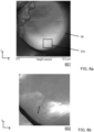
- FIGS. 6a to 6d show as experimental evidence real AFM images of amyloid aggregates resolved in human blood which have been generated by a method according to an embodiment of the invention.
- the AFM images have been generated by an atomic force microscope operated in peak force mode. More particularly, the measurement was done using a Multimode AFM 8 tool of Bruker Corporation. Blood smear on glass slides was used as blood sample. All the AFM measurements were conducted under standard laboratory conditions.
- FIG. 6a shows a height image 601 of a red blood cell 30 provided by a height sensor of the atomic force microscope 10.
- the AFM image 601 was taken from a red blood cell of a blood sample which was encapsulated/covered by viscous liquid silicone.
- FIG. 6b shows a zoom in height image over the black box 32a shown in FIG. 6a , representing an edge area or in other words a part or section of the edge layer of the red blood cell 30 that has been analyzed in more detail.
- FIG. 6c shows a high-resolution height image acquired over several samples on the resolved edge area of the red blood cell 30 of FIG 6b .
- the atomic force microscopy has imaged and detected as amyloid aggregates protofibrils 41.
- the height is gray-scale coded.
- FIG. 6d shows another high-resolution height image acquired over several samples on the resolved edge area of the red blood cell 30 of FIG 6b .
- the height is gray-scale coded.
- aspects of the present invention may be embodied as a measurement method.
Landscapes
- Engineering & Computer Science (AREA)
- Health & Medical Sciences (AREA)
- General Health & Medical Sciences (AREA)
- Physics & Mathematics (AREA)
- General Physics & Mathematics (AREA)
- Life Sciences & Earth Sciences (AREA)
- Chemical & Material Sciences (AREA)
- Nuclear Medicine, Radiotherapy & Molecular Imaging (AREA)
- Radiology & Medical Imaging (AREA)
- Medical Informatics (AREA)
- Biochemistry (AREA)
- Immunology (AREA)
- Pathology (AREA)
- Analytical Chemistry (AREA)
- Biomedical Technology (AREA)
- Quality & Reliability (AREA)
- Theoretical Computer Science (AREA)
- Computer Vision & Pattern Recognition (AREA)
- Epidemiology (AREA)
- Primary Health Care (AREA)
- Public Health (AREA)
- Dispersion Chemistry (AREA)
- Hematology (AREA)
- Molecular Biology (AREA)
- Urology & Nephrology (AREA)
- Food Science & Technology (AREA)
- Medicinal Chemistry (AREA)
- Biophysics (AREA)
- Investigating Or Analysing Biological Materials (AREA)
Claims (15)
- Verfahren zum Erkennen einer neurodegenerativen Krankheit durch die Detektion von Amyloid-Aggregaten (40) in menschlichem Blut, wobei das Verfahren umfasstBereitstellung einer Blutprobe (20) eines Menschen, die ein oder mehrere rote Blutkörperchen (30) enthält;Durchführen einer Analyse einer Oberflächenschicht (22) der Blutprobe (20) mit einem Rasterkraftmikroskop (10), um Amyloidaggregate (40) auf einer Oberfläche der einen oder mehreren roten Blutkörperchen (30) zu detektieren;Bildgebung der detektierten Amyloidaggregate, wodurch Bilder der detektierten Amyloidaggregate, insbesondere Höhenbilder und Phasenkontrastbilder, erzeugt werden; undVerwendung der Bilder der detektierten Amyloidaggregate für die Erkennung der neurodegenerativen Krankheit.
- Verfahren nach Anspruch 1, das ferner die Quantifizierung der Größe, Form und/oder Morphologie der detektierten Amyloidaggregate umfasst.
- Verfahren nach Anspruch 1 oder Anspruch 2, das ferner die Bildgebung detektierter Amyloidaggregate mit unterschiedlicher Größe und Form umfasst.
- Verfahren nach einem der vorhergehenden Ansprüche, wobei die detektierten Amyloidaggregate Tau-Proteine und/oder Alpha-Synucleine umfassen.
- Verfahren nach einem der vorangegangenen Ansprüche, das ferner Folgendes umfasst
Auflösung eines oder mehrerer geometrischer Parameter detektierter Amyloidaggregate, insbesondere der Größe, Form und/oder Morphologie detektierter Amyloidaggregate. - Verfahren nach Anspruch 5, wobei der Schritt des Auflösens des einen oder der mehreren geometrischen Parameter eine Quantifizierung des einen oder der mehreren geometrischen Parameter auf einer Nanometerskala beinhaltet.
- Verfahren nach einem der vorhergehenden Ansprüche, wobei die Bereitstellung der Blutprobe (20) Folgendes umfasstPassivieren der Blutprobe (20) mit einer Passivierungsschicht (21), wobei die Passivierungsschicht (21) ein erstes Material umfasst; undwobei die Blutprobe die Passivierungsschicht während der Durchführung der Analyse der Blutprobe aufweist.
- Verfahren nach Anspruch 7, wobei
das Rasterkraftmikroskop eine Spitze aufweist und die Passivierungsschicht als eine Flüssigkeitsschicht ausgeführt ist, wobei die Spitze während der Analyse in die Flüssigkeitsschicht eintaucht, um die Oberfläche der Blutprobe, die unter der Flüssigkeitsschicht liegt, zu analysieren, wobei die Flüssigkeitsschicht so konfiguriert ist, dass sie die Bewegung der Spitze während der Analyse stabilisiert, wodurch die Bildgebungsstabilität des Rasterkraftmikroskops erhöht wird. - Verfahren nach Anspruch 8, wobei die Flüssigkeitsschicht eine kinematische Viskosität zwischen 100 cSt und 1000 cSt aufweist.
- Verfahren nach einem der vorhergehenden Ansprüche 7 bis 9, wobei das erste Material flüssiges Silikon, insbesondere Silikonöl, ist.
- Verfahren nach einem der vorangehenden Ansprüche 7 bis 10, wobei die Passivierungsschicht (21) eine Dicke zwischen 0,5 µm und 2 µm aufweist.
- Verfahren nach einem der vorangegangenen Ansprüche, das ferner Folgendes umfasstAuflösen von Rändern (31) eines oder mehrerer roter Blutkörperchen (30) in der Oberflächenschicht (22) der Blutprobe (20);Durchführen eines oder mehrerer Scans der einen oder mehreren roten Blutkörperchen (30) durch Betätigen einer Spitze (11) des Rasterkraftmikroskops (10) an den einen oder mehreren aufgelösten roten Blutkörperchen (30), um Amyloidaggregate (40) auf den einen oder mehreren roten Blutkörperchen (30) zu detektieren.
- Verfahren nach einem der vorangegangenen Ansprüche, das ferner Folgendes umfasstAuflösen von Rändern (31) eines oder mehrerer roter Blutkörperchen (30) in der Oberflächenschicht (22) der Blutprobe (20); undDurchführen einer Analyse einer Randschicht (32) der einen oder mehreren aufgelösten roten Blutkörperchen (30) durch Betätigen der Spitze (11) des Rasterkraftmikroskops (10) an der Randschicht (32) der einen oder mehreren aufgelösten roten Blutkörperchen (30), um Amyloidaggregate (40) in der Randschicht (32) der roten Blutkörperchen (30) zu detektieren.
- Verfahren nach einem der vorangegangenen Ansprüche, das ferner Folgendes umfasstDurchführen einer statistischen Analyse der Höhen- und Phasenkontrastbilder; undQuantifizieren der Größe, Form und/oder Morphologie der aufgelösten Amyloidaggregate (40).
- Verfahren nach einem der vorangegangenen Ansprüche, das ferner Folgendes umfasst
Betrieb des Rasterkraftmikroskops (10) im Spitzenkraftmodus.
Applications Claiming Priority (2)
| Application Number | Priority Date | Filing Date | Title |
|---|---|---|---|
| EP20175515 | 2020-05-19 | ||
| PCT/EP2021/063193 WO2021233942A1 (en) | 2020-05-19 | 2021-05-18 | Detection and imaging of amyloid aggregates in blood |
Publications (3)
| Publication Number | Publication Date |
|---|---|
| EP4136446A1 EP4136446A1 (de) | 2023-02-22 |
| EP4136446B1 true EP4136446B1 (de) | 2025-05-14 |
| EP4136446C0 EP4136446C0 (de) | 2025-05-14 |
Family
ID=70779545
Family Applications (1)
| Application Number | Title | Priority Date | Filing Date |
|---|---|---|---|
| EP21727436.4A Active EP4136446B1 (de) | 2020-05-19 | 2021-05-18 | Verfahren zum nachweis von amyloidaggregaten im blut |
Country Status (3)
| Country | Link |
|---|---|
| US (1) | US12405203B2 (de) |
| EP (1) | EP4136446B1 (de) |
| WO (1) | WO2021233942A1 (de) |
Families Citing this family (1)
| Publication number | Priority date | Publication date | Assignee | Title |
|---|---|---|---|---|
| GB202214417D0 (en) * | 2022-09-30 | 2022-11-16 | Imperial College Innovations Ltd | Biomolecules in disease |
Family Cites Families (2)
| Publication number | Priority date | Publication date | Assignee | Title |
|---|---|---|---|---|
| US7345022B2 (en) * | 2002-09-19 | 2008-03-18 | Board Of Supervisors Of Louisiana State University And Agricultural And Mechanical College | Anti-fibril peptides |
| CN113614101B (zh) * | 2019-02-22 | 2024-07-16 | 洛约拉马利蒙特大学 | 淀粉样肽的变体 |
-
2021
- 2021-05-18 WO PCT/EP2021/063193 patent/WO2021233942A1/en not_active Ceased
- 2021-05-18 US US17/926,621 patent/US12405203B2/en active Active
- 2021-05-18 EP EP21727436.4A patent/EP4136446B1/de active Active
Also Published As
| Publication number | Publication date |
|---|---|
| US20230184661A1 (en) | 2023-06-15 |
| US12405203B2 (en) | 2025-09-02 |
| EP4136446C0 (de) | 2025-05-14 |
| EP4136446A1 (de) | 2023-02-22 |
| WO2021233942A1 (en) | 2021-11-25 |
Similar Documents
| Publication | Publication Date | Title |
|---|---|---|
| US10638112B2 (en) | Apparatuses and methods for three-dimensional imaging of an object | |
| Rianna et al. | Direct evidence that tumor cells soften when navigating confined spaces | |
| EP3884424B1 (de) | Teilchencharakterisierung mittels optischer mikroskopie | |
| US7042639B1 (en) | Identification of cells with a compact microscope imaging system with intelligent controls | |
| Liu et al. | In situ mechanical characterization of mouse oocytes using a cell holding device | |
| EP2985719A1 (de) | System und Verfahren für Zellerkennung | |
| JPH11514445A (ja) | 非点収差測定の装置および方法 | |
| US10989736B1 (en) | Cantilever-free scanning probe microscopy | |
| US12405203B2 (en) | Detection and imaging of amyloid aggregates in blood | |
| EP1380005A1 (de) | Computerimplementierte methoden zur mustererkennung in organischem material | |
| Ruggeri et al. | Characterizing individual protein aggregates by infrared nanospectroscopy and atomic force microscopy | |
| US11543356B2 (en) | Rotation and flat-form imaging for microscopic objects | |
| Muth et al. | SynapseNet: Deep learning for automatic synapse reconstruction | |
| US8595859B1 (en) | Controlling atomic force microscope using optical imaging | |
| Link et al. | AI based image analysis of red blood cells in oscillating microchannels | |
| Kodera et al. | Guide to studying intrinsically disordered proteins by high-speed atomic force microscopy | |
| CN115713767A (zh) | 基于监测显微镜测量的机器学习算法自动化训练 | |
| EP3574303B1 (de) | Verfahren und vorrichtung zur untersuchung von zellen | |
| JP4068581B2 (ja) | ナノ空隙制御による微粒子のサイズ分離分析法およびその装置 | |
| Padmakala | Epileptic seizure detection in rodent iEEG using machine learning: a comparative study of random forest, SVM, and gradient boosting models | |
| NL2029685B1 (en) | Detection of interacting cellular bodies | |
| Weiss et al. | Motion analysis of intracellular objects: Trajectories with and without visible tracks | |
| Böke et al. | Vorteile der bildgebenden Durchflusszytometrie für die analytische Bewertung einzelner Nanopartikel und Nanopartikelwolken anhand ihrer Brownschen Molekularbewegung.(eng. Opportunities of Brownian Motion Imaging Flow Cytometry for the Analytical Assessment of Individual Nanoparticles and Nanoparticle Collections) | |
| Lu et al. | The development of image base, portable microfluidic paper-based analytical device | |
| US20250076626A1 (en) | Focus adjustment method, program, and apparatus |
Legal Events
| Date | Code | Title | Description |
|---|---|---|---|
| STAA | Information on the status of an ep patent application or granted ep patent |
Free format text: STATUS: UNKNOWN |
|
| STAA | Information on the status of an ep patent application or granted ep patent |
Free format text: STATUS: THE INTERNATIONAL PUBLICATION HAS BEEN MADE |
|
| PUAI | Public reference made under article 153(3) epc to a published international application that has entered the european phase |
Free format text: ORIGINAL CODE: 0009012 |
|
| STAA | Information on the status of an ep patent application or granted ep patent |
Free format text: STATUS: REQUEST FOR EXAMINATION WAS MADE |
|
| 17P | Request for examination filed |
Effective date: 20221115 |
|
| AK | Designated contracting states |
Kind code of ref document: A1 Designated state(s): AL AT BE BG CH CY CZ DE DK EE ES FI FR GB GR HR HU IE IS IT LI LT LU LV MC MK MT NL NO PL PT RO RS SE SI SK SM TR |
|
| DAV | Request for validation of the european patent (deleted) | ||
| DAX | Request for extension of the european patent (deleted) | ||
| GRAP | Despatch of communication of intention to grant a patent |
Free format text: ORIGINAL CODE: EPIDOSNIGR1 |
|
| STAA | Information on the status of an ep patent application or granted ep patent |
Free format text: STATUS: GRANT OF PATENT IS INTENDED |
|
| INTG | Intention to grant announced |
Effective date: 20241206 |
|
| GRAS | Grant fee paid |
Free format text: ORIGINAL CODE: EPIDOSNIGR3 |
|
| GRAA | (expected) grant |
Free format text: ORIGINAL CODE: 0009210 |
|
| STAA | Information on the status of an ep patent application or granted ep patent |
Free format text: STATUS: THE PATENT HAS BEEN GRANTED |
|
| AK | Designated contracting states |
Kind code of ref document: B1 Designated state(s): AL AT BE BG CH CY CZ DE DK EE ES FI FR GB GR HR HU IE IS IT LI LT LU LV MC MK MT NL NO PL PT RO RS SE SI SK SM TR |
|
| REG | Reference to a national code |
Ref country code: GB Ref legal event code: FG4D |
|
| REG | Reference to a national code |
Ref country code: CH Ref legal event code: EP |
|
| REG | Reference to a national code |
Ref country code: IE Ref legal event code: FG4D |
|
| REG | Reference to a national code |
Ref country code: DE Ref legal event code: R096 Ref document number: 602021030767 Country of ref document: DE |
|
| PGFP | Annual fee paid to national office [announced via postgrant information from national office to epo] |
Ref country code: GB Payment date: 20250618 Year of fee payment: 5 |
|
| U01 | Request for unitary effect filed |
Effective date: 20250612 |
|
| PGFP | Annual fee paid to national office [announced via postgrant information from national office to epo] |
Ref country code: CH Payment date: 20250601 Year of fee payment: 5 |
|
| U07 | Unitary effect registered |
Designated state(s): AT BE BG DE DK EE FI FR IT LT LU LV MT NL PT RO SE SI Effective date: 20250623 |
|
| U20 | Renewal fee for the european patent with unitary effect paid |
Year of fee payment: 5 Effective date: 20250618 |
|
| PGFP | Annual fee paid to national office [announced via postgrant information from national office to epo] |
Ref country code: AT Payment date: 20250721 Year of fee payment: 5 |
|
| PG25 | Lapsed in a contracting state [announced via postgrant information from national office to epo] |
Ref country code: ES Free format text: LAPSE BECAUSE OF FAILURE TO SUBMIT A TRANSLATION OF THE DESCRIPTION OR TO PAY THE FEE WITHIN THE PRESCRIBED TIME-LIMIT Effective date: 20250514 |
|
| PG25 | Lapsed in a contracting state [announced via postgrant information from national office to epo] |
Ref country code: NO Free format text: LAPSE BECAUSE OF FAILURE TO SUBMIT A TRANSLATION OF THE DESCRIPTION OR TO PAY THE FEE WITHIN THE PRESCRIBED TIME-LIMIT Effective date: 20250814 Ref country code: GR Free format text: LAPSE BECAUSE OF FAILURE TO SUBMIT A TRANSLATION OF THE DESCRIPTION OR TO PAY THE FEE WITHIN THE PRESCRIBED TIME-LIMIT Effective date: 20250815 |
|
| PG25 | Lapsed in a contracting state [announced via postgrant information from national office to epo] |
Ref country code: PL Free format text: LAPSE BECAUSE OF FAILURE TO SUBMIT A TRANSLATION OF THE DESCRIPTION OR TO PAY THE FEE WITHIN THE PRESCRIBED TIME-LIMIT Effective date: 20250514 |
|
| PG25 | Lapsed in a contracting state [announced via postgrant information from national office to epo] |
Ref country code: HR Free format text: LAPSE BECAUSE OF FAILURE TO SUBMIT A TRANSLATION OF THE DESCRIPTION OR TO PAY THE FEE WITHIN THE PRESCRIBED TIME-LIMIT Effective date: 20250514 |
|
| PG25 | Lapsed in a contracting state [announced via postgrant information from national office to epo] |
Ref country code: RS Free format text: LAPSE BECAUSE OF FAILURE TO SUBMIT A TRANSLATION OF THE DESCRIPTION OR TO PAY THE FEE WITHIN THE PRESCRIBED TIME-LIMIT Effective date: 20250814 |
|
| PG25 | Lapsed in a contracting state [announced via postgrant information from national office to epo] |
Ref country code: IS Free format text: LAPSE BECAUSE OF FAILURE TO SUBMIT A TRANSLATION OF THE DESCRIPTION OR TO PAY THE FEE WITHIN THE PRESCRIBED TIME-LIMIT Effective date: 20250914 |
|
| PG25 | Lapsed in a contracting state [announced via postgrant information from national office to epo] |
Ref country code: SM Free format text: LAPSE BECAUSE OF FAILURE TO SUBMIT A TRANSLATION OF THE DESCRIPTION OR TO PAY THE FEE WITHIN THE PRESCRIBED TIME-LIMIT Effective date: 20250514 |
|
| PG25 | Lapsed in a contracting state [announced via postgrant information from national office to epo] |
Ref country code: CZ Free format text: LAPSE BECAUSE OF FAILURE TO SUBMIT A TRANSLATION OF THE DESCRIPTION OR TO PAY THE FEE WITHIN THE PRESCRIBED TIME-LIMIT Effective date: 20250514 |
|
| PG25 | Lapsed in a contracting state [announced via postgrant information from national office to epo] |
Ref country code: SK Free format text: LAPSE BECAUSE OF FAILURE TO SUBMIT A TRANSLATION OF THE DESCRIPTION OR TO PAY THE FEE WITHIN THE PRESCRIBED TIME-LIMIT Effective date: 20250514 |
|
| PG25 | Lapsed in a contracting state [announced via postgrant information from national office to epo] |
Ref country code: MC Free format text: LAPSE BECAUSE OF FAILURE TO SUBMIT A TRANSLATION OF THE DESCRIPTION OR TO PAY THE FEE WITHIN THE PRESCRIBED TIME-LIMIT Effective date: 20250514 |
|
| PLBE | No opposition filed within time limit |
Free format text: ORIGINAL CODE: 0009261 |
|
| STAA | Information on the status of an ep patent application or granted ep patent |
Free format text: STATUS: NO OPPOSITION FILED WITHIN TIME LIMIT |
|
| REG | Reference to a national code |
Ref country code: CH Ref legal event code: L10 Free format text: ST27 STATUS EVENT CODE: U-0-0-L10-L00 (AS PROVIDED BY THE NATIONAL OFFICE) Effective date: 20260325 |
|
| PG25 | Lapsed in a contracting state [announced via postgrant information from national office to epo] |
Ref country code: IE Free format text: LAPSE BECAUSE OF NON-PAYMENT OF DUE FEES Effective date: 20250518 |
|
| U20 | Renewal fee for the european patent with unitary effect paid |
Year of fee payment: 6 Effective date: 20260309 |
|
| 26N | No opposition filed |
Effective date: 20260217 |