EP4074352A1 - Vorrichtungen zur herstellung einer perkutanen vaskulären anastomose, insbesondere einer perkutanen arteriovenösen fistel für die hämodialyse - Google Patents
Vorrichtungen zur herstellung einer perkutanen vaskulären anastomose, insbesondere einer perkutanen arteriovenösen fistel für die hämodialyse Download PDFInfo
- Publication number
- EP4074352A1 EP4074352A1 EP21167907.1A EP21167907A EP4074352A1 EP 4074352 A1 EP4074352 A1 EP 4074352A1 EP 21167907 A EP21167907 A EP 21167907A EP 4074352 A1 EP4074352 A1 EP 4074352A1
- Authority
- EP
- European Patent Office
- Prior art keywords
- catheter
- contact area
- surface contact
- catheters
- vein
- Prior art date
- Legal status (The legal status is an assumption and is not a legal conclusion. Google has not performed a legal analysis and makes no representation as to the accuracy of the status listed.)
- Withdrawn
Links
- 206010003226 Arteriovenous fistula Diseases 0.000 title description 36
- 230000003872 anastomosis Effects 0.000 title description 24
- 238000001631 haemodialysis Methods 0.000 title description 20
- 230000000322 hemodialysis Effects 0.000 title description 20
- 230000002792 vascular Effects 0.000 title description 17
- 206010016717 Fistula Diseases 0.000 claims abstract description 24
- 230000003890 fistula Effects 0.000 claims abstract description 24
- 210000003462 vein Anatomy 0.000 description 55
- 238000000034 method Methods 0.000 description 31
- 210000002321 radial artery Anatomy 0.000 description 24
- 210000001367 artery Anatomy 0.000 description 22
- 238000002604 ultrasonography Methods 0.000 description 13
- 210000001519 tissue Anatomy 0.000 description 7
- 238000003384 imaging method Methods 0.000 description 6
- 230000004927 fusion Effects 0.000 description 5
- 238000001356 surgical procedure Methods 0.000 description 5
- 208000005392 Spasm Diseases 0.000 description 4
- 238000002399 angioplasty Methods 0.000 description 4
- 238000000502 dialysis Methods 0.000 description 4
- 230000035800 maturation Effects 0.000 description 4
- 210000002559 ulnar artery Anatomy 0.000 description 4
- 230000008901 benefit Effects 0.000 description 3
- 239000000919 ceramic Substances 0.000 description 3
- 230000003247 decreasing effect Effects 0.000 description 3
- 238000011161 development Methods 0.000 description 3
- 238000005516 engineering process Methods 0.000 description 3
- 238000012552 review Methods 0.000 description 3
- 210000000707 wrist Anatomy 0.000 description 3
- 206010018852 Haematoma Diseases 0.000 description 2
- 206010039897 Sedation Diseases 0.000 description 2
- 238000013459 approach Methods 0.000 description 2
- 230000015572 biosynthetic process Effects 0.000 description 2
- 210000004204 blood vessel Anatomy 0.000 description 2
- 210000002302 brachial artery Anatomy 0.000 description 2
- 230000000694 effects Effects 0.000 description 2
- 230000010102 embolization Effects 0.000 description 2
- 238000011156 evaluation Methods 0.000 description 2
- 210000000245 forearm Anatomy 0.000 description 2
- 206010020718 hyperplasia Diseases 0.000 description 2
- 208000015181 infectious disease Diseases 0.000 description 2
- 238000001990 intravenous administration Methods 0.000 description 2
- 210000003734 kidney Anatomy 0.000 description 2
- 238000002690 local anesthesia Methods 0.000 description 2
- 238000013507 mapping Methods 0.000 description 2
- 210000005036 nerve Anatomy 0.000 description 2
- 230000009467 reduction Effects 0.000 description 2
- 230000036280 sedation Effects 0.000 description 2
- 230000000638 stimulation Effects 0.000 description 2
- 206010002091 Anaesthesia Diseases 0.000 description 1
- 206010051268 Anastomotic stenosis Diseases 0.000 description 1
- HTTJABKRGRZYRN-UHFFFAOYSA-N Heparin Chemical compound OC1C(NC(=O)C)C(O)OC(COS(O)(=O)=O)C1OC1C(OS(O)(=O)=O)C(O)C(OC2C(C(OS(O)(=O)=O)C(OC3C(C(O)C(O)C(O3)C(O)=O)OS(O)(=O)=O)C(CO)O2)NS(O)(=O)=O)C(C(O)=O)O1 HTTJABKRGRZYRN-UHFFFAOYSA-N 0.000 description 1
- 208000007101 Muscle Cramp Diseases 0.000 description 1
- 208000002033 Myoclonus Diseases 0.000 description 1
- 208000004550 Postoperative Pain Diseases 0.000 description 1
- 241000022863 Radosa Species 0.000 description 1
- 208000002847 Surgical Wound Diseases 0.000 description 1
- 206010048671 Venous stenosis Diseases 0.000 description 1
- 230000009471 action Effects 0.000 description 1
- 230000037005 anaesthesia Effects 0.000 description 1
- 210000003484 anatomy Anatomy 0.000 description 1
- 238000002583 angiography Methods 0.000 description 1
- 239000008280 blood Substances 0.000 description 1
- 210000004369 blood Anatomy 0.000 description 1
- 230000017531 blood circulation Effects 0.000 description 1
- 208000020832 chronic kidney disease Diseases 0.000 description 1
- 239000011248 coating agent Substances 0.000 description 1
- 238000000576 coating method Methods 0.000 description 1
- 238000004891 communication Methods 0.000 description 1
- 210000002808 connective tissue Anatomy 0.000 description 1
- 229940039231 contrast media Drugs 0.000 description 1
- 239000002872 contrast media Substances 0.000 description 1
- 230000001186 cumulative effect Effects 0.000 description 1
- 230000007547 defect Effects 0.000 description 1
- 230000003111 delayed effect Effects 0.000 description 1
- 230000001419 dependent effect Effects 0.000 description 1
- 238000009795 derivation Methods 0.000 description 1
- 210000002249 digestive system Anatomy 0.000 description 1
- 210000003414 extremity Anatomy 0.000 description 1
- 230000002349 favourable effect Effects 0.000 description 1
- 238000002594 fluoroscopy Methods 0.000 description 1
- 238000002695 general anesthesia Methods 0.000 description 1
- 238000010438 heat treatment Methods 0.000 description 1
- 229960002897 heparin Drugs 0.000 description 1
- 229920000669 heparin Polymers 0.000 description 1
- 208000014674 injury Diseases 0.000 description 1
- 230000007794 irritation Effects 0.000 description 1
- 238000007726 management method Methods 0.000 description 1
- 238000010197 meta-analysis Methods 0.000 description 1
- 238000012986 modification Methods 0.000 description 1
- 230000004048 modification Effects 0.000 description 1
- 210000000056 organ Anatomy 0.000 description 1
- 229940124583 pain medication Drugs 0.000 description 1
- 230000010412 perfusion Effects 0.000 description 1
- 230000008569 process Effects 0.000 description 1
- 230000005855 radiation Effects 0.000 description 1
- 229910052761 rare earth metal Inorganic materials 0.000 description 1
- 150000002910 rare earth metals Chemical class 0.000 description 1
- 238000002694 regional anesthesia Methods 0.000 description 1
- 210000002345 respiratory system Anatomy 0.000 description 1
- 230000037390 scarring Effects 0.000 description 1
- 238000000926 separation method Methods 0.000 description 1
- 239000013589 supplement Substances 0.000 description 1
- 210000003813 thumb Anatomy 0.000 description 1
- 230000008733 trauma Effects 0.000 description 1
- 230000008016 vaporization Effects 0.000 description 1
- 210000001604 vasa vasorum Anatomy 0.000 description 1
- 238000012800 visualization Methods 0.000 description 1
Images

Classifications
-
- A—HUMAN NECESSITIES
- A61—MEDICAL OR VETERINARY SCIENCE; HYGIENE
- A61B—DIAGNOSIS; SURGERY; IDENTIFICATION
- A61B17/00—Surgical instruments, devices or methods
- A61B17/11—Surgical instruments, devices or methods for performing anastomosis; Buttons for anastomosis
-
- A—HUMAN NECESSITIES
- A61—MEDICAL OR VETERINARY SCIENCE; HYGIENE
- A61B—DIAGNOSIS; SURGERY; IDENTIFICATION
- A61B18/00—Surgical instruments, devices or methods for transferring non-mechanical forms of energy to or from the body
- A61B18/04—Surgical instruments, devices or methods for transferring non-mechanical forms of energy to or from the body by heating
- A61B18/12—Surgical instruments, devices or methods for transferring non-mechanical forms of energy to or from the body by heating by passing a current through the tissue to be heated, e.g. high-frequency current
- A61B18/14—Probes or electrodes therefor
- A61B18/1492—Probes or electrodes therefor having a flexible, catheter-like structure, e.g. for heart ablation
-
- A—HUMAN NECESSITIES
- A61—MEDICAL OR VETERINARY SCIENCE; HYGIENE
- A61B—DIAGNOSIS; SURGERY; IDENTIFICATION
- A61B17/00—Surgical instruments, devices or methods
- A61B2017/00477—Coupling
-
- A—HUMAN NECESSITIES
- A61—MEDICAL OR VETERINARY SCIENCE; HYGIENE
- A61B—DIAGNOSIS; SURGERY; IDENTIFICATION
- A61B17/00—Surgical instruments, devices or methods
- A61B2017/00831—Material properties
- A61B2017/00876—Material properties magnetic
-
- A—HUMAN NECESSITIES
- A61—MEDICAL OR VETERINARY SCIENCE; HYGIENE
- A61B—DIAGNOSIS; SURGERY; IDENTIFICATION
- A61B17/00—Surgical instruments, devices or methods
- A61B17/11—Surgical instruments, devices or methods for performing anastomosis; Buttons for anastomosis
- A61B2017/1107—Surgical instruments, devices or methods for performing anastomosis; Buttons for anastomosis for blood vessels
-
- A—HUMAN NECESSITIES
- A61—MEDICAL OR VETERINARY SCIENCE; HYGIENE
- A61B—DIAGNOSIS; SURGERY; IDENTIFICATION
- A61B17/00—Surgical instruments, devices or methods
- A61B17/11—Surgical instruments, devices or methods for performing anastomosis; Buttons for anastomosis
- A61B2017/1139—Side-to-side connections, e.g. shunt or X-connections
-
- A—HUMAN NECESSITIES
- A61—MEDICAL OR VETERINARY SCIENCE; HYGIENE
- A61B—DIAGNOSIS; SURGERY; IDENTIFICATION
- A61B18/00—Surgical instruments, devices or methods for transferring non-mechanical forms of energy to or from the body
- A61B2018/00315—Surgical instruments, devices or methods for transferring non-mechanical forms of energy to or from the body for treatment of particular body parts
- A61B2018/00345—Vascular system
- A61B2018/00404—Blood vessels other than those in or around the heart
-
- A—HUMAN NECESSITIES
- A61—MEDICAL OR VETERINARY SCIENCE; HYGIENE
- A61B—DIAGNOSIS; SURGERY; IDENTIFICATION
- A61B18/00—Surgical instruments, devices or methods for transferring non-mechanical forms of energy to or from the body
- A61B2018/00571—Surgical instruments, devices or methods for transferring non-mechanical forms of energy to or from the body for achieving a particular surgical effect
- A61B2018/00577—Ablation
-
- A—HUMAN NECESSITIES
- A61—MEDICAL OR VETERINARY SCIENCE; HYGIENE
- A61B—DIAGNOSIS; SURGERY; IDENTIFICATION
- A61B18/00—Surgical instruments, devices or methods for transferring non-mechanical forms of energy to or from the body
- A61B2018/00571—Surgical instruments, devices or methods for transferring non-mechanical forms of energy to or from the body for achieving a particular surgical effect
- A61B2018/00595—Cauterization
-
- A—HUMAN NECESSITIES
- A61—MEDICAL OR VETERINARY SCIENCE; HYGIENE
- A61B—DIAGNOSIS; SURGERY; IDENTIFICATION
- A61B18/00—Surgical instruments, devices or methods for transferring non-mechanical forms of energy to or from the body
- A61B18/04—Surgical instruments, devices or methods for transferring non-mechanical forms of energy to or from the body by heating
- A61B18/12—Surgical instruments, devices or methods for transferring non-mechanical forms of energy to or from the body by heating by passing a current through the tissue to be heated, e.g. high-frequency current
- A61B18/14—Probes or electrodes therefor
- A61B2018/1405—Electrodes having a specific shape
- A61B2018/144—Wire
-
- A—HUMAN NECESSITIES
- A61—MEDICAL OR VETERINARY SCIENCE; HYGIENE
- A61B—DIAGNOSIS; SURGERY; IDENTIFICATION
- A61B90/00—Instruments, implements or accessories specially adapted for surgery or diagnosis and not covered by any of the groups A61B1/00 - A61B50/00, e.g. for luxation treatment or for protecting wound edges
- A61B90/03—Automatic limiting or abutting means, e.g. for safety
- A61B2090/033—Abutting means, stops, e.g. abutting on tissue or skin
- A61B2090/034—Abutting means, stops, e.g. abutting on tissue or skin abutting on parts of the device itself
-
- A—HUMAN NECESSITIES
- A61—MEDICAL OR VETERINARY SCIENCE; HYGIENE
- A61B—DIAGNOSIS; SURGERY; IDENTIFICATION
- A61B90/00—Instruments, implements or accessories specially adapted for surgery or diagnosis and not covered by any of the groups A61B1/00 - A61B50/00, e.g. for luxation treatment or for protecting wound edges
- A61B90/36—Image-producing devices or illumination devices not otherwise provided for
- A61B90/37—Surgical systems with images on a monitor during operation
- A61B2090/378—Surgical systems with images on a monitor during operation using ultrasound
-
- A—HUMAN NECESSITIES
- A61—MEDICAL OR VETERINARY SCIENCE; HYGIENE
- A61M—DEVICES FOR INTRODUCING MEDIA INTO, OR ONTO, THE BODY; DEVICES FOR TRANSDUCING BODY MEDIA OR FOR TAKING MEDIA FROM THE BODY; DEVICES FOR PRODUCING OR ENDING SLEEP OR STUPOR
- A61M1/00—Suction or pumping devices for medical purposes; Devices for carrying-off, for treatment of, or for carrying-over, body-liquids; Drainage systems
- A61M1/36—Other treatment of blood in a by-pass of the natural circulatory system, e.g. temperature adaptation, irradiation ; Extra-corporeal blood circuits
- A61M1/3621—Extra-corporeal blood circuits
- A61M1/3653—Interfaces between patient blood circulation and extra-corporal blood circuit
- A61M1/3655—Arterio-venous shunts or fistulae
Definitions
- the invention generally relates to internal medical procedures performed percutaneously.
- the invention relates to devices and methods for forming percutaneous vascular anastomosis, in particular percutaneous arteriovenous fistula (pAVF) for hemodialysis.
- pAVF percutaneous arteriovenous fistula
- Anastomosis is a surgical connection made between two hollow organs such as blood vessels.
- the traditional surgical procedure involves hand-suturing the two vessels together such that the end of one vessel is fixedly attached to an opening of the second vessel. This approach is time consuming and requires a surgical incision to provide access and control to the vessels involved.
- the arteriovenous fistula is the preferred access for hemodialysis. AVFs are less prone to infection and are more durable than both tunneled catheters and arteriovenous grafts.
- fistula is formed between the brachial artery and the antecubital vein. Fistula creation can be accomplished by creating an incision in the limb, dissecting the vessels, creating a small opening in both vessels and the suturing or clipping the openings together. Eventually, as the vein and artery heal, the fistula matures, which involves enlargement of the vessel diameter and enhancement of their wall.
- Surgical fistula creation has several pitfalls.
- the radial artery is somewhat small at the wrist, which makes anastomosis technically demanding, especially in smaller patients.
- a relatively large vein is needed in the immediate vicinity of the radial artery, and such vein is not always present, or may be of bad quality because of previous intravenous perfusions. If a vein more than a centimeter away is mobilized and brought over to the artery, venous kinking that compromises the internal lumen and blood flow can occur.
- Percutaneous procedures present numerous advantages over open surgery, e.g. decreased risk of infection, decreased scarring, decreased patient trauma, reduced amounts and duration of anesthesia, reduced occurrence of complications, during or after the procedure, faster recovering times, reduction in the need for post-procedural pain medications.
- WO2012068273 discloses two catheters with magnets mounted thereon and used to provide points of shunt contact or placement, the magnetic attraction pulling tissues and catheters together in a fixed reliable geometric manner.
- the magnets of the first catheter are arranged such that their polarities are configured to mate with the magnetic features of the second catheter.
- each of the first and second catheters are either rotated up to 90 degrees toward each other such that their distal ends face each other in an axial alignment. This configuration aligns the two central lumens of the catheters and magnetic features, allowing a tool, such as a needle, RF wire, or sharpened guidewire to be advanced through the first catheter, second catheter or both, to puncture the tissue walls trapped between the magnetic features.
- WO97/33522 (Beth), published in 1997 , disclose a system comprising two discrete catheters for generating an arteriovenous fistula for hemodialysis access. Each catheter has a pair of rare earth magnets. The attraction between these magnets cause intravascular adjustment in position for the venous catheter and the arterial catheter lying within their individual but immediately adjacent blood vessels. A sliding electrode is actuated to perforate both the arterial and the venous vascular wall.
- Both the WavelinQ and the Ellipsys system create a side-to-side AVF.
- This configuration is believed to be associated with less development of intimal hyperplasia.
- This side-to-side configuration is also believed to be associated with more favorable flow characteristics compared with an end-to-side configuration, notably, less turbulent flow and post-anastomotic stenosis
- WavelinQ enables the creation of an AVF between either the proximal radial or ulnar arteries and an adjacent deep vein that drains the upper arm superficial veins.
- Ellipsys connects the proximal radial artery with the adjacent terminal part of the deep communicating vein.
- the first generation everlinQ system had two 6Fr catheters, which were inserted via the upper arm with the vascular system.
- a CE-approved 4Fr device has replaced the 6Fr system in Europe.
- the 4Fr system enables a wrist access either from the radial or the ulnar side if the diameters of the vessel are sufficient, otherwise access vie the brachial vessels is performed.
- the WavelinQ ® system (Becton Dickinson) consists of a pair of over-the-wire catheters with magnets arranged to pull them into alignment and opposition.
- the venous catheter contains a spring-loaded radiofrequency (RF) electrode for delivery of radiofrequency energy.
- RF radiofrequency
- the arterial catheter contains a ceramic backstop intended to align with the venous RF electrode.
- the catheters have radio opaque elements, forming rotational indicators. Following duplex sonography guided puncture 0.014" guidewires are inserted in both vessels up to the planned site of the anastomosis.
- the two catheters are positioned via these wires and fluoroscopic imaging is used to check the correct positioning.
- Fluoroscopic imaging is preferably set perpendicular to the plane of the target vein and artery to maximize accuracy of the rotational indicators.
- the arterial catheter is inserted into the ulnar artery via the brachial artery, and the venous catheter is inserted into the ulnar vein via the brachial vein.
- the catheters are introduced from the upper arm or wrist in a parallel or antiparallel fashion and are guided to the creation site with fluoroscopic imaging.
- the catheters are aligned and rotated before reaching the fistula creation site, so that the electrode and ceramic backstop are facing one another.
- the magnets contained in each catheter attract each other, the magnets holding the artery and vein together.
- the RF electrode is released from the venous catheter and energized for 0.7 second, vaporizing the tissue and vessel walls between the artery and the vein, creating a side-to-side ulnar vein fistula in the arm. A preset energy of 60W is applied.
- the energy creates a small rectangular hole (approximately 5 mm x 1 mm) in the adjoining vessels, creating a nonsurgical, or endovascular AVF.
- the result is documented by angiography.
- Blood then flows from the artery into the veins of the deep venous system and via perforators to the superficial venous system, which include the antecubital vein, basilic vein, and cephalic vein.
- the endo AVF procedure using WavelinQ is carried out under conscious sedation and local anesthesia and does not require an overnight stay in hospital. Patients receive intravenous heparin.
- Patients must have vessels that are more than 2 mm in diameter, patency of the upper arm veins, and no significant central venous stenosis.
- Ellipsys ® (Medtronic) is a device which was developed to create a percutaneous AVF for hemodialysis access using pressure and thermal assistance.
- This thermal resistance anastomosis device creates a side-to-side anastomosis between the proximal radial artery and an adjacent deep communicating vein in the proximal forearm.
- TRAD Thermal resistance anastomosis device
- the TRAD catheter has a 6-F proximal diameter with opposing active surfaces between the base and the coned 5-F distal tip.
- the power controller delivers direct current to the catheter heating element that is controlled with feedback from the temperature sensors and a gap sensor detecting the temperature and the opening distance of the catheter.
- a thumb tab in the handle of the device controls the catheter opening and closing and the fusion pressure.
- the Ellipsys procedure is typically performed in an outpatient setting, under loco regional anesthesia nerve block or local anesthesia with or without moderate sedation.
- the entire procedure is performed using ultrasound guidance, without need for radiation and contrast media.
- the procedure starts with a puncture of a superficial vein, either cephalic or basilica vein, on the upper arm to navigate from there through the perforator to the deep venous systems.
- a standard micropuncture needle and wire are used to obtain retrograde access into the median cubital or brachial vein.
- the access needle is then advanced intravenously under ultrasound guidance to a position where the proximal radial artery is adjacent.
- the needle is then used to puncture the proximal radial artery and the wire is advanced distally into the radial artery.
- a 6Fr glide sheath slender sheath (Terumo Medical Corp, New Jersey) is then advanced into the artery. Through this, the TRAD catheter is advanced into the sheath in an open position with the tip of the device in the proximal radial artery and the base in the accessed vein.
- the catheter has two distinct plates, of which one is placed in the vein and the other in the artery.
- the sheath is then retracted and gentle traction is applied to the Ellipsys catheter until the tip of the device engages the wall of the proximal radial artery, providing tactile resistance to further traction.
- the device is then closed and activated to fuse and cut the anastomosis. Electrical impulses of up to 15s are created.
- the Ellipsys device create an elliptical anastomosis that is roughly 4mm x 2 mm.
- the device can be removed via the sheath.
- An immediate percutaneous transluminal angioplasty (PTA) of the anastomosis and the perforating vein is recommended. Such a procedure is not currently used for the WavelinQ system.
- Beathard et al. described a two year follow up after use of the Ellipsys device at five vascular access programs in the United States with a total of 105 patients. In those patients, percutaneous AVF creation was performed with immediate balloon dilatation of the anastomosis as described by Mallios et al. (Two-year cumulative patency of endovascular arteriovenous fistula, JVS, 2020 May; 21 (3):350-356, doi: 10.1177/1129729819877780 ) .
- the catheters have to be aligned and rotated before reaching the fistula creation site, so that the electrode and ceramic backstop are facing one another.
- Rotational indicators in the eclipsed position indicate misalignment, whereas the open position indicates alignment. Alignment has to be further confirmed by the communication of the venous electrode with the artery backstop during electrode reciprocation.
- the need of aligning the catheters for a certain length that is equal or superior to 4-5cm is forcing the connection to be further away from the deep communicating vein.
- the flow can be restricted from the in between small radial or ulnar vein or be distributed to other branches that are before the deep communicating vein hence having less flow going to the superficial venous system which is necessary for the fistula maturation and use.
- tortuosity and branching of the deep venous system may make the alignment of the catheters cumbersome and time consuming or in some occasions not at all possible.
- the access needle is advanced intravenously under ultrasound guidance to a position where the proximal radial artery is adjacent.
- the needle is then used to puncture the proximal radial artery. Puncture is made in a direction based on information given by ultrasound guidance. This is a delicate and difficult process that requires the operator under ultrasound to navigate inside the center of a 2-3mm vein, come out of it and then come in a 2mm artery, staying always at the center and avoiding hematoma or spasm that will make visualization under ultrasound difficult. Given that these skills of ultrasound guided puncture are not widely diffused amongst physicians and especially surgeons, many hesitate to perform the technique.
- the balloon comes down from the deep communicating vein into the radial artery and towards the distal radial artery.
- the balloon When the balloon is inflated it will squeeze and compress the proximal radial artery and this can cause a temporary or permanent reduction of antegrade flow in the radial artery that is required for fistula function and maturation.
- a second wire needs to be brought from the distal radial artery and with a separate balloon do an angioplasty of the pre-anastomotic radial artery.
- the arm to be treated using RF has to be fixed to prevent movement due to myoclonus trigged by radiofrequency impulse-induced neuro stimulation during energy delivery, such movement being more frequent and more severe in creation of an ulnar fistula due to the proximity of the medial nerve.
- a pronounced intraoperative vessel spasm is often seen after high frequency application, possibly trigged by thermal effects, small hematomas or electrical stimulation.
- WavelinQ procedure required 73 minutes mean, with a range between 2 and 150 minutes ( Single center experience of endovascular AV fistula creation with both wavelinQ and ellipsys systems, European Journal of Vascular & Endovascular Surgery; 2019 Supplement 3, Vol. 58, pe690-e690, 1p, doi: 10.1016/j.ejvs.2019.09.209 ) .
- the present invention is directed to addressing the defects of both prior art devices and create a single device that takes care of one or more of the problems set forth above.
- the following presents a simplified summary of the invention in order to provide a basic understanding of some aspects of the invention. This summary is not an exhaustive overview of the invention. It is not intended to identify key of critical elements of the invention or to delineate the scope of the invention. Its sole purpose is to present some concepts in a simplified form as a prelude to the more detailed description that is discussed later.
- the invention relates to devices and methods for forming percutaneous vascular anastomosis, that can be an arteriovenous fistula (pAVF, endoAVF) for hemodialysis or other type of vascular anastomosis, avoiding the drawbacks and side effects of available systems and methods, and being cost effective, clinically and practically.
- pAVF arteriovenous fistula
- endoAVF arteriovenous fistula
- the present disclosure relates to a device for creating a fistula between two vessels, the device comprising two catheters, each catheter having a proximal end and a distal end, the two catheters comprising corresponding surface contact areas in their distal ends, the surface contact area of a first catheter extending in the tip of the first catheter, the corresponding surface contact area of the second catheter comprising a bearer for the tip of the first catheter, the bearer extending laterally in the distal end of the second catheter, the two catheters comprising indexing means enabling precise relative positioning of the two catheters when the surface contact area of the first catheter is against the surface contact area of the second catheter.
- the first catheter is relatively rigid which allows control and navigation of the tip in order for it to be positioned at the desired position.
- the tip can be equipped with magnet to assist connection with the second catheter or a thermal element to allow for tissue fusion technology to be applied.
- the first catheter has an internal lumen having an aperture in said tip for the passage of a tool or a wire that will assist advancement of the catheter and will also create the connection, the second catheter having a lateral guiding surface facing said aperture when the surface contact area of the first catheter is against the surface contact area of the second catheter.
- the alignment of the catheters will allow the guide wire to go from the lumen of the first catheter to the guiding convex surface of the second catheter and create the connection with the wire advancing in a desired angled and not straight direction.
- the surface contact area of the first catheter is a male surface contact, the tip of the first catheter forming a convex surface area.
- the second catheter has a female surface contact.
- the tip of the first catheter comprises a first convex contact area and a second convex contact area, the first convex contact area having when viewed in cross longitudinal section a curvature that is different from the one of the second convex contact area.
- the bearer of the second catheter comprises a first concave surface contact area and a second concave surface contact area, the shape of these concave areas being in print and counter print with the two convex contact areas of the first catheter.
- the present disclosure also relates to a kit comprising a device as presented above and a tool that is to be guided in the internal lumen of the first catheter, the tool being a sharpened wire, or a RF wire that can allow the perforation of the two adjacent vessel walls and the navigation of the wire from the one vessel to the other and with the desired angle created by the two connected catheters.
- vascular access sites for hemodialysis
- the invention can be utilized to create other vascular access sites for other systems, for example, to create access sites in the respiratory system, digestive system and circulatory system.
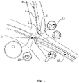
- the system represented in the figures is aimed to create a fistula between two vessels.
- the system comprises two catheters, each being placed within one of the two vessels.
- Placement of the catheter within the vessel can be done e.g. using the Seldinger technique, involving percutaneous puncture of a vessel with a hollow needle, introduction of a guidewire through the needle into the vessel lumen, removal of the needle while maintaining the guidewire in position, followed by advancement of the catheter over the guidewire.
- a sheath can be placed over the puncture site.
- Guidewire have advantageously a hydrophilic coating to enhance their maneuverability.
- the system enables the formation of percutaneous vascular anastomosis, in particular arteriovenous fistula (pAVF, endoAVF) for hemodialysis.
- AVF is advantageously created in the non-dominant arm, in the most distal vessels.
- the radiocephalic AVF is preferred, followed by the brachicephalic and the transposed brachial basilic vein fistula.
- Proximal radial artery AVF and ulnobasilic fistula can also be made.
- Proximal radial artery AVF is created by joining the median antecubital vein or cephalic vein with the radial artery.
- the system advantageously enable the formation of side-to-side anastomosis.
- Vessel mapping is made using Doppler ultrasound or venogram.
- the inner diameter of the artery and vein should advantageously be more than 2.0 and 2.5 mm respectively.
- distal is used to designate the extreme part of the catheters that is to be in the desired place for the fistula between the two vessels.
- Each of the two catheters have a proximal part, not represented, that is to be placed outside the patient body.
- the distal part of one catheter is placed within the proximal radial artery, whereas the distal part of the second catheter is placed within the deep communicating vein.
- the two catheters have a circular cross section, except for the extreme portion of their proximal end.
- the two catheters In the extreme portion of their proximal end, the two catheters have corresponding surface contact area.
- one catheter has a male extreme part
- the other catheter has a female extreme part, the shape of the two extreme parts being in print and counter-print.
- male female
- male can comprise groove, notch, slots corresponding to reliefs, lug, or pin on the female extreme part of the other catheter.
- the female extreme part can comprise reliefs, lug or pin corresponding to groove, notch, slots on the male extreme part of the other catheter.
- the male extreme part and the female extreme part have indexing mean, enabling angular indexation of the male catheter and precise relative positioning of the two catheters.
- one catheter has a male extreme part that extends at the longitudinal end of the catheter, whereas the other catheter has a female extreme part that extends laterally.
- the two catheters are thus movable between a first situation, in which the male catheter is spaced from the female catheter, and a second situation, in which the male catheter is placed in lateral contact with the female catheter.
- the first situation is represented schematically in figure 1 , the distal end of the male catheter being placed in a vein such as the deep communicating vein, the distal end of the female catheter being placed in an artery such as the proximal radial artery.
- the distal end of the male catheter is spaced from the distal end of the female catheter.
- the second situation is represented schematically in figure 2 , the distal end the male catheter being pressed against the distal end of the female catheter, the walls of the vein and artery being placed between the corresponding surface areas of the two catheters.
- the male catheter has advantageously an internal lumen, with a distal exiting hole, for the passage of a wire.
- action on the wire forms a hole in the walls of the vein and artery.
- the female catheter has a guiding surface for the wire exiting the male catheter.
- the hole is made by mechanical effort of the wire on the walls of the vein and artery.
- the internal lumen of the male catheter enables the passage of a cutting tool, a needle, a RF wire, or sharpened guidewire.
- heat is applied on the walls of the vein and artery when the male catheter is pressed against the female catheter, for example using a radio frequency heat generator.
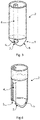
- the male catheter 1 has a distal end 2 having a cylindrical cross section, except for the extreme longitudinal portion that forms a convex tip 3.
- the male catheter 1 has an internal longitudinal lumen 4 having an aperture 5 in the convex tip 3.
- the convex tip 3 has a first surface contact area 6 and a second surface contact area 7.
- the first surface contact area 6 has a curvature that is different from the one of the second surface contact area 7.
- the convex tip 3 does not form an external surface having a circular symmetry around an axis, in particular the longitudinal axis of the male catheter 1.
- the aperture of the internal lumen lies within one of the two surface contact areas.
- the aperture 5 lies within the second surface contact area 7.
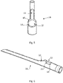
- the female catheter 10 has a distal end 11 comprising a lateral bearer 12 for the convex tip 3 of the male catheter 1.
- the distal end 11 of the female catheter 10 has a lateral guiding surface 13 for a wire passing through the internal lumen 4 of the male catheter 1 and exiting at the opening 5.
- the lateral bearer 12 comprise a first surface contact area 14 and a second surface contact area 15, the shape of these convex areas 14, 15 being in print and counter print with the surfaces 6, 7 of the convex tip 3.
- a wire 16 guided by the internal lumen 4 and exiting at the opening 5 is deviated by the lateral guiding surface 13, after perforating the walls of the vessels.
- the opening 5 is turned in view of the extreme distal part of the female catheter 10.
- the system enables endoAVF procedure, the anatomy associated with the AVF not been disturbed, and the vasa vasorum remaining intact.
- the procedure can be carried out by ultrasound guidance, angiographic imaging is not required. Magnets or tissue fusion can be used. Balloon angioplasty will include the outflow vein, the anastomosis and the pre-anastomotic artery which is not possible with the existing techniques. The anastomosis will be also created exactly at the level of the perforator which will lead to more flow to the superficial system.
- the endoAVF procedure is less invasive than surgery, and does not require general anesthesia. A period of several months may be required between surgical referral for access creation to a functional, surgically created AVF. This delay could influence some patients to start dialysis with a central venous catheter. Using the endoAVF procedure, this delay problem can be eliminated.
Landscapes
- Health & Medical Sciences (AREA)
- Life Sciences & Earth Sciences (AREA)
- Surgery (AREA)
- Heart & Thoracic Surgery (AREA)
- Engineering & Computer Science (AREA)
- Biomedical Technology (AREA)
- Nuclear Medicine, Radiotherapy & Molecular Imaging (AREA)
- Medical Informatics (AREA)
- Molecular Biology (AREA)
- Animal Behavior & Ethology (AREA)
- General Health & Medical Sciences (AREA)
- Public Health (AREA)
- Veterinary Medicine (AREA)
- Surgical Instruments (AREA)
Priority Applications (1)
| Application Number | Priority Date | Filing Date | Title |
|---|---|---|---|
| EP21167907.1A EP4074352A1 (de) | 2021-04-12 | 2021-04-12 | Vorrichtungen zur herstellung einer perkutanen vaskulären anastomose, insbesondere einer perkutanen arteriovenösen fistel für die hämodialyse |
Applications Claiming Priority (1)
| Application Number | Priority Date | Filing Date | Title |
|---|---|---|---|
| EP21167907.1A EP4074352A1 (de) | 2021-04-12 | 2021-04-12 | Vorrichtungen zur herstellung einer perkutanen vaskulären anastomose, insbesondere einer perkutanen arteriovenösen fistel für die hämodialyse |
Publications (1)
| Publication Number | Publication Date |
|---|---|
| EP4074352A1 true EP4074352A1 (de) | 2022-10-19 |
Family
ID=75477971
Family Applications (1)
| Application Number | Title | Priority Date | Filing Date |
|---|---|---|---|
| EP21167907.1A Withdrawn EP4074352A1 (de) | 2021-04-12 | 2021-04-12 | Vorrichtungen zur herstellung einer perkutanen vaskulären anastomose, insbesondere einer perkutanen arteriovenösen fistel für die hämodialyse |
Country Status (1)
| Country | Link |
|---|---|
| EP (1) | EP4074352A1 (de) |
Citations (15)
| Publication number | Priority date | Publication date | Assignee | Title |
|---|---|---|---|---|
| WO1997033522A1 (en) | 1996-03-15 | 1997-09-18 | Beth Israel Deaconess Medical Center | Catheter apparatus and methodology for generating a fistula on-demand between closely associated blood vessels at a pre-chosen anatomic site in-vivo |
| US20070203515A1 (en) * | 2006-01-25 | 2007-08-30 | Heuser Richard R | Catheter system for connecting adjacent blood vessels |
| WO2012068273A1 (en) | 2010-11-16 | 2012-05-24 | Tva Medical, Inc. | Devices and methods for forming a fistula |
| EP2582314A1 (de) | 2010-06-15 | 2013-04-24 | Caymus Medical, Inc. | Intravaskulärer arteriell-venöser anastomose- und gewebeschweissungskatheter |
| WO2014022585A1 (en) | 2012-08-01 | 2014-02-06 | Caymus Medical Inc. | Systems and methods for percutaneous intravascular access for arteriovenous fistula |
| WO2014059351A1 (en) | 2012-10-11 | 2014-04-17 | Tva Medical, Inc. | Devices and methods for fistula formation |
| WO2014078601A1 (en) | 2012-11-14 | 2014-05-22 | Caymus Medical, Inc. | Intravascular arterial to venous anastomosis and tissue welding catheter |
| WO2014153229A1 (en) | 2013-03-14 | 2014-09-25 | Tva Medical, Inc. | Fistula formulation devices and methods therefor |
| EP2812063A1 (de) | 2012-02-08 | 2014-12-17 | Caymus Medical, Inc. | Intravaskulärer arteriell-venöser anastomose- und gewebeschweissungskatheter |
| WO2015138998A1 (en) | 2014-03-14 | 2015-09-17 | Tva Medical, Inc. | Fistula formation devices and methods therefor |
| WO2017124062A1 (en) | 2016-01-15 | 2017-07-20 | Tva Medical, Inc. | Devices and methods for forming a fistula |
| US10070866B1 (en) | 2013-08-01 | 2018-09-11 | Avenu Medical, Inc. | Percutaneous arterial to venous anastomosis clip application catheter system and methods |
| WO2018213626A1 (en) | 2017-05-17 | 2018-11-22 | Avenu Medical, Inc. | Single catheter electrode tissue cutting system for creating anastomoses |
| WO2020242491A1 (en) * | 2019-05-31 | 2020-12-03 | Tva Medical, Inc. | Systems, methods, and catheters for endovascular treatment of a blood vessel |
| WO2021022090A1 (en) | 2019-07-31 | 2021-02-04 | NXT Biomedical | Methods and devices for establishing a connection between adjacent anatomical spaces using magnets |
-
2021
- 2021-04-12 EP EP21167907.1A patent/EP4074352A1/de not_active Withdrawn
Patent Citations (15)
| Publication number | Priority date | Publication date | Assignee | Title |
|---|---|---|---|---|
| WO1997033522A1 (en) | 1996-03-15 | 1997-09-18 | Beth Israel Deaconess Medical Center | Catheter apparatus and methodology for generating a fistula on-demand between closely associated blood vessels at a pre-chosen anatomic site in-vivo |
| US20070203515A1 (en) * | 2006-01-25 | 2007-08-30 | Heuser Richard R | Catheter system for connecting adjacent blood vessels |
| EP2582314A1 (de) | 2010-06-15 | 2013-04-24 | Caymus Medical, Inc. | Intravaskulärer arteriell-venöser anastomose- und gewebeschweissungskatheter |
| WO2012068273A1 (en) | 2010-11-16 | 2012-05-24 | Tva Medical, Inc. | Devices and methods for forming a fistula |
| EP2812063A1 (de) | 2012-02-08 | 2014-12-17 | Caymus Medical, Inc. | Intravaskulärer arteriell-venöser anastomose- und gewebeschweissungskatheter |
| WO2014022585A1 (en) | 2012-08-01 | 2014-02-06 | Caymus Medical Inc. | Systems and methods for percutaneous intravascular access for arteriovenous fistula |
| WO2014059351A1 (en) | 2012-10-11 | 2014-04-17 | Tva Medical, Inc. | Devices and methods for fistula formation |
| WO2014078601A1 (en) | 2012-11-14 | 2014-05-22 | Caymus Medical, Inc. | Intravascular arterial to venous anastomosis and tissue welding catheter |
| WO2014153229A1 (en) | 2013-03-14 | 2014-09-25 | Tva Medical, Inc. | Fistula formulation devices and methods therefor |
| US10070866B1 (en) | 2013-08-01 | 2018-09-11 | Avenu Medical, Inc. | Percutaneous arterial to venous anastomosis clip application catheter system and methods |
| WO2015138998A1 (en) | 2014-03-14 | 2015-09-17 | Tva Medical, Inc. | Fistula formation devices and methods therefor |
| WO2017124062A1 (en) | 2016-01-15 | 2017-07-20 | Tva Medical, Inc. | Devices and methods for forming a fistula |
| WO2018213626A1 (en) | 2017-05-17 | 2018-11-22 | Avenu Medical, Inc. | Single catheter electrode tissue cutting system for creating anastomoses |
| WO2020242491A1 (en) * | 2019-05-31 | 2020-12-03 | Tva Medical, Inc. | Systems, methods, and catheters for endovascular treatment of a blood vessel |
| WO2021022090A1 (en) | 2019-07-31 | 2021-02-04 | NXT Biomedical | Methods and devices for establishing a connection between adjacent anatomical spaces using magnets |
Non-Patent Citations (15)
| Title |
|---|
| "Single center experience of endovascuiar AV fistula creation with both wavelinQ and ellipsys systems", EUROPEAN JOURNAL OF VASCULAR & ENDOVASCUIAR SURGERY, vol. 58, 2019, pages e690 - e690 |
| BERLAND ET AL.: "Endovascular creation of arteriovenous fistulae for hemodialysis access with a 4Fr device: clinical experience from the Ease study", ANN VASC SURG, vol. 60, October 2019 (2019-10-01), pages 182 - 192, XP085834467, DOI: 10.1016/j.avsg.2019.02.023 |
| CHEN: "Endovascular creation of arteriovenous fistulas", DIALYSIS ACCESS MANAGEMENT, 2021, pages 341 - 350, ISBN: ISBN 978-3-030-52993-2 |
| HULL ET AL., J VASC INTERV RADIOL, 2016 |
| HULL: "Early results of percutaneous arteriovenous fistula creation with the Ellipsys vascular access system", JVS, vol. 68, no. 4, October 2018 (2018-10-01), pages 1150 - 1156, XP085481207, DOI: 10.1016/j.jvs.2018.01.036 |
| HULL: "The pivotal multicenter trial of ultrasound guided percutaneous arteriovenous fistula creation for hemodialysis access", JVIR, vol. 29, no. 2, February 2018 (2018-02-01), pages 149 - 158 |
| HULL: "Thermal resistance anastomosis device for the percutaneous creation of arteriovenous fistulae for hemodialysis", JVIR, vol. 28, no. 3, March 2017 (2017-03-01), pages 380 - 387 |
| JONES ET AL.: "A review of the current status of percutaneous endovascular arteriovenous fistula creation for hemodialysis access", CARDIOVASC INTERVENT RADIOL, vol. 42, 2019, pages 1 - 9, XP036651999, DOI: 10.1007/s00270-018-2037-6 |
| KHAWAJA ET AL.: "Preoperative assessment for percutaneous and open surgical arteriovenous fistula creation in patients for hemodialysis", CLINICAL KIDNEY JOURNAL, 2021, pages 408 - 417 |
| KOO ET AL., RADIOLOGY CASE REPORTS, March 2021 (2021-03-01) |
| LOK ET AL.: "Endovascular proximal forearm arteriovenous fistula for hemodialysis access: results of the prospective multicenter novel endovascular access trial Neat", AM J KIDNEY DIS, vol. 70, no. 4, October 2017 (2017-10-01), pages 486 - 497, XP085180457, DOI: 10.1053/j.ajkd.2017.03.026 |
| MALLIOS ET AL.: "have recently presented midterms results obtained on 234 patients who had a pAVF created using Ellipsys vascular access system", JVS, vol. 72, no. 6, December 2020 (2020-12-01), pages 2097 - 2106 |
| MALLIOS ET AL.: "Two-year cumulative patency of endovascular arteriovenous fistula", JVS, vol. 21, no. 3, May 2020 (2020-05-01), pages 350 - 356 |
| RADOSA ET AL.: "Endovascular creation of an arteriovenous fistula for hemodialysis access: first results", CARDIOVASC INTERVENT RADIOL, vol. 40, 2017, pages 1545 - 1551, XP036311883, DOI: 10.1007/s00270-017-1750-x |
| RAJAN ET AL.: "Percutaneous creation of an arteriovenous fistula for hemodialysis access", JVIR, vol. 26, no. 4, April 2015 (2015-04-01), pages 484 - 90, XP029148667, DOI: 10.1016/j.jvir.2014.12.018 |
Similar Documents
| Publication | Publication Date | Title |
|---|---|---|
| JP6886500B2 (ja) | フィステル形成のための装置および方法 | |
| JP5964445B2 (ja) | 経皮的な血管内アクセスおよびガイドワイヤ配置用のシステムおよび方法 | |
| EP0954248B1 (de) | Vorrichtung zur umgehung von arteriellen verengungen und/oder zur ausführung anderer transvaskularer eingriffe | |
| US9782533B2 (en) | Devices, systems, and methods for peripheral arteriovenous fistula creation | |
| US8690901B2 (en) | Arrangement and method for vascular anastomosis | |
| JP3493464B2 (ja) | 血管の間に通路を生成するためのカテーテル装置 | |
| EP2967599B1 (de) | Seitliche lumenwiedereintrittskatheter | |
| US11786299B2 (en) | Single catheter electrode tissue cutting system for creating anastomoses | |
| CN109996580A (zh) | 用于经皮血管内通路和导线放置的装置和方法 | |
| EP4074352A1 (de) | Vorrichtungen zur herstellung einer perkutanen vaskulären anastomose, insbesondere einer perkutanen arteriovenösen fistel für die hämodialyse | |
| Kiang et al. | Techniques of Vascular | |
| Yamaguchi et al. | Manipulation of a Bent Bare Metal Needle for Breaking Calcified Obstruction via a High‐Precision Orifice Puncture Into the Keystone Deep Femoral Artery (Bamboo Hook Technique) | |
| Kiang et al. | Techniques of Vascular Access and Endovascular Surgery | |
| Eleraky | Endovascular arteriovenous fistula creation | |
| MXPA98002870A (en) | Methods and apparatus for deriving arterial obstructions and / or carrying out other transvascular procedures |
Legal Events
| Date | Code | Title | Description |
|---|---|---|---|
| PUAI | Public reference made under article 153(3) epc to a published international application that has entered the european phase |
Free format text: ORIGINAL CODE: 0009012 |
|
| STAA | Information on the status of an ep patent application or granted ep patent |
Free format text: STATUS: THE APPLICATION HAS BEEN PUBLISHED |
|
| AK | Designated contracting states |
Kind code of ref document: A1 Designated state(s): AL AT BE BG CH CY CZ DE DK EE ES FI FR GB GR HR HU IE IS IT LI LT LU LV MC MK MT NL NO PL PT RO RS SE SI SK SM TR |
|
| STAA | Information on the status of an ep patent application or granted ep patent |
Free format text: STATUS: THE APPLICATION IS DEEMED TO BE WITHDRAWN |
|
| 18D | Application deemed to be withdrawn |
Effective date: 20230420 |