EP4069110B1 - Verbesserte implantierbare platte und verfahren zur herstellung davon - Google Patents
Verbesserte implantierbare platte und verfahren zur herstellung davon Download PDFInfo
- Publication number
- EP4069110B1 EP4069110B1 EP20828293.9A EP20828293A EP4069110B1 EP 4069110 B1 EP4069110 B1 EP 4069110B1 EP 20828293 A EP20828293 A EP 20828293A EP 4069110 B1 EP4069110 B1 EP 4069110B1
- Authority
- EP
- European Patent Office
- Prior art keywords
- joint
- bone fragments
- plate
- bone
- guide device
- Prior art date
- Legal status (The legal status is an assumption and is not a legal conclusion. Google has not performed a legal analysis and makes no representation as to the accuracy of the status listed.)
- Active
Links
Images


Classifications
-
- A—HUMAN NECESSITIES
- A61—MEDICAL OR VETERINARY SCIENCE; HYGIENE
- A61B—DIAGNOSIS; SURGERY; IDENTIFICATION
- A61B17/00—Surgical instruments, devices or methods
- A61B17/56—Surgical instruments or methods for treatment of bones or joints; Devices specially adapted therefor
- A61B17/58—Surgical instruments or methods for treatment of bones or joints; Devices specially adapted therefor for osteosynthesis, e.g. bone plates, screws or setting implements
- A61B17/68—Internal fixation devices, including fasteners and spinal fixators, even if a part thereof projects from the skin
- A61B17/80—Cortical plates, i.e. bone plates; Instruments for holding or positioning cortical plates, or for compressing bones attached to cortical plates
- A61B17/8061—Cortical plates, i.e. bone plates; Instruments for holding or positioning cortical plates, or for compressing bones attached to cortical plates specially adapted for particular bones
-
- A—HUMAN NECESSITIES
- A61—MEDICAL OR VETERINARY SCIENCE; HYGIENE
- A61B—DIAGNOSIS; SURGERY; IDENTIFICATION
- A61B17/00—Surgical instruments, devices or methods
- A61B17/16—Instruments for performing osteoclasis; Drills or chisels for bones; Trepans
- A61B17/17—Guides or aligning means for drills, mills, pins or wires
- A61B17/1728—Guides or aligning means for drills, mills, pins or wires for holes for bone plates or plate screws
-
- A—HUMAN NECESSITIES
- A61—MEDICAL OR VETERINARY SCIENCE; HYGIENE
- A61B—DIAGNOSIS; SURGERY; IDENTIFICATION
- A61B17/00—Surgical instruments, devices or methods
- A61B17/56—Surgical instruments or methods for treatment of bones or joints; Devices specially adapted therefor
- A61B17/58—Surgical instruments or methods for treatment of bones or joints; Devices specially adapted therefor for osteosynthesis, e.g. bone plates, screws or setting implements
- A61B17/68—Internal fixation devices, including fasteners and spinal fixators, even if a part thereof projects from the skin
- A61B17/683—Internal fixation devices, including fasteners and spinal fixators, even if a part thereof projects from the skin comprising bone transfixation elements, e.g. bolt with a distal cooperating element such as a nut
-
- A—HUMAN NECESSITIES
- A61—MEDICAL OR VETERINARY SCIENCE; HYGIENE
- A61B—DIAGNOSIS; SURGERY; IDENTIFICATION
- A61B17/00—Surgical instruments, devices or methods
- A61B17/56—Surgical instruments or methods for treatment of bones or joints; Devices specially adapted therefor
- A61B17/58—Surgical instruments or methods for treatment of bones or joints; Devices specially adapted therefor for osteosynthesis, e.g. bone plates, screws or setting implements
- A61B17/68—Internal fixation devices, including fasteners and spinal fixators, even if a part thereof projects from the skin
- A61B17/80—Cortical plates, i.e. bone plates; Instruments for holding or positioning cortical plates, or for compressing bones attached to cortical plates
- A61B17/8004—Cortical plates, i.e. bone plates; Instruments for holding or positioning cortical plates, or for compressing bones attached to cortical plates with means for distracting or compressing the bone or bones
- A61B17/8014—Cortical plates, i.e. bone plates; Instruments for holding or positioning cortical plates, or for compressing bones attached to cortical plates with means for distracting or compressing the bone or bones the extension or compression force being caused by interaction of the plate hole and the screws
-
- A—HUMAN NECESSITIES
- A61—MEDICAL OR VETERINARY SCIENCE; HYGIENE
- A61B—DIAGNOSIS; SURGERY; IDENTIFICATION
- A61B17/00—Surgical instruments, devices or methods
- A61B17/56—Surgical instruments or methods for treatment of bones or joints; Devices specially adapted therefor
- A61B17/58—Surgical instruments or methods for treatment of bones or joints; Devices specially adapted therefor for osteosynthesis, e.g. bone plates, screws or setting implements
- A61B17/68—Internal fixation devices, including fasteners and spinal fixators, even if a part thereof projects from the skin
- A61B17/80—Cortical plates, i.e. bone plates; Instruments for holding or positioning cortical plates, or for compressing bones attached to cortical plates
- A61B17/8033—Cortical plates, i.e. bone plates; Instruments for holding or positioning cortical plates, or for compressing bones attached to cortical plates having indirect contact with screw heads, or having contact with screw heads maintained with the aid of additional components, e.g. nuts, wedges or head covers
- A61B17/8047—Cortical plates, i.e. bone plates; Instruments for holding or positioning cortical plates, or for compressing bones attached to cortical plates having indirect contact with screw heads, or having contact with screw heads maintained with the aid of additional components, e.g. nuts, wedges or head covers wherein the additional element surrounds the screw head in the plate hole
-
- A—HUMAN NECESSITIES
- A61—MEDICAL OR VETERINARY SCIENCE; HYGIENE
- A61B—DIAGNOSIS; SURGERY; IDENTIFICATION
- A61B34/00—Computer-aided surgery; Manipulators or robots specially adapted for use in surgery
- A61B34/10—Computer-aided planning, simulation or modelling of surgical operations
-
- B—PERFORMING OPERATIONS; TRANSPORTING
- B22—CASTING; POWDER METALLURGY
- B22F—WORKING METALLIC POWDER; MANUFACTURE OF ARTICLES FROM METALLIC POWDER; MAKING METALLIC POWDER; APPARATUS OR DEVICES SPECIALLY ADAPTED FOR METALLIC POWDER
- B22F10/00—Additive manufacturing of workpieces or articles from metallic powder
- B22F10/80—Data acquisition or data processing
-
- B—PERFORMING OPERATIONS; TRANSPORTING
- B29—WORKING OF PLASTICS; WORKING OF SUBSTANCES IN A PLASTIC STATE IN GENERAL
- B29C—SHAPING OR JOINING OF PLASTICS; SHAPING OF MATERIAL IN A PLASTIC STATE, NOT OTHERWISE PROVIDED FOR; AFTER-TREATMENT OF THE SHAPED PRODUCTS, e.g. REPAIRING
- B29C64/00—Additive manufacturing, i.e. manufacturing of three-dimensional [3D] objects by additive deposition, additive agglomeration or additive layering, e.g. by 3D printing, stereolithography or selective laser sintering
- B29C64/30—Auxiliary operations or equipment
- B29C64/386—Data acquisition or data processing for additive manufacturing
-
- B—PERFORMING OPERATIONS; TRANSPORTING
- B33—ADDITIVE MANUFACTURING TECHNOLOGY
- B33Y—ADDITIVE MANUFACTURING, i.e. MANUFACTURING OF THREE-DIMENSIONAL [3-D] OBJECTS BY ADDITIVE DEPOSITION, ADDITIVE AGGLOMERATION OR ADDITIVE LAYERING, e.g. BY 3-D PRINTING, STEREOLITHOGRAPHY OR SELECTIVE LASER SINTERING
- B33Y50/00—Data acquisition or data processing for additive manufacturing
-
- B—PERFORMING OPERATIONS; TRANSPORTING
- B33—ADDITIVE MANUFACTURING TECHNOLOGY
- B33Y—ADDITIVE MANUFACTURING, i.e. MANUFACTURING OF THREE-DIMENSIONAL [3-D] OBJECTS BY ADDITIVE DEPOSITION, ADDITIVE AGGLOMERATION OR ADDITIVE LAYERING, e.g. BY 3-D PRINTING, STEREOLITHOGRAPHY OR SELECTIVE LASER SINTERING
- B33Y80/00—Products made by additive manufacturing
-
- A—HUMAN NECESSITIES
- A61—MEDICAL OR VETERINARY SCIENCE; HYGIENE
- A61B—DIAGNOSIS; SURGERY; IDENTIFICATION
- A61B17/00—Surgical instruments, devices or methods
- A61B2017/00526—Methods of manufacturing
-
- A—HUMAN NECESSITIES
- A61—MEDICAL OR VETERINARY SCIENCE; HYGIENE
- A61B—DIAGNOSIS; SURGERY; IDENTIFICATION
- A61B17/00—Surgical instruments, devices or methods
- A61B17/56—Surgical instruments or methods for treatment of bones or joints; Devices specially adapted therefor
- A61B2017/568—Surgical instruments or methods for treatment of bones or joints; Devices specially adapted therefor produced with shape and dimensions specific for an individual patient
-
- A—HUMAN NECESSITIES
- A61—MEDICAL OR VETERINARY SCIENCE; HYGIENE
- A61B—DIAGNOSIS; SURGERY; IDENTIFICATION
- A61B34/00—Computer-aided surgery; Manipulators or robots specially adapted for use in surgery
- A61B34/10—Computer-aided planning, simulation or modelling of surgical operations
- A61B2034/101—Computer-aided simulation of surgical operations
- A61B2034/105—Modelling of the patient, e.g. for ligaments or bones
-
- A—HUMAN NECESSITIES
- A61—MEDICAL OR VETERINARY SCIENCE; HYGIENE
- A61B—DIAGNOSIS; SURGERY; IDENTIFICATION
- A61B34/00—Computer-aided surgery; Manipulators or robots specially adapted for use in surgery
- A61B34/10—Computer-aided planning, simulation or modelling of surgical operations
- A61B2034/108—Computer aided selection or customisation of medical implants or cutting guides
-
- B—PERFORMING OPERATIONS; TRANSPORTING
- B22—CASTING; POWDER METALLURGY
- B22F—WORKING METALLIC POWDER; MANUFACTURE OF ARTICLES FROM METALLIC POWDER; MAKING METALLIC POWDER; APPARATUS OR DEVICES SPECIALLY ADAPTED FOR METALLIC POWDER
- B22F10/00—Additive manufacturing of workpieces or articles from metallic powder
- B22F10/20—Direct sintering or melting
- B22F10/28—Powder bed fusion, e.g. selective laser melting [SLM] or electron beam melting [EBM]
-
- B—PERFORMING OPERATIONS; TRANSPORTING
- B29—WORKING OF PLASTICS; WORKING OF SUBSTANCES IN A PLASTIC STATE IN GENERAL
- B29K—INDEXING SCHEME ASSOCIATED WITH SUBCLASSES B29B, B29C OR B29D, RELATING TO MOULDING MATERIALS OR TO MATERIALS FOR MOULDS, REINFORCEMENTS, FILLERS OR PREFORMED PARTS, e.g. INSERTS
- B29K2995/00—Properties of moulding materials, reinforcements, fillers, preformed parts or moulds
- B29K2995/0037—Other properties
- B29K2995/0059—Degradable
- B29K2995/006—Bio-degradable, e.g. bioabsorbable, bioresorbable or bioerodible
-
- B—PERFORMING OPERATIONS; TRANSPORTING
- B29—WORKING OF PLASTICS; WORKING OF SUBSTANCES IN A PLASTIC STATE IN GENERAL
- B29L—INDEXING SCHEME ASSOCIATED WITH SUBCLASS B29C, RELATING TO PARTICULAR ARTICLES
- B29L2031/00—Other particular articles
- B29L2031/753—Medical equipment; Accessories therefor
- B29L2031/7532—Artificial members, protheses
-
- Y—GENERAL TAGGING OF NEW TECHNOLOGICAL DEVELOPMENTS; GENERAL TAGGING OF CROSS-SECTIONAL TECHNOLOGIES SPANNING OVER SEVERAL SECTIONS OF THE IPC; TECHNICAL SUBJECTS COVERED BY FORMER USPC CROSS-REFERENCE ART COLLECTIONS [XRACs] AND DIGESTS
- Y02—TECHNOLOGIES OR APPLICATIONS FOR MITIGATION OR ADAPTATION AGAINST CLIMATE CHANGE
- Y02P—CLIMATE CHANGE MITIGATION TECHNOLOGIES IN THE PRODUCTION OR PROCESSING OF GOODS
- Y02P10/00—Technologies related to metal processing
- Y02P10/25—Process efficiency
Definitions
- the present invention relates to the technical field of implantable plates used for healing fractures of joints or close to joints.
- the present invention also relates to a method of manufacturing such implantable plates.
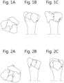
- Fractures close to joints are notoriously difficult to treat.
- One of the most common fractures close to a joint are distal radius wrist fractures (32.000 cases/year in Belgium).
- WO2014047514A1 discloses methods and systems for improving the quality, throughput and efficiency of Solid Free Form manufacture of implant components.
- the joint replacement field has come to embrace the concept of "patient-specific” and “patient-engineered” implant systems.
- the implants, associated tools, and procedures are designed or otherwise modified to account for and accommodate the individual anatomy of the patient undergoing the surgical procedure.
- Such systems typically utilize non-invasive imaging data, taken of the patient pre-operatively, to guide the design and/or selection of the implant, surgical tools, and the planning of the surgical procedure itself.
- Various objectives of these newer systems can include: (1 ) reducing the amount of bony anatomy removed to accommodate the implant, (2) designing/selecting an implant that replicates and/or improves the function of the natural joint, (3) increasing the durability and functional lifetime of the implant, (4) simplifying the surgical procedure for the surgeon, (5) reducing patient recovery time and/or discomfort, and/or (6) improving patient outcomes. Because “patient-specific” and “patient-engineered” implant systems are created using anatomical information from a particular patient, such systems are generally created after the patient has been designated a "surgical candidate" and undergone non-invasive imaging.
- Document WO 2017/127887 A1 discloses a method and system for producing a digital model of a customised device, comprising the steps of : importing a first digital file of a base part; importing a second digital file of a target shape; determining a warping interpolation function based on source point positions associated with the base part and target point positions associated with the target shape; and applying the warping interpolation function to the points of said base part to generate a model of said customised device. It offers the user to manually position a base shape to aligned broken bones. This document, however, is not specifically related to treating joint fractures. It would seem that the teaching of this document does not address reduction of a fractured joint to ensure that all bone fragments of the fractured joint are aligned properly.
- the present invention concerns a method for obtaining an implantable plate for healing a fractured joint of a patient according to claim 1.
- the optimal parameter values comprise:
- the set of positions, orientations and/or diameters of screw holes are optimized to fixate the bone fragments which need to be directly fixated to the implantable plate.
- step 5 comprises the step of: 5a) composing instructions for manufacturing, preferably by 3D printing, the implantable plate on the basis of the calculated parameter values.
- a patient-specific and fracture-specific implantable plate for healing a fractured joint of a patient is manufactured, preferably by 3D printing the implantable plate, using the instructions obtained in step 5a.
- the fractured joint is an intra-articular fracture or a metaphyseal fracture.
- step 2 all different bone fragments comprising a size which is at least a predefined limit size in the zone around the joint fracture are identified, said limit size preferably being at most 3mm, more preferably at most 2mm, still more preferably at most 1mm.
- the present invention also concerns an implantable plate for healing a fractured joint of a patient, said implantable plate being patient-specific and fracture-specific and obtainable by a method according to the present invention.
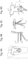
- the implantable plate comprises a number of at least two screw holes, preferably at least three screw holes, which comprise non-parallel orientations with respect to one another.
- the orientation of each of the screw holes is preferably non-parallel with all of the other screw holes. This is particularly advantageous to ensure that the implantable plate cannot easily loosen by forces in the direction of the screws and/or screw holes, since in such case there is no single direction for the crews and/or screw holes.
- the at least two screw holes, and preferably the at least three screw holes being positioned in a shaft portion of the implantable plate, the shaft portion hereby referring to a portion of the plate which is intended to be attached directly to a healthy bone or a healthy portion of a bone of the fractured joint.
- the two, three or more screw holes can be positioned in a shaft portion of the implantable plate which is to be attached to a healthy portion of the ulna.
- the present invention also concerns a method for obtaining a positioning guide device for an implantable plate for healing a fractured joint of a patient according to claim 12.
- step iv) comprises providing an implantable plate or instructions for manufacturing an implantable plate, and calculating the optimal parameter values for the positioning guide device taking into account said provided implantable plate or said instructions for manufacturing the implantable plate. This allows to manufacture the best positioning guide device specific for the implantable plate.
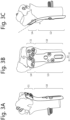
- the method comprises the step of providing indications for the positions and/or orientations of one or more screw holes on the positioning guide device.
- step v comprises the step of: va) composing instructions for manufacturing, preferably 3D printing, the positioning guide device on the basis of the calculated parameter values,
- the method comprises the step of manufacturing, preferably 3D printing, the guide device using the instructions.
- the present invention also concerns a method for manufacturing a patient-specific and fracture-specific implantable plate for healing a fractured joint of a patient by 3D printing the implantable plate using the computer program product according to the present invention, and preferably as described here above, and a patient-specific and fracture-specific implantable plate for healing a fractured joint of a patient thus obtained.
- the present method also concerns a positioning guide device for positioning an implantable plate according to the present invention and a method for manufacturing such guide device by 3D printing the guide device using the computer program product according to the present invention.
- the present invention inventively combines image processing techniques, preferably implemented using an artificial intelligence (AI) algorithm and three-dimensional (3D) printing techniques.
- AI artificial intelligence
- 3D three-dimensional
- one method of manufacturing an implantable plate comprises the step of obtaining a computer program product comprising instructions for three-dimensional (3D) printing of an implantable plate for healing a fractured joint of a patient according to the present invention, and the step of manufacturing the implantable plate using said computer program product.
- the above method enables a surgeon to interact preoperatively with 3D images, get a reduction prediction, finetune the design, pre-manufacture screw directions, preferably by screw directions pre-printing, and have personalized surgical aids.
- the method can be realized through use of automated CT analysis, predictive models for fracture fragment recognition and reduction, an implementation of an optimal material/strength ratio in the manufacturing of the implantable plate, and many further benefits.
- the implantable plate comprises, and more preferably is integrally made of, a biodegradable material such as a biodegradable synthesized polymer. This allows omission of hardware (HW) removal and thus eliminates the need of surgical plate removal procedure.
- the implantable plate comprises, and more preferably is integrally made of, a metal such as titanium or stainless steel.
- the implantable plate is made integrally of a single material.
- the present invention allows obtaining an implantable plate for healing a fractured joint of a patient, by:
- step 5 comprises the step of: 5a) composing instructions for manufacturing, preferably for 3D printing the implantable plate on the basis of the calculated parameter values.
- the implantable plate can then preferably be manufactured, preferably by 3D printing, the implantable plate using the computer program product comprising the instructions.
- document WO 2017/127887 A1 describes a large number of possibilities for producing a digital model of a customised device, but fails to disclose a clear an unambiguous set of steps to follow in order to allow manufacturing an implant for healing a fractured joint. Fractures joints are more difficult to reduce than other fractures, due to a larger number of bones and bone fragments, as well as the positions thereof which allow normal functioning of the joint. It is the inventors insight that all essential fragments of broken or ruptured bones and at least the ends of unbroken bones which form part of the fractured joint need to be incorporated in order to ensure a good reduction simulation. Furthermore, the document does not refer to adapting certain parameters such as the positions and orientations of screw holes taking into account the simulated reduction of the bone fragments.
- Medical images hereby are preferably obtained using X-ray photography or X-ray tomography, more preferably using a CT scan.
- X-ray based medical images are very suitable for imaging bone structure.
- the medical images are provided in the DICOM standard.
- the medical images in step a are provided in such a way as to allow at least a partial three-dimensional reconstruction of the zone around the fracture.
- the zone around the fracture preferably comprises all fragments of broken or ruptured bones, as well as at least the ends of unbroken bones which form part of the fractured joint.
- Reconstruction of a 3D representation of the bone structure may involve an analysis of the medical images.
- the medical images provided in step a should allow such 3D reconstruction.
- reconstructing the 3D representation of the bone structure comprises reconstructing surfaces of bones of said bone structure.
- Step 2 identifying different bone fragments within said 3D representation
- the bone fragments are identified by segmenting the bone fragments on the basis of CT images of the fractured joint.
- the proposed segmentation algorithm uses a probability-based variation of the watershed transform, referred to as the Probabilistic Watershed Transform (PWT).
- PWT uses a set of probability distributions, one for each bone fragment, that model the likelihood that a given pixel is a measurement from one of the bone fragments.
- Step 3 simulating a reduction of said bone fragments into a full joint
- Reduction is a procedure to realign the bone fragments of a fracture.
- the fragments lose their alignment in the form of displacement or angulation.
- the bony fragments must be re-aligned to their normal anatomical position.
- Orthopaedic surgery attempts to recreate the normal anatomy of the fractured bone by reduction of the displacement.
- the reduction of the bones of the fractured joint is first simulated. Simulation of the reduction prior to the actual reduction allows the surgeon to better prepare the operation. Additionally, in the present invention, the simulation allows to compute the parameters for the implantable plate.
- Each match includes two fragment surface patches hypothesized to correspond in the reconstruction.
- Each alignment optimization initialized by the user triggers a 3D surface registration which takes as input: (1) the specified matches and (2) the current position of the fragments.
- the proposed system leverages domain knowledge via user interaction, and incorporates recent advancements in surface registration, to generate fragment reconstructions that are more accurate than manual methods and more reliable than completely automatic methods.
- the method of the prior art discussed here is not particularly related to joint fractures, and furthermore relates to a reduction step which is partially manual and partially automated.
- One main shortcoming of this method is that the technique relies heavily on the surgeon's experience and abilities to obtain a good reduction simulation. And even if the surgeon is experienced and very capable, good reductions remain difficult.
- the present inventor therefore has found that the quality of the reduction simulation can be drastically improved, and fully or nearly fully automated, if the reduction is simulated by fitting said bone fragments to a 3D representation of a healthy joint of said patient.
- the 3D representation of a healthy joint of said patient can be obtained via a number of ways.
Landscapes
- Health & Medical Sciences (AREA)
- Orthopedic Medicine & Surgery (AREA)
- Engineering & Computer Science (AREA)
- Surgery (AREA)
- Life Sciences & Earth Sciences (AREA)
- Animal Behavior & Ethology (AREA)
- Biomedical Technology (AREA)
- Heart & Thoracic Surgery (AREA)
- Medical Informatics (AREA)
- Molecular Biology (AREA)
- General Health & Medical Sciences (AREA)
- Public Health (AREA)
- Veterinary Medicine (AREA)
- Nuclear Medicine, Radiotherapy & Molecular Imaging (AREA)
- Materials Engineering (AREA)
- Chemical & Material Sciences (AREA)
- Manufacturing & Machinery (AREA)
- Neurology (AREA)
- Robotics (AREA)
- Dentistry (AREA)
- Oral & Maxillofacial Surgery (AREA)
- Physics & Mathematics (AREA)
- Mechanical Engineering (AREA)
- Optics & Photonics (AREA)
- Prostheses (AREA)
- Surgical Instruments (AREA)
Claims (15)
- Verfahren zum Erhalten einer implantierbaren Platte zur Heilung eines gebrochenen Gelenks eines Patienten, welches folgende Schritte umfasst:1) Bereitstellen einer 3D-Darstellung einer Knochenstruktur in einer Region rund um eine Gelenkfraktur, wobei die Region alle Fragmente von gebrochenen oder gerissenen Kochen und zumindest die Enden von unversehrten Kochen, welche einen Teil des gebrochenen Gelenks bilden, umfasst;2) automatisches Identifizieren unterschiedlicher Knochenfragmente innerhalb der 3D-Darstellung;3) Simulieren einer Reduktion der Knochenfragmente zu einem vollständigen Gelenk;4) automatisches Berechnen optimaler Parameterwerte für eine implantierbare Platte;5) Erhalten der implantierbaren Platte unter Berücksichtigung der berechneten Parameterwerte,wobei in Schritt 3) die Reduktion durch automatisiertes Einpassen von Positionen und Ausrichtungen der Knochenfragmente in eine 3D-Darstellung eines gesunden Gelenks des Patienten simuliert wird und wobei alle Knochenfragmente, welche direkt an die implantierbare Platte fixiert werden müssen, auf Grundlage der Identifikation der Knochenfragmente in Schritt 2) und/oder auf Grundlage der Simulation der Reduktion in Schritt 3) identifiziert werden,dadurch gekennzeichnet, dass in Schritt 2) alle unterschiedlichen Knochenfragmente, die eine Größe von mindestens einer vordefinierten Grenzgröße aufweisen, in der Region rund um die Gelenkfraktur identifiziert werden.
- Verfahren nach Anspruch 1, wobei die optimalen Parameterwerte Folgende umfassen:- einen Satz von Positionen von Schraublöchern für die implantierbare Platte;- einen Satz von Ausrichtungen von Schraublöchern für die implantierbare Platte, und/oder- einen Satz von Durchmessern von Schraublöchern für die implantierbare Platte.
- Verfahren nach Anspruch 2, wobei der Satz von Positionen, Ausrichtungen und/oder Durchmessern von Schraublöchern optimiert werden, um die Knochenfragmente, welche direkt an die implantierbare Platte fixiert werden müssen, zu fixieren.
- Verfahren nach einem der vorhergehenden Ansprüche, wobei Schritt 5) folgenden Schritt umfasst:
5a) Erstellen von Anweisungen für den 3D-Druck der implantierbaren Platte auf Grundlage der berechneten Parameterwerte. - Verfahren nach einem der vorhergehenden Ansprüche, wobei die 3D-Darstellung des gesunden Gelenks des Patienten auf medizinischen Bildern des kontralateralen Gelenks zum gebrochenen Gelenk basiert, wobei die medizinischen Bilder durch Erhalten medizinischer Bilder der Knochenstruktur in der Region rund um das kontralaterale Gelenk erhalten werden.
- Verfahren nach einem der vorhergehenden Ansprüche, wobei die 3D-Darstellung des gesunden Gelenks des Patienten auf einer Extrapolation der Form der Knochenfragmente basiert, wobei die Form des Gelenks aus Folgendem extrapoliert wird:- aus der Form und Größe eines intakten Abschnitts eines Knochenfragments des gebrochenen Gelenks, und/oder- aus den identifizierten Knochenfragmenten durch Extrapolation der Knochenfragmente in der Nähe der Ränder.
- Verfahren nach einem der vorhergehenden Ansprüche, wobei Schritt 4) jegliche, oder jegliche Kombination, der folgenden Schritte umfasst:a) Auswählen einer Schablonenplatte aus einem Satz von Schablonen für implantierbare Platten zur Heilung des gebrochenen Gelenks auf Grundlage der berechneten optimalen Parameterwerte für die implantierbare Platte;b) Berechnen einer optimalen Plattendicke oder eines optimalen Plattendickenprofils, und/oderc) Auswählen eines Materials aus einem Satz von Materialien für implantierbare Platten.
- Verfahren nach Anspruch 7, welches den Schritt a) umfasst und ferner den Schritt des Herstellens einer Positionierungsführungsvorrichtung durch das Durchführen der folgenden Schritte umfasst:- Berechnen optimaler Parameterwerte für eine Positionierungsführungsvorrichtung, und- Erstellen von Anweisungen für den 3D-Druck der Positionierungsführungsvorrichtung auf Grundlage der berechneten Parameterwerte.
- Verfahren nach einem der vorhergehenden Ansprüche, wobei das gebrochene Gelenk eine intraartikuläre Fraktur oder metaphysäre Fraktur ist.
- Verfahren nach einem der vorhergehenden Ansprüche, wobei in Schritt 2) die Knochenfragmente durch das Segmentieren der Knochenfragmente auf Grundlage von CT-Bildern des gebrochenen Gelenks identifiziert werden.
- Verfahren nach einem der vorhergehenden Ansprüche, wobei die Grenzgröße höchstens 3 mm beträgt, bevorzugter höchstens 2 mm, noch bevorzugter höchstens 1 mm.
- Verfahren zum Erhalten einer Positionierungsführungsvorrichtung für eine implantierbare Platte zur Heilung eines gebrochenen Gelenks eines Patienten, welches folgende Schritte umfasst:i) Bereitstellen einer 3D-Darstellung einer Knochenstruktur in einer Region rund um eine Gelenkfraktur, wobei die Region vorzugsweise alle Fragmente von gebrochenen oder gerissenen Kochen und zumindest die Enden von unversehrten Kochen, welche einen Teil des gebrochenen Gelenks bilden, umfasst;ii) automatisches Identifizieren unterschiedlicher Knochenfragmente innerhalb der 3D-Darstellung;iii) Simulieren einer Reduktion der Knochenfragmente zu einem vollständigen Gelenk;iv) automatisches Berechnen optimaler Parameterwerte für eine Positionierungsführungsvorrichtung;v) Erhalten der Positionierungsführungsvorrichtung unter Berücksichtigung der berechneten Parameterwerte,wobei in Schritt iii) die Reduktion durch automatisiertes Einpassen von Positionen und Ausrichtungen der Knochenfragmente in eine 3D-Darstellung eines gesunden Gelenks des Patienten simuliert wird und wobei alle Knochenfragmente, welche direkt an die implantierbare Platte fixiert werden müssen, auf Grundlage der Identifikation der Knochenfragmente in Schritt 2) und/oder auf Grundlage der Simulation der Reduktion in Schritt 3) identifiziert werden,wobei Schritt v) vorzugsweise folgenden Schritt umfasst:va) Erstellen von Anweisungen für den 3D-Druck der Positionierungsführungsvorrichtung auf Grundlage der berechneten Parameterwerte, undwelches vorzugsweise den Schritt des 3D-Drucks der Führungsvorrichtung unter Verwendung der Anweisungen umfasst,dadurch gekennzeichnet, dass in Schritt ii) alle unterschiedlichen Knochenfragmente, die eine Größe von mindestens einer vordefinierten Grenzgröße aufweisen, in der Region rund um die Gelenkfraktur identifiziert werden.
- Führungsvorrichtung erhalten nach Anspruch 12, wobei das gebrochene Gelenk ein gebrochenes Handgelenk ist, zum Führen des Positionierens einer implantierbaren Platte auf einen distalen Radius des gebrochenen Handgelenks, wobei die Führungsvorrichtung mit Angaben zu den Positionen und/oder Ausrichtungen von einem oder mehreren Schraublöchern versehen ist, wobei die Führungsvorrichtung ferner laterale Positionierungsabschnitte (21, 22) und einen distalen Positionierungsabschnitt (26), welche ein korrektes Positionieren der Führungsvorrichtung auf einem Radiusschaft gestatten, umfasst, wobei die lateralen Positionierungsabschnitte (21, 22) an einen bestimmten Abschnitt des Schafts angepasst sind und der distale Positionierungsabschnitt (26) an einen Abschnitt eines Knochens auf einer Frakturlinie (25) angepasst ist, wobei die Schraublöcher in der Führungsvorrichtung (23, 24) angeben, wo sich die Bohrlöcher in dem Schaft des Radius befinden müssen und wie die Bohrlöcher ausgerichtet sein müssen.
- Medizinisches Kit, welches eine implantierbare Platte zur Heilung eines gebrochenen Gelenks eines Patienten umfasst, wobei die implantierbare Platte patientenspezifisch und frakturspezifisch ist und durch ein Verfahren nach einem der Ansprüche 1 bis 11 erhalten werden kann, sowie ein Fixierungssystem, welches einen Satz von Schrauben umfasst, wobei die Schrauben des Satzes vorzugsweise unter Berücksichtigung der Anzahl der Schraublöcher in der implantierbaren Platte ausgewählt werden, wobei das medizinische Kit ferner eine Führungsvorrichtung nach Anspruch 13 umfasst.
- Medizinisches Kit nach Anspruch 14, wobei der Satz von Schrauben eine oder mehrere Kompressionsschrauben umfasst, wobei mindestens eine und vorzugsweise alle der Kompressionsschrauben vorzugsweise eine Unterlegscheibe, eine neigbare Unterlegscheibe, umfassen und/oder damit versehen sind, wobei die Unterlegscheibe vorzugsweise so gewählt wird, dass sie auf die Kompressionsschraube passt.
Applications Claiming Priority (2)
| Application Number | Priority Date | Filing Date | Title |
|---|---|---|---|
| EP19213859.2A EP3831323A1 (de) | 2019-12-05 | 2019-12-05 | Verbesserte implantierbare platte und verfahren zur herstellung davon |
| PCT/EP2020/084957 WO2021111013A1 (en) | 2019-12-05 | 2020-12-07 | Improved implantable plate and method of manufacturing thereof |
Publications (3)
| Publication Number | Publication Date |
|---|---|
| EP4069110A1 EP4069110A1 (de) | 2022-10-12 |
| EP4069110C0 EP4069110C0 (de) | 2025-04-30 |
| EP4069110B1 true EP4069110B1 (de) | 2025-04-30 |
Family
ID=68806685
Family Applications (2)
| Application Number | Title | Priority Date | Filing Date |
|---|---|---|---|
| EP19213859.2A Pending EP3831323A1 (de) | 2019-12-05 | 2019-12-05 | Verbesserte implantierbare platte und verfahren zur herstellung davon |
| EP20828293.9A Active EP4069110B1 (de) | 2019-12-05 | 2020-12-07 | Verbesserte implantierbare platte und verfahren zur herstellung davon |
Family Applications Before (1)
| Application Number | Title | Priority Date | Filing Date |
|---|---|---|---|
| EP19213859.2A Pending EP3831323A1 (de) | 2019-12-05 | 2019-12-05 | Verbesserte implantierbare platte und verfahren zur herstellung davon |
Country Status (3)
| Country | Link |
|---|---|
| EP (2) | EP3831323A1 (de) |
| JP (1) | JP2023511648A (de) |
| WO (1) | WO2021111013A1 (de) |
Families Citing this family (3)
| Publication number | Priority date | Publication date | Assignee | Title |
|---|---|---|---|---|
| CN113681895B (zh) * | 2021-08-20 | 2023-03-10 | 宜宾显微智能科技有限公司 | 一种导针定位导向板定制及模拟验证系统及其方法 |
| KR20250067848A (ko) * | 2022-11-11 | 2025-05-15 | 애니메디솔루션 주식회사 | 수술 계획을 모델링하는 방법 |
| WO2024237771A1 (ko) * | 2023-05-18 | 2024-11-21 | 임준열 | 3d 프린터를 이용한 견쇄 관절 정복 또는 쇄골 정복 및 골 터널 가이드와 그 제조방법 |
Family Cites Families (6)
| Publication number | Priority date | Publication date | Assignee | Title |
|---|---|---|---|---|
| US6302887B1 (en) * | 1998-07-20 | 2001-10-16 | Joseph John Spranza | Hardware for high strength fastening of bone |
| US7537604B2 (en) * | 2002-11-19 | 2009-05-26 | Acumed Llc | Bone plates with slots |
| US9554868B2 (en) * | 2009-08-07 | 2017-01-31 | DePuy Synthes Products, Inc. | Method and apparatus for reducing malalignment of fractured bone fragments |
| CN104780872B (zh) | 2012-09-21 | 2017-04-05 | 康复米斯公司 | 使用自由实体制造优化植入物组件的设计和制造的方法和系统 |
| US10258256B2 (en) * | 2014-12-09 | 2019-04-16 | TechMah Medical | Bone reconstruction and orthopedic implants |
| WO2017127887A1 (en) * | 2016-01-25 | 2017-08-03 | 3Dmorphic Pty Ltd | Method and system for desi gni ng and fabr icati ng a customi sed device |
-
2019
- 2019-12-05 EP EP19213859.2A patent/EP3831323A1/de active Pending
-
2020
- 2020-12-07 EP EP20828293.9A patent/EP4069110B1/de active Active
- 2020-12-07 WO PCT/EP2020/084957 patent/WO2021111013A1/en not_active Ceased
- 2020-12-07 JP JP2022534285A patent/JP2023511648A/ja active Pending
Also Published As
| Publication number | Publication date |
|---|---|
| WO2021111013A1 (en) | 2021-06-10 |
| EP3831323A1 (de) | 2021-06-09 |
| JP2023511648A (ja) | 2023-03-22 |
| EP4069110A1 (de) | 2022-10-12 |
| WO2021111013A8 (en) | 2022-07-07 |
| EP4069110C0 (de) | 2025-04-30 |
| US20230000557A1 (en) | 2023-01-05 |
Similar Documents
| Publication | Publication Date | Title |
|---|---|---|
| AU2012289973B2 (en) | Automated design, selection, manufacturing and implantation of patient-adapted and improved articular implants, designs and related guide tools | |
| US20220273450A1 (en) | Patient-Adapted and Improved Orthopedic Implants, Designs and Related Guide Tools | |
| US9707044B2 (en) | Surgical guides from scanned implant data | |
| US20180360609A1 (en) | Patient-Adapted and Improved Articular Implants, Designs and Related Guide Tools | |
| EP4069110B1 (de) | Verbesserte implantierbare platte und verfahren zur herstellung davon | |
| JP2019511068A (ja) | カスタマイズされた装置を設計および作製するための方法およびシステム | |
| US20180303491A1 (en) | Two-part surgical guide | |
| US20230404673A1 (en) | Apparatus, system, and method for generating patient-specific implants and/or instrumentation for osteotomies | |
| US20240108414A1 (en) | Apparatus, system, and method for generating patient-specific implants and/or instrumentation | |
| US20250195081A1 (en) | Patient-specific osteotomy systems, methods, and instrumentation | |
| US12121272B2 (en) | Two-part surgical guide | |
| US20230310013A1 (en) | Apparatus, system, and method for patient-specific instrumentation | |
| US20240008880A1 (en) | Apparatus, system, and method for patient-specific systems, methods, and instrumentation | |
| US12496109B2 (en) | Implantable plate and method of manufacturing thereof | |
| US20250057547A1 (en) | Apparatus, system, and method for patient-specific systems, methods, and instrumentation | |
| Vitkovic et al. | Designing of patient-specific implant by using subdivision surface shaped on parametrized cloud of points | |
| US20240252180A1 (en) | Apparatus, system, and method for osteotomies | |
| US20240268966A1 (en) | Apparatus, system, and method for lapidus correction | |
| US20230371966A1 (en) | Apparatus, system, and method for patient-specific methods and instrumentation | |
| Vitković et al. | Contact Surface Model Parameterization of the Extra-Articular Distal Humerus Plate | |
| US20230363773A1 (en) | Apparatus, system, and method for patient-specific methods and instrumentation | |
| US20240252181A1 (en) | Apparatus, system, and method for instrumentation | |
| US20240277350A1 (en) | Apparatus, system, and method for patient-specific harvesting guide | |
| EP4661776A1 (de) | Vorrichtung, system und verfahren zur lapidus-korrektur | |
| Ријебат | Parametric Models of the Plate Implants for Humerus Bone |
Legal Events
| Date | Code | Title | Description |
|---|---|---|---|
| STAA | Information on the status of an ep patent application or granted ep patent |
Free format text: STATUS: UNKNOWN |
|
| STAA | Information on the status of an ep patent application or granted ep patent |
Free format text: STATUS: THE INTERNATIONAL PUBLICATION HAS BEEN MADE |
|
| PUAI | Public reference made under article 153(3) epc to a published international application that has entered the european phase |
Free format text: ORIGINAL CODE: 0009012 |
|
| STAA | Information on the status of an ep patent application or granted ep patent |
Free format text: STATUS: REQUEST FOR EXAMINATION WAS MADE |
|
| 17P | Request for examination filed |
Effective date: 20220705 |
|
| AK | Designated contracting states |
Kind code of ref document: A1 Designated state(s): AL AT BE BG CH CY CZ DE DK EE ES FI FR GB GR HR HU IE IS IT LI LT LU LV MC MK MT NL NO PL PT RO RS SE SI SK SM TR |
|
| DAV | Request for validation of the european patent (deleted) | ||
| DAX | Request for extension of the european patent (deleted) | ||
| STAA | Information on the status of an ep patent application or granted ep patent |
Free format text: STATUS: EXAMINATION IS IN PROGRESS |
|
| 17Q | First examination report despatched |
Effective date: 20240506 |
|
| GRAP | Despatch of communication of intention to grant a patent |
Free format text: ORIGINAL CODE: EPIDOSNIGR1 |
|
| STAA | Information on the status of an ep patent application or granted ep patent |
Free format text: STATUS: GRANT OF PATENT IS INTENDED |
|
| RIC1 | Information provided on ipc code assigned before grant |
Ipc: B33Y 80/00 20150101ALI20241111BHEP Ipc: B33Y 50/00 20150101ALI20241111BHEP Ipc: B22F 10/80 20210101ALI20241111BHEP Ipc: A61B 34/10 20160101ALI20241111BHEP Ipc: A61B 17/68 20060101ALI20241111BHEP Ipc: A61B 17/56 20060101ALI20241111BHEP Ipc: A61B 17/17 20060101ALI20241111BHEP Ipc: A61B 17/80 20060101AFI20241111BHEP |
|
| INTG | Intention to grant announced |
Effective date: 20241125 |
|
| GRAS | Grant fee paid |
Free format text: ORIGINAL CODE: EPIDOSNIGR3 |
|
| GRAA | (expected) grant |
Free format text: ORIGINAL CODE: 0009210 |
|
| STAA | Information on the status of an ep patent application or granted ep patent |
Free format text: STATUS: THE PATENT HAS BEEN GRANTED |
|
| AK | Designated contracting states |
Kind code of ref document: B1 Designated state(s): AL AT BE BG CH CY CZ DE DK EE ES FI FR GB GR HR HU IE IS IT LI LT LU LV MC MK MT NL NO PL PT RO RS SE SI SK SM TR |
|
| REG | Reference to a national code |
Ref country code: CH Ref legal event code: EP Ref country code: GB Ref legal event code: FG4D |
|
| REG | Reference to a national code |
Ref country code: DE Ref legal event code: R096 Ref document number: 602020050542 Country of ref document: DE |
|
| REG | Reference to a national code |
Ref country code: IE Ref legal event code: FG4D |
|
| U01 | Request for unitary effect filed |
Effective date: 20250530 |
|
| U07 | Unitary effect registered |
Designated state(s): AT BE BG DE DK EE FI FR IT LT LU LV MT NL PT RO SE SI Effective date: 20250610 |
|
| PG25 | Lapsed in a contracting state [announced via postgrant information from national office to epo] |
Ref country code: ES Free format text: LAPSE BECAUSE OF FAILURE TO SUBMIT A TRANSLATION OF THE DESCRIPTION OR TO PAY THE FEE WITHIN THE PRESCRIBED TIME-LIMIT Effective date: 20250430 |
|
| PG25 | Lapsed in a contracting state [announced via postgrant information from national office to epo] |
Ref country code: GR Free format text: LAPSE BECAUSE OF FAILURE TO SUBMIT A TRANSLATION OF THE DESCRIPTION OR TO PAY THE FEE WITHIN THE PRESCRIBED TIME-LIMIT Effective date: 20250731 Ref country code: NO Free format text: LAPSE BECAUSE OF FAILURE TO SUBMIT A TRANSLATION OF THE DESCRIPTION OR TO PAY THE FEE WITHIN THE PRESCRIBED TIME-LIMIT Effective date: 20250730 |
|
| PG25 | Lapsed in a contracting state [announced via postgrant information from national office to epo] |
Ref country code: PL Free format text: LAPSE BECAUSE OF FAILURE TO SUBMIT A TRANSLATION OF THE DESCRIPTION OR TO PAY THE FEE WITHIN THE PRESCRIBED TIME-LIMIT Effective date: 20250430 |
|
| PG25 | Lapsed in a contracting state [announced via postgrant information from national office to epo] |
Ref country code: HR Free format text: LAPSE BECAUSE OF FAILURE TO SUBMIT A TRANSLATION OF THE DESCRIPTION OR TO PAY THE FEE WITHIN THE PRESCRIBED TIME-LIMIT Effective date: 20250430 |
|
| PG25 | Lapsed in a contracting state [announced via postgrant information from national office to epo] |
Ref country code: RS Free format text: LAPSE BECAUSE OF FAILURE TO SUBMIT A TRANSLATION OF THE DESCRIPTION OR TO PAY THE FEE WITHIN THE PRESCRIBED TIME-LIMIT Effective date: 20250731 |
|
| PG25 | Lapsed in a contracting state [announced via postgrant information from national office to epo] |
Ref country code: IS Free format text: LAPSE BECAUSE OF FAILURE TO SUBMIT A TRANSLATION OF THE DESCRIPTION OR TO PAY THE FEE WITHIN THE PRESCRIBED TIME-LIMIT Effective date: 20250830 |