EP4069099B1 - Vorrichtung zur freisetzung von einem geweberegenerativer multiwirkstoff-cocktail - Google Patents
Vorrichtung zur freisetzung von einem geweberegenerativer multiwirkstoff-cocktail Download PDFInfo
- Publication number
- EP4069099B1 EP4069099B1 EP20895644.1A EP20895644A EP4069099B1 EP 4069099 B1 EP4069099 B1 EP 4069099B1 EP 20895644 A EP20895644 A EP 20895644A EP 4069099 B1 EP4069099 B1 EP 4069099B1
- Authority
- EP
- European Patent Office
- Prior art keywords
- tissue
- therapeutic composition
- receiving end
- inner sleeve
- pressing member
- Prior art date
- Legal status (The legal status is an assumption and is not a legal conclusion. Google has not performed a legal analysis and makes no representation as to the accuracy of the status listed.)
- Active
Links
Images


Classifications
-
- A—HUMAN NECESSITIES
- A61—MEDICAL OR VETERINARY SCIENCE; HYGIENE
- A61M—DEVICES FOR INTRODUCING MEDIA INTO, OR ONTO, THE BODY; DEVICES FOR TRANSDUCING BODY MEDIA OR FOR TAKING MEDIA FROM THE BODY; DEVICES FOR PRODUCING OR ENDING SLEEP OR STUPOR
- A61M35/00—Devices for applying media, e.g. remedies, on the human body
-
- A—HUMAN NECESSITIES
- A61—MEDICAL OR VETERINARY SCIENCE; HYGIENE
- A61F—FILTERS IMPLANTABLE INTO BLOOD VESSELS; PROSTHESES; DEVICES PROVIDING PATENCY TO, OR PREVENTING COLLAPSING OF, TUBULAR STRUCTURES OF THE BODY, e.g. STENTS; ORTHOPAEDIC, NURSING OR CONTRACEPTIVE DEVICES; FOMENTATION; TREATMENT OR PROTECTION OF EYES OR EARS; BANDAGES, DRESSINGS OR ABSORBENT PADS; FIRST-AID KITS
- A61F2/00—Filters implantable into blood vessels; Prostheses, i.e. artificial substitutes or replacements for parts of the body; Appliances for connecting them with the body; Devices providing patency to, or preventing collapsing of, tubular structures of the body, e.g. stents
- A61F2/0077—Special surfaces of prostheses, e.g. for improving ingrowth
-
- A—HUMAN NECESSITIES
- A61—MEDICAL OR VETERINARY SCIENCE; HYGIENE
- A61F—FILTERS IMPLANTABLE INTO BLOOD VESSELS; PROSTHESES; DEVICES PROVIDING PATENCY TO, OR PREVENTING COLLAPSING OF, TUBULAR STRUCTURES OF THE BODY, e.g. STENTS; ORTHOPAEDIC, NURSING OR CONTRACEPTIVE DEVICES; FOMENTATION; TREATMENT OR PROTECTION OF EYES OR EARS; BANDAGES, DRESSINGS OR ABSORBENT PADS; FIRST-AID KITS
- A61F2/00—Filters implantable into blood vessels; Prostheses, i.e. artificial substitutes or replacements for parts of the body; Appliances for connecting them with the body; Devices providing patency to, or preventing collapsing of, tubular structures of the body, e.g. stents
- A61F2/50—Prostheses not implantable in the body
- A61F2/78—Means for protecting prostheses or for attaching them to the body, e.g. bandages, harnesses, straps, or stockings for the limb stump
- A61F2/80—Sockets, e.g. of suction type
-
- A—HUMAN NECESSITIES
- A61—MEDICAL OR VETERINARY SCIENCE; HYGIENE
- A61L—METHODS OR APPARATUS FOR STERILISING MATERIALS OR OBJECTS IN GENERAL; DISINFECTION, STERILISATION OR DEODORISATION OF AIR; CHEMICAL ASPECTS OF BANDAGES, DRESSINGS, ABSORBENT PADS OR SURGICAL ARTICLES; MATERIALS FOR BANDAGES, DRESSINGS, ABSORBENT PADS OR SURGICAL ARTICLES
- A61L27/00—Materials for grafts or prostheses or for coating grafts or prostheses
- A61L27/14—Macromolecular materials
- A61L27/22—Polypeptides or derivatives thereof, e.g. degradation products
- A61L27/227—Other specific proteins or polypeptides not covered by A61L27/222, A61L27/225 or A61L27/24
-
- A—HUMAN NECESSITIES
- A61—MEDICAL OR VETERINARY SCIENCE; HYGIENE
- A61L—METHODS OR APPARATUS FOR STERILISING MATERIALS OR OBJECTS IN GENERAL; DISINFECTION, STERILISATION OR DEODORISATION OF AIR; CHEMICAL ASPECTS OF BANDAGES, DRESSINGS, ABSORBENT PADS OR SURGICAL ARTICLES; MATERIALS FOR BANDAGES, DRESSINGS, ABSORBENT PADS OR SURGICAL ARTICLES
- A61L27/00—Materials for grafts or prostheses or for coating grafts or prostheses
- A61L27/14—Macromolecular materials
- A61L27/22—Polypeptides or derivatives thereof, e.g. degradation products
- A61L27/24—Collagen
-
- A—HUMAN NECESSITIES
- A61—MEDICAL OR VETERINARY SCIENCE; HYGIENE
- A61L—METHODS OR APPARATUS FOR STERILISING MATERIALS OR OBJECTS IN GENERAL; DISINFECTION, STERILISATION OR DEODORISATION OF AIR; CHEMICAL ASPECTS OF BANDAGES, DRESSINGS, ABSORBENT PADS OR SURGICAL ARTICLES; MATERIALS FOR BANDAGES, DRESSINGS, ABSORBENT PADS OR SURGICAL ARTICLES
- A61L27/00—Materials for grafts or prostheses or for coating grafts or prostheses
- A61L27/36—Materials for grafts or prostheses or for coating grafts or prostheses containing ingredients of undetermined constitution or reaction products thereof, e.g. transplant tissue, natural bone, extracellular matrix
- A61L27/3637—Materials for grafts or prostheses or for coating grafts or prostheses containing ingredients of undetermined constitution or reaction products thereof, e.g. transplant tissue, natural bone, extracellular matrix characterised by the origin of the biological material other than human or animal, e.g. plant extracts, algae
-
- A—HUMAN NECESSITIES
- A61—MEDICAL OR VETERINARY SCIENCE; HYGIENE
- A61L—METHODS OR APPARATUS FOR STERILISING MATERIALS OR OBJECTS IN GENERAL; DISINFECTION, STERILISATION OR DEODORISATION OF AIR; CHEMICAL ASPECTS OF BANDAGES, DRESSINGS, ABSORBENT PADS OR SURGICAL ARTICLES; MATERIALS FOR BANDAGES, DRESSINGS, ABSORBENT PADS OR SURGICAL ARTICLES
- A61L27/00—Materials for grafts or prostheses or for coating grafts or prostheses
- A61L27/50—Materials characterised by their function or physical properties, e.g. injectable or lubricating compositions, shape-memory materials, surface modified materials
- A61L27/54—Biologically active materials, e.g. therapeutic substances
-
- A—HUMAN NECESSITIES
- A61—MEDICAL OR VETERINARY SCIENCE; HYGIENE
- A61L—METHODS OR APPARATUS FOR STERILISING MATERIALS OR OBJECTS IN GENERAL; DISINFECTION, STERILISATION OR DEODORISATION OF AIR; CHEMICAL ASPECTS OF BANDAGES, DRESSINGS, ABSORBENT PADS OR SURGICAL ARTICLES; MATERIALS FOR BANDAGES, DRESSINGS, ABSORBENT PADS OR SURGICAL ARTICLES
- A61L27/00—Materials for grafts or prostheses or for coating grafts or prostheses
- A61L27/50—Materials characterised by their function or physical properties, e.g. injectable or lubricating compositions, shape-memory materials, surface modified materials
- A61L27/56—Porous materials, e.g. foams or sponges
-
- A—HUMAN NECESSITIES
- A61—MEDICAL OR VETERINARY SCIENCE; HYGIENE
- A61N—ELECTROTHERAPY; MAGNETOTHERAPY; RADIATION THERAPY; ULTRASOUND THERAPY
- A61N1/00—Electrotherapy; Circuits therefor
- A61N1/02—Details
- A61N1/04—Electrodes
- A61N1/0404—Electrodes for external use
- A61N1/0408—Use-related aspects
- A61N1/0468—Specially adapted for promoting wound healing
-
- A—HUMAN NECESSITIES
- A61—MEDICAL OR VETERINARY SCIENCE; HYGIENE
- A61N—ELECTROTHERAPY; MAGNETOTHERAPY; RADIATION THERAPY; ULTRASOUND THERAPY
- A61N1/00—Electrotherapy; Circuits therefor
- A61N1/18—Applying electric currents by contact electrodes
- A61N1/20—Applying electric currents by contact electrodes continuous direct currents
- A61N1/205—Applying electric currents by contact electrodes continuous direct currents for promoting a biological process
-
- A—HUMAN NECESSITIES
- A61—MEDICAL OR VETERINARY SCIENCE; HYGIENE
- A61N—ELECTROTHERAPY; MAGNETOTHERAPY; RADIATION THERAPY; ULTRASOUND THERAPY
- A61N1/00—Electrotherapy; Circuits therefor
- A61N1/18—Applying electric currents by contact electrodes
- A61N1/32—Applying electric currents by contact electrodes alternating or intermittent currents
- A61N1/326—Applying electric currents by contact electrodes alternating or intermittent currents for promoting growth of cells, e.g. bone cells
-
- A—HUMAN NECESSITIES
- A61—MEDICAL OR VETERINARY SCIENCE; HYGIENE
- A61N—ELECTROTHERAPY; MAGNETOTHERAPY; RADIATION THERAPY; ULTRASOUND THERAPY
- A61N1/00—Electrotherapy; Circuits therefor
- A61N1/18—Applying electric currents by contact electrodes
- A61N1/32—Applying electric currents by contact electrodes alternating or intermittent currents
- A61N1/36—Applying electric currents by contact electrodes alternating or intermittent currents for stimulation
- A61N1/36014—External stimulators, e.g. with patch electrodes
-
- A—HUMAN NECESSITIES
- A61—MEDICAL OR VETERINARY SCIENCE; HYGIENE
- A61N—ELECTROTHERAPY; MAGNETOTHERAPY; RADIATION THERAPY; ULTRASOUND THERAPY
- A61N1/00—Electrotherapy; Circuits therefor
- A61N1/40—Applying electric fields by inductive or capacitive coupling ; Applying radio-frequency signals
-
- A—HUMAN NECESSITIES
- A61—MEDICAL OR VETERINARY SCIENCE; HYGIENE
- A61L—METHODS OR APPARATUS FOR STERILISING MATERIALS OR OBJECTS IN GENERAL; DISINFECTION, STERILISATION OR DEODORISATION OF AIR; CHEMICAL ASPECTS OF BANDAGES, DRESSINGS, ABSORBENT PADS OR SURGICAL ARTICLES; MATERIALS FOR BANDAGES, DRESSINGS, ABSORBENT PADS OR SURGICAL ARTICLES
- A61L2300/00—Biologically active materials used in bandages, wound dressings, absorbent pads or medical devices
- A61L2300/20—Biologically active materials used in bandages, wound dressings, absorbent pads or medical devices containing or releasing organic materials
- A61L2300/204—Biologically active materials used in bandages, wound dressings, absorbent pads or medical devices containing or releasing organic materials with nitrogen-containing functional groups, e.g. aminoxides, nitriles, guanidines
-
- A—HUMAN NECESSITIES
- A61—MEDICAL OR VETERINARY SCIENCE; HYGIENE
- A61L—METHODS OR APPARATUS FOR STERILISING MATERIALS OR OBJECTS IN GENERAL; DISINFECTION, STERILISATION OR DEODORISATION OF AIR; CHEMICAL ASPECTS OF BANDAGES, DRESSINGS, ABSORBENT PADS OR SURGICAL ARTICLES; MATERIALS FOR BANDAGES, DRESSINGS, ABSORBENT PADS OR SURGICAL ARTICLES
- A61L2300/00—Biologically active materials used in bandages, wound dressings, absorbent pads or medical devices
- A61L2300/20—Biologically active materials used in bandages, wound dressings, absorbent pads or medical devices containing or releasing organic materials
- A61L2300/21—Acids
-
- A—HUMAN NECESSITIES
- A61—MEDICAL OR VETERINARY SCIENCE; HYGIENE
- A61L—METHODS OR APPARATUS FOR STERILISING MATERIALS OR OBJECTS IN GENERAL; DISINFECTION, STERILISATION OR DEODORISATION OF AIR; CHEMICAL ASPECTS OF BANDAGES, DRESSINGS, ABSORBENT PADS OR SURGICAL ARTICLES; MATERIALS FOR BANDAGES, DRESSINGS, ABSORBENT PADS OR SURGICAL ARTICLES
- A61L2300/00—Biologically active materials used in bandages, wound dressings, absorbent pads or medical devices
- A61L2300/20—Biologically active materials used in bandages, wound dressings, absorbent pads or medical devices containing or releasing organic materials
- A61L2300/252—Polypeptides, proteins, e.g. glycoproteins, lipoproteins, cytokines
-
- A—HUMAN NECESSITIES
- A61—MEDICAL OR VETERINARY SCIENCE; HYGIENE
- A61L—METHODS OR APPARATUS FOR STERILISING MATERIALS OR OBJECTS IN GENERAL; DISINFECTION, STERILISATION OR DEODORISATION OF AIR; CHEMICAL ASPECTS OF BANDAGES, DRESSINGS, ABSORBENT PADS OR SURGICAL ARTICLES; MATERIALS FOR BANDAGES, DRESSINGS, ABSORBENT PADS OR SURGICAL ARTICLES
- A61L2300/00—Biologically active materials used in bandages, wound dressings, absorbent pads or medical devices
- A61L2300/40—Biologically active materials used in bandages, wound dressings, absorbent pads or medical devices characterised by a specific therapeutic activity or mode of action
- A61L2300/412—Tissue-regenerating or healing or proliferative agents
- A61L2300/414—Growth factors
-
- A—HUMAN NECESSITIES
- A61—MEDICAL OR VETERINARY SCIENCE; HYGIENE
- A61L—METHODS OR APPARATUS FOR STERILISING MATERIALS OR OBJECTS IN GENERAL; DISINFECTION, STERILISATION OR DEODORISATION OF AIR; CHEMICAL ASPECTS OF BANDAGES, DRESSINGS, ABSORBENT PADS OR SURGICAL ARTICLES; MATERIALS FOR BANDAGES, DRESSINGS, ABSORBENT PADS OR SURGICAL ARTICLES
- A61L2300/00—Biologically active materials used in bandages, wound dressings, absorbent pads or medical devices
- A61L2300/40—Biologically active materials used in bandages, wound dressings, absorbent pads or medical devices characterised by a specific therapeutic activity or mode of action
- A61L2300/426—Immunomodulating agents, i.e. cytokines, interleukins, interferons
-
- A—HUMAN NECESSITIES
- A61—MEDICAL OR VETERINARY SCIENCE; HYGIENE
- A61L—METHODS OR APPARATUS FOR STERILISING MATERIALS OR OBJECTS IN GENERAL; DISINFECTION, STERILISATION OR DEODORISATION OF AIR; CHEMICAL ASPECTS OF BANDAGES, DRESSINGS, ABSORBENT PADS OR SURGICAL ARTICLES; MATERIALS FOR BANDAGES, DRESSINGS, ABSORBENT PADS OR SURGICAL ARTICLES
- A61L2300/00—Biologically active materials used in bandages, wound dressings, absorbent pads or medical devices
- A61L2300/40—Biologically active materials used in bandages, wound dressings, absorbent pads or medical devices characterised by a specific therapeutic activity or mode of action
- A61L2300/428—Vitamins, e.g. tocopherol, riboflavin
-
- A—HUMAN NECESSITIES
- A61—MEDICAL OR VETERINARY SCIENCE; HYGIENE
- A61L—METHODS OR APPARATUS FOR STERILISING MATERIALS OR OBJECTS IN GENERAL; DISINFECTION, STERILISATION OR DEODORISATION OF AIR; CHEMICAL ASPECTS OF BANDAGES, DRESSINGS, ABSORBENT PADS OR SURGICAL ARTICLES; MATERIALS FOR BANDAGES, DRESSINGS, ABSORBENT PADS OR SURGICAL ARTICLES
- A61L2300/00—Biologically active materials used in bandages, wound dressings, absorbent pads or medical devices
- A61L2300/40—Biologically active materials used in bandages, wound dressings, absorbent pads or medical devices characterised by a specific therapeutic activity or mode of action
- A61L2300/43—Hormones, e.g. dexamethasone
-
- A—HUMAN NECESSITIES
- A61—MEDICAL OR VETERINARY SCIENCE; HYGIENE
- A61L—METHODS OR APPARATUS FOR STERILISING MATERIALS OR OBJECTS IN GENERAL; DISINFECTION, STERILISATION OR DEODORISATION OF AIR; CHEMICAL ASPECTS OF BANDAGES, DRESSINGS, ABSORBENT PADS OR SURGICAL ARTICLES; MATERIALS FOR BANDAGES, DRESSINGS, ABSORBENT PADS OR SURGICAL ARTICLES
- A61L2300/00—Biologically active materials used in bandages, wound dressings, absorbent pads or medical devices
- A61L2300/40—Biologically active materials used in bandages, wound dressings, absorbent pads or medical devices characterised by a specific therapeutic activity or mode of action
- A61L2300/432—Inhibitors, antagonists
- A61L2300/434—Inhibitors, antagonists of enzymes
Definitions
- the field of the invention relates to regenerative medicine. More specifically, the present disclosure relates to apparatus for promoting regeneration of tissue on a subject or within a subject in need thereof.
- the disclosed apparatus include wearable sleeves and the disclosed compositions include regenerative compositions
- US 20130245716 A1 discloses an alternatively structured apparatus for stimulation of animal tissue regeneration consisting of a regenerative tubular sleeve enclosing the wound site of an appendage with an outer body sealing a wound space between the wound site and the outer body.”
- Tissue progenitor cells for induction of regeneration processes may also have use; larval limb progenitor cells have been shown to activate the same Wnt, Shh signaling to promote patterning.
- tissue regeneration devices are typically attached to the subject utilizing glue, which is not favorable for long term treatments.
- glue to attach regenerative devices has two primary disadvantages. Glue provides a lack of flexibility of adjustments to the device post attachment. In addition, handling complications exist during surgery and device.
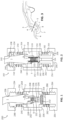
- an apparatus 200 for stimulation of tissue regeneration of a subject 1 is shown.
- the apparatus 200 is used to enclose a wounded or injured tissue 9 of a subject 1.
- the wounded or injured tissue 9 may located on an external or internal location of the subject 1.
- the apparatus 200 is described with respect to stimulating tissue regeneration of wounded tissue 9 located on an appendage 3 (e.g., mouse digit) in which the tip 2 has been amputated along a line 5 through at least a portion of the distal phalange 4, whereby regenerated tissue may include among other tissues, bone tissue 6, muscle tissue 7, and skin tissue 8.
- appendage 3 e.g., mouse digit
- the wounded or injured tissue 9 for stimulation of tissue regeneration using the apparatus 200 includes epithelial tissue, connective tissue, muscular tissue, or nervous tissue.
- Exemplary wounded or injured tissue 9 for regeneration includes, but is not limited to, squamous epithelium, cuboidal epithelium, transitional epithelium, pseudostratified columnar epithelium, columnar epithelium, glandular epithelium, bone, tendons, ligaments, adipose, areolar tissue, blood tissue, visceral muscle, smooth muscle, skeletal muscle, cardiac muscle, and neural tissues.
- the pressing member 214 places the wound 9 in contact with a protein matrix 228 disposed within the internal chamber 222 of the inner sleeve 216.
- the protein matrix 228 may guide tissue growth directionally and/or provide therapeutic agents for the wound 9 to stimulate tissue regeneration.
- Suitable aqueous solutions or dispersing media include, but are not limited to, water, cell culture medium, buffers (e.g., phosphate buffered saline), polyol (for example, glycerol, propylene glycol, liquid polyethylene glycol, and the like), and suitable mixtures thereof.
- the dispersing medium includes a therapeutic agent.
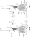
- the apparatus 200 may include a first adjustable adapter 246 disposable within the appendage receiving end 204 for selectively coupling the first end cap 234 engageable with the appendage receiving end 204 to the outer sleeve 202.
- the adjustable adapter 246 is a threaded adapter and the first end cap 234 includes grooves 248 to receive threads 250 of the adjustable adapter 246.
- the first end cap 234 may be tightened such that the gasket or septum 238 is placed in contact with a leading end 252 of the adjustable adapter 246 to secure the appendage 3 within the outer sleeve 202.
- the apparatus 200 includes a second end cap 254 that is engageable with the pressing member receiving end 206 of the outer sleeve 202.
- the second end cap 254 is coupled to the pressing member 214 to bias the pressing member 214 toward the engagement receiving end 220 of the inner sleeve 216.
- the pressing member 214 may be directly attached to or form part of the second end cap 254.
- the pressing member 214 may be separate from the second end cap 254.
- the pressing member 214 includes an engagement end 256 configured to rest on a seat portion 258 of the second end cap 254.
- the pressing member 214 includes a seat portion 260 opposite the engagement end 256 that is configured to receive the engagement end 220 of the inner sleeve 216.
- the pressing member 214 may be formed of a rigid material or an elastic material that deforms or compresses in response to the growth of the tissue at the wound 9.
- the apparatus 200 may include a second adjustable adapter 262 disposable within the pressing member receiving end 206 for selectively coupling the second end cap 254 engageable with the pressing member receiving end 206 to the outer sleeve 202.
- the second adjustable adapter 262 is a threaded adapter and the second end cap 254 includes grooves 264 configured to receive threads 266 of the second adjustable adapter 262.
- the second end cap 254 may be adjusted to control the pressure at the wound 9.
- One disadvantage of conventional devices is a lack of control of pressure at the wound 9 interface, which leads to variability in tissue regeneration outcomes if there is any type of gap (fluid collection, air, etc.).
- the apparatus 200 advantageously provides tunable pressure sufficient to hold the protein matrix 228 in contact with the wound 9.
- the apparatus 200 facilitates long term attachment (weeks, months, years, or longer) and is adjustable to facilitate growth of the regenerating tissue.
- the apparatus 200 includes a moveable or adjustable pressing member 214.
- the pressing member 214 may be moveable or extendable in response to tissue growth (indicated in FIGS. 4 and 5 by ⁇ x).
- an elastic member 268 is configured to extend between a seat portion 270 on the pressing member 214 and a seat portion 272 on the second adjustable adapter 262.
- the elastic member 268 e.g., spring, compressible material, deformable material
- the adjustable pressing member 214 includes interlocking spacers that can be extended over time in response to the tissue growth, or a screw-extension system.
- the apparatus 200 also includes an electrical stimulation device 300 to establish a longitudinal electrical field through the wound site 9, which is considered to provide an internal wound stump current and to provide electrical guidance cues for innervation and migration of cell types near the wound site.
- the electrical stimulation device 300 includes an anode 302 and a cathode 304 that are electrically connected to corresponding terminals of a power source 306 through leads 308, 310.
- cathode 304 is in the form of a stainless steel wire which is disposed adjacent the wound 9. A portion of the cathode 304 resides outside the apparatus 200 and is connectable to the lead 308, and a portion of the cathode 304 resides within the internal chamber 222 of the inner sleeve.
- the anode 302 is a conductive wire that may be inserted in the subject 1 at a location distant from the wound site 9. In the illustrated embodiment in which the apparatus 200 is disposed on an appendage 3, the anode 302 is disposed in the upper portion of the limb (rear leg) from which the appendage 3 extends.
- the anode 302 may comprise a Platinum/Iridium alloy wire which is connected to the power source 306 via the lead 310.
- the anode 302 can be permanently implanted, or temporarily inserted as needed.
- the power source 306 includes a battery pack 312 and circuitry 314, both of which are enclosed in a housing 316 and configured to provide a constant, low level current to the electrodes 302, 304 when connected thereto.
- the power source 306 resides externally of the subject 1, and the electrodes 302, 304 are configured to be detachably connectable to the power source 306.
- the cathode 304 and anode 302 are electrically connected to the power source 306 for the duration of the electrical stimulation treatment, and then disconnected between electrical stimulation treatments. Since the power source 306 and leads 308, 310 may be detached from the respective electrodes 302, 304, this arrangement conveniently reduces the overall bulk of the combined apparatus 200 and electrical stimulation device 300 during treatment paradigms in which electrical stimulation is used only intermittently.
- the protein matrix 228 comprises a biocompatible polymer.
- the biocompatible polymer suitable for use with the apparatus 100 includes, but is not limited to, polyethylene oxide (PEO), polyethylene glycols (PEGs), collagen, fibronectin, keratin, polyaspartic acid, polylysine, alginate, chitosan, chitin, hyaluronic acid, pectin, polycaprolactone, polylactic acid, polyglycolic acid, polyhydroxyalkanoates, dextrans, polyanhydrides, polymer, PLA-PGA, polyanhydride, polyorthoester, polycaprolactone, polyfumarate, collagen, silk fibroin, chitosan, alginate, hyaluronic acid and other biocompatible and/or biodegradable polymers.
- the protein matrix 228 is silk fibroin and/or collagen.
- the protein matrix 228 includes pores that match tissue surface areas (size) of the tissue upon which it is affixed in the apparatus 200 to optimize regrowth of the wounded or injured tissue.
- the protein matrix 228 has a pore size between about 1 ⁇ m to about 1500 ⁇ m, or about 350 ⁇ m, about 400 ⁇ m, about 450 ⁇ m, about 500 ⁇ m, about 550 ⁇ m, about 600 ⁇ m, about 650 ⁇ m, about 700 ⁇ m, about 750 ⁇ m, about 800 ⁇ m, about 850 ⁇ m, about 900 ⁇ m, about 950 ⁇ m, about 1000 ⁇ m, about 1050 ⁇ m, about 1100 ⁇ m, about 1150 ⁇ m, about 1200 ⁇ m, about 1300 ⁇ m, about 13350 ⁇ m, about 1400 ⁇ m, about 1450 ⁇ m, or about 1500.
- the protein matrix comprises silk fibroin.
- silk fibroin or "SF” may refer to a biopolymer produced from silkworm fibroin and insect or spider silk protein.
- silk fibroin useful for the present disclosure may be that produced by a number of species, including, without limitation: Antheraea mylitta; Antheraea pernyi; Antheraea yamamai; Galleria mellonella; Bombyx mori; Bombyx mandarina; Galleria mellonella; Nephila clavipes; Nephila senegalensis; Gasteracantha mammosa; Argiope aurantia; Araneus diadematus; Latrodectus geometricus; Araneus bicentenarius; Tetragnatha versicolor; Araneus ventricosus; Dolomedes tenebrosus; Euagrus chisoseus; Plectreurys
- SF is a structural protein, like collagen, but with a unique feature: it is produced from the extrusion of an amino-acidic solution by a living complex organism into the external environment, while collagen is produced in vivo, in the extracellular space by self-assembly of cell-produced monomers and not secreted to the external environment.
- SF properties are derived from its structure, which consists of hydrophobic blocks staggered by hydrophilic, acidic spacers. In its natural state, SF is organized into semicrystalline materials with ⁇ -sheet crystals alternated with amorphous regions, which provide strength and resilience to the protein materials formed from the protein.
- the multiplicities of forms in which regenerated SF can be processed at a low to high protein concentration and low to high molecular weight make it attractive for several high-tech applications.
- Processing of SF generally involves the partial or total dehydration of a fibroin solution (protein content of about 1wt % to about 15 wt %) to form, e.g., films, sponges, gels, spheres (micron- to nano-sized) and foams with numerous techniques (e.g. solvent casting, freeze drying, salt leaching, sonication). These fabrication processes provide a robust material that combines mechanical strength with biochemical properties.
- a fibroin solution protein content of about 1wt % to about 15 wt %
- foams with numerous techniques (e.g. solvent casting, freeze drying, salt leaching, sonication).
- the silk fibroin solutions used in methods and compositions provided herein may be obtained from a solution containing a dissolved silkworm silk, such as, for example, from Bombyx mori.
- the silk fibroin solution may be obtained from a solution containing a dissolved spider silk, such as, for example, from Nephila clavipes.
- the silk fibroin solution can also be obtained from a solution containing a genetically engineered silk such as from bacteria, yeast, mammalian cells, transgenic animals or transgenic plants. See, for example, WO 97/08315 and US Patent 5,245,012 .
- Genetically engineered silk can, for example, also comprise a therapeutic agent, e.g., a fusion protein with a cytokine, an enzyme, or any number of hormones or peptide- based drugs, antimicrobials and related substrates.
- silk polypeptide compositions utilized in accordance with the present compositions are substantially free of sericins (e.g., contain no detectable sericin or contain sericin at a level that one of ordinary skill in the pertinent art will consider negligible for a particular use).
- B. mori cocoons are boiled for about 30 minutes in an aqueous solution, such as, but not limited to, about 0.02M Na 2 CO 3 .
- the boiling (degumming) time is in a range of about 5 minutes to about 120 minutes and the boiling (degumming) temperature is in a range of about 30°C to about 120°C.
- the cocoons may be rinsed, for example, with water to extract the sericin proteins and the extracted silk is dissolved in an aqueous salt solution.
- Exemplary non-limiting salts useful for this purpose include lithium bromide, lithium thiocyanate, calcium nitrate, and other chemicals capable of solubilizing silk.
- the extracted silk is dissolved in about 9M to about 12 M LiBr solution.
- the salt is then removed, for example, by dialysis.
- the solution can then be concentrated using, any method known in the art.
- dialysis against a hygroscopic polymer for example, PEG, a polyethylene oxide, amylose or sericin can be done.
- PEG having a molecular weight of about 8,000 g/mol to about 10,000 g/mol and has a concentration of about 25% to about 50%.
- Any dialysis system can be used, e.g., a slide-a-lyzer dialysis cassette (Pierce, MW CO 3500).
- the solution is dialyzed for a time period sufficient to result in a final concentration of aqueous silk solution of between about 1% to about 30%. In some cases, dialysis for about 2 hours to about 12 hours is sufficient.
- the present disclosure provides a method of attaching the apparatus 200 to an appendage or tissue of a subject in need of tissue regeneration.
- the method includes contacting a wounded appendage or tissue 9 of the subject 1 to the wound receiving end 218 of the inner sleeve 216.
- the wounded appendage or tissue 9 may be placed in contact, or adjacent to, the protein matrix 228 comprising the provided therapeutic compositions.
- the wounded appendage or tissue 9 is slid through the gasket or septum 238 and the opening 236 of the first end cap 234.
- the method further includes placing the appendage 3 of the subject through the appendage receiving end 204 of the outer sleeve 202 so that the inner sleeve 216 is positioned within the internal chamber 208 of the outer sleeve 202.
- the method includes selectively engaging the first end cap 234 and the second end cap 254 to the outer sleeve 202 such that the pressing member 214 biases the engagement receiving end 220 towards the appendage 3.
- the method includes biasing the pressing member 214 towards the appendage 3 such that the wound 9 is placed in contact with at least a portion of the internal chamber 222 of the inner sleeve.
- the contact pressure between the wounded appendage or tissue 9 and the protein matrix 228 may be adjusted by selectively engaging or disengaging the second end cap 254 (e.g., tightening or loosening the second end cap 254 via grooves 264 and threads 266).
- the wounded appendage or tissue 9 is maintained within the apparatus 200 for a duration to promote tissue regeneration.
- the duration is about 1 minute, or about 10 minutes, or about 30 minutes, or about 1 hour, or about 2 hours, or about 3 hours, or about 4 hours, or about 5 hours, or about 6 hours, or about 12 hours, or about 24 hours, or 2 days, or about 3 days, or about 4 days, or about 5 days, or about a week, or about two weeks, or about three weeks, or about a month, or about six months, or about one year, or within a duration range bounded by any of these values.
- the protein matrix 228 may be kept moist by adding or replacing a buffered solution within the inner sleeve 216.
- therapeutic compositions for tissue regeneration and "multi-drug treatment” compositions (MDT).
- MDT multi-drug treatment compositions
- the therapeutic compositions may be utilized alone or in conjunction with the disclosed apparatus.
- At least two of the components in the provided therapeutic compositions act synergistically to increase the rate of regeneration, or at least three of the components, or at least four of the components, or at least five of the components, or all of the components act synergistically to increase the rate of regeneration.
- the disclosed apparatus may comprise the therapeutic compositions within the inner sleeve (e.g., within a reservoir in the inner sleeve).
- the therapeutic compositions may be present in a material or matrix that contacts the wound site and the therapeutic compositions may be delivered to the wound site when the apparatus is worn on a subject's appendage.
- Wearable apparatus that comprise and deliver therapeutic compositions are known in the art. (See, e.g., Herrera-Rincon et al., Cell Reports 25, 1593-1609 (2018 ).
- the disclosed therapeutic compositions may be present in a polymeric material such as, but not limited to, a silk hydrogel material.
- a silk hydrogel material loaded with a therapeutic composition may be prepared as follows.
- a therapeutic composition may be added to a silk solution (e.g., a 3% w/v silk solution), which then is induced to gel via addition of a reagent such as horseradish peroxidase (e.g., to a concentration of about 20 U/ml silk solution) with hydrogen peroxide (e.g., to a concentration of 0.01% w/v).
- a reagent such as horseradish peroxidase (e.g., to a concentration of about 20 U/ml silk solution) with hydrogen peroxide (e.g., to a concentration of 0.01% w/v).
- the silk can also gel with this enzymatic reaction, via a drop in pH, addition of energy such as via sonication or vortexing, an applied electric field, or addition of
- the disclosed therapeutic compositions may comprise one more agents that increase axonal/neurite growth and/or general cell proliferation.
- the disclosed therapeutic compositions do not promote pluripotency in cells and/or lead to teratoma formation.
- the disclosed therapeutic compositions may comprise one or more agents that promote tissue regeneration and/or healing.
- the therapeutic compositions comprise one or more of a growth factor, an agent that inhibits the inhibitor of the hypoxia-inducible factor 1-alpha (HIF 1-alpha), vitamin A or a derivative thereof, a lipid mediator such as a metabolic product of omega-3 fatty acids and may be derived from eicosapentaenoic acid or docosahexaenoic acid, a growth hormone, a steroid, and a depolarizing agent.
- the growth factor may be present at a dose within the therapeutic composition of at least about 0.1 ⁇ g/ml, about 0.2 ⁇ g/ml, about 0.3 ⁇ g/ml, about 0.4 ⁇ g/ml, about 0.5 ⁇ g/ml, about 0.6 ⁇ g/ml, about 0.7 ⁇ g/ml, about 0.8 ⁇ g/ml, about 0.9 ⁇ g/ml, or about 1.0 ⁇ g/ml or within a dose range bounded by any of these values.
- the apparatus may comprise a concentration of the growth factor of at least about 0.1 ⁇ g/apparatus, about 0.2 ⁇ g/apparatus, about 0.3 ⁇ g/apparatus, about 0.4 ⁇ g/apparatus, about 0.5 ⁇ g/apparatus, about 0.6 ⁇ g/apparatus, about 0.7 ⁇ g/apparatus, about 0.8 ⁇ g/apparatus, about 0.9 ⁇ g/apparatus, or about 1.00 ⁇ g/apparatus or within a concentration range bounded by any of these values.
- the growth factor promotes the growth of one or more tissue types.
- the disclosed therapeutic compositions may include a prolyl hydroxylase domain (PHD) enzyme inhibitor (i.e., a PHD inhibitor), for example, in order to stabilize constitutive expression of the HIF-1 ⁇ protein.
- PHD prolyl hydroxylase domain
- Suitable PHD inhibitors may include, but are not limited to, 4,4 ⁇ -dihydro-4-oxo-1,10-phenanthroline-3-carboxylic acid (1,4-DPCA), N-[(1,3-dicyclohexylhexahydro-2,4,6-trioxo-5-pyrimidinyl)carbonyl]-glycine (i.e., , GSK1278863 or Daprodustat), 6-Amino-1,3-dimethyl-5-[(2-pyridinylthio)acetyl]-2,4(1H,3H)-pyrimidinedione (i.e., TM6089), 6-Amino-1,3-dimethyl-5-[[2-(2-pyridinyl)-4-quinolinyl]carbonyl]-2,4(1H,3H)-pyrimidinedione (i.e., TM60008), N-[(4-hydroxy-1-methyl-7-phenoxy-3-is
- the PHD inhibitor may be present at a dose within the therapeutic composition of at least about 0.004 ⁇ g/ml, about 0.006 ⁇ g/ml, about 0.008 ⁇ g/ml, about 0.010 ⁇ g/ml, about 0.012 ⁇ g/ml, about 0.014 ⁇ g/ml, about 0.016 ⁇ g/ml, 0.018 ⁇ g/ml, about 0.020 ⁇ g/ml, about 0.022 ⁇ g/ml, or 0.024 ⁇ g/ml or within a dose range bounded by any of these values.
- the apparatus may comprise a concentration of the PHD inhibitor of at least about 0.087 ⁇ g/apparatus, about 0.092 ⁇ g/apparatus, about 0.097 ⁇ g/apparatus, about 0.102, about 0.107 ⁇ g/apparatus, about 0.112 ⁇ g/apparatus, about 0.117 ⁇ g/apparatus, about 0.122 ⁇ g/apparatus, about 0.127 ⁇ g/apparatus, or about 0.132 ⁇ g/apparatus or within a concentration range bounded by any of these values.
- the PHD inhibitor controls excess collagen deposition at a wound site.
- compositions may include vitamin A or a metabolite or derivative thereof or any agent that functions in proximo-distal positional information.
- exemplary derivatives of vitamin A include, but are not limited, to retinoic acid, retinol, retinyl carboxylates (e.g., retinyl acetate, retinyl propionate, and retinyl palmitate), tretinoin, and tazarotene, and combinations thereof.
- Agents that may function in proximo-distal position information include, but are not limited to, bone morphogenetic protein 9 (BMP9), nodal growth differentiation factor (i.e .,, HTX5 or NODAL), Activin, transforming growth factor-beta (TGF- ⁇ ), and fibroblast growth factor 8 (FGF8).
- BMP9 bone morphogenetic protein 9
- nodal growth differentiation factor i.e ., HTX5 or NODAL
- Activin transforming growth factor-beta
- FGF8 fibroblast growth factor 8
- the apparatus may comprise a concentration of the vitamin A or the derivative thereof or the agent that functions in proximo-distal positional information of at least about 0.03 ⁇ g/apparatus, about 0.06 ⁇ g/apparatus, about 0.09 ⁇ g/apparatus, about 0.12 ⁇ g/apparatus, about 0.15 ⁇ g/apparatus, about 0.18 ⁇ g/apparatus, about 0.21 ⁇ g/apparatus, about 0.24 ⁇ g/apparatus, or about 0.27 ⁇ g/apparatus or within a concentration range bounded by any of these values.
- the disclosed therapeutic compositions may include a lipid mediator and/or a metabolic byproduct of an omega-3 fatty acid that promotes the resolution of the inflammatory response (i.e., an anti-inflammatory agent).
- Suitable lipid mediators may include derivatives (e.g., metabolic byproducts) of omega-3 fatty, and/or derivatives of eicosapentaeonic acid or docosahexaenoic acid that promote the resolution of the inflammatory response (i.e., anti-inflammatory).
- the lipid mediator may be present at a dose within the therapeutic composition of at least about 0.006 ⁇ g/ml, about 0.012 ⁇ g/ml, about 0.018 ⁇ g/ml, about 0.024 ⁇ g/ml, about 0.030 ⁇ g/ml, about 0.036 ⁇ g/ml, about 0.042 ⁇ g/ml, about 0.048 ⁇ g/ml, or about 0.054 ⁇ g/ml or within a dose range bounded by any of these values.
- the apparatus may comprise a concentration of the lipid mediator of at least about 0.005, ⁇ g/apparatus, about 0.011 ⁇ g/apparatus, about 0.017 ⁇ g/apparatus, about 0.023 ⁇ g/apparatus, about 0.029 ⁇ g/apparatus, about 0.035 ⁇ g/apparatus, about 0.041 ⁇ g/apparatus, about 0.047 ⁇ g/apparatus, or about 0.053 ⁇ g/apparatus or within a concentration range bounded by any of these values.
- the disclosed therapeutic compositions may include peptide hormones, for example, peptide hormones which stimulates growth, cell reproduction, and cell regeneration.
- peptide hormones for example, peptide hormones which stimulates growth, cell reproduction, and cell regeneration.
- exemplary hormone peptides or proteins include, but are not limited to, growth hormone (GH), insulin-like growth factor-1 (IGF-1), transforming growth factor-beta-1 (TGF ⁇ -1), epidermal growth factor (EGF), Granulocyte-colony stimulating factor (G-CSF), and fibroblast growth factor FGF.
- the growth hormone or steroid may be present at a dose within the therapeutic composition of at least about 0.1 ⁇ g/ml, about 0.2 ⁇ g/ml, about 0.3 ⁇ g/ml, about 0.4 ⁇ g/ml, about 0.5 ⁇ g/ml, about 0.6 ⁇ g/ml, about 0.7 ⁇ g/ml, about 0.8 ⁇ g/ml, about 0.9 ⁇ g/ml, or about 1.0 ⁇ g/ml or within a dose range bounded by any of these values.
- the apparatus may comprise a concentration of the growth hormone or steroid of at least about 0.1 ⁇ g/apparatus, about 0.2 ⁇ g/apparatus, about 0.3 ⁇ g/apparatus, about 0.4 ⁇ g/apparatus, about 0.5 ⁇ g/apparatus, about 0.6 ⁇ g/apparatus, about 0.7 ⁇ g/apparatus, about 0.8 ⁇ g/apparatus, about 0.9 ⁇ g/apparatus, or about 1.0 ⁇ g/apparatus or within a concentration range bounded by any of these values.
- the disclosed therapeutic compositions may include depolarizing agents.
- Suitable depolarizing agents may include, but are not limited to ionophores (e.g., an ion channel opener or blocker).
- Suitable depolarizing agents may include, but are not limited to, monensin, potassium gluconate, sodium gluconate, and the like.
- a "subject” means a human or animal. Usually the animal is a vertebrate such as a primate, rodent, domestic animal or game animal. Primates include chimpanzees, cynomologous monkeys, spider monkeys, and macaques, e.g., Rhesus. Rodents include mice, rats, woodchucks, ferrets, rabbits and hamsters.
- domestic and game animals include cows, horses, pigs, deer, bison, buffalo, feline species, e.g., domestic cat, canine species, e.g., dog, fox, wolf, avian species, e.g., chicken, emu, ostrich, and fish, e.g., trout, catfish and salmon.
- the subject is a mammal, e.g., a primate, e.g., a human.
- a subject can be male or female.
- the subject is a mammal.
- the mammal can be a human, non-human primate, mouse, rat, dog, cat, horse, or cow, but are not limited to these examples. Mammals other than humans can be used as subjects that represent animal models of tissue repair, regeneration and/or reconstruction.
- the methods and compositions described herein can be used to treat domesticated animals and/or pets.
- Tissue regeneration may be measured by any method known in the art such as, but not limited to, measuring the expression of Yamanaka factors, Sox2, Oct3/4, Klf4, and/or c-Myc in tissue treated with the apparatus according to the disclosure and/or therapeutic compositions versus tissue not treated with the disclosed apparatus and/or therapeutic compositions.
- BioDome wearable bioreactor
- a brief exposure period e.g., 24 hours to a wearable bioreactor containing silk infused with several small molecule compounds was found to induce dramatic outgrowth, patterning, and sensorimotor function following amputation in Xenopus.
- Treated animals displayed a marked delay of wound closure, followed by long-term (about 16-month) growth outcomes, including increased bone length, soft tissue patterning, and neuromuscular repair. Histologically, the new limbs contained nerve, smooth muscle indicative of blood vessels, and reorganization of the extracellular matrix proteins involved in remodeling of the limb.
- Transcriptomic analysis identified immediate and short-term pathways and transcriptional targets of the intervention in the blastema.
- the sutured biodome was comprised of a soft silicon insert which in turn contained silk hydrogels as a controlled-release substrate and drug carrier.
- the fabrication of the device has been reported elsewhere ( Golding et al., (2016) PLoS One 11, e0155618 ).
- the outer cylindrical silicon sleeve (20-mm H x 18-mm D) were fabricated by casting silicon elastomer (Dragon skin 10, Smooth-on, Macungie, PA) against a 3D-printed mold which was designed using CAD software (Solidworks, Waltham, MA, USA) and printed using a Formlab 3D printer (Somerville, MA, USA).
- the septum was cut along the central hole at four locations, a quarter-circle away from each other.
- Custom washers were either made from PDMS using soft-lithography or 3D-printed using the 3D printer.
- the cylindrical wall of the apparatus 100 insert was fabricated from a transparent polyester membrane filter (0.45 ⁇ m pore size, 12- ⁇ m thick, # 1300016, Sterlitech, Kent, WA). The filter was cut in rectangles (7 mm x 5 mm) and rolled around a metal rod of 1.5-mm diameter (#8907K62, McMaster). Then the wall was glued to a silicone bottom part using a silicone adhesive (Dragon Skin 10 FAST, Smoothon, Macungie, PA) to complete the insert.
- a silicone adhesive Dragon Skin 10 FAST, Smoothon, Macungie, PA
- the protective cage was assembled from threaded caps, a transparent acrylic body, and adapters.
- a donut-shaped septum that only allows one-direction bending was provided to prevent the detachment of the apparatus due to animal movement and tampering.
- the custom washer together with the bottom cap provide tunable pressure sufficient to hold the scaffold insert tightly against the wound bed, as well as keeping its position stable over a long-term experiment. It also comes with an access hole for media exchange.
- the apparatus insert contains a membranous side wall and a silicone bottom. The side wall can hold liquid necessary for keep the tissue moist as well as facilitating the gas exchange the silicone bottom serves as a septum for insertion of needle for media exchange.
- mice were anesthetized and treated with an analgesic as previously described.
- the devices were then removed by cutting the single suture on either side of the leg, and the frogs were placed back into tanks containing a fungicide (Kordon methylene blue at concentrations of 1 mL /10 L frog water. After another 24 hr, the water was replaced with fresh 100% frog water. Once their devices were removed, animals were maintained for 18 months in frog water that was changed daily. Endpoint euthanasia was carried out by full-body immersion in frog water with 0.2% benzocaine. Regenerates, contralateral limbs, and brain tissues were collected and processed for histological analysis.
- the solution was then dialyzed in DI water using a dialysis cassette (molecular weight cut-off of 3.5 kDa, Thermo Fisher Scientific, Waltham, MA) with gentle stirring. Water was changed six times over a 48-h period. The dialyzed solution was centrifuged three times at 13,000 g, 4°C for 20 min each followed by filtering through a cell strainer (40- ⁇ m pore size, Thermo Fisher) to remove impurities. To determine the concentration of the filtered solution, a 0.5 ml sample was dried completely overnight in an oven. Having evaporated the water content, the dried silk was weighed and the concentration in % (wt/v) was calculated as the ratio of the weight of the dried silk over its initial volume of 0.5 ml.
- Silk scaffolds with aligned pores were fabricated using 5 ⁇ l of the 4% (wt/v) silk solution. The solution was placed on top of an aluminum plate and a steep temperature gradient was induced by merging plate in liquid nitrogen (LN2). Finger-like columns of ice crystals growing from the cold surface created channel-like structures internally inside the silk solution as it solidified. After 10 min of cooling, the frozen solution was lyophilized for over 24 h to remove the water. The sponges were trimmed to fit in the Biodome as an insert and were sterilized in ethylene oxide and stored at room temperature in sterile condition until use.
- LN2 liquid nitrogen
- Collagen scaffolds with aligned channel-like porous structure were fabricated by controlled directional freezing and freeze-drying of a 1.5% (wt/wt) collagen solution (in a similar fashion as for the silk scaffolds).
- the scaffolds were cut in a cylindrical shape (4-mm long and 1.5-mm diameter) and placed inside the biodome insert using forceps.
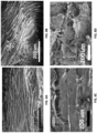
- FIG. 8A is a silk scaffold with channel-like pores aligned along the long axis.
- FIG. 8B is a silk scaffold with pores aligned perpendicular to the long axis.
- FIG. 8C is a SEM image of collagen scaffold with pores aligned along the long axis.
- FIG. 8D is a collagen scaffold with pores aligned perpendicular to the long axis.
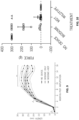
- each hydrogel solution loaded with specific amounts of the specified drugs were added into 1.5-ml microcentrifuge tubes and incubated for 45 min at 37°C to a complete gelation. Then, 1 ml of Dulbecco's phosphate buffered saline (DPBS 1X, Gibco) was added into each vial followed by incubation at 37°C. At fixed time points, 300 ⁇ l of supernatant was collected for analysis. Release solutions of drug-free silk hydrogels were used as controls. Standard curves were determined by measuring the optical density of solutions with known concentrations. All the release experiments were carried out in triplicate to ensure accuracy.
- DPBS 1X Dulbecco's phosphate buffered saline
- optical density of the release solutions was determined on a UV-transparent 96-well plate (Corning, Corning, NY) using a microplate reader SpectraMax M2 (Molecular Devices, San Jose, CA) operated by SoftMax Pro 6 software. Detection was performed at a wavelength of 280 nm and 350 nm for 1,4-DPCA and RA solutions, respectively.
- slides were equilibrated to room temperature for at least 2 hr prior to staining. Slides were post fixed for 5 min in 4% PFA and then blocked in blocking buffer (PBS with 0.1% Triton X-100 and 10% normal goat serum) for 1 hr. Primary antibodies against acetylated ⁇ -tubulin (1:100), TGF- ⁇ (1:250), smooth muscle actin (1:100), laminin (1:100), fibronectin (1:500), and phosphohistone H3 (1:250) were used. Slides were stained individually with each antibody except anti-smooth muscle actin and anti-laminin, which were stained together. Primary antibodies were incubated on the slides overnight.
- Sections were imaged using an EVOS FL automated imaging system (ThermoFisher Scientific). Entire sections were collected and stitched together for analysis.
- NGS next generation sequencing
- the reference genome for Xenopus laevis was downloaded from the NCBI Genome database, assembly GCA_001663975.1. Reads were aligned using STAR aligner ( Dobin et al., Bioinform. (2013);29(1):15-21. doi: 10.1093/bioinformatics/bts635. Epub Oct 25. PubMed PMID: 23104886; PubMed Central PMCID: PMC3530905 .) and aligned reads were counted using featureCounts ( Liao et al., (2014) Bioinform. 30(7):923-30 ).
- Annotated module graphs combined gene-gene interaction data from Kyoto Encyclopedia of Genes and Genomes (KEGG) and Chemical and Genetic Perturbations (CGP) to map the interacting genes contained in each module.
- KEGG Kyoto Encyclopedia of Genes and Genomes
- CGP Chemical and Genetic Perturbations
- Tomograms also revealed marked similarity between unamputated and MDT-treated bone anatomy. Repatterning was evident in the provided MDT regenerates including the re-expression of bone features that are normally associated with the attachment of musculature. Specifically, at 17 mpa, the MDT group displayed bone features characteristic of muscular attachment as well as the presence of joint segmentation.
- the larger wound site would provide a larger blastema, which might contribute more material to limb regrowth.
- Blastema proliferation was assessed via immunohistochemistry for SOX2, a proliferative cell marker.
- SOX2 a proliferative cell marker.
- soft tissue at 2.5 months was similarly predictive, with thicker tissue and increased bone length in the MDT hindlimbs compared to control. Fluorescent images obtained 2.5 mpa revealed that 24 hour exposure to MDT compositions increases stemness, inhibition of wound formation resulting in longer regenerates in early regeneration processes.
- Increased soft tissue growth and re-epithelization was also observed 2.5 months after amputation in the MDT-exposed group, which mirrored the increased length of bone tissues as confirmed by x-ray imaging.
- RNA-sequencing was performed comparing the transcriptome of the blastema that was obtained from MDT vs. untreated animals at 11 hr, 24 hr, and 7 d post- amputation.
- a heatmap comparing gene expression levels of MDT animals compared to a no device treatment shows significant differential gene expression at 11 HPA that persists to 24 HPA.
- 7 DPA dynamic gene expression levels return to normal, indicating a period of dynamic gene expression within 24 hours of amputation.
- the major genes that were downregulated were predominantly associated to muscle structure (myosin-4, microfibril-associated glycoprotein) and metabolism (e.g., sarcolipin).
- the pattern of these differently expressed genes between MDT- treated and control blastema was opposite to that of the upregulated genes, in that, the expression of the down regulated genes increased from 11 hpa to 7 dpa.
- Upregulated genes include nervous system-specific transcripts that suggest an important role of neuroprotective proteins just after amputation.
- Downregulated genes include metabolic and muscle-related transcripts, suggesting that resources are being directed away from muscle maintenance towards stabilization of the tissue.
- GO analysis of metabolic and biosynthetic pathways reveals an early downregulation (at 11HPA and 24HPA) that increases in rate by 7 dpa.
- Table 1 illustrates gene expression levels of MDT animals compared to no device treatment for 11 hpa, 24 hpa, and 7 dpa..
- Module 2 contained 142 genes that are significantly present (p ⁇ 0.00009) within pathways associated with cell junction organization, laminin interactions, cell-cell communication, apoptotic cleavage of cell adhesion proteins, and non-integrin membrane ECM interactions, which may be representative of cell-cell communication and adhesion.
- the 105 genes in Module 3 were significantly present (p ⁇ 0.01096) in muscle contraction, acetylcholine activity, and myogenesis pathways.
- Module 4 contained 54 genes that are significantly (p ⁇ 0.00142) over-represented within glucose metabolism, muscle contraction, gluconeogenesis, and glycolysis pathways.
Landscapes
- Health & Medical Sciences (AREA)
- Life Sciences & Earth Sciences (AREA)
- General Health & Medical Sciences (AREA)
- Veterinary Medicine (AREA)
- Public Health (AREA)
- Animal Behavior & Ethology (AREA)
- Chemical & Material Sciences (AREA)
- Engineering & Computer Science (AREA)
- Biomedical Technology (AREA)
- Medicinal Chemistry (AREA)
- Transplantation (AREA)
- Oral & Maxillofacial Surgery (AREA)
- Epidemiology (AREA)
- Dermatology (AREA)
- Nuclear Medicine, Radiotherapy & Molecular Imaging (AREA)
- Radiology & Medical Imaging (AREA)
- Molecular Biology (AREA)
- Heart & Thoracic Surgery (AREA)
- Dispersion Chemistry (AREA)
- Biophysics (AREA)
- Botany (AREA)
- Vascular Medicine (AREA)
- Cardiology (AREA)
- Cell Biology (AREA)
- Orthopedic Medicine & Surgery (AREA)
- Hematology (AREA)
- Anesthesiology (AREA)
- Biotechnology (AREA)
- Chemical Kinetics & Catalysis (AREA)
- Medicines That Contain Protein Lipid Enzymes And Other Medicines (AREA)
- Medicines Containing Plant Substances (AREA)
- Pharmaceuticals Containing Other Organic And Inorganic Compounds (AREA)
- Acyclic And Carbocyclic Compounds In Medicinal Compositions (AREA)
- Media Introduction/Drainage Providing Device (AREA)
- Materials For Medical Uses (AREA)
- Electrotherapy Devices (AREA)
- Medicines Containing Antibodies Or Antigens For Use As Internal Diagnostic Agents (AREA)
- Medicinal Preparation (AREA)
Claims (16)
- Vorrichtung, die Hüllen aufweist, zur Stimulation der Geweberegeneration an einer Stelle in einem Gewebe eines Säugetiersubjekts,wobei die Hüllen eine Außenhülle (202) und eine Innenhülle (216) aufweisen, wobeidie Außenhülle ein Gewebeaufnahmeende (204) und ein Druckelement-Aufnahmeende (206) gegenüber dem Gewebeaufnahmeende aufweist,wobei die Innenhülle innerhalb der Außenhülle angeordnet ist,wobei die Innenhülle aufweist:ein Ende zur Aufnahme der Stelle des Gewebes zur Regeneration (218),ein Eingriff-Aufnahmeende (220), gegenüber dem Ende zur Aufnahme der Stelle des Gewebes zur Regeneration und zum Eingriff mit einem Druckelement (214), undeine innere Kammer (222), die zur Aufnahme der Stelle des Gewebes zur Regeneration ausgebildet ist;wobei das Druckelement dazu ausgebildet ist, sich durch das Druckelement-Aufnahmeende der Außenhülle zu erstrecken und das Eingriff-Aufnahmeende der Innenhülle zum Gewebe hin vorzuspannen, so dass mindestens ein Abschnitt der Stelle des Gewebes zur Regeneration in Kontakt mit einem Abschnitt der inneren Kammer der Innenhülle platziert ist;eine erste Endkappe (234), die mit dem Gewebeaufnahmeende der Außenhülle in Eingriff bringbar ist und eine Öffnung (236) aufweist, die zur Aufnahme des Gewebes ausgebildet ist; undeine zweite Endkappe (254), die mit dem Druckelement-Aufnahmeende der Außenhülle in Eingriff bringbar ist.
- Vorrichtung nach Anspruch 1, wobei das Druckelement einen Sitzabschnitt (260) aufweist, der zur Aufnahme des Eingriff-Aufnahmeendes der Innenhülle ausgebildet ist, oder ein Eingriffsende (256) aufweist, das dazu ausgebildet ist, auf einem Sitzabschnitt der zweiten Endkappe aufzuliegen.
- Vorrichtung nach Anspruch 1, wobei das Eingriff-Aufnahmeende der Innenhülle ein poröses Filtermedium (230) aufweist, das die innere Kammer an dem Eingriff-Aufnahmeende abdichtend umschließt; und das poröse Filtermedium optional eine synthetische Membran oder Polymermembran ist.
- Vorrichtung nach Anspruch 1, wobei die Vorrichtung ferner ein komprimierbares Element aufweist, das zwischen dem porösen Filtermedium und dem Druckelement (232) positioniert ist, wobei das komprimierbare Element optional Watte oder ein eingekapseltes Gel aufweist.
- Vorrichtung nach Anspruch 1, wobei die Innenhülle eine Protein- oder Polymermatrix (228) aufweist, die die innere Kammer der Innenhülle mindestens teilweise füllt; wobei die Protein- oder Polymermatrix (wie etwa Seidenfibroin und Kollagen oder ein Polymer, das ausgerichtete Poren in einer 3D-Matrix bildet) optional ein dreidimensionales poröses Gerüst aufweist, wobei das poröse Gerüst Poren aufweist, die ein Richtungsmuster bilden; und die Protein- oder Polymermatrix optional einen therapeutischen Wirkstoff aufweist.
- Vorrichtung nach Anspruch 5, wobei das poröse Gerüst ausgerichtete Poren aufweist, die im Wesentlichen ausgerichtete Kanäle bilden, und wobei die ausgerichteten Kanäle in der Protein- oder Polymermatrix parallel zur Längsachse der Innenhülle angeordnet sind.
- Vorrichtung nach Anspruch 1, die ferner eine therapeutische Zusammensetzung aufweist, welche in der inneren Kammer der Innenhülle angeordnet ist, und die therapeutische Zusammensetzung optional aufweist:einen Wachstumsfaktor;einen Inhibitor von Prolylhydroxylase-Domäne-, PHD-, Enzym;Vitamin A oder ein Derivat davon undeinen Lipidmediator.
- Vorrichtung nach Anspruch 7, wobei die therapeutische Zusammensetzung einen Wachstumsfaktor aufweist, der aus der Gruppe ausgewählt ist, die aus aus Gehirn abgeleitetem neurotrophem Faktor, BDNF, Nervenwachstumsfaktor, NGF, Neurotrophin-3, NT-3, Neurotrophin-4, NT-4, Glia-abgeleitetem neurotrophem Faktor, GDNF, ciliarem neurotrophem Faktor, CNTF, und Leukämie-Inhibitorfaktor, LIF, und einer Kombination daraus besteht; und/oderdie therapeutische Zusammensetzung einen Inhibitor von PHD-Enzym aufweist, der aus der Gruppe ausgewählt ist, die aus 4,4a-Dihydro-4-oxo-1,10-Phenanthrolin-3-Carboxylsäure, 1,4-DPCA, N-[(1,3-Dicyclohexylhexahydro-2,4,6-trioxo-5-pyrimidinyl)carbonyl]-glycin, 6-Amino-1,3-dimethyl-5-[(2-pyridinylthio)acetyl]-2,4(1H,3H)-pyrimidinedion, 6-Amino-1,3-dimethyl-5-[[2-(2-pyridinyl)-4-quinolinyl]carbonyl]-2,4(1H,3H)-pyrimidinedion, N-[(4-Hydroxy-1-methyl-7-phenoxy-3-isoquinolinyl)carbonyl]-glycin, Eisenchelatoren und einer Kombination daraus besteht; und/oderdie therapeutische Zusammensetzung ein Derivat von Vitamin A aufweist, das aus der Gruppe ausgewählt ist, die aus Retinsäure, Retinol, Retinylcarboxylaten, Tretinoin, Tazaroten und einer Kombination daraus besteht; und/oderdie therapeutische Zusammensetzung einen Lipidmediator aufweist, der aus der Gruppe ausgewählt ist, die aus einem Resolvin, einem Stoffwechselprodukt von Omega-3-Fettsäuren, einem Derivat von Eicosapentaensäure, einem Derivat von Docosahexaensäure und einer Kombination daraus besteht; und /oderdie therapeutische Zusammensetzung ein Resolvin aufweist, das aus der Gruppe ausgewählt ist, die aus Resolvin 5, Interleukin 6, IL-6, Interleukin 4, IL-4, Tumornekrosefaktor-alpha, TNF-alpha, Nuklearfaktor-kappa-Leichtketten-Verstärker von aktivierten B-Zellen (NF-kB) und einer Kombination daraus besteht; und/oderdie therapeutische Zusammensetzung einen Wirkstoff aufweist, der in proximo-distalen Positionsinformationen funktioniert, wobei der Wirkstoff aus der Gruppe ausgewählt ist, die aus knochenmorphogenetischem Protein 9, BMP9, Nodal-Wachstums-Differenzierungsfaktor, Activin, Transformations-Wachstumsfaktor-beta, TGF-P, Fibroblasten-Wachstumsfaktor 8, FGF8, und einer Kombination daraus besteht; und/oderdie therapeutische Zusammensetzung ein Peptid- oder Proteinhormon aufweist und/oderdie therapeutische Zusammensetzung ein Peptidhormon aufweist, das aus der Gruppe ausgewählt ist, die aus Wachstumshormon, GH, insulinartigem Wachstumsfaktor-I, IGF-1, Transformations-Wachstumsfaktor-beta-I, TGFP-1, epidermalem Wachstumsfaktor, EGF, Granulozytenkolonie-stimulierendem Faktor, G-CSF, Fibroblasten-Wachstumsfaktor, FGF, und einer Kombination daraus besteht.
- Vorrichtung nach Anspruch 8, wobei der Wachstumsfaktor in einer Dosis innerhalb der therapeutischen Zusammensetzung von 0,1 µg/ml bis 1 µg/ml vorhanden ist; und/oderder Inhibitor von PHD-Enzym in einer Dosis innerhalb der therapeutischen Zusammensetzung von 0,004 µg/ml bis 0,024 µg/ml vorhanden ist; und/oderdas Vitamin A oder Derivat desselben in einer Dosis innerhalb der therapeutischen Zusammensetzung von 0,03 µg/ml bis 0,27 µg/ml vorhanden ist; und/oderder Lipidmediator in einer Dosis innerhalb der therapeutischen Zusammensetzung von 0,006 µg/ml bis 0,054 µg/ml vorhanden ist; und/oderdas Peptid- oder Proteinhormon in einer Dosis innerhalb der therapeutischen Zusammensetzung von 0,1 µg/ml bis 1,0 µg/ml vorhanden ist.
- Vorrichtung nach Anspruch 1, wobei das Druckelement als Reaktion auf ein Wachstum an der Stelle des Gewebes zur Regeneration beweglich ist.
- Vorrichtung nach Anspruch 9, wobei das Gewebe Teil einer Gliedmaße oder eines Organs ist.
- Vorrichtung nach Anspruch 9, wobei das Druckelement ein elastisches Element (268) aufweist, das als Reaktion auf wachsendes Gewebe komprimiert wird.
- Vorrichtung nach Anspruch 9, wobei das elastische Element eine Feder aufweist.
- Vorrichtung nach Anspruch 1, wobei die Vorrichtung ferner einen ersten Gewindeadapter (246) aufweist, der innerhalb des Gewebeaufnahmeendes der Außenhülle anordenbar ist, um die erste Endkappe, die mit dem Gewebeaufnahmeende in Eingriff bringbar ist, selektiv mit der Außenhülle zu koppeln, wobei die erste Endkappe Rillen (248) aufweist, die dazu ausgebildet sind, Gewinde (250) des ersten Gewindeadapters aufzunehmen.
- Vorrichtung nach Anspruch 14, wobei die Vorrichtung ferner einen zweiten Gewindeadapter (262) aufweist, der innerhalb das Druckelement-Aufnahmeende der Außenhülle anordenbar ist, um die zweite Endkappe, die mit dem Druckelement in Eingriff bringbar ist, selektiv mit der Außenhülle zu koppeln, wobei die zweite Endkappe Rillen (264) aufweist, die dazu ausgebildet sind, Gewinde (266) des zweiten Gewindeadapters aufzunehmen.
- Vorrichtung nach Anspruch 1 oder 4, wobei die Vorrichtung ferner eine elektrische Stimulationseinrichtung (300) mit einer Anode (302) und einer Kathode (304) aufweist, wobei die Anode und die Kathode dazu ausgebildet sind, elektrisch mit entsprechenden Anschlüssen einer Stromquelle (306) verbunden zu sein, und wobei ein Abschnitt der Kathode in der Innenhülle angeordnet ist.
Priority Applications (1)
| Application Number | Priority Date | Filing Date | Title |
|---|---|---|---|
| EP25167701.9A EP4570309A3 (de) | 2019-12-06 | 2020-12-07 | Vorrichtung zur abgabe eines geweberegenerativen cocktails mit mehreren wirkstoffen |
Applications Claiming Priority (2)
| Application Number | Priority Date | Filing Date | Title |
|---|---|---|---|
| US201962944707P | 2019-12-06 | 2019-12-06 | |
| PCT/US2020/063665 WO2021113844A1 (en) | 2019-12-06 | 2020-12-07 | Tissue regenerative multi-drug cocktail and apparatus for delivery thereof |
Related Child Applications (1)
| Application Number | Title | Priority Date | Filing Date |
|---|---|---|---|
| EP25167701.9A Division EP4570309A3 (de) | 2019-12-06 | 2020-12-07 | Vorrichtung zur abgabe eines geweberegenerativen cocktails mit mehreren wirkstoffen |
Publications (3)
| Publication Number | Publication Date |
|---|---|
| EP4069099A1 EP4069099A1 (de) | 2022-10-12 |
| EP4069099A4 EP4069099A4 (de) | 2023-12-13 |
| EP4069099B1 true EP4069099B1 (de) | 2025-04-09 |
Family
ID=76222106
Family Applications (2)
| Application Number | Title | Priority Date | Filing Date |
|---|---|---|---|
| EP20895644.1A Active EP4069099B1 (de) | 2019-12-06 | 2020-12-07 | Vorrichtung zur freisetzung von einem geweberegenerativer multiwirkstoff-cocktail |
| EP25167701.9A Pending EP4570309A3 (de) | 2019-12-06 | 2020-12-07 | Vorrichtung zur abgabe eines geweberegenerativen cocktails mit mehreren wirkstoffen |
Family Applications After (1)
| Application Number | Title | Priority Date | Filing Date |
|---|---|---|---|
| EP25167701.9A Pending EP4570309A3 (de) | 2019-12-06 | 2020-12-07 | Vorrichtung zur abgabe eines geweberegenerativen cocktails mit mehreren wirkstoffen |
Country Status (13)
| Country | Link |
|---|---|
| US (1) | US20230023616A1 (de) |
| EP (2) | EP4069099B1 (de) |
| JP (2) | JP7670355B2 (de) |
| KR (1) | KR20220115589A (de) |
| CN (2) | CN120168718A (de) |
| AU (1) | AU2020398712B2 (de) |
| BR (1) | BR112022010919A2 (de) |
| CA (1) | CA3160217A1 (de) |
| ES (1) | ES3032851T3 (de) |
| IL (2) | IL319826A (de) |
| MX (2) | MX2022006686A (de) |
| WO (1) | WO2021113844A1 (de) |
| ZA (1) | ZA202206180B (de) |
Families Citing this family (2)
| Publication number | Priority date | Publication date | Assignee | Title |
|---|---|---|---|---|
| WO2024011258A2 (en) * | 2022-07-08 | 2024-01-11 | Trustees Of Tufts College | Tissue regenerative multi-drug cocktail and apparatus for delivery thereof |
| CN117100840A (zh) * | 2023-08-29 | 2023-11-24 | 南通大学 | 丝素融合蛋白在制备皮肤损伤外用产品中的应用 |
Family Cites Families (19)
| Publication number | Priority date | Publication date | Assignee | Title |
|---|---|---|---|---|
| CH539438A (de) * | 1971-04-06 | 1973-07-31 | Kraus Werner | Elektrisches Gerät zur Förderung der Neubildung von Knochen- und Gewebesubstanz |
| US4911688A (en) * | 1986-07-15 | 1990-03-27 | Patent Research And Development Corp. | Fluid containing covers - with electrical circuits |
| US4908037A (en) * | 1988-09-16 | 1990-03-13 | Ross Michael R | Suspension prosthetic sleeve for rigorous activity |
| US5245012A (en) | 1990-04-19 | 1993-09-14 | The United States Of America As Represented By The Secretary Of The Army | Method to achieve solubilization of spider silk proteins |
| BR9612625A (pt) | 1995-08-22 | 1999-06-01 | Agricola Tech Inc | Processos de clonagem para proteínas de séda de aranha de alta resisténcia |
| SE9601243D0 (sv) * | 1996-03-29 | 1996-03-29 | Hans Arne Hansson | Promotion of regeneration of organized tissues |
| US20050250984A1 (en) * | 2004-05-07 | 2005-11-10 | Usgi Medical Inc. | Multiple removable apparatus and methods for manipulating and securing tissue |
| CN102204928B (zh) * | 2004-07-01 | 2014-09-17 | 马克罗珀尔生物外科公司 | 再生细胞用于促进伤口愈合的应用 |
| JP5024780B2 (ja) * | 2005-09-09 | 2012-09-12 | 独立行政法人物質・材料研究機構 | 一方向性多孔質複合体の製造方法および一方向性多孔質複合体 |
| US7741273B2 (en) * | 2006-04-13 | 2010-06-22 | Warsaw Orthopedic, Inc. | Drug depot implant designs |
| WO2008152639A2 (en) * | 2007-06-12 | 2008-12-18 | Bypass, Inc. | Pressure pulse actuating device for delivery systems |
| US20090004253A1 (en) | 2007-06-29 | 2009-01-01 | Brown Laura J | Composite device for the repair or regeneration of tissue |
| JP2009279337A (ja) * | 2008-05-26 | 2009-12-03 | Tohoku Univ | 硬組織再生用デバイス |
| US20130245716A1 (en) * | 2010-04-29 | 2013-09-19 | Tufts University | Sleeve for stimulation of tissue regeneration |
| US8568446B2 (en) * | 2010-05-13 | 2013-10-29 | Ethicon Endo-Surgery, Inc. | Multi-chamber therapeutic cell applicator instrument |
| US10569063B2 (en) * | 2014-10-03 | 2020-02-25 | W. L. Gore & Associates, Inc. | Removable covers for drug eluting medical devices |
| US10675142B2 (en) * | 2015-04-16 | 2020-06-09 | Mv N8Tive, Llc | Fixation device and tissue fixation method for ACL reconstruction |
| WO2018081805A1 (en) * | 2016-10-31 | 2018-05-03 | Sofregen Medical, Inc. | Compositions comprising low molecular weight silk fibroin fragments and plasticizers |
| US20200171208A1 (en) * | 2017-06-19 | 2020-06-04 | Deakin University | Scaffolds for cell culture and tissue regeneration |
-
2020
- 2020-12-07 EP EP20895644.1A patent/EP4069099B1/de active Active
- 2020-12-07 JP JP2022534166A patent/JP7670355B2/ja active Active
- 2020-12-07 MX MX2022006686A patent/MX2022006686A/es unknown
- 2020-12-07 BR BR112022010919A patent/BR112022010919A2/pt unknown
- 2020-12-07 ES ES20895644T patent/ES3032851T3/es active Active
- 2020-12-07 WO PCT/US2020/063665 patent/WO2021113844A1/en not_active Ceased
- 2020-12-07 CA CA3160217A patent/CA3160217A1/en active Pending
- 2020-12-07 EP EP25167701.9A patent/EP4570309A3/de active Pending
- 2020-12-07 IL IL319826A patent/IL319826A/en unknown
- 2020-12-07 AU AU2020398712A patent/AU2020398712B2/en active Active
- 2020-12-07 US US17/782,945 patent/US20230023616A1/en active Pending
- 2020-12-07 KR KR1020227023078A patent/KR20220115589A/ko active Pending
- 2020-12-07 CN CN202510331839.1A patent/CN120168718A/zh active Pending
- 2020-12-07 CN CN202080093526.2A patent/CN115397339B/zh active Active
-
2022
- 2022-06-02 IL IL293559A patent/IL293559B2/en unknown
- 2022-06-02 ZA ZA2022/06180A patent/ZA202206180B/en unknown
- 2022-06-02 MX MX2025009556A patent/MX2025009556A/es unknown
-
2025
- 2025-04-10 JP JP2025064958A patent/JP2025106498A/ja active Pending
Also Published As
| Publication number | Publication date |
|---|---|
| CN115397339A (zh) | 2022-11-25 |
| IL293559B2 (en) | 2026-02-01 |
| MX2022006686A (es) | 2022-07-11 |
| ZA202206180B (en) | 2023-05-31 |
| CN120168718A (zh) | 2025-06-20 |
| ES3032851T3 (en) | 2025-07-28 |
| AU2020398712B2 (en) | 2026-02-26 |
| AU2020398712A1 (en) | 2022-07-14 |
| US20230023616A1 (en) | 2023-01-26 |
| KR20220115589A (ko) | 2022-08-17 |
| JP7670355B2 (ja) | 2025-04-30 |
| MX2025009556A (es) | 2025-09-02 |
| EP4069099A4 (de) | 2023-12-13 |
| JP2023504856A (ja) | 2023-02-07 |
| JP2025106498A (ja) | 2025-07-15 |
| IL319826A (en) | 2025-05-01 |
| CA3160217A1 (en) | 2021-06-10 |
| WO2021113844A1 (en) | 2021-06-10 |
| EP4069099A1 (de) | 2022-10-12 |
| EP4570309A2 (de) | 2025-06-18 |
| IL293559A (en) | 2022-08-01 |
| CN115397339B (zh) | 2025-04-08 |
| IL293559B1 (en) | 2025-10-01 |
| EP4570309A3 (de) | 2025-07-16 |
| BR112022010919A2 (pt) | 2022-09-06 |
Similar Documents
| Publication | Publication Date | Title |
|---|---|---|
| Murugan et al. | Acute multidrug delivery via a wearable bioreactor facilitates long-term limb regeneration and functional recovery in adult Xenopus laevis | |
| JP2025106498A (ja) | 組織再生多剤カクテル及びその送達のための装置 | |
| US20220241464A1 (en) | Methods for Development and Use of Minimally Polarized Function Cell Micro-Aggregate Units in Tissue Applications Using LGR4, LGR5 and LGR6 Expressing Epithelial Stem Cells | |
| Wang et al. | Injectable decellularized extracellular matrix hydrogel promotes salivary gland regeneration via endogenous stem cell recruitment and suppression of fibrogenesis | |
| JP2022547271A (ja) | Npas2抑制による結合組織の機能および表現型の再生 | |
| Qu et al. | Transcriptomic analysis of bone and fibrous tissue morphogenesis during digit tip regeneration in the adult mouse | |
| Chen et al. | Pretreated exosomes by electrical stimulation accelerate bone regeneration | |
| Wang et al. | Effects of local infiltration of insulin around titanium implants in diabetic rats | |
| Luo et al. | Multi-omics profiling of a self-assembling bioactive hydrogel for immunomodulation and myogenesis in volumetric muscle loss | |
| US20250009793A1 (en) | Methods and compositions for repair of tendon-bone interface | |
| US20260007720A1 (en) | Tissue regenerative multi-drug cocktail and apparatus for delivery thereof | |
| BR122025014268A2 (pt) | Aparelho para estimulação de regeneração de tecido, composição terapêutica e uso de uma composição terapêutica, de um material polimérico ou de uma matriz polimérica | |
| EP4487878A1 (de) | Wharton-sulze-produkt zur förderung der osteogenese | |
| Zou et al. | Immunomodulatory 3D hybrid scaffolds functionalized with cytokine transfected stem cells for bone regeneration | |
| Xiao et al. | Brake–Drive Osteo System: Sequential Modulation of the Inflammatory Microenvironment and Osteogenesis for Osteoporotic Bone Defect Regeneration | |
| CN117597154A (zh) | 皮肤移植方法中的机械转导中断介导以及用于实施所述方法的组合物 | |
| OA19578A (en) | Methods for development and use of minimally polarized function cell microaggregate units in tissue applications using LGR4, LGR5 and LGR6 expressing epithelial stem cells |
Legal Events
| Date | Code | Title | Description |
|---|---|---|---|
| STAA | Information on the status of an ep patent application or granted ep patent |
Free format text: STATUS: THE INTERNATIONAL PUBLICATION HAS BEEN MADE |
|
| PUAI | Public reference made under article 153(3) epc to a published international application that has entered the european phase |
Free format text: ORIGINAL CODE: 0009012 |
|
| STAA | Information on the status of an ep patent application or granted ep patent |
Free format text: STATUS: REQUEST FOR EXAMINATION WAS MADE |
|
| 17P | Request for examination filed |
Effective date: 20220706 |
|
| AK | Designated contracting states |
Kind code of ref document: A1 Designated state(s): AL AT BE BG CH CY CZ DE DK EE ES FI FR GB GR HR HU IE IS IT LI LT LU LV MC MK MT NL NO PL PT RO RS SE SI SK SM TR |
|
| DAV | Request for validation of the european patent (deleted) | ||
| DAX | Request for extension of the european patent (deleted) | ||
| REG | Reference to a national code |
Ref country code: HK Ref legal event code: DE Ref document number: 40082067 Country of ref document: HK |
|
| P01 | Opt-out of the competence of the unified patent court (upc) registered |
Effective date: 20230525 |
|
| A4 | Supplementary search report drawn up and despatched |
Effective date: 20231109 |
|
| RIC1 | Information provided on ipc code assigned before grant |
Ipc: A61N 1/40 20060101ALI20231103BHEP Ipc: A61N 1/20 20060101ALI20231103BHEP Ipc: A61N 1/04 20060101ALI20231103BHEP Ipc: A61M 35/00 20060101ALI20231103BHEP Ipc: A61L 27/56 20060101ALI20231103BHEP Ipc: A61L 27/54 20060101ALI20231103BHEP Ipc: A61L 27/24 20060101ALI20231103BHEP Ipc: A61L 27/22 20060101ALI20231103BHEP Ipc: A61L 31/12 20060101ALI20231103BHEP Ipc: A61L 31/04 20060101ALI20231103BHEP Ipc: A61B 17/072 20060101AFI20231103BHEP |
|
| GRAP | Despatch of communication of intention to grant a patent |
Free format text: ORIGINAL CODE: EPIDOSNIGR1 |
|
| STAA | Information on the status of an ep patent application or granted ep patent |
Free format text: STATUS: GRANT OF PATENT IS INTENDED |
|
| RIC1 | Information provided on ipc code assigned before grant |
Ipc: A61N 1/40 20060101ALI20241001BHEP Ipc: A61N 1/20 20060101ALI20241001BHEP Ipc: A61N 1/04 20060101ALI20241001BHEP Ipc: A61M 35/00 20060101ALI20241001BHEP Ipc: A61L 27/56 20060101ALI20241001BHEP Ipc: A61L 27/54 20060101ALI20241001BHEP Ipc: A61L 27/24 20060101ALI20241001BHEP Ipc: A61L 27/22 20060101ALI20241001BHEP Ipc: A61L 31/12 20060101ALI20241001BHEP Ipc: A61L 31/04 20060101ALI20241001BHEP Ipc: A61B 17/072 20060101AFI20241001BHEP |
|
| INTG | Intention to grant announced |
Effective date: 20241018 |
|
| GRAS | Grant fee paid |
Free format text: ORIGINAL CODE: EPIDOSNIGR3 |
|
| GRAA | (expected) grant |
Free format text: ORIGINAL CODE: 0009210 |
|
| STAA | Information on the status of an ep patent application or granted ep patent |
Free format text: STATUS: THE PATENT HAS BEEN GRANTED |
|
| AK | Designated contracting states |
Kind code of ref document: B1 Designated state(s): AL AT BE BG CH CY CZ DE DK EE ES FI FR GB GR HR HU IE IS IT LI LT LU LV MC MK MT NL NO PL PT RO RS SE SI SK SM TR |
|
| REG | Reference to a national code |
Ref country code: GB Ref legal event code: FG4D |
|
| REG | Reference to a national code |
Ref country code: CH Ref legal event code: EP |
|
| REG | Reference to a national code |
Ref country code: DE Ref legal event code: R096 Ref document number: 602020049324 Country of ref document: DE |
|
| REG | Reference to a national code |
Ref country code: IE Ref legal event code: FG4D |
|
| REG | Reference to a national code |
Ref country code: NL Ref legal event code: FP |
|
| REG | Reference to a national code |
Ref country code: ES Ref legal event code: FG2A Ref document number: 3032851 Country of ref document: ES Kind code of ref document: T3 Effective date: 20250728 |
|
| REG | Reference to a national code |
Ref country code: AT Ref legal event code: MK05 Ref document number: 1782800 Country of ref document: AT Kind code of ref document: T Effective date: 20250409 |
|
| PG25 | Lapsed in a contracting state [announced via postgrant information from national office to epo] |
Ref country code: FI Free format text: LAPSE BECAUSE OF FAILURE TO SUBMIT A TRANSLATION OF THE DESCRIPTION OR TO PAY THE FEE WITHIN THE PRESCRIBED TIME-LIMIT Effective date: 20250409 Ref country code: PT Free format text: LAPSE BECAUSE OF FAILURE TO SUBMIT A TRANSLATION OF THE DESCRIPTION OR TO PAY THE FEE WITHIN THE PRESCRIBED TIME-LIMIT Effective date: 20250811 |
|
| REG | Reference to a national code |
Ref country code: LT Ref legal event code: MG9D |
|
| PG25 | Lapsed in a contracting state [announced via postgrant information from national office to epo] |
Ref country code: GR Free format text: LAPSE BECAUSE OF FAILURE TO SUBMIT A TRANSLATION OF THE DESCRIPTION OR TO PAY THE FEE WITHIN THE PRESCRIBED TIME-LIMIT Effective date: 20250710 Ref country code: NO Free format text: LAPSE BECAUSE OF FAILURE TO SUBMIT A TRANSLATION OF THE DESCRIPTION OR TO PAY THE FEE WITHIN THE PRESCRIBED TIME-LIMIT Effective date: 20250709 |
|
| PG25 | Lapsed in a contracting state [announced via postgrant information from national office to epo] |
Ref country code: PL Free format text: LAPSE BECAUSE OF FAILURE TO SUBMIT A TRANSLATION OF THE DESCRIPTION OR TO PAY THE FEE WITHIN THE PRESCRIBED TIME-LIMIT Effective date: 20250409 |
|
| PG25 | Lapsed in a contracting state [announced via postgrant information from national office to epo] |
Ref country code: BG Free format text: LAPSE BECAUSE OF FAILURE TO SUBMIT A TRANSLATION OF THE DESCRIPTION OR TO PAY THE FEE WITHIN THE PRESCRIBED TIME-LIMIT Effective date: 20250409 |
|
| PG25 | Lapsed in a contracting state [announced via postgrant information from national office to epo] |
Ref country code: HR Free format text: LAPSE BECAUSE OF FAILURE TO SUBMIT A TRANSLATION OF THE DESCRIPTION OR TO PAY THE FEE WITHIN THE PRESCRIBED TIME-LIMIT Effective date: 20250409 |
|
| PG25 | Lapsed in a contracting state [announced via postgrant information from national office to epo] |
Ref country code: AT Free format text: LAPSE BECAUSE OF FAILURE TO SUBMIT A TRANSLATION OF THE DESCRIPTION OR TO PAY THE FEE WITHIN THE PRESCRIBED TIME-LIMIT Effective date: 20250409 |
|
| PG25 | Lapsed in a contracting state [announced via postgrant information from national office to epo] |
Ref country code: RS Free format text: LAPSE BECAUSE OF FAILURE TO SUBMIT A TRANSLATION OF THE DESCRIPTION OR TO PAY THE FEE WITHIN THE PRESCRIBED TIME-LIMIT Effective date: 20250709 |
|
| PG25 | Lapsed in a contracting state [announced via postgrant information from national office to epo] |
Ref country code: IS Free format text: LAPSE BECAUSE OF FAILURE TO SUBMIT A TRANSLATION OF THE DESCRIPTION OR TO PAY THE FEE WITHIN THE PRESCRIBED TIME-LIMIT Effective date: 20250809 |
|
| PG25 | Lapsed in a contracting state [announced via postgrant information from national office to epo] |
Ref country code: LV Free format text: LAPSE BECAUSE OF FAILURE TO SUBMIT A TRANSLATION OF THE DESCRIPTION OR TO PAY THE FEE WITHIN THE PRESCRIBED TIME-LIMIT Effective date: 20250409 |
|
| REG | Reference to a national code |
Ref country code: CH Ref legal event code: U11 Free format text: ST27 STATUS EVENT CODE: U-0-0-U10-U11 (AS PROVIDED BY THE NATIONAL OFFICE) Effective date: 20260101 |
|
| PGFP | Annual fee paid to national office [announced via postgrant information from national office to epo] |
Ref country code: GB Payment date: 20251229 Year of fee payment: 6 |
|
| REG | Reference to a national code |
Ref country code: DE Ref legal event code: R097 Ref document number: 602020049324 Country of ref document: DE |
|
| PG25 | Lapsed in a contracting state [announced via postgrant information from national office to epo] |
Ref country code: SM Free format text: LAPSE BECAUSE OF FAILURE TO SUBMIT A TRANSLATION OF THE DESCRIPTION OR TO PAY THE FEE WITHIN THE PRESCRIBED TIME-LIMIT Effective date: 20250409 Ref country code: DK Free format text: LAPSE BECAUSE OF FAILURE TO SUBMIT A TRANSLATION OF THE DESCRIPTION OR TO PAY THE FEE WITHIN THE PRESCRIBED TIME-LIMIT Effective date: 20250409 |
|
| PGFP | Annual fee paid to national office [announced via postgrant information from national office to epo] |
Ref country code: IT Payment date: 20251219 Year of fee payment: 6 |
|
| PGFP | Annual fee paid to national office [announced via postgrant information from national office to epo] |
Ref country code: FR Payment date: 20251226 Year of fee payment: 6 Ref country code: NL Payment date: 20251226 Year of fee payment: 6 |
|
| PGFP | Annual fee paid to national office [announced via postgrant information from national office to epo] |
Ref country code: SE Payment date: 20251227 Year of fee payment: 6 |
|
| PG25 | Lapsed in a contracting state [announced via postgrant information from national office to epo] |
Ref country code: CZ Free format text: LAPSE BECAUSE OF FAILURE TO SUBMIT A TRANSLATION OF THE DESCRIPTION OR TO PAY THE FEE WITHIN THE PRESCRIBED TIME-LIMIT Effective date: 20250409 |
|
| PG25 | Lapsed in a contracting state [announced via postgrant information from national office to epo] |
Ref country code: EE Free format text: LAPSE BECAUSE OF FAILURE TO SUBMIT A TRANSLATION OF THE DESCRIPTION OR TO PAY THE FEE WITHIN THE PRESCRIBED TIME-LIMIT Effective date: 20250409 |
|
| PG25 | Lapsed in a contracting state [announced via postgrant information from national office to epo] |
Ref country code: SK Free format text: LAPSE BECAUSE OF FAILURE TO SUBMIT A TRANSLATION OF THE DESCRIPTION OR TO PAY THE FEE WITHIN THE PRESCRIBED TIME-LIMIT Effective date: 20250409 |
|
| PG25 | Lapsed in a contracting state [announced via postgrant information from national office to epo] |
Ref country code: RO Free format text: LAPSE BECAUSE OF FAILURE TO SUBMIT A TRANSLATION OF THE DESCRIPTION OR TO PAY THE FEE WITHIN THE PRESCRIBED TIME-LIMIT Effective date: 20250409 |
|
| PLBE | No opposition filed within time limit |
Free format text: ORIGINAL CODE: 0009261 |
|
| STAA | Information on the status of an ep patent application or granted ep patent |
Free format text: STATUS: NO OPPOSITION FILED WITHIN TIME LIMIT |
|
| REG | Reference to a national code |
Ref country code: CH Ref legal event code: L10 Free format text: ST27 STATUS EVENT CODE: U-0-0-L10-L00 (AS PROVIDED BY THE NATIONAL OFFICE) Effective date: 20260218 |
|
| 26N | No opposition filed |
Effective date: 20260112 |