EP4056686A1 - Consumable cryopreserved cells transiently overexpressing gene(s) encoding drug transporter protein(s) and/or drug metabolizing enzyme(s) - Google Patents
Consumable cryopreserved cells transiently overexpressing gene(s) encoding drug transporter protein(s) and/or drug metabolizing enzyme(s) Download PDFInfo
- Publication number
- EP4056686A1 EP4056686A1 EP22155725.9A EP22155725A EP4056686A1 EP 4056686 A1 EP4056686 A1 EP 4056686A1 EP 22155725 A EP22155725 A EP 22155725A EP 4056686 A1 EP4056686 A1 EP 4056686A1
- Authority
- EP
- European Patent Office
- Prior art keywords
- cells
- cell
- drug
- uptake
- oatp1b1
- Prior art date
- Legal status (The legal status is an assumption and is not a legal conclusion. Google has not performed a legal analysis and makes no representation as to the accuracy of the status listed.)
- Withdrawn
Links
Images










Classifications
-
- C—CHEMISTRY; METALLURGY
- C07—ORGANIC CHEMISTRY
- C07K—PEPTIDES
- C07K14/00—Peptides having more than 20 amino acids; Gastrins; Somatostatins; Melanotropins; Derivatives thereof
- C07K14/435—Peptides having more than 20 amino acids; Gastrins; Somatostatins; Melanotropins; Derivatives thereof from animals; from humans
- C07K14/705—Receptors; Cell surface antigens; Cell surface determinants
-
- C—CHEMISTRY; METALLURGY
- C12—BIOCHEMISTRY; BEER; SPIRITS; WINE; VINEGAR; MICROBIOLOGY; ENZYMOLOGY; MUTATION OR GENETIC ENGINEERING
- C12N—MICROORGANISMS OR ENZYMES; COMPOSITIONS THEREOF; PROPAGATING, PRESERVING, OR MAINTAINING MICROORGANISMS; MUTATION OR GENETIC ENGINEERING; CULTURE MEDIA
- C12N5/00—Undifferentiated human, animal or plant cells, e.g. cell lines; Tissues; Cultivation or maintenance thereof; Culture media therefor
- C12N5/06—Animal cells or tissues; Human cells or tissues
- C12N5/0602—Vertebrate cells
-
- A—HUMAN NECESSITIES
- A01—AGRICULTURE; FORESTRY; ANIMAL HUSBANDRY; HUNTING; TRAPPING; FISHING
- A01N—PRESERVATION OF BODIES OF HUMANS OR ANIMALS OR PLANTS OR PARTS THEREOF; BIOCIDES, e.g. AS DISINFECTANTS, AS PESTICIDES OR AS HERBICIDES; PEST REPELLANTS OR ATTRACTANTS; PLANT GROWTH REGULATORS
- A01N1/00—Preservation of bodies of humans or animals, or parts thereof
- A01N1/10—Preservation of living parts
- A01N1/12—Chemical aspects of preservation
- A01N1/122—Preservation or perfusion media
-
- A—HUMAN NECESSITIES
- A01—AGRICULTURE; FORESTRY; ANIMAL HUSBANDRY; HUNTING; TRAPPING; FISHING
- A01N—PRESERVATION OF BODIES OF HUMANS OR ANIMALS OR PLANTS OR PARTS THEREOF; BIOCIDES, e.g. AS DISINFECTANTS, AS PESTICIDES OR AS HERBICIDES; PEST REPELLANTS OR ATTRACTANTS; PLANT GROWTH REGULATORS
- A01N1/00—Preservation of bodies of humans or animals, or parts thereof
- A01N1/10—Preservation of living parts
- A01N1/16—Physical preservation processes
- A01N1/162—Temperature processes, e.g. following predefined temperature changes over time
-
- C—CHEMISTRY; METALLURGY
- C12—BIOCHEMISTRY; BEER; SPIRITS; WINE; VINEGAR; MICROBIOLOGY; ENZYMOLOGY; MUTATION OR GENETIC ENGINEERING
- C12N—MICROORGANISMS OR ENZYMES; COMPOSITIONS THEREOF; PROPAGATING, PRESERVING, OR MAINTAINING MICROORGANISMS; MUTATION OR GENETIC ENGINEERING; CULTURE MEDIA
- C12N15/00—Mutation or genetic engineering; DNA or RNA concerning genetic engineering, vectors, e.g. plasmids, or their isolation, preparation or purification; Use of hosts therefor
- C12N15/09—Recombinant DNA-technology
- C12N15/63—Introduction of foreign genetic material using vectors; Vectors; Use of hosts therefor; Regulation of expression
- C12N15/79—Vectors or expression systems specially adapted for eukaryotic hosts
- C12N15/85—Vectors or expression systems specially adapted for eukaryotic hosts for animal cells
-
- C—CHEMISTRY; METALLURGY
- C12—BIOCHEMISTRY; BEER; SPIRITS; WINE; VINEGAR; MICROBIOLOGY; ENZYMOLOGY; MUTATION OR GENETIC ENGINEERING
- C12N—MICROORGANISMS OR ENZYMES; COMPOSITIONS THEREOF; PROPAGATING, PRESERVING, OR MAINTAINING MICROORGANISMS; MUTATION OR GENETIC ENGINEERING; CULTURE MEDIA
- C12N15/00—Mutation or genetic engineering; DNA or RNA concerning genetic engineering, vectors, e.g. plasmids, or their isolation, preparation or purification; Use of hosts therefor
- C12N15/09—Recombinant DNA-technology
- C12N15/87—Introduction of foreign genetic material using processes not otherwise provided for, e.g. co-transformation
-
- C—CHEMISTRY; METALLURGY
- C12—BIOCHEMISTRY; BEER; SPIRITS; WINE; VINEGAR; MICROBIOLOGY; ENZYMOLOGY; MUTATION OR GENETIC ENGINEERING
- C12N—MICROORGANISMS OR ENZYMES; COMPOSITIONS THEREOF; PROPAGATING, PRESERVING, OR MAINTAINING MICROORGANISMS; MUTATION OR GENETIC ENGINEERING; CULTURE MEDIA
- C12N5/00—Undifferentiated human, animal or plant cells, e.g. cell lines; Tissues; Cultivation or maintenance thereof; Culture media therefor
-
- C—CHEMISTRY; METALLURGY
- C12—BIOCHEMISTRY; BEER; SPIRITS; WINE; VINEGAR; MICROBIOLOGY; ENZYMOLOGY; MUTATION OR GENETIC ENGINEERING
- C12N—MICROORGANISMS OR ENZYMES; COMPOSITIONS THEREOF; PROPAGATING, PRESERVING, OR MAINTAINING MICROORGANISMS; MUTATION OR GENETIC ENGINEERING; CULTURE MEDIA
- C12N5/00—Undifferentiated human, animal or plant cells, e.g. cell lines; Tissues; Cultivation or maintenance thereof; Culture media therefor
- C12N5/06—Animal cells or tissues; Human cells or tissues
- C12N5/0602—Vertebrate cells
- C12N5/067—Hepatocytes
-
- C—CHEMISTRY; METALLURGY
- C12—BIOCHEMISTRY; BEER; SPIRITS; WINE; VINEGAR; MICROBIOLOGY; ENZYMOLOGY; MUTATION OR GENETIC ENGINEERING
- C12N—MICROORGANISMS OR ENZYMES; COMPOSITIONS THEREOF; PROPAGATING, PRESERVING, OR MAINTAINING MICROORGANISMS; MUTATION OR GENETIC ENGINEERING; CULTURE MEDIA
- C12N5/00—Undifferentiated human, animal or plant cells, e.g. cell lines; Tissues; Cultivation or maintenance thereof; Culture media therefor
- C12N5/06—Animal cells or tissues; Human cells or tissues
- C12N5/0602—Vertebrate cells
- C12N5/069—Vascular Endothelial cells
-
- G—PHYSICS
- G01—MEASURING; TESTING
- G01N—INVESTIGATING OR ANALYSING MATERIALS BY DETERMINING THEIR CHEMICAL OR PHYSICAL PROPERTIES
- G01N33/00—Investigating or analysing materials by specific methods not covered by groups G01N1/00 - G01N31/00
- G01N33/48—Biological material, e.g. blood, urine; Haemocytometers
- G01N33/50—Chemical analysis of biological material, e.g. blood, urine; Testing involving biospecific ligand binding methods; Immunological testing
- G01N33/5005—Chemical analysis of biological material, e.g. blood, urine; Testing involving biospecific ligand binding methods; Immunological testing involving human or animal cells
- G01N33/5008—Chemical analysis of biological material, e.g. blood, urine; Testing involving biospecific ligand binding methods; Immunological testing involving human or animal cells for testing or evaluating the effect of chemical or biological compounds, e.g. drugs, cosmetics
-
- G—PHYSICS
- G05—CONTROLLING; REGULATING
- G05B—CONTROL OR REGULATING SYSTEMS IN GENERAL; FUNCTIONAL ELEMENTS OF SUCH SYSTEMS; MONITORING OR TESTING ARRANGEMENTS FOR SUCH SYSTEMS OR ELEMENTS
- G05B15/00—Systems controlled by a computer
- G05B15/02—Systems controlled by a computer electric
-
- G—PHYSICS
- G08—SIGNALLING
- G08B—SIGNALLING SYSTEMS, e.g. PERSONAL CALLING SYSTEMS; ORDER TELEGRAPHS; ALARM SYSTEMS
- G08B13/00—Burglar, theft or intruder alarms
-
- G—PHYSICS
- G08—SIGNALLING
- G08B—SIGNALLING SYSTEMS, e.g. PERSONAL CALLING SYSTEMS; ORDER TELEGRAPHS; ALARM SYSTEMS
- G08B13/00—Burglar, theft or intruder alarms
- G08B13/18—Actuation by interference with heat, light, or radiation of shorter wavelength; Actuation by intruding sources of heat, light, or radiation of shorter wavelength
- G08B13/189—Actuation by interference with heat, light, or radiation of shorter wavelength; Actuation by intruding sources of heat, light, or radiation of shorter wavelength using passive radiation detection systems
- G08B13/194—Actuation by interference with heat, light, or radiation of shorter wavelength; Actuation by intruding sources of heat, light, or radiation of shorter wavelength using passive radiation detection systems using image scanning and comparing systems
- G08B13/196—Actuation by interference with heat, light, or radiation of shorter wavelength; Actuation by intruding sources of heat, light, or radiation of shorter wavelength using passive radiation detection systems using image scanning and comparing systems using television cameras
-
- G—PHYSICS
- G08—SIGNALLING
- G08B—SIGNALLING SYSTEMS, e.g. PERSONAL CALLING SYSTEMS; ORDER TELEGRAPHS; ALARM SYSTEMS
- G08B25/00—Alarm systems in which the location of the alarm condition is signalled to a central station, e.g. fire or police telegraphic systems
- G08B25/001—Alarm cancelling procedures or alarm forwarding decisions, e.g. based on absence of alarm confirmation
-
- G—PHYSICS
- G08—SIGNALLING
- G08B—SIGNALLING SYSTEMS, e.g. PERSONAL CALLING SYSTEMS; ORDER TELEGRAPHS; ALARM SYSTEMS
- G08B25/00—Alarm systems in which the location of the alarm condition is signalled to a central station, e.g. fire or police telegraphic systems
- G08B25/01—Alarm systems in which the location of the alarm condition is signalled to a central station, e.g. fire or police telegraphic systems characterised by the transmission medium
- G08B25/08—Alarm systems in which the location of the alarm condition is signalled to a central station, e.g. fire or police telegraphic systems characterised by the transmission medium using communication transmission lines
-
- G—PHYSICS
- G08—SIGNALLING
- G08B—SIGNALLING SYSTEMS, e.g. PERSONAL CALLING SYSTEMS; ORDER TELEGRAPHS; ALARM SYSTEMS
- G08B29/00—Checking or monitoring of signalling or alarm systems; Prevention or correction of operating errors, e.g. preventing unauthorised operation
- G08B29/18—Prevention or correction of operating errors
- G08B29/185—Signal analysis techniques for reducing or preventing false alarms or for enhancing the reliability of the system
-
- C—CHEMISTRY; METALLURGY
- C12—BIOCHEMISTRY; BEER; SPIRITS; WINE; VINEGAR; MICROBIOLOGY; ENZYMOLOGY; MUTATION OR GENETIC ENGINEERING
- C12N—MICROORGANISMS OR ENZYMES; COMPOSITIONS THEREOF; PROPAGATING, PRESERVING, OR MAINTAINING MICROORGANISMS; MUTATION OR GENETIC ENGINEERING; CULTURE MEDIA
- C12N2510/00—Genetically modified cells
-
- C—CHEMISTRY; METALLURGY
- C12—BIOCHEMISTRY; BEER; SPIRITS; WINE; VINEGAR; MICROBIOLOGY; ENZYMOLOGY; MUTATION OR GENETIC ENGINEERING
- C12Y—ENZYMES
- C12Y101/00—Oxidoreductases acting on the CH-OH group of donors (1.1)
- C12Y101/01—Oxidoreductases acting on the CH-OH group of donors (1.1) with NAD+ or NADP+ as acceptor (1.1.1)
- C12Y101/01001—Alcohol dehydrogenase (1.1.1.1)
-
- C—CHEMISTRY; METALLURGY
- C12—BIOCHEMISTRY; BEER; SPIRITS; WINE; VINEGAR; MICROBIOLOGY; ENZYMOLOGY; MUTATION OR GENETIC ENGINEERING
- C12Y—ENZYMES
- C12Y102/00—Oxidoreductases acting on the aldehyde or oxo group of donors (1.2)
- C12Y102/03—Oxidoreductases acting on the aldehyde or oxo group of donors (1.2) with oxygen as acceptor (1.2.3)
- C12Y102/03001—Aldehyde oxidase (1.2.3.1), i.e. retinal oxidase
-
- C—CHEMISTRY; METALLURGY
- C12—BIOCHEMISTRY; BEER; SPIRITS; WINE; VINEGAR; MICROBIOLOGY; ENZYMOLOGY; MUTATION OR GENETIC ENGINEERING
- C12Y—ENZYMES
- C12Y204/00—Glycosyltransferases (2.4)
- C12Y204/01—Hexosyltransferases (2.4.1)
- C12Y204/01017—Glucuronosyltransferase (2.4.1.17)
-
- G—PHYSICS
- G01—MEASURING; TESTING
- G01N—INVESTIGATING OR ANALYSING MATERIALS BY DETERMINING THEIR CHEMICAL OR PHYSICAL PROPERTIES
- G01N2333/00—Assays involving biological materials from specific organisms or of a specific nature
- G01N2333/435—Assays involving biological materials from specific organisms or of a specific nature from animals; from humans
- G01N2333/705—Assays involving receptors, cell surface antigens or cell surface determinants
-
- G—PHYSICS
- G05—CONTROLLING; REGULATING
- G05B—CONTROL OR REGULATING SYSTEMS IN GENERAL; FUNCTIONAL ELEMENTS OF SUCH SYSTEMS; MONITORING OR TESTING ARRANGEMENTS FOR SUCH SYSTEMS OR ELEMENTS
- G05B2219/00—Program-control systems
- G05B2219/20—Pc systems
- G05B2219/26—Pc applications
- G05B2219/2642—Domotique, domestic, home control, automation, smart house
Definitions
- the present invention relates to cryopreserved recombinant cells transiently overexpressing one or more genes encoding drug transporter protein and/or drug metabolizing enzyme such that activity of the encoded protein(s) is detectable in a population of said cells following thaw from cryopreservation.
- Drug development is a costly and time consuming endeavor whereby drug candidates must satisfy certain criteria established by government agencies such as the U.S. Food and Drug Administration and European Medicines Agency prior to receiving regulatory approval for marketing thereof.
- assays are conducted to screen drug candidates to determine whether any are substrates or inhibitors of one or more drug transporter proteins and/or drug metabolizing enzymes as that can have a significant effect on the absorption, distribution, metabolism and elimination of such drugs, their toxicity and drug-drug interactions.
- cell lines stably expressing a gene encoding a drug transporter proteins or a drug metabolizing enzyme may be used for such screening, significant time and resources are required to generate and maintain frozen stocks thereof. Plus, the level of recombinant protein expressed is typically variable (laboratory to laboratory) and may deteriorate and/or become more variable over time with passage of such cells.
- freshly plated cells either stably or transiently expressing a gene encoding a drug transporter protein or a drug metabolizing enzyme may be employed for such screening.
- freshly plated cells have a limited shelf life of a few days and are difficult to ship in a manner that maintains their viability.
- the foreign transfected gene is actually integrated into the host genome of the cells and carried along with it during cycles of cells division.
- the chromosomal integration in the host cells will lead to permanent modification of host genome, potentially leading to abnormal expression of other genes causing unexpected changes of host behavior and unreliable experimental results.
- there is a need for cells suitable for screening drug candidates that reduces the investment of time and resources associated with drug development and provide reliable results.
- the present invention provides cryopreserved recombinant cells suitable for screening drug candidates to determine whether any are substrates or inhibitors of one or more drug transporter proteins and/or drug metabolizing enzymes that provide reliable results and ready convenience. Desirably, the level of activity in a population of the recombinant cells following cryopreservation is comparable to that of freshly transfected cells. Additionally, the cryopreserved recombinant cells are readily packaged in a vial and shipped with dry ice or dry shipper and conveniently stored at -135°C in liquid nitrogen upon receipt with several years shelf life. Hence, such cells provide the end user a consumable "thaw and use" product which provides convenience in conducting experiments and reduces the investment of time and resources in creating and/or maintaining cell stocks for screening drug candidates.
- the present invention provides cryopreserved recombinant cells including one or more transiently overexpressed genes encoding a protein selected from the group consisting of a drug transporter protein and a drug metabolizing enzyme, or a combination thereof wherein activity of the drug transporter protein or the drug metabolizing enzyme or combinations is detectable in a population of the cryopreserved recombinant cells following thaw from cryopreservation.
- the present invention provides processes of preparing transiently transfected recombinant cells which transiently overexpresses one or more genes encoding a protein selected from a drug transporter protein and a drug metabolizing enzyme or a combination thereof including transiently transfecting cells with one or more genes encoding a drug transporter protein or a drug metabolizing enzyme and cryopreserving the transiently transfected recombinant cells within 48 hrs of transfection.
- cell includes both primary cells as well as established cell lines (e.g. , human embryonic kidney HEK293 cells, Chinese hamster ovary CHO, Madin-Darby Canine Kidney Cells MDCK, Pig Kidney Epithelial Cells LLC-PK1, human epithelial colorectal adenocarcinoma cells Caco-2 and Chinese hamster lung fibroblast V79 cells).
- established cell lines e.g. , human embryonic kidney HEK293 cells, Chinese hamster ovary CHO, Madin-Darby Canine Kidney Cells MDCK, Pig Kidney Epithelial Cells LLC-PK1, human epithelial colorectal adenocarcinoma cells Caco-2 and Chinese hamster lung fibroblast V79 cells.
- drug transporter protein refers to a membrane bound transport protein that includes, but is not limited to ATP binding cassette (ABC) transporters and solute carrier (SLC) transporters.
- drug metabolizing enzyme includes, but is not limited to, cytochromes such as cytochrome (CYP) P450; UDP-glucouronyl transferase and other non-CYP drug metabolizing enzymes such as alcohol dehydrogenase, monoamine oxidase and aldehyde oxidase.
- cytochromes such as cytochrome (CYP) P450
- UDP-glucouronyl transferase and other non-CYP drug metabolizing enzymes such as alcohol dehydrogenase, monoamine oxidase and aldehyde oxidase.
- the term "detectable” means that the activity of a selected probe substrate in cells transfected with a drug transporter protein and/or drug metabolizing enzyme shall be higher than the activity of the same probe substrate in cells transfected with empty vector; desirably, the difference in activity will be at least 5-fold.
- transporter nomenclature identifies the human protein/gene, i.e. , MRP2/ABCC2, etc .; smaller case letters indicate the transporter derives from a preclinical (nonhuman mammalian) species, i.e. , Mrp2/Abcc2, etc. Unless otherwise specified, a gene is derived from any species ( e.g. , human or other mammal).
- OATP1B1 As used herein, the terms "OATP1B1" “OATP2” and “SLCO1B1” are interchangeable and refer to a human protein/gene that corresponds to the nonhuman protein/gene Oatp2. Unless noted otherwise, reference to OATP1B1 is to OATP1B1 ⁇ 1b.
- OAT1 and “SLC22A6” are interchangeable and refer to an organic anion transporter 1. Unless noted otherwise, reference to OAT1 is to the full length cDNA encoding with 563 amino acids (also referred to herein as “OAT1 long").
- ABC transporters include, but are not limited to those listed below in Table 1.
- Table 1 GENE NAME PROTEIN NAME MDR1/P-gp/ABCB1 Multidrug Resistance Protein1 MRP1/ABCC1 Multidrug resistance protein 1 MRP2/ABCC2 Multidrug resistance-associated protein 2 MRP3/ABCC3 Multidrug resistance protein 3 MRP4/ABCC4 Multidrug resistance protein 4 MRP5/ABCC5 Multidrug resistance protein 5 MRP6/ABCC6 Multidrug resistance protein 6 MRP7/ABCC7 Multidrug resistance protein 7 MRP8/ABCC8 Multidrug resistance protein 8 BCRP/ABCG2 Breast Cancer Resistance Protein BSEP/ABCB11 Bile Salt Export Pump
- Exemplary SLC transporters include, but are not limited to those listed below in Table 2. Table 2.
- Cells suitable for use in the present invention include mammalian cells, for example, as derived from human or non-human (e.g. , mouse, rat, dog, monkey, hamster and pig, etc .). In certain embodiments, the cells are hepatocytes, or endothelial cells.
- Virus-based gene delivery methods may be used but require special handling of the cells due to safety concerns.
- lipid-based transfection methods may be used, lipid-based transfection reagents are relatively costly and such methods are not amenable to large-scale manufacturing processes. Additionally, lipid-based transfection methods result in relatively low gene delivery efficiency and relatively delayed protein expression (generally 72 to 96 hours post transfection) (data not shown).
- Electroporation (EP) is preferable as it is amenable to large-scale manufacturing processes and avoids the safety issues of viral-based gene delivery methods. Further, EP results in relatively efficient gene delivery.
- gene(s) encoding a drug transporter protein and/or a drug metabolizing enzyme will be overexpressed such that activity of the protein(s) encoded therefrom are detectable following thaw from cryopreservation.
- Drug candidates can be tested to determine if any are substrates or inhibitors of the protein(s) encoded from the overexpressed gene(s) by incubation of the recombinant cells therewith. In particular, if a drug candidate is a substrate of a drug transporter protein and/or a drug metabolizing enzyme, the drug candidate will be affected.
- the drug candidate is a substrate of a drug transporter protein
- the drug candidate will be translocated in or out of the recombinant cell via the drug transporter protein.
- the drug candidate is an inhibitor of the drug transporter protein
- the drug candidate will inhibit translocation of a substrate of the drug transporter protein in or out of the recombinant cell.
- assays can be conducted using whole cells or subcellular fractions thereof (microsome/cytosol).
- recombinant cells of the present invention may be further transfected with RNAi or siRNA of the transiently overexpressed gene(s) to knockdown/knockout the expression thereof as is desirable for certain assays.
- Primary cells e.g. , hepatocytes
- RNAi or siRNA directed against any ABC transporters, SLC transporters or any other drug metabolizing enzymes to knockdown/knockout the expression of specific genes.
- the disclosure provides a cryopreserved recombinant cell including one or more transiently overexpressed genes encoding a protein selected from the group consisting of a drug transporter protein and a drug metabolizing enzyme, or a combination thereof wherein activity of the drug transporter protein or the drug metabolizing enzyme or combinations is detectable in a population of the cryopreserved recombinant cell following thaw from cryopreservation.
- the disclosure provides the invention of aspect (1), wherein said one or more genes encodes a drug metabolizing enzyme.
- the disclosure provides the invention of aspect (2), wherein the drug metabolizing enzyme is selected from the group consisting of cytochrome P450, UDP-glucouronyl transferase, alcohol dehydrogenase, monoamine oxidase and aldehyde oxidase.
- the drug metabolizing enzyme is selected from the group consisting of cytochrome P450, UDP-glucouronyl transferase, alcohol dehydrogenase, monoamine oxidase and aldehyde oxidase.
- the disclosure provides the invention of aspect (1), which transiently overexpresses one or more genes encoding a protein selected from the group consisting of an ATP binding cassette transporter and a solute carrier transporter protein.
- the disclosure provides the invention of aspect (4), wherein said one or more genes is selected from the group consisting of MDR1/Mdr1a/Mdr1b, MRP1/Mrp1, MRP2/Mrp2, MRP3/Mrp3, MRP4/Mrp4, MRP5/Mrp5, MRP6/Mrp6, MRP7/Mrp7, MRP 8/Mrp8, BCRP/Bcrp, BSEP/Bsep, OATP2/Oatp2, OATP1B3/Oatp1b3, OAT1/Oat1, OAT2/Oat2, OAT3/Oat3, OAT4/Oat4, OCT1/Oct1, OCT2/Oct2, OATP1/Oatp1, PEPT1/Pept1, PEPT2/Pept2, OCTN1/Octn1, OCTN2/Octn2, MATE1/Mate1, MATE2K/Mate2, URAT1/Urat1, ASBT/Asbt, and NTCP/Ntc
- the disclosure provides the invention of aspect (4), wherein said one or more genes encodes a protein that is an ATP binding cassette transporter selected from the group consisting of MDR1/Mdr1a/Mdr1b, MRP1/Mrp1, MRP2/Mrp2, MRP3/Mrp3, MRP4/Mrp4, MRP5/Mrp5, MRP6/Mrp6, MRP7/Mrp7, MRP 8/Mrp8, BCRP/Bcrp, and BSEP/Bsep.
- an ATP binding cassette transporter selected from the group consisting of MDR1/Mdr1a/Mdr1b, MRP1/Mrp1, MRP2/Mrp2, MRP3/Mrp3, MRP4/Mrp4, MRP5/Mrp5, MRP6/Mrp6, MRP7/Mrp7, MRP 8/Mrp8, BCRP/Bcrp, and BSEP/Bsep.
- the disclosure provides the invention of aspect (4), wherein said one or more genes encodes a protein that is a solute carrier transporter selected from the group consisting of OATP2/Oatp2, OATP1B3/Oatp1b3, OAT1/Oat1, OAT2/Oat2, OAT3/Oat3, OAT4/Oat4, OCT1/Oct1, OCT2/Oct2, OCT3/Oct3, OATP1/Oatp1, PEPT1/Pept1, PEPT2/Pept2, OCTN1/Octn1, OCTN2/Octn2, MATE1/Mate1, MATE2K/Mate2, URAT1/Urat1, ASBT/Asbt, and NTCP/Ntcp.
- a solute carrier transporter selected from the group consisting of OATP2/Oatp2, OATP1B3/Oatp1b3, OAT1/Oat1, OAT2/Oat2, OAT3/Oat3, OAT4/Oat4, OCT1/Oct1, OCT2/Oct2, OCT3/O
- the disclosure provides the invention of aspect (4), wherein said one or more genes is selected from OATP1B1 ⁇ 1a, OATP1B1 ⁇ 1b. OATP1B3, OAT1, OAT3, OCT1, OCT2, MATE1 and MATE2K.
- the disclosure provides the invention of aspect (1), wherein the one or more genes is derived individually from human or an animal species selected from mouse, rat, guinea pig, dog, and monkey.
- the disclosure provides the invention of aspect (1), wherein said cell is derived from a mammal.
- the disclosure provides the invention of aspect (10), wherein said cell is selected from the group consisting of HEK293, CHO, MDCK, LLC-PK1, Caco-2 and V79 cells.
- the disclosure provides the invention of aspect (10), wherein the mammal is selected from the group consisting of human, monkey, dog, rat, mouse, porcine and hamster.
- the disclosure provides the invention of aspect (1), wherein said cell comprises a hepatocyte.
- the disclosure provides the invention of aspect (1), wherein said cell comprises an endothelial cell.
- the disclosure provides the invention of aspect (1), wherein activity of the protein(s) is detectable in a population of said cell at least 24 hours post plating following thaw from cryopreservation.
- the disclosure provides the invention of aspect (1), wherein activity of the protein(s) is detectable in a population of said cell at least 48 hours post plating following thaw from cryopreservation.
- the disclosure provides the invention of aspect (1), wherein activity of the protein(s) is detectable in a population of said cell at least 72 hours post plating following thaw from cryopreservation.
- the disclosure provides a process of preparing transiently transfected recombinant cells which transiently overexpresses one or more genes encoding a protein selected from a drug transporter protein and a drug metabolizing enzyme including transiently transfecting cells with one or more genes encoding a drug transporter protein or a drug metabolizing enzyme and cryopreserving the transiently transfected recombinant cells within 48 hrs of transfection.
- the disclosure provides the invention of aspect (18), wherein the transient transfection step includes electroporation.
- Cells were cultured under standard sterile practices for cell culture, and transiently transfected using EP. Following EP, cells were assayed for protein activity both before as well as after being frozen, thawed and plated. As detailed below, cells cultured in suspension and adherent cell cultures were both successfully transiently transfected and exhibited substantial activity of the recombinant protein following thaw from cryopreservation.
- FreeStyle 293 Cells and 293-F cells were each passaged into appropriate sized shaker flasks at a density of 0.7-1.0 ⁇ 10 6 cell/ml using supplemented CD293 medium (i.e. , CD293 medium (available from Gibco, Cat. No. 11913-019, Life Technologies Corp., Carlsbad, CA) supplemented with 4 mM L-Glutamine (available from Gibco, Cat. No. 25030-081, Life Technologies Corp., Carlsbad, CA)) or supplemented Excell TM 293 serum free media (available from Sigma, Cat. No. 14571C, Sigma-Aldrich, St. Louis, MO) supplemented with 6 mM L-Glutamine. Cell viability and cell number were determined using a Cellometer (available from Nexcelom Bioscience, Lawrence, MA).
- EP of cells was executed.
- cells were pelleted down by spinning at 100g for 5 min, after which the media was aspirated and cells resuspended in 30 ml EP Buffer (available from MaxCyte, Cat. No. B201, MaxCyte Inc., Gaithersburg, MD).
- the cell suspension was transferred to 50 ml Falcon tubes, pelleted down as described above, and resuspended in an appropriate amount of EP Buffer to reach 100 ⁇ 10 6 cells/ml which was used as the cell stock.
- DNAs to be used for EP were prepared in sterile water at a final concentration of 5 mg/ml.
- recombinant cells may be cryopreserved within 48 hrs of transfection and exhibit activity of protein(s) encoded from transfected gene(s) at a detectable level following thaw from cryopreservation.
- ice-cold freezing media 9 parts supplemented CD293 medium and 1 part DMSO which was syringe filtered to sterilize
- Cryo vials were filled with 1 ml of this cell suspension, and placed on ice-cold Mr Frosty freezing container (available from Thermal Scientific), which was stored in -80°C freezer overnight after which the vials were transferred into liquid nitrogen.
- Cryopreserved cells were assayed for OATP1B1 activity as described below.
- cryopreserved cells were removed from liquid nitrogen to dry ice, and then thawed in a water bath at 37°C for about 2 min.
- Cells were transferred into 10 ml of plating media as described above which is pre-warmed to a temperature of about 37°C and the viability and cell density determined.
- Cells were pelleted down and resuspended in supplemented DMEM media at a cell density of 1 ⁇ 10 6 viable cells/ml.
- Cells were plated in the same manner described above for plating cells following EP (which had not been cryopreserved) and assayed for OATP1B1 activity at 24, 48 and 72 hrs following plating thereof.
- HEK293 cells were cultured in 5 Layer Corning ® CellStack ® (available from Corning Inc. Life Sciences, Tewksbury, MA) using plating media containing DMEM (high glucose) available from Gibco Cat. No. 11965118, Life Technologies Corp., Carlsbad, CA; Penicillin-Streptomycin (10,000 units/ml) available from Gibco Cat. No. 15140-122, Life Technologies Corp., Carlsbad, CA; L-Glutamine (200 mM) available from Gibco Cat. No. 25030-081, Life Technologies Corp., Carlsbad, CA; Sodium Pyruvate, available from Gibco Cat. No.
- HEK293 cells were trypsinized, cell viability and cell number determined after which cells were passaged to fresh multilayer chamber flasks at 30-40% confluency. Cells were incubated at 37°C with 5% CO 2 .
- EP of cells was executed. In short, cells were harvested, cell viability and cell number determined after which cells were pelleted down by spinning at 100g for 5 min and the media aspirated. Cells were resuspended in EP buffer and pelleted down by spinning at 100g for 5 min, then resuspended in an appropriate amount of EP Buffer to reach 50 ⁇ 10 6 cells/ml which was used as the cell stock. DNAs to be used for EP were prepared in sterile water at a final concentration of 5 mg/ml.
- OC-400 or CL-2 processing assembly available from MaxCyte, Cat. No. OC-400R and CL2-R, MaxCyte Inc., Gaithersburg, MD
- the cells were carefully pipetted out and transferred into 6-well tissue culture plates and incubated for 20 min at 37°C with 5% CO 2 , after which cells were removed and placed in a 50 ml conical tube containing pre-warmed plating media. Cell viability and cell density were determined. A portion of cells ( i.e. , 20 ⁇ 10 6 cells) was used for plating and the rest was cryopreserved.
- cells were pelleted down by spinning at 100g for 5 min and then resuspended in pre-warmed plating media (cell density of 1 ⁇ 10 6 cells/ml).
- Cells were placed in 24-well tissue culture plates (poly-D-Lysine coated, Corning Biocoat TM (available from Corning Life Sciences, Tewksbury, MA)) at a density of 0.4 ⁇ 10 6 cells/well and incubated at 37°C with 5% CO 2 . Media was replaced 4 hours later and then every 24 hours until the day of assaying. On Days 4 and 6, cells were assayed for OATP1B1 activity.
- ice-cold freezing media 9 parts plating medium and 1 part DMSO which was syringe filtered to sterilize
- Cryo vials were filled with 1 ml of this cell suspension, and placed on ice-cold Mr Frosty freezing container (available from Thermal Scientific) stored in -80°C freezer overnight after which the vials were stored in liquid nitrogen.
- Cryopreserved cells were assayed for OATP1B1 activity. Notably, cells were plated in the same manner described above for plating cells following EP (which had not been cryopreserved) and assayed for OATP1B1 activity (as described below) at 48 hrs following plating thereof.
- substrate solution was prepared for OATP1B1 ⁇ 1a and OATP1B1 ⁇ 1b using 2 ⁇ M estradiol-17 ⁇ -glucuronide (99% of cold E17 ⁇ G and 1% of [ 3 H]-E17 ⁇ G); for OATP1B3 using 2 ⁇ M CCK-8 (99% of cold CCK-8 and 1% of [ 3 H]-CCK-8); for OAT1 short using 1 ⁇ M Para-aminohippurate (PAH) (90% of cold PAH and 10% of [ 3 H]-PAH); for OAT1 long using 1 ⁇ M or 3 ⁇ M Para-aminohippurate (PAH) (90% of cold PAH and 10% of [ 3 H]-PAH); for OAT3 using 1 ⁇ M or 2 ⁇ M Estrone-3-sulfate (99% of cold E3S and 1% of [ 3 H]-E3S); for OCT1 and OCT2 using 30 ⁇ M Tetraethylammonium Bromide (100% [ 14 C]-TE)
- the reaction was quickly stopped after the incubation period by aspirating substrate solution from cells then washing cells thrice with cold Uptake Buffer. Cells were then incubated with lysing solution (0.1% SDS with 0.1% v/v 1M NaOH in Dulbecco's Phosphate-Buffered Saline (DPBS) buffer) for 15-20 minutes while being shaken. The substrate solution was triturated and 0.4 ml of the resultant cell lysis placed in 5 ml scintillation tube with 5 ml of scintillation liquid for analysis with scintillation counter.
- lysing solution (0.1% SDS with 0.1% v/v 1M NaOH in Dulbecco's Phosphate-Buffered Saline (DPBS) buffer
- cell viability dropped 1-5% after EP relative to that of the cell stock. Additionally, after cryopreservation, cell viability dropped an additional 10-15% relative to that after EP. Nonetheless, even after EP and thaw from cryopreservation, cell viability is greater than 75%.
- FIG. 1 Cell morphology and confluency of transfected cells thawed from cryopreservation were examined after various periods of time following plating at a density of 0.4 ⁇ 10 6 cells per well in 24-well poly-D-lysine coated Corning Biocoat TM plates.
- Figure 2 illustrates OATP1B1 transiently transfected cells cultured at 4 hrs, 24 hrs and 72 hrs post plating. Additionally, cell confluency at 24 hrs, 48 hrs and 72 hrs post-plating of these cells is recorded in Table 6 below. Table 6.
- the cells form a monolayer on poly-D-lysine coated Corning Biocoat TM plates achieving 80-90% confluency at 24 hrs post-plating, 90%-100% confluency at 48 hrs post-plating.
- Figure 4 illustrates cells, transiently transfected with MATE1, MATE2K, OATP1B3, OAT1 long, OAT1 short, OAT3, and pCMV vector respectively, cultured at 24 hrs post plating after thawed from cryopreservation.
- GFP Green Fluorescent Protein
- Uptake activity of suspension cultured 293 cells transfected with OATP1B1 (pOATP1B1) and control vector (pCMV) were assayed at various time points following EP.
- transfected cells were plated at a density of 0.4 ⁇ 10 6 cells/well in 24-well poly-D-lysine coated Corning Biocoat TM plates following EP or after thaw from cryopreservation.
- OATP1B1 uptake activity and uptake ratio were determined using probe substrate, estradiol-17 ⁇ -glucuronide, in both fresh plated cells (“fresh”) and cryopreserved cells (“cryo") at various timepoints post plating as detailed in Table 7 below. Table 7.
- OATP1B1 uptake activity and uptake ratio in transfected cells following thaw from cryopreservation is consistent with those in freshly plated transfected cells.
- the highest uptake activity and uptake ratio is observed at 24 hrs post plating.
- Figure 3 provides images of transfected cells plated at (A) 0.4 ⁇ 10 6 cells per well and (B) 0.2 ⁇ 10 6 cells per well 24 hrs post-plating following thaw from cryopreservation at a confluence of 90-95% and 60-70%, respectively.
- plating cells at a density of 0.4 ⁇ 10 6 is preferable to that of 0.2 ⁇ 10 6 as it achieves higher cell confluency and higher uptake activity.
- Table 9 CELLS UPTAKE ACTIVITY (pmol/mg/min)/ confluence UPTAKE RATIO pOATP1B1 pCMV6 FS293 cells, 0.2 ⁇ 10 6 cells/well 10.5 3.0 3.5 FS293 cells, 0.4 ⁇ 10 6 cells/well 36.4 1.9 19.2 293-F cells, 0.2 ⁇ 10 6 cells/well 20.2 1.5 13.5 293-F cells, 0.4 ⁇ 10 6 cells/well 27.4 1.5 18.3
- OATP1B1 mediated uptake of Estradiol-17 ⁇ -glucuronide in the fresh plated transient transfected adhesion HEK293 cells is time-dependent. Notably, uptake activity and uptake ratio increased with increasing amounts of DNA used in EP. However, OATP1B1 mediated uptake of Estradiol-17 ⁇ -glucuronide reduced at the 96 hr timepoint relative to the 48 hr timepoint. Further, as illustrated in Figure 8 , the signal to noise ratio (i.e. , uptake ratio) of estradiol-17 ⁇ -glucuronide increased with the increase of amount of DNA and assay incubation time, in adhesion HEK293 cells transfected with OATP1B1 relative to empty vector at 48hrs post EP.
- uptake activity and uptake ratio increased with increasing amounts of DNA used in EP.
- OATP1B1 mediated uptake of Estradiol-17 ⁇ -glucuronide reduced at the 96 hr timepoint relative to the 48 hr timepoint.
- estradiol-17 ⁇ -glucuronide uptake in OATP1B1 transiently expressed HEK293 cells using small scale EP device and large scale EP device is consistent for both uptake activity and signal to noise ratio (i.e. , uptake ratio).
- uptake ratio 100 ⁇ g/ml DNA was used in the experiments.
- OATP1B1 uptake activity is compared between the cells transfected using traditional lipid transfection reagent (control: lipofectamine 2000, available from Invitrogen) and EP using STX, MaxCyte Inc., Gaithersburg, MD.
- control lipofectamine 2000, available from Invitrogen
- STX MaxCyte Inc., Gaithersburg, MD.
- cells transfected using EP resulted in a pronouncedly greater signal to noise ratio relative to those cells transfected with lipid transfection reagent.
- Uptake activity of suspension cultured 293 cells transfected with OATP1B1 ⁇ 1a, OATP1B1 ⁇ 1b, OATP1B3, OAT1 long, OAT1 short, OAT3, OCT1, OCT2, MATE1, MATE2K or control vector (pCMV) were assayed at 24 hrs post plating after thaw from cryopreservation.
- the transfected cells were plated at a density of 0.4 ⁇ 10 6 cells/well in 24-well poly-D-lysine coated Corning Biocoat TM plates following EP and after thaw from cryopreservation.
- SLC transporter uptake activity and uptake ratio were determined using probe substrates as indicated at 24 hrs post plating as detailed in Table 10 below. Table 10.
- the recombinant cells exhibited strong uptake activity towards their specific prototypical substrate each of which had an uptake ratio above 10. Notably, an uptake ratio above 5 indicates a successful process.
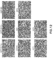
- each of the 8 cryopreserved recombinant cells formed a confluent monolayer following thawing, plating on Poly-D-Lysine plates and incubation for 24-hrs post-plating.
Landscapes
- Health & Medical Sciences (AREA)
- Life Sciences & Earth Sciences (AREA)
- Engineering & Computer Science (AREA)
- Chemical & Material Sciences (AREA)
- Genetics & Genomics (AREA)
- Organic Chemistry (AREA)
- Biomedical Technology (AREA)
- Zoology (AREA)
- Wood Science & Technology (AREA)
- Bioinformatics & Cheminformatics (AREA)
- Biotechnology (AREA)
- General Health & Medical Sciences (AREA)
- General Engineering & Computer Science (AREA)
- Physics & Mathematics (AREA)
- Biochemistry (AREA)
- General Physics & Mathematics (AREA)
- Microbiology (AREA)
- Cell Biology (AREA)
- Molecular Biology (AREA)
- Immunology (AREA)
- Biophysics (AREA)
- Toxicology (AREA)
- Gastroenterology & Hepatology (AREA)
- Medicinal Chemistry (AREA)
- Plant Pathology (AREA)
- Proteomics, Peptides & Aminoacids (AREA)
- Business, Economics & Management (AREA)
- Emergency Management (AREA)
- Environmental Sciences (AREA)
- Dentistry (AREA)
- Hematology (AREA)
- Urology & Nephrology (AREA)
- Automation & Control Theory (AREA)
- Tropical Medicine & Parasitology (AREA)
- Food Science & Technology (AREA)
- Analytical Chemistry (AREA)
- Pathology (AREA)
- Vascular Medicine (AREA)
- Computer Security & Cryptography (AREA)
- Micro-Organisms Or Cultivation Processes Thereof (AREA)
Abstract
Description
- The present invention relates to cryopreserved recombinant cells transiently overexpressing one or more genes encoding drug transporter protein and/or drug metabolizing enzyme such that activity of the encoded protein(s) is detectable in a population of said cells following thaw from cryopreservation.
- Drug development is a costly and time consuming endeavor whereby drug candidates must satisfy certain criteria established by government agencies such as the U.S. Food and Drug Administration and European Medicines Agency prior to receiving regulatory approval for marketing thereof. Importantly, assays are conducted to screen drug candidates to determine whether any are substrates or inhibitors of one or more drug transporter proteins and/or drug metabolizing enzymes as that can have a significant effect on the absorption, distribution, metabolism and elimination of such drugs, their toxicity and drug-drug interactions.
- Although cell lines stably expressing a gene encoding a drug transporter proteins or a drug metabolizing enzyme may be used for such screening, significant time and resources are required to generate and maintain frozen stocks thereof. Plus, the level of recombinant protein expressed is typically variable (laboratory to laboratory) and may deteriorate and/or become more variable over time with passage of such cells. Alternatively, freshly plated cells either stably or transiently expressing a gene encoding a drug transporter protein or a drug metabolizing enzyme may be employed for such screening. However, freshly plated cells have a limited shelf life of a few days and are difficult to ship in a manner that maintains their viability. In addition, to generate stable cell lines, the foreign transfected gene is actually integrated into the host genome of the cells and carried along with it during cycles of cells division. The chromosomal integration in the host cells will lead to permanent modification of host genome, potentially leading to abnormal expression of other genes causing unexpected changes of host behavior and unreliable experimental results. Thus, there is a need for cells suitable for screening drug candidates that reduces the investment of time and resources associated with drug development and provide reliable results.
- The present invention provides cryopreserved recombinant cells suitable for screening drug candidates to determine whether any are substrates or inhibitors of one or more drug transporter proteins and/or drug metabolizing enzymes that provide reliable results and ready convenience. Desirably, the level of activity in a population of the recombinant cells following cryopreservation is comparable to that of freshly transfected cells. Additionally, the cryopreserved recombinant cells are readily packaged in a vial and shipped with dry ice or dry shipper and conveniently stored at -135°C in liquid nitrogen upon receipt with several years shelf life. Hence, such cells provide the end user a consumable "thaw and use" product which provides convenience in conducting experiments and reduces the investment of time and resources in creating and/or maintaining cell stocks for screening drug candidates.
- In one aspect, the present invention provides cryopreserved recombinant cells including one or more transiently overexpressed genes encoding a protein selected from the group consisting of a drug transporter protein and a drug metabolizing enzyme, or a combination thereof wherein activity of the drug transporter protein or the drug metabolizing enzyme or combinations is detectable in a population of the cryopreserved recombinant cells following thaw from cryopreservation.
- In another aspect, the present invention provides processes of preparing transiently transfected recombinant cells which transiently overexpresses one or more genes encoding a protein selected from a drug transporter protein and a drug metabolizing enzyme or a combination thereof including transiently transfecting cells with one or more genes encoding a drug transporter protein or a drug metabolizing enzyme and cryopreserving the transiently transfected recombinant cells within 48 hrs of transfection.
- These and other features of the invention will be better understood through a study of the following detailed description.
-
-
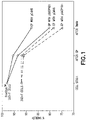
Figure 1 is a graph of the percentage of viable cells from cell stock, cells after electroporation (EP) and cells after thaw from cryopreservation for FreeStyle™ 293-F (FS293) cells and 293-F cells grown in suspension. -
Figure 2 are images of transfectedcells 4 hrs (A), 24 hrs (B) and 48 hrs (C) after plating following thaw from cryopreservation. -
Figure 3 are images of 293-F cells transfected with pOATP1B1 expression plasmid plated at (A) 0.4 × 10⁶ viable cells per well and (B) 0.2 × 10⁶ viable cells per well in 24-well poly-D-lysine coated plates and cultured in plating media at 24 hrs post-plating (following thaw from cryopreservation). -
Figure 4 are images of 293-F cells transfected with MATE1, MATE2K OATP1B3, long isoform OAT1 (full length cDNA with 563 amino acids; referred to herein as "OAT1 long"), short isoform OAT1 (missing 13 amino acid at C-terminus 522-534, with 550 amino acids; referred to herein as "OAT1 short"), OAT3, and pCMV vector plated at 0.4 × 10⁶ cells per well in 24-well poly-D-lysine coated plates at 24 hrs post-plating (following thaw from cryopreservation). -
Figure 5 are fluorescence images of adhered HEK293 cells transfected with 50 µg/ml, 100 µg/ml or 200 µg/ml green fluorescent protein (GFP) 24 hrs (A) and 48 hrs (B) following EP. -
Figure 6 is a graph of the percentage of viable cells following EP of adhered HEK293 cells using varying amounts of DNA. -
Figure 7A is a graph of estradiol-17β-glucuronide (E17βG) uptake activity following various incubation times in adhered HEK293 cells transfected with varying amounts of DNA (i.e., 0, 50 µg/ml, 100 µg/ml, 200 µg/ml or 400 µg/ml OATP2/OATP1B1) at 48 hrs post EP. -
Figure 7B is a graph of estradiol-17β-glucuronide (E17βG) uptake activity following various incubation times in adhered HEK293 cells transfected with varying amounts of DNA (i.e., 0, 50 µg/ml, 100 µg/ml, 200 µg/ml or 400 µg/ml OATP2/OATP1B1) at 96 hrs post EP. -
Figure 8 is a graph of signal to noise ratio of estradiol-17β-glucuronide (E17βG) uptake following various incubation times in adhered HEK293 cells transfected with varying amounts of DNA (i.e., 0, 50 µg/ml, 100 µg/ml, 200 µg/ml or 400 µg/ml OATP2/OATP1B1) at 48 hrs post EP. -
Figure 9 is a graph of estradiol-17β-glucuronide (E17βG) uptake activity in adhered HEK293 cells transfected with either OATP2/OATP1B1 using a small scale EP device (OC400), OATP2/OATP1B1 using a large scale EP device (CL2), or an empty vector control. -
Figure 10 is a graph of signal to noise ratio of estradiol-17β-glucuronide (E17βG) uptake following various incubation times in adhered HEK293 cells transfected with OATP1B1 gene using either "Control" (i.e., traditional lipid transfection reagent (lipofectamine 2000, available from Invitrogen)) or STX, MaxCyte scalable EP device. -
Figure 11 is a graph of signal to noise ratio of estradiol-17β-glucuronide (E17βG) uptake following various incubation times in adhered HEK293 cells transfected with OATP1B1 that are freshly plated or plated following thaw from cryopreservation. -
Figure 12 are images of HEK293 cells transfected withOATP1B1 ∗1a (Gene Accession No. NM_006446.4), OATP1B1∗1b (Gene Accession No. NM_006446.3), OATP1B3, pCMV vector, long isoform OAT1 (full length cDNA with 563 amino acids), OAT3, OCT1 or OCT2 using MaxCyte scalable EP device and scale-up process followed by cryopreservation, thawing, plating on Poly-D-Lysine plates and incubation for 24 hrs post-plating. -
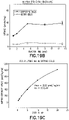
Figure 13A is a graph depicting results of a time-dependent assay of p-Aminohippuric acid (PAH) (prototypical substrate for OAT1) uptake in HEK293 cells overexpressing OAT1 or pCMV vector following various incubation times (i.e., 1, 2, 5, 10 and 15 min.) with PAH at a concentration of 3 µM. -
Figure 13B is a graph depicting results of a kinetic assay whereby uptake of PAH at a concentration in the range of 3 to 200 µM was measured in HEK293 cells overexpressing OAT1 following incubation for 5 min. Km and Vmax, calculated using Sigma-plot, are shown as insert in the graph. -
Figure 13C is a graph depicting results of an inhibition assay whereby HEK293 cells overexpressing OAT1 were incubated with PAH at a concentration of 15 µM and probenecid (OAT1 inhibitor) at a concentration in the range of 0 - 300 µM for 5 min. IC50, calculated using Sigma-plot, is shown as insert in the graph. -
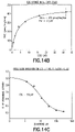
Figure 14A is a graph depicting results of a time-dependent assay of Estrone-3-sulfate (E3S) (prototypical substrate for OAT3) uptake in HEK293 cells overexpressing OAT3 or pCMV vector following various incubation times (i.e., 1, 2, 5, 10 and 15 min.) with E3S at a concentration of 1 µM. -
Figure 14B is a graph depicting results of a kinetic assay whereby uptake of E3S at a concentration in the range of 0.5 to 32 µM was measured in HEK293 cells overexpressing OAT3 following incubation for 1 min. Km and Vmax, calculated using Sigma-plot, are shown as insert in the graph. -
Figure 14C is a graph depicting results of an inhibition assay whereby HEK293 cells overexpressing OAT3 were incubated with E3S at a concentration of 4 µM and probenecid (OAT3 inhibitor) at a concentration in the range of 0 - 300 µM for 5 min. IC50, calculated using Sigma-plot, is shown as insert in the graph. -
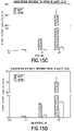
Figure 15A is a graph depicting results of a time-dependent assay of TEA (prototypical substrate for OCT1) uptake in HEK293 cells overexpressing OCT1 or pCMV vector following various incubation times (i.e., 1, 2, 5, 10 and 15 min.) with TEA at a concentration of 31 µM. -
Figure 15B is a graph depicting results of a time-dependent assay of metformin (prototypical substrate for OCT1) uptake in HEK293 cells overexpressing OCT1 or pCMV vector following various incubation times (i.e., 1, 2, 5, 10 and 15 min.) with metformin at a concentration of 3.8 µM. -
Figure 15C is a graph depicting results of a concentration-dependent assay whereby uptake of TEA at a concentration of 1, 10 and 100 µM was measured in HEK293 cells overexpressing OCT1 or pCMV vector following incubation for 10 min. -
Figure 15D is a graph depicting results of a concentration-dependent assay whereby uptake of metformin at a concentration of 0.1, 1 and 10 µM was measured in HEK293 cells overexpressing OCT1 or pCMV vector following incubation for 10 min. -
Figure 15E is a graph depicting results of an inhibition assay whereby HEK293 cells overexpressing OCT1 were incubated with TEA and OCT1 inhibitor (quinidine, verapamil or decynium-22) at various concentrations in the range of 0.1 - 500 µM for 10 min. -
Figure 15F is a graph depicting results of an inhibition assay whereby HEK293 cells overexpressing OCT1 were incubated with metformin at a concentration of 3.8 µM and OCT1 inhibitor cimetidine at various concentrations in the range of 4 µM to 3 mM for 10 min. -
Figure 16A is a graph depicting results of a time-dependent assay of TEA (prototypical substrate for OCT2) uptake in HEK293 cells overexpressing OCT2 or pCMV vector following various incubation times (i.e., 1, 2, 5, 10 and 15 min.) with TEA at a concentration of 31 µM. -
Figure 16B is a graph depicting results of a time-dependent assay of metformin (prototypical substrate for OCT2) uptake in HEK293 cells overexpressing OCT2 or pCMV vector following various incubation times (i.e., 1, 2, 5, 10 and 15 min.) with metformin at a concentration of 3.8 µM. -
Figure 16C is a graph depicting results of a concentration-dependent assay whereby uptake of TEA at a concentration of 1, 10 and 100 µM was measured in HEK293 cells overexpressing OCT2 or pCMV vector following incubation for 10 min. -
Figure 16D is a graph depicting results of a concentration-dependent assay whereby uptake of metformin at a concentration of 0.1, 1 and 10 µM was measured in HEK293 cells overexpressing OCT2 or pCMV vector following incubation for 10 min. -
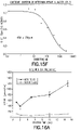
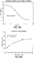
Figure 16E is a graph depicting results of an inhibition assay whereby HEK293 cells overexpressing OCT2 were incubated with metformin at a concentration of 3.8 µM and OCT2 inhibitor cimetidine at a concentration in the range of 4 µM to 3 mM for 10 min. IC50, calculated using Sigma-plot, is shown as insert in the graph. -
Figure 17A is a graph depicting results of a time-dependent assay of estradiol-17β-glucuronide (E17βG) uptake in HEK293cells overexpressing OATP1B1 ∗1a or pCMV vector following various incubation times (i.e., 1, 2, 5, 10 and 15 min.) with E17βG at a concentration of 1 µM. -
Figure 17B is a graph depicting results of a time-dependent assay of estrone-3-sulfate (E3S) uptake in HEK293cells overexpressing OATP1B1 ∗1a or pCMV vector following various incubation times (i.e., 1, 2, 5, 10 and 15 min.) with E3S at a concentration of 1 µM. -
Figure 17C is a graph depicting results of a time-dependent assay of rosuvastatin uptake in HEK293cells overexpressing OATP1B1 ∗1a or pCMV vector following various incubation times (i.e., 1, 2, 5, 10 and 15 min.) with rosuvastatin at a concentration of 1 µM. -
Figure 17D is a graph depicting results of a concentration-dependent assay whereby uptake of E17βG at a concentration in the range of 0.25 to 40 µM was measured in HEK293cells overexpressing OATP1B1 ∗1a following incubation for 1 min. Km and Vmax, calculated using Sigma-plot, are shown as insert in the graph. -
Figure 17E is a graph depicting results of a concentration-dependent assay whereby uptake of rosuvastatin at a concentration in the range of 0.78 to 50 µM was measured in HEK293cells overexpressing OATP1B1 ∗1a following incubation for 5 min. Km and Vmax, calculated using Sigma-plot, are shown as insert in the graph. -
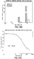
Figure 17F is a graph depicting results of an inhibition assay whereby uptake of E17βG at a concentration of 1 µM was measured in HEK293cells overexpressing OATP1B1 ∗1a following incubation with inhibitor cyclosporin A at a concentration in the range of 0.04 to 30 µM for 5 min. IC50, calculated using Sigma-plot, is shown as insert in the graph. -
Figure 18A is a graph depicting results of a time-dependent assay of E17βG uptake in HEK293cells overexpressing OATP1B1 ∗1b or pCMV vector following various incubation times (i.e., 1, 2, 5, 10 and 15 min.) with E17βG at a concentration of 1 µM. -
Figure 18B is a graph depicting results of a time-dependent assay of E3S uptake in HEK293cells overexpressing OATP1B1 ∗1b or pCMV vector following various incubation times (i.e., 1, 2, 5, 10 and 15 min.) with E3S at a concentration of 1 µM. -
Figure 18C is a graph depicting results of a time-dependent assay of rosuvastatin uptake in HEK293cells overexpressing OATP1B1 ∗1b or pCMV vector following various incubation times (i.e., 1, 2, 5, 10 and 15 min.) with rosuvastatin at a concentration of 1 µM. -
Figure 18D is a graph depicting results of a concentration-dependent assay whereby uptake of E17βG at a concentration in the range of 0.25 to 40 µM was measured in HEK293cells overexpressing OATP1B1 ∗1b following incubation for 1 min. Km and Vmax, calculated using Sigma-plot, are shown as insert in the graph. -
Figure 18E is a graph depicting results of an inhibition assay whereby uptake of E17βG at a concentration of 1 µM was measured in HEK293cells overexpressing OATP1B1 ∗1b following incubation with inhibitor cyclosporin A at a concentration in the range of 0.04 to 30 µM for 5 min. IC50, calculated using Sigma-plot, is shown as insert in the graph. -
Figure 19A is a graph depicting results of a time-dependent assay of cholecystokinin (CCK-8) uptake in HEK293 cells overexpressing OATP1B3 or pCMV vector following various incubation times (i.e., 1, 2, 5, 10 and 15 min.) with CCK-8 at a concentration of 1 µM. -
Figure 19B is a graph depicting results of a time-dependent assay of E17βG uptake in HEK293 cells overexpressing OATP1B3 or pCMV vector following various incubation times (i.e., 1, 2, 5, 10 and 15 min.) with E17βG at a concentration of 1 µM. -
Figure 19C is a graph depicting results of a concentration-dependent assay whereby uptake of CCK-8 at a concentration in the range of 0.5 to 20 µM was measured in HEK293 cells overexpressing OATP1B3 following incubation for 1 min. Km and Vmax, calculated using Sigma-plot, are shown as insert in the graph. -
Figure 19D is a graph depicting results of a concentration-dependent assay whereby uptake of rosuvastatin at a concentration in the range of 0.78 to 50 µM was measured in HEK293 cells overexpressing OATP1B3 following incubation for 5 min. Km and Vmax, calculated using Sigma-plot, are shown as insert in the graph. -
Figure 19E is a graph depicting results of an inhibition assay whereby uptake of CCK-8 at a concentration of 1 µM was measured in HEK293 cells overexpressing OATP1B3 following incubation with inhibitor cyclosporin A at a concentration in the range of 0.04 to 30 µM for 2 min. IC50, calculated using Sigma-plot, is shown as insert in the graph. - As used herein the following terms shall have the definitions set forth below.
- As used herein, the term "cell" includes both primary cells as well as established cell lines (e.g., human embryonic kidney HEK293 cells, Chinese hamster ovary CHO, Madin-Darby Canine Kidney Cells MDCK, Pig Kidney Epithelial Cells LLC-PK1, human epithelial colorectal adenocarcinoma cells Caco-2 and Chinese hamster lung fibroblast V79 cells).
- As used herein, the term "drug transporter protein" refers to a membrane bound transport protein that includes, but is not limited to ATP binding cassette (ABC) transporters and solute carrier (SLC) transporters.
- As used herein, the term "drug metabolizing enzyme" includes, but is not limited to, cytochromes such as cytochrome (CYP) P450; UDP-glucouronyl transferase and other non-CYP drug metabolizing enzymes such as alcohol dehydrogenase, monoamine oxidase and aldehyde oxidase.
- As used herein, the term "detectable" means that the activity of a selected probe substrate in cells transfected with a drug transporter protein and/or drug metabolizing enzyme shall be higher than the activity of the same probe substrate in cells transfected with empty vector; desirably, the difference in activity will be at least 5-fold.
- As used herein, the use of upper case letters in transporter nomenclature identifies the human protein/gene, i.e., MRP2/ABCC2, etc.; smaller case letters indicate the transporter derives from a preclinical (nonhuman mammalian) species, i.e., Mrp2/Abcc2, etc. Unless otherwise specified, a gene is derived from any species (e.g., human or other mammal).
- As used herein, the terms "OATP1B1" "OATP2" and "SLCO1B1" are interchangeable and refer to a human protein/gene that corresponds to the nonhuman protein/gene Oatp2. Unless noted otherwise, reference to OATP1B1 is to
OATP1B1 ∗1b. - As used herein, the terms "OAT1" and "SLC22A6" are interchangeable and refer to an
organic anion transporter 1. Unless noted otherwise, reference to OAT1 is to the full length cDNA encoding with 563 amino acids (also referred to herein as "OAT1 long"). - Exemplary ABC transporters include, but are not limited to those listed below in Table 1.
Table 1. GENE NAME PROTEIN NAME MDR1/P-gp/ABCB1 Multidrug Resistance Protein1 MRP1/ABCC1 Multidrug resistance protein 1MRP2/ABCC2 Multidrug resistance-associated protein 2MRP3/ABCC3 Multidrug resistance protein 3MRP4/ABCC4 Multidrug resistance protein 4MRP5/ABCC5 Multidrug resistance protein 5MRP6/ABCC6 Multidrug resistance protein 6MRP7/ABCC7 Multidrug resistance protein 7 MRP8/ABCC8 Multidrug resistance protein 8BCRP/ABCG2 Breast Cancer Resistance Protein BSEP/ABCB11 Bile Salt Export Pump - Exemplary SLC transporters include, but are not limited to those listed below in Table 2.
Table 2. GENE NAME PROTEIN NAME OSTα Organic solute transporter α OSTβ Organic solute transporter β OATP1B1∗/SLCO 1B1/OATP2 Organic anion-transporting polypeptide 1B1 OATP1B3/SLCO 1B3 Organic anion-transporting polypeptide 1B3 OAT1/SLC22A6 Organic anion transporter 1OAT2/SLC22A7 Organic anion transporter 2OAT3/SLC22A8 Organic anion transporter 3OAT4/SLC22A11 Organic anion transporter 4OCT1/SLC22A1 Organic cation transporter 1OCT2/SLC22A2 Organic cation transporter 2OCT3/SLC22A3 Organic cation transporter 3OATP1A2/SLCO1A2 Organic anion-transporting polypeptide 1A2 OATP2B1/SLCO2B1 Organic anion-transporting polypeptide 2B1 PEPT1/ SLC15A1 Peptide Transporter 1 PEPT2/ SLC15A2 Peptide Transporter 2 OCTN1/SLC22A4 Organic cation/ergothioneine transporter OCTN2/SLC22A5 Organic cation/carnitine transporter MATE1/SLC47A1 Multidrug and toxin extrusion 1MATE2K/SLC47A2 Multidrug and toxin extrusion 2K URAT1/ SLC22A12 Urate Transporter 1 ASBT/SLC10A2 Apical sodium/bile acid co-transporter NTCP/SLC10A1 Sodium/taurocholate co-transporting peptide ∗ In this instance, OATP1B1 includes OATP1B1 ∗1a andOATP1B1 ∗1b. - Exemplary SLC transporters tested, but are not limited to those listed below in Table 3.
Table 3. GENE NAME FULL NAME GENE ACCESSION NUMBER OATP1B1 ∗1a/ SLCO1B1 ∗1aOrganic anion-transporting polypeptide 1B1 Wild Type (388A) NM_006446.4 OATP1B1 ∗1b/SLCO1B1 ∗1bOrganic anion-transporting polypeptide 1B1 SNP 388A>G NM_006446.3 OATP1B3/SLCO1B3 Organic anion-transporting polypeptide 1B3 NM_019844 OAT1/SLC22A6 Organic anion transporter 1NM_004790 OAT3/SLC22A8 Organic anion transporter 3NM_004254 OCT1/SLC22A1 Organic cation transporter 1NM_003057 OCT2/SLC22A2 Organic cation transporter 2NM 003058 - Cells suitable for use in the present invention include mammalian cells, for example, as derived from human or non-human (e.g., mouse, rat, dog, monkey, hamster and pig, etc.). In certain embodiments, the cells are hepatocytes, or endothelial cells.
- Gene delivery systems for introducing gene(s) into a population of cells are known to a skilled artisan. Virus-based gene delivery methods may be used but require special handling of the cells due to safety concerns. Although lipid-based transfection methods may be used, lipid-based transfection reagents are relatively costly and such methods are not amenable to large-scale manufacturing processes. Additionally, lipid-based transfection methods result in relatively low gene delivery efficiency and relatively delayed protein expression (generally 72 to 96 hours post transfection) (data not shown). Electroporation (EP) is preferable as it is amenable to large-scale manufacturing processes and avoids the safety issues of viral-based gene delivery methods. Further, EP results in relatively efficient gene delivery.
- After gene delivery into a population of cells, gene(s) encoding a drug transporter protein and/or a drug metabolizing enzyme will be overexpressed such that activity of the protein(s) encoded therefrom are detectable following thaw from cryopreservation. Drug candidates can be tested to determine if any are substrates or inhibitors of the protein(s) encoded from the overexpressed gene(s) by incubation of the recombinant cells therewith. In particular, if a drug candidate is a substrate of a drug transporter protein and/or a drug metabolizing enzyme, the drug candidate will be affected. For instance, if the drug candidate is a substrate of a drug transporter protein, the drug candidate will be translocated in or out of the recombinant cell via the drug transporter protein. However, if the drug candidate is an inhibitor of the drug transporter protein, the drug candidate will inhibit translocation of a substrate of the drug transporter protein in or out of the recombinant cell.
- Alternatively, assays can be conducted using whole cells or subcellular fractions thereof (microsome/cytosol).
- Additionally, recombinant cells of the present invention may be further transfected with RNAi or siRNA of the transiently overexpressed gene(s) to knockdown/knockout the expression thereof as is desirable for certain assays. Primary cells (e.g., hepatocytes) can be transfected with RNAi or siRNA directed against any ABC transporters, SLC transporters or any other drug metabolizing enzymes to knockdown/knockout the expression of specific genes.
- In one aspect (1), the disclosure provides a cryopreserved recombinant cell including one or more transiently overexpressed genes encoding a protein selected from the group consisting of a drug transporter protein and a drug metabolizing enzyme, or a combination thereof wherein activity of the drug transporter protein or the drug metabolizing enzyme or combinations is detectable in a population of the cryopreserved recombinant cell following thaw from cryopreservation.
- In an aspect (2), the disclosure provides the invention of aspect (1), wherein said one or more genes encodes a drug metabolizing enzyme.
- In an aspect (3), the disclosure provides the invention of aspect (2), wherein the drug metabolizing enzyme is selected from the group consisting of cytochrome P450, UDP-glucouronyl transferase, alcohol dehydrogenase, monoamine oxidase and aldehyde oxidase.
- In an aspect (4), the disclosure provides the invention of aspect (1), which transiently overexpresses one or more genes encoding a protein selected from the group consisting of an ATP binding cassette transporter and a solute carrier transporter protein.
- In an aspect (5), the disclosure provides the invention of aspect (4), wherein said one or more genes is selected from the group consisting of MDR1/Mdr1a/Mdr1b, MRP1/Mrp1, MRP2/Mrp2, MRP3/Mrp3, MRP4/Mrp4, MRP5/Mrp5, MRP6/Mrp6, MRP7/Mrp7,
MRP 8/Mrp8, BCRP/Bcrp, BSEP/Bsep, OATP2/Oatp2, OATP1B3/Oatp1b3, OAT1/Oat1, OAT2/Oat2, OAT3/Oat3, OAT4/Oat4, OCT1/Oct1, OCT2/Oct2, OATP1/Oatp1, PEPT1/Pept1, PEPT2/Pept2, OCTN1/Octn1, OCTN2/Octn2, MATE1/Mate1, MATE2K/Mate2, URAT1/Urat1, ASBT/Asbt, and NTCP/Ntcp. - In an aspect (6), the disclosure provides the invention of aspect (4), wherein said one or more genes encodes a protein that is an ATP binding cassette transporter selected from the group consisting of MDR1/Mdr1a/Mdr1b, MRP1/Mrp1, MRP2/Mrp2, MRP3/Mrp3, MRP4/Mrp4, MRP5/Mrp5, MRP6/Mrp6, MRP7/Mrp7,
MRP 8/Mrp8, BCRP/Bcrp, and BSEP/Bsep. - In an aspect (7), the disclosure provides the invention of aspect (4), wherein said one or more genes encodes a protein that is a solute carrier transporter selected from the group consisting of OATP2/Oatp2, OATP1B3/Oatp1b3, OAT1/Oat1, OAT2/Oat2, OAT3/Oat3, OAT4/Oat4, OCT1/Oct1, OCT2/Oct2, OCT3/Oct3, OATP1/Oatp1, PEPT1/Pept1, PEPT2/Pept2, OCTN1/Octn1, OCTN2/Octn2, MATE1/Mate1, MATE2K/Mate2, URAT1/Urat1, ASBT/Asbt, and NTCP/Ntcp.
- In an aspect (8), the disclosure provides the invention of aspect (4), wherein said one or more genes is selected from
OATP1B1 ∗1a,OATP1B1 ∗1b. OATP1B3, OAT1, OAT3, OCT1, OCT2, MATE1 and MATE2K. - In an aspect (9), the disclosure provides the invention of aspect (1), wherein the one or more genes is derived individually from human or an animal species selected from mouse, rat, guinea pig, dog, and monkey.
- In an aspect (10), the disclosure provides the invention of aspect (1), wherein said cell is derived from a mammal.
- In an aspect (11), the disclosure provides the invention of aspect (10), wherein said cell is selected from the group consisting of HEK293, CHO, MDCK, LLC-PK1, Caco-2 and V79 cells.
- In an aspect (12), the disclosure provides the invention of aspect (10), wherein the mammal is selected from the group consisting of human, monkey, dog, rat, mouse, porcine and hamster.
- In an aspect (13), the disclosure provides the invention of aspect (1), wherein said cell comprises a hepatocyte.
- In an aspect (14), the disclosure provides the invention of aspect (1), wherein said cell comprises an endothelial cell.
- In an aspect (15), the disclosure provides the invention of aspect (1), wherein activity of the protein(s) is detectable in a population of said cell at least 24 hours post plating following thaw from cryopreservation.
- In an aspect (16), the disclosure provides the invention of aspect (1), wherein activity of the protein(s) is detectable in a population of said cell at least 48 hours post plating following thaw from cryopreservation.
- In an aspect (17), the disclosure provides the invention of aspect (1), wherein activity of the protein(s) is detectable in a population of said cell at least 72 hours post plating following thaw from cryopreservation.
- In another aspect (18), the disclosure provides a process of preparing transiently transfected recombinant cells which transiently overexpresses one or more genes encoding a protein selected from a drug transporter protein and a drug metabolizing enzyme including transiently transfecting cells with one or more genes encoding a drug transporter protein or a drug metabolizing enzyme and cryopreserving the transiently transfected recombinant cells within 48 hrs of transfection.
- In an aspect (19), the disclosure provides the invention of aspect (18), wherein the transient transfection step includes electroporation.
- Cells were cultured under standard sterile practices for cell culture, and transiently transfected using EP. Following EP, cells were assayed for protein activity both before as well as after being frozen, thawed and plated. As detailed below, cells cultured in suspension and adherent cell cultures were both successfully transiently transfected and exhibited substantial activity of the recombinant protein following thaw from cryopreservation.
- In brief, on
Day 1,FreeStyle 293 Cells and 293-F cells were each passaged into appropriate sized shaker flasks at a density of 0.7-1.0 × 10⁶ cell/ml using supplemented CD293 medium (i.e., CD293 medium (available from Gibco, Cat. No. 11913-019, Life Technologies Corp., Carlsbad, CA) supplemented with 4 mM L-Glutamine (available from Gibco, Cat. No. 25030-081, Life Technologies Corp., Carlsbad, CA)) or supplementedExcell ™ 293 serum free media (available from Sigma, Cat. No. 14571C, Sigma-Aldrich, St. Louis, MO) supplemented with 6 mM L-Glutamine. Cell viability and cell number were determined using a Cellometer (available from Nexcelom Bioscience, Lawrence, MA). - On
Day 2, EP of cells was executed. In short, following a determination of cell viability and cell density, cells were pelleted down by spinning at 100g for 5 min, after which the media was aspirated and cells resuspended in 30 ml EP Buffer (available from MaxCyte, Cat. No. B201, MaxCyte Inc., Gaithersburg, MD). The cell suspension was transferred to 50 ml Falcon tubes, pelleted down as described above, and resuspended in an appropriate amount of EP Buffer to reach 100 × 106 cells/ml which was used as the cell stock. DNAs to be used for EP were prepared in sterile water at a final concentration of 5 mg/ml. For each sample, 0.4 ml of cell stock and DNA was placed in a sterile 1.5 ml eppendorf tube resulting in a final concentration of 200 µg/ml (Table 4) or 300 µg/ml DNA (Table 10 and Table 11) and cell density of 40 × 106 cells per sample.Table 4. SAMPLE # CELL TYPE PLASMID(S) CELL STOCK VOL. (ml) [DNA] (ug/ml) A 1, 2 FS293 pOATP1B1 0.4 200 B 3, 4 pCMV6 0.4 200 C 5 EP Buffer (16 µl) 0.4 - D 6, 7 293-F pOATP1B1 0.4 200 E 8,9 pCMV6 0.4 200 F 10 EP Buffer (16 µl) 0.4 - - Samples were transferred into an OC-400 Processing Assembly (available from MaxCyte, Cat. No. OC-400R, MaxCyte Inc., Gaithersburg, MD) which followed the manufacture instructions for EP of HEK cells. Following EP, the cells were carefully pipetted out and transferred into the bottom of a 125 ml shaker flask and incubated for 20 min at 37°C with 8% CO2, after which pre-warmed 40 ml CD293 media was added into the shaker flask to reach cell density at 1 × 10⁶ cells/ml. The cells were incubated for 30 min at 37°C and 8%CO2. After 30 min recovery, cell viability and cell density were determined. A portion of cells (i.e., 20 × 106 cells) was used for plating and the rest was cryopreserved, or all of the cells were cryopreserved. It is contemplated that recombinant cells may be cryopreserved within 48 hrs of transfection and exhibit activity of protein(s) encoded from transfected gene(s) at a detectable level following thaw from cryopreservation.
- For plating cells following EP, 20 × 106 cells were pelleted down by spinning at 100g for 5 min and then resuspended in 20 ml pre-warmed plating media (DMEM with high glucose (available from Gibco, Cat. No. 11965092, Life Technologies Corp., Carlsbad, CA), supplemented with 0.1 mM non-essential amino acids (available from Gibco, Cat. No. 11140050, Life Technologies Corp., Carlsbad, CA), 10% FBS (available from SAFC Biosciences, Cat. No. 12016C, Sigma, St. Louis, MO)) (cell density of 1 × 10⁶ cells/ml). Cells were placed in 24-well tissue culture plates poly-D-Lysine coated, Corning Biocoat™ (available from Corning Life Sciences, Tewksbury, MA) at a density of 0.2 × 10⁶ cells/well and 0.4 × 10⁶ cells/well and incubated at 37°C with 8% CO2 so as to determine the impact of seeding density on uptake activity. Media was replaced 4 hours later and then every 24 hours until the day of assaying. On
Days 4, cells were assayed for OATP1B1 activity as described below. - For cryopreservation, cells were pelleted then resuspended in freshly prepared ice-cold freezing media (9 parts supplemented CD293 medium and 1 part DMSO which was syringe filtered to sterilize) at a density of 10 × 106 cell/ml. Cryo vials were filled with 1 ml of this cell suspension, and placed on ice-cold Mr Frosty freezing container (available from Thermal Scientific), which was stored in -80°C freezer overnight after which the vials were transferred into liquid nitrogen.
- Cryopreserved cells were assayed for OATP1B1 activity as described below. In brief, on
Day 1, cryopreserved cells were removed from liquid nitrogen to dry ice, and then thawed in a water bath at 37°C for about 2 min. Cells were transferred into 10 ml of plating media as described above which is pre-warmed to a temperature of about 37°C and the viability and cell density determined. Cells were pelleted down and resuspended in supplemented DMEM media at a cell density of 1 × 10⁶ viable cells/ml. Cells were plated in the same manner described above for plating cells following EP (which had not been cryopreserved) and assayed for OATP1B1 activity at 24, 48 and 72 hrs following plating thereof. - In brief, HEK293 cells were cultured in 5 Layer Corning® CellStack® (available from Corning Inc. Life Sciences, Tewksbury, MA) using plating media containing DMEM (high glucose) available from Gibco Cat. No. 11965118, Life Technologies Corp., Carlsbad, CA; Penicillin-Streptomycin (10,000 units/ml) available from Gibco Cat. No. 15140-122, Life Technologies Corp., Carlsbad, CA; L-Glutamine (200 mM) available from Gibco Cat. No. 25030-081, Life Technologies Corp., Carlsbad, CA; Sodium Pyruvate, available from Gibco Cat. No. 11360, Life Technologies Corp., Carlsbad, CA; FBS available from Sigma-Aldrich Corp., St. Louis, MO in a ratio of 100:1:1:1:10. On
Day 1, about 24 hrs before EP, HEK293 cells were trypsinized, cell viability and cell number determined after which cells were passaged to fresh multilayer chamber flasks at 30-40% confluency. Cells were incubated at 37°C with 5% CO2. - On
Day 2, EP of cells was executed. In short, cells were harvested, cell viability and cell number determined after which cells were pelleted down by spinning at 100g for 5 min and the media aspirated. Cells were resuspended in EP buffer and pelleted down by spinning at 100g for 5 min, then resuspended in an appropriate amount of EP Buffer to reach 50 × 106 cells/ml which was used as the cell stock. DNAs to be used for EP were prepared in sterile water at a final concentration of 5 mg/ml. For each sample used for OC-400 processing assembly, 0.4 ml of cell stock and DNA was placed in a sterile 1.5 ml eppendorf tube resulting in a final concentration of 50 µg/ml, 100 µg/ml, 200 µg/ml or 400 µg/ml DNA as indicated inFigures 5-9 and cell density of 40 × 106 cells per sample. For each sample used for CL-2 processing assembly, 10 ml of cell stock and DNA was placed in 50 ml sterile conical tube resulting in a final concentration of 100 µg/ml DNA. - Samples were transferred into an OC-400 or CL-2 processing assembly (available from MaxCyte, Cat. No. OC-400R and CL2-R, MaxCyte Inc., Gaithersburg, MD) which followed the manufacture instructions for EP of HEK cells. Following EP, the cells were carefully pipetted out and transferred into 6-well tissue culture plates and incubated for 20 min at 37°C with 5% CO2, after which cells were removed and placed in a 50 ml conical tube containing pre-warmed plating media. Cell viability and cell density were determined. A portion of cells (i.e., 20 × 106 cells) was used for plating and the rest was cryopreserved.
- For plating cells following EP, cells were pelleted down by spinning at 100g for 5 min and then resuspended in pre-warmed plating media (cell density of 1 × 10⁶ cells/ml). Cells were placed in 24-well tissue culture plates (poly-D-Lysine coated, Corning Biocoat™ (available from Corning Life Sciences, Tewksbury, MA)) at a density of 0.4 × 10⁶ cells/well and incubated at 37°C with 5% CO2. Media was replaced 4 hours later and then every 24 hours until the day of assaying. On
Days - For cryopreservation, cells were pelleted then resuspended in freshly prepared ice-cold freezing media (9 parts plating medium and 1 part DMSO which was syringe filtered to sterilize) at a density of 10 × 106 cell/ml. Cryo vials were filled with 1 ml of this cell suspension, and placed on ice-cold Mr Frosty freezing container (available from Thermal Scientific) stored in -80°C freezer overnight after which the vials were stored in liquid nitrogen.
- Cryopreserved cells were assayed for OATP1B1 activity. Notably, cells were plated in the same manner described above for plating cells following EP (which had not been cryopreserved) and assayed for OATP1B1 activity (as described below) at 48 hrs following plating thereof.
- In brief, substrate solution was prepared for
OATP1B1 ∗1a andOATP1B1 ∗1b using 2 µM estradiol-17β-glucuronide (99% of cold E17βG and 1% of [3H]-E17βG); for OATP1B3 using 2 µM CCK-8 (99% of cold CCK-8 and 1% of [3H]-CCK-8); for OAT1 short using 1µM Para-aminohippurate (PAH) (90% of cold PAH and 10% of [3H]-PAH); for OAT1 long using 1µM or 3 µM Para-aminohippurate (PAH) (90% of cold PAH and 10% of [3H]-PAH); for OAT3 using 1µM or 2 µM Estrone-3-sulfate (99% of cold E3S and 1% of [3H]-E3S); for OCT1 and OCT2 using 30 µM Tetraethylammonium Bromide (100% [14C]-TEA); for MATE1 and MATE2K using 10 µM Metformin (100% [14C]-Metformin) or 10 µM Tetraethylammonium Bromide (100% [14C]-TEA); in Krebs-Henseleit Buffer pH 7.4 (available from Sigma, Cat. No. K3753, Sigma-Aldrich, St. Louis, MO) and incubated at 37°C for at least 20 min. Culture media was aspirated from cells to be assayed and cells washed thrice with pre-warmed KHB Buffer. Cells were subsequently incubated with Uptake Buffer at 37°C for 10 min. For MATE1 and MATE2K, cells were washed and pre-incubated with KHB buffer containing 20 mM NH4Cl for 10 min. Assays were initiated by adding 0.3 ml substrate solution into each well and incubated at 37°C for 5 min with samples for OCT1 and OCT2 incubated for 10 min. - The reaction was quickly stopped after the incubation period by aspirating substrate solution from cells then washing cells thrice with cold Uptake Buffer. Cells were then incubated with lysing solution (0.1% SDS with 0.1% v/v 1M NaOH in Dulbecco's Phosphate-Buffered Saline (DPBS) buffer) for 15-20 minutes while being shaken. The substrate solution was triturated and 0.4 ml of the resultant cell lysis placed in 5 ml scintillation tube with 5 ml of scintillation liquid for analysis with scintillation counter.
- As illustrated in
Figure 1 , cell viability dropped 1-5% after EP relative to that of the cell stock. Additionally, after cryopreservation, cell viability dropped an additional 10-15% relative to that after EP. Nonetheless, even after EP and thaw from cryopreservation, cell viability is greater than 75%. - Cell morphology and uptake activity was examined following cryopreservation after 30 min recovery and 24 hrs recovery post transfection. Table 5 illustrated cell morphology and uptake activity with 24 hrs recovery was reduced compared to 30 min recovery.
Table 5. SAMPLE Recovery time prior to cryopreservation Cell Confluency at 24 hrs Uptake Activity (pmole/mg/min) S:N OATP1B1 24 HOURS 40-50% 0.59 0.44 OATP1B1 30 MIN 70-75% 5.78 4.32 VECTOR 30 MIN 90-95% 1.34 - Cell morphology and confluency of transfected cells thawed from cryopreservation were examined after various periods of time following plating at a density of 0.4 × 10⁶ cells per well in 24-well poly-D-lysine coated Corning Biocoat™ plates. In particular,
Figure 2 illustrates OATP1B1 transiently transfected cells cultured at 4 hrs, 24 hrs and 72 hrs post plating. Additionally, cell confluency at 24 hrs, 48 hrs and 72 hrs post-plating of these cells is recorded in Table 6 below.Table 6. CELLS 24 hrs 48 hrs 72 hrs FS293 with pOATP1B1 80-90% 90-95% 80-85% FS293 with pCMV6 vector 70-80% 90-95% 90% 293-F with pOATP1B1 90-95% 95-100% 80-85% 293-F with pCMV6 vector 90-95% 95-100% 80-85% - Desirably, after EP and cryopreservation, the cells form a monolayer on poly-D-lysine coated Corning Biocoat™ plates achieving 80-90% confluency at 24 hrs post-plating, 90%-100% confluency at 48 hrs post-plating.
-
Figure 4 illustrates cells, transiently transfected with MATE1, MATE2K, OATP1B3, OAT1 long, OAT1 short, OAT3, and pCMV vector respectively, cultured at 24 hrs post plating after thawed from cryopreservation. - As illustrated in
Figure 5 , the expression of Green Fluorescent Protein (GFP) in adhesion HEK293 cells was increased with increasing concentration of DNA. Additionally, GFP expression increased at the 48 hr timepoint relative to the 24 hr timepoint. In particular, GFP transfection efficiency by EP achieved 100% at 24hrs with 200 µg/ml DNA and 100% fluorescent cell staining at 48 hrs with 100 µg/ml DNA. Hence, GFP protein expression level in transfected cells increased with increased DNA concentration and at 48 hrs relative to 24 hrs. - Uptake activity of suspension cultured 293 cells transfected with OATP1B1 (pOATP1B1) and control vector (pCMV) were assayed at various time points following EP. In brief, transfected cells were plated at a density of 0.4 × 10⁶ cells/well in 24-well poly-D-lysine coated Corning Biocoat™ plates following EP or after thaw from cryopreservation. OATP1B1 uptake activity and uptake ratio were determined using probe substrate, estradiol-17β-glucuronide, in both fresh plated cells ("fresh") and cryopreserved cells ("cryo") at various timepoints post plating as detailed in Table 7 below.
Table 7. CELLS/CULTURE MEDIA, FRESH OR CRYO CELL PLATING TIME POINT (HR) UPTAKE ACTIVITY (pmol/mg/min)/ confluence UPTAKE RATIO pOATP1B1 pCMV 293-F in CD293, fresh 48 15.4(85%) 0.7 (90%) 22.0 293-F in CD293, cryo 48 15.1 (95-100%) 0.9 (95-100%) 16.8 FS293 in Excell, cryo 24 36.4 1.9 19.2 48 10.0 0.7 14.3 72 6.6 1.0 6.6 293-F in CD293, cryo 24 27.4 1.5 18.3 48 15.1 0.9 16.8 72 9.9 1.0 9.9 Note: The number appearing in parentheses is the cell confluency at assay time. - OATP1B1 uptake activity and uptake ratio in transfected cells following thaw from cryopreservation is consistent with those in freshly plated transfected cells. In both cells types 293-F and FS293, the highest uptake activity and uptake ratio is observed at 24 hrs post plating.
- Morphology and cell confluency of transfected cells (FS293 or 293-F) were examined at 24 hrs, 48 hrs and 72 hrs post-plating in 24-well poly-D-lysine coated Corning Biocoat™ plates at plating density of either 0.4 × 10⁶ cells/well or 0.2 ×10⁶ cells/well after thaw from cryopreservation. Cell confluency at 24 hrs post-plating are summarized below in Table 8. Cell confluency at 48 hrs and 72 hrs are similar to those achieved at 24 hrs (data not shown). Additionally,
Figure 3 provides images of transfected cells plated at (A) 0.4 × 10⁶ cells per well and (B) 0.2 × 10⁶ cells per well 24 hrs post-plating following thaw from cryopreservation at a confluence of 90-95% and 60-70%, respectively.Table 8. CELLS, CULTURE MEDIA, TRANSFECTED DNA 0.4 × 10⁶ CELLS/WELL 0.2 × 10⁶ CELLS/WELL FS293 with pOATP1B1 in Excell 80-90% 30-50% FS293 with pCMV vector in Excell 70-80% 50% 293-F with pOATP1B1 in CD293 90-95% 60-70% 293-F with pCMV6 vector in CD293 90-95% 80% - For optimal assay performance, plating cells at a density of 0.4 × 10⁶ is preferable to that of 0.2 × 10⁶ as it achieves higher cell confluency and higher uptake activity.
Table 9. CELLS UPTAKE ACTIVITY (pmol/mg/min)/ confluence UPTAKE RATIO pOATP1B1 pCMV6 FS293 cells, 0.2 × 10⁶cells/well 10.5 3.0 3.5 FS293 cells, 0.4 × 10⁶cells/well 36.4 1.9 19.2 293-F cells, 0.2 × 10⁶cells/well 20.2 1.5 13.5 293-F cells, 0.4 × 10⁶cells/well 27.4 1.5 18.3 - Following EP, cell viability was examined using trypan blue and hemocytometer or cellometer.
- As illustrated in
Figure 6 , when using adhesion HEK293 cells, cell viability post EP dropped with increasing amounts of DNA used in EP. Nonetheless, cell viability following transfection with pOATP1B1 was ranged from 89% to 77% and that following transfection with empty vector was 90%. - As illustrated in
Figure 7 , when using adhesion HEK293 cells, OATP1B1 mediated uptake of Estradiol-17β-glucuronide in the fresh plated transient transfected adhesion HEK293 cells is time-dependent. Notably, uptake activity and uptake ratio increased with increasing amounts of DNA used in EP. However, OATP1B1 mediated uptake of Estradiol-17β-glucuronide reduced at the 96 hr timepoint relative to the 48 hr timepoint. Further, as illustrated inFigure 8 , the signal to noise ratio (i.e., uptake ratio) of estradiol-17β-glucuronide increased with the increase of amount of DNA and assay incubation time, in adhesion HEK293 cells transfected with OATP1B1 relative to empty vector at 48hrs post EP. - As illustrated in
Figure 9 , when using adhesion HEK293 cells, estradiol-17β-glucuronide uptake in OATP1B1 transiently expressed HEK293 cells using small scale EP device and large scale EP device is consistent for both uptake activity and signal to noise ratio (i.e., uptake ratio). 100 µg/ml DNA was used in the experiments. - As illustrated in
Figure 10 , when using adhesion HEK293 cells, OATP1B1 uptake activity is compared between the cells transfected using traditional lipid transfection reagent (control: lipofectamine 2000, available from Invitrogen) and EP using STX, MaxCyte Inc., Gaithersburg, MD. Notably, cells transfected using EP resulted in a pronouncedly greater signal to noise ratio relative to those cells transfected with lipid transfection reagent. - As illustrated in
Figure 11 , when using adhesion HEK293 cells, OATP1B1 uptake activity in both freshly plated EP transfected cells and cells following thaw from cryopreservation was detectable. - Uptake activity of suspension cultured 293 cells transfected with
OATP1B1 ∗1a,OATP1B1 ∗1b, OATP1B3, OAT1 long, OAT1 short, OAT3, OCT1, OCT2, MATE1, MATE2K or control vector (pCMV) were assayed at 24 hrs post plating after thaw from cryopreservation. In brief, the transfected cells were plated at a density of 0.4 × 10⁶ cells/well in 24-well poly-D-lysine coated Corning Biocoat™ plates following EP and after thaw from cryopreservation. SLC transporter uptake activity and uptake ratio were determined using probe substrates as indicated at 24 hrs post plating as detailed in Table 10 below.Table 10. TRANSPORTERS SUBSTRATE UPTAKE ACTIVITY (pmol/mg/min)/ UPTAKE RATIO SLC transporter pCMV6 OATP1B1 ∗1a 2 µM E17bG 41.0 1.03 40 OATP1B1 ∗1b2 µM E17bG 32.6 0.88 37 OATP1B3 2 µM CCK-8 28.7 0.2 145 OATP1B3 2 µM CCK-8 77.0 0.79 98 OAT1 long 1 µM PAH 13.1 0.3 39 OAT1 short 1 µM PAH 9.7 0.3 29 OAT1 long 3 µM PAH 15.0 0.71 21 OAT3 1 µM E3S 44.7 1.2 38 OAT3 2 µM E3S 60.9 1.62 38 OCT1 30 µM TEA 127.6 5.63 23 OCT2 30 µM TEA 100.5 5.53 18 MATE1 10 µM Metformin 71.4 6.0 12 10 µM TEA 46.3 4.3 11 MATE2K 10 µM Metformin 33.5 5.2 6.5 10 µM TEA 46.6 6.1 7.6 - As reflected in Table 10 above, the recombinant cells exhibited strong uptake activity towards their specific prototypical substrate each of which had an uptake ratio above 10. Notably, an uptake ratio above 5 indicates a successful process.
- As reflected in Table 11, the post-thaw viability for recombinant cryopreserved cells was determined to be above 90%.
Table 11. Cells Post-thaw Viability OATP1B1 ∗1a 94.2 % OATP1B1 ∗1b 96.1% OATP1B3 95.5% OAT1 long 93.5% OAT3 93.8% OCT1 95.1% OCT2 96.1% - Each of these recombinant cells as well as a control vector (pCMV) was examined 24 hrs post-plating (after cryopreservation). Confluency for each of these cells 24 hrs post-plating was 85% or greater as reflected in Table 12 below.
Table 12. Transfected Cells 24- h confluency OATP1B1 ∗1a 90 % OATP1B1 ∗1b 95% OATP1B3 95% OAT1 long 90% OAT3 90 % Vector 95 % OCT1 95 % OCT2 85% - As illustrated in
Figure 12 , each of the 8 cryopreserved recombinant cells formed a confluent monolayer following thawing, plating on Poly-D-Lysine plates and incubation for 24-hrs post-plating. - As illustrated in
Figures 13-19 and Tables 13-14, the kinetic and inhibition profiles examined in cryopreserved recombinant cells expressing a transporter protein was consistent with reported values. Specifically, as illustrated inFigures 13A-13C , the kinetics of PAH uptake by recombinant cells expressing OAT1 and inhibition profile of probenecid thereof is consistent with reported values. As illustrated inFigures 14A-14C , the kinetics of E3S uptake by recombinant cells expressing OAT3 and inhibition profile of probenecid thereof is consistent with reported values. As illustrated inFigures 15A-15F , the kinetics of TEA and metformin uptake by recombinant cells expressing OCT1 as well as inhibition profile thereof is consistent with reported values. As illustrated inFigures 16A-16E , the kinetics of TEA and metformin uptake by recombinant cells expressing OCT2 as well as inhibition profile is consistent with reported values. As illustrated inFigures 17A-17F , the kinetics of E17βG, E3S and rosuvastatin uptake by recombinantcells expressing OATP1B1 ∗1a as well as inhibition profile of E17βG uptake by cyclosporin A is consistent with reported values. As illustrated inFigures 18A-18E , the kinetics of E17βG, E3S and rosuvastatin uptake by recombinantcells expressing OATP1B1 ∗1b as well as inhibition profile of E17βG uptake by cyclosporin A is consistent with reported values. As illustrated inFigures 19A-19E and Tables 13-14, the kinetics of CCK-8, E17βG and rosuvastatin uptake by recombinant cells expressing OATP1B3 as well as inhibition profile of CCK-8 uptake by cyclosporin A is consistent with reported values.Table 13. SLC Transporter Cells Literature Report Transporter Substrate Km (µM) Km (µM) Test System Literature OATP1B1 ∗1a E17βG 6.2 6.3 HEK293 cells P. Sharma, et al. Xenobiotica 40:24. 2010 OATP1B3 CCK-8 20.2 16.5 CHO Cells Poirier A, et al., J Pharmacokinet Pharmacodyn, 2009 OAT1 PAH 87.3 28 HEK293 cells Ueo H, et al., Biochem Pharmacol., 2005 OAT3 E3S 4.0 6.3 HEK293 cells Ueo H, et al., Biochem Pharmacol., 2005 Table 14. Corning® SLC TransportoCells™ Literature Report Transporter Substrate Inhibitor IC50 (µM) IC50 (µM) Test System Literature OATP1B1 ∗1a E17βG Cyclosporin A 0.8 0.7 HEK293 Cells MG Soars, et al., Drug Metab Dispos, 2012 OATP1B3 CCK-8 Cyclosporin A 0.7 0.6 HEK293 Cells Bednarczyk D. Anal Biochem. 2010 OAT1 PAH Probenecid 7.2 6.5 CHO Ho ES, et al., J Am Soc Nephrol., 2001 OAT3 E3S Probenecid 8.8 9 S2 Takeda M, et al., Eur J Pharmacol., 2001 OCT1 Metformin Cimetidine 230 104 HEK293 Cells Sumito I, et al., JPET, 2011 OCT2 Metformin Cimetidine 195 124 HEK293 Cells Sumito I, et al., JPET, 2011 - It will be appreciated by those skilled in the art that changes could be made to the embodiments described above without departing from the broad inventive concept thereof. It is understood, therefore, that this invention is not limited to the particular embodiments disclosed, but is intended to cover modifications that are within the spirit and scope of the invention, as defined by the appended claims.
-
- 1. A cryopreserved recombinant cell comprising one or more transiently overexpressed genes encoding a protein selected from the group consisting of a drug transporter protein and a drug metabolizing enzyme, or a combination thereof wherein activity of the drug transporter protein or the drug metabolizing enzyme or combinations is detectable in a population of the cryopreserved recombinant cell following thaw from cryopreservation.
- 2. The recombinant cell of
embodiment 1, wherein said one or more genes encodes a drug metabolizing enzyme. - 3. The recombinant cell of
embodiment 2, wherein the drug metabolizing enzyme is selected from the group consisting of cytochrome P450, UDP-glucouronyl transferase, alcohol dehydrogenase, monoamine oxidase and aldehyde oxidase. - 4. The recombinant cell of
embodiment 1, which transiently overexpresses one or more genes encoding a protein selected from the group consisting of an ATP binding cassette transporter and a solute carrier transporter protein. - 5. The recombinant cell of
embodiment 4, wherein said one or more genes is selected from the group consisting of MDR1/Mdr1a/Mdr1b, MRP1/Mrp1, MRP2/Mrp2, MRP3/Mrp3, MRP4/Mrp4, MRP5/Mrp5, MRP6/Mrp6, MRP7/Mrp7,MRP 8/Mrp8, BCRP/Bcrp, BSEP/Bsep, OATP2/Oatp2, OATP1B3/Oatp1b3, OAT1/Oat1, OAT2/Oat2, OAT3/Oat3, OAT4/Oat4, OCT1/Oct1, OCT2/Oct2, OATP1/Oatp1, PEPT1/Pept1, PEPT2/Pept2, OCTN1/Octn1,OCTN2/Octn2, MATE1/Mate1, MATE2K/Mate2, URAT1/Urat1, ASBT/Asbt, and NTCP/Ntcp. - 6. The recombinant cell of
embodiment 4, wherein said one or more genes encodes a protein that is an ATP binding cassette transporter selected from the group consisting of MDR1/Mdr1a/Mdr1b, MRP1/Mrp1, MRP2/Mrp2, MRP3/Mrp3, MRP4/Mrp4, MRP5/Mrp5, MRP6/Mrp6, MRP7/Mrp7,MRP 8/Mrp8, BCRP/Bcrp, and BSEP/Bsep. - 7. The recombinant cell of
embodiment 4, wherein said one or more genes encodes a protein that is a solute carrier transporter selected from the group consisting of OATP2/Oatp2, OATP1B3/Oatp1b3, OAT1/Oat1, OAT2/Oat2, OAT3/Oat3, OAT4/Oat4, OCT1/Oct1, OCT2/Oct2, OCT3/Oct3, OATP1/Oatp1, PEPT1/Pept1, PEPT2/Pept2, OCTN1/Octn1,OCTN2/Octn2, MATE1/Mate1, MATE2K/Mate2, URAT1/Urat1, ASBT/Asbt, and NTCP/Ntcp. - 8. The recombinant cell of
embodiment 4, wherein said one or more genes is selected fromOATP1B1 ∗1a,OATP1B1 ∗1b, OATP1B3, OAT1, OAT3, OCT1, OCT2, MATE1 and MATE2K. - 9. The recombinant cell of
embodiment 1, wherein the one or more genes is derived individually from human or an animal species selected from mouse, rat, guinea pig, dog, and monkey. - 10. The recombinant cell of
embodiment 1, wherein said cell is derived from a mammal. - 11. The recombinant cell of
embodiment 10, wherein said cell is selected from the group consisting of HEK293, CHO, MDCK, LLC-PK1, Caco-2 and V79 cells. - 12. The recombinant cell of
embodiment 10, wherein the mammal is selected from the group consisting of human, monkey, dog, rat, mouse, porcine and hamster. - 13. The recombinant cell of
embodiment 1, wherein said cell comprises a hepatocyte. - 14. The recombinant cell of
embodiment 1, wherein said cell comprises an endothelial cell. - 15. The recombinant cell of
embodiment 1, wherein activity of the protein(s) is detectable in a population of said cell at least 24 hours post plating following thaw from cryopreservation. - 16. The recombinant cell of
embodiment 1, wherein activity of the protein(s) is detectable in a population of said cell at least 48 hours post plating following thaw from cryopreservation. - 17. The recombinant cell of
embodiment 1, wherein activity of the protein(s) is detectable in a population of said cell at least 72 hours post plating following thaw from cryopreservation. - 18. A process of preparing transiently transfected recombinant cells which transiently overexpresses one or more genes encoding a protein selected from a drug transporter protein and a drug metabolizing enzyme comprising transiently transfecting cells with one or more genes encoding a drug transporter protein or a drug metabolizing enzyme and cryopreserving the transiently transfected recombinant cells within 48 hrs of transfection.
- 19. The process of embodiment 18, wherein the transient transfection step comprises electroporation.
Claims (8)
- A process of preparing transiently transfected recombinant cells which transiently overexpress one or more genes encoding a drug transporter protein, wherein the drug transporter protein is MATE1/Mate1, said process comprising transiently transfecting cells by electroporation with one or more genes encoding MATE1/Mate1, and cryopreserving the transiently transfected recombinant cells within 48 hrs of transfection.
- The process of claim 1, wherein the one or more genes is derived individually from human or other mammalian species.
- The process of claim 1 or 2, wherein said cell is selected from the group consisting of HEK293, CHO, MDCK, LLC-PK1, Caco-2 and V79 cells.
- The process of claim 1, wherein said cell comprises a hepatocyte.
- The process of claim 1, wherein said cell comprises an endothelial cell.
- A recombinant cell obtainable by the process of any of claims 1 to 5, wherein activity of the drug transporter protein is detectable in a population of the recombinant cell following thaw from cryopreservation.
- The recombinant cell according to claim 6, wherein activity of the drug transporter protein is detectable in a population of said cell at least 24 hours post plating following thaw from cryopreservation.
- The recombinant cell according to claim 6, wherein activity of the drug transporter protein is detectable in a population of said cell at least 48 hours post plating following thaw from cryopreservation.
Applications Claiming Priority (5)
| Application Number | Priority Date | Filing Date | Title |
|---|---|---|---|
| US201261699466P | 2012-09-11 | 2012-09-11 | |
| EP20178191.1A EP3736329B1 (en) | 2012-09-11 | 2013-09-11 | Consumable cryopreserved cells transiently overexpressing gene(s) encoding drug transporter protein(s) and/or drug metabolizing enzyme(s) |
| PCT/US2013/059152 WO2014043170A1 (en) | 2012-09-11 | 2013-09-11 | Consumable cryopreserved cells transiently overexpressing gene(s) encoding drug transporter proteins(s) and/or drug metabolizing enzyme(s) |
| EP18207334.6A EP3461888B1 (en) | 2012-09-11 | 2013-09-11 | Consumable cryopreserved cells transiently overexpressing gene(s) encoding drug transporter protein(s) and/or drug metabolizing enzyme(s) |
| EP13774270.6A EP2895598B1 (en) | 2012-09-11 | 2013-09-11 | Consumable cryopreserved cells transiently overexpressing gene(s) encoding drug transporter protein(s) and/or drug metabolizing enzyme(s) |
Related Parent Applications (4)
| Application Number | Title | Priority Date | Filing Date |
|---|---|---|---|
| EP18207334.6A Division EP3461888B1 (en) | 2012-09-11 | 2013-09-11 | Consumable cryopreserved cells transiently overexpressing gene(s) encoding drug transporter protein(s) and/or drug metabolizing enzyme(s) |
| EP13774270.6A Division EP2895598B1 (en) | 2012-09-11 | 2013-09-11 | Consumable cryopreserved cells transiently overexpressing gene(s) encoding drug transporter protein(s) and/or drug metabolizing enzyme(s) |
| EP20178191.1A Division-Into EP3736329B1 (en) | 2012-09-11 | 2013-09-11 | Consumable cryopreserved cells transiently overexpressing gene(s) encoding drug transporter protein(s) and/or drug metabolizing enzyme(s) |
| EP20178191.1A Division EP3736329B1 (en) | 2012-09-11 | 2013-09-11 | Consumable cryopreserved cells transiently overexpressing gene(s) encoding drug transporter protein(s) and/or drug metabolizing enzyme(s) |
Publications (1)
| Publication Number | Publication Date |
|---|---|
| EP4056686A1 true EP4056686A1 (en) | 2022-09-14 |
Family
ID=49322689
Family Applications (4)
| Application Number | Title | Priority Date | Filing Date |
|---|---|---|---|
| EP22155725.9A Withdrawn EP4056686A1 (en) | 2012-09-11 | 2013-09-11 | Consumable cryopreserved cells transiently overexpressing gene(s) encoding drug transporter protein(s) and/or drug metabolizing enzyme(s) |
| EP13774270.6A Active EP2895598B1 (en) | 2012-09-11 | 2013-09-11 | Consumable cryopreserved cells transiently overexpressing gene(s) encoding drug transporter protein(s) and/or drug metabolizing enzyme(s) |
| EP18207334.6A Active EP3461888B1 (en) | 2012-09-11 | 2013-09-11 | Consumable cryopreserved cells transiently overexpressing gene(s) encoding drug transporter protein(s) and/or drug metabolizing enzyme(s) |
| EP20178191.1A Active EP3736329B1 (en) | 2012-09-11 | 2013-09-11 | Consumable cryopreserved cells transiently overexpressing gene(s) encoding drug transporter protein(s) and/or drug metabolizing enzyme(s) |
Family Applications After (3)
| Application Number | Title | Priority Date | Filing Date |
|---|---|---|---|
| EP13774270.6A Active EP2895598B1 (en) | 2012-09-11 | 2013-09-11 | Consumable cryopreserved cells transiently overexpressing gene(s) encoding drug transporter protein(s) and/or drug metabolizing enzyme(s) |
| EP18207334.6A Active EP3461888B1 (en) | 2012-09-11 | 2013-09-11 | Consumable cryopreserved cells transiently overexpressing gene(s) encoding drug transporter protein(s) and/or drug metabolizing enzyme(s) |
| EP20178191.1A Active EP3736329B1 (en) | 2012-09-11 | 2013-09-11 | Consumable cryopreserved cells transiently overexpressing gene(s) encoding drug transporter protein(s) and/or drug metabolizing enzyme(s) |
Country Status (7)
| Country | Link |
|---|---|
| US (11) | US20150184196A1 (en) |
| EP (4) | EP4056686A1 (en) |
| JP (4) | JP6417554B2 (en) |
| KR (5) | KR102113239B1 (en) |
| CN (2) | CN104755612B (en) |
| PL (2) | PL3736329T3 (en) |
| WO (1) | WO2014043170A1 (en) |
Families Citing this family (7)
| Publication number | Priority date | Publication date | Assignee | Title |
|---|---|---|---|---|
| EP4056686A1 (en) | 2012-09-11 | 2022-09-14 | Corning Incorporated | Consumable cryopreserved cells transiently overexpressing gene(s) encoding drug transporter protein(s) and/or drug metabolizing enzyme(s) |
| WO2017091561A1 (en) | 2015-11-25 | 2017-06-01 | Corning Incorporated | Systems and methods for expressing proteins |
| WO2017106753A1 (en) * | 2015-12-16 | 2017-06-22 | Corning Incorporated | Assay-ready recombinant cells transiently overexpressing genes encoding drug transporter proteins and/or drug metabolizing enzymes |
| CN206700770U (en) * | 2017-05-17 | 2017-12-05 | 汕头市信必达早教科技有限公司 | A kind of Magnetic building blockses |
| DE102017219981A1 (en) * | 2017-11-09 | 2019-05-09 | Robert Bosch Gesellschaft Für Medizinische Forschung Mbh | Transgenic insect and its use in methods of testing natural or synthetic substances |
| US10914886B2 (en) | 2018-02-08 | 2021-02-09 | Shenzhen China Star Optoelectronics Technology Co., Ltd. | Quantum dot enhancement film and manufacturing method thereof, quantum dot backlight module and display device |
| WO2023193665A1 (en) * | 2022-04-07 | 2023-10-12 | 威海纽兰生物科技有限公司 | Extracellular vesicle-based esterase-responsive drug delivery carrier, method for preparing same, and use thereof |
Citations (1)
| Publication number | Priority date | Publication date | Assignee | Title |
|---|---|---|---|---|
| WO2003072759A2 (en) * | 2002-02-28 | 2003-09-04 | Vanderbilt University | Cloning and characterization of slc26a6, slc26a1, and slc26a2 anion exchangers |
Family Cites Families (49)
| Publication number | Priority date | Publication date | Assignee | Title |
|---|---|---|---|---|
| JPS5137132A (en) | 1974-09-25 | 1976-03-29 | Ota Toshuki | Aruminiumuno tosoho |
| US4515258A (en) | 1983-03-09 | 1985-05-07 | Allied Corporation | Clutch drive with link spring |
| US5851819A (en) | 1987-06-16 | 1998-12-22 | National Institutes Of Health | Vectors carrying MDR1 cDNA which confer multidrug resistance on transduced cells |
| US5166059A (en) | 1987-06-16 | 1992-11-24 | The United States Of America As Represented By The Department Of Health And Human Services | Gene therapy using gene fusions for genetic or acquired disorders |
| US5928637A (en) | 1987-06-16 | 1999-07-27 | The United States Of America As Represented By The Secretary Of The Department Of Health And Human Services | Methods of inducing multidrug resistance using human MDR1 cDNA |
| IL90514A (en) | 1988-06-03 | 1995-10-31 | Us Health | Fusion gene comprising a multi-drug resistance gene fused to an adenine deaminase gene |
| JP2646921B2 (en) | 1991-11-15 | 1997-08-27 | 日本ビクター株式会社 | Adaptive quantizer |
| US5849525A (en) | 1994-03-09 | 1998-12-15 | Brigham And Women's Hospital, Inc. | Compositions corresponding to a proton-coupled peptide transporter and methods of making and using same |
| DE4424577A1 (en) | 1994-07-13 | 1996-01-18 | Hoechst Ag | Transporter protein for cationic xenobiotic(s) and pharmaceuticals, and related DNA and transformed cells |
| US5972702A (en) | 1996-05-09 | 1999-10-26 | The Brigham And Women's Hospital, Inc. | Osteoclast transporter |
| US6200959B1 (en) | 1996-12-04 | 2001-03-13 | Powerject Vaccines Inc. | Genetic induction of anti-viral immune response and genetic vaccine for filovirus |
| US6485933B1 (en) | 1997-05-07 | 2002-11-26 | Incyte Genomics, Inc. | B cell receptor associated proteins |
| WO1998053064A1 (en) | 1997-05-23 | 1998-11-26 | Tanabe Seiyaku Co., Ltd. | Organic anion transporter and gene coding for the same |
| ATE359291T1 (en) | 1998-02-05 | 2007-05-15 | Univ Maryland | BREAST CANCER RESISTANCE PROTEIN (BCRP) AND DNA CODING FOR IT |
| US6063634A (en) | 1998-04-01 | 2000-05-16 | Abbott Laboratories | Fluid assembly and method for diagnostic instrument |
| US6262333B1 (en) | 1998-06-10 | 2001-07-17 | Bayer Corporation | Human genes and gene expression products |
| ES2294863T3 (en) | 1998-06-11 | 2008-04-01 | Gilead Sciences, Inc. | GEN CODIFYING AN ORGANIC ANION CARRIER. |
| US6025160A (en) | 1998-07-22 | 2000-02-15 | Smithkline Beecham Corporation | Polynucleotide and polypeptide sequences encoding rat mdr1b2 and screening methods thereof |
| US6262334B1 (en) | 1998-08-31 | 2001-07-17 | Bayer Corporation | Human genes and expression products: II |
| EP1700866A3 (en) | 1998-09-18 | 2006-11-29 | Japan Science and Technology Agency | Cerebral organic anion transporter and its gene |
| US6589763B1 (en) | 1998-11-26 | 2003-07-08 | Heinrich-Pette-Institute | Retroviral hybrid vectors pseudotyped with LCMV |
| DE19856463B4 (en) | 1998-11-26 | 2006-02-02 | Heinrich-Pette-Institut | Retroviral LCMV pseudotyped hybrid vectors |
| US7601494B2 (en) | 1999-03-17 | 2009-10-13 | The University Of North Carolina At Chapel Hill | Method of screening candidate compounds for susceptibility to biliary excretion |
| US7105315B2 (en) | 1999-06-16 | 2006-09-12 | Incyte Genomics, Inc. | Transmembrane protein differentially expressed in cancer |
| US6692934B1 (en) | 1999-05-20 | 2004-02-17 | Bristol-Myers Squibb Co. | Organic anion transport proteins |
| AU769361B2 (en) | 1999-05-20 | 2004-01-22 | Bristol-Myers Squibb Company | Novel organic anion transport proteins |
| US6680379B1 (en) | 1999-07-12 | 2004-01-20 | Metabasis Therapeutics, Inc. | Organic anion transporter genes and proteins |
| WO2001004283A2 (en) * | 1999-07-12 | 2001-01-18 | Metabasis Therapeutics, Inc. | Organic anion transporter genes and proteins |
| US6908748B2 (en) | 1999-07-29 | 2005-06-21 | Kenji Sobue | Genes associated with the maintenance of differentiation of smooth muscle cells |
| EP1223217A4 (en) | 1999-09-21 | 2003-06-25 | Chugai Pharmaceutical Co Ltd | OATP-B, C, D AND E TRANSPORTER GENES |
| US6753177B1 (en) | 1999-09-28 | 2004-06-22 | Becton Dickinson And Company | P-glycoproteins and uses thereof |
| US7590493B2 (en) | 2000-07-31 | 2009-09-15 | Ocimum Biosolutions, Inc. | Methods for determining hepatotoxins |
| US20070037165A1 (en) | 2000-09-08 | 2007-02-15 | Applera Corporation | Polymorphisms in known genes associated with human disease, methods of detection and uses thereof |
| US6812339B1 (en) | 2000-09-08 | 2004-11-02 | Applera Corporation | Polymorphisms in known genes associated with human disease, methods of detection and uses thereof |
| JP2002112769A (en) | 2000-10-10 | 2002-04-16 | Japan Science & Technology Corp | Transporter forced expression vector for organ infection |
| DE60234440D1 (en) | 2001-01-12 | 2009-12-31 | Univ Washington | MDR1 VARIANTS AND METHOD OF USE THEREOF |
| CA2447357A1 (en) | 2001-05-22 | 2002-11-28 | Gene Logic, Inc. | Molecular toxicology modeling |
| EP1264880A1 (en) | 2001-06-06 | 2002-12-11 | Deutsches Krebsforschungszentrum Stiftung des öffentlichen Rechts | Double-transfected cell line useful for the identification of transport inhibitors |
| EP1406493A4 (en) | 2001-06-11 | 2006-06-14 | Xenoport Inc | Administration of agents via the pept-2 transporter |
| US20040142325A1 (en) | 2001-09-14 | 2004-07-22 | Liat Mintz | Methods and systems for annotating biomolecular sequences |
| US6640730B2 (en) | 2001-12-06 | 2003-11-04 | Ming-Liang Tsai | Table |
| JPWO2004113558A1 (en) * | 2003-06-20 | 2006-09-14 | 株式会社ジェノメンブレン | Screening method for breast cancer therapeutic agent |
| WO2005063983A1 (en) | 2003-12-29 | 2005-07-14 | Galapagos Genomics N.V. | Modulators of bone homeostasis identified in a high-throughput screen |
| WO2005074994A1 (en) * | 2004-01-30 | 2005-08-18 | Xenoport, Inc. | Oat3 transporter expressed in blood brain barrier cells |
| WO2005089505A2 (en) * | 2004-03-19 | 2005-09-29 | Yale University | Detection, isolation and uses of renalase (monoamine oxidase c) |
| US7892728B2 (en) | 2005-10-14 | 2011-02-22 | Genomembrane, Inc. | Transporter protein in mammal and utilization of the same |
| EP1795599A1 (en) * | 2005-12-09 | 2007-06-13 | Schuler, Gerold, Prof. Dr. | Methods for generating antigen-specific effector T cells |
| US7598186B2 (en) | 2006-04-11 | 2009-10-06 | Day International, Inc. | Printing blanket construction |
| EP4056686A1 (en) | 2012-09-11 | 2022-09-14 | Corning Incorporated | Consumable cryopreserved cells transiently overexpressing gene(s) encoding drug transporter protein(s) and/or drug metabolizing enzyme(s) |
-
2013
- 2013-09-11 EP EP22155725.9A patent/EP4056686A1/en not_active Withdrawn
- 2013-09-11 WO PCT/US2013/059152 patent/WO2014043170A1/en not_active Ceased
- 2013-09-11 EP EP13774270.6A patent/EP2895598B1/en active Active
- 2013-09-11 PL PL20178191.1T patent/PL3736329T3/en unknown
- 2013-09-11 EP EP18207334.6A patent/EP3461888B1/en active Active
- 2013-09-11 KR KR1020157008921A patent/KR102113239B1/en active Active
- 2013-09-11 PL PL18207334T patent/PL3461888T3/en unknown
- 2013-09-11 KR KR1020217002664A patent/KR102269946B1/en active Active
- 2013-09-11 JP JP2015531332A patent/JP6417554B2/en active Active
- 2013-09-11 KR KR1020227006398A patent/KR102429794B1/en active Active
- 2013-09-11 CN CN201380057012.1A patent/CN104755612B/en active Active
- 2013-09-11 KR KR1020217019200A patent/KR102369974B1/en active Active
- 2013-09-11 CN CN201810895949.0A patent/CN108949694B/en active Active
- 2013-09-11 KR KR1020207013758A patent/KR102211189B1/en active Active
- 2013-09-11 EP EP20178191.1A patent/EP3736329B1/en active Active
-
2015
- 2015-03-10 US US14/644,000 patent/US20150184196A1/en not_active Abandoned
- 2015-12-16 US US14/972,012 patent/US9822160B2/en active Active
-
2016
- 2016-05-24 US US15/163,218 patent/US9771409B2/en active Active
-
2017
- 2017-08-29 US US15/688,983 patent/US10047136B2/en active Active
- 2017-08-29 US US15/688,942 patent/US10017558B2/en active Active
-
2018
- 2018-05-24 US US15/988,819 patent/US10336806B2/en active Active
- 2018-05-24 US US15/988,802 patent/US10336805B2/en active Active
- 2018-05-30 JP JP2018103511A patent/JP6698744B2/en active Active
-
2019
- 2019-05-09 US US16/407,943 patent/US10626158B2/en active Active
- 2019-05-09 US US16/407,860 patent/US10629064B2/en active Active
-
2020
- 2020-04-17 US US16/851,649 patent/US20200239540A1/en not_active Abandoned
- 2020-04-17 US US16/851,634 patent/US20200239539A1/en not_active Abandoned
- 2020-04-28 JP JP2020079027A patent/JP6920508B2/en active Active
-
2021
- 2021-07-26 JP JP2021121292A patent/JP7231677B2/en active Active
Patent Citations (1)
| Publication number | Priority date | Publication date | Assignee | Title |
|---|---|---|---|---|
| WO2003072759A2 (en) * | 2002-02-28 | 2003-09-04 | Vanderbilt University | Cloning and characterization of slc26a6, slc26a1, and slc26a2 anion exchangers |
Non-Patent Citations (5)
| Title |
|---|
| CHEN J ET AL: "Application of large-scale transient transfection to cell-based functional assays for ion channels and GPCRs", METHODS IN ENZYMOLOGY 2010 ACADEMIC PRESS INC. USA, vol. 485, no. C, 2010, pages 293 - 309, XP009174426, ISSN: 0076-6879 * |
| LI NA ET AL: "Characterization of ABC transporters and SLC transporters in sandwich cultured human cryopreserved hepatocytes", DRUG METABOLISM REVIEWS, vol. 43, no. Suppl. 2, Sp. Iss. SI, November 2011 (2011-11-01), & 17TH NORTH AMERICAN REGIONAL INTERNATIONAL-SOCIETY-FOR-THE-STUDY-OF-XENOBIOTICS (ISSX) MEETING; ATLANTA, GA, USA; OCTOBER 16 -20, 2011, pages 192, XP002716667 * |
| MONKS NOEL R ET AL: "Potent cytotoxicity of the phosphatase inhibitor microcystin LR and microcystin analogues in OATP1B1- and OATP1B3-expressing HeLa cells", MOLECULAR CANCER THERAPEUTICS, vol. 6, no. 2, February 2007 (2007-02-01), pages 587 - 598, XP002716668, ISSN: 1535-7163 * |
| WRIGHT STEVE ET AL: "Rapid screening of human OATP1B1 and OATP1B3 mediated drug interactions in stably transfected human embryonic kidney HEK-293 cell lines using flow cytometry and fluorescence microplate methods", DRUG METABOLISM REVIEWS, vol. 43, no. Suppl. 2, Sp. Iss. SI, November 2011 (2011-11-01), & 17TH NORTH AMERICAN REGIONAL INTERNATIONAL-SOCIETY-FOR-THE-STUDY-OF-XENOBIOTICS (ISSX) MEETING; ATLANTA, GA, USA; OCTOBER 16 -20, 2011, pages 208 - 209, XP002716665 * |
| ZAMAN ET AL: "Cryopreserved cells facilitate cell-based drug discovery", DRUG DISCOVERY TODAY, ELSEVIER, RAHWAY, NJ, US, vol. 12, no. 13-14, 19 July 2007 (2007-07-19), pages 521 - 526, XP022164785, ISSN: 1359-6446, DOI: 10.1016/J.DRUDIS.2007.05.008 * |
Also Published As
Similar Documents
| Publication | Publication Date | Title |
|---|---|---|
| US10629064B2 (en) | Consumable cryopreserved cells transiently overexpressing gene(s) encoding drug transporter protein(s) and/or drug metabolizing enzyme(s) | |
| US20170067909A1 (en) | Assay-ready recombinant cells transiently overexpressing genes encoding drug transporter proteins and/or drug metabolizing enzymes |
Legal Events
| Date | Code | Title | Description |
|---|---|---|---|
| PUAI | Public reference made under article 153(3) epc to a published international application that has entered the european phase |
Free format text: ORIGINAL CODE: 0009012 |
|
| STAA | Information on the status of an ep patent application or granted ep patent |
Free format text: STATUS: THE APPLICATION HAS BEEN PUBLISHED |
|
| AC | Divisional application: reference to earlier application |
Ref document number: 2895598 Country of ref document: EP Kind code of ref document: P Ref document number: 3461888 Country of ref document: EP Kind code of ref document: P Ref document number: 3736329 Country of ref document: EP Kind code of ref document: P |
|
| AK | Designated contracting states |
Kind code of ref document: A1 Designated state(s): AL AT BE BG CH CY CZ DE DK EE ES FI FR GB GR HR HU IE IS IT LI LT LU LV MC MK MT NL NO PL PT RO RS SE SI SK SM TR |
|
| RAP1 | Party data changed (applicant data changed or rights of an application transferred) |
Owner name: DISCOVERY LIFE SCIENCES, LLC |
|
| STAA | Information on the status of an ep patent application or granted ep patent |
Free format text: STATUS: THE APPLICATION IS DEEMED TO BE WITHDRAWN |
|
| 18D | Application deemed to be withdrawn |
Effective date: 20230315 |