EP4055558B1 - Automatische klassifizierung des schweregrades einer lebererkrankung anhand von nicht-invasiver radiologischer bildgebung - Google Patents
Automatische klassifizierung des schweregrades einer lebererkrankung anhand von nicht-invasiver radiologischer bildgebung Download PDFInfo
- Publication number
- EP4055558B1 EP4055558B1 EP20803157.5A EP20803157A EP4055558B1 EP 4055558 B1 EP4055558 B1 EP 4055558B1 EP 20803157 A EP20803157 A EP 20803157A EP 4055558 B1 EP4055558 B1 EP 4055558B1
- Authority
- EP
- European Patent Office
- Prior art keywords
- liver
- spleen
- radiographic images
- liver disease
- parameter characterizing
- Prior art date
- Legal status (The legal status is an assumption and is not a legal conclusion. Google has not performed a legal analysis and makes no representation as to the accuracy of the status listed.)
- Active
Links
Images

Classifications
-
- G—PHYSICS
- G06—COMPUTING OR CALCULATING; COUNTING
- G06T—IMAGE DATA PROCESSING OR GENERATION, IN GENERAL
- G06T7/00—Image analysis
- G06T7/0002—Inspection of images, e.g. flaw detection
- G06T7/0012—Biomedical image inspection
-
- G—PHYSICS
- G06—COMPUTING OR CALCULATING; COUNTING
- G06T—IMAGE DATA PROCESSING OR GENERATION, IN GENERAL
- G06T7/00—Image analysis
- G06T7/10—Segmentation; Edge detection
- G06T7/11—Region-based segmentation
-
- G—PHYSICS
- G06—COMPUTING OR CALCULATING; COUNTING
- G06T—IMAGE DATA PROCESSING OR GENERATION, IN GENERAL
- G06T2207/00—Indexing scheme for image analysis or image enhancement
- G06T2207/10—Image acquisition modality
- G06T2207/10072—Tomographic images
- G06T2207/10081—Computed x-ray tomography [CT]
-
- G—PHYSICS
- G06—COMPUTING OR CALCULATING; COUNTING
- G06T—IMAGE DATA PROCESSING OR GENERATION, IN GENERAL
- G06T2207/00—Indexing scheme for image analysis or image enhancement
- G06T2207/10—Image acquisition modality
- G06T2207/10072—Tomographic images
- G06T2207/10088—Magnetic resonance imaging [MRI]
-
- G—PHYSICS
- G06—COMPUTING OR CALCULATING; COUNTING
- G06T—IMAGE DATA PROCESSING OR GENERATION, IN GENERAL
- G06T2207/00—Indexing scheme for image analysis or image enhancement
- G06T2207/20—Special algorithmic details
- G06T2207/20081—Training; Learning
-
- G—PHYSICS
- G06—COMPUTING OR CALCULATING; COUNTING
- G06T—IMAGE DATA PROCESSING OR GENERATION, IN GENERAL
- G06T2207/00—Indexing scheme for image analysis or image enhancement
- G06T2207/30—Subject of image; Context of image processing
- G06T2207/30004—Biomedical image processing
- G06T2207/30056—Liver; Hepatic
Definitions
- the disclosure relates to a method for performing classification of severity of at least one liver disease from non-invasive radiographic images.
- NAFLD Non Alcoholic Fatty Liver Disease
- LiverMultiScan ® One among such existing methods is developed by Perspectum Diagnostics Ltd, known as LiverMultiScan ® and consists in: positioning a patient in association with a medical imaging device, preferably a magnetic resonance (MR) scanner; using the medical imaging device to measure the subject's liver for extracellular fluid; measuring the liver for iron overload; determining whether iron overload may be indicated or present from the measurement for iron content, and if indicated correcting the measurement for extra cellular fluid; measuring the liver for hepatic lipid content (HLC); and determining from said measurements the presence or absence of liver disease (Steatosis, Inflammation and Fibrosis).
- MR magnetic resonance
- the needed magnetic resonance imaging has to fulfill a constraining proprietary MRI acquisition protocol.
- this protocol comprises a long procedure that requires about 15 min to be implemented and needs to be done in addition to scans done routinely for a typical patient suffering from liver-related pathologies.
- this protocol requires additional resources in radiology departments, such as subsequent time with respect to a usual MRI scan (not necessarily avoided), initial set up and specific staff training.
- the required MRI can result in unreliable readings, notably if there is significant fat or fluid between the probe and the liver; this is usually the case for patients with obesity or ascites.
- this method has very variable performances across the spectrum of different parameters defining liver diseases (steatosis, inflammation and fibrosis). Indeed, based on published results about said method, it is less accurate for fibrosis than inflammation. This may result from its calibration on morphological liver information and at least partially explain its low specificity. Another limitation of the method at least partially induced by the aforementioned ones is its non-compliance to be applied retrospectively to historical imaging data for all liver disease parameters. Note also that, when magnetic resonance elastography (MRE) is implied by this method, it is only dedicated to assess both liver texture and elasticity within a given expert center.
- MRE magnetic resonance elastography
- FibroScan ® by ECHOSENS provides also a non-invasive diagnosis of liver disease.
- the FibroScan ® is an autonomous and independent device operating by ultrasound, but is imprecise in the quantification of fibrosis and provides results that are not reproducible. It is a simple diagnostic tool that will refer the patient to a radiology center for more information to inform the diagnosis.
- FibroScan ® is a non-invasive technology quantifying fibrosis and steatosis by 'Vibration Controlled Transient Elastography', aka VCTE.
- VCTE is a proprietary ultrasound elastography technology developed by ECHOSENS. It allows for measurement of tissue elasticity with quantitative, reproducible, real time results expressed in kPa (kiloPascal) and is a standard recognized by hepatologists and validated by a large number of publications.
- Transient Elastography aka TE
- TE Transient Elastography
- TE actually measures the shear wave speed through the liver which reflects liver stiffness and not actual amount of fibrosis in the liver.
- LSM for "liver stiffness measurement”
- Initial studies in TE defined reliable results as those with at least 10 validated measurements, a success rate of at least 60% and an IQR/M ratio less than 0.3 (where IQR stands for interquartile range and M stands for median).
- TE has been described to be an operator-independent procedure with a high inter-observer agreement of up to 98%, it may be recognized as suffering from low reproducibility due to high variability in acquisition (coming from tools, probes, patients and operators).
- Another object of the invention is to provide a method for performing classification of the severity of at least one liver disease from non-invasive radiographic images which allows overcoming at least one drawback of the above cited prior art, and notably which significantly simplifies the radiological acquisition protocol requirements with respect to the above cited prior art.
- it is an object of the invention to provide a method for performing classification of the severity of at least one liver disease from non-invasive radiographic images which:
- a method for performing classification of the severity of at least one liver disease from non-invasive radiographic images acquired during a scan comprising:
- the method as introduced above may also be considered as comprised of a step of classifying, in function of both of said at least one parameter characterizing the liver and said at least one parameter characterizing the spleen and by help of a trained Machine Learning model, the severity of at least one liver disease between one among a group of the at least one liver disease at early stage and a group of the at least one liver disease at advanced stage, for instance in place of said inputting step.
- Quantification of the liver disease level is obtained using morphological parameters of the spleen as reference.
- the method as introduced above further comprises: inputting patient clinical information into said trained ML model, with this latter being designed to classify the severity of the at least one liver disease between one among a group of the at least one liver disease at early stage and a group of the at least one liver disease at advanced stage not only in function of both of said at least one parameter characterizing the liver and said at least one parameter characterizing the spleen, but also in function of the patient clinical information.
- the method according to this preferred, but optional embodiment is thus made multimodal and then allows achieving a higher performance level.
- a computer program product comprising instructions which, when implemented by at least one digital processing device, performs at least the steps of the method as introduced above.
- a non-transitory computer readable medium storing instructions which, when implemented by at least one digital processing device, performs at least the steps of the method as introduced above.
- the term "scan” relates to a procedure that provides radiographic images issued from at least one medical imaging technique.
- Two medical imaging techniques are particularly foreseen to be used:
- a non-contrasted radiographic image is generated without injecting a contrast agent to the patient prior to the scanning.
- a contrasted radiographic image is generated with injecting a contrast agent to the patient prior to the scanning.
- the contrast agent may be designed to highlight specific areas to make at least one or one chosen among blood vessels, organs and tissues more visible during a scanning, and potentially at different determined times after the injection.
- a contrasted radiographic image may for instance aim at highlighting the arterial phase during a first scanning step, and the portal venous phase during a second scanning step, by using the same contrast agent at different determined times after the injection or by using different contrast agents.
- delayed phase Another phase might be observed during a scan according to the present invention that is called delayed phase because the corresponding scanning step is delayed, with a determined period of time, with respect to at least one among the first and second scanning steps as introduced above.
- Such a scanning of the delayed phase can allow highlighting other specific areas to make other features more prominent with respect to arterial and/or portal venous phases.
- Delayed phase acquisition during a further scanning step of a scan according to the present invention can follow either acquisition of the arterial phase and/or acquisition of the portal venous phase.
- a scan according to the present invention may comprise a series of scanning steps made by implementing at least once one of the two above mentioned medical imaging techniques in order to provide radiographic images of interest.
- fibrosis or equivalently “liver fibrosis” refers to the presence of fibrous connective tissue of the liver. This presence may generally be observed at microscopic examination of a stained (H&E, trichrome or picrosirius red staining) slice of a liver biopsy or by a non-invasive medical imaging examination of the patient and differential analysis of the inherent patterns within a scan or between two consecutive scans of the same during the same visit.
- H&E trichrome or picrosirius red staining
- patient clinical information refers to data about a patient which have been acquired by clinical examination. It may comprise at least one among: age, sex, height, weight, BMI, ethnic origin, medical conditions and risk factors of each patient.
- the term "registration” or “registering” refers to a scan pre-processing step that aims at aligning the same object across two different acquired phases using a transformation. More specifically, it can be defined as an operation consisting in ensuring the correct relative localization of a region of interest (ROI) across radiographic images of different phases.
- ROI region of interest
- a typical example of "registration” considers two medical images from the same patient, but taken at different dates or acquired through different scanning steps. It is very likely that the patient assumes a different position during each scanning step. By taking both images, a registration procedure allows finding a spatial transformation owing to which a corresponding pixel from one image into the other is found.
- registration considers a geometrical model of an organ, for instance a bone. This model can be used to find the corresponding structure in a medical image. In this case, a spatial transformation is needed in order to find the correct location of the structure in the image.
- normalization refers to a scan pre-processing step that produces data units, potentially in the form of images, fulfilling at least one, preferably several, more preferably each, of the following criteria:
- segmentation refers to a process of partitioning at least one radiographic image into multiple anatomical regions (sets of voxels, also known as image objects) such as liver, spleen, bones, fat, body cavity.
- anatomical regions such as liver, spleen, bones, fat, body cavity.
- the goal of segmentation is to determine a region of interest for exploratory analysis and to simplify and/or change the representation of a radiographic image into something that is more meaningful and easier to analyze.
- machine learning refers to computer algorithm(s) able to automatically make classification(s) without explicit programming.
- the computer algorithm builds said mathematical model from training data.
- the term "survival analysis” is defined as a set of methods (Kaplan-Meir differential survival analysis modelling and testing, Cox-regression hazard ratio modelling and testing) for analyzing data where the outcome variable is the time until the occurrence or non-occurrence of an event of interest.
- Kaplan-Meir differential survival analysis modelling and testing Cox-regression hazard ratio modelling and testing
- HCC hepatocellular carcinoma
- Fig. 1 and Fig. 2 provides a method for robust automatic classification of at least one liver disease from radiographic images (computed tomography (CT) modality or magnetic resonance imaging (MRI)/magnetic resonance elastography (MRE) modality).
- CT computed tomography
- MRI magnetic resonance imaging
- MRE magnetic resonance elastography
- the method according to the invention is based on radiomics, morphometry analysis and machine learning to automate classification of at least one liver disease.
- level of the at least one liver disease is assessed by using machine learning in order to classify pathologically relevant liver unhealthy areas that may be confirmed by following morphometry quantification of CPA (Collagen Proportion Area) and fibrous septa.
- CPA Collagen Proportion Area
- liver disease classification allows:
- machine learning model which have been already used in implementing the method according to the present invention may advantageously be critically reviewed to produce, if needed, even more objective, accurate and reproducible results.
- the invention relates to a method for the classification of the severity of liver disease of a patient as being either part of ⁇ early stage' group (for fibrosis: F0, F1 and F2) or part of ⁇ advanced stage' group (for fibrosis: F3, F4).
- a classification advantageously allows responding to clinical need of non-invasive risk patient stratification.
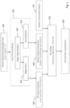
- FIG. 1 shows a flowchart of a first specific embodiment of the method according to the present invention, said first embodiment is not covered by the subject-matter of the claims but is considered as useful for understanding the invention.
- This embodiment implements the aforementioned CT modality. It is particularly adapted to classification of the severity of liver fibrosis.
- the following detailed description of the first specific embodiment of the method according to the present invention is thus concerned by fibrosis as the liver disease, the severity of which has to be classified, but it should be construed as a non-limiting example; that is to say that the first specific embodiment described hereafter can be used for classification of at least one another liver disease as well.
- the method according to said first specific embodiment comprises:
- Patient clinical information can also be inputted 350 into said trained ML model.
- this latter is preferably designed to classify the severity of fibrosis not only in function of both of said at least one parameter characterizing the liver and said at least one parameter characterizing the spleen, but also in function of the patient clinical information.
- An enhanced performance level is hence achieved which may yield to results as shown below into Table 1.
- a CT image of one delayed phase may further be used in order to enhance quality of the classification by allowing other features than the ones made prominent on arterial and/or portal venous phases.
- the spleen mask is used for the arterial and non-contrasted phases, whether the venous network is selected as the most optimal anatomical structure for the portal and delayed phases.
- HPI is relevant here since an HCC lesion that has a predominantly arterial blood supply is expected to show a high HPI, in contrast to normal liver tissue with around 20% arterial blood supply and 80% portal venous blood supply.
- the here proposed method uses perfusion patterns taking the form of HPI maps and thus allows benefiting of a sensitivity superior to 80%, or even superior to 88%, for HCC detection.
- FIG. 1 illustrates an overview of a first embodiment of the here proposed computational pipeline.
- the abdomen, and obviously the liver, of a patient are firstly extracted. More particularly, to have a more accurate and comprehensive view of hepatic fibrosis, not only the liver but also the spleen, as an adjacent organ of the abdomen with respect to the liver, are analyzed.
- a critical step for the analysis is the spatial registration of consecutive slices or scanning steps (i.e. non-contrasted, arterial, portal venous, and eventually delayed).
- a registration method is proposed that allows handling rigid and non-rigid deformations. Hence, the registration of any two radiographic images from the same patient within the same visit is available. More particularly, the arterial phase may be selected as a reference phase and all other phases may be registered against this reference phase.
- the registration may be implemented into two steps: A rigid step which is designed to iteratively search for the optimal affine transformations (translation, rotation, scaling) allows correcting for most rigid deformations and hence provides a near-optimal global correction of deformations; then a second step of non-rigid registration provide a fine grain (for instance 5x5x25 mm) deformation grid, where each Volume Of Interest (VOI) is subject to an independent transformation to better correlate the reference phase with each of the moving phases.
- VOI Volume Of Interest
- a normalization algorithm that estimates the bias from selected anatomical structures as variability in the radiographic image values due to technical, instrumental and scanning protocol, is implemented to be able to compare two patients with each other.
- the masks obtained by segmentation for the CT images are used to estimate normalization parameters.
- the ML model may be constructed by help of a training data set and validated by help of an independent data set.
- FIG. 2 shows a flowchart of a second specific embodiment of the method according to the present invention. Said second specific embodiment is encompassed by the wording of the claims.
- This embodiment implements the aforementioned MRI/MRE modality. It is particularly adapted to classification of the severity of NASH.
- the following detailed description of the second specific embodiment of the method according to the present invention is thus concerned by NASH as the liver disease, the severity of which has to be classified, but it should be construed as a non-limiting example; that is to say that the second specific embodiment described hereafter can be used for classification of at least one another liver disease as well.
- the method according to said second specific embodiment comprises:
- Patient clinical information can also be inputted 360 into said trained ML model.
- this latter is preferably designed to classify the severity of NASH not only in function of both of said at least one parameter characterizing the liver and said at least one parameter characterizing the spleen, but also in function of the patient clinical information.
- An enhanced performance level is hence achieved which may yield to results as shown below into Table 1.
- step of controlling quality the substeps of segmenting 212 and normalizing 214 and the step of displaying 610 may be implemented with the same specificities as those implemented with reference to the first specific embodiment of the invention and described here below.
- a deep learning convolutional network may be applied which allow enhancing NASH relevant features in 2D MRE images.
- ROI Region Of Interest
- said trained ML model may be designed to classify the liver disease level by implementing class prediction method.
- the ML model may be based on one among regression analysis software, Support-Vector Machine (SVM) and Random Forest (RF) classifier.
- SVM Support-Vector Machine
- RF Random Forest
- Said ML model may need to be specifically designed for either CT or MRI/MRE modalities.
- its calibration preferably takes into account for the specificity of the previous steps of the method. It may involve cutting edge methodology and consists in finding the most accurate, reproducible and scalable parameters for each modality (i.e. CT and MRI/MRE). Defining the ad hoc design of the ML model(s) (potentially one per modality) may be considered as feasible by the skilled person by application of his/her common general knowledge and by routine experimentations.
- radiographic images may be advantageously provided as DICOM (Digital Imaging and Communications in Medicine) image files.
- DICOM Digital Imaging and Communications in Medicine
- the normalizing substep of said pre-processing step may comprise, or may consist in, an automatic pixel luminance range adaptation of said radiographic images with each other.
- the segmenting substep of said pre-processing step may comprise, or may consist in, an automatic contouring of spleen and liver for each slice.
- An algorithm allowing segmentation liver, spleen and potentially other organs from a CT image or MRI image is thus implemented.
- Table 1 below shows results of the here proposed method for comparison to published results of LiverMultiscan ® method, for approximately equal total number of tests.
- both modality have been considered: results into column 3 of Table 1 have been acquired using CT images as input of an embodiment of the here proposed method as illustrated on Fig. 1 and results into column 4 of Table 1 have been acquired using CT images as input of an embodiment of the here proposed method as illustrated on Fig. 2 .
- Great differences of values of Sensitivity, Specificity, Positive Predictive Value and Negative Predictive Value have been observed, which each shows enhancement achieved owing to the here proposed method in comparison to LiverMultiscan ® method, for both of either CT modality or MRI/MRE modality.
- the receiver operator curve (ROC) as achieved owing to corresponding embodiments of the here proposed method has also been computed.
- the thus achieved AUC (Area Under Curve) values show the best overall performance of the classification model according to the here proposed method.
- Table 1 Benchmark between published results of LiverMultiscan ® method vs results of the here proposed method with CT modality and with MRI/MRE modality LiverMultiScan ®
- the present invention may also relate to a method for the determination of occurrence and stage of liver disease in a subject, which comprises the steps of the method for performing classification of liver disease levels as detailed above.
- the present invention may also relate to a method of diagnosis and detection of significant or advanced liver fibrosis due to other fibrotic liver diseases such as: viral hepatitis (HBV, HCV,..), Alcoholic steatohepatitis, Biliary diseases (Primary biliary cholangitis, Primary Sclerosing cholangitis, Autoimmune hepatitis, Wilson's disease, Alpha1 anti-trypsine deficiency).
- Said method of diagnosis and detection comprises the steps of the method for performing classification of fibrosis levels as detailed above.
- the invention may further relates to a method for the prognostic of the risk of NAFLD or NASH activity evolution in the absence of a treatment in a subject, with said method for the prognostic comprises the steps of the method for performing classification of fibrosis levels as detailed above.
- the invention may also relates to a method for the prognostic of the risk of fibrosis evolution to cirrhosis and liver clinical outcomes in the absence of treatment in a subject, with said method for the prognostic comprises the steps of the method for performing classification of liver disease severity level as detailed above.
- the invention may further relate to a method for monitoring the evolution (i.e. progression or regression) of NAFLD, NASH activity and HCC in a subject, which comprises the steps of the method for performing classification of liver disease severity level as detailed above.
- the invention may further relate to a method for monitoring the evolution (i.e. progression or regression) of liver disease in a subject, which comprises the steps of the method for performing classification of liver disease severity level as detailed above.
- the aforementioned further aspects of the invention and in particular the ones relative to any kind of evolution, are foreseen because the method according to the first aspect of the invention allows comparing not only scans of the same visit, but also scans from different visits to the hospital by the patient. It is thus possible to perform again the method according to the first aspect of the invention, and to compare the newly obtained results with the old ones to retrieve information about the evolution(s) of interest.
- the methods described herein may be implemented on any form of computer or computers.
- the components thereof may be implemented as dedicated applications or in client-server architectures, including a web-based architecture, and can include functional programs, codes, and code segments.
- Any of the computers may comprise a processor, a memory for storing program data and executing it, a permanent storage such as a disk drive, a communication port for handling communications with external devices, and user interface devices, including a display, keyboard, mouse, etc.
- program instructions or computer readable codes executable on the processor on a computer-readable media such as read-only memory (ROM), random-access memory (RAM), CD-ROMs, magnetic tapes, floppy disks, and optical data storage devices.
- the computer readable recording medium can also be distributed over network coupled computer systems so that the computer readable code is stored and executed in a distributed fashion. This media can be read by the computer, stored in the memory, and executed by the processor.
- the invention may be described in terms of functional block components and various processing steps. Such functional blocks may be realized by any number of hardware and/or software components configured to perform the specified functions.
- the invention may employ various integrated circuit components, e.g., memory elements, processing elements, logic elements, look-up tables, and the like, which may carry out a variety of functions under the control of one or more microprocessors or other control devices.
- the elements of the invention are implemented using software programming or software elements the invention may be implemented with any programming or scripting language such as C, C++, Java, assembler, or the like, with the various algorithms being implemented with any combination of data structures, objects, processes, routines or other programming elements.
- Functional aspects may be implemented in algorithms that execute on one or more processors.
- the invention could employ any number of conventional techniques for electronics configuration, signal processing and/or control, data processing and the like.
- the pipeline comprises a step of registration that allows comparing radiographic images of the same visit as well as radiographic images from different visits to the hospital by the patient.
- the method(s) according the invention can be applied to both prospective and retrospectively acquired imaging data.
- the here proposed method can be applied to each of two modalities making it easier to be integrated in the clinical practice: 1) on CT (Computed Tomography) imaging modality, and 2) on MRI (Magnetic Resonance Imaging) supported by MRE (Magnetic Resonance Elastography).
Landscapes
- Engineering & Computer Science (AREA)
- Computer Vision & Pattern Recognition (AREA)
- Physics & Mathematics (AREA)
- General Physics & Mathematics (AREA)
- Theoretical Computer Science (AREA)
- Health & Medical Sciences (AREA)
- General Health & Medical Sciences (AREA)
- Medical Informatics (AREA)
- Nuclear Medicine, Radiotherapy & Molecular Imaging (AREA)
- Radiology & Medical Imaging (AREA)
- Quality & Reliability (AREA)
- Apparatus For Radiation Diagnosis (AREA)
- Magnetic Resonance Imaging Apparatus (AREA)
- Image Analysis (AREA)
- Image Processing (AREA)
Claims (14)
- Verfahren zum Durchführen einer Klassifizierung des Schweregrades mindestens einer Lebererkrankung anhand von nicht invasiven Röntgenbildern, die während eines Scans erfasst wurden, wobei das Verfahren Folgendes umfasst:- Bereitstellen (100, 110) von Röntgenbildern von Schnitten mindestens des Abdomens eines Patienten, wobei mindestens zwei Röntgenbilder für jeden Schnitt bereitgestellt werden; und- Vorverarbeiten (200, 210) der Röntgenbilder mindestens durch:o Segmentieren (202, 212) von Leber und Milz aus mindestens einem der mindestens zwei Röntgenbilder für jeden Schnitt, wodurch eine binäre Maske der Milz und eine binäre Maske der Leber pro Schnitt erhalten wird, undo Normalisieren (204, 214) der Röntgenbilder untereinander, wodurch mindestens zwei normalisierte Röntgenbilder pro Schnitt erhalten werden;wobei das Verfahren ferner Folgendes umfasst:- für jeden Schnitt, mindestens anhand der binären Maske der Leber und der mindestens beiden normalisierten Röntgenbilder, Extrahieren (300, 310) mindestens eines Parameters, der die Leber charakterisiert;- anhand mindestens einer binären Maske der Milz, Extrahieren (400, 410) mindestens eines Parameters, der die Milz charakterisiert; und- Eingeben (500, 510) des mindestens einen Parameters, der die Leber charakterisiert, und des mindestens einen Parameters, der Milz charakterisiert, in ein trainiertes Modell für maschinelles Lernen, ML-Modell, wobei letzteres dazu ausgelegt ist, den Schweregrad der mindestens einen Lebererkrankung zwischen einer aus einer Gruppe von Lebererkrankungen im Frühstadium und einer Gruppe von Lebererkrankungen im fortgeschrittenen Stadium in Abhängigkeit von sowohl dem mindestens einen Parameter, der die Leber charakterisiert, als auch dem mindestens einen Parameter, der die Milz charakterisiert, zu klassifizieren,wobei das Verfahren dadurch gekennzeichnet ist, dass die mindestens zwei Röntgenbilder mindestens Folgendes umfassen:- ein Röntgenbild, MRI-Bild genannt, das durch Magnetresonanztomographie, MRI, erfasst wird, und- ein Röntgenbild, MRE-Bild genannt, das durch Magnetresonanz-Elastographie, MRE, erfasst wird.
- Verfahren nach Anspruch 1, das ferner das Eingeben (350, 360) klinischer Patienteninformationen in das trainierte ML-Modell umfasst, wobei letzteres dazu ausgelegt ist, den Schweregrad der mindestens einen Lebererkrankung zwischen einer aus einer Gruppe von Lebererkrankungen im Frühstadium und einer Gruppe von Lebererkrankungen im fortgeschrittenen Stadium nicht nur in Abhängigkeit von sowohl dem mindestens einen Parameter, der die Leber charakterisiert, als auch dem mindestens einen Parameter, der die Milz charakterisiert, sondern auch in Abhängigkeit von den klinischen Patienteninformationen zu klassifizieren.
- Verfahren nach einem der vorhergehenden Ansprüche, wobei das trainierte ML-Modell dazu ausgelegt ist, den Schweregrad der mindestens einen Lebererkrankung durch Implementieren eines Klassenvorhersageverfahrens zu klassifizieren.
- Verfahren nach einem der vorhergehenden Ansprüche, wobei das ML-Modell auf einem Klassifikator von einer Regressionsanalyse-Software, einer Support Vector Machine, SVM, und Random Forest, RF, basiert.
- Verfahren nach einem der vorhergehenden Ansprüche, wobei der Segmentierungsunterschritt (212) des Vorverarbeitungsschritts (210) für jeden Schnitt durch Verwenden des MRI-Bilds als das mindestens eine der mindestens zwei Röntgenbilder implementiert wird, und wobei der Schritt, der darin besteht, mindestens einen Parameter, der die Leber charakterisiert, zu extrahieren (310), für jeden Schnitt das Extrahieren von Elastographieinformationen aus dem MRE-Bild unter Verwendung der binären Maske der Leber umfasst.
- Verfahren nach einem der vorhergehenden Ansprüche, wobei der mindestens eine Parameter, der die Milz charakterisiert, mindestens eines von einer Milzfläche, einer Milzform, einem Milzformfaktor, einer Milzdichte, einer Milzexzentrizität und einer Milzfestigkeit umfasst.
- Verfahren nach einem der vorhergehenden Ansprüche, wobei es sich bei der mindestens einen Lebererkrankung eine nichtalkoholische Steatohepatitis, NASH, handelt, und das trainierte ML-Modell dazu ausgelegt ist, den NASH-Schweregrad zwischen einer NASH-Gruppe im Frühstadium und einer NASH-Gruppe im fortgeschrittenen Stadium zu klassifizieren.
- Verfahren nach einem der Ansprüche 1 bis 7, wobei die Röntgenbilder als DICOM-Bilddaten, DICOM = digitale Bildgebung und -kommunikation in der Medizin, bereitgestellt werden.
- Verfahren nach einem der Ansprüche 1 bis 8, das ferner vor dem Vorverarbeitungsschritt (200, 210) Folgendes umfasst:- Steuern der Qualität der Röntgenbilder, um so Röntgenbilder mit einem Qualitätswert über einem bestimmten Schwellenwert festzuhalten.
- Verfahren nach einem der Ansprüche 1 bis 9, wobei der Normalisierungsunterschritt (204, 214) des Vorverarbeitungsschritts (200, 210) eine automatische Pixel-Leuchtdichtebereichsanpassung der Röntgenbilder untereinander umfasst.
- Verfahren nach einem der Ansprüche 1 bis 10, wobei der Segmentierungsunterschritt (204, 214) des Vorverarbeitungsschritts (200, 210) eine automatische Konturierung von Milz und Leber für jeden Schnitt umfasst.
- Verfahren nach Anspruch 11, wobei der Segmentierungsunterschritt (204, 214) des Vorverarbeitungsschritts (200, 210) darin besteht, einen Segmentierungsalgorithmus von Bereichen von Interesse, ROI = Region of Interest, zu implementieren.
- Verfahren nach einem der Ansprüche 1 bis 12, das ferner nach dem Klassifizieren des Fibrosegrades Folgendes umfasst:- Ausgeben (600, 610), zum Beispiel durch Anzeige, des Fibrosegrades, wie er klassifiziert ist.
- Computerprogrammprodukt, das Anweisungen umfasst, die, wenn sie von mindestens einer digitalen Verarbeitungsvorrichtung implementiert werden, mindestens die Schritte des Verfahrens nach den Ansprüchen 1 bis 13 durchführen.
Priority Applications (1)
| Application Number | Priority Date | Filing Date | Title |
|---|---|---|---|
| EP24199314.6A EP4465237A3 (de) | 2019-11-06 | 2020-11-05 | Automatisierte klassifizierung der schwere einer lebererkrankung aus nichtinvasiver radiologiebildgebung |
Applications Claiming Priority (2)
| Application Number | Priority Date | Filing Date | Title |
|---|---|---|---|
| US201962931286P | 2019-11-06 | 2019-11-06 | |
| PCT/EP2020/081207 WO2021089741A1 (en) | 2019-11-06 | 2020-11-05 | Automated classification of severity of liver disease from non invasive radiology imaging |
Related Child Applications (2)
| Application Number | Title | Priority Date | Filing Date |
|---|---|---|---|
| EP24199314.6A Division EP4465237A3 (de) | 2019-11-06 | 2020-11-05 | Automatisierte klassifizierung der schwere einer lebererkrankung aus nichtinvasiver radiologiebildgebung |
| EP24199314.6A Division-Into EP4465237A3 (de) | 2019-11-06 | 2020-11-05 | Automatisierte klassifizierung der schwere einer lebererkrankung aus nichtinvasiver radiologiebildgebung |
Publications (2)
| Publication Number | Publication Date |
|---|---|
| EP4055558A1 EP4055558A1 (de) | 2022-09-14 |
| EP4055558B1 true EP4055558B1 (de) | 2024-10-16 |
Family
ID=73138853
Family Applications (2)
| Application Number | Title | Priority Date | Filing Date |
|---|---|---|---|
| EP24199314.6A Pending EP4465237A3 (de) | 2019-11-06 | 2020-11-05 | Automatisierte klassifizierung der schwere einer lebererkrankung aus nichtinvasiver radiologiebildgebung |
| EP20803157.5A Active EP4055558B1 (de) | 2019-11-06 | 2020-11-05 | Automatische klassifizierung des schweregrades einer lebererkrankung anhand von nicht-invasiver radiologischer bildgebung |
Family Applications Before (1)
| Application Number | Title | Priority Date | Filing Date |
|---|---|---|---|
| EP24199314.6A Pending EP4465237A3 (de) | 2019-11-06 | 2020-11-05 | Automatisierte klassifizierung der schwere einer lebererkrankung aus nichtinvasiver radiologiebildgebung |
Country Status (6)
| Country | Link |
|---|---|
| US (1) | US12315147B2 (de) |
| EP (2) | EP4465237A3 (de) |
| JP (1) | JP7539981B2 (de) |
| CN (1) | CN115516498A (de) |
| ES (1) | ES3004960T3 (de) |
| WO (1) | WO2021089741A1 (de) |
Families Citing this family (6)
| Publication number | Priority date | Publication date | Assignee | Title |
|---|---|---|---|---|
| EP4476684A1 (de) * | 2022-02-09 | 2024-12-18 | F. Hoffmann-La Roche AG | Leberzirrhoseerkennung in digitalen bildern |
| KR102756982B1 (ko) * | 2022-11-30 | 2025-01-21 | 숭실대학교 산학협력단 | 지방간 질환 자동 예측을 위한 3d 간 ct 영상의 분할 및 강체 정합 장치 및 방법 |
| CN116503655A (zh) * | 2023-04-27 | 2023-07-28 | 祁小龙 | 一种自动化门脉高压分类系统、方法、设备及介质 |
| US12530770B2 (en) * | 2023-06-15 | 2026-01-20 | Case Western Reserve University | Deep-learning-based hepatic fat assessment |
| CN117011242B (zh) * | 2023-07-10 | 2024-05-14 | 珠海市人民医院 | 经颈静脉肝内门体分流术后肝性脑病预测方法和系统 |
| CN118365970B (zh) | 2024-06-19 | 2024-09-10 | 张家港江苏科技大学产业技术研究院 | 基于相互矫正和信息融合的医学数据分类方法及装置 |
Family Cites Families (12)
| Publication number | Priority date | Publication date | Assignee | Title |
|---|---|---|---|---|
| US8355553B2 (en) | 2008-11-22 | 2013-01-15 | General Electric Company | Systems, apparatus and processes for automated medical image segmentation using a statistical model |
| US20180098728A1 (en) * | 2011-03-11 | 2018-04-12 | Centre Hospitalier Universitaire D'angers | Non-invasive method for assessing the presence or severity of liver fibrosis based on a new detailed classification |
| KR20140074969A (ko) * | 2011-09-26 | 2014-06-18 | 아쿠이타스 메디컬 리미티드 | 기관 섬유증 특징의 검출방법 |
| EP2873017B1 (de) * | 2012-07-11 | 2021-09-01 | University of Mississippi Medical Center | Verfahren zum nachweis und zur einstufung einer leberfibrose aus erfassten daten |
| US9155513B2 (en) * | 2013-07-17 | 2015-10-13 | Hepatiq Llc | Systems and methods for determining hepatic function from liver scans |
| CN104657624A (zh) * | 2015-03-18 | 2015-05-27 | 汪艳 | 一种肝硬化全量化分析方法 |
| CN104835112B (zh) * | 2015-05-07 | 2018-06-08 | 厦门大学 | 一种肝脏多相期ct图像融合方法 |
| JP7007361B2 (ja) | 2016-08-01 | 2022-01-24 | センター ホスピタリア ユニバーシタイア ダンガース | 複数標的線維症検査 |
| US20190021677A1 (en) * | 2017-07-18 | 2019-01-24 | Siemens Healthcare Gmbh | Methods and systems for classification and assessment using machine learning |
| CN108171709A (zh) * | 2018-01-30 | 2018-06-15 | 北京青燕祥云科技有限公司 | 肝占位性病灶区域的检测方法、装置和实现装置 |
| CN109670515A (zh) * | 2018-12-13 | 2019-04-23 | 南京工业大学 | 一种用于无人机影像中建筑物变化的检测方法及系统 |
| US11055822B2 (en) * | 2019-05-03 | 2021-07-06 | International Business Machines Corporation | Artificially intelligent, machine learning-based, image enhancement, processing, improvement and feedback algorithms |
-
2020
- 2020-11-05 EP EP24199314.6A patent/EP4465237A3/de active Pending
- 2020-11-05 WO PCT/EP2020/081207 patent/WO2021089741A1/en not_active Ceased
- 2020-11-05 ES ES20803157T patent/ES3004960T3/es active Active
- 2020-11-05 US US17/772,881 patent/US12315147B2/en active Active
- 2020-11-05 CN CN202080077043.3A patent/CN115516498A/zh active Pending
- 2020-11-05 JP JP2022525628A patent/JP7539981B2/ja active Active
- 2020-11-05 EP EP20803157.5A patent/EP4055558B1/de active Active
Also Published As
| Publication number | Publication date |
|---|---|
| US12315147B2 (en) | 2025-05-27 |
| EP4465237A2 (de) | 2024-11-20 |
| US20220414870A1 (en) | 2022-12-29 |
| WO2021089741A1 (en) | 2021-05-14 |
| EP4055558A1 (de) | 2022-09-14 |
| EP4465237A3 (de) | 2025-02-19 |
| CN115516498A (zh) | 2022-12-23 |
| JP2023501282A (ja) | 2023-01-18 |
| ES3004960T3 (en) | 2025-03-13 |
| JP7539981B2 (ja) | 2024-08-26 |
Similar Documents
| Publication | Publication Date | Title |
|---|---|---|
| US12315147B2 (en) | Automated pattern recognition and scoring method of fibrosis from non invasive radiology imaging | |
| EP3021753B1 (de) | Systeme und verfahren zur bestimmung der leberfunktion aufgrund von leber-scanvorgängen | |
| US9218661B2 (en) | Image analysis for specific objects | |
| US9230331B2 (en) | Systems and methods for registration of ultrasound and CT images | |
| Bonekamp et al. | Interobserver agreement of semi-automated and manual measurements of functional MRI metrics of treatment response in hepatocellular carcinoma | |
| EP3528261A1 (de) | Prädiktionsmodell zur gruppierung des hepatozellulären karzinoms, prädiktionssystem dafür und verfahren zum bestimmen der hepatozellulären karzinomgruppe | |
| US11151722B2 (en) | System and method for estimating synthetic quantitative health values from medical images | |
| Ivashchenko et al. | A workflow for automated segmentation of the liver surface, hepatic vasculature and biliary tree anatomy from multiphase MR images | |
| Jirapatnakul et al. | Automated measurement of liver attenuation to identify moderate-to-severe hepatic steatosis from chest CT scans | |
| US20250272837A1 (en) | Automated classification of severity of liver disease from non invasive radiology imaging | |
| Tong et al. | Disease quantification on PET/CT images without explicit object delineation | |
| Wong et al. | A comparison of peripheral imaging technologies for bone and muscle quantification: a review of segmentation techniques | |
| Cui et al. | Predicting the stages of liver fibrosis with multiphase CT radiomics based on volumetric features | |
| Delsanto et al. | CULEX-completely user-independent layers extraction: ultrasonic carotid artery images segmentation | |
| Molinari et al. | CAUDLES-EF: carotid automated ultrasound double line extraction system using edge flow | |
| Bonekamp et al. | An elevated arterial enhancement fraction is associated with clinical and imaging indices of liver fibrosis and cirrhosis | |
| US20140094679A1 (en) | Systems and methods for performing organ detection | |
| Chi et al. | Segmentation of the bladder wall using coupled level set methods | |
| Pithani et al. | Enhanced non-alcoholic fatty liver detection: Computed tomography scan image analysis and noise reduction with morphological dilation | |
| Mesanovic et al. | Application of lung segmentation algorithm to disease quantification from CT images | |
| WO2012083350A1 (en) | Coronary calcium measurement | |
| Dubey et al. | An advanced technique for volumetric analysis | |
| Correra et al. | A method for automatic breast density classification in magnetic resonance imaging | |
| Fehr et al. | Automated volumetric estimation of six basic thoracic and abdominal organs in postmortem computed tomography data using deep learning techniques | |
| Cen et al. | A Computer-aided radiomics approach based on classifier ensemble to differentiate malignancy of hepatocellular carcinoma with Contrast-enhanced MR |
Legal Events
| Date | Code | Title | Description |
|---|---|---|---|
| STAA | Information on the status of an ep patent application or granted ep patent |
Free format text: STATUS: UNKNOWN |
|
| STAA | Information on the status of an ep patent application or granted ep patent |
Free format text: STATUS: THE INTERNATIONAL PUBLICATION HAS BEEN MADE |
|
| PUAI | Public reference made under article 153(3) epc to a published international application that has entered the european phase |
Free format text: ORIGINAL CODE: 0009012 |
|
| STAA | Information on the status of an ep patent application or granted ep patent |
Free format text: STATUS: REQUEST FOR EXAMINATION WAS MADE |
|
| 17P | Request for examination filed |
Effective date: 20220426 |
|
| AK | Designated contracting states |
Kind code of ref document: A1 Designated state(s): AL AT BE BG CH CY CZ DE DK EE ES FI FR GB GR HR HU IE IS IT LI LT LU LV MC MK MT NL NO PL PT RO RS SE SI SK SM TR |
|
| DAV | Request for validation of the european patent (deleted) | ||
| DAX | Request for extension of the european patent (deleted) | ||
| GRAP | Despatch of communication of intention to grant a patent |
Free format text: ORIGINAL CODE: EPIDOSNIGR1 |
|
| STAA | Information on the status of an ep patent application or granted ep patent |
Free format text: STATUS: GRANT OF PATENT IS INTENDED |
|
| INTG | Intention to grant announced |
Effective date: 20240510 |
|
| GRAS | Grant fee paid |
Free format text: ORIGINAL CODE: EPIDOSNIGR3 |
|
| GRAA | (expected) grant |
Free format text: ORIGINAL CODE: 0009210 |
|
| STAA | Information on the status of an ep patent application or granted ep patent |
Free format text: STATUS: THE PATENT HAS BEEN GRANTED |
|
| AK | Designated contracting states |
Kind code of ref document: B1 Designated state(s): AL AT BE BG CH CY CZ DE DK EE ES FI FR GB GR HR HU IE IS IT LI LT LU LV MC MK MT NL NO PL PT RO RS SE SI SK SM TR |
|
| REG | Reference to a national code |
Ref country code: GB Ref legal event code: FG4D |
|
| REG | Reference to a national code |
Ref country code: CH Ref legal event code: EP Ref country code: DE Ref legal event code: R096 Ref document number: 602020039624 Country of ref document: DE |
|
| REG | Reference to a national code |
Ref country code: IE Ref legal event code: FG4D |
|
| REG | Reference to a national code |
Ref country code: LT Ref legal event code: MG9D |
|
| REG | Reference to a national code |
Ref country code: NL Ref legal event code: MP Effective date: 20241016 |
|
| REG | Reference to a national code |
Ref country code: ES Ref legal event code: FG2A Ref document number: 3004960 Country of ref document: ES Kind code of ref document: T3 Effective date: 20250313 |
|
| REG | Reference to a national code |
Ref country code: AT Ref legal event code: MK05 Ref document number: 1733539 Country of ref document: AT Kind code of ref document: T Effective date: 20241016 |
|
| PG25 | Lapsed in a contracting state [announced via postgrant information from national office to epo] |
Ref country code: NL Free format text: LAPSE BECAUSE OF FAILURE TO SUBMIT A TRANSLATION OF THE DESCRIPTION OR TO PAY THE FEE WITHIN THE PRESCRIBED TIME-LIMIT Effective date: 20241016 |
|
| PG25 | Lapsed in a contracting state [announced via postgrant information from national office to epo] |
Ref country code: NL Free format text: LAPSE BECAUSE OF FAILURE TO SUBMIT A TRANSLATION OF THE DESCRIPTION OR TO PAY THE FEE WITHIN THE PRESCRIBED TIME-LIMIT Effective date: 20241016 |
|
| PG25 | Lapsed in a contracting state [announced via postgrant information from national office to epo] |
Ref country code: HR Free format text: LAPSE BECAUSE OF FAILURE TO SUBMIT A TRANSLATION OF THE DESCRIPTION OR TO PAY THE FEE WITHIN THE PRESCRIBED TIME-LIMIT Effective date: 20241016 Ref country code: PT Free format text: LAPSE BECAUSE OF FAILURE TO SUBMIT A TRANSLATION OF THE DESCRIPTION OR TO PAY THE FEE WITHIN THE PRESCRIBED TIME-LIMIT Effective date: 20250217 Ref country code: IS Free format text: LAPSE BECAUSE OF FAILURE TO SUBMIT A TRANSLATION OF THE DESCRIPTION OR TO PAY THE FEE WITHIN THE PRESCRIBED TIME-LIMIT Effective date: 20250216 |
|
| PG25 | Lapsed in a contracting state [announced via postgrant information from national office to epo] |
Ref country code: FI Free format text: LAPSE BECAUSE OF FAILURE TO SUBMIT A TRANSLATION OF THE DESCRIPTION OR TO PAY THE FEE WITHIN THE PRESCRIBED TIME-LIMIT Effective date: 20241016 |
|
| PG25 | Lapsed in a contracting state [announced via postgrant information from national office to epo] |
Ref country code: BG Free format text: LAPSE BECAUSE OF FAILURE TO SUBMIT A TRANSLATION OF THE DESCRIPTION OR TO PAY THE FEE WITHIN THE PRESCRIBED TIME-LIMIT Effective date: 20241016 |
|
| PG25 | Lapsed in a contracting state [announced via postgrant information from national office to epo] |
Ref country code: NO Free format text: LAPSE BECAUSE OF FAILURE TO SUBMIT A TRANSLATION OF THE DESCRIPTION OR TO PAY THE FEE WITHIN THE PRESCRIBED TIME-LIMIT Effective date: 20250116 |
|
| PG25 | Lapsed in a contracting state [announced via postgrant information from national office to epo] |
Ref country code: LV Free format text: LAPSE BECAUSE OF FAILURE TO SUBMIT A TRANSLATION OF THE DESCRIPTION OR TO PAY THE FEE WITHIN THE PRESCRIBED TIME-LIMIT Effective date: 20241016 Ref country code: AT Free format text: LAPSE BECAUSE OF FAILURE TO SUBMIT A TRANSLATION OF THE DESCRIPTION OR TO PAY THE FEE WITHIN THE PRESCRIBED TIME-LIMIT Effective date: 20241016 Ref country code: GR Free format text: LAPSE BECAUSE OF FAILURE TO SUBMIT A TRANSLATION OF THE DESCRIPTION OR TO PAY THE FEE WITHIN THE PRESCRIBED TIME-LIMIT Effective date: 20250117 |
|
| PG25 | Lapsed in a contracting state [announced via postgrant information from national office to epo] |
Ref country code: PL Free format text: LAPSE BECAUSE OF FAILURE TO SUBMIT A TRANSLATION OF THE DESCRIPTION OR TO PAY THE FEE WITHIN THE PRESCRIBED TIME-LIMIT Effective date: 20241016 |
|
| PG25 | Lapsed in a contracting state [announced via postgrant information from national office to epo] |
Ref country code: RS Free format text: LAPSE BECAUSE OF FAILURE TO SUBMIT A TRANSLATION OF THE DESCRIPTION OR TO PAY THE FEE WITHIN THE PRESCRIBED TIME-LIMIT Effective date: 20250116 |
|
| REG | Reference to a national code |
Ref country code: CH Ref legal event code: PL |
|
| PG25 | Lapsed in a contracting state [announced via postgrant information from national office to epo] |
Ref country code: SM Free format text: LAPSE BECAUSE OF FAILURE TO SUBMIT A TRANSLATION OF THE DESCRIPTION OR TO PAY THE FEE WITHIN THE PRESCRIBED TIME-LIMIT Effective date: 20241016 |
|
| PG25 | Lapsed in a contracting state [announced via postgrant information from national office to epo] |
Ref country code: MC Free format text: LAPSE BECAUSE OF FAILURE TO SUBMIT A TRANSLATION OF THE DESCRIPTION OR TO PAY THE FEE WITHIN THE PRESCRIBED TIME-LIMIT Effective date: 20241016 |
|
| PG25 | Lapsed in a contracting state [announced via postgrant information from national office to epo] |
Ref country code: DK Free format text: LAPSE BECAUSE OF FAILURE TO SUBMIT A TRANSLATION OF THE DESCRIPTION OR TO PAY THE FEE WITHIN THE PRESCRIBED TIME-LIMIT Effective date: 20241016 |
|
| PG25 | Lapsed in a contracting state [announced via postgrant information from national office to epo] |
Ref country code: LU Free format text: LAPSE BECAUSE OF NON-PAYMENT OF DUE FEES Effective date: 20241105 |
|
| REG | Reference to a national code |
Ref country code: CH Ref legal event code: PL |
|
| REG | Reference to a national code |
Ref country code: DE Ref legal event code: R097 Ref document number: 602020039624 Country of ref document: DE |
|
| PG25 | Lapsed in a contracting state [announced via postgrant information from national office to epo] |
Ref country code: EE Free format text: LAPSE BECAUSE OF FAILURE TO SUBMIT A TRANSLATION OF THE DESCRIPTION OR TO PAY THE FEE WITHIN THE PRESCRIBED TIME-LIMIT Effective date: 20241016 |
|
| PG25 | Lapsed in a contracting state [announced via postgrant information from national office to epo] |
Ref country code: CH Free format text: LAPSE BECAUSE OF NON-PAYMENT OF DUE FEES Effective date: 20241130 |
|
| PG25 | Lapsed in a contracting state [announced via postgrant information from national office to epo] |
Ref country code: RO Free format text: LAPSE BECAUSE OF FAILURE TO SUBMIT A TRANSLATION OF THE DESCRIPTION OR TO PAY THE FEE WITHIN THE PRESCRIBED TIME-LIMIT Effective date: 20241016 |
|
| PG25 | Lapsed in a contracting state [announced via postgrant information from national office to epo] |
Ref country code: SK Free format text: LAPSE BECAUSE OF FAILURE TO SUBMIT A TRANSLATION OF THE DESCRIPTION OR TO PAY THE FEE WITHIN THE PRESCRIBED TIME-LIMIT Effective date: 20241016 |
|
| PG25 | Lapsed in a contracting state [announced via postgrant information from national office to epo] |
Ref country code: CZ Free format text: LAPSE BECAUSE OF FAILURE TO SUBMIT A TRANSLATION OF THE DESCRIPTION OR TO PAY THE FEE WITHIN THE PRESCRIBED TIME-LIMIT Effective date: 20241016 |
|
| PLBE | No opposition filed within time limit |
Free format text: ORIGINAL CODE: 0009261 |
|
| STAA | Information on the status of an ep patent application or granted ep patent |
Free format text: STATUS: NO OPPOSITION FILED WITHIN TIME LIMIT |
|
| REG | Reference to a national code |
Ref country code: BE Ref legal event code: MM Effective date: 20241130 |
|
| PG25 | Lapsed in a contracting state [announced via postgrant information from national office to epo] |
Ref country code: SE Free format text: LAPSE BECAUSE OF FAILURE TO SUBMIT A TRANSLATION OF THE DESCRIPTION OR TO PAY THE FEE WITHIN THE PRESCRIBED TIME-LIMIT Effective date: 20241016 |
|
| 26N | No opposition filed |
Effective date: 20250717 |
|
| PG25 | Lapsed in a contracting state [announced via postgrant information from national office to epo] |
Ref country code: BE Free format text: LAPSE BECAUSE OF NON-PAYMENT OF DUE FEES Effective date: 20241130 |
|
| PG25 | Lapsed in a contracting state [announced via postgrant information from national office to epo] |
Ref country code: IE Free format text: LAPSE BECAUSE OF NON-PAYMENT OF DUE FEES Effective date: 20241105 |
|
| PGFP | Annual fee paid to national office [announced via postgrant information from national office to epo] |
Ref country code: GB Payment date: 20251226 Year of fee payment: 6 |
|
| PGFP | Annual fee paid to national office [announced via postgrant information from national office to epo] |
Ref country code: IT Payment date: 20251121 Year of fee payment: 6 |
|
| PGFP | Annual fee paid to national office [announced via postgrant information from national office to epo] |
Ref country code: FR Payment date: 20251125 Year of fee payment: 6 |
|
| PGFP | Annual fee paid to national office [announced via postgrant information from national office to epo] |
Ref country code: ES Payment date: 20251212 Year of fee payment: 6 |
|
| PG25 | Lapsed in a contracting state [announced via postgrant information from national office to epo] |
Ref country code: HU Free format text: LAPSE BECAUSE OF FAILURE TO SUBMIT A TRANSLATION OF THE DESCRIPTION OR TO PAY THE FEE WITHIN THE PRESCRIBED TIME-LIMIT; INVALID AB INITIO Effective date: 20201105 |
|
| PG25 | Lapsed in a contracting state [announced via postgrant information from national office to epo] |
Ref country code: CY Free format text: LAPSE BECAUSE OF FAILURE TO SUBMIT A TRANSLATION OF THE DESCRIPTION OR TO PAY THE FEE WITHIN THE PRESCRIBED TIME-LIMIT; INVALID AB INITIO Effective date: 20201105 |
|
| PGFP | Annual fee paid to national office [announced via postgrant information from national office to epo] |
Ref country code: DE Payment date: 20251231 Year of fee payment: 6 |