EP4002201A1 - Vorrichtung und verfahren zur biologischen klassifizierung von morbus alzheimer unter verwendung eines multimodalen gehirnbildes - Google Patents
Vorrichtung und verfahren zur biologischen klassifizierung von morbus alzheimer unter verwendung eines multimodalen gehirnbildes Download PDFInfo
- Publication number
- EP4002201A1 EP4002201A1 EP21208975.9A EP21208975A EP4002201A1 EP 4002201 A1 EP4002201 A1 EP 4002201A1 EP 21208975 A EP21208975 A EP 21208975A EP 4002201 A1 EP4002201 A1 EP 4002201A1
- Authority
- EP
- European Patent Office
- Prior art keywords
- image
- determination
- brain
- subject
- normal
- Prior art date
- Legal status (The legal status is an assumption and is not a legal conclusion. Google has not performed a legal analysis and makes no representation as to the accuracy of the status listed.)
- Pending
Links
Images







Classifications
-
- A—HUMAN NECESSITIES
- A61—MEDICAL OR VETERINARY SCIENCE; HYGIENE
- A61B—DIAGNOSIS; SURGERY; IDENTIFICATION
- A61B5/00—Measuring for diagnostic purposes; Identification of persons
- A61B5/40—Detecting, measuring or recording for evaluating the nervous system
- A61B5/4076—Diagnosing or monitoring particular conditions of the nervous system
- A61B5/4088—Diagnosing of monitoring cognitive diseases, e.g. Alzheimer, prion diseases or dementia
-
- A—HUMAN NECESSITIES
- A61—MEDICAL OR VETERINARY SCIENCE; HYGIENE
- A61B—DIAGNOSIS; SURGERY; IDENTIFICATION
- A61B5/00—Measuring for diagnostic purposes; Identification of persons
- A61B5/0033—Features or image-related aspects of imaging apparatus, e.g. for MRI, optical tomography or impedance tomography apparatus; Arrangements of imaging apparatus in a room
- A61B5/0035—Features or image-related aspects of imaging apparatus, e.g. for MRI, optical tomography or impedance tomography apparatus; Arrangements of imaging apparatus in a room adapted for acquisition of images from more than one imaging mode, e.g. combining MRI and optical tomography
-
- A—HUMAN NECESSITIES
- A61—MEDICAL OR VETERINARY SCIENCE; HYGIENE
- A61B—DIAGNOSIS; SURGERY; IDENTIFICATION
- A61B5/00—Measuring for diagnostic purposes; Identification of persons
- A61B5/0033—Features or image-related aspects of imaging apparatus, e.g. for MRI, optical tomography or impedance tomography apparatus; Arrangements of imaging apparatus in a room
- A61B5/004—Features or image-related aspects of imaging apparatus, e.g. for MRI, optical tomography or impedance tomography apparatus; Arrangements of imaging apparatus in a room adapted for image acquisition of a particular organ or body part
- A61B5/0042—Features or image-related aspects of imaging apparatus, e.g. for MRI, optical tomography or impedance tomography apparatus; Arrangements of imaging apparatus in a room adapted for image acquisition of a particular organ or body part for the brain
-
- A—HUMAN NECESSITIES
- A61—MEDICAL OR VETERINARY SCIENCE; HYGIENE
- A61B—DIAGNOSIS; SURGERY; IDENTIFICATION
- A61B5/00—Measuring for diagnostic purposes; Identification of persons
- A61B5/72—Signal processing specially adapted for physiological signals or for diagnostic purposes
- A61B5/7235—Details of waveform analysis
- A61B5/7264—Classification of physiological signals or data, e.g. using neural networks, statistical classifiers, expert systems or fuzzy systems
-
- A—HUMAN NECESSITIES
- A61—MEDICAL OR VETERINARY SCIENCE; HYGIENE
- A61B—DIAGNOSIS; SURGERY; IDENTIFICATION
- A61B5/00—Measuring for diagnostic purposes; Identification of persons
- A61B5/72—Signal processing specially adapted for physiological signals or for diagnostic purposes
- A61B5/7271—Specific aspects of physiological measurement analysis
- A61B5/7275—Determining trends in physiological measurement data; Predicting development of a medical condition based on physiological measurements, e.g. determining a risk factor
-
- G—PHYSICS
- G06—COMPUTING OR CALCULATING; COUNTING
- G06T—IMAGE DATA PROCESSING OR GENERATION, IN GENERAL
- G06T7/00—Image analysis
- G06T7/0002—Inspection of images, e.g. flaw detection
- G06T7/0012—Biomedical image inspection
-
- G—PHYSICS
- G06—COMPUTING OR CALCULATING; COUNTING
- G06V—IMAGE OR VIDEO RECOGNITION OR UNDERSTANDING
- G06V10/00—Arrangements for image or video recognition or understanding
- G06V10/70—Arrangements for image or video recognition or understanding using pattern recognition or machine learning
- G06V10/764—Arrangements for image or video recognition or understanding using pattern recognition or machine learning using classification, e.g. of video objects
-
- G—PHYSICS
- G16—INFORMATION AND COMMUNICATION TECHNOLOGY [ICT] SPECIALLY ADAPTED FOR SPECIFIC APPLICATION FIELDS
- G16H—HEALTHCARE INFORMATICS, i.e. INFORMATION AND COMMUNICATION TECHNOLOGY [ICT] SPECIALLY ADAPTED FOR THE HANDLING OR PROCESSING OF MEDICAL OR HEALTHCARE DATA
- G16H30/00—ICT specially adapted for the handling or processing of medical images
- G16H30/40—ICT specially adapted for the handling or processing of medical images for processing medical images, e.g. editing
-
- G—PHYSICS
- G16—INFORMATION AND COMMUNICATION TECHNOLOGY [ICT] SPECIALLY ADAPTED FOR SPECIFIC APPLICATION FIELDS
- G16H—HEALTHCARE INFORMATICS, i.e. INFORMATION AND COMMUNICATION TECHNOLOGY [ICT] SPECIALLY ADAPTED FOR THE HANDLING OR PROCESSING OF MEDICAL OR HEALTHCARE DATA
- G16H50/00—ICT specially adapted for medical diagnosis, medical simulation or medical data mining; ICT specially adapted for detecting, monitoring or modelling epidemics or pandemics
- G16H50/20—ICT specially adapted for medical diagnosis, medical simulation or medical data mining; ICT specially adapted for detecting, monitoring or modelling epidemics or pandemics for computer-aided diagnosis, e.g. based on medical expert systems
-
- G—PHYSICS
- G16—INFORMATION AND COMMUNICATION TECHNOLOGY [ICT] SPECIALLY ADAPTED FOR SPECIFIC APPLICATION FIELDS
- G16H—HEALTHCARE INFORMATICS, i.e. INFORMATION AND COMMUNICATION TECHNOLOGY [ICT] SPECIALLY ADAPTED FOR THE HANDLING OR PROCESSING OF MEDICAL OR HEALTHCARE DATA
- G16H50/00—ICT specially adapted for medical diagnosis, medical simulation or medical data mining; ICT specially adapted for detecting, monitoring or modelling epidemics or pandemics
- G16H50/30—ICT specially adapted for medical diagnosis, medical simulation or medical data mining; ICT specially adapted for detecting, monitoring or modelling epidemics or pandemics for calculating health indices; for individual health risk assessment
-
- G—PHYSICS
- G16—INFORMATION AND COMMUNICATION TECHNOLOGY [ICT] SPECIALLY ADAPTED FOR SPECIFIC APPLICATION FIELDS
- G16H—HEALTHCARE INFORMATICS, i.e. INFORMATION AND COMMUNICATION TECHNOLOGY [ICT] SPECIALLY ADAPTED FOR THE HANDLING OR PROCESSING OF MEDICAL OR HEALTHCARE DATA
- G16H70/00—ICT specially adapted for the handling or processing of medical references
- G16H70/60—ICT specially adapted for the handling or processing of medical references relating to pathologies
-
- G—PHYSICS
- G06—COMPUTING OR CALCULATING; COUNTING
- G06T—IMAGE DATA PROCESSING OR GENERATION, IN GENERAL
- G06T2207/00—Indexing scheme for image analysis or image enhancement
- G06T2207/10—Image acquisition modality
- G06T2207/10072—Tomographic images
- G06T2207/10088—Magnetic resonance imaging [MRI]
-
- G—PHYSICS
- G06—COMPUTING OR CALCULATING; COUNTING
- G06T—IMAGE DATA PROCESSING OR GENERATION, IN GENERAL
- G06T2207/00—Indexing scheme for image analysis or image enhancement
- G06T2207/20—Special algorithmic details
- G06T2207/20084—Artificial neural networks [ANN]
-
- G—PHYSICS
- G06—COMPUTING OR CALCULATING; COUNTING
- G06T—IMAGE DATA PROCESSING OR GENERATION, IN GENERAL
- G06T2207/00—Indexing scheme for image analysis or image enhancement
- G06T2207/30—Subject of image; Context of image processing
- G06T2207/30004—Biomedical image processing
- G06T2207/30016—Brain
-
- G—PHYSICS
- G06—COMPUTING OR CALCULATING; COUNTING
- G06V—IMAGE OR VIDEO RECOGNITION OR UNDERSTANDING
- G06V2201/00—Indexing scheme relating to image or video recognition or understanding
- G06V2201/03—Recognition of patterns in medical or anatomical images
- G06V2201/031—Recognition of patterns in medical or anatomical images of internal organs
Definitions
- the present disclosure relates to biological classification device and method for Alzheimer's disease using a brain image, and more particularly, to a device and a method of performing biological classification of Alzheimer's disease by analyzing an MRI image, an amyloid PET image, and a tau PET image related to the brain.
- Alzheimer's disease which accounts for 50 to 60% of cases of dementia is the most widely known neurodegenerative disease. According to a recent report, approximately 50 million people suffer from dementia worldwide and are expected to increase to approximately 152 million by 2050.
- Alzheimer's disease begins 20 years ago but changes in the brain may not be easily noticed until symptoms appear. The noticeable symptoms such as memory loss or speech impairment appear on the outside only after some changes in the brain have occurred. These symptoms are caused by the damage or destruction of nerve cells in the brain which involve in thinking, learning, and memory (cognitive function). As the disease progresses, other neurons in the brain are also damaged and destroyed, which eventually affects basic physical activities such as walking and swallowing.
- beta-amyloid As a typical biological change of Alzheimer's disease, beta-amyloid (A ⁇ ) which is a protein fragment outside the neurons and tau proteins which are abnormal proteins in the neurons are accumulated. This change disrupts the communication between neurons in the synapse to affect the damage and the death of the neurons.
- a ⁇ beta-amyloid
- Alzheimer's disease was diagnosed by performing various tests such as medical examination, neuropsychological test, and blood test and then collecting the test results and was confirmed only by post-mortem autopsy. However, in many cases, Alzheimer's disease may not be accurately diagnosed with only these tests.
- PET which may test amyloid and hyperphosphorylated tau in the brain has been developed and deposition of beta-amyloid and abnormally phosphorylated tau neurofibrillary tangles which are typical features of Alzheimer's disease may be observed from living people.
- brain atrophy may be observed through MRI.
- Alzheimer's disease is diagnosed when Alzheimer's disease related biomarkers which have been mentioned above are positive, regardless of a decline in the cognitive function.
- the certainty of unbiased diagnosis of Alzheimer's disease may be increased using amyloid PET, tau PET, and MRI.
- An object of the present disclosure is to provide a device and a method for performing biological classification related to Alzheimer's disease by determining and combining whether it is normal or abnormal with respect to a predetermined biomarker after acquiring a neurodegeneration feature related to the brain of a patient from an MRI image and acquiring standardized uptake value ratio (SUVR) images from an amyloid PET image and a tau PET image.
- SUVR standardized uptake value ratio
- an object of the present disclosure is to provide a device and a method for performing first determination of whether it is normal or abnormal with respect to a neurodegeneration feature biomarker, second determination of whether it is normal or abnormal with respect to an amyloid PET image biomarker, and determination of whether it is normal or abnormal with respect to a tau PET image biomarker, and performing biological classification related to Alzheimer's disease based on a combination of three determination results.
- An object of the present disclosure is to provide a biological classification device and method including first classification indicating that a patient is a normal stage, second classification indicating that the patient corresponds to an early stage of Alzheimer's disease, third classification indicating that the patient corresponds to Alzheimer's disease, fourth classification indicating that the patient has another pathology as well as Alzheimer's disease, and fifth classification indicating that the patient has a pathology other than Alzheimer's disease to a user.
- an object of the present disclosure is to provide a device and a method for classifying an entire region of the brain into a plurality of regions based on an MRI image, acquiring a neurodegeneration feature from the plurality of classified brain regions and acquiring an SUVR image from an amyloid PET image and an SUVR image from a tau PET image based on the plurality of classified brain regions to a user.
- an object of the present disclosure is to provide a device and a method for classifying and analyzing a brain of a patient into a plurality of regions based on an MRI image by applying a deep neural network module trained using at least one of a first model trained with a brain image in an axial direction and labelling data, a second model trained with a brain image in a coronal direction and the labelling data, and a third model trained with a brain image in a sagittal direction and the labelling data to a user.
- an object of the present disclosure is to provide a device and a method for acquiring an SUVR image from an amyloid PET image and a tau PET image based on region of interest (ROI) information acquired from an operation of a device of processing an MRI image in a plurality of classified brain regions to a user.
- ROI region of interest
- an object of the present disclosure is to provide a device and a method for performing pre-processing such as partial volume correction (PVC) processing and co-registration processing, with regard to an amyloid PET image and a tau PET image to a user.
- pre-processing such as partial volume correction (PVC) processing and co-registration processing
- an object of the present disclosure is to provide a device, a system, and a method which increase a probability of successful clinical trials by utilizing biological classification of Alzheimer's disease using a brain image to screen a patient group and a normal group.
- a biological classification device for Alzheimer's disease using a brain image may include an image receiving unit which receives a plurality of images obtained by capturing a brain of a subject; an image processing unit which acquires neurodegeneration feature related to the brain of the subject and SUVR information from the plurality of images; an image analysis unit which performs first determination of whether it is normal or abnormal with respect to cranial nerves based on the neurodegeneration feature and second determination and third determination of whether it is normal or abnormal with respect to beta amyloid protein and tau protein based on the SUVR information; and a classifying unit which performs biological classification of the subject related to the Alzheimer's disease using the first determination, the second determination, and the third determination together.
- the plurality of images may include an MRI image related to a brain of the subject and an amyloid PET image and a tau PET image related to the brain of the subject.
- the SUVR information includes a first SUVR image related to the amyloid PET image and a second SUVR image related to the tau PET image
- the image processing unit may classify the entire region of the brain of the subject into a plurality of regions and acquire the neurodegeneration feature, the first SUVR image, and the second SUVR image from the plurality of classified brain regions.
- the image analysis unit may include a first image analysis unit which performs first determination of whether it is normal or abnormal with regard to the cranial nerves based on the acquired neurodegeneration feature; a second image analysis unit which performs second determination of whether it is normal or abnormal with regard to beta amyloid protein based on the first SUVR image; and a third image analysis unit which performs third determination of whether it is normal or abnormal with regard to tau protein based on the second SUVR image.
- the biological classification performed by the classifying unit may include first classification indicating that a subject is a normal stage, second classification indicating that the subject is in an early stage of Alzheimer's disease, third classification indicating that the subject corresponds to Alzheimer's disease, fourth classification indicating that the subject has another pathology as well as Alzheimer's disease, and fifth classification indicating that the subject has a pathology other than Alzheimer's disease.
- the classifying unit may perform first classification of the subject when the first determination is normal, the second determination is normal, and the third determination is normal, second classification of the subject when the first determination is normal, the second determination is abnormal, and the third determination is normal, third classification of the subject when the first determination is normal, the second determination is abnormal, and the third determination is abnormal and when the first determination is abnormal, the second determination is abnormal, and the third determination is abnormal, fourth classification of the subject when the first determination is abnormal, the second determination is abnormal, and the third determination is normal, and fifth classification of the subject when the first determination is normal, the second determination is normal, and the third determination is abnormal, when the first determination is abnormal, the second determination is normal, and the third determination is normal, and when the first determination is abnormal, the second determination is normal, and the third determination is normal, and when the first determination is abnormal, the second determination is normal, and the third determination is abnormal.
- the image processing unit may include: a first image processing unit which classifies the entire region of the brain of the subject into a plurality of regions based on the MRI image related to the brain of the subject and acquires the neurodegeneration feature from the plurality of classified brain regions; a second image processing unit which acquires the first SUVR image from the amyloid PET image related to the brain of the subject, based on the plurality of classified brain regions; and a third image processing unit which acquires the second SUVR image from the tau PET image related to the brain of the subject, based on the plurality of classified brain regions.
- the first image processing unit may include: a deep neural network module which is trained using at least one of a first model trained with a brain image in an axial direction and labelling data, a second model trained with a brain image in a coronal direction and the labelling data, and a third model trained with a brain image in a sagittal direction and the labelling data; a classification module which classifies the entire region of the brain of the subject into a plurality of regions based on the MRI image; and an analysis module which acquires neurodegeneration feature related to the brain of the subject based on the plurality of classified brain region.
- a deep neural network module which is trained using at least one of a first model trained with a brain image in an axial direction and labelling data, a second model trained with a brain image in a coronal direction and the labelling data, and a third model trained with a brain image in a sagittal direction and the labelling data
- a classification module which classifies the entire region of the brain of the subject into a plurality
- the analysis module may generate a neurodegeneration feature map based on the classified brain regions and acquires the neurodegeneration information from the neurodegeneration feature map and the neurodegeneration feature may include a cortical thickness, a volume, a surface area, and a gyrification index.
- the classification module may classify the entire region of the brain of the subject into a plurality of regions using any one of the first MRI image classified with respect to the axial direction by the first model, the second MRI image classified with respect to the coronal direction by the second model, and the third MRI image classified with respect to the sagittal direction by the third model.
- the deep neural network module may three-dimensionally reconstruct the MRI image using all the first MRI image classified with respect to the axial direction by the first model, the second MRI image classified with respect to the coronal direction by the second model, and the third MRI image classified with respect to the sagittal direction by the third model.
- the classification module may classify the entire region of the brain of the subject into 95 classes based on the MRI image which is three-dimensionally reconstructed and classify a hippocampus region among the 95 classes into 13 sub regions again.
- the classification module may reclassify the region which is classified into 95 classes and the hippocampus region which is classified into 13 sub regions again into a composite region by at least one of a task of additionally classifying into two or more regions and a task of composing two or more of the classified regions.
- the classification module may select and reclassify only regions related to a predetermined brain disease excluding regions which are not related to the predetermined brain disease from the region which is classified into 95 classes and the hippocampus region which is classified into 13 sub regions again.
- the analysis module may calculate a regional volume from the region, which is classified into 95 classes, a subfield volume from the hippocampus region which is classified into 13 sub regions again, and a composite region volume from the composite region.
- the second image processing unit and the third image processing unit may additionally apply region of interest (ROI) information used for the region classifying operation and the neurodegeneration feature operation of the first image processing unit to acquire the first SUVR image and the second SUVR image.
- ROI region of interest
- the ROI may include a cerebellum grey matter region, a cerebellum white matter region, a whole cerebellum region, a pons region, and a brainstem region and the ROI used in the second image processing unit and the third image processing unit may vary depending on a tracer of the amyloid PET image and the tau PET image.
- the second image processing unit and the third image processing unit may perform a predetermined pre-processing process, and the pre-processing process may include partial volume correction (PVC) processing and co-registration processing.
- PVC partial volume correction
- the partial volume correction (PVC) processing is performed to correct a spill-out phenomenon that an image is blurred due to a resolution lower than a predetermined reference by the influence of a partial volume effect so that a concentration is measured to be low and a spill-in phenomenon that when the concentration around the region of interest is high, the concentration is measured to be higher than an actual concentration in the region of interest and the partial volume correction (PVC) processing method may include a geometric transfer matrix method and a Muller-Gartner method.
- a biological classification method for Alzheimer's disease using a brain image may include a first step of receiving a plurality of images obtained by capturing a brain of a subject, by an image receiving unit; a second step of acquiring a neurodegeneration feature related to the brain of the subject and SUVR information from the plurality of images, by an image processing unit; a third step of performing first determination of whether it is normal or abnormal with respect to cranial nerves based on the neurodegeneration feature and second determination and third determination of whether it is normal or abnormal with respect to beta amyloid protein and tau protein based on the SUVR information, by an image analysis unit; and a fourth step of performing biological classification of the subject related to the Alzheimer's disease using the first determination, the second determination, and the third determination together, by the classifying unit.
- the plurality of images may include an MRI image related to a brain of the subject and an amyloid PET image and a tau PET image related to the brain of the subject.
- the SUVR information includes a first SUVR image related to the amyloid PET image and a second SUVR image related to the tau PET image
- the image processing unit may classify the entire region of the brain of the subject into a plurality of regions and acquire the neurodegeneration feature, the first SUVR image, and the second SUVR image from the plurality of classified brain regions.
- the third step may include: a 3-1 step of performing first determination of whether it is normal or abnormal with regard to the cranial nerves based on the acquired neurodegeneration feature, by a first image analysis unit of the image analysis unit; a 3-2 step of performing second determination of whether it is normal or abnormal with regard to the beta amyloid protein based on the first SUVR image, by a second image analysis unit of the image analysis unit; and a 3-3 step of performing third determination of whether it is normal or abnormal with regard to the tau protein based on the second SUVR image, by a third image analysis unit of the image analysis unit.
- the biological classification performed by the classifying unit may include first classification indicating that a subject is a normal stage, second classification indicating that the subject is in an early stage of Alzheimer's disease, third classification indicating that the subject corresponds to Alzheimer's disease, fourth classification indicating that the subject has another pathology as well as Alzheimer's disease, and fifth classification indicating that the subject has a pathology other than Alzheimer's disease.
- the classifying unit may perform first classification of the subject when the first determination is normal, the second determination is normal, and the third determination is normal, second classification of the subject when the first determination is normal, the second determination is abnormal, and the third determination is normal, third classification of the subject when the first determination is normal, the second determination is abnormal, and the third determination is abnormal and when the first determination is abnormal, the second determination is abnormal, and the third determination is abnormal, fourth classification of the subject when the first determination is abnormal, the second determination is abnormal, and the third determination is normal, and fifth classification of the subject when the first determination is normal, the second determination is normal, and the third determination is abnormal, when the first determination is abnormal, the second determination is normal, and the third determination is normal, and when the first determination is abnormal, the second determination is normal, and the third determination is normal, and when the first determination is abnormal, the second determination is normal, and the third determination is abnormal.
- the second step may include a 2-1 step of classifying the entire region of the brain of the subject into a plurality of regions based on the MRI image related to the brain of the subject and acquiring the neurodegeneration feature from the plurality of classified brain regions, by a first image processing unit of the image processing unit; a 2-2 step of acquiring the first SUVR image from the amyloid PET image related to the brain of the subject, based on the plurality of classified brain regions, by a second image processing unit of the image processing unit; and a 2-3 step of acquiring the second SUVR image from the tau PET image related to the brain of the subject, based on the plurality of classified brain regions, by a third image processing unit of the image processing unit.
- the 2-1 step may include training a deep neural network module of the first image processing unit using at least one of a first model trained with a brain image in an axial direction and labelling data, a second model trained with a brain image in a coronal direction and the labelling data, and a third model trained with a brain image in a sagittal direction and the labelling data; classifying an entire region of a brain of the subject based on the MRI image into a plurality of regions, by a classification module of the first image processing unit; and acquiring a neurodegeneration feature related to the brain of the subject, based on the plurality of classified brain regions, by an analysis module of the first image processing unit.
- the analysis module may generate a neurodegeneration feature map based on the classified brain regions and acquires the neurodegeneration information from the neurodegeneration feature map and the neurodegeneration feature may include a cortical thickness, a volume, a surface area, and a gyrification index.
- the classification module may classify the entire region of the brain of the subject into a plurality of regions using any one of the first MRI image classified with respect to the axial direction by the first model, the second MRI image classified with respect to the coronal direction by the second model, and the third MRI image classified with respect to the sagittal direction by the third model.
- the deep neural network module may three-dimensionally reconstruct the MRI image using all the first MRI image classified with respect to the axial direction by the first model, the second MRI image classified with respect to the coronal direction by the second model, and the third MRI image classified with respect to the sagittal direction by the third model.
- the second image processing unit and the third image processing unit may additionally apply region of interest (ROI) information used for the region classifying operation and the neurodegeneration feature operation of the first image processing unit to acquire the first SUVR image and the second SUVR image.
- ROI region of interest
- the ROI includes a cerebellum grey matter region, a cerebellum white matter region, a whole cerebellum region, a pons region, and a brainstem region
- the ROI used in the second image processing unit and the third image processing unit may vary depending on a tracer of the amyloid PET image and the tau PET image.
- the second image processing unit and the third image processing unit may perform a predetermined pre-processing process, and the pre-processing process may include partial volume correction (PVC) processing and co-registration processing.
- PVC partial volume correction
- a method of increasing a probability of successful clinical trials by screening a subject group using a biological classification device for Alzheimer's disease using a brain image which includes an image receiving unit, an image processing unit, an image analysis unit, a classifying unit, and a central control unit may include a first step of receiving a plurality of images obtained by capturing brains of a plurality of subjects which is a candidate group of a clinical trial for proving a drug efficacy, by the image receiving unit; a second step of acquiring a neurodegeneration feature related to the brain of the plurality of subjects and SUVR information from the plurality of images, by the image processing unit; a third step of performing first determination of whether it is normal or abnormal with respect to cranial nerves based on the neurodegeneration feature and second determination and third determination of whether it is normal or abnormal with respect to beta amyloid protein and tau protein based on the SUVR information, by the image analysis unit; a fourth step of performing biological classification of the plurality of subjects related
- a method of increasing a probability of successful clinical trials by screening a subject group using a biological classification device for Alzheimer's disease using a brain image which includes an image receiving unit, an image processing unit, an image analysis unit, a classifying unit, and a server which communicates with the biological classification device for Alzheimer's disease may include a first step of receiving a plurality of images obtained by capturing brains of a plurality of subjects which is a candidate group of a clinical trial for proving a drug efficacy, by the image receiving unit; a second step of acquiring a neurodegeneration feature related to the brain of the plurality of subjects and SUVR information from the plurality of images, by the image processing unit; a third step of performing first determination of whether it is normal or abnormal with respect to cranial nerves based on the neurodegeneration feature and second determination and third determination of whether it is normal or abnormal with respect to beta amyloid protein and tau protein based on the SUVR information, by the image analysis unit; a fourth step
- a device and a method of performing biological classification related to Alzheimer's disease by determining and combining whether it is normal or abnormal with respect to a predetermined biomarker after acquiring a neurodegeneration feature related to the brain of a patient from an MRI image and acquiring a standardized uptake value ratio (SUVR) image from an amyloid PET image and a tau PET image.
- SUVR standardized uptake value ratio
- the present disclosure may provide a device and a method for performing first determination of whether it is normal or abnormal with respect to a neurodegeneration feature biomarker, second determination of whether it is normal or abnormal with respect to an amyloid PET image biomarker, and determination of whether it is normal or abnormal with respect to a tau PET image biomarker, and performing biological classification regard to Alzheimer's disease based on a combination of three determination results.
- the present disclosure may provide a biological classification device and method including first classification indicating that a patient is a normal stage, second classification indicating that the patient is in an early stage of Alzheimer's disease, third classification indicating that the patient corresponds to Alzheimer's disease, fourth classification indicating that the patient has another pathology as well as Alzheimer's disease, and fifth classification indicating that the patient has a pathology other than Alzheimer's disease to a user.
- the present disclosure may provide a device and a method for classifying an entire region of the brain into a plurality of regions based on an MRI image, acquiring a neurodegeneration feature from the plurality of classified brain regions and, at the same time, acquiring an SUVR image from an amyloid PET image and an SUVR image from a tau PET image based on the plurality of classified brain regions to a user.
- the present disclosure may provide a device and a method for classifying and analyzing a brain of a patient into a plurality of regions based on an MRI image by applying a deep neural network module trained using at least one of a first model trained with a brain image in an axial direction and labelling data, a second model trained with a brain image in a coronal direction and the labelling data, and a third model trained with a brain image in a sagittal direction and the labelling data to a user.
- the present disclosure may provide a device and a method for acquiring an SUVR image from an amyloid PET image and a tau PET image based on region of interest (ROI) information acquired from an operation of a device of processing an MRI image in a plurality of classified brain regions to a user.
- ROI region of interest
- the present disclosure may provide a device and a method for performing pre-processing such as partial volume correction (PVC) processing and co-registration processing, with regard to an amyloid PET image and a tau PET image to a user.
- pre-processing such as partial volume correction (PVC) processing and co-registration processing
- the present disclosure may provide a device, a system, and a method which increase a probability of successful clinical trials by utilizing biological classification of Alzheimer's disease using a brain image to screen a patient group and a normal group.
- FIG. 1 illustrates an example of a block diagram of a biological classification device for Alzheimer's disease using a brain image according to the present disclosure.
- FIG. 2 illustrates an example of a block diagram including a function of a biological classification device for Alzheimer's disease using a brain image according to the present disclosure.
- a biological classification device for Alzheimer's disease 1 using a brain image may include an image receiving unit 10, an image processing unit 20, an image analysis unit 30, and a classifying unit 40.
- the image receiving unit 10 receives a plurality of images obtained by capturing a brain of a patient.
- the plurality of images 11 received by the image receiving unit 10 may include an MRI image related to the brain of the patient and an amyloid PET image and a tau PET image related to the brain of the patient.
- the image processing unit 20 may acquire neurodegeneration feature related to a brain of a patient and a standardized uptake value ratio (SUVR) image from a plurality of images.
- SUVR standardized uptake value ratio
- the image processing unit 20 may include an MRI image processing unit 21 which acquires neurodegeneration feature related to a brain of a patient from a plurality of images and A and T PET image processing units 22 and 23 which acquire standardized uptake value ratio (SUVR) images from an amyloid PET image and a tau PET image related to the brain of the patient.
- MRI image processing unit 21 which acquires neurodegeneration feature related to a brain of a patient from a plurality of images
- a and T PET image processing units 22 and 23 which acquire standardized uptake value ratio (SUVR) images from an amyloid PET image and a tau PET image related to the brain of the patient.
- SUVR standardized uptake value ratio
- the image analysis unit 30 may determine whether it is normal or abnormal with respect to a plurality of predetermined biomarkers.
- the image analysis unit 30 may include an MRI image analysis unit 31, an amyloid PET image analysis unit 32, and a tau PET image analysis unit 33.
- the MRI image analysis unit 31 makes first determination of whether it is normal or abnormal with respect to a predetermined neurodegeneration feature biomarker.
- amyloid PET image analysis unit 32 makes second determination of whether it is normal or abnormal with respect to a predetermined amyloid PET image biomarker.
- the tau PET image analysis unit 33 makes third determination of whether it is normal or abnormal with respect to a predetermined tau PET image biomarker.
- the classifying unit 40 performs biological classification of a patient related to Alzheimer's disease using the first determination, the second determination, and the third determination of the image analysis unit 30.
- the biological classification determined by the classifying unit 40 may include first classification indicating that a patient is in a normal stage, second classification indicating that the patient corresponds to an early stage of Alzheimer's disease, third classification indicating that the patient corresponds to Alzheimer's disease, fourth classification indicating that the patient has another pathology as well as Alzheimer's disease, and fifth classification indicating that the patient has a pathology other than Alzheimer's disease.
- a biological classification method for Alzheimer's disease proposed by the present disclosure will be described based on a configuration of the biological classification device for Alzheimer's disease 1 using a brain image which has been described with respect to FIGS. 1 and 2 .
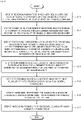
- FIG. 3 is a flowchart explaining for a biological classification method for Alzheimer's disease using a brain image according to the present disclosure.
- a step S1 of receiving a plurality of images obtained by capturing a brain of a patient by the image receiving unit 10 is performed.
- the plurality of images 11 received by the image receiving unit 10 may include an MRI image related to the brain of the patient, an amyloid PET image and a tau PET image related to the brain of the patient.
- a step S2 of acquiring a neurodegeneration feature with regard to the brain of the patient and the standardized uptake value ratio (SUVR) image from the plurality of images by the image processing unit 20 is performed.
- the MRI image processing unit 21 acquires the neurodegeneration feature related to the brain of the patient from the plurality of images and the A and T PET image processing units 22 and 23 may acquire SUVR images from the amyloid PET image and the tau PET image related to the brain of the patient, respectively.
- a step S3 of performing first determination of whether it is normal or abnormal with respect to a predetermined neurodegeneration feature biomarker, second determination of whether it is normal or abnormal with respect to a predetermined amyloid PET image biomarker, and third determination of whether it is normal or abnormal with respect to a predetermined tau PET image biomarker by the image analysis unit 30 is performed.
- the MRI image analysis unit 31 makes first determination of whether it is normal or abnormal with respect to a predetermined neurodegeneration feature biomarker, second determination of whether it is normal or abnormal with respect to a predetermined amyloid PET image biomarker, and third determination of whether it is normal or abnormal with respect to a predetermined tau PET image biomarker.
- step S4 of performing biological classification of the patient related to Alzheimer's disease using the first determination, the second determination, and the third determination by the classifying unit 40 is performed.
- the biological classification determined by the classifying unit 40 includes first classification indicating that a patient is in a normal stage, second classification indicating that the patient corresponds to an early stage of Alzheimer's disease, third classification indicating that the patient corresponds to Alzheimer's disease, fourth classification indicating that the patient has another pathology as well as Alzheimer's disease, and fifth classification indicating that the patient has a pathology other than Alzheimer's disease.
- FIG. 4 is a view explaining for a function of an image processing unit according to the present disclosure.
- the image processing unit 20 may acquire neurodegeneration feature related to a brain of a patient and a standardized uptake value ratio (SUVR) image from a plurality of images.
- SUVR standardized uptake value ratio
- the image processing unit 20 may include an MRI image processing unit 21, an amyloid PET image processing unit 22, and a tau PET image processing unit 23.
- the MRI image processing unit 21 may acquire neurodegeneration feature related to the brain of the patient from a plurality of images.
- the amyloid PET image processing unit 22 may acquire a first SUVR image from the amyloid PET image related to the brain of the patient.
- the tau PET image processing unit 23 may acquire a second SUVR image from the tau PET image related to the brain of the patient.
- the MRI image processing unit 21 classifies the entire region of the brain into a plurality of regions based on the MRI image related to the brain of the patient and the amyloid PET image processing unit 22 and the tau PET image processing unit 23 acquire the first SUVR image and the second SUVR image, as well as the neurodegeneration feature, from the plurality of classified brain regions.
- FIG. 5 is a view explaining for a function of an MRI image processing unit according to the present disclosure.
- the image receiving unit 10 receives the MRI image, and the MRI image processing unit 21 classifies the entire region of the brain into a plurality of regions (b), based on the MRI image related to the brain of the patient transmitted from the image receiving unit 10 (a), and generates the neurodegeneration feature from the plurality of classified brain regions (c).
- FIG. 6 illustrates an example of a block diagram of an MRI image processing unit according to the present disclosure.
- the MRI image processing unit 21 may include a deep neural network module 21a, a classification module 21b, and an analysis module 21c.
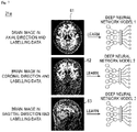
- FIG. 7 is a view explaining for a function of a trained deep neural network module according to the present disclosure.
- FIGS. 8A and 8B are views explaining for a process of classifying a brain region into a plurality of regions based on a trained deep neural network module by an image processing unit and acquiring a neurodegeneration feature, according to the present disclosure.
- the deep neural network module 21a is trained using at least one of a first model trained with a brain image in an axial direction and labelling data 61, a second model trained with a brain image in a coronal direction and the labelling data 62, and a third model trained with a brain image in a sagittal direction and the labelling data 63.
- the deep neural network module 21a may classify the entire region of the brain of a subject into a plurality of regions 21b using any one of a first MRI image 61a classified with respect to the axial direction by the first model, a second MRI image 61b classified with respect to the coronal direction by the second model, and a third MRI image 61c classified with respect to the sagittal direction by the third model.
- an image which is three-dimensionally reconstructed may be used by using the results of all the first model, the second model, and the third model.
- the deep neural network module 21a may three-dimensionally reconstruct the MRI image using all a first MRI image classified with respect to the axial direction by the first model, a second MRI image classified with respect to the coronal direction by the second model, and a third MRI image classified with respect to the sagittal direction by the third model.
- the classification of the entire region of the brain of the patient into a plurality of regions based on the MRI image transmitted from the deep neural network module 21a by the classification module 21 will be described in more detail.
- the classification module 21b may classify the entire region of the brain of the patient into 95 classes based on the three-dimensionally reconstructed MRI image by means of the deep neural network module 21a.
- the classification module 21b may reclassify a hippocampus region among 95 classes into 13 sub regions again.
- the classification module 21b may reclassify the region which is classified into 95 classes and the hippocampus region which is classified into 13 sub regions again into a composite region by at least one of a task of additionally classifying the regions into two or more regions and a task of composing two or more of the classified regions.
- the classification module 21b may select and reclassify only regions related to a predetermined brain disease excluding regions which are not related to the predetermined brain disease from the region which is classified into 95 classes and the hippocampus region which is classified into 13 sub regions again.
- the analysis module 21c acquires the neurodegeneration feature related to the brain of the patient based on the plurality of classified brain regions.
- the analysis module 21c may generate a neurodegeneration feature map based on the classified brain region.
- the analysis module 21c may acquire the neurodegeneration feature from the neurodegeneration feature map and the neurodegeneration feature may include a cortical thickness, a volume, a surface area, and a gyrification index.
- the analysis module 21c may use at least one of the region, which is classified into 95 classes and the hippocampus region, which is classified into 13 sub regions again by the classification module 21b.
- the analysis module 21c may calculate a regional volume from the region, which is classified into 95 classes, a subfield volume from the hippocampus region, which is classified into 13 sub regions again, and a composite region volume from the composite region.
- Amyloid PET image processing unit and tau PET image processing unit are associated with Amyloid PET image processing unit and tau PET image processing unit
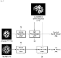
- FIG. 9 is a view explaining for a process of acquiring an SUVR image from an amyloid PET image and a tau PET image based on a plurality of classified brain regions, according to the present disclosure.
- the plurality of input images 11 may include the MRI image 12 related to the brain of the patient, the amyloid PET image 13 and the tau PET image 14 related to the brain of the patient.
- the SUVR image may include a first SUVR image 68 acquired by the amyloid PET image processing unit 22 and a second SUVR image 69 acquired by the tau PET image processing unit 23.
- the MRI image processing unit 21 classifies the entire region of the brain of the patient into a plurality of regions and the amyloid PET image processing unit 22 and the tau PET image processing unit 23 may acquire the first SUVR image 68 and the second SUVR image 69 from the plurality of classified brain regions.
- FIG. 10 is a view explaining for a process of acquiring an SUVR image based on region of interest (ROI) information in accordance with an operation of an MRI image processing unit in a plurality of classified brain regions, according to the present disclosure.
- ROI region of interest
- the amyloid PET image processing unit 22 and the tau PET image processing unit 23 may acquire a first SUVR image 68 and a second SUVR image 69 based on region of interest (ROI) information 70 according to the operation of the MRI image processing unit 21 from the plurality of classified brain regions.
- ROI region of interest
- the ROI (Region of Interest) information 70 is utilized during a process of acquiring specific information 67 such as a cortical thickness, a volume, a surface area, a gyrification index, a volume for every region from the classified region, hippocampus subfield volume, or a composite region volume by the MRI image processing unit 21.
- the ROI 70 applied in FIG. 10 may include a cerebellum gray matter region, a cerebellum white matter region, a whole cerebellum region, a pons region, and a brainstem region.
- the ROI which is used by the amyloid PET image processing unit 22 and the tau PET image processing unit 23 may vary depending on a tracer of the amyloid PET image and the tau PET image.
- amyloid PET image processing unit 22 and the tau PET image processing unit 23 may perform a predetermined pre-processing process.
- the pre-processing process may include partial volume correction (PVC) processing and co-registration processing.
- PVC partial volume correction
- the partial volume correction (PVC) processing is performed to correct a spill-out phenomenon that an image is blurred due to a resolution lower than a predetermined reference by the influence of a partial volume effect so that a concentration is measured to be low and a spill-in phenomenon that when the concentration around the region of interest is high, the concentration is measured to be higher than an actual concentration in the region of interest.
- the partial volume correction (PVC) processing method may include a geometric transfer matrix method and a Muller-Gartner method.
- the image analysis unit 30 may determine whether it is normal or abnormal with respect to a plurality of predetermined biomarkers.
- the image analysis unit 30 may include an MRI image analysis unit 31, an amyloid PET image analysis unit 32, and a tau PET image analysis unit 33.
- the MRI image analysis unit 31 makes first determination of whether it is normal or abnormal with respect to a predetermined neurodegeneration feature biomarker.
- amyloid PET image analysis unit 32 makes second determination of whether it is normal or abnormal with respect to a predetermined amyloid PET image biomarker.
- the tau PET image analysis unit 33 makes third determination of whether it is normal or abnormal with respect to a predetermined tau PET image biomarker.
- the classifying unit 40 performs biological classification of the patient related to Alzheimer's disease using the first determination, the second determination, and the third determination.
- the biological classification performed by the classifying unit includes first classification indicating that a patient is a normal stage, second classification indicating that the patient is in an early stage of Alzheimer's disease, third classification indicating that the patient corresponds to Alzheimer's disease, fourth classification indicating that the patient has another pathology as well as Alzheimer's disease, and fifth classification indicating that the patient has a pathology other than Alzheimer's disease.
- FIG. 11 is a table obtained by summarizing biological classification contents performed by a classifying unit as a table, according to the present disclosure.
- the classifying unit 40 performs first classification indicating that the patient is a normal stage when the first determination is normal with respect to the neurodegeneration feature biomarker, the second determination is normal with respect to the amyloid PET image biomarker, and the third determination is normal with respect to the tau PET image biomarker (81).
- the classifying unit 40 performs second classification indicating that the patient corresponds to an early stage of Alzheimer's disease when the first determination is normal with respect to the neurodegeneration feature biomarker, the second determination is abnormal with respect to the amyloid PET image biomarker, and the third determination is normal with respect to the tau PET image biomarker (82).
- the classifying unit 40 performs third classification indicating that the patient corresponds to Alzheimer's disease when the first determination is normal with respect to the neurodegeneration feature biomarker, the second determination is abnormal with respect to the amyloid PET image biomarker, and the third determination is abnormal with respect to the tau PET image biomarker (83).
- the classifying unit 40 performs third classification indicating that the patient corresponds to Alzheimer's disease when the first determination is abnormal with respect to the neurodegeneration feature biomarker, the second determination is abnormal with respect to the amyloid PET image biomarker, and the third determination is abnormal with respect to the tau PET image biomarker (84).
- the classifying unit 40 performs fourth classification indicating that the patient has another pathology as well as a pathology of Alzheimer's disease when the first determination is abnormal with respect to the neurodegeneration feature biomarker, the second determination is abnormal with respect to the amyloid PET image biomarker, and the third determination is normal with respect to the tau PET image biomarker (85).
- the classifying unit 40 performs fifth classification indicating that the patient has a pathology other than Alzheimer's disease when the first determination is normal with respect to the neurodegeneration feature biomarker, the second determination is normal with respect to the amyloid PET image biomarker, and the third determination is abnormal with respect to the tau PET image biomarker (86).
- the classifying unit 40 performs fifth classification indicating that the patient has a pathology other than Alzheimer's disease when the first determination is abnormal with respect to the neurodegeneration feature biomarker, the second determination is normal with respect to the amyloid PET image biomarker, and the third determination is normal with respect to the tau PET image biomarker (87).
- the classifying unit 40 performs fifth classification indicating that the patient has a pathology other than Alzheimer's disease when the first determination is abnormal with respect to the neurodegeneration feature biomarker, the second determination is normal with respect to the amyloid PET image biomarker, and the third determination is abnormal with respect to the tau PET image biomarker (88).
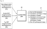
- FIG. 12 is a view illustrating a process of biologically classifying Alzheimer's disease by analyzing an MRI image, an amyloid PET image, and a tau PET image related to the brain, according to the present disclosure.
- the classifying unit 40 receives a first determination result from the MRI image analysis unit 31, a second determination result from the amyloid PET image analysis unit 32, and a third determination result from the tau PET image analysis unit 33.
- the classifying unit 40 performs biological classification including the first classification indicating that the patient is a normal stage, the second classification indicating that the patient corresponds to an early stage of Alzheimer's disease, the third classification indicating that the patient corresponds to Alzheimer's disease, the fourth classification indicating that the patient has another pathology as well as a pathology of Alzheimer's disease, and the fifth classification indicating that the patient is a pathology other than Alzheimer's disease, as illustrated in FIG. 11 , by combining the first determination, the second determination, and the third determination of whether it is normal.
- the above-described biological classification device and method for Alzheimer's disease using a brain image according to the present disclosure are utilized to screen the patient group and the normal group to increase a probability of successful clinical trials.
- the present disclosure may provide a device, a system, and a method which increase a probability of successful clinical trials by utilizing the biological classification device and method for Alzheimer's disease using a brain image to screen a patient group and a normal group.
- a result of clinical trials for demonstration of drug efficacy is determined by showing a statistical significance indicating whether to achieve a predicted expected effect for clinical trial participants.
- the biological classification device and method for Alzheimer's disease using a brain image according to the present disclosure are applied, only Alzheimer's disease patients exactly targeted by new drugs are included as clinical trial subjects so that the probability of successful clinical trials may be increased as much as possible.
- a result of clinical trials for demonstration of drug efficacy is determined by showing a statistical significance indicating whether to achieve a predicted expected effect for clinical trial participants.
- a numerical value of an evaluation scale needs to be statistically significantly increased before and after medication or as compared to a placebo group.
- Alzheimer's disease patients who are exactly targeted by the new drug are included as subjects of the clinical trials to increase a probability of successful clinical trials as much as possible.
- Alzheimer's disease since it takes a long time to confirm the Alzheimer's disease, a screening test is difficult so that there is a problem in that it is very difficult to include only the Alzheimer's disease patients targeted by new drugs as subjects of clinical trials.
- the biological classification device and method for Alzheimer's disease using a brain image proposed by the present disclosure are utilized to screen the patient group and the normal group to increase a probability of successful clinical trials.
- FIG. 13 is a view explaining for a method of increasing a probability of successful clinical trials by utilizing biological classification of Alzheimer's disease using a brain image to screen a patient group and a normal group, according to the present disclosure.
- FIG. 13 a method of increasing a probability of successful clinical trials by screening a patient group using a biological classification device for Alzheimer's disease using a brain image including a central control unit (not illustrated) as well as the image receiving unit 10, the image processing unit 20, the image analysis unit 30, and the classifying unit 40 described above.
- a step S11 of receiving a plurality of images obtained by capturing brains of a plurality of patients which is a candidate group of a clinical trial for proving the drug efficacy by the image receiving unit 10 is performed.
- a step S12 of acquiring a neurodegeneration feature with regard to the brains of the plurality of patients and the standardized uptake value ratio (SUVR) image from the plurality of images by the image processing unit 20 is performed.
- a step S13 of performing first determination of whether it is normal or abnormal with respect to a predetermined neurodegeneration feature biomarker, second determination of whether it is normal or abnormal with respect to a predetermined amyloid PET image biomarker, and third determination of whether it is normal or abnormal with respect to a predetermined tau PET image biomarker is performed by the image analysis unit 30.
- the classifying unit 40 performs biological classification of the plurality of patient related to Alzheimer's disease using the first determination, the second determination, and the third determination (S14).
- a step S15 of providing the biological classification information of the plurality of patients from the classifying unit 40 to the central control unit (not illustrates) is performed.
- the central control unit may screen a first patient for the clinical trial based on the biological classification information of the plurality of patients (S16).
- the clinical trial is performed on the screened patient group to increase the probability of successful clinical trials (S17).
- Alzheimer's disease patients who are exactly targeted by the new drugs are included as a clinical trial subject so that the probability of successful clinical trials may be increased as much as possible.
- the biological classification device and method for Alzheimer's disease using a brain image according to the present disclosure are utilized to screen the patient group and the normal group to increase a probability of successful clinical trials.
- steps S1 to S4 may be independently performed by the biological classification device for Alzheimer's disease 1 using a brain image or may be applied by providing a separate server (not illustrated) or a separate central control device (not illustrated) to perform the entire operations together with the biological classification device for Alzheimer's disease 1.
- the second method explains a method of using a separate server (not illustrated).
- FIG. 14 is a view explaining for another method of increasing a probability of successful clinical trials by utilizing biological classification of Alzheimer's disease using a brain image to screen a patient group and a normal group, according to the present disclosure.
- Steps S21 to S24 of FIG. 14 correspond to steps S11 to S14 of FIG. 13 which have been described above so that a detailed description will be omitted for the purpose of simplicity of the description.
- a step S25 of providing biological classification information of the plurality of patients from the classifying unit 40 to a server (not illustrated) by wireless or wired communication is performed.
- the server may screen a first patient for the clinical trial based on the biological classification information of the plurality of patients (S26).
- the clinical trials are performed on the screened patient group to increase the probability of successful clinical trials (S27) and only the Alzheimer's disease patients who are exactly targeted by the new drugs are included as clinical trial subjects to increase the probability of successful clinical trials as much as possible.
- a device and a method of performing biological classification related to Alzheimer's disease by determining and combining whether it is normal or abnormal with respect to a predetermined biomarker after acquiring a neurodegeneration feature related to the brain of a patient from an MRI image and acquiring a standardized uptake value ratio (SUVR) from an amyloid PET image and a tau PET image.
- SUVR standardized uptake value ratio
- the present disclosure provides a device and a method for performing determination of whether it is normal or abnormal with respect to a neurodegeneration feature biomarker, second determination of whether it is normal or abnormal with respect to an amyloid PET image biomarker, and determination of whether it is normal or abnormal with respect to a tau PET image biomarker, and performing biological classification with respect to Alzheimer's disease based on a combination of three determination results.
- the present disclosure provides a biological classification device and method including first classification indicating that a patient is a normal stage, second classification indicating that the patient corresponds to an early stage of Alzheimer's disease, third classification indicating that the patient corresponds to Alzheimer's disease, fourth classification indicating that the patient has another pathology as well as Alzheimer's disease, and fifth classification indicating that the patient has a pathology other than Alzheimer's disease to a user.
- the present disclosure provides a device and a method of classifying an entire region of the brain into a plurality of regions based on an MRI image, acquiring a neurodegeneration feature from the plurality of classified brain regions and, at the same time, acquiring an SUVR image from an amyloid PET image and an SUVR image from a tau PET image based on the plurality of classified brain regions to a user.
- the present disclosure provides a device and a method of classifying and analyzing a brain of a patient into a plurality of regions based on an MRI image by applying a deep neural network module trained using at least one of a first model trained with a brain image in an axial direction and labelling data, a second model trained with a brain image in a coronal direction and the labelling data, and a third model trained with a brain image in a sagittal direction and the labelling data to a user.
- the present disclosure provides a device and a method of acquiring an SUVR image from an amyloid PET image and a tau PET image based on region of interest (ROI) information acquired from an operation of a device of processing an MRI image in a plurality of classified brain regions to a user.
- ROI region of interest
- the present disclosure provides a device and a method of performing pre-processing such as partial volume correction (PVC) processing and co-registration processing, with regard to an amyloid PET image and a tau PET image to a user.
- pre-processing such as partial volume correction (PVC) processing and co-registration processing
- the present disclosure provides a device, a system, and a method which increase a probability of successful clinical trials by utilizing biological classification of Alzheimer's disease using a brain image to screen a patient group and a normal group.
- the above-described exemplary embodiments of the present invention may be implemented through various methods.
- the exemplary embodiments of the present disclosure may be implemented by a hardware, a firm ware, a software, and a combination thereof.
- the method according to the exemplary embodiment of the present disclosure may be implemented by one or more application specific integrated circuits(ASICs), digital signal processors (DSPs), digital signal processing devices(DSPDs), programmable logic devices(PLDs), field programmable gate arrays(FPGAs), a processor, a controller, a microcontroller, or a microprocessor.
- ASICs application specific integrated circuits
- DSPs digital signal processors
- DSPDs digital signal processing devices
- PLDs programmable logic devices
- FPGAs field programmable gate arrays
- the method according to the exemplary embodiment of the present disclosure may be implemented by a module, a procedure, or a function which performs a function or operations described above.
- the software code is stored in the memory unit to be driven by the processor.
- the memory unit is located inside or outside the processor and exchanges data with the processor, by various known units.
Landscapes
- Health & Medical Sciences (AREA)
- Engineering & Computer Science (AREA)
- Life Sciences & Earth Sciences (AREA)
- Medical Informatics (AREA)
- Public Health (AREA)
- General Health & Medical Sciences (AREA)
- Biomedical Technology (AREA)
- Physics & Mathematics (AREA)
- Pathology (AREA)
- Nuclear Medicine, Radiotherapy & Molecular Imaging (AREA)
- Radiology & Medical Imaging (AREA)
- Neurology (AREA)
- Molecular Biology (AREA)
- Veterinary Medicine (AREA)
- Animal Behavior & Ethology (AREA)
- Surgery (AREA)
- Biophysics (AREA)
- Heart & Thoracic Surgery (AREA)
- Epidemiology (AREA)
- Primary Health Care (AREA)
- Computer Vision & Pattern Recognition (AREA)
- Databases & Information Systems (AREA)
- Theoretical Computer Science (AREA)
- Artificial Intelligence (AREA)
- Psychiatry (AREA)
- Physiology (AREA)
- Data Mining & Analysis (AREA)
- General Physics & Mathematics (AREA)
- Quality & Reliability (AREA)
- Psychology (AREA)
- Neurosurgery (AREA)
- Evolutionary Computation (AREA)
- Signal Processing (AREA)
- Child & Adolescent Psychology (AREA)
- Developmental Disabilities (AREA)
- Hospice & Palliative Care (AREA)
- Software Systems (AREA)
- Computing Systems (AREA)
- Mathematical Physics (AREA)
- Multimedia (AREA)
Applications Claiming Priority (1)
| Application Number | Priority Date | Filing Date | Title |
|---|---|---|---|
| KR1020200155063A KR102321601B1 (ko) | 2020-11-19 | 2020-11-19 | 뇌 영상을 이용한 알츠하이머병의 생물학적 분류 장치 및 방법 |
Publications (1)
| Publication Number | Publication Date |
|---|---|
| EP4002201A1 true EP4002201A1 (de) | 2022-05-25 |
Family
ID=74187082
Family Applications (1)
| Application Number | Title | Priority Date | Filing Date |
|---|---|---|---|
| EP21208975.9A Pending EP4002201A1 (de) | 2020-11-19 | 2021-11-18 | Vorrichtung und verfahren zur biologischen klassifizierung von morbus alzheimer unter verwendung eines multimodalen gehirnbildes |
Country Status (5)
| Country | Link |
|---|---|
| US (1) | US20220151539A1 (de) |
| EP (1) | EP4002201A1 (de) |
| JP (1) | JP7101856B2 (de) |
| KR (1) | KR102321601B1 (de) |
| CN (1) | CN114343605B (de) |
Families Citing this family (9)
| Publication number | Priority date | Publication date | Assignee | Title |
|---|---|---|---|---|
| CA3046939A1 (en) * | 2016-12-21 | 2018-06-28 | Innereye Ltd. | System and method for iterative classification using neurophysiological signals |
| KR102649096B1 (ko) * | 2021-11-23 | 2024-03-18 | 연세대학교 산학협력단 | Pet 영상 변환 방법 및 이를 이용한 뇌질환 진단 방법 |
| KR102759077B1 (ko) * | 2022-02-08 | 2025-01-23 | 고려대학교 산학협력단 | Mri를 기반으로 알츠하이머 관련 바이오마커 축적 정도를 예측하는 방법 및 이를 이용한 동반진단 방법 |
| KR102451108B1 (ko) * | 2022-02-22 | 2022-10-06 | 뉴로핏 주식회사 | 치매 진단에 필요한 정보 제공 방법 및 장치 |
| KR102728495B1 (ko) * | 2022-04-11 | 2024-11-11 | 고려대학교 산학협력단 | 자기공명 영상의 딥러닝 판독 기술에 기반한 다양한 퇴행성 뇌질환 감별진단 방법 및 장치 |
| KR102784896B1 (ko) * | 2022-06-23 | 2025-03-24 | 고려대학교 산학협력단 | 멀티모달 뇌 영상 기반 뇌질환 바이오마커 추출 방법론 및 파이프라인 |
| CN116245833B (zh) * | 2023-02-13 | 2024-12-27 | 复旦大学 | 一种大脑多模态pet影像suvr的预测方法 |
| TWI860680B (zh) | 2023-04-11 | 2024-11-01 | 臺北醫學大學 | 腦部類澱粉蛋白正子造影處理系統及其運作方法 |
| WO2025038966A1 (en) * | 2023-08-17 | 2025-02-20 | University Of Louisville Research Foundation, Inc. | Computer-aided system for classification of neurological disorders |
Citations (5)
| Publication number | Priority date | Publication date | Assignee | Title |
|---|---|---|---|---|
| KR20160107831A (ko) * | 2015-03-05 | 2016-09-19 | 재단법인 아산사회복지재단 | 퇴행성 뇌질환 진단에 적합한 이미지 바이오 마커를 획득하는 방법, 장치 및 이를 이용하여 퇴행성 뇌질환을 진단하는 방법 |
| WO2017011746A1 (en) * | 2015-07-15 | 2017-01-19 | Adm Diagnostics, Llc | System and methods for determining a brain condition of a patient sybject to multiple disease states |
| KR101995383B1 (ko) | 2018-10-02 | 2019-07-02 | 주식회사 뷰노 | 의료 영상에 대한 특징 순위에 기반하여 뇌질환을 판정하는 방법 및 이를 이용한 장치 |
| KR102020157B1 (ko) | 2018-11-12 | 2019-11-04 | 사회복지법인 삼성생명공익재단 | Flair 기반 병변 검출과 현황 파악 방법 및 시스템 |
| US20200345746A1 (en) * | 2016-07-19 | 2020-11-05 | Novartis Ag | Oxazine Derivative for Use in the Prevention of Alzheimer's Disease in at Risk Patents |
Family Cites Families (7)
| Publication number | Priority date | Publication date | Assignee | Title |
|---|---|---|---|---|
| WO2009146388A1 (en) * | 2008-05-28 | 2009-12-03 | The Trustees Of Columbia University In The City Of New York | Voxel-based methods for assessing subjects using positron emission tomography |
| EP3464350A1 (de) * | 2016-06-07 | 2019-04-10 | Biogen International Neuroscience GmbH | Verfahren zur behandlung von morbus alzheimer |
| KR101950815B1 (ko) | 2017-08-25 | 2019-02-21 | 뉴로핏 주식회사 | 패치 가이드 방법 및 프로그램 |
| CN111247595B (zh) * | 2017-10-31 | 2024-07-23 | 通用电气健康护理有限公司 | 用于诊断认知疾病病理和/或结果的医疗系统 |
| JP6705528B2 (ja) * | 2019-04-08 | 2020-06-03 | 大日本印刷株式会社 | 医用画像表示処理方法、医用画像表示処理装置およびプログラム |
| JP7574181B2 (ja) | 2019-04-26 | 2024-10-28 | エーザイ・アール・アンド・ディー・マネジメント株式会社 | 診断支援装置、推測装置、診断支援システム、診断支援方法、診断支援プログラム及び学習済みモデル |
| US11751832B2 (en) * | 2020-01-30 | 2023-09-12 | GE Precision Healthcare LLC | CTA large vessel occlusion model |
-
2020
- 2020-11-19 KR KR1020200155063A patent/KR102321601B1/ko active Active
-
2021
- 2021-11-18 EP EP21208975.9A patent/EP4002201A1/de active Pending
- 2021-11-18 JP JP2021187865A patent/JP7101856B2/ja active Active
- 2021-11-18 US US17/529,852 patent/US20220151539A1/en not_active Abandoned
- 2021-11-19 CN CN202111400639.5A patent/CN114343605B/zh active Active
Patent Citations (5)
| Publication number | Priority date | Publication date | Assignee | Title |
|---|---|---|---|---|
| KR20160107831A (ko) * | 2015-03-05 | 2016-09-19 | 재단법인 아산사회복지재단 | 퇴행성 뇌질환 진단에 적합한 이미지 바이오 마커를 획득하는 방법, 장치 및 이를 이용하여 퇴행성 뇌질환을 진단하는 방법 |
| WO2017011746A1 (en) * | 2015-07-15 | 2017-01-19 | Adm Diagnostics, Llc | System and methods for determining a brain condition of a patient sybject to multiple disease states |
| US20200345746A1 (en) * | 2016-07-19 | 2020-11-05 | Novartis Ag | Oxazine Derivative for Use in the Prevention of Alzheimer's Disease in at Risk Patents |
| KR101995383B1 (ko) | 2018-10-02 | 2019-07-02 | 주식회사 뷰노 | 의료 영상에 대한 특징 순위에 기반하여 뇌질환을 판정하는 방법 및 이를 이용한 장치 |
| KR102020157B1 (ko) | 2018-11-12 | 2019-11-04 | 사회복지법인 삼성생명공익재단 | Flair 기반 병변 검출과 현황 파악 방법 및 시스템 |
Non-Patent Citations (3)
| Title |
|---|
| CHOI MYUNGWON ET AL: "Comparison of neurodegenerative types using different brain MRI analysis metrics in older adults with normal cognition, mild cognitive impairment, and Alzheimer's dementia", PLOS ONE, vol. 14, no. 8, 1 August 2019 (2019-08-01), US, pages e0220739, XP093248224, ISSN: 1932-6203, DOI: 10.1371/journal.pone.0220739 * |
| JACK CLIFFORD R ET AL: "NIA-AA Research Framework: Toward a biological definition of Alzheimer's disease", ALZHEIMER'S & DEMENTIA: THE JOURNAL OF THE ALZHEIMER'SASSOCIATION, ELSEVIER, NEW YORK, NY, US, vol. 14, no. 4, 10 April 2018 (2018-04-10), pages 535 - 562, XP085378277, ISSN: 1552-5260, DOI: 10.1016/J.JALZ.2018.02.018 * |
| ZHENG WEIHAO ET AL: "Topological Characterization of the Multi-feature based Network in Patients with Alzheimer's Disease and Mild Cognitive Impairment", 2019 IEEE INTERNATIONAL CONFERENCE ON BIOINFORMATICS AND BIOMEDICINE (BIBM), IEEE, 18 November 2019 (2019-11-18), pages 1162 - 1167, XP033704051, DOI: 10.1109/BIBM47256.2019.8983218 * |
Also Published As
| Publication number | Publication date |
|---|---|
| CN114343605A (zh) | 2022-04-15 |
| US20220151539A1 (en) | 2022-05-19 |
| KR102321601B1 (ko) | 2021-11-05 |
| CN114343605B (zh) | 2022-12-13 |
| JP2022081457A (ja) | 2022-05-31 |
| JP7101856B2 (ja) | 2022-07-15 |
Similar Documents
| Publication | Publication Date | Title |
|---|---|---|
| US20220151539A1 (en) | Biological classification device and method for alzheimer's disease using multimodal brain image | |
| Putcha et al. | Fractionating the Rey Auditory Verbal Learning Test: Distinct roles of large-scale cortical networks in prodromal Alzheimer's disease | |
| Shakir et al. | Advances in deep neuropathological phenotyping of Alzheimer disease: past, present, and future | |
| Zhang et al. | Progression of regional microstructural degeneration in Parkinson’s disease: a multicenter diffusion tensor imaging study | |
| Anor et al. | The longitudinal assessment of neuropsychiatric symptoms in mild cognitive impairment and Alzheimer’s disease and their association with white matter hyperintensities in the National Alzheimer’s Coordinating Center’s uniform data set | |
| Ferraro et al. | Multimodal structural MRI in the diagnosis of motor neuron diseases | |
| US20230245772A1 (en) | A Machine Learning System and Method for Predicting Alzheimer's Disease Based on Retinal Fundus Images | |
| JP7600138B2 (ja) | 病状によって影響を受ける可能性がある被験者を識別するための方法およびシステム | |
| RU2020114290A (ru) | Медицинская система для диагностики патологии и / или исхода когнитивного заболевания | |
| Ferreira et al. | Electroencephalography is a good complement to currently established dementia biomarkers | |
| KR102288077B1 (ko) | 뉴로멜라닌 영역의 볼륨을 이용한 파킨슨 병 진단을 위한 정보제공 방법 | |
| Zhang et al. | Parkinson’s Progression Markers Initiative (2016). Progression of regional microstructural degeneration in Parkinson’s disease: a multicenter diffusion tensor imaging study | |
| EP3761871A1 (de) | Verfahren zur frühzeitigen vorhersage von neurodegenerativem verfall | |
| KR101989695B1 (ko) | 뇌 아밀로이드 축적 양성 예측 모델 및 이를 이용한 예측 방법 | |
| WO2020179950A1 (ko) | 딥러닝 기반 뇌 질환 진행 예측 방법 및 장치 | |
| Parra et al. | Exploring the association between amyloid-β and memory markers for Alzheimer’s disease in cognitively unimpaired older adults | |
| RU2425374C2 (ru) | Устройство и способ для количественной оценки полипептида, содержащегося в пробе жидкости организма, а также маркер для выявления патологических состояний | |
| Neophytou et al. | The right hemisphere’s capacity for language: evidence from primary progressive aphasia | |
| Engedal et al. | Diagnosis of dementia–automatic quantification of brain structures | |
| Rezende et al. | RFC1‐related disorder: in vivo evaluation of spinal cord damage | |
| Slaby et al. | Properties of white matter tract diffusivity in children with developmental dyslexia and comorbid attention deficit/hyperactivity disorder | |
| KR20180118948A (ko) | 대각선 귓불주름을 이용한 퇴행성 뇌질환 진단 방법 | |
| Jann et al. | Classifying mild cognitive impairment from normal cognition: fMRI complexity matches tau PET performance | |
| Van Gils et al. | Separating dementia with Lewy bodies from Alzheimer’s disease dementia using a volumetric MRI classifier | |
| Gong et al. | Early axonal degeneration linked to clinical decline in Alzheimer's disease progression revealed with diffusion MRI |
Legal Events
| Date | Code | Title | Description |
|---|---|---|---|
| PUAI | Public reference made under article 153(3) epc to a published international application that has entered the european phase |
Free format text: ORIGINAL CODE: 0009012 |
|
| STAA | Information on the status of an ep patent application or granted ep patent |
Free format text: STATUS: REQUEST FOR EXAMINATION WAS MADE |
|
| 17P | Request for examination filed |
Effective date: 20211118 |
|
| AK | Designated contracting states |
Kind code of ref document: A1 Designated state(s): AL AT BE BG CH CY CZ DE DK EE ES FI FR GB GR HR HU IE IS IT LI LT LU LV MC MK MT NL NO PL PT RO RS SE SI SK SM TR |
|
| RBV | Designated contracting states (corrected) |
Designated state(s): AL AT BE BG CH CY CZ DE DK EE ES FI FR GB GR HR HU IE IS IT LI LT LU LV MC MK MT NL NO PL PT RO RS SE SI SK SM TR |
|
| STAA | Information on the status of an ep patent application or granted ep patent |
Free format text: STATUS: EXAMINATION IS IN PROGRESS |
|
| 17Q | First examination report despatched |
Effective date: 20231212 |