EP3967331A1 - Ultrakleine fluoreszierende organische nanopartikel - Google Patents
Ultrakleine fluoreszierende organische nanopartikel Download PDFInfo
- Publication number
- EP3967331A1 EP3967331A1 EP20306027.2A EP20306027A EP3967331A1 EP 3967331 A1 EP3967331 A1 EP 3967331A1 EP 20306027 A EP20306027 A EP 20306027A EP 3967331 A1 EP3967331 A1 EP 3967331A1
- Authority
- EP
- European Patent Office
- Prior art keywords
- nps
- formula
- polymeric
- group
- radicals
- Prior art date
- Legal status (The legal status is an assumption and is not a legal conclusion. Google has not performed a legal analysis and makes no representation as to the accuracy of the status listed.)
- Withdrawn
Links
- 0 CCCC(C1)=CC1C(CC(C(CC)(CC)C(C)C(C)CC)C1(C(CC)CC=CC)C2C1CC*2)(C(CC)CC=*)C(*)(CC)CC Chemical compound CCCC(C1)=CC1C(CC(C(CC)(CC)C(C)C(C)CC)C1(C(CC)CC=CC)C2C1CC*2)(C(CC)CC=*)C(*)(CC)CC 0.000 description 7
- PIZZBDLGMMZDMB-UHFFFAOYSA-N Nc(c(CCCc(c(N)c(c(N)c1N)N)c1N)c(c(N)c1N)N)c1N Chemical compound Nc(c(CCCc(c(N)c(c(N)c1N)N)c1N)c(c(N)c1N)N)c1N PIZZBDLGMMZDMB-UHFFFAOYSA-N 0.000 description 1
Images







Classifications
-
- A—HUMAN NECESSITIES
- A61—MEDICAL OR VETERINARY SCIENCE; HYGIENE
- A61K—PREPARATIONS FOR MEDICAL, DENTAL OR TOILETRY PURPOSES
- A61K49/00—Preparations for testing in vivo
- A61K49/001—Preparation for luminescence or biological staining
- A61K49/0013—Luminescence
- A61K49/0017—Fluorescence in vivo
- A61K49/005—Fluorescence in vivo characterised by the carrier molecule carrying the fluorescent agent
- A61K49/0054—Macromolecular compounds, i.e. oligomers, polymers, dendrimers
-
- A—HUMAN NECESSITIES
- A61—MEDICAL OR VETERINARY SCIENCE; HYGIENE
- A61K—PREPARATIONS FOR MEDICAL, DENTAL OR TOILETRY PURPOSES
- A61K49/00—Preparations for testing in vivo
- A61K49/001—Preparation for luminescence or biological staining
- A61K49/0013—Luminescence
- A61K49/0017—Fluorescence in vivo
- A61K49/0019—Fluorescence in vivo characterised by the fluorescent group, e.g. oligomeric, polymeric or dendritic molecules
- A61K49/0021—Fluorescence in vivo characterised by the fluorescent group, e.g. oligomeric, polymeric or dendritic molecules the fluorescent group being a small organic molecule
-
- A—HUMAN NECESSITIES
- A61—MEDICAL OR VETERINARY SCIENCE; HYGIENE
- A61K—PREPARATIONS FOR MEDICAL, DENTAL OR TOILETRY PURPOSES
- A61K49/00—Preparations for testing in vivo
- A61K49/001—Preparation for luminescence or biological staining
- A61K49/0063—Preparation for luminescence or biological staining characterised by a special physical or galenical form, e.g. emulsions, microspheres
- A61K49/0069—Preparation for luminescence or biological staining characterised by a special physical or galenical form, e.g. emulsions, microspheres the agent being in a particular physical galenical form
- A61K49/0076—Preparation for luminescence or biological staining characterised by a special physical or galenical form, e.g. emulsions, microspheres the agent being in a particular physical galenical form dispersion, suspension, e.g. particles in a liquid, colloid, emulsion
- A61K49/0082—Preparation for luminescence or biological staining characterised by a special physical or galenical form, e.g. emulsions, microspheres the agent being in a particular physical galenical form dispersion, suspension, e.g. particles in a liquid, colloid, emulsion micelle, e.g. phospholipidic micelle and polymeric micelle
Definitions
- the present invention relates to organic nanoparticles (NPs).
- the present invention relates to a fluorescent organic nanoparticles (FONs).
- the present invention relates to the preparation of NPs or FONs, their properties and their applications.
- Fluorescent organic nanoparticles are attractive alternative to the less biocompatible inorganic based nanoparticles.
- FONs Fluorescent organic nanoparticles
- Nanoparticles (NPs) of small size become more and more important for in vivo applications due to their better penetration into target tumors, proper elimination and lower propensity to accumulate into liver (- Cabral, H.; Matsumoto, Y.; Mizuno, K.; Chen, Q.; Murakami, M.; Kimura, M.; Terada, Y.; Kano, M. R.; Miyazono, K.; Uesaka, M.; Nishiyama, N.; Kataoka, K.
- Smallest dye-loaded SiNPs of about 10 nm core are also around 20 nm hydrodynamic diameter due to large PEG shell.
- All organic systems exploiting self-assembly suggest effective approaches in fabrication of ultra-small fluorescent NPs.
- Such systems are for example micellar NPs composed of amphiphilic molecules coupled to a fluorophore.
- the micelle is polymerized in order to prevent its dissociation in biological media.
- the micelles are composed of polymer chains which ensures a high stability in biological media.
- An alternative approach that mimics micellar assembly is to directly use an amphiphilic polymer that can fold as a single molecule into micelle-like structure.
- PMAO is a low-cost amphiphilic polymer widely used to wrap nanocrystals including QDs, iron and gold nanoparticles in order to make them water-soluble
- the present invention aims to solve the technical problem of providing monomolecular nanoparticles formed by self-folding of an amphiphilic polymer.
- the present invention aims to solve the technical problem of providing nanoparticles having of small and uniform size, and preferably a size of less than 35 nm or even less than 20 nm, and preferably less than 15 nm.
- the present invention aims to solve the technical problem of providing nanoparticles having fluorescent properties.
- the present invention aims to solve the technical problem of providing nanoparticles having a high brightness value (fluorescence quantum yield ⁇ extinction coefficient), typically higher than 1,000,000 M -1 .cm -1 .
- the present invention also aims to solve the technical problem of providing such nanoparticles with reduced non-specific interactions in biological media.
- the present invention also aims to solve the technical problem of providing nanoparticles targeting a specific target, such as for example a membrane receptor.
- the present invention also aims to solve the technical problem of providing stable nanoparticles in biological media.
- the present invention relates to nanoparticles solving one or more of the technical problem explained or suggested by the present invention.
- the present invention relates to a monomolecular polymeric nanoparticle (NP) having a core and a shell, wherein said NP consists of a polymeric molecule having a lipophilic unit and a unit comprising a polyether (amphiphilic polymer), said lipophilic unit forming said core and said unit comprising a polyether forming said shell.
- NP monomolecular polymeric nanoparticle
- the present invention relates in particular to a polymeric nanoparticle (NP) having a core and a shell, wherein said NP comprises or consists of 1 to 10 polymeric molecules having a lipophilic unit and a unit comprising a polyether, said lipophilic unit forming said core and said unit comprising a polyether forming said shell, wherein said polymeric molecule has the following formula (A): wherein
- the present invention relates to a core-shell polymeric nanoparticle consisting of 2 to 10 polymeric molecules, typically of structure (A).
- said NP has from 2 to 5 polymeric molecules.
- said NP has 2 or 3 polymeric molecules.
- the present invention relates to a core-shell polymeric nanoparticle consisting of single polymeric molecule, defined as "monomolecular polymeric nanoparticle".
- said NP has 1 or 2 polymeric molecules, and typically from 1 to below 1.5 polymeric molecules (in average).
- the nanoparticle is deemed “monomolecular” when the number of polymer molecules in the NP is below 1.5.
- the nanoparticle is formed by self-folding of said polymeric molecule(s).
- the polymeric molecule has the formula (A) as defined above, said polymeric nanoparticle having a star structure (N) (see figure 13 ) with a central core and branches in the shell.
- said polymeric molecule has:
- the present invention also relates to a polymeric nanoparticle consisting of 1 to 10 polymeric molecules having the formula (A) as defined above, said polymeric nanoparticle having the following structure:
- said polymeric molecule comprises or consists of the following structure: wherein p ranges from 1 to 30, for example from 8 to 30, preferably from 10 to 25, and preferably from 10 to 20, and is for example 15, wherein *, n, Ra and Rb are as defined according to the present invention.
- the percentage of the radicals Rc being of formula (C) is ranging from 40% to 100%, preferably from 50% to 100%, based on the total number of the radicals Ra and Rb in said compound of formula (A) (referred to as "PEG loading 5%)" when X in Rc is a PEG radical or derivative).
- the percentage of the radicals Rc being of formula (C) is ranging from 80% to 100%, preferably from 90% to 100%, based on the total number of the radicals Ra and Rb in said compound of formula (A).
- the percentage of the radicals Rc being of formula (C) is of about 100%.
- the percentage of the radicals Rc being a labelling moiety or a targeting moiety is ranging from 1 to 50%, preferably from 20 to 50%, based on the total number of Ra and Rb radicals in said compound of formula (A).
- the percentage of the radicals Rc is calculated by the amount of NH2-Rc involved in the reaction compared to the amount of repeating units of the starting polymer (for example PMAO) and the final percentage of the radicals Rc is evaluated by NMR.
- Rc when Rc is a group of formula (C), Rc has the following formula (D): wherein:
- Such a structure (D) covers compounds wherein the "m” ethylene oxide units are at the terminal end linked to Y and wherein the "p" units are ethylene oxide units, propylene oxide units or a mixture of ethylene oxide and propylene oxide units.
- Rc has formula (C)
- X is chosen among PEG derivatives. More preferably, said PEG derivative is a PEG having a molecular mass Mw of 1,000 g.mol -1 or more.
- Rc represents a labelling moiety or a mixture of molecules having formula (D) wherein m is 6 and y is 35, wherein Q is a mixture of H for (EO), or CH 3 for (PO) units.
- Rc has one of the following formulae:
- Rc represents a labelling moiety or the formulae (D) wherein Q is CH 3 , m is 3 and p is 19.
- the polymeric molecule is a PEGylated PMAO (poly(maleic anhydride-alt-1-octadecene) (PMAO) with polyethylene glycol (PEG)), i.e. said polymeric molecule is a polyethylene glycolated poly(maleic anhydride-alt-1-octadecene).
- PMAO poly(maleic anhydride-alt-1-octadecene)
- PEG polyethylene glycol
- the X-Y group corresponds to the Jeffamine ® commercialized by Huntsman ® , such as JEFFAMINE ® M-1000 polyetheramine and JEFFAMINE ® M-2070 polyetheramine.
- radicals Rc independently of each other, are:
- said fluorophore moiety is selected from the group consisting of:
- the fluorophore group having or containing one of the following structures or a derivative thereof:
- a BODIPY fluorophore can be chosen among :
- BODIPY fluorophores structures are also illustrated in Figures 3 and 5 .
- the fluorophore moiety is located in the core using a hydrophobic linker. In one embodiment, the fluorophore moiety is located in the shell using a hydrophilic PEG linker. In one embodiment, the fluorophore moiety is located at the interface of the core and the shell using a short linker (from 1 to 5 carbon atoms, preferably from 1 to 3 carbon atoms).
- MRI Magnetic Resonance Imaging
- X-Ray X-Ray
- phonoacoustic contrast agents for example Magnetic Resonance Imaging (MRI), X-Ray or phonoacoustic contrast agents.
- a in the fluorophore moiety comprises from 2 to 10 carbon atoms.
- the fluorophore moiety is a cationic fluorophore moiety, preferentially rhodamine or a cyanine, with a counter-ion.
- said counter ion comprises a borate ion (B - ).
- the counter-ion is chosen among tetraphenylborate ions, preferably among fluorinated tetraphenylborate ions.
- said counter-ion is selected from the group consisting of:
- the counter-ion is in an amount ranging from 1 to 5, preferably from 2 to 3 equivalents, regarding the amount of the cationic fluorophore moiety.
- the percentage of the radicals Rc is a fluorophore ranging from 1% to 50%, preferably from 10% to 50%, based on the total number of the radicals Ra and Rb in said compound of formula (A).
- radicals Rc independently of each other, are:
- the nanoparticles have a mean diameter of less than 35 nm, preferably comprised between 5 nm and 25 nm, more preferably between 10 nm and 20 nm.
- said nanoparticles are spherical particles. It may be appreciated by Transmission Electron Microscopy (TEM).
- the nanoparticles have a mean diameter of less than 15 nm, preferably comprised between 5 nm and 15 nm, more preferably between 10 nm and 20 nm.
- Such size or dimension or diameter may be measured by dynamic light scattering (typically measured three times from three independent formulation in milliQ water at 25°C using a Malvern Zetasizer Nano ZSP (Malvern, U.K.); DLS laser was 633 nm.)
- Such size or dimension or diameter may be measured by TEM.
- Such size or dimension or diameter may be measured by fluorescence correlation spectroscopy (FCS): For Fluorescence Correlation Spectroscopy: The concentration of polymer was adjusted to 1 ⁇ M Bodipy using 80,000 M -1 cm -1 as molar extinction coefficient value, therefore providing a polymer concentration of 142 nM (7 fluorophores per polymer). The samples were placed in a 96 well plate and were measured with a FCS microscope. Excitation wavelength was 488 nm (power: XXX mW). Fluorescein (50 nM in 0.1 M NaOH) was used as a reference of size (1 nm) and brightness. The data were processed with PyCorrFit.
- FCS fluorescence correlation spectroscopy
- the dye or fluorescent moiety concentration in the NP is ranging from 10 mol% to 60 mol %, for example from 20 mol % to 50 mol%
- the nanoparticles according to the invention have a zeta potential ( ⁇ ) is higher than -80 mV, ad preferably higher than -50 mV and preferably higher than -20 mV. "Higher” means here that the zeta potential is closer to 0 mV than the given zeta potential.
- the present invention also relates to a method for preparing the nanoparticles of the present invention.
- said particles are prepared by reacting compound (I) comprising anhydride functions, said compound (I) having the following structure:
- said particles are prepared by reacting compound (Ia) comprising anhydride functions, said compound (I) having the following structure: with one or more compounds having the following formula (II): NH 2 - Rc (II) under conditions wherein NH 2 group of compound(s) (II) reacts with the anhydride of compound (la), thereby giving a compound of formula (A).
- said method of preparation of a polymeric nanoparticle comprising the steps of:
- said method comprises reacting a compound of formula (A) wherein Ra and/or Rb represent OH with NH 2 -Rc said compound(s) (II) in the presence of an activating agent for forming an amide function (N-acylation), such as for example HATU (1-[Bis(dimethylamino)methylene]-1H-1,2,3-triazolo[4,5-b]pyridinium 3-oxid hexafluorophosphate ; Hexafluorophosphate Azabenzotriazole Tetramethyl Uronium).
- HATU is typically used in the presence of N,N-diisopropylethylamine (DIPEA).
- said aqueous medium is miliQ water, buffered water (Phosphate buffer) or PBS.
- Phosphate buffer buffered water
- PBS Phosphate buffer
- An example of suitable pH is from 5 to 7.4.
- Methods to prepare a compound of formula (I), including compounds of formula (Ia), are known in the art.
- method to prepare PMAO are known in the art.
- Such method could be radical polymerization, for example by using a polymerization initiator which generate a radical at both terminal ends.
- polymerization initiators and generated radicals are as follows: Initiator Radical Generated Benzoyl peroxide (BPO) Di- tert -butyl peroxide (DTBP) Azobisisobutyronitrile (AIBN)
- the polymeric molecules according to the present invention are capable of self-folding in aqueous media and form the monomolecular NPs as defined in the present invention.
- Polymer self-folding is more prone to provide monomolecular NPs with increasing number of radicals Rc comprising a polyether (PEG).
- nanoparticles according to the present invention are stable.
- the present invention also relates to an aqueous composition
- an aqueous composition comprising:
- said composition is an injectable composition.
- the nanoparticles according to the present invention have a good brightness when bearing a fluorescent moiety. It is preferred to have the fluorescent moiety at the interface of the core/shell to enhance brightness. It is preferred to have a fluorescent moiety bearing a group preventing H-aggregation, for example a bulky group.
- the nanoparticles according to the present invention target a specific target, such as for example a membrane receptor or for example cancer cells, such as for example integrin receptor.
- the nanoparticles according to the present invention have few or non-specific interaction so that such NPs are stealth particles.
- Such stealth particles can for example circulate in the body for extended periods of time without being rejected by the body's immune system.
- Such stealth property may be assessed by electrophoresis.
- the nanoparticles according to the present invention are drug delivery systems.
- the present invention relates to a pharmaceutical composition comprising one or more type of nanoparticles according to the invention.
- the present invention relates to nanoparticles according of the invention for use in a pharmaceutical composition or for preparing a pharmaceutical composition
- the present invention also relates to a pharmaceutical composition, said composition comprising at least one polymeric nanoparticle according to to the present invention.
- the present invention also relates to a composition for use in a method of diagnosis, said composition comprising at least one polymeric nanoparticle according to to the present invention.
- the present invention relates to nanoparticles according of the invention or to a pharmaceutical composition comprising one or more type of nanoparticles according of the invention for use in a method of therapeutic treatment.
- the present invention relates to nanoparticles according of the invention or to a pharmaceutical composition comprising one or more type of nanoparticles according of the invention for use in a method of treatment of a cancer, preferably by targeting cancer cells.
- the present invention relates to nanoparticles according of the invention or to a composition comprising one or more type of nanoparticles according of the invention for use in a method of diagnosis.
- the present invention relates to nanoparticles according of the invention or to a pharmaceutical composition comprising one or more type of nanoparticles according of the invention for use in a method of bioimaging, for example cell imaging, typically fluorescence imaging or cell fluorescence.
- a method of bioimaging for example cell imaging, typically fluorescence imaging or cell fluorescence.
- the present invention also relates to a method of therapeutic treatment or bioimaging comprising the administration of nanoparticles according of the invention or of a pharmaceutical composition comprising one or more type of nanoparticles according of the invention.
- nanoparticles or a composition is administered to a human or animal in need thereof.
- nanoparticles or a composition is administered in vitro.
- the present invention relates to nanoparticles according of the invention or to a pharmaceutical composition comprising one or more type of nanoparticles according of the invention for use in a method of diagnostic, in vitro or in vivo.
- the present invention also relates to the use of at least one polymeric nanoparticle according to the present invention, as a nanoprobe or as a drug delivery system.
- the present invention also relates to the use of at least one polymeric nanoparticle according to the present invention, in bioimaging, cellular imaging or medical imaging.
- the formulation of the nanoparticles in an eppendorf was placed 50 microL (100 micro g of polymer) of stock solution of amphiphilic polymer in dioxane at a concentration of 2 mg.mL -1 . 950 ⁇ L of miliQ water or PBS was quickly added and the solution was vigorously mixed with help of pipetting followed by vortexing for 5 s. The final concentration of polymer is 0.1 mg.mL -1 .
- PMAO is an alternating copolymer composed of maleic anhydrides ( Figure 1 ) that readily react with amines to form stable amides.
- Figure 1 the PMAO's reactivity and the effect of PEGylation level on the size of NPs assembled in water were investigated.
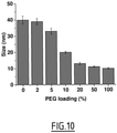
- PMAO was reacted with an increasing amount (from 0 to 100 mol% with respect to reactive anhydrides) of Jeffamine ® M-1000 (J-1000), a primary amine-terminated copolymer of propylene oxide (PO) and ethylene oxide (EO) with a PO/EO ratio of 3/19 (H 2 N-(PO) 3 -(EO) 19 -OMe).
- the resulting polymers were purified by exclusion phase chromatography and analyzed by 1 H-NMR ( Figure 11 ).
- the PEGylation yields determined by 1H NMR, were 96 and 88%, respectively.
- the polymers were dissolved in dioxane (2 mg/mL) and then diluted in water to induce assembly of NPs.
- the size of formed NPs was first measured by dynamic light scattering (DLS, Table 1). Although the non-PEGylated polymer gave NPs of 33.6 ⁇ 2.4 nm ; the half-PEGylation led to ultra-small NPs of 9.7 ⁇ 0.8 nm. Interestingly, the fully-PEGylated polymer provided slightly larger NPs of 11.5 ⁇ 0.4 nm, probably due to an increased hydrodynamic diameter provoked by the denser PEG shell.
- the polymer concentration was adjusted in order to fix the dye concentration at 2.5 ⁇ M, considering a molar extinction coefficient of 80,000 M -1 cm -1 for a single BODIPY.
- Dynamic Light Scattering and Zeta potential The size and the surface Zeta potential (z) of the NPs were measured three times from three independent formulations in milliQ water at 25°C using a Malvern Zetasizer Nano ZSP (Malvern, U.K.). DLS laser was 633 nm.
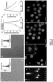
- the NPs were imaged by transmission emission microscopy ( Figure 2A and 2B ).
- the water used for spectroscopy was Milli-Q water (Millipore), all the solvents were spectro grade.
- Absorption and emission spectra were recorded on a Cary 400 Scan ultraviolet-visible spectrophotometer (Varian) and a FluoroMax-4 spectrofluorometer (Horiba Jobin Yvon) equipped with a thermostated cell compartment, respectively.
- the emission was collected 10 nm after the excitation wavelength. All the spectra were corrected from wavelength-dependent response of the detector.
- the non-PEGylated NPs displayed a round shape with sizes similar to those measured by DLS (32.4 ⁇ 5.2).
- fully-PEGylated NPs revealed ultrasmall round-shaped nanostructures of quite narrow distribution (9.8 ⁇ 0.8 nm). These images tend to indicate that PEGylation in PMAO leads to formation of particles containing a single polymer molecule or only few of them.
- the round shape and small size of fully-PEGylated NPs also suggest that the polymers are folded into spherical (globular) structures rather than present in form of out-stretched polymer brush. Theoretically, the latter form would be 42-70 nm long considering all -trans configuration of the hydrocarbon backbone of PEGylated PMAO with molecular weight 30,000-50,000 Da (85-140 repetitive units).
- BDP1-amine BDP1-amine
- fully-PEGylated polymer 5 mol% of BDP1
- absorption and emission spectra of BDP1-polymer were very similar to those of free dye BDP4-NH 2 , indicating that grafting to the polymer does not modify spectroscopic properties of the dye in solubilized form.
- NPs were analyzed by electrophoresis.
- the electrophoresis was performed on an agarose gel (0.5%) in TAE buffer (TRIS, acetate, EDTA).
- TAE buffer TriS, acetate, EDTA
- 20 ⁇ L of a solution of NPs (0.01 mg/mL in water) were cautiously added (0.2 ⁇ g polymer per well).
- the gel was gently covered with TAE then a tension of 125 V was applied for one hour.
- the gels were visualized with an ImageQuant LAS 4000 (GE Healthcare Life Sciences) using Cy2 (473 nm) or Cy3 (532 nm) excitation channels and by acquiring 10 images with an exposition time of 10 seconds.
- the images were treated with ImageJ.
- the non-PEGylated NPs migrated fast to the anode, whereas the PEGylated NPs moved slower and displayed clear spots denoting their high homogeneity ( Figure 2E ).
- the non-PEGylated NPs migrated as a smear and reduced their migration towards the anode, probably because they interacted non-specifically with BSA.
- the PEGylated NPs did not change their migration profile when incubated in the presence of BSA, showing the absence of non-specific interactions.
- the NPs were incubated for 1h in the presence of KB cells before being washed out ( Figure 2F-H ).
- the non-PEGylated NPs showed clear non-specific binding to the cells and aggregation due to precipitation in the high ionic strength medium (Opti-MEM).
- the cells incubated with PEGylated NPs did not display any fluorescence signal thus demonstrating the protecting effect of the PEG shell against non-specific interactions.
- cytotoxicity assays using MTT test revealed that unlike non-PEGylated polymer, PEGylated polymers did not affect the cells viability at concentrations up to 50 ⁇ g/mL.
- Cytotoxicity assay of the NPs was quantified by the MTT assay (3-(4,5-dimethylthiazol-2-yl)-2,5-diphenyltetrazolium bromide).
- MTT assay 3-(4,5-dimethylthiazol-2-yl)-2,5-diphenyltetrazolium bromide.
- a total of 1x10 4 HeLa (ATCC ® CCL-2 TM ) cells/well were seeded in a 96-well plate 24 h prior to the cytotoxicity assay in Dulbecco's Modified Eagle Medium (Gibco Lifetechnologies -DMEM) complemented with 10% fetal bovine serum, Gentamicin (100 ⁇ g/mL), L-Glutamine (2 mM), non-essential amino acids (1 mM), MEM vitamin solution (1%) and were incubated in a 5% CO 2 incubator at 37°C.
- Dulbecco's Modified Eagle Medium Gibco Lifetechnologies -DM
- DMEM containing NPs at various concentrations (10 to 50 ⁇ g polymer/mL) was added to the cells and incubated for 24 h at 37°C (5% CO 2 ).
- the cells were incubated with DMEM containing the same percentage of dioxane as the solution with the tested NPs or with triton X-100 0.1% as a positive control of cytotoxicity.
- the medium was replaced by 100 ⁇ L of a mixture containing DMEM and MTT solution (diluted in PBS beforehand) and the cells were incubated for 4 h at 37°C.
- Example 3 Measure of monomolecular nature of PEGylated polymer based NPs (example with PMAO):
- FCS is a powerful spectroscopic technique providing simultaneously information on size, concentration, and brightness of fluorescent nanomaterials, including polymeric NPs.
- FCS measurements were performed on a home-built confocal set-up based on a Nikon inverted microscope with a Nikon 60x 1.2NA water immersion objective. Excitation was provided by a cw laser diode (488 nm, Oxxius).
- a fibered Avalanche Photodiode (APD, PerkinElmer, Fremont, CA) connected to an on-line hardware correlator (ALV7000-USB, ALV GmbH, Germany) was used.
- APD Aborescence-Demand Photodiode
- the fluorescence signal was filtered through a 405/488/ 532/635 nm BrightLines quad-edge laser-grade dichroic (Semrock, NY) and ET525/50 single band-pass filter (Chroma Corp., Rockingham, VT).
- the fluorescence signal was further processed by an ALV7002/USB digital correlator (ALV, Langen, Germany). Typical acquisition time was 5 min (10-30 scans) with an excitation power of 0.5 mW at the laser output level.
- G ⁇ 1 N 1 ⁇ F + F e ⁇ ⁇ F 1 ⁇ F 1 + ⁇ ⁇ d ⁇ 1 1 + 1 S 2 ⁇ ⁇ d ⁇ 1 / 2
- ⁇ d the diffusion time
- N the mean number of fluorescent species within the excitation volume
- S the ratio between the axial and lateral radii of the excitation volume
- F the fraction of emissive species that have entered the triplet state
- ⁇ F is the corresponding triplet state relaxation time.
- NPs ⁇ d(NPs) / ⁇ d(FL) ⁇ d FL , where d FL is the hydrodynamic diameter of fluorescein (1.0 nm).
- FCS also provided information regarding the brightness of the formed NPs. Fluorescein was used as a reference (50 nM in NaOH 0.1 M, ⁇ 0.95) and the brightness of the NPs was expressed in equivalent number of fluorescein molecules. Even though non-PEGylated NPs in water are composed of 24 polymers ( ⁇ 168 dyes) the brightness was equivalent to 23 fluorescein molecules due to high quenching of the fluorophores within the compacted polymers. On the other hand, half-PEGylated NPs and fully PEGylated NPs, composed of a single polymer, are as bright as 4 and 5 fluorescein molecules, respectively. Thus, the FCS data also confirmed that a high level of PEGylation enhanced the brightness of the obtained NPs.
- the folded monomolecular nanoparticle is expected to bear a hydrophobic core ( Figure 1 ), similar to that of micelles made of surfactants.
- a solvatochromic dye Nile Red which is a common tool for identifying low polar environment of micelles 81 and single-chain polymeric NPs. Fluorescence spectra of Nile Red at fixed concentration (200 nM) were recorded with addition of different concentrations of fully PEGylated PMAO polymer with 5 mol% BDP1 ( Figure12 .
- Nile Red A drastic increase in the fluorescence intensity of Nile Red was observed accompanied by ⁇ 30 nm blue shift of the emission band ( Figure12A,B,D .
- the blue shifted emission of Nile Red at ⁇ 630 nm indicates the presence of a hydrophobic binding site in these NPs.
- the NPs were compared with micelles of Tween 80, having a hydrophobic core formed by hydrocarbon chains.
- the brightness (B) of a NP composed of several fluorophores is defined by Equation 1 where n is the number of fluorophores that compose the NP, ⁇ is the molar extinction coefficient value of the used fluorophore and ⁇ is the measured quantum yield of the NP.
- B n ⁇ ⁇ ⁇ ⁇
- the brightness of a NP can theoretically be enhanced by increasing the fluorophore loading (n).
- concentrating fluorophores within a small volume tends to provoke H -aggregation by ⁇ -stacking of the fluorophores thus leading to the aggregation caused quenching (ACQ) phenomenon that lowers the quantum yield.
- ACQ aggregation caused quenching
- linkers between the polymer and the fluorophore were used: i) a short linker that maintains the fluorophores at the interface ii) a long hydrocarbon chain (C12) that directs the fluorophores into the core of the NP by hydrophobic interactions with the hydrocarbon chains of PMAO and iii) a hydrophilic PEG 12 that displays the fluorophore in the PEG shell of the NP.
- these three different locations may alter hydration, polarity or viscosity of the dye environment that should also affect its quantum yield.
- NPs with 5% dye functionalization were compared by electrophoresis in the absence and presence of BSA.
- the NPs bearing the fluorophore through a short linker i.e. dye located at the interface were less smeary indicating their higher homogeneity.
- the second approach to prevent aggregation-caused quenching and to improve the brightness of the NPs was to investigate on the BODIPY's bulkiness. Enhancing the bulkiness of fluorophores to prevent aggregation was successfully applied for different dyes, including BODIPY and perylene diimide.
- BDP2 dimethyl phenyl group
- BDP3 beta positions with ethyl
- BDP4 diphenyl groups
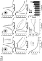
- BDP4-based NPs can be an advantage as it can be excited by various excitation sources (e.g. 488 and 532 nm). Further spectroscopic studies showed a dramatic influence of BODIPY's bulkiness on the self-quenching effect. In comparison to BDP1, bulkier BDP2 and BDP4 showed systematically lower increase in the short-wavelength shoulder ( Figure 6A-C ), and all three new derivatives BDP2-4 displayed narrower emission bands at high dye functionalization level ( Figure 6D-F ).
- BDP4 the spectroscopic changes upon increase in the dye functionalization level were the smallest, showing the important effect of the bulky groups ( Figure 6C and F ).
- BDP2 and BDP3 helped in slowing down the quantum yield decrease compared to the initial BDP1
- the brightness was limited to 800,000 M -1 cm -1 with 20% functionalization level using BDP3.
- the use of BDP4 allowed to maintaining quantum yield values above 0.6 regardless of the dye functionalization level thus leading to impressive brightness values up to ⁇ 2,500,000 M -1 cm -1 per monomolecular NP.
- the presented images were an average of the first 20 frames (recorded at a 100 ms integration time.
- the single-particle analysis was performed using Fiji software, similarly to the previously described protocol. Briefly, particle locations were detected through a Fiji routine applied to a projection (maximum intensity) of 100 frames using an appropriate threshold. The mean intensities of circular regions of interest with a diameter of 5 pixels around the found particle locations were then measured. Background subtraction was then achieved by measuring the mean intensities in circular bands around the circular regions of interest and subtracting them. Finally, integrated intensity in all circular regions of interest was calculated and used for building the histograms of particle intensities. For BDP4 NPs and QDots-585, 150 and 50 particles were analyzed, respectively. Average intensity ( ⁇ sem) of the two types of NPs was compared.
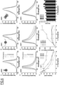
- the NPs loaded with 50% BDP4 was found to be 5.3 ⁇ 1.3 fold brighter than QDots-585 under the same conditions of illumination, recording and processing ( Figure 7B-E ).
- the intensity distribution histograms confirmed the significantly higher brightness of our NPs vs QDots-585, although the size distribution of the former was larger, probably related to the distribution of the PMAO polymer size and the dye grafting ratio.
- the estimated brightness following equation (1) provided a value of 2.4 ⁇ 10 6 M -1 cm -1 for BDP4 NPs at an excitation wavelength at 532 nm.
- the fluorescence intensity was monitored at the maximum emission wavelength determined by emission spectra prior to kinetic measurements. For fair comparison, the concentrations were adjusted to obtain identical optical density of 0.015 at the excitation wavelength of 486 nm. Emission slit opening was set to obtain optimal signal.
- BDP4-loaded NPs were studied where a correlation between dye loading and photostability was also observed.
- BDP4-loaded NPs were significantly more photostable with similar photostability of our NPs with 20 mol% BDP4 and QDots-585.
- This photostability improvement might be ascribed to the difference in the bulkiness between BDP4 and BDP1.
- the steric hindrance of the dyes prevents from ACQ and collisions that might promote reaction and then bleaching of the dye.
- the NPs displayed spectacular decay of fluorescence intensity depicting a low photostability. The latter indicates that a compromise should be found between dye loading and photostability to achieve the best performance.
- the design is based on highly PEGylated poly(maleic anhydride-alt-1-octadecene) functionalized with large amount (up to 50 mol%) of BODIPY dyes.
- the inventors found that strong PEGylation rate is required to ensure folding into the monomolecular particles. Fluorescence correlation spectroscopy suggested that the obtained NPs are stable in physiological salt conditions and do not exhibit non-specific interactions with proteins, probably due to highly dense PEG shell.
- the linkers of different length and polarity connecting BODIPY to the polymer were tested and the inventors found that short medium-polar linker ensured small size and good fluorescent properties of NPs, which suggest that the particle interface is the most optimal location of the dye.
- the intracellular spreading of our NPs was similar to that of a red fluorescent protein, which shows that they can be considered as artificial protein analogues, compatible with the intracellular applications.
- the proposed fluorescent monomolecular polymeric NPs is a new nanomaterial with controlled small and uniform size and bright fluorescence for numerous bioimaging applications.
Landscapes
- Health & Medical Sciences (AREA)
- Engineering & Computer Science (AREA)
- Biomedical Technology (AREA)
- Epidemiology (AREA)
- Life Sciences & Earth Sciences (AREA)
- Animal Behavior & Ethology (AREA)
- General Health & Medical Sciences (AREA)
- Public Health (AREA)
- Veterinary Medicine (AREA)
- Chemical & Material Sciences (AREA)
- Dispersion Chemistry (AREA)
- Polyethers (AREA)
- Medicines Containing Antibodies Or Antigens For Use As Internal Diagnostic Agents (AREA)
- Medicinal Preparation (AREA)
Priority Applications (1)
| Application Number | Priority Date | Filing Date | Title |
|---|---|---|---|
| EP20306027.2A EP3967331A1 (de) | 2020-09-14 | 2020-09-14 | Ultrakleine fluoreszierende organische nanopartikel |
Applications Claiming Priority (1)
| Application Number | Priority Date | Filing Date | Title |
|---|---|---|---|
| EP20306027.2A EP3967331A1 (de) | 2020-09-14 | 2020-09-14 | Ultrakleine fluoreszierende organische nanopartikel |
Publications (1)
| Publication Number | Publication Date |
|---|---|
| EP3967331A1 true EP3967331A1 (de) | 2022-03-16 |
Family
ID=72615794
Family Applications (1)
| Application Number | Title | Priority Date | Filing Date |
|---|---|---|---|
| EP20306027.2A Withdrawn EP3967331A1 (de) | 2020-09-14 | 2020-09-14 | Ultrakleine fluoreszierende organische nanopartikel |
Country Status (1)
| Country | Link |
|---|---|
| EP (1) | EP3967331A1 (de) |
-
2020
- 2020-09-14 EP EP20306027.2A patent/EP3967331A1/de not_active Withdrawn
Non-Patent Citations (8)
| Title |
|---|
| ANDREAS REISCH ET AL: "Fluorescent Polymer Nanoparticles Based on Dyes: Seeking Brighter Tools for Bioimaging", SMALL, vol. 12, no. 15, 1 April 2016 (2016-04-01), pages 1968 - 1992, XP055700865, ISSN: 1613-6810, DOI: 10.1002/smll.201503396 * |
| BOU SOPHIE ET AL: "Lipid-core/polymer-shell hybrid nanoparticles: synthesis and characterization by fluorescence labeling and electrophoresis", SOFT MATTER, vol. 16, no. 17, 1 April 2020 (2020-04-01), pages 4173 - 4181, XP055775873, ISSN: 1744-683X, Retrieved from the Internet <URL:https://pubs.rsc.org/en/content/articlepdf/2020/sm/d0sm00077a> DOI: 10.1039/D0SM00077A * |
| CABRAL, H.MATSUMOTO, Y.MIZUNO, K.CHEN, Q.MURAKAMI, M.KIMURA, M.TERADA, Y.KANO, M. R.MIYAZONO, K.UESAKA, M.: "Accumulation of sub-100 nm polymeric micelles in poorly permeable tumours depends on size", NATURE NANOTECHNOLOGY, vol. 6, 2011, pages 815, XP055243966, DOI: 10.1038/nnano.2011.166 |
| COLLOT MAYEUL ET AL: "Stealth and Bright Monomolecular Fluorescent Organic Nanoparticles Based on Folded Amphiphilic Polymer", ACS NANO, vol. 14, no. 10, 6 October 2020 (2020-10-06), pages 13924 - 13937, XP055775924, ISSN: 1936-0851, Retrieved from the Internet <URL:https://pubs.acs.org/doi/pdf/10.1021/acsnano.0c06348> DOI: 10.1021/acsnano.0c06348 * |
| DI CORATO, R.QUARTA, A.PIACENZA, P.RAGUSA, A.FIGUEROLA, A.BUONSANTI, R.CINGOLANI, R.MANNA, L.PELLEGRINO, T.: "Water solubilization of hydrophobic nanocrystals by means of poly(maleic anhydride-alt-1-octadecene", J. MATER. CHEM., vol. 18, 2008, pages 1991, XP055510939, DOI: 10.1039/b717801h |
| REISCH, A.DIDIER, P.RICHERT, L.ONCUL, S.ARNTZ, Y.MELY, Y.KLYMCHENKO, A. S.: "Collective fluorescence switching of counterion-assembled dyes in polymer nanoparticles", NAT. COMMUN., vol. 5, 2014, pages 4089 |
| REISCH, A.KLYMCHENKO, A. S.: "Fluorescent Polymer Nanoparticles Based on Dyes: Seeking Brighter Tools for Bioimaging", SMALL, vol. 12, 2016, pages 1968, XP055700865, DOI: 10.1002/smll.201503396 |
| ZHANG, C.LI, C.LIU, Y.ZHANG, J.BAO, C.LIANG, S.WANG, Q.YANG, Y.FU, H.WANG, K.: "Gold Nanoclusters-Based Nanoprobes for Simultaneous Fluorescence Imaging and Targeted Photodynamic Therapy with Superior Penetration and Retention Behavior", TUMORS. ADV. FUNCT. MATER., vol. 25, 2015, pages 1314 |
Similar Documents
| Publication | Publication Date | Title |
|---|---|---|
| JP6812471B2 (ja) | 新規ブロックコポリマーおよびミセル組成物ならびにその使用法 | |
| Laskar et al. | Camptothecin-based dendrimersomes for gene delivery and redox-responsive drug delivery to cancer cells | |
| Choi et al. | Self-assembled hyaluronic acid nanoparticles as a potential drug carrier for cancer therapy: synthesis, characterization, and in vivo biodistribution | |
| Collot et al. | Stealth and bright monomolecular fluorescent organic nanoparticles based on folded amphiphilic polymer | |
| Gordijo et al. | Design of hybrid MnO2‐polymer‐lipid nanoparticles with tunable oxygen generation rates and tumor accumulation for cancer treatment | |
| Jenkins et al. | Mini-review: fluorescence imaging in cancer cells using dye-doped nanoparticles | |
| Lee et al. | Tumor pH-responsive flower-like micelles of poly (L-lactic acid)-b-poly (ethylene glycol)-b-poly (L-histidine) | |
| US9511152B2 (en) | Multicolored pH-activatable fluorescence nanoplatform | |
| Ribeiro et al. | NIR and visible perylenediimide-silica nanoparticles for laser scanning bioimaging | |
| Yan et al. | Bright and stable near-infrared Pluronic–silica nanoparticles as contrast agents for in vivo optical imaging | |
| EP4447936B1 (de) | Polysarcosin-vitamin-e-derivate | |
| Knop et al. | Monomeric pheophorbide (a)-containing poly (ethyleneglycol-b-ε-caprolactone) micelles for photodynamic therapy | |
| Wang et al. | An unusual role of folate in the self-assembly of heparin–folate conjugates into nanoparticles | |
| Yuan et al. | A facile approach to fabricate self-assembled magnetic nanotheranostics for drug delivery and imaging | |
| EP3967331A1 (de) | Ultrakleine fluoreszierende organische nanopartikel | |
| US9999599B2 (en) | Polymer-polymer composite nanoassemblies and applications thereof | |
| Elter et al. | Solid-phase synthesis as a tool to create exactly defined, branched polymer vectors for cell membrane targeting | |
| Zhang et al. | Tumor-targeted and self-assembled mixed micelles as carriers for enhanced anticancer efficacy of gemcitabine | |
| Soultan et al. | Nanocarrier systems assembled from PEGylated hyperbranched poly (arylene oxindole) | |
| Jeong et al. | Fluorescence color-changeable branched-form heptamethine cyanine dye as a redox-responsive multi-functional drug delivery system for enhanced cancer diagnosis and chemophototherapy | |
| Manchanda et al. | Nanoplexes for cell imaging and hyperthermia: in vitro studies | |
| Navarro-Real et al. | Polyelectrolyte complexed alginate–oligochitosan nanoparticles for co-delivery of doxorubicin and indocyanine green in triple negative breast cancer therapy | |
| Amos et al. | Tuning polymersome surfaces: functionalization with dendritic groups | |
| KR102574572B1 (ko) | 알긴산-엽산 컨쥬게이트의 제조방법, 이로부터 제조된 알긴산-엽산 컨쥬게이트 및 이를 포함하는 약학 조성물 | |
| Vo | Effects of Physicochemical Properties on the Biological Activity of Single-Chain Nanoparticles in Drug Delivery |
Legal Events
| Date | Code | Title | Description |
|---|---|---|---|
| PUAI | Public reference made under article 153(3) epc to a published international application that has entered the european phase |
Free format text: ORIGINAL CODE: 0009012 |
|
| STAA | Information on the status of an ep patent application or granted ep patent |
Free format text: STATUS: THE APPLICATION HAS BEEN PUBLISHED |
|
| AK | Designated contracting states |
Kind code of ref document: A1 Designated state(s): AL AT BE BG CH CY CZ DE DK EE ES FI FR GB GR HR HU IE IS IT LI LT LU LV MC MK MT NL NO PL PT RO RS SE SI SK SM TR |
|
| STAA | Information on the status of an ep patent application or granted ep patent |
Free format text: STATUS: THE APPLICATION IS DEEMED TO BE WITHDRAWN |
|
| 18D | Application deemed to be withdrawn |
Effective date: 20220917 |