EP3945319A1 - Method for measuring viral antigen in sample, antibody set, and reagent kit - Google Patents
Method for measuring viral antigen in sample, antibody set, and reagent kit Download PDFInfo
- Publication number
- EP3945319A1 EP3945319A1 EP21188360.8A EP21188360A EP3945319A1 EP 3945319 A1 EP3945319 A1 EP 3945319A1 EP 21188360 A EP21188360 A EP 21188360A EP 3945319 A1 EP3945319 A1 EP 3945319A1
- Authority
- EP
- European Patent Office
- Prior art keywords
- antibody
- seq
- amino acid
- acid sequence
- variable region
- Prior art date
- Legal status (The legal status is an assumption and is not a legal conclusion. Google has not performed a legal analysis and makes no representation as to the accuracy of the status listed.)
- Pending
Links
Images


Classifications
-
- G—PHYSICS
- G01—MEASURING; TESTING
- G01N—INVESTIGATING OR ANALYSING MATERIALS BY DETERMINING THEIR CHEMICAL OR PHYSICAL PROPERTIES
- G01N33/00—Investigating or analysing materials by specific methods not covered by groups G01N1/00 - G01N31/00
- G01N33/48—Biological material, e.g. blood, urine; Haemocytometers
- G01N33/50—Chemical analysis of biological material, e.g. blood, urine; Testing involving biospecific ligand binding methods; Immunological testing
- G01N33/53—Immunoassay; Biospecific binding assay; Materials therefor
- G01N33/569—Immunoassay; Biospecific binding assay; Materials therefor for microorganisms, e.g. protozoa, bacteria, viruses
- G01N33/56983—Viruses
-
- C—CHEMISTRY; METALLURGY
- C07—ORGANIC CHEMISTRY
- C07K—PEPTIDES
- C07K16/00—Immunoglobulins [IG], e.g. monoclonal or polyclonal antibodies
- C07K16/08—Immunoglobulins [IG], e.g. monoclonal or polyclonal antibodies against material from viruses
- C07K16/10—RNA viruses
-
- G—PHYSICS
- G01—MEASURING; TESTING
- G01N—INVESTIGATING OR ANALYSING MATERIALS BY DETERMINING THEIR CHEMICAL OR PHYSICAL PROPERTIES
- G01N33/00—Investigating or analysing materials by specific methods not covered by groups G01N1/00 - G01N31/00
- G01N33/48—Biological material, e.g. blood, urine; Haemocytometers
- G01N33/50—Chemical analysis of biological material, e.g. blood, urine; Testing involving biospecific ligand binding methods; Immunological testing
- G01N33/53—Immunoassay; Biospecific binding assay; Materials therefor
- G01N33/543—Immunoassay; Biospecific binding assay; Materials therefor with an insoluble carrier for immobilising immunochemicals
- G01N33/54313—Immunoassay; Biospecific binding assay; Materials therefor with an insoluble carrier for immobilising immunochemicals the carrier being characterised by its particulate form
- G01N33/54326—Magnetic particles
-
- G—PHYSICS
- G01—MEASURING; TESTING
- G01N—INVESTIGATING OR ANALYSING MATERIALS BY DETERMINING THEIR CHEMICAL OR PHYSICAL PROPERTIES
- G01N33/00—Investigating or analysing materials by specific methods not covered by groups G01N1/00 - G01N31/00
- G01N33/48—Biological material, e.g. blood, urine; Haemocytometers
- G01N33/50—Chemical analysis of biological material, e.g. blood, urine; Testing involving biospecific ligand binding methods; Immunological testing
- G01N33/53—Immunoassay; Biospecific binding assay; Materials therefor
- G01N33/563—Immunoassay; Biospecific binding assay; Materials therefor involving antibody fragments
-
- G—PHYSICS
- G01—MEASURING; TESTING
- G01N—INVESTIGATING OR ANALYSING MATERIALS BY DETERMINING THEIR CHEMICAL OR PHYSICAL PROPERTIES
- G01N33/00—Investigating or analysing materials by specific methods not covered by groups G01N1/00 - G01N31/00
- G01N33/48—Biological material, e.g. blood, urine; Haemocytometers
- G01N33/50—Chemical analysis of biological material, e.g. blood, urine; Testing involving biospecific ligand binding methods; Immunological testing
- G01N33/53—Immunoassay; Biospecific binding assay; Materials therefor
- G01N33/577—Immunoassay; Biospecific binding assay; Materials therefor involving monoclonal antibodies binding reaction mechanisms characterised by the use of monoclonal antibodies
-
- G—PHYSICS
- G01—MEASURING; TESTING
- G01N—INVESTIGATING OR ANALYSING MATERIALS BY DETERMINING THEIR CHEMICAL OR PHYSICAL PROPERTIES
- G01N33/00—Investigating or analysing materials by specific methods not covered by groups G01N1/00 - G01N31/00
- G01N33/48—Biological material, e.g. blood, urine; Haemocytometers
- G01N33/50—Chemical analysis of biological material, e.g. blood, urine; Testing involving biospecific ligand binding methods; Immunological testing
- G01N33/58—Chemical analysis of biological material, e.g. blood, urine; Testing involving biospecific ligand binding methods; Immunological testing involving labelled substances
- G01N33/581—Chemical analysis of biological material, e.g. blood, urine; Testing involving biospecific ligand binding methods; Immunological testing involving labelled substances with enzyme label (including co-enzymes, co-factors, enzyme inhibitors or substrates)
-
- C—CHEMISTRY; METALLURGY
- C07—ORGANIC CHEMISTRY
- C07K—PEPTIDES
- C07K2317/00—Immunoglobulins specific features
- C07K2317/50—Immunoglobulins specific features characterized by immunoglobulin fragments
- C07K2317/56—Immunoglobulins specific features characterized by immunoglobulin fragments variable (Fv) region, i.e. VH and/or VL
-
- C—CHEMISTRY; METALLURGY
- C07—ORGANIC CHEMISTRY
- C07K—PEPTIDES
- C07K2317/00—Immunoglobulins specific features
- C07K2317/50—Immunoglobulins specific features characterized by immunoglobulin fragments
- C07K2317/56—Immunoglobulins specific features characterized by immunoglobulin fragments variable (Fv) region, i.e. VH and/or VL
- C07K2317/565—Complementarity determining region [CDR]
-
- G—PHYSICS
- G01—MEASURING; TESTING
- G01N—INVESTIGATING OR ANALYSING MATERIALS BY DETERMINING THEIR CHEMICAL OR PHYSICAL PROPERTIES
- G01N2333/00—Assays involving biological materials from specific organisms or of a specific nature
- G01N2333/005—Assays involving biological materials from specific organisms or of a specific nature from viruses
- G01N2333/08—RNA viruses
- G01N2333/165—Coronaviridae, e.g. avian infectious bronchitis virus
-
- G—PHYSICS
- G01—MEASURING; TESTING
- G01N—INVESTIGATING OR ANALYSING MATERIALS BY DETERMINING THEIR CHEMICAL OR PHYSICAL PROPERTIES
- G01N2469/00—Immunoassays for the detection of microorganisms
- G01N2469/10—Detection of antigens from microorganism in sample from host
Definitions
- the present invention relates to a method for measuring a viral antigen in a sample by using a capture antibody and a detection antibody.
- the present invention relates to an antibody set for measuring a viral antigen in a sample by an immunological measurement method.
- the present invention relates to a reagent kit that contains a capture antibody and a detection antibody.
- Measurement of a viral antigen in a sample has been known to determine whether a target virus is present in the sample or not.
- ELISA method enzyme-linked immunosorbent assay
- An object of the present invention is to provide a novel antibody set capable of measuring a nucleoprotein of SARS-CoV-2 in a sample, a measurement method with use of the antibody set, and a reagent kit.
- the present invention provides a method for measuring a viral antigen in a sample by using a capture antibody and a detection antibody, the method comprising: forming a sandwich immune complex that contains the capture antibody, the viral antigen and the detection antibody, the capture antibody comprising a heavy chain variable region that comprises CDR1 comprising amino acid sequence of SEQ ID NO: 1, CDR2 comprising amino acid sequence of SEQ ID NO: 2 and CDR3 comprising amino acid sequence of SEQ ID NO: 3, and a light chain variable region that comprises CDR1 comprising amino acid sequence of SEQ ID NO: 4, CDR2 comprising amino acid sequence of SEQ ID NO: 5 and CDR3 comprising amino acid sequence of SEQ ID NO: 6, and the detection antibody comprising a heavy chain variable region that comprises CDR1 comprising amino acid sequence of SEQ ID NO: 7, CDR2 comprising amino acid sequence of SEQ ID NO: 8 and CDR3 comprising amino acid sequence of SEQ ID NO: 9, and a light chain variable region that comprises CDR1 comprising amino acid sequence of SEQ ID NO:
- the present invention also provides an antibody set for measuring a viral antigen in a sample by an immunological measurement method, the antibody comprising a capture antibody and a detection antibody that form a sandwich immune complex with the viral antigen in the immunological measurement method, the capture antibody comprising a heavy chain variable region that comprises CDR1 comprising amino acid sequence of SEQ ID NO: 1, CDR2 comprising amino acid sequence of SEQ ID NO: 2 and CDR3 comprising amino acid sequence of SEQ ID NO: 3; and a light chain variable region that comprises CDR1 comprising the amino acid sequence of SEQ ID NO: 4, CDR2 comprising the amino acid sequence of SEQ ID NO: 5 and CDR3 comprising the amino acid sequence of SEQ ID NO: 6, and the detection antibody comprises a heavy chain variable region that comprises CDR1 comprising amino acid sequence of SEQ ID NO: 7, CDR2 comprising amino acid sequence of SEQ ID NO: 8 and CDR3 comprising amino acid sequence of SEQ ID NO: 9; and a light chain variable region that comprises CDR1 comprising amino acid sequence of
- the capture antibody and the detection antibody form the sandwich immune complex with a nucleoprotein of SARS-CoV or SARS-CoV-2, but do not form the sandwich immune complex with a nucleoprotein of MERS-CoV, and viral antigens of HCoV-OC43, HCoV-NL63 and HCoV-229.
- the capture antibody comprises the heavy chain variable region comprising amino acid sequence of SEQ ID NO: 13, and the light chain variable region comprising amino acid sequence of SEQ ID NO: 14, and the detection antibody comprises the heavy chain variable region comprising amino acid sequence of SEQ ID NO: 15, and the light chain variable region comprising amino acid sequence of SEQ ID NO: 16.
- the capture antibody is IgG, reduced IgG, Fab, Fab', F(ab') 2 , Fv, scFv, diabody or triabody.
- the detection antibody is IgG, reduced IgG, Fab, Fab', F(ab') 2 , Fv, scFv, diabody or triabody.
- the sample is a biological sample.
- the viral antigen is a nucleoprotein of SARS-CoV or SARS-CoV-2.
- the capture antibody is immobilized on a solid phase.
- the solid phase is a magnetic particle or a microplate.
- the detection antibody is labeled with a labeling substance.
- the labeling substance is at least one enzyme selected from the group consisting of alkaline phosphatase, peroxidase, ⁇ -galactosidase, glucosidase, polyphenol oxidase, tyrosinase, acid phosphatase and luciferase.
- the present invention also provides a reagent kit comprising the capture antibody and the detection antibody described above.
- the reagent kit comprises a first reagent and a second reagent, the first reagent comprising the capture antibody and the second reagent comprising the detection antibody.
- a novel antibody set capable of measuring a nucleoprotein of SARS-CoV-2 in a sample, a measurement method with use of the antibody set, and a reagent kit are provided.
- the method for measuring a viral antigen of this invention measures in vitro a viral antigen in a sample, by an immunological measurement method with use of a capture antibody and a detection antibody each containing predetermined CDRs.
- the capture antibody means an antibody that specifically binds to a test substance.
- the capture antibody can capture the test substance on the solid phase.
- the detection antibody means an antibody that specifically binds to a test substance.
- the detection antibody can provide a detectable signal.
- the detection antibody is not immobilized on a solid phase in most cases, the labeling substance per se may occasionally serve as a solid phase typically in the form of particle.
- the immunological measurement method is not specially limited as long as it includes forming a sandwich immune complex.
- the sandwich immune complex means a complex that contains the capture antibody, the test substance and the detection antibody, in which the capture antibody and the detection antibody are bound to different sites of the test substance.
- This sort of immunological measurement method is exemplified by sandwich ELISA method and immunochromatography method.
- the sandwich ELISA method is particularly preferred.
- the sandwich ELISA method applicable here may be an immune complex transfer method described in Japanese Patent Application Laid-Open No. H01-254868 .
- the term "measuring a viral antigen” encompasses acquiring a measured value of a viral antigen, and, quantifying the amount or concentration of a viral antigen in a sample.
- the measured value of the viral antigen may be a value that reflects the amount or concentration of the viral antigen in the sample.
- the "value that reflects the amount or concentration” is a value that depends on the type of the labeling substance described later, and is exemplified by measured value of luminescence intensity, measured value of fluorescence intensity, measured value of radiation intensity, and measured value of optical density.
- the value of the amount or concentration of the viral antigen in the sample can be determined, for example, on the basis of the measured value of the viral antigen and a measured value of a calibrator.
- the calibrator is a kind of control sample used for quantifying the test substance, which is a sample that contains a known concentration of the test substance or a corresponding standard substance.
- antibody herein encompasses full-length antibody and fragment thereof.
- Class of antibody may be any of IgG, IgA, IgM, IgD and IgE, among which IgG is preferred.
- Subclass of IgG is not specially limited, and may be any of IgG1, IgG2, IgG3 and IgG4.
- the fragment of the antibody is exemplified by reduced IgG (rIgG), Fab, Fab', F(ab') 2 , Fv, single chain antibody (scFv), diabody and triabody. Methods per se for preparing these antibody fragments have been already known.
- the antibody may be either monoclonal antibody or polyclonal antibody, where monoclonal antibody is preferred.
- the antibody may be derived from any animal including mouse, rat, hamster, rabbit, goat, horse, camel, alpaca and chicken.
- the antibody is preferably a murine monoclonal antibody.
- the capture antibody and the detection antibody may be, equally or differently, full-length antibody or antibody fragment.
- type of the fragments may be same or different between the capture antibody and the detection antibody.
- Both of the capture antibody and the detection antibody may preferably be monoclonal antibodies.
- the capture antibody includes a heavy chain variable region that includes CDR1 having amino acid sequence of SEQ ID NO: 1, CDR2 having amino acid sequence of SEQ ID NO: 2, and CDR3 having amino acid sequence of SEQ ID NO: 3.
- the capture antibody includes a light chain variable region that includes CDR1 having amino acid sequence of SEQ ID NO: 4, CDR2 having amino acid sequence of SEQ ID NO: 5 and CDR3 having amino acid sequence of SEQ ID NO: 6.
- the amino acid sequences of the individual CDRs of the capture antibody are enumerated below.
- CDRH1, CDRH2 and CDRH3 represent CDR1, CDR2 and CDR3 of the heavy chain, respectively.
- CDRL1, CDRL2 and CDRL3 represent CDR1, CDR2 and CDR3 of the light chain, respectively.
- Amino acid sequences of the individual CDRs are those determined on the basis of the Kabat method.
- "based on the Kabat method” means that the numbering of the amino acid residues in the CDR and the variable region follows the numbering scheme advocated by Kabat et al. (see Kabat E.A. et al., Sequences of Proteins of Immunological Interest, 5th ed. Public Health Service, National Institutes of Health, Bethesda, MD (1991), NIH Publication No. 91-3242 ).
- the detection antibody includes a heavy chain variable region that includes CDR1 having amino acid sequence of SEQ ID NO: 7, CDR2 having amino acid sequence of SEQ ID NO: 8, and CDR3 having amino acid sequence of SEQ ID NO: 9.
- the detection antibody includes a light chain variable region that includes CDR1 having amino acid sequence of SEQ ID NO: 10, CDR2 having amino acid sequence of SEQ ID NO: 11 and CDR3 having amino acid sequence of SEQ ID NO: 12.
- Amino acid sequences of the individual CDRs of the detection antibody are enumerated below. Amino acid sequences of the individual CDRs are those determined on the basis of the Kabat method.
- the capture antibody that includes six CDRs respectively having amino acid sequences of SEQ ID NOs: 1 to 6 and the detection antibody that includes six CDRs respectively having amino acid sequences of SEQ ID NOs: 7 to 12 can specifically bind to the same viral antigen, epitopes recognized by them are different from each other. Therefore, the capture antibody and the detection antibody bind to the viral antigen, thereby forming a sandwich immune complex.
- the capture antibody may be a humanized antibody that includes six CDRs respectively having amino acid sequences of SEQ ID NOs: 1 to 6.
- the detection antibody may be a humanized antibody that includes six CDRs respectively having amino acid sequences of SEQ ID NOs: 7 to 12.
- the humanized antibody is an antibody obtained by transplanting a gene sequence of CDR of a non-human-derived antibody into a human antibody gene, by a known CDR grafting method.
- the capture antibody may include a heavy chain variable region having amino acid sequence of SEQ ID NO: 13, as the heavy chain variable region that includes CDRH1, CDRH2 and CDRH3, respectively having amino acid sequences of SEQ ID NOs: 1 to 3.
- the capture antibody may include a light chain variable region having amino acid sequence of SEQ ID NO: 14, as the light chain variable region that includes CDRL1, CDRL2 and CDRL3, respectively having amino acid sequences of SEQ ID NOs: 4 to 6.
- the amino acid sequences of the variable regions, represented by SEQ ID NOs: 13 and 14, are shown below. Underlined parts indicate CDRs. The amino acid sequences of these variable regions are those determined on the basis of the Kabat method.
- the detection antibody may include a heavy chain variable region having amino acid sequence of SEQ ID NO: 15, as the heavy chain variable region that includes CDRH1, CDRH2 and CDRH3, respectively having amino acid sequences of SEQ ID NOs: 7 to 9.
- the detection antibody may include a light chain variable region having amino acid sequence of SEQ ID NO: 16, as the light chain variable region that includes CDRL1, CDRL2 and CDRL3, respectively having amino acid sequences of SEQ ID NOs: 10 to 12.
- the amino acid sequences of the variable regions, represented by SEQ ID NOs: 15 and 16, are shown below. Underlined parts indicate CDRs. The amino acid sequences of these variable regions are those determined on the basis of the Kabat method.
- the capture antibody may also be a chimeric antibody that includes the heavy chain variable region having amino acid sequence of SEQ ID NO: 13, and the light chain variable region having amino acid sequence of SEQ ID NO: 14.
- the detection antibody may also be a chimeric antibody that includes the heavy chain variable region having amino acid sequence of SEQ ID NO: 15, and the light chain variable region having amino acid sequence of SEQ ID NO: 16.
- the chimeric antibody is an antibody in which a variable region of an antibody derived from a certain species and a constant region of an antibody derived from a different species are joined.
- the capture antibody and the detection antibody in the methods of the invention may have the amino acid sequences modified so as not to reduce the binding activity towards viral antigen.
- modification of the amino acid sequence is exemplified by substitution, deletion, addition and/or insertion of amino acid residue.
- the site where the amino acid sequence of each antibody is modified may reside either in the constant region or in the variable region, and either in the heavy chain or in the light chain.
- the variable region, when modified, is preferably modified in the framework region (FR).
- FR means a region other than CDR, which resides in each of the light chain variable region and the heavy chain variable region of the antibody. FRs serve as a scaffold that links the three CDRs, and contributes to structural stability of CDRs.
- the amino acid sequence of an antibody may be modified by introducing a mutation into an antibody gene, with use of a known technique such as DNA recombination or other molecular biological techniques.
- the number of amino acid residues to be modified is usually 10 residues or less, preferably 5 residues or less, and more preferably 3 residues or less.
- Type of modification of the amino acid sequence of the antibody is preferably conservative substitution.
- Conservative substitution means substitution of an amino acid residue with some other amino acid residue having a side chain chemically similar to the side chain of the original residue.
- Conservative substitution per se of amino acid sequences is known in the art.
- the amino acid sequence of the antibody may be modified by a method for controlling affinity of the antibody for an antigen, which includes modification of amino acid residue in FR3 of the antibody, as described in U.S. Patent Application Publication No. 2018/0179298 .
- the amino acid sequence of an antibody may be modified alternatively by a method for improving thermal stability of an antibody, which includes substituting the 80th amino acid residue in the variable region and the 171st amino acid residue in the constant region of the antibody, with cysteine as described in U.S. Patent Application Publication No. 2019/0040119 .
- the capture antibody in the methods of the invention may be preliminarily immobilized on a solid phase.
- Mode of immobilization of the capture antibody on the solid phase is not specially limited.
- the capture antibody and the solid phase may be directly bound, or the capture antibody and the solid phase may be indirectly bound through some other substance.
- the direct binding is exemplified by physical adsorption.
- the indirect binding is exemplified by immobilizing a molecule specifically bindable with an antibody on the solid phase, and then binding the antibody to the molecule, thereby immobilizing the antibody on the solid phase.
- the molecule specifically bindable with an antibody is exemplified by protein A or G, and an antibody that specifically recognizes the antibody (secondary antibody).
- the capture antibody may also be immobilized on the solid phase, with use of combination of substances interposed between the antibody and the solid phase.
- Such combination of substances is exemplified by combination of biotins and avidins, and combination of hapten and anti-hapten antibody.
- the biotins include biotin, and biotin analogs such as desthiobiotin and oxybiotin.
- the avidins include avidin, and avidin analogs such as streptavidin and Tamavidin (registered trademark).
- the combination of hapten and anti-hapten antibody is exemplified by combination of a compound having 2,4-dinitrophenyl (DNP) group and anti-DNP antibody.
- DNP 2,4-dinitrophenyl
- the capture antibody may be immobilized on the solid phase, making use of binding between biotin and avidin (or binding between DNP group and anti-DNP antibody).
- Material for composing the solid phase is not specially limited, and is typically selectable from organic polymer compound, inorganic compound and biopolymer.
- the organic polymer compound is exemplified by latex, polystyrene and polypropylene.
- the inorganic compound is exemplified by magnetic substance (iron oxide, chromium oxide, ferrite, etc.), silica, alumina and glass.
- the biopolymer is exemplified by insoluble agarose, insoluble dextran, gelatin and cellulose. Two or more of them may be used in combination.
- Shape of the solid phase is not specially limited, and is exemplified by particle, membrane, microplate, microtube and test tube. Among them, particle and microplate are preferred. The particles is particularly preferably magnetic particle.
- the detection antibody may also be preliminarily labeled with a labeling substance.
- the labeling substance is not specially limited, and is exemplified by a substance that can generate a signal by itself (also referred to as "signal generating substance", hereinafter), and a substance that catalyzes reaction of some other substance to generate a signal.
- the signal generating substance is exemplified by fluorescent substance, radioisotope, and color developing substance.
- the substance that catalyzes reaction of some other substance to generate a detectable signal is exemplified by enzyme.
- the fluorescent substance is exemplified by fluorescent dyes such as fluorescein isothiocyanate (FITC), rhodamine and Alexa Fluor (registered trademark); and fluorescent proteins such as GFP.
- fluorescent dyes such as fluorescein isothiocyanate (FITC), rhodamine and Alexa Fluor (registered trademark); and fluorescent proteins such as GFP.
- radioisotope examples include 125 I, 14 C and 32 P.
- the color developing substance is exemplified by metal colloids such as gold nanocolloid.
- the enzyme is exemplified by alkaline phosphatase, peroxidase, ⁇ -galactosidase, glucosidase, polyphenol oxidase, tyrosinase, acid phosphatase and luciferase. Enzyme is preferred as the labeling substance, for which alkaline phosphatase is particularly preferred.
- a labeled secondary antibody may be used as the labeling substance.
- the labeled secondary antibody means an antibody that is labeled with the labeling substance, and can specifically recognize the detection antibody. As a result of binding of the labeled secondary antibody to the detection antibody, the detection antibody is indirectly labeled with the labeling substance.
- the capture antibody and the detection antibody may be prepared by a known technique such as DNA recombination technique or other molecular biological technique, typically as described below.
- RNA is extracted from a hybridoma that produces any murine antibody.
- the hybridoma is obtainable by a known method such as described in Kohler G. and Milstein C., Nature, vol. 256, pp. 495-497, 1975 .
- polynucleotides that individually encode the heavy chain and the light chain of the antibody are synthesized according to a reverse transcription reaction and rapid amplification of cDNA ends (RACE).
- the individual base sequences that encode CDRH1, CDRH2, CDRH3, CDRL1, CDRL2 and CDRL3 are substituted respectively with base sequences that encode amino acid sequences of SEQ ID NOs: 1 to 6, or SEQ ID NOs: 7 to 12 by the PCR method, whereby polynucleotides that individually code the heavy chain and the light chain of the capture antibody or the detection antibody are obtainable.
- the base sequence that encodes the variable region may be substituted with the base sequence that encodes amino acid sequence SEQ ID NO: 13 or 15, and the base sequence of a region that encodes the variable region in the polynucleotide that encodes the light chain of the antibody may be substituted with the base sequence that encodes amino acid sequence of SEQ ID NO: 14 or 16.
- the obtained polynucleotide is integrated into a known expression vector.
- the polynucleotide that encodes the heavy chain and the polynucleotide that encodes the light chain may be integrated into a single expression vector so that each of them can be expressed independently, or may be separately integrated into two expression vectors.
- the expression vector that contains the polynucleotides that encode the heavy chain and the light chain of the capture antibody or the detection antibody is acquired.
- Type of the expression vector is not specially limited, and may be expression vector for mammalian cell or expression vector for E. coli.
- the capture antibody or the detection antibody is obtainable by integrating the thus obtained expression vector into a suitable host cell (mammalian cell or E. coli, for example), by transformation or transfection.
- the capture antibody and the detection antibody as humanized antibodies may also be produced by using the base sequences that encodes amino acid sequences of SEQ ID NOs: 1 to 6 and SEQ ID NOs: 7 to 12, according to the known CDR grafting method (see, Jones P.T. et a/., Nature, vol. 321, pp. 522 -525, 1986 ; Co M.S. et al., Proc. Natl. Acad. Sci. USA, vol. 93, pp. 7843 -7848, 1996 , for example).
- the viral antigen is not specially limited as long as it binds to the capture antibody and the detection antibody to form the sandwich immune complex.
- Such viral antigens is exemplified by nucleoproteins of SARS-CoV and nucleoprotein of SARS-CoV-2.
- Nucleoprotein also referred to as "N protein”, hereinafter
- N protein means a protein that coexists with viral nucleic acid, and is also called nucleocapsid protein.
- the capture antibody and/or the detection antibody do not specifically bind to the viral antigen, and therefore do not substantially form the sandwich immune complex, the viral antigen is not a target substance of the measurement method of the invention.
- the capture antibody and the detection antibody form the sandwich immune complex with the N protein of SARS-CoV or SARS-CoV-2, but do not substantially form the sandwich immune complex with the N protein of MERS-CoV and viral antigens of HCoV-NL63, HCoV-229E and HCoV-OC43 (viral lysates that contain N protein, for example).
- the sample encompasses not only a sample that contains viral antigen or viral antigen-containing virus, but also a sample suspected of containing viral antigen or viral antigen-containing virus.
- a biological sample is suitably used.
- the biological sample means a sample collected from living body.
- the biological sample is exemplified by nasopharyngeal swab specimen, saliva, nasal secretion, sputum, whole blood, plasma, serum, bronchoalveolar lavage fluid, cerebrospinal fluid and lymph fluid.
- employable are excreta, sewage, river water, sea water and soil.
- the excreta include urine and feces.
- the sample in the methods of the invention is preferably in liquid form.
- the liquid sample is not limited to solution, and encompass suspension and sol.
- the sample if not in liquid form, may be converted to liquid by adding thereto an appropriate aqueous medium.
- aqueous medium is not specially limited as long as it does not interfere with measurements described later, and is exemplified by water, saline and buffer solution.
- the buffer solution is not specially limited as long as it can demonstrate buffering action at pH around neutral (pH of 6 or higher and 8 or lower, for example).
- Such buffer is exemplified by Good's buffers such as ACES, HEPES, MES and PIPES; triethanolamine hydrochloride buffer, Tris hydrochloride buffer, phosphate buffered saline (PBS) and Tris buffered saline (TBS). Any insoluble impurity, if found to reside in the liquid sample, may be removed from the sample by a known technique such as centrifugation or filtration. The liquid sample may optionally be diluted with the aqueous medium described above.
- an extraction reagent may be added to the sample in order to release the viral antigen from the virus.
- the extraction reagent means a reagent that lyses virus in a sample, and enables extraction of the viral antigen such as N protein.
- Such reagent is exemplified by buffer solution that contains a surfactant.
- the surfactant is not specially limited as long as it does not interfere with the measurement described later, and is exemplified by NP-40, Tween (registered trademark)-20 and Triton (registered trademark) X-100.
- a commercially available extraction reagent may be employable.
- the extraction reagent may include a chelating agent.
- the chelating agent is exemplified by sodium salt or potassium salt of ethylenediaminetetraacetic acid (EDTA).
- the sandwich immune complex that contains the capture antibody, the viral antigen and the detection antibody is formed.
- the sandwich immune complex may be formed by mixing a sample that expectedly contains a viral antigen, the capture antibody, and the detection antibody.
- the sandwich immune complex that includes the capture antibody, the viral antigen and the detection antibody is formed on the solid phase.
- the sample that expectedly contains a viral antigen, the capture antibody and the detection antibody are mixed to form the sandwich immune complex, and then a solution that contains the complex is contacted with the solid phase capable of immobilizing thereon the capture antibody, whereby the complex may be formed on the solid phase.
- the capture antibody preliminarily immobilized on the solid phase may be employed. That is, the complex may be formed on the solid phase, by mixing the capture antibody immobilized on the solid phase, the sample that expectedly contains the target substance, and the detection antibody.
- the measurement method of the invention includes detecting the sandwich immune complex formed on the solid phase.
- the viral antigen can be measured by detecting the sandwich immune complex on the solid phase by a method known in the art.
- the viral antigen can be measured by detecting a signal generated by the labeling substance.
- the labeled secondary antibody corresponded to the detection antibody
- the viral antigen can be measured by detecting a signal generated by the labeling substance of the labeled secondary antibody bound to the detection antibody.
- the detection antibody preliminarily labeled with a labeling substance is preferably used.
- detecting a signal encompasses qualitative detection of presence or absence of a signal, quantification of signal intensity, and semi-quantitative detection of the signal intensity.
- the semi-quantitative detection relies upon grade indication of the signal intensity which includes “no signal”, “weak”, “medium” and “strong”, for example.
- the signal intensity is preferably detected in a quantitative or semi-quantitative manner.
- the method for detecting signal per se has been already known in the art.
- a measurement method adapted to the type of signal obtainable from the labeling substance is properly selected.
- the signal of the labeling substance if being an enzyme, is detectable by reacting the enzyme with a corresponded substrate, and by measuring the generated signal such as light or color with use of a known instrument such as a spectrophotometer.
- the substrate for the enzyme is properly selectable from known substrates, depending on the type of enzyme.
- the substrate is exemplified by chemiluminescent substrates such as CDP-Star (registered trademark) (disodium 4-chloro-3-(methoxyspiro[1,2-dioxetane - 3,2'-(5'-chloro)tricyclo [3.3.1.13,7]decane]-4-yl)phenylphosphate), and CSPD (registered trademark) (disodium 3-(4-methoxyspiro[1,2-dioxetane-3,2-(5'-chloro)tricyclo[3.3.1.13,7]decane]-4-yl) phenylphosphate); and chromogenic substrate such as 5-bromo-4-chloro-3-indolylphosphate (BCIP), disodium 5-bromo-6-chloro-in
- BCIP 5-bromo-4-chloro-3-ind
- the substrate is exemplified by chemiluminescent substrates such as luminol and derivatives thereof; and chromogenic substrates such as ammonium 2,2'-azino-bis(3-ethylbenzothiazoline-6-sulfonate) (ABTS), 1,2-phenylenediamine (OPD), and 3,3',5,5'-tetramethylbenzidine (TMB).
- chemiluminescent substrates such as luminol and derivatives thereof
- chromogenic substrates such as ammonium 2,2'-azino-bis(3-ethylbenzothiazoline-6-sulfonate) (ABTS), 1,2-phenylenediamine (OPD), and 3,3',5,5'-tetramethylbenzidine (TMB).
- the labeling substance is a radioisotope
- radiation as a signal may be measured by using a known instrument such as a scintillation counter.
- fluorescence as a signal may be measured by using a known instrument such as a fluorescence microplate reader. Excitation wavelength and fluorescence wavelength may properly be determined depending on the type of fluorescent substance employed.
- Results of detection of the signal may be utilized as results of measurement of the viral antigen in the sample.
- the measured value of the signal intensity per se, or a value acquired from the measured value may be used as the measured value of the viral antigen.
- the value acquired from the measured value of the signal intensity is exemplified by a value obtained by subtracting a measured value of a negative control or a background value, from the measured value.
- the negative control is properly selectable, and is exemplified by a buffer solution free of viral antigen.
- the amount or concentration value of the viral antigen in the sample may also be determined by reading, on an analytical curve, a value corresponded to the measured value of signal intensity.
- the analytical curve may be created on the basis of measured values of a plurality of calibrators. The measured values of the calibrators are obtainable by measuring the calibrators according to the measurement method of the invention, in the same way as for the sample.
- the analytical curve may be prepared by plotting the measured values of the plurality of calibrators on X-Y plane, having concentration of the viral antigen scaled on X-axis, and the measured value (signal intensity, for example) scaled on Y-axis, and by fitting thereto a straight line or a curve according to a known technique such as the least square method.
- the calibrators may be prepared, typically by adding a recombinant viral antigen to a buffer solution free of viral antigen, while adjusting the concentration to freely selectable values.
- the calibrator free of viral antigen employable here may also be a buffer solution per se free of viral antigen.
- the measurement method of the invention may be conducted on a commercially available automated immunoassay analyzer.
- the automated immunoassay analyzer is an instrument that automatically conducts preparation and immunoassay of a sample to be measured, and outputs a measurement result of a target substance, upon setting of the sample and entry of a command to start the measurement by the user.
- This sort of automated immunoassay analyzer is exemplified by HISCL Series (Sysmex Corporation) that includes HISCL (registered trademark)-5000 and HISCL-800, and HI-1000 (Sysmex Corporation).
- HISCL Series instrument conducts the measurement according to the sandwich ELISA method, by using a magnetic particle as the solid phase.
- B/F (bound/free) separation for removing any unreacted free component not forming the complex may be interposed between formation of the sandwich immune complex and detection of the complex.
- the unreacted free component means a component that does not compose the sandwich immune complex. This is exemplified by excessive capture antibody and detection antibody that remain unbound to the viral antigen.
- Technique for the B/F separation is not specially limited, and may be conducted, if the solid phase is a particle, by centrifugation whereby only the solid phase having the complex captured thereon may be recovered. With the solid phase given as a container such as a microplate or a microtube, the B/F separation is enabled by removing a liquid that contains the unreacted free component.
- the B/F separation is enabled by removing a liquid that contains the unreacted free component under suction through a nozzle, while keeping the magnetic particle magnetically restrained by a magnet, which is preferred from the viewpoint of automation.
- the solid phase having the complex captured thereon may be washed with a suitable aqueous medium such as PBS.
- a further aspect of the invention relates to an antibody set used for measuring a viral antigen in a sample by an immunological measurement method (also referred to as "antibody set”, hereinafter).
- antibody set means a combination of a plurality of antibodies that includes at least the capture antibody and the detection antibody used for the immunological measurement method. Details of the immunological measurement method are the same as those described for the measurement method of the invention.
- the antibody set of this embodiment includes the capture antibody and the detection antibody that form the sandwich immune complex with the viral antigen in the immunological measurement method. Details of the capture antibody and the detection antibody contained in the antibody set are same as those described for the measurement method of the invention.
- the antibody set of the invention is suitably used in the measurement method of the invention.
- a further aspect relates to a reagent kit that includes the capture antibody and the detection antibody (also referred to as a "reagent kit", hereinafter).
- the reagent kit of the invention is used for measuring the viral antigen in the sample by the immunological measurement method.
- the reagent kit contains one or more reagents. Details of the immunological measurement method, the capture antibody and the detection antibody are same as those described for the measurement method of the invention.
- the reagent kit includes a reagent that contains the capture antibody, and a reagent that contains the detection antibody.
- Form of the reagent that contains the antibody is not specially limited, and may be solid (powder, crystal or freeze-dried product, for example), or liquid (solution, suspension or emulsions, for example).
- the solvent is not specially limited as long as it can solubilize and store the antibody. The solvent is exemplified by water, saline, PBS, TBS and Good's buffers.
- the Good's buffers are exemplified by MES, Bis-Tris, ADA, PIPES, Bis-Tris-propane, ACES, MOPS, MOPSO, BES, TES, HEPES, HEPPS, Tricine, Tris, Bicine and TAPS.
- the reagent that contains an antibody may contain any of known additives.
- the additives are exemplified by protein stabilizers such as bovine serum albumin (BSA), preservatives such as sodium azide, and inorganic salts such as sodium chloride.
- BSA bovine serum albumin
- preservatives such as sodium azide
- inorganic salts such as sodium chloride.
- the reagent kit may include the reagent that contains the capture antibody, the reagent that contains the detection antibody, and a solid phase.
- the capture antibody may be preliminarily immobilized on the solid phase. Details of the solid phase are same as those described previously in relation to the measurement method of the invention.
- a preferred solid phase is magnetic particle or microplate.
- the detection antibody may preliminarily be labeled with a labeling substance.
- the reagent kit may alternatively include the reagent that contains the capture antibody, the reagent that contains the detection antibody, and a reagent that contains a labeled secondary antibody. Details of the labeling substance and the labeled secondary antibody are same as those described previously in relation to the measurement method of the invention.
- a preferred labeling substance is an enzyme. The enzyme is exemplified by alkaline phosphatase, peroxidase, ⁇ -galactosidase, glucosidase, polyphenol oxidase, tyrosinase, acid phosphatase and luciferase. A preferred enzyme is alkaline phosphatase.
- the reagent kit may further contain a substrate for the enzyme. Details of the substrate are same as those described previously in relation to the measurement method of the invention.
- the reagent kit of the invention may be provided with the individual reagents filled in vials and packaged in a box. Also a package insert may be enclosed in the box. The package insert may contain descriptions on contents of the reagent kit, compositions of the individual reagents, instructions for use and storage, and so forth.
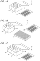
- FIG. 1A An example of the reagent kit of the invention is illustrated in Fig. 1A .
- reference numeral 11 denotes a reagent kit
- reference numeral 12 denotes a first vial that contains a capture antibody-containing reagent
- reference numeral 13 denotes a second vial that contains a detection antibody-containing reagent
- reference numeral 14 denotes a packaging box
- reference numeral 15 denotes a package insert.
- the capture antibody may be immobilized on a solid phase (magnetic particles, for example).
- the detection antibody may be labeled with a labeling substance.
- FIG. 1B An example of a reagent kit of another embodiment is illustrated in Fig. 1B .
- reference numeral 21 denotes a reagent kit
- reference numeral 22 denotes a first vial that contains a capture antibody-containing reagent
- reference numeral 23 denotes a second vial that contains a detection antibody-containing reagent
- reference numeral 24 denotes a packaging box
- reference numeral 25 denotes a package insert
- reference numeral 26 denotes a microplate as the solid phase.
- the detection antibody may be labeled with a labeling substance.
- FIG. 1C An example of a reagent kit of still another embodiment is shown in Fig. 1C .
- reference numeral 31 denotes a reagent kit
- reference numeral 32 denotes a first vial that contains a capture antibody-containing reagent
- reference numeral 33 denotes a second vial that contains a reagent containing an enzyme-labeled detection antibody
- reference numeral 34 denotes a third vial that contains a reagent containing a substrate for the enzyme
- reference numeral 35 denotes a packaging box
- reference numeral 36 denotes a package insert.
- the capture antibody may be immobilized on a solid phase (magnetic particles, for example).
- the reagent kit of the invention may further contain a calibrator.
- the calibrator may include, for example, a buffer free of viral antigen (negative control), and a buffer that contains a known concentration of a recombinant viral antigen.
- a full length cDNA that encodes N protein of SARS-CoV was synthesized by using a genomic RNA sequence of SARS-CoV (GenBank Accession No.: AY274119), according to a method described in Fujimoto K. et al., 2008, J. Clin. Microbiol., vol. 46, p.302-310 .
- the obtained cDNA was integrated downstream of a sequence that encodes a His tag (6 histidine residues) of pQE30 vector (from QIAGEN), thereby obtaining a SARS-CoV NP expression vector.
- a Histagged recombinant N protein was expressed in E. coli according to a usual method. E. coli was then lysed by a usual method to obtain a soluble fraction, and the recombinant N protein was purified through a His-Trap HP column (from QIAGEN).
- mice (7 weeks of age, female) were immunized with a mixture of the thus prepared recombinant N protein and complete Freund's adjuvant, and hybridomas that produce antibodies against SARS-CoV N-protein were prepared according to the method described by Kohler G. and Milstein C., Nature, vol. 256, pp.495-497, 1975 . From among the thus obtained hybridomas, strains that produce antibodies reactive with an antigen were selected by ELISA. The selected hybridomas were cloned by the limiting dilution method, and strains that can stably produce antibodies were further selected. In this way, two types of hybridoma that can produce antibodies specifically bindable to the N protein of SARS-CoV were obtained. These hybridomas will also be referred to as "Clone 1" and "Clone 2", hereinafter.
- the obtained culture supernatant (100 mL) of each hybridoma was filtered through a 0.22 ⁇ m filter to remove insoluble matters.
- the filtered culture supernatant was passed through a column packed with 1 mL of Protein G-Sepharose 4B (from GE Healthcare), whereby the antibody was adsorbed to the column. After removing any non-specific adsorption component from the column, the column was kept under an acidic condition to release a monoclonal antibody.
- the recovered monoclonal antibody was purified by dialysis against 100 times the amount of phosphate buffered saline (PBS).
- PBS phosphate buffered saline
- the monoclonal antibody purified from the culture supernatant of the hybridoma of Clone 1 is also referred to as a "first antibody”
- the monoclonal antibody purified from the culture supernatant of the hybridoma of Clone 2 is also referred to as a "second antibody”.
- RNA was extracted from each of the thus obtained hybridomas, and cDNA that encodes a site including the variable regions of the heavy chain and the light chain was synthesized by the 5'-RACE method.
- the obtained cDNA was integrated into a plasmid vector, and the base sequences that encode the variable regions of the heavy chain and the light chain of each antibody were analyzed by sequencing. Amino acid sequences were predicted from the thus determined base sequences, and the amino acid sequences of the variable regions of the heavy chain and the light chain of each antibody were obtained on the basis of the Kabat method. The amino acid sequence of each CDR was also obtained.
- amino acid sequences of the variable regions of the heavy chain and the light chain of the first antibody were as follows.
- the amino acid sequences of CDR1, CDR2, and CDR3 of the light chain and the heavy chain of the first antibody were as follows.
- the amino acid sequences of these CDRs are those determined based on the Kabat method.
- amino acid sequences of the variable regions of the heavy chain and the light chain of the second antibody were as follows.
- the amino acid sequences of CDR1, CDR2, and CDR3 of the light chain and the heavy chain of the second antibody were as follows.
- the amino acid sequences of these CDRs are those determined based on the Kabat method.
- Reagents applicable to an automated immunoassay analyzer were prepared by using the first antibody and the second antibody obtained in Exemplary Preparation, and the N protein of SARS-CoV-2 was measured with use of these reagents.
- SARS-CoV-2 Nucleocapsid-His recombinant Protein (from Sino Biological Inc.), which is a recombinant N protein of SARS-CoV-2, was used as the viral antigen.
- the antigen was diluted with the extraction reagent or the dilution buffer to prepare antigen solution, whose concentration of viral antigen was varied among 100 pg/mL, 1,000 pg/mL and 10,000 pg/mL.
- composition of the extraction reagent was given by 0.3% NP-40 (polyoxyethylene(9) octylphenyl ether), 15 mM EDTA ⁇ 2Na, 60 mMNaOH, 6 mM ACES (N-(2-acetamide) -2-aminoethanesulfonic acid), 0.22 M NaCl, and 15 mM NaN 3 , with a pH of 7.0.
- Composition of the dilution buffer is given by 80 mM triethanolamine hydrochloride, 1% BSA, and 0.1% NaN 3 , with a pH of 7.5.
- R1 reagent capture antibody-containing reagent
- R1 reagent was prepared by labeling the first antibody or the second antibody with biotin according to a usual method, and by dissolving it in a buffer for R1 reagent (100 mM HEPES, 150 mM NaCl, 25 mM EDTA ⁇ 2Na, 2.5% casein sodium salt, 1% BSA, 0.09% NaN 3 , 0.015% Antifoam SI, pH 7.5). Concentration of each antibody in R1 reagent was found to be 1 ⁇ g/mL.
- R2 reagent solid phase
- HISCL R2 reagent from Sysmex Corporation
- R3 reagent detection antibody-containing reagent
- ALP alkaline phosphatase
- R4 reagent HISCL R4 reagent (from Sysmex Corporation) was used.
- R5 reagent substrate solution
- HISCL R5 reagent from Sysmex Corporation that contains CDP-Star (registered trademark) was used.
- HISCL cleaning liquid from Sysmex Corporation was used.
- Measurement was conducted on an automated immunoassay analyzer HISCL-800 (from Sysmex Corporation), with use of the aforementioned R1 to R5 reagents. The measurement was conducted first by using the first antibody as the capture antibody, and the second antibody as the detection antibody. Operational details are as follows. The antigen solution (10 ⁇ L) was added to R1 reagent (60 ⁇ L) that contains the first antibody, followed by mixing, to which R2 reagent (30 ⁇ L) was added, and again followed by mixing. The magnetic particle in the resulting mixed liquid was magnetically collected to remove the supernatant, to which HISCL washing solution (300 ⁇ L) was added to wash the magnetic particle.
- HISCL washing solution 300 ⁇ L
- the supernatant was removed, and R3 reagent (60 ⁇ L) that contains the second antibody was added to the magnetic particle, followed by mixing.
- the magnetic particle in the resulting mixed liquid was magnetically collected to remove the supernatant, to which HISCL washing solution (300 ⁇ L) was added to wash the magnetic particle.
- HISCL washing solution 300 ⁇ L
- the supernatant was removed, and R4 reagent (50 ⁇ L) and R5 reagent (100 ⁇ L) were added to the magnetic particle, followed by thorough mixing. Chemiluminescence intensity was then measured. Next, the measurement was conducted by using the second antibody as the capture antibody, and the first antibody as the detection antibody.
- the measurement was conducted in the same way as described above, except that R1 reagent that contains the second antibody was used in place of R1 reagent that contains the first antibody, and R3 reagent that contains the first antibody was used in place of R3 reagent that contains the second antibody.
- the measurement was conducted in the same way as described above, except that an extraction reagent free of antigen or the dilution buffer was used in place of the antigen solution. SN ratio of the measurement was calculated from measured values of each antigen solution and the background.
- the SN ratios of the individual measurements are summarized in Table 1.
- “Measurement 1” is a measurement with use of the first antibody as the capture antibody and the second antibody as the detection antibody
- “Measurement 2” is a measurement with use of the second antibody as the capture antibody and the first antibody as the detection antibody.
- “Antigen solution A” is an antigen solution prepared by using the dilution buffer
- “Antigen solution B” is an antigen solution prepared by using the extraction reagent.
- the SN ratio of Measurement 1 was significantly higher than the SN ratio of Measurement 2. This proves that the first antibody is suitable as the capture antibody in the immunological measurement, and the second antibody is suitable as the detection antibody in the immunological measurement. That is, the N protein of SARS-CoV-2 in the sample was found to be successfully measured by the sandwich ELISA method, with use of the first antibody as the capture antibody and the second antibody as the detection antibody. Since Measurement 1 showed high SN ratios both in the antigen solutions A and B, so that the surfactant was found not to specially affect the measurement.
- N proteins of SARS-CoV and MERS-CoV can be measured by immunological measurement with use of the first antibody and the second antibody as the capture antibody and the detection antibody, respectively, was examined. For comparison, the N protein of SARS-CoV-2 was also measured.
- the recombinant N protein of SARS-CoV prepared in Exemplary Preparation the commercially available recombinant N protein of SARS-CoV-2 same as that used in Example 1, and a recombinant N protein of MERS-CoV were used.
- the recombinant N protein of MERS-CoV was prepared by a usual method by using a known silkworm expression system. These recombinant N proteins were diluted with the dilution buffer of Example 1, to prepare antigen solutions each containing 100 pg/mL of each viral antigen.
- R1 reagent that contains the first antibody and R3 reagent that contains the second antibody, both prepared in Example 1; and HISCL R2 reagent, HISCL R4 reagent and HISCL R5 reagent were used as the reagents.
- HISCL R2 reagent, HISCL R4 reagent and HISCL R5 reagent were used as the reagents.
- each antigen solution was measured on HISCL-800 (Sysmex Corporation), in the same way as in Example 1.
- the background was measured in the same way as described above, except that the dilution buffer free of antigen was used in place of the antigen solution.
- Luminescence intensities (HISCL counts) of the individual measurements are summarized in Table 2. [Table 2] SARS-CoV-2 SARS-CoV MERS-CoV Background 40,946 31,709 1,711 1,648
- the luminescence intensities observed in the measurement of the N proteins of SARS-CoV-2 and SARS-CoV were found to be significantly higher than the background.
- the luminescence intensity observed in the measurement of the N protein of MERS-CoV was found to be comparable to that of the background. This suggests that the first antibody and the second antibody can form the sandwich immune complex with the N proteins of SARS-CoV-2 and SARS-CoV, but do not form the sandwich immune complex with the N protein of MERS-CoV.
- Example 3 Reactivity with Viral Antigens of HCoV-OC43, HCoV-NL63 and HCoV-229
- viral antigens of HCoV-OC43, HCoV-NL63 and HCoV-229 can be measured by the immunological measurement with use of the first antibody and the second antibody as the capture antibody and the detection antibody, respectively, was examined.
- Coronavirus Strain: 229E Lysate (1 mg) which are inactivated solutions of HCoV-OC43, HCoV-NL63 and HCoV-229E (all from ZeptoMetrix, LLC), respectively.
- These inactivated solutions had been prepared by lysing the individual purified viruses in a solution that contains a surfactant, and by inactivating the viruses under heating, with a protein content of 1 mg.
- Each of these inactivation solutions was diluted with the dilution buffer of Example 1 to prepare antigen solutions whose protein concentration was varied among 10 ng/mL, 100 ng/mL and 1,000 ng/mL.
- Example 3 Each antigen solution was measured in the same way as in Example 1 on HISCL-800 (from Sysmex Corporation) with use of R1 to R5 reagents same as those in Example 2. The background was measured in the same way as described above, except that the dilution buffer free of antigen was used in place of the antigen solution.
- Luminescence intensities (HISCL counts) of the individual measurements are summarized in Table 3. [Table 3] OC43 (pg/mL) NL63 (pg/mL) 229E (pg/mL) Background 1,000 100 10 1,000 100 10 1,000 100 10 1,000 100 10 1,000 100 10 1,000 100 10 1,000 100 10 1,000 100 10 1,000 100 10 1,000 100 10 584 521 497 532 569 522 583 564 536 524
- SARS-CoV-2 Nucleocapsid-His recombinant Protein (from Sino Biological Inc.) was used as the viral antigen.
- the antigen was added to a calibrator dilution buffer or the extraction reagent of Example 1 to prepare an antigen solution that contains the viral antigen whose concentration varied among 1 pg/mL, 2 pg/mL, 3 pg/mL, 4 pg/mL, 5 pg/mL and 10 pg/mL.
- the calibrator dilution buffer means a buffer free of surfactant, and is used for diluting any standard substance solution for preparing an analytical curve.
- Composition of the calibrator dilution buffer was given by 80 mM triethanolamine hydrochloride, 1% BSA and 0.1% NaN 3 , with a pH of 7.5.
- Each antigen solution was measured in the same way as in Example 1 on HISCL-800 (from Sysmex Corporation) with use of R1 to R5 reagents same as those in Example 2.
- the background was measured in the same way as described above, except that the calibrator dilution buffer free of antigen or the extraction reagent was used in place of the antigen solution.
- Average value (Av), standard deviation (SD) and coefficient of variation (CV) of luminescence intensity (HISCL counts) of each antigen solution are summarized in Table 4.
- “Antigen solution C” is an antigen solution prepared by using the calibrator dilution buffer
- the "Antigen solution D” is an antigen solution prepared by using the extraction reagent.
- Figs. 2A and 2B Graphs that plot the averaged luminescence intensity versus antigen concentration are illustrated in Figs. 2A and 2B .
- Antigen concentration pg/mL Antigen solution C Antigen solution D
- Av SD CV Av SD CV 0 2069.1 100.6 4.9 2561.6 168.1 6.6 1 2864.6 78.3 2.7 3350.7 194.9 5.8 2 3801.0 399.0 10.5 4106.6 115.2 2.8 3 4457.9 126.6 2.8 5041.2 128.1 2.5 4 5283.8 159.4 3.0 5718.1 155.7 2.7 5 6331.4 164.6 2.6 6548.6 172.3 2.6 10 10388.3 187.9 1.8 10754.3 272.3 2.5
- a commercially available biological sample that contains SARS-CoV-2 was measured by the measurement method of the invention and the PCR method, and the measured values obtained by both methods were compared to examine whether any correlation is found between the measurement method of the invention and the PCR method.
- Nasopharyngeal swab specimens (36 specimens) from SARS-CoV-2 infected patients were purchased from Cantor Bioconnect, Inc. Each specimen (500 ⁇ L) and the extraction reagent (500 ⁇ L) of Example 1 were mixed to prepare an antigen solution. The obtained antigen solution was used for immunological measurement. RNA was extracted from each of the aforementioned specimens, by using QIAamp Viral RNA Mini kit (from QIAGEN). The obtained RNA solution was used for measurement by PCR method.
- the antigen solution was measured in the same way as in Example 1 on HISCL-800 (from Sysmex Corporation) with use of R1 to R5 reagents same as those in Example 2.
- the background was measured in the same way as described above, except that the calibrator dilution buffer free of antigen was used in place of the antigen solution.
- Measured values for the individual specimens obtained by the measurement method of the invention and the PCR method were plotted on X-Y plane, having luminescence intensity (HISCL counts) scaled on Y-axis, and the viral RNA concentration (copies/ ⁇ L) scaled on X-axis.
- the obtained graph is illustrated in Fig. 3 .
- Fig. 3 correlation between the measurement method of the invention and the PCR method was observed.
Landscapes
- Health & Medical Sciences (AREA)
- Life Sciences & Earth Sciences (AREA)
- Immunology (AREA)
- Engineering & Computer Science (AREA)
- Chemical & Material Sciences (AREA)
- Molecular Biology (AREA)
- Urology & Nephrology (AREA)
- Biomedical Technology (AREA)
- Hematology (AREA)
- General Health & Medical Sciences (AREA)
- Biochemistry (AREA)
- Medicinal Chemistry (AREA)
- Food Science & Technology (AREA)
- Pathology (AREA)
- Cell Biology (AREA)
- Biotechnology (AREA)
- Physics & Mathematics (AREA)
- Analytical Chemistry (AREA)
- Microbiology (AREA)
- General Physics & Mathematics (AREA)
- Virology (AREA)
- Tropical Medicine & Parasitology (AREA)
- Organic Chemistry (AREA)
- Biophysics (AREA)
- Genetics & Genomics (AREA)
- Proteomics, Peptides & Aminoacids (AREA)
- Chemical Kinetics & Catalysis (AREA)
- Peptides Or Proteins (AREA)
- Enzymes And Modification Thereof (AREA)
- Measuring Or Testing Involving Enzymes Or Micro-Organisms (AREA)
Abstract
Description
- The present invention relates to a method for measuring a viral antigen in a sample by using a capture antibody and a detection antibody. The present invention relates to an antibody set for measuring a viral antigen in a sample by an immunological measurement method. The present invention relates to a reagent kit that contains a capture antibody and a detection antibody.
- Measurement of a viral antigen in a sample has been known to determine whether a target virus is present in the sample or not. For example, an instruction manual of SARS-CoV-2 (2019-nCoV) Nucleoprotein/NP ELISA Kit (catalog number: KIT40588) from Sino Biological Inc., and an instruction manual of SARS-CoV-2 NP Antigen ELISA Kit (DEIA2020) from Creative Diagnostics describe reagent kits for measuring a nucleoprotein of SARS-CoV-2 in a sample according to enzyme-linked immunosorbent assay (ELISA method), by using a capture antibody and a detection antibody.
- An object of the present invention is to provide a novel antibody set capable of measuring a nucleoprotein of SARS-CoV-2 in a sample, a measurement method with use of the antibody set, and a reagent kit.
- The present invention provides a method for measuring a viral antigen in a sample by using a capture antibody and a detection antibody, the method comprising: forming a sandwich immune complex that contains the capture antibody, the viral antigen and the detection antibody, the capture antibody comprising a heavy chain variable region that comprises CDR1 comprising amino acid sequence of SEQ ID NO: 1, CDR2 comprising amino acid sequence of SEQ ID NO: 2 and CDR3 comprising amino acid sequence of SEQ ID NO: 3, and a light chain variable region that comprises CDR1 comprising amino acid sequence of SEQ ID NO: 4, CDR2 comprising amino acid sequence of SEQ ID NO: 5 and CDR3 comprising amino acid sequence of SEQ ID NO: 6, and the detection antibody comprising a heavy chain variable region that comprises CDR1 comprising amino acid sequence of SEQ ID NO: 7, CDR2 comprising amino acid sequence of SEQ ID NO: 8 and CDR3 comprising amino acid sequence of SEQ ID NO: 9, and a light chain variable region that comprises CDR1 comprising amino acid sequence of SEQ ID NO: 10, CDR2 comprising amino acid sequence of SEQ ID NO: 11 and CDR3 comprising amino acid sequence of SEQ ID NO: 12.
- The present invention also provides an antibody set for measuring a viral antigen in a sample by an immunological measurement method, the antibody comprising a capture antibody and a detection antibody that form a sandwich immune complex with the viral antigen in the immunological measurement method, the capture antibody comprising a heavy chain variable region that comprises CDR1 comprising amino acid sequence of SEQ ID NO: 1, CDR2 comprising amino acid sequence of SEQ ID NO: 2 and CDR3 comprising amino acid sequence of SEQ ID NO: 3; and a light chain variable region that comprises CDR1 comprising the amino acid sequence of SEQ ID NO: 4, CDR2 comprising the amino acid sequence of SEQ ID NO: 5 and CDR3 comprising the amino acid sequence of SEQ ID NO: 6, and the detection antibody comprises a heavy chain variable region that comprises CDR1 comprising amino acid sequence of SEQ ID NO: 7, CDR2 comprising amino acid sequence of SEQ ID NO: 8 and CDR3 comprising amino acid sequence of SEQ ID NO: 9; and a light chain variable region that comprises CDR1 comprising amino acid sequence of SEQ ID NO: 10, CDR2 comprising amino sequence of SEQ ID NO: 11 and CDR3 comprising amino acid sequence of SEQ ID NO: 12.
- In a certain embodiment, the capture antibody and the detection antibody form the sandwich immune complex with a nucleoprotein of SARS-CoV or SARS-CoV-2, but do not form the sandwich immune complex with a nucleoprotein of MERS-CoV, and viral antigens of HCoV-OC43, HCoV-NL63 and HCoV-229.
- In a certain embodiment, the capture antibody comprises the heavy chain variable region comprising amino acid sequence of SEQ ID NO: 13, and the light chain variable region comprising amino acid sequence of SEQ ID NO: 14, and the detection antibody comprises the heavy chain variable region comprising amino acid sequence of SEQ ID NO: 15, and the light chain variable region comprising amino acid sequence of SEQ ID NO: 16.
- In a certain embodiment, the capture antibody is IgG, reduced IgG, Fab, Fab', F(ab')2, Fv, scFv, diabody or triabody.
- In a certain embodiment, the detection antibody is IgG, reduced IgG, Fab, Fab', F(ab')2, Fv, scFv, diabody or triabody.
- In a certain embodiment, the sample is a biological sample.
- In a certain embodiment, the viral antigen is a nucleoprotein of SARS-CoV or SARS-CoV-2.
- In a certain embodiment, the capture antibody is immobilized on a solid phase. In a certain embodiment, the solid phase is a magnetic particle or a microplate.
- In a certain embodiment, the detection antibody is labeled with a labeling substance.
- In a certain embodiment, the labeling substance is at least one enzyme selected from the group consisting of alkaline phosphatase, peroxidase, β-galactosidase, glucosidase, polyphenol oxidase, tyrosinase, acid phosphatase and luciferase.
- The present invention also provides a reagent kit comprising the capture antibody and the detection antibody described above.
- In a certain embodiment, the reagent kit comprises a first reagent and a second reagent, the first reagent comprising the capture antibody and the second reagent comprising the detection antibody.
- According to the present invention, a novel antibody set capable of measuring a nucleoprotein of SARS-CoV-2 in a sample, a measurement method with use of the antibody set, and a reagent kit are provided.
-
-
Fig. 1A is a schematic drawing illustrating an exemplary reagent kit of the invention. -
Fig. 1B is a schematic drawing illustrating an exemplary reagent kit of the invention. -
Fig. 1C is a schematic drawing illustrating an exemplary reagent kit of the invention. -
Fig. 2A is a graph plotting averaged luminescence intensity versus antigen concentration, obtained when a sample that contains a viral antigen in an extraction reagent was measured by the measurement method of the invention. -
Fig. 2B is a graph plotting averaged luminescence intensity versus antigen concentration, obtained when a sample that contains a viral antigen in a dilution buffer was measured by the measurement method of the invention. -
Fig. 3 is a graph plotting measured values of a sample collected from patients infected with SARS-CoV-2, obtained both by the measurement method of the invention and by the PCR method. - The method for measuring a viral antigen of this invention (also referred to as "measurement method", hereinafter) measures in vitro a viral antigen in a sample, by an immunological measurement method with use of a capture antibody and a detection antibody each containing predetermined CDRs. In this method, the capture antibody means an antibody that specifically binds to a test substance. When immobilized on a solid phase, the capture antibody can capture the test substance on the solid phase. The detection antibody means an antibody that specifically binds to a test substance. When labeled with a labeling substance, the detection antibody can provide a detectable signal. Although the detection antibody is not immobilized on a solid phase in most cases, the labeling substance per se may occasionally serve as a solid phase typically in the form of particle.
- The immunological measurement method is not specially limited as long as it includes forming a sandwich immune complex. The sandwich immune complex means a complex that contains the capture antibody, the test substance and the detection antibody, in which the capture antibody and the detection antibody are bound to different sites of the test substance. This sort of immunological measurement method is exemplified by sandwich ELISA method and immunochromatography method. Among them, the sandwich ELISA method is particularly preferred. The sandwich ELISA method applicable here may be an immune complex transfer method described in
Japanese Patent Application Laid-Open No. H01-254868 - In this patent specification, the term "measuring a viral antigen" encompasses acquiring a measured value of a viral antigen, and, quantifying the amount or concentration of a viral antigen in a sample. The measured value of the viral antigen may be a value that reflects the amount or concentration of the viral antigen in the sample. The "value that reflects the amount or concentration" is a value that depends on the type of the labeling substance described later, and is exemplified by measured value of luminescence intensity, measured value of fluorescence intensity, measured value of radiation intensity, and measured value of optical density. The value of the amount or concentration of the viral antigen in the sample can be determined, for example, on the basis of the measured value of the viral antigen and a measured value of a calibrator. The calibrator is a kind of control sample used for quantifying the test substance, which is a sample that contains a known concentration of the test substance or a corresponding standard substance.
- The term "antibody" herein encompasses full-length antibody and fragment thereof. Class of antibody may be any of IgG, IgA, IgM, IgD and IgE, among which IgG is preferred. Subclass of IgG is not specially limited, and may be any of IgG1, IgG2, IgG3 and IgG4. The fragment of the antibody is exemplified by reduced IgG (rIgG), Fab, Fab', F(ab')2, Fv, single chain antibody (scFv), diabody and triabody. Methods per se for preparing these antibody fragments have been already known. The antibody may be either monoclonal antibody or polyclonal antibody, where monoclonal antibody is preferred. The antibody may be derived from any animal including mouse, rat, hamster, rabbit, goat, horse, camel, alpaca and chicken. The antibody is preferably a murine monoclonal antibody.
- In the methods of the invention, the capture antibody and the detection antibody may be, equally or differently, full-length antibody or antibody fragment. When both of the capture antibody and the detection antibody are antibody fragments, type of the fragments may be same or different between the capture antibody and the detection antibody. Both of the capture antibody and the detection antibody may preferably be monoclonal antibodies.
- In the method of the invention, the capture antibody includes a heavy chain variable region that includes CDR1 having amino acid sequence of SEQ ID NO: 1, CDR2 having amino acid sequence of SEQ ID NO: 2, and CDR3 having amino acid sequence of SEQ ID NO: 3. The capture antibody includes a light chain variable region that includes CDR1 having amino acid sequence of SEQ ID NO: 4, CDR2 having amino acid sequence of SEQ ID NO: 5 and CDR3 having amino acid sequence of SEQ ID NO: 6. The amino acid sequences of the individual CDRs of the capture antibody are enumerated below. Hereinafter, CDRH1, CDRH2 and CDRH3 represent CDR1, CDR2 and CDR3 of the heavy chain, respectively. CDRL1, CDRL2 and CDRL3 represent CDR1, CDR2 and CDR3 of the light chain, respectively. Amino acid sequences of the individual CDRs are those determined on the basis of the Kabat method. In this patent specification, "based on the Kabat method" means that the numbering of the amino acid residues in the CDR and the variable region follows the numbering scheme advocated by Kabat et al. (see Kabat E.A. et al., Sequences of Proteins of Immunological Interest, 5th ed. Public Health Service, National Institutes of Health, Bethesda, MD (1991), NIH Publication No. 91-3242).
-
- CDRH1: TSGTGVS (SEQ ID NO: 1)
- CDRH2: HIYWDDDKRYNPSLKS (SEQ ID NO: 2)
- CDRH3: SNYGYDLDY (SEQ ID NO: 3)
- CDRL1: KASQNVGTNVV (SEQ ID NO: 4)
- CDRL2: SASYRYS (SEQ ID NO: 5)
- CDRL3: QQYNNYPLT (SEQ ID NO: 6)
- In the methods of the invention, the detection antibody includes a heavy chain variable region that includes CDR1 having amino acid sequence of SEQ ID NO: 7, CDR2 having amino acid sequence of SEQ ID NO: 8, and CDR3 having amino acid sequence of SEQ ID NO: 9. The detection antibody includes a light chain variable region that includes CDR1 having amino acid sequence of SEQ ID NO: 10, CDR2 having amino acid sequence of SEQ ID NO: 11 and CDR3 having amino acid sequence of SEQ ID NO: 12. Amino acid sequences of the individual CDRs of the detection antibody are enumerated below. Amino acid sequences of the individual CDRs are those determined on the basis of the Kabat method.
-
- CDRH1: DYYMY (SEQ ID NO: 7)
- CDRH2: TISDGGSYTYYPDSVKG (SEQ ID NO: 8)
- CDRH3: AADYGGYFDY (SEQ ID NO: 9)
- CDRL1: SASQGISN (SEQ ID NO: 10)
- CDRL2: YTSSLHS (SEQ ID NO: 11)
- CDRL3: QQYSKLPYT (SEQ ID NO: 12)
- Although the capture antibody that includes six CDRs respectively having amino acid sequences of SEQ ID NOs: 1 to 6, and the detection antibody that includes six CDRs respectively having amino acid sequences of SEQ ID NOs: 7 to 12 can specifically bind to the same viral antigen, epitopes recognized by them are different from each other. Therefore, the capture antibody and the detection antibody bind to the viral antigen, thereby forming a sandwich immune complex.
- In one embodiment, the capture antibody may be a humanized antibody that includes six CDRs respectively having amino acid sequences of SEQ ID NOs: 1 to 6. The detection antibody may be a humanized antibody that includes six CDRs respectively having amino acid sequences of SEQ ID NOs: 7 to 12. The humanized antibody is an antibody obtained by transplanting a gene sequence of CDR of a non-human-derived antibody into a human antibody gene, by a known CDR grafting method.
- The capture antibody may include a heavy chain variable region having amino acid sequence of SEQ ID NO: 13, as the heavy chain variable region that includes CDRH1, CDRH2 and CDRH3, respectively having amino acid sequences of SEQ ID NOs: 1 to 3. The capture antibody may include a light chain variable region having amino acid sequence of SEQ ID NO: 14, as the light chain variable region that includes CDRL1, CDRL2 and CDRL3, respectively having amino acid sequences of SEQ ID NOs: 4 to 6. The amino acid sequences of the variable regions, represented by SEQ ID NOs: 13 and 14, are shown below. Underlined parts indicate CDRs. The amino acid sequences of these variable regions are those determined on the basis of the Kabat method.
-
- Heavy Chain Variable Region

- Light Chain Variable Region

- The detection antibody may include a heavy chain variable region having amino acid sequence of SEQ ID NO: 15, as the heavy chain variable region that includes CDRH1, CDRH2 and CDRH3, respectively having amino acid sequences of SEQ ID NOs: 7 to 9. The detection antibody may include a light chain variable region having amino acid sequence of SEQ ID NO: 16, as the light chain variable region that includes CDRL1, CDRL2 and CDRL3, respectively having amino acid sequences of SEQ ID NOs: 10 to 12. The amino acid sequences of the variable regions, represented by SEQ ID NOs: 15 and 16, are shown below. Underlined parts indicate CDRs. The amino acid sequences of these variable regions are those determined on the basis of the Kabat method.
-
- Heavy Chain Variable Region


- Light Chain Variable Region

- In one embodiment, the capture antibody may also be a chimeric antibody that includes the heavy chain variable region having amino acid sequence of SEQ ID NO: 13, and the light chain variable region having amino acid sequence of SEQ ID NO: 14. The detection antibody may also be a chimeric antibody that includes the heavy chain variable region having amino acid sequence of SEQ ID NO: 15, and the light chain variable region having amino acid sequence of SEQ ID NO: 16. The chimeric antibody is an antibody in which a variable region of an antibody derived from a certain species and a constant region of an antibody derived from a different species are joined.
- The capture antibody and the detection antibody in the methods of the invention may have the amino acid sequences modified so as not to reduce the binding activity towards viral antigen. Such modification of the amino acid sequence is exemplified by substitution, deletion, addition and/or insertion of amino acid residue. The site where the amino acid sequence of each antibody is modified may reside either in the constant region or in the variable region, and either in the heavy chain or in the light chain. The variable region, when modified, is preferably modified in the framework region (FR). FR means a region other than CDR, which resides in each of the light chain variable region and the heavy chain variable region of the antibody. FRs serve as a scaffold that links the three CDRs, and contributes to structural stability of CDRs. The amino acid sequence of an antibody may be modified by introducing a mutation into an antibody gene, with use of a known technique such as DNA recombination or other molecular biological techniques.
- The number of amino acid residues to be modified is usually 10 residues or less, preferably 5 residues or less, and more preferably 3 residues or less. Type of modification of the amino acid sequence of the antibody is preferably conservative substitution. Conservative substitution means substitution of an amino acid residue with some other amino acid residue having a side chain chemically similar to the side chain of the original residue. Conservative substitution per se of amino acid sequences is known in the art. Alternatively, the amino acid sequence of the antibody may be modified by a method for controlling affinity of the antibody for an antigen, which includes modification of amino acid residue in FR3 of the antibody, as described in
U.S. Patent Application Publication No. 2018/0179298 . The amino acid sequence of an antibody may be modified alternatively by a method for improving thermal stability of an antibody, which includes substituting the 80th amino acid residue in the variable region and the 171st amino acid residue in the constant region of the antibody, with cysteine as described inU.S. Patent Application Publication No. 2019/0040119 . - The capture antibody in the methods of the invention may be preliminarily immobilized on a solid phase. Mode of immobilization of the capture antibody on the solid phase is not specially limited. For example, the capture antibody and the solid phase may be directly bound, or the capture antibody and the solid phase may be indirectly bound through some other substance. The direct binding is exemplified by physical adsorption. The indirect binding is exemplified by immobilizing a molecule specifically bindable with an antibody on the solid phase, and then binding the antibody to the molecule, thereby immobilizing the antibody on the solid phase. The molecule specifically bindable with an antibody is exemplified by protein A or G, and an antibody that specifically recognizes the antibody (secondary antibody). The capture antibody may also be immobilized on the solid phase, with use of combination of substances interposed between the antibody and the solid phase. Such combination of substances is exemplified by combination of biotins and avidins, and combination of hapten and anti-hapten antibody. The biotins include biotin, and biotin analogs such as desthiobiotin and oxybiotin. The avidins include avidin, and avidin analogs such as streptavidin and Tamavidin (registered trademark). The combination of hapten and anti-hapten antibody is exemplified by combination of a compound having 2,4-dinitrophenyl (DNP) group and anti-DNP antibody. For example, by using a capture antibody preliminarily modified with any of biotins (or a compound having DNP group), and a solid phase having any of avidins (or anti-DNP antibody) preliminarily immobilized thereon, the capture antibody may be immobilized on the solid phase, making use of binding between biotin and avidin (or binding between DNP group and anti-DNP antibody).
- Material for composing the solid phase is not specially limited, and is typically selectable from organic polymer compound, inorganic compound and biopolymer. The organic polymer compound is exemplified by latex, polystyrene and polypropylene.
The inorganic compound is exemplified by magnetic substance (iron oxide, chromium oxide, ferrite, etc.), silica, alumina and glass. The biopolymer is exemplified by insoluble agarose, insoluble dextran, gelatin and cellulose. Two or more of them may be used in combination. Shape of the solid phase is not specially limited, and is exemplified by particle, membrane, microplate, microtube and test tube. Among them, particle and microplate are preferred. The particles is particularly preferably magnetic particle. - In the methods of the invention, the detection antibody may also be preliminarily labeled with a labeling substance. The labeling substance is not specially limited, and is exemplified by a substance that can generate a signal by itself (also referred to as "signal generating substance", hereinafter), and a substance that catalyzes reaction of some other substance to generate a signal. The signal generating substance is exemplified by fluorescent substance, radioisotope, and color developing substance. The substance that catalyzes reaction of some other substance to generate a detectable signal is exemplified by enzyme. The fluorescent substance is exemplified by fluorescent dyes such as fluorescein isothiocyanate (FITC), rhodamine and Alexa Fluor (registered trademark); and fluorescent proteins such as GFP. Examples of the radioisotope include 125I, 14C and 32P. The color developing substance is exemplified by metal colloids such as gold nanocolloid. The enzyme is exemplified by alkaline phosphatase, peroxidase, β-galactosidase, glucosidase, polyphenol oxidase, tyrosinase, acid phosphatase and luciferase. Enzyme is preferred as the labeling substance, for which alkaline phosphatase is particularly preferred.
- In the methods of the invention, a labeled secondary antibody may be used as the labeling substance. The labeled secondary antibody means an antibody that is labeled with the labeling substance, and can specifically recognize the detection antibody. As a result of binding of the labeled secondary antibody to the detection antibody, the detection antibody is indirectly labeled with the labeling substance.
- The capture antibody and the detection antibody may be prepared by a known technique such as DNA recombination technique or other molecular biological technique, typically as described below. First, RNA is extracted from a hybridoma that produces any murine antibody. The hybridoma is obtainable by a known method such as described in Kohler G. and Milstein C., Nature, vol. 256, pp. 495-497, 1975. By using the extracted RNA, polynucleotides that individually encode the heavy chain and the light chain of the antibody are synthesized according to a reverse transcription reaction and rapid amplification of cDNA ends (RACE). In these polynucleotides, the individual base sequences that encode CDRH1, CDRH2, CDRH3, CDRL1, CDRL2 and CDRL3 are substituted respectively with base sequences that encode amino acid sequences of SEQ ID NOs: 1 to 6, or SEQ ID NOs: 7 to 12 by the PCR method, whereby polynucleotides that individually code the heavy chain and the light chain of the capture antibody or the detection antibody are obtainable. Alternatively, in polynucleotides that encode the heavy chain of any murine antibody, the base sequence that encodes the variable region may be substituted with the base sequence that encodes amino acid sequence SEQ ID NO: 13 or 15, and the base sequence of a region that encodes the variable region in the polynucleotide that encodes the light chain of the antibody may be substituted with the base sequence that encodes amino acid sequence of SEQ ID NO: 14 or 16. The obtained polynucleotide is integrated into a known expression vector. The polynucleotide that encodes the heavy chain and the polynucleotide that encodes the light chain may be integrated into a single expression vector so that each of them can be expressed independently, or may be separately integrated into two expression vectors. In this way, the expression vector that contains the polynucleotides that encode the heavy chain and the light chain of the capture antibody or the detection antibody is acquired. Type of the expression vector is not specially limited, and may be expression vector for mammalian cell or expression vector for E. coli. The capture antibody or the detection antibody is obtainable by integrating the thus obtained expression vector into a suitable host cell (mammalian cell or E. coli, for example), by transformation or transfection. The capture antibody and the detection antibody as humanized antibodies may also be produced by using the base sequences that encodes amino acid sequences of SEQ ID NOs: 1 to 6 and SEQ ID NOs: 7 to 12, according to the known CDR grafting method (see, Jones P.T. et a/., Nature, vol. 321, pp. 522 -525, 1986; Co M.S. et al., Proc. Natl. Acad. Sci. USA, vol. 93, pp. 7843 -7848, 1996, for example).
- In the methods of the invention, the viral antigen is not specially limited as long as it binds to the capture antibody and the detection antibody to form the sandwich immune complex. Such viral antigens is exemplified by nucleoproteins of SARS-CoV and nucleoprotein of SARS-CoV-2. Nucleoprotein (also referred to as "N protein", hereinafter) means a protein that coexists with viral nucleic acid, and is also called nucleocapsid protein.
- If the capture antibody and/or the detection antibody do not specifically bind to the viral antigen, and therefore do not substantially form the sandwich immune complex, the viral antigen is not a target substance of the measurement method of the invention. In one embodiment, the capture antibody and the detection antibody form the sandwich immune complex with the N protein of SARS-CoV or SARS-CoV-2, but do not substantially form the sandwich immune complex with the N protein of MERS-CoV and viral antigens of HCoV-NL63, HCoV-229E and HCoV-OC43 (viral lysates that contain N protein, for example).
- In the methods of the invention, the sample encompasses not only a sample that contains viral antigen or viral antigen-containing virus, but also a sample suspected of containing viral antigen or viral antigen-containing virus. As this sort of sample, a biological sample is suitably used. The biological sample means a sample collected from living body. The biological sample is exemplified by nasopharyngeal swab specimen, saliva, nasal secretion, sputum, whole blood, plasma, serum, bronchoalveolar lavage fluid, cerebrospinal fluid and lymph fluid. Besides the biological sample, employable are excreta, sewage, river water, sea water and soil. The excreta include urine and feces.
- The sample in the methods of the invention is preferably in liquid form. The liquid sample is not limited to solution, and encompass suspension and sol. The sample, if not in liquid form, may be converted to liquid by adding thereto an appropriate aqueous medium. Such aqueous medium is not specially limited as long as it does not interfere with measurements described later, and is exemplified by water, saline and buffer solution. The buffer solution is not specially limited as long as it can demonstrate buffering action at pH around neutral (pH of 6 or higher and 8 or lower, for example). Such buffer is exemplified by Good's buffers such as ACES, HEPES, MES and PIPES; triethanolamine hydrochloride buffer, Tris hydrochloride buffer, phosphate buffered saline (PBS) and Tris buffered saline (TBS). Any insoluble impurity, if found to reside in the liquid sample, may be removed from the sample by a known technique such as centrifugation or filtration. The liquid sample may optionally be diluted with the aqueous medium described above.
- For the sample that contains the viral antigen-containing virus, an extraction reagent may be added to the sample in order to release the viral antigen from the virus. The extraction reagent means a reagent that lyses virus in a sample, and enables extraction of the viral antigen such as N protein. Such reagent is exemplified by buffer solution that contains a surfactant. The surfactant is not specially limited as long as it does not interfere with the measurement described later, and is exemplified by NP-40, Tween (registered trademark)-20 and Triton (registered trademark) X-100. A commercially available extraction reagent may be employable. The extraction reagent may include a chelating agent. The chelating agent is exemplified by sodium salt or potassium salt of ethylenediaminetetraacetic acid (EDTA).
- In the measurement method of the methods of the invention, first, the sandwich immune complex that contains the capture antibody, the viral antigen and the detection antibody is formed. The sandwich immune complex may be formed by mixing a sample that expectedly contains a viral antigen, the capture antibody, and the detection antibody. In a preferred embodiment, the sandwich immune complex that includes the capture antibody, the viral antigen and the detection antibody is formed on the solid phase. For example, the sample that expectedly contains a viral antigen, the capture antibody and the detection antibody are mixed to form the sandwich immune complex, and then a solution that contains the complex is contacted with the solid phase capable of immobilizing thereon the capture antibody, whereby the complex may be formed on the solid phase. Alternatively, the capture antibody preliminarily immobilized on the solid phase may be employed. That is, the complex may be formed on the solid phase, by mixing the capture antibody immobilized on the solid phase, the sample that expectedly contains the target substance, and the detection antibody.
- The measurement method of the invention includes detecting the sandwich immune complex formed on the solid phase. The viral antigen can be measured by detecting the sandwich immune complex on the solid phase by a method known in the art. For example, with use of the detection antibody labeled with a labeling substance, the viral antigen can be measured by detecting a signal generated by the labeling substance. With use of the labeled secondary antibody corresponded to the detection antibody, the viral antigen can be measured by detecting a signal generated by the labeling substance of the labeled secondary antibody bound to the detection antibody. In particular embodiments, the detection antibody preliminarily labeled with a labeling substance is preferably used.
- In this patent specification, "detecting a signal" encompasses qualitative detection of presence or absence of a signal, quantification of signal intensity, and semi-quantitative detection of the signal intensity. The semi-quantitative detection relies upon grade indication of the signal intensity which includes "no signal", "weak", "medium" and "strong", for example. In particular embodiments, the signal intensity is preferably detected in a quantitative or semi-quantitative manner.
- The method for detecting signal per se has been already known in the art. In particular embodiments, it suffices that a measurement method adapted to the type of signal obtainable from the labeling substance is properly selected. For example, the signal of the labeling substance, if being an enzyme, is detectable by reacting the enzyme with a corresponded substrate, and by measuring the generated signal such as light or color with use of a known instrument such as a spectrophotometer.
- The substrate for the enzyme is properly selectable from known substrates, depending on the type of enzyme. In an exemplary case where alkaline phosphatase is used as the enzyme, the substrate is exemplified by chemiluminescent substrates such as CDP-Star (registered trademark) (disodium 4-chloro-3-(methoxyspiro[1,2-dioxetane - 3,2'-(5'-chloro)tricyclo [3.3.1.13,7]decane]-4-yl)phenylphosphate), and CSPD (registered trademark) (disodium 3-(4-methoxyspiro[1,2-dioxetane-3,2-(5'-chloro)tricyclo[3.3.1.13,7]decane]-4-yl) phenylphosphate); and chromogenic substrate such as 5-bromo-4-chloro-3-indolylphosphate (BCIP), disodium 5-bromo-6-chloro-indolyl phosphate, and p-nitrophenyl phosphate. In an exemplary case where peroxidase is used as the enzyme, the substrate is exemplified by chemiluminescent substrates such as luminol and derivatives thereof; and chromogenic substrates such as
ammonium 2,2'-azino-bis(3-ethylbenzothiazoline-6-sulfonate) (ABTS), 1,2-phenylenediamine (OPD), and 3,3',5,5'-tetramethylbenzidine (TMB). - In a case where the labeling substance is a radioisotope, radiation as a signal may be measured by using a known instrument such as a scintillation counter. In a case where the labeling substance is a fluorescent substance, fluorescence as a signal may be measured by using a known instrument such as a fluorescence microplate reader. Excitation wavelength and fluorescence wavelength may properly be determined depending on the type of fluorescent substance employed.
- Results of detection of the signal may be utilized as results of measurement of the viral antigen in the sample. In an exemplary case where a signal intensity is quantified, the measured value of the signal intensity per se, or a value acquired from the measured value may be used as the measured value of the viral antigen. The value acquired from the measured value of the signal intensity is exemplified by a value obtained by subtracting a measured value of a negative control or a background value, from the measured value. The negative control is properly selectable, and is exemplified by a buffer solution free of viral antigen.
- In particular embodiments, the amount or concentration value of the viral antigen in the sample may also be determined by reading, on an analytical curve, a value corresponded to the measured value of signal intensity. The analytical curve may be created on the basis of measured values of a plurality of calibrators. The measured values of the calibrators are obtainable by measuring the calibrators according to the measurement method of the invention, in the same way as for the sample. The analytical curve may be prepared by plotting the measured values of the plurality of calibrators on X-Y plane, having concentration of the viral antigen scaled on X-axis, and the measured value (signal intensity, for example) scaled on Y-axis, and by fitting thereto a straight line or a curve according to a known technique such as the least square method. In particular embodiments, the calibrators may be prepared, typically by adding a recombinant viral antigen to a buffer solution free of viral antigen, while adjusting the concentration to freely selectable values. The calibrator free of viral antigen employable here may also be a buffer solution per se free of viral antigen.
- The measurement method of the invention may be conducted on a commercially available automated immunoassay analyzer. The automated immunoassay analyzer is an instrument that automatically conducts preparation and immunoassay of a sample to be measured, and outputs a measurement result of a target substance, upon setting of the sample and entry of a command to start the measurement by the user. This sort of automated immunoassay analyzer is exemplified by HISCL Series (Sysmex Corporation) that includes HISCL (registered trademark)-5000 and HISCL-800, and HI-1000 (Sysmex Corporation). The HISCL Series instrument conducts the measurement according to the sandwich ELISA method, by using a magnetic particle as the solid phase.
- In particular embodiments, B/F (bound/free) separation for removing any unreacted free component not forming the complex may be interposed between formation of the sandwich immune complex and detection of the complex. The unreacted free component means a component that does not compose the sandwich immune complex. This is exemplified by excessive capture antibody and detection antibody that remain unbound to the viral antigen. Technique for the B/F separation is not specially limited, and may be conducted, if the solid phase is a particle, by centrifugation whereby only the solid phase having the complex captured thereon may be recovered. With the solid phase given as a container such as a microplate or a microtube, the B/F separation is enabled by removing a liquid that contains the unreacted free component. With the solid phase given as a magnetic particle, the B/F separation is enabled by removing a liquid that contains the unreacted free component under suction through a nozzle, while keeping the magnetic particle magnetically restrained by a magnet, which is preferred from the viewpoint of automation. After removing the unreacted free components, the solid phase having the complex captured thereon may be washed with a suitable aqueous medium such as PBS.
- A further aspect of the invention relates to an antibody set used for measuring a viral antigen in a sample by an immunological measurement method (also referred to as "antibody set", hereinafter). In this patent specification, the term "antibody set" means a combination of a plurality of antibodies that includes at least the capture antibody and the detection antibody used for the immunological measurement method. Details of the immunological measurement method are the same as those described for the measurement method of the invention. The antibody set of this embodiment includes the capture antibody and the detection antibody that form the sandwich immune complex with the viral antigen in the immunological measurement method. Details of the capture antibody and the detection antibody contained in the antibody set are same as those described for the measurement method of the invention. The antibody set of the invention is suitably used in the measurement method of the invention.
- A further aspect relates to a reagent kit that includes the capture antibody and the detection antibody (also referred to as a "reagent kit", hereinafter). The reagent kit of the invention is used for measuring the viral antigen in the sample by the immunological measurement method. The reagent kit contains one or more reagents. Details of the immunological measurement method, the capture antibody and the detection antibody are same as those described for the measurement method of the invention.
- In one embodiment, the reagent kit includes a reagent that contains the capture antibody, and a reagent that contains the detection antibody. Form of the reagent that contains the antibody is not specially limited, and may be solid (powder, crystal or freeze-dried product, for example), or liquid (solution, suspension or emulsions, for example). For the reagent that contains the antibody in liquid form, the solvent is not specially limited as long as it can solubilize and store the antibody. The solvent is exemplified by water, saline, PBS, TBS and Good's buffers. The Good's buffers are exemplified by MES, Bis-Tris, ADA, PIPES, Bis-Tris-propane, ACES, MOPS, MOPSO, BES, TES, HEPES, HEPPS, Tricine, Tris, Bicine and TAPS.
- The reagent that contains an antibody may contain any of known additives. The additives are exemplified by protein stabilizers such as bovine serum albumin (BSA), preservatives such as sodium azide, and inorganic salts such as sodium chloride.
- In one embodiment, the reagent kit may include the reagent that contains the capture antibody, the reagent that contains the detection antibody, and a solid phase. Alternatively, the capture antibody may be preliminarily immobilized on the solid phase. Details of the solid phase are same as those described previously in relation to the measurement method of the invention. A preferred solid phase is magnetic particle or microplate.
- The detection antibody may preliminarily be labeled with a labeling substance. The reagent kit may alternatively include the reagent that contains the capture antibody, the reagent that contains the detection antibody, and a reagent that contains a labeled secondary antibody. Details of the labeling substance and the labeled secondary antibody are same as those described previously in relation to the measurement method of the invention. A preferred labeling substance is an enzyme. The enzyme is exemplified by alkaline phosphatase, peroxidase, β-galactosidase, glucosidase, polyphenol oxidase, tyrosinase, acid phosphatase and luciferase. A preferred enzyme is alkaline phosphatase. For the detection antibody labeled with an enzyme, or for the reagent kit having enzyme-labeled secondary antibody contained therein, the reagent kit may further contain a substrate for the enzyme. Details of the substrate are same as those described previously in relation to the measurement method of the invention.
- The reagent kit of the invention may be provided with the individual reagents filled in vials and packaged in a box. Also a package insert may be enclosed in the box. The package insert may contain descriptions on contents of the reagent kit, compositions of the individual reagents, instructions for use and storage, and so forth.
- An example of the reagent kit of the invention is illustrated in
Fig. 1A . InFig. 1A ,reference numeral 11 denotes a reagent kit,reference numeral 12 denotes a first vial that contains a capture antibody-containing reagent,reference numeral 13 denotes a second vial that contains a detection antibody-containing reagent,reference numeral 14 denotes a packaging box, andreference numeral 15 denotes a package insert. In this example, the capture antibody may be immobilized on a solid phase (magnetic particles, for example). The detection antibody may be labeled with a labeling substance. - An example of a reagent kit of another embodiment is illustrated in
Fig. 1B . InFig. 1B ,reference numeral 21 denotes a reagent kit,reference numeral 22 denotes a first vial that contains a capture antibody-containing reagent,reference numeral 23 denotes a second vial that contains a detection antibody-containing reagent,reference numeral 24 denotes a packaging box,reference numeral 25 denotes a package insert, andreference numeral 26 denotes a microplate as the solid phase. In this example, the detection antibody may be labeled with a labeling substance. - An example of a reagent kit of still another embodiment is shown in
Fig. 1C . InFig. 1C ,reference numeral 31 denotes a reagent kit,reference numeral 32 denotes a first vial that contains a capture antibody-containing reagent,reference numeral 33 denotes a second vial that contains a reagent containing an enzyme-labeled detection antibody,reference numeral 34 denotes a third vial that contains a reagent containing a substrate for the enzyme,reference numeral 35 denotes a packaging box, andreference numeral 36 denotes a package insert. In this example, the capture antibody may be immobilized on a solid phase (magnetic particles, for example). - The reagent kit of the invention may further contain a calibrator. The calibrator may include, for example, a buffer free of viral antigen (negative control), and a buffer that contains a known concentration of a recombinant viral antigen.
- Hereinafter, the present disclosure will be described in further detail referring to Examples, to which the disclosure is by no means limited.
- Exemplary Preparation: Preparation of Antibodies Specifically Bindable to SARS-CoV N Protein
- A full length cDNA that encodes N protein of SARS-CoV was synthesized by using a genomic RNA sequence of SARS-CoV (GenBank Accession No.: AY274119), according to a method described in Fujimoto K. et al., 2008, J. Clin. Microbiol., vol. 46, p.302-310. The obtained cDNA was integrated downstream of a sequence that encodes a His tag (6 histidine residues) of pQE30 vector (from QIAGEN), thereby obtaining a SARS-CoV NP expression vector. By using the thus obtained expression vector, a Histagged recombinant N protein was expressed in E. coli according to a usual method. E. coli was then lysed by a usual method to obtain a soluble fraction, and the recombinant N protein was purified through a His-Trap HP column (from QIAGEN).
- Balb/c mice (7 weeks of age, female) were immunized with a mixture of the thus prepared recombinant N protein and complete Freund's adjuvant, and hybridomas that produce antibodies against SARS-CoV N-protein were prepared according to the method described by Kohler G. and Milstein C., Nature, vol. 256, pp.495-497, 1975. From among the thus obtained hybridomas, strains that produce antibodies reactive with an antigen were selected by ELISA. The selected hybridomas were cloned by the limiting dilution method, and strains that can stably produce antibodies were further selected. In this way, two types of hybridoma that can produce antibodies specifically bindable to the N protein of SARS-CoV were obtained. These hybridomas will also be referred to as "Clone 1" and "
Clone 2", hereinafter. - The obtained culture supernatant (100 mL) of each hybridoma was filtered through a 0.22 µm filter to remove insoluble matters. The filtered culture supernatant was passed through a column packed with 1 mL of Protein G-Sepharose 4B (from GE Healthcare), whereby the antibody was adsorbed to the column. After removing any non-specific adsorption component from the column, the column was kept under an acidic condition to release a monoclonal antibody. The recovered monoclonal antibody was purified by dialysis against 100 times the amount of phosphate buffered saline (PBS). Hereinafter, the monoclonal antibody purified from the culture supernatant of the hybridoma of Clone 1 is also referred to as a "first antibody", and the monoclonal antibody purified from the culture supernatant of the hybridoma of
Clone 2 is also referred to as a "second antibody". - Total RNA was extracted from each of the thus obtained hybridomas, and cDNA that encodes a site including the variable regions of the heavy chain and the light chain was synthesized by the 5'-RACE method. The obtained cDNA was integrated into a plasmid vector, and the base sequences that encode the variable regions of the heavy chain and the light chain of each antibody were analyzed by sequencing. Amino acid sequences were predicted from the thus determined base sequences, and the amino acid sequences of the variable regions of the heavy chain and the light chain of each antibody were obtained on the basis of the Kabat method. The amino acid sequence of each CDR was also obtained.
- The amino acid sequences of the variable regions of the heavy chain and the light chain of the first antibody were as follows.
- Heavy Chain Variable Region

- Light Chain Variable Region

- The amino acid sequences of CDR1, CDR2, and CDR3 of the light chain and the heavy chain of the first antibody were as follows. The amino acid sequences of these CDRs are those determined based on the Kabat method.
- CDRH1: TSGTGVS (SEQ ID NO: 1)
- CDRH2: HIYWDDDKRYNPSLKS (SEQ ID NO: 2)
- CDRH3: SNYGYDLDY (SEQ ID NO: 3)
- CDRL1: KASQNVGTNVV (SEQ ID NO: 4)
- CDRL2: SASYRYS (SEQ ID NO: 5)
- CDRL3: QQYNNYPLT (SEQ ID NO: 6)
- The amino acid sequences of the variable regions of the heavy chain and the light chain of the second antibody were as follows.
- Heavy Chain Variable Region

- Light Chain Variable Region


- The amino acid sequences of CDR1, CDR2, and CDR3 of the light chain and the heavy chain of the second antibody were as follows. The amino acid sequences of these CDRs are those determined based on the Kabat method.
- CDRH1: DYYMY (SEQ ID NO: 7)
- CDRH2: TISDGGSYTYYPDSVKG (SEQ ID NO: 8)
- CDRH3: AADYGGYFDY (SEQ ID NO: 9)
- CDRL1: SASQGISN (SEQ ID NO: 10)
- CDRL2: YTSSLHS (SEQ ID NO: 11)
- CDRL3: QQYSKLPYT (SEQ ID NO: 12)
- Reagents applicable to an automated immunoassay analyzer were prepared by using the first antibody and the second antibody obtained in Exemplary Preparation, and the N protein of SARS-CoV-2 was measured with use of these reagents.
- SARS-CoV-2 Nucleocapsid-His recombinant Protein (from Sino Biological Inc.), which is a recombinant N protein of SARS-CoV-2, was used as the viral antigen. The antigen was diluted with the extraction reagent or the dilution buffer to prepare antigen solution, whose concentration of viral antigen was varied among 100 pg/mL, 1,000 pg/mL and 10,000 pg/mL. Composition of the extraction reagent was given by 0.3% NP-40 (polyoxyethylene(9) octylphenyl ether), 15 mM EDTA·2Na, 60 mMNaOH, 6 mM ACES (N-(2-acetamide) -2-aminoethanesulfonic acid), 0.22 M NaCl, and 15 mM NaN3, with a pH of 7.0. Composition of the dilution buffer is given by 80 mM triethanolamine hydrochloride, 1% BSA, and 0.1% NaN3, with a pH of 7.5.
- R1 reagent (capture antibody-containing reagent) was prepared by labeling the first antibody or the second antibody with biotin according to a usual method, and by dissolving it in a buffer for R1 reagent (100 mM HEPES, 150 mM NaCl, 25 mM EDTA·2Na, 2.5% casein sodium salt, 1% BSA, 0.09% NaN3, 0.015% Antifoam SI, pH 7.5). Concentration of each antibody in R1 reagent was found to be 1 µg/mL. As R2 reagent (solid phase), HISCL R2 reagent (from Sysmex Corporation) that contains streptavidin-immobilized magnetic particle was used. R3 reagent (detection antibody-containing reagent) was prepared by labeling the first antibody or the second antibody with alkaline phosphatase (ALP) according to a usual method, and by dissolving it in a buffer for R3 reagent (100 mM MES, 150 mM NaCl, 1 mM MgCl2, 0.1 mM ZnCl2, 1% BSA, 50 mg/L scavenger ALP, 0.09% NaN3, 0.015% Antifoam SI, pH 6.5). Concentration of each antibody in R3 reagent was found to be 400 ng/mL. As R4 reagent (measurement buffer), HISCL R4 reagent (from Sysmex Corporation) was used. As R5 reagent (substrate solution), HISCL R5 reagent (from Sysmex Corporation) that contains CDP-Star (registered trademark) was used. As a washing liquid for the magnetic particle, HISCL cleaning liquid (from Sysmex Corporation) was used.
- Measurement was conducted on an automated immunoassay analyzer HISCL-800 (from Sysmex Corporation), with use of the aforementioned R1 to R5 reagents. The measurement was conducted first by using the first antibody as the capture antibody, and the second antibody as the detection antibody. Operational details are as follows. The antigen solution (10 µL) was added to R1 reagent (60 µL) that contains the first antibody, followed by mixing, to which R2 reagent (30 µL) was added, and again followed by mixing. The magnetic particle in the resulting mixed liquid was magnetically collected to remove the supernatant, to which HISCL washing solution (300 µL) was added to wash the magnetic particle. The supernatant was removed, and R3 reagent (60 µL) that contains the second antibody was added to the magnetic particle, followed by mixing. The magnetic particle in the resulting mixed liquid was magnetically collected to remove the supernatant, to which HISCL washing solution (300 µL) was added to wash the magnetic particle. The supernatant was removed, and R4 reagent (50 µL) and R5 reagent (100 µL) were added to the magnetic particle, followed by thorough mixing. Chemiluminescence intensity was then measured. Next, the measurement was conducted by using the second antibody as the capture antibody, and the first antibody as the detection antibody. The measurement was conducted in the same way as described above, except that R1 reagent that contains the second antibody was used in place of R1 reagent that contains the first antibody, and R3 reagent that contains the first antibody was used in place of R3 reagent that contains the second antibody. For determination of the background, the measurement was conducted in the same way as described above, except that an extraction reagent free of antigen or the dilution buffer was used in place of the antigen solution. SN ratio of the measurement was calculated from measured values of each antigen solution and the background.
- The SN ratios of the individual measurements are summarized in Table 1. In the table, "Measurement 1" is a measurement with use of the first antibody as the capture antibody and the second antibody as the detection antibody, and "
Measurement 2" is a measurement with use of the second antibody as the capture antibody and the first antibody as the detection antibody. "Antigen solution A" is an antigen solution prepared by using the dilution buffer, and "Antigen solution B" is an antigen solution prepared by using the extraction reagent.[Table 1] Antibody for R1 reagent (Capture antibody) Antibody for R3 reagent (Detection antibody) Antigen solution A (pg/mL) Antigen solution B (pg/mL) 10,000 1,000 100 10,000 1,000 100 Measurement 1 First antibody Second antibody 106.72 9.56 0.92 139.85 14.05 1.37 Measurement 2Second antibody First antibody 10.74 1.18 0.08 1.60 0.18 0.00 - As can be understood from Table 1, the SN ratio of Measurement 1 was significantly higher than the SN ratio of
Measurement 2. This proves that the first antibody is suitable as the capture antibody in the immunological measurement, and the second antibody is suitable as the detection antibody in the immunological measurement. That is, the N protein of SARS-CoV-2 in the sample was found to be successfully measured by the sandwich ELISA method, with use of the first antibody as the capture antibody and the second antibody as the detection antibody. Since Measurement 1 showed high SN ratios both in the antigen solutions A and B, so that the surfactant was found not to specially affect the measurement. - Whether the N proteins of SARS-CoV and MERS-CoV can be measured by immunological measurement with use of the first antibody and the second antibody as the capture antibody and the detection antibody, respectively, was examined. For comparison, the N protein of SARS-CoV-2 was also measured.
- As viral antigens, the recombinant N protein of SARS-CoV prepared in Exemplary Preparation, the commercially available recombinant N protein of SARS-CoV-2 same as that used in Example 1, and a recombinant N protein of MERS-CoV were used. The recombinant N protein of MERS-CoV was prepared by a usual method by using a known silkworm expression system. These recombinant N proteins were diluted with the dilution buffer of Example 1, to prepare antigen solutions each containing 100 pg/mL of each viral antigen.
- As the reagents, R1 reagent that contains the first antibody and R3 reagent that contains the second antibody, both prepared in Example 1; and HISCL R2 reagent, HISCL R4 reagent and HISCL R5 reagent (all from by Sysmex Corporation) were used. By using these reagents, each antigen solution was measured on HISCL-800 (Sysmex Corporation), in the same way as in Example 1. The background was measured in the same way as described above, except that the dilution buffer free of antigen was used in place of the antigen solution. Luminescence intensities (HISCL counts) of the individual measurements are summarized in Table 2.
[Table 2] SARS-CoV-2 SARS-CoV MERS-CoV Background 40,946 31,709 1,711 1,648 - As can be understood from Table 2, the luminescence intensities observed in the measurement of the N proteins of SARS-CoV-2 and SARS-CoV were found to be significantly higher than the background. On the other hand, the luminescence intensity observed in the measurement of the N protein of MERS-CoV was found to be comparable to that of the background. This suggests that the first antibody and the second antibody can form the sandwich immune complex with the N proteins of SARS-CoV-2 and SARS-CoV, but do not form the sandwich immune complex with the N protein of MERS-CoV.
- Whether viral antigens of HCoV-OC43, HCoV-NL63 and HCoV-229 can be measured by the immunological measurement with use of the first antibody and the second antibody as the capture antibody and the detection antibody, respectively, was examined.
- As viral antigens, employed were Coronavirus Strain: OC43 Lysate (1 mg), Coronavirus Strain: NL63 Lysate (1 mg) and Coronavirus Strain: 229E Lysate (1 mg), which are inactivated solutions of HCoV-OC43, HCoV-NL63 and HCoV-229E (all from ZeptoMetrix, LLC), respectively. These inactivated solutions had been prepared by lysing the individual purified viruses in a solution that contains a surfactant, and by inactivating the viruses under heating, with a protein content of 1 mg. Each of these inactivation solutions was diluted with the dilution buffer of Example 1 to prepare antigen solutions whose protein concentration was varied among 10 ng/mL, 100 ng/mL and 1,000 ng/mL.
- Each antigen solution was measured in the same way as in Example 1 on HISCL-800 (from Sysmex Corporation) with use of R1 to R5 reagents same as those in Example 2. The background was measured in the same way as described above, except that the dilution buffer free of antigen was used in place of the antigen solution. Luminescence intensities (HISCL counts) of the individual measurements are summarized in Table 3.
[Table 3] OC43 (pg/mL) NL63 (pg/mL) 229E (pg/mL) Background 1,000 100 10 1,000 100 10 1,000 100 10 584 521 497 532 569 522 583 564 536 524 - As can be understood from Table 3, the luminescence intensities observed in the measurement of the proteins contained in the inactivated solutions of HCoV-OC43, HCoV-NL63 and HCoV-229E were found to be comparable to that of the background. This suggests that the first antibody and the second antibody do not form the sandwich immune complex with any of viral antigens of HCoV-OC43, HCoV-NL63 and HCoV-229E.
- Limit of detection of the immunological measurement with use of the first antibody and the second antibody as the capture antibody and the detection antibody, respectively, was examined by using an antigen solution that contains a low concentration of N protein of SARS-CoV-2.
- SARS-CoV-2 Nucleocapsid-His recombinant Protein (from Sino Biological Inc.) was used as the viral antigen. The antigen was added to a calibrator dilution buffer or the extraction reagent of Example 1 to prepare an antigen solution that contains the viral antigen whose concentration varied among 1 pg/mL, 2 pg/mL, 3 pg/mL, 4 pg/mL, 5 pg/mL and 10 pg/mL. Ten aliquots (n = 10) of the antigen solution were prepared for each concentration. The calibrator dilution buffer means a buffer free of surfactant, and is used for diluting any standard substance solution for preparing an analytical curve. Composition of the calibrator dilution buffer was given by 80 mM triethanolamine hydrochloride, 1% BSA and 0.1% NaN3, with a pH of 7.5.
- Each antigen solution was measured in the same way as in Example 1 on HISCL-800 (from Sysmex Corporation) with use of R1 to R5 reagents same as those in Example 2. The background was measured in the same way as described above, except that the calibrator dilution buffer free of antigen or the extraction reagent was used in place of the antigen solution. Average value (Av), standard deviation (SD) and coefficient of variation (CV) of luminescence intensity (HISCL counts) of each antigen solution are summarized in Table 4. In the Table, "Antigen solution C" is an antigen solution prepared by using the calibrator dilution buffer, and the "Antigen solution D" is an antigen solution prepared by using the extraction reagent. Graphs that plot the averaged luminescence intensity versus antigen concentration are illustrated in
Figs. 2A and 2B .[Table 4] Antigen concentration pg/mL Antigen solution C Antigen solution D Av SD CV Av SD CV 0 2069.1 100.6 4.9 2561.6 168.1 6.6 1 2864.6 78.3 2.7 3350.7 194.9 5.8 2 3801.0 399.0 10.5 4106.6 115.2 2.8 3 4457.9 126.6 2.8 5041.2 128.1 2.5 4 5283.8 159.4 3.0 5718.1 155.7 2.7 5 6331.4 164.6 2.6 6548.6 172.3 2.6 10 10388.3 187.9 1.8 10754.3 272.3 2.5 - As can be understood from
Figs. 2A and 2B , signals corresponded to the antigen concentration were detected even from the samples that contain the N protein of SARS-CoV-2, whose concentration was as low as 10 pg/mL or below. As seen in Table 4, the limit of detection (LoD) was found to be 1 pg/mL or above for the antigen solution prepared by using the calibrator dilution buffer, and LoD was found to be 2 pg/mL or above for the antigen solution prepared by using the extraction reagent. - A commercially available biological sample that contains SARS-CoV-2 was measured by the measurement method of the invention and the PCR method, and the measured values obtained by both methods were compared to examine whether any correlation is found between the measurement method of the invention and the PCR method.
- Nasopharyngeal swab specimens (36 specimens) from SARS-CoV-2 infected patients were purchased from Cantor Bioconnect, Inc. Each specimen (500 µL) and the extraction reagent (500 µL) of Example 1 were mixed to prepare an antigen solution. The obtained antigen solution was used for immunological measurement. RNA was extracted from each of the aforementioned specimens, by using QIAamp Viral RNA Mini kit (from QIAGEN). The obtained RNA solution was used for measurement by PCR method.
- The antigen solution was measured in the same way as in Example 1 on HISCL-800 (from Sysmex Corporation) with use of R1 to R5 reagents same as those in Example 2. The background was measured in the same way as described above, except that the calibrator dilution buffer free of antigen was used in place of the antigen solution.
- Measurement by the PCR method was conducted according to the procedures of "detection of 2019-nCoV by a real time one-step RT-PCR method using the TaqMan Probe", described in "Manual for the Detection of Pathogen 2019-nCoV Ver. 2.9.1" (March 19, 2020), issued by National Institute of Infectious Diseases. QuantiTect (registered trademark) Probe RT-PCR kit (from QIAGEN) was used as the real-time RT-PCR reagent. The amplification was conducted under the conditions described in the manual attached to the kit. The sequences of the primer set and the probe used for real-time RT-PCR were as follows. Hereinbelow, "FAM" represents fluorescent dye (fluorescein amidite), and "BHQ" represents black hole quencher.
- Forward: 5'- CACATTGGCACCCGCAATC -3' (SEQ ID NO: 17)
- Reverse: 5'- GAGGAACGAGAAGAGGCTTG -3' (SEQ ID NO: 18)
- Probe: 5'- FAM-ACTTCCTCAAGGAACAACATTGCCA-BHQ -3' (SEQ ID NO: 19)
- Measured values for the individual specimens obtained by the measurement method of the invention and the PCR method were plotted on X-Y plane, having luminescence intensity (HISCL counts) scaled on Y-axis, and the viral RNA concentration (copies/µL) scaled on X-axis. The obtained graph is illustrated in
Fig. 3 . As suggested byFig. 3 , correlation between the measurement method of the invention and the PCR method was observed.


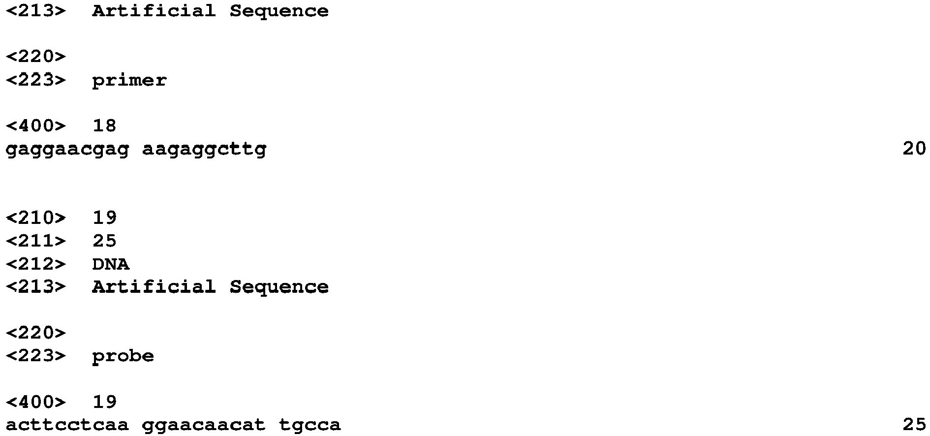
Claims (14)
- A method for measuring a viral antigen in a sample by using a capture antibody and a detection antibody, the method comprisingforming a sandwich immune complex that contains the capture antibody, the viral antigen and the detection antibody,the capture antibody comprising a heavy chain variable region that comprises CDR1 comprising amino acid sequence of SEQ ID NO: 1, CDR2 comprising amino acid sequence of SEQ ID NO: 2 and CDR3 comprising amino acid sequence of SEQ ID NO: 3, and a light chain variable region that comprises CDR1 comprising amino acid sequence of SEQ ID NO: 4, CDR2 comprising amino acid sequence of SEQ ID NO: 5 and CDR3 comprising amino acid sequence of SEQ ID NO: 6, andthe detection antibody comprising a heavy chain variable region that comprises CDR1 comprising amino acid sequence of SEQ ID NO: 7, CDR2 comprising amino acid sequence of SEQ ID NO: 8 and CDR3 comprising amino acid sequence of SEQ ID NO: 9, and a light chain variable region that comprises CDR1 comprising amino acid sequence of SEQ ID NO: 10, CDR2 comprising amino acid sequence of SEQ ID NO: 11 and CDR3 comprising amino acid sequence of SEQ ID NO: 12.
- An antibody set for measuring a viral antigen in a sample by an immunological measurement method,the antibody comprising a capture antibody and a detection antibody that form a sandwich immune complex with the viral antigen in the immunological measurement method,the capture antibody comprising a heavy chain variable region that comprises CDR1 comprising amino acid sequence of SEQ ID NO: 1, CDR2 comprising amino acid sequence of SEQ ID NO: 2 and CDR3 comprising amino acid sequence of SEQ ID NO: 3; and a light chain variable region that comprises CDR1 comprising the amino acid sequence of SEQ ID NO: 4, CDR2 comprising the amino acid sequence of SEQ ID NO: 5 and CDR3 comprising the amino acid sequence of SEQ ID NO: 6, andthe detection antibody comprises a heavy chain variable region that comprises CDR1 comprising amino acid sequence of SEQ ID NO: 7, CDR2 comprising amino acid sequence of SEQ ID NO: 8 and CDR3 comprising amino acid sequence of SEQ ID NO: 9; and a light chain variable region that comprises CDR1 comprising amino acid sequence of SEQ ID NO: 10, CDR2 comprising amino sequence of SEQ ID NO: 11 and CDR3 comprising amino acid sequence of SEQ ID NO: 12.
- The method according to claim 1 or the antibody set according to claim 2, wherein the capture antibody and the detection antibody form the sandwich immune complex with a nucleoprotein of SARS-CoV or SARS-CoV-2, but do not form the sandwich immune complex with a nucleoprotein of MERS-CoV, and viral antigens of HCoV-OC43, HCoV-NL63 and HCoV-229.
- The method or the antibody set according to any one of claims 1 to 3, whereinthe capture antibody comprises the heavy chain variable region comprising amino acid sequence of SEQ ID NO: 13, and the light chain variable region comprising amino acid sequence of SEQ ID NO: 14, andthe detection antibody comprises the heavy chain variable region comprising amino acid sequence of SEQ ID NO: 15, and the light chain variable region comprising amino acid sequence of SEQ ID NO: 16.
- The method or the antibody set according to any one of claims 1 to 4, wherein the capture antibody is IgG, reduced IgG, Fab, Fab', F(ab')2, Fv, scFv, diabody or triabody.
- The method or the antibody set according to any one of claims 1 to 4, wherein the detection antibody is IgG, reduced IgG, Fab, Fab', F(ab')2, Fv, scFv, diabody or triabody.
- The method or the antibody set according to any one of claims 1 to 6, wherein the sample is a biological sample.
- The method or the antibody set according to any one of claims 1 to 7, wherein the viral antigen is a nucleoprotein of SARS-CoV or SARS-CoV-2.
- The method or the antibody set according to any one of claims 1 to 8, wherein the capture antibody is immobilized on a solid phase.
- The method or the antibody set according to claim 9, wherein the solid phase is a magnetic particle or a microplate.
- The method or the antibody set according to any one of claims 1 to 10, wherein the detection antibody is labeled with a labeling substance.
- The method or the antibody set according to claim 11, wherein the labeling substance is at least one enzyme selected from the group consisting of alkaline phosphatase, peroxidase, β-galactosidase, glucosidase, polyphenol oxidase, tyrosinase, acid phosphatase and luciferase.
- A reagent kit comprising the capture antibody and the detection antibody described in any one of claims 1 to 12.
- The reagent kit according to claim 13, comprising a first reagent and a second reagent, the first reagent comprising the capture antibody and the second reagent comprising the detection antibody.
Applications Claiming Priority (1)
| Application Number | Priority Date | Filing Date | Title |
|---|---|---|---|
| JP2020128480A JP6960508B1 (en) | 2020-07-29 | 2020-07-29 | Methods for measuring viral antigens in samples, antibody sets and reagent kits |
Publications (1)
| Publication Number | Publication Date |
|---|---|
| EP3945319A1 true EP3945319A1 (en) | 2022-02-02 |
Family
ID=77367240
Family Applications (1)
| Application Number | Title | Priority Date | Filing Date |
|---|---|---|---|
| EP21188360.8A Pending EP3945319A1 (en) | 2020-07-29 | 2021-07-29 | Method for measuring viral antigen in sample, antibody set, and reagent kit |
Country Status (4)
| Country | Link |
|---|---|
| US (1) | US12320809B2 (en) |
| EP (1) | EP3945319A1 (en) |
| JP (1) | JP6960508B1 (en) |
| CN (1) | CN114062670B (en) |
Families Citing this family (5)
| Publication number | Priority date | Publication date | Assignee | Title |
|---|---|---|---|---|
| JP7728100B2 (en) * | 2021-05-11 | 2025-08-22 | 富士レビオ株式会社 | Immunological detection method and reagent for SARS-CoV-2 |
| JP7216948B1 (en) * | 2021-06-16 | 2023-02-02 | 積水メディカル株式会社 | SARS-CoV-2 immunoassay method and immunoassay kit, and monoclonal antibody or antibody fragment thereof |
| JP7105970B1 (en) | 2021-06-16 | 2022-07-25 | 積水メディカル株式会社 | SARS-CoV-2 immunoassay method and immunoassay kit |
| CN115718197B (en) * | 2022-09-03 | 2023-07-04 | 长沙海柯生物科技有限公司 | Kit for detecting content of free lgE and application thereof |
| CN118067981B (en) * | 2024-04-19 | 2024-12-31 | 大连泰嘉瑞佰科技有限公司 | Kit for microorganism detection and application thereof |
Citations (6)
| Publication number | Priority date | Publication date | Assignee | Title |
|---|---|---|---|---|
| JPH01254868A (en) | 1988-04-05 | 1989-10-11 | Eiji Ishikawa | Method of measuring antigen material with extra-high sensitivity |
| US20080254440A1 (en) * | 2003-10-31 | 2008-10-16 | Yoshiaki Uchida | Anti-Sars Virus Antibody, Hybridoma Producing the Antibody and Immunoassay Reagent Using the Antibody |
| US20090280507A1 (en) * | 2005-10-11 | 2009-11-12 | Sysmex Corporation | Method for measurement of sars virus nucleocapsid protein, reagent kit for the measurement, test device, monoclonal antibody directed against sars virus nucleocapsid protein, and hybridoma capable of producing the monoclonal antibody |
| US20180179298A1 (en) | 2016-12-28 | 2018-06-28 | Sysmex Corporation | Method for controlling affinity of antibody for antigen, antibody whose affinity for antigen has been altered, and its production method |
| US20190040119A1 (en) | 2017-08-04 | 2019-02-07 | Sysmex Corporation | Method for producing antibody |
| CN111153991A (en) * | 2020-02-26 | 2020-05-15 | 北京博奥森生物技术有限公司 | Human SARS-CoV-2 monoclonal antibody and its preparation method and use |
Family Cites Families (18)
| Publication number | Priority date | Publication date | Assignee | Title |
|---|---|---|---|---|
| JP5431644B2 (en) * | 2006-12-28 | 2014-03-05 | シスメックス株式会社 | Examination method of respiratory infection |
| JP2009085753A (en) * | 2007-09-28 | 2009-04-23 | Sysmex Corp | Sandwich immunoassay |
| JP5340575B2 (en) * | 2007-09-28 | 2013-11-13 | シスメックス株式会社 | Immunochromatographic test equipment |
| JP2009109426A (en) | 2007-10-31 | 2009-05-21 | Sysmex Corp | Analyte pretreatment liquid, virus measuring kit and virus detecting method |
| JP2009236685A (en) * | 2008-03-27 | 2009-10-15 | Sysmex Corp | Chromatographic test device |
| JP6022441B2 (en) * | 2010-04-30 | 2016-11-09 | アレクシオン ファーマシューティカルズ, インコーポレイテッド | Anti-C5a antibodies and methods for use of the antibodies |
| WO2015035260A1 (en) * | 2013-09-06 | 2015-03-12 | Theranos, Inc. | Systems and methods for detecting infectious diseases |
| CN111398583A (en) * | 2020-02-13 | 2020-07-10 | 北京华科泰生物技术股份有限公司 | Kit for detecting novel coronavirus N protein and application thereof |
| CN111398589A (en) * | 2020-02-13 | 2020-07-10 | 北京华科泰生物技术股份有限公司 | Immunochromatography kit for rapidly detecting novel coronavirus N protein and preparation method and application thereof |
| CN111239391B (en) * | 2020-02-19 | 2024-05-03 | 南开大学 | 2019-NCoV novel coronavirus antigen detection reagent and detection device |
| CN111303254A (en) * | 2020-02-20 | 2020-06-19 | 北京新创生物工程有限公司 | Novel coronavirus (SARS-CoV-2) antigen detection kit |
| CN111303280B (en) * | 2020-03-22 | 2022-01-07 | 中国人民解放军军事科学院军事医学研究院 | High-neutralization-activity anti-SARS-CoV-2 fully human monoclonal antibody and application |
| CN111423508A (en) * | 2020-03-31 | 2020-07-17 | 江苏省疾病预防控制中心(江苏省公共卫生研究院) | Separated SARS-CoV-2 protein binding molecule for resisting virus infection |
| CN111704666B (en) * | 2020-04-22 | 2023-09-26 | 北京科卫临床诊断试剂有限公司 | Paired monoclonal antibodies against novel coronavirus N protein and their applications |
| US20220056112A1 (en) * | 2020-08-11 | 2022-02-24 | Yurogen Biosystems Llc | MONOCLONAL ANTIBODIES AGAINST SARS-CoV-2 NUCLEOCAPSID PROTEIN AND USES THEREOF |
| US20230391855A1 (en) * | 2020-10-21 | 2023-12-07 | La Jolla Institute For Immunology | Chimeric anti-sars-cov2 nucleoprotein antibodies |
| CN116568710A (en) * | 2020-10-28 | 2023-08-08 | 奥索临床诊断有限公司 | High-Throughput Immunoassays and Methods for Detection of SARS-CoV-2 Antigens |
| CN112229994B (en) * | 2020-12-10 | 2021-03-16 | 丹娜(天津)生物科技股份有限公司 | Novel coronavirus antibody detection kit based on magnetic particle chemiluminescence |
-
2020
- 2020-07-29 JP JP2020128480A patent/JP6960508B1/en active Active
-
2021
- 2021-07-14 CN CN202110793411.0A patent/CN114062670B/en active Active
- 2021-07-28 US US17/387,409 patent/US12320809B2/en active Active
- 2021-07-29 EP EP21188360.8A patent/EP3945319A1/en active Pending
Patent Citations (6)
| Publication number | Priority date | Publication date | Assignee | Title |
|---|---|---|---|---|
| JPH01254868A (en) | 1988-04-05 | 1989-10-11 | Eiji Ishikawa | Method of measuring antigen material with extra-high sensitivity |
| US20080254440A1 (en) * | 2003-10-31 | 2008-10-16 | Yoshiaki Uchida | Anti-Sars Virus Antibody, Hybridoma Producing the Antibody and Immunoassay Reagent Using the Antibody |
| US20090280507A1 (en) * | 2005-10-11 | 2009-11-12 | Sysmex Corporation | Method for measurement of sars virus nucleocapsid protein, reagent kit for the measurement, test device, monoclonal antibody directed against sars virus nucleocapsid protein, and hybridoma capable of producing the monoclonal antibody |
| US20180179298A1 (en) | 2016-12-28 | 2018-06-28 | Sysmex Corporation | Method for controlling affinity of antibody for antigen, antibody whose affinity for antigen has been altered, and its production method |
| US20190040119A1 (en) | 2017-08-04 | 2019-02-07 | Sysmex Corporation | Method for producing antibody |
| CN111153991A (en) * | 2020-02-26 | 2020-05-15 | 北京博奥森生物技术有限公司 | Human SARS-CoV-2 monoclonal antibody and its preparation method and use |
Non-Patent Citations (6)
| Title |
|---|
| "Manual for the Detection of Pathogen 2019-nCoV Ver. 2.9.1", 19 March 2020 |
| CO M.S. ET AL., PROC. NATL. ACAD. SCI. USA, vol. 93, 1996, pages 7843 - 7848 |
| FUJIMOTO K ET AL., J. CLIN. MICROBIOL., vol. 46, 2008, pages 302 - 310 |
| JONES P.T. ET AL., NATURE, vol. 321, 1986, pages 522 - 525 |
| KABAT E.A. ET AL.: "Sequences of Proteins of Immunological Interest", 1991, PUBLIC HEALTH SERVICE, NATIONAL INSTITUTES OF HEALTH |
| KOHLER G.MILSTEIN C., NATURE, vol. 256, 1975, pages 495 - 497 |
Also Published As
| Publication number | Publication date |
|---|---|
| US20220034883A1 (en) | 2022-02-03 |
| CN114062670A (en) | 2022-02-18 |
| JP2022025577A (en) | 2022-02-10 |
| US12320809B2 (en) | 2025-06-03 |
| CN114062670B (en) | 2025-02-25 |
| JP6960508B1 (en) | 2021-11-05 |
Similar Documents
| Publication | Publication Date | Title |
|---|---|---|
| US12320809B2 (en) | Method for measuring viral antigen in sample, antibody set, and reagent kit | |
| US20100055675A1 (en) | Method for detecting measles virus, membrane assay test device, and membrane assay test kit | |
| Kammila et al. | A rapid point of care immunoswab assay for SARS-CoV detection | |
| CN112679605A (en) | Antibodies or antigen binding fragments thereof against novel coronavirus nucleocapsid proteins and uses thereof | |
| JP2022174541A (en) | Immunological detection method and reagent for sars-cov-2 | |
| US20220120737A1 (en) | Method for detecting sars-cov-2-specific serum human immunoglobulins | |
| JP2024012473A (en) | Detection reagent or kit for detecting RS virus and method for detecting RS virus | |
| JP7308914B2 (en) | Serological detection method for viral antigens | |
| WO2015037635A1 (en) | Sample processing method for influenza virus immunoassay, and immunoassay method | |
| JP7455947B2 (en) | Adenovirus immunoassay method and immunoassay device | |
| JP2024003083A (en) | Detection reagent or kit for detecting RS virus and method for detecting RS virus | |
| JP7671791B2 (en) | Method and instrument for immunoassay of adenovirus | |
| CA3125206A1 (en) | Specific monoclonal antibodies for the hepatitis pb2 of the influenza humana (flu), nucleic acid sequences; method and kit of inprol produced by flu | |
| CN120623326A (en) | An antibody or antigen-binding fragment thereof and related products and applications | |
| CN116425866B (en) | HPV-related protein monoclonal antibody and its application | |
| JP2023126867A (en) | Adenovirus immunoassay method and immunoassay device | |
| US20180364231A1 (en) | Method and kit for simultaneously detecting human parvovirus b19 antigen and antibody | |
| JP6641841B2 (en) | Method for measuring human parvovirus B19 antigen | |
| JP7595632B2 (en) | Method and instrument for immunoassay of adenovirus | |
| WO2024166920A1 (en) | Anti-sars-cov-2 monoclonal antibody and immunoassay method and immunoassay instrument for sars-cov-2 using same | |
| CN115290892A (en) | Application of a buffer in the detection kit of brain-specific protein product 9.5 |
Legal Events
| Date | Code | Title | Description |
|---|---|---|---|
| PUAI | Public reference made under article 153(3) epc to a published international application that has entered the european phase |
Free format text: ORIGINAL CODE: 0009012 |
|
| STAA | Information on the status of an ep patent application or granted ep patent |
Free format text: STATUS: THE APPLICATION HAS BEEN PUBLISHED |
|
| AK | Designated contracting states |
Kind code of ref document: A1 Designated state(s): AL AT BE BG CH CY CZ DE DK EE ES FI FR GB GR HR HU IE IS IT LI LT LU LV MC MK MT NL NO PL PT RO RS SE SI SK SM TR |
|
| STAA | Information on the status of an ep patent application or granted ep patent |
Free format text: STATUS: REQUEST FOR EXAMINATION WAS MADE |
|
| 17P | Request for examination filed |
Effective date: 20220708 |
|
| RBV | Designated contracting states (corrected) |
Designated state(s): AL AT BE BG CH CY CZ DE DK EE ES FI FR GB GR HR HU IE IS IT LI LT LU LV MC MK MT NL NO PL PT RO RS SE SI SK SM TR |
|
| STAA | Information on the status of an ep patent application or granted ep patent |
Free format text: STATUS: EXAMINATION IS IN PROGRESS |
|
| 17Q | First examination report despatched |
Effective date: 20250929 |
|
| STAA | Information on the status of an ep patent application or granted ep patent |
Free format text: STATUS: THE APPLICATION IS DEEMED TO BE WITHDRAWN |