EP3944253B1 - Maschinenlernen von verrauschten etiketten zur anomaliebewertung in der medizinischen bildgebung - Google Patents
Maschinenlernen von verrauschten etiketten zur anomaliebewertung in der medizinischen bildgebung Download PDFInfo
- Publication number
- EP3944253B1 EP3944253B1 EP21186575.3A EP21186575A EP3944253B1 EP 3944253 B1 EP3944253 B1 EP 3944253B1 EP 21186575 A EP21186575 A EP 21186575A EP 3944253 B1 EP3944253 B1 EP 3944253B1
- Authority
- EP
- European Patent Office
- Prior art keywords
- machine
- regularization
- abnormality
- noise
- training
- Prior art date
- Legal status (The legal status is an assumption and is not a legal conclusion. Google has not performed a legal analysis and makes no representation as to the accuracy of the status listed.)
- Active
Links
Images
Classifications
-
- G—PHYSICS
- G16—INFORMATION AND COMMUNICATION TECHNOLOGY [ICT] SPECIALLY ADAPTED FOR SPECIFIC APPLICATION FIELDS
- G16H—HEALTHCARE INFORMATICS, i.e. INFORMATION AND COMMUNICATION TECHNOLOGY [ICT] SPECIALLY ADAPTED FOR THE HANDLING OR PROCESSING OF MEDICAL OR HEALTHCARE DATA
- G16H50/00—ICT specially adapted for medical diagnosis, medical simulation or medical data mining; ICT specially adapted for detecting, monitoring or modelling epidemics or pandemics
- G16H50/20—ICT specially adapted for medical diagnosis, medical simulation or medical data mining; ICT specially adapted for detecting, monitoring or modelling epidemics or pandemics for computer-aided diagnosis, e.g. based on medical expert systems
-
- G—PHYSICS
- G06—COMPUTING OR CALCULATING; COUNTING
- G06F—ELECTRIC DIGITAL DATA PROCESSING
- G06F18/00—Pattern recognition
- G06F18/20—Analysing
- G06F18/21—Design or setup of recognition systems or techniques; Extraction of features in feature space; Blind source separation
- G06F18/214—Generating training patterns; Bootstrap methods, e.g. bagging or boosting
-
- G—PHYSICS
- G06—COMPUTING OR CALCULATING; COUNTING
- G06F—ELECTRIC DIGITAL DATA PROCESSING
- G06F18/00—Pattern recognition
- G06F18/20—Analysing
- G06F18/24—Classification techniques
-
- G—PHYSICS
- G06—COMPUTING OR CALCULATING; COUNTING
- G06N—COMPUTING ARRANGEMENTS BASED ON SPECIFIC COMPUTATIONAL MODELS
- G06N20/00—Machine learning
-
- G—PHYSICS
- G06—COMPUTING OR CALCULATING; COUNTING
- G06N—COMPUTING ARRANGEMENTS BASED ON SPECIFIC COMPUTATIONAL MODELS
- G06N3/00—Computing arrangements based on biological models
- G06N3/02—Neural networks
- G06N3/04—Architecture, e.g. interconnection topology
- G06N3/045—Combinations of networks
-
- G—PHYSICS
- G06—COMPUTING OR CALCULATING; COUNTING
- G06N—COMPUTING ARRANGEMENTS BASED ON SPECIFIC COMPUTATIONAL MODELS
- G06N3/00—Computing arrangements based on biological models
- G06N3/02—Neural networks
- G06N3/08—Learning methods
-
- G—PHYSICS
- G06—COMPUTING OR CALCULATING; COUNTING
- G06T—IMAGE DATA PROCESSING OR GENERATION, IN GENERAL
- G06T5/00—Image enhancement or restoration
- G06T5/70—Denoising; Smoothing
-
- G—PHYSICS
- G06—COMPUTING OR CALCULATING; COUNTING
- G06T—IMAGE DATA PROCESSING OR GENERATION, IN GENERAL
- G06T7/00—Image analysis
- G06T7/0002—Inspection of images, e.g. flaw detection
- G06T7/0012—Biomedical image inspection
-
- G—PHYSICS
- G16—INFORMATION AND COMMUNICATION TECHNOLOGY [ICT] SPECIALLY ADAPTED FOR SPECIFIC APPLICATION FIELDS
- G16H—HEALTHCARE INFORMATICS, i.e. INFORMATION AND COMMUNICATION TECHNOLOGY [ICT] SPECIALLY ADAPTED FOR THE HANDLING OR PROCESSING OF MEDICAL OR HEALTHCARE DATA
- G16H30/00—ICT specially adapted for the handling or processing of medical images
- G16H30/40—ICT specially adapted for the handling or processing of medical images for processing medical images, e.g. editing
-
- G—PHYSICS
- G16—INFORMATION AND COMMUNICATION TECHNOLOGY [ICT] SPECIALLY ADAPTED FOR SPECIFIC APPLICATION FIELDS
- G16H—HEALTHCARE INFORMATICS, i.e. INFORMATION AND COMMUNICATION TECHNOLOGY [ICT] SPECIALLY ADAPTED FOR THE HANDLING OR PROCESSING OF MEDICAL OR HEALTHCARE DATA
- G16H50/00—ICT specially adapted for medical diagnosis, medical simulation or medical data mining; ICT specially adapted for detecting, monitoring or modelling epidemics or pandemics
- G16H50/70—ICT specially adapted for medical diagnosis, medical simulation or medical data mining; ICT specially adapted for detecting, monitoring or modelling epidemics or pandemics for mining of medical data, e.g. analysing previous cases of other patients
-
- G—PHYSICS
- G06—COMPUTING OR CALCULATING; COUNTING
- G06T—IMAGE DATA PROCESSING OR GENERATION, IN GENERAL
- G06T2207/00—Indexing scheme for image analysis or image enhancement
- G06T2207/10—Image acquisition modality
- G06T2207/10072—Tomographic images
-
- G—PHYSICS
- G06—COMPUTING OR CALCULATING; COUNTING
- G06T—IMAGE DATA PROCESSING OR GENERATION, IN GENERAL
- G06T2207/00—Indexing scheme for image analysis or image enhancement
- G06T2207/10—Image acquisition modality
- G06T2207/10116—X-ray image
-
- G—PHYSICS
- G06—COMPUTING OR CALCULATING; COUNTING
- G06T—IMAGE DATA PROCESSING OR GENERATION, IN GENERAL
- G06T2207/00—Indexing scheme for image analysis or image enhancement
- G06T2207/20—Special algorithmic details
- G06T2207/20081—Training; Learning
-
- G—PHYSICS
- G06—COMPUTING OR CALCULATING; COUNTING
- G06T—IMAGE DATA PROCESSING OR GENERATION, IN GENERAL
- G06T2207/00—Indexing scheme for image analysis or image enhancement
- G06T2207/20—Special algorithmic details
- G06T2207/20084—Artificial neural networks [ANN]
-
- G—PHYSICS
- G06—COMPUTING OR CALCULATING; COUNTING
- G06T—IMAGE DATA PROCESSING OR GENERATION, IN GENERAL
- G06T2207/00—Indexing scheme for image analysis or image enhancement
- G06T2207/30—Subject of image; Context of image processing
- G06T2207/30004—Biomedical image processing
- G06T2207/30061—Lung
Definitions
- the present embodiments relate to machine learning.
- Machine learning algorithms have shown great promise for the computer-aided classification of medical images.
- machine learning is used to develop automated chest radiograph systems.
- the assessment of chest radiographs is used for detection of thoracic diseases and abnormalities.
- developing these systems is challenging because of the high inter-rater variability in the interpretation of chest radiographs.
- High error rates in annotations due to the methods of annotation, e.g., natural language processing (NLP)-based methods, and inherent ambiguity in pathology appearance lead to incorrect dataset labels.
- NLP natural language processing
- Deep learning methods which are known to perform well in other domains, may still be overconfident.
- radiologist-re-annotated test sets may be used to train.
- Predictive uncertainty may be estimated as an orthogonal measure to the predicted abnormality probability using subjective logic.
- the label noise may still result in poor performing machine-learned models.
- the preferred embodiments described below include methods, systems, instructions, and computer readable media for machine learning for abnormality assessment in medical imaging and application of a machine-learned model.
- the machine learning uses regularization of the loss, such as regularization being used for training for abnormality classification in chest radiographs.
- the regularization may be a noise and/or correlation regularization directed to the noisy ground truth labels of the training data.
- the resulting machine-learned model may better classify abnormalities in medical images due to the use of the noise and/or correlation regularization in the training.
- a method for machine learning abnormality assessment in medical imaging by a machine.
- Training data including medical images and ground truth labels for the medical images is obtained.
- the ground truth labels designate any abnormality represented by the medical images.
- the machine machine trains a model from the training data.
- the machine training uses a loss function including a regularization.
- the regularization is a noise regularization and/or a correlation regularization.
- the model resulting from the machine training is stored in a memory.
- the machine training includes machine training with the loss function being a cross-entropy function comparing a classification of abnormality output of the model with the ground truth labels. Other loss functions may be used.
- the machine training includes machine training with the ground truth labels being binary labels for absence or presence of the abnormality and the loss function being weighted as a function of number of positive and number of negative instances of the abnormality in the medical images of the training data. Other labels, such as grades or scores, may be used.
- the regularization of the loss function is the noise regularization.
- a noise level of the ground truth labels is measured.
- the machine training includes machine training with the noise regularization being a function of the noise level.
- the noise level is represented by a specificity and a sensitivity of the ground truth labels for the abnormality.
- the noise regularization includes a first weight that is a function of the specificity and a second weight that is a function of the sensitivity or any other measure which describes a noise ratio of the labels.
- the noise regularization may be any function, such as an inverse binary cross-entropy function.
- the ground truth labels designate at least first and second types of abnormalities.
- the regularization of the loss function is the correlation regularization.
- the correlation regularization correlates the ground truth labels for the first type of abnormality to the ground truth labels for the second type of abnormality.
- the correlation regularization is a covariance. For example, at least four types of abnormalities are provided.
- the correlation regularization is a sum of the covariance between all of the at least four types of abnormalities.
- both the noise regularization and the correlation regularization are used to train.
- the medical images of the training data are chest radiographs, and the abnormalities include effusion, cardiomegaly, consolidation, atelectasis, and mass.
- the model resulting from the machine training is applied to a patient image for a patient.
- the application outputs a classification of the patient image has having or not having any abnormality.
- a system for abnormality detection in medical imaging.
- a medical imaging system configured to generate an image of a patient.
- a processor is configured to apply a machine-learned model to the image of the patient.
- the machine-learned model was trained with noise and/or correlation regularization to detect an abnormality in the image.
- a display is configured to display a classification of the patient as having or not having the abnormality based on the detection from the application.
- the machine-learned model was trained with the noise regularization.
- the noise regularization accounts for noise in ground truth labels used in machine training.
- the machine-learned model was trained with correlation regularization accounting for mischaracterization between different types of abnormalities.
- the noise or correlation regularization may be for ground truth labels for abnormalities in x-ray images from an x-ray imaging system.
- a system for machine training for abnormality classification.
- a memory is configured to store training data including images of anatomy and ground truth classifications for the images and to store a machine-learned classifier.
- a processor is configured to machine train from the training data.
- the machine training includes calculation of loss with a noise and/or correlation regularization.
- the processor is configured to machine train with the loss, resulting in the machine-learned classifier.
- the processor is configured to machine train with the noise regularization. In another embodiment, the processor is configured to machine train with the correlation regularization.
- Machine-learning improves the generalization of abnormality classification based on label error rates assessment in chest radiography or other medical imaging.
- Different regularization techniques may deal with label noise, such as dropout regularization or dimensionality-driven learning strategies.
- Regularization may be applied in many medical imaging fields such as image reconstruction or image segmentation.
- regularization is applied on the classification loss.
- regularization is applied on the classification loss.
- Two example regularization components are noise regularization based on the calculation of prior label noise probabilities and correlation regularization based on correlation between abnormalities. Both noise and correlation regularization lead to an improvement in terms of the generalization performance of abnormality detection and classification.
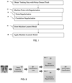
- Figure 1 shows one embodiment of a method for machine learning abnormality assessment in medical imaging by a machine.
- regularization such as noise, correlation, or drop-out
- CT computed tomography
- MR magnetic resonance
- SPECT single photon emission computed tomography
- PET positron emission tomography
- the regularization is at least a noise and/or correlation regularization.
- the noise and/or correlation regularization for medical image classification reduces the effects of noisy labels in the training data.
- the method is implemented by the system of Figure 4 or another system.
- the method is implemented by the system 40 for training, including a memory 41 to store training data and the learned model and a processor 43 to perform machine training with the regularizer.
- the system 40 for training including a memory 41 to store training data and the learned model and a processor 43 to perform machine training with the regularizer.
- Different devices may be used.
- acts for designing an architecture of the model are included.
- act 16 may be applied after act 18.
- acts 13 and 14 may be performed in any order or simultaneously (e.g., where noise and correlation regularization terms are both included in the loss function).
- training data is obtained.
- the data is obtained by searching, data mining, loading from memory, identifying, transfer over a computer network, and/or gathering.
- a designer e.g., computer scientist
- obtains the training data such as data for a particular type of medical imaging, organ of interest, disease of interest, and/or abnormality or abnormalities of interest.
- a computer, study, and/or database may be used to obtain the data.
- the training data includes medical images. Tens, hundreds, or thousands of sample medical images are obtained. For example, x-ray radiographs from many different patients are obtained. Actual medical images from patients may be used. Alternatively, simulation of medical imaging is used to generate the medical images. In yet other embodiments, images of phantoms are used.
- the medical images of the training data may be from multiple sources, such as actual images of patients, simulation, and imaging of phantoms. Any subset of data for any domain (e.g., ultrasound, MR, CT, PET, or SPECT) may be used. Chest radiographs are used as an example herein as chest radiographs tend to have noisy ground truths and/or a large number of types of abnormalities represented in the images.
- sample images Other information may be included with the sample images. For example, clinical and/or lab results for the patients associated with the images are included. The age, weight, smoking history, blood work, and/or other information may be provided as samples with the medical images to train the classifier to detect abnormalities from input images and other types of information. In other embodiments, only medical images are used in the samples of the training data.
- the training data includes ground truth labels for each of the samples.
- the ground truth labels are mined from patient records, indicated by a measure (e.g., application of another classifier), and/or provided by expert review of the samples.
- the ground truth labels are for the existence or not of the abnormality, the location of the abnormality, and/or an extent or level of the abnormality (e.g., size or score).
- the ground truth label is provided for each type of abnormality for each sample. For example, one chest radiograph includes a positive label for one type of abnormality and a negative label for another type of abnormality.
- the ground truth labels designate any abnormality represented by each of the samples (e.g., medical images).
- the ground truth labels may be noisy. Some of the labels may be incorrect. Since the machine learning relies on accuracy of the ground truth labels to learn to classify whether images include abnormalities, the noisy labels introduce error in the trained classifier. The error may be due to incorrect labeling in a binary sense (e.g., abnormality X is represented or is not) and/or in an incorrect identification sense (e.g., a mass is labeled as an effusion).
- a binary sense e.g., abnormality X is represented or is not
- an incorrect identification sense e.g., a mass is labeled as an effusion
- the samples and labels may be for any number or types of abnormalities.
- the labels are for a single type of abnormality (e.g., cancerous lesion).
- Each sample is labeled with a ground truth for whether or not the abnormality is represented in the sample.
- the labels are for two or more, three or more, or four or more types of abnormalities.
- the types of abnormalities include effusion, cardiomegaly, consolidation, atelectasis, and mass. Additional, different, or fewer types of abnormalities may be classified or labeled.
- Each sample e.g., medical image of the training set
- a machine performs machine training.
- a processor or computer uses the training data to machine learn.
- a model is defined and trained by establishing values for learnable parameters based on the training data. The samples are input and resulting outputs are compared to the ground truth labels. Through optimization (e.g., Adam), the training data is used to establish the values for the learnable parameters of the defined model that result in accurate output.
- Adam e.g., the training data is used to establish the values for the learnable parameters of the defined model that result in accurate output.
- Any training may be used, such as deep learning for a neural network.
- a support vector machine, regression, or other machine learning and corresponding model may be used.
- deep learning is used.
- the machine trains the network to output a classification (e.g., detection or not of an abnormality) in response to an input sample (e.g., medical image).
- the machine trains the network through regression.
- the neural network is a fully connected network (FCN) or a convolutional neural network. Other models may be used.
- the defined model is trained to estimate with a loss function.
- Any loss function may be used, such as a cross-entropy function, L2 (e.g., least squares error), L1 distance, or other loss to obtain optimal values for the network parameters.
- L2 e.g., least squares error
- L1 distance e.g., L1 distance
- the difference between the ground truth labels for the training images and the predictions by the model are minimized based on the measure of loss or difference by the loss function. Through optimization, the values of the learnable parameters are adjusted to minimize the loss.
- the loss function includes regularization.
- the regularizer may be a term summed with the loss.
- the regularizer is a weight or adaptive alteration in the loss calculation that accounts for the noisy labels.
- the regularization is a noise regularization.
- the regularization is a correlation regularization.
- both the noise regularization and correlation regularization are used.
- Other regularizations may additionally or alternatively be used, such as drop-out regularization and/or dimensionality-driven learning.
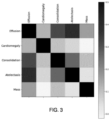
- Figure 2 shows an example arrangement or architecture for machine training as a pipeline.
- the images 20 are used as inputs to the deep learning architecture 22 (i.e., model of interrelated learnable parameters).
- the output of the model and the ground truth are used to determine the classification loss 24 during training.
- the classification loss 24 includes noise regularization 25 and/or correlation regularization 26.
- the model outputs the abnormality classification 28 without loss and/or regularization.
- the previously used loss and regularization provides the values for the model of the architecture 22 to provide an accurate abnormality classification 28.
- the ground truth labels of the training data are binary labels for the absence or presence of the abnormality in the sample, so the loss is based on binary prediction for many or all samples.
- the labels are continuous values or have more than two discrete values, so the loss is based on accuracy along the continuum or across the discrete set.
- the labels are the set of [c (1) c (2) . . . c (d) ] ⁇ 0, 1 ⁇ (absence or presence of the abnormality, respectively) and are compared with the network output [p (1) p (2) ... p (d) ] ⁇ [0, 1].
- the loss is measured based on the comparison.
- the loss function provides the comparison.
- the loss function includes noise regularization.
- the machine training is performed with the regularization of the loss function including noise regularization.
- the noise level of the ground truth labels is measured and used to regularize. For example, the specificity and/or sensitivity of the ground truth labels of the training set are used to regularize.
- an expert reading procedure is defined. Expert radiologists read the samples and blindly re-labeled the samples. Without access to the ground truth labels and/or classification by other experts, the expert or experts classify (i.e., identify the ground truth) for each sample. The original dataset labels were not provided during the expert reading process to avoid a biased decision towards the original labels. Multiple experts may perform the reading, providing multiple instances of ground truth labels for each sample. For all cases where consensus was not reached on all labels through the independent read, an open discussion or majority vote may be carried out to establish consensus labels. Assuming that the re-defined labels are the correct labels, prior probabilities are calculated with the original and re-defined labels.
- Table 1 show sensitivity s sens and specificity s spec of five selected types of abnormalities for chest radiographs for the original ground truth labels verses expert re-labeling.
- Table 1 Abnormality s sens s spec Effusion 0.300 0.966 Cardiomegaly 0.342 0.986 Consolidation 0.129 0.949 Atelectasis 0.221 0.970 Mass 0.364 0.972 Average 0.271 0.969 Low scores indicate stronger label noise.
- noise regularization To incorporate the noise regularization into the loss function, a term is added to the loss function. Any regularization term may be used, such as an inverse binary cross-entropy function.
- the added term is a noise regularization, which is a function of the level of noise. Any function may be used. In one embodiment, two weights are added where one weight is a function of the specificity and another weight is a function of the sensitivity. In other embodiments, only sensitivity, only specificity, or another measure of noise level is used.
- the noise regularization as an inverse binary cross-entropy function is added to the loss function of equation 1.
- the additional parameter ⁇ noise is another weight to define the overall influence of the regularization term. Any value may be used for the additional parameter, such as 0.1.
- the noise may be integrated into the regularization and/or loss function in a different way, such as a weight, ratio, subtraction, or use of different regularization function (e.g., L2). Instead of weights, the noise level may be integrated through addition, subtraction, or other function.
- correlation regularization is used in the loss function during machine training.
- the correlation regularization uses correlation of the ground truth labels for the first type of abnormality to the ground truth labels for the second type of abnormality. More strongly correlated abnormalities are more likely to be miss-classified in the ground truth labels, introducing a source of label noise.
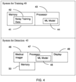
- Figure 3 shows an example in chest radiography. The strength of correlation between each abnormality is visualized. The level of correlation between each of five different types of abnormalities (effusion, cardiomegaly, consolidation, atelectasis, and mass) is shown graphically. The darker colors correspond to the level of correlation where black is full correlation (e.g., correlation coefficient is 1.0) and white is no correlation (e.g., correlation coefficient is 0.0). The correlations in these examples are 1.0 for the abnormality's correlation with itself and between 0.0 and 0.5 for correlations between different abnormalities.
- the correlation regularization is based on how strongly a set of class labels c (n) for abnormality n correlate with a set of class labels c(r) for abnormality r where r ⁇ ⁇ 1...D ⁇ n ⁇ . D denotes the number of abnormalities.
- Any correlation coefficient may be used as the measure of correlation.
- Figure 3 uses the Pearson correlation coefficient. In other embodiments, the correlation coefficient is the covariance.
- a term is added to the loss function.
- Any regularization term may be used, such as a cross-entropy function.
- the added term is a correlation regularization, which is a function of the levels of correlation among the different types of abnormalities to be classified. Any function may be used.
- a sum across the different types of abnormalities of the cross-entropy weighted by the correlation coefficient is added. For example, the sum across two, three, four, or more types of abnormalities, such as the sum across five abnormalities of the example of Figure 3 , is used.
- ⁇ corr is a weight (e.g., set at 1.0) and conv (n,r) with element (n,r) measures the covariance between the label indexed as n and the label indexed as r.
- all abnormality labels may influence on another given abnormality
- the loss function includes two or more additional terms. For example, both noise and correlation regularization terms are included. Relative weighting may be used to control the relative contribution of the regularizers to the loss.
- the model is machine trained using any number of regularizers, such as the noise and correlation regularizations. Additional regularization components may be added.
- the machine stores the model resulting from the machine training in a memory in act 16.
- the model and/or copies for use by different machines may be transferred over a computer network.
- the machine-learned classifier is stored.
- the machine-learned network includes one or more layers with values for various learnable parameters, such as convolution kernels, down sampling weights, and/or connections.
- the values of the parameters and/or the networks as trained are stored.
- the machine-learned networks are stored in a memory, such as memory of the machine or the database with the examples.
- the machine-learned network may be transmitted to a different memory.
- the machine-learned network may be duplicated for application by other devices or machines, such as processors of x-ray scanners.
- the memories of x-ray or other scanners may store copies of the machine-learned network for application for specific patients.
- the machine-learned model is applied.
- a processor or computer applies the model to a patient image with or without clinical data for a patient.
- the same or different machine used to train applies the model.
- the patient image such as from an x-ray scan of the patient, is applied with or without other data (e.g., clinical data) as input to the machine-learned model.
- the machine-learned model outputs a classification of the patient image.
- the classification may be a detection of one or more types of abnormalities. One available class may be no abnormality.
- the patient image is classified as including or not including one or more different types of abnormalities.
- the patient has or does not have one or more different types of abnormalities.
- the machine-learned classifier having been trained to classify based on noise and/or correlation regularization, classifies the input image and/or data.
- the patient is imaged, and the resulting image is classified using the machine-learned model.
- An image showing results of the application may be generated.
- the image may be color coded, annotated, or labeled to indicate the classification.
- the image may be of the classification or of the classification with a representation of the anatomy (e.g., chest radiograph with an annotation showing the classification for the image or by region of the image).
- the classification may be added to the patient record.
- the noise and correlation regularization improve performance of the machine-learned model in classification.
- a neural network is trained with equation 1 as a baseline loss, with equation 2 for noise regularization, and with equation three for correlation regularization.
- Table 2 shows the area under the curve scores for the resulting machine-learned models.
- Table 2 Effusion Cardiomegaly Consolidation Atelectasis Mass Baseline 0.923 0.926 0.812 0.821 0.804 Prior label noise 0.940 0.927 0.836 0.845 0.829 label correlation 0.915 0.940 0.831 0.831 0.838
- the performance and generalizability of the detection and classification system is increased.
- the robustness against label noise is increased based on loss regularization. These improvements are achieved by the regularization components that avoids generating over-confident systems by regularization components applied on the loss function.
- the knowledge about label noise for each abnormality is increased.
- the expert reading procedure leads to noise ratios between original and expert labels.
- the received label noise ratios help to analyze interpretation difficulties of different abnormalities in chest radiograph assessment.
- the training time may be decreased due to regularization.
- a baseline classification loss is extended with one or two regularization components to deal with label noise. Prior label noise probabilities and abnormality correlation information is integrated, which increases the accuracy of the classification system.
- Figure 4 shows a block diagram of one embodiment of arrangement including a system 40 for machine training for abnormality classification and a system 45 for abnormality detection in medical imaging.
- the system 40 for training trains the machine learning model 44 with the noisy training data 42.
- the resulting machine-learned model 48 having been previously trained with the regularization, is used by the system 45 for application to a patient.
- the systems 40, 45 are two separate systems.
- the only shared component is the final machine learning model 44 of the system 40 for training being copied and/or used as the machine-learned model 48 of the system 45 for detection.
- one or more components are shared, such as the memories 41 and 50 being the same memory and/or the processors 43, 47 being the same processor.
- One system 40, 45 may be provided without the other system 45, 40.
- the system 40 for training includes the memory 41 and the processor 43.
- the memory 41 is for storing the training data 42 and/or the machine learning model 44, such as storing the defined architecture of the model and values for the learnable parameters.
- the processor 43 is for machine learning. Additional, different, or fewer components may be provided.
- a network or network connection is provided, such as for networking the memory 41 with the processor 43.
- a user interface or user input device is provided with the processor 43 for defining the machine learning model 44, controlling training, and/or obtaining the training data 42.
- the memory 41 and processor 43 are part of a server, workstation, or computer.
- the memory 41 is part of the computer associated with the processor 43 or is a separate or remote database for access over a computer network, such as being in a cloud hosted electronic health record or electronic medical records system.
- the system 45 for detection includes one or more medical imagers 46, the processor 47, the memory 50 (e.g., a medical records database), and a display 49. Additional, different, or fewer components may be provided. For example, a user interface or input device is provided on the medical imager 46 and/or for the processor 47. In another example, a network or network connection is provided, such as for networking different components (e.g., medical imager 46 with the processor 47 and/or the processor 47 with the memory 50).
- a user interface or input device is provided on the medical imager 46 and/or for the processor 47.
- a network or network connection is provided, such as for networking different components (e.g., medical imager 46 with the processor 47 and/or the processor 47 with the memory 50).
- the memory 50, processor 47, and/or display 49 are part of a server, workstation, or computer. In one embodiment, the memory 50, processor 47, and/or display 49 are a server or workstation.
- the memory 50 may be part of a same computer or a separate computer from the processor 47, such as being in a cloud hosted electronic health record or electronic medical records system.
- the medical imager 46 and the processor 47 are at different facilities, such as being remote from each other, or at a same facility. Alternatively, the processor 47 is part of or at a same facility (i.e., local to) the medical imager 46.
- the memories 41 and 50 are a random-access memory, system memory, cache memory, hard drive, optical media, magnetic media, flash drive, buffer, database, combinations thereof, or other now known or later developed memory device for data.
- the memory 41 stores the training data 42, loss data, regularization data, and/or the machine learning model 44.
- images of anatomy and ground truth classifications for the images are stored as the training data 42.
- the training data 42 is x-ray images, such as chest radiographs.
- the memory 50 stores patient information (e.g., image or images and clinical data), the machine-learned model 48, and/or output detections.
- the memories 41, 50 or other memories are alternatively or additionally non-transitory computer readable storage media storing data representing instructions executable by the programmed processor 43, the programmed processor 47, and/or medical imager 46.
- the instructions for implementing the processes, methods, and/or techniques discussed herein are provided on non-transitory computer-readable storage media or memories, such as a cache, buffer, RAM, removable media, hard drive, or other computer readable storage media.
- Non-transitory computer readable storage media include various types of volatile and nonvolatile storage media.
- the functions, acts or tasks illustrated in the figures or described herein are executed in response to one or more sets of instructions stored in or on computer readable storage media.
- processing strategies may include multiprocessing, multitasking, parallel processing, and the like.
- the instructions are stored on a removable media device for reading by local or remote systems.
- the instructions are stored in a remote location for transfer through a computer network or over telephone lines.
- the instructions are stored within a given computer, CPU, GPU, tensor processing unit (TPU), neural processing unit, AI accelerator, or system.
- the processors 43, 47 are general processors, control processors, digital signal processors, application specific integrated circuits, field programmable gate arrays, GPUs, AI accelerators, neural processing units, TPUs, or other hardware processors for machine training the model 44 and/or for applying the machine-learned model 48.
- the processor 43 is part of a computer, workstation, server, or other device configured to machine train.
- the processor 47 is part of a computer, workstation, server, or other device configured to apply image processing and/or apply the machine-learned model 48 for a given patient.
- the processors 43, 47 may be networks of computing devices, such as multiple computers or servers.
- the processors 43, 47 are configured by software, hardware, and/or firmware.
- the processor 43 is configured to machine train from the training data 42.
- the machine training includes calculation of loss with a noise and/or correlation regularization. Noise in the labels for an abnormality and/or between abnormalities is countered by use of the regularization. An expert reading study and/or correlation of abnormalities and comorbidity are used to determine weights, functions, or other aspects of the regularization.
- the machine training with the regularized loss results in the machine-learned classifier or model 44. This trained model 44 or a copy is provided to the system 45 for detection as the machine-learned model 48.
- the medical imager 46 scans the patient and/or a stored image or images from previous scans are loaded from the memory 50.
- the medical imager 46 is a MR, CT, x-ray, ultrasound, nuclear medicine (e.g., PET or SPECT), or another scanner.
- the medical imager 46 is a multi-modality device, such as a combination of nuclear medicine and x-ray or CT.
- invasive, other non-invasive, or minimally invasive imaging systems are used.
- the medical imager 46 is configured to scan or image a patient. The same imager 46 may be used to scan different patients at different times. Other imagers 46 may be used to scan other patients.
- the medical imager 46 is configured to output scan data to the processor 47, memory 50, and/or display 49.
- the scan data is data resulting from the scan at any stage of processing. For example, an image generated from the scan is provided. For an x-ray system, the image may be a chest radiograph.
- the medical imager 46 provides image data as scan data resulting from scanning with any amount of processing towards generating an image.
- the image data may be formatted for display, such as RGB values, or may be in a scan format (e.g., scalar values).
- the processor 47 is configured to apply the machine-learned model 48 to the image of the patient.
- the machine-learned model 48 was trained with noise and/or correlation regularization to detect an abnormality in the image.
- the noise regularization accounted for noise in ground truth labels of the training data 42 for any given abnormality used in machine training.
- the correlation regularization accounted for mischaracterization between different types of abnormalities of the training data 42 used in machine training.
- the processor 47 is configured to apply the machine-learned model 48 to the scan data with or without other data (e.g., clinical data for the patient).
- the display 49 is a monitor, LCD, projector, plasma display, CRT, printer, or other now known or later developed device for displaying an image of the classification of the patient as having or not having one or more abnormalities based on the detection from the application.
- the display 49 is at the medical imager 46, the processor 47, a physician's computer, or another location.
- the display 49 receives the output from the processor 47, medical imager 46, or memory 50.
- the processor 47 formats the data for display (e.g., mapping to RGB values) and stores the image in a buffer, configuring the display 49.
- the display 49 uses the image in the buffer to generate an image for viewing.
- the output from the machine-learned model 48 is displayed.
- the classification may be indicated along with an image of anatomy.
- the image includes graphics, alphanumeric text, anatomical scan, coded spatial representation of anatomy, and/or combinations showing the classification with or without also showing anatomy or the medical image.
Landscapes
- Engineering & Computer Science (AREA)
- Theoretical Computer Science (AREA)
- Physics & Mathematics (AREA)
- Health & Medical Sciences (AREA)
- General Physics & Mathematics (AREA)
- Data Mining & Analysis (AREA)
- Medical Informatics (AREA)
- General Health & Medical Sciences (AREA)
- Computer Vision & Pattern Recognition (AREA)
- Software Systems (AREA)
- General Engineering & Computer Science (AREA)
- Evolutionary Computation (AREA)
- Artificial Intelligence (AREA)
- Biomedical Technology (AREA)
- Public Health (AREA)
- Radiology & Medical Imaging (AREA)
- Nuclear Medicine, Radiotherapy & Molecular Imaging (AREA)
- Mathematical Physics (AREA)
- Computing Systems (AREA)
- Life Sciences & Earth Sciences (AREA)
- Quality & Reliability (AREA)
- Primary Health Care (AREA)
- Epidemiology (AREA)
- Evolutionary Biology (AREA)
- Computational Linguistics (AREA)
- Bioinformatics & Cheminformatics (AREA)
- Databases & Information Systems (AREA)
- Pathology (AREA)
- Molecular Biology (AREA)
- Biophysics (AREA)
- Bioinformatics & Computational Biology (AREA)
- Apparatus For Radiation Diagnosis (AREA)
- Image Analysis (AREA)
Claims (10)
- Computerimplementiertes Verfahren zur Maschinenlernanomaliebewertung in der Medizinbildgebung durch eine Maschine, wobei das Verfahren Folgendes umfasst:Erhalten (10) von Trainingsdaten (42), die medizinische Bilder und Ground-Truth-Kennzeichnungen für die medizinischen Bilder umfassen, wobei die Ground-Truth-Kennzeichnungen beliebige von den medizinischen Bildern dargestellte medizinische Anomalien bezeichnen;maschinelles Trainieren (12), durch die Maschine, eines Modells mittels der Trainingsdaten (42), wobei bei dem maschinellen Trainieren (12) eine Verlustfunktion genutzt wird, wobei die Verlustfunktion eine Regularisierung einschließt, wobei die Regularisierung eine Rauschregularisierung (25) und eine Korrelationsregularisierung (26) umfasst; undSpeichern (16) des aus dem maschinellen Trainieren (12) resultierenden Modells in einem Speicher (41),wobeidie Verlustfunktion eine Kreuzentropiefunktion, die eine Klassifikation einer Anomalieausgabe des Modells mit den Ground-Truth-Kennzeichnungen vergleicht, umfasst,das Verfahren ferner das Messen eines Rauschpegels der Ground-Truth-Kennzeichnungen umfasst,die Rauschregularisierung (25) eine Funktion des Rauschpegels ist, der Rauschpegel eine Spezifität und eine Empfindlichkeit der Ground-Truth-Kennzeichnungen für die Anomalie umfasst,die Rauschregularisierung (25) ein erstes Gewicht, das eine Funktion der Spezifität ist, und ein zweites Gewicht, das eine Funktion der Empfindlichkeit ist, umfasst,die Rauschregularisierung (25) eine Funktion einer inversen binären Kreuzentropie umfasst,die Ground-Truth-Kennzeichnungen mindestens einen ersten und einen zweiten Anomalietyp bezeichnen,bei der Korrelationsregularisierung (26) die Ground-Truth-Kennzeichnungen für den ersten Anomalietyp mit den Ground-Truth-Kennzeichnungen für den zweiten Anomalietyp korreliert werden unddie Korrelationsregularisierung (26) eine Kovarianz umfasst.
- Verfahren nach Anspruch 1, wobei das maschinelle Trainieren (12) das maschinelle Trainieren (12) mit den Ground-Truth-Kennzeichnungen, die binäre Kennzeichnungen für das Nichtvorliegen oder Vorliegen der Anomalie umfassen, und der Verlustfunktion, die als eine Funktion einer Anzahl positiver und einer Anzahl negativer Instanzen der Anomalie in den medizinischen Bildern der Trainingsdaten (42) gewichtet wird, umfasst.
- Verfahren nach Anspruch 1 oder 2, wobei der mindestens eine erste und eine zweite Anomalietyp mindestens vier Anomalietypen umfasst und wobei das maschinelle Trainieren (12) das maschinelle Trainieren (12) mit der Korrelationsregularisierung (26) als eine Summe der Kovarianz zwischen allen der mindestens vier Anomalietypen umfasst.
- Verfahren nach einem der Ansprüche 1-3, wobei das Erhalten (10) das Erhalten (10) der medizinischen Bilder der Trainingsdaten (42) als Bruströntgenbilder umfasst und wobei die Anomalien Erguss, Kardiomegalie, Konsolidierung, Atelektase und Masse umfassen.
- Verfahren nach einem der Ansprüche 1-4, das ferner das Anwenden des aus dem maschinellen Trainieren (12) resultierenden Modells auf ein Patientenbild für einen Patienten umfasst, wobei bei dem Anwenden eine Klassifikation des Patientenbilds als eine beliebige Anomalie aufweisend oder nicht aufweisend ausgegeben wird.
- System zur Anomalieerkennung in der Medizinbildgebung, wobei das System Folgendes umfasst:ein Medizinbildgebungssystem (46), das konfiguriert ist, um ein Bild eines Patienten zu erzeugen;einen Prozessor (47), der konfiguriert ist, um ein maschinell gelerntes Modell auf das Bild des Patienten anzuwenden, wobei das maschinell gelernte Modell mit einer Rausch- und/oder einer Korrelationsregularisierung (25, 26) trainiert worden ist, um eine Anomalie in dem Bild zu erkennen, wobei das maschinell gelernte Modell gemäß dem Verfahren nach einem der Ansprüche 1-5 trainiert wird; undeine Anzeige (49), die konfiguriert ist, um eine Klassifikation des Patienten als die Anomalie aufweisend oder nicht aufweisend basierend auf der Erkennung aus der Anwendung anzuzeigen.
- System nach Anspruch 6, wobei der Prozessor (47) konfiguriert ist, um das maschinell gelernte Modell, das mit der Rauschregularisierung (25) trainiert worden ist, anzuwenden, wobei bei der Rauschregularisierung (25) ein Rauschen in bei dem maschinellen Trainieren (12) genutzten Ground-Truth-Kennzeichnungen berücksichtigt wird.
- System nach Anspruch 6 oder 7, wobei der Prozessor (47) konfiguriert ist, um das maschinell gelernte Modell, das mit der Korrelationsregularisierung (26) trainiert worden ist, bei der eine Fehlkennzeichnung zwischen unterschiedlichen Anomalietypen berücksichtigt wird, anzuwenden.
- System nach einem der Ansprüche 6-8, wobei das Medizinbildgebungssystem (46) ein Röntgensystem umfasst und wobei die Rausch- oder die Korrelationsregularisierung (26) für Ground-Truth-Kennzeichnungen für Anomalien in Röntgenbildern vorgesehen ist.
- System zum maschinellen Trainieren (12) zur Anomalieklassifikation, wobei das System Folgendes umfasst:einen Speicher (41), der konfiguriert ist, um Trainingsdaten (42), die Bilder von Anatomie- und Ground-Truth-Klassifikationen für die Bilder einschließen, zu speichern und einen maschinell gelernten Klassifikator (44) zu speichern; undeinen Prozessor (43), der zum maschinellen Trainieren mittels der Trainingsdaten (42) konfiguriert ist, wobei das maschinelle Trainieren (12) die Berechnung eines Verlusts mit einer Rausch- und einer Korrelationsregularisierung (26) einschließt und aus dem maschinellen Trainieren (12) mit dem Verlust der maschinell gelernte Klassifikator (44) resultiert,wobei die Trainingsdaten (42) die Trainingsdaten nach einem der Ansprüche 1-5 sind, wobei die Ground-Truth-Kennzeichnungen bezeichnend für die Ground-Truth-Klassifikationen sind und der maschinell gelernte Klassifikator (44) das Modell implementiert, wobei der Prozessor (43) konfiguriert ist, um das maschinelle Trainieren gemäß einem der Ansprüche 1-5 auszuführen.
Applications Claiming Priority (2)
| Application Number | Priority Date | Filing Date | Title |
|---|---|---|---|
| US202063054823P | 2020-07-22 | 2020-07-22 | |
| US17/072,424 US11776117B2 (en) | 2020-07-22 | 2020-10-16 | Machine learning from noisy labels for abnormality assessment in medical imaging |
Publications (3)
| Publication Number | Publication Date |
|---|---|
| EP3944253A1 EP3944253A1 (de) | 2022-01-26 |
| EP3944253B1 true EP3944253B1 (de) | 2024-08-28 |
| EP3944253C0 EP3944253C0 (de) | 2024-08-28 |
Family
ID=76999660
Family Applications (1)
| Application Number | Title | Priority Date | Filing Date |
|---|---|---|---|
| EP21186575.3A Active EP3944253B1 (de) | 2020-07-22 | 2021-07-20 | Maschinenlernen von verrauschten etiketten zur anomaliebewertung in der medizinischen bildgebung |
Country Status (2)
| Country | Link |
|---|---|
| US (1) | US11776117B2 (de) |
| EP (1) | EP3944253B1 (de) |
Families Citing this family (8)
| Publication number | Priority date | Publication date | Assignee | Title |
|---|---|---|---|---|
| EP3907696A1 (de) * | 2020-05-06 | 2021-11-10 | Koninklijke Philips N.V. | Verfahren und system zur identifizierung von abnormalen bildern in einem satz von medizinischen bildern |
| US11615644B2 (en) * | 2020-07-08 | 2023-03-28 | Ebay Inc. | Face detection to address privacy in publishing image datasets |
| US20220172040A1 (en) * | 2020-11-30 | 2022-06-02 | Microsoft Technology Licensing, Llc | Training a machine-learned model based on feedback |
| EP4057187A1 (de) * | 2021-03-11 | 2022-09-14 | Institut Gustave Roussy | Verfahren zum trainieren eines entscheidungssystems zur segmentierung medizinischer bilder |
| JP7409343B2 (ja) * | 2021-03-17 | 2024-01-09 | 横河電機株式会社 | コントローラ、制御方法及び制御プログラム |
| CN114495013B (zh) * | 2022-03-01 | 2025-03-21 | 中南大学 | 异常行为检测方法、装置及存储介质 |
| EP4365782A1 (de) | 2022-11-01 | 2024-05-08 | Tata Consultancy Services Limited | Verfahren und system zum lernen mit widersprüchenvermeidung für mehrklassen-multilabel-klassifizierung |
| US20240242339A1 (en) * | 2023-01-18 | 2024-07-18 | Siemens Healthcare Gmbh | Automatic personalization of ai systems for medical imaging analysis |
Family Cites Families (2)
| Publication number | Priority date | Publication date | Assignee | Title |
|---|---|---|---|---|
| US10691980B1 (en) | 2019-04-18 | 2020-06-23 | Siemens Healthcare Gmbh | Multi-task learning for chest X-ray abnormality classification |
| CN110490880A (zh) | 2019-08-16 | 2019-11-22 | 重庆邮电大学 | 一种基于局部视觉线索的髋关节x光图像分割方法及系统 |
-
2020
- 2020-10-16 US US17/072,424 patent/US11776117B2/en active Active
-
2021
- 2021-07-20 EP EP21186575.3A patent/EP3944253B1/de active Active
Also Published As
| Publication number | Publication date |
|---|---|
| US11776117B2 (en) | 2023-10-03 |
| EP3944253C0 (de) | 2024-08-28 |
| EP3944253A1 (de) | 2022-01-26 |
| US20220028063A1 (en) | 2022-01-27 |
| CN113971657A (zh) | 2022-01-25 |
Similar Documents
| Publication | Publication Date | Title |
|---|---|---|
| EP3944253B1 (de) | Maschinenlernen von verrauschten etiketten zur anomaliebewertung in der medizinischen bildgebung | |
| US10691980B1 (en) | Multi-task learning for chest X-ray abnormality classification | |
| US11263744B2 (en) | Saliency mapping by feature reduction and perturbation modeling in medical imaging | |
| US10499857B1 (en) | Medical protocol change in real-time imaging | |
| US10489907B2 (en) | Artifact identification and/or correction for medical imaging | |
| US10853449B1 (en) | Report formatting for automated or assisted analysis of medical imaging data and medical diagnosis | |
| US20240386603A1 (en) | Training a machine learning algorithm using digitally reconstructed radiographs | |
| US10043088B2 (en) | Image quality score using a deep generative machine-learning model | |
| US20210059612A1 (en) | Risk prediction for sudden cardiac death from image derived cardiac motion and structure features | |
| US11717233B2 (en) | Assessment of abnormality patterns associated with COVID-19 from x-ray images | |
| EP3705047B1 (de) | Auf künstlicher intelligenz basierende materialzerlegung in der medizinischen bildgebung | |
| US12112844B2 (en) | Machine learning for automatic detection of intracranial hemorrhages with uncertainty measures from medical images | |
| CN115205192B (zh) | 根据头部ct图像的自动出血扩张检测 | |
| US12354259B2 (en) | Semi-supervised learning leveraging cross-domain data for medical imaging analysis | |
| Priya et al. | An intellectual caries segmentation and classification using modified optimization-assisted transformer DenseUNet++ and ViT-based multiscale residual densenet with GRU | |
| Sekkat et al. | Automated detection of hydrocephalus in pediatric head computed tomography using VGG 16 CNN deep learning architecture and based automated segmentation workflow for ventricular volume estimation | |
| Pawar et al. | A Comparative Study of Artificial Intelligence and eXplainable AI Techniques for Pulmonary Disease Detection and Its Severity Classification | |
| US20240177343A1 (en) | Lesion tracking in 4d longitudinal imaging studies | |
| US20230252623A1 (en) | Quality assurance workflows for low-field mri prostate diagnostic systems | |
| CN113971657B (zh) | 用于医学成像中的异常评估的基于噪声标签的机器学习 | |
| US20230281804A1 (en) | Landmark detection in medical images | |
| US20250149177A1 (en) | Deep learning based unsupervised domain adaptation via a unified model for multi-site prostate lesion detection | |
| US20250078258A1 (en) | Disease-specific longitudinal change analysis in medical imaging | |
| US20250311992A1 (en) | Computer-aided diagnosis system for pulmonary nodule analysis using pcct images | |
| US20250315943A1 (en) | Generating synthetic healthy-for-age brain images |
Legal Events
| Date | Code | Title | Description |
|---|---|---|---|
| PUAI | Public reference made under article 153(3) epc to a published international application that has entered the european phase |
Free format text: ORIGINAL CODE: 0009012 |
|
| STAA | Information on the status of an ep patent application or granted ep patent |
Free format text: STATUS: REQUEST FOR EXAMINATION WAS MADE |
|
| 17P | Request for examination filed |
Effective date: 20210720 |
|
| AK | Designated contracting states |
Kind code of ref document: A1 Designated state(s): AL AT BE BG CH CY CZ DE DK EE ES FI FR GB GR HR HU IE IS IT LI LT LU LV MC MK MT NL NO PL PT RO RS SE SI SK SM TR |
|
| RBV | Designated contracting states (corrected) |
Designated state(s): AL AT BE BG CH CY CZ DE DK EE ES FI FR GB GR HR HU IE IS IT LI LT LU LV MC MK MT NL NO PL PT RO RS SE SI SK SM TR |
|
| RAP1 | Party data changed (applicant data changed or rights of an application transferred) |
Owner name: SIEMENS HEALTHINEERS AG |
|
| GRAP | Despatch of communication of intention to grant a patent |
Free format text: ORIGINAL CODE: EPIDOSNIGR1 |
|
| STAA | Information on the status of an ep patent application or granted ep patent |
Free format text: STATUS: GRANT OF PATENT IS INTENDED |
|
| RIC1 | Information provided on ipc code assigned before grant |
Ipc: G06T 7/00 20170101ALI20240312BHEP Ipc: G06N 20/00 20190101ALI20240312BHEP Ipc: G16H 50/70 20180101ALI20240312BHEP Ipc: G16H 50/20 20180101ALI20240312BHEP Ipc: G16H 30/40 20180101AFI20240312BHEP |
|
| INTG | Intention to grant announced |
Effective date: 20240328 |
|
| GRAS | Grant fee paid |
Free format text: ORIGINAL CODE: EPIDOSNIGR3 |
|
| GRAA | (expected) grant |
Free format text: ORIGINAL CODE: 0009210 |
|
| STAA | Information on the status of an ep patent application or granted ep patent |
Free format text: STATUS: THE PATENT HAS BEEN GRANTED |
|
| AK | Designated contracting states |
Kind code of ref document: B1 Designated state(s): AL AT BE BG CH CY CZ DE DK EE ES FI FR GB GR HR HU IE IS IT LI LT LU LV MC MK MT NL NO PL PT RO RS SE SI SK SM TR |
|
| REG | Reference to a national code |
Ref country code: CH Ref legal event code: EP |
|
| REG | Reference to a national code |
Ref country code: DE Ref legal event code: R096 Ref document number: 602021017799 Country of ref document: DE |
|
| REG | Reference to a national code |
Ref country code: IE Ref legal event code: FG4D |
|
| U01 | Request for unitary effect filed |
Effective date: 20240828 |
|
| U07 | Unitary effect registered |
Designated state(s): AT BE BG DE DK EE FI FR IT LT LU LV MT NL PT RO SE SI Effective date: 20240905 |
|
| PG25 | Lapsed in a contracting state [announced via postgrant information from national office to epo] |
Ref country code: NO Free format text: LAPSE BECAUSE OF FAILURE TO SUBMIT A TRANSLATION OF THE DESCRIPTION OR TO PAY THE FEE WITHIN THE PRESCRIBED TIME-LIMIT Effective date: 20241128 |
|
| PG25 | Lapsed in a contracting state [announced via postgrant information from national office to epo] |
Ref country code: PL Free format text: LAPSE BECAUSE OF FAILURE TO SUBMIT A TRANSLATION OF THE DESCRIPTION OR TO PAY THE FEE WITHIN THE PRESCRIBED TIME-LIMIT Effective date: 20240828 Ref country code: GR Free format text: LAPSE BECAUSE OF FAILURE TO SUBMIT A TRANSLATION OF THE DESCRIPTION OR TO PAY THE FEE WITHIN THE PRESCRIBED TIME-LIMIT Effective date: 20241129 |
|
| PG25 | Lapsed in a contracting state [announced via postgrant information from national office to epo] |
Ref country code: IS Free format text: LAPSE BECAUSE OF FAILURE TO SUBMIT A TRANSLATION OF THE DESCRIPTION OR TO PAY THE FEE WITHIN THE PRESCRIBED TIME-LIMIT Effective date: 20241228 |
|
| PG25 | Lapsed in a contracting state [announced via postgrant information from national office to epo] |
Ref country code: HR Free format text: LAPSE BECAUSE OF FAILURE TO SUBMIT A TRANSLATION OF THE DESCRIPTION OR TO PAY THE FEE WITHIN THE PRESCRIBED TIME-LIMIT Effective date: 20240828 |
|
| PG25 | Lapsed in a contracting state [announced via postgrant information from national office to epo] |
Ref country code: RS Free format text: LAPSE BECAUSE OF FAILURE TO SUBMIT A TRANSLATION OF THE DESCRIPTION OR TO PAY THE FEE WITHIN THE PRESCRIBED TIME-LIMIT Effective date: 20241128 Ref country code: ES Free format text: LAPSE BECAUSE OF FAILURE TO SUBMIT A TRANSLATION OF THE DESCRIPTION OR TO PAY THE FEE WITHIN THE PRESCRIBED TIME-LIMIT Effective date: 20240828 |
|
| PG25 | Lapsed in a contracting state [announced via postgrant information from national office to epo] |
Ref country code: RS Free format text: LAPSE BECAUSE OF FAILURE TO SUBMIT A TRANSLATION OF THE DESCRIPTION OR TO PAY THE FEE WITHIN THE PRESCRIBED TIME-LIMIT Effective date: 20241128 Ref country code: PL Free format text: LAPSE BECAUSE OF FAILURE TO SUBMIT A TRANSLATION OF THE DESCRIPTION OR TO PAY THE FEE WITHIN THE PRESCRIBED TIME-LIMIT Effective date: 20240828 Ref country code: NO Free format text: LAPSE BECAUSE OF FAILURE TO SUBMIT A TRANSLATION OF THE DESCRIPTION OR TO PAY THE FEE WITHIN THE PRESCRIBED TIME-LIMIT Effective date: 20241128 Ref country code: IS Free format text: LAPSE BECAUSE OF FAILURE TO SUBMIT A TRANSLATION OF THE DESCRIPTION OR TO PAY THE FEE WITHIN THE PRESCRIBED TIME-LIMIT Effective date: 20241228 Ref country code: HR Free format text: LAPSE BECAUSE OF FAILURE TO SUBMIT A TRANSLATION OF THE DESCRIPTION OR TO PAY THE FEE WITHIN THE PRESCRIBED TIME-LIMIT Effective date: 20240828 Ref country code: GR Free format text: LAPSE BECAUSE OF FAILURE TO SUBMIT A TRANSLATION OF THE DESCRIPTION OR TO PAY THE FEE WITHIN THE PRESCRIBED TIME-LIMIT Effective date: 20241129 Ref country code: ES Free format text: LAPSE BECAUSE OF FAILURE TO SUBMIT A TRANSLATION OF THE DESCRIPTION OR TO PAY THE FEE WITHIN THE PRESCRIBED TIME-LIMIT Effective date: 20240828 |
|
| PG25 | Lapsed in a contracting state [announced via postgrant information from national office to epo] |
Ref country code: SM Free format text: LAPSE BECAUSE OF FAILURE TO SUBMIT A TRANSLATION OF THE DESCRIPTION OR TO PAY THE FEE WITHIN THE PRESCRIBED TIME-LIMIT Effective date: 20240828 |
|
| PG25 | Lapsed in a contracting state [announced via postgrant information from national office to epo] |
Ref country code: CZ Free format text: LAPSE BECAUSE OF FAILURE TO SUBMIT A TRANSLATION OF THE DESCRIPTION OR TO PAY THE FEE WITHIN THE PRESCRIBED TIME-LIMIT Effective date: 20240828 |
|
| PG25 | Lapsed in a contracting state [announced via postgrant information from national office to epo] |
Ref country code: SK Free format text: LAPSE BECAUSE OF FAILURE TO SUBMIT A TRANSLATION OF THE DESCRIPTION OR TO PAY THE FEE WITHIN THE PRESCRIBED TIME-LIMIT Effective date: 20240828 |
|
| PLBE | No opposition filed within time limit |
Free format text: ORIGINAL CODE: 0009261 |
|
| STAA | Information on the status of an ep patent application or granted ep patent |
Free format text: STATUS: NO OPPOSITION FILED WITHIN TIME LIMIT |
|
| 26N | No opposition filed |
Effective date: 20250530 |
|
| U20 | Renewal fee for the european patent with unitary effect paid |
Year of fee payment: 5 Effective date: 20250718 |
|
| PGFP | Annual fee paid to national office [announced via postgrant information from national office to epo] |
Ref country code: GB Payment date: 20250811 Year of fee payment: 5 |