EP3942285B1 - Differenzsignalbiosensor zum nachweis eines analyten - Google Patents
Differenzsignalbiosensor zum nachweis eines analyten Download PDFInfo
- Publication number
- EP3942285B1 EP3942285B1 EP20774143.0A EP20774143A EP3942285B1 EP 3942285 B1 EP3942285 B1 EP 3942285B1 EP 20774143 A EP20774143 A EP 20774143A EP 3942285 B1 EP3942285 B1 EP 3942285B1
- Authority
- EP
- European Patent Office
- Prior art keywords
- analyte
- signal
- working electrode
- biosensor
- electrodes
- Prior art date
- Legal status (The legal status is an assumption and is not a legal conclusion. Google has not performed a legal analysis and makes no representation as to the accuracy of the status listed.)
- Active
Links
Images


Classifications
-
- G—PHYSICS
- G01—MEASURING; TESTING
- G01N—INVESTIGATING OR ANALYSING MATERIALS BY DETERMINING THEIR CHEMICAL OR PHYSICAL PROPERTIES
- G01N27/00—Investigating or analysing materials by the use of electric, electrochemical, or magnetic means
- G01N27/26—Investigating or analysing materials by the use of electric, electrochemical, or magnetic means by investigating electrochemical variables; by using electrolysis or electrophoresis
- G01N27/28—Electrolytic cell components
- G01N27/30—Electrodes, e.g. test electrodes; Half-cells
- G01N27/327—Biochemical electrodes, e.g. electrical or mechanical details for in vitro measurements
- G01N27/3275—Sensing specific biomolecules, e.g. nucleic acid strands, based on an electrode surface reaction
- G01N27/3276—Sensing specific biomolecules, e.g. nucleic acid strands, based on an electrode surface reaction being a hybridisation with immobilised receptors
-
- C—CHEMISTRY; METALLURGY
- C12—BIOCHEMISTRY; BEER; SPIRITS; WINE; VINEGAR; MICROBIOLOGY; ENZYMOLOGY; MUTATION OR GENETIC ENGINEERING
- C12Q—MEASURING OR TESTING PROCESSES INVOLVING ENZYMES, NUCLEIC ACIDS OR MICROORGANISMS; COMPOSITIONS OR TEST PAPERS THEREFOR; PROCESSES OF PREPARING SUCH COMPOSITIONS; CONDITION-RESPONSIVE CONTROL IN MICROBIOLOGICAL OR ENZYMOLOGICAL PROCESSES
- C12Q1/00—Measuring or testing processes involving enzymes, nucleic acids or microorganisms; Compositions therefor; Processes of preparing such compositions
- C12Q1/68—Measuring or testing processes involving enzymes, nucleic acids or microorganisms; Compositions therefor; Processes of preparing such compositions involving nucleic acids
- C12Q1/6804—Nucleic acid analysis using immunogens
-
- C—CHEMISTRY; METALLURGY
- C12—BIOCHEMISTRY; BEER; SPIRITS; WINE; VINEGAR; MICROBIOLOGY; ENZYMOLOGY; MUTATION OR GENETIC ENGINEERING
- C12Q—MEASURING OR TESTING PROCESSES INVOLVING ENZYMES, NUCLEIC ACIDS OR MICROORGANISMS; COMPOSITIONS OR TEST PAPERS THEREFOR; PROCESSES OF PREPARING SUCH COMPOSITIONS; CONDITION-RESPONSIVE CONTROL IN MICROBIOLOGICAL OR ENZYMOLOGICAL PROCESSES
- C12Q1/00—Measuring or testing processes involving enzymes, nucleic acids or microorganisms; Compositions therefor; Processes of preparing such compositions
- C12Q1/68—Measuring or testing processes involving enzymes, nucleic acids or microorganisms; Compositions therefor; Processes of preparing such compositions involving nucleic acids
- C12Q1/6813—Hybridisation assays
- C12Q1/6816—Hybridisation assays characterised by the detection means
- C12Q1/6825—Nucleic acid detection involving sensors
-
- G—PHYSICS
- G01—MEASURING; TESTING
- G01N—INVESTIGATING OR ANALYSING MATERIALS BY DETERMINING THEIR CHEMICAL OR PHYSICAL PROPERTIES
- G01N33/00—Investigating or analysing materials by specific methods not covered by groups G01N1/00 - G01N31/00
- G01N33/48—Biological material, e.g. blood, urine; Haemocytometers
- G01N33/50—Chemical analysis of biological material, e.g. blood, urine; Testing involving biospecific ligand binding methods; Immunological testing
- G01N33/53—Immunoassay; Biospecific binding assay; Materials therefor
- G01N33/543—Immunoassay; Biospecific binding assay; Materials therefor with an insoluble carrier for immobilising immunochemicals
- G01N33/54366—Apparatus specially adapted for solid-phase testing
- G01N33/54373—Apparatus specially adapted for solid-phase testing involving physiochemical end-point determination, e.g. wave-guides, FETS, gratings
- G01N33/5438—Electrodes
-
- B—PERFORMING OPERATIONS; TRANSPORTING
- B82—NANOTECHNOLOGY
- B82Y—SPECIFIC USES OR APPLICATIONS OF NANOSTRUCTURES; MEASUREMENT OR ANALYSIS OF NANOSTRUCTURES; MANUFACTURE OR TREATMENT OF NANOSTRUCTURES
- B82Y15/00—Nanotechnology for interacting, sensing or actuating, e.g. quantum dots as markers in protein assays or molecular motors
Definitions
- the present application relates to biosensors, and in particular to biosensors and methods for detecting analytes.
- POC point of care
- antibiotic consumption rates are at a record high, contributing significantly to the increased emergence of antibiotic resistance.
- Advanced diagnostic technologies for identifying infection-causing pathogens enable antibiotics to be prescribed more effectively. Such systems can decrease the sample-to-result time of infectious disease testing, allowing clinicians to intervene and practice more precision medicine.
- molecular diagnostic tests are improving in certain areas, such diagnostic technologies may be costly and more operationally complex to implement at primary health care facilities, particularly in the context of replacing the large volume of cultures performed in the day-to-day diagnosis of common infections such as urinary tract infections (UTIs).
- UTIs urinary tract infections
- WO 2018/165566 A1 describes an apparatus for target molecule detection including a sample cell configured to receive a sample, reference, counter, and first and second working electrodes in communication with the sample cell, first and second working electrodes, and a differential amplifier circuit.
- the first working electrode is coated with a first recognition element that is to interact with a target molecule in the sample, while the second working electrode is not coated with the first recognition element.
- the first working electrode is configured to measure a first signal responsive to interaction of the first recognition element with the target molecule, while the second working electrode is configured to measure a second signal indicative of background noise from the sample.
- the differential amplifier circuit configured to generate a modified signal, indicative of an amount of the target molecule present in the sample, that is proportional to a difference between the first and second signals.
- WO 2010/003212 A1 describes a biosensing device for detecting a presence of target biomolecules, including at least one working electrode having a systematic array of nano-electrode wires projecting vertically from an electrode pad. The nano-electrode wires all have a same shape and size and are distributed non-randomly over the electrode pad.
- Biosensor probes are attached to the nano-electrode wire, each including a bioreceptor selected to bind with a complementary target biomolecule to create a binding event; and an electrochemical transducer transducing this binding event into an electrical signal conducted by the corresponding nano-electrode wire.
- US 2018/045668 A1 describes apparatuses and methods for analyzing the presence of a target analyte, which can be operated in a multiplexed format to perform various assays of clinical significance.
- US 2008/185295 A1 describes sensor devices, methods and kits for detection of biomolecules. The devices employ nanostructured electrode elements including nanotubes, nanoparticles, nanowires, and nanocones.
- single walled carbon nanotubes disposed in interconnected networks are used as electrodes.
- US 2014/005068 A1 describes systems and methods for detecting a target analyte in a sample with electrodes, comprising a linker and an antibody attached to the linker, and measuring an electrocatalytic signal changes generated by binding of an analyte in the sample to the antibody.
- WO 2018/176042 A1 describes an apparatus including a first carbon nanotube array that is patterned to define a first electrode having a first plurality of electrode segments. The apparatus also includes a second carbon nanotube array that is patterned to define a second electrode having a second plurality of electrode segments. The second plurality of electrode segments is interdigitated with the first plurality of electrode segments.
- the apparatus further includes a biorecognition agent disposed on a surface of the first electrode and disposed on a surface of the second electrode WO 2019/099856 A1 was filed before the priority date of the present application but published after.
- the document describes electrochemical aptamer-based (EAB) biosensing devices that provide drift correction and calibration to EAB sensor measurements of biofluid analyte concentrations by disclosing reference sensors that are configured to not interact with a target analyte, but otherwise mirror the performance of active EAB sensors within the expected application parameters of the device.
- EAB electrochemical aptamer-based
- Such reference sensors are configured to allow comparisons with their companion active sensors to track aptamer sensing element dissociation from an electrode surface, temperature-induced effects, redox moiety dissociation, and/or the effects of surface fouling.
- SEKER et al. “Electrically Guided DNA Immobilization and Multiplexed DNA Detection with Nanoporous Gold electrodes", Nanomaterials, (20180000), vol. 8, no. 5, pages 1 - 18 , describes a study investigate the influence of electrode nanostructure on electrically-guided immobilization of DNA probes for nucleic acid detection in a multiplexed format.
- KELLEY et al. “Ultrasensitive Electrochemical Biomolecular Detection Using Nanostructured Microelectrodes", Acc. Chem. Res., (20140000), vol. 47, doi:10.1021/ar500130m, pages 2417 - 2425 , describes ultrasensitive electrochemical biomolecular detection using nanostructured microelectrodes
- sample or “test sample” as used herein may refer to any material in which the presence or amount of a target analyte is unknown and can be determined in an assay.
- the sample may be from any source, for example, any biological (e.g. human or animal samples, including clinical samples), environmental (e.g. water, soil or air) or natural (e.g. plants) source, or from any manufactured or synthetic source (e.g. food or drinks).
- the sample may be comprised or is suspected of comprising one or more analytes.
- the sample may be a "biological sample” comprising cellular and non-cellular material, including, but not limited to, tissue samples, urine, blood, serum, other bodily fluids. In an embodiment, the sample comprises urine.
- target may refer to any agent, including, but not limited to, a small inorganic molecule, small organic molecule, metal ion, biomolecule, toxin, biopolymer (such as a nucleic acid, carbohydrate, lipid, peptide, protein), cell, tissue, microorganism and virus, for which one would like to sense or detect.
- the analyte is either isolated from a natural source or is synthetic.
- the analyte may be a single compound or a class of compounds, such as a class of compounds that share structural or functional features.
- the term analyte also includes combinations (e.g. mixtures) of compounds or agents such as, but not limited, to combinatorial libraries and samples from an organism or a natural environment.
- the analyte comprises a microorganism target.
- a microorganism target may be a molecule, compound or substance that is present in or on a microorganism or is generated, excreted, secreted or metabolized by a microorganism.
- a microorganism target is present in the extracellular matrix of a microorganism.
- a microorganism target is present in the intracellular matrix of a microorganism.
- a microorganism target comprises a protein, a nucleic acid, a small molecule, extracellular matrix, intracellular matrix, a cell of the microorganism, or any combination thereof.
- a microorganism target is a crude or purified extracellular matrix or a crude or purified intracellular matrix.
- a microorganism target is specific to a particular species or strain of microorganism.
- microorganism as used herein may refer to a microscopic organism that comprises either a single cell or a cluster of single cells including, but not limited to, bacteria, fungi, archaea, protists, algae, plankton and planarian.
- the microorganism is a gram-negative bacterium, for example Escherichia coli, Salmonella typhimurium, Pseudomonas peli, Brevundimonas diminuta, Hafnia alvei, Yersinia ruckeri, Ochrobactrum grumbleese, Achromobacter xylosoxidans, Moraxella osloensis, Acinetobacter lwoffi , and Serratia fonticola.
- Escherichia coli Salmonella typhimurium, Pseudomonas peli, Brevundimonas diminuta, Hafnia alvei, Yersinia ruckeri, Ochrobactrum grumbleese, Achromobacter xylosoxidans, Moraxella osloensis, Acinetobacter lwoffi , and Serratia fonticola.
- the microorganism is a gram-positive bacterium, for example Listeria monocytogenes, Bacillus subtilis, Clostridium difficile, Actinomyces orientalis, Pediococcus acidilactici, Leuconostoc mesenteroides, and Lactobacillus planturum.
- the microorganism is a pathogenic bacterium (for example, a bacterium that causes bacterial infection), such as Escherichia coli O157:H7, Listeria monocytogenes, Salmonella typhimurium or Clostridium difficile.
- detection probe refers to a probe that comprises a recognition moiety and a reporter moiety.
- the reporter moiety is coupled to a recognition moiety.
- Coupled is used herein to include, for example, covalent binding interactions and/or noncovalent binding interactions (e.g. ionic bonds, Van Der Waals forces, hydrogen bonds, etc.).
- the reporter moiety hybridizes to the recognition moiety.
- the recognition moiety and the reporter moiety comprise a single molecule.
- recognition moiety refers to a moiety comprising a molecule (e.g. compound) such as, but not limited to, a DNAzyme, aptamer, antibody, and/or nucleic acid that is able to recognize the presence of an analyte (e.g. bind to the analyte).
- the recognition moiety comprises a DNAzyme.
- reporter moiety refers to a moiety comprising a molecule (e.g. compound) for reporting the presence of an analyte.
- the moiety is used for transducing the presence of an analyte recognized by the recognition moiety to a detectable signal.
- the reporter moiety comprises a molecule modified with a redox, photoelectrochemical, passivating, semi-conductive and/or conductive species.
- the redox, photoelectrochemical, semi-conductive and/or conductive species decreases a signal on a first working electrode and increases a signal on the second working electrode in the presence of an analyte.
- the passivating species increases a signal on a first working electrode and decreases a signal on the second working electrode in the presence of an analyte.
- the reporter moiety comprises a biopolymer modified with a redox, photoelectrochemical, passivating, semi-conductive, and/or conductive species.
- the reporter moiety comprises a nucleic acid sequence modified with a redox, photoelectrochemical, passivating, semi-conductive, and/or conductive species.
- the reporter moiety comprises a specific DNA barcode modified with a redox, photoelectrochemical, passivating, semi-conductive, and/or conductive species.
- redox species include, for example, methylene blue, methylene blue succinymide, methylene blue maleimide, Atto MB2 maleimide (Sigma Aldrich) and other methylene blue derivatives; 3,7-Bis-[(2-Ammoniumethyl) (methyl)amino]phenothiazin-5-ium trifluoroacetate; 3,7-Bis-(piperazin-4-ium-1-yl)phenothiazin-5-ium trifluoroacetate; 3,7-Bis-[(2-ammoniumethyl)(methyl)amino] phenothiazin-5-ium chloride; and 3,7-Bis-(piperazin-4-ium-1-yl)phenothiazin-5-ium chloride, and/or ferrocene, etc.
- photoelectrochemical, semi-conductive and/or conductive species include, for example, photo-active dyes, quantum dots, surface plasmon resonance (SPR) nanoparticles, semi-quantum dots, metal oxides, polymer nanoparticles, and/or metal nanoparticles.
- passivating species include insulating compounds/molecules such as insulating polymers and/or insulating metal oxides. These may include particles and beads such as silica beads and/or polystyrene beads.
- antibody as used herein may refer to a glycoprotein, or antigen-binding fragments thereof, that has specific binding affinity for an antigen as the target analyte.
- Antibodies may be monoclonal and/or polyclonal antibodies.
- aptamer may refer to a short, chemically synthesized nucleic acid molecule or oligonucleotide sequence which can be generated by in vitro selection to fold into specific three-dimensional (3D) structures that bind to a specific analyte with dissociation constants, for example, in the pico- to nano-molar range.
- Aptamers may be single-stranded DNA, and may include RNA, modified nucleotides and/or nucleotide derivatives. Aptamers may also be naturally occurring RNA aptamers termed "riboswitches".
- DNAzyme may refer to a nucleic acid molecule or oligonucleotide sequence for specifically recognizing and binding to an analyte. For example, it can catalyze or initiate a reaction in response to specifically recognizing to a target analyte.
- the DNAzyme may recognize a target analyte and release a reporter moiety upon interaction of the DNAzyme with the target.
- DNAzymes may be single-stranded DNA, and may include RNA, modified nucleotides and/or nucleotide derivatives.
- the DNAzyme catalyzes the cleavage of a particular substrate, for example a nucleic acid sequence comprising one or more ribonucleotides.
- the DNAzyme cleaves a single ribonucleotide (RNA) linkage in a nucleic acid sequence wherein the remaining nucleotides are deoxyribonucleotides (DNA).
- the DNAzyme cleaves a nucleic acid sequence at a single ribonucleotide linkage thereby producing a nucleic acid cleavage fragment.
- the DNAzyme recognizes a microorganism target and cleaves a nucleic acid sequence at a single ribonucleotide linkage upon interaction of the DNAzyme with the target thereby releasing the reporter moiety as the cleavage fragment.
- capture probe refers to a probe that recognizes and binds to a reporter moiety.
- the capture probe comprises a molecule that recognizes and binds (e.g. hybridizes) to a reporter moiety comprising a molecule modified with a redox, photoelectrochemical, passivating, semi-conductive and/or conductive species.
- the capture probe comprises a biopolymer.
- the capture probe comprises a nucleic acid sequence that hybridizes to the reporter moiety. Examples include ssDNA, ssPNA, dsDNA, ssRNA, and/or dsRNA.
- hybridizes or “hybridization” as used herein refers to the sequence specific non-covalent binding interaction with a complementary nucleic acid sequence.
- the term “functionalizing” or “functionalizing on” as used herein may refer to various common approaches for functionalizing a material, which can be classified as mechanical, physical, chemical and biological. Any suitable form of coupling may be utilized (e.g. coating, binding, etc.).
- edge as used herein may be understood to be an area where two plane surfaces meet.
- the angle between the two plane surfaces may be any suitable range. In particular, in an embodiment, the range is between about 70° and 120°.
- the area where the two plane surfaces meet may be blunt, in that the intersection between the two planes is not precise or well defined and the transition from one plane to the other may be gradual.
- a “sharp edge” used herein may be understood to be an edge that has not been rounded. For example, the area where the two plane surfaces meet, in the intersection between the two planes is precise or well defined.
- the term “comprising” and its derivatives, as used herein, are intended to be open ended terms that specify the presence of the stated features, elements, components, groups, integers, and/or steps, but do not exclude the presence of other unstated features, elements, components, groups, integers and/or steps.
- the foregoing also applies to words having similar meanings such as the terms, “including”, “having” and their derivatives.
- the term “consisting” and its derivatives, as used herein, are intended to be closed terms that specify the presence of the stated features, elements, components, groups, integers, and/or steps, but exclude the presence of other unstated features, elements, components, groups, integers and/or steps.
- the second component as used herein is chemically different from the other components or first component.
- a “third” component is different from the other, first, and second components, and further enumerated or “additional” components are similarly different.
- Biosensors can bring together signal transduction and biorecognition elements to analyze biologically-relevant targets. These devices can have a wide range of applications in clinical and agricultural diagnostics, agri-food quality control, environmental monitoring, health monitoring, and pharmaceutical development. Furthermore, these devices can be used in screening. There are several transduction mechanisms that can be used for biosensing (e.g. generating a single signal for each target capture). Using a single signal response in a complex analyte sample may be inefficient due to potential degradation of the biorecognition elements, as well as the interference of background biological materials with the biorecognition and transducer elements, leading to false positive and/or negative results.
- a biosensor for detecting an analyte.
- the biosensor comprises a first and second working electrode, a detection probe functionalized on the first working electrode, a capture probe functionalized on the second working electrode, and a counter electrode.
- the detection probe functionalized on the first working electrode comprises a reporter moiety linked to a recognition moiety for an analyte.
- Each working electrode is configured to provide a change in signal if the analyte is present.
- each of the working electrodes and the counter electrode are configured for current to flow therebetween.
- the biosensors described herein can allow bacterial analysis to be performed with minimal user intervention in a single incubation step and without, for example, the use of external reagents or labels.
- the biosensor further comprises a reference electrode.
- the counter electrode acts as both the counter and reference electrode.
- the biosensors described herein can be used for label-free, and reagent-free detection of analytes. For example, additional labels and/or reagents do not need to be added after the sample is exposed (e.g. introduced, contacted, etc.) to the biosensor.
- the biosensors can be used for rapid detection of analytes, including rapid, label-free, and reagent-free detection of analytes.
- the biosensor has a first working electrode functionalized with biorecognition elements, such as DNAzymes, for the detection of target analytes by generating dual signal responses from a single redox species and another working electrode functionalized with a capture probe for receiving the reporter moiety with the single redox species.
- the electrodes can allow signals to be gathered from two channels (e.g. signal-on and signal-off channels) to increase the sensitivity, specificity and reliability of the biosensor.
- the nature of the electrodes can also facilitate the use of a single redox species as part of the reporter moiety to generate a dual response by its release from one electrode and spatial movement to another electrode, where it can be captured.
- the signal changes of the two electrodes are correlated: analyte capture causes a signal decrease in the first electrode and a signal increase in the second electrode.
- the geometries of the working electrodes are such that the diffusion length of the reporter moiety comprising the redox species between electrodes can be minimized, and the response time shortened.
- the multi-electrode biosensor may reduce the signal variation between the electrodes using common on-chip reference and counter electrodes.
- binding of the analyte to the detection probe results in delocalization of the reporter moiety from the first to second working electrode and the signal (e.g. current, potential, impedance, etc.) is measured from both the first and second working electrodes.
- the reporter moiety comprises a biopolymer modified with a redox, photoelectrochemical, passivating, semi-conductive and/or conductive species.
- the recognition moiety comprises a DNAzyme that cleaves RNA in the presence of the analyte, an aptamer that changes conformation in the presence of the analyte, or an antibody.
- the first working electrode is functionalized with DNAzymes ligated to redox-tagged DNA oligomers and the second working electrode is functionalized with single-stranded DNA (ssDNA) capture probes.
- ssDNA single-stranded DNA
- FIGs 1a to 1d show an embodiment of a biosensor 10.
- the biosensor 10 comprising a four-electrode electrochemical chip 20, which is shown in Figure 1a .
- the four-electrode electrochemical chip 20 comprises an on-chip reference electrode 22, counter electrode 24, a first working electrode E1 and a second working electrode E2 for detecting an analyte.
- Figure 1b shows the biosensor 10, including the four-electrode electrochemical chip 20, wherein the first working electrode E1 comprises a detection probe 30.
- the detection probe 30 is functionalized on the first working electrode E1.
- the detection probe 30 comprises a reporter moiety 32 linked to a recognition moiety 34 for an analyte 36.
- the recognition moiety 34 comprises biorecognition elements that are ligated to the reporter moiety 32, which comprise redox-tagged DNA oligomers.
- the second working electrode E2 comprises a capture probe 40.
- the capture probe 40 comprises single-stranded DNA (ssDNA).
- RNA-cleaving DNAzymes when using RNA-cleaving DNAzymes as the detection probe 30 and the chip 20 is exposed to the analyte 36, the DNAzyme on E1 cleaves at a ribonucleotide site in its sequence to release the redox-DNA reporter as the reporter moiety 32.
- the reporter moiety 32 diffuses towards E2 and hybridizes with the capture probe 40 on E2.
- the dual signal approaches can reduce the possibility of false results by providing a secondary signal to validate a primary signal.
- the dual signal approach is used in fluorescence ( Wernette DP, Mead C, Bohn PW, Lu Y. Surface immobilization of catalytic beacons based on ratiometric fluorescent DNAzyme sensors: A systematic study. Langmuir. 2007; 23(18): 9513-9521 ; Wang J, Wang X, Tang H, et al. A ratiometric magnesium sensor using DNAzyme-templated CdTe quantum dots and Cy5. Sensors Actuators, B Chem. 2018; 272 (October 2017): 146-150 ; Ling Y, Zhou J, Zhang XF, Wang XH, Li NB, Luo HQ.
- a ratiometric fluorescent sensor for sensitive detection of UDG using poly(thymine)-templated copper nanoclusters and DAPI with exonuclease III assisted amplification Sensors Actuators, B Chem. 2019; 286 (January): 46-51 ; Huang PJJ, Vazin M, Liu J. In vitro selection of a new lanthanide-dependent DNAzyme for ratiometric sensing lanthanides.
- signal-off and signal-on information can be transmitted as one differential signal by adding or subtracting the two complementary signals to reduce the background and enhance the signal-to-noise ratio.
- any suitable electrodes and multiple electrode designs may be used in the biosensors described herein, in particular, in the differential signaling approach.
- Two or more working electrodes may be used.
- the electrodes may be any suitable shape, geometry or pattern such as, and without being limited thereto, squares, rectangles, parallelograms, rhombuses, stars, spiral, serpentine, circles, triangles, jigsaw-shaped, etc.
- the electrodes may be interdigitated, intermeshed, interleaved or intertwined geometries or patterns such as fingers, dendritic or sawtooth electrode patterns, or indeed any of a number of possibilities that will now become apparent to those skilled in the art, including irregular patterns.
- biosensor design is also compatible with multiple intertwined electrodes integrated on the same chip for multiplexed analyte detection.
- the biosensor provided herein comprises nanostructures with high-aspect ratio on the electrode surface.
- the electrodes are nanostructures. In other embodiments, the electrodes have high-aspect ratios.
- the high-aspect ratio may be any suitable ratio. Examples of the high-aspect ratio include a vertical-to-lateral ratio ranging from about 2 to about 10, about 3 to about 10, about 4 to about 10, about 5 to about 10, about 6 to about 10, about 7 to about 10. about 8 to about 10, about 2 to about 9, about 3 to about 9, about 4 to about 9, about 5 to about 9, about 6 to about 9, about 7 to about 9, about 8 to about 9, about 2 to about 8, about 3 to about 8, about 4 to about 8, about 5 to about 8, about 6 to about 8, about 7 to about 8, about 2 to about 7, about 3 to about 7, about 4 to about 7, about 5 to about 7, or about 6 to about 7.
- the electrodes have multiple edges and/or points (see e.g. Figure 3 ). Such edges and/or points may contribute to high-aspect ratio electrodes (e.g. nanostructured electrodes). The edges and/or points may be sharp. Typical surface areas are about 7 to about 14 times higher in electrodes with such edges and/or points.
- the electrodes described herein have surface areas of about 1.0 cm 2 to 5.0 cm 2 , about 1.5 cm 2 to 5.0 cm 2 , about 1.5 cm 2 to 4.5 cm 2 , about 1.5 cm 2 to 4.0 cm 2 , about 1.5 cm 2 to 3.9 cm 2 , about 1.7 cm 2 to 3.8 cm 2 , or about 1.7 cm 2 to 3.7 cm 2 , about 1.7 cm 2 to 3.6 cm 2 , about 1.7 cm 2 to 3.5 cm 2 , about 1.7 cm 2 to 3.4 cm 2 , about 1.7 cm 2 to 3.3 cm 2 , or about 1.7 cm 2 to 3.2 cm 2 .
- the nanostructuring of the electrodes enhanced their limit-of-detection in detecting electroactive oligonucleotides by about 1 to about 3 orders of magnitude compared to their planar counterparts.
- intertwining may be used to enhance the proximity of the two or more electrodes and maximize interaction over a smaller area.
- Shakir I Sarfraz M. Evaluation of Electrochemical Charge Storage Mechanism and Structural Changes in Intertwined MoO3-MWCNTs Composites for Supercapacitor Applications. Electrochim. Acta. 2014; 147:380-384 ; Hou ZQ, Wang ZY, Yang LX, Yang ZG. Nitrogen-doped reduced graphene oxide intertwined with V 2 O 3 nanoflakes as self-supported electrodes for flexible all-solid-state supercapacitors. RSC Adv.
- the electrodes can be intertwined and define a gap therebetween.
- the distance between the working electrodes e.g. E1 and E2 is such that the reporter moiety is capable of transporting (e.g. diffusing and/or active transport) from the detection probe to the capture probe.
- the electrodes are adjacent to one another and do not contact each other.
- the gap may be generally constant along the lengths of the electrodes depending on the geometry and shape, for example, such as in spiral and serpentine line electrodes.
- the gap between the first working electrode and the second working electrode is generally constant along its' length (e.g. serpentine example in Figure 2 ).
- the gap may be from about 10 ⁇ m to about 2 mm, about 10 ⁇ m to about 1 mm, about 10 ⁇ m to about 500 ⁇ m, about 10 ⁇ m to about 400 ⁇ m, about 10 ⁇ m to about 300 ⁇ m, about 10 ⁇ m to about 200 ⁇ m, or about 10 ⁇ m to about 100 ⁇ m.
- the intertwined electrodes may have a three-dimensional (3D) nanostructured architecture that increase the sensitivity of the biosensor by 1) increasing the density of the capture probes, 2) improving the accessibility of the capture probes by the target analyte, and/or 3) shortening the diffusion length between the two electrodes.
- the intertwined working electrodes are 3D nanostructured electrodes having a high-aspect ratio.
- the high-aspect ratio is from about 2 to about 10, about 3 to about 10, about 4 to about 10, about 5 to about 10, about 6 to about 10, about 7 to about 10.
- the embodiments described herein may improve the reproducibility and reliability of a sensing platform and augment the signal-to-noise ratio that makes a biosensor electrochemically robust in terms of sensitivity, detection limit and Pearson correlation coefficient ( Zeng H, Yan Y, Luo X, et al. A review of ratiometric electrochemical sensors: From design schemes to future prospects. Sensors Actuators B Chem. 2018; 274 (July): 501-516 )
- the intertwined electrodes may incorporate edges that serve as charge density hot spots for depositing high-aspect ratio 3D nanostructures.
- the electrodeposition of such 3D and high-aspect ratio structures on the electrode with solution-penetrating geometry increases the signal magnitude and reduces the mass transport time from E1 to E2.
- the electrodes may be constructed of any suitable conductive and/or semi-conductive material(s) comprising a metal, metal alloy, metal oxides, superconductor, semi-conductor, carbon-based materials, conductive polymer, or combinations thereof.
- the metal may be selected from aluminum (Al), antimony (Sb), bismuth (Bi), boron (B), cadmium (Cd), carbon (C), cerium (Ce), chromium (Cr), cobalt (Co), copper (Cu), dysprosium (Dy), erbium (Er), europium (Eu), gadolinium (Gd), germanium (Ge), gold (Au), graphite (C), hafnium (Hf), holmium (Ho), indium (In), iridium (Ir), iron (Fe), lanthanum (La), lutetium (Lu), magnesium (Mg), manganese (Mn), molybdenum (Mo), neodymium (Nd), nickel
- the metals are selected from gold, other noble metals, or combinations thereof.
- the metal alloy may be selected from the group consisting of aluminum copper (AlCu), aluminum chromium (AlCr), aluminum magnesium (AlMg), aluminum silicon (AlSi), aluminum silver (AlAg), cerium gadolinium (CeGd), cerium samarium (CeSm), chromium silicon (CrSi), cobalt chromium (CoCr), cobalt iron (CoFe), cobalt iron boron (CoFeB), copper cobalt (CuCo), copper gallium (CuGa), copper indium (CuIn), copper nickel (CuNi), copper zirconium (CuZr), hafnium iron (HfFe), iron boron (FeB), iron carbon (FeC), iron manganese (FeMn), iridium manganese (IrMn), iridium rhenium (IrRe), indium tin (InS
- the electrodes, and more specifically, the nanostructured electrodes may be made from any suitable method, for example, a seed layer for the nanostructured electrodes may be made by sputter-coating, evaporation, chemical vapor deposition, or a pulsed laser method, ink jet printing.
- the nanostructures may be made using direct solution based deposition using covalent bonding, electroless or electroplating, electrospinning, template based synthesis, electrophoretic deposition, etching, printing, self-assembly of nanoparticles, etc.
- the method comprises applying a patterned mask to a substrate, depositing a conductive material on the mask forming a conductive layer, exposing the conductive layer to pre-conductive material, and using a potentiostatic technique to convert the pre-conductive material to the conductive material, forming the nanostructured electrodes.
- Functionalizing of the electrode with the detection probe or capture probe may be done by any suitable techniques, for example, as described in Putzbach, W., & Ronkainen, N. J. (2013). Immobilization techniques in the fabrication of nanomaterial-based electrochemical biosensors: A review. In Sensors (Switzerland) (Vol. 13, Issue 4, pp. 4811-4840 ). MDPI AG.
- the method may include binding a detection probe molecule to the surface of the electrode.
- the detection probe, the reporter moiety, the recognition moiety, and/or the capture probe may be any suitable species as described above under the definitions section.
- a biosensor comprises a multi-electrode electrochemical chip.
- the chip has nanostructured intertwined electrodes E1 and E2.
- a detection probe e.g. biorecognition elements and redox moieties
- E1 and E2 e.g. redox moieties capture probes
- a detection probe comprises a reporter moiety (e.g.
- redox moiety which is located on E1 before target introduction and generates a measurable electrochemical current in response to a potential applied to both E1 and E2 electrodes with respect to the reference electrode. Current is still measured as a signal from E1 to provide a quality control checkpoint for the functionality of the assay.
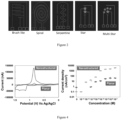
- FIG 3 shows another embodiment of the biosensor.
- the biosensor comprises three components: 1) a detection probe 130 having a recognition moiety 134, which is electroactive biorecognition elements, such as e-DNAzymes, for releasing a reporter moiety 132, which is electroactive DNA barcodes, in the presence of an analyte 136, which is specific bacterial targets ( Figure 3 a) , i); 2) electrochemical differential chips 120 (e.g. a four-electrode chip) with a first working electrode E1 and a second working electrode E2.
- the first working electrode E1 is a release channel E10 and the second working electrode is a capture channel E20.
- the two-channel design for translating e-DNAzyme cleavage on the release channel E10 to barcode binding on the capture channel E20 using a single redox moiety e.g. methylene blue.
- This can suppress the effect of non-specific e-DNAzyme cleavage that typically reduces signal-to-blank ratios in biological samples ( Figure 3 a) , ii); and 3) three-dimensional (3D) nanostructured electrochemical E1 and E2 channels, for example, a dendritic morphology, to increase the capture and hybridization efficiency of the DNA barcodes to improve sensitivity ( Figure 3 a) , iii).
- the capture and release channels E10 and E20 can be nanostructured using electrodeposition.
- the capture and release channels E10 and E20 have multiple edges (e.g. sharp) which can result in diffusion-limited growth and thus the deposition of a dense layer of nanostructures organized in high-aspect ratio architectures ( Figure 3 a) , iii).
- a large current peak is generated on the release channel E10 due to the reduction of the single redox moiety (e.g. methylene blue) (see bottom graph in Figure 3 b) iv)) but no current peak was generated on the capture channel E20 (see top graph in Figure 3 b) iv)).
- the reporter moiety 132 e.g. electroactive barcode
- the detection probe 130 e.g. DNAzyme
- the capture probe 140 e.g. ssDNA probe
- the biosensors can allow bacterial analysis to be performed with minimal user intervention in a single incubation step and without the use of external reagents or labels.
- the biosensors described herein can be used for label-free, and reagent-free detection of analytes.
- the biosensors can be used for rapid detection of analytes, including rapid, label-free, and reagent-free detection of analytes.
- the biosensors, kits and methods of use described herein can be used for detecting any suitable analyte, such as, and without being limited thereto, a wide range of small molecule, protein and nucleic acid analytes, including infection-causing pathogens in point-of-care diagnostics and health monitoring.
- the biosensor and/or method are used to detect E. coli in buffer and urine test samples.
- the biosensors enable an ultra-sensitivity (10 CFU) and high specificity against a panel of bacteria.
- the kit comprises a biosensor described herein and instructions for use.
- the biosensors described herein can be used in any suitable device, including a hand-held device and/or diagnostic device.
- the device may resemble the glucose monitor in terms of ease-of-use, portability, cost, and/or amplification-free bacterial detection based, for example, on an electrochemical readout.
- the device may be a handheld electrical reader comprises the biosensor (e.g. self-contained e-differential chip).
- the biosensors described herein may be used in a handheld screening and/or diagnostic device that performs benchtop testing and generates test results in less than about 30 minutes, less than about 20 minutes, or less than about 15 minutes, for example, while matching the clinical sensitivity and specificity offered by comparative tests (e.g. urine cultures that require days to complete).
- the biosensor may be used without the need for sample pre-treatment, target labeling, and/or amplification.
- Urinary tract infections despite being the second most common bacterial infectious disease, lacks the developments towards affirmative early diagnosis albeit the challenges of antibiotic resistance associated with it.
- the existing in-patient or outpatient approaches to urine analysis suffer from false positive results and sample-to-answer time of 1-2 days causing delays in the disease treatment, and an increased cost of care.
- the devices described herein may increase the accuracy and decrease the timeline for diagnosis.
- a method for determining the presence of an analyte in a sample The biosensor described herein is exposed to the sample.
- the reporter moiety delocalizes from the first working electrode to the second working electrode in the presence of the analyte, providing a change in signal (e.g. current, potential, impedance, etc.) at each of the first working electrode and the second working electrode.
- the change in signal indicates the presence of the analyte.
- the change in signal for the first working electrode is from about 0.1 to about 0.6 fold decrease and the change in signal for the second working electrode is from about 50 to about 200 fold increase or the change in signal for the first working electrode is from about 50 to about 200 fold increase and the change in signal for the second working electrode is from about 0.1 to about 0.6 fold decrease.
- the signal is current.
- the ranges may be from about 0.1 to about 0.6 fold decrease, about 0.1 to about 0.5 fold decrease, about 0.1 to about 0.4 fold decrease, about 0.1 to about 0.3 fold decrease, about 0.1 to about 0.2 fold decrease, about 0.2 to about 0.6 fold decrease, about 0.3 to about 0.6 fold decrease, about 0.4 to about 0.6 fold decrease, or about 0.5 to about 0.6 fold decrease.
- the ranges may be from about 50 to about 200 fold increase, from about 50 to about 150 fold increase, from about 50 to about 100 fold increase, from about 50 to about 90 fold increase, from about 50 to about 80 fold increase, from about 50 to about 70 fold increase, from about 50 to about 60 fold increase, from about 60 to about 200 fold increase, from about 70 to about 200 fold increase, from about 80 to about 200 fold increase, from about 90 to about 200 fold increase, from about 100 to about 200 fold increase, from about 125 to about 200 fold increase, from about 150 to about 200 fold increase, or from about 175 to about 200 fold increase.
- the analyte if there is no measurable signal change, the analyte is not present in the sample. In another embodiment, if the change in signal from the first working electrode is a decrease in signal and the change in signal from the second working electrode is an increase in signal, the analyte is present. In another embodiment, if the change in signal from the first working electrode is an increase in signal and the change in signal from the second working electrode is a decrease in signal, the analyte is present.
- the analyte if no measurable current change, the analyte is not present in the sample. In another embodiment, the change in current from the first working electrode is a decrease in current and the change in current from the second working electrode is an increase in current, the analyte is present. In another embodiment, if the change in current from the first working electrode is an increase in current and the change in current from the second working electrode is a decrease in current, the analyte is present.
- the biosensor described herein is exposed to the sample under conditions to delocalize the reporter moiety from the first working electrode to the second working electrode in the presence of the analyte, providing the change in signal.
- the signal is measured from each of the working electrodes.
- the current is measured from each of the working electrodes.
- an initial signal is measured at each of the first and second working electrodes.
- a final signal is measured at each of the first and second working electrodes. A change in signal between the initial signal and the final signal of the first working electrode and a change in signal between the initial signal and the final signal of the second working electrode, indicates the presence of the analyte in the sample.
- an initial current is measured at each of the first and second working electrodes.
- a final current is measured at each of the first and second working electrodes. A change in current between the initial current and the final current of the first working electrode and a change in current between the initial current and the final current of the second working electrode, indicates the presence of the analyte in the sample.
- a four-electrode electrochemical chip was fabricated on pre-stressed polystyrene (PS) sheets (Graphix Shrink Film, Graphix).
- PS polystyrene
- the PS was cleaned with ethanol and DI water, after which a vinyl mask (FDC 4304, FDC graphic films) was applied onto the sheet.
- the vinyl mask was then cut into the star-shaped working electrode pattern (as defined by Adobe Illustrator) using a Robo Pro CE5000- 40-CRP cutter (Graphtec America Inc.).
- the vinyl was then peeled off, and a 100 nm gold (Au) layer was sputtered on top using DC sputtering (MagSput TM , Torr International).
- the chip comprises two star-shaped working electrodes (WEs; release channel, E1 and capture channel, E2) and both counter (CE) and reference (RE) electrodes.
- This chip with the conductive layer served as a seed layer for the nucleation and growth of high-aspect ratio nanostructures using an electrodeposition technique.
- a 10 mM gold chloride (HAuCl 4 ) in 5 mM HCl solution was bubbled with N 2 , before use, for about 10 minutes.
- the conductive layer containing the electrodes was dipped in this solution and gold nanostructures were electrodeposited on the two working electrodes by applying a static potential of about -0.6V (anodic negative) for about 600 s using a potentiostatic technique to reduce the gold ions.
- E1 and E2 were deposited with gold (Au) nanostructures.
- the resulting chip comprised two nanostructured star-shaped working electrodes (WE; release channel, E1 and capture channel, E2) and both counter (CE) and reference (RE) electrodes.
- WE nanostructured star-shaped working electrodes
- CE counter
- RE reference
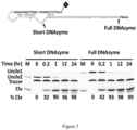
- RNA-cleaving fluorescent DNAzyme for E. coli detection (RFD-EC1) as described in Ali et al. ( Ali MM, Aguirre SD, Lazim H, Li Y. Fluorogenic DNAzyme Probes as Bacterial Indicators. Angew. Chem. Int. Ed. 2011; 50:3751-3754 ) was used.
- the truncated DNAzyme has a secondary structure for better release of the cleaved redox-tagged DNA segment. The shorter sequence increased the loading capacity on the electrode and reduced cost.
- the truncated DNAzyme had similar activity and specificity to the RFD-EC1 ( Figure 5 ).
- the e-DNAzymes were prepared by T4 DNA ligase-mediated DNA ligation of the biotinylated DNAzyme sequence (BD) and labeled DNA reporter (DR) in the presence of a ligation template (LT) as the template to complete the biotinylated DNAzyme reporter (BDR).
- the methylene blue labeled DNA was purchased from Biosearch Inc, and all other DNA oligonucleotides (including the thiolated probe, TP) were purchased from Integrated DNA Technologies. All DNA oligonucleotides were purified via standard 10% denaturing (7 M urea) polyacrylamide gel electrophoresis (dPAGE), with their concentrations determined spectroscopically.
- DNAzyme was also prepared by T4 DNA ligase-mediated DNA ligation of BD and DR in the presence of LT as the template using the same ligation protocol.
- the electrochemical surface area was calculated by performing a cyclic voltammetry scan from 0 to 1.5V (cathodic positive) in 0.1 M H 2 SO 4 using a scan rate of 50 mV/s. The area under the reduction peak was integrated to calculate the electrochemical charge involved in the redox process, which was subsequently divided by the surface charge density involved in forming a monolayer of AuO x (386 ⁇ C/cm 2 ).
- the immobilization of BDR on release channel E1 was characterized by square wave voltammetry (SWV) over a voltage range of 0 V to -0.6 V (anodic negative) before and after the DNAzyme deposition steps in 25 mM PBS and 25 mM NaCl buffer (25:25 buffer).
- E. coli was chosen as the target due to its predominance in urinary tract infections (UTIs), which is one of the most prevalent bacterial infections in clinical practice; however, the e-DNAzyme strategy could in principle be applied to any relevant bacteria.
- UTIs urinary tract infections
- pathogenic samples comprised a crude intracellular mixture (CIM) of E. coli (test sample containing target analyte) or B. subtilis, Y. ruckeri, C. difficile, H. alvei or L. monocytogenes bacteria (control samples).
- Escherichia coli K12 E. coli K12; MG1655
- CFU/mL colony-forming units
- the heat-treated cell suspension was then vortexed to dissolve the cell pellet completely and stored at -20 °C.
- Different concentrations of bacterial CIM were spiked into both the 25:25 buffer and healthy human urine samples.
- 10 ⁇ L of bacteria-doped samples were incubated on top of the chip for 30 minutes at 37°C.
- the signal generated with E. coli was compared to a panel of the five control samples containing CIM from the other bacteria ( B. subtilis, Y. ruckeri, C. difficile, H. alvei and L. monocytogenes ) to assess the specificity toward E. coli.
- a small volume (10 ⁇ L) of these samples were dropped on the two working electrodes (E1 and E2) for 30 min at 37 °C.
- the signal quantification was performed using SWV over a voltage range of 0 V to -0.6 V (anodic negative) in 25:25 buffer and urine.
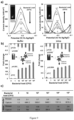
- E. coli samples having a bacterial load of 0-10 5 CFU were introduced on the biosensor and the current generated on the release and capture channels were measured using SWV ( Figure 8 and Figure 9(a) ).
- the signal changes induced on each channel were quantified by calculating the difference between the current measured before and after introducing the bacterial load ( Figure 9(b) ). This improved the reliability of the sensor by eliminating false results and enhanced the performance by reducing the signal-to-noise ratio.
- target binding the DNAzyme caused the ribonucleotide cleavage that released the redox-DNA reporter, leading to a decrease in the signal from E1.
- An increase in the signal is observed from E2 due to the capture of the redox-DNA.
- the e-DNAzyme test could sensitively detect E. coli in both buffer and urine, the specificity of this assay was assessed against a panel of gram-negative and gram-positive bacterial targets: B. subtilis, Y. ruckeri, C. difficile, H. alvei and L. monocytogenes ( Figure 10 ).
- B. subtilis B. subtilis
- Y. ruckeri C. difficile
- H. alvei and L. monocytogenes Figure 10
- the pathogen-containing control samples with non-target bacteria caused a decrease in the E1 signal due to non-specific cleavage of DNAzyme.
- no significant signal increase is observed from E2 given that the non-specific DNAzyme cleavage does not release a redox-DNA reporter that is complementary to the probe immobilized on E2.
- the signal from urine samples show a larger decrease in E1 signal than from the buffer samples due to the presence of non-specific exo/endonuclease activity in urine. Therefore, specific identification of E. coli relative to other bacteria in urine or buffer was possible using the release channel signal and the capture channel, which allowed precise identification of the E. coli sample.
- Samples containing E. coli in buffer and urine resulted in signal changes of at least 200 and 100-folds respectively, while samples containing bacteria other than E. coli yielded signal changes of less than 0.4-fold in both urine and buffer.
- the high specificity of this assay is rooted in the use of two biorecognition events (specific cleavage on release channel and efficient hybridization on capture channel), which reduced the generation of signals through non-specific cleavage.
- non-specific cleavage of DNAzymes by DNA/RNA cleaving enzymes is increased in biological materials, these non-specific interactions do not necessarily result in cleaved segments complementary to the full sequence of the ssDNA probe on the capture channel ( Figure 11 ).
- the generation of the redox current on the capture electrode can be dependent on proximity between the electrode and the methylene blue molecule, which can be dependent on the length and sequence of the redox barcode.
- the RCDs specific to Escherichia coli were used to assess the operation of the e-DNAzyme test towards the identification of this pathogen in UTIs using clinical human urine samples that were bacteria negative, bacteria positive but E. coli negative, and E. coli positive.
- a set of 41 urine samples including 20 clinically-significant E. coli-only, 10 clinically-insignificant E. coli -only, 2 K. pneumonie-only, 2 E. faecalis -only, 1 S. aureus-only, and 1 P. mirabilis-only urine samples, and 5 culture-negative (i.e. no bacterial growth) urine samples were obtained from Hamilton General Hospital.
- the urine samples were heated at 55°C for 15 minutes followed by centrifugation at 11,000 ⁇ g for 5 minutes.
- a 10 ⁇ L of the suspension of the urine was then incubated on the chip for 30 minutes at 37°C, after which SWV over a voltage range of 0 V to -0.6 V (anodic negative) was conducted in 25:25 buffer.
- Clinical sensitivity was calculated by dividing "True Positive” results from a sum of “True Positive” and “False Negative” results, while clinical specificity was calculated by dividing "True Negative” results from a sum of "True Negative” and “False Positive” results.
- the disclosed biosensor was used to analyze the 41 urine samples collected from patients who were suspected of having UTIs due to the presence of clinical symptoms.
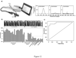
- a handheld electrical reader comprises the self-contained e-differential chip to demonstrate the potential of the e-DNAzyme test for point-of-care analysis ( Figure 12 (a left)).
- a set of randomized samples belonging to the following categories was analyzed: culture-, culture+/ E . coli-, and culture+/ ⁇ E . coli+. These sample categories resulted in specific redox signatures: the culture- samples showed no peak, the culture+/ E . coli- samples exhibited a peak with a low signal-to-noise ratio and culture+/ E .
- the biosensor allowed the analysis of clinical urine samples using a handheld reader without the need for sample pre-treatment, target labeling, enrichment or amplification and readout reagents within 30 minutes while matching the clinical sensitivity and specificity offered by urine cultures that require days to complete.
- RNA-cleaving DNAzyme technology design and fabrication of a highly specific and sensitive electrochemical assay based on the integration of RNA-cleaving DNAzyme technology, differential electrochemical signal readout, and nanostructured electrodes for rapidly identifying bacteria without time-consuming growth cultures is disclosed herein.
- Differential electrochemical signaling is made possible due to the programmable nature of electroactive RNA-cleaving DNAzymes combined with a two-channel electrochemical chip.
- Each of the building blocks of the e-DNAzyme assay contributes to enhanced sensitivity, specificity, or speed of operation.
- This system can be engineered to target and identify multiple bacteria in parallel for performing rapid, multiplexed, and reagent-free bacterial analysis.
Landscapes
- Chemical & Material Sciences (AREA)
- Health & Medical Sciences (AREA)
- Life Sciences & Earth Sciences (AREA)
- Immunology (AREA)
- Engineering & Computer Science (AREA)
- Analytical Chemistry (AREA)
- Organic Chemistry (AREA)
- Molecular Biology (AREA)
- Physics & Mathematics (AREA)
- Biochemistry (AREA)
- General Health & Medical Sciences (AREA)
- Proteomics, Peptides & Aminoacids (AREA)
- Wood Science & Technology (AREA)
- Zoology (AREA)
- Microbiology (AREA)
- Biotechnology (AREA)
- Pathology (AREA)
- Urology & Nephrology (AREA)
- Biomedical Technology (AREA)
- Bioinformatics & Cheminformatics (AREA)
- Genetics & Genomics (AREA)
- Biophysics (AREA)
- Hematology (AREA)
- General Engineering & Computer Science (AREA)
- General Physics & Mathematics (AREA)
- Cell Biology (AREA)
- Electrochemistry (AREA)
- Chemical Kinetics & Catalysis (AREA)
- Spectroscopy & Molecular Physics (AREA)
- Food Science & Technology (AREA)
- Medicinal Chemistry (AREA)
- Measuring Or Testing Involving Enzymes Or Micro-Organisms (AREA)
- Investigating Or Analyzing Materials By The Use Of Electric Means (AREA)
- Investigating Or Analysing Biological Materials (AREA)
Claims (15)
- Biosensor (10) zum Nachweis eines Analyten (36), umfassend:a) eine erste (E1) und eine zweite (E2) Arbeitselektrode;b) eine auf der ersten Arbeitselektrode funktionalisierte Detektionssonde (30), die eine Reportereinheit (32) umfasst, die an eine Erkennungseinheit (34) für einen Analyten gekoppelt ist, wobei die Detektionssonde so ausgelegt ist, dass sie die Reportereinheit von der ersten Arbeitselektrode zur zweiten Arbeitselektrode delokalisiert, wenn der Analyt an die Erkennungseinheit koppelt;c) eine auf der zweiten Arbeitselektrode funktionalisierte Fängersonde (40), die die Reportereinheit erkennt und an sie koppelt; undd) eine Gegenelektrode (24);wobei jede Arbeitselektrode so konfiguriert ist, dass sie bei Vorhandensein des Analyten eine Signaländerung erzeugt.
- Biosensor nach Anspruch 1, wobei:der Biosensor ferner eine Referenzelektrode (22) umfasst; oderdie Gegenelektrode sowohl Gegenelektrode als auch Referenzelektrode ist.
- Biosensor nach Anspruch 1 oder 2, wobei eines oder mehrere von:der ersten (E1) und der zweiten (E2) Arbeitselektrode konzentrische Spiralelektroden, Serpentinenlinienelektroden, Bürstenelektroden, verschlungene Sternelektroden und/oder mehrere verschlungene Elektroden sind;der ersten und der zweiten Arbeitselektrode nahe beieinander liegen, sodass der Reporteranteil von der Detektionssonde zur Abfangsonde transportiert werden kann;den Arbeitselektroden nebeneinander liegen; undzwischen mindestens einem Teil der ersten Arbeitselektrode und mindestens einem Teil der zweiten Arbeitselektrode ein Spalt besteht, in dem die Reportereinheit von der Detektionssonde zur Einfangssonde transportieren kann; optional der Spalt etwa 10 µm bis etwa 2 mm, etwa 10 µm bis etwa 1 mm, etwa 10 µm bis etwa 500 µm, etwa 10 µm bis etwa 400 µm, etwa 10 µm bis etwa 300 µm, etwa 10 µm bis etwa 200 µm oder etwa 10 µm bis etwa 100 µm beträgt.
- Biosensor nach einem der Ansprüche 1 bis 3, wobei eines oder mehrere von:mindestens einer der ersten (E1) und zweiten (E2) Arbeitselektroden eine nanostrukturierte Elektrode ist;mindestens einer der ersten und zweiten Arbeitselektroden Kanten aufweist; optional mindestens eine der ersten und zweiten Arbeitselektroden scharfe Kanten und/oder Spitzen aufweist;mindestens eine der ersten und zweiten Arbeitselektroden ein hohes Aspektverhältnis aufweist;optional das hohe Aspektverhältnis etwa 2 bis etwa 10, etwa 3 bis etwa 10, etwa 4 bis etwa 10, etwa 5 bis etwa 10, etwa 6 bis etwa 10, etwa 7 bis etwa 10, etwa 8 bis etwa 10, etwa 2 bis etwa 9, etwa 3 bis etwa 9, etwa 4 bis etwa 9, etwa 5 bis etwa 9, etwa 6 bis etwa 9, etwa 7 bis etwa 9, etwa 8 bis etwa 9, etwa 2 bis etwa 8, etwa 3 bis etwa 8, etwa 4 bis etwa 8, etwa 5 bis etwa 8, etwa 6 bis etwa 8, etwa 7 bis etwa 8, etwa 2 bis etwa 7, etwa 3 bis etwa 7, etwa 4 bis etwa 7, etwa 5 bis etwa 7 oder etwa 6 bis etwa 7 beträgt; unddie Oberflächenbereiche von mindestens einer der ersten und zweiten Arbeitselektroden etwa 1,0 cm2 bis 5,0 cm2, etwa 1,5 cm2 bis 5,0 cm2, etwa 1,5 cm2 bis 4,5 cm2, etwa 1,5 cm2 bis 4,0 cm2, etwa 1,5 cm2 bis 3,9 cm2, etwa 1,7 cm2 bis 3,8 cm2, etwa 1,7 cm2 bis 3,7 cm2, etwa 1,7 cm2 bis 3,6 cm2, etwa 1,7 cm2 bis 3,5 cm2, etwa 1,7 cm2 bis 3,4 cm2, etwa 1,7 cm2 bis 3,3 cm2 oder etwa 1,7 cm2 bis 3,2 cm2 betragen.
- Biosensor nach einem der Ansprüche 1 bis 4, wobei die erste Arbeitselektrode (E1) so konfiguriert ist, dass sie eine Verringerung des Signals bewirkt, und die zweite Arbeitselektrode (E2) so konfiguriert ist, dass sie in Gegenwart des Analyten eine Erhöhung des Signals bewirkt;
optional die Abnahme des Signals etwa das 0,1- bis 0,6-Fache und die Zunahme des Signals etwa das 50- bis 200-Fache beträgt. - Biosensor nach einem der Ansprüche 1 bis 5, wobei der Biosensor ein elektrochemischer Chip (20) ist.
- Biosensor nach einem der Ansprüche 1 bis 6, wobei die Reportereinheit eine Einheit mit einer Redox-, photoelektrochemischen, halbleitenden und/oder leitenden Spezies umfasst; optional die Reportereinheit eine Redoxspezies umfasst; optional wobei eines oder mehrere von Folgendem:die Reportereinheit aus einem mit der Redoxspezies modifizierten Biopolymer besteht;die Redoxspezies ausgewählt ist aus Methylenblau, Methylenblausuccinymid, Methylenblaumaleimid, Atto MB2 Maleimid (Sigma Aldrich) und anderen Methylenblauderivaten; 3,7-Bis-[(2-Ammoniumethyl)(methyl)amino]phenothiazin-5-iumtrifluoracetat; 3,7-Bis-(piperazin-4-ium-1-yl)phenothiazin-5-iumtrifluoracetat; 3,7-Bis-[(2-ammoniumethyl)(methyl)amino]phenothiazin-5-iumchlorid;und 3,7-Bis-(piperazin-4-ium-1-yl)phenothiazin-5-iumchlorid und/oder Ferrocen; unddie Redoxspezies Methylenblau und/oder Ferrocen ist.
- Biosensor nach einem der Ansprüche 1 bis 7, wobei eines oder mehrere von Folgendem zutreffen:die Reportereinheit dazu dient, die Anwesenheit eines vom Erkennungselement erkannten Analyten in ein nachweisbares Signal umzuwandeln;die Reportereinheit einen Antikörper, ein Antigen, ein Oligonukleotid, ein Oligopeptid und/oder ein Oligosaccharid umfasst; unddie Erkennungseinheit ein DNAzym, Aptamer, einen Antikörper und/oder eine Nukleinsäure umfasst, die die Anwesenheit des Analyten erkennen kann, optional die Erkennungseinheit ein DNAzym, das RNA in Gegenwart des Analyten spaltet, ein Aptamer, das in Gegenwart des Analyten seine Konformation ändert, oder ein Antikörper ist, optional die Erkennungseinheit ein DNAzym ist, das RNA in Gegenwart des Analyten spaltet, um die Reportereinheit von der ersten Arbeitselektrode (E1) freizusetzen.
- Biosensor nach einem der Ansprüche 1 bis 8, wobei die Erfassungssonde (40) ssDNA, ssPNA, dsDNA, ssRNA und/oder dsRNA umfasst; optional die Erfassungssonde ssDNA umfasst.
- Biosensor nach einem der Ansprüche 1 bis 9, wobei eines oder mehrere von Folgendem zutreffen:der Analyt ein Toxin, ein Biopolymer, eine Zelle, ein Gewebe und/oder ein Pathogen ist; undder Analyt ein Mikroorganismus und/oder ein Virus ist;optional, wobei eines oder mehrere von Folgendem zutreffen:der Mikroorganismus ein gramnegatives Bakterium ist;der Mikroorganismus Eschenchia coli, Salmonella tyohimurium, Pseudomonas peli, Brevundimonas diminuta, Hafnia alvei, Yersinia ruckeri, Ochrobactrum grignonese, Achromobacter xylosoxidans, Moraxella osloensis, Acinetobacter Iwoffi oder Serratia fonticola ist;der Mikroorganismus ein grampositives Bakterium ist;der Mikroorganismus Listeria monocytogenes, Bacillus subtilis, Clostridium difficile, Actinomyces orientalis, Pediococcus acidilactici, Leuconostoc mesenteroides, Lactobacillus planturum oder eine Kombination davon ist; undder Mikroorganismus ein pathogenes Bakterium ist.
- Gerät, das den Biosensor nach den Ansprüchen 1 bis 10 umfasst, optional wobei eines oder mehrere von Folgendem zutreffen:das Gerät für die klinische und landwirtschaftliche Diagnostik, die Qualitätskontrolle im Agrar- und Lebensmittelbereich, das Umweltmonitoring, das Gesundheitsmonitoring und/oder die Arzneimittelentwicklung verwendet wird;das Gerät zum Screening dient; undes sich um ein Handgerät handelt.
- Kit zum Nachweis eines Analyten, umfassend einen Biosensor nach einem der Ansprüche 1 bis 10 oder ein Gerät nach Anspruch 11 sowie eine Gebrauchsanweisung.
- Verfahren zur Bestimmung des Vorhandenseins eines Analyten in einer Probe, umfassend:Einwirkenlassen des Biosensors nach einem der Ansprüche 1 bis 10 auf die Probe, um die Reportereinheit in Gegenwart des Analyten von der ersten Arbeitselektrode zur zweiten Arbeitselektrode zu delokalisieren; undMessen eines Signals an der ersten und zweiten Arbeitselektrode, um festzustellen, ob eine Signaländerung an der ersten und zweiten Arbeitselektrode aufgetreten ist, die das Vorhandensein des Analyten anzeigt.
- Verfahren nach Anspruch 13, wobei eines oder mehrere von Folgendem zutreffen:der Biosensor der Probe unter Bedingungen ausgesetzt wird, die eine Delokalisierung der Reportereinheit von der ersten Arbeitselektrode zur zweiten Arbeitselektrode ermöglichen;die Signaländerung der ersten Arbeitselektrode eine Signalabnahme und die Signaländerung der zweiten Arbeitselektrode eine Signalzunahme ist;bevor der Biosensor der Probe ausgesetzt wird, an der ersten und der zweiten Arbeitselektrode jeweils ein Anfangssignal gemessen wird und nachdem der Biosensor dem Analyten ausgesetzt wird, an der ersten und der zweiten Arbeitselektrode jeweils ein Endsignal gemessen wird, wobei eine differenzielle Signaländerung zwischen dem Anfangssignal und dem Endsignal der ersten Arbeitselektrode und eine Signaländerung zwischen dem Anfangssignal und dem Endsignal der zweiten Arbeitselektrode das Vorhandensein des Analyten in der Probe anzeigt;die Signaländerung für die erste Arbeitselektrode und/oder die zweite Arbeitselektrode eine Strom-, Potential- oder Impedanzänderung umfasst; optional die Signaländerung für die erste Arbeitselektrode und/oder die zweite Arbeitselektrode eine Stromänderung ist; undder Analyt ein Toxin, ein Biopolymer, eine Zelle, ein Gewebe und/oder ein Pathogen ist; optional, wobei:i) der Analyt ein Mikroorganismus und/oder ein Virus ist; optional, wobei:der Mikroorganismus ein gramnegatives Bakterium ist;der Mikroorganismus Eschenchia coli, Salmonella tyohimurium, Pseudomonas peli, Brevundimonas diminuta, Hafnia alvei, Yersinia ruckeri, Ochrobactrum grignonese, Achromobacter xylosoxidans, Moraxella osloensis, Acinetobacter Iwoffi oder Serratia fonticola ist;der Mikroorganismus ein grampositives Bakterium ist;der Mikroorganismus Listeria monocytogenes, Bacillus subtilis, Clostridium difficile, Actinomyces orientalis, Pediococcus acidilactici, Leuconostoc mesenteroides, Lactobacillus planturum oder eine Kombination davon ist; oderder Mikroorganismus ein pathogenes Bakterium ist; oderii) der Analyt der Pathogen ist und der Pathogen E. coli ist.
- Verfahren nach Anspruch 13 oder 14, wobei das Aussetzen des Biosensors gegenüber der Probe das Kontaktieren des Biosensors mit der Probe umfasst und/oder die Probe eine Urin- oder Blutprobe ist.
Applications Claiming Priority (2)
| Application Number | Priority Date | Filing Date | Title |
|---|---|---|---|
| US201962821219P | 2019-03-20 | 2019-03-20 | |
| PCT/CA2020/050375 WO2020186362A1 (en) | 2019-03-20 | 2020-03-20 | Differential signal biosensing for detecting an analyte |
Publications (4)
| Publication Number | Publication Date |
|---|---|
| EP3942285A1 EP3942285A1 (de) | 2022-01-26 |
| EP3942285A4 EP3942285A4 (de) | 2022-12-21 |
| EP3942285B1 true EP3942285B1 (de) | 2025-05-14 |
| EP3942285C0 EP3942285C0 (de) | 2025-05-14 |
Family
ID=72519573
Family Applications (1)
| Application Number | Title | Priority Date | Filing Date |
|---|---|---|---|
| EP20774143.0A Active EP3942285B1 (de) | 2019-03-20 | 2020-03-20 | Differenzsignalbiosensor zum nachweis eines analyten |
Country Status (4)
| Country | Link |
|---|---|
| US (1) | US20220317082A1 (de) |
| EP (1) | EP3942285B1 (de) |
| CA (1) | CA3133839A1 (de) |
| WO (1) | WO2020186362A1 (de) |
Families Citing this family (6)
| Publication number | Priority date | Publication date | Assignee | Title |
|---|---|---|---|---|
| US20230175044A1 (en) * | 2019-11-01 | 2023-06-08 | The Governing Council Of The University Of Toronto | An Electrochemical Interface for Molecular Circuit-Based Outputs |
| CN112816106B (zh) * | 2020-12-24 | 2022-03-22 | 太原理工大学 | 一种铽镝铁柔性磁弹性薄膜生物传感器及其制备方法 |
| CN113252892B (zh) * | 2021-06-22 | 2021-11-02 | 北京市肝病研究所 | 一种用于提高病原微生物抗原检测灵敏度的探针及试剂盒 |
| CN113702459B (zh) * | 2021-07-26 | 2022-08-02 | 山东大学 | 一种udg检测用电化学生物传感器及其检测方法 |
| WO2023193118A1 (en) * | 2022-04-07 | 2023-10-12 | Mcmaster University | Aptamers for porcine epidemic diarrhea virus |
| CN115825185B (zh) * | 2022-07-05 | 2024-10-11 | 集美大学 | 一种光电化学传感器及其构建方法和应用 |
Citations (1)
| Publication number | Priority date | Publication date | Assignee | Title |
|---|---|---|---|---|
| US20070154909A1 (en) * | 2005-11-29 | 2007-07-05 | The Regents Of The University Of California | Signal-on architecture for electronic, oligonucleotide-based detectors |
Family Cites Families (8)
| Publication number | Priority date | Publication date | Assignee | Title |
|---|---|---|---|---|
| US8425745B2 (en) | 2006-10-06 | 2013-04-23 | Nanomix, Inc. | Electrochemical nanosensors for biomolecule detection |
| EP2362941A1 (de) | 2008-07-11 | 2011-09-07 | Early Warning Inc. | Biosensorvorrichtung und verfahren zum nachweis von zielbiomolekülen in einer lösung |
| JP5989668B2 (ja) | 2011-01-11 | 2016-09-07 | ザ ガバニング カウンシル オブ ザ ユニバーシティ オブ トロント | タンパク質検出方法 |
| CA2922821C (en) * | 2013-08-30 | 2023-01-03 | University Of Maryland, College Park | Device and methods of using device for detection of hyperammonemia |
| US10444179B2 (en) | 2016-08-10 | 2019-10-15 | Multerra Bio, Inc. | Apparatuses and methods for detecting molecules and binding energy |
| US10852274B2 (en) | 2017-03-09 | 2020-12-01 | Auburn University | Differential circuit for background correction in electrochemical measurements |
| US11536721B2 (en) | 2017-03-24 | 2022-12-27 | Iowa State University Research Foundation, Inc. | Electrochemical immunosensors |
| WO2019099856A1 (en) * | 2017-11-17 | 2019-05-23 | Eccrine Systems, Inc. | Reference aptamer sensing elements for eab biosensors |
-
2020
- 2020-03-20 CA CA3133839A patent/CA3133839A1/en active Pending
- 2020-03-20 US US17/441,169 patent/US20220317082A1/en active Pending
- 2020-03-20 EP EP20774143.0A patent/EP3942285B1/de active Active
- 2020-03-20 WO PCT/CA2020/050375 patent/WO2020186362A1/en not_active Ceased
Patent Citations (1)
| Publication number | Priority date | Publication date | Assignee | Title |
|---|---|---|---|---|
| US20070154909A1 (en) * | 2005-11-29 | 2007-07-05 | The Regents Of The University Of California | Signal-on architecture for electronic, oligonucleotide-based detectors |
Also Published As
| Publication number | Publication date |
|---|---|
| WO2020186362A1 (en) | 2020-09-24 |
| US20220317082A1 (en) | 2022-10-06 |
| EP3942285C0 (de) | 2025-05-14 |
| CA3133839A1 (en) | 2020-09-24 |
| EP3942285A4 (de) | 2022-12-21 |
| EP3942285A1 (de) | 2022-01-26 |
Similar Documents
| Publication | Publication Date | Title |
|---|---|---|
| EP3942285B1 (de) | Differenzsignalbiosensor zum nachweis eines analyten | |
| Radi | Electrochemical aptamer‐based biosensors: recent advances and perspectives | |
| Song et al. | Aptamer-based biosensors | |
| Lubin et al. | Sequence-specific, electronic detection of oligonucleotides in blood, soil, and foodstuffs with the reagentless, reusable E-DNA sensor | |
| Wong et al. | Charge transfer through DNA: A selective electrochemical DNA biosensor | |
| Liu et al. | An enzyme-based E-DNA sensor for sequence-specific detection of femtomolar DNA targets | |
| Du et al. | Solid-state probe based electrochemical aptasensor for cocaine: a potentially convenient, sensitive, repeatable, and integrated sensing platform for drugs | |
| Das et al. | Tuning the bacterial detection sensitivity of nanostructured microelectrodes | |
| Zhu et al. | Highly sensitive electrochemical sensor for mercury (II) ions by using a mercury-specific oligonucleotide probe and gold nanoparticle-based amplification | |
| Zhang et al. | Label-free electrochemical DNA biosensor array for simultaneous detection of the HIV-1 and HIV-2 oligonucleotides incorporating different hairpin-DNA probes and redox indicator | |
| Xu et al. | Label-free electrochemical detection for aptamer-based array electrodes | |
| Phares et al. | Improving the stability and sensing of electrochemical biosensors by employing trithiol-anchoring groups in a six-carbon self-assembled monolayer | |
| Deng et al. | Sensitive bifunctional aptamer-based electrochemical biosensor for small molecules and protein | |
| Khodadoust et al. | A ratiometric electrochemical DNA-biosensor for detection of miR-141 | |
| Hernández-Santos et al. | Enzymatic genosensor on streptavidin-modified screen-printed carbon electrodes | |
| Liao et al. | Hybridization chain reaction triggered poly adenine to absorb silver nanoparticles for label-free electrochemical detection of Alzheimer's disease biomarkers amyloid β-peptide oligomers | |
| Chiorcea-Paquim et al. | Electrochemical and AFM Characterization of G‐Quadruplex Electrochemical Biosensors and Applications | |
| US11156582B2 (en) | Systems for detecting and quantifying nucleic acids | |
| Zhang et al. | Fabrication of a sensitive impedance biosensor of DNA hybridization based on gold nanoparticles modified gold electrode | |
| Taufik et al. | Bismuth oxide nanoparticles/chitosan/modified electrode as biosensor for DNA hybridization | |
| Huang et al. | Recent progresses on biosensors for Escherichia coli detection | |
| Malecka et al. | Silver or gold? A comparison of nanoparticle modified electrochemical genosensors based on cobalt porphyrin-DNA | |
| Aoki et al. | 384-Channel electrochemical sensor array chips based on hybridization-triggered switching for simultaneous oligonucleotide detection | |
| Wang et al. | Vertically aligned nitrogen-doped carbon nanotube carpet electrodes: highly sensitive interfaces for the analysis of serum from patients with inflammatory bowel disease | |
| Kim et al. | Covalent attachment of biomacromolecules to plasma-patterned and functionalized carbon nanotube-based devices for electrochemical biosensing |
Legal Events
| Date | Code | Title | Description |
|---|---|---|---|
| STAA | Information on the status of an ep patent application or granted ep patent |
Free format text: STATUS: THE INTERNATIONAL PUBLICATION HAS BEEN MADE |
|
| PUAI | Public reference made under article 153(3) epc to a published international application that has entered the european phase |
Free format text: ORIGINAL CODE: 0009012 |
|
| STAA | Information on the status of an ep patent application or granted ep patent |
Free format text: STATUS: REQUEST FOR EXAMINATION WAS MADE |
|
| 17P | Request for examination filed |
Effective date: 20211018 |
|
| AK | Designated contracting states |
Kind code of ref document: A1 Designated state(s): AL AT BE BG CH CY CZ DE DK EE ES FI FR GB GR HR HU IE IS IT LI LT LU LV MC MK MT NL NO PL PT RO RS SE SI SK SM TR |
|
| DAV | Request for validation of the european patent (deleted) | ||
| DAX | Request for extension of the european patent (deleted) | ||
| A4 | Supplementary search report drawn up and despatched |
Effective date: 20221121 |
|
| RIC1 | Information provided on ipc code assigned before grant |
Ipc: C12Q 1/6825 20180101ALI20221115BHEP Ipc: C12Q 1/6804 20180101ALI20221115BHEP Ipc: B82Y 15/00 20110101ALI20221115BHEP Ipc: G01N 33/569 20060101ALI20221115BHEP Ipc: G01N 33/543 20060101ALI20221115BHEP Ipc: G01N 33/53 20060101ALI20221115BHEP Ipc: G01N 27/416 20060101ALI20221115BHEP Ipc: G01N 27/327 20060101ALI20221115BHEP Ipc: C12Q 1/68 20180101ALI20221115BHEP Ipc: C12Q 1/00 20060101ALI20221115BHEP Ipc: C12M 1/34 20060101ALI20221115BHEP Ipc: B82Y 5/00 20110101ALI20221115BHEP Ipc: G01N 27/403 20060101AFI20221115BHEP |
|
| GRAP | Despatch of communication of intention to grant a patent |
Free format text: ORIGINAL CODE: EPIDOSNIGR1 |
|
| STAA | Information on the status of an ep patent application or granted ep patent |
Free format text: STATUS: GRANT OF PATENT IS INTENDED |
|
| INTG | Intention to grant announced |
Effective date: 20241210 |
|
| GRAS | Grant fee paid |
Free format text: ORIGINAL CODE: EPIDOSNIGR3 |
|
| GRAA | (expected) grant |
Free format text: ORIGINAL CODE: 0009210 |
|
| STAA | Information on the status of an ep patent application or granted ep patent |
Free format text: STATUS: THE PATENT HAS BEEN GRANTED |
|
| AK | Designated contracting states |
Kind code of ref document: B1 Designated state(s): AL AT BE BG CH CY CZ DE DK EE ES FI FR GB GR HR HU IE IS IT LI LT LU LV MC MK MT NL NO PL PT RO RS SE SI SK SM TR |
|
| REG | Reference to a national code |
Ref country code: GB Ref legal event code: FG4D |
|
| REG | Reference to a national code |
Ref country code: CH Ref legal event code: EP |
|
| REG | Reference to a national code |
Ref country code: IE Ref legal event code: FG4D |
|
| U01 | Request for unitary effect filed |
Effective date: 20250515 |
|
| U07 | Unitary effect registered |
Designated state(s): AT BE BG DE DK EE FI FR IT LT LU LV MT NL PT RO SE SI Effective date: 20250521 |
|
| PG25 | Lapsed in a contracting state [announced via postgrant information from national office to epo] |
Ref country code: ES Free format text: LAPSE BECAUSE OF FAILURE TO SUBMIT A TRANSLATION OF THE DESCRIPTION OR TO PAY THE FEE WITHIN THE PRESCRIBED TIME-LIMIT Effective date: 20250514 |
|
| PG25 | Lapsed in a contracting state [announced via postgrant information from national office to epo] |
Ref country code: NO Free format text: LAPSE BECAUSE OF FAILURE TO SUBMIT A TRANSLATION OF THE DESCRIPTION OR TO PAY THE FEE WITHIN THE PRESCRIBED TIME-LIMIT Effective date: 20250814 Ref country code: GR Free format text: LAPSE BECAUSE OF FAILURE TO SUBMIT A TRANSLATION OF THE DESCRIPTION OR TO PAY THE FEE WITHIN THE PRESCRIBED TIME-LIMIT Effective date: 20250815 |
|
| PG25 | Lapsed in a contracting state [announced via postgrant information from national office to epo] |
Ref country code: PL Free format text: LAPSE BECAUSE OF FAILURE TO SUBMIT A TRANSLATION OF THE DESCRIPTION OR TO PAY THE FEE WITHIN THE PRESCRIBED TIME-LIMIT Effective date: 20250514 |
|
| PG25 | Lapsed in a contracting state [announced via postgrant information from national office to epo] |
Ref country code: HR Free format text: LAPSE BECAUSE OF FAILURE TO SUBMIT A TRANSLATION OF THE DESCRIPTION OR TO PAY THE FEE WITHIN THE PRESCRIBED TIME-LIMIT Effective date: 20250514 |
|
| PG25 | Lapsed in a contracting state [announced via postgrant information from national office to epo] |
Ref country code: RS Free format text: LAPSE BECAUSE OF FAILURE TO SUBMIT A TRANSLATION OF THE DESCRIPTION OR TO PAY THE FEE WITHIN THE PRESCRIBED TIME-LIMIT Effective date: 20250814 |
|
| PG25 | Lapsed in a contracting state [announced via postgrant information from national office to epo] |
Ref country code: IS Free format text: LAPSE BECAUSE OF FAILURE TO SUBMIT A TRANSLATION OF THE DESCRIPTION OR TO PAY THE FEE WITHIN THE PRESCRIBED TIME-LIMIT Effective date: 20250914 |
|
| PG25 | Lapsed in a contracting state [announced via postgrant information from national office to epo] |
Ref country code: SM Free format text: LAPSE BECAUSE OF FAILURE TO SUBMIT A TRANSLATION OF THE DESCRIPTION OR TO PAY THE FEE WITHIN THE PRESCRIBED TIME-LIMIT Effective date: 20250514 |
|
| PG25 | Lapsed in a contracting state [announced via postgrant information from national office to epo] |
Ref country code: CZ Free format text: LAPSE BECAUSE OF FAILURE TO SUBMIT A TRANSLATION OF THE DESCRIPTION OR TO PAY THE FEE WITHIN THE PRESCRIBED TIME-LIMIT Effective date: 20250514 |
|
| PG25 | Lapsed in a contracting state [announced via postgrant information from national office to epo] |
Ref country code: SK Free format text: LAPSE BECAUSE OF FAILURE TO SUBMIT A TRANSLATION OF THE DESCRIPTION OR TO PAY THE FEE WITHIN THE PRESCRIBED TIME-LIMIT Effective date: 20250514 |
|
| PLBE | No opposition filed within time limit |
Free format text: ORIGINAL CODE: 0009261 |
|
| STAA | Information on the status of an ep patent application or granted ep patent |
Free format text: STATUS: NO OPPOSITION FILED WITHIN TIME LIMIT |
|
| REG | Reference to a national code |
Ref country code: CH Ref legal event code: L10 Free format text: ST27 STATUS EVENT CODE: U-0-0-L10-L00 (AS PROVIDED BY THE NATIONAL OFFICE) Effective date: 20260325 |