EP3916012A2 - Alkaline phosphatase fusion antibody and method for producing the same, and immunoassay method - Google Patents
Alkaline phosphatase fusion antibody and method for producing the same, and immunoassay method Download PDFInfo
- Publication number
- EP3916012A2 EP3916012A2 EP21176405.5A EP21176405A EP3916012A2 EP 3916012 A2 EP3916012 A2 EP 3916012A2 EP 21176405 A EP21176405 A EP 21176405A EP 3916012 A2 EP3916012 A2 EP 3916012A2
- Authority
- EP
- European Patent Office
- Prior art keywords
- antibody
- alp
- alkaline phosphatase
- fusion
- expression vector
- Prior art date
- Legal status (The legal status is an assumption and is not a legal conclusion. Google has not performed a legal analysis and makes no representation as to the accuracy of the status listed.)
- Pending
Links
Images













Classifications
-
- C—CHEMISTRY; METALLURGY
- C12—BIOCHEMISTRY; BEER; SPIRITS; WINE; VINEGAR; MICROBIOLOGY; ENZYMOLOGY; MUTATION OR GENETIC ENGINEERING
- C12N—MICROORGANISMS OR ENZYMES; COMPOSITIONS THEREOF; PROPAGATING, PRESERVING, OR MAINTAINING MICROORGANISMS; MUTATION OR GENETIC ENGINEERING; CULTURE MEDIA
- C12N15/00—Mutation or genetic engineering; DNA or RNA concerning genetic engineering, vectors, e.g. plasmids, or their isolation, preparation or purification; Use of hosts therefor
- C12N15/09—Recombinant DNA-technology
- C12N15/63—Introduction of foreign genetic material using vectors; Vectors; Use of hosts therefor; Regulation of expression
- C12N15/79—Vectors or expression systems specially adapted for eukaryotic hosts
- C12N15/85—Vectors or expression systems specially adapted for eukaryotic hosts for animal cells
-
- C—CHEMISTRY; METALLURGY
- C12—BIOCHEMISTRY; BEER; SPIRITS; WINE; VINEGAR; MICROBIOLOGY; ENZYMOLOGY; MUTATION OR GENETIC ENGINEERING
- C12N—MICROORGANISMS OR ENZYMES; COMPOSITIONS THEREOF; PROPAGATING, PRESERVING, OR MAINTAINING MICROORGANISMS; MUTATION OR GENETIC ENGINEERING; CULTURE MEDIA
- C12N15/00—Mutation or genetic engineering; DNA or RNA concerning genetic engineering, vectors, e.g. plasmids, or their isolation, preparation or purification; Use of hosts therefor
- C12N15/09—Recombinant DNA-technology
- C12N15/11—DNA or RNA fragments; Modified forms thereof; Non-coding nucleic acids having a biological activity
- C12N15/62—DNA sequences coding for fusion proteins
-
- C—CHEMISTRY; METALLURGY
- C07—ORGANIC CHEMISTRY
- C07K—PEPTIDES
- C07K14/00—Peptides having more than 20 amino acids; Gastrins; Somatostatins; Melanotropins; Derivatives thereof
- C07K14/195—Peptides having more than 20 amino acids; Gastrins; Somatostatins; Melanotropins; Derivatives thereof from bacteria
-
- C—CHEMISTRY; METALLURGY
- C07—ORGANIC CHEMISTRY
- C07K—PEPTIDES
- C07K14/00—Peptides having more than 20 amino acids; Gastrins; Somatostatins; Melanotropins; Derivatives thereof
- C07K14/435—Peptides having more than 20 amino acids; Gastrins; Somatostatins; Melanotropins; Derivatives thereof from animals; from humans
- C07K14/46—Peptides having more than 20 amino acids; Gastrins; Somatostatins; Melanotropins; Derivatives thereof from animals; from humans from vertebrates
- C07K14/47—Peptides having more than 20 amino acids; Gastrins; Somatostatins; Melanotropins; Derivatives thereof from animals; from humans from vertebrates from mammals
-
- C—CHEMISTRY; METALLURGY
- C07—ORGANIC CHEMISTRY
- C07K—PEPTIDES
- C07K16/00—Immunoglobulins [IG], e.g. monoclonal or polyclonal antibodies
- C07K16/18—Immunoglobulins [IG], e.g. monoclonal or polyclonal antibodies against material from animals or humans
- C07K16/22—Immunoglobulins [IG], e.g. monoclonal or polyclonal antibodies against material from animals or humans against growth factors ; against growth regulators
-
- C—CHEMISTRY; METALLURGY
- C07—ORGANIC CHEMISTRY
- C07K—PEPTIDES
- C07K16/00—Immunoglobulins [IG], e.g. monoclonal or polyclonal antibodies
- C07K16/18—Immunoglobulins [IG], e.g. monoclonal or polyclonal antibodies against material from animals or humans
- C07K16/28—Immunoglobulins [IG], e.g. monoclonal or polyclonal antibodies against material from animals or humans against receptors, cell surface antigens or cell surface determinants
- C07K16/2803—Immunoglobulins [IG], e.g. monoclonal or polyclonal antibodies against material from animals or humans against receptors, cell surface antigens or cell surface determinants against the immunoglobulin superfamily
- C07K16/2818—Immunoglobulins [IG], e.g. monoclonal or polyclonal antibodies against material from animals or humans against receptors, cell surface antigens or cell surface determinants against the immunoglobulin superfamily against CD28 or CD152
-
- C—CHEMISTRY; METALLURGY
- C07—ORGANIC CHEMISTRY
- C07K—PEPTIDES
- C07K16/00—Immunoglobulins [IG], e.g. monoclonal or polyclonal antibodies
- C07K16/18—Immunoglobulins [IG], e.g. monoclonal or polyclonal antibodies against material from animals or humans
- C07K16/28—Immunoglobulins [IG], e.g. monoclonal or polyclonal antibodies against material from animals or humans against receptors, cell surface antigens or cell surface determinants
- C07K16/2803—Immunoglobulins [IG], e.g. monoclonal or polyclonal antibodies against material from animals or humans against receptors, cell surface antigens or cell surface determinants against the immunoglobulin superfamily
- C07K16/2827—Immunoglobulins [IG], e.g. monoclonal or polyclonal antibodies against material from animals or humans against receptors, cell surface antigens or cell surface determinants against the immunoglobulin superfamily against B7 molecules, e.g. CD80, CD86
-
- C—CHEMISTRY; METALLURGY
- C07—ORGANIC CHEMISTRY
- C07K—PEPTIDES
- C07K16/00—Immunoglobulins [IG], e.g. monoclonal or polyclonal antibodies
- C07K16/46—Hybrid immunoglobulins
-
- C—CHEMISTRY; METALLURGY
- C12—BIOCHEMISTRY; BEER; SPIRITS; WINE; VINEGAR; MICROBIOLOGY; ENZYMOLOGY; MUTATION OR GENETIC ENGINEERING
- C12N—MICROORGANISMS OR ENZYMES; COMPOSITIONS THEREOF; PROPAGATING, PRESERVING, OR MAINTAINING MICROORGANISMS; MUTATION OR GENETIC ENGINEERING; CULTURE MEDIA
- C12N9/00—Enzymes; Proenzymes; Compositions thereof; Processes for preparing, activating, inhibiting, separating or purifying enzymes
- C12N9/14—Hydrolases (3)
- C12N9/16—Hydrolases (3) acting on ester bonds (3.1)
-
- C—CHEMISTRY; METALLURGY
- C12—BIOCHEMISTRY; BEER; SPIRITS; WINE; VINEGAR; MICROBIOLOGY; ENZYMOLOGY; MUTATION OR GENETIC ENGINEERING
- C12Q—MEASURING OR TESTING PROCESSES INVOLVING ENZYMES, NUCLEIC ACIDS OR MICROORGANISMS; COMPOSITIONS OR TEST PAPERS THEREFOR; PROCESSES OF PREPARING SUCH COMPOSITIONS; CONDITION-RESPONSIVE CONTROL IN MICROBIOLOGICAL OR ENZYMOLOGICAL PROCESSES
- C12Q1/00—Measuring or testing processes involving enzymes, nucleic acids or microorganisms; Compositions therefor; Processes of preparing such compositions
- C12Q1/34—Measuring or testing processes involving enzymes, nucleic acids or microorganisms; Compositions therefor; Processes of preparing such compositions involving hydrolase
- C12Q1/42—Measuring or testing processes involving enzymes, nucleic acids or microorganisms; Compositions therefor; Processes of preparing such compositions involving hydrolase involving phosphatase
-
- C—CHEMISTRY; METALLURGY
- C12—BIOCHEMISTRY; BEER; SPIRITS; WINE; VINEGAR; MICROBIOLOGY; ENZYMOLOGY; MUTATION OR GENETIC ENGINEERING
- C12Y—ENZYMES
- C12Y301/00—Hydrolases acting on ester bonds (3.1)
- C12Y301/03—Phosphoric monoester hydrolases (3.1.3)
- C12Y301/03001—Alkaline phosphatase (3.1.3.1)
-
- G—PHYSICS
- G01—MEASURING; TESTING
- G01N—INVESTIGATING OR ANALYSING MATERIALS BY DETERMINING THEIR CHEMICAL OR PHYSICAL PROPERTIES
- G01N33/00—Investigating or analysing materials by specific methods not covered by groups G01N1/00 - G01N31/00
- G01N33/48—Biological material, e.g. blood, urine; Haemocytometers
- G01N33/50—Chemical analysis of biological material, e.g. blood, urine; Testing involving biospecific ligand binding methods; Immunological testing
- G01N33/53—Immunoassay; Biospecific binding assay; Materials therefor
- G01N33/531—Production of immunochemical test materials
- G01N33/532—Production of labelled immunochemicals
- G01N33/535—Production of labelled immunochemicals with enzyme label or co-enzymes, co-factors, enzyme inhibitors or enzyme substrates
-
- G—PHYSICS
- G01—MEASURING; TESTING
- G01N—INVESTIGATING OR ANALYSING MATERIALS BY DETERMINING THEIR CHEMICAL OR PHYSICAL PROPERTIES
- G01N33/00—Investigating or analysing materials by specific methods not covered by groups G01N1/00 - G01N31/00
- G01N33/48—Biological material, e.g. blood, urine; Haemocytometers
- G01N33/50—Chemical analysis of biological material, e.g. blood, urine; Testing involving biospecific ligand binding methods; Immunological testing
- G01N33/53—Immunoassay; Biospecific binding assay; Materials therefor
- G01N33/543—Immunoassay; Biospecific binding assay; Materials therefor with an insoluble carrier for immobilising immunochemicals
- G01N33/54306—Solid-phase reaction mechanisms
-
- C—CHEMISTRY; METALLURGY
- C07—ORGANIC CHEMISTRY
- C07K—PEPTIDES
- C07K2317/00—Immunoglobulins specific features
- C07K2317/10—Immunoglobulins specific features characterized by their source of isolation or production
- C07K2317/14—Specific host cells or culture conditions, e.g. components, pH or temperature
-
- C—CHEMISTRY; METALLURGY
- C07—ORGANIC CHEMISTRY
- C07K—PEPTIDES
- C07K2317/00—Immunoglobulins specific features
- C07K2317/50—Immunoglobulins specific features characterized by immunoglobulin fragments
- C07K2317/56—Immunoglobulins specific features characterized by immunoglobulin fragments variable (Fv) region, i.e. VH and/or VL
- C07K2317/569—Single domain, e.g. dAb, sdAb, VHH, VNAR or nanobody®
-
- C—CHEMISTRY; METALLURGY
- C07—ORGANIC CHEMISTRY
- C07K—PEPTIDES
- C07K2319/00—Fusion polypeptide
-
- C—CHEMISTRY; METALLURGY
- C07—ORGANIC CHEMISTRY
- C07K—PEPTIDES
- C07K2319/00—Fusion polypeptide
- C07K2319/61—Fusion polypeptide containing an enzyme fusion for detection (lacZ, luciferase)
-
- C—CHEMISTRY; METALLURGY
- C12—BIOCHEMISTRY; BEER; SPIRITS; WINE; VINEGAR; MICROBIOLOGY; ENZYMOLOGY; MUTATION OR GENETIC ENGINEERING
- C12N—MICROORGANISMS OR ENZYMES; COMPOSITIONS THEREOF; PROPAGATING, PRESERVING, OR MAINTAINING MICROORGANISMS; MUTATION OR GENETIC ENGINEERING; CULTURE MEDIA
- C12N2800/00—Nucleic acids vectors
- C12N2800/10—Plasmid DNA
- C12N2800/106—Plasmid DNA for vertebrates
- C12N2800/107—Plasmid DNA for vertebrates for mammalian
Definitions
- the present invention relates to an alkaline phosphatase fusion antibody and a method for producing the same.
- the present invention relates to an immunoassay method using an alkaline phosphatase fusion antibody.
- Alkaline phosphatase is an enzyme that hydrolyzes a phosphate monoester bond to produce inorganic phosphate, and is often used to label an antibody in immunoassay such as enzyme immunoassay (EIA). Labeling of an antibody with ALP is conventionally performed by a chemical modification method in which ALP and an antibody are chemically bonded using a crosslinking agent or the like. For example, Japanese Laid-Open Patent Publication No. 2001-183375 describes that ALP and an antibody were bound using a crosslinking agent and purified by a plurality of chromatography columns to obtain a homogeneous ALP-labeled antibody.
- the ALP-labeled antibody obtained by the chemical modification method is heterogeneous. Use of such a heterogeneous ALP-labeled antibody in immunoassay affects measurement accuracy.
- a plurality of purification steps is required as described in Japanese Laid-Open Patent Publication No. 2001-183375 , which is complicated.
- An object of the present invention is to provide an ALP fusion antibody and a method for producing the same, and an immunoassay reagent and an immunoassay method.
- ALP fusion antibody using ALP derived from bovine small intestine or Shewanella bacterium is suitable for immunoassay such as EIA, and completed the present invention.
- the present invention provides a method for producing an ALP fusion antibody, including culturing a cell containing an expression vector containing a gene encoding ALP derived from bovine small intestine or Shewanella bacterium and a gene encoding an antibody in a medium containing a zinc ion, and acquiring an ALP fusion antibody expressed by the cell.
- the expression vector further comprises a gene encoding a peptide linker between the gene encoding alkaline phosphatase and the gene encoding an antibody, and in the alkaline phosphatase fusion antibody, the alkaline phosphatase and the antibody are bound via the peptide linker.
- the gene encoding alkaline phosphatase and the gene encoding an antibody are directly linked, and in the alkaline phosphatase fusion antibody, the alkaline phosphatase and the antibody are directly bound.
- the alkaline phosphatase comprised in the alkaline phosphatase fusion antibody comprises two subunits.
- the antibody comprised in the alkaline phosphatase fusion antibody is bound to one or both of the two subunits.
- the antibody is at least one selected from a group consisting of IgG, reduced IgG, Fab, Fab', VHH, Fd, Fd', light chain, F(ab')2, Fv and scFv.
- the peptide linker comprises an amino acid sequence represented by any one of SEQ ID NOs: 5 to 10.
- zinc ion concentration in the medium is 0.01 mM or more and 0.25 mM or less.
- the present invention provides an ALP fusion antibody in which an ALP derived from bovine small intestine or Shewanella bacterium and an antibody are bound directly or via a peptide linker.
- a C-terminal amino acid residue of the alkaline phosphatase and an N-terminal amino acid residue of the antibody are bound directly or via a peptide linker, or the N-terminal amino acid residue of the alkaline phosphatase and the C-terminal amino acid residue of the amino acid sequence of the antibody are bound directly or via a peptide linker.
- the alkaline phosphatase comprised in the alkaline phosphatase fusion antibody comprises two subunits.
- the antibody comprised in the alkaline phosphatase fusion antibody is bound to one or both of the two subunits.
- the antibody is at least one selected from a group consisting of IgG, reduced IgG, Fab, Fab', VHH, Fd, Fd', light chain, F(ab')2, Fv and scFv.
- the peptide linker comprises an amino acid sequence represented by any one of SEQ ID NOs: 5 to 10.
- the present invention provides a method for assaying a test substance, comprising: forming a complex on a solid phase, wherein the complex comprises: a capture substance that specifically binds to the test substance and is immobilized on the solid phase; the test substance; and a detection antibody that specifically binds to the test substance, and wherein the detection antibody is an alkaline phosphatase fusion antibody in which alkaline phosphatase derived from bovine small intestine or Shewanella bacterium and an antibody are bound directly or via a peptide linker; reacting the complex on the solid phase with a substrate of the alkaline phosphatase; and detecting a signal generated in the reacting to detect the test substance.
- a more uniform ALP fusion antibody with higher homogeneity and a method for producing the same are provided.
- a cell containing an expression vector containing a gene encoding ALP derived from bovine small intestine or Shewanella bacterium and a gene encoding an antibody is cultured in a medium containing a zinc ion.
- a polypeptide in which ALP and an antibody are bound directly or by a peptide bond via a peptide linker that is, a polypeptide in which ALP and an antibody are integrated at an amino acid level is acquired.
- an antibody and ALP are acquired separately, and these proteins are bonded by a crosslinking agent to acquire an ALP-labeled antibody in which ALP is linked to the antibody via the crosslinking agent.
- the types of bovine small intestine-derived ALP (BIAP) and Shewanella bacterium-derived ALP (S-AP) are not particularly limited, and can be appropriately selected from known BIAP and S-AP. Since an ALP fusion antibody is produced by gene recombination in the production method of the present invention, it is preferable that an amino acid sequence of BIAP and S-AP or a base sequence encoding the same are determined.
- the amino acid sequence or base sequence of BIAP and S-AP may be acquired from a known database. Examples of the database include GenBank and the like.
- BIAP examples include BIAP I, BIAP II, BIAP III, BIAP IV, BIAP V, BIAP VI, BIAP VII, and the like (see Manes T. et al., (1998) J. Biol. Chem., Vol. 273, pp. 23353-23360 , U.S. Patent No. 6,406,899 , etc.).
- BIAP II is particularly preferable.
- An amino acid sequence of BIAPII and a base sequence encoding the same are shown in SEQ ID NOs: 1 and 2, respectively.
- Manes T. et al., (1998) J. Biol. Chem., vol. 273, pp. 23353-23360 and U.S. Patent No. 6,406,899 are incorporated herein by reference.
- S-AP examples include ALP derived from bacteria selected from Shewanella sp. T3-3 (see U.S. Patent No. 9,133,446 ), Shewanella putrefaciens CN-32, Shewanella xiamenensis, Shewanella oneidensis, Shewanella frigidimarina, Shewanella Arctica sp., Shewanella frigidimarina NCIMB400, Shewanella livingstonensis, Shewanella sp. R106 (or M2), Shewanella sp. ALD9, Shewanella colwelliana, Shewanella sp.
- Amino acid sequences themselves of ALPs derived from these bacteria are known, and can be acquired from known databases such as database provided by NCBI (National Center for Biotechnology Information). Amino acid sequences of ALPs derived from Shewanella bacteria other than Shewanella sp. T3-3 are registered in the NCBI database with accession numbers shown in Reference Examples described later. U.S. Patent No. 9,133,446 is incorporated herein by reference.
- ALP derived from bacteria selected from Shewanella sp. T3-3, Shewanella putrefaciens CN-32, Shewanella xiamenensis, Shewanella frigidimarina, Shewanella livingstonensis and Shewanella vesiculosa is preferable.
- An amino acid sequence of S-AP derived from Shewanella sp. T3-3 and a base sequence encoding the same are shown in SEQ ID NOs: 3 and 4, respectively.
- a gene encoding BIAP or S-AP may be cloned from a genomic DNA of bovine small intestine or Shewanella bacterium by a conventional method, or may be synthesized based on a known amino acid sequence or base sequence.
- antibody includes full-length antibodies and fragments thereof.
- the full-length antibody may be IgG, IgA, IgM, IgD or IgE, and is preferably IgG.
- the antibody fragments include Fab, Fab', F(ab')2, Fd, Fd', Fv, light chain, heavy chain variable region (VHH) of heavy chain antibody, reduced IgG (rIgG), one chain antibodies (scFv), and the like.
- the antibody is not particularly limited, and may be an antibody recognizing any antigen.
- the antibody may be an antibody having a natural amino acid sequence (wild-type antibody) or an artificially produced antibody.
- the artificially produced antibody refers to an antibody in which the amino acid sequence is artificially altered. Examples of such an antibody include antibodies in which an amino acid sequence of complementarity determining region (CDR) is altered, chimeric antibodies, humanized antibodies, bispecific antibodies, and the like.
- CDR complementarity determining region
- the antibody may be an antibody derived from any animal. Such animals are preferably mammals, and examples thereof include rabbits, mice, alpacas, camels, rats, pigs, sheep, goats, cows, horses, humans, and the like.
- the antibody has an amino acid sequence or a base sequence encoding the same.
- an antibody examples include an antibody in which the amino acid sequence of the antibody or a base sequence encoding the antibody is disclosed in a known database, an antibody for which a hybridoma that produces the antibody is available, and the like.
- Examples of the database include GenBank, abYsis, IMGT, and the like.
- the gene encoding the antibody (hereinafter, also referred to as "antibody gene”) may be synthesized based on a known amino acid sequence or base sequence. When there is a hybridoma that produces an antibody, the antibody gene can be acquired by a known method using RNA extracted from the hybridoma.
- a hybridoma that produces an antibody may be prepared by known methods such as those described in, for example, Kohler and Milstein, Nature, vol. 256, p. 495-497, 1975 .
- RNA obtained from peripheral blood or spleen of an animal such as a mouse or rabbit immunized with a predetermined antigen may be used.
- cDNA may be synthesized from the RNA and a Fab phage library may be prepared from the obtained cDNA, as shown in Example 1 described later. Using this library, a gene encoding Fab can be acquired as an antibody gene by a phage display method or the like.
- a cell containing an expression vector containing an ALP gene and an antibody gene is used.
- the expression vector has a promoter that enables protein expression in a host cell, and is not particularly limited as long as a desired gene can be inserted downstream of the promoter.
- the expression vector may be a plasmid vector or a viral vector. A commercially available expression vector may be used. If necessary, the expression vector may contain a gene other than the ALP gene and the antibody gene. Examples of such a gene include a gene encoding a peptide linker described later, a drug resistance gene, and the like.
- the cell is not particularly limited as long as it can be used as a gene recombination expression system, and examples thereof include mammalian cells, insect cells, plant cells, yeast, Escherichia coli, and the like.
- transforming or transfecting a cell with an expression vector containing an ALP gene and an antibody gene By transforming or transfecting a cell with an expression vector containing an ALP gene and an antibody gene, the cell containing an expression vector can be obtained. Transformation and transfection can be performed by known methods depending on the type of expression vector. Examples of such a method include a lipofection method, a calcium phosphate method, an electroporation method, and the like. Commercially available transfection kits may be used.
- the ALP gene and antibody gene are preferably incorporated into an expression vector so that a fusion protein of BIAP or S-AP and an antibody can be expressed.
- the ALP gene may be incorporated upstream or downstream of the antibody gene.
- an ALP fusion antibody is expressed in which a C-terminal amino acid residue of BIAP or S-AP and an N-terminal amino acid residue of the antibody are bound directly or via a peptide linker described later.
- an ALP fusion antibody is expressed in which an N-terminal amino acid residue of BIAP or S-AP and a C-terminal amino acid residue of the antibody are bound directly or via a peptide linker described later.
- the antibody gene includes a gene encoding all or part of the heavy chain (hereinafter, also referred to as "heavy chain gene”) and a gene encoding all or a part of the light chain (hereinafter, also referred to as "light chain gene”).
- the heavy chain gene and the light chain gene may be incorporated into one expression vector so that each can be expressed independently, or they may be incorporated into two expression vectors separately.
- the ALP gene is preferably incorporated upstream or downstream of either the heavy chain gene or the light chain gene. As a result, a fusion protein of ALP and all or part of the heavy chain, or a fusion protein of ALP and all or part of the light chain is expressed.
- the ALP gene and the antibody gene may be directly linked in the expression vector.
- an ALP fusion antibody in which BIAP or S-AP and the antibody are directly bound is expressed.
- a gene encoding a peptide linker (hereinafter, also referred to as "linker gene") may be further included between the ALP gene and the antibody gene.
- linker gene a gene encoding a peptide linker in which BIAP or S-AP and the antibody are bound via a peptide linker is expressed.
- the amino acid sequence of the peptide linker is not particularly limited as long as it does not affect antigen-binding ability and ALP activity of the antibody.
- Length of the peptide linker is not particularly limited, but is, for example, 3 to 20 amino acid residues.
- the peptide linker is preferably any one selected from GS1, GS2, GS3, EK1, EK2 and EK3 having each of the following amino acid sequences.
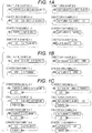
- Figs. 1A to 1C Constitution examples of expression vectors containing an ALP gene and an antibody gene in the present invention will be described with reference to Figs. 1A to 1C .
- the expression vector is exemplified as plasmid DNA, and an arrow indicates a promoter.
- Fd(HC) represents a gene encoding Fd.
- Fd is a heavy chain portion of Fab.
- LC represents a gene encoding a light chain
- linker represents a linker gene
- ALP subunit represents an ALP gene.
- ALP subunit as a protein is intended to refer to monomeric ALP.
- Fig. 1A shows expression vectors in which a gene encoding Fd or a light chain and an ALP gene are linked directly or via a gene encoding a peptide linker.
- Fig. 1B shows expression vectors in which a gene encoding VHH and an ALP gene are linked directly or via a gene encoding a peptide linker.
- Fig. 1C shows expression vectors further containing a gene encoding a light chain or Fd in each expression vector shown in Fig. 1A .
- the expression vectors used in the present disclosure are not limited to these examples.
- a full-length heavy chain or a gene encoding Fd' may be used instead of the gene encoding Fd.
- a gene encoding scFv may be used instead of the gene encoding VHH.
- the expression vector of Constitution Example 1-1 contains the gene encoding Fd, the linker gene and the ALP gene in this order downstream of the promoter. This expression vector expresses a fusion protein in which a C-terminal amino acid residue of Fd and an N-terminal amino acid residue of the ALP subunit are bound via a peptide linker.
- a Fab containing the fusion protein and the light chain is obtained as an ALP fusion antibody.
- the expression vector of Constitution Example 1-2 contains the gene encoding a light chain, the linker gene and the ALP gene in this order downstream of the promoter. This expression vector expresses a fusion protein in which a C-terminal amino acid residue of the light chain and an N-terminal amino acid residue of the ALP subunit are bound via a peptide linker.
- a Fab containing the fusion protein and Fd is obtained as an ALP fusion antibody.
- the expression vector of Constitution Example 1-3 contains the gene encoding Fd and the ALP gene in this order downstream of the promoter. This expression vector expresses a fusion protein in which a C-terminal amino acid residue of Fd and an N-terminal amino acid residue of the ALP subunit are directly bound.
- a Fab containing the fusion protein and the light chain is obtained as an ALP fusion antibody.
- the expression vector of Constitution Example 1-4 contains the gene encoding a light chain and the ALP gene in this order downstream of the promoter. This expression vector expresses a fusion protein in which a C-terminal amino acid residue of the light chain and an N-terminal amino acid residue of the ALP subunit are directly bound.
- a Fab containing the fusion protein and Fd is obtained as an ALP fusion antibody.
- the expression vector of Constitution Example 2-1 contains the ALP gene, the linker gene and the gene encoding Fd in this order downstream of the promoter. This expression vector expresses a fusion protein in which an N-terminal amino acid residue of Fd and a C-terminal amino acid residue of the ALP subunit are bound via a peptide linker.
- a Fab containing the fusion protein and the light chain is obtained as an ALP fusion antibody.
- the expression vector of Constitution Example 2-2 contains the ALP gene, the linker gene and the gene encoding a light chain in this order downstream of the promoter. This expression vector expresses a fusion protein in which an N-terminal amino acid residue of the light chain and a C-terminal amino acid residue of the ALP subunit are bound via a peptide linker.
- a Fab containing the fusion protein and Fd is obtained as an ALP fusion antibody.
- the expression vector of Constitution Example 2-3 contains the ALP gene and the gene encoding Fd in this order downstream of the promoter. This expression vector expresses a fusion protein in which an N-terminal amino acid residue of Fd and a C-terminal amino acid residue of the ALP subunit are directly bound.
- a Fab containing the fusion protein and the light chain is obtained as an ALP fusion antibody.
- the expression vector of Constitution Example 2-4 contains the ALP gene and the gene encoding a light chain in this order downstream of the promoter. This expression vector expresses a fusion protein in which an N-terminal amino acid residue of the light chain and a C-terminal amino acid residue of the ALP subunit are directly bound.
- a Fab containing the fusion protein and Fd is obtained as an ALP fusion antibody.
- the expression vector of Constitution Example 3-1 contains the gene encoding VHH, the linker gene and the ALP gene in this order downstream of the promoter.
- the expression vector of Constitution Example 3-2 contains the gene encoding VHH and the ALP gene in this order downstream of the promoter. By transforming or transfecting a cell using the expression vector of Constitution Example 3-2, an ALP fusion antibody in which a C-terminal amino acid residue of VHH and an N-terminal amino acid residue of the ALP subunit were directly bound is obtained.
- the expression vector of Constitution Example 3-3 contains the ALP gene, the linker gene and the gene encoding VHH in this order downstream of the promoter.
- the expression vector of Constitution Example 3-4 contains the ALP gene and the gene encoding VHH in this order downstream of the promoter. By transforming or transfecting a cell using the expression vector of Constitution Example 3-4, an ALP fusion antibody in which an N-terminal amino acid residue of VHH and a C-terminal amino acid residue of the ALP subunit were directly bound is obtained.
- the expression vector of Constitution Example 4-1 is an expression vector containing two promoters, which contains a gene encoding Fd, a linker gene and an ALP gene in this order downstream of one promoter, and contains a gene encoding a light chain downstream of the other promoter.
- a Fab containing a fusion protein in which a C-terminal amino acid residue of Fd and an N-terminal amino acid residue of the ALP subunit are bound via a peptide linker and the light chain is obtained as an ALP fusion antibody.
- the expression vector of Constitution Example 4-2 is an expression vector containing two promoters, which contains a gene encoding a light chain, a linker gene and an ALP gene in this order downstream of one promoter, and contains a gene encoding Fd downstream of the other promoter.
- a Fab containing a fusion protein in which a C-terminal amino acid residue of the light chain and an N-terminal amino acid residue of the ALP subunit are bound via a peptide linker and Fd is obtained as an ALP fusion antibody.
- the expression vector of Constitution Example 4-3 is an expression vector containing two promoters, which contains a gene encoding Fd and an ALP gene in this order downstream of one promoter, and contains a gene encoding a light chain downstream of the other promoter.
- a Fab containing a fusion protein in which a C-terminal amino acid residue of Fd and an N-terminal amino acid residue of the ALP subunit are directly bound and the light chain is obtained as an ALP fusion antibody.
- the expression vector of Constitution Example 4-4 is an expression vector containing two promoters, which contains a gene encoding a light chain and an ALP gene in this order downstream of one promoter, and contains a gene encoding Fd downstream of the other promoter.
- a Fab containing a fusion protein in which a C-terminal amino acid residue of the light chain and an N-terminal amino acid residue of the ALP subunit are directly bound and Fd is obtained as an ALP fusion antibody.
- the expression vector of Constitution Example 5-1 is an expression vector containing two promoters, which contains an ALP gene, a linker gene and a gene encoding Fd in this order downstream of one promoter, and contains a gene encoding a light chain downstream of the other promoter.
- a Fab containing a fusion protein in which an N-terminal amino acid residue of Fd and a C-terminal amino acid residue of the ALP subunit are bound via a peptide linker and the light chain is obtained as an ALP fusion antibody.
- the expression vector of Constitution Example 5-2 is an expression vector containing two promoters, which contains an ALP gene, a linker gene and a gene encoding a light chain in this order downstream of one promoter, and contains a gene encoding Fd downstream of the other promoter.
- a Fab containing a fusion protein in which an N-terminal amino acid residue of the light chain and a C-terminal amino acid residue of the ALP subunit are bound via a peptide linker and Fd is obtained as an ALP fusion antibody.
- the expression vector of Constitution Example 5-3 is an expression vector containing two promoters, which contains an ALP gene and a gene encoding Fd in this order downstream of one promoter, and contains a gene encoding a light chain downstream of the other promoter.
- a Fab containing a fusion protein in which an N-terminal amino acid residue of Fd and a C-terminal amino acid residue of the ALP subunit are directly bound and the light chain is obtained as an ALP fusion antibody.
- the expression vector of Constitution Example 5-4 is an expression vector containing two promoters, which contains an ALP gene and a gene encoding a light chain in this order downstream of one promoter, and contains a gene encoding Fd downstream of the other promoter.
- a Fab containing a fusion protein in which an N-terminal amino acid residue of the light chain and a C-terminal amino acid residue of the ALP subunit are directly bound and Fd is obtained as an ALP fusion antibody.
- ALP is known to be a type of zinc enzyme.
- Zinc ion concentration in the medium is preferably 0.01 mM or more, more preferably 0.05 mM or more, and further preferably 0.07 mM or more.
- the zinc ion concentration is preferably 0.25 mM or less, more preferably 0.20 mM or less, and further preferably 0.15 mM or less, because too high a zinc ion concentration in the medium may affect cell culture.
- the cell can be appropriately cultured by a known culture method according to the cell to be used.
- the medium containing a zinc ion can be prepared by adding a compound capable of supplying a zinc ion or a solution thereof to a medium suitable for the cell to be used.
- a compound is preferably a salt of zinc and an inorganic or an organic acid.
- examples of such a salt include zinc chloride, zinc sulfate, zinc acetate, and the like.
- the concentration of the zinc ion in the medium may be expressed by final concentration of the salt.
- test Example 2 even when an ALP fusion antibody was expressed in a cell and then cultured in a medium containing a zinc ion, ALP in the obtained ALP fusion antibody showed no activity. Therefore, in particular embodiments, it is preferable to add a zinc ion to a medium or replace it with a medium containing a zinc ion, during a period from transforming or transfecting a cell using an expression vector until an ALP fusion antibody is expressed in the cell.
- the ALP fusion antibody is recovered from the above cell.
- the ALP fusion antibody of the present invention in which BIAP or S-AP and the antibody are bound directly or via a peptide linker is obtained.
- a cell expressing the ALP fusion antibody may be dissolved in a solution containing a suitable solubilizer to recover the ALP fusion antibody liberated in the solution.
- a culture supernatant is recovered.
- the liberated ALP fusion antibody can be recovered by a known method such as affinity chromatography. If necessary, the recovered ALP fusion antibody may be purified by a method known in the art such as gel filtration.
- a C-terminal amino acid residue of ALP and an N-terminal amino acid residue of the antibody may be bound directly or via a peptide linker.
- an N-terminal amino acid residue of ALP and a C-terminal amino acid residue of the amino acid sequence of the antibody may be bound directly or via a peptide linker.
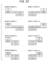
- the ALP contained in the ALP fusion antibody may contain two subunits.
- the two subunits may form a dimer.
- the antibody contained in the ALP fusion antibody may be bound to one of the two subunits or to both. That is, the ALP fusion antibody of the present embodiment contains an ALP subunit to which an antibody is bound and an ALP subunit to which an antibody is not bound, or contains two ALP subunits to which an antibody is bound.
- the ALP subunit to which an antibody is not bound can be obtained by an expression vector containing an ALP gene.
- Fd(HC) represents Fd
- LC represents a light chain
- linker represents a peptide linker
- ALP subunit represents monomeric ALP.
- a line between Fd(HC) and LC represents a disulfide bond
- a line between ALP subunits represents formation of a dimer.
- the ALP fusion antibody of the present invention is not limited to these examples.
- Fd in the ALP fusion antibody may be a full-length heavy chain or Fd'.
- VHH in the ALP fusion antibody may be scFv.
- ALP fusion antibody of Antibody Example 1-1 two molecules of ALP fusion Fab, in which a C-terminal amino acid residue of the heavy chain portion (Fd) and an N-terminal amino acid residue of the ALP subunit are bound via a peptide linker, form a complex by dimer formation between the ALP subunits.
- ALP fusion antibody of Antibody Example 1-2 two molecules of ALP fusion Fab, in which a C-terminal amino acid residue of a light chain and an N-terminal amino acid residue of the ALP subunit are bound via a peptide linker, form a complex by dimer formation between the ALP subunits.
- ALP fusion antibody of Antibody Example 1-3 two molecules of ALP fusion Fab, in which a C-terminal amino acid residue of the heavy chain portion (Fd) and an N-terminal amino acid residue of the ALP subunit are directly bound, form a complex by dimer formation between the ALP subunits.
- ALP fusion antibody of Antibody Example 1-4 two molecules of ALP fusion Fab, in which a C-terminal amino acid residue of the light chain and an N-terminal amino acid residue of the ALP subunit are directly bound, form a complex by dimer formation between the ALP subunits.
- ALP fusion antibody of Antibody Example 2-1 two molecules of ALP fusion Fab, in which an N-terminal amino acid residue of the heavy chain portion (Fd) and a C-terminal amino acid residue of the ALP subunit are bound via a peptide linker, form a complex by dimer formation between the ALP subunits.
- ALP fusion antibody of Antibody Example 2-2 two molecules of ALP fusion Fab, in which an N-terminal amino acid residue of the light chain and a C-terminal amino acid residue of the ALP subunit are bound via a peptide linker, form a complex by dimer formation between the ALP subunits.
- ALP fusion antibody of Antibody Example 2-3 two molecules of ALP fusion Fab, in which an N-terminal amino acid residue of the heavy chain portion (Fd) and a C-terminal amino acid residue of the ALP subunit are directly bound, form a complex by dimer formation between the ALP subunits.
- ALP fusion antibody of Antibody Example 2-4 two molecules of ALP fusion Fab, in which an N-terminal amino acid residue of the light chain and a C-terminal amino acid residue of the ALP subunit are directly bound, form a complex by dimer formation between the ALP subunits.
- ALP fusion antibody of Antibody Example 3-1 two molecules of ALP fusion VHH, in which a C-terminal amino acid residue of VHH and an N-terminal amino acid residue of the ALP subunit are bound via a peptide linker, form a complex by dimer formation between the ALP subunits.
- ALP fusion antibody of Antibody Example 3-2 two molecules of ALP fusion VHH, in which a C-terminal amino acid residue of VHH and an N-terminal amino acid residue of the ALP subunit are directly bound, form a complex by dimer formation between the ALP subunits.
- ALP fusion antibody of Antibody Example 3-3 two molecules of ALP fusion VHH, in which an N-terminal amino acid residue of VHH and a C-terminal amino acid residue of the ALP subunit are bound via a peptide linker, form a complex by dimer formation between the ALP subunits.
- ALP fusion antibody of Antibody Example 3-4 two molecules of ALP fusion VHH, in which an N-terminal amino acid residue of VHH and a C-terminal amino acid residue of the ALP subunit are directly bound, form a complex by dimer formation between the ALP subunits.
- ALP fusion VHH in which a C-terminal amino acid residue of VHH and an N-terminal amino acid residue of the ALP subunit are bound via a peptide linker, and the ALP subunit to which an antibody is not bound form a dimer between the ALP subunits.
- the ALP fusion antibody of Antibody Example 3-5 can be obtained by transforming or transfecting a cell using the expression vector of Constitution Example 3-1 and the expression vector containing an ALP gene.
- ALP fusion VHH in which a C-terminal amino acid residue of VHH and an N-terminal amino acid residue of the ALP subunit are directly bound, and the ALP subunit to which an antibody is not bound form a dimer between the ALP subunits.
- the ALP fusion antibody of Antibody Example 3-6 can be obtained by transforming or transfecting a cell using the expression vector of Constitution Example 3-2 and the expression vector containing an ALP gene.
- ALP fusion VHH in which an N-terminal amino acid residue of VHH and a C-terminal amino acid residue of the ALP subunit are bound via a peptide linker, and the ALP subunit to which an antibody is not bound form a dimer between the ALP subunits.
- the ALP fusion antibody of Antibody Example 3-7 can be obtained by transforming or transfecting a cell using the expression vector of Constitution Example 3-3 and the expression vector containing an ALP gene.
- ALP fusion VHH in which an N-terminal amino acid residue of VHH and a C-terminal amino acid residue of the ALP subunit are directly bound, and the ALP subunit to which an antibody is not bound form a dimer between the ALP subunits.
- the ALP fusion antibody of Antibody Example 3-8 can be obtained by transforming or transfecting a cell using the expression vector of Constitution Example 3-4 and the expression vector containing an ALP gene.
- ALP fusion antibody of Antibody Example 4-1 ALP fusion Fab, in which a C-terminal amino acid residue of the heavy chain portion (Fd) and an N-terminal amino acid residue of the ALP subunit are bound via a peptide linker, and the ALP subunit to which an antibody is not bound form a dimer between the ALP subunits.
- the ALP fusion antibody of Antibody Example 4-1 can be obtained, for example, by transforming or transfecting a cell using the expression vector of Constitution Example 1-1, the expression vector containing the gene encoding a light chain, and the expression vector containing an ALP gene.
- ALP fusion antibody of Antibody Example 4-2 ALP fusion Fab, in which a C-terminal amino acid residue of the light chain and an N-terminal amino acid residue of the ALP subunit are bound via a peptide linker, and the ALP subunit to which an antibody is not bound form a dimer between the ALP subunits.
- the ALP fusion antibody of Antibody Example 4-2 can be obtained, for example, by transforming or transfecting a cell using the expression vector of Constitution Example 1-2, the expression vector containing the gene encoding Fd, and the expression vector containing an ALP gene.
- ALP fusion antibody of Antibody Example 4-3 ALP fusion Fab, in which a C-terminal amino acid residue of the heavy chain portion (Fd) and an N-terminal amino acid residue of the ALP subunit are directly bound, and the ALP subunit to which an antibody is not bound form a dimer between the ALP subunits.
- the ALP fusion antibody of Antibody Example 4-3 can be obtained, for example, by transforming or transfecting a cell using the expression vector of Constitution Example 1-3, the expression vector containing the gene encoding a light chain, and the expression vector containing an ALP gene.
- ALP fusion antibody of Antibody Example 4-4 In the ALP fusion antibody of Antibody Example 4-4, ALP fusion Fab, in which a C-terminal amino acid residue of the light chain and an N-terminal amino acid residue of the ALP subunit are directly bound, and the ALP subunit to which an antibody is not bound form a dimer between the ALP subunits.
- the ALP fusion antibody of Antibody Example 4-4 can be obtained, for example, by transforming or transfecting a cell using the expression vector of Constitution Example 1-4, the expression vector containing the gene encoding Fd, and the expression vector containing an ALP gene.
- ALP fusion antibody of Antibody Example 5-1 ALP fusion Fab, in which an N-terminal amino acid residue of the heavy chain portion (Fd) and a C-terminal amino acid residue of the ALP subunit are bound via a peptide linker, and the ALP subunit to which an antibody is not bound form a dimer between the ALP subunits.
- the ALP fusion antibody of Antibody Example 5-1 can be obtained, for example, by transforming or transfecting a cell using the expression vector of Constitution Example 2-1, the expression vector containing the gene encoding a light chain, and the expression vector containing an ALP gene.
- ALP fusion antibody of Antibody Example 5-2 ALP fusion Fab, in which an N-terminal amino acid residue of the light chain and a C-terminal amino acid residue of the ALP subunit are bound via a peptide linker, and the ALP subunit to which an antibody is not bound form a dimer between the ALP subunits.
- the ALP fusion antibody of Antibody Example 5-2 can be obtained, for example, by transforming or transfecting a cell using the expression vector of Constitution Example 2-2, the expression vector containing the gene encoding Fd, and the expression vector containing an ALP gene.
- ALP fusion antibody of Antibody Example 5-3 ALP fusion Fab, in which an N-terminal amino acid residue of the heavy chain portion (Fd) and a C-terminal amino acid residue of the ALP subunit are directly bound, and the ALP subunit to which an antibody is not bound form a dimer between the ALP subunits.
- the ALP fusion antibody of Antibody Example 5-3 can be obtained, for example, by transforming or transfecting a cell using the expression vector of Constitution Example 2-3, the expression vector containing the gene encoding a light chain, and the expression vector containing an ALP gene.
- ALP fusion antibody of Antibody Example 5-4 In the ALP fusion antibody of Antibody Example 5-4, ALP fusion Fab, in which an N-terminal amino acid residue of the light chain and a C-terminal amino acid residue of the ALP subunit are directly bound, and the ALP subunit to which an antibody is not bound form a dimer between the ALP subunits.
- the ALP fusion antibody of Antibody Example 5-4 can be obtained, for example, by transforming or transfecting a cell using the expression vector of Constitution Example 2-4, the expression vector containing the gene encoding Fd, and the expression vector containing an ALP gene.
- the ALP fusion antibody of the present invention can be used to detect a test substance in immunoassay. Therefore, one embodiment is an immunoassay reagent containing the ALP fusion antibody.
- the type of immunoassay is not particularly limited.
- the type of immunoassay can be selected as appropriate from known immunoassay methods such as ELISA, Western blotting, and immune complex transfer method (see Japanese Laid-Open Patent Publication No. H1-254868 ).
- the ELISA is preferred.
- the ELISA may be any of a sandwich method, a competitive method, a direct method, an indirect method, and the like.
- the nature of the reagent is not particularly limited, and may be solid (for example, powder, crystal, freeze-dried product, or the like) or liquid (for example, solution, suspension, emulsion, or the like).
- a solvent is not particularly limited as long as the ALP fusion antibody of the present invention can be dissolved and stored. Examples of the solvent include water, physiological saline, phosphate buffered saline (PBS), Tris buffered saline (TBS), Good's buffer, and the like.
- Examples of the Good's buffers include MES, Bis-Tris, ADA, PIPES, Bis-Tris-Propane, ACES, MOPS, MOPSO, BES, TES, HEPES, HEPPS, Tricine, Tris, Bicine, TAPS, and the like.
- the immunoassay reagent of the present invention may contain known additives.
- the additive include protein stabilizers such as bovine serum albumin (BSA), preservatives such as sodium azide, inorganic salts such as sodium chloride, and the like.
- BSA bovine serum albumin
- preservatives such as sodium azide
- inorganic salts such as sodium chloride, and the like.
- a container containing an immunoassay reagent may be packed in a box and provided to the user as a reagent kit.
- the box may contain an attached document. Composition, usage, storage method, etc. of the immunoassay reagent may be described in the attached document.
- Fig. 3 shows an example of the reagent kit.
- 11 denotes a reagent kit according to an embodiment of the invention
- 12 denotes a first container containing an immunoassay reagent containing an ALP fusion antibody according to an embodiment of the invention
- 13 denotes a packing box
- 14 denotes an attached document.
- a further embodiment is an immunoassay reagent kit containing a first reagent containing the ALP fusion antibody and a second reagent containing a capture substance that specifically binds to a test substance. Details of the first reagent are the same as those described for the immunoassay reagent of the present invention.
- the capture substance that specifically binds to a test substance refers to a substance that captures a test substance on a solid phase by being immobilized on the solid phase.
- the type of the capture substance is not particularly limited, and can be appropriately selected depending on the type of the test substance.
- the capture substance include antibodies, antigens, oligonucleotide probes, receptors, ligands that bind to the receptors, aptamers, and the like.
- an antibody used as a capture substance is also referred to as "capture antibody”.
- the capture antibody may be either a monoclonal antibody or a polyclonal antibody, but is preferably a monoclonal antibody. When the capture antibody is a monoclonal antibody, an epitope recognized by the capture antibody is preferably different from an epitope recognized by the ALP fusion antibody of the present invention.
- the ALP fusion antibody of the present invention contained in the first reagent is preferably used as a detection antibody in the sandwich ELISA.
- the detection antibody refers to an antibody that specifically binds to a test substance and has a labeling substance, and provides an antibody that provides a detectable signal via the labeling substance. It is preferable that the detection antibody is not immobilized on a solid phase.
- containers containing each of the first reagent and the second reagent may be packed in a box and provided to the user as a reagent kit.
- the box may contain an attached document. Configuration, composition of each reagent, usage, storage method, etc. of the immunoassay reagent kit of the present invention may be described in the attached document.
- Fig. 4 shows an example of the reagent kit. In Fig.
- 21 denotes a reagent kit according to an embodiment of the present invention
- 22 denotes a first container containing a first reagent containing an ALP fusion antibody according to an embodiment of the present invention
- 23 denotes a second container containing a second reagent containing a capture substance that specifically binds to a test substance
- 24 denotes a packing box
- 25 denotes an attached document.
- the immunoassay reagent kit of the present invention may further contain a solid phase for immobilizing the capture substance.
- the solid phase may be any insoluble carrier capable of immobilizing the capture substance.
- the material of the solid phase is not particularly limited.
- the material can be selected from organic polymer compounds, inorganic compounds, biopolymers, and the like.
- the organic polymer compound include latex, polystyrene, polypropylene, and the like.
- the inorganic compound include magnetic bodies (iron oxide, chromium oxide, ferrite, and the like), silica, alumina, glass, and the like.
- the biopolymer include insoluble agarose, insoluble dextran, gelatin, cellulose, and the like.
- the shape of the solid phase is not particularly limited, and examples thereof include microplates, microtubes, test tubes, particles, membranes, and the like. Among them, microplates and particles (particularly magnetic particles) are preferable.
- the immunoassay reagent kit of the present invention may further contain a substrate of ALP.
- the substrate include chemiluminescent substrates such as CDP-Star (registered trademark) (disodium 4-chloro-3-(methoxyspiro[1,2-dioxetane-3,2'-(5'-chloro)tricyclo[3.3.1.13,7]decan]-4-yl)phenyl phosphate) and CSPD (registered trademark) (disodium 3-(4-methoxyspiro[1,2-dioxetane-3,2-(5'-chloro)tricyclo[3.3.1.13,7]decan]-4-yl)phenyl phosphate), and chromogenic substrates such as 5-bromo-4-chloro-3-indolyl phosphate (BCIP), disodium 5-bromo-6-chloro-indolyl phosphate and p-nitrophenyl phosphate.
- the immunoassay reagent kit of the present invention may further contain a calibrator of the test substance.
- the calibrator include a calibrator for quantification of a test substance.
- This calibrator may include, for example, a buffer solution containing no test substance (negative control) and a buffer solution containing test substance at a known concentration.
- the test substance is a protein
- the test substance contained in the calibrator may be a recombinant protein or a polypeptide synthesized based on the amino acid sequence of the test substance.
- a further aspect is an immunoassay method using the ALP fusion antibody.
- This measurement method includes, for example, measuring a test substance in a sample using the ALP fusion antibody of the present invention.
- the test substance is not particularly limited as long as it is a substance to which the ALP fusion antibody of the present invention can specifically bind.
- the test substance include proteins, nucleic acids, polysaccharides, lipids, haptens, compounds, bacteria, viruses, and the like.
- the sample is not particularly limited as long as it contains a test substance. Examples of the sample include biological samples such as blood and lymph fluid, excreta such as urine and feces, environmental samples such as river water, sea water and soil, and the like.
- the ALP fusion antibody of the present invention is used as a detection antibody.
- a complex containing a test substance, a capture substance that specifically binds to the test substance, and the ALP fusion antibody (detection antibody) of the present invention is formed on a solid phase.
- the complex can be formed by mixing a sample that may contain a test substance, a capture substance, and a detection antibody. Then, a solution containing the complex is brought into contact with a solid phase capable of immobilizing the capture substance, whereby the complex can be formed on the solid phase. Alternatively, a solid phase preliminarily immobilized with the capture substance may be used.
- a solid phase immobilized with the capture substance, the sample that may contain a test substance, and the detection antibody are brought into contact with each other, whereby the complex can be formed on the solid phase.
- the test substance contained in the sample can be measured by detecting the complex formed on the solid phase by a method known in the art.
- the test substance in the sample can be measured by detecting a signal generated by ALP contained in the detection antibody.
- the mode of immobilization of the capture substance on the solid phase is not particularly limited.
- the capture substance and the solid phase may be bound directly, or the capture substance and the solid phase may be indirectly bound via another substance.
- Examples of the direct binding include physical adsorption and the like.
- Examples of the indirect bond include a bond via a combination of biotin or its analog and a biotin-binding site.
- the capture substance and the solid phase can be indirectly bound via the bond between the biotin or its analog and the biotin-binding site.
- the biotin and its analogs include biotin and biotin analogs such as desthiobiotin.
- the biotin-binding sites include avidin and avidin analogs such as streptavidin and tamavidin (registered trademark).
- B/F (Bound/Free) separation for removing an unreacted free component not forming a complex may be performed between formation of the complex and detection of the complex.
- the unreacted free component refers to a component not constituting a complex. Examples thereof include capture substances not bound to the test substance, detection antibodies, and the like.
- the means of B/F separation is not particularly limited, and when the solid phase is a particle, B/F separation can be performed by recovering only the solid phase capturing the complex by centrifugation. When the solid phase is a container such as a microplate or a microtube, B/F separation can be performed by removing a liquid containing an unreacted free component.
- B/F separation can be performed by aspirating and removing a liquid containing an unreacted free component by a nozzle while magnetically constraining the magnetic particles with a magnet, which is preferable from the viewpoint of automation.
- the solid phase capturing the complex may be washed with a suitable aqueous medium such as PBS.
- the phrase "detecting a signal” includes qualitatively detecting the presence or absence of a signal, quantifying a signal intensity, and semi-quantitatively detecting the intensity of a signal.
- signals such as light and color generated by reacting the complex on the solid phase with the substrate of ALP can be measured by using a known apparatus such as a spectrophotometer.
- the substrate for ALP is as described above.
- the detection result of the signal can be used as the measurement result of the test substance.
- a measured value of the signal intensity itself or a value acquired from the measured value can be used as the measurement result of the test substance.
- the value acquired from the measured value of the signal intensity include a value obtained by subtracting the measured value of a negative control sample or the background value from the measured value, and the like.
- the measured value of the signal intensity may be applied to a calibration curve to determine the amount or concentration value of the test substance.
- the negative control sample can be appropriately selected, and examples thereof include a sample known not to contain a test substance.
- test substance in the sample it is preferable to measure the test substance in the sample by a sandwich ELISA method using a capture antibody immobilized on magnetic particles and the ALP fusion antibody (detection antibody) of the present invention.
- measurement may be carried out using a commercially available fully automated immunoassay system such as HISCL series (manufactured by Sysmex Corporation).
- a fusion protein in which rabbit antibody Fab and bovine small intestine-derived ALP subunit were bound was prepared.
- the fusion protein becomes an ALP fusion antibody having two molecules of Fab by forming a dimer of ALP subunits. This corresponds to the ALP fusion antibody having a structure shown in Antibody Example 1-1 above.
- Lymphocytes were acquired from peripheral blood of a rabbit immunized with CD80, and mRNA was extracted from the lymphocytes to synthesize cDNA.
- the obtained cDNA was amplified using a known primer for cloning an antibody gene to prepare a Fab phage library.
- a Fab clone of a rabbit anti-CD80 antibody was obtained by a known Fab phage display method and biopanning (see Lang IM, Barbas CF 3rd, Schleef RR., Recombinant rabbit Fab with binding activity to type-1 plasminogen activator inhibitor derived from a phage-display library against human alpha-granules, (1996) Gene 172(2): 295-8 and Philippa M.
- a gene of the acquired Fab clone of the rabbit anti-CD80 antibody was incorporated into a plasmid DNA containing a gene encoding a Fc region of the rabbit antibody to acquire a plasmid DNA containing the gene of the rabbit anti-CD80 antibody.
- Amino acid sequences of Fd (heavy chain portion of Fab) and light chain of the rabbit anti-CD80 antibody are shown in SEQ ID NOs: 11 and 12, respectively.
- Base sequences encoding these amino acid sequences are shown in SEQ ID NOs: 13 and 14, respectively.
- a plasmid DNA containing a BIAPII gene was acquired by outsourcing gene synthesis to GenScript, based on BIAPII amino acid sequence described in U.S. Patent No. 6,406,899 .
- the amino acid sequence of BIAPII and a base sequence encoding the same are shown in SEQ ID NOs: 1 and 2, respectively.
- a DNA fragment encoding BIAPII (BIAPII insert) was acquired by PCR.
- a DNA fragment encoding BIAPII (BIAPII insert) was acquired by PCR.
- pcDNA3.4 vector as a template, linearized vector DNA was obtained by inverse PCR.
- PCR was performed using KOD Plus neo (Toyobo Co., Ltd.) as described in the attached document. Base sequences of primers used for PCR were as follows.
- a DNA fragment (Fd-linker insert) encoding Fd and a peptide linker was acquired by PCR.
- a gene encoding a peptide linker was linked downstream of a gene encoding the Fd of the rabbit anti-CD80 antibody.
- Types of peptide linkers were GS1, GS2, GS3, EK1, EK2 and EK3 described above.
- linearized vector DNA containing the BIAPII gene was obtained by inverse PCR. PCR was performed using KOD Plus neo (Toyobo Co., Ltd.) as described in the attached document. Base sequences of primers used for PCR were as follows.
- the Fd insert was linked to the linearized vector DNA containing the BIAPII gene to obtain an Fd-linker-BIAPII expression vector.
- An In-Fusion reaction was performed according to the attached document of the kit.
- the gene encoding a peptide linker and the BIAPII gene were linked downstream of the gene encoding the Fd of the rabbit anti-CD80 antibody.
- PCR PCR was performed using KOD Plus neo (Toyobo Co., Ltd.) as described in the attached document. Base sequences of primers used for PCR were as follows.
- the LC insert was linked to the linearized vector DNA of pcDNA3.4 to obtain a light chain expression vector.
- An In-Fusion reaction was performed according to the attached document of the kit.
- Expi293F (trademark) cells were shake-cultured (125 rpm) at 37°C under a 5% CO 2 atmosphere in a medium added with a solution of zinc chloride (KISHIDA CHEMICAL Co., Ltd.) to a final concentration of 0.1 mM. 25.5 mL of cell culture (3.0 ⁇ 10 6 cells/mL) was prepared according to the number of samples. An appropriate amount of Opti-MEMI (trademark) was added to a mixture of the Fd-linker-BIAPII expression vector (about 15 ⁇ g) and the light chain expression vector (about 15 ⁇ g) to make 1.5 mL, and the mixture was gently stirred to prepare a DNA solution.
- the ExpiFectamine 293 reagent (80 ⁇ L) and Opti-MEMI (1.5 mL) were gently stirred and allowed to stand at room temperature for 5 minutes to prepare a transfection reagent.
- the DNA solution and the transfection reagent were gently stirred and allowed to stand at room temperature for 20 minutes.
- the resulting mixture (3 mL) was added to the cell culture (25.5 mL).
- the mixture was shake-cultured (125 rpm) at 37°C for 20 hours in a 5% CO 2 atmosphere.
- 150 ⁇ L and 1.5 mL of ExpiFectamine (trademark) transfection enhancers 1 and 2 were added to each culture, respectively.
- Each mixture was shake-cultured (125 rpm) at 37°C for 5 days in a 5% CO 2 atmosphere.
- Culture supernatant was collected 5 days after transfection.
- the culture supernatant contains each ALP fusion antibody secreted from transfected Expi293F (trademark) cells.
- the collected culture supernatant was transferred to a centrifuge tube and centrifuged (1000 g, 5 minutes, 4°C), and the supernatant was transferred to a new centrifuge tube.
- the supernatant was centrifuged again (10000 g, 10 minutes, 4°C), and the supernatant was transferred to a new centrifuge tube.
- the supernatant was concentrated using Amicon Ultra-4, 30k (Merck).
- the concentrated supernatant was purified by gel filtration using AKTA york25 (GE Healthcare) and Superdex 200 Increase 10/300GL (GE Healthcare).
- ALP buffer 100 mM triethanolamine (TEA), 150 mM NaCl, 1 mM MgCl 2 , 0.1 mM ZnCl 2 , pH 7.0
- TAA triethanolamine
- the concentration of the ALP fusion antibody purified by gel filtration was measured from absorbance at 280 nm using nanodrop-1000 (Thermo Fisher).
- Rabbit anti-CD80 antibody (IgG) was diluted with PBS to 1 ⁇ g/mL. The resulting antibody solution was added to a 96-well black plate at 100 ⁇ L/well and allowed to stand overnight at 4°C to immobilize the antibody in the wells. After removing the antibody solution from the plate, Buffer I' (100 mM HEPES, 1% BSA, 0.05% ProClin (trademark), 1 mM MgCl 2 , 0.1 mM ZnCl 2 , pH 8.0) was added at 300 ⁇ L/well and allowed to stand overnight at 4°C for blocking.
- Buffer I' 100 mM HEPES, 1% BSA, 0.05% ProClin (trademark), 1 mM MgCl 2 , 0.1 mM ZnCl 2 , pH 8.0
- a human CD80/B7-1 protein (His tag) solution (0.005, 0.020, 0.078, 0.313, 1.25, 5.0 or 20 ng/mL) was added as an antigen solution at 100 ⁇ L/well.
- the plate was shaken at room temperature at 500 rpm for 1 hour to perform an antigen-antibody reaction.
- the plate was washed three times with a HISCL washing solution (Sysmex Corporation), and then each ALP fusion antibody (200 ng/mL) of Example 1 diluted with Buffer I' was added at 100 ⁇ L/well.
- the plate was shaken at room temperature at 500 rpm for 1 hour to perform an antigen-antibody reaction.
- HISCL R5 reagent (Sysmex Corporation) as a solution of a chemiluminescent substrate of ALP was added at 100 ⁇ L/well, and luminescence intensity was immediately measured with FLUOstar OPTIMA (BMG LABTECH).
- GS1, GS2, GS3, EK1, EK2 and EK3 are types of peptide linkers in the ALP fusion antibody.
- the luminescence intensity increased according to antigen concentration regardless of which ALP fusion antibody was used. Therefore, it was shown that all ALP fusion antibodies had the same level of antigen detection ability.
- the cells containing an expression vector obtained in Example 1 were cultured in a medium added with zinc or a medium not added with zinc under various conditions, and ALP activity of the ALP fusion antibody was measured.
- the Fd-linker-BIAPII expression vector and the light chain expression vector were transfected to cells in the same manner as in Example 1 except that Expi293F (trademark) cells cultured in a normal medium not added with zinc were used. After 20 hours, the medium was replaced with a medium not added with zinc or a medium added with a zinc chloride solution (final concentration 0.1 mM). Then, ExpiFectamine (trademark) transfection enhancers 1 and 2 were added thereto, and the mixture was shake-cultured (125 rpm) at 37°C for 5 days in a 5% CO 2 atmosphere. After culturing, the ALP fusion antibody (GS1 linker) was recovered and purified from culture supernatant in the same manner as in Example 1.
- the ALP fusion antibody was diluted with the ALP buffer.
- the resulting antibody solution was added to a 96-well black plate at 10 ⁇ L/well.
- a HISCL R5 reagent (Sysmex Corporation) was added at 100 ⁇ L/well, and luminescence intensity was immediately measured with FLUOstar OPTIMA (BMG LABTECH).
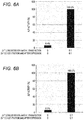
- the measurement results are shown in Figs. 6A to 6C .
- Figs. 6A and 6B show ALP activities of ALP fusion antibodies obtained from the cells cultured in the medium not added with zinc and the cells cultured in the zinc-added medium after expression of ALP fusion antibody when an ALP activity of an ALP fusion antibody obtained from the cells cultured in the zinc-added medium after transfection is 100%.
- Fig. 6A and 6B show ALP activities of ALP fusion antibodies obtained from the cells cultured in the medium not added with zinc and the cells cultured in the zinc-added medium after expression of ALP fusion antibody when an ALP
- 6C shows ALP activities of ALP fusion antibodies obtained from the cells cultured in a medium with a zinc chloride concentration of 0.25 mM or 0.5 mM when an ALP fusion antibody of an ALP fusion antibody obtained from the cells cultured in a medium having a zinc chloride concentration of 0.1 mM is 100%.
- the ALP activity was significantly low in the ALP fusion antibody obtained from the cells cultured in the medium not added with zinc. From this result, it was shown that it is necessary to add zinc to the medium in order to exert the ALP activity of the ALP fusion antibody. As shown in Fig. 6B , the ALP activity was also significantly low in the ALP fusion antibody obtained from the cells cultured in the zinc-added medium after expression of ALP fusion antibody. From this result, it was shown that the ALP activity of the ALP fusion antibody does not increase even if zinc is added to the medium at the time when the ALP fusion antibody is considered to be expressed and it is necessary to add zinc from the time of transfection.
- a rabbit anti-CD80 antibody was obtained using the plasmid DNA containing the gene of the rabbit anti-CD80 antibody obtained in Example 1. From the obtained antibody, Fab was obtained by a conventional method. The obtained Fab was bound to bovine small intestine-derived ALP (ALP55, Oriental Yeast Co., ltd.) or recombinant ALP (rALP, Roche) using a crosslinking agent to obtain an ALP-labeled antibody.
- ALP55 Oriental Yeast Co., ltd.
- rALP recombinant ALP
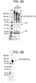
- the ALP fusion antibody (GS1 linker) and ALP-labeled antibody of Example 1 were separated by gel filtration using KTA york 25 (GE Healthcare) and Superdex 200 Increase 10/300GL (GE Healthcare), respectively. Gel filtration was performed in the same manner as in Example 1. Each fraction of the ALP fusion antibody and the ALP-labeled antibody separated by gel filtration was analyzed by SDS-PAGE under non-reducing conditions.
- Fab+ refers to a labeled antibody in which one molecule of ALP and one molecule of Fab are bound
- Fab++ refers to a labeled antibody in which one molecule of ALP and two molecules of Fab are bound
- Fab+++ refers to a labeled antibody in which one molecule of ALP and three molecules of Fab are bound.
- the ALP fusion antibody of the present invention is a homogeneous group of molecules.
- a reagent for a fully automatic immunoassay device was prepared using the ALP fusion antibody of Example 1, and performance of this reagent was examined. For comparison, reagents using each fraction of the ALP-labeled antibody isolated in Test Example 3 were also examined.
- R1 reagent capture antibody reagent
- a buffer for R1 reagent 50 mM HEPES, 150 mM NaCl, 1% BSA, pH 7.4
- R2 reagent solid phase
- a HISCL registered trademark
- An R3 reagent (detection antibody reagent) was prepared by dissolving the ALP fusion antibody (GS1 linker) of Example 1 in a buffer for R3 reagent (50 mM HEPES, 1 mM MgCl 2 , 0.1 mM ZnCl 2 , 1% BSA, pH 7.3). The concentration of the ALP fusion antibody in the R3 reagent was 200 ng/mL. For comparison, R3 reagents containing each fraction of the ALP-labeled antibody (Fab+, Fab++ or Fab+++) were prepared in the same manner.
- the concentration of the ALP-labeled antibody in the R3 reagent was adjusted so as to be approximately equal to the ALP activity of the R3 reagent containing the ALP fusion antibody.
- an R4 reagent (measurement buffer) a HISCL R4 reagent (Sysmex Corporation) was used.
- an R5 reagent (substrate solution), a HISCL R5 reagent (Sysmex Corporation) was used.
- An antigen solution was prepared by serially diluting human CD80/B7-1 protein (His tag) with Buffer I'.
- a HISCL washing solution (Sysmex Corporation) was used as a washing solution for magnetic particles.
- the magnetic particles in the obtained mixed solution were magnetically collected to remove the supernatant, and a HISCL washing solution (300 ⁇ L) was added to wash the magnetic particles.
- the supernatant was removed, and the R4 reagent (50 ⁇ L) and the R5 reagent (100 ⁇ L) were added to the magnetic particles and thoroughly mixed, and chemiluminescence intensity was measured.
- a measurement was performed in the same manner as above except that Buffer I' containing no antigen was used instead of the antigen solution. SN ratios of measurement were calculated from the measured values of each antigen solution and the backgrounds.
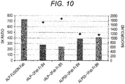
- Figs. 9A and 9B The measurement results are shown in Figs. 9A and 9B .
- the background was significantly lower when the ALP fusion antibody was used as the detection antibody than when the ALP-labeled antibody was used.
- the SN ratio was significantly higher when the ALP fusion antibody was used as the detection antibody than when the ALP-labeled antibody was used. From these results, it was found that the antigen can be detected with higher sensitivity by using the detection antibody reagent containing the ALP fusion antibody than by using the detection antibody reagent containing the ALP-labeled antibody obtained by chemical modification method.
- ALP fusion antibody having an antibody portion different from that of the ALP fusion antibody of Example 1 a fusion protein in which Fab of a mouse anti-human IgG antibody and the ALP subunit were bound was prepared. This corresponds to the ALP fusion antibody having a structure shown in Antibody Example 1-1 above.
- Lymphocytes were acquired from peripheral blood of a mouse immunized with human IgG, and mRNA was extracted from the lymphocytes to synthesize cDNA.
- Example 1 Using the obtained cDNA, a plasmid DNA containing a gene of the mouse anti-human IgG antibody was acquired in the same manner as in Example 1. Using the plasmid DNA containing this antibody gene and the BIAPII expression vector of Example 1, an Fd-linker-BIAPII expression vector was obtained in the same manner as in Example 1.
- the gene encoding the peptide linker GS1, GS3, EK1 or EK3 and the BIAPII gene were linked downstream of a gene encoding Fd of the mouse anti-human IgG antibody.
- a light chain expression vector was obtained in the same manner as in Example 1.
- Example 2 In the same manner as in Example 1, the above expression vector was transfected into Expi293F (trademark) cells cultured in a medium added with a zinc chloride solution. Culture supernatant was collected 5 days after transfection, and the ALP fusion antibody was concentrated and purified in the same manner as in Example 1.
- a reagent for a fully automatic immunoassay device was prepared using the ALP fusion antibody obtained in Example 2, and performance and storage stability of this reagent were examined. For comparison, reagents using the ALP-labeled antibody were also examined.
- Fab of the mouse anti-human IgG antibody was bound to ALP55 (Oriental Yeast Co., ltd.) or rALP (Roche) to obtain an ALP-labeled antibody.
- ALP55 Oriental Yeast Co., ltd.
- rALP rALP
- Each of the ALP fusion antibody and the ALP-labeled antibody obtained in Example 2 was separated by gel filtration in the same manner as in Test Example 3.
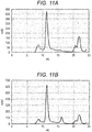
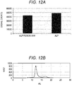
- Each fraction of the ALP fusion antibody and the ALP-labeled antibody separated by gel filtration was analyzed by SDS-PAGE under non-reducing conditions.
- the ALP-labeled antibody obtained by chemical modification method was a mixture of labeled antibodies with various molecular weights, unmodified ALP and Fab'.
- the ALP fusion antibody was a homogeneous group of molecules.
- fractions of ALP-labeled antibody fractions (rALP-2Fab Fr.B4, rALP-2Fab Fr.B5 and ALP55-2Fab Fr.B4) containing a labeled antibody in which one molecule of ALP and two molecules of Fab are bound, and a fraction (ALP55-1Fab Fr B6) containing a labeled antibody in which one molecule of ALP and one molecule of Fab are bound were used.
- the antibody concentration in each R3 reagent was adjusted based on the ALP activity of each antibody.
- An antigen solution was prepared by diluting human IgG antibody with Buffer I'.
- the R2 reagent, R4 reagent, R5 reagent and washing solution were the same as those in Example 4.
- the background was significantly lower when the ALP fusion antibody was used than when the ALP-labeled antibody was used.
- the SN ratio was significantly higher when the ALP fusion antibody was used than when the ALP-labeled antibody was used.
- Table 1 it was found that when each R3 reagent was stored at 40°C for 1 week, PC measured values with the R3 reagent containing the ALP fusion antibody did not decrease much as compared to PC measured values with the R3 reagent containing the ALP-labeled antibody. Therefore, it was shown that the reagent containing the ALP fusion antibody has higher storage stability than the reagent containing the ALP-labeled antibody.
- Example 2 Using the plasmid DNA containing the antibody gene obtained in Example 2 and the BIAPII expression vector, expression vectors having a structure shown in any of Constitution Examples 1-2, 1-3, 2-1 and 2-2 were shown were prepared by the same method as in Example 1. Using these expression vectors, the ALP fusion antibodies shown in Antibody Examples 1-2, 1-3, 2-1 and 2-2 were prepared.
- a DNA fragment encoding a light chain and a peptide linker was acquired.
- the obtained DNA fragment was linked to a linearized BIAPII expression vector DNA to prepare an LC-linker-BIAPII expression vector.
- the gene encoding a peptide linker and the BIAPII gene were linked downstream of the gene encoding a light chain.
- a DNA fragment encoding Fd was acquired, and an expression vector containing the gene encoding Fd (Fd expression vector) was prepared in the same manner as in Example 1.
- These expression vectors were transfected into Expi293F (trademark) cells to obtain the ALP fusion antibody of Antibody Example 1-2.
- Example 2 Using the plasmid DNA containing the antibody gene obtained in Example 2 as a template, a DNA fragment encoding Fd was acquired. The obtained DNA fragment was linked to a linearized BIAPII expression vector DNA to prepare an Fd-BIAPII expression vector. In this expression vector, the BIAPII gene was linked downstream of the gene encoding Fd.
- This expression vector and the light chain expression vector obtained in Example 2 were transfected into Expi293F (trademark) cells to obtain the ALP fusion antibody of Antibody Example 1-3.
- a DNA fragment encoding a peptide linker and Fd was acquired.
- the obtained DNA fragment was linked to a linearized BIAPII expression vector DNA to prepare a BIAPII-linker-Fd expression vector.
- the gene encoding a peptide linker and the gene encoding Fd were linked downstream of the BIAPII gene.
- This expression vector and the light chain expression vector obtained in Example 2 were transfected into Expi293F (trademark) cells to obtain the ALP fusion antibody of Antibody Example 2-1.
- a DNA fragment encoding a peptide linker and a light chain was acquired.
- the obtained DNA fragment was linked to a linearized BIAPII expression vector DNA to prepare a BIAPII-linker-LC expression vector.
- the gene encoding a peptide linker and the gene encoding a light chain were linked downstream of the BIAPII gene.
- This expression vector and the Fd expression vector were transfected into Expi293F (trademark) cells to obtain the ALP fusion antibody of Antibody Example 2-2.
- the ALP fusion antibodies of Antibody Examples 1-2, 1-3, 2-1 and 2-2 were all eluted into a fraction of 11 mL to 11.5 mL.
- the obtained ALP fusion antibodies were homogeneous molecular assemblies.
- the obtained ALP fusion antibody had ALP activity.
- the luminescence intensity of each ALP fusion antibody of Antibody Examples 1-2, 1-3 and 2-1 increased according to antigen concentration, similar to the ALP fusion antibody of Antibody Example 1-1. Therefore, the ALP fusion antibodies of Antibody Examples 1-2, 1-3 and 2-1 all had antigen detection ability.
- a fusion protein in which VHH of an alpaca anti-CTLA-4 antibody and the ALP subunit were bound was prepared to obtain an ALP fusion antibody. This corresponds to the ALP fusion antibody having a structure shown in Antibody Example 3-1 above.
- a DNA fragment encoding VHH and a peptide linker (GS1 linker) was acquired.
- the obtained DNA fragment was linked to a linearized BIAPII expression vector DNA to prepare a VHH-linker-BIAPII expression vector.
- the gene encoding a peptide linker and the BIAPII gene were linked downstream of the gene encoding VHH.
- the VHH-linker-BIAPII expression vector was transfected into Expi293F (trademark) cells to obtain the ALP fusion antibody of Antibody Example 3-1.
- ALP activity of the ALP fusion antibody was measured in the same manner as in Test Example 2.
- an activity of ALP of BIAPII obtained by transfecting the BIAPII expression vector of Example 1 into Expi293F (trademark) cells was also measured.
- the results are shown in Fig. 12A .
- Homogeneity of the antibody molecule was examined by gel filtration in the same manner as in Test Example 3.
- An elution curve is shown in Fig. 12B .
- the ALP-fused anti-CTLA4 antibody of Antibody Example 3-1 was eluted in a fraction of 12 mL to 12.5 mL.
- the obtained ALP fusion antibody had ALP activity.
- the obtained ALP fusion antibody was a homogeneous molecular assembly.
- ALP fusion antibodies having an antibody portion different from that of the ALP fusion antibody of Example 1 fusion proteins in which Fab of rabbit anti-PD-1 antibody, rabbit anti-PD-Ll antibody or mouse anti-VEGF antibody and the ALP subunit were bound were prepared. This corresponds to the ALP fusion antibody having a structure shown in Antibody Example 1-1 above.
- An Fd-linker-BIAPII expression vector containing a gene encoding Fd of a rabbit anti-PD-1 antibody or a rabbit anti-PD-Ll antibody and a light chain expression vector containing a gene encoding a light chain of the antibody were obtained in the same manner as in Example 1, except that PD-1 or PD-L1 was used as an immunogen.
- PD-1 or PD-L1 was used as an immunogen.
- ALP fusion antibodies of each clone were prepared.
- An Fd-linker-BIAPII expression vector containing a gene encoding Fd of a mouse anti-VEGF antibody and a light chain expression vector containing a gene encoding a light chain of the antibody were obtained in the same manner as in Example 2, except that VEGF was used as an immunogen.
- the peptide linker was a GS1 linker.
- ALP fusion antibody having a different ALP portion from that of the ALP fusion antibody of Example 1 a fusion protein in which Fab of a rabbit antibody and ALP derived from Shewanella bacteria were bound was prepared. This corresponds to the ALP fusion antibody having a structure shown in Antibody Example 1-1 above.
- a plasmid DNA containing the gene of the rabbit anti-CD80 antibody of Example 1 was used as an expression vector for the rabbit antibody.
- a T3-3AP expression vector a plasmid DNA containing a T3-3AP gene was acquired by outsourcing gene synthesis to GenScript, based on an amino acid sequence of ALP (T3-3AP) derived from the T3-3 strain described in U.S. Patent No. 9,133,446 .
- the amino acid sequence of T3-3AP and a base sequence encoding the same are shown in SEQ ID NOs: 3 and 4, respectively.
- An Fd-linker-T33AP expression vector was obtained in the same manner as in Example 1 except that the T3-3AP expression vector was used instead of the BIAPII expression vector.
- the peptide linkers were GS1, EK1 and EK3 linkers.
- This expression vector and the light chain expression vector obtained in Example 1 were transfected into Expi293F (trademark) cells to obtain an ALP fusion antibody.
- each ALP-fused anti-CD80 antibody was eluted in a fraction of 11.5 mL to 12 mL.
- ALP activities were measured in the same manner as in Test Example 2.
- an ALP activity of the ALP fusion antibody (Fab-GS1-BIAPII) of Example 1 was also measured.
- the results are shown in Fig. 16 .
- the obtained ALP fusion antibodies were homogeneous molecular assemblies.
- the ALP fusion antibodies having T3-3AP showed high ALP activity as well as the ALP fusion antibody having BIAPII.
- ELISA was performed in the same manner as in Test Example 1 except that the ALP fusion antibodies obtained in (1) above were used as detection antibodies instead of the ALP fusion antibody of Example 1.
- measurements using the ALP fusion antibody of Example 1 were also performed. The results are shown in Fig. 17 .
- the luminescence intensity of the ALP fusion antibody having T3-3AP increased according to antigen concentration, similar to the ALP fusion antibody having BIAPII. Therefore, the ALP fusion antibodies having T3-3AP all had antigen detection ability.
- Measurements were performed in the same manner as in Test Example 4 except that the R3 reagents containing the ALP fusion antibody obtained in (1) above were used instead of the ALP fusion antibody of Example 1.
- measurements using the R3 reagents containing the ALP fusion antibody of Example 1 were also performed.
- the results are shown in Figs. 18A and 18B .
- Fig. 18A the luminescence intensity increased according to antigen concentration in the measurements using the R3 reagents containing the ALP fusion antibody having T3-3AP.
- the measurements using the R3 reagents showed high SN ratios.
- Shewanella bacterium-derived ALPs which are useful for ALP fusion antibody, were searched as follows. Homology search program BLAST (NCBI) was used to search for Shewanella-derived ALPs having 69% or more homology with the amino acid sequence (excluding a signal sequence) of T3-3AP. As a result, 21 kinds of ALPs shown in Table 2 were selected as Shewanella bacterium-derived ALPs other than T3-3AP. [Table 2] No. Strain Accession No.
- Shewanella CG18 PIQ00530.1 70.10 16 Shewanella vesiculosa WP_124017315.1 69.85 17 Shewanella benthica KT99 EDQ02045.1 71.54 18 Shewanella benthica WP_040571562.1 71.54 19 Shewanella benthica SQH78322.1 70.79 20 Shewanella algidipiscicola WP_110455832.1 69.68 21 Shewanella benthica WP_112354238.1 70.79
- amino acid sequences of ALPs were clustered using sequence analysis software GENETYX (registered trademark) (GENETYX CORPORATION). Based on the clustering results, 6 types of ALPs (S-AP1, S-AP2, S-AP3, S-AP4, S-AP5 and S-AP6) shown in Table 3 were selected. [Table 3] Code No. Strain Accession No.
- S-AP1 1 Shewanella putrefaciens CN-32 WP_011788159.1
- S-AP2 Shewanella xiamenensis WP_037428906.1
- S-AP3 Shewanella frigidimarina WP_011636029.1
- S-AP4 9 Shewanella livingstonensis WP_124729648.1
- S-AP5 12 Shewanella firigidimarina WP_101032902.1
- S-AP6 Shewanella vesiculosa WP_124017315.1
- ALPs were predicted by signal peptide prediction program SingalP-5.0 (DTU Health Tech).
- a plasmid DNA containing each ALP gene was acquired by outsourcing gene synthesis to GenScript, based on these amino acid sequences of ALPs.
- a signal sequence of luciferase was inserted in place of the predicted signal sequence.
- the expression vectors of S-AP1, SAP2, S-AP3, S-AP4, S-AP5 and S-AP6 were transfected into Expi293F (trademark) cells to recover ALPs and purified by gel filtration.
- Expi293F trademark
- Fig. 19 The results are shown in Fig. 19 . As shown in Fig. 19 , it was shown that S-AP1, S-AP2, S-AP3, S-AP4, S-AP5 and S-AP6 are less active than T3-AP, but are promising candidates as ALPs used for ALP fusion antibodies.
Landscapes
- Health & Medical Sciences (AREA)
- Chemical & Material Sciences (AREA)
- Life Sciences & Earth Sciences (AREA)
- Organic Chemistry (AREA)
- Genetics & Genomics (AREA)
- Immunology (AREA)
- Engineering & Computer Science (AREA)
- Molecular Biology (AREA)
- General Health & Medical Sciences (AREA)
- Biochemistry (AREA)
- Zoology (AREA)
- Wood Science & Technology (AREA)
- Bioinformatics & Cheminformatics (AREA)
- Biomedical Technology (AREA)
- Medicinal Chemistry (AREA)
- Biotechnology (AREA)
- Biophysics (AREA)
- General Engineering & Computer Science (AREA)
- Proteomics, Peptides & Aminoacids (AREA)
- Microbiology (AREA)
- Physics & Mathematics (AREA)
- Hematology (AREA)
- Urology & Nephrology (AREA)
- Analytical Chemistry (AREA)
- Gastroenterology & Hepatology (AREA)
- Food Science & Technology (AREA)
- Cell Biology (AREA)
- Plant Pathology (AREA)
- General Physics & Mathematics (AREA)
- Pathology (AREA)
- Chemical Kinetics & Catalysis (AREA)
- Toxicology (AREA)
- Peptides Or Proteins (AREA)
- Preparation Of Compounds By Using Micro-Organisms (AREA)
- Micro-Organisms Or Cultivation Processes Thereof (AREA)
Abstract
Description
- The present invention relates to an alkaline phosphatase fusion antibody and a method for producing the same. The present invention relates to an immunoassay method using an alkaline phosphatase fusion antibody.
- Alkaline phosphatase (ALP) is an enzyme that hydrolyzes a phosphate monoester bond to produce inorganic phosphate, and is often used to label an antibody in immunoassay such as enzyme immunoassay (EIA). Labeling of an antibody with ALP is conventionally performed by a chemical modification method in which ALP and an antibody are chemically bonded using a crosslinking agent or the like. For example,
Japanese Laid-Open Patent Publication No. 2001-183375 - It is difficult to control a binding position between ALP and an antibody and the number of antibodies that bind to ALP in the chemical modification method. Therefore, the ALP-labeled antibody obtained by the chemical modification method is heterogeneous. Use of such a heterogeneous ALP-labeled antibody in immunoassay affects measurement accuracy. In order to obtain a homogeneous ALP-labeled antibody by the chemical modification method, a plurality of purification steps is required as described in
Japanese Laid-Open Patent Publication No. 2001-183375 - An object of the present invention is to provide an ALP fusion antibody and a method for producing the same, and an immunoassay reagent and an immunoassay method.
- The present inventors have found that an ALP fusion antibody using ALP derived from bovine small intestine or Shewanella bacterium is suitable for immunoassay such as EIA, and completed the present invention.
- The present invention provides a method for producing an ALP fusion antibody, including culturing a cell containing an expression vector containing a gene encoding ALP derived from bovine small intestine or Shewanella bacterium and a gene encoding an antibody in a medium containing a zinc ion, and acquiring an ALP fusion antibody expressed by the cell.
- In a certain embodiment, the expression vector further comprises a gene encoding a peptide linker between the gene encoding alkaline phosphatase and the gene encoding an antibody, and in the alkaline phosphatase fusion antibody, the alkaline phosphatase and the antibody are bound via the peptide linker.
- In a certain embodiment, in the expression vector, the gene encoding alkaline phosphatase and the gene encoding an antibody are directly linked, and in the alkaline phosphatase fusion antibody, the alkaline phosphatase and the antibody are directly bound.
- In a certain embodiment, the alkaline phosphatase comprised in the alkaline phosphatase fusion antibody comprises two subunits.
- In a certain embodiment, the antibody comprised in the alkaline phosphatase fusion antibody is bound to one or both of the two subunits.
- In a certain embodiment, the antibody is at least one selected from a group consisting of IgG, reduced IgG, Fab, Fab', VHH, Fd, Fd', light chain, F(ab')2, Fv and scFv.
- In a certain embodiment, the peptide linker comprises an amino acid sequence represented by any one of SEQ ID NOs: 5 to 10.
- In a certain embodiment, zinc ion concentration in the medium is 0.01 mM or more and 0.25 mM or less.
- The present invention provides an ALP fusion antibody in which an ALP derived from bovine small intestine or Shewanella bacterium and an antibody are bound directly or via a peptide linker.
- In a certain embodiment, a C-terminal amino acid residue of the alkaline phosphatase and an N-terminal amino acid residue of the antibody are bound directly or via a peptide linker, or the N-terminal amino acid residue of the alkaline phosphatase and the C-terminal amino acid residue of the amino acid sequence of the antibody are bound directly or via a peptide linker.
- In a certain embodiment, the alkaline phosphatase comprised in the alkaline phosphatase fusion antibody comprises two subunits.
- In a certain embodiment, the antibody comprised in the alkaline phosphatase fusion antibody is bound to one or both of the two subunits.
- In a certain embodiment, the antibody is at least one selected from a group consisting of IgG, reduced IgG, Fab, Fab', VHH, Fd, Fd', light chain, F(ab')2, Fv and scFv.
- In a certain embodiment, the peptide linker comprises an amino acid sequence represented by any one of SEQ ID NOs: 5 to 10.
- The present invention provides a method for assaying a test substance, comprising: forming a complex on a solid phase, wherein the complex comprises: a capture substance that specifically binds to the test substance and is immobilized on the solid phase; the test substance; and a detection antibody that specifically binds to the test substance, and wherein the detection antibody is an alkaline phosphatase fusion antibody in which alkaline phosphatase derived from bovine small intestine or Shewanella bacterium and an antibody are bound directly or via a peptide linker; reacting the complex on the solid phase with a substrate of the alkaline phosphatase; and detecting a signal generated in the reacting to detect the test substance.
- According to the present invention, a more uniform ALP fusion antibody with higher homogeneity and a method for producing the same are provided.
-
-
Fig. 1A shows schematic diagrams of constitution examples of expression vectors. -
Fig. 1B shows schematic diagrams of constitution examples of expression vectors. -
Fig. 1C shows schematic diagrams of constitution examples of expression vectors. -
Fig. 2A shows schematic diagrams of examples of the ALP fusion antibodies of the present invention. -
Fig. 2B shows schematic diagrams of examples of the ALP fusion antibodies of the present invention. -
Fig. 2C shows schematic diagrams of examples of the ALP fusion antibodies of the present invention. -
Fig. 3 is a schematic diagram showing an example of an immunoassay reagent kit of the present invention. -
Fig. 4 is a schematic diagram showing an example of an immunoassay reagent kit of the present invention. -
Fig. 5 is a graph showing results of measuring antigens by ELISA using ALP fusion antibodies of Example 1. -
Fig. 6A is a graph showing ALP activities of ALP fusion antibodies obtained from cells cultured in media with different zinc ion addition conditions. -
Fig. 6B is a graph showing ALP activities of ALP fusion antibodies obtained from cells cultured in media with different zinc ion addition conditions. -
Fig. 6C is a graph showing ALP activities of ALP fusion antibodies obtained from cells cultured in media with different zinc ion concentrations. -
Fig. 7A is an elution curve when ALP-labeled antibodies obtained by chemical modification method was gel-filtered. -
Fig. 7B is an elution curve when the ALP fusion antibody of the present invention was gel-filtered. -
Fig. 8A is a gel when separating the ALP-labeled antibodies obtained by chemical modification method by SDS-PAGE. -
Fig. 8B is a gel when separating the ALP fusion antibody of the present invention by SDS-PAGE. -
Fig. 9A is a graph showing backgrounds of immunoassay using the ALP-labeled antibodies and the ALP fusion antibody of the present invention. -
Fig. 9B is a graph showing SN ratios of immunoassay using the ALP-labeled antibodies and the ALP fusion antibody of the present invention. -
Fig. 10 is a graph showing backgrounds and SN ratios of immunoassay using the ALP-labeled antibodies and the ALP fusion antibody of the present invention. -
Fig. 11A is an elution curve when the ALP fusion antibody of Example 3 (Antibody Example 1-2) was gel-filtered. -
Fig. 11B is an elution curve when the ALP fusion antibody of Example 3 (Antibody Example 1-3) was gel-filtered. -
Fig. 11C is an elution curve when the ALP fusion antibody of Example 3 (Antibody Example 2-1) was gel-filtered. -
Fig. 11D is an elution curve when the ALP fusion antibody of Example 3 (Antibody Example 2-2) was gel-filtered. -
Fig. 11E is a graph showing ALP activities of the ALP fusion antibodies of Example 3. -
Fig. 11F is a graph showing results of measuring antigens by ELISA using the ALP fusion antibodies of Example 3. -
Fig. 12A is a graph showing ALP activities of the ALP fusion antibody of Example 4. -
Fig. 12B is an elution curve when the ALP fusion antibody of Example 4 was gel-filtered. -
Fig. 13A is an elution curve when the ALP fusion antibody (Fab of rabbit anti-PD-1 antibody) of Example 5 was gel-filtered. -
Fig. 13B is an elution curve when the ALP fusion antibody (Fab of rabbit anti-PD-L1 antibody) of Example 5 was gel-filtered. -
Fig. 13C is an elution curve when the ALP fusion antibody (Fab of mouse anti-VEGF antibody) of Example 5 was gel-filtered. -
Fig. 14A is a graph showing ALP activities of the ALP fusion antibodies (Fab of rabbit anti-PD-1 antibodies) of Example 5. -
Fig. 14B is a graph showing ALP activity of the ALP fusion antibody (Fab of rabbit anti-PD-Ll antibodies) of Example 5. -
Fig. 15A is an elution curve when the ALP fusion antibody of Example 6 was gel-filtered. -
Fig. 15B is an elution curve when the ALP fusion antibody of Example 6 was gel-filtered. -
Fig. 15C is an elution curve when the ALP fusion antibody of Example 6 was gel-filtered. -
Fig. 16 is a graph showing ALP activities of the ALP fusion antibodies of Example 6. -
Fig. 17 is a graph showing results of measuring antigens by ELISA using the ALP fusion antibodies of Example 6. -
Fig. 18A is a graph showing luminescence intensities when immunoassayed with an automatic measuring device using the ALP fusion antibodies of Example 6. -
Fig. 18B is a graph showing SN ratios when immunoassayed with an automatic measuring device using the ALP fusion antibodies of Example 6. -
Fig. 19 is a graph showing ALP activities of Shewanella bacterium-derived ALP of Reference Examples. - In the method for producing an ALP fusion antibody of the present invention (hereinafter, also referred to as "production method"), a cell containing an expression vector containing a gene encoding ALP derived from bovine small intestine or Shewanella bacterium and a gene encoding an antibody is cultured in a medium containing a zinc ion.
- In the production method of the present invention, a polypeptide in which ALP and an antibody are bound directly or by a peptide bond via a peptide linker, that is, a polypeptide in which ALP and an antibody are integrated at an amino acid level is acquired. In contrast, in the chemical modification method, an antibody and ALP are acquired separately, and these proteins are bonded by a crosslinking agent to acquire an ALP-labeled antibody in which ALP is linked to the antibody via the crosslinking agent.
- In the present invention, the types of bovine small intestine-derived ALP (BIAP) and Shewanella bacterium-derived ALP (S-AP) are not particularly limited, and can be appropriately selected from known BIAP and S-AP. Since an ALP fusion antibody is produced by gene recombination in the production method of the present invention, it is preferable that an amino acid sequence of BIAP and S-AP or a base sequence encoding the same are determined. The amino acid sequence or base sequence of BIAP and S-AP may be acquired from a known database. Examples of the database include GenBank and the like.
- Examples of BIAP include BIAP I, BIAP II, BIAP III, BIAP IV, BIAP V, BIAP VI, BIAP VII, and the like (see Manes T. et al., (1998) J. Biol. Chem., Vol. 273, pp. 23353-23360,
U.S. Patent No. 6,406,899 , etc.). Among them, BIAP II is particularly preferable. An amino acid sequence of BIAPII and a base sequence encoding the same are shown in SEQ ID NOs: 1 and 2, respectively. Manes T. et al., (1998) J. Biol. Chem., vol. 273, pp. 23353-23360 andU.S. Patent No. 6,406,899 are incorporated herein by reference. - Examples of S-AP include ALP derived from bacteria selected from Shewanella sp. T3-3 (see
U.S. Patent No. 9,133,446 U.S. Patent No. 9,133,446 - In the present invention, ALP derived from bacteria selected from Shewanella sp. T3-3, Shewanella putrefaciens CN-32, Shewanella xiamenensis, Shewanella frigidimarina, Shewanella livingstonensis and Shewanella vesiculosa is preferable. An amino acid sequence of S-AP derived from Shewanella sp. T3-3 and a base sequence encoding the same are shown in SEQ ID NOs: 3 and 4, respectively.
- In the present invention, a gene encoding BIAP or S-AP (hereinafter, also referred to as "ALP gene") may be cloned from a genomic DNA of bovine small intestine or Shewanella bacterium by a conventional method, or may be synthesized based on a known amino acid sequence or base sequence.
- As used herein, the term "antibody" includes full-length antibodies and fragments thereof. The full-length antibody may be IgG, IgA, IgM, IgD or IgE, and is preferably IgG. Examples of the antibody fragments include Fab, Fab', F(ab')2, Fd, Fd', Fv, light chain, heavy chain variable region (VHH) of heavy chain antibody, reduced IgG (rIgG), one chain antibodies (scFv), and the like.
- In the present invention, the antibody is not particularly limited, and may be an antibody recognizing any antigen. The antibody may be an antibody having a natural amino acid sequence (wild-type antibody) or an artificially produced antibody. The artificially produced antibody refers to an antibody in which the amino acid sequence is artificially altered. Examples of such an antibody include antibodies in which an amino acid sequence of complementarity determining region (CDR) is altered, chimeric antibodies, humanized antibodies, bispecific antibodies, and the like. The antibody may be an antibody derived from any animal. Such animals are preferably mammals, and examples thereof include rabbits, mice, alpacas, camels, rats, pigs, sheep, goats, cows, horses, humans, and the like.
- In the present invention, it is preferable that the antibody has an amino acid sequence or a base sequence encoding the same. Examples of such an antibody include an antibody in which the amino acid sequence of the antibody or a base sequence encoding the antibody is disclosed in a known database, an antibody for which a hybridoma that produces the antibody is available, and the like. Examples of the database include GenBank, abYsis, IMGT, and the like. The gene encoding the antibody (hereinafter, also referred to as "antibody gene") may be synthesized based on a known amino acid sequence or base sequence. When there is a hybridoma that produces an antibody, the antibody gene can be acquired by a known method using RNA extracted from the hybridoma.
- When there is no hybridoma that produces an antibody, a hybridoma that produces an antibody may be prepared by known methods such as those described in, for example, Kohler and Milstein, Nature, vol. 256, p. 495-497, 1975. Alternatively, RNA obtained from peripheral blood or spleen of an animal such as a mouse or rabbit immunized with a predetermined antigen may be used. When RNA obtained from peripheral blood or spleen is used, cDNA may be synthesized from the RNA and a Fab phage library may be prepared from the obtained cDNA, as shown in Example 1 described later. Using this library, a gene encoding Fab can be acquired as an antibody gene by a phage display method or the like.
- In the present invention, a cell containing an expression vector containing an ALP gene and an antibody gene is used. The expression vector has a promoter that enables protein expression in a host cell, and is not particularly limited as long as a desired gene can be inserted downstream of the promoter. The expression vector may be a plasmid vector or a viral vector. A commercially available expression vector may be used. If necessary, the expression vector may contain a gene other than the ALP gene and the antibody gene. Examples of such a gene include a gene encoding a peptide linker described later, a drug resistance gene, and the like.
- In the present invention, the cell is not particularly limited as long as it can be used as a gene recombination expression system, and examples thereof include mammalian cells, insect cells, plant cells, yeast, Escherichia coli, and the like. By transforming or transfecting a cell with an expression vector containing an ALP gene and an antibody gene, the cell containing an expression vector can be obtained. Transformation and transfection can be performed by known methods depending on the type of expression vector. Examples of such a method include a lipofection method, a calcium phosphate method, an electroporation method, and the like. Commercially available transfection kits may be used.
- The ALP gene and antibody gene are preferably incorporated into an expression vector so that a fusion protein of BIAP or S-AP and an antibody can be expressed. In the expression vector, the ALP gene may be incorporated upstream or downstream of the antibody gene. When the ALP gene is incorporated upstream of the antibody gene, an ALP fusion antibody is expressed in which a C-terminal amino acid residue of BIAP or S-AP and an N-terminal amino acid residue of the antibody are bound directly or via a peptide linker described later. When the ALP gene is incorporated downstream of the antibody gene, an ALP fusion antibody is expressed in which an N-terminal amino acid residue of BIAP or S-AP and a C-terminal amino acid residue of the antibody are bound directly or via a peptide linker described later.
- When the antibody has all or part of a heavy chain and all or part of a light chain, like a full-length antibody or fragment such as Fab, the antibody gene includes a gene encoding all or part of the heavy chain (hereinafter, also referred to as "heavy chain gene") and a gene encoding all or a part of the light chain (hereinafter, also referred to as "light chain gene"). The heavy chain gene and the light chain gene may be incorporated into one expression vector so that each can be expressed independently, or they may be incorporated into two expression vectors separately. The ALP gene is preferably incorporated upstream or downstream of either the heavy chain gene or the light chain gene. As a result, a fusion protein of ALP and all or part of the heavy chain, or a fusion protein of ALP and all or part of the light chain is expressed.
- In a particular embodiment, the ALP gene and the antibody gene may be directly linked in the expression vector. In this case, an ALP fusion antibody in which BIAP or S-AP and the antibody are directly bound is expressed. In a further embodiment, a gene encoding a peptide linker (hereinafter, also referred to as "linker gene") may be further included between the ALP gene and the antibody gene. In this case, an ALP fusion antibody in which BIAP or S-AP and the antibody are bound via a peptide linker is expressed. The amino acid sequence of the peptide linker is not particularly limited as long as it does not affect antigen-binding ability and ALP activity of the antibody. Length of the peptide linker is not particularly limited, but is, for example, 3 to 20 amino acid residues. In the present invention, the peptide linker is preferably any one selected from GS1, GS2, GS3, EK1, EK2 and EK3 having each of the following amino acid sequences.
- GS1: Gly-Gly-Gly-Gly-Ser (SEQ ID NO: 5)
- GS2: (Gly-Gly-Gly-Gly-Ser)2 (SEQ ID NO: 6)
- GS3: (Gly-Gly-Gly-Gly-Ser)3 (SEQ ID NO: 7)
- EK1: Glu-Ala-Ala-Ala-Lys (SEQ ID NO: 8)
- EK2: (Glu-Ala-Ala-Ala-Lys)2 (SEQ ID NO: 9)
- EK3: (Glu-Ala-Ala-Ala-Lys)3 (SEQ ID NO: 10)
- Constitution examples of expression vectors containing an ALP gene and an antibody gene in the present invention will be described with reference to
Figs. 1A to 1C . InFigs. 1A to 1C , the expression vector is exemplified as plasmid DNA, and an arrow indicates a promoter. In the figures, "Fd(HC)" represents a gene encoding Fd. Fd is a heavy chain portion of Fab. In the figures, "LC" represents a gene encoding a light chain, "linker" represents a linker gene, and "ALP subunit" represents an ALP gene. Whereas ALP as an enzyme usually exists in the form of a homodimer, the "ALP subunit" as a protein is intended to refer to monomeric ALP. -
Fig. 1A shows expression vectors in which a gene encoding Fd or a light chain and an ALP gene are linked directly or via a gene encoding a peptide linker.Fig. 1B shows expression vectors in which a gene encoding VHH and an ALP gene are linked directly or via a gene encoding a peptide linker.Fig. 1C shows expression vectors further containing a gene encoding a light chain or Fd in each expression vector shown inFig. 1A . However, the expression vectors used in the present disclosure are not limited to these examples. For example, a full-length heavy chain or a gene encoding Fd' may be used instead of the gene encoding Fd. A gene encoding scFv may be used instead of the gene encoding VHH. - The expression vector of Constitution Example 1-1 contains the gene encoding Fd, the linker gene and the ALP gene in this order downstream of the promoter. This expression vector expresses a fusion protein in which a C-terminal amino acid residue of Fd and an N-terminal amino acid residue of the ALP subunit are bound via a peptide linker. By transforming or transfecting a cell with the expression vector of Constitution Example 1-1 and the expression vector containing the gene encoding a light chain, a Fab containing the fusion protein and the light chain is obtained as an ALP fusion antibody.
- The expression vector of Constitution Example 1-2 contains the gene encoding a light chain, the linker gene and the ALP gene in this order downstream of the promoter. This expression vector expresses a fusion protein in which a C-terminal amino acid residue of the light chain and an N-terminal amino acid residue of the ALP subunit are bound via a peptide linker. By transforming or transfecting a cell with the expression vector of Constitution Example 1-2 and the expression vector containing the gene encoding Fd, a Fab containing the fusion protein and Fd is obtained as an ALP fusion antibody.
- The expression vector of Constitution Example 1-3 contains the gene encoding Fd and the ALP gene in this order downstream of the promoter. This expression vector expresses a fusion protein in which a C-terminal amino acid residue of Fd and an N-terminal amino acid residue of the ALP subunit are directly bound. By transforming or transfecting a cell with the expression vector of Constitution Example 1-3 and the expression vector containing the gene encoding a light chain, a Fab containing the fusion protein and the light chain is obtained as an ALP fusion antibody.
- The expression vector of Constitution Example 1-4 contains the gene encoding a light chain and the ALP gene in this order downstream of the promoter. This expression vector expresses a fusion protein in which a C-terminal amino acid residue of the light chain and an N-terminal amino acid residue of the ALP subunit are directly bound. By transforming or transfecting a cell with the expression vector of Constitution Example 1-4 and the expression vector containing the gene encoding Fd, a Fab containing the fusion protein and Fd is obtained as an ALP fusion antibody.
- The expression vector of Constitution Example 2-1 contains the ALP gene, the linker gene and the gene encoding Fd in this order downstream of the promoter. This expression vector expresses a fusion protein in which an N-terminal amino acid residue of Fd and a C-terminal amino acid residue of the ALP subunit are bound via a peptide linker. By transforming or transfecting a cell with the expression vector of Constitution Example 2-1 and the expression vector containing the gene encoding a light chain, a Fab containing the fusion protein and the light chain is obtained as an ALP fusion antibody.
- The expression vector of Constitution Example 2-2 contains the ALP gene, the linker gene and the gene encoding a light chain in this order downstream of the promoter. This expression vector expresses a fusion protein in which an N-terminal amino acid residue of the light chain and a C-terminal amino acid residue of the ALP subunit are bound via a peptide linker. By transforming or transfecting a cell with the expression vector of Constitution Example 2-2 and the expression vector containing the gene encoding Fd, a Fab containing the fusion protein and Fd is obtained as an ALP fusion antibody.
- The expression vector of Constitution Example 2-3 contains the ALP gene and the gene encoding Fd in this order downstream of the promoter. This expression vector expresses a fusion protein in which an N-terminal amino acid residue of Fd and a C-terminal amino acid residue of the ALP subunit are directly bound. By transforming or transfecting a cell with the expression vector of Constitution Example 2-3 and the expression vector containing the gene encoding a light chain, a Fab containing the fusion protein and the light chain is obtained as an ALP fusion antibody.
- The expression vector of Constitution Example 2-4 contains the ALP gene and the gene encoding a light chain in this order downstream of the promoter. This expression vector expresses a fusion protein in which an N-terminal amino acid residue of the light chain and a C-terminal amino acid residue of the ALP subunit are directly bound. By transforming or transfecting a cell with the expression vector of Constitution Example 2-4 and the expression vector containing the gene encoding Fd, a Fab containing the fusion protein and Fd is obtained as an ALP fusion antibody.
- The expression vector of Constitution Example 3-1 contains the gene encoding VHH, the linker gene and the ALP gene in this order downstream of the promoter. By transforming or transfecting a cell with the expression vector of Constitution Example 3-1, an ALP fusion antibody in which a C-terminal amino acid residue of VHH and an N-terminal amino acid residue of the ALP subunit were bound via a peptide linker is obtained.
- The expression vector of Constitution Example 3-2 contains the gene encoding VHH and the ALP gene in this order downstream of the promoter. By transforming or transfecting a cell using the expression vector of Constitution Example 3-2, an ALP fusion antibody in which a C-terminal amino acid residue of VHH and an N-terminal amino acid residue of the ALP subunit were directly bound is obtained.
- The expression vector of Constitution Example 3-3 contains the ALP gene, the linker gene and the gene encoding VHH in this order downstream of the promoter. By transforming or transfecting a cell with the expression vector of Constitution Example 3-3, an ALP fusion antibody in which an N-terminal amino acid residue of VHH and a C-terminal amino acid residue of the ALP subunit were bound via a peptide linker is obtained.
- The expression vector of Constitution Example 3-4 contains the ALP gene and the gene encoding VHH in this order downstream of the promoter. By transforming or transfecting a cell using the expression vector of Constitution Example 3-4, an ALP fusion antibody in which an N-terminal amino acid residue of VHH and a C-terminal amino acid residue of the ALP subunit were directly bound is obtained.
- The expression vector of Constitution Example 4-1 is an expression vector containing two promoters, which contains a gene encoding Fd, a linker gene and an ALP gene in this order downstream of one promoter, and contains a gene encoding a light chain downstream of the other promoter. By transforming or transfecting a cell with the expression vector of Constitution Example 4-1, a Fab containing a fusion protein in which a C-terminal amino acid residue of Fd and an N-terminal amino acid residue of the ALP subunit are bound via a peptide linker and the light chain is obtained as an ALP fusion antibody.
- The expression vector of Constitution Example 4-2 is an expression vector containing two promoters, which contains a gene encoding a light chain, a linker gene and an ALP gene in this order downstream of one promoter, and contains a gene encoding Fd downstream of the other promoter. By transforming or transfecting a cell with the expression vector of Constitution Example 4-2, a Fab containing a fusion protein in which a C-terminal amino acid residue of the light chain and an N-terminal amino acid residue of the ALP subunit are bound via a peptide linker and Fd is obtained as an ALP fusion antibody.
- The expression vector of Constitution Example 4-3 is an expression vector containing two promoters, which contains a gene encoding Fd and an ALP gene in this order downstream of one promoter, and contains a gene encoding a light chain downstream of the other promoter. By transforming or transfecting a cell with the expression vector of Constitution Example 4-3, a Fab containing a fusion protein in which a C-terminal amino acid residue of Fd and an N-terminal amino acid residue of the ALP subunit are directly bound and the light chain is obtained as an ALP fusion antibody.
- The expression vector of Constitution Example 4-4 is an expression vector containing two promoters, which contains a gene encoding a light chain and an ALP gene in this order downstream of one promoter, and contains a gene encoding Fd downstream of the other promoter. By transforming or transfecting a cell with the expression vector of Constitution Example 4-4, a Fab containing a fusion protein in which a C-terminal amino acid residue of the light chain and an N-terminal amino acid residue of the ALP subunit are directly bound and Fd is obtained as an ALP fusion antibody.
- The expression vector of Constitution Example 5-1 is an expression vector containing two promoters, which contains an ALP gene, a linker gene and a gene encoding Fd in this order downstream of one promoter, and contains a gene encoding a light chain downstream of the other promoter. By transforming or transfecting a cell with the expression vector of Constitution Example 5-1, a Fab containing a fusion protein in which an N-terminal amino acid residue of Fd and a C-terminal amino acid residue of the ALP subunit are bound via a peptide linker and the light chain is obtained as an ALP fusion antibody.
- The expression vector of Constitution Example 5-2 is an expression vector containing two promoters, which contains an ALP gene, a linker gene and a gene encoding a light chain in this order downstream of one promoter, and contains a gene encoding Fd downstream of the other promoter. By transforming or transfecting a cell with the expression vector of Constitution Example 5-2, a Fab containing a fusion protein in which an N-terminal amino acid residue of the light chain and a C-terminal amino acid residue of the ALP subunit are bound via a peptide linker and Fd is obtained as an ALP fusion antibody.
- The expression vector of Constitution Example 5-3 is an expression vector containing two promoters, which contains an ALP gene and a gene encoding Fd in this order downstream of one promoter, and contains a gene encoding a light chain downstream of the other promoter. By transforming or transfecting a cell with the expression vector of Constitution Example 5-3, a Fab containing a fusion protein in which an N-terminal amino acid residue of Fd and a C-terminal amino acid residue of the ALP subunit are directly bound and the light chain is obtained as an ALP fusion antibody.
- The expression vector of Constitution Example 5-4 is an expression vector containing two promoters, which contains an ALP gene and a gene encoding a light chain in this order downstream of one promoter, and contains a gene encoding Fd downstream of the other promoter. By transforming or transfecting a cell with the expression vector of Constitution Example 5-4, a Fab containing a fusion protein in which an N-terminal amino acid residue of the light chain and a C-terminal amino acid residue of the ALP subunit are directly bound and Fd is obtained as an ALP fusion antibody.
- ALP is known to be a type of zinc enzyme. In particular embodiments, it is desirable to culture cells containing an expression vector containing an ALP gene and an antibody gene in a medium containing a zinc ion. Zinc ion concentration in the medium is preferably 0.01 mM or more, more preferably 0.05 mM or more, and further preferably 0.07 mM or more. The zinc ion concentration is preferably 0.25 mM or less, more preferably 0.20 mM or less, and further preferably 0.15 mM or less, because too high a zinc ion concentration in the medium may affect cell culture. The cell can be appropriately cultured by a known culture method according to the cell to be used. The medium containing a zinc ion can be prepared by adding a compound capable of supplying a zinc ion or a solution thereof to a medium suitable for the cell to be used. Such a compound is preferably a salt of zinc and an inorganic or an organic acid. Examples of such a salt include zinc chloride, zinc sulfate, zinc acetate, and the like. In particular embodiments, when a salt of zinc and an inorganic acid or an organic acid is added to the medium, the concentration of the zinc ion in the medium may be expressed by final concentration of the salt.
- As shown in Test Example 2, even when an ALP fusion antibody was expressed in a cell and then cultured in a medium containing a zinc ion, ALP in the obtained ALP fusion antibody showed no activity. Therefore, in particular embodiments, it is preferable to add a zinc ion to a medium or replace it with a medium containing a zinc ion, during a period from transforming or transfecting a cell using an expression vector until an ALP fusion antibody is expressed in the cell.
- In the production method of the present invention, the ALP fusion antibody is recovered from the above cell. As a result, the ALP fusion antibody of the present invention in which BIAP or S-AP and the antibody are bound directly or via a peptide linker is obtained. For example, a cell expressing the ALP fusion antibody may be dissolved in a solution containing a suitable solubilizer to recover the ALP fusion antibody liberated in the solution. When the cell secretes the ALP fusion antibody into a medium, a culture supernatant is recovered. The liberated ALP fusion antibody can be recovered by a known method such as affinity chromatography. If necessary, the recovered ALP fusion antibody may be purified by a method known in the art such as gel filtration.
- In the ALP fusion antibody of the present invention, a C-terminal amino acid residue of ALP and an N-terminal amino acid residue of the antibody may be bound directly or via a peptide linker. Alternatively, in the ALP fusion antibody of the present invention, an N-terminal amino acid residue of ALP and a C-terminal amino acid residue of the amino acid sequence of the antibody may be bound directly or via a peptide linker.
- As mentioned above, ALP as an enzyme usually exists in the form of a homodimer. Therefore, in particular embodiments, the ALP contained in the ALP fusion antibody may contain two subunits. The two subunits may form a dimer. In this case, the antibody contained in the ALP fusion antibody may be bound to one of the two subunits or to both. That is, the ALP fusion antibody of the present embodiment contains an ALP subunit to which an antibody is bound and an ALP subunit to which an antibody is not bound, or contains two ALP subunits to which an antibody is bound.
- The ALP subunit to which an antibody is not bound can be obtained by an expression vector containing an ALP gene.
- Examples of the ALP fusion antibody of the present invention will be described with reference to
Figs. 2A to 2C . InFigs. 2A to 2C , "Fd(HC)" represents Fd, "LC" represents a light chain, "linker" represents a peptide linker, and "ALP subunit" represents monomeric ALP. In the figures, a line between Fd(HC) and LC represents a disulfide bond, and a line between ALP subunits represents formation of a dimer. However, the ALP fusion antibody of the present invention is not limited to these examples. For example, Fd in the ALP fusion antibody may be a full-length heavy chain or Fd'. VHH in the ALP fusion antibody may be scFv. - In the ALP fusion antibody of Antibody Example 1-1, two molecules of ALP fusion Fab, in which a C-terminal amino acid residue of the heavy chain portion (Fd) and an N-terminal amino acid residue of the ALP subunit are bound via a peptide linker, form a complex by dimer formation between the ALP subunits. In the ALP fusion antibody of Antibody Example 1-2, two molecules of ALP fusion Fab, in which a C-terminal amino acid residue of a light chain and an N-terminal amino acid residue of the ALP subunit are bound via a peptide linker, form a complex by dimer formation between the ALP subunits.
- In the ALP fusion antibody of Antibody Example 1-3, two molecules of ALP fusion Fab, in which a C-terminal amino acid residue of the heavy chain portion (Fd) and an N-terminal amino acid residue of the ALP subunit are directly bound, form a complex by dimer formation between the ALP subunits. In the ALP fusion antibody of Antibody Example 1-4, two molecules of ALP fusion Fab, in which a C-terminal amino acid residue of the light chain and an N-terminal amino acid residue of the ALP subunit are directly bound, form a complex by dimer formation between the ALP subunits.
- In the ALP fusion antibody of Antibody Example 2-1, two molecules of ALP fusion Fab, in which an N-terminal amino acid residue of the heavy chain portion (Fd) and a C-terminal amino acid residue of the ALP subunit are bound via a peptide linker, form a complex by dimer formation between the ALP subunits. In the ALP fusion antibody of Antibody Example 2-2, two molecules of ALP fusion Fab, in which an N-terminal amino acid residue of the light chain and a C-terminal amino acid residue of the ALP subunit are bound via a peptide linker, form a complex by dimer formation between the ALP subunits.
- In the ALP fusion antibody of Antibody Example 2-3, two molecules of ALP fusion Fab, in which an N-terminal amino acid residue of the heavy chain portion (Fd) and a C-terminal amino acid residue of the ALP subunit are directly bound, form a complex by dimer formation between the ALP subunits. In the ALP fusion antibody of Antibody Example 2-4, two molecules of ALP fusion Fab, in which an N-terminal amino acid residue of the light chain and a C-terminal amino acid residue of the ALP subunit are directly bound, form a complex by dimer formation between the ALP subunits.
- In the ALP fusion antibody of Antibody Example 3-1, two molecules of ALP fusion VHH, in which a C-terminal amino acid residue of VHH and an N-terminal amino acid residue of the ALP subunit are bound via a peptide linker, form a complex by dimer formation between the ALP subunits. In the ALP fusion antibody of Antibody Example 3-2, two molecules of ALP fusion VHH, in which a C-terminal amino acid residue of VHH and an N-terminal amino acid residue of the ALP subunit are directly bound, form a complex by dimer formation between the ALP subunits.
- In the ALP fusion antibody of Antibody Example 3-3, two molecules of ALP fusion VHH, in which an N-terminal amino acid residue of VHH and a C-terminal amino acid residue of the ALP subunit are bound via a peptide linker, form a complex by dimer formation between the ALP subunits. In the ALP fusion antibody of Antibody Example 3-4, two molecules of ALP fusion VHH, in which an N-terminal amino acid residue of VHH and a C-terminal amino acid residue of the ALP subunit are directly bound, form a complex by dimer formation between the ALP subunits.
- In the ALP fusion antibody of Antibody Example 3-5, ALP fusion VHH, in which a C-terminal amino acid residue of VHH and an N-terminal amino acid residue of the ALP subunit are bound via a peptide linker, and the ALP subunit to which an antibody is not bound form a dimer between the ALP subunits. The ALP fusion antibody of Antibody Example 3-5 can be obtained by transforming or transfecting a cell using the expression vector of Constitution Example 3-1 and the expression vector containing an ALP gene.
- In the ALP fusion antibody of Antibody Example 3-6, ALP fusion VHH, in which a C-terminal amino acid residue of VHH and an N-terminal amino acid residue of the ALP subunit are directly bound, and the ALP subunit to which an antibody is not bound form a dimer between the ALP subunits. The ALP fusion antibody of Antibody Example 3-6 can be obtained by transforming or transfecting a cell using the expression vector of Constitution Example 3-2 and the expression vector containing an ALP gene.
- In the ALP fusion antibody of Antibody Example 3-7, ALP fusion VHH, in which an N-terminal amino acid residue of VHH and a C-terminal amino acid residue of the ALP subunit are bound via a peptide linker, and the ALP subunit to which an antibody is not bound form a dimer between the ALP subunits. The ALP fusion antibody of Antibody Example 3-7 can be obtained by transforming or transfecting a cell using the expression vector of Constitution Example 3-3 and the expression vector containing an ALP gene.
- In the ALP fusion antibody of Antibody Example 3-8, ALP fusion VHH, in which an N-terminal amino acid residue of VHH and a C-terminal amino acid residue of the ALP subunit are directly bound, and the ALP subunit to which an antibody is not bound form a dimer between the ALP subunits. The ALP fusion antibody of Antibody Example 3-8 can be obtained by transforming or transfecting a cell using the expression vector of Constitution Example 3-4 and the expression vector containing an ALP gene.
- In the ALP fusion antibody of Antibody Example 4-1, ALP fusion Fab, in which a C-terminal amino acid residue of the heavy chain portion (Fd) and an N-terminal amino acid residue of the ALP subunit are bound via a peptide linker, and the ALP subunit to which an antibody is not bound form a dimer between the ALP subunits. The ALP fusion antibody of Antibody Example 4-1 can be obtained, for example, by transforming or transfecting a cell using the expression vector of Constitution Example 1-1, the expression vector containing the gene encoding a light chain, and the expression vector containing an ALP gene.
- In the ALP fusion antibody of Antibody Example 4-2, ALP fusion Fab, in which a C-terminal amino acid residue of the light chain and an N-terminal amino acid residue of the ALP subunit are bound via a peptide linker, and the ALP subunit to which an antibody is not bound form a dimer between the ALP subunits. The ALP fusion antibody of Antibody Example 4-2 can be obtained, for example, by transforming or transfecting a cell using the expression vector of Constitution Example 1-2, the expression vector containing the gene encoding Fd, and the expression vector containing an ALP gene.
- In the ALP fusion antibody of Antibody Example 4-3, ALP fusion Fab, in which a C-terminal amino acid residue of the heavy chain portion (Fd) and an N-terminal amino acid residue of the ALP subunit are directly bound, and the ALP subunit to which an antibody is not bound form a dimer between the ALP subunits. The ALP fusion antibody of Antibody Example 4-3 can be obtained, for example, by transforming or transfecting a cell using the expression vector of Constitution Example 1-3, the expression vector containing the gene encoding a light chain, and the expression vector containing an ALP gene.
- In the ALP fusion antibody of Antibody Example 4-4, ALP fusion Fab, in which a C-terminal amino acid residue of the light chain and an N-terminal amino acid residue of the ALP subunit are directly bound, and the ALP subunit to which an antibody is not bound form a dimer between the ALP subunits. The ALP fusion antibody of Antibody Example 4-4 can be obtained, for example, by transforming or transfecting a cell using the expression vector of Constitution Example 1-4, the expression vector containing the gene encoding Fd, and the expression vector containing an ALP gene.
- In the ALP fusion antibody of Antibody Example 5-1, ALP fusion Fab, in which an N-terminal amino acid residue of the heavy chain portion (Fd) and a C-terminal amino acid residue of the ALP subunit are bound via a peptide linker, and the ALP subunit to which an antibody is not bound form a dimer between the ALP subunits. The ALP fusion antibody of Antibody Example 5-1 can be obtained, for example, by transforming or transfecting a cell using the expression vector of Constitution Example 2-1, the expression vector containing the gene encoding a light chain, and the expression vector containing an ALP gene.
- In the ALP fusion antibody of Antibody Example 5-2, ALP fusion Fab, in which an N-terminal amino acid residue of the light chain and a C-terminal amino acid residue of the ALP subunit are bound via a peptide linker, and the ALP subunit to which an antibody is not bound form a dimer between the ALP subunits. The ALP fusion antibody of Antibody Example 5-2 can be obtained, for example, by transforming or transfecting a cell using the expression vector of Constitution Example 2-2, the expression vector containing the gene encoding Fd, and the expression vector containing an ALP gene.
- In the ALP fusion antibody of Antibody Example 5-3, ALP fusion Fab, in which an N-terminal amino acid residue of the heavy chain portion (Fd) and a C-terminal amino acid residue of the ALP subunit are directly bound, and the ALP subunit to which an antibody is not bound form a dimer between the ALP subunits. The ALP fusion antibody of Antibody Example 5-3 can be obtained, for example, by transforming or transfecting a cell using the expression vector of Constitution Example 2-3, the expression vector containing the gene encoding a light chain, and the expression vector containing an ALP gene.
- In the ALP fusion antibody of Antibody Example 5-4, ALP fusion Fab, in which an N-terminal amino acid residue of the light chain and a C-terminal amino acid residue of the ALP subunit are directly bound, and the ALP subunit to which an antibody is not bound form a dimer between the ALP subunits. The ALP fusion antibody of Antibody Example 5-4 can be obtained, for example, by transforming or transfecting a cell using the expression vector of Constitution Example 2-4, the expression vector containing the gene encoding Fd, and the expression vector containing an ALP gene.
- The ALP fusion antibody of the present invention can be used to detect a test substance in immunoassay. Therefore, one embodiment is an immunoassay reagent containing the ALP fusion antibody. The type of immunoassay is not particularly limited. For example, the type of immunoassay can be selected as appropriate from known immunoassay methods such as ELISA, Western blotting, and immune complex transfer method (see
Japanese Laid-Open Patent Publication No. H1-254868 - In the present invention, the nature of the reagent is not particularly limited, and may be solid (for example, powder, crystal, freeze-dried product, or the like) or liquid (for example, solution, suspension, emulsion, or the like). When the reagent is a liquid, a solvent is not particularly limited as long as the ALP fusion antibody of the present invention can be dissolved and stored. Examples of the solvent include water, physiological saline, phosphate buffered saline (PBS), Tris buffered saline (TBS), Good's buffer, and the like. Examples of the Good's buffers include MES, Bis-Tris, ADA, PIPES, Bis-Tris-Propane, ACES, MOPS, MOPSO, BES, TES, HEPES, HEPPS, Tricine, Tris, Bicine, TAPS, and the like.
- The immunoassay reagent of the present invention may contain known additives. Examples of the additive include protein stabilizers such as bovine serum albumin (BSA), preservatives such as sodium azide, inorganic salts such as sodium chloride, and the like.
- In the present invention, a container containing an immunoassay reagent may be packed in a box and provided to the user as a reagent kit. The box may contain an attached document. Composition, usage, storage method, etc. of the immunoassay reagent may be described in the attached document.
Fig. 3 shows an example of the reagent kit. InFig. 3 ,11 denotes a reagent kit according to an embodiment of the invention, 12 denotes a first container containing an immunoassay reagent containing an ALP fusion antibody according to an embodiment of the invention, 13 denotes a packing box, and 14 denotes an attached document. - When the immunoassay is a measurement by sandwich ELISA, a capture substance that specifically binds to a test substance is used in addition to the ALP fusion antibody of the present invention. Therefore, a further embodiment is an immunoassay reagent kit containing a first reagent containing the ALP fusion antibody and a second reagent containing a capture substance that specifically binds to a test substance. Details of the first reagent are the same as those described for the immunoassay reagent of the present invention.
- The capture substance that specifically binds to a test substance refers to a substance that captures a test substance on a solid phase by being immobilized on the solid phase. The type of the capture substance is not particularly limited, and can be appropriately selected depending on the type of the test substance. Examples of the capture substance include antibodies, antigens, oligonucleotide probes, receptors, ligands that bind to the receptors, aptamers, and the like. Hereinafter, an antibody used as a capture substance is also referred to as "capture antibody". The capture antibody may be either a monoclonal antibody or a polyclonal antibody, but is preferably a monoclonal antibody. When the capture antibody is a monoclonal antibody, an epitope recognized by the capture antibody is preferably different from an epitope recognized by the ALP fusion antibody of the present invention.
- In the immunoassay reagent kit of the present invention, the ALP fusion antibody of the present invention contained in the first reagent is preferably used as a detection antibody in the sandwich ELISA. The detection antibody refers to an antibody that specifically binds to a test substance and has a labeling substance, and provides an antibody that provides a detectable signal via the labeling substance. It is preferable that the detection antibody is not immobilized on a solid phase.
- In the present invention, containers containing each of the first reagent and the second reagent may be packed in a box and provided to the user as a reagent kit. The box may contain an attached document. Configuration, composition of each reagent, usage, storage method, etc. of the immunoassay reagent kit of the present invention may be described in the attached document.
Fig. 4 shows an example of the reagent kit. InFig. 4 , 21 denotes a reagent kit according to an embodiment of the present invention, 22 denotes a first container containing a first reagent containing an ALP fusion antibody according to an embodiment of the present invention, 23 denotes a second container containing a second reagent containing a capture substance that specifically binds to a test substance, 24 denotes a packing box, and 25 denotes an attached document. - The immunoassay reagent kit of the present invention may further contain a solid phase for immobilizing the capture substance. The solid phase may be any insoluble carrier capable of immobilizing the capture substance. The material of the solid phase is not particularly limited. For example, the material can be selected from organic polymer compounds, inorganic compounds, biopolymers, and the like. Examples of the organic polymer compound include latex, polystyrene, polypropylene, and the like. Examples of the inorganic compound include magnetic bodies (iron oxide, chromium oxide, ferrite, and the like), silica, alumina, glass, and the like. Examples of the biopolymer include insoluble agarose, insoluble dextran, gelatin, cellulose, and the like. Two or more of these may be used in combination. The shape of the solid phase is not particularly limited, and examples thereof include microplates, microtubes, test tubes, particles, membranes, and the like. Among them, microplates and particles (particularly magnetic particles) are preferable.
- The immunoassay reagent kit of the present invention may further contain a substrate of ALP. Examples of the substrate include chemiluminescent substrates such as CDP-Star (registered trademark) (disodium 4-chloro-3-(methoxyspiro[1,2-dioxetane-3,2'-(5'-chloro)tricyclo[3.3.1.13,7]decan]-4-yl)phenyl phosphate) and CSPD (registered trademark) (disodium 3-(4-methoxyspiro[1,2-dioxetane-3,2-(5'-chloro)tricyclo[3.3.1.13,7]decan]-4-yl)phenyl phosphate), and chromogenic substrates such as 5-bromo-4-chloro-3-indolyl phosphate (BCIP), disodium 5-bromo-6-chloro-indolyl phosphate and p-nitrophenyl phosphate.
- The immunoassay reagent kit of the present invention may further contain a calibrator of the test substance. Examples of the calibrator include a calibrator for quantification of a test substance. This calibrator may include, for example, a buffer solution containing no test substance (negative control) and a buffer solution containing test substance at a known concentration. When the test substance is a protein, the test substance contained in the calibrator may be a recombinant protein or a polypeptide synthesized based on the amino acid sequence of the test substance.
- A further aspect is an immunoassay method using the ALP fusion antibody. This measurement method includes, for example, measuring a test substance in a sample using the ALP fusion antibody of the present invention. The test substance is not particularly limited as long as it is a substance to which the ALP fusion antibody of the present invention can specifically bind. Examples of the test substance include proteins, nucleic acids, polysaccharides, lipids, haptens, compounds, bacteria, viruses, and the like. The sample is not particularly limited as long as it contains a test substance. Examples of the sample include biological samples such as blood and lymph fluid, excreta such as urine and feces, environmental samples such as river water, sea water and soil, and the like.
- As an example, the case of measuring by sandwich ELISA will be described below. In this example, the ALP fusion antibody of the present invention is used as a detection antibody. First, a complex containing a test substance, a capture substance that specifically binds to the test substance, and the ALP fusion antibody (detection antibody) of the present invention is formed on a solid phase. The complex can be formed by mixing a sample that may contain a test substance, a capture substance, and a detection antibody. Then, a solution containing the complex is brought into contact with a solid phase capable of immobilizing the capture substance, whereby the complex can be formed on the solid phase. Alternatively, a solid phase preliminarily immobilized with the capture substance may be used. That is, a solid phase immobilized with the capture substance, the sample that may contain a test substance, and the detection antibody are brought into contact with each other, whereby the complex can be formed on the solid phase. Moreover, the test substance contained in the sample can be measured by detecting the complex formed on the solid phase by a method known in the art. In the present embodiment, the test substance in the sample can be measured by detecting a signal generated by ALP contained in the detection antibody.
- The mode of immobilization of the capture substance on the solid phase is not particularly limited. For example, the capture substance and the solid phase may be bound directly, or the capture substance and the solid phase may be indirectly bound via another substance. Examples of the direct binding include physical adsorption and the like. Examples of the indirect bond include a bond via a combination of biotin or its analog and a biotin-binding site. In this case, by preliminarily modifying the capture substance with biotin or its analog and previously binding a biotin-binding site to the solid phase, the capture substance and the solid phase can be indirectly bound via the bond between the biotin or its analog and the biotin-binding site. The biotin and its analogs include biotin and biotin analogs such as desthiobiotin. The biotin-binding sites include avidin and avidin analogs such as streptavidin and tamavidin (registered trademark).
- In particular embodiments, B/F (Bound/Free) separation for removing an unreacted free component not forming a complex may be performed between formation of the complex and detection of the complex. The unreacted free component refers to a component not constituting a complex. Examples thereof include capture substances not bound to the test substance, detection antibodies, and the like. The means of B/F separation is not particularly limited, and when the solid phase is a particle, B/F separation can be performed by recovering only the solid phase capturing the complex by centrifugation. When the solid phase is a container such as a microplate or a microtube, B/F separation can be performed by removing a liquid containing an unreacted free component. When the solid phase is a magnetic particle, B/F separation can be performed by aspirating and removing a liquid containing an unreacted free component by a nozzle while magnetically constraining the magnetic particles with a magnet, which is preferable from the viewpoint of automation. After removing the unreacted free component, the solid phase capturing the complex may be washed with a suitable aqueous medium such as PBS.
- As used herein, the phrase "detecting a signal" includes qualitatively detecting the presence or absence of a signal, quantifying a signal intensity, and semi-quantitatively detecting the intensity of a signal. Semi-quantitative detection means to show the intensity of the signal in stages like "no signal generated", "weak", "medium", "strong", and the like. In the present embodiment, it is preferable to detect the intensity of a signal quantitatively or semi-quantitatively.
- Methods for detecting a signal themselves are known in the art. In the present invention, signals such as light and color generated by reacting the complex on the solid phase with the substrate of ALP can be measured by using a known apparatus such as a spectrophotometer. The substrate for ALP is as described above.
- The detection result of the signal can be used as the measurement result of the test substance. For example, when quantitatively detecting the intensity of a signal, a measured value of the signal intensity itself or a value acquired from the measured value can be used as the measurement result of the test substance. Examples of the value acquired from the measured value of the signal intensity include a value obtained by subtracting the measured value of a negative control sample or the background value from the measured value, and the like. The measured value of the signal intensity may be applied to a calibration curve to determine the amount or concentration value of the test substance. The negative control sample can be appropriately selected, and examples thereof include a sample known not to contain a test substance.
- In the present invention, it is preferable to measure the test substance in the sample by a sandwich ELISA method using a capture antibody immobilized on magnetic particles and the ALP fusion antibody (detection antibody) of the present invention. In this case, measurement may be carried out using a commercially available fully automated immunoassay system such as HISCL series (manufactured by Sysmex Corporation).
- Hereinafter, the present disclosure will be described in more detail by examples, but the present disclosure is not limited to these examples.
- A fusion protein in which rabbit antibody Fab and bovine small intestine-derived ALP subunit were bound was prepared. The fusion protein becomes an ALP fusion antibody having two molecules of Fab by forming a dimer of ALP subunits. This corresponds to the ALP fusion antibody having a structure shown in Antibody Example 1-1 above.
- Lymphocytes were acquired from peripheral blood of a rabbit immunized with CD80, and mRNA was extracted from the lymphocytes to synthesize cDNA. The obtained cDNA was amplified using a known primer for cloning an antibody gene to prepare a Fab phage library. Using the obtained library, a Fab clone of a rabbit anti-CD80 antibody was obtained by a known Fab phage display method and biopanning (see Lang IM, Barbas CF 3rd, Schleef RR., Recombinant rabbit Fab with binding activity to type-1 plasminogen activator inhibitor derived from a phage-display library against human alpha-granules, (1996) Gene 172(2): 295-8 and Philippa M. O'Brien, Robert Aitken, Antibody Phage Display, (2002) Methods in Molecular Biology Volume No. 178). A gene of the acquired Fab clone of the rabbit anti-CD80 antibody was incorporated into a plasmid DNA containing a gene encoding a Fc region of the rabbit antibody to acquire a plasmid DNA containing the gene of the rabbit anti-CD80 antibody. Amino acid sequences of Fd (heavy chain portion of Fab) and light chain of the rabbit anti-CD80 antibody are shown in SEQ ID NOs: 11 and 12, respectively. Base sequences encoding these amino acid sequences are shown in SEQ ID NOs: 13 and 14, respectively.
- A plasmid DNA containing a BIAPII gene was acquired by outsourcing gene synthesis to GenScript, based on BIAPII amino acid sequence described in
U.S. Patent No. 6,406,899 . The amino acid sequence of BIAPII and a base sequence encoding the same are shown in SEQ ID NOs: 1 and 2, respectively. - Using a plasmid DNA containing a BIAPII gene as a template, a DNA fragment encoding BIAPII (BIAPII insert) was acquired by PCR. Using pcDNA3.4 vector as a template, linearized vector DNA was obtained by inverse PCR. PCR was performed using KOD Plus neo (Toyobo Co., Ltd.) as described in the attached document. Base sequences of primers used for PCR were as follows.
-
- Forward: 5'-TGATAAAAGGGTTCGATCCCTACC-3' (SEQ ID NO: 15)
- Reverse: 5'-GCAGTGCACGGTGGCGCAGTACACC-3' (SEQ ID NO: 16)
-
- Forward: 5'-GCCACCGTGCACTGCTTAATTCCGGCAGAAGAAGAAAACC-3' (SEQ ID NO: 17)
- Reverse: 5'-CGAACCCTTTTATCACGCAGGTGCAGGCAAGTTACAATC-3' (SEQ ID NO: 18)
- Using In-Fusion (registered trademark) HD Cloning Kit (Takara Bio Inc.), the BIAPII insert was linked to the linearized vector DNA of pcDNA3.4 to obtain a BIAPII expression vector. An In-Fusion reaction was performed according to the attached document of the kit. (ii) Insertion of Fd gene into BIAPII expression vector
- Using the plasmid DNA containing the gene of the rabbit anti-CD80 antibody as a template, a DNA fragment (Fd-linker insert) encoding Fd and a peptide linker was acquired by PCR. In the obtained Fd insert, a gene encoding a peptide linker was linked downstream of a gene encoding the Fd of the rabbit anti-CD80 antibody. Types of peptide linkers were GS1, GS2, GS3, EK1, EK2 and EK3 described above. Using the BIAPII expression vector as a template, linearized vector DNA containing the BIAPII gene was obtained by inverse PCR. PCR was performed using KOD Plus neo (Toyobo Co., Ltd.) as described in the attached document. Base sequences of primers used for PCR were as follows.
-
- Forward GS1: 5'-GGTGGCGGTGGATCCTTAATTCCGGCAGAAGAAGAAAACC-3' (SEQ ID NO: 19)
- Forward GS2: 5'-GTGGATCCGGAGGGGGCGGAAGTTTAATTCCGGCAGAAGAAGAAAACC-3' (SEQ ID NO: 20)
- Forward GS3: 5'-

- Forward EK1: 5'-GAAGCCGCTGCTAAGTTAATTCCGGCAGAAGAAGAAAACC-3' (SEQ ID NO: 22)
- Forward EK2: 5'-CTGCTAAGGAGGCAGCCGCGAAATTAATTCCGGCAGAAGAAGAAAACC-3' (SEQ ID NO: 23)
- Forward EK3: 5'-

- Reverse: 5'-GCAGTGCACGGTGGCGCAGTACACC-3' (SEQ ID NO: 25)
-
- Forward: 5'-GCCACCGTGCACTGCCAGTCGGTGGAGGAGTCCGG-3' (SEQ ID NO: 26)
- Reverse GS1: 5'-GGATCCACCGCCACCCGTGGGCTTGCTGCATGTCGAGGG-3' (SEQ ID NO: 27)
- Reverse GS2: 5'-CCCCCTCCGGATCCACCGCCACCCGTGGGCTTGCTGCATGTCGAGGG-3' (SEQ ID NO: 28)
- Reverse GS3: 5'-

- Reverse EK1: 5'-CTTAGCAGCGGCTTCCGTGGGCTTGCTGCATGTCGAGGG-3' (SEQ ID NO: 30)
- Reverse EK2: 5'-GCTGCCTCCTTAGCAGCGGCTTCCGTGGGCTTGCTGCATGTCGAGGG-3' (SEQ ID NO: 31)
- Reverse EK3: 5'-

- Using In-Fusion (registered trademark) HD Cloning Kit (Takara Bio Inc.), the Fd insert was linked to the linearized vector DNA containing the BIAPII gene to obtain an Fd-linker-BIAPII expression vector. An In-Fusion reaction was performed according to the attached document of the kit. In this expression vector, the gene encoding a peptide linker and the BIAPII gene were linked downstream of the gene encoding the Fd of the rabbit anti-CD80 antibody. These expression vectors correspond to the expression vectors having a structure shown in Constitution Example 1-1 above.
- Using the plasmid DNA containing the gene of the rabbit anti-CD80 antibody as a template, a DNA fragment (LC insert) encoding a light chain was acquired by PCR. Using pcDNA3.4 vector as a template, linearized vector DNA was obtained by inverse PCR. PCR was performed using KOD Plus neo (Toyobo Co., Ltd.) as described in the attached document. Base sequences of primers used for PCR were as follows.
-
- Forward: 5'-TAATCTAGATAATTAAAGGGTTCG-3' (SEQ ID NO: 33)
- Reverse: 5'-GCTGCGATAGCCCGGAAACAGTACC-3' (SEQ ID NO: 34)
-
- Forward: 5'-CCGGGCTATCGCAGCGAGCTCGTGATGACCCAGAC-3' (SEQ ID NO: 35)
- Reverse: 5'-TAATTATCTAGATTATCAACAGTCACCCCTATTGAAGC-3' (SEQ ID NO: 36)
- Using In-Fusion (registered trademark) HD Cloning Kit (Takara Bio Inc.), the LC insert was linked to the linearized vector DNA of pcDNA3.4 to obtain a light chain expression vector. An In-Fusion reaction was performed according to the attached document of the kit.
- Expi293F (trademark) cells were shake-cultured (125 rpm) at 37°C under a 5% CO2 atmosphere in a medium added with a solution of zinc chloride (KISHIDA CHEMICAL Co., Ltd.) to a final concentration of 0.1 mM. 25.5 mL of cell culture (3.0 × 10⁶ cells/mL) was prepared according to the number of samples. An appropriate amount of Opti-MEMI (trademark) was added to a mixture of the Fd-linker-BIAPII expression vector (about 15 µg) and the light chain expression vector (about 15 µg) to make 1.5 mL, and the mixture was gently stirred to prepare a DNA solution. The ExpiFectamine 293 reagent (80 µL) and Opti-MEMI (1.5 mL) were gently stirred and allowed to stand at room temperature for 5 minutes to prepare a transfection reagent. The DNA solution and the transfection reagent were gently stirred and allowed to stand at room temperature for 20 minutes. The resulting mixture (3 mL) was added to the cell culture (25.5 mL). The mixture was shake-cultured (125 rpm) at 37°C for 20 hours in a 5% CO2 atmosphere. After 20 hours, 150 µL and 1.5 mL of ExpiFectamine (trademark)
transfection enhancers - Culture supernatant was collected 5 days after transfection. The culture supernatant contains each ALP fusion antibody secreted from transfected Expi293F (trademark) cells. The collected culture supernatant was transferred to a centrifuge tube and centrifuged (1000 g, 5 minutes, 4°C), and the supernatant was transferred to a new centrifuge tube. The supernatant was centrifuged again (10000 g, 10 minutes, 4°C), and the supernatant was transferred to a new centrifuge tube. The supernatant was concentrated using Amicon Ultra-4, 30k (Merck). The concentrated supernatant was purified by gel filtration using AKTA avant25 (GE Healthcare) and
Superdex 200Increase 10/300GL (GE Healthcare). For gel filtration, by setting the injection amount to 500 µL and using an ALP buffer (100 mM triethanolamine (TEA), 150 mM NaCl, 1 mM MgCl2, 0.1 mM ZnCl2, pH 7.0) a running buffer, separation was performed at a flow velocity of 1 mL/min. The concentration of the ALP fusion antibody purified by gel filtration was measured from absorbance at 280 nm using nanodrop-1000 (Thermo Fisher). - ELISA was performed using the ALP fusion antibody of Example 1 as a detection antibody, and antigen detection ability was examined.
- Rabbit anti-CD80 antibody (IgG) was diluted with PBS to 1 µg/mL. The resulting antibody solution was added to a 96-well black plate at 100 µL/well and allowed to stand overnight at 4°C to immobilize the antibody in the wells. After removing the antibody solution from the plate, Buffer I' (100 mM HEPES, 1% BSA, 0.05% ProClin (trademark), 1 mM MgCl2, 0.1 mM ZnCl2, pH 8.0) was added at 300 µL/well and allowed to stand overnight at 4°C for blocking. After removing the solution from the plate, a human CD80/B7-1 protein (His tag) solution (0.005, 0.020, 0.078, 0.313, 1.25, 5.0 or 20 ng/mL) was added as an antigen solution at 100 µL/well. The plate was shaken at room temperature at 500 rpm for 1 hour to perform an antigen-antibody reaction. The plate was washed three times with a HISCL washing solution (Sysmex Corporation), and then each ALP fusion antibody (200 ng/mL) of Example 1 diluted with Buffer I' was added at 100 µL/well. The plate was shaken at room temperature at 500 rpm for 1 hour to perform an antigen-antibody reaction. After washing the plate with a HISCL washing solution three times, a HISCL R5 reagent (Sysmex Corporation) as a solution of a chemiluminescent substrate of ALP was added at 100 µL/well, and luminescence intensity was immediately measured with FLUOstar OPTIMA (BMG LABTECH).
- The measurement result is shown in
Fig. 5 . In the figure, GS1, GS2, GS3, EK1, EK2 and EK3 are types of peptide linkers in the ALP fusion antibody. As shown in the figure, the luminescence intensity increased according to antigen concentration regardless of which ALP fusion antibody was used. Therefore, it was shown that all ALP fusion antibodies had the same level of antigen detection ability. - The cells containing an expression vector obtained in Example 1 were cultured in a medium added with zinc or a medium not added with zinc under various conditions, and ALP activity of the ALP fusion antibody was measured.
- The Fd-linker-BIAPII expression vector and the light chain expression vector were transfected to cells in the same manner as in Example 1 except that Expi293F (trademark) cells cultured in a normal medium not added with zinc were used. After 20 hours, the medium was replaced with a medium not added with zinc or a medium added with a zinc chloride solution (final concentration 0.1 mM). Then, ExpiFectamine (trademark)
transfection enhancers - In the same manner as in (1.1) above, transfection, medium exchange after 20 hours and culture for 5 days were performed. After 5 days, the medium was replaced with a medium not added with zinc or a medium added with a zinc chloride solution (final concentration 0.1 mM), and the cells were cultured for another 1 hour. After culturing, the ALP fusion antibody was recovered and purified from culture supernatant in the same manner as in Example 1.
- Transfection, medium exchange after 20 hours and culture for 5 days were performed in the same manner as in Example 1 except that a medium with a zinc chloride concentration of 0.25 mM or 0.5 mM was used. After culturing, the ALP fusion antibody (GS1 linker) was recovered and purified from culture supernatant in the same manner as in Example 1. For comparison, the ALP fusion antibody (GS1 linker) was recovered and purified in the same manner as in Example 1 using a medium with a zinc chloride concentration of 0.1 mM.
- The ALP fusion antibody was diluted with the ALP buffer. The resulting antibody solution was added to a 96-well black plate at 10 µL/well. A HISCL R5 reagent (Sysmex Corporation) was added at 100 µL/well, and luminescence intensity was immediately measured with FLUOstar OPTIMA (BMG LABTECH). The measurement results are shown in
Figs. 6A to 6C .Figs. 6A and 6B show ALP activities of ALP fusion antibodies obtained from the cells cultured in the medium not added with zinc and the cells cultured in the zinc-added medium after expression of ALP fusion antibody when an ALP activity of an ALP fusion antibody obtained from the cells cultured in the zinc-added medium after transfection is 100%.Fig. 6C shows ALP activities of ALP fusion antibodies obtained from the cells cultured in a medium with a zinc chloride concentration of 0.25 mM or 0.5 mM when an ALP fusion antibody of an ALP fusion antibody obtained from the cells cultured in a medium having a zinc chloride concentration of 0.1 mM is 100%. - As shown in
Fig. 6A , the ALP activity was significantly low in the ALP fusion antibody obtained from the cells cultured in the medium not added with zinc. From this result, it was shown that it is necessary to add zinc to the medium in order to exert the ALP activity of the ALP fusion antibody. As shown inFig. 6B , the ALP activity was also significantly low in the ALP fusion antibody obtained from the cells cultured in the zinc-added medium after expression of ALP fusion antibody. From this result, it was shown that the ALP activity of the ALP fusion antibody does not increase even if zinc is added to the medium at the time when the ALP fusion antibody is considered to be expressed and it is necessary to add zinc from the time of transfection. - As shown in
Fig. 6C , a sufficient ALP activity was observed even when a medium containing 0.25 mM zinc chloride was used. When a medium containing 0.5 mM zinc chloride was used, ALP activity could not be measured because the cells were dead. - Homogeneity of antibody molecules was compared between the ALP fusion antibody of Example 1 and the ALP-labeled antibody obtained by chemical modification method.
- A rabbit anti-CD80 antibody was obtained using the plasmid DNA containing the gene of the rabbit anti-CD80 antibody obtained in Example 1. From the obtained antibody, Fab was obtained by a conventional method. The obtained Fab was bound to bovine small intestine-derived ALP (ALP55, Oriental Yeast Co., ltd.) or recombinant ALP (rALP, Roche) using a crosslinking agent to obtain an ALP-labeled antibody.
- The ALP fusion antibody (GS1 linker) and ALP-labeled antibody of Example 1 were separated by gel filtration using KTA avant 25 (GE Healthcare) and
Superdex 200Increase 10/300GL (GE Healthcare), respectively. Gel filtration was performed in the same manner as in Example 1. Each fraction of the ALP fusion antibody and the ALP-labeled antibody separated by gel filtration was analyzed by SDS-PAGE under non-reducing conditions. - Elution curves of gel filtration are shown in
Figs. 7A and 7B . Results of SDS-PAGE are shown inFigs. 8A and 8B . In the figures, "Fab+" refers to a labeled antibody in which one molecule of ALP and one molecule of Fab are bound, "Fab++" refers to a labeled antibody in which one molecule of ALP and two molecules of Fab are bound, and "Fab+++" refers to a labeled antibody in which one molecule of ALP and three molecules of Fab are bound. With reference toFig. 7A , in gel filtration, Fab+++ was eluted in a fraction of 10 mL to 10.5 mL, Fab++ was eluted in a fraction of 11 mL to 11.5 mL, and Fab+ was eluted in a fraction of 12 mL to 12.5 mL. With reference toFig. 7B , in gel filtration, the ALP fusion antibody was eluted in a fraction of 12 mL to 12.5 mL. As shown inFig. 7A , it was found that the ALP-labeled antibody obtained by chemical modification method was a mixture of labeled antibodies with various molecular weights, unmodified ALP and Fab'.Fig. 8A also showed that the ALP-labeled antibody obtained by chemical modification method was not uniform. On the other hand, as shown inFigs. 7B and8B , a sharp single peak was observed in the elution curve of the ALP fusion antibody. Therefore, it was found that the ALP fusion antibody of the present invention is a homogeneous group of molecules. - A reagent for a fully automatic immunoassay device was prepared using the ALP fusion antibody of Example 1, and performance of this reagent was examined. For comparison, reagents using each fraction of the ALP-labeled antibody isolated in Test Example 3 were also examined.
- An R1 reagent (capture antibody reagent) was prepared by labeling a rabbit anti-CD80 antibody (semi-IgG) with biotin by a conventional method and dissolving it in a buffer for R1 reagent (50 mM HEPES, 150 mM NaCl, 1% BSA, pH 7.4). As an R2 reagent (solid phase), a HISCL (registered trademark) R2 reagent (Sysmex Corporation) containing streptavidin-coupled magnetic particles was used. An R3 reagent (detection antibody reagent) was prepared by dissolving the ALP fusion antibody (GS1 linker) of Example 1 in a buffer for R3 reagent (50 mM HEPES, 1 mM MgCl2, 0.1 mM ZnCl2, 1% BSA, pH 7.3). The concentration of the ALP fusion antibody in the R3 reagent was 200 ng/mL. For comparison, R3 reagents containing each fraction of the ALP-labeled antibody (Fab+, Fab++ or Fab+++) were prepared in the same manner. The concentration of the ALP-labeled antibody in the R3 reagent was adjusted so as to be approximately equal to the ALP activity of the R3 reagent containing the ALP fusion antibody. As an R4 reagent (measurement buffer), a HISCL R4 reagent (Sysmex Corporation) was used. As an R5 reagent (substrate solution), a HISCL R5 reagent (Sysmex Corporation) was used. An antigen solution was prepared by serially diluting human CD80/B7-1 protein (His tag) with Buffer I'. A HISCL washing solution (Sysmex Corporation) was used as a washing solution for magnetic particles.
- A measurement was performed using the reagents with a fully automatic immunoassay device HISCL-800 (manufactured by Sysmex Corporation). This measurement is based on sandwich ELISA on magnetic particles. Specific operations are as follows. The antigen solution (20 µL) was added to the R1 reagent (50 µL) and mixed, and then the R2 reagent (30 µL) was added and mixed. The magnetic particles in the obtained mixed solution were magnetically collected to remove the supernatant, and a HISCL washing solution (300 µL) was added to wash the magnetic particles. The supernatant was removed, and the R3 reagent (100 µL) was added to the magnetic particles and mixed. The magnetic particles in the obtained mixed solution were magnetically collected to remove the supernatant, and a HISCL washing solution (300 µL) was added to wash the magnetic particles. The supernatant was removed, and the R4 reagent (50 µL) and the R5 reagent (100 µL) were added to the magnetic particles and thoroughly mixed, and chemiluminescence intensity was measured. In order to examine a background of the measurement, a measurement was performed in the same manner as above except that Buffer I' containing no antigen was used instead of the antigen solution. SN ratios of measurement were calculated from the measured values of each antigen solution and the backgrounds.
- The measurement results are shown in
Figs. 9A and 9B . As shown inFig. 9A , the background was significantly lower when the ALP fusion antibody was used as the detection antibody than when the ALP-labeled antibody was used. As shown inFig. 9B , the SN ratio was significantly higher when the ALP fusion antibody was used as the detection antibody than when the ALP-labeled antibody was used. From these results, it was found that the antigen can be detected with higher sensitivity by using the detection antibody reagent containing the ALP fusion antibody than by using the detection antibody reagent containing the ALP-labeled antibody obtained by chemical modification method. - As an ALP fusion antibody having an antibody portion different from that of the ALP fusion antibody of Example 1, a fusion protein in which Fab of a mouse anti-human IgG antibody and the ALP subunit were bound was prepared. This corresponds to the ALP fusion antibody having a structure shown in Antibody Example 1-1 above. (1) Preparation of Expression Vector
- Lymphocytes were acquired from peripheral blood of a mouse immunized with human IgG, and mRNA was extracted from the lymphocytes to synthesize cDNA.
- Using the obtained cDNA, a plasmid DNA containing a gene of the mouse anti-human IgG antibody was acquired in the same manner as in Example 1. Using the plasmid DNA containing this antibody gene and the BIAPII expression vector of Example 1, an Fd-linker-BIAPII expression vector was obtained in the same manner as in Example 1.
- In the obtained expression vector, the gene encoding the peptide linker GS1, GS3, EK1 or EK3 and the BIAPII gene were linked downstream of a gene encoding Fd of the mouse anti-human IgG antibody. Using the plasmid DNA containing a gene of the mouse anti-human IgG antibody, a light chain expression vector was obtained in the same manner as in Example 1.
- In the same manner as in Example 1, the above expression vector was transfected into Expi293F (trademark) cells cultured in a medium added with a zinc chloride solution. Culture supernatant was collected 5 days after transfection, and the ALP fusion antibody was concentrated and purified in the same manner as in Example 1.
- Homogeneity of molecules was compared between the ALP fusion antibody obtained in Example 2 and the ALP-labeled antibody obtained by chemical modification method. A reagent for a fully automatic immunoassay device was prepared using the ALP fusion antibody obtained in Example 2, and performance and storage stability of this reagent were examined. For comparison, reagents using the ALP-labeled antibody were also examined.
- In the same manner as in Test Example 3, Fab of the mouse anti-human IgG antibody was bound to ALP55 (Oriental Yeast Co., ltd.) or rALP (Roche) to obtain an ALP-labeled antibody. Each of the ALP fusion antibody and the ALP-labeled antibody obtained in Example 2 was separated by gel filtration in the same manner as in Test Example 3. Each fraction of the ALP fusion antibody and the ALP-labeled antibody separated by gel filtration was analyzed by SDS-PAGE under non-reducing conditions. As for the results of gel filtration and SDS-PAGE, similar to the results of Test Example 3, the ALP-labeled antibody obtained by chemical modification method was a mixture of labeled antibodies with various molecular weights, unmodified ALP and Fab'. On the other hand, the ALP fusion antibody was a homogeneous group of molecules.
- In the same manner as in Test Example 4, an R1 reagent containing a biotin-labeled mouse anti-human IgG antibody (semi-IgG) and an R3 reagent containing the ALP fusion antibody obtained in Example 2 were prepared. For comparison, R3 reagents containing the ALP-labeled antibody of the fraction obtained by gel filtration were prepared in the same manner. As the fractions of ALP-labeled antibody, fractions (rALP-2Fab Fr.B4, rALP-2Fab Fr.B5 and ALP55-2Fab Fr.B4) containing a labeled antibody in which one molecule of ALP and two molecules of Fab are bound, and a fraction (ALP55-1Fab Fr B6) containing a labeled antibody in which one molecule of ALP and one molecule of Fab are bound were used. The antibody concentration in each R3 reagent was adjusted based on the ALP activity of each antibody. An antigen solution was prepared by diluting human IgG antibody with Buffer I'. The R2 reagent, R4 reagent, R5 reagent and washing solution were the same as those in Example 4. Measurements were performed with HISCL-800 (manufactured by Sysmex Corporation) using these reagents, and backgrounds and SN ratios of measurement were calculated. The measurement result is shown in
Fig. 10 . InFig. 10 , a point indicated by ◆ indicates the background value, and a bar in the graph indicates the SN ratio. - Using the R3 reagent stored at 4°C and the R3 reagent stored at 40°C for 1 week, Buffer I' containing no antigen solution and antigen was measured with HISCL-800 (manufactured by Sysmex Corporation), and the measured values were compared. The results are shown in Table 1. In the table, "NC" indicates Buffer I' containing no antigen, PC indicates an antigen solution, "Counts" indicates a measured value, and "vs. 4°C" indicates a ratio of the measured value obtained using the R3 reagent stored at 40°C for 1 week to the measured value obtained using the R3 reagent stored at 4°C.
[Table 1] R3 Reagent Antibody Concentration (U/mL) Activity (4°C storage) Activity (40°C, 1 week) NC PC S/N NC PC Counts CV Counts CV Counts vs.4°C Counts vs.4°C ALP Fusion antibody (GS1) 0.051 487 3.1% 358746 0.8% 737 467 96% 320009.7 89% ALP Fusion antibody (GS3) 0.051 494 1.6% 364493 1.3% 737 504 102% 330667.7 91% ALP Fusion antibody (EK1) 0.052 500 1.1% 463350 2.4% 927 517 103% 421357.3 91% ALP Fusion antibody (EK3) 0.055 699 3.3% 464567 2.0% 665 716 102% 435997.7 94% rALP-2Fab Fr.B4 0.086 1699 4.0% 476857 1.5% 281 1310 77% 368836.0 77% rALP-2Fab Fr.B5 0.085 1866 0.9% 454067 0.9% 243 1403 75% 348915.0 77% ALP55-2Fab Fr.B4 0.129 1181 2.9% 464031 1.6% 393 1169 99% 379968.7 82% ALP55-1Fab Fr.B6 0.172 1152 4.4% 479008 3.2% 416 1127 98% 393871.3 82% - As shown in
Fig. 10 , the background was significantly lower when the ALP fusion antibody was used than when the ALP-labeled antibody was used. The SN ratio was significantly higher when the ALP fusion antibody was used than when the ALP-labeled antibody was used. As shown in Table 1, it was found that when each R3 reagent was stored at 40°C for 1 week, PC measured values with the R3 reagent containing the ALP fusion antibody did not decrease much as compared to PC measured values with the R3 reagent containing the ALP-labeled antibody. Therefore, it was shown that the reagent containing the ALP fusion antibody has higher storage stability than the reagent containing the ALP-labeled antibody. - Using the plasmid DNA containing the antibody gene obtained in Example 2 and the BIAPII expression vector, expression vectors having a structure shown in any of Constitution Examples 1-2, 1-3, 2-1 and 2-2 were shown were prepared by the same method as in Example 1. Using these expression vectors, the ALP fusion antibodies shown in Antibody Examples 1-2, 1-3, 2-1 and 2-2 were prepared.
- Using the plasmid DNA containing the antibody gene obtained in Example 2 as a template, a DNA fragment encoding a light chain and a peptide linker was acquired. The obtained DNA fragment was linked to a linearized BIAPII expression vector DNA to prepare an LC-linker-BIAPII expression vector. In this expression vector, the gene encoding a peptide linker and the BIAPII gene were linked downstream of the gene encoding a light chain. Using the plasmid DNA containing the antibody gene as a template, a DNA fragment encoding Fd was acquired, and an expression vector containing the gene encoding Fd (Fd expression vector) was prepared in the same manner as in Example 1. These expression vectors were transfected into Expi293F (trademark) cells to obtain the ALP fusion antibody of Antibody Example 1-2.
- Using the plasmid DNA containing the antibody gene obtained in Example 2 as a template, a DNA fragment encoding Fd was acquired. The obtained DNA fragment was linked to a linearized BIAPII expression vector DNA to prepare an Fd-BIAPII expression vector. In this expression vector, the BIAPII gene was linked downstream of the gene encoding Fd. This expression vector and the light chain expression vector obtained in Example 2 were transfected into Expi293F (trademark) cells to obtain the ALP fusion antibody of Antibody Example 1-3.
- Using the plasmid DNA containing the antibody gene obtained in Example 2 as a template, a DNA fragment encoding a peptide linker and Fd was acquired. The obtained DNA fragment was linked to a linearized BIAPII expression vector DNA to prepare a BIAPII-linker-Fd expression vector. In this expression vector, the gene encoding a peptide linker and the gene encoding Fd were linked downstream of the BIAPII gene. This expression vector and the light chain expression vector obtained in Example 2 were transfected into Expi293F (trademark) cells to obtain the ALP fusion antibody of Antibody Example 2-1.
- Using the plasmid DNA containing the antibody gene obtained in Example 2 as a template, a DNA fragment encoding a peptide linker and a light chain was acquired. The obtained DNA fragment was linked to a linearized BIAPII expression vector DNA to prepare a BIAPII-linker-LC expression vector. In this expression vector, the gene encoding a peptide linker and the gene encoding a light chain were linked downstream of the BIAPII gene. This expression vector and the Fd expression vector were transfected into Expi293F (trademark) cells to obtain the ALP fusion antibody of Antibody Example 2-2.
- Using each of the prepared ALP fusion antibodies, homogeneity of the antibody molecule was examined in the same manner as in Test Example 3. Results of gel filtration are shown in
Figs. 11A to 11D . ALP activity of each ALP fusion antibody was measured in the same manner as in Test Example 2. For comparison, measurements using the ALP fusion antibody of Example 1 (Antibody Example 1-1) were also performed. The results are shown inFig. 11E . ELISA was performed in the same manner as in Test Example 1 except that the ALP fusion antibodies of Antibody Examples 1-2, 1-3 and 2-1 obtained in (1) above were used as detection antibodies instead of the ALP fusion antibody of Example 1. For comparison, measurements using the ALP fusion antibody of Example 1 (Antibody Example 1-1) were also performed. The results are shown inFig. 11F . - With reference to
Figs. 11A to 11D , in gel filtration, the ALP fusion antibodies of Antibody Examples 1-2, 1-3, 2-1 and 2-2 were all eluted into a fraction of 11 mL to 11.5 mL. As shown inFigs. 11A to 11D , the obtained ALP fusion antibodies were homogeneous molecular assemblies. As shown inFig. 11E , the obtained ALP fusion antibody had ALP activity. As shown inFig. 11F , the luminescence intensity of each ALP fusion antibody of Antibody Examples 1-2, 1-3 and 2-1 increased according to antigen concentration, similar to the ALP fusion antibody of Antibody Example 1-1. Therefore, the ALP fusion antibodies of Antibody Examples 1-2, 1-3 and 2-1 all had antigen detection ability. - A fusion protein in which VHH of an alpaca anti-CTLA-4 antibody and the ALP subunit were bound was prepared to obtain an ALP fusion antibody. This corresponds to the ALP fusion antibody having a structure shown in Antibody Example 3-1 above.
- Using a plasmid DNA containing a gene encoding VHH of the alpaca anti-CTLA-4 antibody as a template, a DNA fragment encoding VHH and a peptide linker (GS1 linker) was acquired. The obtained DNA fragment was linked to a linearized BIAPII expression vector DNA to prepare a VHH-linker-BIAPII expression vector. In this expression vector, the gene encoding a peptide linker and the BIAPII gene were linked downstream of the gene encoding VHH. The VHH-linker-BIAPII expression vector was transfected into Expi293F (trademark) cells to obtain the ALP fusion antibody of Antibody Example 3-1.
- ALP activity of the ALP fusion antibody was measured in the same manner as in Test Example 2. For comparison, an activity of ALP of BIAPII obtained by transfecting the BIAPII expression vector of Example 1 into Expi293F (trademark) cells was also measured. The results are shown in
Fig. 12A . Homogeneity of the antibody molecule was examined by gel filtration in the same manner as in Test Example 3. An elution curve is shown inFig. 12B . In gel filtration, the ALP-fused anti-CTLA4 antibody of Antibody Example 3-1 was eluted in a fraction of 12 mL to 12.5 mL. As shown inFig. 12A , the obtained ALP fusion antibody had ALP activity. As shown inFig. 12B , the obtained ALP fusion antibody was a homogeneous molecular assembly. - As ALP fusion antibodies having an antibody portion different from that of the ALP fusion antibody of Example 1, fusion proteins in which Fab of rabbit anti-PD-1 antibody, rabbit anti-PD-Ll antibody or mouse anti-VEGF antibody and the ALP subunit were bound were prepared. This corresponds to the ALP fusion antibody having a structure shown in Antibody Example 1-1 above. (1) Preparation of Expression Vector
- An Fd-linker-BIAPII expression vector containing a gene encoding Fd of a rabbit anti-PD-1 antibody or a rabbit anti-PD-Ll antibody and a light chain expression vector containing a gene encoding a light chain of the antibody were obtained in the same manner as in Example 1, except that PD-1 or PD-L1 was used as an immunogen. As for an antibody gene of the anti-PD-1 antibody, since multiple clones were obtained, ALP fusion antibodies of each clone were prepared. An Fd-linker-BIAPII expression vector containing a gene encoding Fd of a mouse anti-VEGF antibody and a light chain expression vector containing a gene encoding a light chain of the antibody were obtained in the same manner as in Example 2, except that VEGF was used as an immunogen. The peptide linker was a GS1 linker.
- Homogeneity of molecules of each ALP fusion antibody were examined by gel filtration in the same manner as in Test Example 3. Elution curves are shown in
Figs. 13A to 13C . Using an ALP-fused anti-PD-1 antibody or an ALP-fused anti-PD-Ll antibody, ALP activities measured in the same manner as in Test Example 2. The results are shown inFigs. 14A and 14B . With reference toFigs. 13A to 13C , in gel filtration, the ALP-fused anti-PD1 antibody, the ALP-fused anti-PD-Ll antibody and the ALP-fused anti-VEGF antibody were all eluted into a fraction of 11.5 mL to 12 mL. As shown inFigs. 13A to 13C , the obtained ALP fusion antibodies were homogeneous molecular assemblies. As shown inFigs. 14A and 14B , the obtained ALP fusion antibodies had ALP activity. - As an ALP fusion antibody having a different ALP portion from that of the ALP fusion antibody of Example 1, a fusion protein in which Fab of a rabbit antibody and ALP derived from Shewanella bacteria were bound was prepared. This corresponds to the ALP fusion antibody having a structure shown in Antibody Example 1-1 above.
- As an expression vector for the rabbit antibody, a plasmid DNA containing the gene of the rabbit anti-CD80 antibody of Example 1 was used. As a T3-3AP expression vector, a plasmid DNA containing a T3-3AP gene was acquired by outsourcing gene synthesis to GenScript, based on an amino acid sequence of ALP (T3-3AP) derived from the T3-3 strain described in
U.S. Patent No. 9,133,446 - Homogeneity of molecules of each ALP fusion antibody were examined by gel filtration in the same manner as in Test Example 3. Elution curves are shown in
Figs. 15A to 15C . In gel filtration, each ALP-fused anti-CD80 antibody was eluted in a fraction of 11.5 mL to 12 mL. Using each ALP fusion antibody, ALP activities were measured in the same manner as in Test Example 2. For comparison, an ALP activity of the ALP fusion antibody (Fab-GS1-BIAPII) of Example 1 was also measured. The results are shown inFig. 16 . As shown inFigs. 15A to 15C , the obtained ALP fusion antibodies were homogeneous molecular assemblies. As shown inFig. 16 , the ALP fusion antibodies having T3-3AP showed high ALP activity as well as the ALP fusion antibody having BIAPII. - ELISA was performed in the same manner as in Test Example 1 except that the ALP fusion antibodies obtained in (1) above were used as detection antibodies instead of the ALP fusion antibody of Example 1. For comparison, measurements using the ALP fusion antibody of Example 1 were also performed. The results are shown in
Fig. 17 . As shown inFig. 17 , the luminescence intensity of the ALP fusion antibody having T3-3AP increased according to antigen concentration, similar to the ALP fusion antibody having BIAPII. Therefore, the ALP fusion antibodies having T3-3AP all had antigen detection ability. - Measurements were performed in the same manner as in Test Example 4 except that the R3 reagents containing the ALP fusion antibody obtained in (1) above were used instead of the ALP fusion antibody of Example 1. For comparison, measurements using the R3 reagents containing the ALP fusion antibody of Example 1 were also performed. The results are shown in
Figs. 18A and 18B . As shown inFig. 18A , the luminescence intensity increased according to antigen concentration in the measurements using the R3 reagents containing the ALP fusion antibody having T3-3AP. As shown inFig. 18B , the measurements using the R3 reagents showed high SN ratios. - In addition to T3-3AP, Shewanella bacterium-derived ALPs, which are useful for ALP fusion antibody, were searched as follows. Homology search program BLAST (NCBI) was used to search for Shewanella-derived ALPs having 69% or more homology with the amino acid sequence (excluding a signal sequence) of T3-3AP. As a result, 21 kinds of ALPs shown in Table 2 were selected as Shewanella bacterium-derived ALPs other than T3-3AP.
[Table 2] No. Strain Accession No. Homology (%) 1 Shewanella putrefaciens CN-32 WP_011788159.1 86.06 2 Shewanella xiamenensis WP_037428906.1 84.77 3 Shewanella oneidensis WP_011071122.1 83.78 4 Shewanella frigidimarina WP_011636029.1 71.57 5 Shewanella arctica sp. WP_123778009.1 71.57 6 Shewanella frigidimarina WP_082707432.1 71.08 7 Shewanella frigidimarina NCIMB 400WP_102035892.1 71.57 8 Shewanella frigidimarina KVX00286.1 71.08 9 Shewanella livingstonensis WP_124729648.1 70.10 10 Shewanella sp. R106 or M2 WP_124014147.1 70.83 11 Shewanella sp. ALD9 WP_101086197.1 70.34 12 Shewanella frigidimarina WP_101032902.1 70.83 13 Shewanella colwelliana WP_037428448.1 73.40 14 Shewanella colwelliana WP_069670968.1 73.40 15 Shewanella sp. CG18 PIQ00530.1 70.10 16 Shewanella vesiculosa WP_124017315.1 69.85 17 Shewanella benthica KT99 EDQ02045.1 71.54 18 Shewanella benthica WP_040571562.1 71.54 19 Shewanella benthica SQH78322.1 70.79 20 Shewanella algidipiscicola WP_110455832.1 69.68 21 Shewanella benthica WP_112354238.1 70.79 - The amino acid sequences of ALPs were clustered using sequence analysis software GENETYX (registered trademark) (GENETYX CORPORATION). Based on the clustering results, 6 types of ALPs (S-AP1, S-AP2, S-AP3, S-AP4, S-AP5 and S-AP6) shown in Table 3 were selected.
[Table 3] Code No. Strain Accession No. S- AP1 1 Shewanella putrefaciens CN-32 WP_011788159.1 S- AP2 2 Shewanella xiamenensis WP_037428906.1 S- AP3 4 Shewanella frigidimarina WP_011636029.1 S- AP4 9 Shewanella livingstonensis WP_124729648.1 S- AP5 12 Shewanella firigidimarina WP_101032902.1 S- AP6 16 Shewanella vesiculosa WP_124017315.1 - Signal sequences of these ALPs were predicted by signal peptide prediction program SingalP-5.0 (DTU Health Tech). A plasmid DNA containing each ALP gene was acquired by outsourcing gene synthesis to GenScript, based on these amino acid sequences of ALPs. At the time of subcloning, a signal sequence of luciferase was inserted in place of the predicted signal sequence. The expression vectors of S-AP1, SAP2, S-AP3, S-AP4, S-AP5 and S-AP6 were transfected into Expi293F (trademark) cells to recover ALPs and purified by gel filtration. Using purified ALPs, ALP activities were measured in the same manner as in Test Example 2. For comparison, an ALP activity of T3-3AP was also measured. The results are shown in
Fig. 19 . As shown inFig. 19 , it was shown that S-AP1, S-AP2, S-AP3, S-AP4, S-AP5 and S-AP6 are less active than T3-AP, but are promising candidates as ALPs used for ALP fusion antibodies.


Claims (15)
- A method for producing an alkaline phosphatase fusion antibody, comprising:culturing a cell comprising an expression vector comprising a gene encoding alkaline phosphatase derived from bovine small intestine or Shewanella bacterium and a gene encoding an antibody in a medium comprising a zinc ion, andacquiring an alkaline phosphatase fusion antibody expressed by the cell.
- The production method according to claim 1, wherein
the expression vector further comprises a gene encoding a peptide linker between the gene encoding alkaline phosphatase and the gene encoding an antibody, and
in the alkaline phosphatase fusion antibody, the alkaline phosphatase and the antibody are bound via the peptide linker. - The production method according to claim 1, wherein
in the expression vector, the gene encoding alkaline phosphatase and the gene encoding an antibody are directly linked, and
in the alkaline phosphatase fusion antibody, the alkaline phosphatase and the antibody are directly bound. - The production method according to any one of claims 1 to 3, wherein the alkaline phosphatase comprised in the alkaline phosphatase fusion antibody comprises two subunits.
- The production method according to claim 4, wherein the antibody comprised in the alkaline phosphatase fusion antibody is bound to one or both of the two subunits.
- The production method according to any one of claims 1 to 5, wherein the antibody is at least one selected from a group consisting of IgG, reduced IgG, Fab, Fab', VHH, Fd, Fd', light chain, F(ab')2, Fv and scFv.
- The production method according to any one of claims 1 to 6, wherein the peptide linker comprises an amino acid sequence represented by any one of SEQ ID NOs: 5 to 10.
- The production method according to any one of claims 1 to 7, wherein zinc ion concentration in the medium is 0.01 mM or more and 0.25 mM or less.
- An alkaline phosphatase fusion antibody in which alkaline phosphatase derived from bovine small intestine or Shewanella bacterium and an antibody are bound directly or via a peptide linker.
- The alkaline phosphatase fusion antibody according to claim 9, wherein
a C-terminal amino acid residue of the alkaline phosphatase and an N-terminal amino acid residue of the antibody are bound directly or via a peptide linker, or
the N-terminal amino acid residue of the alkaline phosphatase and the C-terminal amino acid residue of the amino acid sequence of the antibody are bound directly or via a peptide linker. - The alkaline phosphatase fusion antibody according to claim 9 or 10, wherein the alkaline phosphatase comprised in the alkaline phosphatase fusion antibody comprises two subunits.
- The alkaline phosphatase fusion antibody according to claim 11, wherein the antibody comprised in the alkaline phosphatase fusion antibody is bound to one or both of the two subunits.
- The alkaline phosphatase fusion antibody according to any one of claims 9 to 12, wherein the antibody is at least one selected from a group consisting of IgG, reduced IgG, Fab, Fab', VHH, Fd, Fd', light chain, F(ab')2, Fv and scFv.
- The alkaline phosphatase fusion antibody according to any one of claims 9 to 13, wherein the peptide linker comprises an amino acid sequence represented by any one of SEQ ID NOs: 5 to 10.
- A method for assaying a test substance, comprising:forming a complex on a solid phase, wherein the complex comprises: a capture substance that specifically binds to the test substance and immobilized on the solid phase; the test substance; and a detection antibody that specifically binds to the test substance, and wherein the detection antibody is an alkaline phosphatase fusion antibody in which alkaline phosphatase derived from bovine small intestine or Shewanella bacterium and an antibody are bound directly or via a peptide linker;reacting the complex on the solid phase with a substrate of the alkaline phosphatase; anddetecting a signal generated in the reacting to detect the test substance.
Applications Claiming Priority (1)
| Application Number | Priority Date | Filing Date | Title |
|---|---|---|---|
| JP2020093429A JP7510787B2 (en) | 2020-05-28 | 2020-05-28 | Alkaline phosphatase fusion antibody and its production method, as well as immunoassay reagent and immunoassay method |
Publications (2)
| Publication Number | Publication Date |
|---|---|
| EP3916012A2 true EP3916012A2 (en) | 2021-12-01 |
| EP3916012A3 EP3916012A3 (en) | 2022-02-23 |
Family
ID=76269567
Family Applications (1)
| Application Number | Title | Priority Date | Filing Date |
|---|---|---|---|
| EP21176405.5A Pending EP3916012A3 (en) | 2020-05-28 | 2021-05-28 | Alkaline phosphatase fusion antibody and method for producing the same, and immunoassay method |
Country Status (4)
| Country | Link |
|---|---|
| US (1) | US11827887B2 (en) |
| EP (1) | EP3916012A3 (en) |
| JP (1) | JP7510787B2 (en) |
| CN (1) | CN113736823A (en) |
Families Citing this family (1)
| Publication number | Priority date | Publication date | Assignee | Title |
|---|---|---|---|---|
| CN116047055A (en) * | 2022-11-18 | 2023-05-02 | 常州伯仪生物科技有限公司 | A method for large-scale directed labeling of alkaline phosphatase |
Citations (4)
| Publication number | Priority date | Publication date | Assignee | Title |
|---|---|---|---|---|
| JPH01254868A (en) | 1988-04-05 | 1989-10-11 | Eiji Ishikawa | Method of measuring antigen material with extra-high sensitivity |
| JP2001183375A (en) | 1999-12-27 | 2001-07-06 | Tosoh Corp | Enzyme-labeled antibody |
| US6406899B1 (en) | 1998-05-05 | 2002-06-18 | Roche Diagnostics Gmbh | Highly active alkaline phosphatase |
| US9133446B2 (en) | 2011-02-23 | 2015-09-15 | Toyobo Co., Ltd. | Alkaline phosphatase |
Family Cites Families (11)
| Publication number | Priority date | Publication date | Assignee | Title |
|---|---|---|---|---|
| WO1993018139A1 (en) | 1992-03-10 | 1993-09-16 | La Jolla Cancer Research Foundation | Recombinant calf intestinal alkaline phosphatase |
| JPH0870875A (en) * | 1994-09-05 | 1996-03-19 | Tosoh Corp | Recombinant alkaline phosphatase fusion protein |
| DE10036491A1 (en) | 2000-07-25 | 2002-02-07 | Roche Diagnostics Gmbh | Expression of alkaline phosphatase in yeast |
| JP6080040B2 (en) | 2012-11-20 | 2017-02-15 | 国立大学法人九州大学 | Fusion protein for protein detection and protein detection method |
| CN103739714B (en) * | 2013-12-30 | 2016-06-01 | 江苏众红生物工程创药研究院有限公司 | The fusion rotein of TNF �� and DC-SIGN and application thereof |
| AU2016308624B2 (en) | 2015-08-17 | 2022-06-23 | Alexion Pharmaceuticals, Inc. | Manufacturing of alkaline phosphatases |
| US10988744B2 (en) | 2016-06-06 | 2021-04-27 | Alexion Pharmaceuticals, Inc. | Method of producing alkaline phosphatase |
| EP3592372A4 (en) | 2017-03-09 | 2021-01-13 | Alexion Pharmaceuticals, Inc. | Glycoprotein manufacturing process |
| CN108484779B (en) * | 2018-04-14 | 2022-01-11 | 深圳市国创纳米抗体技术有限公司 | Fusion protein of nano antibody and human placenta alkaline phosphatase and application |
| CN108659131B (en) * | 2018-05-28 | 2021-09-14 | 长春力太生物技术有限公司 | anti-CEACAM-5 single-domain antibody and application thereof |
| CN110903390B (en) * | 2019-11-23 | 2022-02-08 | 深圳市国创纳米抗体技术有限公司 | Anti-procalcitonin nano antibody and application thereof |
-
2020
- 2020-05-28 JP JP2020093429A patent/JP7510787B2/en active Active
-
2021
- 2021-05-27 US US17/332,208 patent/US11827887B2/en active Active
- 2021-05-27 CN CN202110617405.XA patent/CN113736823A/en active Pending
- 2021-05-28 EP EP21176405.5A patent/EP3916012A3/en active Pending
Patent Citations (4)
| Publication number | Priority date | Publication date | Assignee | Title |
|---|---|---|---|---|
| JPH01254868A (en) | 1988-04-05 | 1989-10-11 | Eiji Ishikawa | Method of measuring antigen material with extra-high sensitivity |
| US6406899B1 (en) | 1998-05-05 | 2002-06-18 | Roche Diagnostics Gmbh | Highly active alkaline phosphatase |
| JP2001183375A (en) | 1999-12-27 | 2001-07-06 | Tosoh Corp | Enzyme-labeled antibody |
| US9133446B2 (en) | 2011-02-23 | 2015-09-15 | Toyobo Co., Ltd. | Alkaline phosphatase |
Non-Patent Citations (5)
| Title |
|---|
| KOHLERMILSTEIN, NATURE, vol. 256, 1975, pages 495 - 497 |
| LANG IMBARBAS CF 3RDSCHLEEF RR.: "Recombinant rabbit Fab with binding activity to type-1 plasminogen activator inhibitor derived from a phage-display library against human alpha-granules", GENE, vol. 172, no. 2, 1996, pages 295 - 8, XP002103151, DOI: 10.1016/0378-1119(96)00021-2 |
| MANES T. ET AL., J. BIOL. CHEM., vol. 273, 1998, pages 23353 - 23360 |
| PHILIPPA M. O'BRIENROBERT AITKEN: "Antibody Phage Display", METHODS IN MOLECULAR BIOLOGY, no. 178, 2002 |
| YANG DAO-FENG ET AL: "Construction of single chain Fv antibody against transferrin receptor and its protein fusion with alkaline phosphatase", WORLD JOURNAL OF GASTROENTEROLOGY, vol. 11, no. 21, 1 January 2005 (2005-01-01), CN, pages 3300, XP093304049, ISSN: 1007-9327, Retrieved from the Internet <URL:https://pmc.ncbi.nlm.nih.gov/articles/PMC4316069/pdf/WJG-11-3300.pdf> DOI: 10.3748/wjg.v11.i21.3300 * |
Also Published As
| Publication number | Publication date |
|---|---|
| CN113736823A (en) | 2021-12-03 |
| US11827887B2 (en) | 2023-11-28 |
| US20210371867A1 (en) | 2021-12-02 |
| EP3916012A3 (en) | 2022-02-23 |
| JP7510787B2 (en) | 2024-07-04 |
| JP2021185806A (en) | 2021-12-13 |
Similar Documents
| Publication | Publication Date | Title |
|---|---|---|
| CN116143931B (en) | Anti-human IgM antibody and preparation method and application thereof | |
| JP4459299B2 (en) | Method for quantifying soluble LR11 | |
| KR20130032869A (en) | Novel monoclonal antibody and method for immunoassaying d dimer | |
| AU2018294758B2 (en) | Method for measuring serum amyloid A of various animals and reagent for measurement thereof | |
| EP3264085A1 (en) | Immunoassay method and assay reagent used in said method | |
| GB2612724A (en) | Antibodies to symmetrically dimethylated arginine analytes and use thereof | |
| JP3652029B2 (en) | Highly sensitive immunoassay | |
| CN102232087A (en) | Antibodies to modified human IGF-1/E peptides | |
| US11827887B2 (en) | Alkaline phosphatase fusion antibody and method for producing the same, and immunoassay method | |
| EP4459281A1 (en) | Detection method and detection reagent | |
| CN113671193A (en) | Immunoassay method for L-FABP and assay reagent used for the same | |
| CN114369160B (en) | Binding anti Lees' tube hormone AMH N High affinity rabbit monoclonal antibodies to dimeric proteins | |
| JP3537331B2 (en) | Type IV collagen immunoassay and reagents | |
| EP4621065A1 (en) | Method for measuring activity or concentration of alkaline phosphatase contained in extracellular vesicle in sample, polypeptide molecule, method for producing the same, and calibrator | |
| KR20080109819A (en) | New analytical methods for the detection of antibodies bound to cell membrane receptors | |
| EP4623995A1 (en) | Monoclonal antibody, reagent for measuring crtac1b, reagent kit, and method for measuring crtac1b | |
| CN117327186B (en) | Bispecific antibodies that bind MMP3 proteins and uses thereof | |
| CN117030997B (en) | Homogeneous phase immunoassay method of small molecule compound | |
| CN119264259B (en) | Human-mouse chimeric monoclonal antibody of NF155 protein, product thereof, method for detecting NF155 protein and application | |
| CN119735680B (en) | Anti-CD 28 monoclonal antibody and cell | |
| JP2000069963A (en) | Apolipoprotein E4 specific monoclonal antibody | |
| CN121699009A (en) | Monoclonal antibody mutant capable of being used for T4 detection and preparation method and application thereof | |
| CN113075403B (en) | A molecular marker and kit for gastric cancer diagnosis | |
| EP1813668A1 (en) | Hybridoma capable of producing anti-dectin-1 monoclonal antibody | |
| JPH09318629A (en) | Immunoassay method |
Legal Events
| Date | Code | Title | Description |
|---|---|---|---|
| PUAI | Public reference made under article 153(3) epc to a published international application that has entered the european phase |
Free format text: ORIGINAL CODE: 0009012 |
|
| STAA | Information on the status of an ep patent application or granted ep patent |
Free format text: STATUS: THE APPLICATION HAS BEEN PUBLISHED |
|
| AK | Designated contracting states |
Kind code of ref document: A2 Designated state(s): AL AT BE BG CH CY CZ DE DK EE ES FI FR GB GR HR HU IE IS IT LI LT LU LV MC MK MT NL NO PL PT RO RS SE SI SK SM TR |
|
| PUAL | Search report despatched |
Free format text: ORIGINAL CODE: 0009013 |
|
| AK | Designated contracting states |
Kind code of ref document: A3 Designated state(s): AL AT BE BG CH CY CZ DE DK EE ES FI FR GB GR HR HU IE IS IT LI LT LU LV MC MK MT NL NO PL PT RO RS SE SI SK SM TR |
|
| RIC1 | Information provided on ipc code assigned before grant |
Ipc: G01N 33/53 20060101ALI20220118BHEP Ipc: C12N 9/16 20060101ALI20220118BHEP Ipc: C07K 16/28 20060101ALI20220118BHEP Ipc: A61K 47/68 20170101ALI20220118BHEP Ipc: C07K 16/22 20060101AFI20220118BHEP |
|
| STAA | Information on the status of an ep patent application or granted ep patent |
Free format text: STATUS: REQUEST FOR EXAMINATION WAS MADE |
|
| 17P | Request for examination filed |
Effective date: 20220811 |
|
| RBV | Designated contracting states (corrected) |
Designated state(s): AL AT BE BG CH CY CZ DE DK EE ES FI FR GB GR HR HU IE IS IT LI LT LU LV MC MK MT NL NO PL PT RO RS SE SI SK SM TR |
|
| STAA | Information on the status of an ep patent application or granted ep patent |
Free format text: STATUS: EXAMINATION IS IN PROGRESS |
|
| 17Q | First examination report despatched |
Effective date: 20250820 |