EP3909015B1 - Bewertung der qualität der segmentierung eines bildes in verschiedene gewebearten zur planung der behandlung mit tumorbehandlungsfeldern (ttfields) - Google Patents
Bewertung der qualität der segmentierung eines bildes in verschiedene gewebearten zur planung der behandlung mit tumorbehandlungsfeldern (ttfields) Download PDFInfo
- Publication number
- EP3909015B1 EP3909015B1 EP20702348.2A EP20702348A EP3909015B1 EP 3909015 B1 EP3909015 B1 EP 3909015B1 EP 20702348 A EP20702348 A EP 20702348A EP 3909015 B1 EP3909015 B1 EP 3909015B1
- Authority
- EP
- European Patent Office
- Prior art keywords
- segmentation
- quality
- image
- machine learning
- ttfields
- Prior art date
- Legal status (The legal status is an assumption and is not a legal conclusion. Google has not performed a legal analysis and makes no representation as to the accuracy of the status listed.)
- Active
Links
Images


Classifications
-
- G—PHYSICS
- G06—COMPUTING OR CALCULATING; COUNTING
- G06T—IMAGE DATA PROCESSING OR GENERATION, IN GENERAL
- G06T7/00—Image analysis
- G06T7/10—Segmentation; Edge detection
- G06T7/11—Region-based segmentation
-
- G—PHYSICS
- G06—COMPUTING OR CALCULATING; COUNTING
- G06T—IMAGE DATA PROCESSING OR GENERATION, IN GENERAL
- G06T7/00—Image analysis
- G06T7/0002—Inspection of images, e.g. flaw detection
- G06T7/0012—Biomedical image inspection
- G06T7/0014—Biomedical image inspection using an image reference approach
-
- G—PHYSICS
- G06—COMPUTING OR CALCULATING; COUNTING
- G06T—IMAGE DATA PROCESSING OR GENERATION, IN GENERAL
- G06T17/00—Three-dimensional [3D] modelling for computer graphics
-
- G—PHYSICS
- G06—COMPUTING OR CALCULATING; COUNTING
- G06T—IMAGE DATA PROCESSING OR GENERATION, IN GENERAL
- G06T7/00—Image analysis
- G06T7/10—Segmentation; Edge detection
-
- G—PHYSICS
- G06—COMPUTING OR CALCULATING; COUNTING
- G06T—IMAGE DATA PROCESSING OR GENERATION, IN GENERAL
- G06T7/00—Image analysis
- G06T7/10—Segmentation; Edge detection
- G06T7/149—Segmentation; Edge detection involving deformable models, e.g. active contour models
-
- G—PHYSICS
- G06—COMPUTING OR CALCULATING; COUNTING
- G06T—IMAGE DATA PROCESSING OR GENERATION, IN GENERAL
- G06T2207/00—Indexing scheme for image analysis or image enhancement
- G06T2207/10—Image acquisition modality
- G06T2207/10072—Tomographic images
- G06T2207/10088—Magnetic resonance imaging [MRI]
-
- G—PHYSICS
- G06—COMPUTING OR CALCULATING; COUNTING
- G06T—IMAGE DATA PROCESSING OR GENERATION, IN GENERAL
- G06T2207/00—Indexing scheme for image analysis or image enhancement
- G06T2207/20—Special algorithmic details
- G06T2207/20081—Training; Learning
-
- G—PHYSICS
- G06—COMPUTING OR CALCULATING; COUNTING
- G06T—IMAGE DATA PROCESSING OR GENERATION, IN GENERAL
- G06T2207/00—Indexing scheme for image analysis or image enhancement
- G06T2207/20—Special algorithmic details
- G06T2207/20112—Image segmentation details
- G06T2207/20128—Atlas-based segmentation
-
- G—PHYSICS
- G06—COMPUTING OR CALCULATING; COUNTING
- G06T—IMAGE DATA PROCESSING OR GENERATION, IN GENERAL
- G06T2207/00—Indexing scheme for image analysis or image enhancement
- G06T2207/30—Subject of image; Context of image processing
- G06T2207/30004—Biomedical image processing
- G06T2207/30016—Brain
-
- G—PHYSICS
- G06—COMPUTING OR CALCULATING; COUNTING
- G06T—IMAGE DATA PROCESSING OR GENERATION, IN GENERAL
- G06T2207/00—Indexing scheme for image analysis or image enhancement
- G06T2207/30—Subject of image; Context of image processing
- G06T2207/30004—Biomedical image processing
- G06T2207/30096—Tumor; Lesion
Definitions
- TTFields Tumor treating fields is an FDA approved therapy for the management of Glioblastoma Multiform (GBM) and under study for multiple additional indications. See, e.g., Stupp, R., et al.: Effect of Tumor-Treating Fields Plus Maintenance Temozolomide vs Maintenance Temozolomide Alone on Survival in Patients With Glioblastoma: A Randomized Clinical Trial. JAMA. 318, 2306-2316 (2017 ).
- TTFields are delivered to the tumor through the placement of transducer arrays (TAs) placed on the patient's scalp and delivery of an electric field to the tumor area. Larger electric field intensities or power densities within the tumor are associated with improved treatment outcomes. Both of these parameters directly depend on the location of the TAs.
- TAs transducer arrays
- One approach for determining where to place the TAs on a particular patient's head is to create an accurate model of the patient's head that includes a tissue-type (e.g., white matter, grey matter, CSF, etc.) for each voxel in the image; position simulated TAs on the model; apply simulated voltages to the TAs; and compute the electric field or power density at each tumor voxel in the model.
- a tissue-type e.g., white matter, grey matter, CSF, etc.
- One aspect of the invention is directed to a method of estimating a quality of a segmentation of an image according to claim 1.
- a key step to improving the placement of TAs in order to obtain the desired field strength (or power density) in the tumor is to correctly segment the head into tissues of similar electrical properties.
- the conventional approach of visual inspection of segmentation quality is invaluable but time-consuming.
- This application describes approaches for estimating the quality or accuracy of a model after the model has been generated.
- the model may be generated using a manual, automatic, or semi-automatic approach, or a combination of two or more of those approaches.
- Obtaining an estimate of the quality of a given model can be useful in a number of situations.
- this outcome lets the user know that steps should be taken to improve the quality of the model before proceeding to the TA placement/simulation stage.
- knowledge of this outcome can provide the operator with an indication that they have spent enough time refining the segmentation, and can proceed to the TA placement simulation stage.
- Automatic quality assessment can assist in automatic refinement of the segmentation parameters, suggest flaw points to the user, and indicate if the segmented method is of sufficient accuracy for TTFields simulation.
- FIG. 1A depicts an MRI T1w image of a GBM patient's head
- FIG. 1B depicts the segmentation of the FIG. 1A MRI using a reference algorithm
- FIG. 1C depicts a validated segmentation that corresponds to the FIG. 1A MRI.
- the tumor was semiautomatically pre-segmented in both segmentations.
- the embodiments described herein make it possible to predict the similarity between a segmentation generated by a given algorithm (e.g., the FIG. 1B segmentation) and an expected validated segmentation without ever generating that expected validated segmentation (i.e., a figure that, if generated, would correspond to FIG. 1C ).
- One example of a semi-automatic protocol for estimating the electric field within the tumor of a specific GBM patient for different TA layouts includes three steps: 1) head segmentation and the assignment of electrical tissue properties (conductivity and/or permittivity) to each voxel in the 3D image; 2) virtual placement of the TAs on the outer surface of the head, and; 3) simulation of the electric field propagation and estimation of the dose within the tumor. See, e.g., Bomzon, Z., et al.: Using computational phantoms to improve delivery of Tumor Treating Fields (TTFields) to patients: 2016 38th Annual International Conference of the IEEE Engineering in Medicine and Biology Society (EMBC), pp. 6461-6464. IEEE (2016 ).
- the head segmentation may be performed semiautomatically, for example by first using SPM-MARS and tweaking its parameters, and then by manually fixing the segmentation errors. See, e.g., Huang, Y., Parra, L.C.: Fully Automated Whole-Head Segmentation with Improved Smoothness and Continuity, with Theory Reviewed. PLoS One. 10, e0125477 (2015 ).
- One approach that is suitable for performing the first step is an atlas-based automatic head segmentation method.
- the inventors developed a process for estimating the quality of the atlas-based segmentation method. This method was specifically designed for evaluating atlas-based segmentation algorithms, aiming to facilitate a better estimation.
- more general methods can also be incorporated for this purpose. See, e.g., Gerig, G., et al.: Valmet: A New Validation Tool for Assessing and Improving 3D Object Segmentation.
- the method for estimating the quality of a given segmentation relies on predicting a similarity measure between computed segmentations and validated segmentations of the head, but in the absence of the validated segmentation.
- the Dice coefficient was measured between the computed- and validated- head segmentations of a training set. Then, four categories of features that seem to be relevant for atlas based segmentation methods were investigated: 1) quality of global (affine) registration; 2) quality of local (deformable) registration; 3) input image properties, and; 4) geometrical properties of the segmented tissues.
- global registration quality is estimated with inverse consistency. See, e.g., Rivest-Henault, et al.: Robust inverse-consistent affine CT-MR registration in MRI-assisted and MRI-alone prostate radiation therapy. Med. Image Anal. 23, 56-69 (2015 ).
- the following features are extracted to account for the deformable registration quality: 1) deformation's field bias (mean of all vectors); 2) directional variability (SD of the 3-element vector that is the mean of each axis), and; 3) mean per-axis variability (the mean of 3-element vector that is the SD of each axis).
- the shortest axis length and the signal to noise ratio of each tissue are used as indicators of input image quality.
- two measures were selected in this example: the volume of the shape and the number of connected components. These measures were computed per tissue as depicted in FIG. 2A .
- FIG. 2A depicts Absolute Pearson's correlation between computed features (columns) and the Dice coefficients of each segmented tissue (rows). The Dice coefficients were computed between the validated head segmentations and those that were computed with a new automatic segmentation method (* p ⁇ 0.05).
- CSF - cerebrospinal fluid CSF - cerebrospinal fluid; Skin - skin and muscle; GM - grey matter; WM - white matter; SNR - signal to noise ratio; cc - # connected components.
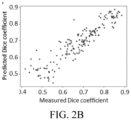
- the results depicted in FIGS. 2A-2B show that intra-cranial tissues were significantly (p ⁇ 0.05) correlated with the registration (global and local) quality features and the shortest axis length; and in contrast, extra-cranial structures were significantly (p ⁇ 0.05) correlated with image quality and tissue's shape properties.
- the results described herein show that a segmentation's quality estimation is feasible by incorporating a machine learning approach and features that are relevant to the segmentation.
- the simultaneous truth and performance level estimation (STAPLE) method or one of its variants may be incorporated to improve the prediction of segmentation quality. See, e.g., Akhondi-Asl, et al.: Simultaneous truth and performance level estimation through fusion of probabilistic segmentations. IEEE Trans. Med. Imaging, 32, 1840-52 (2013 ), as well as the Warfield and Commowick references identified above.
- the segmentation quality estimation can improve the TTFields planning, and quantifying the effects of segmentation errors on the TTFields simulation results is described below.
- the system can be programmed to make an automatic adjustment to the segmentation, in order to arrive at an improved segmentation.
- the system could change one or more parameters that have a global effect on the borders between certain tissue types.
- One example of such a change is to make an adjustment that, for all voxels in the image that correspond to tissue, (a) increases a calculated probability that a given voxel belongs to a first tissue type and (b) decreases a calculated probability that a given voxel belongs to a second tissue type.
- the quality of the segmentation is reevaluated by presenting the adjusted segmentation to the machine learning system.
- the machine learning system outputs at least one second quality score for the adjusted segmentation.
- An indication is output when the at least one second quality score indicates an improved quality with respect to the at least one first quality score.
- One common prior art approach for planning TTFields therapy involves the following four steps: create segmentation; user's visual evaluation of segmentation quality; segmentation correction using standard tools; and user's clinical decision making based on TTFields estimation.
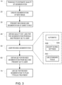
- FIG. 3 is a flowchart depicting a set of steps for using the quality-estimation machine learning system to improve planning of the positioning of transducer arrays for TTFields therapy.
- Step 10 is training a machine learning system to estimate quality of a segmentation, and this can be done based on a plurality of reference images and at least one quality score that has been assigned to each of the reference images, as described above in connection with FIGS. 1 and 2 .
- Step 20 the first step of the process for any new image will be step 20.
- Steps 20-70 in FIG. 3 depict an example of a TTFields planning protocol that improves upon the prior art approach by incorporating an automatic segmentation quality estimation (e.g., the one described above in connection with FIGS. 1-2 ) and guidance regarding segmentation errors and TTFields computation quality.
- the user creates a segmentation of the medical image (e.g., using computational methods or manually). Any of a wide variety of conventional approaches that will be apparent to persons skilled in the relevant art may be used for creating the initial segmentation.
- this segmentation as well as the associated original image are presented to the machine learning system.
- step 40 a quality estimate for the presented segmentation is obtained from the machine learning system (e.g., as described above in connection with FIG. 1-2 ) and this quality estimate is presented to the user.
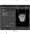
- FIG. 4 depicts an example of a suitable user interface that may be used to make the quality presentation to the user, and also to allow the user to make corrections to the segmentation.
- the quality presentation may be made in a variety of ways, including but not limited to (a) a single overall score number for any given segmentation (e.g., the 60% score depicted on the lower left of FIG. 4 ), (b) a recommendation that specific tissues should be revised due to a low quality score in any given segmentation (e.g., as depicted on the upper left of FIG. 4 ), and/or (c) an identification of particular regions on the image where the calculated quality of the segmentation is low for any given segmentation.
- a single overall score number for any given segmentation e.g., the 60% score depicted on the lower left of FIG. 4
- a recommendation that specific tissues should be revised due to a low quality score in any given segmentation e.g., as depicted on the upper left of FIG. 4
- One way to indicate the latter is by overlaying zones that are associated with low segmentation quality with a transparency (e.g., the circle 42 depicted in FIG. 4 ) or a region of opacity (e.g., the region 44 in FIG. 4 ).
- step 50 the user revises the segmentation based on the quality data that was presented in step 40.
- This may be accomplished using a variety of approaches.
- step 50A the user executes an automatic correction method.
- step 50B the user changes parameters that have a global effect on the borders. This may be accomplished, for example, by making an adjustment that, for all voxels in the new image that correspond to tissue, (a) increases a calculated probability that a given voxel belongs to a first tissue type and (b) decreases a calculated probability that a given voxel belongs to a second tissue type.
- This adjustment may be made for any given segmentation using any appropriate user interface, including but not limited to the sliders depicted on the bottom of FIG. 4 .
- sliding the first slider 46 labeled "Skull to Muscles" to the left progressively increases the calculated probability that any given voxel is a skull voxel and progressively decreases the calculated probability that the given voxel is a muscle voxel.
- sliding the first slider 46 to the right progressively increases the calculated probability that any given voxel is a muscle voxel and progressively decreases the calculated probability that the given voxel is a skull voxel.
- step 50C the user uses standard segmentation tools such as brush or polygon marking for correction of local errors.
- standard segmentation tools such as brush or polygon marking
- the system recalculates the quality of segmentation in step 60 (e.g. using the approaches described above in connection with FIGS. 1 and 2 ) and presents the revised results to the user. This may be done in real time in response to adjustments that are made by the user in step 50. Alternatively, this may be done upon user request (e.g., by including a "refresh quality estimate" button on the user interface.
- the next step (i.e., step 70) of the FIG. 3 protocol occurs after corrections to the segmentation have been made.
- the estimated quality for the segmentation that has been selected by the user is used to plan TTFields treatment, taking into account the quality estimate for the selected segmentation.
- the quality of the TTFields simulation is directly related to the quality of tissue segmentation, because assigning the wrong tissue can result in inaccurate estimation of the field and suboptimal TTFields treatment recommendation.
- the relation between segmentation and TTFields errors can be modeled such that an estimate of the segmentation quality will facilitate an estimate for the computed TTFields quality.
- TTFields quality estimates may be utilized to assist the caregiver in the treatment decision making.
- TTFields quality is much better for one layout than the other.
- the embodiments described herein provide this information to the user so that they can make a better-informed clinical decision (e.g., by selecting the layout with the higher quality).
- the error in segmentation can be propagated to effect the recommended treatment automatically. That is, the software will prefer setups in which the confidence regarding the TTFields dose is high in addition to other factors.
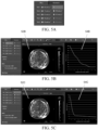
- FIGS. 5A-5C depicts an example of a suitable user interface that may be used to summarize the estimated field strength (or power density) in the tumor bed for five different candidate transducer array layouts (TAL 1-TAL 5), and also show how the quality of the segmentation might impact the field-strength estimates. The operator can use this information as a basis for selecting a given candidate transducer array layout over the other candidates.
- the system calculates how a determined quality of the given segmentation can lower an average expected field strength in a target region (e.g., the region that corresponds to a tumor) by running a finite element simulation using tissue types selected based on the given segmentation.
- the system calculates how a determined quality of the given segmentation can lower a minimum expected field strength in a target region by running a finite element simulation using tissue types selected based on the given segmentation.
- FIG. 5A shows a table that presents two values for each of the candidate transducer array layouts being considered (TAL 1 - TAL 5).
- the value on the left is the average estimated TTFields power density in the tumor when the quality of the segmentation is ignored; and the value on the right is the lowest (or 5 th percentile) average estimated TTFields power density in the tumor when the quality of the segmentation is considered.
- the first candidate transducer array layout (TAL1) is associated with the highest average estimated TTFields power density in the tumor of 80 mW /cm 2 . But when the impact of possible errors in the segmentation is considered, the TTFields power density for that same layout (TAL1) could be as low as 30 mW/cm 2 .
- the third candidate transducer array layout (TAL3) has a lower average estimated TTFields power density in the tumor when possible error in segmentation are ignored (63 mW /cm 2 , see the left column for TAL3).
- the lowest reasonably expected (e.g., 5 th percentile) power density for the TAL3 layout will also be 63 mW/cm 2 (right column for TAL3). Because the quality of the segmentation will not have as big a negative impact on the field power density when the TAL3 layout is selected, and because the lowest reasonably expected power density for that layout (i.e. 63 mW/cm 2 ) is significantly higher than the lowest reasonably expected power density for the TAL1 layout (30 mW/cm 2 ), the TAL3 layout is preferable to the TAL1 layout, and should therefore be selected.
- determining which TTFields simulation is the best can be accomplished by maximizing X, where X is the lowest reasonably expected power density in the tumor bed.
- the system can be programmed to calculate which layout maximizes X, and to recommend that layout.
- a confidence score may be associated with one or more possible layouts, where the confidence score is indicative of odds that a given layout is the best layout.
- the effect of the TTFields spatial distribution for each of the candidate transducer array layouts may be displayed to the user to help the user select which TAL is the best.
- clicking on the left column in FIG. 5A for TAL 1 will bring up FIG. 5B , which shows a spatial map of the estimated field power density overlaid on the MRI (in the middle panel 52B) and a graph of the local minimum power density (in the right panel 54B) when the quality of the segmentation is ignored.
- clicking on the right column in FIG. 5A for TAL 1 will bring up FIG.
- 5C which shows a spatial map of the lowest (or 5% percentile) average estimated TTFields power density overlaid on the , MRI (in the middle panel 52C) and a graph of the local minimum power density (in the right panel 54C) when the quality of the segmentation is considered.
Landscapes
- Engineering & Computer Science (AREA)
- Physics & Mathematics (AREA)
- General Physics & Mathematics (AREA)
- Theoretical Computer Science (AREA)
- Computer Vision & Pattern Recognition (AREA)
- Software Systems (AREA)
- Health & Medical Sciences (AREA)
- Geometry (AREA)
- Computer Graphics (AREA)
- General Health & Medical Sciences (AREA)
- Medical Informatics (AREA)
- Nuclear Medicine, Radiotherapy & Molecular Imaging (AREA)
- Radiology & Medical Imaging (AREA)
- Quality & Reliability (AREA)
- Magnetic Resonance Imaging Apparatus (AREA)
- Radiation-Therapy Devices (AREA)
- Image Analysis (AREA)
- Apparatus For Radiation Diagnosis (AREA)
Claims (5)
- Verfahren zum Schätzen der Qualität einer Segmentierung eines Bildes, wobei das Verfahren umfasst:Trainieren eines maschinellen Lernsystems, eine Qualität einer atlasbasierten Segmentierung eines Bildes zu schätzen, auf der Grundlage mehrerer Referenzbilder und mindestens eines trainierten Qualitätswertes, der jedem der mehreren Referenzbilder zugeordnet ist, wobei das maschinelle Lernsystem unter Verwendung von berechneten und validierten Segmentierungen aus den mehreren Referenzbildern trainiert wird;Präsentieren eines neuen Bildes und einer atlasbasierten Segmentierung des neuen Bildes an das maschinelle Lernsystem;Empfangen mindestens eines ersten Qualitätswertes für die präsentierte Segmentierung von dem maschinellen Lernsystem, die auf der Grundlage einer Qualität der affinen Registrierung zwischen der präsentierten Segmentierung und dem neuen Bild, einer Qualität der deformierbaren Registrierung zwischen der präsentierten Segmentierung und dem neuen Bild, Eingangsbildeigenschaften und geometrischen Eigenschaften segmentierter Gewebe geschätzt wird; undAusgeben des mindestens einen ersten Qualitätswertes für die präsentierte Segmentierung.
- Verfahren nach Anspruch 1, wobei die Qualität der deformierbaren Registrierung auf der Grundlage der Feldverzerrung einer Deformation, der Richtungsvariabilität und der mittleren Variabilität pro Achse bestimmt wird.
- Verfahren nach Anspruch 1, wobei die Eingangsbildeigenschaften eine kürzeste Achsenlänge und ein Signal-Rausch-Verhältnis für jedes Gewebe separat im neuen Bild umfassen.
- Verfahren nach Anspruch 1, wobei die geometrischen Eigenschaften der segmentierten Gewebe ein Volumen einer Form und eine Anzahl verbundener Komponenten umfassen, von denen jede pro Gewebe berechnet wird.
- Verfahren nach Anspruch 1, welches ferner umfasst:Vornehmen einer automatischen Anpassung der präsentierten Segmentierung;Präsentieren der angepassten Segmentierung an das maschinelle Lernsystem;Empfangen mindestens eines zweiten Qualitätswertes für die angepasste Segmentierung von dem maschinellen Lernsystem, die auf der Grundlage einer Qualität der affinen Registrierung zwischen der angepassten Segmentierung und dem neuen Bild, einer Qualität der deformierbaren Registrierung zwischen der angepassten Segmentierung und dem neuen Bild, Eingangsbildeigenschaften und geometrischen Eigenschaften segmentierter Gewebe geschätzt wird; undAusgeben einer Anzeige, wenn der mindestens eine zweite Qualitätswert eine verbesserte Qualität in Bezug auf den mindestens einen ersten Qualitätswert anzeigt.
Priority Applications (3)
| Application Number | Priority Date | Filing Date | Title |
|---|---|---|---|
| EP21202149.7A EP3961566A1 (de) | 2019-01-08 | 2020-01-07 | Bewertung der qualität der segmentierung eines bildes in verschiedene gewebearten zur planung der behandlung mit tumorbehandlungsfeldern (ttfields) |
| EP21205147.8A EP3968279B1 (de) | 2019-01-08 | 2020-01-07 | Bewertung der qualität der segmentierung eines bildes in verschiedene gewebearten zur planung der behandlung mit tumorbehandlungsfeldern (ttfields) |
| EP21202128.1A EP3961565A1 (de) | 2019-01-08 | 2020-01-07 | Bewertung der qualität der segmentierung eines bildes in verschiedene gewebearten zur planung der behandlung mit tumorbehandlungsfeldern (ttfields) |
Applications Claiming Priority (2)
| Application Number | Priority Date | Filing Date | Title |
|---|---|---|---|
| US201962789660P | 2019-01-08 | 2019-01-08 | |
| PCT/IB2020/050096 WO2020144582A1 (en) | 2019-01-08 | 2020-01-07 | Evaluating quality of segmentation of an image into different types of tissue for planning treatment using tumor treating fields (ttfields) |
Related Child Applications (6)
| Application Number | Title | Priority Date | Filing Date |
|---|---|---|---|
| EP21205147.8A Division EP3968279B1 (de) | 2019-01-08 | 2020-01-07 | Bewertung der qualität der segmentierung eines bildes in verschiedene gewebearten zur planung der behandlung mit tumorbehandlungsfeldern (ttfields) |
| EP21205147.8A Division-Into EP3968279B1 (de) | 2019-01-08 | 2020-01-07 | Bewertung der qualität der segmentierung eines bildes in verschiedene gewebearten zur planung der behandlung mit tumorbehandlungsfeldern (ttfields) |
| EP21202149.7A Division-Into EP3961566A1 (de) | 2019-01-08 | 2020-01-07 | Bewertung der qualität der segmentierung eines bildes in verschiedene gewebearten zur planung der behandlung mit tumorbehandlungsfeldern (ttfields) |
| EP21202149.7A Division EP3961566A1 (de) | 2019-01-08 | 2020-01-07 | Bewertung der qualität der segmentierung eines bildes in verschiedene gewebearten zur planung der behandlung mit tumorbehandlungsfeldern (ttfields) |
| EP21202128.1A Division-Into EP3961565A1 (de) | 2019-01-08 | 2020-01-07 | Bewertung der qualität der segmentierung eines bildes in verschiedene gewebearten zur planung der behandlung mit tumorbehandlungsfeldern (ttfields) |
| EP21202128.1A Division EP3961565A1 (de) | 2019-01-08 | 2020-01-07 | Bewertung der qualität der segmentierung eines bildes in verschiedene gewebearten zur planung der behandlung mit tumorbehandlungsfeldern (ttfields) |
Publications (3)
| Publication Number | Publication Date |
|---|---|
| EP3909015A1 EP3909015A1 (de) | 2021-11-17 |
| EP3909015B1 true EP3909015B1 (de) | 2024-08-14 |
| EP3909015C0 EP3909015C0 (de) | 2024-08-14 |
Family
ID=69326565
Family Applications (4)
| Application Number | Title | Priority Date | Filing Date |
|---|---|---|---|
| EP21202128.1A Pending EP3961565A1 (de) | 2019-01-08 | 2020-01-07 | Bewertung der qualität der segmentierung eines bildes in verschiedene gewebearten zur planung der behandlung mit tumorbehandlungsfeldern (ttfields) |
| EP20702348.2A Active EP3909015B1 (de) | 2019-01-08 | 2020-01-07 | Bewertung der qualität der segmentierung eines bildes in verschiedene gewebearten zur planung der behandlung mit tumorbehandlungsfeldern (ttfields) |
| EP21205147.8A Active EP3968279B1 (de) | 2019-01-08 | 2020-01-07 | Bewertung der qualität der segmentierung eines bildes in verschiedene gewebearten zur planung der behandlung mit tumorbehandlungsfeldern (ttfields) |
| EP21202149.7A Pending EP3961566A1 (de) | 2019-01-08 | 2020-01-07 | Bewertung der qualität der segmentierung eines bildes in verschiedene gewebearten zur planung der behandlung mit tumorbehandlungsfeldern (ttfields) |
Family Applications Before (1)
| Application Number | Title | Priority Date | Filing Date |
|---|---|---|---|
| EP21202128.1A Pending EP3961565A1 (de) | 2019-01-08 | 2020-01-07 | Bewertung der qualität der segmentierung eines bildes in verschiedene gewebearten zur planung der behandlung mit tumorbehandlungsfeldern (ttfields) |
Family Applications After (2)
| Application Number | Title | Priority Date | Filing Date |
|---|---|---|---|
| EP21205147.8A Active EP3968279B1 (de) | 2019-01-08 | 2020-01-07 | Bewertung der qualität der segmentierung eines bildes in verschiedene gewebearten zur planung der behandlung mit tumorbehandlungsfeldern (ttfields) |
| EP21202149.7A Pending EP3961566A1 (de) | 2019-01-08 | 2020-01-07 | Bewertung der qualität der segmentierung eines bildes in verschiedene gewebearten zur planung der behandlung mit tumorbehandlungsfeldern (ttfields) |
Country Status (5)
| Country | Link |
|---|---|
| US (3) | US11276171B2 (de) |
| EP (4) | EP3961565A1 (de) |
| JP (1) | JP7246486B2 (de) |
| CN (1) | CN113330485B (de) |
| WO (1) | WO2020144582A1 (de) |
Families Citing this family (47)
| Publication number | Priority date | Publication date | Assignee | Title |
|---|---|---|---|---|
| US10779875B2 (en) | 2013-05-06 | 2020-09-22 | Novocure Gmbh | Optimizing treatment using TTfields by changing the frequency during the course of long term tumor treatment |
| US10188851B2 (en) | 2015-10-28 | 2019-01-29 | Novocure Limited | TTField treatment with optimization of electrode positions on the head based on MRI-based conductivity measurements |
| US10821283B2 (en) | 2016-04-04 | 2020-11-03 | Novocure Gmbh | Reducing motility of cancer cells using tumor treating fields (TTFields) |
| US20180001075A1 (en) | 2016-06-30 | 2018-01-04 | Novocure Limited | Arrays for Longitudinal Delivery of TTFields to a Body |
| WO2018134733A1 (en) | 2017-01-19 | 2018-07-26 | Moshe Giladi | System for viewing cell cultures under a microscope whilst applying ttfields |
| US11986647B2 (en) | 2018-09-07 | 2024-05-21 | Novocure Gmbh | Treating autoinflammatory and mitochondrial diseases using an alternating electric field |
| EP3984590B1 (de) | 2018-10-15 | 2022-12-07 | Novocure GmbH | Erzeugung von tumorbehandlungsfeldern (ttfields) mit hoher gleichförmigkeit im ganzen gehirn |
| JP7148722B2 (ja) | 2018-10-25 | 2022-10-05 | ゼーヴ・ボンゾン | 被験者の脊椎構造体に対する交番電界(例えばTTField)の送達 |
| IL280843B2 (en) | 2018-11-19 | 2025-10-01 | Novocure Gmbh | Arrays for providing fields treat growth with selectively addressable sub-elements |
| SG11202101810SA (en) | 2018-11-29 | 2021-03-30 | Novocure Gmbh | Enhanced-flexibility transducer arrays for delivering ttfields (tumor treating fields) |
| EP3954314B1 (de) | 2019-02-27 | 2025-01-08 | Novocure GmbH | Abgabe von tumorbehandlungsfeldern (ttfelds) mittels implantierbarer wandleranordnungen |
| CN113613651B (zh) | 2019-03-29 | 2025-08-12 | 诺沃库勒有限责任公司 | 用PTGER3抑制剂恢复TTField抗性癌细胞中对TTField的敏感性的方法 |
| US11291837B2 (en) | 2019-04-17 | 2022-04-05 | Novocure Gmbh | Uploading data from an isolated system without compromising isolation |
| US11654279B2 (en) | 2019-07-31 | 2023-05-23 | Novocure Gmbh | Applying tumor treating fields (TTFields) via electrodes embedded into skull implants |
| WO2021024170A1 (en) | 2019-08-05 | 2021-02-11 | Voloshin Sela Tali | Increasing cancer cells' sensitivity to tumor treating fields (ttfields) by inhibiting il11 activity |
| US11890467B2 (en) | 2019-08-30 | 2024-02-06 | Novocure Gmbh | Delivering tumor treating fields (TTFields) to the neck |
| US12282309B2 (en) * | 2019-10-07 | 2025-04-22 | Novocure Gmbh | Methods, systems, and apparatuses for guiding transducer array placement |
| WO2021111186A1 (en) | 2019-12-02 | 2021-06-10 | Novocure Gmbh | Methods and apparatuses for optimizing transducer array placement |
| EP4091665B1 (de) | 2019-12-31 | 2024-10-30 | Novocure GmbH | Hochfrequenzgenerator mit hohem wirkungsgrad, der spitzen bei amplitudeneinstellungen und kanalumschaltungen verhindert |
| WO2021136965A2 (en) | 2019-12-31 | 2021-07-08 | Novocure Gmbh | Methods, systems, and apparatuses for image segmentation |
| US12121739B2 (en) | 2019-12-31 | 2024-10-22 | Novocure Gmbh | Methods, systems, and apparatuses for managing temperatures induced by alternating fields |
| ES2950234T3 (es) | 2019-12-31 | 2023-10-06 | Novocure Gmbh | Matrices para administrar campos de tratamiento de tumores (camposTT) con elementos de electrodos y sensores de temperatura accesibles individualmente |
| EP4075442A1 (de) | 2019-12-31 | 2022-10-19 | Novocure GmbH | Verfahren, systeme und vorrichtungen zur schnellen annäherung elektrischer feldverteilung |
| US20210299440A1 (en) | 2020-03-30 | 2021-09-30 | Novocure Gmbh | Intravenous / Intra-Spinal / Intra-Cavity / Intraventricular Delivery of TTFields (Tumor Treating Fields) for Treating Cancer and Metastases |
| BR112022021484A2 (pt) | 2020-04-24 | 2022-12-13 | Novocure Gmbh | Uso de campos elétricos alternados para aumentar a permeabilidade da barreira hematoencefálica |
| US11818943B2 (en) | 2020-06-25 | 2023-11-14 | Novocure Gmbh | Fabricating organic light emitting diodes (OLEDs) using tubulin |
| KR102490645B1 (ko) | 2020-07-16 | 2023-01-25 | 고려대학교 산학협력단 | 흡수에너지 기반 전기장 암치료 계획 시스템 및 방법 |
| JP7777118B2 (ja) | 2020-09-25 | 2025-11-27 | ノボキュア ゲーエムベーハー | 過熱することなく電流を最大化するために腫瘍治療電場(TTField)における個々の電極要素における金属化範囲を変化させること |
| CN112330613B (zh) * | 2020-10-27 | 2024-04-12 | 深思考人工智能科技(上海)有限公司 | 一种细胞病理数字图像质量的评价方法及系统 |
| CN114418925B (zh) * | 2020-10-28 | 2025-10-17 | 佳能医疗系统株式会社 | 医学图像处理方法及医学图像处理装置 |
| EP4240470A1 (de) | 2021-02-17 | 2023-09-13 | Novocure Gmbh | Arrays zur abgabe von tumorbehandlungsfeldern (ttfields) mit sätzen von elektrodenelementen mit individuell verstellbaren aktiven bereichen |
| US12322112B2 (en) * | 2021-03-02 | 2025-06-03 | Novocure Gmbh | Methods and apparatuses for visualization of tumor segmentation |
| US12440680B2 (en) | 2021-03-12 | 2025-10-14 | Novocure Gmbh | Methods of treating disease by skewing macrophage phenotype with alternating electric fields |
| EP4284489B1 (de) | 2021-03-31 | 2025-07-16 | Novocure Gmbh | Impedanztomografie mit elektroden eines tumorbehandlungsfeldsystems |
| KR20240027008A (ko) | 2021-06-30 | 2024-02-29 | 노보큐어 게엠베하 | 종양 치료 필드(ttfield)를 인가하기 위한 트랜스듀서 어레이에서 전류를 제한하기 위한 커패시터의 사용 |
| US12599765B2 (en) | 2021-12-14 | 2026-04-14 | Novocure Gmbh | Shifting of transducer array to reduce skin irritation |
| WO2023126842A1 (en) | 2021-12-29 | 2023-07-06 | Novocure Gmbh | Apparatus for reducing electrosensation using alternating electric fields with larger cathodes and smaller anodes |
| EP4456971A1 (de) | 2021-12-30 | 2024-11-06 | Novocure GmbH | Auswahl von parameterwerten zur behandlung mit tumorbehandlungsfeldern (ttfields) |
| US12478793B2 (en) | 2022-03-30 | 2025-11-25 | Novocure Gmbh | Reducing electrosensation whilst treating a subject using alternating electric fields by pairing transducer arrays together |
| EP4460361B1 (de) | 2022-03-30 | 2025-03-19 | Novocure GmbH | Verwendung verschachtelter kühlperioden zur erhöhung der spitzenintensität von tumorbehandlungsfeldern |
| US12268863B2 (en) | 2023-02-06 | 2025-04-08 | Novocure Gmbh | Shiftable transducer array with anisotropic material layer |
| CN116912546A (zh) * | 2023-03-30 | 2023-10-20 | 北京罗克维尔斯科技有限公司 | 图像质量确定方法、装置、电子设备及存储介质 |
| US20250001167A1 (en) * | 2023-06-30 | 2025-01-02 | Novocure Gmbh | User tools for treatment planning for tumor treating fields |
| US12515045B2 (en) | 2023-06-30 | 2026-01-06 | Novocure Gmbh | Treatment planning for tumor treating fields |
| CN116580037B (zh) * | 2023-07-10 | 2023-10-13 | 天津医科大学第二医院 | 一种基于深度学习的鼻咽癌图像分割方法及系统 |
| CN121925295A (zh) | 2023-09-29 | 2026-04-24 | 诺沃库勒有限责任公司 | 使用振幅和频率的交错变化来改善用交变电场进行治疗期间的电感觉 |
| CN119360258B (zh) * | 2024-07-15 | 2025-10-21 | 深圳职业技术大学 | 一种城市场景的目标检测和定位方法、无人机及存储介质 |
Family Cites Families (76)
| Publication number | Priority date | Publication date | Assignee | Title |
|---|---|---|---|---|
| US7599745B2 (en) | 2000-02-17 | 2009-10-06 | Standen Ltd | Treating a tumor or the like with an electric field |
| US7136699B2 (en) | 2002-10-02 | 2006-11-14 | Standen, Ltd. | Apparatus for destroying dividing cells |
| US8175698B2 (en) | 2000-02-17 | 2012-05-08 | Novocure Ltd. | Treating bacteria with electric fields |
| US7016725B2 (en) | 2001-11-06 | 2006-03-21 | Standen Ltd. | Method and apparatus for destroying dividing cells |
| US7599746B2 (en) | 2000-02-17 | 2009-10-06 | Standen Ltd | Apparatus and method for preventing the spread of cancerous metastases and for elimination of metastases |
| US8447395B2 (en) | 2000-02-17 | 2013-05-21 | Novocure Ltd | Treating bacteria with electric fields |
| US7089054B2 (en) | 2002-10-02 | 2006-08-08 | Standen Ltd. | Apparatus and method for treating a tumor or the like |
| US6868289B2 (en) | 2002-10-02 | 2005-03-15 | Standen Ltd. | Apparatus for treating a tumor or the like and articles incorporating the apparatus for treatment of the tumor |
| EP2335776A1 (de) | 2000-02-17 | 2011-06-22 | Yoram Palti | Verfahren und vorrichtung zur zerstörung von teilenden zellen |
| US20080108894A1 (en) * | 2004-11-15 | 2008-05-08 | Elgavish Gabriel A | Methods and Systems of Analyzing Clinical Parameters and Methods of Producing Visual Images |
| ES2344215T3 (es) | 2004-12-07 | 2010-08-20 | Standen Ltd. | Electrodos para aplicar un campo electrico in vivo durante un dilatado periodo de tiempo. |
| JP5559460B2 (ja) | 2004-12-27 | 2014-07-23 | スタンデン・リミテッド | 異なる配向の電界を用いて腫瘍またはその類似物を処理する方法 |
| US8358819B2 (en) * | 2005-06-24 | 2013-01-22 | University Of Iowa Research Foundation | System and methods for image segmentation in N-dimensional space |
| CA2624624C (en) | 2005-10-03 | 2016-07-19 | Novocure Limited | Optimizing characteristics of an electric field to increase the field's effect on proliferating cells |
| US8019414B2 (en) | 2006-04-05 | 2011-09-13 | Novocure Ltd. | Treating cancer using electromagnetic fields in combination with other treatment regimens |
| WO2009044289A1 (en) | 2007-03-06 | 2009-04-09 | Novocure Ltd. | Treating cancer using electromagnetic fields in combination with photodynamic therapy |
| CN101820947A (zh) | 2007-08-14 | 2010-09-01 | 诺沃库勒有限公司 | 用电场治疗寄生物 |
| US8715203B2 (en) | 2007-09-17 | 2014-05-06 | Novocure Limited | Composite electrode |
| WO2010017392A2 (en) | 2008-08-07 | 2010-02-11 | Highland Instruments, Inc. | Interface apparatus for stimulation of biological tissue |
| BR112012003847B1 (pt) * | 2009-08-22 | 2020-12-01 | The Board Of Trustees Of The Leland Standford Junior University | método para selecionar um ou mais embriões humanos que provavelmente alcançarão o estágio de blastocisto |
| US8160357B2 (en) * | 2010-07-30 | 2012-04-17 | Kabushiki Kaisha Toshiba | Image segmentation |
| US20170337711A1 (en) * | 2011-03-29 | 2017-11-23 | Lyrical Labs Video Compression Technology, LLC | Video processing and encoding |
| US9501837B2 (en) * | 2014-10-01 | 2016-11-22 | Lyrical Labs Video Compression Technology, LLC | Method and system for unsupervised image segmentation using a trained quality metric |
| WO2014161766A1 (en) * | 2013-04-02 | 2014-10-09 | Koninklijke Philips N.V. | Detection of bone tissue using magnetic resonance imaging |
| US10779875B2 (en) | 2013-05-06 | 2020-09-22 | Novocure Gmbh | Optimizing treatment using TTfields by changing the frequency during the course of long term tumor treatment |
| US9655669B2 (en) | 2013-05-06 | 2017-05-23 | Novocure Limited | Optimizing treatment using TTFields by changing the frequency during the course of long term tumor treatment |
| US20190117963A1 (en) | 2014-07-25 | 2019-04-25 | Loyalty Based Innovations, LLC | Apparatus and method for treating multiple tumors in patients with metastatic disease by electric fields |
| WO2016059385A1 (en) * | 2014-10-17 | 2016-04-21 | University College Cardiff Consultants Limited | A method for optimising segmentation of a tumour on an image, and apparatus therefor |
| US9910453B2 (en) | 2015-09-25 | 2018-03-06 | Novocure Limited | High voltage, high efficiency sine wave generator with pre-set frequency and adjustable amplitude |
| US10188851B2 (en) * | 2015-10-28 | 2019-01-29 | Novocure Limited | TTField treatment with optimization of electrode positions on the head based on MRI-based conductivity measurements |
| US10083340B2 (en) * | 2016-01-26 | 2018-09-25 | Ge Healthcare Bio-Sciences Corp. | Automated cell segmentation quality control |
| US10821283B2 (en) | 2016-04-04 | 2020-11-03 | Novocure Gmbh | Reducing motility of cancer cells using tumor treating fields (TTFields) |
| US11445914B2 (en) * | 2016-05-13 | 2022-09-20 | The University Of North Carolina At Chapel Hill | Methods and systems for assessing material anisotropy and other characteristics |
| US10282588B2 (en) * | 2016-06-09 | 2019-05-07 | Siemens Healthcare Gmbh | Image-based tumor phenotyping with machine learning from synthetic data |
| US20180001075A1 (en) | 2016-06-30 | 2018-01-04 | Novocure Limited | Arrays for Longitudinal Delivery of TTFields to a Body |
| US20180008708A1 (en) | 2016-07-10 | 2018-01-11 | Novocure Limited | Synchronizing Tumor Cells to the G2/M Phase Using TTFields Combined with Taxane or Other Anti-Microtubule Agents |
| CN116747440A (zh) | 2016-08-18 | 2023-09-15 | 诺沃库勒有限责任公司 | 用于递送肿瘤治疗电场的阵列中的温度测量 |
| US10607342B2 (en) * | 2016-09-30 | 2020-03-31 | Siemenes Healthcare GmbH | Atlas-based contouring of organs at risk for radiation therapy |
| DK3554631T3 (da) | 2016-12-13 | 2022-05-30 | Novocure Gmbh | Behandling af patienter med ttfields med elektrodepositionerne optimeret under anvendelse af deformerbare skabeloner |
| WO2018134733A1 (en) | 2017-01-19 | 2018-07-26 | Moshe Giladi | System for viewing cell cultures under a microscope whilst applying ttfields |
| US20180295375A1 (en) * | 2017-04-05 | 2018-10-11 | Lyrical Labs Video Compression Technology, LLC | Video processing and encoding |
| WO2018209057A1 (en) * | 2017-05-11 | 2018-11-15 | The Research Foundation For The State University Of New York | System and method associated with predicting segmentation quality of objects in analysis of copious image data |
| CN112543661A (zh) | 2017-11-17 | 2021-03-23 | 艾伯维公司 | 治疗胶质母细胞瘤的方法 |
| US10953209B2 (en) | 2018-03-28 | 2021-03-23 | Board Of Regents Of The University Of Texas System | Treating tumors using TTFields combined with a PARP inhibitor |
| EP3773726B1 (de) | 2018-04-09 | 2024-06-05 | Novocure GmbH | Behandlung von tumoren mit ttfields und einem aurora-kinase-inhibitor |
| EP3775956B1 (de) | 2018-04-10 | 2022-03-23 | Novocure GmbH | Aus zwei mrt-bildern mit unterschiedlichen wiederholzeiten abgeleitete niederfrequente (<1 mhz) wechselstromleitfähigkeitsschätzungen |
| US11399779B2 (en) * | 2018-05-16 | 2022-08-02 | Case Western Reserve University | System-independent quantitative perfusion imaging |
| ES3047748T3 (en) | 2018-07-03 | 2025-12-04 | Univ Leland Stanford Junior | Using alternating electric fields to increase cell membrane permeability |
| US11179322B2 (en) | 2018-07-10 | 2021-11-23 | Novocure Gmbh | Methods and compositions for treating tumors with TTFields and sorafenib |
| US11521742B2 (en) * | 2018-07-18 | 2022-12-06 | SCA Robotics | Methods of implementing an artificial intelligence based neuroradiology platform for neurological tumor identification and for T-Cell therapy initiation and tracking and related precision medical treatment predictive modeling |
| MX2020013431A (es) | 2018-07-18 | 2021-05-27 | Novocure Gmbh | Uso de densidad de perdida de potencia y mediciones relacionadas para cuantificar la dosis de campos de tratamiento de tumores (ttfield). |
| EP3878389B1 (de) | 2018-08-23 | 2025-08-06 | Novocure GmbH | Verwendung von elektrischen wechselfeldern zur erhöhung der permeabilität der blut-hirn-schranke |
| US11160977B2 (en) | 2018-09-04 | 2021-11-02 | Novocure Gmbh | Delivering tumor treating fields (TTFields) to the infratentorial brain |
| US11986647B2 (en) | 2018-09-07 | 2024-05-21 | Novocure Gmbh | Treating autoinflammatory and mitochondrial diseases using an alternating electric field |
| WO2020049482A1 (en) | 2018-09-07 | 2020-03-12 | Yaniv Alon | Treating autoimmune diseases using an alternating electric field to reduce the proliferation of t-cells |
| US20200108031A1 (en) | 2018-10-05 | 2020-04-09 | Novocure Gmbh | Treating Tumors Using TTFields Combined with ABT-751 |
| CN113164525B (zh) | 2018-10-23 | 2025-03-14 | 利兰·斯坦福青年大学托管委员会 | 在基于干细胞的疗法中使用交变电场预防和治疗畸胎瘤形成 |
| JP7148722B2 (ja) | 2018-10-25 | 2022-10-05 | ゼーヴ・ボンゾン | 被験者の脊椎構造体に対する交番電界(例えばTTField)の送達 |
| SG11202101810SA (en) | 2018-11-29 | 2021-03-30 | Novocure Gmbh | Enhanced-flexibility transducer arrays for delivering ttfields (tumor treating fields) |
| JP7216829B2 (ja) | 2019-02-22 | 2023-02-01 | ノボキュア ゲーエムベーハー | Xelox又はfolfoxと組み合わせたttフィールドを用いる胃癌の治療 |
| IL284284B2 (en) | 2019-02-26 | 2026-03-01 | Novocure Gmbh | Frequency determination for tumor-treating field therapy is based on the electrical characteristic of targeted cancer cells. |
| EP3954314B1 (de) | 2019-02-27 | 2025-01-08 | Novocure GmbH | Abgabe von tumorbehandlungsfeldern (ttfelds) mittels implantierbarer wandleranordnungen |
| CN113613651B (zh) | 2019-03-29 | 2025-08-12 | 诺沃库勒有限责任公司 | 用PTGER3抑制剂恢复TTField抗性癌细胞中对TTField的敏感性的方法 |
| US20200327674A1 (en) * | 2019-04-10 | 2020-10-15 | Nvidia Corporation | Identifying Image Segmentation Quality Using Neural Networks |
| US11291837B2 (en) | 2019-04-17 | 2022-04-05 | Novocure Gmbh | Uploading data from an isolated system without compromising isolation |
| US11164317B2 (en) * | 2019-06-28 | 2021-11-02 | Intel Corporation | Real-time mask quality predictor |
| US11654279B2 (en) | 2019-07-31 | 2023-05-23 | Novocure Gmbh | Applying tumor treating fields (TTFields) via electrodes embedded into skull implants |
| WO2021024170A1 (en) | 2019-08-05 | 2021-02-11 | Voloshin Sela Tali | Increasing cancer cells' sensitivity to tumor treating fields (ttfields) by inhibiting il11 activity |
| US11890467B2 (en) | 2019-08-30 | 2024-02-06 | Novocure Gmbh | Delivering tumor treating fields (TTFields) to the neck |
| WO2021050093A1 (en) | 2019-09-10 | 2021-03-18 | Novocure Gmbh | A method of reducing viability of cancer cells by applying alternating electric fields and administering checkpoint inhibitors to the cancer cells |
| JP7687698B2 (ja) | 2019-12-26 | 2025-06-03 | ザ ボード オブ トラスティーズ オブ ザ レランド スタンフォード ジュニア ユニバーシティー | 癌細胞における異常な解糖的代謝を正常化する方法 |
| EP4091665B1 (de) | 2019-12-31 | 2024-10-30 | Novocure GmbH | Hochfrequenzgenerator mit hohem wirkungsgrad, der spitzen bei amplitudeneinstellungen und kanalumschaltungen verhindert |
| ES2950234T3 (es) | 2019-12-31 | 2023-10-06 | Novocure Gmbh | Matrices para administrar campos de tratamiento de tumores (camposTT) con elementos de electrodos y sensores de temperatura accesibles individualmente |
| US20210299440A1 (en) | 2020-03-30 | 2021-09-30 | Novocure Gmbh | Intravenous / Intra-Spinal / Intra-Cavity / Intraventricular Delivery of TTFields (Tumor Treating Fields) for Treating Cancer and Metastases |
| BR112022021484A2 (pt) | 2020-04-24 | 2022-12-13 | Novocure Gmbh | Uso de campos elétricos alternados para aumentar a permeabilidade da barreira hematoencefálica |
| US11818943B2 (en) | 2020-06-25 | 2023-11-14 | Novocure Gmbh | Fabricating organic light emitting diodes (OLEDs) using tubulin |
-
2020
- 2020-01-07 EP EP21202128.1A patent/EP3961565A1/de active Pending
- 2020-01-07 EP EP20702348.2A patent/EP3909015B1/de active Active
- 2020-01-07 EP EP21205147.8A patent/EP3968279B1/de active Active
- 2020-01-07 US US16/736,604 patent/US11276171B2/en active Active
- 2020-01-07 JP JP2021536208A patent/JP7246486B2/ja active Active
- 2020-01-07 WO PCT/IB2020/050096 patent/WO2020144582A1/en not_active Ceased
- 2020-01-07 EP EP21202149.7A patent/EP3961566A1/de active Pending
- 2020-01-07 CN CN202080008438.8A patent/CN113330485B/zh active Active
-
2022
- 2022-02-09 US US17/667,844 patent/US11915424B2/en active Active
-
2024
- 2024-01-30 US US18/426,816 patent/US20240169536A1/en active Pending
Also Published As
| Publication number | Publication date |
|---|---|
| CN113330485A (zh) | 2021-08-31 |
| CN113330485B (zh) | 2025-09-26 |
| WO2020144582A1 (en) | 2020-07-16 |
| EP3968279C0 (de) | 2025-01-01 |
| US20200219261A1 (en) | 2020-07-09 |
| US20220254025A1 (en) | 2022-08-11 |
| EP3909015A1 (de) | 2021-11-17 |
| US20240169536A1 (en) | 2024-05-23 |
| EP3961566A1 (de) | 2022-03-02 |
| EP3968279B1 (de) | 2025-01-01 |
| JP2022516020A (ja) | 2022-02-24 |
| US11276171B2 (en) | 2022-03-15 |
| EP3909015C0 (de) | 2024-08-14 |
| US11915424B2 (en) | 2024-02-27 |
| EP3968279A1 (de) | 2022-03-16 |
| EP3961565A1 (de) | 2022-03-02 |
| JP7246486B2 (ja) | 2023-03-27 |
Similar Documents
| Publication | Publication Date | Title |
|---|---|---|
| US11915424B2 (en) | Evaluating quality of segmentation of an image into different types of tissue for planning treatment using tumor treating fields (TTFields) | |
| US5926568A (en) | Image object matching using core analysis and deformable shape loci | |
| Jannin et al. | Validation in medical image processing. | |
| Huyskens et al. | A qualitative and a quantitative analysis of an auto-segmentation module for prostate cancer | |
| JP6949256B2 (ja) | 線量誤差決定装置 | |
| CN112601582A (zh) | 用于加速的在线自适应放射疗法的系统和方法 | |
| US10186031B2 (en) | Shape similarity measure for body tissue | |
| Vaassen et al. | The impact of organ-at-risk contour variations on automatically generated treatment plans for NSCLC | |
| Acosta et al. | Evaluation of multi-atlas-based segmentation of CT scans in prostate cancer radiotherapy | |
| Miura et al. | Impact of deformable image registration accuracy on thoracic images with different regularization weight parameter settings | |
| EP4076207B1 (de) | Verfahren und system zur verbesserten ultraschallebenenerfassung | |
| Mishra et al. | Medical image processing: A challenging analysis | |
| US11295462B2 (en) | Determination of registration accuracy | |
| HK40064471A (en) | Evaluating quality of segmentation of an image into different types of tissue for planning treatment using tumor treating fields (ttfields) | |
| HK40061474A (en) | Evaluating quality of segmentation of an image into different types of tissue for planning treatment using tumor treating fields (ttfields) | |
| HK40061843A (en) | Evaluating quality of segmentation of an image into different types of tissue for planning treatment using tumor treating fields (ttfields) | |
| HK40064471B (en) | Evaluating quality of segmentation of an image into different types of tissue for planning treatment using tumor treating fields (ttfields) | |
| HK40054120B (en) | Evaluating quality of segmentation of an image into different types of tissue for planning treatment using tumor treating fields (ttfields) | |
| HK40054120A (en) | Evaluating quality of segmentation of an image into different types of tissue for planning treatment using tumor treating fields (ttfields) | |
| Coevoet et al. | Introducing interactive inverse FEM simulation and its application for adaptive radiotherapy | |
| Nascimento et al. | Estimating the Registration Error | |
| Stiehl | Biomechanical Modeling Applications for Function-Preserving Lung Interventions | |
| Guy | An Algorithm to Improve Deformable Image Registration Accuracy in Challenging Cases of Locally-Advanced Non-Small Cell Lung Cancer | |
| Winkler | Statistical analysis of areal quantities in the brain through permutation tests |
Legal Events
| Date | Code | Title | Description |
|---|---|---|---|
| STAA | Information on the status of an ep patent application or granted ep patent |
Free format text: STATUS: UNKNOWN |
|
| STAA | Information on the status of an ep patent application or granted ep patent |
Free format text: STATUS: THE INTERNATIONAL PUBLICATION HAS BEEN MADE |
|
| PUAI | Public reference made under article 153(3) epc to a published international application that has entered the european phase |
Free format text: ORIGINAL CODE: 0009012 |
|
| STAA | Information on the status of an ep patent application or granted ep patent |
Free format text: STATUS: REQUEST FOR EXAMINATION WAS MADE |
|
| 17P | Request for examination filed |
Effective date: 20210522 |
|
| AK | Designated contracting states |
Kind code of ref document: A1 Designated state(s): AL AT BE BG CH CY CZ DE DK EE ES FI FR GB GR HR HU IE IS IT LI LT LU LV MC MK MT NL NO PL PT RO RS SE SI SK SM TR |
|
| REG | Reference to a national code |
Ref country code: HK Ref legal event code: DE Ref document number: 40054120 Country of ref document: HK |
|
| DAV | Request for validation of the european patent (deleted) | ||
| DAX | Request for extension of the european patent (deleted) | ||
| GRAP | Despatch of communication of intention to grant a patent |
Free format text: ORIGINAL CODE: EPIDOSNIGR1 |
|
| STAA | Information on the status of an ep patent application or granted ep patent |
Free format text: STATUS: GRANT OF PATENT IS INTENDED |
|
| GRAS | Grant fee paid |
Free format text: ORIGINAL CODE: EPIDOSNIGR3 |
|
| INTG | Intention to grant announced |
Effective date: 20240318 |
|
| GRAA | (expected) grant |
Free format text: ORIGINAL CODE: 0009210 |
|
| STAA | Information on the status of an ep patent application or granted ep patent |
Free format text: STATUS: THE PATENT HAS BEEN GRANTED |
|
| AK | Designated contracting states |
Kind code of ref document: B1 Designated state(s): AL AT BE BG CH CY CZ DE DK EE ES FI FR GB GR HR HU IE IS IT LI LT LU LV MC MK MT NL NO PL PT RO RS SE SI SK SM TR |
|
| REG | Reference to a national code |
Ref country code: GB Ref legal event code: FG4D |
|
| REG | Reference to a national code |
Ref country code: CH Ref legal event code: EP |
|
| REG | Reference to a national code |
Ref country code: DE Ref legal event code: R096 Ref document number: 602020035695 Country of ref document: DE |
|
| REG | Reference to a national code |
Ref country code: IE Ref legal event code: FG4D |
|
| U01 | Request for unitary effect filed |
Effective date: 20240904 |
|
| U07 | Unitary effect registered |
Designated state(s): AT BE BG DE DK EE FI FR IT LT LU LV MT NL PT RO SE SI Effective date: 20240923 |
|
| PG25 | Lapsed in a contracting state [announced via postgrant information from national office to epo] |
Ref country code: NO Free format text: LAPSE BECAUSE OF FAILURE TO SUBMIT A TRANSLATION OF THE DESCRIPTION OR TO PAY THE FEE WITHIN THE PRESCRIBED TIME-LIMIT Effective date: 20241114 |
|
| PG25 | Lapsed in a contracting state [announced via postgrant information from national office to epo] |
Ref country code: GR Free format text: LAPSE BECAUSE OF FAILURE TO SUBMIT A TRANSLATION OF THE DESCRIPTION OR TO PAY THE FEE WITHIN THE PRESCRIBED TIME-LIMIT Effective date: 20241115 Ref country code: PL Free format text: LAPSE BECAUSE OF FAILURE TO SUBMIT A TRANSLATION OF THE DESCRIPTION OR TO PAY THE FEE WITHIN THE PRESCRIBED TIME-LIMIT Effective date: 20240814 |
|
| PG25 | Lapsed in a contracting state [announced via postgrant information from national office to epo] |
Ref country code: IS Free format text: LAPSE BECAUSE OF FAILURE TO SUBMIT A TRANSLATION OF THE DESCRIPTION OR TO PAY THE FEE WITHIN THE PRESCRIBED TIME-LIMIT Effective date: 20241214 |
|
| PG25 | Lapsed in a contracting state [announced via postgrant information from national office to epo] |
Ref country code: HR Free format text: LAPSE BECAUSE OF FAILURE TO SUBMIT A TRANSLATION OF THE DESCRIPTION OR TO PAY THE FEE WITHIN THE PRESCRIBED TIME-LIMIT Effective date: 20240814 |
|
| PG25 | Lapsed in a contracting state [announced via postgrant information from national office to epo] |
Ref country code: ES Free format text: LAPSE BECAUSE OF FAILURE TO SUBMIT A TRANSLATION OF THE DESCRIPTION OR TO PAY THE FEE WITHIN THE PRESCRIBED TIME-LIMIT Effective date: 20240814 Ref country code: RS Free format text: LAPSE BECAUSE OF FAILURE TO SUBMIT A TRANSLATION OF THE DESCRIPTION OR TO PAY THE FEE WITHIN THE PRESCRIBED TIME-LIMIT Effective date: 20241114 |
|
| PG25 | Lapsed in a contracting state [announced via postgrant information from national office to epo] |
Ref country code: RS Free format text: LAPSE BECAUSE OF FAILURE TO SUBMIT A TRANSLATION OF THE DESCRIPTION OR TO PAY THE FEE WITHIN THE PRESCRIBED TIME-LIMIT Effective date: 20241114 Ref country code: PL Free format text: LAPSE BECAUSE OF FAILURE TO SUBMIT A TRANSLATION OF THE DESCRIPTION OR TO PAY THE FEE WITHIN THE PRESCRIBED TIME-LIMIT Effective date: 20240814 Ref country code: NO Free format text: LAPSE BECAUSE OF FAILURE TO SUBMIT A TRANSLATION OF THE DESCRIPTION OR TO PAY THE FEE WITHIN THE PRESCRIBED TIME-LIMIT Effective date: 20241114 Ref country code: IS Free format text: LAPSE BECAUSE OF FAILURE TO SUBMIT A TRANSLATION OF THE DESCRIPTION OR TO PAY THE FEE WITHIN THE PRESCRIBED TIME-LIMIT Effective date: 20241214 Ref country code: HR Free format text: LAPSE BECAUSE OF FAILURE TO SUBMIT A TRANSLATION OF THE DESCRIPTION OR TO PAY THE FEE WITHIN THE PRESCRIBED TIME-LIMIT Effective date: 20240814 Ref country code: GR Free format text: LAPSE BECAUSE OF FAILURE TO SUBMIT A TRANSLATION OF THE DESCRIPTION OR TO PAY THE FEE WITHIN THE PRESCRIBED TIME-LIMIT Effective date: 20241115 Ref country code: ES Free format text: LAPSE BECAUSE OF FAILURE TO SUBMIT A TRANSLATION OF THE DESCRIPTION OR TO PAY THE FEE WITHIN THE PRESCRIBED TIME-LIMIT Effective date: 20240814 |
|
| U20 | Renewal fee for the european patent with unitary effect paid |
Year of fee payment: 6 Effective date: 20250127 |
|
| PG25 | Lapsed in a contracting state [announced via postgrant information from national office to epo] |
Ref country code: SM Free format text: LAPSE BECAUSE OF FAILURE TO SUBMIT A TRANSLATION OF THE DESCRIPTION OR TO PAY THE FEE WITHIN THE PRESCRIBED TIME-LIMIT Effective date: 20240814 |
|
| PG25 | Lapsed in a contracting state [announced via postgrant information from national office to epo] |
Ref country code: CZ Free format text: LAPSE BECAUSE OF FAILURE TO SUBMIT A TRANSLATION OF THE DESCRIPTION OR TO PAY THE FEE WITHIN THE PRESCRIBED TIME-LIMIT Effective date: 20240814 |
|
| PG25 | Lapsed in a contracting state [announced via postgrant information from national office to epo] |
Ref country code: SK Free format text: LAPSE BECAUSE OF FAILURE TO SUBMIT A TRANSLATION OF THE DESCRIPTION OR TO PAY THE FEE WITHIN THE PRESCRIBED TIME-LIMIT Effective date: 20240814 |
|
| RAP4 | Party data changed (patent owner data changed or rights of a patent transferred) |
Owner name: NOVOCURE GMBH |
|
| U1H | Name or address of the proprietor changed after the registration of the unitary effect |
Owner name: NOVOCURE GMBH; CH |
|
| PLBE | No opposition filed within time limit |
Free format text: ORIGINAL CODE: 0009261 |
|
| STAA | Information on the status of an ep patent application or granted ep patent |
Free format text: STATUS: NO OPPOSITION FILED WITHIN TIME LIMIT |
|
| 26N | No opposition filed |
Effective date: 20250515 |
|
| REG | Reference to a national code |
Ref country code: CH Ref legal event code: PL |
|
| PG25 | Lapsed in a contracting state [announced via postgrant information from national office to epo] |
Ref country code: MC Free format text: LAPSE BECAUSE OF FAILURE TO SUBMIT A TRANSLATION OF THE DESCRIPTION OR TO PAY THE FEE WITHIN THE PRESCRIBED TIME-LIMIT Effective date: 20240814 |
|
| PG25 | Lapsed in a contracting state [announced via postgrant information from national office to epo] |
Ref country code: CH Free format text: LAPSE BECAUSE OF NON-PAYMENT OF DUE FEES Effective date: 20250131 |
|
| PG25 | Lapsed in a contracting state [announced via postgrant information from national office to epo] |
Ref country code: IE Free format text: LAPSE BECAUSE OF NON-PAYMENT OF DUE FEES Effective date: 20250107 |
|
| U20 | Renewal fee for the european patent with unitary effect paid |
Year of fee payment: 7 Effective date: 20260126 |
|
| PGFP | Annual fee paid to national office [announced via postgrant information from national office to epo] |
Ref country code: GB Payment date: 20260126 Year of fee payment: 7 |