EP3907698B1 - Röntgenbildsynthese aus ct-bildern zum trainieren von knotenerkennungssystemen - Google Patents
Röntgenbildsynthese aus ct-bildern zum trainieren von knotenerkennungssystemen Download PDFInfo
- Publication number
- EP3907698B1 EP3907698B1 EP21171503.2A EP21171503A EP3907698B1 EP 3907698 B1 EP3907698 B1 EP 3907698B1 EP 21171503 A EP21171503 A EP 21171503A EP 3907698 B1 EP3907698 B1 EP 3907698B1
- Authority
- EP
- European Patent Office
- Prior art keywords
- synthesized
- nodule
- image
- medical image
- nodules
- Prior art date
- Legal status (The legal status is an assumption and is not a legal conclusion. Google has not performed a legal analysis and makes no representation as to the accuracy of the status listed.)
- Active
Links
Images



Classifications
-
- G—PHYSICS
- G06—COMPUTING OR CALCULATING; COUNTING
- G06T—IMAGE DATA PROCESSING OR GENERATION, IN GENERAL
- G06T12/00—Tomographic reconstruction from projections
-
- G—PHYSICS
- G06—COMPUTING OR CALCULATING; COUNTING
- G06T—IMAGE DATA PROCESSING OR GENERATION, IN GENERAL
- G06T7/00—Image analysis
- G06T7/0002—Inspection of images, e.g. flaw detection
- G06T7/0012—Biomedical image inspection
-
- G—PHYSICS
- G06—COMPUTING OR CALCULATING; COUNTING
- G06F—ELECTRIC DIGITAL DATA PROCESSING
- G06F18/00—Pattern recognition
- G06F18/20—Analysing
- G06F18/21—Design or setup of recognition systems or techniques; Extraction of features in feature space; Blind source separation
- G06F18/214—Generating training patterns; Bootstrap methods, e.g. bagging or boosting
-
- G—PHYSICS
- G06—COMPUTING OR CALCULATING; COUNTING
- G06F—ELECTRIC DIGITAL DATA PROCESSING
- G06F18/00—Pattern recognition
- G06F18/20—Analysing
- G06F18/25—Fusion techniques
-
- G—PHYSICS
- G06—COMPUTING OR CALCULATING; COUNTING
- G06N—COMPUTING ARRANGEMENTS BASED ON SPECIFIC COMPUTATIONAL MODELS
- G06N20/00—Machine learning
-
- G—PHYSICS
- G06—COMPUTING OR CALCULATING; COUNTING
- G06T—IMAGE DATA PROCESSING OR GENERATION, IN GENERAL
- G06T3/00—Geometric image transformations in the plane of the image
- G06T3/60—Rotation of whole images or parts thereof
-
- G—PHYSICS
- G06—COMPUTING OR CALCULATING; COUNTING
- G06T—IMAGE DATA PROCESSING OR GENERATION, IN GENERAL
- G06T5/00—Image enhancement or restoration
- G06T5/50—Image enhancement or restoration using two or more images, e.g. averaging or subtraction
-
- G—PHYSICS
- G06—COMPUTING OR CALCULATING; COUNTING
- G06T—IMAGE DATA PROCESSING OR GENERATION, IN GENERAL
- G06T2207/00—Indexing scheme for image analysis or image enhancement
- G06T2207/10—Image acquisition modality
- G06T2207/10072—Tomographic images
-
- G—PHYSICS
- G06—COMPUTING OR CALCULATING; COUNTING
- G06T—IMAGE DATA PROCESSING OR GENERATION, IN GENERAL
- G06T2207/00—Indexing scheme for image analysis or image enhancement
- G06T2207/10—Image acquisition modality
- G06T2207/10072—Tomographic images
- G06T2207/10081—Computed x-ray tomography [CT]
-
- G—PHYSICS
- G06—COMPUTING OR CALCULATING; COUNTING
- G06T—IMAGE DATA PROCESSING OR GENERATION, IN GENERAL
- G06T2207/00—Indexing scheme for image analysis or image enhancement
- G06T2207/10—Image acquisition modality
- G06T2207/10116—X-ray image
-
- G—PHYSICS
- G06—COMPUTING OR CALCULATING; COUNTING
- G06T—IMAGE DATA PROCESSING OR GENERATION, IN GENERAL
- G06T2207/00—Indexing scheme for image analysis or image enhancement
- G06T2207/20—Special algorithmic details
- G06T2207/20081—Training; Learning
-
- G—PHYSICS
- G06—COMPUTING OR CALCULATING; COUNTING
- G06T—IMAGE DATA PROCESSING OR GENERATION, IN GENERAL
- G06T2207/00—Indexing scheme for image analysis or image enhancement
- G06T2207/30—Subject of image; Context of image processing
- G06T2207/30004—Biomedical image processing
- G06T2207/30061—Lung
- G06T2207/30064—Lung nodule
-
- G—PHYSICS
- G06—COMPUTING OR CALCULATING; COUNTING
- G06T—IMAGE DATA PROCESSING OR GENERATION, IN GENERAL
- G06T2207/00—Indexing scheme for image analysis or image enhancement
- G06T2207/30—Subject of image; Context of image processing
- G06T2207/30168—Image quality inspection
-
- G—PHYSICS
- G06—COMPUTING OR CALCULATING; COUNTING
- G06V—IMAGE OR VIDEO RECOGNITION OR UNDERSTANDING
- G06V2201/00—Indexing scheme relating to image or video recognition or understanding
- G06V2201/03—Recognition of patterns in medical or anatomical images
- G06V2201/032—Recognition of patterns in medical or anatomical images of protuberances, polyps nodules, etc.
Definitions
- the present invention relates generally to x-ray image synthesis from CT (computed tomography) images, and in particular to x-ray image synthesis from CT images for training nodule detection systems and other machine learning based systems.
- CT computed tomography
- DI2I Deep Image-to-Image network
- DDRs Digitally Reconstructed Radiographs
- TD-GAN Task Driven Generative Adversarial Network
- systems and methods for generating synthesized medical images for training a machine learning based network are provided.
- An input medical image in a first modality is received.
- the input medical image comprises a nodule region for each of one or more nodules and a remaining region.
- the input medical image comprises an annotation for each of the one or more nodules.
- a synthesized medical image in a second modality is generated from the input medical image.
- the synthesized medical image comprises the annotation for each of the one or more nodules.
- a synthesized nodule image of each of the nodule regions and synthesized remaining image of the remaining region are generated in the second modality.
- each particular nodule of the one or more nodules is visible in the synthesized medical image based on at least one of the synthesized nodule image for the particular nodule and the synthesized remaining image.
- the annotation for the at least one not visible nodule is removed from the synthesized nodule image.
- a machine learning based network may be trained based on the synthesized medical image.
- the first modality is computed tomography and the second modality is x-ray.
- the locations of nodule region 208 and remaining region 210 in synthesized x-ray medical image 210 respectively correspond to the locations of nodule region 202 and remaining region 106 in CT medical image 200 of Figure 2A such that the relative location of nodule region 208 and remaining region 210 in synthesized x-ray medical image 210 is the same as the relative location of nodule region 202 and remaining region 206 in CT medical image 200.
- x-ray intensities are computed using Beer's law to generate a projected x-ray image.
- the projected x-ray image is further processed to resemble image processing techniques applied in x-ray machines to generate the synthesized x-ray medical image.
- the projection may be separated into linear and reduction operations.
- the non-linear operations (e.g., in Equation 2 or the gamma correction or adaptive histogram equalization) in image space are relatively fast and can be computed online. Doing so has the advantage that the hyperparameters (e.g., ⁇ ) may be chosen randomly (e.g., for data augmentation).
- the synthesized medical image may also be generated using any other suitable technique.
- the synthesized medical image may be generated from the input medical image using a machine learning based network, such as, e.g., a generative adversarial network.
- a synthesized nodule image of each of the nodule regions and a synthesized remaining image of the remaining region are generated in the second modality.
- the synthesized nodule image is a synthesized image of nodule region 202 in Figure 2A and the synthesized remaining image is a synthesized image of remaining region 206 with intensity values of pixels in nodule region 202 set to zero in Figure 2A .
- the synthesized nodule images and the synthesized remaining image are generated by separately projecting the nodule regions and the remaining region in the input medical image to the second modality.
- a separate projection of each nodule region of the input medical image is performed to generate synthesized nodule image n ⁇ xy i .
- a separate projection of the remaining region of the input medical image is performed to generate a synthesized remaining image r ⁇ xy i , where the intensity values of all pixels of nodules (i.e., all pixels in the nodule regions) are set to zero.
- the projections may be performed as described with respect to step 104.
- p ⁇ xy r ⁇ xy + ⁇ i ⁇ k n ⁇ xy i
- the synthesized nodule images n ⁇ xy i and the synthesized remaining image r ⁇ xy i may be saved to memory, allowing for the dynamic removal and addition of nodules for data augmentation.
- the synthesized nodule images n ⁇ xy i and the synthesized remaining image r ⁇ xy i are generated by extracting the synthesized nodule images n ⁇ xy i and the synthesized remaining image r ⁇ xy i from the synthesized medical image.
- the synthesized nodule images and the synthesized remaining image may also be generated using any other suitable technique (e.g., machine learning based network).
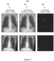
- Figure 3 shows exemplary synthesized images 300, in accordance with one or more embodiments.
- Images in column 302 show synthesized medical images p ⁇ xy
- images in column 304 show synthesized remaining images r ⁇ xy i
- images in column 306 show synthesized nodule images ⁇ n ⁇ xy i .
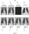
- the visibility of the nodules may be increased by highlighting the pixel intensity values of nodules which are close to the visibility threshold.
- the highlighting factor h i may be arbitrarily chosen.
- a highlighting factor h i may be randomly selected between [1,2] to generate synthetic medical images with nodules of a variety of intensities.
- Figure 4 shows a comparison 400 of nodules highlighted with different highlighting factors, in accordance with one or more embodiments.
- Image 402 is a synthesized medical image p ⁇ xy
- image 404 is a synthesized remaining image r ⁇ xy i
- image 406 is a synthesized nodule images ⁇ n ⁇ xy i
- image 408 is a mask of dense anatomical structures that are not visible (as determined at step 108)
- image 410 is synthesized medical image 402 with a highlighting factor of 1
- image 412 is synthesized medical image 402 with a highlighting factor of 2
- image 414 is synthesized medical image 402 with a highlighting factor of 5
- image 416 is synthesized medical image 402 with a highlighting factor of 10.
- each particular nodule of the one or more nodules is visible in the synthesized medical image based on at least one of the synthesized nodule image for the particular nodule and the synthesized remaining image.
- Nodules visible in the input medical image e.g., a 3D CT image
- the synthesized medical image e.g., x-ray image
- the synthesized medical image should not include annotations for nodules that are not visible in the synthesized medical image.
- nodules may be hidden in the synthesized medical images.
- a dense anatomical structure such as, e.g., bones, hearth, liver, etc.
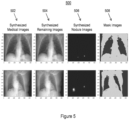
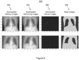
- a nodule i with center coordinate ( x, y ) is considered to be not visible in the synthesized medical image if r ⁇ xy i ⁇ t and visible in the synthesized medical image if r ⁇ xy i > t .
- nodules may be hidden in the synthesized medical image.
- the nodule is considered visible if two conditions are met.
- a change between an intensity at the center of the nodules in the synthesized remaining image and an intensity at the center of the nodules in the synthesized nodule images increases by at least a certain threshold t (e.g., 15%).
- threshold t e.g. 15%
- a change between an average intensity of pixels within half of the radius of the nodules in the synthesized remaining image and an average intensity of pixels within half of the radius of the nodules in the synthesized nodule image increases by at least a certain threshold t (e.g., 10%) amount.
- the annotation for the at least one not visible nodule is removed from the synthesized nodule image.
- the synthesized medical image is output.
- the synthesized medical image can be output by displaying the synthesized medical image on a display device of a computer system, storing the synthesized medical image on a memory or storage of a computer system, or by transmitting the synthesized medical image to a remote computer system.
- synthesized medical images generated according to method 100 may be applied for training machine learning based networks for medical image analysis tasks, such as, e.g., nodule detection or nodule segmentation.
- a nodule detector network implemented by a 2D Faster R-CNN (convolutional neural network) or a lung segmentation network implemented by a 2D U-Net may be respectively trained for nodule detection or lung segmentation in x-ray images using synthesized x-ray medical images generated according to method 100.
- such networks are trained using only the synthesized x-ray medical images.
- such networks are trained using a combination of synthesized x-ray medical images and real x-ray medical images.
- the synthesized x-ray medical images provides additional examples and examples of difficult to detect nodules in the training dataset, while having real x-ray medical images in the training dataset ensures that the feature extractors in the early layers of the networks are well adapted to the data distribution found in real x-ray medical images.
- various nodule augmentation techniques may be applied to generate the synthesized medical image in method 100.
- a nodule may be positioned in the synthesized medical image at step 104 by utilizing a database of previously acquired synthesized nodule images.
- a suitable position of the nodule may be determined based on visibility considerations described herein as well as a lung segmentation. Nodules with an intensity of less than, e.g., 0.5 (in linear space) may be removed from the database since there are no positions where they are visible.
- the input medical image received at step 102 may be rotated before generating the synthesized medical image at step 104.
- the input medical image received at step 102 may be rotated before generating the synthesized medical image at step 104.
- By rotating the input medical image a variety of different viewpoint angles of the nodules are obtained, multiplying the amount of relevant features available per nodule.
- the rotations are performed during a prior offline stage, since they are operations in 3D space.
- a limited number e.g., 20
- one of the images may be selected (e.g., randomly).
- the nodules may be resized.
- a bi-cubic up-sampling or down-sampling of the synthesized medical image is applied.
- all synthesized medical images may be multiplied by a resize factor. This follows Beer's law since the ray has to travel through more tissue.
- the input medical image received at step 102 may be generated to include artificial or synthesized nodules as described in U.S. Patent Application No. 16/445,435, filed June 19, 2019 , and U.S. Patent Application No. 16/570,214, filed September 13, 2019 . Synthesizing nodules in 3D space (e.g., of a CT image) is easier than in 2D space (e.g., an x-ray image).
- Systems, apparatus, and methods described herein may be implemented using computers operating in a client-server relationship.
- the client computers are located remotely from the server computer and interact via a network.
- the client-server relationship may be defined and controlled by computer programs running on the respective client and server computers.
- Systems, apparatus, and methods described herein may be implemented within a network-based cloud computing system.
- a server or another processor that is connected to a network communicates with one or more client computers via a network.
- a client computer may communicate with the server via a network browser application residing and operating on the client computer, for example.
- a client computer may store data on the server and access the data via the network.
- a client computer may transmit requests for data, or requests for online services, to the server via the network.
- the server may perform requested services and provide data to the client computer(s).
- the server may also transmit data adapted to cause a client computer to perform a specified function, e.g., to perform a calculation, to display specified data on a screen, etc.
- Systems, apparatus, and methods described herein may be implemented using a computer program product tangibly embodied in an information carrier, e.g., in a non-transitory machine-readable storage device, for execution by a programmable processor; and the method and workflow steps described herein, including one or more of the steps or functions of Figure 1 , may be implemented using one or more computer programs that are executable by such a processor.
- a computer program is a set of computer program instructions that can be used, directly or indirectly, in a computer to perform a certain activity or bring about a certain result.
- a computer program can be written in any form of programming language, including compiled or interpreted languages, and it can be deployed in any form, including as a stand-alone program or as a module, component, subroutine, or other unit suitable for use in a computing environment.
- Computer 702 includes a processor 704 operatively coupled to a data storage device 712 and a memory 710.
- Processor 704 controls the overall operation of computer 702 by executing computer program instructions that define such operations.
- the computer program instructions may be stored in data storage device 712, or other computer readable medium, and loaded into memory 710 when execution of the computer program instructions is desired.
- the method and workflow steps or functions of Figure 1 can be defined by the computer program instructions stored in memory 710 and/or data storage device 712 and controlled by processor 704 executing the computer program instructions.
Landscapes
- Engineering & Computer Science (AREA)
- Theoretical Computer Science (AREA)
- General Physics & Mathematics (AREA)
- Physics & Mathematics (AREA)
- Computer Vision & Pattern Recognition (AREA)
- Data Mining & Analysis (AREA)
- Software Systems (AREA)
- Medical Informatics (AREA)
- Evolutionary Computation (AREA)
- Artificial Intelligence (AREA)
- General Engineering & Computer Science (AREA)
- Health & Medical Sciences (AREA)
- General Health & Medical Sciences (AREA)
- Nuclear Medicine, Radiotherapy & Molecular Imaging (AREA)
- Radiology & Medical Imaging (AREA)
- Quality & Reliability (AREA)
- Computing Systems (AREA)
- Mathematical Physics (AREA)
- Life Sciences & Earth Sciences (AREA)
- Bioinformatics & Computational Biology (AREA)
- Evolutionary Biology (AREA)
- Bioinformatics & Cheminformatics (AREA)
- Image Analysis (AREA)
- Apparatus For Radiation Diagnosis (AREA)
Claims (15)
- Computerimplementiertes Verfahren, umfassend:Empfangen (102) eines medizinischen Eingabebildes in einer ersten Modalität, wobei das medizinische Eingabebild einen Knotenbereich für jeden von einem oder mehreren Knoten und einen verbleibenden Bereich außerhalb der Knotenbereiche umfasst, wobei das medizinische Eingabebild für jeden des einen oder der mehreren Knoten eine Annotation umfasst, die die Stelle des einen oder der mehreren Knoten identifiziert;Erzeugen (104) eines synthetisierten medizinischen Bildes aus dem medizinischen Eingabebild in einer zweiten Modalität, wobei das synthetisierte medizinische Bild die Annotation für jeden des einen oder der mehreren Knoten umfasst;Erzeugen (106) 1) eines synthetisierten Knotenbildes jedes der Knotenbereiche und 2) eines synthetisierten verbleibenden Bildes des verbleibenden Bereichs in der zweiten Modalität;Bestimmen (108), ob jeder spezifische Knoten des einen oder der mehreren Knoten in dem synthetisierten medizinischen Bild sichtbar ist, basierend auf mindestens einem von dem synthetisierten Knotenbild für den spezifischen Knoten und dem synthetisierten verbleibenden Bild; undin Reaktion auf das Bestimmen, dass mindestens ein Knoten des einen oder der mehreren Knoten in dem synthetisierten medizinischen Bild nicht sichtbar ist, Entfernen (110) der Annotation für den mindestens einen nicht sichtbaren Knoten aus dem synthetisierten Knotenbild.
- Verfahren nach Anspruch 1, wobei das Bestimmen, ob jeder spezifische Knoten des einen oder der mehreren Knoten in dem synthetisierten medizinischen Bild sichtbar ist, basierend auf mindestens einem von dem synthetisierten Knotenbild für den spezifischen Knoten und dem synthetisierten verbleibenden Bild Folgendes umfasst:
Vergleichen einer Intensität von Pixeln in dem synthetisierten verbleibenden Bild mit einem Schwellenwert. - Verfahren nach Anspruch 1 oder 2, wobei das Bestimmen, ob jeder spezifische Knoten des einen oder der mehreren Knoten in dem synthetisierten medizinischen Bild sichtbar ist, basierend auf mindestens einem von dem synthetisierten Knotenbild für den spezifischen Knoten und dem synthetisierten verbleibenden Bild Folgendes umfasst:Bestimmen einer Änderung zwischen einer Intensität in einem Zentrum des spezifischen Knotens in dem synthetisierten verbleibenden Bild und einer Intensität in dem Zentrum des spezifischen Knotens in dem synthetisierten Knotenbild; undVergleichen der Intensitätsänderung mit einem Schwellenwert.
- Verfahren nach einem der Ansprüche 1-3, wobei das Bestimmen, ob jeder spezifische Knoten des einen oder der mehreren Knoten in dem synthetisierten medizinischen Bild sichtbar ist, basierend auf mindestens einem von dem synthetisierten Knotenbild für den spezifischen Knoten und dem synthetisierten verbleibenden Bild Folgendes umfasst:Bestimmen einer Änderung zwischen einer durchschnittlichen Intensität von Pixeln innerhalb eines halben Radius des spezifischen Knotens in dem synthetisierten verbleibenden Bild und einer durchschnittlichen Intensität von Pixeln innerhalb des halben Radius des spezifischen Knotens in dem synthetisierten Knotenbild; undVergleichen der Änderung mit einem Schwellenwert.
- Verfahren nach einem der Ansprüche 1-4, wobei das Erzeugen eines synthetisierten medizinischen Bildes aus dem medizinischen Eingabebild in einer zweiten Modalität Folgendes umfasst:
Erhöhen einer Intensität von Pixeln des einen oder der mehreren Knoten in dem synthetisierten medizinischen Bild um einen Hervorhebungsfaktor. - Verfahren nach einem der Ansprüche 1-5, wobei das Erzeugen eines synthetisierten medizinischen Bildes aus dem medizinischen Eingabebild in einer zweiten Modalität Folgendes umfasst:
Positionieren eines zuvor erfassten synthetisierten Knotenbildes in dem synthetisierten medizinischen Bild. - Verfahren nach einem der Ansprüche 1-6, wobei das Erzeugen eines synthetisierten medizinischen Bildes aus dem medizinischen Eingabebild in einer zweiten Modalität Folgendes umfasst:Drehen des medizinischen Eingabebildes; undErzeugen des synthetisierten medizinischen Bildes aus dem gedrehten medizinischen Eingabebild.
- Verfahren nach einem der Ansprüche 1-7, wobei das Empfangen eines medizinischen Eingabebildes in einer ersten Modalität Folgendes umfasst:
Erzeugen des medizinischen Eingabebildes, um einen oder mehrere synthetisierte Knoten zu beinhalten. - Verfahren nach einem der Ansprüche 1-8, wobei es sich bei der ersten Modalität um Computertomographie handelt und bei der zweiten Modalität um Röntgenstrahlen handelt.
- Verfahren nach einem der Ansprüche 1-9, ferner umfassend:
Trainieren eines auf maschinellem Lernen basierenden Netzwerks basierend auf dem synthetisierten medizinischen Bild. - Knotenerkennungssystem zum Erkennen von Knoten in medizinischen Bildern, umfassend ein auf maschinellem Lernen basierendes Netzwerk, wobei das auf maschinellem Lernen basierende Netzwerk nach Anspruch 10 trainiert wird.
- Vorrichtung, umfassend:Mittel zum Empfangen (102) eines medizinischen Eingabebildes in einer ersten Modalität, wobei das medizinische Eingabebild einen Knotenbereich für jeden von einem oder mehreren Knoten und einen verbleibenden Bereich außerhalb der Knotenbereiche umfasst, wobei das medizinische Eingabebild für jeden des einen oder der mehreren Knoten eine Annotation umfasst, die die Stelle des einen oder der mehreren Knoten identifiziert;Mittel zum Erzeugen (104) eines synthetisierten medizinischen Bildes aus dem medizinischen Eingabebild in einer zweiten Modalität, wobei das synthetisierte medizinische Bild die Annotation für jeden des einen oder der mehreren Knoten umfasst;Mittel zum Erzeugen (106) 1) eines synthetisierten Knotenbildes jedes der Knotenbereiche und 2) eines synthetisierten verbleibenden Bildes des verbleibenden Bereichs in der zweiten Modalität;Mittel zum Bestimmen (108), ob jeder spezifische Knoten des einen oder der mehreren Knoten in dem synthetisierten medizinischen Bild sichtbar ist, basierend auf mindestens einem von dem synthetisierten Knotenbild für den spezifischen Knoten und dem synthetisierten verbleibenden Bild; und Mittel zum Entfernen (110) der Annotation für mindestens einen nicht sichtbaren Knoten aus dem synthetisierten Knotenbild in Reaktion auf das Bestimmen, dass der mindestens eine Knoten des einen oder der mehreren Knoten in dem synthetisierten medizinischen Bild nicht sichtbar ist.
- Vorrichtung nach Anspruch 12, ferner umfassend Mittel zum Ausführen des Verfahrens nach einem der Ansprüche 2-10.
- Nichtflüchtiges computerlesbares Medium, das Computerprogrammanweisungen speichert, wobei die Computerprogrammanweisungen, wenn sie von einem Prozessor ausgeführt werden, den Prozessor veranlassen, Operationen durchzuführen, die Folgendes umfassen:Empfangen (102) eines medizinischen Eingabebildes in einer ersten Modalität, wobei das medizinische Eingabebild einen Knotenbereich für jeden von einem oder mehreren Knoten und einen verbleibenden Bereich außerhalb der Knotenbereiche umfasst, wobei das medizinische Eingabebild für jeden des einen oder der mehreren Knoten eine Annotation umfasst, die die Stelle des einen oder der mehreren Knoten identifiziert;Erzeugen (104) eines synthetisierten medizinischen Bildes aus dem medizinischen Eingabebild in einer zweiten Modalität, wobei das synthetisierte medizinische Bild die Annotation für jeden des einen oder der mehreren Knoten umfasst;Erzeugen (106) 1) eines synthetisierten Knotenbildes jedes der Knotenbereiche und 2) eines synthetisierten verbleibenden Bildes des verbleibenden Bereichs in der zweiten Modalität;Bestimmen (108), ob jeder spezifische Knoten des einen oder der mehreren Knoten in dem synthetisierten medizinischen Bild sichtbar ist, basierend auf mindestens einem von dem synthetisierten Knotenbild für den spezifischen Knoten und dem synthetisierten verbleibenden Bild; undin Reaktion auf das Bestimmen, dass mindestens ein Knoten des einen oder der mehreren Knoten in dem synthetisierten medizinischen Bild nicht sichtbar ist, Entfernen (110) der Annotation für den mindestens einen nicht sichtbaren Knoten aus dem synthetisierten Knotenbild.
- Nichtflüchtiges computerlesbares Medium nach Anspruch 14, wobei die Computerprogrammanweisungen, wenn sie von einem Prozessor ausgeführt werden, den Prozessor veranlassen, Operationen durchzuführen, die das Verfahren nach einem der Ansprüche 2-10 implementieren.
Applications Claiming Priority (1)
| Application Number | Priority Date | Filing Date | Title |
|---|---|---|---|
| US15/929,427 US11615267B2 (en) | 2020-05-01 | 2020-05-01 | X-ray image synthesis from CT images for training nodule detection systems |
Publications (3)
| Publication Number | Publication Date |
|---|---|
| EP3907698A1 EP3907698A1 (de) | 2021-11-10 |
| EP3907698C0 EP3907698C0 (de) | 2025-04-16 |
| EP3907698B1 true EP3907698B1 (de) | 2025-04-16 |
Family
ID=75746445
Family Applications (1)
| Application Number | Title | Priority Date | Filing Date |
|---|---|---|---|
| EP21171503.2A Active EP3907698B1 (de) | 2020-05-01 | 2021-04-30 | Röntgenbildsynthese aus ct-bildern zum trainieren von knotenerkennungssystemen |
Country Status (3)
| Country | Link |
|---|---|
| US (1) | US11615267B2 (de) |
| EP (1) | EP3907698B1 (de) |
| CN (1) | CN113658284B (de) |
Families Citing this family (6)
| Publication number | Priority date | Publication date | Assignee | Title |
|---|---|---|---|---|
| US11531837B2 (en) * | 2020-07-17 | 2022-12-20 | Sap Se | Query image synthesis for image retrieval systems based on data augmentation techniques |
| US11908047B2 (en) * | 2021-03-11 | 2024-02-20 | Siemens Healthineers Ag | Generating synthetic x-ray images and object annotations from CT scans for augmenting x-ray abnormality assessment systems |
| CN114612618B (zh) * | 2022-03-15 | 2026-01-02 | 腾讯医疗健康(深圳)有限公司 | 图像生成方法、装置、设备、存储介质及计算机程序产品 |
| EP4300427A1 (de) * | 2022-07-01 | 2024-01-03 | Peek Health, S.A. | Standardisierung von bildern anatomischer strukturen zur analyse durch maschinenlernsysteme |
| US20240145068A1 (en) * | 2022-11-01 | 2024-05-02 | The Trustees Of The University Of Pennsylvania | Medical image analysis platform and associated methods |
| WO2024147978A1 (en) | 2023-01-03 | 2024-07-11 | Mandelker Eiran | Medical image diagnostic predictor |
Family Cites Families (11)
| Publication number | Priority date | Publication date | Assignee | Title |
|---|---|---|---|---|
| US20040151358A1 (en) * | 2003-01-31 | 2004-08-05 | Akiko Yanagita | Medical image processing system and method for processing medical image |
| US8442280B2 (en) * | 2004-01-21 | 2013-05-14 | Edda Technology, Inc. | Method and system for intelligent qualitative and quantitative analysis of digital radiography softcopy reading |
| JP4599191B2 (ja) * | 2005-03-01 | 2010-12-15 | 国立大学法人神戸大学 | 画像診断処理装置および画像診断処理プログラム |
| US10127662B1 (en) * | 2014-08-11 | 2018-11-13 | D.R. Systems, Inc. | Systems and user interfaces for automated generation of matching 2D series of medical images and efficient annotation of matching 2D medical images |
| US9892361B2 (en) * | 2015-01-21 | 2018-02-13 | Siemens Healthcare Gmbh | Method and system for cross-domain synthesis of medical images using contextual deep network |
| US9760807B2 (en) * | 2016-01-08 | 2017-09-12 | Siemens Healthcare Gmbh | Deep image-to-image network learning for medical image analysis |
| US11132792B2 (en) | 2018-02-22 | 2021-09-28 | Siemens Healthcare Gmbh | Cross domain medical image segmentation |
| EP3791316A1 (de) * | 2018-06-13 | 2021-03-17 | Siemens Healthcare GmbH | Lokalisierung und klassifizierung von anomalien auf medizinischen bildern |
| CN108742679B (zh) * | 2018-06-29 | 2022-07-05 | 上海联影医疗科技股份有限公司 | 结节检测装置和方法 |
| EP3611733B1 (de) * | 2018-08-15 | 2024-09-25 | Siemens Healthineers AG | Suchen eines medizinischen referenzbildes |
| EP3843038B1 (de) * | 2019-12-23 | 2023-09-20 | HTC Corporation | Bildbearbeitungsverfahren und -system |
-
2020
- 2020-05-01 US US15/929,427 patent/US11615267B2/en active Active
-
2021
- 2021-04-30 EP EP21171503.2A patent/EP3907698B1/de active Active
- 2021-04-30 CN CN202110482961.0A patent/CN113658284B/zh active Active
Also Published As
| Publication number | Publication date |
|---|---|
| US20210342638A1 (en) | 2021-11-04 |
| US11615267B2 (en) | 2023-03-28 |
| EP3907698C0 (de) | 2025-04-16 |
| EP3907698A1 (de) | 2021-11-10 |
| CN113658284B (zh) | 2024-10-01 |
| CN113658284A (zh) | 2021-11-16 |
Similar Documents
| Publication | Publication Date | Title |
|---|---|---|
| US11615267B2 (en) | X-ray image synthesis from CT images for training nodule detection systems | |
| US10970829B2 (en) | Synthesizing and segmenting cross-domain medical images | |
| US9427173B2 (en) | Determining mechanical force on aneurysms from a fluid dynamic model driven by vessel blood flow information | |
| US11132792B2 (en) | Cross domain medical image segmentation | |
| US8116542B2 (en) | Determining hazard of an aneurysm by change determination | |
| CN102947862B (zh) | 基于模型的分割的概率细化 | |
| US8229188B2 (en) | Systems, methods and apparatus automatic segmentation of liver in multiphase contrast-enhanced medical images | |
| US8644575B2 (en) | Processing of abdominal images | |
| CN111739614B (zh) | 医学图像增强 | |
| US8064673B2 (en) | Combined segmentation and registration framework for parametric shapes | |
| EP2620909B1 (de) | Verfahren, System und computerlesbares Medium zur automatischen Segmentierung eines medizinischen Bildes | |
| US10192352B2 (en) | Method, device and system for simulating shadow images | |
| US8428316B2 (en) | Coronary reconstruction from rotational X-ray projection sequence | |
| US20160005192A1 (en) | Prior Image Based Three Dimensional Imaging | |
| US20140301624A1 (en) | Method for interactive threshold segmentation of medical images | |
| JPH08138078A (ja) | 画像処理装置 | |
| US9424680B2 (en) | Image data reformatting | |
| US7860331B2 (en) | Purpose-driven enhancement filtering of anatomical data | |
| CN110910342B (zh) | 通过使用深度学习来分析骨骼创伤 | |
| US20080080770A1 (en) | Method and system for identifying regions in an image | |
| EP3648057B1 (de) | Bestimmung der malignität von lungenknötchen unter verwendung von tiefenlernen | |
| EP3433862B1 (de) | Binäre verfolgung von medizinischen bildern | |
| Macho et al. | Segmenting Teeth from Volumetric CT Data with a Hierarchical CNN-based Approach. |
Legal Events
| Date | Code | Title | Description |
|---|---|---|---|
| PUAI | Public reference made under article 153(3) epc to a published international application that has entered the european phase |
Free format text: ORIGINAL CODE: 0009012 |
|
| STAA | Information on the status of an ep patent application or granted ep patent |
Free format text: STATUS: REQUEST FOR EXAMINATION WAS MADE |
|
| 17P | Request for examination filed |
Effective date: 20210430 |
|
| AK | Designated contracting states |
Kind code of ref document: A1 Designated state(s): AL AT BE BG CH CY CZ DE DK EE ES FI FR GB GR HR HU IE IS IT LI LT LU LV MC MK MT NL NO PL PT RO RS SE SI SK SM TR |
|
| B565 | Issuance of search results under rule 164(2) epc |
Effective date: 20211007 |
|
| RBV | Designated contracting states (corrected) |
Designated state(s): AL AT BE BG CH CY CZ DE DK EE ES FI FR GB GR HR HU IE IS IT LI LT LU LV MC MK MT NL NO PL PT RO RS SE SI SK SM TR |
|
| RAP1 | Party data changed (applicant data changed or rights of an application transferred) |
Owner name: SIEMENS HEALTHINEERS AG |
|
| GRAP | Despatch of communication of intention to grant a patent |
Free format text: ORIGINAL CODE: EPIDOSNIGR1 |
|
| STAA | Information on the status of an ep patent application or granted ep patent |
Free format text: STATUS: GRANT OF PATENT IS INTENDED |
|
| INTG | Intention to grant announced |
Effective date: 20241114 |
|
| GRAS | Grant fee paid |
Free format text: ORIGINAL CODE: EPIDOSNIGR3 |
|
| GRAA | (expected) grant |
Free format text: ORIGINAL CODE: 0009210 |
|
| STAA | Information on the status of an ep patent application or granted ep patent |
Free format text: STATUS: THE PATENT HAS BEEN GRANTED |
|
| AK | Designated contracting states |
Kind code of ref document: B1 Designated state(s): AL AT BE BG CH CY CZ DE DK EE ES FI FR GB GR HR HU IE IS IT LI LT LU LV MC MK MT NL NO PL PT RO RS SE SI SK SM TR |
|
| REG | Reference to a national code |
Ref country code: GB Ref legal event code: FG4D |
|
| REG | Reference to a national code |
Ref country code: CH Ref legal event code: EP Ref country code: DE Ref legal event code: R096 Ref document number: 602021029112 Country of ref document: DE |
|
| REG | Reference to a national code |
Ref country code: IE Ref legal event code: FG4D |
|
| U01 | Request for unitary effect filed |
Effective date: 20250416 |
|
| U07 | Unitary effect registered |
Designated state(s): AT BE BG DE DK EE FI FR IT LT LU LV MT NL PT RO SE SI Effective date: 20250424 |
|
| U20 | Renewal fee for the european patent with unitary effect paid |
Year of fee payment: 5 Effective date: 20250424 |
|
| PGFP | Annual fee paid to national office [announced via postgrant information from national office to epo] |
Ref country code: GB Payment date: 20250507 Year of fee payment: 5 |
|
| PG25 | Lapsed in a contracting state [announced via postgrant information from national office to epo] |
Ref country code: ES Free format text: LAPSE BECAUSE OF FAILURE TO SUBMIT A TRANSLATION OF THE DESCRIPTION OR TO PAY THE FEE WITHIN THE PRESCRIBED TIME-LIMIT Effective date: 20250416 |
|
| PG25 | Lapsed in a contracting state [announced via postgrant information from national office to epo] |
Ref country code: NO Free format text: LAPSE BECAUSE OF FAILURE TO SUBMIT A TRANSLATION OF THE DESCRIPTION OR TO PAY THE FEE WITHIN THE PRESCRIBED TIME-LIMIT Effective date: 20250716 Ref country code: GR Free format text: LAPSE BECAUSE OF FAILURE TO SUBMIT A TRANSLATION OF THE DESCRIPTION OR TO PAY THE FEE WITHIN THE PRESCRIBED TIME-LIMIT Effective date: 20250717 |
|
| PG25 | Lapsed in a contracting state [announced via postgrant information from national office to epo] |
Ref country code: PL Free format text: LAPSE BECAUSE OF FAILURE TO SUBMIT A TRANSLATION OF THE DESCRIPTION OR TO PAY THE FEE WITHIN THE PRESCRIBED TIME-LIMIT Effective date: 20250416 |
|
| PG25 | Lapsed in a contracting state [announced via postgrant information from national office to epo] |
Ref country code: HR Free format text: LAPSE BECAUSE OF FAILURE TO SUBMIT A TRANSLATION OF THE DESCRIPTION OR TO PAY THE FEE WITHIN THE PRESCRIBED TIME-LIMIT Effective date: 20250416 |
|
| PG25 | Lapsed in a contracting state [announced via postgrant information from national office to epo] |
Ref country code: RS Free format text: LAPSE BECAUSE OF FAILURE TO SUBMIT A TRANSLATION OF THE DESCRIPTION OR TO PAY THE FEE WITHIN THE PRESCRIBED TIME-LIMIT Effective date: 20250716 |
|
| PG25 | Lapsed in a contracting state [announced via postgrant information from national office to epo] |
Ref country code: IS Free format text: LAPSE BECAUSE OF FAILURE TO SUBMIT A TRANSLATION OF THE DESCRIPTION OR TO PAY THE FEE WITHIN THE PRESCRIBED TIME-LIMIT Effective date: 20250816 |
|
| REG | Reference to a national code |
Ref country code: CH Ref legal event code: H13 Free format text: ST27 STATUS EVENT CODE: U-0-0-H10-H13 (AS PROVIDED BY THE NATIONAL OFFICE) Effective date: 20251125 |
|
| PG25 | Lapsed in a contracting state [announced via postgrant information from national office to epo] |
Ref country code: SM Free format text: LAPSE BECAUSE OF FAILURE TO SUBMIT A TRANSLATION OF THE DESCRIPTION OR TO PAY THE FEE WITHIN THE PRESCRIBED TIME-LIMIT Effective date: 20250416 |
|
| PG25 | Lapsed in a contracting state [announced via postgrant information from national office to epo] |
Ref country code: CH Free format text: LAPSE BECAUSE OF NON-PAYMENT OF DUE FEES Effective date: 20250430 |
|
| PG25 | Lapsed in a contracting state [announced via postgrant information from national office to epo] |
Ref country code: CZ Free format text: LAPSE BECAUSE OF FAILURE TO SUBMIT A TRANSLATION OF THE DESCRIPTION OR TO PAY THE FEE WITHIN THE PRESCRIBED TIME-LIMIT Effective date: 20250416 |
|
| PG25 | Lapsed in a contracting state [announced via postgrant information from national office to epo] |
Ref country code: SK Free format text: LAPSE BECAUSE OF FAILURE TO SUBMIT A TRANSLATION OF THE DESCRIPTION OR TO PAY THE FEE WITHIN THE PRESCRIBED TIME-LIMIT Effective date: 20250416 |
|
| PG25 | Lapsed in a contracting state [announced via postgrant information from national office to epo] |
Ref country code: MC Free format text: LAPSE BECAUSE OF FAILURE TO SUBMIT A TRANSLATION OF THE DESCRIPTION OR TO PAY THE FEE WITHIN THE PRESCRIBED TIME-LIMIT Effective date: 20250416 |
|
| PLBE | No opposition filed within time limit |
Free format text: ORIGINAL CODE: 0009261 |
|
| STAA | Information on the status of an ep patent application or granted ep patent |
Free format text: STATUS: NO OPPOSITION FILED WITHIN TIME LIMIT |
|
| REG | Reference to a national code |
Ref country code: CH Ref legal event code: L10 Free format text: ST27 STATUS EVENT CODE: U-0-0-L10-L00 (AS PROVIDED BY THE NATIONAL OFFICE) Effective date: 20260225 |
|
| 26N | No opposition filed |
Effective date: 20260119 |
|
| PG25 | Lapsed in a contracting state [announced via postgrant information from national office to epo] |
Ref country code: IE Free format text: LAPSE BECAUSE OF NON-PAYMENT OF DUE FEES Effective date: 20250430 |