EP3877949B1 - Systeme und verfahren zur halbautomatischen tumorsegmentierung - Google Patents
Systeme und verfahren zur halbautomatischen tumorsegmentierung Download PDFInfo
- Publication number
- EP3877949B1 EP3877949B1 EP19881201.8A EP19881201A EP3877949B1 EP 3877949 B1 EP3877949 B1 EP 3877949B1 EP 19881201 A EP19881201 A EP 19881201A EP 3877949 B1 EP3877949 B1 EP 3877949B1
- Authority
- EP
- European Patent Office
- Prior art keywords
- planar
- medical image
- boundary
- segmentation
- plane
- Prior art date
- Legal status (The legal status is an assumption and is not a legal conclusion. Google has not performed a legal analysis and makes no representation as to the accuracy of the status listed.)
- Active
Links
Images


Classifications
-
- G—PHYSICS
- G06—COMPUTING OR CALCULATING; COUNTING
- G06T—IMAGE DATA PROCESSING OR GENERATION, IN GENERAL
- G06T11/00—Two-dimensional [2D] image generation
- G06T11/60—Creating or editing images; Combining images with text
-
- G—PHYSICS
- G06—COMPUTING OR CALCULATING; COUNTING
- G06T—IMAGE DATA PROCESSING OR GENERATION, IN GENERAL
- G06T12/00—Tomographic reconstruction from projections
- G06T12/30—Image post-processing, e.g. metal artefact correction
-
- G—PHYSICS
- G06—COMPUTING OR CALCULATING; COUNTING
- G06T—IMAGE DATA PROCESSING OR GENERATION, IN GENERAL
- G06T7/00—Image analysis
- G06T7/0002—Inspection of images, e.g. flaw detection
- G06T7/0012—Biomedical image inspection
- G06T7/0014—Biomedical image inspection using an image reference approach
-
- G—PHYSICS
- G06—COMPUTING OR CALCULATING; COUNTING
- G06T—IMAGE DATA PROCESSING OR GENERATION, IN GENERAL
- G06T7/00—Image analysis
- G06T7/10—Segmentation; Edge detection
- G06T7/12—Edge-based segmentation
-
- G—PHYSICS
- G06—COMPUTING OR CALCULATING; COUNTING
- G06T—IMAGE DATA PROCESSING OR GENERATION, IN GENERAL
- G06T7/00—Image analysis
- G06T7/10—Segmentation; Edge detection
- G06T7/174—Segmentation; Edge detection involving the use of two or more images
-
- G—PHYSICS
- G16—INFORMATION AND COMMUNICATION TECHNOLOGY [ICT] SPECIALLY ADAPTED FOR SPECIFIC APPLICATION FIELDS
- G16H—HEALTHCARE INFORMATICS, i.e. INFORMATION AND COMMUNICATION TECHNOLOGY [ICT] SPECIALLY ADAPTED FOR THE HANDLING OR PROCESSING OF MEDICAL OR HEALTHCARE DATA
- G16H30/00—ICT specially adapted for the handling or processing of medical images
- G16H30/40—ICT specially adapted for the handling or processing of medical images for processing medical images, e.g. editing
-
- G—PHYSICS
- G16—INFORMATION AND COMMUNICATION TECHNOLOGY [ICT] SPECIALLY ADAPTED FOR SPECIFIC APPLICATION FIELDS
- G16H—HEALTHCARE INFORMATICS, i.e. INFORMATION AND COMMUNICATION TECHNOLOGY [ICT] SPECIALLY ADAPTED FOR THE HANDLING OR PROCESSING OF MEDICAL OR HEALTHCARE DATA
- G16H50/00—ICT specially adapted for medical diagnosis, medical simulation or medical data mining; ICT specially adapted for detecting, monitoring or modelling epidemics or pandemics
- G16H50/20—ICT specially adapted for medical diagnosis, medical simulation or medical data mining; ICT specially adapted for detecting, monitoring or modelling epidemics or pandemics for computer-aided diagnosis, e.g. based on medical expert systems
-
- G—PHYSICS
- G06—COMPUTING OR CALCULATING; COUNTING
- G06T—IMAGE DATA PROCESSING OR GENERATION, IN GENERAL
- G06T2207/00—Indexing scheme for image analysis or image enhancement
- G06T2207/10—Image acquisition modality
- G06T2207/10072—Tomographic images
-
- G—PHYSICS
- G06—COMPUTING OR CALCULATING; COUNTING
- G06T—IMAGE DATA PROCESSING OR GENERATION, IN GENERAL
- G06T2207/00—Indexing scheme for image analysis or image enhancement
- G06T2207/10—Image acquisition modality
- G06T2207/10072—Tomographic images
- G06T2207/10088—Magnetic resonance imaging [MRI]
-
- G—PHYSICS
- G06—COMPUTING OR CALCULATING; COUNTING
- G06T—IMAGE DATA PROCESSING OR GENERATION, IN GENERAL
- G06T2207/00—Indexing scheme for image analysis or image enhancement
- G06T2207/20—Special algorithmic details
- G06T2207/20081—Training; Learning
-
- G—PHYSICS
- G06—COMPUTING OR CALCULATING; COUNTING
- G06T—IMAGE DATA PROCESSING OR GENERATION, IN GENERAL
- G06T2207/00—Indexing scheme for image analysis or image enhancement
- G06T2207/20—Special algorithmic details
- G06T2207/20084—Artificial neural networks [ANN]
-
- G—PHYSICS
- G06—COMPUTING OR CALCULATING; COUNTING
- G06T—IMAGE DATA PROCESSING OR GENERATION, IN GENERAL
- G06T2207/00—Indexing scheme for image analysis or image enhancement
- G06T2207/20—Special algorithmic details
- G06T2207/20092—Interactive image processing based on input by user
- G06T2207/20104—Interactive definition of region of interest [ROI]
-
- G—PHYSICS
- G06—COMPUTING OR CALCULATING; COUNTING
- G06T—IMAGE DATA PROCESSING OR GENERATION, IN GENERAL
- G06T2207/00—Indexing scheme for image analysis or image enhancement
- G06T2207/30—Subject of image; Context of image processing
- G06T2207/30004—Biomedical image processing
- G06T2207/30016—Brain
-
- G—PHYSICS
- G06—COMPUTING OR CALCULATING; COUNTING
- G06T—IMAGE DATA PROCESSING OR GENERATION, IN GENERAL
- G06T2207/00—Indexing scheme for image analysis or image enhancement
- G06T2207/30—Subject of image; Context of image processing
- G06T2207/30004—Biomedical image processing
- G06T2207/30096—Tumor; Lesion
Definitions
- Embodiments of the invention relate to systems and methods for volumetric segmentation in a plurality of planar medical images. Examples involve analyzing medical images and, more particularly, to integrating offline analysis with user interaction to segment brain and other types of tumors.
- Extracting quantitative variables from medical images contributes more information for decision support in management of cancer patients, helping to improve patient outcomes.
- radiological studies are generally limited to detection and staging along with qualitative descriptions.
- Quantitative descriptors are not yet in the standard of care primarily due to a lack of infrastructure and tools to derive, test, and deploy these quantitative metrics at the point-of-care for all patients.
- a system for volumetric segmentation in a plurality of planar medical images includes a display, a user interface, an electronic processor coupled to the display and the user interface, and a memory coupled to the electronic processor and storing instructions that, when executed by the electronic processor, cause the system to receive the plurality of planar medical images.
- a boundary of a candidate structure in the plurality of medical images is generated using a segmentation model.
- a first planar medical image from the plurality of planar medical images is displayed on a display.
- a user input is received using a user interface indicating a region in the first planar medical image, the user input comprising an axis drawn on the first planar medical image.
- a first planar contour of the candidate structure is generated. The region is compared to the boundary. Responsive to the region being at least partially within the boundary, the first planar medical image is re-displayed on the display showing the first planar contour of the structure, and a finding record for the candidate structure including the boundary is generated.
- a method for volumetric segmentation in a plurality of planar medical images includes, receiving, at an electronic processor, the plurality of planar medical images.
- a boundary of a candidate structure in the plurality of medical images is generated using a segmentation model.
- a first planar medical image from the plurality of planar medical images is displayed on a display.
- a user input is received using a user interface indicating a region in the first planar medical image, the user input comprising an axis drawn on the first planar medical image.
- a first planar contour of the candidate structure is generated. The region is compared to the boundary. Responsive to the region being at least partially within the boundary, the first planar medical image is re-displayed on the display showing the first planar contour of the structure, and a finding record for the candidate structure including the boundary is generated.
- non-transitory computer-readable medium comprises all computer-readable media but does not consist of a transitory, propagating signal. Accordingly, non-transitory computer-readable medium may include, for example, a hard disk, a CD-ROM, an optical storage device, a magnetic storage device, a ROM (Read Only Memory), a RAM (Random Access Memory), register memory, a processor cache, or any combination thereof.
- Example systems and methods disclosed and contemplated herein relate to segmentation of structures in medical images. Although the following description focuses on image segmentation in a medical context, it will be appreciated that various systems, methods, and techniques disclosed herein are applicable to other domains, such as autonomous vehicles. For example, with the autonomous vehicle space, images may be segmented to identify a road centerline, a stop sign, or other findings of interested similar to findings of interest in medical images.
- CAD Computer Aided Detection
- embodiments described herein combine automated segmentation with a real-time user interface that allow a radiologist to make an initial indication regarding a finding that allows a previously identified structure generated by the CAD system to be matched to the radiologist's finding.
- Such embodiments provide clinical radiologists with control over the segmentation, real-time feedback, and an algorithm that is ready to run without the need to first be trained on a large database from their site.
- Such embodiments improve efficiency and accuracy over existing methods, including editing of a fully automated segmentation method.
- Volumetric contouring of a structure included in medical images provides advantages, such as inter-observer consistency, while also catering to individual preferences for accuracy and style. Consistency results from initialization strategies that are reproducible. For example, using a user may identify a structure using a straight stroke approach rather than free-form drawing, and the contouring of the structure may be performed automatically. Tailoring to individual preferences is accomplished by editing tools that may be used by a user if the initial contours are unsatisfactory. In some embodiments, there is exactly one tool in a reading room, generally applicable to all organs, yet simultaneously specialized with organ-specific features. The organ is automatically identified upon tool initialization.
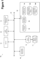
- Figure 1 schematically illustrates one embodiment of a system 100 for performing segmentation of features on medical images, according to some embodiments.
- the system 100 may combine one or more of hardware, software, and firmware, to implement a method provided herein.
- the system 100 includes a display 101, an electronic processor 102, a user interface 103, a memory 104, data storage device 110, a communication interface 112 and a bus 120.
- the memory 104 may store an operating system 106 and software programs 108.
- the electronic processor 102 includes at least one processor or microprocessor that interprets and executes a set of instructions stored in the memory 104.
- the software programs 108 include a segmentation application 109.
- the electronic processor 102 includes multiple computing devices that may be distributed across multiple entities.
- the electronic processor 102 is implemented by a virtual processor executing on a cloud-based processing service.
- the user interface 103 may include one or more input devices, one or more output devices, or a combination thereof. Accordingly, in some embodiments, the user interface 103 allows a user to interact with (for example, provide input to and receive output from) the system 100.
- the user interface 103 may include a keyboard, a cursor-control device (for example, a mouse), a touch screen, a scroll ball, a mechanical button, a display device (for example, a liquid crystal display (LCD)), a printer, a speaker, a microphone, or a combination thereof.
- a keyboard for example, a cursor-control device (for example, a mouse), a touch screen, a scroll ball, a mechanical button, a display device (for example, a liquid crystal display (LCD)), a printer, a speaker, a microphone, or a combination thereof.
- LCD liquid crystal display
- the data storage device 110 may include a non-transitory, tangible, machine-readable storage medium that stores machine-readable code or instructions.
- the data storage device 110 stores a set of instructions detailing a method provide herein that when executed by one or more processors cause the one or more processors to perform the method.
- the data storage device 110 may also be a database or a database interface for storing an application module.
- the data storage device 110 is located external to the system 100.
- the data storage 110 includes a medical image database 111.
- the bus 120 may permit communication among the components of the system 100.
- the bus 120 may be, for example, one or more buses or other wired or wireless connections, as is known in the art.
- the bus 120 may have additional elements, which are omitted for simplicity, such as controllers, buffers (for example, caches), drivers, repeaters and receivers, or other similar components, to enable communications.
- the bus 120 may also include address, control, data connections, or a combination of the foregoing to enable appropriate communications among the aforementioned components.
- the communication interface 112 provides the system 100 a communication gateway with one or more external networks (for example, a wireless network, the Internet, etc.).
- the communication interface 112 may include, for example, an Ethernet card or adapter or a wireless local area network (WLAN) card or adapter (for example, operating according to the IEEE standard 802.11a/b/g/n).
- the communication interface 112 may include address, control, and/or data connections to enable appropriate communications on the external network(s).
- the methods provided herein are performed by a software application executed by a server, and a user may access and interact with the software application using a computing device. Also, in some embodiments, functionality provided by the software application may be distributed between a software application executed by a local device and a software application executed by another electronic process or device (for example, a server) external to the local device.
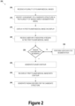
- FIG. 2 illustrates a flowchart for a method 200 of structure segmentation, according to some embodiments.
- a plurality of medical images is received.
- the medical images may be generated by a scanning tool, such as a computed tomography (CT) tool, a positron emission tomography (PET) tool, a magnetic resonance imaging (MRI) tool, etc., and stored in the medical image database 111.
- CT computed tomography
- PET positron emission tomography
- MRI magnetic resonance imaging
- the segmentation application 109 employs one or more segmentation models to identify one or more candidate structures depicted in the plurality of medical images and generates a boundary of each candidate structure. Since segmentation models 109A, 109B are known to generate false positives, the boundaries identified by the off-line analysis using the segmentation models 109A, 109B may be referred to as boundaries for candidate structures.
- Figure 3 illustrates a process flow for performing automatic segmentation by the segmentation application 109, according to some embodiments.
- the segmentation application 109 employs a deep learning segmentation model 109A or an algorithmic segmentation model 109B, or both (see Figure 1 ).
- the segmentation models 109A, 109B are relatively computationally intensive (e.g., requiring about one minute of computing time each on a typical minicomputer).
- the segmentation application 109 performs the segmentation analysis off-line using the segmentation models 109A, 109B.
- the segmentation application 109 may perform the segmentation analysis relatively soon after the medical images are generated, and prior to them being reviewed by a radiologist of other user. In this manner, the segmentation need not be performed in real time coincident with review by the radiologist.
- the deep learning segmentation model 190A generates boundaries 305 of one or more candidate structures identified in the plurality of medical images. In some embodiments, the boundaries are three-dimensional boundaries.
- the plurality of images include multispectral images of the same scan region.
- MRI data may include T1 weighted images, T2 weighted images, T1 contrast enhanced (CE) images, or fluid-attenuated inversion recovery (FLAIR) images.
- the segmentation models 109A, 109B receive the multispectral images as inputs allowing data from the different spectrums to be factored into the segmentation.
- the segmentation models 109A, 109B perform whole organ segmentation (e.g., whole brain segmentation) and partitioning of the tissue into its constituent parts: edema, necrosis, and actively enhancing regions.
- a radiologist or other user may interface with the segmentation application 109 in real-time to review and or modify the segmentation because only a portion of the image is being segmented and the whole organ analysis has already been completed by the segmentation models 109A, 109B off-line.
- the deep learning segmentation model 109A employs a convolution neural network (CNN).
- CNN convolution neural network
- the input image data is set to 128 x 128 x 128 voxels, constrained by the limited memory in the GPU.
- the 3D image volume is sequentially reduced in spatial resolution with multiple 3 x 3 x 3 convolution layers while increasing the number of filters or feature maps as the levels move deeper.
- the extracted feature maps are then upsampled to sequentially restore the spatial resolution at each level, concatenating with feature maps preserved during the downsampling to help restore lost information.
- a Softmax function classifies the 3 tumor classes. Dropouts with probability 0.3 are included to minimize overfitting.
- a multiclass Jaccard loss function is used in some embodiments.
- the four classes include 0 for background, 1 for tumor core, 2 for edema, and 4 for enhancing tumor.
- the Jaccard coefficient is a measure of similarity between the segmented prediction and truth image volumes, where higher value indicates greater overlap.
- the multiclass version is the intersection over union of the two volumes averaged over the four classes. A negative term was added to the loss function to ensure the minimum loss function was optimized.
- CNN development uses the open-source machine learning library, TensorFlow, and neural networking API, Keras. Other embodiments may use other machine learning libraries, for example, CNTK.
- the SVP is estimated through affine registration of the SPM atlas. Rules of logic are applied to the set of all four MR spectra to derive the other tissues. For example, enhancing tumor is described by areas that show hyper-intensity under contrast-enhancement when compared to the non-enhanced image, but also when compared to healthy white matter.
- the resultant tissue segmentation is used by the segmentation application 109 for anatomic context. For example, the segmentation application 109 may exclude vessels and ventricles from tumor structures.
- the segmentation application 109 displays a first planar medical image on the display 101.
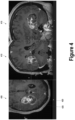
- Figures 4 and 5 illustrate medical images on which visual indication of planar contours are generated responsive to a user confirming a candidate structure identified by the segmentation in Figure 3 , according to some embodiments.
- Figure 4 illustrates a first medical image 405 (e.g., an axial plane image), a second medical image 410 (e.g., a coronal plane image, and a third medical image 415 (e.g., a sagittal plane image).
- the segmentation application 109 displays the medical images 405, 410, 415 without any annotations.
- the segmentation application 109 receives, via the user interface 103, a user input 420 indicating a region in the first planar medical image.
- the user provides the user input 420 by drawing an axis through a region of interest.
- the axis is a straight line.
- the axis may be a long axis, a short axis, or a partial axis.
- the user provides the user input 420 by clicking within the region, circling the region, circling a portion of the region, drawing a line in a portion of the region, or the like.
- the segmentation application 109 determines whether the region identified by the user input in the first planar medical image coincides with a boundary 305 of a candidate structure identified by the segmentation models 109A, 109B.
- the user indicates that the user input 420 is complete (e.g., the long axis is drawn as intended), and the segmentation application 109 proceeds with the determination in block 225.
- the segmentation application 109 determines whether the region identified by the user input in the first planar medical image coincides with a boundary 305 of a candidate structure by determining whether a long axis drawn by the user has endpoints that intersect or are proximal to the boundary 305.
- the segmentation application 109 re-displays the first medical image 405 on the display to provide a visual indication of the planar contour 425, as illustrated in Figure 4 .
- the segmentation application 109 also generates planar contours 430, 435 for the other medical images 410, 415 by projecting the boundary 305 into the plane corresponding to the particular medical image 410, 415.
- the planar contours 430, 435 are shown in the medical images 410, 415, as illustrated in Figure 5 .
- the planar contours 425, 430, 435 appear more coarsely pixilated because the medical images 405, 410, 415 displays super-sampled images, whereas the deep learning segmentation model 109A operates on full resolution images.
- the segmentation application generates planar contours for other medical images in the set of medical images after receiving the user input.
- medical images having the same orientation as the medical image 405, but in different slices, may be annotated with planar contours to allow the user to scroll through the slices to see the boundaries of the structure throughout its 3D volume.
- the user may draw additional axes or move the envelope defined by the planar contours 425, 430, 435.
- Data from the deep learning segmentation model 109A and the tissue map 310 generated by the algorithmic segmentation model 109 may be used to alter the boundary 305 and the planar contours 425, 430, 435.
- the segmentation application generates a finding record for the candidate structure contained within the boundary 305.
- a finding record includes a 3-D mesh and metadata describing the characteristics of the mesh.
- the finding record is a 3-D image volume.
- the user indicates that the candidate structure contained within the boundary 305 identified by the segmentation application 109 (e.g., using the deep learning segmentation model 109A) is a valid finding.
- the method 200 may be repeated for each structure. Since the candidate structures are not initially provided to the user, the user is not burdened with false positives. Only those candidate structures that correspond to findings indicated by the user are presented.
Landscapes
- Engineering & Computer Science (AREA)
- Health & Medical Sciences (AREA)
- General Physics & Mathematics (AREA)
- Theoretical Computer Science (AREA)
- Physics & Mathematics (AREA)
- Medical Informatics (AREA)
- Computer Vision & Pattern Recognition (AREA)
- General Health & Medical Sciences (AREA)
- Public Health (AREA)
- Nuclear Medicine, Radiotherapy & Molecular Imaging (AREA)
- Radiology & Medical Imaging (AREA)
- Primary Health Care (AREA)
- Epidemiology (AREA)
- Biomedical Technology (AREA)
- Quality & Reliability (AREA)
- Data Mining & Analysis (AREA)
- Databases & Information Systems (AREA)
- Pathology (AREA)
- Apparatus For Radiation Diagnosis (AREA)
- Magnetic Resonance Imaging Apparatus (AREA)
Claims (18)
- System (100) zur volumetrischen Segmentierung in einer Vielzahl von planaren medizinischen Bildern, umfassend:eine Anzeige (101);eine Benutzerschnittstelle (103);einen elektronischen Prozessor (102), der an die Anzeige (101) und die Benutzerschnittstelle (103) gekoppelt ist; undeinen Speicher (104), der an den elektronischen Prozessor (102) gekoppelt ist und Anweisungen speichert, die, wenn sie von dem elektronischen Prozessor (102) ausgeführt werden, das System (100) zu Folgendem veranlassen:Empfangen der Vielzahl von planaren medizinischen Bildern;Erzeugen einer Begrenzung (305) einer Kandidatenstruktur in der Vielzahl von planaren medizinischen Bildern unter Verwendung eines Segmentierungsmodells (109A, 109B);Anzeigen eines ersten planaren medizinischen Bildes (405) von der Vielzahl von planaren medizinischen Bildern auf der Anzeige (101);Empfangen einer Benutzereingabe (420), die eine Region in dem ersten planaren medizinischen Bild (405) angibt, mittels der Benutzerschnittstelle (103), wobei die Benutzereingabe (420) eine Achse umfasst, die auf das erste planare medizinische Bild (405) gezeichnet ist;Vergleichen der von der Benutzereingabe (420) angegebenen Region mit der Begrenzung (305) der Kandidatenstruktur undals Reaktion darauf, dass die Region mindestens teilweise innerhalb der Begrenzung liegt:Erzeugen einer ersten planaren Kontur (425) der Kandidatenstruktur; undWiederanzeigen des ersten planaren medizinischen Bildes (405), das die erste planare Kontur (425) der Kandidatenstruktur zeigt, auf der Anzeige (101); undErzeugen eines Ergebnisdatensatzes für die Kandidatenstruktur, die die Begrenzung (305) einschließt.
- System nach Anspruch 1, wobei die Anweisungen, wenn sie von dem elektronischen Prozessor (102) ausgeführt werden, das System (100) zu Folgendem veranlassen:
Erzeugen des Ergebnisdatensatzes als Reaktion auf das Empfangen der Benutzereingabe (420), die die erste planare Kontur (425) akzeptiert. - System nach Anspruch 1, wobei die Anweisungen, wenn sie von dem elektronischen Prozessor (102) ausgeführt werden, das System (100) zu Folgendem veranlassen:
Vergleichen der Region mit der Begrenzung (305) durch Bestimmen, ob Endpunkte der Achse innerhalb eines vorbestimmten Abstandes von der Begrenzung (305) liegen. - System nach Anspruch 1, wobei die Anweisungen, wenn sie von dem elektronischen Prozessor (102) ausgeführt werden, das System (100) zu Folgendem veranlassen:
Vergleichen der Region mit der Begrenzung (305) durch Bestimmen, ob Endpunkte der Achse innerhalb eines vorbestimmten Abstandes von der ersten planaren Kontur (425) liegen. - System nach Anspruch 1, wobei die Anweisungen, wenn sie von dem elektronischen Prozessor (102) ausgeführt werden, das System (100) zu Folgendem veranlassen:Erzeugen einer zweiten planaren Kontur (430, 435) der Kandidatenstruktur undAnzeigen eines zweiten planaren medizinischen Bildes (410, 415) von der Vielzahl von planaren medizinischen Bildern, das die zweite planare Kontur (430, 435) zeigt, wobei das erste (405) und zweite medizinische Bild (410, 415) mit verschiedenen Ebenenorientierungen in Verbindung stehen.
- System nach Anspruch 5, wobei das erste planare medizinische Bild (405) mit einer von einer Achsenebene, einer Koronalebene oder einer Sagittalebene in Verbindung steht und das zweite planare medizinische Bild mit einer anderen von der Achsenebene, der Koronalebene oder der Sagittalebene in Verbindung steht.
- System nach Anspruch 1, wobei die Anweisungen, wenn sie von dem elektronischen Prozessor (102) ausgeführt werden, das System (100) zu Folgendem veranlassen:Erzeugen einer zweiten planaren Kontur (430, 435) der Kandidatenstruktur undAnzeigen eines zweiten planaren medizinischen Bildes von der Vielzahl von planaren medizinischen Bildern, das die zweite planare Kontur zeigt, auf der Anzeige (101), wobei das erste planare medizinische Bild (405) mit einer von einer Achsenebene, einer Koronalebene oder einer Sagittalebene in Verbindung steht und das zweite planare medizinische Bild mit derselben von der Achsenebene, der Koronalebene oder der Sagittalebene in Verbindung steht.
- System nach Anspruch 1, wobei das Segmentierungsmodell ein Deep-Learning-Segmentierungsmodell (109A) umfasst.
- System nach Anspruch 1, wobei das Segmentierungsmodell ein algorithmisches Segmentierungsmodell (109B) umfasst.
- Verfahren zur volumetrischen Segmentierung in einer Vielzahl von planaren medizinischen Bildern, umfassend:Empfangen der Vielzahl von planaren medizinischen Bildern auf einem elektronischen Prozessor (102);Erzeugen einer Begrenzung (305) einer Kandidatenstruktur in der Vielzahl von medizinischen Bildern unter Verwendung eines Segmentierungsmodells (109A, 109B);Anzeigen eines ersten planaren medizinischen Bildes (405) von der Vielzahl von planaren medizinischen Bildern auf einer Anzeige (101);Empfangen einer Benutzereingabe (420), die eine Region in dem ersten planaren medizinischen Bild (405) angibt, mittels einer Benutzerschnittstelle (103), wobei die Benutzereingabe (420) eine Achse umfasst, die auf das erste planare medizinische Bild (405) gezeichnet ist;Vergleichen der von der Benutzereingabe (420) angegebenen Region mit der Begrenzung (305) der Kandidatenstruktur undals Reaktion darauf, dass die Region mindestens teilweise innerhalb der Begrenzung liegt:Erzeugen einer ersten planaren Kontur (425) der Kandidatenstruktur; undWiederanzeigen des ersten planaren medizinischen Bildes (405), das die erste planare Kontur (425) der Kandidatenstruktur zeigt, auf der Anzeige (101); undErzeugen eines Ergebnisdatensatzes für die Kandidatenstruktur, die die Begrenzung (305) einschließt.
- Verfahren nach Anspruch 10, wobei der Ergebnisdatensatz als Reaktion auf das Empfangen einer Benutzereingabe, die die erste planare Kontur akzeptiert, erzeugt wird.
- Verfahren nach Anspruch 10, wobei das Vergleichen der Region mit der Begrenzung (305) das Bestimmen, ob Endpunkte der Achse innerhalb eines vorbestimmten Abstandes von der Begrenzung (305) liegen, umfasst.
- Verfahren nach Anspruch 10, wobei das Vergleichen der Region mit der Begrenzung (305) das Bestimmen, ob Endpunkte der Achse innerhalb eines vorbestimmten Abstandes von der ersten planaren Kontur (425) liegen, umfasst.
- Verfahren nach Anspruch 10, ferner umfassend:
Erzeugen einer zweiten planaren Kontur (430, 435) der Kandidatenstruktur und Anzeigen eines zweiten medizinischen Bildes (410, 415) von der Vielzahl von planaren medizinischen Bildern, das die zweite planare Kontur (430, 435) zeigt, auf der Anzeige (101), wobei das erste und zweite medizinische Bild (410, 415) mit verschiedenen Ebenenorientierungen in Verbindung stehen. - Verfahren nach Anspruch 14, wobei das erste planare medizinische Bild (405) mit einer von einer Achsenebene, einer Koronalebene oder einer Sagittalebene in Verbindung steht und das zweite planare medizinische Bild (410, 415) mit einer anderen von der Achsenebene, der Koronalebene oder der Sagittalebene in Verbindung steht.
- Verfahren nach Anspruch 10, ferner umfassend:
Erzeugen einer zweiten planaren Kontur (430, 435) der Kandidatenstruktur und Anzeigen eines zweiten medizinischen Bildes (410, 415) von der Vielzahl von planaren medizinischen Bildern, das die zweite planare Kontur (430, 435) zeigt, auf der Anzeige (101), wobei das erste planare medizinische Bild mit einer von einer Achsenebene, einer Koronalebene oder einer Sagittalebene in Verbindung steht und das zweite planare medizinische Bild mit derselben von der Achsenebene, der Koronalebene oder der Sagittalebene in Verbindung steht. - Verfahren nach Anspruch 10, wobei das Segmentierungsmodell ein Deep-Learning-Segmentierungsmodell (109A) umfasst.
- Verfahren nach Anspruch 10, wobei das Segmentierungsmodell ein algorithmisches Segmentierungsmodell (109B) umfasst.
Applications Claiming Priority (2)
| Application Number | Priority Date | Filing Date | Title |
|---|---|---|---|
| US201862755873P | 2018-11-05 | 2018-11-05 | |
| PCT/US2019/059897 WO2020097100A1 (en) | 2018-11-05 | 2019-11-05 | Systems and methods for semi-automatic tumor segmentation |
Publications (4)
| Publication Number | Publication Date |
|---|---|
| EP3877949A1 EP3877949A1 (de) | 2021-09-15 |
| EP3877949A4 EP3877949A4 (de) | 2022-08-17 |
| EP3877949B1 true EP3877949B1 (de) | 2025-04-09 |
| EP3877949C0 EP3877949C0 (de) | 2025-04-09 |
Family
ID=70459056
Family Applications (1)
| Application Number | Title | Priority Date | Filing Date |
|---|---|---|---|
| EP19881201.8A Active EP3877949B1 (de) | 2018-11-05 | 2019-11-05 | Systeme und verfahren zur halbautomatischen tumorsegmentierung |
Country Status (3)
| Country | Link |
|---|---|
| US (1) | US11043296B2 (de) |
| EP (1) | EP3877949B1 (de) |
| WO (1) | WO2020097100A1 (de) |
Families Citing this family (6)
| Publication number | Priority date | Publication date | Assignee | Title |
|---|---|---|---|---|
| US10762629B1 (en) * | 2019-11-14 | 2020-09-01 | SegAI LLC | Segmenting medical images |
| US11423544B1 (en) * | 2019-11-14 | 2022-08-23 | Seg AI LLC | Segmenting medical images |
| EP3838209A1 (de) * | 2019-12-20 | 2021-06-23 | Koninklijke Philips N.V. | Erzeugung interaktiver zonen für interventionelle medizinische vorrichtungen |
| US11030747B1 (en) * | 2020-03-04 | 2021-06-08 | Xue Feng | System and method for automatic thoracic organ segmentation from CT using a deep learning framework |
| CN112767407B (zh) * | 2021-02-02 | 2023-07-07 | 南京信息工程大学 | 一种基于级联门控3DUnet模型的CT图像肾脏肿瘤分割方法 |
| US20250148777A1 (en) * | 2023-11-08 | 2025-05-08 | Qualcomm Incorporated | Systems and methods for segmentation map error correction |
Family Cites Families (22)
| Publication number | Priority date | Publication date | Assignee | Title |
|---|---|---|---|---|
| US7466848B2 (en) * | 2002-12-13 | 2008-12-16 | Rutgers, The State University Of New Jersey | Method and apparatus for automatically detecting breast lesions and tumors in images |
| JP4758351B2 (ja) * | 2003-10-17 | 2011-08-24 | コーニンクレッカ フィリップス エレクトロニクス エヌ ヴィ | モデルベースの画像セグメンテーションのための手動ツール |
| US7376252B2 (en) | 2003-11-25 | 2008-05-20 | Ge Medical Systems Global Technology Company, Llc | User interactive method and user interface for detecting a contour of an object |
| US20080021502A1 (en) | 2004-06-21 | 2008-01-24 | The Trustees Of Columbia University In The City Of New York | Systems and methods for automatic symmetry identification and for quantification of asymmetry for analytic, diagnostic and therapeutic purposes |
| US7519209B2 (en) * | 2004-06-23 | 2009-04-14 | Vanderbilt University | System and methods of organ segmentation and applications of same |
| US8155405B2 (en) | 2007-04-20 | 2012-04-10 | Siemens Aktiengsellschaft | System and method for lesion segmentation in whole body magnetic resonance images |
| US20080281182A1 (en) * | 2007-05-07 | 2008-11-13 | General Electric Company | Method and apparatus for improving and/or validating 3D segmentations |
| US7876947B2 (en) | 2007-10-10 | 2011-01-25 | Siemens Medical Solutions Usa, Inc. | System and method for detecting tagged material using alpha matting |
| JP5198883B2 (ja) | 2008-01-16 | 2013-05-15 | 富士フイルム株式会社 | 腫瘍領域サイズ測定方法および装置ならびにプログラム |
| WO2010115885A1 (en) | 2009-04-03 | 2010-10-14 | Oslo Universitetssykehus Hf | Predictive classifier score for cancer patient outcome |
| DE102010022307A1 (de) * | 2010-06-01 | 2011-12-01 | Siemens Aktiengesellschaft | Verfahren zur Überprüfung der Segmentierung einer Struktur in Bilddaten |
| WO2013155300A1 (en) | 2012-04-11 | 2013-10-17 | The Trustees Of Columbia University In The City Of New York | Techniques for segmentation of organs and tumors and objects |
| US10025479B2 (en) | 2013-09-25 | 2018-07-17 | Terarecon, Inc. | Advanced medical image processing wizard |
| GB2547399B (en) | 2014-12-02 | 2018-05-02 | Shanghai United Imaging Healthcare Co Ltd | A method and system for image processing |
| US11576578B2 (en) * | 2015-03-02 | 2023-02-14 | Shanghai United Imaging Healthcare Co., Ltd. | Systems and methods for scanning a patient in an imaging system |
| US11576645B2 (en) * | 2015-03-02 | 2023-02-14 | Shanghai United Imaging Healthcare Co., Ltd. | Systems and methods for scanning a patient in an imaging system |
| US9990712B2 (en) | 2015-04-08 | 2018-06-05 | Algotec Systems Ltd. | Organ detection and segmentation |
| WO2016186746A1 (en) * | 2015-05-18 | 2016-11-24 | General Electric Company | Methods and systems for automatic segmentation |
| WO2016191870A1 (en) * | 2015-06-01 | 2016-12-08 | The Governors Of The University Of Alberta | Surface modeling of a segmented echogenic structure for detection and measurement of anatomical anomalies |
| US9978150B2 (en) | 2015-08-05 | 2018-05-22 | Algotec Systems Ltd. | Method and system for spatial segmentation of anatomical structures |
| JP6019369B1 (ja) | 2015-08-31 | 2016-11-02 | 本多電子株式会社 | 超音波画像表示装置及びその作動方法、並びにプログラムを格納した記録媒体 |
| EP3784139A4 (de) * | 2018-04-27 | 2021-12-29 | Delphinus Medical Technologies, Inc. | System und verfahren zur merkmalsextraktion und zur klassifizierung auf ultraschall-tomografiebildern |
-
2019
- 2019-11-05 US US16/674,944 patent/US11043296B2/en active Active
- 2019-11-05 WO PCT/US2019/059897 patent/WO2020097100A1/en not_active Ceased
- 2019-11-05 EP EP19881201.8A patent/EP3877949B1/de active Active
Also Published As
| Publication number | Publication date |
|---|---|
| WO2020097100A1 (en) | 2020-05-14 |
| EP3877949A4 (de) | 2022-08-17 |
| EP3877949C0 (de) | 2025-04-09 |
| EP3877949A1 (de) | 2021-09-15 |
| US11043296B2 (en) | 2021-06-22 |
| US20200143934A1 (en) | 2020-05-07 |
Similar Documents
| Publication | Publication Date | Title |
|---|---|---|
| EP3877949B1 (de) | Systeme und verfahren zur halbautomatischen tumorsegmentierung | |
| Bakas et al. | Advancing the cancer genome atlas glioma MRI collections with expert segmentation labels and radiomic features | |
| US8335359B2 (en) | Systems, apparatus and processes for automated medical image segmentation | |
| CN111210401B (zh) | 根据医学图像的主动脉自动检测和量化 | |
| US12467997B2 (en) | Fiber tracking and segmentation | |
| US10762636B2 (en) | Systems and methods for volumetric segmentation of structures in planar medical images | |
| US12106533B2 (en) | Method and system for segmenting interventional device in image | |
| CN110853743A (zh) | 医学影像的显示方法、信息处理方法及存储介质 | |
| US10706534B2 (en) | Method and apparatus for classifying a data point in imaging data | |
| US12456197B2 (en) | Lesion detection and segmentation | |
| US11229377B2 (en) | System and method for next-generation MRI spine evaluation | |
| JP7851965B2 (ja) | 医用画像データの自動追跡読影方法及びシステム | |
| CN111081352A (zh) | 医学影像的显示方法、信息处理方法及存储介质 | |
| CN110400302B (zh) | 一种确定、显示乳房图像中病灶信息的方法及装置 | |
| US20240087697A1 (en) | Methods and systems for providing a template data structure for a medical report | |
| Liu et al. | RPLS-Net: pulmonary lobe segmentation based on 3D fully convolutional networks and multi-task learning | |
| Amran et al. | BV-GAN: 3D time-of-flight magnetic resonance angiography cerebrovascular vessel segmentation using adversarial CNNs | |
| US20240290471A1 (en) | Method for automated processing of volumetric medical images | |
| CN112712507A (zh) | 一种确定冠状动脉的钙化区域的方法及装置 | |
| US12530868B2 (en) | Systems and methods for medical image analysis and classification | |
| CN120997133B (zh) | 用于多示踪剂图像的分析方法及系统、存储介质、计算机设备 | |
| EP4715732A1 (de) | System und verfahren zur analyse einer karte eines biologischen objekts mit optimierter längskompatibilität | |
| EP4715735A1 (de) | Computerimplementiertes verfahren zum vergleich medizinischer bilder einer längsstudie | |
| Mardo | Evaluation of Synthetic Ground Truth for Semantic Brain Image Segmentation-Developing a Database of Synthetic Images with Ground-Truth Segmentations for Practical Application Assessment | |
| KR20230151865A (ko) | 의료 영상에서 세기의 분포에 기반한 보조 정보를 생성하고 시각화하는 의료 영상 진단 보조 장치 및 방법 |
Legal Events
| Date | Code | Title | Description |
|---|---|---|---|
| STAA | Information on the status of an ep patent application or granted ep patent |
Free format text: STATUS: THE INTERNATIONAL PUBLICATION HAS BEEN MADE |
|
| PUAI | Public reference made under article 153(3) epc to a published international application that has entered the european phase |
Free format text: ORIGINAL CODE: 0009012 |
|
| STAA | Information on the status of an ep patent application or granted ep patent |
Free format text: STATUS: REQUEST FOR EXAMINATION WAS MADE |
|
| 17P | Request for examination filed |
Effective date: 20210217 |
|
| AK | Designated contracting states |
Kind code of ref document: A1 Designated state(s): AL AT BE BG CH CY CZ DE DK EE ES FI FR GB GR HR HU IE IS IT LI LT LU LV MC MK MT NL NO PL PT RO RS SE SI SK SM TR |
|
| DAV | Request for validation of the european patent (deleted) | ||
| DAX | Request for extension of the european patent (deleted) | ||
| A4 | Supplementary search report drawn up and despatched |
Effective date: 20220715 |
|
| RIC1 | Information provided on ipc code assigned before grant |
Ipc: G01R 33/56 20060101ALI20220711BHEP Ipc: G06T 7/174 20170101ALI20220711BHEP Ipc: G06T 7/12 20170101ALI20220711BHEP Ipc: G06T 7/11 20170101ALI20220711BHEP Ipc: G06T 7/10 20170101AFI20220711BHEP |
|
| RAP1 | Party data changed (applicant data changed or rights of an application transferred) |
Owner name: GENENTECH, INC. |
|
| GRAP | Despatch of communication of intention to grant a patent |
Free format text: ORIGINAL CODE: EPIDOSNIGR1 |
|
| STAA | Information on the status of an ep patent application or granted ep patent |
Free format text: STATUS: GRANT OF PATENT IS INTENDED |
|
| INTG | Intention to grant announced |
Effective date: 20241113 |
|
| GRAS | Grant fee paid |
Free format text: ORIGINAL CODE: EPIDOSNIGR3 |
|
| GRAA | (expected) grant |
Free format text: ORIGINAL CODE: 0009210 |
|
| STAA | Information on the status of an ep patent application or granted ep patent |
Free format text: STATUS: THE PATENT HAS BEEN GRANTED |
|
| AK | Designated contracting states |
Kind code of ref document: B1 Designated state(s): AL AT BE BG CH CY CZ DE DK EE ES FI FR GB GR HR HU IE IS IT LI LT LU LV MC MK MT NL NO PL PT RO RS SE SI SK SM TR |
|
| REG | Reference to a national code |
Ref country code: GB Ref legal event code: FG4D |
|
| REG | Reference to a national code |
Ref country code: CH Ref legal event code: EP |
|
| REG | Reference to a national code |
Ref country code: IE Ref legal event code: FG4D |
|
| U01 | Request for unitary effect filed |
Effective date: 20250409 |
|
| U07 | Unitary effect registered |
Designated state(s): AT BE BG DE DK EE FI FR IT LT LU LV MT NL PT RO SE SI Effective date: 20250415 |
|
| PG25 | Lapsed in a contracting state [announced via postgrant information from national office to epo] |
Ref country code: ES Free format text: LAPSE BECAUSE OF FAILURE TO SUBMIT A TRANSLATION OF THE DESCRIPTION OR TO PAY THE FEE WITHIN THE PRESCRIBED TIME-LIMIT Effective date: 20250409 |
|
| PG25 | Lapsed in a contracting state [announced via postgrant information from national office to epo] |
Ref country code: NO Free format text: LAPSE BECAUSE OF FAILURE TO SUBMIT A TRANSLATION OF THE DESCRIPTION OR TO PAY THE FEE WITHIN THE PRESCRIBED TIME-LIMIT Effective date: 20250709 Ref country code: GR Free format text: LAPSE BECAUSE OF FAILURE TO SUBMIT A TRANSLATION OF THE DESCRIPTION OR TO PAY THE FEE WITHIN THE PRESCRIBED TIME-LIMIT Effective date: 20250710 |
|
| PG25 | Lapsed in a contracting state [announced via postgrant information from national office to epo] |
Ref country code: PL Free format text: LAPSE BECAUSE OF FAILURE TO SUBMIT A TRANSLATION OF THE DESCRIPTION OR TO PAY THE FEE WITHIN THE PRESCRIBED TIME-LIMIT Effective date: 20250409 |
|
| PG25 | Lapsed in a contracting state [announced via postgrant information from national office to epo] |
Ref country code: HR Free format text: LAPSE BECAUSE OF FAILURE TO SUBMIT A TRANSLATION OF THE DESCRIPTION OR TO PAY THE FEE WITHIN THE PRESCRIBED TIME-LIMIT Effective date: 20250409 |
|
| PG25 | Lapsed in a contracting state [announced via postgrant information from national office to epo] |
Ref country code: RS Free format text: LAPSE BECAUSE OF FAILURE TO SUBMIT A TRANSLATION OF THE DESCRIPTION OR TO PAY THE FEE WITHIN THE PRESCRIBED TIME-LIMIT Effective date: 20250709 |
|
| PG25 | Lapsed in a contracting state [announced via postgrant information from national office to epo] |
Ref country code: IS Free format text: LAPSE BECAUSE OF FAILURE TO SUBMIT A TRANSLATION OF THE DESCRIPTION OR TO PAY THE FEE WITHIN THE PRESCRIBED TIME-LIMIT Effective date: 20250809 |
|
| U20 | Renewal fee for the european patent with unitary effect paid |
Year of fee payment: 7 Effective date: 20251022 |
|
| REG | Reference to a national code |
Ref country code: CH Ref legal event code: U11 Free format text: ST27 STATUS EVENT CODE: U-0-0-U10-U11 (AS PROVIDED BY THE NATIONAL OFFICE) Effective date: 20251201 |
|
| PGFP | Annual fee paid to national office [announced via postgrant information from national office to epo] |
Ref country code: GB Payment date: 20251023 Year of fee payment: 7 |
|
| PG25 | Lapsed in a contracting state [announced via postgrant information from national office to epo] |
Ref country code: SM Free format text: LAPSE BECAUSE OF FAILURE TO SUBMIT A TRANSLATION OF THE DESCRIPTION OR TO PAY THE FEE WITHIN THE PRESCRIBED TIME-LIMIT Effective date: 20250409 |
|
| PGFP | Annual fee paid to national office [announced via postgrant information from national office to epo] |
Ref country code: CH Payment date: 20251201 Year of fee payment: 7 |
|
| PG25 | Lapsed in a contracting state [announced via postgrant information from national office to epo] |
Ref country code: CZ Free format text: LAPSE BECAUSE OF FAILURE TO SUBMIT A TRANSLATION OF THE DESCRIPTION OR TO PAY THE FEE WITHIN THE PRESCRIBED TIME-LIMIT Effective date: 20250409 |
|
| PG25 | Lapsed in a contracting state [announced via postgrant information from national office to epo] |
Ref country code: SK Free format text: LAPSE BECAUSE OF FAILURE TO SUBMIT A TRANSLATION OF THE DESCRIPTION OR TO PAY THE FEE WITHIN THE PRESCRIBED TIME-LIMIT Effective date: 20250409 |
|
| PLBE | No opposition filed within time limit |
Free format text: ORIGINAL CODE: 0009261 |
|
| STAA | Information on the status of an ep patent application or granted ep patent |
Free format text: STATUS: NO OPPOSITION FILED WITHIN TIME LIMIT |
|
| REG | Reference to a national code |
Ref country code: CH Ref legal event code: L10 Free format text: ST27 STATUS EVENT CODE: U-0-0-L10-L00 (AS PROVIDED BY THE NATIONAL OFFICE) Effective date: 20260218 |
|
| 26N | No opposition filed |
Effective date: 20260112 |