EP3867406B1 - Detection of co-occurring receptor-coding nucleic acid segments - Google Patents
Detection of co-occurring receptor-coding nucleic acid segments Download PDFInfo
- Publication number
- EP3867406B1 EP3867406B1 EP19801456.5A EP19801456A EP3867406B1 EP 3867406 B1 EP3867406 B1 EP 3867406B1 EP 19801456 A EP19801456 A EP 19801456A EP 3867406 B1 EP3867406 B1 EP 3867406B1
- Authority
- EP
- European Patent Office
- Prior art keywords
- detection
- probes
- nucleic acid
- sequences
- sample
- Prior art date
- Legal status (The legal status is an assumption and is not a legal conclusion. Google has not performed a legal analysis and makes no representation as to the accuracy of the status listed.)
- Active
Links
- 238000001514 detection method Methods 0.000 title claims description 253
- 150000007523 nucleic acids Chemical class 0.000 title claims description 189
- 108020004707 nucleic acids Proteins 0.000 title claims description 166
- 102000039446 nucleic acids Human genes 0.000 title claims description 166
- 239000000523 sample Substances 0.000 claims description 441
- 238000000034 method Methods 0.000 claims description 76
- 108091034117 Oligonucleotide Proteins 0.000 claims description 63
- 210000004027 cell Anatomy 0.000 claims description 45
- 238000009396 hybridization Methods 0.000 claims description 36
- YBJHBAHKTGYVGT-ZKWXMUAHSA-N (+)-Biotin Chemical compound N1C(=O)N[C@@H]2[C@H](CCCCC(=O)O)SC[C@@H]21 YBJHBAHKTGYVGT-ZKWXMUAHSA-N 0.000 claims description 31
- 206010028980 Neoplasm Diseases 0.000 claims description 25
- JLCPHMBAVCMARE-UHFFFAOYSA-N [3-[[3-[[3-[[3-[[3-[[3-[[3-[[3-[[3-[[3-[[3-[[5-(2-amino-6-oxo-1H-purin-9-yl)-3-[[3-[[3-[[3-[[3-[[3-[[5-(2-amino-6-oxo-1H-purin-9-yl)-3-[[5-(2-amino-6-oxo-1H-purin-9-yl)-3-hydroxyoxolan-2-yl]methoxy-hydroxyphosphoryl]oxyoxolan-2-yl]methoxy-hydroxyphosphoryl]oxy-5-(5-methyl-2,4-dioxopyrimidin-1-yl)oxolan-2-yl]methoxy-hydroxyphosphoryl]oxy-5-(6-aminopurin-9-yl)oxolan-2-yl]methoxy-hydroxyphosphoryl]oxy-5-(6-aminopurin-9-yl)oxolan-2-yl]methoxy-hydroxyphosphoryl]oxy-5-(6-aminopurin-9-yl)oxolan-2-yl]methoxy-hydroxyphosphoryl]oxy-5-(6-aminopurin-9-yl)oxolan-2-yl]methoxy-hydroxyphosphoryl]oxyoxolan-2-yl]methoxy-hydroxyphosphoryl]oxy-5-(5-methyl-2,4-dioxopyrimidin-1-yl)oxolan-2-yl]methoxy-hydroxyphosphoryl]oxy-5-(4-amino-2-oxopyrimidin-1-yl)oxolan-2-yl]methoxy-hydroxyphosphoryl]oxy-5-(5-methyl-2,4-dioxopyrimidin-1-yl)oxolan-2-yl]methoxy-hydroxyphosphoryl]oxy-5-(5-methyl-2,4-dioxopyrimidin-1-yl)oxolan-2-yl]methoxy-hydroxyphosphoryl]oxy-5-(6-aminopurin-9-yl)oxolan-2-yl]methoxy-hydroxyphosphoryl]oxy-5-(6-aminopurin-9-yl)oxolan-2-yl]methoxy-hydroxyphosphoryl]oxy-5-(4-amino-2-oxopyrimidin-1-yl)oxolan-2-yl]methoxy-hydroxyphosphoryl]oxy-5-(4-amino-2-oxopyrimidin-1-yl)oxolan-2-yl]methoxy-hydroxyphosphoryl]oxy-5-(4-amino-2-oxopyrimidin-1-yl)oxolan-2-yl]methoxy-hydroxyphosphoryl]oxy-5-(6-aminopurin-9-yl)oxolan-2-yl]methoxy-hydroxyphosphoryl]oxy-5-(4-amino-2-oxopyrimidin-1-yl)oxolan-2-yl]methyl [5-(6-aminopurin-9-yl)-2-(hydroxymethyl)oxolan-3-yl] hydrogen phosphate Polymers Cc1cn(C2CC(OP(O)(=O)OCC3OC(CC3OP(O)(=O)OCC3OC(CC3O)n3cnc4c3nc(N)[nH]c4=O)n3cnc4c3nc(N)[nH]c4=O)C(COP(O)(=O)OC3CC(OC3COP(O)(=O)OC3CC(OC3COP(O)(=O)OC3CC(OC3COP(O)(=O)OC3CC(OC3COP(O)(=O)OC3CC(OC3COP(O)(=O)OC3CC(OC3COP(O)(=O)OC3CC(OC3COP(O)(=O)OC3CC(OC3COP(O)(=O)OC3CC(OC3COP(O)(=O)OC3CC(OC3COP(O)(=O)OC3CC(OC3COP(O)(=O)OC3CC(OC3COP(O)(=O)OC3CC(OC3COP(O)(=O)OC3CC(OC3COP(O)(=O)OC3CC(OC3COP(O)(=O)OC3CC(OC3COP(O)(=O)OC3CC(OC3CO)n3cnc4c(N)ncnc34)n3ccc(N)nc3=O)n3cnc4c(N)ncnc34)n3ccc(N)nc3=O)n3ccc(N)nc3=O)n3ccc(N)nc3=O)n3cnc4c(N)ncnc34)n3cnc4c(N)ncnc34)n3cc(C)c(=O)[nH]c3=O)n3cc(C)c(=O)[nH]c3=O)n3ccc(N)nc3=O)n3cc(C)c(=O)[nH]c3=O)n3cnc4c3nc(N)[nH]c4=O)n3cnc4c(N)ncnc34)n3cnc4c(N)ncnc34)n3cnc4c(N)ncnc34)n3cnc4c(N)ncnc34)O2)c(=O)[nH]c1=O JLCPHMBAVCMARE-UHFFFAOYSA-N 0.000 claims description 25
- 108091032973 (ribonucleotides)n+m Proteins 0.000 claims description 22
- 102000016266 T-Cell Antigen Receptors Human genes 0.000 claims description 20
- 238000004458 analytical method Methods 0.000 claims description 19
- 229910021645 metal ion Inorganic materials 0.000 claims description 18
- 108010047041 Complementarity Determining Regions Proteins 0.000 claims description 16
- 108091028043 Nucleic acid sequence Proteins 0.000 claims description 16
- 229960002685 biotin Drugs 0.000 claims description 16
- 235000020958 biotin Nutrition 0.000 claims description 16
- 239000011616 biotin Substances 0.000 claims description 16
- 108020004414 DNA Proteins 0.000 claims description 11
- 230000000295 complement effect Effects 0.000 claims description 11
- 229920001184 polypeptide Polymers 0.000 claims description 10
- 108090000765 processed proteins & peptides Proteins 0.000 claims description 10
- 102000004196 processed proteins & peptides Human genes 0.000 claims description 10
- 102000006306 Antigen Receptors Human genes 0.000 claims description 9
- 108010083359 Antigen Receptors Proteins 0.000 claims description 9
- 210000002865 immune cell Anatomy 0.000 claims description 9
- 239000000203 mixture Substances 0.000 claims description 9
- 238000000605 extraction Methods 0.000 claims description 8
- 210000000265 leukocyte Anatomy 0.000 claims description 8
- 210000003819 peripheral blood mononuclear cell Anatomy 0.000 claims description 8
- 108090000623 proteins and genes Proteins 0.000 claims description 8
- 108091093037 Peptide nucleic acid Proteins 0.000 claims description 7
- 239000003795 chemical substances by application Substances 0.000 claims description 7
- 210000003171 tumor-infiltrating lymphocyte Anatomy 0.000 claims description 7
- 102000019260 B-Cell Antigen Receptors Human genes 0.000 claims description 6
- 108010012919 B-Cell Antigen Receptors Proteins 0.000 claims description 6
- WSFSSNUMVMOOMR-UHFFFAOYSA-N Formaldehyde Chemical compound O=C WSFSSNUMVMOOMR-UHFFFAOYSA-N 0.000 claims description 6
- 108010092262 T-Cell Antigen Receptors Proteins 0.000 claims description 6
- 108020005544 Antisense RNA Proteins 0.000 claims description 5
- 238000006243 chemical reaction Methods 0.000 claims description 5
- 239000003184 complementary RNA Substances 0.000 claims description 5
- 238000004949 mass spectrometry Methods 0.000 claims description 5
- 102000004169 proteins and genes Human genes 0.000 claims description 5
- 230000014509 gene expression Effects 0.000 claims description 4
- 239000012188 paraffin wax Substances 0.000 claims description 4
- 108010090804 Streptavidin Proteins 0.000 claims description 3
- 150000004676 glycans Chemical class 0.000 claims description 3
- 239000002502 liposome Substances 0.000 claims description 3
- 229920001282 polysaccharide Polymers 0.000 claims description 3
- 239000005017 polysaccharide Substances 0.000 claims description 3
- 108090001008 Avidin Proteins 0.000 claims description 2
- 239000002773 nucleotide Substances 0.000 claims description 2
- 125000003729 nucleotide group Chemical group 0.000 claims description 2
- 210000001744 T-lymphocyte Anatomy 0.000 description 31
- 239000012634 fragment Substances 0.000 description 25
- 238000012163 sequencing technique Methods 0.000 description 16
- 108020003175 receptors Proteins 0.000 description 15
- 102000005962 receptors Human genes 0.000 description 15
- 108091008874 T cell receptors Proteins 0.000 description 14
- 210000003719 b-lymphocyte Anatomy 0.000 description 14
- 238000002073 fluorescence micrograph Methods 0.000 description 14
- 230000001225 therapeutic effect Effects 0.000 description 12
- 239000000427 antigen Substances 0.000 description 6
- 102000036639 antigens Human genes 0.000 description 6
- 108091007433 antigens Proteins 0.000 description 6
- 238000010586 diagram Methods 0.000 description 6
- 210000004369 blood Anatomy 0.000 description 5
- 239000008280 blood Substances 0.000 description 5
- 238000003384 imaging method Methods 0.000 description 5
- 238000005259 measurement Methods 0.000 description 5
- 230000008878 coupling Effects 0.000 description 4
- 238000010168 coupling process Methods 0.000 description 4
- 238000005859 coupling reaction Methods 0.000 description 4
- HWYHZTIRURJOHG-UHFFFAOYSA-N luminol Chemical group O=C1NNC(=O)C2=C1C(N)=CC=C2 HWYHZTIRURJOHG-UHFFFAOYSA-N 0.000 description 4
- 238000002560 therapeutic procedure Methods 0.000 description 4
- 108010019670 Chimeric Antigen Receptors Proteins 0.000 description 3
- 102000053602 DNA Human genes 0.000 description 3
- 108010001336 Horseradish Peroxidase Proteins 0.000 description 3
- 230000015572 biosynthetic process Effects 0.000 description 3
- 238000010367 cloning Methods 0.000 description 3
- 238000011161 development Methods 0.000 description 3
- 230000018109 developmental process Effects 0.000 description 3
- 239000003814 drug Substances 0.000 description 3
- 238000009169 immunotherapy Methods 0.000 description 3
- 150000002500 ions Chemical class 0.000 description 3
- 238000000370 laser capture micro-dissection Methods 0.000 description 3
- 230000036210 malignancy Effects 0.000 description 3
- 230000003595 spectral effect Effects 0.000 description 3
- 238000003786 synthesis reaction Methods 0.000 description 3
- 210000004881 tumor cell Anatomy 0.000 description 3
- QGKMIGUHVLGJBR-UHFFFAOYSA-M (4z)-1-(3-methylbutyl)-4-[[1-(3-methylbutyl)quinolin-1-ium-4-yl]methylidene]quinoline;iodide Chemical compound [I-].C12=CC=CC=C2N(CCC(C)C)C=CC1=CC1=CC=[N+](CCC(C)C)C2=CC=CC=C12 QGKMIGUHVLGJBR-UHFFFAOYSA-M 0.000 description 2
- IHXWECHPYNPJRR-UHFFFAOYSA-N 3-hydroxycyclobut-2-en-1-one Chemical compound OC1=CC(=O)C1 IHXWECHPYNPJRR-UHFFFAOYSA-N 0.000 description 2
- BPYKTIZUTYGOLE-IFADSCNNSA-N Bilirubin Chemical compound N1C(=O)C(C)=C(C=C)\C1=C\C1=C(C)C(CCC(O)=O)=C(CC2=C(C(C)=C(\C=C/3C(=C(C=C)C(=O)N\3)C)N2)CCC(O)=O)N1 BPYKTIZUTYGOLE-IFADSCNNSA-N 0.000 description 2
- 108091026890 Coding region Proteins 0.000 description 2
- UFWIBTONFRDIAS-UHFFFAOYSA-N Naphthalene Chemical compound C1=CC=CC2=CC=CC=C21 UFWIBTONFRDIAS-UHFFFAOYSA-N 0.000 description 2
- 239000013614 RNA sample Substances 0.000 description 2
- 108020004682 Single-Stranded DNA Proteins 0.000 description 2
- 239000002253 acid Substances 0.000 description 2
- 150000007513 acids Chemical class 0.000 description 2
- MWPLVEDNUUSJAV-UHFFFAOYSA-N anthracene Chemical compound C1=CC=CC2=CC3=CC=CC=C3C=C21 MWPLVEDNUUSJAV-UHFFFAOYSA-N 0.000 description 2
- 230000000259 anti-tumor effect Effects 0.000 description 2
- 238000003556 assay Methods 0.000 description 2
- 238000001574 biopsy Methods 0.000 description 2
- 210000001124 body fluid Anatomy 0.000 description 2
- 239000010839 body fluid Substances 0.000 description 2
- 238000002659 cell therapy Methods 0.000 description 2
- 238000010225 co-occurrence analysis Methods 0.000 description 2
- ZYGHJZDHTFUPRJ-UHFFFAOYSA-N coumarin Chemical compound C1=CC=C2OC(=O)C=CC2=C1 ZYGHJZDHTFUPRJ-UHFFFAOYSA-N 0.000 description 2
- 230000001419 dependent effect Effects 0.000 description 2
- 229940079593 drug Drugs 0.000 description 2
- 230000005284 excitation Effects 0.000 description 2
- 230000006870 function Effects 0.000 description 2
- 210000004698 lymphocyte Anatomy 0.000 description 2
- 230000001404 mediated effect Effects 0.000 description 2
- 238000007481 next generation sequencing Methods 0.000 description 2
- 230000003287 optical effect Effects 0.000 description 2
- 150000008300 phosphoramidites Chemical class 0.000 description 2
- BBEAQIROQSPTKN-UHFFFAOYSA-N pyrene Chemical compound C1=CC=C2C=CC3=CC=CC4=CC=C1C2=C43 BBEAQIROQSPTKN-UHFFFAOYSA-N 0.000 description 2
- 230000019491 signal transduction Effects 0.000 description 2
- 230000008685 targeting Effects 0.000 description 2
- 238000005406 washing Methods 0.000 description 2
- SLLFVLKNXABYGI-UHFFFAOYSA-N 1,2,3-benzoxadiazole Chemical compound C1=CC=C2ON=NC2=C1 SLLFVLKNXABYGI-UHFFFAOYSA-N 0.000 description 1
- VGIRNWJSIRVFRT-UHFFFAOYSA-N 2',7'-difluorofluorescein Chemical compound OC(=O)C1=CC=CC=C1C1=C2C=C(F)C(=O)C=C2OC2=CC(O)=C(F)C=C21 VGIRNWJSIRVFRT-UHFFFAOYSA-N 0.000 description 1
- 125000006325 2-propenyl amino group Chemical group [H]C([H])=C([H])C([H])([H])N([H])* 0.000 description 1
- BNBQQYFXBLBYJK-UHFFFAOYSA-N 2-pyridin-2-yl-1,3-oxazole Chemical compound C1=COC(C=2N=CC=CC=2)=N1 BNBQQYFXBLBYJK-UHFFFAOYSA-N 0.000 description 1
- BCHZICNRHXRCHY-UHFFFAOYSA-N 2h-oxazine Chemical compound N1OC=CC=C1 BCHZICNRHXRCHY-UHFFFAOYSA-N 0.000 description 1
- WGLQHUKCXBXUDV-UHFFFAOYSA-L 3-aminophthalate Chemical compound NC1=CC=CC(C([O-])=O)=C1C([O-])=O WGLQHUKCXBXUDV-UHFFFAOYSA-L 0.000 description 1
- UWAUSMGZOHPBJJ-UHFFFAOYSA-N 4-nitro-1,2,3-benzoxadiazole Chemical compound [O-][N+](=O)C1=CC=CC2=C1N=NO2 UWAUSMGZOHPBJJ-UHFFFAOYSA-N 0.000 description 1
- GJCOSYZMQJWQCA-UHFFFAOYSA-N 9H-xanthene Chemical compound C1=CC=C2CC3=CC=CC=C3OC2=C1 GJCOSYZMQJWQCA-UHFFFAOYSA-N 0.000 description 1
- 206010069754 Acquired gene mutation Diseases 0.000 description 1
- 241000271566 Aves Species 0.000 description 1
- 108091008875 B cell receptors Proteins 0.000 description 1
- 102100024222 B-lymphocyte antigen CD19 Human genes 0.000 description 1
- 102000000844 Cell Surface Receptors Human genes 0.000 description 1
- 108010001857 Cell Surface Receptors Proteins 0.000 description 1
- 238000001712 DNA sequencing Methods 0.000 description 1
- 108090000790 Enzymes Proteins 0.000 description 1
- 102000004190 Enzymes Human genes 0.000 description 1
- 101000980825 Homo sapiens B-lymphocyte antigen CD19 Proteins 0.000 description 1
- 206010025323 Lymphomas Diseases 0.000 description 1
- -1 N-hydroxysulfosuccinimide ester Chemical class 0.000 description 1
- DFYDBKOGNVKSGO-XIQFXCCNSA-N ON1C(CCC1=O)=O.C(CCCC[C@@H]1SC[C@@H]2NC(=O)N[C@H]12)(=O)NCCCCCC(=O)O Chemical compound ON1C(CCC1=O)=O.C(CCCC[C@@H]1SC[C@@H]2NC(=O)N[C@H]12)(=O)NCCCCCC(=O)O DFYDBKOGNVKSGO-XIQFXCCNSA-N 0.000 description 1
- 101710124239 Poly(A) polymerase Proteins 0.000 description 1
- 238000002123 RNA extraction Methods 0.000 description 1
- 230000006044 T cell activation Effects 0.000 description 1
- ZHAFUINZIZIXFC-UHFFFAOYSA-N [9-(dimethylamino)-10-methylbenzo[a]phenoxazin-5-ylidene]azanium;chloride Chemical compound [Cl-].O1C2=CC(=[NH2+])C3=CC=CC=C3C2=NC2=C1C=C(N(C)C)C(C)=C2 ZHAFUINZIZIXFC-UHFFFAOYSA-N 0.000 description 1
- 150000001412 amines Chemical class 0.000 description 1
- 230000003321 amplification Effects 0.000 description 1
- 150000004056 anthraquinones Chemical class 0.000 description 1
- 230000000692 anti-sense effect Effects 0.000 description 1
- 230000006023 anti-tumor response Effects 0.000 description 1
- 230000005975 antitumor immune response Effects 0.000 description 1
- 238000013459 approach Methods 0.000 description 1
- 210000000227 basophil cell of anterior lobe of hypophysis Anatomy 0.000 description 1
- 102000012740 beta Adrenergic Receptors Human genes 0.000 description 1
- 108010079452 beta Adrenergic Receptors Proteins 0.000 description 1
- 239000012472 biological sample Substances 0.000 description 1
- 239000000090 biomarker Substances 0.000 description 1
- AIYUHDOJVYHVIT-UHFFFAOYSA-M caesium chloride Chemical compound [Cl-].[Cs+] AIYUHDOJVYHVIT-UHFFFAOYSA-M 0.000 description 1
- 230000001413 cellular effect Effects 0.000 description 1
- 238000005119 centrifugation Methods 0.000 description 1
- 238000002038 chemiluminescence detection Methods 0.000 description 1
- YDQXYRCYDMRJGD-UHFFFAOYSA-N chloroform;phenol;thiocyanic acid Chemical compound SC#N.ClC(Cl)Cl.OC1=CC=CC=C1 YDQXYRCYDMRJGD-UHFFFAOYSA-N 0.000 description 1
- 230000004186 co-expression Effects 0.000 description 1
- 230000001268 conjugating effect Effects 0.000 description 1
- 229960000956 coumarin Drugs 0.000 description 1
- 235000001671 coumarin Nutrition 0.000 description 1
- 230000001351 cycling effect Effects 0.000 description 1
- 238000002224 dissection Methods 0.000 description 1
- 238000005516 engineering process Methods 0.000 description 1
- 239000003623 enhancer Substances 0.000 description 1
- YQGOJNYOYNNSMM-UHFFFAOYSA-N eosin Chemical compound [Na+].OC(=O)C1=CC=CC=C1C1=C2C=C(Br)C(=O)C(Br)=C2OC2=C(Br)C(O)=C(Br)C=C21 YQGOJNYOYNNSMM-UHFFFAOYSA-N 0.000 description 1
- 239000000834 fixative Substances 0.000 description 1
- GVEPBJHOBDJJJI-UHFFFAOYSA-N fluoranthrene Natural products C1=CC(C2=CC=CC=C22)=C3C2=CC=CC3=C1 GVEPBJHOBDJJJI-UHFFFAOYSA-N 0.000 description 1
- GNBHRKFJIUUOQI-UHFFFAOYSA-N fluorescein Chemical compound O1C(=O)C2=CC=CC=C2C21C1=CC=C(O)C=C1OC1=CC(O)=CC=C21 GNBHRKFJIUUOQI-UHFFFAOYSA-N 0.000 description 1
- 150000003278 haem Chemical class 0.000 description 1
- 230000032832 immune response to tumor cell Effects 0.000 description 1
- 208000026278 immune system disease Diseases 0.000 description 1
- 238000009177 immunoglobulin therapy Methods 0.000 description 1
- 230000006054 immunological memory Effects 0.000 description 1
- 238000007901 in situ hybridization Methods 0.000 description 1
- 238000001802 infusion Methods 0.000 description 1
- 238000002955 isolation Methods 0.000 description 1
- 238000002372 labelling Methods 0.000 description 1
- 229910052747 lanthanoid Inorganic materials 0.000 description 1
- 150000002602 lanthanoids Chemical class 0.000 description 1
- QDLAGTHXVHQKRE-UHFFFAOYSA-N lichenxanthone Natural products COC1=CC(O)=C2C(=O)C3=C(C)C=C(OC)C=C3OC2=C1 QDLAGTHXVHQKRE-UHFFFAOYSA-N 0.000 description 1
- 238000004020 luminiscence type Methods 0.000 description 1
- 235000019689 luncheon sausage Nutrition 0.000 description 1
- FDZZZRQASAIRJF-UHFFFAOYSA-M malachite green Chemical compound [Cl-].C1=CC(N(C)C)=CC=C1C(C=1C=CC=CC=1)=C1C=CC(=[N+](C)C)C=C1 FDZZZRQASAIRJF-UHFFFAOYSA-M 0.000 description 1
- 229940107698 malachite green Drugs 0.000 description 1
- 238000013507 mapping Methods 0.000 description 1
- 239000000463 material Substances 0.000 description 1
- DZVCFNFOPIZQKX-LTHRDKTGSA-M merocyanine Chemical compound [Na+].O=C1N(CCCC)C(=O)N(CCCC)C(=O)C1=C\C=C\C=C/1N(CCCS([O-])(=O)=O)C2=CC=CC=C2O\1 DZVCFNFOPIZQKX-LTHRDKTGSA-M 0.000 description 1
- 229910052751 metal Inorganic materials 0.000 description 1
- 239000002184 metal Substances 0.000 description 1
- 238000012544 monitoring process Methods 0.000 description 1
- SHXOKQKTZJXHHR-UHFFFAOYSA-N n,n-diethyl-5-iminobenzo[a]phenoxazin-9-amine;hydrochloride Chemical compound [Cl-].C1=CC=C2C3=NC4=CC=C(N(CC)CC)C=C4OC3=CC(=[NH2+])C2=C1 SHXOKQKTZJXHHR-UHFFFAOYSA-N 0.000 description 1
- 239000011807 nanoball Substances 0.000 description 1
- VOFUROIFQGPCGE-UHFFFAOYSA-N nile red Chemical compound C1=CC=C2C3=NC4=CC=C(N(CC)CC)C=C4OC3=CC(=O)C2=C1 VOFUROIFQGPCGE-UHFFFAOYSA-N 0.000 description 1
- 238000002414 normal-phase solid-phase extraction Methods 0.000 description 1
- 238000003199 nucleic acid amplification method Methods 0.000 description 1
- 238000001668 nucleic acid synthesis Methods 0.000 description 1
- 238000002515 oligonucleotide synthesis Methods 0.000 description 1
- WCPAKWJPBJAGKN-UHFFFAOYSA-N oxadiazole Chemical compound C1=CON=N1 WCPAKWJPBJAGKN-UHFFFAOYSA-N 0.000 description 1
- 238000002205 phenol-chloroform extraction Methods 0.000 description 1
- IEQIEDJGQAUEQZ-UHFFFAOYSA-N phthalocyanine Chemical compound N1C(N=C2C3=CC=CC=C3C(N=C3C4=CC=CC=C4C(=N4)N3)=N2)=C(C=CC=C2)C2=C1N=C1C2=CC=CC=C2C4=N1 IEQIEDJGQAUEQZ-UHFFFAOYSA-N 0.000 description 1
- 229920000642 polymer Polymers 0.000 description 1
- RKCAIXNGYQCCAL-UHFFFAOYSA-N porphin Chemical compound N1C(C=C2N=C(C=C3NC(=C4)C=C3)C=C2)=CC=C1C=C1C=CC4=N1 RKCAIXNGYQCCAL-UHFFFAOYSA-N 0.000 description 1
- 238000000746 purification Methods 0.000 description 1
- 238000005215 recombination Methods 0.000 description 1
- 230000006798 recombination Effects 0.000 description 1
- 230000009467 reduction Effects 0.000 description 1
- PYWVYCXTNDRMGF-UHFFFAOYSA-N rhodamine B Chemical compound [Cl-].C=12C=CC(=[N+](CC)CC)C=C2OC2=CC(N(CC)CC)=CC=C2C=1C1=CC=CC=C1C(O)=O PYWVYCXTNDRMGF-UHFFFAOYSA-N 0.000 description 1
- 150000003384 small molecules Chemical class 0.000 description 1
- 239000007787 solid Substances 0.000 description 1
- 230000037439 somatic mutation Effects 0.000 description 1
- 241000894007 species Species 0.000 description 1
- 238000010186 staining Methods 0.000 description 1
- 239000000126 substance Substances 0.000 description 1
- 239000000758 substrate Substances 0.000 description 1
- 238000002626 targeted therapy Methods 0.000 description 1
- MPLHNVLQVRSVEE-UHFFFAOYSA-N texas red Chemical compound [O-]S(=O)(=O)C1=CC(S(Cl)(=O)=O)=CC=C1C(C1=CC=2CCCN3CCCC(C=23)=C1O1)=C2C1=C(CCC1)C3=[N+]1CCCC3=C2 MPLHNVLQVRSVEE-UHFFFAOYSA-N 0.000 description 1
- 238000011282 treatment Methods 0.000 description 1
Images


Classifications
-
- C—CHEMISTRY; METALLURGY
- C12—BIOCHEMISTRY; BEER; SPIRITS; WINE; VINEGAR; MICROBIOLOGY; ENZYMOLOGY; MUTATION OR GENETIC ENGINEERING
- C12Q—MEASURING OR TESTING PROCESSES INVOLVING ENZYMES, NUCLEIC ACIDS OR MICROORGANISMS; COMPOSITIONS OR TEST PAPERS THEREFOR; PROCESSES OF PREPARING SUCH COMPOSITIONS; CONDITION-RESPONSIVE CONTROL IN MICROBIOLOGICAL OR ENZYMOLOGICAL PROCESSES
- C12Q1/00—Measuring or testing processes involving enzymes, nucleic acids or microorganisms; Compositions therefor; Processes of preparing such compositions
- C12Q1/68—Measuring or testing processes involving enzymes, nucleic acids or microorganisms; Compositions therefor; Processes of preparing such compositions involving nucleic acids
- C12Q1/6813—Hybridisation assays
- C12Q1/6816—Hybridisation assays characterised by the detection means
- C12Q1/6818—Hybridisation assays characterised by the detection means involving interaction of two or more labels, e.g. resonant energy transfer
-
- C—CHEMISTRY; METALLURGY
- C12—BIOCHEMISTRY; BEER; SPIRITS; WINE; VINEGAR; MICROBIOLOGY; ENZYMOLOGY; MUTATION OR GENETIC ENGINEERING
- C12Q—MEASURING OR TESTING PROCESSES INVOLVING ENZYMES, NUCLEIC ACIDS OR MICROORGANISMS; COMPOSITIONS OR TEST PAPERS THEREFOR; PROCESSES OF PREPARING SUCH COMPOSITIONS; CONDITION-RESPONSIVE CONTROL IN MICROBIOLOGICAL OR ENZYMOLOGICAL PROCESSES
- C12Q1/00—Measuring or testing processes involving enzymes, nucleic acids or microorganisms; Compositions therefor; Processes of preparing such compositions
- C12Q1/68—Measuring or testing processes involving enzymes, nucleic acids or microorganisms; Compositions therefor; Processes of preparing such compositions involving nucleic acids
- C12Q1/6813—Hybridisation assays
- C12Q1/6841—In situ hybridisation
-
- C—CHEMISTRY; METALLURGY
- C07—ORGANIC CHEMISTRY
- C07K—PEPTIDES
- C07K14/00—Peptides having more than 20 amino acids; Gastrins; Somatostatins; Melanotropins; Derivatives thereof
- C07K14/435—Peptides having more than 20 amino acids; Gastrins; Somatostatins; Melanotropins; Derivatives thereof from animals; from humans
- C07K14/705—Receptors; Cell surface antigens; Cell surface determinants
-
- C—CHEMISTRY; METALLURGY
- C12—BIOCHEMISTRY; BEER; SPIRITS; WINE; VINEGAR; MICROBIOLOGY; ENZYMOLOGY; MUTATION OR GENETIC ENGINEERING
- C12Q—MEASURING OR TESTING PROCESSES INVOLVING ENZYMES, NUCLEIC ACIDS OR MICROORGANISMS; COMPOSITIONS OR TEST PAPERS THEREFOR; PROCESSES OF PREPARING SUCH COMPOSITIONS; CONDITION-RESPONSIVE CONTROL IN MICROBIOLOGICAL OR ENZYMOLOGICAL PROCESSES
- C12Q1/00—Measuring or testing processes involving enzymes, nucleic acids or microorganisms; Compositions therefor; Processes of preparing such compositions
- C12Q1/68—Measuring or testing processes involving enzymes, nucleic acids or microorganisms; Compositions therefor; Processes of preparing such compositions involving nucleic acids
- C12Q1/6869—Methods for sequencing
-
- C—CHEMISTRY; METALLURGY
- C12—BIOCHEMISTRY; BEER; SPIRITS; WINE; VINEGAR; MICROBIOLOGY; ENZYMOLOGY; MUTATION OR GENETIC ENGINEERING
- C12Q—MEASURING OR TESTING PROCESSES INVOLVING ENZYMES, NUCLEIC ACIDS OR MICROORGANISMS; COMPOSITIONS OR TEST PAPERS THEREFOR; PROCESSES OF PREPARING SUCH COMPOSITIONS; CONDITION-RESPONSIVE CONTROL IN MICROBIOLOGICAL OR ENZYMOLOGICAL PROCESSES
- C12Q2563/00—Nucleic acid detection characterized by the use of physical, structural and functional properties
- C12Q2563/103—Nucleic acid detection characterized by the use of physical, structural and functional properties luminescence
-
- C—CHEMISTRY; METALLURGY
- C12—BIOCHEMISTRY; BEER; SPIRITS; WINE; VINEGAR; MICROBIOLOGY; ENZYMOLOGY; MUTATION OR GENETIC ENGINEERING
- C12Q—MEASURING OR TESTING PROCESSES INVOLVING ENZYMES, NUCLEIC ACIDS OR MICROORGANISMS; COMPOSITIONS OR TEST PAPERS THEREFOR; PROCESSES OF PREPARING SUCH COMPOSITIONS; CONDITION-RESPONSIVE CONTROL IN MICROBIOLOGICAL OR ENZYMOLOGICAL PROCESSES
- C12Q2563/00—Nucleic acid detection characterized by the use of physical, structural and functional properties
- C12Q2563/107—Nucleic acid detection characterized by the use of physical, structural and functional properties fluorescence
-
- C—CHEMISTRY; METALLURGY
- C12—BIOCHEMISTRY; BEER; SPIRITS; WINE; VINEGAR; MICROBIOLOGY; ENZYMOLOGY; MUTATION OR GENETIC ENGINEERING
- C12Q—MEASURING OR TESTING PROCESSES INVOLVING ENZYMES, NUCLEIC ACIDS OR MICROORGANISMS; COMPOSITIONS OR TEST PAPERS THEREFOR; PROCESSES OF PREPARING SUCH COMPOSITIONS; CONDITION-RESPONSIVE CONTROL IN MICROBIOLOGICAL OR ENZYMOLOGICAL PROCESSES
- C12Q2565/00—Nucleic acid analysis characterised by mode or means of detection
- C12Q2565/10—Detection mode being characterised by the assay principle
- C12Q2565/101—Interaction between at least two labels
- C12Q2565/1015—Interaction between at least two labels labels being on the same oligonucleotide
Definitions
- WO 2012/048340 A2 describes methods and compositions for determining and/or monitoring the immune state of an individual.
- Next-generation sequencing methods can readily be used to obtain T-cell receptor ⁇ and ⁇ coding sequences.
- identifying which of tens or hundreds of ⁇ coding segments obtained from a typical sample functionally pairs with which of tens or hundreds of ⁇ coding segments obtained from the same sample remains a challenging problem.
- certain existing analytical methods are not well adapted for use with solid tumor tissues, and in particular, formalin-fixed, paraffin-embedded samples, which is the preferred clinical format for most tumor specimens.
- a first set of probes corresponding to the first nucleic acid segment e.g., a segment that codes for a portion of an ⁇ -chain of a T-cell receptor
- a second set of probes corresponding to the second nucleic acid segment e.g., a segment that codes for a portion of a ⁇ -chain of a T-cell receptor
- detection moiety 304 is not directly detectable, and a detection probe is conjugated to detection moiety 304 to permit probe hybridization to be detected.
- FIG. 4 is a schematic diagram showing an example structure of a detection probe 400.
- Probe 400 includes a binding group 402 and a label 404.
- label 404 includes a fluorescent moiety that generates fluorescence emission, which is detected to identify probe hybridization to the detection sample. Any of the fluorescent moieties and their derivatives discussed above can generally be used in label 404.
- Detector 514 is an imaging detector such as a CCD detector, and obtains one or more images of light emitted from detection sample 514.
- Chemiluminescence is generated directly by probes in the detection sample and imaged by detector 514.
- light source 502 e.g., a multi-diode array
- the excitation light stimulates fluorescence emission from hybridized probes in detection sample 508, and the emitted fluorescence is reflected by reflective element 510, passes through filter assembly 512, and is detected by detector 514.
- fluorescence measurement is discussed by way of example, but it should be understood that the methods described apply equally to detection of probe hybridization in the detection sample by measurement of chemiluminescence.
- Q fluorescence images of the detection sample are obtained, each of the Q images corresponding to emission from a different one of the Q probe types.
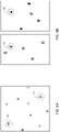
- co-occurrence of specific nucleic acid segments in a single cell can be determined.
- two specific nucleic acid segments co-occur in the sample cell if the fluorescence emission corresponding to the two nucleic acid segments is co-localized in sample fluorescence images.
- Co-location is readily identified if a fluorescence image shows emission corresponding to both of the two nucleic acid segments in the same image, as the fluorescence emission will arise from a common spatial location in the image.
Description
- This application claims priority to
U.S. Provisional Patent Application No. 62/747,785, filed on October 19, 2018 - This disclosure relates to the detection of co-occurring of nucleic acid segments, such as coding segments of cell surface receptors.
- Neoantigen-specific T-cells direct the anti-tumor immune response in tissue, and in turn, neoantigen specificity is mediated through surface-expressed T-cell receptors. T-cell receptors are heterodimers consisting of alpha and beta subunit pairs. Techniques exist for sequencing nucleic acid segments that code for both subunit pairs. However, identifying co-occurring pairs of coding segments from among a population of fragments extracted from a tissue sample remains a challenging problem.
-
WO 2016/176322 A1 describes methods and compositions that can be used to pair two sequences originating from a single cell, such as heavy and light chain antibody sequences. -
WO 2015/176162 A1 describes sequencing methods to study lymphocyte receptor chains. -
WO 2017/048614 A1 describes methods for generating or obtaining tumor-reactive T cells. -
WO 2012/048340 A2 describes methods and compositions for determining and/or monitoring the immune state of an individual. - Howie et al describe in Science Translational Medicine, vol. 7, no. 301, 19 August 2015, a high-throughput pairing of T cell receptor sequences.
- The present invention is defined by independent claim 1. The dependent claims depict additional embodiments of the invention.
- The present disclosure features methods for identifying co-occurring nucleic acid segments in a sample. The segment sequences can be used for the engineering of T-cells that express neoantigen-specific T-cell receptors, which are then infused into a patient for therapeutic applications. The methods can be used with a wide variety of samples, including fresh and fresh-frozen tissue sections, formalin-fixed, paraffin-embedded (FFPE) tissue sections, and substrate-mounted smears (e.g., blood-derived white blood cells such as peripheral blood mononuclear cells). Probes derived from a first sample or a first portion of a sample are hybridized to a second sample or a second portion of the sample, and detection of specific hybridization events associated with probes related to specific segment sequences is used to determine pairs of co-occurring segments.
- In one aspect, the disclosure features methods for identifying co-occurrence of nucleic acid segments in a nucleic acid sample from a specimen, the methods including: obtaining a nucleic acid sample from a specimen, where the specimen includes target immune cells, and where the nucleic acid sample features a plurality of nucleic acid fragments associated with expression of an antigen receptor molecule in the specimen; determining sequences of first and second nucleic acid segments in the nucleic acid fragments of the sample to generate a first set of sequences corresponding to the first nucleic acid segment and a second set of sequences corresponding to the second nucleic acid in the sample, where the first and second nucleic acid segments are coding segments of the antigen receptor molecule; generating a first set of probes from the first set of sequences, where each member of the first set of probes features an oligonucleotide corresponding to a different one of the first set of sequences linked to a detection moiety; generating a second set of probes from the second set of sequences, where each member of the second set of probes features an oligonucleotide corresponding to a different one of the second set of sequences linked to a detection moiety; exposing a detection sample obtained from the specimen to a member of the first set of probes and a member of the second set of probes; performing a hybridization analysis to determine whether the member of the first set of probes hybridizes to the detection sample, and to determine whether the member of the second set of probes hybridizes to the detection sample; and determining whether the first and second nucleic acid segments co-occur in a common cell of the specimen.
- The antigen receptor molecule can be a T-cell antigen receptor molecule. The first nucleic acid segment can be associated with an α-chain of the T-cell antigen receptor molecule, and the second nucleic acid segment can be associated with a β-chain of the T-cell antigen receptor molecule.
- The antigen receptor molecule can be a B-cell antigen receptor molecule. The first nucleic acid segment can be associated with a heavy chain of the B-cell antigen receptor molecule, and the second nucleic acid segment can be associated with a light chain of the B-cell antigen receptor molecule.
- The first and second nucleic acid segments can be located in a complementarity determining region 3 (CDR3) portion of the nucleic acid fragments.
- The nucleic acid sample can include genomic DNA. The nucleic acid sample can include total RNA. The nucleic acid sample can include nucleic acid molecules from tumor infiltrating lymphocytes.
- The specimen can include a fresh or frozen tumor tissue specimen including the target immune cells. Obtaining the nucleic acid sample from the specimen can include fixing and embedding the specimen in paraffin, excising a portion of the specimen, and extracting the nucleic acid sample from the excised portion of the specimen. The target immune cells can include tumor infiltrating lymphocytes.
- The sequences of the first and second nucleic acid segments can each include between 50 and 200 base pairs (e.g., between 75 and 150 base pairs). The first and second sets of sequences can each include at least 20 different sequences (e.g., at least 50 different sequences).
- The first set of sequences can include N different sequences, and generating the first set of probes can include, for each sequence of M of the different sequences that are expressed in highest abundance in the nucleic acid sample, generating a population of oligonucleotides corresponding to the sequence, and linking each member of the population of oligonucleotides to a detection moiety. M can be equal to N or less than N. M can be 48 or less (e.g., 24 or less).
- The oligonucleotides of the population can include DNA sequences. The DNA sequences can be complementary to the M different sequences. The oligonucleotides of the population can include RNA sequences. The RNA sequences can be antisense RNA sequences. The antisense RNA sequences can be complementary to RNA transcript sequences corresponding to the M different sequences.
- The oligonucleotides of the population can include modified nucleic acids. The modified nucleic acids can include peptide nucleic acids.
- The detection moiety can include biotin or a derivative thereof. The detection moiety can include a hapten. The detection moiety can include a fluorescent moiety. The detection moiety can include at least one chelated metal ion. The detection moiety can include a reactive moiety that reacts with an agent to generate chemiluminescence. The detection moiety can include a molecular barcode. The molecular barcode can include an oligonucleotide. Each member of the population can be linked to a common detection moiety that includes the same molecular barcode.
- Among the populations of nucleotides corresponding to the M different sequences, the common detection moiety can be unique to one population of oligonucleotides corresponding to only one of the M different sequences. The oligonucleotide can include a DNA sequence, and the DNA sequence can be linked to a 3' end of the member of the population of oligonucleotides.
- The detection sample can include a tissue section. The tissue section can include a formalin fixed, paraffin embedded tissue section. The detection sample can include a remaining portion of the specimen following extraction of a portion of the specimen to obtain the nucleic acid sample. The detection sample can include a smear of white blood cells. The white blood cells can include peripheral blood mononuclear cells (PBMCs).
- Exposing the detection sample to the members of the first and second sets of probes can include contacting the detection sample with a composition featuring all members of the first and second sets of probes, and removing members of the first and second sets of probes that do not hybridize to the detection sample from contact with the detection sample.
- The first set of probes can include J different types of probes, each of the J different types of probes corresponding to one of the first set of sequences, the second set of probes can include K different types of probes, each of the K different types of probes corresponding to one of the second set of sequences, each member of the first and second sets of probes can correspond to only one of the J different types of probes or to only one of the K different types of probes, each member of the first and second sets of probes includes a detection moiety featuring a molecular barcode that is unique to only one type of probe among the J and K different types of probes, and
the molecular barcode can include an oligonucleotide. - Performing the hybridization analysis can include: (a) exposing the detection sample to a set of detection probes, each member of the set of detection probes featuring an oligonucleotide sequence that hybridizes to a single type of molecular barcode, and a fluorescent moiety linked to the oligonucleotide sequence; (b) detecting fluorescence emission from members of the set of detection probes hybridized to molecular barcodes in the detection sample; (c) removing from the detection sample the detection probes that are hybridized to molecular barcodes in the sample; and (d) repeating steps (a)-(c) with additional sets of detection probes.
- The first set of probes can include J different types of probes, each of the J different types of probes corresponding to one of the first set of sequences, the second set of probes can include K different types of probes, each of the K different types of probes corresponding to one of the second set of sequences, and exposing the detection sample to the members of the first and second sets of probes can include contacting the detection sample with a composition featuring one of the J different types of probes and one of the K different types of probes, and removing from contact with the detection sample any probes of the composition that do not hybridize to the detection sample. Performing the hybridization analysis can include detecting the detection moiety linked to the one of the J different types of probes and detecting the detection moiety linked to the one of the K different types of probes.
- The detection moiety linked to the one of the J different types of probes can include biotin or a derivative thereof, and detecting the detection moiety can include binding the detection moiety to a detection probe, the detection probe featuring a moiety that binds to biotin or a derivative thereof and a fluorescent moiety, and detecting fluorescence emission from the fluorescent moiety following binding of the detection moiety to the detection probe. The moiety that binds to biotin or a derivative thereof can include at least one of avidin and streptavidin.
- The detection moiety linked to the one of the J different types of probes can include a hapten, and detecting the detection moiety can include binding the detection moiety to a detection probe, the detection probe featuring a moiety that binds to the hapten and a fluorescent moiety, and detecting fluorescence emission from the fluorescent moiety following binding of the detection moiety to the detection probe. The moiety that binds to the hapten can include at least one member selected from the group consisting of a protein, a polypeptide, a polysaccharide, or a liposome.
- The detection moiety linked to the one of the J different types of probes can include a fluorescent moiety, and detecting the detection moiety can include detecting fluorescence emission from the fluorescent moiety.
- The detection moiety linked to the one of the J different types of probes can include at least one chelated metal ion, and detecting the detection moiety can include liberating the chelated metal ion from the detection moiety, and detecting the liberated metal ion by mass spectrometry.
- The detection moiety linked to the one of the J different types of probes can include a reactive group, and detecting the detection moiety can include exposing the reactive group to an agent that reacts with the reactive group to generate chemiluminescence emission, and detecting the chemiluminescence emission following the reaction.
- Embodiments of the methods can also include any of the other features described herein, and can include combinations of any features, including those described in connection with different embodiments, except as expressly stated otherwise.
- Unless otherwise defined, all technical and scientific terms used herein have the same meaning as commonly understood by one of ordinary skill in the art to which this disclosure belongs. In case of conflict between the present specification and a reference mentioned herein, the present specification, including definitions, will control. In addition, the materials, methods, and examples are illustrative only and not intended to be limiting.
- The details of one or more embodiments are set forth in the accompanying drawings and the description below. Other features and advantages will be apparent from the description, drawings, and claims.
-
-
FIG. 1 is a flow chart showing a series of example steps for identifying co-occurrence of nucleic acid segments in a sample. -
FIG. 2 is a table showing example CDR3 sequences for a particular nucleic acid segment derived from nucleic acid fragments extracted from a sample. -
FIG. 3 is a schematic diagram of a nucleic acid segment probe. -
FIG. 4 is a schematic diagram of a detection probe. -
FIG. 5 is a schematic diagram of a system for obtaining fluorescence and chemiluminescence images indicating hybridization of nucleic acid segment probes in a sample. -
FIG. 6A is a schematic fluorescence image showing fluorescence emission corresponding to co-occurring nucleic acid segments in two sample cells. -
FIG. 6B is a set of two schematic fluorescence images showing fluorescence emission corresponding to the occurrence of different nucleic acid segments in a sample. - Like reference symbols in the various drawings indicate like elements.
- Chimeric antigen receptor (CAR) T-cell immunotherapies are available to treat a variety of immune diseases, and are readily applied to cancers in which circulating T-cells are extracted from a patient's blood. The extracted T-cells can be re-engineered for surface expression of CAR, replicated in large numbers for dosing, and infused into the patient to specifically target tumor cells. Commercial therapies exist, particularly for lymphomas, and target surface antigens such as CD19 which are common in heme malignancies.
- Therapies targeting solid tumors have been more challenging to develop, in part because the cells of such tumors typically express fewer surface markers. However, neoantigens are generated in such tumors through somatic mutations, and may be expressed on the surface of solid tumor cells. Neoantigen specific T-cells direct the patient's immune anti-tumor response. Accordingly, a promising approach to immunotherapies for solid tumors involves re-engineering of T-cells to express surface antigen receptors that interact with a patient's neoantigens. Cloning of re-engineered T-cells and infusion into the patient may yield therapeutic outcomes that are more effective than other, more conventional treatments.
- Neoantigen specificity is mediated through T-cell receptors, which are heterodimers consisting of alpha (α) and beta (β) subunit pairs. Commercial methods exist to determine nucleic acid sequences that code specifically for α and β chain fragments in T-cell receptors. However, for a sample extracted from tumor tissue and containing a large number of α and β chain coding nucleic acid fragments, sequencing leads to a distribution of coding sequences for both chain fragments, with no indication of which pairs of coding sequences (i.e., α and β coding sequence pairs) yield functional T-cell receptors.
- Identification of T-cell receptors with anti-tumor functionality is a prerequisite to engineered T-cell therapies. By sequencing nucleic acids extracted from such samples and identifying co-occurring nucleic acid coding segments for α and β receptor chain fragments among the populations of both α and β coding segments, specific functional T-cell receptors can be identified. The co-occurrence information for specific α and β coding segments can then be used to engineer populations of T-cells with functional neoantigen receptors, and therefore, anti-tumor functionality.
- Next-generation sequencing methods can readily be used to obtain T-cell receptor α and β coding sequences. However, identifying which of tens or hundreds of α coding segments obtained from a typical sample functionally pairs with which of tens or hundreds of β coding segments obtained from the same sample remains a challenging problem. In addition, certain existing analytical methods are not well adapted for use with solid tumor tissues, and in particular, formalin-fixed, paraffin-embedded samples, which is the preferred clinical format for most tumor specimens.
- This disclosure features methods for identifying the co-occurrence of nucleic acid segments in a biological sample. The methods can be used with a wide variety of different samples, including tissue sections and blood smears. Sample-specific probes are generated from nucleic acids extracted from the sample, and the probes are hybridized to the sample serially or in parallel. Detection of hybridization events associated with particular, individual probes provides information about which nucleic acid segments are present in the sample, and therefore, which polypeptide chain fragments are co-expressed in the sample. Where the polypeptide chain fragments are paired chain fragments corresponding to a functional receptor, the co-expression information leads directly to identification of functional receptors in the sample.
- As described above, an important application of the methods described above is in providing engineered T-cell therapies targeting solid tumors. For such applications, the methods described herein can be applied to the identification of co-occurring nucleic acid segments that code for co-expressed α and β chain fragments of functional T-cell neoantigen receptors in a sample. The discussion that follows describes such applications in detail to provide an illustrative example of the methods.
- However, the methods described herein are not limited to co-occurrence analysis of nucleic acid segments that code for α and β chain fragments of T-cells. To the contrary, the methods can be used for co-occurrence analysis of nucleic acid segments that code for a variety of polypeptides (or fragments thereof) in many different types of cells and samples. In some embodiments, for example, the methods can be applied to determine co-occurrence of nucleic acid segments that code for other polypeptides in T-cells, including other chain fragments.
- In certain embodiments, the methods can be applied to determine co-occurrence of nucleic acid segments that code for polypeptides or fragments thereof in other types of cells. As an example, B-cells can be engineered to provide immunoglobulin therapies, provided that functional B-cells can be identified. B-cells contain paired heavy and light chain fragments, and identifying which B-cells are functional for purposes of engineering and cloning B-cells for therapeutic purposes effectively involves determining co-occurring nucleic acid segments in a sample that code for the heavy and light chain fragments. With this information, B-cells that contain the co-occurring nucleic acid segments can be engineered, and will exhibit therapeutic efficacy.
- Similarly, B-cells expressing a specific antigen-specific antibody protein can be used to mass produce the antibody in sufficient quantities that the antibody can be used delivered as a therapeutic, or it can be linked to a separate therapeutic moiety, which can be a small molecule or another therapeutic immune cell that is used to target that moiety to a tumor or other cell type of interest.
-
FIG. 1 is a flow chart 100 showing a set of example steps (e.g., a workflow) for identifying co-occurrence of nucleic acid segments. In afirst step 102, a nucleic acid sample is obtained from a specimen. The specimen is typically a tissue sample removed from an organism, e.g., by biopsy. A suitable specimen can be obtained from a wide variety of organisms, and can for example be a human specimen, or a specimen removed from any other mammalian or non-mammalian organism, including a mouse, a rat, an avian organism, and a simian organism. - Specimens can generally be of various types. In some embodiments, for example, the specimen is a fresh sample of tissue. In certain embodiments, the specimen is a fresh-frozen tissue sample. The specimen can also be a tissue that is fixed in formalin (or another fixative), embedded in a structural medium, and then sectioned (e.g., in a microtome) to form a tissue section (e.g., a formalin-fixed, paraffin-embedded (FFPE)) tissue section.
- The specimen can be derived from any of a variety of different types of tissue. In some embodiments, for example, the specimen is extracted from tumor tissue in a patient. The tumor can be a solid tumor, accessed via biopsy or dissection during a surgical or post-surgical operation.
- In certain embodiments, the specimen corresponds to one or more cells circulating in a body fluid. For example, the specimen can include one or more white blood cells derived from a patient blood sample. Suitable white blood cells include, but are not limited to, peripheral blood mononuclear cells (PBMCs), for example.
- After a suitable specimen has been obtained, a nucleic acid sample is extracted from the specimen. In some embodiments, for example, where the specimen is a tissue section, the specimen can be stained with one or more chromogenic stains and imaged to identify different portions of the specimen. For a specimen that includes tumor tissue, chromogenic staining can be used to reveal tumor margins and identify cells suitable for nucleic acid extraction. More generally, any of a variety of techniques can be used to identify cells in the specimen that are suitable for nucleic acid extraction.
- Once a suitable cell (or cells) have been identified, the cell (or cells) is/are isolated from the specimen, and nucleic acids from the cell (or cells) are extracted. Various techniques can be used to isolate a cell or cells from a specimen. For example, in some embodiments, one or more cells are isolated from the specimen using laser capture microdissection. Methods for performing laser capture microdissection are disclosed, for example, in Emmert-Buck et al., Science 274: 998-1001 (1996), and in Espina et al., Expert Rev. Mol. Diagn. 7(5): 647-657 (2007).
- Various types of cells (e.g., target immune cells) can be isolated from the specimen for nucleic acid extraction. In certain embodiments, for example, the isolated cells are tumor-infiltrating lymphocytes, which are functional lymphocytes that are responsible for the immune response to tumor cells in the specimen.
- Next, a nucleic acid sample is extracted from one of the isolated cells. Nucleic acid extraction can be performed using a variety of methods, depending upon the nature of the nucleic acid. For extraction of DNA, techniques such as cesium chloride gradient centrifugation and solid phase extraction can be used. Suitable examples of such methods include, but are not limited to, those described in Ali et al., Biomed. Res. Int. 2017: 9306564 (2017). For extraction of RNA, techniques such as guanidium thiocyanate-phenol-chloroform extraction can be used. Examples of RNA extraction and isolation methods are described in, for example, Doleshal et al., J. Mol. Diagn. 10(3): 203-211 (2008), in Peirson et al., Methods Mol. Biol. 362: 315-327 (2007), and in Chomcyznski et al., Nat. Protocols 1(2): 581-585 (2006).
- As is evident from the foregoing discussion, the nucleic acid sample extracted from the specimen can be a DNA sample or an RNA sample. In some embodiments, the nucleic acid sample corresponds to genomic DNA extracted from the nucleus of one or more cells such as a tumor infiltrating lymphocyte. In certain embodiments, the nucleic acid sample corresponds to total RNA isolated from one or more cells isolated from the specimen, such as a tumor infiltrating lymphocyte.
- While RNA can, in certain circumstances, be more difficult to isolate due to the presence of hardy RNAses in cells which degrade isolated RNA strands, in some embodiments RNA provides certain advantages as a nucleic acid sample. First, the relative abundance of RNA in the isolated cells may reflect more accurately the underlying biology of the T-cells, where the most activated, tumor-specific T-cells may express higher amounts of neoantigen-specific receptor transcripts. For purposes of cloning a therapeutically-effective population of T-cells from co-occurrence information, this inherent mapping of relative T-cell activation to RNA abundance may yield co-occurrence information that can be used to engineer T-cell populations with improved efficacy.
- Second, in some embodiments, depending of the portion of the nucleic acid sample that is sequenced, it may be easier to obtain the sequence information from an RNA sample than a corresponding DNA sample. For the segments of the complementarity determining region 3 (CDR3) region that code for the α and β chains of T-cell receptors, there is a 5 kb intron between the hypervariable diversity/joining and constant gene that is spliced out in the RNA, which increases the chances of capturing the full receptor coding sequence including the CDR3 region by sequencing RNA rather than genomic DNA. As will be explained in greater detail below, sequencing the CDR3 region can be particularly relevant for identifying co-occurrence of nucleic acid segments that code for functional T-cell receptors.
- Obtained from the specimen in
step 102 ofFIG. 1 , the nucleic acid sample includes multiple nucleic acid fragments that are associated with expression of a protein or polypeptide of interest in the specimen. As discussed previously, in some embodiments, the expressed polypeptide is a functional T-cell neoantigen receptor, and in particular, the α and β chains of the neoantigen receptor. Accordingly, the nucleic acid fragments correspond to partial or complete coding fragments for the α and β chains of the neoantigen receptor. - To begin determining whether two nucleic acid segments co-occur in the nucleic acid sample, the sequences of the two nucleic acid segments within each of the sample fragments are determined in
step 104. In general, any two nucleic acid segments within each of the sample's nucleic acid fragments can be tested for co-occurrence. However, certain segments may be more relevant than others in some circumstances. - Nucleic acid sequences that code for portions of the α and β chains of the neoantigen receptor can be found in complementarity determining regions 1, 2, and 3 (CDR1, CDR2, CDR3), among others. Among CDR1, CDR2, and CDR3, region CDR3 in particular is hypervariable due to natural VDJ recombination in T-cells and B-cells. In these cells, variable (V), diversity (D), and joining (J) gene segments are randomly assembled to generate unique antigen (or neoantigen) receptors that can recognize different antigens and neoantigens. When the T-cell or B-cell successfully recognizes an antigen or neoantigen, a signal transduction occurs and the gene segments that code for the receptor enter the immunological memory.
- Due to hypervariable diversity in the CDR3 region, nucleic acid sequences in this region are almost absolutely unique. In other words, each cell (e.g., T-cell or B-cell) will have a different nucleic acid sequence in this region, which functions as a type of molecular barcode for the cell. Accordingly, by comparing nucleic acid segments within the CDR3 region of nucleic acid fragments, co-occurrence of two particular segments can readily be determined with comparatively little chance of error due to non-uniqueness of segment sequences.
- To determine the sequences of the two nucleic acid segments of interest in
step 104, a variety of published and commercially-available next-generation sequencing methods can be used. Suitable sequencing methods include, but are not limited to, massively parallel signature sequencing, polony sequencing, 454 sequencing, Illumina sequencing, ion torrent sequencing, SOLiD DNA sequencing technology, and DNA nanoball sequencing. Aspects of suitable sequencing methods are described, for example, in Rajesh et al., Current Developments in Biotechnology and Bioengineering, pp. 143-158 (2017). - Sequencing nucleic acid segments of nucleic acid fragments derived from multiple cells yields a distribution of segment sequences, each present at a different frequency in a nucleic acid sample.
FIG. 2 is a table showing a portion of a distribution of nucleic acid segment sequences obtained from different nucleic acid fragments in a sample. Each of the sequences inFIG. 2 corresponds to the same portion of different nucleic acid fragments (e.g., a portion of the CDR3 region in multiple fragments) in the sample. The left column of the table shows each of the segment sequences, and the right column shows the frequency of each of the sequences in the nucleic acid sample. - Sequencing the two nucleic acid segments in each of the nucleic acid fragments of the sample yields two sets of sequences: a first set of sequences corresponding to the first nucleic acid segment of each of the fragments, and a second set of sequences corresponding to the second nucleic acid segment of each of the fragments. Within each set of sequences, different sequences are present at different frequencies, with the most common sequences corresponding to most commonly present cells in the specimen.
- The number of base pairs in the first nucleic acid segment, in the second nucleic acid segment, or in both the first and second nucleic acid segments, can generally be selected as desired. In some embodiments, for example, the number of base pairs is between 50 and 200 (e.g., between 50 and 190, between 60 and 190, between 60 and 180, between 70 and 180, between 70 and 170, between 70 and 160, between 75 and 150, between 80 and 140, between 80 and 130, between 80 and 120).
- The first set of sequences, the second set of sequences, or both the first and second sets of sequences can generally include any number of different sequences, depending upon the number of specimen cells from which the nucleic acid sample is derived. In some embodiments, for example, the number of different sequences is 10 or more (e.g., 12 or more, 15 or more, 20 or more, 30 or more, 40 or more, 45 or more, 48 or more, 50 or more, 60 or more, 70 or more, 80 or more, 90 or more, 100 or more, or even more).
- Returning to
FIG. 1 , in thenext step 106, a first set of probes are generated from the first set of sequences determined instep 104. Optionally, step 104 can also include ordering the first set of sequences in order of frequency, with the most frequently occurring sequences earlier. Further still, in certain embodiments, sequences in the first set that occur less frequently relative to other sequences can be eliminated from the first set, so that the first set includes only the N different, most-commonly occurring sequences. N can be, for example, 10 or more (e.g., 20 or more, 25 or more, 30 or more, 35 or more, 40 or more, 45 or more, 48 or more, 50 or more, 60 or more, 70 or more, 80 or more, 90 or more, 100 or more, or even more). - The first set of sequences (optionally restricted to only the N most frequent sequences, as described above) is then used to generate a first set of probes for the specimen. To generate the first set of probes, M different sequences among the different (or N different) sequences of the first set (e.g., the M most abundant sequences in the first set) can be used. M can be equal to or less than N, and in some embodiments, can be 48 or less (e.g., 40 or less, 30 or less, 24 or less, 20 or less, 10 or less).
- To generate the first set of probes, a population of oligonucleotides is synthesized for each one of the M different sequences. Each of the oligonucleotides in a given population corresponds to only one of the M different sequences, and is linked to a detection moiety to form a probe.
FIG. 3 is a schematic diagram showing a probe, which includes anoligonucleotide 302 linked to adetection moiety 304. Just asoligonucleotide 302 corresponds to only one of the M different sequences,detection moiety 304 also corresponds to only one of the M different sequences. Accordingly, after cycling through each of the M different sequences, M different populations of probes are obtained, such that each probe population corresponds to only one of the M different sequences, and the probes of the population include anoligonucleotide 302 and adetection moiety 304 that are unique to only one of the M different sequences. - In general, the oligonucleotides synthesized for probe generation can correspond to different types of nucleic acids. For example, in some embodiments, the oligonucleotides can include DNA sequences (e.g., DNA sequences that are complementary to each of the M different sequences described above). In certain embodiments, the oligonucleotides can include RNA sequences (e.g., RNA antisense sequences that hybridize to M different RNA transcript sequences). In some embodiments, the oligonucleotides can be synthesized and/or modified nucleic acid-containing species, such as peptide nucleic acids (PNAs) and/or xeno nucleic acids (XNAs).
- A variety of different methods can be used to synthesize the oligonucleotides from the M different sequences. Examples of such methods include, but are not limited to phosphoramidite-based methods, H-phosphonate-based methods, and phosphotriester methods. Methods for oligonucleotide and peptide nucleic acid synthesis are described, for example, in Herdewijn, P. (ed.), Oligonucleotide Synthesis, Springer (2005), and in Braasch et al., "Synthesis and purification of peptide nucleic acids," Current Protocols in Nucleic Acid Chemistry, Chapter 4 (2002).
- Following synthesis of the oligonucleotide populations, each
oligonucleotide 302 is linked to adetection moiety 304 to form a probe. Thedetection moiety 304 facilitates hybridization detection of each probe. In general, the detection moiety can correspond to a molecular fragment that generates a detectable signal, or alternatively, to a molecular fragment that binds to another fragment that generates a detectable signal. A wide variety of different detection moieties can be used to form the probes described above. - In some embodiments,
detection moiety 304 includes biotin or a derivative thereof. Methods for linking biotin and its derivatives tooligonucleotide 302 include, for example, binding biotin substituents to allylamino residues on functionalized oligonucleotides via reaction with N-biotinyl-6-aminocaproic acid N-hydroxysuccinimide ester, extension of RNA with poly(A) polymerase, and reaction of an N-hydroxysulfosuccinimide ester linked to biotin with a primary oligonucleotide amine. Additional methods for linking biotin and derivatives thereof tooligonucleotide 302 are described for example in Cook et al., Nucleic Acids Res. 16(9): 4077-4095 (1988), in Moritz et al., RNA, 20(3): 421-427 (2014), and in Soukup et al., Bioconj. Chem. 6: 135-138 (1995). - In certain embodiments,
detection moiety 304 includes a hapten. Suitable methods for conjugating haptens to oligonucleotide 302 (which is sometimes referred to as hapten labeling) include phosphoramidite-based methods, as described for example in Luehrsen et al., J. Histochem. Cytochem. 48(1): 133-145 (2000). - In some embodiments,
detection moiety 304 includes a fluorescent moiety. A wide variety of different fluorescent moieties can be used, including, but not limited to: xanthene-based fluorophores such as fluorescein, rhodamine, Oregon green, eosin, and Texas red; cyanine-based fluorophores such as cyanine, indocarbocyanine, oxacarbocyanine, thiacarbocyanine, and merocyanine; squaraine-based fluorophores, including squaraine rotaxane derivatives; naphthalene-based fluorophores, coumarin-based fluorophores, oxadiazole-based fluorophores, such as pyridyloxazole, nitrobenzoxadiazole, and benzoxadiazole; anthracene-based fluorophores such as anthraquinones, pyrene-based fluorophores; oxazine-based fluorophores such as Nile red, Nile blue, cresyl violet, and malachite green; and tetrapyrrole-based fluorophores such as porphin, phthalocyanine, and bilirubin. Methods for linking fluorophores tooligonucleotide 302 are described for example in Proudnikov et al., Nucl. Acids Res. 24(22): 4535-4542 (1996). - In certain embodiments,
detection moiety 304 includes at least one chelated metal ion. Probes with chelated metal ions can be detected by liberating the metal ion from the probe, and detecting the liberated ion using mass spectrometry techniques. Suitable types of metal ions include, but are not limited to, lanthanide metal ions. Chelating moieties for such ions can be implemented as one or more metal-chelating groups bound to a polymer backbone. Examples of such chelating moieties and methods for preparing oligonucleotide-metal chelates are described in Majonis et al., Anal. Chem. 82(21): 8961-8969 (2010), and in Kwiatskowski et al., Nucl. Acids. Res. 22(13): 2604-2611 (1994). - In some embodiments,
detection moiety 304 includes a molecular barcode. In general, a molecular barcode is an oligonucleotide having a specific sequence that is unique to a particular population of probes. Each of the probes in a particular population (that corresponds to a single one of the M sequences) includes the same molecular barcode. The molecular barcode is not linked to oligonucleotides in any of the other populations, however. In this manner, the probes of each population contain a common molecular barcode that is unique among the different probe populations. - The oligonucleotide that functions as the molecular barcode can generally be a DNA sequence, an RNA sequence, or a modified nucleic acid sequence such as a peptide nucleic acid sequence. The oligonucleotide can include any number of bases or base pairs (e.g., between 5 and 200, between 10 and 200, between 15 and 200, between 20 and 200, between 20 and 180, between 20 and 150, between 30 and 150, between 40 and 150, between 50 and 150, between 50 and 100, between 10 and 50, between 10 and 40, between 10 and 30, between 20 and 50, between 30 and 50).
- In certain embodiments, CODEX® barcodes - each of which corresponds to a unique oligonucleotide sequence - can be linked to oligonucleotide 302 (e.g., at the 3' end of oligonucleotide 302). CODEX® barcodes are available from Akoya Biosciences (Menlo Park, CA). Methods for linking these barcodes to oligonucleotides are described for example in Goltsev et al., Cell 174(4): 968-981 (2018), and in
U.S. Patent No. 9,909,167 - In some embodiments, detection moiety 403 includes a reactive moiety that generates chemiluminescence. Specifically, the reactive moiety reacts with a second substance, introduced during a detection step, to generate luminescence that can be detected to identify the presence of the probe. A wide variety of different reactive moieties can be linked to
oligonucleotide 302 to generate a chemiluminescent probe. Examples of such reactive moieties include, but are not limited to, luminol and derivatives thereof. Horseradish peroxidase (HRP) catalyzes the conversion of luminol to 3-aminophthalate. When performed in the presence of an enhancer, chemiluminescence is readily observed and permits detection of extremely low probe concentrations. Methods for linking reactive moieties for chemiluminescence detection to oligonucleotides are described, for example, in Khan et al., Appl. Biochem. Biotechnol. 173(2): 333-355 (2014). - The M probe populations generated as described above form a first set of probes that correspond to the first nucleic acid segment. Following generation of the first set of probes, a second set of probes is generated in
step 108 that correspond to the second nucleic acid fragment. Methods for generating the second set of probes instep 108 can generally correspond to any of the methods described above for forming the first set of probes instep 106, and therefore are not repeated. After completion ofstep 108, a first set of probes corresponding to the first nucleic acid segment (e.g., a segment that codes for a portion of an α-chain of a T-cell receptor) and a second set of probes corresponding to the second nucleic acid segment (e.g., a segment that codes for a portion of a β-chain of a T-cell receptor) have been generated. - Next, in
step 110, a detection sample is exposed to one or more members of the first and second sets of probes. Exposure to the probe sets can be performed in different ways, depending upon the nature of the hybridization analysis that is subsequently performed to determine which of the probes hybridize to the detection sample. - In some embodiments, the detection sample is exposed to the probes in a "pairwise" manner. The detection sample is exposed to a population of probes corresponding to one of the M sequences for the first nucleic acid segment, and to a population of probes corresponding to one of the M sequences for the second nucleic acid segment. Thus, the sample is exposed to only two different types of probes at once (e.g., one corresponding to an α-chain coding fragment and one corresponding to a β-chain coding fragment). Following the hybridization analysis (discussed further below), the two different types of probes are either removed from (e.g., via dehybridization) or quenched in the detection sample and the detection sample is re-used by exposing the detection sample to another pair of different probe populations corresponding to the first and second nucleic acid segments respectively, or a new detection sample is obtained and exposed to the next pair of probe populations. The pairwise exposure cycle is repeated until all pairwise combinations of the probe populations corresponding to the first and second nucleic acid segments have been hybridized and analyzed.
- In certain embodiments, the detection sample is exposed to the probes in a "serial" manner. That is, the detection sample is exposed to a population of only one type of probes, corresponding to one of the M sequences for either the first nucleic acid segment or the second nucleic acid segment. Thus, the sample is exposed to only one type of probe at once. Following hybridization analysis, the type of probe is either removed from (e.g., via dehybridization) or quenched in the detection sample and the detection sample is re-used by exposing the detection sample to another single probe population corresponding to one of the first and second nucleic acid segments, or a new detection sample is obtained and exposed to the next one of the probe populations. The single population exposure cycle is repeated until all probe populations corresponding to the first and second nucleic acid segments have been individually hybridized and analyzed.
- In some embodiments, the detection sample is exposed to the probes in a "pooled" manner. In other words, the detection sample is exposed to populations of more than one type of probes corresponding to the more than one of the M sequences for the first nucleic acid segment, and/or populations of more than one type of probes corresponding to more than one of the M sequences for the second nucleic acid segment. Following hybridization analysis, the probes are removed from (e.g., via dehybridization) or quenched in the detection sample and a new combination of different types of probes is used to expose the detection sample. Alternatively, a new detection sample can be obtained and exposed to the new combination of different types of probes. The exposure cycles are repeated until the one or more detection samples have been exposed to all combinations of the different probe types corresponding to the first and second nucleic acid segments, so that co-occurrence can be identified for each pair of different sequences corresponding to the first and second nucleic acid segments.
- In some embodiments, the detection sample is exposed to the first and second sets of probes in a fully pooled manner. In other words, populations of each of the different types of probes corresponding to the first and second nucleic acid segments are combined and hybridized to the detection sample simultaneously. By using a fully pooled exposure strategy, only a single hybridization cycle is used to evaluate co-occurrence of all pairs of nucleic acid segment sequences, resulting in a considerable simplification of the workflow and corresponding reduction in assay time.
- The detection sample is generally obtained from the same specimen as the nucleic acid sample described in
step 102. In some embodiments, the detection sample corresponds to a remaining portion of a sample (e.g., a FFPE tissue section) from which the nucleic acid sample has been excised using a technique such as laser capture microdissection. In certain embodiments, the detection sample corresponds to a separate tissue section obtained from the specimen, and can be a fresh section, a fresh-frozen section, or a FFPE tissue section. - Alternatively, in some embodiments, the detection sample is a smear of cells obtained from a body fluid. As an example, the detection sample can correspond to a blood smear mounted on a substrate and including white blood cells (e.g., peripheral blood mononuclear cells).
- Returning to
FIG. 1 , interleaved with the exposure step instep 110, a hybridization analysis is performed instep 112 to determine which of the different types of probes hybridize to the detection sample. In general, the first step in the hybridization analysis includes washing away unhybridized probes from the detection sample, so that such probes do not generate measurement signals. After this washing step, the nature of the hybridization analysis depends upon the nature of the detection moieties that are present in the probes. - The hybridization analysis determines which of the probes to which the detection sample was exposed hybridized to the detection sample. Accordingly, the hybridization analysis involves detecting the hybridized detection probes, and more specifically, detecting the detection moieties of the hybridized detection probes. The method by which the detection moieties are detected depends upon the nature of the detection moieties.
- As discussed above, in some embodiments,
detection moiety 304 is directly detectable. For example, wheredetection moiety 304 includes a fluorescent moiety, probes hybridized to the detection sample can be detected directly by measuring fluorescent emission from the detection sample, as will be discussed in greater detail below. Wheredetection moiety 304 includes a chelated metal ion, probes hybridized to the detection sample can be detected directly by liberating the metal ion, and detecting the metal ion using mass spectrometry techniques. Methods for mass spectrometry-based detection of metal ion-labeled samples are described for example in Keren et al., "MIBI-TOF: A multiplexed imaging platform relates cellular phenotypes and tissue structure," Sci. Adv. 5(10): eaax5851 (2019), and in Angelo et al., Nat. Medicine 20: 436-442 (2014). - In contrast, in certain embodiments,
detection moiety 304 is not directly detectable, and a detection probe is conjugated todetection moiety 304 to permit probe hybridization to be detected.FIG. 4 is a schematic diagram showing an example structure of adetection probe 400.Probe 400 includes abinding group 402 and alabel 404. In general,label 404 includes a fluorescent moiety that generates fluorescence emission, which is detected to identify probe hybridization to the detection sample. Any of the fluorescent moieties and their derivatives discussed above can generally be used inlabel 404. - The nature of
binding group 402 depends on the nature of theprobe detection moiety 304. In embodiments wheredetection moiety 304 includes biotin or a derivative thereof, binding group can include a moiety that binds to biotin or a derivative thereof. Examples of such moieties include, but are not limited to, an avidin- or streptavidin-based binding group that conjugates biotin to linkdetection probe 400 todetection moiety 304. - In embodiments where
detection moiety 304 includes a hapten, bindinggroup 402 can include a moiety that binds to the hapten. Examples of suitable moieties include, but are not limited to, proteins, polypeptides, polysaccharides, and liposomes. - In embodiments where
detection moiety 304 includes a reactive moiety that generates chemiluminescence, theprobe detection moiety 304 is typically not conjugated to adetection probe 400. Instead,detection moiety 304 is exposed to an agent that reacts with the reactive moiety, generating detectable chemiluminescence in the detection sample. A variety of different agents can be used, depending upon the nature of the reactive moiety. For example, where the reactive moiety is luminol-based, the agent can include an enzyme such as horseradish peroxidase that reacts with luminol to generate chemiluminescence. - In embodiments where
detection moiety 304 is an oligonucleotide such as a molecular barcode, bindinggroup 402 can be an oligonucleotide that is at least partially complementary withdetection moiety 304, and hybridizes todetection moiety 304. The extent of complementarity betweendetection moiety 304 andbinding group 402 can be 70% or more (e.g., 80% or more, 85% or more, 90% or more, 95% or more, 97% or more, 99% or more, or even 100%). - In embodiments where
detection moiety 304 is a CODEX® barcode (obtained from Akoya Biosciences),detection probe 400 can correspond to a complementary CODEX® Reporter consisting of a dye-labeled reporter sequence. Hybridization of the CODEX® Reporter to thedetection moiety 304 yields a conjugate probe structure from which fluorescence emission can be detected to identify hybridization of specific probes to the detection sample. - In addition to CODEX® Reporters,
detection probe 400 can correspond more generally to a wide variety of fluorescence in-situ hybridization (FISH) probes. Such probes generally include anoligonucleotide binding group 402, and a conjugated fluorophore. Measurement of fluorescence emission from the fluorophore reveals hybridization of a probe conjugated to a corresponding FISH probe. In certain embodiments, FISH detection probes can include adetection probe 400 that includes anoligonucleotide binding group 402 and a multiplicity of coupling groups. Suitable coupling groups include, for example, antigens. After the FISH detection probe has hybridized to thedetection moiety 304, multiple amplification probes each consisting of a fluorophore conjugated to complementary coupling group are introduced and bound to the FISH detection probe. Suitable complementary coupling groups include antibodies, for example. In this manner, an amplified FISH detection probe can be hybridized to a sample-hybridized nucleic acid segment probe, increasing the intensity of fluorescence emission that is indicative of hybridization of the nucleic acid segment probe. - The nucleic acid character of
binding group 402 depends on the nucleic acid character ofdetection moiety 304. For example, whendetection moiety 304 is an oligonucleotide with a double-stranded DNA sequence, bindinggroup 402 corresponds to a complementary single-stranded DNA sequence.Detection moiety 304 can be denatured prior to hybridization ofbinding group 402 to one of the double strands ofdetection moiety 304. Whendetection moiety 304 is a single-stranded DNA sequence, hybridization ofbinding group 402 can be directly performed. Whendetection moiety 304 is an oligonucleotide with an RNA sequence, bindinggroup 402 corresponds to an antisense RNA sequence that is complementary to at least a portion of the RNA sequence. - As described above, when
detection moiety 304 reacts to yield chemiluminescence, or whendetection moiety 304 includes (or is linked to adetection probe 400 that includes) a fluorescent moiety, light emission from the detection sample is measured to determine which probes are hybridized to the detection sample.FIG. 5 shows a schematic diagram of a system 500 for detecting light emission (e.g., chemiluminescence, fluorescence emission) from a detection sample. System 500 includes alight source 502, anoptical assembly 504, astage 506 supporting adetection sample 508, a wavelength-dependent reflection element 510, afilter assembly 512, and adetector 514. To detect chemiluminescence fromdetection sample 508,light source 502,reflection element 510, and filterassembly 512 can optionally be omitted from system 500. -
Detector 514 is an imaging detector such as a CCD detector, and obtains one or more images of light emitted fromdetection sample 514. Chemiluminescence is generated directly by probes in the detection sample and imaged bydetector 514. To measure fluorescence from probes in the detection sample, light source 502 (e.g., a multi-diode array) generates excitation light in a selected wavelength band that passes throughreflective element 510 and is directed byoptical assembly 504 ontodetection sample 508. The excitation light stimulates fluorescence emission from hybridized probes indetection sample 508, and the emitted fluorescence is reflected byreflective element 510, passes throughfilter assembly 512, and is detected bydetector 514. Note that in the following discussion, fluorescence measurement is discussed by way of example, but it should be understood that the methods described apply equally to detection of probe hybridization in the detection sample by measurement of chemiluminescence. - The nature of image acquisition by
detector 514 depends on the nature of the hybridization analysis instep 112. In general, images of fluorescence emission in a single wavelength band or in multiple wavelength bands can be acquired by system 500. In some embodiments, where the serial probe exposure strategy is used as discussed above, and optionally where paired or pooled probe exposure strategies are used, detection sample images showing fluorescence emission in a single wavelength band are acquired. Thus, for example, in a paired exposure strategy with different fluorescent labels attached to each of two different types of probes hybridized to the sample, two fluorescence images of the sample are obtained, each image corresponding to fluorescence emission in a different wavelength band associated with one of the fluorescent labels. For a pooled probe exposure strategy in which Q different fluorescent labels are each attached to one of Q different and corresponding hybridized sample probe types, Q fluorescence images of the detection sample are obtained, each of the Q images corresponding to emission from a different one of the Q probe types. - In certain embodiments, where paired or pooled exposure strategies are used so that the detection sample includes multiple different types of hybridized probes, multi-band fluorescence images can be obtained showing fluorescence emission in multiple spectral bands, each corresponding to emission from a different one of the unique fluorescent labels attached to the different types of probes.
- In some embodiments, as discussed above, the probes in each population can include a
detection moiety 304 that corresponds to a molecular barcode such as a CODEX® barcode, and the probe populations can be pooled to form a composition that includes probes corresponding to all of the sequences corresponding to the first and second nucleic acid segments. The fully pooled probes can then be simultaneously hybridized to the detection sample in a single hybridization step. To facilitate probe hybridization detection when the probe detection moieties correspond to CODEX® barcodes, CODEX® Reporters can be used in multiplexed fashion to perform multiple detection cycles, each of which yields a sample fluorescence image in which emission from multiple CODEX® Reporters is measured. - For example, in a first detection cycle, multiple (e.g., 3, 4, 5, or even more; generally k) CODEX® Reporters are hybridized to k nucleic acid segment probes hybridized to the detection sample. A fluorescence image of the detection sample is obtained showing fluorescence emission in k different spectral bands, each of which corresponds to a different one of the k CODEX® Reporters, and therefore, to a different one of the hybridized nucleic acid segment probes. After imaging, the k CODEX® Reporters are washed out of the detection sample, and new analysis cycle in which a new set of k different CODEX® Reporters is hybridized to a different set of k nucleic acid segment probes hybridized to the detection sample begins. Another fluorescence image showing emission in k different spectral bands, each corresponding to a different one of the new set of k nucleic acid segment probes is obtained, before the new set of k CODEX® Reporters is washed out of the detection sample. The CODEX® hybridize-image-wash cycles continue until hybridization of each of the different probe types in the detection sample is analyzed.
- For each different type of probe, the extent of hybridization of the probe to the detection sample is determined by the presence of fluorescence emission corresponding to the probe in a sample image. If the probe exhibits no fluorescence emission, then the probe did not hybridize to the detection sample (and was presumably washed out of the sample prior to imaging). If the probe exhibits fluorescence emission in a sample image, then the probe is hybridized to the sample. Since each type of probe is associated with one of the sequences corresponding to either the first or second nucleic acid segment, hybridization of a particular type of probe to the detection sample indicates that the nucleic acid segment corresponding to the sequence associated with the hybridized probe occurs in the detection sample. Thus, by imaging fluorescence emission from each of the different types of probes, the occurrence of each type of nucleic acid segment sequence in the detection sample can be assessed.
- It should be further noted that while the foregoing and subsequent discussion involves fluorescence measurements for identifying nucleic acid segment occurrence in a sample, other methods as described herein (e.g., detection of liberated metal ions, chemiluminescence) can also be used, as the analytical workflow is analogous for non-fluorescence images.
- Returning to
FIG. 1 , once the hybridization of different probes to the detection sample has been determined and the occurrence of specific nucleic acid segment sequences has been identified, co-occurrence of specific nucleic acid segments in a single cell can be determined. In general, for a particular sample cell, two specific nucleic acid segments co-occur in the sample cell if the fluorescence emission corresponding to the two nucleic acid segments is co-localized in sample fluorescence images. Co-location is readily identified if a fluorescence image shows emission corresponding to both of the two nucleic acid segments in the same image, as the fluorescence emission will arise from a common spatial location in the image. - For example,
FIG. 6A is a schematic fluorescence image showing fluorescence emission corresponding to two different nucleic acid segment probes. Atlocations locations - Where the fluorescence emission from the two nucleic acid segments is shown in different sample images, the spatial locations of the fluorescence emission in the different sample images is measured. If the spatial locations in the different images correspond to within an error threshold for the assay, the two nucleic acid segments are deemed to co-occur in cell. If the difference in spatial locations is too large, the two nucleic acid segments do not co-occur in the cell.
- As an example,
FIG. 6B shows two schematic fluorescence images, each of which shows fluorescence emission corresponding to a different nucleic acid segment probe hybridized to a sample. At most of the locations in the two images, no fluorescence emission is observed, or fluorescence emission corresponding to only one of the nucleic acid segment probes is observed. However, atlocation 606 in left image, fluorescence emission corresponding to the first nucleic acid segment probe is observed, and atlocation 606 in the right image, fluorescence emission corresponding to the second nucleic acid segment probe is observed. Accordingly, the two nucleic acid segments co-occur in the cell atlocation 606 in the sample, but not in the other sample cells. - Once the sample fluorescence images have been obtained, the foregoing analysis can be rapidly applied to determine which pairs of nucleic acid segments co-occur in each of multiple sample cells. For a specimen in which the first and second nucleic acid segments correspond to coding sequences for α- and β-chains of a functional T-cell, the co-occurrence information reveals which combinations of coding sequences for the α- and β -chains may yield functional T-cells. This information can then be used to engineer specific T-cell clonal populations for targeted immunotherapies.
- Information about co-occurrence of nucleic acid segments can be used for a variety of different therapeutic applications, and in particular, for synthesis of therapeutics that are delivered to a patient. As mentioned above, the development of therapies for treating solid tumor malignancies has not been as rapid as the development of therapies for hematic malignancies, due in part to the absence of reliable biomarkers. However, solid tumor cells produce neoantigens which are recognized by surface neoantigen receptors of functional T-cells. Accordingly, co-occurrence information for nucleic acid segments that correspond to α- and β-chains of neoantigen receptors of functional T-cells can be used to engineer populations of the functional T-cells, which can then be therapeutically administered to a patient.
- A variety of different methods for T-cell engineering that use the co-occurrence information obtained as described herein can be used to prepare therapeutic functional T-cell populations. Examples of such methods are described, for instance, in Li et al., Signal Transduction and Targeted Therapy 4: 35 (2019).
- Information about co-occurrence of nucleic acid segments can also be used for targeted B-cell engineering as part of a therapeutic program. In particular, co-occurrence information for nucleic acid segments that code for heavy and light chain fragments of B-cell receptors can be used to identify functional β-cells. Populations of therapeutic B-cells can then be engineered and delivered to a patient. Suitable methods for B-cell engineering of therapeutic B-cell lines are described, for example, in Moffett et al., Science Immunology 4(35): eaax0644 (2019), in Wu et al., J. Immunol. Methods 457: 33-40 (2018), and in Johnson et al., Sci. Reports 8: 12144 (2018).
Claims (18)
- A method for identifying co-occurrence of nucleic acid segments in a nucleic acid sample from a specimen, the method comprising:obtaining a nucleic acid sample from a specimen, wherein the specimen comprises target immune cells, and wherein the nucleic acid sample comprises a plurality of nucleic acid fragments associated with expression of an antigen receptor molecule in the specimen;determining sequences of first and second nucleic acid segments in the nucleic acid fragments of the sample to generate a first set of sequences corresponding to the first nucleic acid segment and a second set of sequences corresponding to the second nucleic acid in the sample, wherein the first and second nucleic acid segments are coding segments of the antigen receptor molecule;generating a first set of probes from the first set of sequences, wherein each member of the first set of probes comprises an oligonucleotide corresponding to a different one of the first set of sequences linked to a detection moiety;generating a second set of probes from the second set of sequences, wherein each member of the second set of probes comprises an oligonucleotide corresponding to a different one of the second set of sequences linked to a detection moiety;exposing a detection sample obtained from the specimen to a member of the first set of probes and a member of the second set of probes;performing a hybridization analysis to determine whether the member of the first set of probes hybridizes to the detection sample, and to determine whether the member of the second set of probes hybridizes to the detection sample; anddetermining whether the first and second nucleic acid segments co-occur in a common cell of the target immune cells of the specimen.
- The method of claim 1, wherein the antigen receptor molecule is a T-cell antigen receptor molecule, optionally wherein the first nucleic acid segment is associated with an α-chain of the T-cell antigen receptor molecule, and wherein the second nucleic acid segment is associated with a β-chain of the T-cell antigen receptor molecule,
optionally wherein the first and second nucleic acid segments are located in a complementarity determining region 3 (CDR3) portion of the nucleic acid fragments. - The method of claim 1, wherein the antigen receptor molecule is a B-cell antigen receptor molecule, optionally wherein the first nucleic acid segment is associated with a heavy chain of the B-cell antigen receptor molecule, and wherein the second nucleic acid segment is associated with a light chain of the B-cell antigen receptor molecule.
- The method of claim 1, wherein the nucleic acid sample comprises genomic DNA, or wherein the nucleic acid sample comprises total RNA, and/or wherein the nucleic acid sample comprises nucleic acid molecules from tumor infiltrating lymphocytes.
- The method of claim 1, wherein the specimen comprises a fresh or frozen tumor tissue specimen comprising the target immune cells, optionally wherein obtaining the nucleic acid sample from the specimen comprises:fixing and embedding the specimen in paraffin;excising a portion of the specimen; andextracting the nucleic acid sample from the excised portion of the specimen, optionally wherein the target immune cells comprise tumor infiltrating lymphocytes.
- The method of claim 1, wherein the sequences of the first and second nucleic acid segments each comprise between 50 and 200 base pairs, optionally wherein the sequences of the first and second nucleic acid segments each comprise between 75 and 150 base pairs, or wherein the first and second sets of sequences each comprise at least 20 different sequences, optionally wherein the first and second sets of sequences each comprise at least 50 different sequences.
- The method of claim 1, wherein the first set of sequences comprises N different sequences, and wherein generating the first set of probes comprises:for each sequence of M of the different sequences that are expressed in highest abundance in the nucleic acid sample, generating a population of oligonucleotides corresponding to the sequence; andlinking each member of the population of oligonucleotides to a detection moiety, optionally wherein M is equal to N, or optionally wherein M < N, or optionally wherein M is 48 or less, or wherein M is 24 or less.
- The method of claim 7, wherein the oligonucleotides of the population comprise DNA sequences, optionally wherein the DNA sequences are complementary to the M different sequences.
- The method of claim 7, wherein the oligonucleotides of the population comprise RNA sequences, optionally wherein the RNA sequences are antisense RNA sequences, optionally wherein the antisense RNA sequences are complementary to RNA transcript sequences corresponding to the M different sequences.
- The method of claim 7, wherein the oligonucleotides of the population comprise modified nucleic acids, optionally wherein the modified nucleic acids comprise peptide nucleic acids.
- The method of claim 7, wherein the detection moiety comprises biotin or a derivative thereof, or wherein the detection moiety comprises a hapten, or wherein the detection moiety comprises a fluorescent moiety, or wherein the detection moiety comprises at least one chelated metal ion, or wherein the detection moiety comprises a reactive moiety that reacts with an agent to generate chemiluminescence.
- The method of claim 7, wherein the detection moiety comprises a molecular barcode, optionally wherein the molecular barcode comprises an oligonucleotide, or optionally wherein each member of the population is linked to a common detection moiety comprising the same molecular barcode, further optionally wherein among the populations of nucleotides corresponding to the M different sequences, the common detection moiety is unique to one population of oligonucleotides corresponding to only one of the M different sequences, or wherein the oligonucleotide comprises a DNA sequence, and wherein the DNA sequence is linked to a 3' end of the member of the population of oligonucleotides.
- The method of claim 1, wherein the detection sample comprises a tissue section, optionally wherein the tissue section comprises a formalin fixed, paraffin embedded tissue section, or optionally wherein the detection sample comprises a remaining portion of the specimen following extraction of a portion of the specimen to obtain the nucleic acid sample.
- The method of claim 1, wherein the detection sample comprises a smear of white blood cells, optionally wherein the white blood cells comprise peripheral blood mononuclear cells (PBMCs).
- The method of claim 1, wherein exposing the detection sample to the members of the first and second sets of probes comprises:contacting the detection sample with a composition comprising all members of the first and second sets of probes; andremoving members of the first and second sets of probes that do not hybridize to the detection sample from contact with the detection sample, optionally wherein:the first set of probes comprises J different types of probes, each of the J different types of probes corresponding to one of the first set of sequences;the second set of probes comprises K different types of probes, each of the K different types of probes corresponding to one of the second set of sequences;each member of the first and second sets of probes corresponds to only one of the J different types of probes or to only one of the K different types of probes;each member of the first and second sets of probes comprises a detection moiety comprising a molecular barcode that is unique to only one type of probe among the J and K different types of probes; andthe molecular barcode comprises an oligonucleotide, optionally wherein performing the hybridization analysis comprises:(a) exposing the detection sample to a set of detection probes, each member of the set of detection probes comprising an oligonucleotide sequence that hybridizes to a single type of molecular barcode, and a fluorescent moiety linked to the oligonucleotide sequence;(b) detecting fluorescence emission from members of the set of detection probes hybridized to molecular barcodes in the detection sample;(c) removing from the detection sample the detection probes that are hybridized to molecular barcodes in the sample; and(d) repeating steps (a)-(c) with additional sets of detection probes.
- The method of claim 1, wherein:the first set of probes comprises J different types of probes, each of the J different types of probes corresponding to one of the first set of sequences;the second set of probes comprises K different types of probes, each of the K different types of probes corresponding to one of the second set of sequences; andexposing the detection sample to the members of the first and second sets of probes comprises contacting the detection sample with a composition comprising one of the J different types of probes and one of the K different types of probes, and removing from contact with the detection sample any probes of the composition that do not hybridize to the detection sample, optionally wherein performing the hybridization analysis comprises detecting the detection moiety linked to the one of the J different types of probes and detecting the detection moiety linked to the one of the K different types of probes, optionally wherein the detection moiety linked to the one of the J different types of probes comprises biotin or a derivative thereof, and wherein detecting the detection moiety comprises:binding the detection moiety to a detection probe, the detection probe comprising a moiety that binds to biotin or a derivative thereof and a fluorescent moiety; anddetecting fluorescence emission from the fluorescent moiety following binding of the detection moiety to the detection probe, optionally wherein the moiety that binds to biotin or a derivative thereof comprises at least one of avidin and streptavidin.
- The method of claim 16, wherein the detection moiety linked to the one of the J different types of probes comprises a hapten, and wherein detecting the detection moiety comprises:binding the detection moiety to a detection probe, the detection probe comprising a moiety that binds to the hapten and a fluorescent moiety; anddetecting fluorescence emission from the fluorescent moiety following binding of the detection moiety to the detection probe, optionally wherein the moiety that binds to the hapten comprises at least one member selected from the group consisting of a protein, a polypeptide, a polysaccharide, or a liposome.
- The method of claim 16, wherein the detection moiety linked to the one of the J different types of probes comprises a fluorescent moiety, and wherein detecting the detection moiety comprises detecting fluorescence emission from the fluorescent moiety, or wherein the detection moiety linked to the one of the J different types of probes comprises at least one chelated metal ion, and wherein detecting the detection moiety comprises:liberating the chelated metal ion from the detection moiety; anddetecting the liberated metal ion by mass spectrometry, or wherein the detection moiety linked to the one of the J different types of probes comprises a reactive group, and wherein detecting the detection moiety comprises:exposing the reactive group to an agent that reacts with the reactive group to generate chemiluminescence emission; anddetecting the chemiluminescence emission following the reaction.
Priority Applications (1)
| Application Number | Priority Date | Filing Date | Title |
|---|---|---|---|
| EP23150183.4A EP4219753A3 (en) | 2018-10-19 | 2019-10-21 | Detection of co-occurring receptor-coding nucleic acid segments |
Applications Claiming Priority (2)
| Application Number | Priority Date | Filing Date | Title |
|---|---|---|---|
| US201862747785P | 2018-10-19 | 2018-10-19 | |
| PCT/US2019/057275 WO2020082087A1 (en) | 2018-10-19 | 2019-10-21 | Detection of co-occurring receptor-coding nucleic acid segments |
Related Child Applications (1)
| Application Number | Title | Priority Date | Filing Date |
|---|---|---|---|
| EP23150183.4A Division EP4219753A3 (en) | 2018-10-19 | 2019-10-21 | Detection of co-occurring receptor-coding nucleic acid segments |
Publications (2)
| Publication Number | Publication Date |
|---|---|
| EP3867406A1 EP3867406A1 (en) | 2021-08-25 |
| EP3867406B1 true EP3867406B1 (en) | 2023-01-04 |
Family
ID=68503221
Family Applications (2)
| Application Number | Title | Priority Date | Filing Date |
|---|---|---|---|
| EP19801456.5A Active EP3867406B1 (en) | 2018-10-19 | 2019-10-21 | Detection of co-occurring receptor-coding nucleic acid segments |
| EP23150183.4A Pending EP4219753A3 (en) | 2018-10-19 | 2019-10-21 | Detection of co-occurring receptor-coding nucleic acid segments |
Family Applications After (1)
| Application Number | Title | Priority Date | Filing Date |
|---|---|---|---|
| EP23150183.4A Pending EP4219753A3 (en) | 2018-10-19 | 2019-10-21 | Detection of co-occurring receptor-coding nucleic acid segments |
Country Status (4)
| Country | Link |
|---|---|
| US (2) | US11466310B2 (en) |
| EP (2) | EP3867406B1 (en) |
| CN (1) | CN113195735A (en) |
| WO (1) | WO2020082087A1 (en) |
Families Citing this family (23)
| Publication number | Priority date | Publication date | Assignee | Title |
|---|---|---|---|---|
| EP4050112A1 (en) | 2016-06-21 | 2022-08-31 | 10X Genomics, Inc. | Nucleic acid sequencing |
| CN111263819A (en) | 2017-10-06 | 2020-06-09 | 卡特阿纳公司 | RNA templated ligation |
| WO2020123319A2 (en) | 2018-12-10 | 2020-06-18 | 10X Genomics, Inc. | Methods of using master / copy arrays for spatial detection |
| US11926867B2 (en) | 2019-01-06 | 2024-03-12 | 10X Genomics, Inc. | Generating capture probes for spatial analysis |
| US11649485B2 (en) | 2019-01-06 | 2023-05-16 | 10X Genomics, Inc. | Generating capture probes for spatial analysis |
| EP3976817A1 (en) | 2019-05-31 | 2022-04-06 | 10X Genomics, Inc. | Method of detecting target nucleic acid molecules |
| EP4025711A2 (en) | 2019-11-08 | 2022-07-13 | 10X Genomics, Inc. | Enhancing specificity of analyte binding |
| SG11202106899SA (en) | 2019-12-23 | 2021-09-29 | 10X Genomics Inc | Methods for spatial analysis using rna-templated ligation |
| US11732299B2 (en) | 2020-01-21 | 2023-08-22 | 10X Genomics, Inc. | Spatial assays with perturbed cells |
| US11702693B2 (en) | 2020-01-21 | 2023-07-18 | 10X Genomics, Inc. | Methods for printing cells and generating arrays of barcoded cells |
| US11898205B2 (en) | 2020-02-03 | 2024-02-13 | 10X Genomics, Inc. | Increasing capture efficiency of spatial assays |
| US11732300B2 (en) | 2020-02-05 | 2023-08-22 | 10X Genomics, Inc. | Increasing efficiency of spatial analysis in a biological sample |
| US11891654B2 (en) | 2020-02-24 | 2024-02-06 | 10X Genomics, Inc. | Methods of making gene expression libraries |
| WO2021216708A1 (en) | 2020-04-22 | 2021-10-28 | 10X Genomics, Inc. | Methods for spatial analysis using targeted rna depletion |
| WO2021237087A1 (en) | 2020-05-22 | 2021-11-25 | 10X Genomics, Inc. | Spatial analysis to detect sequence variants |
| EP4153775A1 (en) | 2020-05-22 | 2023-03-29 | 10X Genomics, Inc. | Simultaneous spatio-temporal measurement of gene expression and cellular activity |
| EP4162074B1 (en) | 2020-06-08 | 2024-04-24 | 10X Genomics, Inc. | Methods of determining a surgical margin and methods of use thereof |
| CN116034166A (en) | 2020-06-25 | 2023-04-28 | 10X基因组学有限公司 | Spatial analysis of DNA methylation |
| US11761038B1 (en) | 2020-07-06 | 2023-09-19 | 10X Genomics, Inc. | Methods for identifying a location of an RNA in a biological sample |
| US11926822B1 (en) | 2020-09-23 | 2024-03-12 | 10X Genomics, Inc. | Three-dimensional spatial analysis |
| US11827935B1 (en) | 2020-11-19 | 2023-11-28 | 10X Genomics, Inc. | Methods for spatial analysis using rolling circle amplification and detection probes |
| AU2021409136A1 (en) | 2020-12-21 | 2023-06-29 | 10X Genomics, Inc. | Methods, compositions, and systems for capturing probes and/or barcodes |
| WO2023034489A1 (en) | 2021-09-01 | 2023-03-09 | 10X Genomics, Inc. | Methods, compositions, and kits for blocking a capture probe on a spatial array |
Family Cites Families (9)
| Publication number | Priority date | Publication date | Assignee | Title |
|---|---|---|---|---|
| JP2004532034A (en) * | 2001-05-07 | 2004-10-21 | アマシャム・バイオサイエンス・コーポレイション | Designing artificial genes for use as controls in gene expression analysis systems |
| WO2005053603A2 (en) * | 2003-12-08 | 2005-06-16 | Yeda Research And Development Co. Ltd. | Antigen receptor variable region typing |
| US8691510B2 (en) * | 2008-11-07 | 2014-04-08 | Sequenta, Inc. | Sequence analysis of complex amplicons |
| WO2012048340A2 (en) | 2010-10-08 | 2012-04-12 | President And Fellows Of Harvard College | High-throughput immune sequencing |
| DK2652155T3 (en) * | 2010-12-16 | 2017-02-13 | Gigagen Inc | Methods for Massive Parallel Analysis of Nucleic Acids in Single Cells |
| EP3146079B1 (en) * | 2014-05-22 | 2019-01-30 | The University Of British Columbia | Methods for determining lymphocyte receptor chain pairs |
| US9909167B2 (en) | 2014-06-23 | 2018-03-06 | The Board Of Trustees Of The Leland Stanford Junior University | On-slide staining by primer extension |
| CN107849560B (en) | 2015-04-27 | 2021-10-22 | 阿布维特罗有限责任公司 | Methods for sequencing, identifying, pairing and validating therapeutic agents and disease-specific antigens |
| WO2017048614A1 (en) * | 2015-09-15 | 2017-03-23 | The United States Of America, As Represented By The Secretary, Department Of Health And Human Services | Methods of isolating tumor-reactive t cell receptors from tumor or peripheral blood |
-
2019
- 2019-10-21 EP EP19801456.5A patent/EP3867406B1/en active Active
- 2019-10-21 CN CN201980082338.7A patent/CN113195735A/en active Pending
- 2019-10-21 US US16/659,509 patent/US11466310B2/en active Active
- 2019-10-21 EP EP23150183.4A patent/EP4219753A3/en active Pending
- 2019-10-21 WO PCT/US2019/057275 patent/WO2020082087A1/en unknown
-
2022
- 2022-10-07 US US17/961,630 patent/US20240060116A1/en active Pending
Also Published As
| Publication number | Publication date |
|---|---|
| WO2020082087A1 (en) | 2020-04-23 |
| US20240060116A1 (en) | 2024-02-22 |
| US20200123597A1 (en) | 2020-04-23 |
| EP3867406A1 (en) | 2021-08-25 |
| US11466310B2 (en) | 2022-10-11 |
| EP4219753A3 (en) | 2023-09-06 |
| CN113195735A (en) | 2021-07-30 |
| EP4219753A2 (en) | 2023-08-02 |
Similar Documents
| Publication | Publication Date | Title |
|---|---|---|
| EP3867406B1 (en) | Detection of co-occurring receptor-coding nucleic acid segments | |
| EP3490616B1 (en) | Highly-multiplexed fluorescent imaging | |
| AU2015280326B2 (en) | On-slide staining by primer extension | |
| US9382533B2 (en) | Method for generating aptamers with improved off-rates | |
| CA2765857C (en) | Method for generating aptamers with improved off-rates | |
| US20220026433A1 (en) | Cleavable fluorescent tyramide for sensitive and multiplexed analysis of biological samples | |
| CN116829733A (en) | Compositions and methods for binding analytes to capture probes | |
| US10648026B2 (en) | Raman cluster tagged molecules for biological imaging | |
| JP4435259B2 (en) | Detection method of trace gastric cancer cells | |
| CN108474032A (en) | Capture of missing the target in sequencing technologies reduces | |
| CN107835809A (en) | Polymethine compound with long Stokes shift and its purposes as fluorescent marker | |
| EP3332253A1 (en) | Antigen detection using photocleavable labels | |
| WO2022241227A1 (en) | Amplification of rna detection signals in biological samples | |
| CN110747275B (en) | Tumor cell marker molecule and application thereof | |
| McNamara et al. | New technologies to image tumors | |
| CA3185265A1 (en) | Polymers and conjugates comprising the same | |
| US20230055832A1 (en) | Methods and systems for sensitive and multiplexed analysis of biological samples using cleavable fluorescent tyramide and probe stripping | |
| KR100531748B1 (en) | DNA Chip for Diagnosis of Leukemia | |
| US20220298562A1 (en) | Methods and systems for sensitive and multiplexed analysis of biological samples using high-performance cleavable, detectably-labeled tyramide | |
| JP2023530889A (en) | Multiplexed methods for detecting different analytes and different subgroups/variants of analytes in a sample | |
| AU2015255156B2 (en) | Method for generating aptamers with improved off-rates | |
| CN112831513A (en) | Fusion gene PAOX-MTG1 and application thereof | |
| AU2013203588C1 (en) | Method for generating aptamers with improved off-rates | |
| CN116875673A (en) | System for diagnosing myocardial infarction | |
| CN112831514A (en) | Fusion gene SCHLAP1-UBE2E3 and application thereof |
Legal Events
| Date | Code | Title | Description |
|---|---|---|---|
| STAA | Information on the status of an ep patent application or granted ep patent |
Free format text: STATUS: UNKNOWN |
|
| STAA | Information on the status of an ep patent application or granted ep patent |
Free format text: STATUS: THE INTERNATIONAL PUBLICATION HAS BEEN MADE |
|
| PUAI | Public reference made under article 153(3) epc to a published international application that has entered the european phase |
Free format text: ORIGINAL CODE: 0009012 |
|
| STAA | Information on the status of an ep patent application or granted ep patent |
Free format text: STATUS: REQUEST FOR EXAMINATION WAS MADE |
|
| 17P | Request for examination filed |
Effective date: 20210421 |
|
| AK | Designated contracting states |
Kind code of ref document: A1 Designated state(s): AL AT BE BG CH CY CZ DE DK EE ES FI FR GB GR HR HU IE IS IT LI LT LU LV MC MK MT NL NO PL PT RO RS SE SI SK SM TR |
|
| DAV | Request for validation of the european patent (deleted) | ||
| DAX | Request for extension of the european patent (deleted) | ||
| GRAP | Despatch of communication of intention to grant a patent |
Free format text: ORIGINAL CODE: EPIDOSNIGR1 |
|
| STAA | Information on the status of an ep patent application or granted ep patent |
Free format text: STATUS: GRANT OF PATENT IS INTENDED |
|
| INTG | Intention to grant announced |
Effective date: 20220907 |
|
| GRAS | Grant fee paid |
Free format text: ORIGINAL CODE: EPIDOSNIGR3 |
|
| GRAA | (expected) grant |
Free format text: ORIGINAL CODE: 0009210 |
|
| STAA | Information on the status of an ep patent application or granted ep patent |
Free format text: STATUS: THE PATENT HAS BEEN GRANTED |
|
| AK | Designated contracting states |
Kind code of ref document: B1 Designated state(s): AL AT BE BG CH CY CZ DE DK EE ES FI FR GB GR HR HU IE IS IT LI LT LU LV MC MK MT NL NO PL PT RO RS SE SI SK SM TR |
|
| REG | Reference to a national code |
Ref country code: GB Ref legal event code: FG4D |
|
| REG | Reference to a national code |
Ref country code: CH Ref legal event code: EP |
|
| REG | Reference to a national code |
Ref country code: AT Ref legal event code: REF Ref document number: 1541964 Country of ref document: AT Kind code of ref document: T Effective date: 20230115 |
|
| REG | Reference to a national code |
Ref country code: DE Ref legal event code: R096 Ref document number: 602019024042 Country of ref document: DE |
|
| REG | Reference to a national code |
Ref country code: IE Ref legal event code: FG4D |
|
| REG | Reference to a national code |
Ref country code: LT Ref legal event code: MG9D |
|
| REG | Reference to a national code |
Ref country code: NL Ref legal event code: MP Effective date: 20230104 |
|
| REG | Reference to a national code |
Ref country code: AT Ref legal event code: MK05 Ref document number: 1541964 Country of ref document: AT Kind code of ref document: T Effective date: 20230104 |
|
| PG25 | Lapsed in a contracting state [announced via postgrant information from national office to epo] |
Ref country code: NL Free format text: LAPSE BECAUSE OF FAILURE TO SUBMIT A TRANSLATION OF THE DESCRIPTION OR TO PAY THE FEE WITHIN THE PRESCRIBED TIME-LIMIT Effective date: 20230104 |
|
| P01 | Opt-out of the competence of the unified patent court (upc) registered |
Effective date: 20230530 |
|
| PG25 | Lapsed in a contracting state [announced via postgrant information from national office to epo] |
Ref country code: RS Free format text: LAPSE BECAUSE OF FAILURE TO SUBMIT A TRANSLATION OF THE DESCRIPTION OR TO PAY THE FEE WITHIN THE PRESCRIBED TIME-LIMIT Effective date: 20230104 Ref country code: PT Free format text: LAPSE BECAUSE OF FAILURE TO SUBMIT A TRANSLATION OF THE DESCRIPTION OR TO PAY THE FEE WITHIN THE PRESCRIBED TIME-LIMIT Effective date: 20230504 Ref country code: NO Free format text: LAPSE BECAUSE OF FAILURE TO SUBMIT A TRANSLATION OF THE DESCRIPTION OR TO PAY THE FEE WITHIN THE PRESCRIBED TIME-LIMIT Effective date: 20230404 Ref country code: LV Free format text: LAPSE BECAUSE OF FAILURE TO SUBMIT A TRANSLATION OF THE DESCRIPTION OR TO PAY THE FEE WITHIN THE PRESCRIBED TIME-LIMIT Effective date: 20230104 Ref country code: LT Free format text: LAPSE BECAUSE OF FAILURE TO SUBMIT A TRANSLATION OF THE DESCRIPTION OR TO PAY THE FEE WITHIN THE PRESCRIBED TIME-LIMIT Effective date: 20230104 Ref country code: HR Free format text: LAPSE BECAUSE OF FAILURE TO SUBMIT A TRANSLATION OF THE DESCRIPTION OR TO PAY THE FEE WITHIN THE PRESCRIBED TIME-LIMIT Effective date: 20230104 Ref country code: ES Free format text: LAPSE BECAUSE OF FAILURE TO SUBMIT A TRANSLATION OF THE DESCRIPTION OR TO PAY THE FEE WITHIN THE PRESCRIBED TIME-LIMIT Effective date: 20230104 Ref country code: AT Free format text: LAPSE BECAUSE OF FAILURE TO SUBMIT A TRANSLATION OF THE DESCRIPTION OR TO PAY THE FEE WITHIN THE PRESCRIBED TIME-LIMIT Effective date: 20230104 |
|
| PG25 | Lapsed in a contracting state [announced via postgrant information from national office to epo] |
Ref country code: SE Free format text: LAPSE BECAUSE OF FAILURE TO SUBMIT A TRANSLATION OF THE DESCRIPTION OR TO PAY THE FEE WITHIN THE PRESCRIBED TIME-LIMIT Effective date: 20230104 Ref country code: PL Free format text: LAPSE BECAUSE OF FAILURE TO SUBMIT A TRANSLATION OF THE DESCRIPTION OR TO PAY THE FEE WITHIN THE PRESCRIBED TIME-LIMIT Effective date: 20230104 Ref country code: IS Free format text: LAPSE BECAUSE OF FAILURE TO SUBMIT A TRANSLATION OF THE DESCRIPTION OR TO PAY THE FEE WITHIN THE PRESCRIBED TIME-LIMIT Effective date: 20230504 Ref country code: GR Free format text: LAPSE BECAUSE OF FAILURE TO SUBMIT A TRANSLATION OF THE DESCRIPTION OR TO PAY THE FEE WITHIN THE PRESCRIBED TIME-LIMIT Effective date: 20230405 Ref country code: FI Free format text: LAPSE BECAUSE OF FAILURE TO SUBMIT A TRANSLATION OF THE DESCRIPTION OR TO PAY THE FEE WITHIN THE PRESCRIBED TIME-LIMIT Effective date: 20230104 |
|
| REG | Reference to a national code |
Ref country code: DE Ref legal event code: R097 Ref document number: 602019024042 Country of ref document: DE |
|
| PG25 | Lapsed in a contracting state [announced via postgrant information from national office to epo] |
Ref country code: SM Free format text: LAPSE BECAUSE OF FAILURE TO SUBMIT A TRANSLATION OF THE DESCRIPTION OR TO PAY THE FEE WITHIN THE PRESCRIBED TIME-LIMIT Effective date: 20230104 Ref country code: RO Free format text: LAPSE BECAUSE OF FAILURE TO SUBMIT A TRANSLATION OF THE DESCRIPTION OR TO PAY THE FEE WITHIN THE PRESCRIBED TIME-LIMIT Effective date: 20230104 Ref country code: EE Free format text: LAPSE BECAUSE OF FAILURE TO SUBMIT A TRANSLATION OF THE DESCRIPTION OR TO PAY THE FEE WITHIN THE PRESCRIBED TIME-LIMIT Effective date: 20230104 Ref country code: DK Free format text: LAPSE BECAUSE OF FAILURE TO SUBMIT A TRANSLATION OF THE DESCRIPTION OR TO PAY THE FEE WITHIN THE PRESCRIBED TIME-LIMIT Effective date: 20230104 Ref country code: CZ Free format text: LAPSE BECAUSE OF FAILURE TO SUBMIT A TRANSLATION OF THE DESCRIPTION OR TO PAY THE FEE WITHIN THE PRESCRIBED TIME-LIMIT Effective date: 20230104 |
|
| PLBE | No opposition filed within time limit |
Free format text: ORIGINAL CODE: 0009261 |
|
| STAA | Information on the status of an ep patent application or granted ep patent |
Free format text: STATUS: NO OPPOSITION FILED WITHIN TIME LIMIT |
|
| PG25 | Lapsed in a contracting state [announced via postgrant information from national office to epo] |
Ref country code: SK Free format text: LAPSE BECAUSE OF FAILURE TO SUBMIT A TRANSLATION OF THE DESCRIPTION OR TO PAY THE FEE WITHIN THE PRESCRIBED TIME-LIMIT Effective date: 20230104 |
|
| 26N | No opposition filed |
Effective date: 20231005 |
|
| PGFP | Annual fee paid to national office [announced via postgrant information from national office to epo] |
Ref country code: GB Payment date: 20231027 Year of fee payment: 5 |
|
| PG25 | Lapsed in a contracting state [announced via postgrant information from national office to epo] |
Ref country code: SI Free format text: LAPSE BECAUSE OF FAILURE TO SUBMIT A TRANSLATION OF THE DESCRIPTION OR TO PAY THE FEE WITHIN THE PRESCRIBED TIME-LIMIT Effective date: 20230104 |
|
| PGFP | Annual fee paid to national office [announced via postgrant information from national office to epo] |
Ref country code: FR Payment date: 20231025 Year of fee payment: 5 Ref country code: DE Payment date: 20231027 Year of fee payment: 5 |