EP3858991A1 - Glypican-3 specific chimeric antigen receptors for adoptive immunotherapy - Google Patents
Glypican-3 specific chimeric antigen receptors for adoptive immunotherapy Download PDFInfo
- Publication number
- EP3858991A1 EP3858991A1 EP20196219.8A EP20196219A EP3858991A1 EP 3858991 A1 EP3858991 A1 EP 3858991A1 EP 20196219 A EP20196219 A EP 20196219A EP 3858991 A1 EP3858991 A1 EP 3858991A1
- Authority
- EP
- European Patent Office
- Prior art keywords
- cells
- gpc3
- car
- seq
- cancer
- Prior art date
- Legal status (The legal status is an assumption and is not a legal conclusion. Google has not performed a legal analysis and makes no representation as to the accuracy of the status listed.)
- Pending
Links
- 108010019670 Chimeric Antigen Receptors Proteins 0.000 title claims abstract description 134
- 108050001154 Glypican Proteins 0.000 title claims abstract description 134
- 108050007237 Glypican-3 Proteins 0.000 title claims abstract description 134
- 238000009169 immunotherapy Methods 0.000 title claims abstract description 16
- 102000010956 Glypican Human genes 0.000 title claims abstract description 12
- 238000000034 method Methods 0.000 claims abstract description 61
- 210000004027 cell Anatomy 0.000 claims description 234
- 210000001744 T-lymphocyte Anatomy 0.000 claims description 132
- 206010028980 Neoplasm Diseases 0.000 claims description 115
- 201000011510 cancer Diseases 0.000 claims description 63
- 101000914514 Homo sapiens T-cell-specific surface glycoprotein CD28 Proteins 0.000 claims description 42
- 102100027213 T-cell-specific surface glycoprotein CD28 Human genes 0.000 claims description 42
- 108090000623 proteins and genes Proteins 0.000 claims description 37
- 238000011282 treatment Methods 0.000 claims description 34
- 206010073071 hepatocellular carcinoma Diseases 0.000 claims description 28
- 231100000844 hepatocellular carcinoma Toxicity 0.000 claims description 28
- 201000010099 disease Diseases 0.000 claims description 21
- 208000037265 diseases, disorders, signs and symptoms Diseases 0.000 claims description 21
- 238000001727 in vivo Methods 0.000 claims description 19
- 102000040430 polynucleotide Human genes 0.000 claims description 18
- 108091033319 polynucleotide Proteins 0.000 claims description 18
- 239000002157 polynucleotide Substances 0.000 claims description 18
- 208000008938 Rhabdoid tumor Diseases 0.000 claims description 16
- 230000000694 effects Effects 0.000 claims description 16
- 230000000139 costimulatory effect Effects 0.000 claims description 14
- 238000002512 chemotherapy Methods 0.000 claims description 11
- 208000008383 Wilms tumor Diseases 0.000 claims description 10
- 238000001356 surgical procedure Methods 0.000 claims description 9
- 208000006332 Choriocarcinoma Diseases 0.000 claims description 8
- 208000001991 endodermal sinus tumor Diseases 0.000 claims description 8
- 238000000338 in vitro Methods 0.000 claims description 8
- 230000035755 proliferation Effects 0.000 claims description 8
- 208000022810 undifferentiated (embryonal) sarcoma Diseases 0.000 claims description 8
- 208000003874 Simpson-Golabi-Behmel syndrome Diseases 0.000 claims description 6
- 238000004113 cell culture Methods 0.000 claims description 6
- 238000001794 hormone therapy Methods 0.000 claims description 6
- 230000005855 radiation Effects 0.000 claims description 6
- 206010073334 Rhabdoid tumour Diseases 0.000 claims description 5
- 208000012018 Yolk sac tumor Diseases 0.000 claims description 5
- 201000008026 nephroblastoma Diseases 0.000 claims description 4
- 230000000735 allogeneic effect Effects 0.000 claims description 3
- 201000007270 liver cancer Diseases 0.000 claims description 3
- 208000014018 liver neoplasm Diseases 0.000 claims description 3
- 125000003275 alpha amino acid group Chemical group 0.000 claims 1
- 102100032530 Glypican-3 Human genes 0.000 abstract description 131
- 230000014509 gene expression Effects 0.000 abstract description 51
- 210000002865 immune cell Anatomy 0.000 abstract description 40
- 239000000203 mixture Substances 0.000 abstract description 26
- 101001014668 Homo sapiens Glypican-3 Proteins 0.000 abstract description 7
- 230000002018 overexpression Effects 0.000 abstract 1
- 239000013598 vector Substances 0.000 description 62
- 150000007523 nucleic acids Chemical group 0.000 description 42
- 239000000427 antigen Substances 0.000 description 27
- 108091007433 antigens Proteins 0.000 description 26
- 102000036639 antigens Human genes 0.000 description 26
- 239000003795 chemical substances by application Substances 0.000 description 25
- 102000039446 nucleic acids Human genes 0.000 description 22
- 108020004707 nucleic acids Proteins 0.000 description 22
- 230000011664 signaling Effects 0.000 description 22
- -1 CD8α Proteins 0.000 description 19
- 210000001519 tissue Anatomy 0.000 description 19
- 230000000259 anti-tumor effect Effects 0.000 description 18
- 210000004881 tumor cell Anatomy 0.000 description 18
- 108020004414 DNA Proteins 0.000 description 16
- 230000001177 retroviral effect Effects 0.000 description 16
- 101000851370 Homo sapiens Tumor necrosis factor receptor superfamily member 9 Proteins 0.000 description 15
- 108091028043 Nucleic acid sequence Proteins 0.000 description 15
- 102100036856 Tumor necrosis factor receptor superfamily member 9 Human genes 0.000 description 15
- 101000600434 Homo sapiens Putative uncharacterized protein encoded by MIR7-3HG Proteins 0.000 description 14
- 102100037401 Putative uncharacterized protein encoded by MIR7-3HG Human genes 0.000 description 14
- 241000700605 Viruses Species 0.000 description 14
- 210000001151 cytotoxic T lymphocyte Anatomy 0.000 description 13
- 239000003623 enhancer Substances 0.000 description 13
- 210000000581 natural killer T-cell Anatomy 0.000 description 13
- 230000008685 targeting Effects 0.000 description 13
- 210000000822 natural killer cell Anatomy 0.000 description 12
- 208000006359 hepatoblastoma Diseases 0.000 description 11
- 102000004127 Cytokines Human genes 0.000 description 10
- 108090000695 Cytokines Proteins 0.000 description 10
- 239000012636 effector Substances 0.000 description 10
- 230000006870 function Effects 0.000 description 10
- 241000699670 Mus sp. Species 0.000 description 9
- 230000029918 bioluminescence Effects 0.000 description 9
- 238000005415 bioluminescence Methods 0.000 description 9
- 239000013604 expression vector Substances 0.000 description 9
- 102000016266 T-Cell Antigen Receptors Human genes 0.000 description 8
- 238000002659 cell therapy Methods 0.000 description 8
- 238000002347 injection Methods 0.000 description 8
- 239000007924 injection Substances 0.000 description 8
- 239000002773 nucleotide Substances 0.000 description 8
- 125000003729 nucleotide group Chemical group 0.000 description 8
- 108090000765 processed proteins & peptides Proteins 0.000 description 8
- 230000001105 regulatory effect Effects 0.000 description 8
- 238000013518 transcription Methods 0.000 description 8
- 230000035897 transcription Effects 0.000 description 8
- 241000701161 unidentified adenovirus Species 0.000 description 8
- 230000003612 virological effect Effects 0.000 description 8
- 108091032973 (ribonucleotides)n+m Proteins 0.000 description 7
- 108010074328 Interferon-gamma Proteins 0.000 description 7
- 102100022153 Tumor necrosis factor receptor superfamily member 4 Human genes 0.000 description 7
- 101710165473 Tumor necrosis factor receptor superfamily member 4 Proteins 0.000 description 7
- 239000003446 ligand Substances 0.000 description 7
- 102000004196 processed proteins & peptides Human genes 0.000 description 7
- 102000004169 proteins and genes Human genes 0.000 description 7
- 238000002560 therapeutic procedure Methods 0.000 description 7
- 238000001890 transfection Methods 0.000 description 7
- 238000012546 transfer Methods 0.000 description 7
- 108091007741 Chimeric antigen receptor T cells Proteins 0.000 description 6
- 102100037850 Interferon gamma Human genes 0.000 description 6
- 108091008874 T cell receptors Proteins 0.000 description 6
- 239000003153 chemical reaction reagent Substances 0.000 description 6
- 230000001086 cytosolic effect Effects 0.000 description 6
- 239000003112 inhibitor Substances 0.000 description 6
- 230000000977 initiatory effect Effects 0.000 description 6
- 206010024627 liposarcoma Diseases 0.000 description 6
- 230000036210 malignancy Effects 0.000 description 6
- 238000004806 packaging method and process Methods 0.000 description 6
- 239000013612 plasmid Substances 0.000 description 6
- 102000005962 receptors Human genes 0.000 description 6
- 108020003175 receptors Proteins 0.000 description 6
- 230000010076 replication Effects 0.000 description 6
- 230000004083 survival effect Effects 0.000 description 6
- 208000024891 symptom Diseases 0.000 description 6
- 241001430294 unidentified retrovirus Species 0.000 description 6
- 239000013603 viral vector Substances 0.000 description 6
- 241000282414 Homo sapiens Species 0.000 description 5
- 102000000588 Interleukin-2 Human genes 0.000 description 5
- 108010002350 Interleukin-2 Proteins 0.000 description 5
- 238000002648 combination therapy Methods 0.000 description 5
- 210000003527 eukaryotic cell Anatomy 0.000 description 5
- 239000012634 fragment Substances 0.000 description 5
- 230000003463 hyperproliferative effect Effects 0.000 description 5
- 230000001024 immunotherapeutic effect Effects 0.000 description 5
- 230000001939 inductive effect Effects 0.000 description 5
- 230000002401 inhibitory effect Effects 0.000 description 5
- 230000010354 integration Effects 0.000 description 5
- 229920001184 polypeptide Polymers 0.000 description 5
- 230000008569 process Effects 0.000 description 5
- 238000001959 radiotherapy Methods 0.000 description 5
- 108091008146 restriction endonucleases Proteins 0.000 description 5
- 230000028327 secretion Effects 0.000 description 5
- 239000000126 substance Substances 0.000 description 5
- 238000012360 testing method Methods 0.000 description 5
- 230000001225 therapeutic effect Effects 0.000 description 5
- 238000010361 transduction Methods 0.000 description 5
- 230000026683 transduction Effects 0.000 description 5
- 102100027207 CD27 antigen Human genes 0.000 description 4
- 210000001266 CD8-positive T-lymphocyte Anatomy 0.000 description 4
- 241000588724 Escherichia coli Species 0.000 description 4
- 101000914511 Homo sapiens CD27 antigen Proteins 0.000 description 4
- 206010021143 Hypoxia Diseases 0.000 description 4
- 241000700584 Simplexvirus Species 0.000 description 4
- 208000000102 Squamous Cell Carcinoma of Head and Neck Diseases 0.000 description 4
- 108091081024 Start codon Proteins 0.000 description 4
- 238000013459 approach Methods 0.000 description 4
- 230000001580 bacterial effect Effects 0.000 description 4
- 230000003013 cytotoxicity Effects 0.000 description 4
- 231100000135 cytotoxicity Toxicity 0.000 description 4
- 230000001419 dependent effect Effects 0.000 description 4
- 238000005516 engineering process Methods 0.000 description 4
- 238000001943 fluorescence-activated cell sorting Methods 0.000 description 4
- 238000009472 formulation Methods 0.000 description 4
- 230000002068 genetic effect Effects 0.000 description 4
- 201000000459 head and neck squamous cell carcinoma Diseases 0.000 description 4
- 238000003384 imaging method Methods 0.000 description 4
- 230000001965 increasing effect Effects 0.000 description 4
- 201000005243 lung squamous cell carcinoma Diseases 0.000 description 4
- 210000004698 lymphocyte Anatomy 0.000 description 4
- 238000004519 manufacturing process Methods 0.000 description 4
- 239000003550 marker Substances 0.000 description 4
- 230000035772 mutation Effects 0.000 description 4
- 238000011275 oncology therapy Methods 0.000 description 4
- 239000013600 plasmid vector Substances 0.000 description 4
- 230000002265 prevention Effects 0.000 description 4
- 230000009467 reduction Effects 0.000 description 4
- 210000003289 regulatory T cell Anatomy 0.000 description 4
- 230000000638 stimulation Effects 0.000 description 4
- 230000002103 transcriptional effect Effects 0.000 description 4
- 238000013519 translation Methods 0.000 description 4
- 238000011144 upstream manufacturing Methods 0.000 description 4
- 102000040650 (ribonucleotides)n+m Human genes 0.000 description 3
- VQFKFAKEUMHBLV-BYSUZVQFSA-N 1-O-(alpha-D-galactosyl)-N-hexacosanoylphytosphingosine Chemical compound CCCCCCCCCCCCCCCCCCCCCCCCCC(=O)N[C@H]([C@H](O)[C@H](O)CCCCCCCCCCCCCC)CO[C@H]1O[C@H](CO)[C@H](O)[C@H](O)[C@H]1O VQFKFAKEUMHBLV-BYSUZVQFSA-N 0.000 description 3
- 102100024423 Carbonic anhydrase 9 Human genes 0.000 description 3
- 102000004039 Caspase-9 Human genes 0.000 description 3
- 108090000566 Caspase-9 Proteins 0.000 description 3
- 108091026890 Coding region Proteins 0.000 description 3
- 238000012413 Fluorescence activated cell sorting analysis Methods 0.000 description 3
- 241000713666 Lentivirus Species 0.000 description 3
- 206010058467 Lung neoplasm malignant Diseases 0.000 description 3
- 206010027406 Mesothelioma Diseases 0.000 description 3
- 210000000447 Th1 cell Anatomy 0.000 description 3
- 210000000068 Th17 cell Anatomy 0.000 description 3
- 210000004241 Th2 cell Anatomy 0.000 description 3
- 102000006601 Thymidine Kinase Human genes 0.000 description 3
- 108020004440 Thymidine kinase Proteins 0.000 description 3
- 150000001413 amino acids Chemical group 0.000 description 3
- 238000003556 assay Methods 0.000 description 3
- 230000008901 benefit Effects 0.000 description 3
- 239000000872 buffer Substances 0.000 description 3
- 230000021164 cell adhesion Effects 0.000 description 3
- 230000000973 chemotherapeutic effect Effects 0.000 description 3
- 238000010367 cloning Methods 0.000 description 3
- 238000011260 co-administration Methods 0.000 description 3
- 150000001875 compounds Chemical class 0.000 description 3
- 238000011498 curative surgery Methods 0.000 description 3
- 210000004443 dendritic cell Anatomy 0.000 description 3
- 239000003814 drug Substances 0.000 description 3
- 238000001415 gene therapy Methods 0.000 description 3
- 230000012010 growth Effects 0.000 description 3
- 238000002744 homologous recombination Methods 0.000 description 3
- 230000006801 homologous recombination Effects 0.000 description 3
- 230000007954 hypoxia Effects 0.000 description 3
- 239000000411 inducer Substances 0.000 description 3
- 208000015181 infectious disease Diseases 0.000 description 3
- 230000004068 intracellular signaling Effects 0.000 description 3
- 230000002147 killing effect Effects 0.000 description 3
- 201000005202 lung cancer Diseases 0.000 description 3
- 208000020816 lung neoplasm Diseases 0.000 description 3
- 210000004962 mammalian cell Anatomy 0.000 description 3
- 239000000463 material Substances 0.000 description 3
- 230000000813 microbial effect Effects 0.000 description 3
- 210000003819 peripheral blood mononuclear cell Anatomy 0.000 description 3
- 239000008194 pharmaceutical composition Substances 0.000 description 3
- 230000003389 potentiating effect Effects 0.000 description 3
- 239000000843 powder Substances 0.000 description 3
- 239000000047 product Substances 0.000 description 3
- 238000011160 research Methods 0.000 description 3
- 238000002271 resection Methods 0.000 description 3
- 210000000130 stem cell Anatomy 0.000 description 3
- YBJHBAHKTGYVGT-ZKWXMUAHSA-N (+)-Biotin Chemical compound N1C(=O)N[C@@H]2[C@H](CCCCC(=O)O)SC[C@@H]21 YBJHBAHKTGYVGT-ZKWXMUAHSA-N 0.000 description 2
- YXHLJMWYDTXDHS-IRFLANFNSA-N 7-aminoactinomycin D Chemical compound C[C@H]1OC(=O)[C@H](C(C)C)N(C)C(=O)CN(C)C(=O)[C@@H]2CCCN2C(=O)[C@@H](C(C)C)NC(=O)[C@H]1NC(=O)C1=C(N)C(=O)C(C)=C2OC(C(C)=C(N)C=C3C(=O)N[C@@H]4C(=O)N[C@@H](C(N5CCC[C@H]5C(=O)N(C)CC(=O)N(C)[C@@H](C(C)C)C(=O)O[C@@H]4C)=O)C(C)C)=C3N=C21 YXHLJMWYDTXDHS-IRFLANFNSA-N 0.000 description 2
- 108700012813 7-aminoactinomycin D Proteins 0.000 description 2
- 201000008271 Atypical teratoid rhabdoid tumor Diseases 0.000 description 2
- 241000894006 Bacteria Species 0.000 description 2
- 208000003174 Brain Neoplasms Diseases 0.000 description 2
- 206010006187 Breast cancer Diseases 0.000 description 2
- 208000026310 Breast neoplasm Diseases 0.000 description 2
- 108010022366 Carcinoembryonic Antigen Proteins 0.000 description 2
- 102100025475 Carcinoembryonic antigen-related cell adhesion molecule 5 Human genes 0.000 description 2
- 108010001857 Cell Surface Receptors Proteins 0.000 description 2
- 102000000844 Cell Surface Receptors Human genes 0.000 description 2
- 102000019034 Chemokines Human genes 0.000 description 2
- 108010012236 Chemokines Proteins 0.000 description 2
- 102000053602 DNA Human genes 0.000 description 2
- 241000702421 Dependoparvovirus Species 0.000 description 2
- AOJJSUZBOXZQNB-TZSSRYMLSA-N Doxorubicin Chemical compound O([C@H]1C[C@@](O)(CC=2C(O)=C3C(=O)C=4C=CC=C(C=4C(=O)C3=C(O)C=21)OC)C(=O)CO)[C@H]1C[C@H](N)[C@H](O)[C@H](C)O1 AOJJSUZBOXZQNB-TZSSRYMLSA-N 0.000 description 2
- 102100027723 Endogenous retrovirus group K member 6 Rec protein Human genes 0.000 description 2
- 101710091045 Envelope protein Proteins 0.000 description 2
- 102000004190 Enzymes Human genes 0.000 description 2
- 108090000790 Enzymes Proteins 0.000 description 2
- 102100038595 Estrogen receptor Human genes 0.000 description 2
- 102000016621 Focal Adhesion Protein-Tyrosine Kinases Human genes 0.000 description 2
- 108010067715 Focal Adhesion Protein-Tyrosine Kinases Proteins 0.000 description 2
- 101150002414 GLC3 gene Proteins 0.000 description 2
- 102000005720 Glutathione transferase Human genes 0.000 description 2
- 108010070675 Glutathione transferase Proteins 0.000 description 2
- 229920002971 Heparan sulfate Polymers 0.000 description 2
- 241000700721 Hepatitis B virus Species 0.000 description 2
- 101001109501 Homo sapiens NKG2-D type II integral membrane protein Proteins 0.000 description 2
- 241000725303 Human immunodeficiency virus Species 0.000 description 2
- 102000003814 Interleukin-10 Human genes 0.000 description 2
- 108090000174 Interleukin-10 Proteins 0.000 description 2
- 108020004684 Internal Ribosome Entry Sites Proteins 0.000 description 2
- FBOZXECLQNJBKD-ZDUSSCGKSA-N L-methotrexate Chemical compound C=1N=C2N=C(N)N=C(N)C2=NC=1CN(C)C1=CC=C(C(=O)N[C@@H](CCC(O)=O)C(O)=O)C=C1 FBOZXECLQNJBKD-ZDUSSCGKSA-N 0.000 description 2
- GUBGYTABKSRVRQ-QKKXKWKRSA-N Lactose Natural products OC[C@H]1O[C@@H](O[C@H]2[C@H](O)[C@@H](O)C(O)O[C@@H]2CO)[C@H](O)[C@@H](O)[C@H]1O GUBGYTABKSRVRQ-QKKXKWKRSA-N 0.000 description 2
- 241001465754 Metazoa Species 0.000 description 2
- 102100022680 NKG2-D type II integral membrane protein Human genes 0.000 description 2
- 206010061309 Neoplasm progression Diseases 0.000 description 2
- 101710188315 Protein X Proteins 0.000 description 2
- 102000007056 Recombinant Fusion Proteins Human genes 0.000 description 2
- 108010008281 Recombinant Fusion Proteins Proteins 0.000 description 2
- 208000003837 Second Primary Neoplasms Diseases 0.000 description 2
- 108010092262 T-Cell Antigen Receptors Proteins 0.000 description 2
- 108700026226 TATA Box Proteins 0.000 description 2
- NKANXQFJJICGDU-QPLCGJKRSA-N Tamoxifen Chemical compound C=1C=CC=CC=1C(/CC)=C(C=1C=CC(OCCN(C)C)=CC=1)/C1=CC=CC=C1 NKANXQFJJICGDU-QPLCGJKRSA-N 0.000 description 2
- 108091023040 Transcription factor Proteins 0.000 description 2
- 102000040945 Transcription factor Human genes 0.000 description 2
- 108060008683 Tumor Necrosis Factor Receptor Proteins 0.000 description 2
- RJURFGZVJUQBHK-UHFFFAOYSA-N actinomycin D Natural products CC1OC(=O)C(C(C)C)N(C)C(=O)CN(C)C(=O)C2CCCN2C(=O)C(C(C)C)NC(=O)C1NC(=O)C1=C(N)C(=O)C(C)=C2OC(C(C)=CC=C3C(=O)NC4C(=O)NC(C(N5CCCC5C(=O)N(C)CC(=O)N(C)C(C(C)C)C(=O)OC4C)=O)C(C)C)=C3N=C21 RJURFGZVJUQBHK-UHFFFAOYSA-N 0.000 description 2
- 230000001640 apoptogenic effect Effects 0.000 description 2
- 230000006907 apoptotic process Effects 0.000 description 2
- 239000007864 aqueous solution Substances 0.000 description 2
- 230000027455 binding Effects 0.000 description 2
- 201000008275 breast carcinoma Diseases 0.000 description 2
- 230000022534 cell killing Effects 0.000 description 2
- 230000004663 cell proliferation Effects 0.000 description 2
- 108700010039 chimeric receptor Proteins 0.000 description 2
- 238000003776 cleavage reaction Methods 0.000 description 2
- 238000003501 co-culture Methods 0.000 description 2
- 230000004186 co-expression Effects 0.000 description 2
- 238000010276 construction Methods 0.000 description 2
- 230000016396 cytokine production Effects 0.000 description 2
- 210000004405 cytokine-induced killer cell Anatomy 0.000 description 2
- 239000000824 cytostatic agent Substances 0.000 description 2
- 230000001085 cytostatic effect Effects 0.000 description 2
- 210000004544 dc2 Anatomy 0.000 description 2
- 230000002950 deficient Effects 0.000 description 2
- 238000013461 design Methods 0.000 description 2
- 238000011161 development Methods 0.000 description 2
- 230000018109 developmental process Effects 0.000 description 2
- 230000004069 differentiation Effects 0.000 description 2
- 238000010790 dilution Methods 0.000 description 2
- 239000012895 dilution Substances 0.000 description 2
- 239000003937 drug carrier Substances 0.000 description 2
- 108010038795 estrogen receptors Proteins 0.000 description 2
- 230000001605 fetal effect Effects 0.000 description 2
- 230000004927 fusion Effects 0.000 description 2
- 108020001507 fusion proteins Proteins 0.000 description 2
- 102000037865 fusion proteins Human genes 0.000 description 2
- 210000003976 gap junction Anatomy 0.000 description 2
- 229930004094 glycosylphosphatidylinositol Natural products 0.000 description 2
- 210000003494 hepatocyte Anatomy 0.000 description 2
- 210000005260 human cell Anatomy 0.000 description 2
- 230000002519 immonomodulatory effect Effects 0.000 description 2
- 239000012642 immune effector Substances 0.000 description 2
- 229940121354 immunomodulator Drugs 0.000 description 2
- 238000001802 infusion Methods 0.000 description 2
- 210000004964 innate lymphoid cell Anatomy 0.000 description 2
- 150000002500 ions Chemical class 0.000 description 2
- JVTAAEKCZFNVCJ-UHFFFAOYSA-N lactic acid Chemical compound CC(O)C(O)=O JVTAAEKCZFNVCJ-UHFFFAOYSA-N 0.000 description 2
- 238000011031 large-scale manufacturing process Methods 0.000 description 2
- 230000000670 limiting effect Effects 0.000 description 2
- 239000006193 liquid solution Substances 0.000 description 2
- 210000004185 liver Anatomy 0.000 description 2
- 238000012423 maintenance Methods 0.000 description 2
- 229960000485 methotrexate Drugs 0.000 description 2
- 210000004877 mucosa Anatomy 0.000 description 2
- 210000003463 organelle Anatomy 0.000 description 2
- 230000008520 organization Effects 0.000 description 2
- 239000002245 particle Substances 0.000 description 2
- 239000000546 pharmaceutical excipient Substances 0.000 description 2
- 230000000144 pharmacologic effect Effects 0.000 description 2
- 239000000902 placebo Substances 0.000 description 2
- 229940068196 placebo Drugs 0.000 description 2
- 210000001236 prokaryotic cell Anatomy 0.000 description 2
- 230000003439 radiotherapeutic effect Effects 0.000 description 2
- 230000009257 reactivity Effects 0.000 description 2
- 230000008439 repair process Effects 0.000 description 2
- 230000003362 replicative effect Effects 0.000 description 2
- 230000000717 retained effect Effects 0.000 description 2
- 230000007017 scission Effects 0.000 description 2
- 230000035945 sensitivity Effects 0.000 description 2
- 239000002904 solvent Substances 0.000 description 2
- 241000894007 species Species 0.000 description 2
- 230000002269 spontaneous effect Effects 0.000 description 2
- 238000010186 staining Methods 0.000 description 2
- 101150047061 tag-72 gene Proteins 0.000 description 2
- RCINICONZNJXQF-MZXODVADSA-N taxol Chemical compound O([C@@H]1[C@@]2(C[C@@H](C(C)=C(C2(C)C)[C@H](C([C@]2(C)[C@@H](O)C[C@H]3OC[C@]3([C@H]21)OC(C)=O)=O)OC(=O)C)OC(=O)[C@H](O)[C@@H](NC(=O)C=1C=CC=CC=1)C=1C=CC=CC=1)O)C(=O)C1=CC=CC=C1 RCINICONZNJXQF-MZXODVADSA-N 0.000 description 2
- 239000003053 toxin Substances 0.000 description 2
- 231100000765 toxin Toxicity 0.000 description 2
- 108700012359 toxins Proteins 0.000 description 2
- 230000009466 transformation Effects 0.000 description 2
- 238000012250 transgenic expression Methods 0.000 description 2
- 102000003298 tumor necrosis factor receptor Human genes 0.000 description 2
- 230000005751 tumor progression Effects 0.000 description 2
- 208000028294 undifferentiated embryonal sarcoma of the liver Diseases 0.000 description 2
- 230000003827 upregulation Effects 0.000 description 2
- BGFTWECWAICPDG-UHFFFAOYSA-N 2-[bis(4-chlorophenyl)methyl]-4-n-[3-[bis(4-chlorophenyl)methyl]-4-(dimethylamino)phenyl]-1-n,1-n-dimethylbenzene-1,4-diamine Chemical compound C1=C(C(C=2C=CC(Cl)=CC=2)C=2C=CC(Cl)=CC=2)C(N(C)C)=CC=C1NC(C=1)=CC=C(N(C)C)C=1C(C=1C=CC(Cl)=CC=1)C1=CC=C(Cl)C=C1 BGFTWECWAICPDG-UHFFFAOYSA-N 0.000 description 1
- STQGQHZAVUOBTE-UHFFFAOYSA-N 7-Cyan-hept-2t-en-4,6-diinsaeure Natural products C1=2C(O)=C3C(=O)C=4C(OC)=CC=CC=4C(=O)C3=C(O)C=2CC(O)(C(C)=O)CC1OC1CC(N)C(O)C(C)O1 STQGQHZAVUOBTE-UHFFFAOYSA-N 0.000 description 1
- 239000013607 AAV vector Substances 0.000 description 1
- 102100031585 ADP-ribosyl cyclase/cyclic ADP-ribose hydrolase 1 Human genes 0.000 description 1
- 108010012934 Albumin-Bound Paclitaxel Proteins 0.000 description 1
- GUBGYTABKSRVRQ-XLOQQCSPSA-N Alpha-Lactose Chemical compound O[C@@H]1[C@@H](O)[C@@H](O)[C@@H](CO)O[C@H]1O[C@@H]1[C@@H](CO)O[C@H](O)[C@H](O)[C@H]1O GUBGYTABKSRVRQ-XLOQQCSPSA-N 0.000 description 1
- 102000006942 B-Cell Maturation Antigen Human genes 0.000 description 1
- 108010008014 B-Cell Maturation Antigen Proteins 0.000 description 1
- 102100038080 B-cell receptor CD22 Human genes 0.000 description 1
- 102100024222 B-lymphocyte antigen CD19 Human genes 0.000 description 1
- 102100022005 B-lymphocyte antigen CD20 Human genes 0.000 description 1
- 102100026189 Beta-galactosidase Human genes 0.000 description 1
- 108010006654 Bleomycin Proteins 0.000 description 1
- 206010005949 Bone cancer Diseases 0.000 description 1
- 208000018084 Bone neoplasm Diseases 0.000 description 1
- COVZYZSDYWQREU-UHFFFAOYSA-N Busulfan Chemical compound CS(=O)(=O)OCCCCOS(C)(=O)=O COVZYZSDYWQREU-UHFFFAOYSA-N 0.000 description 1
- 102100021943 C-C motif chemokine 2 Human genes 0.000 description 1
- 101710155857 C-C motif chemokine 2 Proteins 0.000 description 1
- 102100032367 C-C motif chemokine 5 Human genes 0.000 description 1
- 108700012439 CA9 Proteins 0.000 description 1
- 102000017420 CD3 protein, epsilon/gamma/delta subunit Human genes 0.000 description 1
- 108050005493 CD3 protein, epsilon/gamma/delta subunit Proteins 0.000 description 1
- 108010058905 CD44v6 antigen Proteins 0.000 description 1
- 210000001239 CD8-positive, alpha-beta cytotoxic T lymphocyte Anatomy 0.000 description 1
- KLWPJMFMVPTNCC-UHFFFAOYSA-N Camptothecin Natural products CCC1(O)C(=O)OCC2=C1C=C3C4Nc5ccccc5C=C4CN3C2=O KLWPJMFMVPTNCC-UHFFFAOYSA-N 0.000 description 1
- 102100025570 Cancer/testis antigen 1 Human genes 0.000 description 1
- 241000283707 Capra Species 0.000 description 1
- 102100026548 Caspase-8 Human genes 0.000 description 1
- 108090000538 Caspase-8 Proteins 0.000 description 1
- 102000053642 Catalytic RNA Human genes 0.000 description 1
- 108090000994 Catalytic RNA Proteins 0.000 description 1
- 206010008342 Cervix carcinoma Diseases 0.000 description 1
- 102000001326 Chemokine CCL4 Human genes 0.000 description 1
- 108010055165 Chemokine CCL4 Proteins 0.000 description 1
- 108010055166 Chemokine CCL5 Proteins 0.000 description 1
- 108010049048 Cholera Toxin Proteins 0.000 description 1
- 102000009016 Cholera Toxin Human genes 0.000 description 1
- 102100028757 Chondroitin sulfate proteoglycan 4 Human genes 0.000 description 1
- 206010009944 Colon cancer Diseases 0.000 description 1
- 206010010144 Completed suicide Diseases 0.000 description 1
- CMSMOCZEIVJLDB-UHFFFAOYSA-N Cyclophosphamide Chemical compound ClCCN(CCCl)P1(=O)NCCCO1 CMSMOCZEIVJLDB-UHFFFAOYSA-N 0.000 description 1
- 241000701022 Cytomegalovirus Species 0.000 description 1
- 102000000311 Cytosine Deaminase Human genes 0.000 description 1
- 108010080611 Cytosine Deaminase Proteins 0.000 description 1
- 230000005778 DNA damage Effects 0.000 description 1
- 231100000277 DNA damage Toxicity 0.000 description 1
- 230000007067 DNA methylation Effects 0.000 description 1
- 108010008286 DNA nucleotidylexotransferase Proteins 0.000 description 1
- 102000004163 DNA-directed RNA polymerases Human genes 0.000 description 1
- 108090000626 DNA-directed RNA polymerases Proteins 0.000 description 1
- 108010092160 Dactinomycin Proteins 0.000 description 1
- 108090000204 Dipeptidase 1 Proteins 0.000 description 1
- 241000196324 Embryophyta Species 0.000 description 1
- YQYJSBFKSSDGFO-UHFFFAOYSA-N Epihygromycin Natural products OC1C(O)C(C(=O)C)OC1OC(C(=C1)O)=CC=C1C=C(C)C(=O)NC1C(O)C(O)C2OCOC2C1O YQYJSBFKSSDGFO-UHFFFAOYSA-N 0.000 description 1
- 102100031940 Epithelial cell adhesion molecule Human genes 0.000 description 1
- 241000701959 Escherichia virus Lambda Species 0.000 description 1
- 102100029951 Estrogen receptor beta Human genes 0.000 description 1
- 241000206602 Eukaryota Species 0.000 description 1
- 108090000331 Firefly luciferases Proteins 0.000 description 1
- GHASVSINZRGABV-UHFFFAOYSA-N Fluorouracil Chemical compound FC1=CNC(=O)NC1=O GHASVSINZRGABV-UHFFFAOYSA-N 0.000 description 1
- 102000010451 Folate receptor alpha Human genes 0.000 description 1
- 108050001931 Folate receptor alpha Proteins 0.000 description 1
- 108700028146 Genetic Enhancer Elements Proteins 0.000 description 1
- 108700039691 Genetic Promoter Regions Proteins 0.000 description 1
- 102100041003 Glutamate carboxypeptidase 2 Human genes 0.000 description 1
- 229930186217 Glycolipid Natural products 0.000 description 1
- BLCLNMBMMGCOAS-URPVMXJPSA-N Goserelin Chemical compound C([C@@H](C(=O)N[C@H](COC(C)(C)C)C(=O)N[C@@H](CC(C)C)C(=O)N[C@@H](CCCN=C(N)N)C(=O)N1[C@@H](CCC1)C(=O)NNC(N)=O)NC(=O)[C@H](CO)NC(=O)[C@H](CC=1C2=CC=CC=C2NC=1)NC(=O)[C@H](CC=1NC=NC=1)NC(=O)[C@H]1NC(=O)CC1)C1=CC=C(O)C=C1 BLCLNMBMMGCOAS-URPVMXJPSA-N 0.000 description 1
- 108010069236 Goserelin Proteins 0.000 description 1
- 208000031886 HIV Infections Diseases 0.000 description 1
- 102000025850 HLA-A2 Antigen Human genes 0.000 description 1
- 108010074032 HLA-A2 Antigen Proteins 0.000 description 1
- 102000008055 Heparan Sulfate Proteoglycans Human genes 0.000 description 1
- 208000009889 Herpes Simplex Diseases 0.000 description 1
- 108010027412 Histocompatibility Antigens Class II Proteins 0.000 description 1
- 102000018713 Histocompatibility Antigens Class II Human genes 0.000 description 1
- 101000777636 Homo sapiens ADP-ribosyl cyclase/cyclic ADP-ribose hydrolase 1 Proteins 0.000 description 1
- 101000884305 Homo sapiens B-cell receptor CD22 Proteins 0.000 description 1
- 101000980825 Homo sapiens B-lymphocyte antigen CD19 Proteins 0.000 description 1
- 101000897405 Homo sapiens B-lymphocyte antigen CD20 Proteins 0.000 description 1
- 101000856237 Homo sapiens Cancer/testis antigen 1 Proteins 0.000 description 1
- 101000910338 Homo sapiens Carbonic anhydrase 9 Proteins 0.000 description 1
- 101000916489 Homo sapiens Chondroitin sulfate proteoglycan 4 Proteins 0.000 description 1
- 101000920667 Homo sapiens Epithelial cell adhesion molecule Proteins 0.000 description 1
- 101001010910 Homo sapiens Estrogen receptor beta Proteins 0.000 description 1
- 101000892862 Homo sapiens Glutamate carboxypeptidase 2 Proteins 0.000 description 1
- 101001046870 Homo sapiens Hypoxia-inducible factor 1-alpha Proteins 0.000 description 1
- 101001103039 Homo sapiens Inactive tyrosine-protein kinase transmembrane receptor ROR1 Proteins 0.000 description 1
- 101000998120 Homo sapiens Interleukin-3 receptor subunit alpha Proteins 0.000 description 1
- 101001034314 Homo sapiens Lactadherin Proteins 0.000 description 1
- 101001133056 Homo sapiens Mucin-1 Proteins 0.000 description 1
- 101000623901 Homo sapiens Mucin-16 Proteins 0.000 description 1
- 101000934338 Homo sapiens Myeloid cell surface antigen CD33 Proteins 0.000 description 1
- 101001103036 Homo sapiens Nuclear receptor ROR-alpha Proteins 0.000 description 1
- 101001076732 Homo sapiens RNA-binding protein 27 Proteins 0.000 description 1
- 101001012157 Homo sapiens Receptor tyrosine-protein kinase erbB-2 Proteins 0.000 description 1
- 101000801433 Homo sapiens Trophoblast glycoprotein Proteins 0.000 description 1
- 101000851376 Homo sapiens Tumor necrosis factor receptor superfamily member 8 Proteins 0.000 description 1
- 101150003028 Hprt1 gene Proteins 0.000 description 1
- 241000713772 Human immunodeficiency virus 1 Species 0.000 description 1
- 241000713340 Human immunodeficiency virus 2 Species 0.000 description 1
- 102100022875 Hypoxia-inducible factor 1-alpha Human genes 0.000 description 1
- 102100039615 Inactive tyrosine-protein kinase transmembrane receptor ROR1 Human genes 0.000 description 1
- 102000006992 Interferon-alpha Human genes 0.000 description 1
- 108010047761 Interferon-alpha Proteins 0.000 description 1
- 102000003996 Interferon-beta Human genes 0.000 description 1
- 108090000467 Interferon-beta Proteins 0.000 description 1
- 102000008070 Interferon-gamma Human genes 0.000 description 1
- 102000004553 Interleukin-11 Receptors Human genes 0.000 description 1
- 108010017521 Interleukin-11 Receptors Proteins 0.000 description 1
- 102000007482 Interleukin-13 Receptor alpha2 Subunit Human genes 0.000 description 1
- 108010085418 Interleukin-13 Receptor alpha2 Subunit Proteins 0.000 description 1
- 102000003812 Interleukin-15 Human genes 0.000 description 1
- 108090000172 Interleukin-15 Proteins 0.000 description 1
- 102100033493 Interleukin-3 receptor subunit alpha Human genes 0.000 description 1
- 108010002586 Interleukin-7 Proteins 0.000 description 1
- 102000000704 Interleukin-7 Human genes 0.000 description 1
- 208000008839 Kidney Neoplasms Diseases 0.000 description 1
- QIVBCDIJIAJPQS-VIFPVBQESA-N L-tryptophane Chemical compound C1=CC=C2C(C[C@H](N)C(O)=O)=CNC2=C1 QIVBCDIJIAJPQS-VIFPVBQESA-N 0.000 description 1
- 102100039648 Lactadherin Human genes 0.000 description 1
- 108010000851 Laminin Receptors Proteins 0.000 description 1
- 102000002297 Laminin Receptors Human genes 0.000 description 1
- 102100020872 Leucyl-cystinyl aminopeptidase Human genes 0.000 description 1
- 102000043129 MHC class I family Human genes 0.000 description 1
- 108091054437 MHC class I family Proteins 0.000 description 1
- 241000124008 Mammalia Species 0.000 description 1
- 102000003735 Mesothelin Human genes 0.000 description 1
- 108090000015 Mesothelin Proteins 0.000 description 1
- 206010027476 Metastases Diseases 0.000 description 1
- 101710151805 Mitochondrial intermediate peptidase 1 Proteins 0.000 description 1
- 229930192392 Mitomycin Natural products 0.000 description 1
- PCZOHLXUXFIOCF-UHFFFAOYSA-N Monacolin X Natural products C12C(OC(=O)C(C)CC)CC(C)C=C2C=CC(C)C1CCC1CC(O)CC(=O)O1 PCZOHLXUXFIOCF-UHFFFAOYSA-N 0.000 description 1
- 102100034256 Mucin-1 Human genes 0.000 description 1
- 102100023123 Mucin-16 Human genes 0.000 description 1
- 102100025243 Myeloid cell surface antigen CD33 Human genes 0.000 description 1
- NWIBSHFKIJFRCO-WUDYKRTCSA-N Mytomycin Chemical compound C1N2C(C(C(C)=C(N)C3=O)=O)=C3[C@@H](COC(N)=O)[C@@]2(OC)[C@@H]2[C@H]1N2 NWIBSHFKIJFRCO-WUDYKRTCSA-N 0.000 description 1
- ZDZOTLJHXYCWBA-VCVYQWHSSA-N N-debenzoyl-N-(tert-butoxycarbonyl)-10-deacetyltaxol Chemical compound O([C@H]1[C@H]2[C@@](C([C@H](O)C3=C(C)[C@@H](OC(=O)[C@H](O)[C@@H](NC(=O)OC(C)(C)C)C=4C=CC=CC=4)C[C@]1(O)C3(C)C)=O)(C)[C@@H](O)C[C@H]1OC[C@]12OC(=O)C)C(=O)C1=CC=CC=C1 ZDZOTLJHXYCWBA-VCVYQWHSSA-N 0.000 description 1
- 102100029527 Natural cytotoxicity triggering receptor 3 ligand 1 Human genes 0.000 description 1
- 101710201161 Natural cytotoxicity triggering receptor 3 ligand 1 Proteins 0.000 description 1
- 229930193140 Neomycin Natural products 0.000 description 1
- 108010069196 Neural Cell Adhesion Molecules Proteins 0.000 description 1
- 102100027347 Neural cell adhesion molecule 1 Human genes 0.000 description 1
- 241001028048 Nicola Species 0.000 description 1
- 206010033128 Ovarian cancer Diseases 0.000 description 1
- 206010061535 Ovarian neoplasm Diseases 0.000 description 1
- 229930012538 Paclitaxel Natural products 0.000 description 1
- 206010061902 Pancreatic neoplasm Diseases 0.000 description 1
- 108010087702 Penicillinase Proteins 0.000 description 1
- 108010081690 Pertussis Toxin Proteins 0.000 description 1
- 101710114878 Phospholipase A-2-activating protein Proteins 0.000 description 1
- 108091000080 Phosphotransferase Proteins 0.000 description 1
- 102100022427 Plasmalemma vesicle-associated protein Human genes 0.000 description 1
- 101710193105 Plasmalemma vesicle-associated protein Proteins 0.000 description 1
- 206010060862 Prostate cancer Diseases 0.000 description 1
- 102000007066 Prostate-Specific Antigen Human genes 0.000 description 1
- 108010072866 Prostate-Specific Antigen Proteins 0.000 description 1
- 208000000236 Prostatic Neoplasms Diseases 0.000 description 1
- 102000016611 Proteoglycans Human genes 0.000 description 1
- 108010067787 Proteoglycans Proteins 0.000 description 1
- 230000006819 RNA synthesis Effects 0.000 description 1
- 102100025873 RNA-binding protein 27 Human genes 0.000 description 1
- 102100030086 Receptor tyrosine-protein kinase erbB-2 Human genes 0.000 description 1
- 108020004511 Recombinant DNA Proteins 0.000 description 1
- 206010070308 Refractory cancer Diseases 0.000 description 1
- 206010038389 Renal cancer Diseases 0.000 description 1
- 208000007660 Residual Neoplasm Diseases 0.000 description 1
- 108091027981 Response element Proteins 0.000 description 1
- 108010039491 Ricin Proteins 0.000 description 1
- 240000004808 Saccharomyces cerevisiae Species 0.000 description 1
- 108010032838 Sialoglycoproteins Proteins 0.000 description 1
- 102000007365 Sialoglycoproteins Human genes 0.000 description 1
- 241000713311 Simian immunodeficiency virus Species 0.000 description 1
- 201000002946 Simpson-Golabi-Behmel syndrome type 1 Diseases 0.000 description 1
- 241000710960 Sindbis virus Species 0.000 description 1
- 208000000453 Skin Neoplasms Diseases 0.000 description 1
- 208000000277 Splenic Neoplasms Diseases 0.000 description 1
- 108010090804 Streptavidin Proteins 0.000 description 1
- 108090000054 Syndecan-2 Proteins 0.000 description 1
- 208000024313 Testicular Neoplasms Diseases 0.000 description 1
- 206010057644 Testis cancer Diseases 0.000 description 1
- 239000004098 Tetracycline Substances 0.000 description 1
- 208000024770 Thyroid neoplasm Diseases 0.000 description 1
- 108700009124 Transcription Initiation Site Proteins 0.000 description 1
- 102100033579 Trophoblast glycoprotein Human genes 0.000 description 1
- QIVBCDIJIAJPQS-UHFFFAOYSA-N Tryptophan Natural products C1=CC=C2C(CC(N)C(O)=O)=CNC2=C1 QIVBCDIJIAJPQS-UHFFFAOYSA-N 0.000 description 1
- 108060008682 Tumor Necrosis Factor Proteins 0.000 description 1
- 102100031988 Tumor necrosis factor ligand superfamily member 6 Human genes 0.000 description 1
- 108050002568 Tumor necrosis factor ligand superfamily member 6 Proteins 0.000 description 1
- 102100036857 Tumor necrosis factor receptor superfamily member 8 Human genes 0.000 description 1
- 102000003425 Tyrosinase Human genes 0.000 description 1
- 108060008724 Tyrosinase Proteins 0.000 description 1
- 108090000848 Ubiquitin Proteins 0.000 description 1
- 102000044159 Ubiquitin Human genes 0.000 description 1
- 208000006105 Uterine Cervical Neoplasms Diseases 0.000 description 1
- 108091008605 VEGF receptors Proteins 0.000 description 1
- 241000700618 Vaccinia virus Species 0.000 description 1
- 102000009484 Vascular Endothelial Growth Factor Receptors Human genes 0.000 description 1
- 108010003533 Viral Envelope Proteins Proteins 0.000 description 1
- 101710145727 Viral Fc-gamma receptor-like protein UL119 Proteins 0.000 description 1
- 108700005077 Viral Genes Proteins 0.000 description 1
- 102000013814 Wnt Human genes 0.000 description 1
- 108050003627 Wnt Proteins 0.000 description 1
- 230000005856 abnormality Effects 0.000 description 1
- 229940028652 abraxane Drugs 0.000 description 1
- RJURFGZVJUQBHK-IIXSONLDSA-N actinomycin D Chemical compound C[C@H]1OC(=O)[C@H](C(C)C)N(C)C(=O)CN(C)C(=O)[C@@H]2CCCN2C(=O)[C@@H](C(C)C)NC(=O)[C@H]1NC(=O)C1=C(N)C(=O)C(C)=C2OC(C(C)=CC=C3C(=O)N[C@@H]4C(=O)N[C@@H](C(N5CCC[C@H]5C(=O)N(C)CC(=O)N(C)[C@@H](C(C)C)C(=O)O[C@@H]4C)=O)C(C)C)=C3N=C21 RJURFGZVJUQBHK-IIXSONLDSA-N 0.000 description 1
- 230000004913 activation Effects 0.000 description 1
- 229960000473 altretamine Drugs 0.000 description 1
- 229960000723 ampicillin Drugs 0.000 description 1
- AVKUERGKIZMTKX-NJBDSQKTSA-N ampicillin Chemical compound C1([C@@H](N)C(=O)N[C@H]2[C@H]3SC([C@@H](N3C2=O)C(O)=O)(C)C)=CC=CC=C1 AVKUERGKIZMTKX-NJBDSQKTSA-N 0.000 description 1
- 230000003321 amplification Effects 0.000 description 1
- 238000004458 analytical method Methods 0.000 description 1
- 238000010171 animal model Methods 0.000 description 1
- 230000001093 anti-cancer Effects 0.000 description 1
- 230000000692 anti-sense effect Effects 0.000 description 1
- 230000006023 anti-tumor response Effects 0.000 description 1
- 238000011319 anticancer therapy Methods 0.000 description 1
- 239000002246 antineoplastic agent Substances 0.000 description 1
- 239000003443 antiviral agent Substances 0.000 description 1
- 238000003782 apoptosis assay Methods 0.000 description 1
- 239000012736 aqueous medium Substances 0.000 description 1
- 238000003491 array Methods 0.000 description 1
- 210000004507 artificial chromosome Anatomy 0.000 description 1
- QVGXLLKOCUKJST-UHFFFAOYSA-N atomic oxygen Chemical compound [O] QVGXLLKOCUKJST-UHFFFAOYSA-N 0.000 description 1
- 230000003305 autocrine Effects 0.000 description 1
- VSRXQHXAPYXROS-UHFFFAOYSA-N azanide;cyclobutane-1,1-dicarboxylic acid;platinum(2+) Chemical compound [NH2-].[NH2-].[Pt+2].OC(=O)C1(C(O)=O)CCC1 VSRXQHXAPYXROS-UHFFFAOYSA-N 0.000 description 1
- 230000009286 beneficial effect Effects 0.000 description 1
- 108010005774 beta-Galactosidase Proteins 0.000 description 1
- 102000006635 beta-lactamase Human genes 0.000 description 1
- 239000011230 binding agent Substances 0.000 description 1
- 229960002685 biotin Drugs 0.000 description 1
- 235000020958 biotin Nutrition 0.000 description 1
- 239000011616 biotin Substances 0.000 description 1
- 229960001561 bleomycin Drugs 0.000 description 1
- OYVAGSVQBOHSSS-UAPAGMARSA-O bleomycin A2 Chemical compound N([C@H](C(=O)N[C@H](C)[C@@H](O)[C@H](C)C(=O)N[C@@H]([C@H](O)C)C(=O)NCCC=1SC=C(N=1)C=1SC=C(N=1)C(=O)NCCC[S+](C)C)[C@@H](O[C@H]1[C@H]([C@@H](O)[C@H](O)[C@H](CO)O1)O[C@@H]1[C@H]([C@@H](OC(N)=O)[C@H](O)[C@@H](CO)O1)O)C=1N=CNC=1)C(=O)C1=NC([C@H](CC(N)=O)NC[C@H](N)C(N)=O)=NC(N)=C1C OYVAGSVQBOHSSS-UAPAGMARSA-O 0.000 description 1
- 230000036770 blood supply Effects 0.000 description 1
- 210000004556 brain Anatomy 0.000 description 1
- 210000000481 breast Anatomy 0.000 description 1
- 229960002092 busulfan Drugs 0.000 description 1
- 239000001506 calcium phosphate Substances 0.000 description 1
- 229910000389 calcium phosphate Inorganic materials 0.000 description 1
- 235000011010 calcium phosphates Nutrition 0.000 description 1
- 229940127093 camptothecin Drugs 0.000 description 1
- VSJKWCGYPAHWDS-FQEVSTJZSA-N camptothecin Chemical compound C1=CC=C2C=C(CN3C4=CC5=C(C3=O)COC(=O)[C@]5(O)CC)C4=NC2=C1 VSJKWCGYPAHWDS-FQEVSTJZSA-N 0.000 description 1
- 229960004562 carboplatin Drugs 0.000 description 1
- 230000015556 catabolic process Effects 0.000 description 1
- 230000003197 catalytic effect Effects 0.000 description 1
- 230000022131 cell cycle Effects 0.000 description 1
- 230000030833 cell death Effects 0.000 description 1
- 230000032823 cell division Effects 0.000 description 1
- 230000006037 cell lysis Effects 0.000 description 1
- 210000000170 cell membrane Anatomy 0.000 description 1
- 230000010307 cell transformation Effects 0.000 description 1
- 230000001413 cellular effect Effects 0.000 description 1
- 230000036755 cellular response Effects 0.000 description 1
- 238000005119 centrifugation Methods 0.000 description 1
- 201000010881 cervical cancer Diseases 0.000 description 1
- 238000007385 chemical modification Methods 0.000 description 1
- 238000006243 chemical reaction Methods 0.000 description 1
- 229960004630 chlorambucil Drugs 0.000 description 1
- JCKYGMPEJWAADB-UHFFFAOYSA-N chlorambucil Chemical compound OC(=O)CCCC1=CC=C(N(CCCl)CCCl)C=C1 JCKYGMPEJWAADB-UHFFFAOYSA-N 0.000 description 1
- 210000003763 chloroplast Anatomy 0.000 description 1
- 210000000349 chromosome Anatomy 0.000 description 1
- DQLATGHUWYMOKM-UHFFFAOYSA-L cisplatin Chemical compound N[Pt](N)(Cl)Cl DQLATGHUWYMOKM-UHFFFAOYSA-L 0.000 description 1
- 229960004316 cisplatin Drugs 0.000 description 1
- 210000001072 colon Anatomy 0.000 description 1
- 208000029742 colonic neoplasm Diseases 0.000 description 1
- 238000009096 combination chemotherapy Methods 0.000 description 1
- 230000002301 combined effect Effects 0.000 description 1
- 239000002299 complementary DNA Substances 0.000 description 1
- 238000002681 cryosurgery Methods 0.000 description 1
- 239000012228 culture supernatant Substances 0.000 description 1
- 238000012258 culturing Methods 0.000 description 1
- 229960004397 cyclophosphamide Drugs 0.000 description 1
- 102000003675 cytokine receptors Human genes 0.000 description 1
- 108010057085 cytokine receptors Proteins 0.000 description 1
- 230000001461 cytolytic effect Effects 0.000 description 1
- 231100000433 cytotoxic Toxicity 0.000 description 1
- 230000001472 cytotoxic effect Effects 0.000 description 1
- 229960000640 dactinomycin Drugs 0.000 description 1
- 230000006378 damage Effects 0.000 description 1
- STQGQHZAVUOBTE-VGBVRHCVSA-N daunorubicin Chemical compound O([C@H]1C[C@@](O)(CC=2C(O)=C3C(=O)C=4C=CC=C(C=4C(=O)C3=C(O)C=21)OC)C(C)=O)[C@H]1C[C@H](N)[C@H](O)[C@H](C)O1 STQGQHZAVUOBTE-VGBVRHCVSA-N 0.000 description 1
- 229960000975 daunorubicin Drugs 0.000 description 1
- 230000007423 decrease Effects 0.000 description 1
- 230000007547 defect Effects 0.000 description 1
- 238000006731 degradation reaction Methods 0.000 description 1
- 239000003085 diluting agent Substances 0.000 description 1
- 238000006471 dimerization reaction Methods 0.000 description 1
- 239000006185 dispersion Substances 0.000 description 1
- VSJKWCGYPAHWDS-UHFFFAOYSA-N dl-camptothecin Natural products C1=CC=C2C=C(CN3C4=CC5=C(C3=O)COC(=O)C5(O)CC)C4=NC2=C1 VSJKWCGYPAHWDS-UHFFFAOYSA-N 0.000 description 1
- 229960003668 docetaxel Drugs 0.000 description 1
- 229960004679 doxorubicin Drugs 0.000 description 1
- 229940079593 drug Drugs 0.000 description 1
- 230000009977 dual effect Effects 0.000 description 1
- 230000002900 effect on cell Effects 0.000 description 1
- 238000004520 electroporation Methods 0.000 description 1
- 230000003028 elevating effect Effects 0.000 description 1
- 108700004025 env Genes Proteins 0.000 description 1
- 238000001976 enzyme digestion Methods 0.000 description 1
- 102000052116 epidermal growth factor receptor activity proteins Human genes 0.000 description 1
- 108700015053 epidermal growth factor receptor activity proteins Proteins 0.000 description 1
- 230000008029 eradication Effects 0.000 description 1
- VJJPUSNTGOMMGY-MRVIYFEKSA-N etoposide Chemical compound COC1=C(O)C(OC)=CC([C@@H]2C3=CC=4OCOC=4C=C3[C@@H](O[C@H]3[C@@H]([C@@H](O)[C@@H]4O[C@H](C)OC[C@H]4O3)O)[C@@H]3[C@@H]2C(OC3)=O)=C1 VJJPUSNTGOMMGY-MRVIYFEKSA-N 0.000 description 1
- 230000017188 evasion or tolerance of host immune response Effects 0.000 description 1
- 238000002474 experimental method Methods 0.000 description 1
- 230000008175 fetal development Effects 0.000 description 1
- 238000000684 flow cytometry Methods 0.000 description 1
- 229960002949 fluorouracil Drugs 0.000 description 1
- 108700004026 gag Genes Proteins 0.000 description 1
- 229960002963 ganciclovir Drugs 0.000 description 1
- IRSCQMHQWWYFCW-UHFFFAOYSA-N ganciclovir Chemical compound O=C1NC(N)=NC2=C1N=CN2COC(CO)CO IRSCQMHQWWYFCW-UHFFFAOYSA-N 0.000 description 1
- 238000001476 gene delivery Methods 0.000 description 1
- 238000011223 gene expression profiling Methods 0.000 description 1
- 238000012239 gene modification Methods 0.000 description 1
- 102000034356 gene-regulatory proteins Human genes 0.000 description 1
- 108091006104 gene-regulatory proteins Proteins 0.000 description 1
- 238000010353 genetic engineering Methods 0.000 description 1
- 230000005017 genetic modification Effects 0.000 description 1
- 235000013617 genetically modified food Nutrition 0.000 description 1
- 238000011331 genomic analysis Methods 0.000 description 1
- 208000005017 glioblastoma Diseases 0.000 description 1
- 239000001963 growth medium Substances 0.000 description 1
- 201000010536 head and neck cancer Diseases 0.000 description 1
- 208000014829 head and neck neoplasm Diseases 0.000 description 1
- 230000036541 health Effects 0.000 description 1
- 210000004024 hepatic stellate cell Anatomy 0.000 description 1
- 229940022353 herceptin Drugs 0.000 description 1
- UUVWYPNAQBNQJQ-UHFFFAOYSA-N hexamethylmelamine Chemical compound CN(C)C1=NC(N(C)C)=NC(N(C)C)=N1 UUVWYPNAQBNQJQ-UHFFFAOYSA-N 0.000 description 1
- 230000001146 hypoxic effect Effects 0.000 description 1
- 229960001101 ifosfamide Drugs 0.000 description 1
- HOMGKSMUEGBAAB-UHFFFAOYSA-N ifosfamide Chemical compound ClCCNP1(=O)OCCCN1CCCl HOMGKSMUEGBAAB-UHFFFAOYSA-N 0.000 description 1
- 230000008073 immune recognition Effects 0.000 description 1
- 230000028993 immune response Effects 0.000 description 1
- 230000002163 immunogen Effects 0.000 description 1
- 230000001976 improved effect Effects 0.000 description 1
- 238000011534 incubation Methods 0.000 description 1
- 230000001524 infective effect Effects 0.000 description 1
- 230000035990 intercellular signaling Effects 0.000 description 1
- 230000003834 intracellular effect Effects 0.000 description 1
- 238000001990 intravenous administration Methods 0.000 description 1
- BPHPUYQFMNQIOC-NXRLNHOXSA-N isopropyl beta-D-thiogalactopyranoside Chemical compound CC(C)S[C@@H]1O[C@H](CO)[C@H](O)[C@H](O)[C@H]1O BPHPUYQFMNQIOC-NXRLNHOXSA-N 0.000 description 1
- 238000005304 joining Methods 0.000 description 1
- 210000003734 kidney Anatomy 0.000 description 1
- 201000010982 kidney cancer Diseases 0.000 description 1
- 239000004310 lactic acid Substances 0.000 description 1
- 235000014655 lactic acid Nutrition 0.000 description 1
- 239000008101 lactose Substances 0.000 description 1
- 238000002430 laser surgery Methods 0.000 description 1
- 238000001638 lipofection Methods 0.000 description 1
- PCZOHLXUXFIOCF-BXMDZJJMSA-N lovastatin Chemical compound C([C@H]1[C@@H](C)C=CC2=C[C@H](C)C[C@@H]([C@H]12)OC(=O)[C@@H](C)CC)C[C@@H]1C[C@@H](O)CC(=O)O1 PCZOHLXUXFIOCF-BXMDZJJMSA-N 0.000 description 1
- 229960004844 lovastatin Drugs 0.000 description 1
- QLJODMDSTUBWDW-UHFFFAOYSA-N lovastatin hydroxy acid Natural products C1=CC(C)C(CCC(O)CC(O)CC(O)=O)C2C(OC(=O)C(C)CC)CC(C)C=C21 QLJODMDSTUBWDW-UHFFFAOYSA-N 0.000 description 1
- 210000004072 lung Anatomy 0.000 description 1
- 210000002540 macrophage Anatomy 0.000 description 1
- 208000015486 malignant pancreatic neoplasm Diseases 0.000 description 1
- 230000007246 mechanism Effects 0.000 description 1
- 229960004961 mechlorethamine Drugs 0.000 description 1
- HAWPXGHAZFHHAD-UHFFFAOYSA-N mechlorethamine Chemical compound ClCCN(C)CCCl HAWPXGHAZFHHAD-UHFFFAOYSA-N 0.000 description 1
- 239000002609 medium Substances 0.000 description 1
- 201000001441 melanoma Diseases 0.000 description 1
- 229960001924 melphalan Drugs 0.000 description 1
- SGDBTWWWUNNDEQ-LBPRGKRZSA-N melphalan Chemical compound OC(=O)[C@@H](N)CC1=CC=C(N(CCCl)CCCl)C=C1 SGDBTWWWUNNDEQ-LBPRGKRZSA-N 0.000 description 1
- 210000004779 membrane envelope Anatomy 0.000 description 1
- 208000037819 metastatic cancer Diseases 0.000 description 1
- 208000011575 metastatic malignant neoplasm Diseases 0.000 description 1
- 238000012737 microarray-based gene expression Methods 0.000 description 1
- 239000011325 microbead Substances 0.000 description 1
- 244000005700 microbiome Species 0.000 description 1
- 210000003470 mitochondria Anatomy 0.000 description 1
- 229960004857 mitomycin Drugs 0.000 description 1
- KKZJGLLVHKMTCM-UHFFFAOYSA-N mitoxantrone Chemical compound O=C1C2=C(O)C=CC(O)=C2C(=O)C2=C1C(NCCNCCO)=CC=C2NCCNCCO KKZJGLLVHKMTCM-UHFFFAOYSA-N 0.000 description 1
- 230000004048 modification Effects 0.000 description 1
- 238000012986 modification Methods 0.000 description 1
- 239000002991 molded plastic Substances 0.000 description 1
- 238000012544 monitoring process Methods 0.000 description 1
- 238000012243 multiplex automated genomic engineering Methods 0.000 description 1
- 238000002703 mutagenesis Methods 0.000 description 1
- 231100000350 mutagenesis Toxicity 0.000 description 1
- YOHYSYJDKVYCJI-UHFFFAOYSA-N n-[3-[[6-[3-(trifluoromethyl)anilino]pyrimidin-4-yl]amino]phenyl]cyclopropanecarboxamide Chemical compound FC(F)(F)C1=CC=CC(NC=2N=CN=C(NC=3C=C(NC(=O)C4CC4)C=CC=3)C=2)=C1 YOHYSYJDKVYCJI-UHFFFAOYSA-N 0.000 description 1
- 229940086322 navelbine Drugs 0.000 description 1
- 229960004927 neomycin Drugs 0.000 description 1
- 210000005170 neoplastic cell Anatomy 0.000 description 1
- 230000001613 neoplastic effect Effects 0.000 description 1
- 238000003199 nucleic acid amplification method Methods 0.000 description 1
- 210000000056 organ Anatomy 0.000 description 1
- 230000002611 ovarian Effects 0.000 description 1
- 229910052760 oxygen Inorganic materials 0.000 description 1
- 239000001301 oxygen Substances 0.000 description 1
- 229960001592 paclitaxel Drugs 0.000 description 1
- 238000011499 palliative surgery Methods 0.000 description 1
- 201000002528 pancreatic cancer Diseases 0.000 description 1
- 208000008443 pancreatic carcinoma Diseases 0.000 description 1
- 230000003076 paracrine Effects 0.000 description 1
- 230000001575 pathological effect Effects 0.000 description 1
- 230000007170 pathology Effects 0.000 description 1
- 230000037361 pathway Effects 0.000 description 1
- 229950009506 penicillinase Drugs 0.000 description 1
- 230000010412 perfusion Effects 0.000 description 1
- 102000020233 phosphotransferase Human genes 0.000 description 1
- BLFWHYXWBKKRHI-JYBILGDPSA-N plap Chemical compound N([C@@H](CC(C)C)C(=O)N[C@@H]([C@@H](C)CC)C(=O)N[C@@H](C)C(=O)N[C@@H](CCCCN)C(=O)N[C@@H](C(C)C)C(=O)N[C@@H](CC(C)C)C(=O)N[C@@H]([C@@H](C)O)C(=O)N[C@@H]([C@@H](C)O)C(=O)N[C@@H](CCC(O)=O)C(=O)N1[C@@H](CCC1)C(=O)N1[C@@H](CCC1)C(=O)N[C@@H]([C@@H](C)CC)C(=O)N[C@@H]([C@@H](C)CC)C(=O)N[C@@H]([C@@H](C)O)C(=O)N1[C@@H](CCC1)C(=O)N[C@@H](C(C)C)C(=O)N[C@@H](CCCNC(N)=N)C(=O)N[C@@H](CCCNC(N)=N)C(O)=O)C(=O)[C@@H]1CCCN1C(=O)[C@H](CO)NC(=O)[C@@H](N)CCC(O)=O BLFWHYXWBKKRHI-JYBILGDPSA-N 0.000 description 1
- 108700004029 pol Genes Proteins 0.000 description 1
- 229920001481 poly(stearyl methacrylate) Polymers 0.000 description 1
- 238000003752 polymerase chain reaction Methods 0.000 description 1
- 230000029279 positive regulation of transcription, DNA-dependent Effects 0.000 description 1
- 238000001556 precipitation Methods 0.000 description 1
- 239000002243 precursor Substances 0.000 description 1
- 230000000861 pro-apoptotic effect Effects 0.000 description 1
- 229960000624 procarbazine Drugs 0.000 description 1
- CPTBDICYNRMXFX-UHFFFAOYSA-N procarbazine Chemical compound CNNCC1=CC=C(C(=O)NC(C)C)C=C1 CPTBDICYNRMXFX-UHFFFAOYSA-N 0.000 description 1
- 238000012545 processing Methods 0.000 description 1
- 230000005522 programmed cell death Effects 0.000 description 1
- 230000002062 proliferating effect Effects 0.000 description 1
- 230000001737 promoting effect Effects 0.000 description 1
- 210000002307 prostate Anatomy 0.000 description 1
- 238000000746 purification Methods 0.000 description 1
- 239000013608 rAAV vector Substances 0.000 description 1
- GZUITABIAKMVPG-UHFFFAOYSA-N raloxifene Chemical compound C1=CC(O)=CC=C1C1=C(C(=O)C=2C=CC(OCCN3CCCCC3)=CC=2)C2=CC=C(O)C=C2S1 GZUITABIAKMVPG-UHFFFAOYSA-N 0.000 description 1
- 229960004622 raloxifene Drugs 0.000 description 1
- 230000010837 receptor-mediated endocytosis Effects 0.000 description 1
- 238000003259 recombinant expression Methods 0.000 description 1
- 238000010188 recombinant method Methods 0.000 description 1
- 208000016691 refractory malignant neoplasm Diseases 0.000 description 1
- 230000022532 regulation of transcription, DNA-dependent Effects 0.000 description 1
- 230000004044 response Effects 0.000 description 1
- 108091092562 ribozyme Proteins 0.000 description 1
- 150000003839 salts Chemical class 0.000 description 1
- 238000000926 separation method Methods 0.000 description 1
- 238000012163 sequencing technique Methods 0.000 description 1
- 125000005630 sialyl group Chemical group 0.000 description 1
- 238000004088 simulation Methods 0.000 description 1
- 238000009097 single-agent therapy Methods 0.000 description 1
- 210000003491 skin Anatomy 0.000 description 1
- 201000000849 skin cancer Diseases 0.000 description 1
- 150000003384 small molecules Chemical class 0.000 description 1
- 230000009870 specific binding Effects 0.000 description 1
- 210000000952 spleen Anatomy 0.000 description 1
- 201000002471 spleen cancer Diseases 0.000 description 1
- 230000010473 stable expression Effects 0.000 description 1
- 238000011301 standard therapy Methods 0.000 description 1
- 230000004936 stimulating effect Effects 0.000 description 1
- 238000006467 substitution reaction Methods 0.000 description 1
- 239000006228 supernatant Substances 0.000 description 1
- 230000008093 supporting effect Effects 0.000 description 1
- 230000001629 suppression Effects 0.000 description 1
- 238000011477 surgical intervention Methods 0.000 description 1
- 208000011580 syndromic disease Diseases 0.000 description 1
- 229960001603 tamoxifen Drugs 0.000 description 1
- 230000002381 testicular Effects 0.000 description 1
- 201000003120 testicular cancer Diseases 0.000 description 1
- 229960002180 tetracycline Drugs 0.000 description 1
- 229930101283 tetracycline Natural products 0.000 description 1
- 235000019364 tetracycline Nutrition 0.000 description 1
- 150000003522 tetracyclines Chemical class 0.000 description 1
- 229940104230 thymidine Drugs 0.000 description 1
- 201000002510 thyroid cancer Diseases 0.000 description 1
- 210000001685 thyroid gland Anatomy 0.000 description 1
- 230000001988 toxicity Effects 0.000 description 1
- 231100000419 toxicity Toxicity 0.000 description 1
- 230000001131 transforming effect Effects 0.000 description 1
- 230000009261 transgenic effect Effects 0.000 description 1
- 230000001052 transient effect Effects 0.000 description 1
- 238000011277 treatment modality Methods 0.000 description 1
- QORWJWZARLRLPR-UHFFFAOYSA-H tricalcium bis(phosphate) Chemical compound [Ca+2].[Ca+2].[Ca+2].[O-]P([O-])([O-])=O.[O-]P([O-])([O-])=O QORWJWZARLRLPR-UHFFFAOYSA-H 0.000 description 1
- 230000004614 tumor growth Effects 0.000 description 1
- 102000003390 tumor necrosis factor Human genes 0.000 description 1
- 238000013414 tumor xenograft model Methods 0.000 description 1
- 238000009281 ultraviolet germicidal irradiation Methods 0.000 description 1
- 241001515965 unidentified phage Species 0.000 description 1
- 230000002485 urinary effect Effects 0.000 description 1
- 229960005486 vaccine Drugs 0.000 description 1
- 108700026220 vif Genes Proteins 0.000 description 1
- JXLYSJRDGCGARV-CFWMRBGOSA-N vinblastine Chemical compound C([C@H](C[C@]1(C(=O)OC)C=2C(=CC3=C([C@]45[C@H]([C@@]([C@H](OC(C)=O)[C@]6(CC)C=CCN([C@H]56)CC4)(O)C(=O)OC)N3C)C=2)OC)C[C@@](C2)(O)CC)N2CCC2=C1NC1=CC=CC=C21 JXLYSJRDGCGARV-CFWMRBGOSA-N 0.000 description 1
- 229960003048 vinblastine Drugs 0.000 description 1
- 229960004528 vincristine Drugs 0.000 description 1
- OGWKCGZFUXNPDA-XQKSVPLYSA-N vincristine Chemical compound C([N@]1C[C@@H](C[C@]2(C(=O)OC)C=3C(=CC4=C([C@]56[C@H]([C@@]([C@H](OC(C)=O)[C@]7(CC)C=CCN([C@H]67)CC5)(O)C(=O)OC)N4C=O)C=3)OC)C[C@@](C1)(O)CC)CC1=C2NC2=CC=CC=C12 OGWKCGZFUXNPDA-XQKSVPLYSA-N 0.000 description 1
- OGWKCGZFUXNPDA-UHFFFAOYSA-N vincristine Natural products C1C(CC)(O)CC(CC2(C(=O)OC)C=3C(=CC4=C(C56C(C(C(OC(C)=O)C7(CC)C=CCN(C67)CC5)(O)C(=O)OC)N4C=O)C=3)OC)CN1CCC1=C2NC2=CC=CC=C12 OGWKCGZFUXNPDA-UHFFFAOYSA-N 0.000 description 1
- CILBMBUYJCWATM-PYGJLNRPSA-N vinorelbine ditartrate Chemical compound OC(=O)[C@H](O)[C@@H](O)C(O)=O.OC(=O)[C@H](O)[C@@H](O)C(O)=O.C1N(CC=2C3=CC=CC=C3NC=22)CC(CC)=C[C@H]1C[C@]2(C(=O)OC)C1=CC([C@]23[C@H]([C@@]([C@H](OC(C)=O)[C@]4(CC)C=CCN([C@H]34)CC2)(O)C(=O)OC)N2C)=C2C=C1OC CILBMBUYJCWATM-PYGJLNRPSA-N 0.000 description 1
- 210000002845 virion Anatomy 0.000 description 1
- 230000001018 virulence Effects 0.000 description 1
- 229940033942 zoladex Drugs 0.000 description 1
Images


Classifications
-
- A—HUMAN NECESSITIES
- A61—MEDICAL OR VETERINARY SCIENCE; HYGIENE
- A61K—PREPARATIONS FOR MEDICAL, DENTAL OR TOILETRY PURPOSES
- A61K35/00—Medicinal preparations containing materials or reaction products thereof with undetermined constitution
- A61K35/12—Materials from mammals; Compositions comprising non-specified tissues or cells; Compositions comprising non-embryonic stem cells; Genetically modified cells
- A61K35/14—Blood; Artificial blood
- A61K35/17—Lymphocytes; B-cells; T-cells; Natural killer cells; Interferon-activated or cytokine-activated lymphocytes
-
- A—HUMAN NECESSITIES
- A61—MEDICAL OR VETERINARY SCIENCE; HYGIENE
- A61K—PREPARATIONS FOR MEDICAL, DENTAL OR TOILETRY PURPOSES
- A61K39/00—Medicinal preparations containing antigens or antibodies
- A61K39/46—Cellular immunotherapy
- A61K39/461—Cellular immunotherapy characterised by the cell type used
- A61K39/4611—T-cells, e.g. tumor infiltrating lymphocytes [TIL], lymphokine-activated killer cells [LAK] or regulatory T cells [Treg]
-
- A—HUMAN NECESSITIES
- A61—MEDICAL OR VETERINARY SCIENCE; HYGIENE
- A61K—PREPARATIONS FOR MEDICAL, DENTAL OR TOILETRY PURPOSES
- A61K39/00—Medicinal preparations containing antigens or antibodies
- A61K39/46—Cellular immunotherapy
- A61K39/461—Cellular immunotherapy characterised by the cell type used
- A61K39/4613—Natural-killer cells [NK or NK-T]
-
- A—HUMAN NECESSITIES
- A61—MEDICAL OR VETERINARY SCIENCE; HYGIENE
- A61K—PREPARATIONS FOR MEDICAL, DENTAL OR TOILETRY PURPOSES
- A61K39/00—Medicinal preparations containing antigens or antibodies
- A61K39/46—Cellular immunotherapy
- A61K39/463—Cellular immunotherapy characterised by recombinant expression
- A61K39/4631—Chimeric Antigen Receptors [CAR]
-
- A—HUMAN NECESSITIES
- A61—MEDICAL OR VETERINARY SCIENCE; HYGIENE
- A61K—PREPARATIONS FOR MEDICAL, DENTAL OR TOILETRY PURPOSES
- A61K39/00—Medicinal preparations containing antigens or antibodies
- A61K39/46—Cellular immunotherapy
- A61K39/464—Cellular immunotherapy characterised by the antigen targeted or presented
- A61K39/4643—Vertebrate antigens
- A61K39/4644—Cancer antigens
- A61K39/464469—Tumor associated carbohydrates
- A61K39/464471—Gangliosides, e.g. GM2, GD2 or GD3
-
- A—HUMAN NECESSITIES
- A61—MEDICAL OR VETERINARY SCIENCE; HYGIENE
- A61K—PREPARATIONS FOR MEDICAL, DENTAL OR TOILETRY PURPOSES
- A61K39/00—Medicinal preparations containing antigens or antibodies
- A61K39/46—Cellular immunotherapy
- A61K39/464—Cellular immunotherapy characterised by the antigen targeted or presented
- A61K39/4643—Vertebrate antigens
- A61K39/4644—Cancer antigens
- A61K39/464474—Proteoglycans, e.g. glypican, brevican or CSPG4
-
- A—HUMAN NECESSITIES
- A61—MEDICAL OR VETERINARY SCIENCE; HYGIENE
- A61P—SPECIFIC THERAPEUTIC ACTIVITY OF CHEMICAL COMPOUNDS OR MEDICINAL PREPARATIONS
- A61P35/00—Antineoplastic agents
-
- C—CHEMISTRY; METALLURGY
- C07—ORGANIC CHEMISTRY
- C07K—PEPTIDES
- C07K14/00—Peptides having more than 20 amino acids; Gastrins; Somatostatins; Melanotropins; Derivatives thereof
- C07K14/435—Peptides having more than 20 amino acids; Gastrins; Somatostatins; Melanotropins; Derivatives thereof from animals; from humans
- C07K14/705—Receptors; Cell surface antigens; Cell surface determinants
- C07K14/70503—Immunoglobulin superfamily
- C07K14/7051—T-cell receptor (TcR)-CD3 complex
-
- C—CHEMISTRY; METALLURGY
- C07—ORGANIC CHEMISTRY
- C07K—PEPTIDES
- C07K14/00—Peptides having more than 20 amino acids; Gastrins; Somatostatins; Melanotropins; Derivatives thereof
- C07K14/435—Peptides having more than 20 amino acids; Gastrins; Somatostatins; Melanotropins; Derivatives thereof from animals; from humans
- C07K14/705—Receptors; Cell surface antigens; Cell surface determinants
- C07K14/70503—Immunoglobulin superfamily
- C07K14/70521—CD28, CD152
-
- C—CHEMISTRY; METALLURGY
- C07—ORGANIC CHEMISTRY
- C07K—PEPTIDES
- C07K14/00—Peptides having more than 20 amino acids; Gastrins; Somatostatins; Melanotropins; Derivatives thereof
- C07K14/435—Peptides having more than 20 amino acids; Gastrins; Somatostatins; Melanotropins; Derivatives thereof from animals; from humans
- C07K14/705—Receptors; Cell surface antigens; Cell surface determinants
- C07K14/70578—NGF-receptor/TNF-receptor superfamily, e.g. CD27, CD30, CD40, CD95
-
- C—CHEMISTRY; METALLURGY
- C07—ORGANIC CHEMISTRY
- C07K—PEPTIDES
- C07K16/00—Immunoglobulins [IGs], e.g. monoclonal or polyclonal antibodies
- C07K16/18—Immunoglobulins [IGs], e.g. monoclonal or polyclonal antibodies against material from animals or humans
- C07K16/28—Immunoglobulins [IGs], e.g. monoclonal or polyclonal antibodies against material from animals or humans against receptors, cell surface antigens or cell surface determinants
- C07K16/30—Immunoglobulins [IGs], e.g. monoclonal or polyclonal antibodies against material from animals or humans against receptors, cell surface antigens or cell surface determinants from tumour cells
- C07K16/303—Liver or Pancreas
-
- C—CHEMISTRY; METALLURGY
- C12—BIOCHEMISTRY; BEER; SPIRITS; WINE; VINEGAR; MICROBIOLOGY; ENZYMOLOGY; MUTATION OR GENETIC ENGINEERING
- C12N—MICROORGANISMS OR ENZYMES; COMPOSITIONS THEREOF; PROPAGATING, PRESERVING, OR MAINTAINING MICROORGANISMS; MUTATION OR GENETIC ENGINEERING; CULTURE MEDIA
- C12N5/00—Undifferentiated human, animal or plant cells, e.g. cell lines; Tissues; Cultivation or maintenance thereof; Culture media therefor
- C12N5/06—Animal cells or tissues; Human cells or tissues
- C12N5/0602—Vertebrate cells
- C12N5/0634—Cells from the blood or the immune system
- C12N5/0636—T lymphocytes
- C12N5/0637—Immunosuppressive T lymphocytes, e.g. regulatory T cells or Treg
-
- C—CHEMISTRY; METALLURGY
- C12—BIOCHEMISTRY; BEER; SPIRITS; WINE; VINEGAR; MICROBIOLOGY; ENZYMOLOGY; MUTATION OR GENETIC ENGINEERING
- C12N—MICROORGANISMS OR ENZYMES; COMPOSITIONS THEREOF; PROPAGATING, PRESERVING, OR MAINTAINING MICROORGANISMS; MUTATION OR GENETIC ENGINEERING; CULTURE MEDIA
- C12N5/00—Undifferentiated human, animal or plant cells, e.g. cell lines; Tissues; Cultivation or maintenance thereof; Culture media therefor
- C12N5/06—Animal cells or tissues; Human cells or tissues
- C12N5/0602—Vertebrate cells
- C12N5/0634—Cells from the blood or the immune system
- C12N5/0636—T lymphocytes
- C12N5/0638—Cytotoxic T lymphocytes [CTL] or lymphokine activated killer cells [LAK]
-
- C—CHEMISTRY; METALLURGY
- C12—BIOCHEMISTRY; BEER; SPIRITS; WINE; VINEGAR; MICROBIOLOGY; ENZYMOLOGY; MUTATION OR GENETIC ENGINEERING
- C12N—MICROORGANISMS OR ENZYMES; COMPOSITIONS THEREOF; PROPAGATING, PRESERVING, OR MAINTAINING MICROORGANISMS; MUTATION OR GENETIC ENGINEERING; CULTURE MEDIA
- C12N5/00—Undifferentiated human, animal or plant cells, e.g. cell lines; Tissues; Cultivation or maintenance thereof; Culture media therefor
- C12N5/06—Animal cells or tissues; Human cells or tissues
- C12N5/0602—Vertebrate cells
- C12N5/0634—Cells from the blood or the immune system
- C12N5/0646—Natural killers cells [NK], NKT cells
-
- A—HUMAN NECESSITIES
- A61—MEDICAL OR VETERINARY SCIENCE; HYGIENE
- A61K—PREPARATIONS FOR MEDICAL, DENTAL OR TOILETRY PURPOSES
- A61K39/00—Medicinal preparations containing antigens or antibodies
- A61K2039/505—Medicinal preparations containing antigens or antibodies comprising antibodies
-
- A—HUMAN NECESSITIES
- A61—MEDICAL OR VETERINARY SCIENCE; HYGIENE
- A61K—PREPARATIONS FOR MEDICAL, DENTAL OR TOILETRY PURPOSES
- A61K2239/00—Indexing codes associated with cellular immunotherapy of group A61K39/46
- A61K2239/31—Indexing codes associated with cellular immunotherapy of group A61K39/46 characterized by the route of administration
-
- A—HUMAN NECESSITIES
- A61—MEDICAL OR VETERINARY SCIENCE; HYGIENE
- A61K—PREPARATIONS FOR MEDICAL, DENTAL OR TOILETRY PURPOSES
- A61K2239/00—Indexing codes associated with cellular immunotherapy of group A61K39/46
- A61K2239/38—Indexing codes associated with cellular immunotherapy of group A61K39/46 characterised by the dose, timing or administration schedule
-
- A—HUMAN NECESSITIES
- A61—MEDICAL OR VETERINARY SCIENCE; HYGIENE
- A61K—PREPARATIONS FOR MEDICAL, DENTAL OR TOILETRY PURPOSES
- A61K2239/00—Indexing codes associated with cellular immunotherapy of group A61K39/46
- A61K2239/46—Indexing codes associated with cellular immunotherapy of group A61K39/46 characterised by the cancer treated
- A61K2239/53—Liver
-
- A—HUMAN NECESSITIES
- A61—MEDICAL OR VETERINARY SCIENCE; HYGIENE
- A61K—PREPARATIONS FOR MEDICAL, DENTAL OR TOILETRY PURPOSES
- A61K2239/00—Indexing codes associated with cellular immunotherapy of group A61K39/46
- A61K2239/46—Indexing codes associated with cellular immunotherapy of group A61K39/46 characterised by the cancer treated
- A61K2239/56—Kidney
-
- C—CHEMISTRY; METALLURGY
- C07—ORGANIC CHEMISTRY
- C07K—PEPTIDES
- C07K2317/00—Immunoglobulins specific features
- C07K2317/60—Immunoglobulins specific features characterized by non-natural combinations of immunoglobulin fragments
- C07K2317/62—Immunoglobulins specific features characterized by non-natural combinations of immunoglobulin fragments comprising only variable region components
- C07K2317/622—Single chain antibody (scFv)
-
- C—CHEMISTRY; METALLURGY
- C07—ORGANIC CHEMISTRY
- C07K—PEPTIDES
- C07K2317/00—Immunoglobulins specific features
- C07K2317/70—Immunoglobulins specific features characterized by effect upon binding to a cell or to an antigen
- C07K2317/76—Antagonist effect on antigen, e.g. neutralization or inhibition of binding
-
- C—CHEMISTRY; METALLURGY
- C07—ORGANIC CHEMISTRY
- C07K—PEPTIDES
- C07K2319/00—Fusion polypeptide
- C07K2319/01—Fusion polypeptide containing a localisation/targetting motif
- C07K2319/03—Fusion polypeptide containing a localisation/targetting motif containing a transmembrane segment
-
- C—CHEMISTRY; METALLURGY
- C07—ORGANIC CHEMISTRY
- C07K—PEPTIDES
- C07K2319/00—Fusion polypeptide
- C07K2319/70—Fusion polypeptide containing domain for protein-protein interaction
- C07K2319/74—Fusion polypeptide containing domain for protein-protein interaction containing a fusion for binding to a cell surface receptor
-
- C—CHEMISTRY; METALLURGY
- C12—BIOCHEMISTRY; BEER; SPIRITS; WINE; VINEGAR; MICROBIOLOGY; ENZYMOLOGY; MUTATION OR GENETIC ENGINEERING
- C12N—MICROORGANISMS OR ENZYMES; COMPOSITIONS THEREOF; PROPAGATING, PRESERVING, OR MAINTAINING MICROORGANISMS; MUTATION OR GENETIC ENGINEERING; CULTURE MEDIA
- C12N2510/00—Genetically modified cells
Definitions
- the present disclosure concerns at least the fields of immunology, cell biology, molecular biology, and medicine.
- CARs chimeric antigen receptors
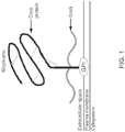
- Glypican-3 is a member of the glypican family, a group of heparan sulfate proteoglycans linked to the cell surface through a glycosyl-phosphatidylinositol anchor.
- GPC3 is encoded on Xp26 and its defect causes Simson-Golabi-Behmel syndrome Type 1.
- GPC3 is expressed in a wide variety of tissues during development, but not in mature tissues because of suppression by DNA methylation within the promoter region.
- GPC3 is detected on 100% of epithelial hepatoblastoma (HB), up to 100% of hepatocellular carcinoma (HCC), 58% of embryonal sarcoma (ES) 100% of atypical teratoid rhabdoid tumors (ATRT), 38-75% of Wilms tumor (WT), 67% of malignant rhabdoid tumors (RT), up to 100% of choriocarcinoma (CC) and up to 100% of yolk sac tumors (YST).
- HB epithelial hepatoblastoma
- HCC hepatocellular carcinoma
- ES embryonal sarcoma
- ART atypical teratoid rhabdoid tumors
- WT 38-75%
- RT malignant rhabdoid tumors
- CC choriocarcinoma
- YST yolk sac tumors
- the present embodiments are directed to methods and/or compositions for the treatment of cancer.
- the disclosure concerns methods and/or compositions for the treatment of cancers in which the cancer cells comprise GPC3, for example as a tumor antigen.
- the cancer may be of any kind, in particular cases the cancer is hepatoblastoma, hepatocellular carcinoma, malignant rhabdoid tumors, yok sac tumors, undifferentiated sarcoma of the liver, liposarcoma, Wilm's tumor, or choriocarcinoma.
- the cancer comprises solid tumors. In at least some cases, the cancer is not hepatocellular carcinoma.
- immune cells that express a GPC3-targeting chimeric antigen receptor (CAR) wherein in at least specific embodiments the CAR does not comprise an antigen targeting domain that is, or is derived from, antibodies 3E11, 2G9, 4G5, 3D8, or 2E10.
- the CAR comprises a single chain variable fragment (scFv) specific for GPC3.
- the GPC3 CAR comprises an scFv derived from antibody GC33.
- the GPC3 CAR comprises an scFv that comprises an amino acid sequence that is at least 90%, 91%, 92%, 93%, 94%, 95%, 96%, 97%, 98%, 99%, or 100% identical to the sequence of SEQ ID NO:1.
- the GPC3 CAR may be encoded by a polynucleotide sequence encoded by SEQ ID NO:2.
- the GPC3-specific CARs described herein may include one or more costimulatory endodomains, such as CD28, CD27, 4-1BB, OX40, ICOS, or a combination thereof.
- the CAR may include one or more transmembrane domains, such as one selected from the group consisting of CD3-zeta, CD28, CD8 ⁇ , CD4, and a combination thereof.
- the CAR comprises a transmembrane domain derived from CD28 and not from CD8.
- the GPC3-specific CAR provided herein comprises a transmembrane domain selected from the group consisting of CD3-zeta, CD28, CD8 ⁇ , CD4, or a combination thereof.
- the CAR comprises a transmembrane domain derived from CD28 and not from CD8.
- the CAR comprises a co-stimulatory molecule endodomain selected from the group consisting of CD28, CD27, 4-1BB, OX40 ICOS, and a combination thereof.
- the co-stimulatory domain of the GPC3-specific CARs provided herein comprises a CD28 costimulatory domain.
- the co-stimulatory domain comprises a 4-1BB (CD137) costimulatory domain.
- the co-stimulatory domain comprises a CD28 costimulatory domain and a 4-1BB costimulatory domain.
- the immune cells are T cells, NK cells, dendritic cells, NKT cells, or a mixture thereof.
- the T cells may be CD4+ T cells, CD8+ T cells, Treg cells, Th1 T cells, Th2 T cells, Th17 T cells, unspecific T cells, or a population of T cells that comprises a combination of any of the foregoing.
- the GPC3-specific CARs described herein are expressed in other immune cells, including but not limited to, NK cells, NKT cells, ⁇ T cells, or T cells that recognize specific antigens (e.g., viral or other tumor associated antigens) through to their native T-cell receptor.
- the immune cells may harbor a polynucleotide that encodes the CAR, and the polynucleotide may further comprise a suicide gene.
- the immune cells are one of T cells, Natural Killer (NK) cells, Natural Killer T (NKT) cells, ⁇ -T cells, Mucosa Associated Invariant T cells (MAIT cells), Innate lymphoid cells, dendritic cells, or a mixture thereof.
- GPC3-CARs may be expressed in stem and/or progenitor cells that are subsequently differentiated into the aforementioned immune cells.
- T cells redirected against GPC3 control the growth of GPC3-expressing cells, including cancer cells, either in vitro or in vivo, e.g., in an individual having a cancer comprising tumor cells that express GPC3.
- the cells are effective against multiple solid tumors, in particular embodiments.
- GPC3 As described in detail herein, the expression of GPC3 is well described in the scientific literature in various types of cancer in extensive panel of tumor arrays and normal tissues and in gene expression profiling datasets.
- GPC3 expression was validated in exemplary tested tumor cell lines.
- a GPC3-specific CAR was generated that showed that when it was expressed by T cells, hepatoblastoma, hepatocellular carcinoma and malignant rhabdoid tumor (as an example) was effectively targeted in vitro, and antitumor activity was observed in vitro and in vivo against hepatocellular carcinoma and malignant rhabdoid tumor, for example.
- Redirecting effector cells to GPC3 using CARs thus represents a robust platform to target multiple types of solid tumors.
- GPC3 protein is expressed by several types of tumors, this antigen is an optimal target for adoptive T-cell immunotherapy based on GPC3-CAR-redirected T cells.
- this antigen is an optimal target for adoptive T-cell immunotherapy based on GPC3-CAR-redirected T cells.
- the lack of significant expression of GPC3 in normal tissues further highlights its relevance for immunotherapy.
- a method of inhibiting proliferation and/or activity of GPC3-positive cells comprising the step of contacting the cells with a therapeutically effective amount of immune cells that express a CAR that targets GPC3 , e.g., a GPC3-specific CAR as provided herein, wherein the CAR does not comprise an antigen targeting domain that is, or is derived from, antibodies 3E11, 2G9, 4G5, 3D8, or 2E10.
- the cancer is hepatocellular carcinoma, a hepatoblastoma, an embryonal sarcoma, a rhabdoid tumor, a Wilm's tumor, a squamous cell carcinoma of the lung, a liposarcoma, yolk sac tumor, choriocarcinoma, a breast carcinoma, a head and neck squamous cell carcinoma (HNSCC), or mesothelioma.
- the cancer is a GPC3-expressing cancer that is not melanoma.
- the cancer is a GPC3-expressing cancer that is not hepatocellular carcinoma.
- the contacting is performed in vitro, in cell culture, or in vivo.
- the contacting is performed in vivo, and the immune cells are cells in an individual, such as T cells.
- the immune cells are autologous or allogeneic to the individual.
- a method of treating an individual who has a GPC3-expressing cancer comprising administering to the individual a therapeutically-effective amount of immune cells that express a GPC3-specific CAR, e.g., a GPC3-specific CAR as described herein.
- the immune cells are T cells, NK cells, dendritic cells, NKT cells, MAIT cells, ⁇ -T cells, or a mixture thereof.
- the T cells may be CD4+ T cells, CD8+ T cells, or Treg cells, Th1 T cells, Th2 T cells, Th17 T cells, unspecific T cells, or a population of T cells that comprises a combination of any of the foregoing.
- the GLC3-specific CARs provided herein are expressed in other immune cells, including but not limited to, NK cells, NKT cells, ⁇ T cells, or T cells that recognize specific antigens (e.g., viral or other tumor associated antigens) through to their native T-cell receptor.
- the immune cells may harbor a polynucleotide that encodes the CAR, and the polynucleotide may further comprise a suicide gene.
- GPC3-specific CARs transmit signals to activate immune cells through CD3zeta, CD28, and/or 4-1BB pathways, although the intracellular CAR domain could be readily modified to include other signaling moieties.
- an individual who has received GPC3-CAR-expressing immune cells is receiving, or will receive an additional cancer treatment, such as chemotherapy, immunotherapy, radiation, surgery, hormone therapy, or a combination thereof.
- an additional cancer treatment such as chemotherapy, immunotherapy, radiation, surgery, hormone therapy, or a combination thereof.
- a or “an” may mean one or more.
- the words “a” or “an” when used in conjunction with the word “comprising”, the words “a” or “an” may mean one or more than one.
- another may mean at least a second or more.
- aspects of the subject matter may "consist essentially of' or “consist of' one or more elements or steps of the subject matter, for example.
- Some embodiments of the subject matter may consist of or consist essentially of one or more elements, method steps, and/or methods of the subject matter. It is contemplated that any method or composition described herein can be implemented with respect to any other method or composition described herein.
- Adoptive transfer of CAR-redirected T lymphocytes represents a useful therapy for patients with malignancies.
- the applicability of this strategy is extended to a broad array of solid tumors by targeting the GPC3 antigen.
- Particular aspects of the disclosure include methods of treating GPC3-expressing cancers.
- the cancers may be of any kind, including liver, testicular, lung, ovarian, head and neck cancer, mesothelioma, breast, glioblastoma, kidney, brain, skin, colon, prostate, pancreatic, cervical, thyroid, spleen, or bone cancer, for example.
- the cancer is hepatoblastoma, hepatocellular carcinoma, malignant rhabdoid tumors, yok sac tumors, undifferentiated sarcoma of the liver, liposarcoma, Wilm's tumor, or choriocarcinoma, although in specific cases the cancer is not hepatocellular carcinoma.
- a particular scFv for GPC3 is employed.
- Indicia of successful treatment could be, e.g., detectable reduction in the growth of a tumor (e.g., as seen by MRI or the like), or reduction in one or more symptoms of a cancer or other medical condition that expresses GPC3, including aberrantly expresses GPC3.
- GPC3 may also be referred to as OCI-5, SDYS, GTR2-2, SGB, SGBS, SGBS1, MXR7, or DGSX, for example.
- An example of a GPC3 human nucleotide sequence is L47125 in GenBank® (with corresponding protein sequence in AAA98132 of GenBank®).
- CAR tumor-directed chimeric antigen receptors
- CTLs cytotoxic T lymphocytes
- the GPC3 CARs do not include scFvs derived from 3E11, 2G9, 4G5, 3D8, or 2E10.
- the scFV is GC33, and in certain embodiments the scFv comprises SEQ ID NO:1.
- immune cells include a CAR receptor that is chimeric, non-natural and engineered at least in part by the hand of man.
- the engineered chimeric antigen receptor has one, two, three, four, or more components, and in some embodiments the one or more components facilitate targeting or binding of the T lymphocyte to the GPC3 -comprising cancer cell.
- the CAR comprises an antibody for GPC3, part or all of a cytoplasmic signaling domain, and/or part or all of one or more co-stimulatory molecules, for example endodomains of co-stimulatory molecules.
- the antibody is a scFv.
- a cytoplasmic signaling domain such as those derived from the T cell receptor zeta-chain, is employed as at least part of the chimeric receptor in order to produce stimulatory signals for T lymphocyte proliferation and effector function following engagement of the chimeric receptor with the target antigen.
- Examples would include, but are not limited to, endodomains from co-stimulatory molecules such as CD28, CD27, 4-1BB, ICOS, OX40, a combination thereof, or the signaling components of cytokine receptors such as IL7 and IL15.
- co-stimulatory molecules are employed to enhance the activation, proliferation, and cytotoxicity of T cells produced by the GPC3 CAR after antigen engagement.
- the co-stimulatory molecules are CD28, OX40, and 4-1BB, for example.
- the CAR may be first generation, second generation, or third generation (CAR in which signaling is provided by CD3 ⁇ together with co-stimulation provided by CD28 and a tumor necrosis factor receptor (TNFR), such as 4-1BB or OX40), for example.
- TNFR tumor necrosis factor receptor
- the CAR may be specific for GPC3, and in some embodiments a GPC3-specific CAR-expressing cell may also express a second CAR targeting another antigen, including one or more CARs specific for CD19, CD20, CD22, Kappa or light chain, CD30, CD33, CD123, CD38, ROR1, ErbB2, ErbB3/4, EGFR vIII, carcinoembryonic antigen, EGP2, EGP40, mesothelin, TAG72, PSMA, NKG2D ligands, B7-H6, IL-13 receptor ⁇ 2, IL-11 receptor R ⁇ , MUC1, MUC16, CA9, GD2, GD3, HMW-MAA, CD171, Lewis Y, G250/CAIX, HLA-AI MAGE A1, HLA-A2 NY-ESO-1, PSC1, folate receptor- ⁇ , CD44v7/8, 8H9, NCAM, VEGF receptors, 5T4, Fetal AchR, NKG2D ligand
- the CAR is specific for GPC3, and in certain embodiments, the present disclosure provides chimeric T cells specific for GPC3 by joining an extracellular antigen-binding domain derived from a GPC3-specific antibody to cytoplasmic signaling domains derived from the T-cell receptor ⁇ -chain, optionally with the endodomains of the exemplary costimulatory molecules CD28 and OX40, for examples.
- This CAR is expressed in human cells, including human T cells, and the targeting of GPC3-positive cancers is encompassed herein.
- sequence for a scFv including the GC33 antibody is as follows (where the underlined region is the leader sequence and the remaining sequence is the scFv):
- a scFv sequence without the leader sequence is as follows:
- nucleotide sequence for the scFv with the leader is as follows (where the underlined region encodes the leader sequence and the remaining sequence encodes the scFv):
- a scFv-encoding nucleotide sequence without the leader sequence is as follows:
- GPC3 CAR having the CD28 transmembrane domain is as follows (wherein the single underlined region is the leader and where SH refers to short hinge and TM refers to transmembrane):
- the double underlined region is the zeta signaling domain
- EPKSCDKTHTCPPCP (SEQ ID NO:4) is the short hinge
- the linker amino acids are DPK
- the CD28 transmembrane domain is FWVLVVVGGVLACYSLLVTVAFII (SEQ ID NO:5).
- a scFvGC33.SH.CD28TM.zeta CAR sequence that excludes the leader is as follows:
- a corresponding nucleotide sequence for scFvGC33.SH.CD28TM.zeta CAR is as follows (wherein the single underlined region is the leader):
- the double underlined region is the zeta signaling domain; the short hinge region is GAGCCCAAATCTTGTGACAAAACTCACACATGCCCACCGTGCCCGGATCCGAAA (SEQ ID NO:7); the linker region is GATCCCAAA (SEQ ID NO:8); and the CD28 transmembrane domain is
- a scFvGC33.SH.CD28TM.zeta CAR polynucleotide sequence without the leader is as follows:
- GPC3 CAR having the CD28 transmembrane and signaling domain is as follows (wherein the single underlined region is the leader):
- the double underlined region is the zeta signaling domain
- SEQ ID NO:4 is the short hinge
- the linker amino acids are DPK
- CD28 transmembrane and signaling domain is
- a scFvGC33.SH.CD28.zeta CAR polypeptide sequence without the leader is as follows:
- a corresponding nucleotide sequence for scFvGC33.SH.CD28.zeta CAR is as follows (where the single underlined region is the leader):
- the double underlined region is the zeta signaling domain
- SEQ ID NO:7 is the short hinge region
- the linker region is SEQ ID NO:8
- CD28 transmembrane and signaling domain is
- a scFvGC33.SH.CD28.zeta CAR polynucleotide sequence without the leader sequence is as follows:
- GPC3 CAR having the CD28 transmembrane domain and the 41BB endodomain (scFvGC33.SH.CD28TM.41BB.zeta CAR) is as follows (where the single underlined region is the leader sequence):
- the double underlined region is the zeta signaling domain; SEQ ID NO:4 is the short hinge; DPK is the linker amino acids; the CD28 transmembrane domain is SEQ ID NO:5; and the 41BB endodomain is KRGRKKLLYIFKQPFMRPVQTTQEEDGCSCRFPEEEEGGCEL (SEQ ID NO:15).
- a scFvGC33.SH.CD28TM.41BB.zeta CAR polypeptide sequence without the leader is as follows:
- a corresponding nucleotide sequence for scFvGC33.SH.CD28TM.41BB.zeta CAR is as follows (where the single underlined is the leader sequence):
- the double underlined is the zeta signaling domain; the short hinge region is SEQ ID NO:7; the linker amino acids are SEQ ID NO:8; the CD28 transmembrane domain is SEQ ID NO:9; and the 41BB endodomain is
- a scFvGC33.SH.CD28TM.41BB.zeta CAR polynucleotide sequence that excludes the leader sequence is as follows:
- GPC3 CAR having the CD28 transmembrane and endodomain and the 41BB endodomain (scFvGC33.SH.CD28.41BB.zeta CAR) is as follows (where the single underlined region is the leader sequence):
- the zeta signaling domain is double underlined, the short hinge region is SEQ ID NO:4; the CD28 transmembrane and endodomain is SEQ ID NO:11; the linker amino acids are DPK; and the 41BB endodomain is SEQ ID NO:15.
- a scFvGC33.SH.CD28.41BB.zeta CAR polypeptide sequence is as follows:
- a corresponding nucleotide sequence for scFvGC33.SH.CD28.41BB.zeta CAR is as follows, wherein the single underlined region is the leader sequence:
- the double underlined is the zeta signaling domain; the short hinge region is SEQ ID NO:7; the linker region is SEQ ID NO:8; the CD28 transmembrane domain and endodomain is SEQ ID NO:13; and the 41BB endodomain is SEQ ID NO:17.
- a scFvGC33.SH.CD28.41BB.zeta CAR polynucleotide sequence that excludes the leader is as follows:
- V H and V L sequences are employed in the GC33 compositions of the disclosure, such as follows:
- the GPC3-specific CAR comprises sequences that are 70, 71, 72, 73, 74, 75, 76, 77, 78, 79, 80, 81, 82, 83, 84, 85, 86, 87, 88, 89, 90, 91, 92, 93, 94, 95, 96, 97, 98, or 99% identical to at least one of SEQ ID NO:1, 2, 3, 4, 5, 6, 7, 8, 9, 10, 11, 12, 13,14, 15, 16, 17, 18, 19, 20, 21, 22, 23, 24, 25, 26, 27, 28, 29, 30, 31, 32, or 33.
- Cells of the disclosure include immune cells that express a GPC3 CAR.
- immune cells expressing a GPC3-specific CAR described herein are T cells, NK cells, dendritic cells, NKT cells, or a mixture thereof.
- the T cells may be CD4+ T cells, CD8+ T cells, Treg cells, Th1 T cells, Th2 T cells, Th17 T cells, unspecific T cells, or a population of T cells that comprises a combination of any of the foregoing.
- the GLC3-specific CARs described herein are expressed in other immune cells, including but not limited to, NK cells, NKT cells, ⁇ T cells, or T cells that recognize specific antigens (e.g., viral or other tumor associated antigens) through to their native T-cell receptor.
- NK cells including but not limited to, NK cells, NKT cells, ⁇ T cells, or T cells that recognize specific antigens (e.g., viral or other tumor associated antigens) through to their native T-cell receptor.
- host cell refers to a eukaryotic cell that is capable of replicating a vector and/or expressing a heterologous gene encoded by a vector.
- a host cell can, and has been, used as a recipient for vectors.
- a host cell may be "transfected” or “transformed,” which refers to a process by which exogenous nucleic acid is transferred or introduced into the host cell.
- a transformed cell includes the primary subject cell and its progeny.
- the terms “engineered” and “recombinant” cells or host cells are intended to refer to a cell into which an exogenous nucleic acid sequence, such as, for example, a vector, has been introduced. Therefore, recombinant cells are distinguishable from naturally occurring cells which do not contain a recombinantly introduced nucleic acid.
- a host cell is a T cell, including a cytotoxic T cell (also known as TC, Cytotoxic T Lymphocyte, CTL, T-Killer cell, cytolytic T cell, CD8+ T-cells or killer T cell); NK cells and NKT cells are also encompassed in the disclosure.
- RNAs or proteinaceous sequences may be co expressed with other selected RNAs or proteinaceous sequences in the same cell, such as the same CTL. Co-expression may be achieved by co-transfecting the CTL with two or more distinct recombinant vectors. Alternatively, a single recombinant vector may be constructed to include multiple distinct coding regions for RNAs, which could then be expressed in CTLs transfected with the single vector.
- Some vectors may employ control sequences that allow it to be replicated and/or expressed in both prokaryotic and eukaryotic cells.
- control sequences that allow it to be replicated and/or expressed in both prokaryotic and eukaryotic cells.
- One of skill in the art would further understand the conditions under which to incubate all of the above described host cells to maintain them and to permit replication of a vector. Also understood and known are techniques and conditions that would allow large-scale production of vectors, as well as production of the nucleic acids encoded by vectors and their cognate polypeptides, proteins, or peptides.
- the cells can be autologous cells, syngeneic cells, allogenic cells and even in some cases, xenogeneic cells.
- the cells become neoplastic, in research where the absence of the cells after their presence is of interest, or other event, for example.
- the suicide gene is caspase-9 or HSV thymidine kinase, for example.
- An inducible suicide gene may be used to reduce the risk of direct toxicity and/or uncontrolled proliferation, for example.
- the suicide gene is not immunogenic to the host harboring the polynucleotide or cell.
- a certain example of a suicide gene that may be used is caspase-9 or caspase-8 or cytosine deaminase.
- Caspase-9 can be activated using a specific chemical inducer of dimerization (CID), for example.
- CID chemical inducer of dimerization
- Thymidine kinase-based suicide systems may be utilized.
- individuals with cancer or at risk for cancer may be treated as follows.
- Effector lymphocytes such as CTLs modified as described herein may be administered to the individual and retained for extended periods of time.
- the individual may receive one or more administrations of the cells, and the administrations may or may not occur in conjunction with one or more other cancer therapies.
- the genetically modified cells are encapsulated to inhibit immune recognition and placed at the site of the tumor.
- an individual is provided with therapeutic CTLs modified to comprise a CAR specific for GPC3 in addition to other types of therapeutic cells.
- the cells may be delivered at the same time or at different times.
- the cells may be delivered in the same or separate formulations.
- the cells may be provided to the individual in separate delivery routes.
- the cells may be delivered by injection at a tumor site or intravenously or orally, for example.
- the cells may be delivered systemically or locally. Routine delivery routes for such compositions are known in the art.
- Expression vectors that encode the GPC3 CARs can be introduced into cells such as immune cells, including effector immune cells such as CTLs, as a DNA molecule or construct, where there may be at least one marker that will allow for selection of host cells that contain the construct(s).
- the constructs can be prepared in conventional ways, where the genes and regulatory regions may be isolated, as appropriate, ligated, cloned in an appropriate cloning host, analyzed by restriction or sequencing, or other convenient means. Particularly, using PCR, individual fragments including all or portions of a functional unit may be isolated, where one or more mutations may be introduced using "primer repair", ligation, in vitro mutagenesis, etc., as appropriate.
- the construct(s) once completed and demonstrated to have the appropriate sequences may then be introduced into the CTL by any convenient means.
- the constructs may be integrated and packaged into non-replicating, defective viral genomes like Adenovirus, Adeno-associated virus (AAV), or Herpes simplex virus (HSV) or others, including retroviral vectors, for infection or transduction into cells.
- the constructs may include viral sequences for transfection, if desired.
- the construct may be introduced by fusion, electroporation, biolistics, transfection, lipofection, or the like.
- the host cells may be grown and expanded in culture before introduction of the construct(s), followed by the appropriate treatment for introduction of the construct(s) and integration of the construct(s).
- the cells are then expanded and screened by virtue of a marker present in the construct.
- markers that may be used successfully include hprt, neomycin resistance, thymidine kinase, hygromycin resistance, etc.
- homologous recombination one may use either OMEGA or O-vectors. See, for example, Thomas and Capecchi, Cell (1987) 51, 503-512 ; Mansour, et al., Nature (1988) 336, 348-352 ; and Joyner, et al., Nature (1989) 338, 153-156 .
- Vectors containing useful elements such as bacterial or yeast origins of replication, selectable and/or amplifiable markers, promoter/enhancer elements for expression in prokaryotes or eukaryotes, etc. that may be used to prepare stocks of construct DNAs and for carrying out transfections are well known in the art, and many are commercially available.
- the exemplary T cells that have been modified with the construct(s) are then grown in culture under selective conditions and cells that are selected as having the construct may then be expanded and further analyzed, using, for example; the polymerase chain reaction for determining the presence of the construct in the host cells.
- the modified host cells Once the modified host cells have been identified, they may then be used as planned, e.g. expanded in culture or introduced into a host organism.
- the cells may be introduced into a host organism, e.g. a mammal, in a wide variety of ways.
- the cells may be introduced at the site of the tumor, in specific embodiments, although in alternative embodiments the cells hone to the cancer or are modified to hone to the cancer.
- the number of cells that are employed will depend upon a number of circumstances, the purpose for the introduction, the lifetime of the cells, the protocol to be used, for example, the number of administrations, the ability of the cells to multiply, the stability of the recombinant construct, and the like.
- the cells may be applied as a dispersion, generally being injected at or near the site of interest.
- the cells may be in a physiologically-acceptable medium.
- the DNA introduction need not result in integration in every case. In some situations, transient maintenance of the DNA introduced may be sufficient. In this way, one could have a short term effect, where cells could be introduced into the host and then turned on after a predetermined time, for example, after the cells have been able to home to a particular site.
- the cells may be administered as desired. Depending upon the response desired, the manner of administration, the life of the cells, the number of cells present, various protocols may be employed. The number of administrations will depend upon the factors described above at least in part.
- the system is subject to many variables, such as the cellular response to the ligand, the efficiency of expression and, as appropriate, the level of secretion, the activity of the expression product, the particular need of the patient, which may vary with time and circumstances, the rate of loss of the cellular activity as a result of loss of cells or expression activity of individual cells, and the like. Therefore, it is expected that for each individual patient, even if there were universal cells which could be administered to the population at large, each patient would be monitored for the proper dosage for the individual, and such practices of monitoring a patient are routine in the art
- a polynucleotide encoding the GPC3 CAR and optionally a suicide gene may comprise an expression vector
- vector is used to refer to a carrier nucleic acid molecule into which a nucleic acid sequence can be inserted for introduction into a cell where it can be replicated.
- a nucleic acid sequence can be "exogenous,” which means that it is foreign to the cell into which the vector is being introduced or that the sequence is homologous to a sequence in the cell but in a position within the host cell nucleic acid in which the sequence is ordinarily not found.
- Vectors include plasmids, cosmids, viruses (bacteriophage, animal viruses, and plant viruses), and artificial chromosomes (e.g., YACs).
- YACs artificial chromosomes
- expression vector refers to any type of genetic construct comprising a nucleic acid coding for a RNA capable of being transcribed. In some cases, RNA molecules are then translated into a protein, polypeptide, or peptide. In other cases, these sequences are not translated, for example, in the production of antisense molecules or ribozymes.
- Expression vectors can contain a variety of "control sequences,” which refer to nucleic acid sequences necessary for the transcription and possibly translation of an operably linked coding sequence in a particular host cell. In addition to control sequences that govern transcription and translation, vectors and expression vectors may contain nucleic acid sequences that serve other functions as well and are described infra.
- a “promoter” is a control sequence that is a region of a nucleic acid sequence at which initiation and rate of transcription are controlled. It may contain genetic elements at which regulatory proteins and molecules may bind, such as RNA polymerase and other transcription factors, to initiate the specific transcription a nucleic acid sequence.
- the phrases "operatively positioned,” “operatively linked,” “under control,” and “under transcriptional control” mean that a promoter is in a correct functional location and/or orientation in relation to a nucleic acid sequence to control transcriptional initiation and/or expression of that sequence.
- a promoter generally comprises a sequence that functions to position the start site for RNA synthesis.
- the best known example of this is the TATA box, but in some promoters lacking a TATA box, such as, for example, the promoter for the mammalian terminal deoxynucleotidyl transferase gene and the promoter for the SV40 late genes, a discrete element overlying the start site itself helps to fix the place of initiation. Additional promoter elements regulate the frequency of transcriptional initiation. Typically, these are located in the region 30 110 bp upstream of the start site, although a number of promoters have been shown to contain functional elements downstream of the start site as well.
- a promoter To bring a coding sequence "under the control of” a promoter, one positions the 5' end of the transcription initiation site of the transcriptional reading frame "downstream" of (i.e., 3' of) the chosen promoter.
- the "upstream” promoter stimulates transcription of the DNA and promotes expression of the encoded RNA.
- promoter elements frequently are flexible, so that promoter function is preserved when elements are inverted or moved relative to one another.
- the spacing between promoter elements can be increased to 50 bp apart before activity begins to decline.
- individual elements can function either cooperatively or independently to activate transcription.
- a promoter may or may not be used in conjunction with an "enhancer,” which refers to a cis-acting regulatory sequence involved in the transcriptional activation of a nucleic acid sequence.
- a promoter may be one naturally associated with a nucleic acid sequence, as may be obtained by isolating the 5' non-coding sequences located upstream of the coding segment and/or exon. Such a promoter can be referred to as "endogenous.”
- an enhancer may be one naturally associated with a nucleic acid sequence, located either downstream or upstream of that sequence.
- certain advantages will be gained by positioning the coding nucleic acid segment under the control of a recombinant or heterologous promoter, which refers to a promoter that is not normally associated with a nucleic acid sequence in its natural environment.
- a recombinant or heterologous enhancer refers also to an enhancer not normally associated with a nucleic acid sequence in its natural environment.
- promoters or enhancers may include promoters or enhancers of other genes, and promoters or enhancers isolated from any other virus, or prokaryotic or eukaryotic cell, and promoters or enhancers not "naturally occurring," i.e., containing different elements of different transcriptional regulatory regions, and/or mutations that alter expression.
- promoters that are most commonly used in recombinant DNA construction include the beta-lactamase (penicillinase), lactose and tryptophan (trp) promoter systems.
- sequences may be produced using recombinant cloning and/or nucleic acid amplification technology, including PCRTM, in connection with the compositions disclosed herein (see U.S. Patent Nos. 4,683,202 and 5,928,906 , each incorporated herein by reference).
- control sequences that direct transcription and/or expression of sequences within non-nuclear organelles such as mitochondria, chloroplasts, and the like, can be employed as well.
- promoter and/or enhancer that effectively directs the expression of the DNA segment in the organelle, cell type, tissue, organ, or organism chosen for expression.
- Those of skill in the art of molecular biology generally know the use of promoters, enhancers, and cell type combinations for protein expression, (see, for example Sambrook et al. 1989, incorporated herein by reference).
- the promoters employed may be constitutive, tissue-specific, inducible, and/or useful under the appropriate conditions to direct high level expression of the introduced DNA segment, such as is advantageous in the large-scale production of recombinant proteins and/or peptides.
- the promoter may be heterologous or endogenous.
- any promoter/enhancer combination could also be used to drive expression.
- Use of a T3, T7 or SP6 cytoplasmic expression system is another possible embodiment.
- Eukaryotic cells can support cytoplasmic transcription from certain bacterial promoters if the appropriate bacterial polymerase is provided, either as part of the delivery complex or as an additional genetic expression construct.
- tissue-specific promoters or elements as well as assays to characterize their activity, is well known to those of skill in the art.
- a specific initiation signal also may be required for efficient translation of coding sequences. These signals include the ATG initiation codon or adjacent sequences. Exogenous translational control signals, including the ATG initiation codon, may need to be provided. One of ordinary skill in the art would readily be capable of determining this and providing the necessary signals.
- the expression of the GPC3-CAR is modulated upon exposure of a corresponding regulatory sequence to one or more factors.
- the expression is modulated upon exposure to tumor-associated factors.
- tumor-associated factors include factors present in hypoxic tissue.
- the factors are cytokines and/or chemokines.
- hypoxia induces the expression of HIF-1 ⁇ , a transcription factor that could induce engager expression that is under the control of a hypoxia response element (HRE).
- Hypoxia could also stabilize engager molecules that contain an oxygen-dependent degradation domain (ODDD).
- ODDD oxygen-dependent degradation domain
- a specific initiation signal also may be required for efficient translation of coding sequences. These signals include the ATG initiation codon or adjacent sequences. Exogenous translational control signals, including the ATG initiation codon, may need to be provided. One of ordinary skill in the art would readily be capable of determining this and providing the necessary signals.
- the GPC3-CAR may be expressed in two fragments that are inactive without the addition of an exogenous substance.
- the GPC3-specific ectodomain and the transmembrane domain of the CAR and the cytoplasmic domain of the CAR could be each linked to a heterodimerizer domain (Exto-TM-HD; Cyto-HD).
- Exto-TM-HD and Cyto-HD in cells would result in an inactive GPC3-CAR unless a small molecule is added that links Exto-TM-HD and Cyto-HD, allowing for pharmacological control of GPC3-CAR activity.
- IVS internal ribosome entry sites
- 2A sequences are used to create multigene messages, and these may be used in the embodiments.
- Vectors can include a multiple cloning site (MCS), which is a nucleic acid region that contains multiple restriction enzyme sites, any of which can be used in conjunction with standard recombinant technology to digest the vector.
- MCS multiple cloning site
- Restriction enzyme digestion refers to catalytic cleavage of a nucleic acid molecule with an enzyme that functions only at specific locations in a nucleic acid molecule. Many of these restriction enzymes are commercially available. Use of such enzymes is widely understood by those of skill in the art. Frequently, a vector is linearized or fragmented using a restriction enzyme that cuts within the MCS to enable exogenous sequences to be ligated to the vector.
- Ligand refers to the process of forming phosphodiester bonds between two nucleic acid fragments, which may or may not be contiguous with each other. Techniques involving restriction enzymes and ligation reactions are well known to those of skill in the art of recombinant technology.
- Splicing sites termination signals, origins of replication, and selectable markers may also be employed.
- a plasmid vector is contemplated for use to transform a host cell.
- plasmid vectors containing replicon and control sequences which are derived from species compatible with the host cell are used in connection with these hosts.
- the vector ordinarily carries a replication site, as well as marking sequences which are capable of providing phenotypic selection in transformed cells.
- E. coli is often transformed using derivatives of pBR322, a plasmid derived from an E. coli species.
- pBR322 contains genes for ampicillin and tetracycline resistance and thus provides easy means for identifying transformed cells.
- the pBR plasmid, or other microbial plasmid or phage must also contain, or be modified to contain, for example, promoters which can be used by the microbial organism for expression of its own proteins.
- phage vectors containing replicon and control sequences that are compatible with the host microorganism can be used as transforming vectors in connection with these hosts.
- the phage lambda GEMTM 11 may be utilized in making a recombinant phage vector which can be used to transform host cells, such as, for example, E. coli LE392.
- plasmid vectors include pIN vectors (Inouye et al., 1985); and pGEX vectors, for use in generating glutathione S transferase (GST) soluble fusion proteins for later purification and separation or cleavage.
- GST glutathione S transferase
- Other suitable fusion proteins are those with beta-galactosidase, ubiquitin, and the like.
- Bacterial host cells for example, E. coli, comprising the expression vector, are grown in any of a number of suitable media, for example, LB.
- suitable media for example, LB.
- the expression of the recombinant protein in certain vectors may be induced, as would be understood by those of skill in the art, by contacting a host cell with an agent specific for certain promoters, e.g ., by adding IPTG to the media or by switching incubation to a higher temperature. After culturing the bacteria for a further period, generally of between 2 and 24 h, the cells are collected by centrifugation and washed to remove residual media.
- Components of the present disclosure may be a viral vector that encodes one or more CARs of the disclosure.
- Non-limiting examples of virus vectors that may be used to deliver a nucleic acid of the present disclosure are described below.
- a particular method for delivery of the nucleic acid involves the use of an adenovirus expression vector.
- adenovirus vectors are known to have a low capacity for integration into genomic DNA, this feature is counterbalanced by the high efficiency of gene transfer afforded by these vectors.
- "Adenovirus expression vector” is meant to include those constructs containing adenovirus sequences sufficient to (a) support packaging of the construct and (b) to ultimately express a tissue or cell specific construct that has been cloned therein.
- Knowledge of the genetic organization or adenovirus, a 36 kb, linear, double stranded DNA virus allows substitution of large pieces of adenoviral DNA with foreign sequences up to 7 kb (Grunhaus and Horwitz, 1992).
- the nucleic acid may be introduced into the cell using adenovirus assisted transfection. Increased transfection efficiencies have been reported in cell systems using adenovirus coupled systems (Kelleher and Vos, 1994; Cotten et al., 1992; Curiel, 1994).
- Adeno associated virus (AAV) is an attractive vector system for use in the cells of the present disclosureas it has a high frequency of integration and it can infect nondividing cells, thus making it useful for delivery of genes into mammalian cells, for example, in tissue culture (Muzyczka, 1992) or in vivo.
- AAV has a broad host range for infectivity (Tratschin et al., 1984; Laughlin et al., 1986; Lebkowski et al., 1988; McLaughlin et al., 1988). Details concerning the generation and use of rAAV vectors are described in U.S. Patent Nos. 5,139,941 and 4,797,368 , each incorporated herein by reference.
- Retroviruses are useful as delivery vectors because of their ability to integrate their genes into the host genome, transferring a large amount of foreign genetic material, infecting a broad spectrum of species and cell types and of being packaged in special cell lines (Miller, 1992).
- a nucleic acid e.g., one encoding the desired sequence
- a packaging cell line containing the gag, pol, and env genes but without the LTR and packaging components is constructed (Mann et al., 1983).
- Retroviral vectors are able to infect a broad variety of cell types. However, integration and stable expression require the division of host cells (Paskind et al., 1975).
- Lentiviruses are complex retroviruses, which, in addition to the common retroviral genes gag, pol, and env, contain other genes with regulatory or structural function. Lentiviral vectors are well known in the art (see, for example, Naldini et al., 1996; Zufferey et al., 1997; Blomer et al., 1997; U.S. Pat. Nos. 6,013,516 and 5,994,136 ). Some examples of lentivirus include the Human Immunodeficiency Viruses: HIV-1, HIV-2 and the Simian Immunodeficiency Virus: SIV. Lentiviral vectors have been generated by multiply attenuating the HIV virulence genes, for example, the genes env, vif, vpr, vpu and nef are deleted making the vector biologically safe.
- Recombinant lentiviral vectors are capable of infecting non-dividing cells and can be used for both in vivo and ex vivo gene transfer and expression of nucleic acid sequences.
- recombinant lentivirus capable of infecting a non-dividing cell wherein a suitable host cell is transfected with two or more vectors carrying the packaging functions, namely gag, pol and env, as well as rev and tat is described in U.S. Pat. No. 5,994,136 , incorporated herein by reference.
- One may target the recombinant virus by linkage of the envelope protein with an antibody or a particular ligand for targeting to a receptor of a particular cell-type.
- a sequence (including a regulatory region) of interest into the viral vector, along with another gene which encodes the ligand for a receptor on a specific target cell, for example, the vector is now target-specific.
- viral vectors may be employed as vaccine constructs in the present disclosure.
- Vectors derived from viruses such as vaccinia virus (Ridgeway, 1988; Baichwal and Sugden, 1986; Coupar et al., 1988), Sindbis virus, cytomegalovirus and herpes simplex virus may be employed. They offer several attractive features for various mammalian cells (Friedmann, 1989; Ridgeway, 1988; Baichwal and Sugden, 1986; Coupar et al., 1988; Horwich et al., 1990).
- a nucleic acid to be delivered may be housed within an infective virus that has been engineered to express a specific binding ligand.
- the virus particle will thus bind specifically to the cognate receptors of the target cell and deliver the contents to the cell.
- a novel approach designed to allow specific targeting of retrovirus vectors was developed based on the chemical modification of a retrovirus by the chemical addition of lactose residues to the viral envelope. This modification can permit the specific infection of hepatocytes via sialoglycoprotein receptors.
- Suitable methods for nucleic acid delivery for transfection or transformation of cells are known to one of ordinary skill in the art. Such methods include, but are not limited to, direct delivery of DNA such as by ex vivo transfection, by injection, and so forth. Through the application of techniques known in the art, cells may be stably or transiently transformed.
- eukaryotic cells and tissues removed from an organism in an ex vivo setting are known to those of skill in the art.
- cells or tissues may be removed and transfected ex vivo using nucleic acids of the present disclosure.
- the transplanted cells or tissues may be placed into an organism.
- a nucleic acid is expressed in the transplanted cells.
- any of the GPC3-CAR compositions described herein may be comprised in a kit.
- one or more cells for use in cell therapy and/or the reagents to generate one or more cells for use in cell therapy that harbors recombinant expression vectors may be comprised in a kit.
- Polynucleotides that encodes the GPC3-CAR or portions thereof may be included in the kit.
- the kit components are provided in suitable container means.
- kits may be packaged either in aqueous media or in lyophilized form.
- the container means of the kits will generally include at least one vial, test tube, flask, bottle, syringe or other container means, into which a component may be placed, and preferably, suitably aliquoted. Where there are more than one component in the kit, the kit also will generally contain a second, third or other additional container into which the additional components may be separately placed. However, various combinations of components may be comprised in a vial.
- the kits of the present disclosure also will typically include a means for containing the components in close confinement for commercial sale. Such containers may include injection or blow molded plastic containers into which the desired vials are retained.
- the liquid solution is an aqueous solution, with a sterile aqueous solution being particularly ueful.
- the container means may itself be a syringe, pipette, and/or other such like apparatus, from which the formulation may be applied to an infected area of the body, injected into an animal, and/or even applied to and/or mixed with the other components of the kit.
- kits may be provided as dried powder(s).
- the powder can be reconstituted by the addition of a suitable solvent.
- the solvent may also be provided in another container means.
- kits may also comprise a second container means for containing a sterile, pharmaceutically acceptable buffer and/or other diluent.
- kits that are to be used for cell therapy are provided in a kit, and in some cases the cells are essentially the sole component of the kit.
- the kit may comprise reagents and materials to make the desired cell.
- the reagents and materials include primers for amplifying desired sequences, nucleotides, suitable buffers or buffer reagents, salt, and so forth, and in some cases the reagents include vectors and/or DNA that encodes a CAR as described herein and/or regulatory elements therefor.
- the kit suitable for extracting one or more samples from an individual.
- the apparatus may be a syringe, scalpel, and so forth.
- the kit in addition to cell therapy embodiments, also includes a second cancer therapy, such as chemotherapy, hormone therapy, and/or immunotherapy, for example.
- a second cancer therapy such as chemotherapy, hormone therapy, and/or immunotherapy, for example.
- the kit(s) may be tailored to a particular cancer for an individual and comprise respective second cancer therapies for the individual.
- GPC3-targeting CAR constructs, nucleic acid sequences, vectors, host cells , as contemplated herein and/or pharmaceutical compositions comprising the same are used for the prevention, treatment or amelioration of a cancerous disease, such as a tumorous disease.
- the pharmaceutical composition of the present disclosure may be particularly useful in preventing, ameliorating and/or treating cancer, including cancer that express GPC3 and that may or may not be solid tumors, for example.
- the individual has Simpson-Golabi-Behmel syndrome.
- treatment includes any beneficial or desirable effect on the symptoms or pathology of a disease or pathological condition, and may include even minimal reductions in one or more measurable markers of the disease or condition being treated, e.g., cancer. Treatment can involve optionally either the reduction or amelioration of symptoms of the disease or condition, or the delaying of the progression of the disease or condition. “Treatment” does not necessarily indicate complete eradication or cure of the disease or condition, or associated symptoms thereof.
- prevent and similar words such as “prevented,” “preventing” etc., indicate an approach for preventing, inhibiting, or reducing the likelihood of the occurrence or recurrence of, a disease or condition, e.g ., cancer. It also refers to delaying the onset or recurrence of a disease or condition or delaying the occurrence or recurrence of the symptoms of a disease or condition. As used herein, "prevention” and similar words also includes reducing the intensity, effect, symptoms and/or burden of a disease or condition prior to onset or recurrence of the disease or condition.
- the present invention contemplates, in part, GPC3 CAR-expressing cells, GPC3 CAR constructs, GPC3 CAR nucleic acid molecules and GPC3 CAR vectors that can administered either alone or in any combination using standard vectors and/or gene delivery systems, and in at least some aspects, together with a pharmaceutically acceptable carrier or excipient.
- the nucleic acid molecules or vectors may be stably integrated into the genome of the subject.
- viral vectors may be used that are specific for certain cells or tissues and persist in said cells.
- Suitable pharmaceutical carriers and excipients are well known in the art.
- the compositions prepared according to the disclosure can be used for the prevention or treatment or delaying the above identified diseases.
- the disclosure relates to a method for the prevention, treatment or amelioration of a tumorous disease comprising the step of administering to a subject in the need thereof an effective amount of cells that express a GPC3-targeting CAR, a nucleic acid sequence, a vector, as contemplated herein and/or produced by a process as contemplated herein.
- Possible indications for administration of the composition(s) of the exemplary GPC3 CAR cells are cancerous diseases, including tumorous diseases, including hepatocellular carcinoma, a hepatoblastoma, an embryonal sarcoma, a rhabdoid tumor, a Wilm's tumor, yolk sac tumor, choriocarcinoma, a squamous cell carcinoma of the lung, a liposarcoma, a breast carcinoma, a head and neck squamous cell carcinoma (HNSCC), or mesothelioma, for example.
- Exemplary indications for administration of the composition(s) of GPC3 CAR cells are cancerous diseases, including any malignancies that express GPC3.
- the administration of the composition(s) of the disclosure is useful for all stages and types of cancer, including for minimal residual disease, early cancer, advanced cancer, and/or metastatic cancer and/or refractory cancer, for example.
- the disclosure further encompasses co-administration protocols with other compounds, e.g. bispecific antibody constructs, targeted toxins or other compounds, which act via immune cells.
- the clinical regimen for co-administration of the inventive compound(s) may encompass co-administration at the same time, before or after the administration of the other component.
- Particular combination therapies include chemotherapy, radiation, surgery, hormone therapy, or other types of immunotherapy.
- Embodiments relate to a kit comprising a GPC3 CAR construct as defined herein, a nucleic acid sequence as defined herein, a vector as defined herein and/or a host as defined herein. It is also contemplated that the kit of this disclosure comprises a pharmaceutical composition as described herein above, either alone or in combination with further medicaments to be administered to an individual in need of medical treatment or intervention.
- methods of the present disclosure for clinical aspects are combined with other agents effective in the treatment of hyperproliferative disease, such as anti-cancer agents.
- An "anti-cancer” agent is capable of negatively affecting cancer in a subject, for example, by killing cancer cells, inducing apoptosis in cancer cells, reducing the growth rate of cancer cells, reducing the incidence or number of metastases, reducing tumor size, inhibiting tumor growth, reducing the blood supply to a tumor or cancer cells, promoting an immune response against cancer cells or a tumor, preventing or inhibiting the progression of cancer, or increasing the lifespan of a subject with cancer. More generally, these other compositions would be provided in a combined amount effective to kill or inhibit proliferation of the cell.
- This process may involve contacting the cancer cells with the expression construct and the agent(s) or multiple factor(s) at the same time. This may be achieved by contacting the cell with a single composition or pharmacological formulation that includes both agents, or by contacting the cell with two distinct compositions or formulations, at the same time, wherein one composition includes the expression construct and the other includes the second agent(s).
- HSV-tK herpes simplex virus-thymidine kinase
- the present inventive therapy may precede or follow the other agent treatment by intervals ranging from minutes to weeks.
- the other agent and present disclosure are applied separately to the individual, one would generally ensure that a significant period of time did not expire between the time of each delivery, such that the agent and inventive therapy would still be able to exert an advantageously combined effect on the cell.
- Cancer therapies also include a variety of combination therapies with both chemical and radiation based treatments.
- Combination chemotherapies include, for example, abraxane, altretamine, docetaxel, herceptin, methotrexate, novantrone, zoladex, cisplatin (CDDP), carboplatin, procarbazine, mechlorethamine, cyclophosphamide, camptothecin, ifosfamide, melphalan, chlorambucil, busulfan, nitrosurea, dactinomycin, daunorubicin, doxorubicin, bleomycin, plicomycin, mitomycin, etoposide (VP16), tamoxifen, raloxifene, estrogen receptor binding agents, taxol, gemcitabien, navelbine, farnesyl-protein tansferase inhibitors, transplatinum, 5-fluorouracil, vincristin, vinblastin and methotre
- chemotherapy for the individual is employed in conjunction with the disclosure, for example before, during and/or after administration of the disclosure.
- Dosage ranges for X-rays range from daily doses of 50 to 200 roentgens for prolonged periods of time (3 to 4 wk), to single doses of 2000 to 6000 roentgens.
- Dosage ranges for radioisotopes vary widely, and depend on the half-life of the isotope, the strength and type of radiation emitted, and the uptake by the neoplastic cells.
- contacted and “exposed,” when applied to a cell are used herein to describe the process by which a therapeutic construct and a chemotherapeutic or radiotherapeutic agent are delivered to a target cell or are placed in direct juxtaposition with the target cell.
- both agents are delivered to a cell in a combined amount effective to kill the cell or prevent it from dividing.
- Immunotherapeutics generally rely on the use of immune effector cells and molecules to target and destroy cancer cells.
- the immune effector may be, for example, an antibody specific for some marker on the surface of a tumor cell.
- the antibody alone may serve as an effector of therapy or it may recruit other cells to actually effect cell killing.
- the antibody also may be conjugated to a drug or toxin (chemotherapeutic, radionuclide, ricin A chain, cholera toxin, pertussis toxin, etc.) and serve merely as a targeting agent.
- the effector may be a lymphocyte carrying a surface molecule that interacts, either directly or indirectly, with a tumor cell target.
- Various effector cells include cytotoxic T cells and NK cells.
- Immunotherapy other than the inventive therapy described herein could thus be used as part of a combined therapy, in conjunction with the present cell therapy.
- the general approach for combined therapy is discussed below.
- the tumor cell must bear some marker that is amenable to targeting, i.e., is not present on the majority of other cells.
- Common tumor markers include carcinoembryonic antigen, prostate specific antigen, urinary tumor associated antigen, fetal antigen, tyrosinase (p97), gp68, TAG-72, HMFG, Sialyl Lewis Antigen, MucA, MucB, PLAP, estrogen receptor, laminin receptor, erb B and p155.
- the secondary treatment is a gene therapy in which a therapeutic polynucleotide is administered before, after, or at the same time as the present disclosure clinical embodiments.
- a variety of expression products are encompassed within the disclosure, including inducers of cellular proliferation, inhibitors of cellular proliferation, or regulators of programmed cell death.
- Curative surgery is a cancer treatment that may be used in conjunction with other therapies, such as the treatment of the present disclosure, chemotherapy, radiotherapy, hormonal therapy, gene therapy, immunotherapy and/or alternative therapies.
- Curative surgery includes resection in which all or part of cancerous tissue is physically removed, excised, and/or destroyed.
- Tumor resection refers to physical removal of at least part of a tumor.
- treatment by surgery includes laser surgery, cryosurgery, electrosurgery, and miscopically controlled surgery (Mohs' surgery). It is further contemplated that the present disclosuremay be used in conjunction with removal of superficial cancers, precancers, or incidental amounts of normal tissue.
- a cavity may be formed in the body.
- Treatment may be accomplished by perfusion, direct injection or local application of the area with an additional anti-cancer therapy.
- Such treatment may be repeated, for example, every 1, 2, 3, 4, 5, 6, or 7 days, or every 1, 2, 3, 4, and 5 weeks or every 1, 2, 3, 4, 5, 6, 7, 8, 9, 10, 11, or 12 months.
- These treatments may be of varying dosages as well.
- agents may be used in combination with the present disclosure to improve the therapeutic efficacy of treatment.
- additional agents include immunomodulatory agents, agents that affect the upregulation of cell surface receptors and GAP junctions, cytostatic and differentiation agents, inhibitors of cell adhesion, or agents that increase the sensitivity of the hyperproliferative cells to apoptotic inducers.
- Immunomodulatory agents include tumor necrosis factor; interferon alpha, beta, and gamma; IL-2 and other cytokines; F42K and other cytokine analogs; or MIP-1, MIP-1beta, MCP-1, RANTES, and other chemokines.
- cell surface receptors or their ligands such as Fas / Fas ligand, DR4 or DR5 / TRAIL would potentiate the apoptotic inducing abilities of the present disclosure by establishment of an autocrine or paracrine effect on hyperproliferative cells. Increases intercellular signaling by elevating the number of GAP junctions would increase the anti-hyperproliferative effects on the neighboring hyperproliferative cell population.
- cytostatic or differentiation agents can be used in combination with the present disclosure to improve the anti-hyperproliferative efficacy of the treatments.
- Inhibitors of cell adhesion are contemplated to improve the efficacy of the present disclosure.
- cell adhesion inhibitors are focal adhesion kinase (FAKs) inhibitors and Lovastatin. It is further contemplated that other agents that increase the sensitivity of a hyperproliferative cell to apoptosis, such as the antibody c225, could be used in combination with the present disclosure to improve the treatment efficacy.
- FAKs focal adhesion kinase
- Lovastatin Lovastatin
- GPC3-CAR glypican-3 specific CARs
- GPC3-CAR glypican-3 specific CARs
- High cell surface expression of all CARs on T cells was detected.
- Glypican-3 specific T cells were generated by retroviral transduction, and they recognize glypican-3 expressing tumor cells.
- CARs were generated with GPC3-specific scFvs fused to CAR backbones ( FIG. 2 ).
- GPC3-CARs T cells were stimulated with OKT3 and anti-CD28 followed by retroviral transduction. CAR expression was measured with anti-Fab mab-AF647 by FACS. GPC3-CAR expression was detected at or above 80% of cells.
- GPC3-CAR T cells recognize and kill GPC3 pos targets: GPC3 pos (HepG2, HUH7 and Hep3B) as well as GPC3 neg (A549) cell lines were loaded with Cr 51 and incubated with GPC3-CAR T cells in a standard 4hr Cr 51 -release assay. Effective killing was detected at all 3 GPC3 pos targets dependent on effector-to-target ration while GPC3 neg target was not killed.
- GPC3-CAR T cells release IFN- ⁇ upon stimulation with GPC3 pos targets: GPC3-CAR T cells were co-cultured for 24hrs with GPC3 pos (HepG2, HUH7 and Hep3B) as well as GPC3 neg (A549) cell lines at 1:1 ratio. IFN- ⁇ level of tissue culture supernatant was measured by Luminex (Millipore) according to the manufacturer's manual. High level of IFN- ⁇ production was detected for all GPC3-CAR constructs when stimulated with GPC3 pos targets while non-transduced T cells did not produce IFN- ⁇ . No IFN- ⁇ production was detected for GPC3-CAR T cells when stimulated with GPC neg target except for GBBz which induced low level spontaneous IFN- ⁇ release.
- GPC3-specific CARs demonstrate and kill GPC3-positive tumor cells in an antigen-specific manner.
- This technology has broad applications for the immunotherapy of GPC3-positive diseases either as monotherapy or in combination with other treatment modalities.
- Glypican-3 is one of the six mammalian members of the Glypican family of proteoglycans. Glypican-3 contains heparan sulfate sidechains, is anchored to the cell surface by a glycosyl-phosphatidyl-inositol (Filmus and Selleck, 2001). GPC3 does not directly signal through the cell membrane, the main function of GPC3 is to stabilize wnt thereby increasing its effect on cell proliferation (Capurro et al., 2014).
- GPC3 is a unique immunotherapeutic target as it is expressed on several solid tumors including the majority of hepatocellular carcinomas (not expressed on normal hepatocytes after fetal development or on cirrhotic liver), hepatoblastomas, embryonal sarcomas, rhabdoid tumors, yolk sac tumors, Wilm's tumors, squamous cell carcinoma of the lung, and liposarcomas (Wang et al., 2006; Yamauchi et al., 2005; Enan et al., 2013; Coston et al., 2008; Baumhoer et al., 2008; Chan et al., 2013; Levy et al., 2012; Tretiakova et al., 2015; Zynger et al., 2008; Zynger et al., 2006; Zynger et al., 2008)).
- GPC3 is an attractive immunotherapeutic target recently demonstrated in early phase clinical trials, where a GPC3 targeting monoclonal antibody (MAb) GC33 was well tolerated and induced antitumor responses in patients with advanced HCC (Zhu et al., 2013; Ikeda et al., 2014).
- MAb monoclonal antibody
- HepG2 (HB), HUH7 (HCC) and Hep3B (Hepatitis B virus positive HCC), G401 (malignant rhabdoid tumor) and A549 (lung cancer) were evaluated for the expression of GPC3 by FACS analysis using the glypican-3 specific YP7 antibody.
- HepG2 (HB), HUH7 (HCC) and Hep3B (Hepatitis B virus positive HCC), G401 (malignant rhabdoid tumor) were GPC3 positive whereas A549 (lung cancer) negative ( FIG. 6 ).
- This panel of cell lines was used to characterize the function of T cells expressing novel GPC3-specific chimeric antigen receptors (GPC3-CARs).
- GPC3-CARs Structure and cell surface expression of GPC3-CARs.
- the single chain variable fragment of the anti-glypican-3 specific MAb GC33 was cloned in frame into SFG retroviral vectors containing CAR expression cassettes.
- the following four GPC3-CARs were generated: GC33. ⁇ (Gz), GC33.CD28. ⁇ (G28z), GC33.4-1BB. ⁇ (GBBz) and GC33.CD28.4-1BB. ⁇ (G28BBz) ( FIG. 7A ).
- T cells were transduced with retroviral vectors encoding the indicated constructs.
- Cell surface expression of CARs was measured by flow cytometry using goat anti-mouse F(ab)2 fragment IgG conjugated with AF647. All 4 constructs were stably expressed on the cell surface of transduced T cells.
- FIG. 7B shows one representative FACS plot
- FIG. 7C shows CAR expressions of cells from 10 independent
- GPC3-CAR T cells kill GPC3 pos cell lines.
- a standard 4hr 51 Cr assay was performed. In brief, target cells were loaded with 51 Cr, washed and co-cultured with the indicated GPC3 CAR expressing cells in at 37C standard cell culture incubator. Supernatants were collected after 4 hrs percent cell lysis was determined.
- GPC3-CAR T cells killed GPC3 pos HepG2, HUH7, Hep3B, and G401 cells at all effector to target ratios tested. The GPC3 neg A549 cell line was not killed by any of the GPC3-CAR T cells, confirming specificity ( FIG. 8 ).
- GPC3-CAR T cells secrete cytokines in the presence of GPC3 pos cells. Having shown that GPC3-CAR T cells kill GPC3-positive tumor cells, it was next determined if they also produce cytokines. GPC3-CAR T cells were co-cultured with indicated cell lines and after 24 hr of coculture, cytokine levels were measured by Luminex analysis. T cells expressing all four GPC3-CARs produced high levels of cytokines when engaging GPC3 pos cell lines ( FIG. 10 ). GBBz produced less IL-2 ( FIG. 10A-10E ) and IL-10 ( FIG. 10F-10J ).
- GPC3-CAR T cells proliferate upon encounter of GPC3 pos cells. Having shown that GPC3-CAR T cells recognize (as judged by cytokine production) and kill GPC3-positive tumor cells, it was next evaluated if they had ability to proliferate post encounter of GPC3-positive tumor cells.
- GPC3-CAR T cells were co-cultured with HUH7 cells and expanded without cytokines.
- GPC3-CAR T cells expanded in a GPC3 dependent manner and G28z, GBBz and G28BBz CAR T cells outperformed Gz CAR T cells. Absolute number of GPC3-CAR T cells is shown at indicated time points ( FIG. 9A ).
- CFSE dilution (as measure of cell division) of GPC3-CAR T cells is shown 3 days post stimulation ( FIG. 9B ).
- 7-AAD staining (as a measure of cell death) is shown on day 5 post stimulation ( FIG. 9C ).
- GPC3-CAR T cells expand after adoptive transfer in vivo.
- NOD/SCID/IL2 ⁇ null mice were injected with GPC3 pos HUH7 HCC cell line intraperitoneally followed 14 days later by the intravenous (iv) injection of 1x10 7 GPC3-CAR T cells that also were transduced with a retroviral vector encoding a GFP firefly luciferase fusion gene (GFP.ffLuc) to allow for serial bioluminescence imaging.
- GFP.ffLuc GFP firefly luciferase fusion gene
- GPC3-CAR T cells expanded for up to 6 days post injection. T cells expressing GPC3-CARs with 41BB containing costimulatory endodomains had showed superior expansion compared to Gz or G28z CAR T cells ( FIG. 11A,B ).
- GPC3-CAR T cells have potent antitumor activity in the HUH-7 HCC xenograft models in vivo.
- NOD/SCID/IL2 ⁇ null mice were injected with the GPC3 pos HUH7 HCC cell line, which was genetically modified with GFP.ffLuc to allow for serial bioluminescence imaging, intraperitoneally followed 14 days later by the iv infusion of 1x10 7 GPC3-CAR T cells ( FIG. 12 ). Regardless of endodomain, GPC3 CAR T cells produced robust antitumor effect resulting in complete cure mice with large tumor burden.
- mice and tumor bioluminescence are shown in FIG. 12A-C respectively.
- mice injected with placebo, non-transduced T cells (NT T cells) or GD2-CAR T cells died of tumor progression by Day 45 post tumor cell injection. Since there was not observe significant differences between different GPC3-CARs, only 1/10 of the cell dose (1x10 6 CAR T cells) were injected in the follow up study. T cells expressing GPC3-CARs with costimulatory domains had greater antitumor activity in comparison to the GPC3-CAR with only a zeta endodomain ( FIG. 13A-13C ).
- GPC3-CAR T cells have potent antitumor activity in the G401 malignant rhabdoid tumor xenograft model in vivo.
- NOD/SCID/IL2 ⁇ null mice were injected with the GPC3 pos G401 malignant rhabdoid tumor cell line, which was genetically modified with GFP.ffLuc to allow for serial bioluminescence imaging, intraperitoneally followed 21 days later by the iv infusion of 2x10 7 GPC3-CAR T cells ( FIG. 12 ).
- GPC3 CAR T cells produced robust antitumor effect resulting in complete cure mice with large tumor burden. Survival of mice and tumor bioluminescence is shown in FIG. 14A-C respectively. In contrast, mice injected with placebo, non-transduced T cells (NT T cells) or GD2-CAR T cells died of tumor progression by Day 46 post tumor cell injection. T cells expressing GPC3-CARs with costimulatory domains had greater antitumor activity in comparison to the GPC3-CAR with only a zeta endodomain ( FIG. 14A,14B ).
- GPC3-CARs (Gz, G28z, GBBz and G28BBz) were produced and stably expressed them on the cell surface of T cells by retroviral transduction ( FIG. 7 ). It was demonstrated in cell culture experiments that GPC3-CAR T cells recognize GPC3-positive cancer cells in an antigen-dependent fashion as judged by their i) cytolytic activity ( FIG. 8 ), and ii) ability to produce cytokines ( FIG. 9 ) and proliferate ( FIG. 10 ) in the presence of GPC3-positive cancer cells. In addition, GPC3-CAR T cells with costimulatory endodomains (G28z, GBBz, G28BBz) expanded in xenograft models ( FIG.
- GPC3-targeted cell therapy with broad application to malignancies that express GPC3 including but not limited to HCC, hepatoblastoma, embryonal sarcoma, rhabdoid tumor, yolk sac tumor, Wilm's tumor, squamous cell carcinoma of the lung, and liposarcoma.
- novel GPC3-CARs may be expressed in a broad range of i) immune cells including but not limited to NK cells, NKT cells, cytokine-induced killer cells (CIKs), or ⁇ T cells, or ii) stem cells that can be differentiated into immune cells, for example.
- immune cells including but not limited to NK cells, NKT cells, cytokine-induced killer cells (CIKs), or ⁇ T cells, or ii) stem cells that can be differentiated into immune cells, for example.
- NKTs are an evolutionary conserved subset of innate lymphocytes that are characterized by the expression of an invariant TCR ⁇ -chain V ⁇ 24-J ⁇ 18 and reactivity to self- and microbial-derived glycolipids presented by monomorphic HLA class-I-like molecule CD1d.
- NKTs can directly target tumor-supportive macrophages and have an indirect antitumor activity even in CD 1d-negative malignancies (Song et al,. 2009; Liu et al., 2012).
- NKTs can activate NK-cell cytotoxicity which is a well-studied axis of overall NKT-cell antitumor activity (Metelitsa et al., 2001; Smyth et al., 2002).
- NKTs hold special promise for immunotherapy of hepatocellular carcinoma (HCC) as they can eliminate HCC supporting activated hepatic stellate cells (aHSCs) (Anson et al., 2012) and the presence of NKTs in HCC is associated with improved survival (Guo et al., 2012).
- HCC hepatocellular carcinoma
- aHSCs activated hepatic stellate cells
- Transgenic expression of GPC3 CARs in NKTs would therefore enable them with dual reactivity against HCC tumor cells and tumor-supportive aHSCs.
- NKTs were positively sorted from PBMC of three healthy donors with V ⁇ 24-J ⁇ 18-specific Milteneyi microbeads, activated with alpha-galactosylceramide (aGalCer)-pulsed negative fraction of autologous irradiated PBMC, transduced on day 3 with GPC3 CAR (GC33.CD28.4-1BB.Z) retroviral vector, and expanded in culture with IL-2.
- GPC3 CAR GC33.CD28.4-1BB.Z
Abstract
Description
- This application claims priority to
U.S. Provisional Patent Application Serial No. 62/055979, filed September 26, 2014 - This invention was made with government support under K12CA090433, R01CA173750, R01CA116548, and R01CA142636, all awarded by National Institutes of Health. The government has certain rights in the invention.
- The present disclosure concerns at least the fields of immunology, cell biology, molecular biology, and medicine.
- Immunotherapy with antigen-specific T cells has shown promise in the treatment of malignancies in preclinical models as well as in Phase I/II clinical studies. One attractive strategy to generate tumor-specific T cells is by genetic modification with chimeric antigen receptors (CARs), which comprise an extracellular antigen recognition domain, a transmembrane domain, and an intracellular signaling domain derived from the T-cell receptor CD3-ζ chain often linked to costimulatory molecule endodomains.
- There is a continued search for antigens expressed on cancer cells that are not found on normal mature tissues. Glypican-3 (GPC3) is a member of the glypican family, a group of heparan sulfate proteoglycans linked to the cell surface through a glycosyl-phosphatidylinositol anchor. GPC3 is encoded on Xp26 and its defect causes Simson-Golabi-Behmel
syndrome Type 1. GPC3 is expressed in a wide variety of tissues during development, but not in mature tissues because of suppression by DNA methylation within the promoter region. GPC3 is detected on 100% of epithelial hepatoblastoma (HB), up to 100% of hepatocellular carcinoma (HCC), 58% of embryonal sarcoma (ES) 100% of atypical teratoid rhabdoid tumors (ATRT), 38-75% of Wilms tumor (WT), 67% of malignant rhabdoid tumors (RT), up to 100% of choriocarcinoma (CC) and up to 100% of yolk sac tumors (YST). Thus, GPC3 is an ideal immunotherapeutic target. - The present embodiments are directed to methods and/or compositions for the treatment of cancer. In particular cases, the disclosure concerns methods and/or compositions for the treatment of cancers in which the cancer cells comprise GPC3, for example as a tumor antigen. Although in certain aspects the cancer may be of any kind, in particular cases the cancer is hepatoblastoma, hepatocellular carcinoma, malignant rhabdoid tumors, yok sac tumors, undifferentiated sarcoma of the liver, liposarcoma, Wilm's tumor, or choriocarcinoma. In specific embodiments, the cancer comprises solid tumors. In at least some cases, the cancer is not hepatocellular carcinoma.
- In one aspect, provided herein are immune cells that express a GPC3-targeting chimeric antigen receptor (CAR) wherein in at least specific embodiments the CAR does not comprise an antigen targeting domain that is, or is derived from, antibodies 3E11, 2G9, 4G5, 3D8, or 2E10. In certain embodiments, the CAR comprises a single chain variable fragment (scFv) specific for GPC3. In a more specific embodiment, the GPC3 CAR comprises an scFv derived from antibody GC33. In another specific embodiment, the GPC3 CAR comprises an scFv that comprises an amino acid sequence that is at least 90%, 91%, 92%, 93%, 94%, 95%, 96%, 97%, 98%, 99%, or 100% identical to the sequence of SEQ ID NO:1. The GPC3 CAR may be encoded by a polynucleotide sequence encoded by SEQ ID NO:2.
- The GPC3-specific CARs described herein (which may be referred to as GPC3 CAR) may include one or more costimulatory endodomains, such as CD28, CD27, 4-1BB, OX40, ICOS, or a combination thereof. The CAR may include one or more transmembrane domains, such as one selected from the group consisting of CD3-zeta, CD28, CD8α, CD4, and a combination thereof. In a specific embodiment, the CAR comprises a transmembrane domain derived from CD28 and not from CD8.
- In some embodiments, the GPC3-specific CAR provided herein comprises a transmembrane domain selected from the group consisting of CD3-zeta, CD28, CD8α, CD4, or a combination thereof. In a specific embodiment, the CAR comprises a transmembrane domain derived from CD28 and not from CD8. In particular embodiments, the CAR comprises a co-stimulatory molecule endodomain selected from the group consisting of CD28, CD27, 4-1BB, OX40 ICOS, and a combination thereof. In another specific embodiment, the co-stimulatory domain of the GPC3-specific CARs provided herein comprises a CD28 costimulatory domain. In another specific embodiment, the co-stimulatory domain comprises a 4-1BB (CD137) costimulatory domain. In another specific embodiment, the co-stimulatory domain comprises a CD28 costimulatory domain and a 4-1BB costimulatory domain.
- In another aspect, provided herein are immune cells expressing a GPC3-specific CAR described herein. In certain embodiments, the immune cells are T cells, NK cells, dendritic cells, NKT cells, or a mixture thereof. In specific embodiments in which the immune cells are T cells, the T cells may be CD4+ T cells, CD8+ T cells, Treg cells, Th1 T cells, Th2 T cells, Th17 T cells, unspecific T cells, or a population of T cells that comprises a combination of any of the foregoing. In certain other embodiments, the GPC3-specific CARs described herein are expressed in other immune cells, including but not limited to, NK cells, NKT cells, γδT cells, or T cells that recognize specific antigens (e.g., viral or other tumor associated antigens) through to their native T-cell receptor. The immune cells may harbor a polynucleotide that encodes the CAR, and the polynucleotide may further comprise a suicide gene. In some embodiments, the immune cells are one of T cells, Natural Killer (NK) cells, Natural Killer T (NKT) cells, γδ-T cells, Mucosa Associated Invariant T cells (MAIT cells), Innate lymphoid cells, dendritic cells, or a mixture thereof. In addition, GPC3-CARs may be expressed in stem and/or progenitor cells that are subsequently differentiated into the aforementioned immune cells. In certain aspects, T cells redirected against GPC3 control the growth of GPC3-expressing cells, including cancer cells, either in vitro or in vivo, e.g., in an individual having a cancer comprising tumor cells that express GPC3. The cells are effective against multiple solid tumors, in particular embodiments.
- As described in detail herein, the expression of GPC3 is well described in the scientific literature in various types of cancer in extensive panel of tumor arrays and normal tissues and in gene expression profiling datasets. Herein, GPC3 expression was validated in exemplary tested tumor cell lines. A GPC3-specific CAR was generated that showed that when it was expressed by T cells, hepatoblastoma, hepatocellular carcinoma and malignant rhabdoid tumor (as an example) was effectively targeted in vitro, and antitumor activity was observed in vitro and in vivo against hepatocellular carcinoma and malignant rhabdoid tumor, for example. Redirecting effector cells to GPC3 using CARs thus represents a robust platform to target multiple types of solid tumors.
- ] Because GPC3 protein is expressed by several types of tumors, this antigen is an optimal target for adoptive T-cell immunotherapy based on GPC3-CAR-redirected T cells. In addition, the lack of significant expression of GPC3 in normal tissues further highlights its relevance for immunotherapy.
- In another aspect, therefore, provided herein is a method of inhibiting proliferation and/or activity of GPC3-positive cells, such as cancer cells, comprising the step of contacting the cells with a therapeutically effective amount of immune cells that express a CAR that targets GPC3 , e.g., a GPC3-specific CAR as provided herein, wherein the CAR does not comprise an antigen targeting domain that is, or is derived from, antibodies 3E11, 2G9, 4G5, 3D8, or 2E10. In certain embodiments, the cancer is hepatocellular carcinoma, a hepatoblastoma, an embryonal sarcoma, a rhabdoid tumor, a Wilm's tumor, a squamous cell carcinoma of the lung, a liposarcoma, yolk sac tumor, choriocarcinoma, a breast carcinoma, a head and neck squamous cell carcinoma (HNSCC), or mesothelioma. In certain embodiments, the cancer is a GPC3-expressing cancer that is not melanoma. In certain embodiments, the cancer is a GPC3-expressing cancer that is not hepatocellular carcinoma. In some embodiments, the contacting is performed in vitro, in cell culture, or in vivo. In particular cases, the contacting is performed in vivo, and the immune cells are cells in an individual, such as T cells. In particular cases, the immune cells are autologous or allogeneic to the individual. In a related aspect, provided herein is a method of treating an individual who has a GPC3-expressing cancer, comprising administering to the individual a therapeutically-effective amount of immune cells that express a GPC3-specific CAR, e.g., a GPC3-specific CAR as described herein.
- In particular embodiments of the methods, the immune cells are T cells, NK cells, dendritic cells, NKT cells, MAIT cells, γδ-T cells, or a mixture thereof. The T cells may be CD4+ T cells, CD8+ T cells, or Treg cells, Th1 T cells, Th2 T cells, Th17 T cells, unspecific T cells, or a population of T cells that comprises a combination of any of the foregoing. In certain other embodiments, the GLC3-specific CARs provided herein are expressed in other immune cells, including but not limited to, NK cells, NKT cells, γδT cells, or T cells that recognize specific antigens (e.g., viral or other tumor associated antigens) through to their native T-cell receptor. The immune cells may harbor a polynucleotide that encodes the CAR, and the polynucleotide may further comprise a suicide gene.
- In certain embodiments, GPC3-specific CARs transmit signals to activate immune cells through CD3zeta, CD28, and/or 4-1BB pathways, although the intracellular CAR domain could be readily modified to include other signaling moieties.
- In specific methods of the disclosure, an individual who has received GPC3-CAR-expressing immune cells, is receiving, or will receive an additional cancer treatment, such as chemotherapy, immunotherapy, radiation, surgery, hormone therapy, or a combination thereof.
- Additional features and advantages of the invention will be described hereinafter which form the subject of the claims of the invention. It should be appreciated by those skilled in the art that the conception and specific embodiment disclosed may be readily utilized as a basis for modifying or designing other structures for carrying out the same purposes of the present invention. It should also be realized by those skilled in the art that such equivalent constructions do not depart from the spirit and scope of the invention as set forth in the appended claims. The novel features which are believed to be characteristic of the invention, both as to its organization and method of operation, together with further objects and advantages will be better understood from the following description when considered in connection with the accompanying figures. It is to be expressly understood, however, that each of the figures is provided for the purpose of illustration and description only and is not intended as a definition of the limits of the present invention.
- For a more complete understanding of the present invention, reference is now made to the following descriptions taken in conjunction with the accompanying drawing, in which:
-
FIG. 1 shows the structure of GPC3; -
FIG. 2 illustrates the design of certain GPC3 specific CARs: GPC3-specific scFv was linked with hinge and CD28 transmembrane domain to intracellular signaling endodomains combinations as indicated; -
FIG. 3 shows GPC3-CAR expression of T cells: All GPC3-CARs (Gz, G28z, GBBz and G28BBz as shown inFIG. 2 ) were highly expressed on T cells as detected by FACS analysis. NT: non-transduced T cells; -
FIG. 4 demonstrates cytotoxicity of GPC3-CAR T cells: % of killing based on Cr51 release is shown at indicated effector to target ratios for GPC3-CAR T cells and non-transduced T cells. NT: non-transduced T cell. GPC3pos targets: HepG2, HUH7 and Hep3B. GPC3neg target A549; -
FIG. 5 shows IFNγ release of GPC3-CAR T cells: IFNγ levels in pg/ml are shown of GPC3-CAR T cells after 24 hrs co-culture with GPC3pos and GPC3neg targets. NT: non-transduced T cell. GPC3pos targets: HepG2, HUH7 and Hep3B. GPC3neg target A549; -
FIG. 6 shows FACS analysis with a panel of cell lines expressing GPC3-CARs; -
FIGS. 7A-7C. FIG. 7A illustrates a variety of representative expression cassettes for GPC3-CARs. GPC3-specific scFv was linked with hinge and CD28 transmembrane domain to intracellular signaling endodomains combinations as indicated.FIG. 7B shows a representative FACS plot for T cells expressing the respective different expression cassettes.FIG. 7C demonstrates CAR expressions of cells from multiple donors; -
FIGS. 8A-8E demonstrates cytotoxicity for a variety of GPC3-CAR T cells that killed a variety of GPC3-expressing cells (HepG2 inFIG. 8A ; HUH7 inFIG. 8B ; Hep3B inFIG. 8C ; G401 inFIG. 8D ; and G401 inFIG. 8D ) but not the control cells that lack expression of GPC3 (8E; A549); -
FIGS. 9A-9C show that GPC3-CAR T cells proliferate upon exposure to GPC3pos cells.FIG. 9A show the absolute number of GPC3-CAR T cells.FIG. 9B shows the CFSE dilution of GPC3-CAR T cells 3 days post simulation.FIG. 9C demonstrates 7-AAD staining onday 4 post stimulation; -
FIGS. 10A-10O demonstrate secretion of cytokines by the GPC3-CAR T cells in the presence of GPC3pos cells.FIGS. 10A, 10B, 10C, 10D and 10E show results for secretion of IL-2 upon co-culture of the T cells with HepG2 cells (10A); HUH7 cells (10B); Hep3B cells (10C); G401 cells (10D); and and control A549 cells (10E).FIGS. 10F, 10G, 10H, 10I, and 10J show results for secretion of IL10 in the presence of HepG2 cells (10F); HUH7 cells (10G); Hep3B cells (10H); G401 (101) and control A549 cells (10J).FIGS. 10K, 10L, 10M , ION, and 100 show results for secretion of IFNγ in the presence of HepG2 cells (10K); HUH7 cells (10L); Hep3B cells (10M); G401 (ION) and control A549 cells (10O) -
FIGS. 11A and 11B demonstrate that GPC3-CAR T cells expand after adoptive transfer in vivo, as shown by bioluminescence imaging (FIG. 11A ) and its quantitation (FIG. 11B ). -
FIGS. 12A-12C show that GPC3-CAR T cells have antitumor activity in exemplary xenografts in vivo.FIG. 12A demonstrates bioluminescence over time and its quantitation is shown inFIG. 12B; FIG. 12C shows percent survival. -
FIGS. 13A-13C also show that GPC3-CAR T cells have antitumor activity in exemplary xenografts in vivo. Using a smaller dose of the GPC3-CAR T cells in comparison toFIGS. 12A-12C ,FIG. 13A demonstrates bioluminescence over time and its quantitation is shown inFIG. 13B; FIG. 13C shows percent survival. -
FIGS. 14A-14C show that GPC3-CAR T cells have antitumor activity in second exemplary xenograft model in vivo. 14A demonstrates administration schedule and bioluminescence over time with its quantitation is shown inFIG. 14B; FIG. 14C shows percent survival. -
FIG. 15 . Generation of GPC3 CAR NKT cells. NKTs were isolated from PBMC of 3 healthy donors, stimulated with aGalCer, transduced with GC33.CD28.4-1BB.Z CAR retroviral vector, and expanded in culture with IL-2 for 7 days. The resulting cells were analyzed by FACS using 6B11 (iNKT TCR), anti-CD3, and anti-CAR mAbs - As used herein the specification, "a" or "an" may mean one or more. As used herein in the claim(s), when used in conjunction with the word "comprising", the words "a" or "an" may mean one or more than one. As used herein "another" may mean at least a second or more. In specific embodiments, aspects of the subject matter may "consist essentially of' or "consist of' one or more elements or steps of the subject matter, for example. Some embodiments of the subject matter may consist of or consist essentially of one or more elements, method steps, and/or methods of the subject matter. It is contemplated that any method or composition described herein can be implemented with respect to any other method or composition described herein.
- Adoptive transfer of CAR-redirected T lymphocytes represents a useful therapy for patients with malignancies. Here the applicability of this strategy is extended to a broad array of solid tumors by targeting the GPC3 antigen. Particular aspects of the disclosure include methods of treating GPC3-expressing cancers. The cancers may be of any kind, including liver, testicular, lung, ovarian, head and neck cancer, mesothelioma, breast, glioblastoma, kidney, brain, skin, colon, prostate, pancreatic, cervical, thyroid, spleen, or bone cancer, for example. In particular cases, the cancer is hepatoblastoma, hepatocellular carcinoma, malignant rhabdoid tumors, yok sac tumors, undifferentiated sarcoma of the liver, liposarcoma, Wilm's tumor, or choriocarcinoma, although in specific cases the cancer is not hepatocellular carcinoma. In specific embodiments of the disclosure, a particular scFv for GPC3 is employed.
- Indicia of successful treatment could be, e.g., detectable reduction in the growth of a tumor (e.g., as seen by MRI or the like), or reduction in one or more symptoms of a cancer or other medical condition that expresses GPC3, including aberrantly expresses GPC3.
- GPC3 may also be referred to as OCI-5, SDYS, GTR2-2, SGB, SGBS, SGBS1, MXR7, or DGSX, for example. An example of a GPC3 human nucleotide sequence is L47125 in GenBank® (with corresponding protein sequence in AAA98132 of GenBank®).
- Genetic engineering of human lymphocytes or other immune cells to express tumor-directed chimeric antigen receptors (CAR) can produce antitumor effector cells that bypass tumor immune escape mechanisms that are due to abnormalities in protein-antigen processing and presentation. Moreover, these transgenic receptors can be directed to tumor-associated antigens that are not protein-derived. In certain embodiments of the disclosure there are cytotoxic T lymphocytes (CTLs) that are modified to comprise a CAR that targets GPC3. In specific embodiments the GPC3 CARs do not include scFvs derived from 3E11, 2G9, 4G5, 3D8, or 2E10. In particular embodiments the scFV is GC33, and in certain embodiments the scFv comprises SEQ ID NO:1.
- In particular cases, immune cells include a CAR receptor that is chimeric, non-natural and engineered at least in part by the hand of man. In particular cases, the engineered chimeric antigen receptor (CAR) has one, two, three, four, or more components, and in some embodiments the one or more components facilitate targeting or binding of the T lymphocyte to the GPC3 -comprising cancer cell. In specific embodiments, the CAR comprises an antibody for GPC3, part or all of a cytoplasmic signaling domain, and/or part or all of one or more co-stimulatory molecules, for example endodomains of co-stimulatory molecules. In specific embodiments, the antibody is a scFv.
- In certain embodiments, a cytoplasmic signaling domain, such as those derived from the T cell receptor zeta-chain, is employed as at least part of the chimeric receptor in order to produce stimulatory signals for T lymphocyte proliferation and effector function following engagement of the chimeric receptor with the target antigen. Examples would include, but are not limited to, endodomains from co-stimulatory molecules such as CD28, CD27, 4-1BB, ICOS, OX40, a combination thereof, or the signaling components of cytokine receptors such as IL7 and IL15. In particular embodiments, co-stimulatory molecules are employed to enhance the activation, proliferation, and cytotoxicity of T cells produced by the GPC3 CAR after antigen engagement. In specific embodiments, the co-stimulatory molecules are CD28, OX40, and 4-1BB, for example.
- The CAR may be first generation, second generation, or third generation (CAR in which signaling is provided by CD3ζ together with co-stimulation provided by CD28 and a tumor necrosis factor receptor (TNFR), such as 4-1BB or OX40), for example. The CAR may be specific for GPC3, and in some embodiments a GPC3-specific CAR-expressing cell may also express a second CAR targeting another antigen, including one or more CARs specific for CD19, CD20, CD22, Kappa or light chain, CD30, CD33, CD123, CD38, ROR1, ErbB2, ErbB3/4, EGFR vIII, carcinoembryonic antigen, EGP2, EGP40, mesothelin, TAG72, PSMA, NKG2D ligands, B7-H6, IL-13 receptor α2, IL-11 receptor R α, MUC1, MUC16, CA9, GD2, GD3, HMW-MAA, CD171, Lewis Y, G250/CAIX, HLA-AI MAGE A1, HLA-A2 NY-ESO-1, PSC1, folate receptor- α, CD44v7/8, 8H9, NCAM, VEGF receptors, 5T4, Fetal AchR, NKG2D ligands, HER2, BCMA, or CD44v6, or other tumor-associated antigens or actionable mutations that are identified through genomic analysis and or differential expression studies of tumors, for example.
- In particular cases the CAR is specific for GPC3, and in certain embodiments, the present disclosure provides chimeric T cells specific for GPC3 by joining an extracellular antigen-binding domain derived from a GPC3-specific antibody to cytoplasmic signaling domains derived from the T-cell receptor ζ-chain, optionally with the endodomains of the exemplary costimulatory molecules CD28 and OX40, for examples. This CAR is expressed in human cells, including human T cells, and the targeting of GPC3-positive cancers is encompassed herein.
- An example of sequence for a scFv including the GC33 antibody is as follows (where the underlined region is the leader sequence and the remaining sequence is the scFv):

- A scFv sequence without the leader sequence is as follows:

- The nucleotide sequence for the scFv with the leader is as follows (where the underlined region encodes the leader sequence and the remaining sequence encodes the scFv):


- A scFv-encoding nucleotide sequence without the leader sequence is as follows:

- An example of a GPC3 CAR having the CD28 transmembrane domain (scFvGC33.SH.CD28TM.zeta CAR) is as follows (wherein the single underlined region is the leader and where SH refers to short hinge and TM refers to transmembrane):


- In SEQ ID NO:3, the double underlined region is the zeta signaling domain; EPKSCDKTHTCPPCP (SEQ ID NO:4) is the short hinge; the linker amino acids are DPK; and the CD28 transmembrane domain is FWVLVVVGGVLACYSLLVTVAFII (SEQ ID NO:5).
- A scFvGC33.SH.CD28TM.zeta CAR sequence that excludes the leader is as follows:

- A corresponding nucleotide sequence for scFvGC33.SH.CD28TM.zeta CAR is as follows (wherein the single underlined region is the leader):


- In SEQ ID NO:6, the double underlined region is the zeta signaling domain; the short hinge region is GAGCCCAAATCTTGTGACAAAACTCACACATGCCCACCGTGCCCGGATCCGAAA (SEQ ID NO:7); the linker region is GATCCCAAA (SEQ ID NO:8); and the CD28 transmembrane domain is

- A scFvGC33.SH.CD28TM.zeta CAR polynucleotide sequence without the leader is as follows:


- An example of a GPC3 CAR having the CD28 transmembrane and signaling domain (scFvGC33.SH.CD28.zeta CAR) is as follows (wherein the single underlined region is the leader):

- In SEQ ID NO:10, the double underlined region is the zeta signaling domain; SEQ ID NO:4 is the short hinge; the linker amino acids are DPK; and the CD28 transmembrane and signaling domain is

- A scFvGC33.SH.CD28.zeta CAR polypeptide sequence without the leader is as follows:

- A corresponding nucleotide sequence for scFvGC33.SH.CD28.zeta CAR is as follows (where the single underlined region is the leader):


- In SEQ ID NO:12, the double underlined region is the zeta signaling domain; SEQ ID NO:7 is the short hinge region; the linker region is SEQ ID NO:8; and the CD28 transmembrane and signaling domain is

- A scFvGC33.SH.CD28.zeta CAR polynucleotide sequence without the leader sequence is as follows:


- An example of a GPC3 CAR having the CD28 transmembrane domain and the 41BB endodomain (scFvGC33.SH.CD28TM.41BB.zeta CAR) is as follows (where the single underlined region is the leader sequence):

- In SEQ ID NO:14, the double underlined region is the zeta signaling domain; SEQ ID NO:4 is the short hinge; DPK is the linker amino acids; the CD28 transmembrane domain is SEQ ID NO:5; and the 41BB endodomain is KRGRKKLLYIFKQPFMRPVQTTQEEDGCSCRFPEEEEGGCEL (SEQ ID NO:15).
- A scFvGC33.SH.CD28TM.41BB.zeta CAR polypeptide sequence without the leader is as follows:


- A corresponding nucleotide sequence for scFvGC33.SH.CD28TM.41BB.zeta CAR is as follows (where the single underlined is the leader sequence):

- In SEQ ID NO:16, the double underlined is the zeta signaling domain; the short hinge region is SEQ ID NO:7; the linker amino acids are SEQ ID NO:8; the CD28 transmembrane domain is SEQ ID NO:9; and the 41BB endodomain is

- A scFvGC33.SH.CD28TM.41BB.zeta CAR polynucleotide sequence that excludes the leader sequence is as follows:


- An example of a GPC3 CAR having the CD28 transmembrane and endodomain and the 41BB endodomain (scFvGC33.SH.CD28.41BB.zeta CAR) is as follows (where the single underlined region is the leader sequence):

- In SEQ ID NO:18, the zeta signaling domain is double underlined, the short hinge region is SEQ ID NO:4; the CD28 transmembrane and endodomain is SEQ ID NO:11; the linker amino acids are DPK; and the 41BB endodomain is SEQ ID NO:15.
- A scFvGC33.SH.CD28.41BB.zeta CAR polypeptide sequence is as follows:

- A corresponding nucleotide sequence for scFvGC33.SH.CD28.41BB.zeta CAR is as follows, wherein the single underlined region is the leader sequence:

- In SEQ ID NO:19, the double underlined is the zeta signaling domain; the short hinge region is SEQ ID NO:7; the linker region is SEQ ID NO:8; the CD28 transmembrane domain and endodomain is SEQ ID NO:13; and the 41BB endodomain is SEQ ID NO:17.
- A scFvGC33.SH.CD28.41BB.zeta CAR polynucleotide sequence that excludes the leader is as follows:

- In particular embodiments, certain VH and VL sequences are employed in the GC33 compositions of the disclosure, such as follows:
- Nucleotide sequences:
VHVL

- Amino acid sequences
VHVL

- In specific embodiments, the GPC3-specific CAR comprises sequences that are 70, 71, 72, 73, 74, 75, 76, 77, 78, 79, 80, 81, 82, 83, 84, 85, 86, 87, 88, 89, 90, 91, 92, 93, 94, 95, 96, 97, 98, or 99% identical to at least one of SEQ ID NO:1, 2, 3, 4, 5, 6, 7, 8, 9, 10, 11, 12, 13,14, 15, 16, 17, 18, 19, 20, 21, 22, 23, 24, 25, 26, 27, 28, 29, 30, 31, 32, or 33.
- Cells of the disclosure include immune cells that express a GPC3 CAR. In another aspect, provided herein are immune cells expressing a GPC3-specific CAR described herein. In certain embodiments, the immune cells are T cells, NK cells, dendritic cells, NKT cells, or a mixture thereof. In specific embodiments in which the immune cells are T cells, the T cells may be CD4+ T cells, CD8+ T cells, Treg cells, Th1 T cells, Th2 T cells, Th17 T cells, unspecific T cells, or a population of T cells that comprises a combination of any of the foregoing. In certain other embodiments, the GLC3-specific CARs described herein are expressed in other immune cells, including but not limited to, NK cells, NKT cells, γδT cells, or T cells that recognize specific antigens (e.g., viral or other tumor associated antigens) through to their native T-cell receptor.
- As used herein, the terms "cell," "cell line," and "cell culture" may be used interchangeably. All of these terms also include their progeny, which is any and all subsequent generations. It is understood that all progeny may not be identical due to deliberate or inadvertent mutations. In the context of expressing a heterologous nucleic acid sequence, "host cell" refers to a eukaryotic cell that is capable of replicating a vector and/or expressing a heterologous gene encoded by a vector. A host cell can, and has been, used as a recipient for vectors. A host cell may be "transfected" or "transformed," which refers to a process by which exogenous nucleic acid is transferred or introduced into the host cell. A transformed cell includes the primary subject cell and its progeny. As used herein, the terms "engineered" and "recombinant" cells or host cells are intended to refer to a cell into which an exogenous nucleic acid sequence, such as, for example, a vector, has been introduced. Therefore, recombinant cells are distinguishable from naturally occurring cells which do not contain a recombinantly introduced nucleic acid. In embodiments of the disclosure, a host cell is a T cell, including a cytotoxic T cell (also known as TC, Cytotoxic T Lymphocyte, CTL, T-Killer cell, cytolytic T cell, CD8+ T-cells or killer T cell); NK cells and NKT cells are also encompassed in the disclosure.
- In certain embodiments, it is contemplated that RNAs or proteinaceous sequences may be co expressed with other selected RNAs or proteinaceous sequences in the same cell, such as the same CTL. Co-expression may be achieved by co-transfecting the CTL with two or more distinct recombinant vectors. Alternatively, a single recombinant vector may be constructed to include multiple distinct coding regions for RNAs, which could then be expressed in CTLs transfected with the single vector.
- Some vectors may employ control sequences that allow it to be replicated and/or expressed in both prokaryotic and eukaryotic cells. One of skill in the art would further understand the conditions under which to incubate all of the above described host cells to maintain them and to permit replication of a vector. Also understood and known are techniques and conditions that would allow large-scale production of vectors, as well as production of the nucleic acids encoded by vectors and their cognate polypeptides, proteins, or peptides.
- The cells can be autologous cells, syngeneic cells, allogenic cells and even in some cases, xenogeneic cells.
- In many situations one may wish to be able to kill the modified CTLs, where one wishes to terminate the treatment, the cells become neoplastic, in research where the absence of the cells after their presence is of interest, or other event, for example. For this purpose one can provide for the expression of certain gene products in which one can kill the modified cells under controlled conditions, such as inducible suicide genes. In certain embodiments, the suicide gene is caspase-9 or HSV thymidine kinase, for example. An inducible suicide gene may be used to reduce the risk of direct toxicity and/or uncontrolled proliferation, for example. In specific aspects, the suicide gene is not immunogenic to the host harboring the polynucleotide or cell. A certain example of a suicide gene that may be used is caspase-9 or caspase-8 or cytosine deaminase. Caspase-9 can be activated using a specific chemical inducer of dimerization (CID), for example. Thymidine kinase-based suicide systems may be utilized.
- ] By way of illustration, individuals with cancer or at risk for cancer (such as having one or more risk factors) or suspected of having cancer may be treated as follows. Effector lymphocytes such as CTLs modified as described herein may be administered to the individual and retained for extended periods of time. The individual may receive one or more administrations of the cells, and the administrations may or may not occur in conjunction with one or more other cancer therapies. In some embodiments, the genetically modified cells are encapsulated to inhibit immune recognition and placed at the site of the tumor.
- In particular cases, an individual is provided with therapeutic CTLs modified to comprise a CAR specific for GPC3 in addition to other types of therapeutic cells. The cells may be delivered at the same time or at different times. The cells may be delivered in the same or separate formulations. The cells may be provided to the individual in separate delivery routes. The cells may be delivered by injection at a tumor site or intravenously or orally, for example. The cells may be delivered systemically or locally. Routine delivery routes for such compositions are known in the art.
- Expression vectors that encode the GPC3 CARs can be introduced into cells such as immune cells, including effector immune cells such as CTLs, as a DNA molecule or construct, where there may be at least one marker that will allow for selection of host cells that contain the construct(s). The constructs can be prepared in conventional ways, where the genes and regulatory regions may be isolated, as appropriate, ligated, cloned in an appropriate cloning host, analyzed by restriction or sequencing, or other convenient means. Particularly, using PCR, individual fragments including all or portions of a functional unit may be isolated, where one or more mutations may be introduced using "primer repair", ligation, in vitro mutagenesis, etc., as appropriate. The construct(s) once completed and demonstrated to have the appropriate sequences may then be introduced into the CTL by any convenient means. The constructs may be integrated and packaged into non-replicating, defective viral genomes like Adenovirus, Adeno-associated virus (AAV), or Herpes simplex virus (HSV) or others, including retroviral vectors, for infection or transduction into cells. The constructs may include viral sequences for transfection, if desired. Alternatively, the construct may be introduced by fusion, electroporation, biolistics, transfection, lipofection, or the like. The host cells may be grown and expanded in culture before introduction of the construct(s), followed by the appropriate treatment for introduction of the construct(s) and integration of the construct(s). The cells are then expanded and screened by virtue of a marker present in the construct. Various markers that may be used successfully include hprt, neomycin resistance, thymidine kinase, hygromycin resistance, etc.
- In some instances, one may have a target site for homologous recombination, where it is desired that a construct be integrated at a particular locus. For example,) can knockout an endogenous gene and replace it (at the same locus or elsewhere) with the gene encoded for by the construct using materials and methods as are known in the art for homologous recombination. For homologous recombination, one may use either OMEGA or O-vectors. See, for example, Thomas and Capecchi, Cell (1987) 51, 503-512; Mansour, et al., Nature (1988) 336, 348-352; and Joyner, et al., Nature (1989) 338, 153-156.
- Vectors containing useful elements such as bacterial or yeast origins of replication, selectable and/or amplifiable markers, promoter/enhancer elements for expression in prokaryotes or eukaryotes, etc. that may be used to prepare stocks of construct DNAs and for carrying out transfections are well known in the art, and many are commercially available.
- The exemplary T cells that have been modified with the construct(s) are then grown in culture under selective conditions and cells that are selected as having the construct may then be expanded and further analyzed, using, for example; the polymerase chain reaction for determining the presence of the construct in the host cells. Once the modified host cells have been identified, they may then be used as planned, e.g. expanded in culture or introduced into a host organism.
- Depending upon the nature of the cells, the cells may be introduced into a host organism, e.g. a mammal, in a wide variety of ways. The cells may be introduced at the site of the tumor, in specific embodiments, although in alternative embodiments the cells hone to the cancer or are modified to hone to the cancer. The number of cells that are employed will depend upon a number of circumstances, the purpose for the introduction, the lifetime of the cells, the protocol to be used, for example, the number of administrations, the ability of the cells to multiply, the stability of the recombinant construct, and the like. The cells may be applied as a dispersion, generally being injected at or near the site of interest. The cells may be in a physiologically-acceptable medium.
- The DNA introduction need not result in integration in every case. In some situations, transient maintenance of the DNA introduced may be sufficient. In this way, one could have a short term effect, where cells could be introduced into the host and then turned on after a predetermined time, for example, after the cells have been able to home to a particular site.
- The cells may be administered as desired. Depending upon the response desired, the manner of administration, the life of the cells, the number of cells present, various protocols may be employed. The number of administrations will depend upon the factors described above at least in part.
- It should be appreciated that the system is subject to many variables, such as the cellular response to the ligand, the efficiency of expression and, as appropriate, the level of secretion, the activity of the expression product, the particular need of the patient, which may vary with time and circumstances, the rate of loss of the cellular activity as a result of loss of cells or expression activity of individual cells, and the like. Therefore, it is expected that for each individual patient, even if there were universal cells which could be administered to the population at large, each patient would be monitored for the proper dosage for the individual, and such practices of monitoring a patient are routine in the art
- A polynucleotide encoding the GPC3 CAR and optionally a suicide gene may comprise an expression vector
- The term "vector" is used to refer to a carrier nucleic acid molecule into which a nucleic acid sequence can be inserted for introduction into a cell where it can be replicated. A nucleic acid sequence can be "exogenous," which means that it is foreign to the cell into which the vector is being introduced or that the sequence is homologous to a sequence in the cell but in a position within the host cell nucleic acid in which the sequence is ordinarily not found. Vectors include plasmids, cosmids, viruses (bacteriophage, animal viruses, and plant viruses), and artificial chromosomes (e.g., YACs). One of skill in the art would be well equipped to construct a vector through standard recombinant techniques (see, for example, Maniatis et al., 1988 and Ausubel et al., 1994, both incorporated herein by reference).
- The term "expression vector" refers to any type of genetic construct comprising a nucleic acid coding for a RNA capable of being transcribed. In some cases, RNA molecules are then translated into a protein, polypeptide, or peptide. In other cases, these sequences are not translated, for example, in the production of antisense molecules or ribozymes. Expression vectors can contain a variety of "control sequences," which refer to nucleic acid sequences necessary for the transcription and possibly translation of an operably linked coding sequence in a particular host cell. In addition to control sequences that govern transcription and translation, vectors and expression vectors may contain nucleic acid sequences that serve other functions as well and are described infra.
- A "promoter" is a control sequence that is a region of a nucleic acid sequence at which initiation and rate of transcription are controlled. It may contain genetic elements at which regulatory proteins and molecules may bind, such as RNA polymerase and other transcription factors, to initiate the specific transcription a nucleic acid sequence. The phrases "operatively positioned," "operatively linked," "under control," and "under transcriptional control" mean that a promoter is in a correct functional location and/or orientation in relation to a nucleic acid sequence to control transcriptional initiation and/or expression of that sequence.
- A promoter generally comprises a sequence that functions to position the start site for RNA synthesis. The best known example of this is the TATA box, but in some promoters lacking a TATA box, such as, for example, the promoter for the mammalian terminal deoxynucleotidyl transferase gene and the promoter for the SV40 late genes, a discrete element overlying the start site itself helps to fix the place of initiation. Additional promoter elements regulate the frequency of transcriptional initiation. Typically, these are located in the region 30 110 bp upstream of the start site, although a number of promoters have been shown to contain functional elements downstream of the start site as well. To bring a coding sequence "under the control of" a promoter, one positions the 5' end of the transcription initiation site of the transcriptional reading frame "downstream" of (i.e., 3' of) the chosen promoter. The "upstream" promoter stimulates transcription of the DNA and promotes expression of the encoded RNA.
- The spacing between promoter elements frequently is flexible, so that promoter function is preserved when elements are inverted or moved relative to one another. In the tk promoter, the spacing between promoter elements can be increased to 50 bp apart before activity begins to decline. Depending on the promoter, it appears that individual elements can function either cooperatively or independently to activate transcription. A promoter may or may not be used in conjunction with an "enhancer," which refers to a cis-acting regulatory sequence involved in the transcriptional activation of a nucleic acid sequence.
- A promoter may be one naturally associated with a nucleic acid sequence, as may be obtained by isolating the 5' non-coding sequences located upstream of the coding segment and/or exon. Such a promoter can be referred to as "endogenous." Similarly, an enhancer may be one naturally associated with a nucleic acid sequence, located either downstream or upstream of that sequence. Alternatively, certain advantages will be gained by positioning the coding nucleic acid segment under the control of a recombinant or heterologous promoter, which refers to a promoter that is not normally associated with a nucleic acid sequence in its natural environment. A recombinant or heterologous enhancer refers also to an enhancer not normally associated with a nucleic acid sequence in its natural environment. Such promoters or enhancers may include promoters or enhancers of other genes, and promoters or enhancers isolated from any other virus, or prokaryotic or eukaryotic cell, and promoters or enhancers not "naturally occurring," i.e., containing different elements of different transcriptional regulatory regions, and/or mutations that alter expression. For example, promoters that are most commonly used in recombinant DNA construction include the beta-lactamase (penicillinase), lactose and tryptophan (trp) promoter systems. In addition to producing nucleic acid sequences of promoters and enhancers synthetically, sequences may be produced using recombinant cloning and/or nucleic acid amplification technology, including PCR™, in connection with the compositions disclosed herein (see
U.S. Patent Nos. 4,683,202 and5,928,906 , each incorporated herein by reference). Furthermore, it is contemplated the control sequences that direct transcription and/or expression of sequences within non-nuclear organelles such as mitochondria, chloroplasts, and the like, can be employed as well. - Naturally, it will be important to employ a promoter and/or enhancer that effectively directs the expression of the DNA segment in the organelle, cell type, tissue, organ, or organism chosen for expression. Those of skill in the art of molecular biology generally know the use of promoters, enhancers, and cell type combinations for protein expression, (see, for example Sambrook et al. 1989, incorporated herein by reference). The promoters employed may be constitutive, tissue-specific, inducible, and/or useful under the appropriate conditions to direct high level expression of the introduced DNA segment, such as is advantageous in the large-scale production of recombinant proteins and/or peptides. The promoter may be heterologous or endogenous.
- Additionally any promoter/enhancer combination could also be used to drive expression. Use of a T3, T7 or SP6 cytoplasmic expression system is another possible embodiment. Eukaryotic cells can support cytoplasmic transcription from certain bacterial promoters if the appropriate bacterial polymerase is provided, either as part of the delivery complex or as an additional genetic expression construct.
- The identity of tissue-specific promoters or elements, as well as assays to characterize their activity, is well known to those of skill in the art.
- A specific initiation signal also may be required for efficient translation of coding sequences. These signals include the ATG initiation codon or adjacent sequences. Exogenous translational control signals, including the ATG initiation codon, may need to be provided. One of ordinary skill in the art would readily be capable of determining this and providing the necessary signals.
- In certain embodiments, the expression of the GPC3-CAR is modulated upon exposure of a corresponding regulatory sequence to one or more factors. In specific embodiments, the expression is modulated upon exposure to tumor-associated factors. Illustrative examples of tumor-associated factors include factors present in hypoxic tissue. In some embodiments, the factors are cytokines and/or chemokines. For example, hypoxia induces the expression of HIF-1α, a transcription factor that could induce engager expression that is under the control of a hypoxia response element (HRE). Hypoxia could also stabilize engager molecules that contain an oxygen-dependent degradation domain (ODDD). Another example of a substance, which is produced by tumor cells and could regulate engager gene expression, is lactic acid. A specific initiation signal also may be required for efficient translation of coding sequences. These signals include the ATG initiation codon or adjacent sequences. Exogenous translational control signals, including the ATG initiation codon, may need to be provided. One of ordinary skill in the art would readily be capable of determining this and providing the necessary signals.
- In certain embodiments, the GPC3-CAR may be expressed in two fragments that are inactive without the addition of an exogenous substance. For example, but not limited to, the GPC3-specific ectodomain and the transmembrane domain of the CAR and the cytoplasmic domain of the CAR could be each linked to a heterodimerizer domain (Exto-TM-HD; Cyto-HD). Expression of Exto-TM-HD and Cyto-HD in cells would result in an inactive GPC3-CAR unless a small molecule is added that links Exto-TM-HD and Cyto-HD, allowing for pharmacological control of GPC3-CAR activity.
- In certain embodiments of the disclosure, the use of internal ribosome entry sites (IRES) elements are used to create multigene, or polycistronic, messages, and these may be used in the disclosure.
- In certain embodiments 2A sequences are used to create multigene messages, and these may be used in the embodiments.
- Vectors can include a multiple cloning site (MCS), which is a nucleic acid region that contains multiple restriction enzyme sites, any of which can be used in conjunction with standard recombinant technology to digest the vector. "Restriction enzyme digestion" refers to catalytic cleavage of a nucleic acid molecule with an enzyme that functions only at specific locations in a nucleic acid molecule. Many of these restriction enzymes are commercially available. Use of such enzymes is widely understood by those of skill in the art. Frequently, a vector is linearized or fragmented using a restriction enzyme that cuts within the MCS to enable exogenous sequences to be ligated to the vector. "Ligation" refers to the process of forming phosphodiester bonds between two nucleic acid fragments, which may or may not be contiguous with each other. Techniques involving restriction enzymes and ligation reactions are well known to those of skill in the art of recombinant technology.
- Splicing sites, termination signals, origins of replication, and selectable markers may also be employed.
- In certain embodiments, a plasmid vector is contemplated for use to transform a host cell. In general, plasmid vectors containing replicon and control sequences which are derived from species compatible with the host cell are used in connection with these hosts. The vector ordinarily carries a replication site, as well as marking sequences which are capable of providing phenotypic selection in transformed cells. In a non-limiting example, E. coli is often transformed using derivatives of pBR322, a plasmid derived from an E. coli species. pBR322 contains genes for ampicillin and tetracycline resistance and thus provides easy means for identifying transformed cells. The pBR plasmid, or other microbial plasmid or phage must also contain, or be modified to contain, for example, promoters which can be used by the microbial organism for expression of its own proteins.
- In addition, phage vectors containing replicon and control sequences that are compatible with the host microorganism can be used as transforming vectors in connection with these hosts. For example, the phage lambda GEMTM 11 may be utilized in making a recombinant phage vector which can be used to transform host cells, such as, for example, E. coli LE392.
- Further useful plasmid vectors include pIN vectors (Inouye et al., 1985); and pGEX vectors, for use in generating glutathione S transferase (GST) soluble fusion proteins for later purification and separation or cleavage. Other suitable fusion proteins are those with beta-galactosidase, ubiquitin, and the like.
- Bacterial host cells, for example, E. coli, comprising the expression vector, are grown in any of a number of suitable media, for example, LB. The expression of the recombinant protein in certain vectors may be induced, as would be understood by those of skill in the art, by contacting a host cell with an agent specific for certain promoters, e.g., by adding IPTG to the media or by switching incubation to a higher temperature. After culturing the bacteria for a further period, generally of between 2 and 24 h, the cells are collected by centrifugation and washed to remove residual media.
- The ability of certain viruses to infect cells or enter cells via receptor mediated endocytosis, and to integrate into host cell genome and express viral genes stably and efficiently have made them attractive candidates for the transfer of foreign nucleic acids into cells (e.g., mammalian cells). Components of the present disclosure may be a viral vector that encodes one or more CARs of the disclosure. Non-limiting examples of virus vectors that may be used to deliver a nucleic acid of the present disclosure are described below.
- A particular method for delivery of the nucleic acid involves the use of an adenovirus expression vector. Although adenovirus vectors are known to have a low capacity for integration into genomic DNA, this feature is counterbalanced by the high efficiency of gene transfer afforded by these vectors. "Adenovirus expression vector" is meant to include those constructs containing adenovirus sequences sufficient to (a) support packaging of the construct and (b) to ultimately express a tissue or cell specific construct that has been cloned therein. Knowledge of the genetic organization or adenovirus, a 36 kb, linear, double stranded DNA virus, allows substitution of large pieces of adenoviral DNA with foreign sequences up to 7 kb (Grunhaus and Horwitz, 1992).
- The nucleic acid may be introduced into the cell using adenovirus assisted transfection. Increased transfection efficiencies have been reported in cell systems using adenovirus coupled systems (Kelleher and Vos, 1994; Cotten et al., 1992; Curiel, 1994). Adeno associated virus (AAV) is an attractive vector system for use in the cells of the present disclosureas it has a high frequency of integration and it can infect nondividing cells, thus making it useful for delivery of genes into mammalian cells, for example, in tissue culture (Muzyczka, 1992) or in vivo. AAV has a broad host range for infectivity (Tratschin et al., 1984; Laughlin et al., 1986; Lebkowski et al., 1988; McLaughlin et al., 1988). Details concerning the generation and use of rAAV vectors are described in
U.S. Patent Nos. 5,139,941 and4,797,368 , each incorporated herein by reference. - Retroviruses are useful as delivery vectors because of their ability to integrate their genes into the host genome, transferring a large amount of foreign genetic material, infecting a broad spectrum of species and cell types and of being packaged in special cell lines (Miller, 1992).
- In order to construct a retroviral vector, a nucleic acid (e.g., one encoding the desired sequence) is inserted into the viral genome in the place of certain viral sequences to produce a virus that is replication defective. In order to produce virions, a packaging cell line containing the gag, pol, and env genes but without the LTR and packaging components is constructed (Mann et al., 1983). When a recombinant plasmid containing a cDNA, together with the retroviral LTR and packaging sequences is introduced into a special cell line (e.g., by calcium phosphate precipitation for example), the packaging sequence allows the RNA transcript of the recombinant plasmid to be packaged into viral particles, which are then secreted into the culture media (Nicolas and Rubenstein, 1988; Temin, 1986; Mann et al., 1983). The media containing the recombinant retroviruses is then collected, optionally concentrated, and used for gene transfer. Retroviral vectors are able to infect a broad variety of cell types. However, integration and stable expression require the division of host cells (Paskind et al., 1975).
- Lentiviruses are complex retroviruses, which, in addition to the common retroviral genes gag, pol, and env, contain other genes with regulatory or structural function. Lentiviral vectors are well known in the art (see, for example, Naldini et al., 1996; Zufferey et al., 1997; Blomer et al., 1997;
U.S. Pat. Nos. 6,013,516 and5,994,136 ). Some examples of lentivirus include the Human Immunodeficiency Viruses: HIV-1, HIV-2 and the Simian Immunodeficiency Virus: SIV. Lentiviral vectors have been generated by multiply attenuating the HIV virulence genes, for example, the genes env, vif, vpr, vpu and nef are deleted making the vector biologically safe. - Recombinant lentiviral vectors are capable of infecting non-dividing cells and can be used for both in vivo and ex vivo gene transfer and expression of nucleic acid sequences. For example, recombinant lentivirus capable of infecting a non-dividing cell wherein a suitable host cell is transfected with two or more vectors carrying the packaging functions, namely gag, pol and env, as well as rev and tat is described in
U.S. Pat. No. 5,994,136 , incorporated herein by reference. One may target the recombinant virus by linkage of the envelope protein with an antibody or a particular ligand for targeting to a receptor of a particular cell-type. By inserting a sequence (including a regulatory region) of interest into the viral vector, along with another gene which encodes the ligand for a receptor on a specific target cell, for example, the vector is now target-specific. - Other viral vectors may be employed as vaccine constructs in the present disclosure. Vectors derived from viruses such as vaccinia virus (Ridgeway, 1988; Baichwal and Sugden, 1986; Coupar et al., 1988), sindbis virus, cytomegalovirus and herpes simplex virus may be employed. They offer several attractive features for various mammalian cells (Friedmann, 1989; Ridgeway, 1988; Baichwal and Sugden, 1986; Coupar et al., 1988; Horwich et al., 1990).
- A nucleic acid to be delivered may be housed within an infective virus that has been engineered to express a specific binding ligand. The virus particle will thus bind specifically to the cognate receptors of the target cell and deliver the contents to the cell. A novel approach designed to allow specific targeting of retrovirus vectors was developed based on the chemical modification of a retrovirus by the chemical addition of lactose residues to the viral envelope. This modification can permit the specific infection of hepatocytes via sialoglycoprotein receptors.
- Another approach to targeting of recombinant retroviruses was designed in which biotinylated antibodies against a retroviral envelope protein and against a specific cell receptor were used. The antibodies were coupled via the biotin components by using streptavidin (Roux et al., 1989). Using antibodies against major histocompatibility complex class I and class II antigens, they demonstrated the infection of a variety of human cells that bore those surface antigens with an ecotropic virus in vitro (Roux et al., 1989).
- Suitable methods for nucleic acid delivery for transfection or transformation of cells are known to one of ordinary skill in the art. Such methods include, but are not limited to, direct delivery of DNA such as by ex vivo transfection, by injection, and so forth. Through the application of techniques known in the art, cells may be stably or transiently transformed.
- Methods for transfecting eukaryotic cells and tissues removed from an organism in an ex vivo setting are known to those of skill in the art. Thus, it is contemplated that cells or tissues may be removed and transfected ex vivo using nucleic acids of the present disclosure. In particular aspects, the transplanted cells or tissues may be placed into an organism. In certain facets, a nucleic acid is expressed in the transplanted cells.
- Any of the GPC3-CAR compositions described herein may be comprised in a kit. In a non-limiting example, one or more cells for use in cell therapy and/or the reagents to generate one or more cells for use in cell therapy that harbors recombinant expression vectors may be comprised in a kit. Polynucleotides that encodes the GPC3-CAR or portions thereof may be included in the kit. The kit components are provided in suitable container means.
- Some components of the kits may be packaged either in aqueous media or in lyophilized form. The container means of the kits will generally include at least one vial, test tube, flask, bottle, syringe or other container means, into which a component may be placed, and preferably, suitably aliquoted. Where there are more than one component in the kit, the kit also will generally contain a second, third or other additional container into which the additional components may be separately placed. However, various combinations of components may be comprised in a vial. The kits of the present disclosure also will typically include a means for containing the components in close confinement for commercial sale. Such containers may include injection or blow molded plastic containers into which the desired vials are retained.
- When the components of the kit are provided in one and/or more liquid solutions, the liquid solution is an aqueous solution, with a sterile aqueous solution being particularly ueful. In some cases, the container means may itself be a syringe, pipette, and/or other such like apparatus, from which the formulation may be applied to an infected area of the body, injected into an animal, and/or even applied to and/or mixed with the other components of the kit.
- However, the components of the kit may be provided as dried powder(s). When reagents and/or components are provided as a dry powder, the powder can be reconstituted by the addition of a suitable solvent. It is envisioned that the solvent may also be provided in another container means. The kits may also comprise a second container means for containing a sterile, pharmaceutically acceptable buffer and/or other diluent.
- In particular embodiments of the disclosure, cells that are to be used for cell therapy are provided in a kit, and in some cases the cells are essentially the sole component of the kit. The kit may comprise reagents and materials to make the desired cell. In specific embodiments, the reagents and materials include primers for amplifying desired sequences, nucleotides, suitable buffers or buffer reagents, salt, and so forth, and in some cases the reagents include vectors and/or DNA that encodes a CAR as described herein and/or regulatory elements therefor.
- In particular embodiments, there are one or more apparatuses in the kit suitable for extracting one or more samples from an individual. The apparatus may be a syringe, scalpel, and so forth.
- In some cases of the disclosure, the kit, in addition to cell therapy embodiments, also includes a second cancer therapy, such as chemotherapy, hormone therapy, and/or immunotherapy, for example. The kit(s) may be tailored to a particular cancer for an individual and comprise respective second cancer therapies for the individual.
- In various embodiments GPC3-targeting CAR constructs, nucleic acid sequences, vectors, host cells , as contemplated herein and/or pharmaceutical compositions comprising the same are used for the prevention, treatment or amelioration of a cancerous disease, such as a tumorous disease. In particular embodiments, the pharmaceutical composition of the present disclosure may be particularly useful in preventing, ameliorating and/or treating cancer, including cancer that express GPC3 and that may or may not be solid tumors, for example. In specific embodiments, the individual has Simpson-Golabi-Behmel syndrome.
- As used herein "treatment" or "treating," includes any beneficial or desirable effect on the symptoms or pathology of a disease or pathological condition, and may include even minimal reductions in one or more measurable markers of the disease or condition being treated, e.g., cancer. Treatment can involve optionally either the reduction or amelioration of symptoms of the disease or condition, or the delaying of the progression of the disease or condition. "Treatment" does not necessarily indicate complete eradication or cure of the disease or condition, or associated symptoms thereof.
- As used herein, "prevent," and similar words such as "prevented," "preventing" etc., indicate an approach for preventing, inhibiting, or reducing the likelihood of the occurrence or recurrence of, a disease or condition, e.g., cancer. It also refers to delaying the onset or recurrence of a disease or condition or delaying the occurrence or recurrence of the symptoms of a disease or condition. As used herein, "prevention" and similar words also includes reducing the intensity, effect, symptoms and/or burden of a disease or condition prior to onset or recurrence of the disease or condition.
- In particular embodiments, the present invention contemplates, in part, GPC3 CAR-expressing cells, GPC3 CAR constructs, GPC3 CAR nucleic acid molecules and GPC3 CAR vectors that can administered either alone or in any combination using standard vectors and/or gene delivery systems, and in at least some aspects, together with a pharmaceutically acceptable carrier or excipient. In certain embodiments, subsequent to administration, the nucleic acid molecules or vectors may be stably integrated into the genome of the subject.
- In specific embodiments, viral vectors may be used that are specific for certain cells or tissues and persist in said cells. Suitable pharmaceutical carriers and excipients are well known in the art. The compositions prepared according to the disclosure can be used for the prevention or treatment or delaying the above identified diseases.
- Furthermore, the disclosure relates to a method for the prevention, treatment or amelioration of a tumorous disease comprising the step of administering to a subject in the need thereof an effective amount of cells that express a GPC3-targeting CAR, a nucleic acid sequence, a vector, as contemplated herein and/or produced by a process as contemplated herein.
- Possible indications for administration of the composition(s) of the exemplary GPC3 CAR cells are cancerous diseases, including tumorous diseases, including hepatocellular carcinoma, a hepatoblastoma, an embryonal sarcoma, a rhabdoid tumor, a Wilm's tumor, yolk sac tumor, choriocarcinoma, a squamous cell carcinoma of the lung, a liposarcoma, a breast carcinoma, a head and neck squamous cell carcinoma (HNSCC), or mesothelioma, for example. Exemplary indications for administration of the composition(s) of GPC3 CAR cells are cancerous diseases, including any malignancies that express GPC3. The administration of the composition(s) of the disclosure is useful for all stages and types of cancer, including for minimal residual disease, early cancer, advanced cancer, and/or metastatic cancer and/or refractory cancer, for example.
- The disclosure further encompasses co-administration protocols with other compounds, e.g. bispecific antibody constructs, targeted toxins or other compounds, which act via immune cells. The clinical regimen for co-administration of the inventive compound(s) may encompass co-administration at the same time, before or after the administration of the other component. Particular combination therapies include chemotherapy, radiation, surgery, hormone therapy, or other types of immunotherapy.
- Embodiments relate to a kit comprising a GPC3 CAR construct as defined herein, a nucleic acid sequence as defined herein, a vector as defined herein and/or a host as defined herein. It is also contemplated that the kit of this disclosure comprises a pharmaceutical composition as described herein above, either alone or in combination with further medicaments to be administered to an individual in need of medical treatment or intervention.
- In certain embodiments of the disclosure, methods of the present disclosure for clinical aspects are combined with other agents effective in the treatment of hyperproliferative disease, such as anti-cancer agents. An "anti-cancer" agent is capable of negatively affecting cancer in a subject, for example, by killing cancer cells, inducing apoptosis in cancer cells, reducing the growth rate of cancer cells, reducing the incidence or number of metastases, reducing tumor size, inhibiting tumor growth, reducing the blood supply to a tumor or cancer cells, promoting an immune response against cancer cells or a tumor, preventing or inhibiting the progression of cancer, or increasing the lifespan of a subject with cancer. More generally, these other compositions would be provided in a combined amount effective to kill or inhibit proliferation of the cell. This process may involve contacting the cancer cells with the expression construct and the agent(s) or multiple factor(s) at the same time. This may be achieved by contacting the cell with a single composition or pharmacological formulation that includes both agents, or by contacting the cell with two distinct compositions or formulations, at the same time, wherein one composition includes the expression construct and the other includes the second agent(s).
- Tumor cell resistance to chemotherapy and radiotherapy agents represents a major problem in clinical oncology. One goal of current cancer research is to find ways to improve the efficacy of chemo- and radiotherapy by combining it with gene therapy. For example, the herpes simplex virus-thymidine kinase (HSV-tK) gene, when delivered to brain tumors by a retroviral vector system, successfully induced susceptibility to the antiviral agent ganciclovir (Culver, et al., 1992). In the context of the present disclosure, it is contemplated that cell therapy could be used similarly in conjunction with chemotherapeutic, radiotherapeutic, or immunotherapeutic intervention, in addition to other pro-apoptotic or cell cycle regulating agents.
- Alternatively, the present inventive therapy may precede or follow the other agent treatment by intervals ranging from minutes to weeks. In embodiments where the other agent and present disclosure are applied separately to the individual, one would generally ensure that a significant period of time did not expire between the time of each delivery, such that the agent and inventive therapy would still be able to exert an advantageously combined effect on the cell. In such instances, it is contemplated that one may contact the cell with both modalities within about 12-24 h of each other and, more preferably, within about 6-12 h of each other. In some situations, it may be desirable to extend the time period for treatment significantly, however, where several d (2, 3, 4, 5, 6 or 7) to several wk (1, 2, 3, 4, 5, 6, 7 or 8) lapse between the respective administrations.
- Various combinations may be employed, present disclosure is "A" and the secondary agent, such as radio- or chemotherapy, is "B":
- A/B/A B/A/B B/B/A A/A/B A/B/B B/A/A A/B/B/B B/A/B/B
- B/B/B/A B/B/A/B A/A/B/B A/B/A/B A/B/B/A B/B/A/A
- B/A/B/A B/A/A/B A/A/A/B B/A/A/A A/B/A/A A/A/B/A
- It is expected that the treatment cycles would be repeated as necessary. It also is contemplated that various standard therapies, as well as surgical intervention, may be applied in combination with the inventive cell therapy.
- Cancer therapies also include a variety of combination therapies with both chemical and radiation based treatments. Combination chemotherapies include, for example, abraxane, altretamine, docetaxel, herceptin, methotrexate, novantrone, zoladex, cisplatin (CDDP), carboplatin, procarbazine, mechlorethamine, cyclophosphamide, camptothecin, ifosfamide, melphalan, chlorambucil, busulfan, nitrosurea, dactinomycin, daunorubicin, doxorubicin, bleomycin, plicomycin, mitomycin, etoposide (VP16), tamoxifen, raloxifene, estrogen receptor binding agents, taxol, gemcitabien, navelbine, farnesyl-protein tansferase inhibitors, transplatinum, 5-fluorouracil, vincristin, vinblastin and methotrexate, or any analog or derivative variant of the foregoing and also combinations thereof.
- In specific embodiments, chemotherapy for the individual is employed in conjunction with the disclosure, for example before, during and/or after administration of the disclosure.
- Other factors that cause DNA damage and have been used extensively include what are commonly known as γ-rays, X-rays, and/or the directed delivery of radioisotopes to tumor cells. Other forms of DNA damaging factors are also contemplated such as microwaves and UV-irradiation. It is most likely that all of these factors effect a broad range of damage on DNA, on the precursors of DNA, on the replication and repair of DNA, and on the assembly and maintenance of chromosomes. Dosage ranges for X-rays range from daily doses of 50 to 200 roentgens for prolonged periods of time (3 to 4 wk), to single doses of 2000 to 6000 roentgens. Dosage ranges for radioisotopes vary widely, and depend on the half-life of the isotope, the strength and type of radiation emitted, and the uptake by the neoplastic cells.
- The terms "contacted" and "exposed," when applied to a cell, are used herein to describe the process by which a therapeutic construct and a chemotherapeutic or radiotherapeutic agent are delivered to a target cell or are placed in direct juxtaposition with the target cell. To achieve cell killing or stasis, both agents are delivered to a cell in a combined amount effective to kill the cell or prevent it from dividing.
- Immunotherapeutics generally rely on the use of immune effector cells and molecules to target and destroy cancer cells. The immune effector may be, for example, an antibody specific for some marker on the surface of a tumor cell. The antibody alone may serve as an effector of therapy or it may recruit other cells to actually effect cell killing. The antibody also may be conjugated to a drug or toxin (chemotherapeutic, radionuclide, ricin A chain, cholera toxin, pertussis toxin, etc.) and serve merely as a targeting agent. Alternatively, the effector may be a lymphocyte carrying a surface molecule that interacts, either directly or indirectly, with a tumor cell target. Various effector cells include cytotoxic T cells and NK cells.
- Immunotherapy other than the inventive therapy described herein could thus be used as part of a combined therapy, in conjunction with the present cell therapy. The general approach for combined therapy is discussed below. Generally, the tumor cell must bear some marker that is amenable to targeting, i.e., is not present on the majority of other cells. Many tumor markers exist and any of these may be suitable for targeting in the context of the present disclosure. Common tumor markers include carcinoembryonic antigen, prostate specific antigen, urinary tumor associated antigen, fetal antigen, tyrosinase (p97), gp68, TAG-72, HMFG, Sialyl Lewis Antigen, MucA, MucB, PLAP, estrogen receptor, laminin receptor, erb B and p155.
- In yet another embodiment, the secondary treatment is a gene therapy in which a therapeutic polynucleotide is administered before, after, or at the same time as the present disclosure clinical embodiments. A variety of expression products are encompassed within the disclosure, including inducers of cellular proliferation, inhibitors of cellular proliferation, or regulators of programmed cell death.
- Approximately 60% of persons with cancer will undergo surgery of some type, which includes preventative, diagnostic or staging, curative and palliative surgery. Curative surgery is a cancer treatment that may be used in conjunction with other therapies, such as the treatment of the present disclosure, chemotherapy, radiotherapy, hormonal therapy, gene therapy, immunotherapy and/or alternative therapies.
- Curative surgery includes resection in which all or part of cancerous tissue is physically removed, excised, and/or destroyed. Tumor resection refers to physical removal of at least part of a tumor. In addition to tumor resection, treatment by surgery includes laser surgery, cryosurgery, electrosurgery, and miscopically controlled surgery (Mohs' surgery). It is further contemplated that the present disclosuremay be used in conjunction with removal of superficial cancers, precancers, or incidental amounts of normal tissue.
- Upon excision of part of all of cancerous cells, tissue, or tumor, a cavity may be formed in the body. Treatment may be accomplished by perfusion, direct injection or local application of the area with an additional anti-cancer therapy. Such treatment may be repeated, for example, every 1, 2, 3, 4, 5, 6, or 7 days, or every 1, 2, 3, 4, and 5 weeks or every 1, 2, 3, 4, 5, 6, 7, 8, 9, 10, 11, or 12 months. These treatments may be of varying dosages as well.
- It is contemplated that other agents may be used in combination with the present disclosure to improve the therapeutic efficacy of treatment. These additional agents include immunomodulatory agents, agents that affect the upregulation of cell surface receptors and GAP junctions, cytostatic and differentiation agents, inhibitors of cell adhesion, or agents that increase the sensitivity of the hyperproliferative cells to apoptotic inducers. Immunomodulatory agents include tumor necrosis factor; interferon alpha, beta, and gamma; IL-2 and other cytokines; F42K and other cytokine analogs; or MIP-1, MIP-1beta, MCP-1, RANTES, and other chemokines. It is further contemplated that the upregulation of cell surface receptors or their ligands such as Fas / Fas ligand, DR4 or DR5 / TRAIL would potentiate the apoptotic inducing abilities of the present disclosure by establishment of an autocrine or paracrine effect on hyperproliferative cells. Increases intercellular signaling by elevating the number of GAP junctions would increase the anti-hyperproliferative effects on the neighboring hyperproliferative cell population. In other embodiments, cytostatic or differentiation agents can be used in combination with the present disclosure to improve the anti-hyperproliferative efficacy of the treatments. Inhibitors of cell adhesion are contemplated to improve the efficacy of the present disclosure. Examples of cell adhesion inhibitors are focal adhesion kinase (FAKs) inhibitors and Lovastatin. It is further contemplated that other agents that increase the sensitivity of a hyperproliferative cell to apoptosis, such as the antibody c225, could be used in combination with the present disclosure to improve the treatment efficacy.
- As described herein, there are glypican-3 specific CARs (GPC3-CAR) with distinct combination of costimulatory endodomains based on published sequences of monoclonal antibodies. High cell surface expression of all CARs on T cells was detected. Glypican-3 specific T cells were generated by retroviral transduction, and they recognize glypican-3 expressing tumor cells.
- Design of glypican-3 specific CARs: CARs were generated with GPC3-specific scFvs fused to CAR backbones (
FIG. 2 ). - Cell surface expression of GPC3-CARs: T cells were stimulated with OKT3 and anti-CD28 followed by retroviral transduction. CAR expression was measured with anti-Fab mab-AF647 by FACS. GPC3-CAR expression was detected at or above 80% of cells.
- GPC3-CAR T cells recognize and kill GPC3pos targets: GPC3pos (HepG2, HUH7 and Hep3B) as well as GPC3neg (A549) cell lines were loaded with Cr51 and incubated with GPC3-CAR T cells in a standard 4hr Cr51-release assay. Effective killing was detected at all 3 GPC3pos targets dependent on effector-to-target ration while GPC3neg target was not killed.
- GPC3-CAR T cells release IFN-γ upon stimulation with GPC3pos targets: GPC3-CAR T cells were co-cultured for 24hrs with GPC3pos (HepG2, HUH7 and Hep3B) as well as GPC3neg (A549) cell lines at 1:1 ratio. IFN-γ level of tissue culture supernatant was measured by Luminex (Millipore) according to the manufacturer's manual. High level of IFN-γ production was detected for all GPC3-CAR constructs when stimulated with GPC3pos targets while non-transduced T cells did not produce IFN-γ. No IFN-γ production was detected for GPC3-CAR T cells when stimulated with GPCneg target except for GBBz which induced low level spontaneous IFN-γ release.
- Thus, provided herein are new GPC3-specific CARs, and demonstrated herein are T cells genetically modified with this CAR that recognize and kill GPC3-positive tumor cells in an antigen-specific manner. This technology has broad applications for the immunotherapy of GPC3-positive diseases either as monotherapy or in combination with other treatment modalities.
- Glypican-3 (GPC3) is one of the six mammalian members of the Glypican family of proteoglycans. Glypican-3 contains heparan sulfate sidechains, is anchored to the cell surface by a glycosyl-phosphatidyl-inositol (Filmus and Selleck, 2001). GPC3 does not directly signal through the cell membrane, the main function of GPC3 is to stabilize wnt thereby increasing its effect on cell proliferation (Capurro et al., 2014). GPC3 is a unique immunotherapeutic target as it is expressed on several solid tumors including the majority of hepatocellular carcinomas (not expressed on normal hepatocytes after fetal development or on cirrhotic liver), hepatoblastomas, embryonal sarcomas, rhabdoid tumors, yolk sac tumors, Wilm's tumors, squamous cell carcinoma of the lung, and liposarcomas (Wang et al., 2006; Yamauchi et al., 2005; Enan et al., 2013; Coston et al., 2008; Baumhoer et al., 2008; Chan et al., 2013; Levy et al., 2012; Tretiakova et al., 2015; Zynger et al., 2008; Zynger et al., 2006; Zynger et al., 2008)). Given its specific expression on cancer cells and its role in cancer progression, GPC3 is an attractive immunotherapeutic target recently demonstrated in early phase clinical trials, where a GPC3 targeting monoclonal antibody (MAb) GC33 was well tolerated and induced antitumor responses in patients with advanced HCC (Zhu et al., 2013; Ikeda et al., 2014).
- Glypican-3 expression on tumor cell lines. HepG2 (HB), HUH7 (HCC) and Hep3B (Hepatitis B virus positive HCC), G401 (malignant rhabdoid tumor) and A549 (lung cancer) were evaluated for the expression of GPC3 by FACS analysis using the glypican-3 specific YP7 antibody. HepG2 (HB), HUH7 (HCC) and Hep3B (Hepatitis B virus positive HCC), G401 (malignant rhabdoid tumor) were GPC3 positive whereas A549 (lung cancer) negative (
FIG. 6 ). This panel of cell lines was used to characterize the function of T cells expressing novel GPC3-specific chimeric antigen receptors (GPC3-CARs). - Structure and cell surface expression of GPC3-CARs. The single chain variable fragment of the anti-glypican-3 specific MAb GC33 was cloned in frame into SFG retroviral vectors containing CAR expression cassettes. The following four GPC3-CARs were generated: GC33.ζ (Gz), GC33.CD28.ζ (G28z), GC33.4-1BB.ζ (GBBz) and GC33.CD28.4-1BB.ζ (G28BBz) (
FIG. 7A ). T cells were transduced with retroviral vectors encoding the indicated constructs. Cell surface expression of CARs was measured by flow cytometry using goat anti-mouse F(ab)2 fragment IgG conjugated with AF647. All 4 constructs were stably expressed on the cell surface of transduced T cells. (FIG. 7B shows one representative FACS plot,FIG. 7C shows CAR expressions of cells from 10 independent donors, error bars represent standard deviations). - GPC3-CAR T cells kill GPC3pos cell lines. To test whether transgenic expression of GPC3-CARs renders the T cells cytotoxic to GPC3pos targets, a standard 4hr 51Cr assay was performed. In brief, target cells were loaded with 51Cr, washed and co-cultured with the indicated GPC3 CAR expressing cells in at 37C standard cell culture incubator. Supernatants were collected after 4 hrs percent cell lysis was determined. GPC3-CAR T cells killed GPC3pos HepG2, HUH7, Hep3B, and G401 cells at all effector to target ratios tested. The GPC3neg A549 cell line was not killed by any of the GPC3-CAR T cells, confirming specificity (
FIG. 8 ). - GPC3-CAR T cells secrete cytokines in the presence of GPC3pos cells. Having shown that GPC3-CAR T cells kill GPC3-positive tumor cells, it was next determined if they also produce cytokines. GPC3-CAR T cells were co-cultured with indicated cell lines and after 24 hr of coculture, cytokine levels were measured by Luminex analysis. T cells expressing all four GPC3-CARs produced high levels of cytokines when engaging GPC3pos cell lines (
FIG. 10 ). GBBz produced less IL-2 (FIG. 10A-10E ) and IL-10 (FIG. 10F-10J ). While no IFNγ was detected when T cells expressing Gz, G28z or G28BBz were co-cultured with GPC3neg A549 cell line, spontaneous cytokine production was detected by T cells expressing GBBz CAR (FIG. 10K-10O ). - GPC3-CAR T cells proliferate upon encounter of GPC3pos cells. Having shown that GPC3-CAR T cells recognize (as judged by cytokine production) and kill GPC3-positive tumor cells, it was next evaluated if they had ability to proliferate post encounter of GPC3-positive tumor cells. GPC3-CAR T cells were co-cultured with HUH7 cells and expanded without cytokines. GPC3-CAR T cells expanded in a GPC3 dependent manner and G28z, GBBz and G28BBz CAR T cells outperformed Gz CAR T cells. Absolute number of GPC3-CAR T cells is shown at indicated time points (
FIG. 9A ). CFSE dilution (as measure of cell division) of GPC3-CAR T cells is shown 3 days post stimulation (FIG. 9B ). 7-AAD staining (as a measure of cell death) is shown onday 5 post stimulation (FIG. 9C ). - GPC3-CAR T cells expand after adoptive transfer in vivo. To the test the proliferative potential of GPC3-CAR T cells in vivo, NOD/SCID/IL2γnull mice were injected with GPC3pos HUH7 HCC cell line intraperitoneally followed 14 days later by the intravenous (iv) injection of 1x10⁷ GPC3-CAR T cells that also were transduced with a retroviral vector encoding a GFP firefly luciferase fusion gene (GFP.ffLuc) to allow for serial bioluminescence imaging. GD2-CAR T cells and T cells that were only transduced with GFP.ffLuc served as controls. GPC3-CAR T cells expanded for up to 6 days post injection. T cells expressing GPC3-CARs with 41BB containing costimulatory endodomains had showed superior expansion compared to Gz or G28z CAR T cells (
FIG. 11A,B ). - GPC3-CAR T cells have potent antitumor activity in the HUH-7 HCC xenograft models in vivo. To test the antitumor activity of GPC3-CAR T cells in vivo and find the construct with the greatest antitumor potential, NOD/SCID/IL2γnull mice were injected with the GPC3pos HUH7 HCC cell line, which was genetically modified with GFP.ffLuc to allow for serial bioluminescence imaging, intraperitoneally followed 14 days later by the iv infusion of 1x10⁷ GPC3-CAR T cells (
FIG. 12 ). Regardless of endodomain, GPC3 CAR T cells produced robust antitumor effect resulting in complete cure mice with large tumor burden. Survival of mice and tumor bioluminescence is shown inFIG. 12A-C respectively. In contrast, mice injected with placebo, non-transduced T cells (NT T cells) or GD2-CAR T cells died of tumor progression byDay 45 post tumor cell injection. Since there was not observe significant differences between different GPC3-CARs, only 1/10 of the cell dose (1x10⁶ CAR T cells) were injected in the follow up study. T cells expressing GPC3-CARs with costimulatory domains had greater antitumor activity in comparison to the GPC3-CAR with only a zeta endodomain (FIG. 13A-13C ). - GPC3-CAR T cells have potent antitumor activity in the G401 malignant rhabdoid tumor xenograft model in vivo. To test the antitumor activity of GPC3-CAR T cells in a second exemplary animal model in vivo and find the construct with the greatest antitumor potential, NOD/SCID/IL2γnull mice were injected with the GPC3pos G401 malignant rhabdoid tumor cell line, which was genetically modified with GFP.ffLuc to allow for serial bioluminescence imaging, intraperitoneally followed 21 days later by the iv infusion of 2x10⁷ GPC3-CAR T cells (
FIG. 12 ). GPC3 CAR T cells produced robust antitumor effect resulting in complete cure mice with large tumor burden. Survival of mice and tumor bioluminescence is shown inFIG. 14A-C respectively. In contrast, mice injected with placebo, non-transduced T cells (NT T cells) or GD2-CAR T cells died of tumor progression by Day 46 post tumor cell injection. T cells expressing GPC3-CARs with costimulatory domains had greater antitumor activity in comparison to the GPC3-CAR with only a zeta endodomain (FIG. 14A,14B ). - Summary of results: Four novel GPC3-CARs (Gz, G28z, GBBz and G28BBz) were produced and stably expressed them on the cell surface of T cells by retroviral transduction (
FIG. 7 ). It was demonstrated in cell culture experiments that GPC3-CAR T cells recognize GPC3-positive cancer cells in an antigen-dependent fashion as judged by their i) cytolytic activity (FIG. 8 ), and ii) ability to produce cytokines (FIG. 9 ) and proliferate (FIG. 10 ) in the presence of GPC3-positive cancer cells. In addition, GPC3-CAR T cells with costimulatory endodomains (G28z, GBBz, G28BBz) expanded in xenograft models (FIG. 11 ), and had robust antitumor activity (FIG. 12 ,13 ,14 ). Thus therein there is GPC3-targeted cell therapy with broad application to malignancies that express GPC3 including but not limited to HCC, hepatoblastoma, embryonal sarcoma, rhabdoid tumor, yolk sac tumor, Wilm's tumor, squamous cell carcinoma of the lung, and liposarcoma. While here, as an example, these CARs are expressed in T cells, novel GPC3-CARs may be expressed in a broad range of i) immune cells including but not limited to NK cells, NKT cells, cytokine-induced killer cells (CIKs), or γδT cells, or ii) stem cells that can be differentiated into immune cells, for example. - NKTs are an evolutionary conserved subset of innate lymphocytes that are characterized by the expression of an invariant TCR α-chain Vα24-Jα18 and reactivity to self- and microbial-derived glycolipids presented by monomorphic HLA class-I-like molecule CD1d. (Porcelli et al., 1993; Lantz et al., 1994; Bendelac et al., 1995). NKTs can directly target tumor-supportive macrophages and have an indirect antitumor activity even in CD 1d-negative malignancies (Song et al,. 2009; Liu et al., 2012). In addition, NKTs can activate NK-cell cytotoxicity which is a well-studied axis of overall NKT-cell antitumor activity (Metelitsa et al., 2001; Smyth et al., 2002).
- NKTs hold special promise for immunotherapy of hepatocellular carcinoma (HCC) as they can eliminate HCC supporting activated hepatic stellate cells (aHSCs) (Anson et al., 2012) and the presence of NKTs in HCC is associated with improved survival (Guo et al., 2012). Transgenic expression of GPC3 CARs in NKTs would therefore enable them with dual reactivity against HCC tumor cells and tumor-supportive aHSCs.
- To demonstrate the feasibility of using NKTs as a carrier of GPC3 CAR, primary NKTs were positively sorted from PBMC of three healthy donors with Vα24-Jα18-specific Milteneyi microbeads, activated with alpha-galactosylceramide (aGalCer)-pulsed negative fraction of autologous irradiated PBMC, transduced on
day 3 with GPC3 CAR (GC33.CD28.4-1BB.Z) retroviral vector, and expanded in culture with IL-2. Onday 7 after transduction, at least 98% of cells in the culture were NKTs as determined by co-expression of CD3 and the invariant TCRα chain (Vα24Jα18). Importantly, 60% - 93% of NKTs expressed GPC3 CAR (FIG. 15 ). Therefore, primary human NKTs can be stably transduced with a GPC3 CAR and used for immunotherapy of GPC3-positive malignancies. -
- Anson,M. et al. Oncogenic beta-catenin triggers an inflammatory response that determines the aggressiveness of hepatocellular carcinoma in mice. J. Clin. Invest 122, 586-599 (2012).
- Baumhoer D, Tornillo L, Stadlmann S, Roncalli M, Diamantis EK, Terracciano LM.
- Bendelac,A. et al. CD1 recognition by mouse NK1+ T lymphocytes. Science 268, 863-865 (1995).
- Capurro M, Martin T, Shi W, Filmus J. Glypican-3 binds to Frizzled and plays a direct role in the stimulation of canonical Wnt signaling. J Cell Sci. 2014
- Chan ES, Pawel BR, Corao DA, Venneti S, Russo P, Santi M, Sullivan LM. Immunohistochemical expression of glypican-3 in pediatric tumors: an analysis of 414 cases. Pediatr.Dev.Pathol. 2013 Jul;16(4):272-7
- Coston WM, Loera S, Lau SK, Ishizawa S, Jiang Z, Wu CL, Yen Y, Weiss LM, Chu PG. Distinction of hepatocellular carcinoma from benign hepatic mimickers using Glypican-3 and CD34 immunohistochemistry. Am.J Surg.Pathol 2008 Mar;32(3):433-44
- Enan ET, El-Hawary AK, El-Tantawy DAE-A, Abu-Hashim MM, Helal NM. Diagnostic role of
- Filmus J, Selleck SB. Glypicans: proteoglycans with a surprise. J.Clin. Invest 2001 Aug;108(4):497-501. PMCID:PMC209407
- Filmus J, Capurro M. Glypican-3: a marker and a therapeutic target in hepatocellular carcinoma. FEBS J. 2013 May;280(10):2471-6
- Guo,C.L. et al. Associations between infiltrating lymphocyte subsets and hepatocellular carcinoma. Asian Pac. J. Cancer Prev. 13, 5909-5913 (2012).
- Ikeda M, Ohkawa S, Okusaka T, Mitsunaga S, Kobayashi S, Morizane C, Suzuki I, Yamamoto S, Furuse J. Japanese phase I study of GC33, a humanized antibody against glypican-3 for advanced hepatocellular carcinoma. Cancer Sci. 2014 Apr;105(4):455-62
- Lantz,O. & Bendelac,A. An invariant T cell receptor alpha chain is used by a unique subset of major histocompatibility complex class I-specific CD4+ and CD4-8- T cells in mice and humans. J. Exp. Med. 180, 1097-1106 (1994).
- Levy M, Trivedi A, Zhang J, Miles L, Mattis AN, Kim GE, Lassman C, Anders RA, Misdraji J, Yerian LM, et al. Expression of glypican-3 in undifferentiated embryonal sarcoma and mesenchymal hamartoma of the liver. Hum.Pathol. 2012 May;43(5):695-701. PMCID:PMC3568522
- Liu,D. et al. IL-15 protects NKT cells from inhibition by tumor-associated macrophages and enhances antimetastatic activity. J. Clin. Invest 122, 2221-2233 (2012).
- Metelitsa,L.S. et al. Human NKT cells mediate antitumor cytotoxicity directly by recognizing target cell CD1d with bound ligand or indirectly by producing IL-2 to activate NK cells. J. Immunol. 167, 3114-3122 (2001).
- Porcelli,S., Yockey,C.E., Brenner,M.B., & Balk,S.P. Analysis of T cell antigen receptor (TCR) expression by human peripheral blood CD4-8- alpha/beta T cells demonstrates preferential use of several V beta genes and an invariant TCR alpha chain. J. Exp. Med. 178, 1-16 (1993).
- Smyth,M.J. et al. Sequential production of interferon-gamma by NK1.1(+) T cells and natural killer cells is essential for the antimetastatic effect of alpha-galactosylceramide. Blood 99, 1259-1266 (2002).
- Song,L. et al. Valpha24-invariant NKT cells mediate antitumor activity via killing of tumor-associated macrophages. J. Clin. Invest 119, 1524-1536 (2009).
- Tretiakova M, Zynger DL, Luan C, Andeen NK, Finn LS, Kocherginsky M, Teh BT, Yang XJ.
- Wang XY, Degos F, Dubois S, Tessiore S, Allegretta M, Guttmann RD, Jothy S, Belghiti J, Bedossa P, Paradis Vr. Glypican-3 expression in hepatocellular tumors: diagnostic value for preneoplastic lesions and hepatocellular carcinomas. Human Pathology 2006 Nov;37(11):1435-41
- Yamauchi N, Watanabe A, Hishinuma M, Ohashi Ki, Midorikawa Y, Morishita Y, Niki T, Shibahara J, Mori M, Makuuchi M, et al. The
- Zynger DL, Gupta A, Luan C, Chou PM, Yang GY, Yang XJ. Expression of
- Zynger DL, Dimov ND, Luan C, Teh BT, Yang XJ. Glypican 3: a novel marker in testicular germ cell tumors. Am.J.Surg.Pathol. 2006 Dec;30(12):1570-5
- Zynger DL, Everton MJ, Dimov ND, Chou PM, Yang XJ. Expression of
- Zhu AX, Gold PJ, El-Khoueiry AB, Abrams TA, Morikawa H, Ohishi N, Ohtomo T, Philip PA. First-in-man phase I study of GC33, a novel recombinant humanized antibody against glypican-3, in patients with advanced hepatocellular carcinoma. Clin.Cancer Res. 2013 Feb 15;19(4):920-8
-
- 1. A method of inhibiting proliferation and/or activity of Glypican-3 (GPC3)-positive cells in an individual, comprising the step of contacting the cells with a therapeutically effective amount of immune cells that express a chimeric antigen receptor (CAR) that targets Glypican-3 (GPC3), wherein the CAR comprises an scFv antibody other than 3E11, 2G9, 4G5, 3D8, or 2E10.
- 2. The method of
item 1, wherein the cells are cancer cells. - 3. The method of
item 2, wherein the cancer is liver cancer, embryonal sarcoma, rhabdoid tumor, Wilms tumor, choriocarcinoma, or yolk sac tumor. - 4. The method of
item 2, wherein said cancer cells are not hepatocellular carcinoma cells. - 5. The method of
item 1, wherein the individual has Simpson-Golabi-Behmel syndrome. - 6. The method of any one of items 1-5, wherein said contacting is performed in vitro.
- 7. The method of any one of items 1-5, wherein said contacting is performed in cell culture.
- 8. The method of any one of items 1-5, wherein said contacting is performed in vivo, and said immune cells are cells in an individual.
- 9. The method of any one of items 1-5 and 8, wherein said contacting is performed in vivo, and said immune cells are immune cells in an individual.
- 10. The method of
item 8 or 9, wherein said immune cells are autologous to the individual. - 11. The method of
item 8 or 9, wherein said immune cells are allogeneic to the individual. - 12. The method of any one of items 1-11, wherein said immune cells are T cells, NK cells, Natural Killer T cells, Mucosa Associated Invariant Cells (MAIT cells), γδ T cells, Innate Lymphoid cells, dendritic cells, or a mixture thereof.
- 13. The method of any one of items 1-12, wherein said immune cells are T cells.
- 14. The method of item 12, wherein said T cells are CD4+ T cells.
- 15. The method of item 12, wherein said T cells are CD8+ T cells.
- 16. The method of item 12, wherein said T cells are Treg cells.
- 17. The method of any one of items 1-16, wherein the CAR comprises a transmembrane domain selected from the group consisting of CD3-zeta, CD28, CD8α, CD4, or a combination thereof.
- 18. The method of any one of items 1-16, wherein the CAR comprises a costimulatory molecule endodomain selected from the group consisting of CD28, CD27, 4-1BB, OX40 ICOS, and a combination thereof.
- 19. The method of any one of
items 2 and 7-18, wherein the individual has received, is receiving, or will receive an additional cancer treatment. - 20. The method of item 19, wherein the additional cancer treatment comprises chemotherapy, immunotherapy, radiation, surgery, hormone therapy, or a combination thereof.
- 21. The method of any one of items 1-20, wherein the immune cells harbor a polynucleotide that encodes the CAR.
- 22. The method of
item 21, wherein the polynucleotide comprises SEQ ID NO:2, SEQ ID NO:6, SEQ ID NO:7, SEQ ID NO:8, SEQ ID NO:9, SEQ ID NO: 12, SEQ ID NO: 13, SEQ ID NO: 16, SEQ ID NO: 17, SEQ ID NO: 19, SEQ ID NO:20, SEQ ID NO:21, SEQ ID NO:25, SEQ ID NO:27, SEQ ID NO:29, SEQ ID NO:31, SEQ ID NO:33, or a combination thereof. - 23. The method of
item 21 or 22, wherein the polynucleotide further comprises a suicide gene. - 24. The method of any one of items 1-23, wherein the CAR comprises the GC33 antibody.
- 25. The method of any one of items 1-24, wherein a transmembrane domain for the CAR comprises the CD28 transmembrane domain.
- 26. The method of any one of items 1-25, wherein the CAR comprises the amino acid sequence of SEQ ID NO: 1, SEQ ID NO:3, SEQ ID NO: 10, SEQ ID NO:14, SEQ ID NO:18, SEQ ID NO:24, SEQ ID NO:26, SEQ ID NO:28, SEQ ID NO:30, SEQ ID NO:32, or a combination thereof.
- 27. The method of any one of items 1-26, wherein the CAR comprises the GC33 scFv, linker amino acids, a short hinge, a CD28 transmembrane domain, and a zeta signaling domain.
- 28. The method of any one of items 1-26, wherein the CAR comprises the GC33 scFv, linker amino acids, a short hinge, a CD28 transmembrane domain, a CD28 signaling domain, and a zeta signaling domain.
- 29. The method of any one of items 1-26, wherein the CAR comprises the GC33 scFv, linker amino acids, a short hinge, a CD28 transmembrane domain, a 4-1BB endodomain, and a zeta signaling domain.
- 30. The method of any one of items 1-26, wherein the CAR comprises the GC33 scFv, linker amino acids, a short hinge, a CD28 transmembrane domain, a CD28 endodomain, a 4-1BB endodomain, and a zeta signaling domain.


Claims (8)
- T cells that express a chimeric antigen receptor (CAR) that targets Glypican-3 (GPC3) for use in a method of treating a disease associated with proliferation and/or activity of GPC3-positive cells in an individual, the method comprising the step of contacting the cells with a therapeutically effective amount of the T cells, and the CAR comprising (i) an scFv antibody other than 3E11, 2G9, 4G5, 3D8, or 2E10, and (ii) a CD28 costimulatory domain, wherein:(a) the T cells harbour a polynucleotide that encodes the CAR and comprises SEQ ID NO: 19, SEQ ID NO: 33, SEQ ID NO:12 or SEQ ID NO: 29, or a combination thereof; and/or(b) the CAR comprises the amino acid sequence of SEQ ID NO: 18, SEQ ID, SEQ ID NO: 32, SEQ ID NO: 10 or SEQ ID NO: 28, or a combination thereof;wherein the disease is cancer or Simpson-Golabi-Behmel syndrome.
- The T cells for use of claim 1, wherein the GPC3-positive cells are cancer cells and:(a) the cancer is liver cancer, embryonal sarcoma, rhabdoid tumor, Wilms tumor, choriocarcinoma, or yolk sac tumor; or(b) said cancer cells are not hepatocellular carcinoma cells.
- The T cells for use of claim 1 or 2, wherein:(a) said contacting is performed in vitro; or(b) said contacting is performed in cell culture.
- The T cells for use of claim 1 or 2, wherein said contacting is performed in vivo.
- The T cells for use of claim 4, wherein:(a) said T cells are autologous to the individual; or(b) said T cells are allogeneic to the individual.
- The T cells for use of any one of claims 1, 3(b), 4 and 5, wherein the GPC3-positive cells are cancer cells, further wherein the individual has received, is receiving, or will receive an additional cancer treatment.
- The T cells for use of claim 6, wherein the additional cancer treatment comprises chemotherapy, immunotherapy, radiation, surgery, hormone therapy, or a combination thereof.
- The T cells for use of any one of the preceding claims, wherein the polynucleotide further comprises a suicide gene.
Priority Applications (1)
| Application Number | Priority Date | Filing Date | Title |
|---|---|---|---|
| DE20196219.8T DE20196219T1 (en) | 2014-09-26 | 2015-09-25 | Glypican-3-Specific Chimeric Antigen Receptors for Adoptive Immunotherapy |
Applications Claiming Priority (3)
| Application Number | Priority Date | Filing Date | Title |
|---|---|---|---|
| US201462055979P | 2014-09-26 | 2014-09-26 | |
| PCT/US2015/052227 WO2016049459A1 (en) | 2014-09-26 | 2015-09-25 | Glypican-3 specific chimeric antigen receptors for adoptive immunotherapy |
| EP15845117.9A EP3198010B1 (en) | 2014-09-26 | 2015-09-25 | Glypican-3 specific chimeric antigen receptors for adoptive immunotherapy |
Related Parent Applications (2)
| Application Number | Title | Priority Date | Filing Date |
|---|---|---|---|
| EP15845117.9A Division EP3198010B1 (en) | 2014-09-26 | 2015-09-25 | Glypican-3 specific chimeric antigen receptors for adoptive immunotherapy |
| EP15845117.9A Division-Into EP3198010B1 (en) | 2014-09-26 | 2015-09-25 | Glypican-3 specific chimeric antigen receptors for adoptive immunotherapy |
Publications (1)
| Publication Number | Publication Date |
|---|---|
| EP3858991A1 true EP3858991A1 (en) | 2021-08-04 |
Family
ID=55582053
Family Applications (2)
| Application Number | Title | Priority Date | Filing Date |
|---|---|---|---|
| EP20196219.8A Pending EP3858991A1 (en) | 2014-09-26 | 2015-09-25 | Glypican-3 specific chimeric antigen receptors for adoptive immunotherapy |
| EP15845117.9A Active EP3198010B1 (en) | 2014-09-26 | 2015-09-25 | Glypican-3 specific chimeric antigen receptors for adoptive immunotherapy |
Family Applications After (1)
| Application Number | Title | Priority Date | Filing Date |
|---|---|---|---|
| EP15845117.9A Active EP3198010B1 (en) | 2014-09-26 | 2015-09-25 | Glypican-3 specific chimeric antigen receptors for adoptive immunotherapy |
Country Status (6)
| Country | Link |
|---|---|
| US (1) | US20170281683A1 (en) |
| EP (2) | EP3858991A1 (en) |
| JP (1) | JP2017529851A (en) |
| DE (1) | DE20196219T1 (en) |
| ES (2) | ES2851123T1 (en) |
| WO (1) | WO2016049459A1 (en) |
Families Citing this family (27)
| Publication number | Priority date | Publication date | Assignee | Title |
|---|---|---|---|---|
| CN108699153B (en) | 2016-02-25 | 2022-09-23 | 细胞医学瑞士公司 | Binding members of PD-L1 |
| ES2937699T3 (en) * | 2016-04-22 | 2023-03-30 | Crage Medical Co Ltd | Compositions and methods of cell immunotherapy |
| CN110035768A (en) | 2016-07-26 | 2019-07-19 | 泰莎治疗私人有限公司 | Chimeric antigen receptor |
| JP2018093823A (en) * | 2016-12-15 | 2018-06-21 | Heartseed株式会社 | Undifferentiated stem cell removing agent and method of removing undifferentiated stem cells |
| MX2019008221A (en) * | 2017-01-10 | 2019-12-09 | Univ Yamaguchi | Anti-gpc3 antibody. |
| CA3059820A1 (en) * | 2017-04-26 | 2018-11-01 | Eureka Therapeutics, Inc. | Constructs specifically recognizing glypican 3 and uses thereof |
| EP3441461A1 (en) * | 2017-08-11 | 2019-02-13 | Baylor College of Medicine | Cd1d-restricted nkt cells as a platform for off-the-shelf cancer immunotherapy |
| US20220403001A1 (en) | 2018-06-12 | 2022-12-22 | Obsidian Therapeutics, Inc. | Pde5 derived regulatory constructs and methods of use in immunotherapy |
| US11708401B2 (en) | 2018-06-22 | 2023-07-25 | Kite Pharma, Inc. | Chimeric transmembrane proteins and uses thereof |
| US20210261646A1 (en) * | 2018-07-03 | 2021-08-26 | Sotio, LLC | Chimeric receptors in combination with trans metabolism molecules enhancing glucose import and therapeutic uses thereof |
| JPWO2020017479A1 (en) * | 2018-07-17 | 2021-08-02 | ノイルイミューン・バイオテック株式会社 | CAR containing anti-GPC3 single chain antibody |
| CA3108126A1 (en) | 2018-08-14 | 2020-02-20 | Sotio, LLC | Chimeric antigen receptor polypeptides in combination with trans metabolism molecules modulating krebs cycle and therapeutic uses thereof |
| CN113272016A (en) | 2018-10-01 | 2021-08-17 | 阿迪塞特生物股份有限公司 | Compositions and methods relating to engineered and non-engineered γ δ -T cells for treatment of solid tumors |
| EP3870600A1 (en) | 2018-10-24 | 2021-09-01 | Obsidian Therapeutics, Inc. | Er tunable protein regulation |
| CN113260368B (en) * | 2018-11-07 | 2023-08-29 | 舒迪安生物技术公司 | Combination of anti-GPC 3 Chimeric Antigen Receptor (CAR) and trans-costimulatory molecules and therapeutic uses thereof |
| EP3894011A1 (en) | 2018-12-11 | 2021-10-20 | Obsidian Therapeutics, Inc. | Membrane bound il12 compositions and methods for tunable regulation |
| CA3132840A1 (en) | 2019-03-08 | 2020-09-17 | Obsidian Therapeutics, Inc. | Human carbonic anhydrase 2 compositions and methods for tunable regulation |
| WO2020200303A1 (en) * | 2019-04-04 | 2020-10-08 | 上海医药集团股份有限公司 | Immune cell containing tumor antigen recognition receptor and application thereof |
| WO2020252404A1 (en) | 2019-06-12 | 2020-12-17 | Obsidian Therapeutics, Inc. | Ca2 compositions and methods for tunable regulation |
| CA3140668A1 (en) | 2019-06-12 | 2020-12-17 | Obsidian Therapeutics, Inc. | Ca2 compositions and methods for tunable regulation |
| WO2021046451A1 (en) | 2019-09-06 | 2021-03-11 | Obsidian Therapeutics, Inc. | Compositions and methods for dhfr tunable protein regulation |
| KR20220155588A (en) * | 2020-03-18 | 2022-11-23 | 주식회사 유틸렉스 | GPC3 CAR-T cell compositions and methods of making and using the same |
| CA3176125A1 (en) * | 2020-03-18 | 2021-09-23 | Eutilex Co., Ltd. | Gpc3 car- t cells secreting il-18 and methods of making and using the same |
| CN116490518A (en) | 2020-07-17 | 2023-07-25 | 西穆尔克斯股份有限公司 | Chimeric MyD88 receptor for redirecting immunosuppressive signaling and related compositions and methods |
| US11883432B2 (en) | 2020-12-18 | 2024-01-30 | Century Therapeutics, Inc. | Chimeric antigen receptor system with adaptable receptor specificity |
| EP4321533A1 (en) | 2021-04-08 | 2024-02-14 | Crage Medical Co., Limited | Cellular immunotherapy use |
| WO2023091909A1 (en) | 2021-11-16 | 2023-05-25 | Sotio Biotech Inc. | Treatment of myxoid/round cell liposarcoma patients |
Citations (10)
| Publication number | Priority date | Publication date | Assignee | Title |
|---|---|---|---|---|
| US4683202A (en) | 1985-03-28 | 1987-07-28 | Cetus Corporation | Process for amplifying nucleic acid sequences |
| US4797368A (en) | 1985-03-15 | 1989-01-10 | The United States Of America As Represented By The Department Of Health And Human Services | Adeno-associated virus as eukaryotic expression vector |
| US5139941A (en) | 1985-10-31 | 1992-08-18 | University Of Florida Research Foundation, Inc. | AAV transduction vectors |
| US5928906A (en) | 1996-05-09 | 1999-07-27 | Sequenom, Inc. | Process for direct sequencing during template amplification |
| US5994136A (en) | 1997-12-12 | 1999-11-30 | Cell Genesys, Inc. | Method and means for producing high titer, safe, recombinant lentivirus vectors |
| US6013516A (en) | 1995-10-06 | 2000-01-11 | The Salk Institute For Biological Studies | Vector and method of use for nucleic acid delivery to non-dividing cells |
| WO2013070468A1 (en) * | 2011-11-08 | 2013-05-16 | The Trustees Of The University Of Pennsylvania | Glypican-3-specific antibody and uses thereof |
| US20140044714A1 (en) * | 2011-04-19 | 2014-02-13 | Mitchell Ho | Human monoclonal antibodies specific for glypican-3 and use thereof |
| EP2995682A1 (en) * | 2013-05-08 | 2016-03-16 | Carsgen Therapeutics Limited | Nucleic acid of coded gpc3 chimeric antigen receptor protein and t lymphocyte expressing gpc3 chimeric antigen receptor protein |
| WO2017020812A1 (en) * | 2015-08-03 | 2017-02-09 | 科济生物医药(上海)有限公司 | Antibody against glypican-3 and application thereof |
Family Cites Families (3)
| Publication number | Priority date | Publication date | Assignee | Title |
|---|---|---|---|---|
| CN104877008B (en) * | 2005-08-09 | 2018-06-05 | 肿瘤疗法·科学股份有限公司 | The cancer rejection antigen peptide from GPC3 for HLA-A2 Positive Populations and the drug containing the peptide |
| US20140255363A1 (en) * | 2011-09-16 | 2014-09-11 | Baylor College Of Medicine | Targeting the tumor microenvironment using manipulated nkt cells |
| CA2903096A1 (en) * | 2013-03-05 | 2014-09-12 | Baylor College Of Medicine | Oncolytic virus |
-
2015
- 2015-09-25 EP EP20196219.8A patent/EP3858991A1/en active Pending
- 2015-09-25 US US15/513,900 patent/US20170281683A1/en not_active Abandoned
- 2015-09-25 EP EP15845117.9A patent/EP3198010B1/en active Active
- 2015-09-25 WO PCT/US2015/052227 patent/WO2016049459A1/en active Application Filing
- 2015-09-25 JP JP2017516350A patent/JP2017529851A/en active Pending
- 2015-09-25 ES ES20196219T patent/ES2851123T1/en active Pending
- 2015-09-25 ES ES15845117T patent/ES2844700T3/en active Active
- 2015-09-25 DE DE20196219.8T patent/DE20196219T1/en active Pending
Patent Citations (11)
| Publication number | Priority date | Publication date | Assignee | Title |
|---|---|---|---|---|
| US4797368A (en) | 1985-03-15 | 1989-01-10 | The United States Of America As Represented By The Department Of Health And Human Services | Adeno-associated virus as eukaryotic expression vector |
| US4683202A (en) | 1985-03-28 | 1987-07-28 | Cetus Corporation | Process for amplifying nucleic acid sequences |
| US4683202B1 (en) | 1985-03-28 | 1990-11-27 | Cetus Corp | |
| US5139941A (en) | 1985-10-31 | 1992-08-18 | University Of Florida Research Foundation, Inc. | AAV transduction vectors |
| US6013516A (en) | 1995-10-06 | 2000-01-11 | The Salk Institute For Biological Studies | Vector and method of use for nucleic acid delivery to non-dividing cells |
| US5928906A (en) | 1996-05-09 | 1999-07-27 | Sequenom, Inc. | Process for direct sequencing during template amplification |
| US5994136A (en) | 1997-12-12 | 1999-11-30 | Cell Genesys, Inc. | Method and means for producing high titer, safe, recombinant lentivirus vectors |
| US20140044714A1 (en) * | 2011-04-19 | 2014-02-13 | Mitchell Ho | Human monoclonal antibodies specific for glypican-3 and use thereof |
| WO2013070468A1 (en) * | 2011-11-08 | 2013-05-16 | The Trustees Of The University Of Pennsylvania | Glypican-3-specific antibody and uses thereof |
| EP2995682A1 (en) * | 2013-05-08 | 2016-03-16 | Carsgen Therapeutics Limited | Nucleic acid of coded gpc3 chimeric antigen receptor protein and t lymphocyte expressing gpc3 chimeric antigen receptor protein |
| WO2017020812A1 (en) * | 2015-08-03 | 2017-02-09 | 科济生物医药(上海)有限公司 | Antibody against glypican-3 and application thereof |
Non-Patent Citations (41)
| Title |
|---|
| A. HECZEY ET AL: "Invariant NKT cells with chimeric antigen receptor provide a novel platform for safe and effective cancer immunotherapy", BLOOD, vol. 124, no. 18, 30 October 2014 (2014-10-30), US, pages 2824 - 2833, XP055467113, ISSN: 0006-4971, DOI: 10.1182/blood-2013-11-541235 * |
| ANSON,M. ET AL.: "Oncogenic beta-catenin triggers an inflammatory response that determines the aggressiveness of hepatocellular carcinoma in mice", J. CLIN. INVEST, vol. 122, 2012, pages 586 - 599 |
| BAUMHOER DTORNILLO LSTADLMANN SRONCALLI MDIAMANTIS EKTERRACCIANO LM: "Glypican 3 expression in human nonneoplastic, preneoplastic, and neoplastic tissues: a tissue microarray analysis of 4,387 tissue samples", AM.J CLIN.PATHOL, vol. 129, no. 6, June 2008 (2008-06-01), pages 899 - 906 |
| BENDELAC,A. ET AL.: "CD1 recognition by mouse NK1+ T lymphocytes", SCIENCE, vol. 268, 1995, pages 863 - 865, XP001152706, DOI: 10.1126/science.7538697 |
| C. QUINTARELLI ET AL: "Co-expression of cytokine and suicide genes to enhance the activity and safety of tumor-specific cytotoxic T lymphocytes", BLOOD, vol. 110, no. 8, 17 July 2007 (2007-07-17), US, pages 2793 - 2802, XP055421458, ISSN: 0006-4971, DOI: 10.1182/blood-2007-02-072843 * |
| CAPURRO MMARTIN TSHI WFILMUS J: "Glypican-3 binds to Frizzled and plays a direct role in the stimulation of canonical Wnt signaling", J CELL SCI, vol. 127, no. 7, 1 April 2014 (2014-04-01), pages 1565 - 75 |
| CHAN ESPAWEL BRCORAO DAVENNETI SRUSSO PSANTI MSULLIVAN LM: "Immunohistochemical expression of glypican-3 in pediatric tumors: an analysis of 414 cases. Pediatr", DEV.PATHOL., vol. 16, no. 4, July 2013 (2013-07-01), pages 272 - 7 |
| COSTON WMLOERA SLAU SKISHIZAWA SJIANG ZWU CLYEN YWEISS LMCHU PG: "Distinction of hepatocellular carcinoma from benign hepatic mimickers using Glypican-3 and CD34 immunohistochemistry", AM.J SURG.PATHOL, vol. 32, no. 3, March 2008 (2008-03-01), pages 433 - 44, XP008159162, DOI: 10.1097/PAS.0b013e318158142f |
| D.-G. SONG ET AL: "In Vivo Persistence, Tumor Localization, and Antitumor Activity of CAR-Engineered T Cells Is Enhanced by Costimulatory Signaling through CD137 (4-1BB)", CANCER RESEARCH, vol. 71, no. 13, 5 May 2011 (2011-05-05), US, pages 4617 - 4627, XP055526221, ISSN: 0008-5472, DOI: 10.1158/0008-5472.CAN-11-0422 * |
| ENAN ETEL-HAWARY AKEL-TANTAWY DAE-AABU-HASHIM MMHELAL NM: "Diagnostic role of glypican 3 and CD34 for differentiating hepatocellular carcinoma from nonmalignant hepatocellular lesions", ANNALS OF DIAGNOSTIC PATHOLOGY, vol. 17, no. 6, December 2013 (2013-12-01), pages 490 - 3 |
| FILMUS JCAPURRO M: "Glypican-3: a marker and a therapeutic target in hepatocellular carcinoma", FEBS J, vol. 280, no. 10, May 2013 (2013-05-01), pages 2471 - 6 |
| FILMUS JSELLECK SB: "Glypicans: proteoglycans with a surprise", J.CLIN. INVEST, vol. 108, no. 4, August 2001 (2001-08-01), pages 497 - 501, XP002350773, DOI: 10.1172/JCI200113712 |
| GUO,C.L. ET AL.: "Associations between infiltrating lymphocyte subsets and hepatocellular carcinoma", ASIAN PAC. J. CANCER PREV., vol. 13, 2012, pages 5909 - 5913 |
| H. GAO ET AL: "Development of T Cells Redirected to Glypican-3 for the Treatment of Hepatocellular Carcinoma", CLINICAL CANCER RESEARCH, vol. 20, no. 24, 15 December 2014 (2014-12-15), US, pages 6418 - 6428, XP055368289, ISSN: 1078-0432, DOI: 10.1158/1078-0432.CCR-14-1170 * |
| H. GAO ET AL: "Supplementary Data: Development of T Cells Redirected to Glypican-3 for the Treatment of Hepatocellular Carcinoma", CLINICAL CANCER RESEARCH, 15 December 2014 (2014-12-15), pages 6418 - 6428, XP055470177, Retrieved from the Internet <URL:http://clincancerres.aacrjournals.org/content/20/24/6418.figures-only> [retrieved on 20180424], DOI: 10.1158/1078-0432.CCR-14-1170 * |
| HECZEY ANDRAS ET AL: "NKT cells as a novel platform for cancer immunotherapy with chimeric antigen receptors", THE JOURNAL OF IMMUNOLOGY, WILLIAMS & WILKINS CO, US, vol. 190, 1 May 2013 (2013-05-01), pages P2038, XP009188409, ISSN: 0022-1767 * |
| IKEDA MOHKAWA SOKUSAKA TMITSUNAGA SKOBAYASHI SMORIZANE CSUZUKI IYAMAMOTO SFURUSE J: "Japanese phase I study of GC33, a humanized antibody against glypican-3 for advanced hepatocellular carcinoma", CANCER SCI, vol. 105, no. 4, April 2014 (2014-04-01), pages 455 - 62, XP055360433, DOI: 10.1111/cas.12368 |
| JOYNER ET AL., NATURE, vol. 338, 1989, pages 153 - 156 |
| LANTZ,O.BENDELAC,A.: "An invariant T cell receptor alpha chain is used by a unique subset of major histocompatibility complex class I-specific CD4+ and CD4-8- T cells in mice and humans", J. EXP. MED., vol. 180, 1994, pages 1097 - 1106, XP055382011, DOI: 10.1084/jem.180.3.1097 |
| LEVY MTRIVEDI AZHANG JMILES LMATTIS ANKIM GELASSMAN CANDERS RAMISDRAJI JYERIAN LM ET AL.: "Expression of glypican-3 in undifferentiated embryonal sarcoma and mesenchymal hamartoma of the liver", HUM.PATHOL., vol. 43, no. 5, May 2012 (2012-05-01), pages 695 - 701 |
| LI WENPENG ET AL: "Immunotherapy of Hepatocellular Carcinoma With T Cells Engineered To Express Glypican-3-Specific Chimeric Antigen Receptors", vol. 23, no. Suppl. 1, May 2015 (2015-05-01), & 18TH ANNUAL MEETING OF THE AMERICAN-SOCIETY-OF-GENE-AND-CELL-THERAPY (ASGCT); NEW ORLEANS, LA, USA; MAY 13 -16, 2015, pages S164 - S165, XP055469684, ISSN: 1525-0016, Retrieved from the Internet <URL:https://doi.org/10.1016/S1525-0016(16)34025-4> [retrieved on 20180424] * |
| LI YONGHAI ET AL: "In vitro validation of human glypican-3 specific chimeric antigen receptors for hepatocellular carcinoma", HEPATOLOGY, vol. 60, no. Suppl. 1, Sp. Iss. SI, 2014, & 65TH ANNUAL MEETING OF THE AMERICAN-ASSOCIATION-FOR-THE-STUDY-OF-LIVER-DISEASES; BOSTON, MA, USA; NOVEMBER 07 -11, 2014, pages 870A, XP002780485, ISSN: 0270-9139 * |
| LIU,D. ET AL.: "IL-15 protects NKT cells from inhibition by tumor-associated macrophages and enhances antimetastatic activity", J. CLIN. INVEST, vol. 122, 2012, pages 2221 - 2233, XP055194465, DOI: 10.1172/JCI59535 |
| MANSOUR ET AL., NATURE, vol. 336, 1988, pages 348 - 352 |
| MARC CARTELLIERI ET AL: "Chimeric Antigen Receptor-Engineered T Cells for Immunotherapy of Cancer", JOURNAL OF BIOMEDICINE AND BIOTECHNOLOGY, vol. 21, no. 4, 5 May 2021 (2021-05-05), pages 427 - 13, XP055206629, ISSN: 1110-7243, DOI: 10.1155/2010/956304 * |
| METELITSA,L.S. ET AL.: "Human NKT cells mediate antitumor cytotoxicity directly by recognizing target cell CD Id with bound ligand or indirectly by producing IL-2 to activate NK cells", J. IMMUNOL., vol. 167, 2001, pages 3114 - 3122, XP003017208 |
| PORCELLI,S.YOCKEY,C.E.BRENNER,M.B.BALK,S.P.: "Analysis of T cell antigen receptor (TCR) expression by human peripheral blood CD4-8- alpha/beta T cells demonstrates preferential use of several V beta genes and an invariant TCR alpha chain", J. EXP. MED., vol. 178, 1993, pages 1 - 16, XP002104251, DOI: 10.1084/jem.178.1.1 |
| SAI A BATRA ET AL: "Armored Glypican-3-Specific CAR T Cells for the Immunotherapy of Hepatocellular Carcinoma", MOLECULAR THERAPY, 21ST ANNUAL MEETING OF THE AMERICAN-SOCIETY-OF-GENE-AND-CELL-THERAPY (ASGCT); CHICAGO, IL, USA; 16-19. MAY, vol. 26, no. 5S1, 1 May 2018 (2018-05-01), pages 441, XP055690840 * |
| SMYTH,M.J. ET AL.: "Sequential production of interferon-gamma by NK1.1(+) T cells and natural killer cells is essential for the antimetastatic effect of alpha-galactosylceramide", BLOOD, vol. 99, 2002, pages 1259 - 1266 |
| SONG,L. ET AL.: "Valpha24-invariant NKT cells mediate antitumor activity via killing of tumor-associated macrophages", J. CLIN. INVEST, vol. 119, 2009, pages 1524 - 1536, XP055347582, DOI: 10.1172/JCI37869 |
| THAO NGUYEN ET AL: "Glypican-3-Specific CAR NKT Cells Armored with IL-15 Exhibit Potent and Sustained Anti-Tumor Activity against Hepatocellular Carcinoma", MOLECULAR THERAPY, 21ST ANNUAL MEETING OF THE AMERICAN-SOCIETY-OF-GENE-AND-CELL-THERAPY (ASGCT); CHICAGO, IL, USA; 16-19. MAY 2018, vol. 26, no. 5S1, 1 May 2018 (2018-05-01), pages 62, XP055690837 * |
| THOMASCAPECCHI, CELL, vol. 51, 1987, pages 503 - 512 |
| TORETSKY JEFFREY A ET AL: "Glypican-3 Expression in Wilms Tumor and Hepatoblastoma", vol. 23, no. 8, 1 November 2001 (2001-11-01), pages 496 - 499, XP009501403, ISSN: 1077-4114, Retrieved from the Internet <URL:http://journals.lww.com/jpho-online/Abstract/2001/11000/Glypican_3_Expression_in_Wilms_Tumor_and.6.aspx> * |
| TRETIAKOVA MZYNGER DLLUAN CANDEEN NKFINN LSKOCHERGINSKY MTEH BTYANG XJ: "Glypican 3 overexpression in primary and metastatic Wilms tumors", VIRCHOWS ARCH, vol. 466, no. 1, January 2015 (2015-01-01), pages 67 - 76, XP035440512, DOI: 10.1007/s00428-014-1669-4 |
| WANG XYDEGOS FDUBOIS STESSIORE SALLEGRETTA MGUTTMANN RDJOTHY SBELGHITI JBEDOSSA PPARADIS VR: "Glypican-3 expression in hepatocellular tumors: diagnostic value for preneoplastic lesions and hepatocellular carcinomas", HUMAN PATHOLOGY, vol. 37, no. 11, November 2006 (2006-11-01), pages 1435 - 41, XP005711687, DOI: 10.1016/j.humpath.2006.05.016 |
| YAMAUCHI NWATANABE AHISHINUMA MOHASHI KIMIDORIKAWA YMORISHITA YNIKI TSHIBAHARA JMORI MMAKUUCHI M ET AL.: "The glypican 3 oncofetal protein is a promising diagnostic marker for hepatocellular carcinoma", MOD PATHOL, vol. 18, no. 12, 13 May 2005 (2005-05-13), pages 1591 - 8 |
| YONGHAI LI ET AL: "Validation of glypican-3-specific scFv isolated from paired display/secretory yeast display library", BMC BIOTECHNOLOGY, BIOMED CENTRAL LTD. LONDON, GB, vol. 12, no. 1, 7 May 2012 (2012-05-07), pages 23, XP021132817, ISSN: 1472-6750, DOI: 10.1186/1472-6750-12-23 * |
| ZHU AXGOLD PJEL-KHOUEIRY ABABRAMS TAMORIKAWA HOHISHI NOHTOMO TPHILIP PA: "First-in-man phase I study of GC33, a novel recombinant humanized antibody against glypican-3, in patients with advanced hepatocellular carcinoma", CLIN.CANCER RES., vol. 19, no. 4, 15 February 2013 (2013-02-15), pages 920 - 8, XP055207838, DOI: 10.1158/1078-0432.CCR-12-2616 |
| ZYNGER DLDIMOV NDLUAN CTEH BTYANG XJ: "Glypican 3: a novel marker in testicular germ cell tumors", AM.J.SURG.PATHOL., vol. 30, no. 12, December 2006 (2006-12-01), pages 1570 - 5, XP009148062, DOI: 10.1097/01.pas.0000213322.89670.48 |
| ZYNGER DLEVERTON MJDIMOV NDCHOU PMYANG XJ: "Expression of glypican 3 in ovarian and extragonadal germ cell tumors", AM.J CLIN.PATHOL, vol. 130, no. 2, August 2008 (2008-08-01), pages 224 - 30 |
| ZYNGER DLGUPTA ALUAN CCHOU PMYANG GYYANG XJ: "Expression of glypican 3 in hepatoblastoma: an immunohistochemical study of 65 cases", HUM.PATHOL., vol. 39, no. 2, February 2008 (2008-02-01), pages 224 - 30, XP022421106, DOI: 10.1016/j.humpath.2007.06.006 |
Also Published As
| Publication number | Publication date |
|---|---|
| DE20196219T1 (en) | 2021-10-28 |
| US20170281683A1 (en) | 2017-10-05 |
| JP2017529851A (en) | 2017-10-12 |
| EP3198010B1 (en) | 2020-11-04 |
| ES2851123T1 (en) | 2021-09-03 |
| ES2844700T3 (en) | 2021-07-22 |
| EP3198010A1 (en) | 2017-08-02 |
| EP3198010A4 (en) | 2018-06-06 |
| WO2016049459A1 (en) | 2016-03-31 |
Similar Documents
| Publication | Publication Date | Title |
|---|---|---|
| EP3198010B1 (en) | Glypican-3 specific chimeric antigen receptors for adoptive immunotherapy | |
| US11725061B2 (en) | CSGP4—specific chimeric antigen receptor for cancer | |
| US20200405811A1 (en) | Cd5 chimeric antigen receptor for adoptive t cell therapy | |
| JP6678215B2 (en) | Targeting CD138 in cancer | |
| AU2015364245B2 (en) | Chimeric antigen receptors and methods of use thereof | |
| EP2964675B1 (en) | Engager cells for immunotherapy | |
| US20210221906A1 (en) | Carbonic anhydrase ix specific chimeric antigen receptors and methods of use thereof | |
| EP3887509A2 (en) | Chimeric antigen receptor factories and methods of use thereof | |
| WO2023081878A2 (en) | Engineered immune cells and methods for use | |
| WO2023172694A1 (en) | Genetically engineered b cells and methods of use thereof |
Legal Events
| Date | Code | Title | Description |
|---|---|---|---|
| PUAI | Public reference made under article 153(3) epc to a published international application that has entered the european phase |
Free format text: ORIGINAL CODE: 0009012 |
|
| STAA | Information on the status of an ep patent application or granted ep patent |
Free format text: STATUS: THE APPLICATION HAS BEEN PUBLISHED |
|
| AC | Divisional application: reference to earlier application |
Ref document number: 3198010 Country of ref document: EP Kind code of ref document: P |
|
| AK | Designated contracting states |
Kind code of ref document: A1 Designated state(s): AL AT BE BG CH CY CZ DE DK EE ES FI FR GB GR HR HU IE IS IT LI LT LU LV MC MK MT NL NO PL PT RO RS SE SI SK SM TR |
|
| REG | Reference to a national code |
Ref country code: ES Ref legal event code: BA2A Ref document number: 2851123 Country of ref document: ES Kind code of ref document: T1 Effective date: 20210903 |
|
| REG | Reference to a national code |
Ref country code: SE Ref legal event code: TRCL |
|
| REG | Reference to a national code |
Ref country code: DE Ref legal event code: R210 |
|
| STAA | Information on the status of an ep patent application or granted ep patent |
Free format text: STATUS: REQUEST FOR EXAMINATION WAS MADE |
|
| 17P | Request for examination filed |
Effective date: 20220202 |
|
| RBV | Designated contracting states (corrected) |
Designated state(s): AL AT BE BG CH CY CZ DE DK EE ES FI FR GB GR HR HU IE IS IT LI LT LU LV MC MK MT NL NO PL PT RO RS SE SI SK SM TR |
|
| REG | Reference to a national code |
Ref country code: HK Ref legal event code: DE Ref document number: 40056951 Country of ref document: HK |
|
| STAA | Information on the status of an ep patent application or granted ep patent |
Free format text: STATUS: EXAMINATION IS IN PROGRESS |
|
| 17Q | First examination report despatched |
Effective date: 20230727 |
|
| P01 | Opt-out of the competence of the unified patent court (upc) registered |
Effective date: 20231115 |