EP2964675B1 - Engager cells for immunotherapy - Google Patents
Engager cells for immunotherapy Download PDFInfo
- Publication number
- EP2964675B1 EP2964675B1 EP14714473.7A EP14714473A EP2964675B1 EP 2964675 B1 EP2964675 B1 EP 2964675B1 EP 14714473 A EP14714473 A EP 14714473A EP 2964675 B1 EP2964675 B1 EP 2964675B1
- Authority
- EP
- European Patent Office
- Prior art keywords
- cells
- cell
- cancer
- epha2
- eng
- Prior art date
- Legal status (The legal status is an assumption and is not a legal conclusion. Google has not performed a legal analysis and makes no representation as to the accuracy of the status listed.)
- Revoked
Links
- 238000009169 immunotherapy Methods 0.000 title claims description 11
- 210000001744 T-lymphocyte Anatomy 0.000 claims description 393
- 210000004027 cell Anatomy 0.000 claims description 321
- 108091007433 antigens Proteins 0.000 claims description 172
- 102000036639 antigens Human genes 0.000 claims description 172
- 239000000427 antigen Substances 0.000 claims description 171
- 206010028980 Neoplasm Diseases 0.000 claims description 144
- 239000013598 vector Substances 0.000 claims description 117
- 230000004913 activation Effects 0.000 claims description 82
- 108010055196 EphA2 Receptor Proteins 0.000 claims description 71
- 201000011510 cancer Diseases 0.000 claims description 66
- 238000000034 method Methods 0.000 claims description 56
- 102100024222 B-lymphocyte antigen CD19 Human genes 0.000 claims description 51
- 101000980825 Homo sapiens B-lymphocyte antigen CD19 Proteins 0.000 claims description 49
- 210000000822 natural killer cell Anatomy 0.000 claims description 46
- 102000040430 polynucleotide Human genes 0.000 claims description 41
- 108091033319 polynucleotide Proteins 0.000 claims description 41
- 239000002157 polynucleotide Substances 0.000 claims description 41
- 230000001177 retroviral effect Effects 0.000 claims description 36
- 239000012634 fragment Substances 0.000 claims description 21
- 239000013603 viral vector Substances 0.000 claims description 18
- 238000002512 chemotherapy Methods 0.000 claims description 10
- 208000020816 lung neoplasm Diseases 0.000 claims description 9
- 230000003612 virological effect Effects 0.000 claims description 9
- 238000001356 surgical procedure Methods 0.000 claims description 8
- 206010058467 Lung neoplasm malignant Diseases 0.000 claims description 7
- 201000005202 lung cancer Diseases 0.000 claims description 7
- 238000011275 oncology therapy Methods 0.000 claims description 7
- 208000000236 Prostatic Neoplasms Diseases 0.000 claims description 5
- 208000005017 glioblastoma Diseases 0.000 claims description 5
- 238000001794 hormone therapy Methods 0.000 claims description 5
- 230000005855 radiation Effects 0.000 claims description 5
- 206010006187 Breast cancer Diseases 0.000 claims description 4
- 208000026310 Breast neoplasm Diseases 0.000 claims description 4
- 102000051096 EphA2 Receptor Human genes 0.000 claims description 4
- 206010060862 Prostate cancer Diseases 0.000 claims description 4
- 210000003719 b-lymphocyte Anatomy 0.000 claims description 4
- 208000029742 colonic neoplasm Diseases 0.000 claims description 4
- 230000036210 malignancy Effects 0.000 claims description 4
- 208000008839 Kidney Neoplasms Diseases 0.000 claims description 3
- 206010061902 Pancreatic neoplasm Diseases 0.000 claims description 3
- 206010038389 Renal cancer Diseases 0.000 claims description 3
- 208000000453 Skin Neoplasms Diseases 0.000 claims description 3
- 208000005718 Stomach Neoplasms Diseases 0.000 claims description 3
- 208000024770 Thyroid neoplasm Diseases 0.000 claims description 3
- 206010017758 gastric cancer Diseases 0.000 claims description 3
- 201000010982 kidney cancer Diseases 0.000 claims description 3
- 201000007270 liver cancer Diseases 0.000 claims description 3
- 208000014018 liver neoplasm Diseases 0.000 claims description 3
- 208000015486 malignant pancreatic neoplasm Diseases 0.000 claims description 3
- 208000008443 pancreatic carcinoma Diseases 0.000 claims description 3
- 201000000849 skin cancer Diseases 0.000 claims description 3
- 201000011549 stomach cancer Diseases 0.000 claims description 3
- 206010014733 Endometrial cancer Diseases 0.000 claims description 2
- 206010014759 Endometrial neoplasm Diseases 0.000 claims description 2
- 208000000461 Esophageal Neoplasms Diseases 0.000 claims description 2
- 208000022072 Gallbladder Neoplasms Diseases 0.000 claims description 2
- 206010066476 Haematological malignancy Diseases 0.000 claims description 2
- 206010033128 Ovarian cancer Diseases 0.000 claims description 2
- 206010061535 Ovarian neoplasm Diseases 0.000 claims description 2
- 208000004337 Salivary Gland Neoplasms Diseases 0.000 claims description 2
- 206010061934 Salivary gland cancer Diseases 0.000 claims description 2
- 208000006105 Uterine Cervical Neoplasms Diseases 0.000 claims description 2
- 201000010881 cervical cancer Diseases 0.000 claims description 2
- 201000004101 esophageal cancer Diseases 0.000 claims description 2
- 201000010175 gallbladder cancer Diseases 0.000 claims description 2
- 201000010536 head and neck cancer Diseases 0.000 claims description 2
- 208000014829 head and neck neoplasm Diseases 0.000 claims description 2
- 230000000174 oncolytic effect Effects 0.000 claims description 2
- 210000000813 small intestine Anatomy 0.000 claims description 2
- 210000001685 thyroid gland Anatomy 0.000 claims description 2
- 208000026900 bile duct neoplasm Diseases 0.000 claims 1
- 238000002569 electronystagmography Methods 0.000 description 98
- 210000004881 tumor cell Anatomy 0.000 description 75
- 150000007523 nucleic acids Chemical class 0.000 description 72
- 102100030340 Ephrin type-A receptor 2 Human genes 0.000 description 70
- 230000014509 gene expression Effects 0.000 description 65
- 108090000765 processed proteins & peptides Proteins 0.000 description 65
- 210000002865 immune cell Anatomy 0.000 description 63
- 239000000203 mixture Substances 0.000 description 49
- 102000039446 nucleic acids Human genes 0.000 description 49
- 108020004707 nucleic acids Proteins 0.000 description 49
- 108090000623 proteins and genes Proteins 0.000 description 45
- 102000004196 processed proteins & peptides Human genes 0.000 description 44
- 241000699670 Mus sp. Species 0.000 description 37
- 102000017420 CD3 protein, epsilon/gamma/delta subunit Human genes 0.000 description 36
- 108050005493 CD3 protein, epsilon/gamma/delta subunit Proteins 0.000 description 36
- 101000998120 Homo sapiens Interleukin-3 receptor subunit alpha Proteins 0.000 description 34
- 102100033493 Interleukin-3 receptor subunit alpha Human genes 0.000 description 34
- 230000027455 binding Effects 0.000 description 34
- 108091008874 T cell receptors Proteins 0.000 description 33
- 229920001184 polypeptide Polymers 0.000 description 33
- 230000001105 regulatory effect Effects 0.000 description 33
- 102000016266 T-Cell Antigen Receptors Human genes 0.000 description 32
- 102000004127 Cytokines Human genes 0.000 description 30
- 108090000695 Cytokines Proteins 0.000 description 30
- 239000003795 chemical substances by application Substances 0.000 description 29
- 108010002350 Interleukin-2 Proteins 0.000 description 28
- 102000000588 Interleukin-2 Human genes 0.000 description 28
- 238000011282 treatment Methods 0.000 description 28
- 239000003446 ligand Substances 0.000 description 24
- 102100037241 Endoglin Human genes 0.000 description 23
- 101000881679 Homo sapiens Endoglin Proteins 0.000 description 23
- 108090000172 Interleukin-15 Proteins 0.000 description 23
- 102000003812 Interleukin-15 Human genes 0.000 description 23
- 230000000259 anti-tumor effect Effects 0.000 description 23
- 101001012157 Homo sapiens Receptor tyrosine-protein kinase erbB-2 Proteins 0.000 description 21
- 108091028043 Nucleic acid sequence Proteins 0.000 description 21
- 102100030086 Receptor tyrosine-protein kinase erbB-2 Human genes 0.000 description 21
- 102000052116 epidermal growth factor receptor activity proteins Human genes 0.000 description 21
- 108700015053 epidermal growth factor receptor activity proteins Proteins 0.000 description 21
- YOHYSYJDKVYCJI-UHFFFAOYSA-N n-[3-[[6-[3-(trifluoromethyl)anilino]pyrimidin-4-yl]amino]phenyl]cyclopropanecarboxamide Chemical compound FC(F)(F)C1=CC=CC(NC=2N=CN=C(NC=3C=C(NC(=O)C4CC4)C=CC=3)C=2)=C1 YOHYSYJDKVYCJI-UHFFFAOYSA-N 0.000 description 21
- 201000010099 disease Diseases 0.000 description 20
- 208000037265 diseases, disorders, signs and symptoms Diseases 0.000 description 20
- 210000001519 tissue Anatomy 0.000 description 20
- -1 CCL1 Proteins 0.000 description 19
- 239000013604 expression vector Substances 0.000 description 19
- 102000004169 proteins and genes Human genes 0.000 description 19
- 108020004414 DNA Proteins 0.000 description 18
- 239000003623 enhancer Substances 0.000 description 18
- 239000008194 pharmaceutical composition Substances 0.000 description 18
- 101000917858 Homo sapiens Low affinity immunoglobulin gamma Fc region receptor III-A Proteins 0.000 description 17
- 101000917839 Homo sapiens Low affinity immunoglobulin gamma Fc region receptor III-B Proteins 0.000 description 17
- 102100029185 Low affinity immunoglobulin gamma Fc region receptor III-B Human genes 0.000 description 17
- 241001465754 Metazoa Species 0.000 description 17
- 230000006044 T cell activation Effects 0.000 description 17
- 241000700605 Viruses Species 0.000 description 17
- 230000006870 function Effects 0.000 description 17
- 238000004519 manufacturing process Methods 0.000 description 17
- 102100038078 CD276 antigen Human genes 0.000 description 16
- 230000000981 bystander Effects 0.000 description 16
- 230000000694 effects Effects 0.000 description 15
- 101000884279 Homo sapiens CD276 antigen Proteins 0.000 description 14
- 101000914484 Homo sapiens T-lymphocyte activation antigen CD80 Proteins 0.000 description 14
- 102100027222 T-lymphocyte activation antigen CD80 Human genes 0.000 description 14
- 238000003556 assay Methods 0.000 description 14
- 102000005962 receptors Human genes 0.000 description 14
- 108020003175 receptors Proteins 0.000 description 14
- 230000003248 secreting effect Effects 0.000 description 14
- 238000013518 transcription Methods 0.000 description 13
- 230000035897 transcription Effects 0.000 description 13
- 150000001413 amino acids Chemical group 0.000 description 12
- 230000029918 bioluminescence Effects 0.000 description 12
- 238000005415 bioluminescence Methods 0.000 description 12
- 238000007912 intraperitoneal administration Methods 0.000 description 12
- 239000013612 plasmid Substances 0.000 description 12
- 230000003389 potentiating effect Effects 0.000 description 12
- 150000001875 compounds Chemical class 0.000 description 11
- 230000000875 corresponding effect Effects 0.000 description 11
- 238000003384 imaging method Methods 0.000 description 11
- 238000001727 in vivo Methods 0.000 description 11
- 238000002347 injection Methods 0.000 description 11
- 239000007924 injection Substances 0.000 description 11
- 241001430294 unidentified retrovirus Species 0.000 description 11
- 101001109501 Homo sapiens NKG2-D type II integral membrane protein Proteins 0.000 description 10
- 102100022680 NKG2-D type II integral membrane protein Human genes 0.000 description 10
- 239000003814 drug Substances 0.000 description 10
- 108010021843 fluorescent protein 583 Proteins 0.000 description 10
- 230000003993 interaction Effects 0.000 description 10
- 230000028327 secretion Effects 0.000 description 10
- 230000008685 targeting Effects 0.000 description 10
- 238000010361 transduction Methods 0.000 description 10
- 230000026683 transduction Effects 0.000 description 10
- 238000012413 Fluorescence activated cell sorting analysis Methods 0.000 description 9
- 238000013459 approach Methods 0.000 description 9
- 230000001419 dependent effect Effects 0.000 description 9
- 239000012636 effector Substances 0.000 description 9
- 108010087914 epidermal growth factor receptor VIII Proteins 0.000 description 9
- 230000001965 increasing effect Effects 0.000 description 9
- 230000000977 initiatory effect Effects 0.000 description 9
- 210000000581 natural killer T-cell Anatomy 0.000 description 9
- 238000002560 therapeutic procedure Methods 0.000 description 9
- 101000914514 Homo sapiens T-cell-specific surface glycoprotein CD28 Proteins 0.000 description 8
- 108010004222 Natural Cytotoxicity Triggering Receptor 3 Proteins 0.000 description 8
- 102100032852 Natural cytotoxicity triggering receptor 3 Human genes 0.000 description 8
- 102100027213 T-cell-specific surface glycoprotein CD28 Human genes 0.000 description 8
- 229940079593 drug Drugs 0.000 description 8
- 210000003527 eukaryotic cell Anatomy 0.000 description 8
- 230000002401 inhibitory effect Effects 0.000 description 8
- 239000003550 marker Substances 0.000 description 8
- 230000002265 prevention Effects 0.000 description 8
- 230000008569 process Effects 0.000 description 8
- 108010074708 B7-H1 Antigen Proteins 0.000 description 7
- 206010021143 Hypoxia Diseases 0.000 description 7
- 102100024216 Programmed cell death 1 ligand 1 Human genes 0.000 description 7
- 230000004663 cell proliferation Effects 0.000 description 7
- 238000002659 cell therapy Methods 0.000 description 7
- 239000003153 chemical reaction reagent Substances 0.000 description 7
- 230000001225 therapeutic effect Effects 0.000 description 7
- 241000701161 unidentified adenovirus Species 0.000 description 7
- 108091032973 (ribonucleotides)n+m Proteins 0.000 description 6
- 241000894006 Bacteria Species 0.000 description 6
- 102100025570 Cancer/testis antigen 1 Human genes 0.000 description 6
- 108010019670 Chimeric Antigen Receptors Proteins 0.000 description 6
- 108091026890 Coding region Proteins 0.000 description 6
- 102100039498 Cytotoxic T-lymphocyte protein 4 Human genes 0.000 description 6
- 101000856237 Homo sapiens Cancer/testis antigen 1 Proteins 0.000 description 6
- 101000889276 Homo sapiens Cytotoxic T-lymphocyte protein 4 Proteins 0.000 description 6
- 108010002586 Interleukin-7 Proteins 0.000 description 6
- 108020004684 Internal Ribosome Entry Sites Proteins 0.000 description 6
- 238000002648 combination therapy Methods 0.000 description 6
- 230000000139 costimulatory effect Effects 0.000 description 6
- 238000002784 cytotoxicity assay Methods 0.000 description 6
- 231100000263 cytotoxicity test Toxicity 0.000 description 6
- 230000007423 decrease Effects 0.000 description 6
- 230000007954 hypoxia Effects 0.000 description 6
- 239000012642 immune effector Substances 0.000 description 6
- 229940121354 immunomodulator Drugs 0.000 description 6
- 230000001939 inductive effect Effects 0.000 description 6
- 239000003112 inhibitor Substances 0.000 description 6
- 230000005764 inhibitory process Effects 0.000 description 6
- 210000002901 mesenchymal stem cell Anatomy 0.000 description 6
- 230000004048 modification Effects 0.000 description 6
- 238000012986 modification Methods 0.000 description 6
- 238000004806 packaging method and process Methods 0.000 description 6
- 208000024891 symptom Diseases 0.000 description 6
- 230000009885 systemic effect Effects 0.000 description 6
- 230000002103 transcriptional effect Effects 0.000 description 6
- 102100025221 CD70 antigen Human genes 0.000 description 5
- 102000019034 Chemokines Human genes 0.000 description 5
- 108010012236 Chemokines Proteins 0.000 description 5
- 238000002965 ELISA Methods 0.000 description 5
- 241000588724 Escherichia coli Species 0.000 description 5
- 208000002250 Hematologic Neoplasms Diseases 0.000 description 5
- 101000934356 Homo sapiens CD70 antigen Proteins 0.000 description 5
- 108010021625 Immunoglobulin Fragments Proteins 0.000 description 5
- 102000008394 Immunoglobulin Fragments Human genes 0.000 description 5
- FAPWRFPIFSIZLT-UHFFFAOYSA-M Sodium chloride Chemical compound [Na+].[Cl-] FAPWRFPIFSIZLT-UHFFFAOYSA-M 0.000 description 5
- 102100032101 Tumor necrosis factor ligand superfamily member 9 Human genes 0.000 description 5
- 238000011198 co-culture assay Methods 0.000 description 5
- 230000003463 hyperproliferative effect Effects 0.000 description 5
- 230000002519 immonomodulatory effect Effects 0.000 description 5
- 230000004073 interleukin-2 production Effects 0.000 description 5
- 238000001990 intravenous administration Methods 0.000 description 5
- 208000032839 leukemia Diseases 0.000 description 5
- 210000004962 mammalian cell Anatomy 0.000 description 5
- 230000002688 persistence Effects 0.000 description 5
- 238000001959 radiotherapy Methods 0.000 description 5
- 230000009467 reduction Effects 0.000 description 5
- 108091008146 restriction endonucleases Proteins 0.000 description 5
- 230000000638 stimulation Effects 0.000 description 5
- 230000004083 survival effect Effects 0.000 description 5
- 238000001890 transfection Methods 0.000 description 5
- 238000012546 transfer Methods 0.000 description 5
- 238000013519 translation Methods 0.000 description 5
- 230000004614 tumor growth Effects 0.000 description 5
- 102100027207 CD27 antigen Human genes 0.000 description 4
- 102100025475 Carcinoembryonic antigen-related cell adhesion molecule 5 Human genes 0.000 description 4
- 102100028757 Chondroitin sulfate proteoglycan 4 Human genes 0.000 description 4
- 241000196324 Embryophyta Species 0.000 description 4
- 102000010956 Glypican Human genes 0.000 description 4
- 108050001154 Glypican Proteins 0.000 description 4
- 108050007237 Glypican-3 Proteins 0.000 description 4
- 108010035452 HLA-A1 Antigen Proteins 0.000 description 4
- 101100383038 Homo sapiens CD19 gene Proteins 0.000 description 4
- 101000914511 Homo sapiens CD27 antigen Proteins 0.000 description 4
- 101000916489 Homo sapiens Chondroitin sulfate proteoglycan 4 Proteins 0.000 description 4
- 101001005728 Homo sapiens Melanoma-associated antigen 1 Proteins 0.000 description 4
- 101001057131 Homo sapiens Melanoma-associated antigen D4 Proteins 0.000 description 4
- 108060003951 Immunoglobulin Proteins 0.000 description 4
- 108010074328 Interferon-gamma Proteins 0.000 description 4
- 102000000704 Interleukin-7 Human genes 0.000 description 4
- 108060001084 Luciferase Proteins 0.000 description 4
- 239000005089 Luciferase Substances 0.000 description 4
- 102100025050 Melanoma-associated antigen 1 Human genes 0.000 description 4
- 102000004473 OX40 Ligand Human genes 0.000 description 4
- 108010042215 OX40 Ligand Proteins 0.000 description 4
- 101710089372 Programmed cell death protein 1 Proteins 0.000 description 4
- 108091023040 Transcription factor Proteins 0.000 description 4
- 230000003213 activating effect Effects 0.000 description 4
- 238000010171 animal model Methods 0.000 description 4
- 239000007864 aqueous solution Substances 0.000 description 4
- 230000001580 bacterial effect Effects 0.000 description 4
- 230000022534 cell killing Effects 0.000 description 4
- 238000010367 cloning Methods 0.000 description 4
- 238000010276 construction Methods 0.000 description 4
- 238000003235 crystal violet staining Methods 0.000 description 4
- 230000001461 cytolytic effect Effects 0.000 description 4
- 239000003937 drug carrier Substances 0.000 description 4
- 239000000839 emulsion Substances 0.000 description 4
- 238000002474 experimental method Methods 0.000 description 4
- 238000012239 gene modification Methods 0.000 description 4
- 230000002068 genetic effect Effects 0.000 description 4
- 230000005017 genetic modification Effects 0.000 description 4
- 235000013617 genetically modified food Nutrition 0.000 description 4
- 230000012010 growth Effects 0.000 description 4
- 210000003958 hematopoietic stem cell Anatomy 0.000 description 4
- 102000018358 immunoglobulin Human genes 0.000 description 4
- 238000001802 infusion Methods 0.000 description 4
- 230000002147 killing effect Effects 0.000 description 4
- 230000000670 limiting effect Effects 0.000 description 4
- 230000001537 neural effect Effects 0.000 description 4
- 125000003729 nucleotide group Chemical group 0.000 description 4
- 239000002245 particle Substances 0.000 description 4
- 239000002953 phosphate buffered saline Substances 0.000 description 4
- 239000013600 plasmid vector Substances 0.000 description 4
- 238000000746 purification Methods 0.000 description 4
- 230000010076 replication Effects 0.000 description 4
- 238000010186 staining Methods 0.000 description 4
- 210000000130 stem cell Anatomy 0.000 description 4
- 239000000126 substance Substances 0.000 description 4
- 238000011144 upstream manufacturing Methods 0.000 description 4
- 108010082808 4-1BB Ligand Proteins 0.000 description 3
- 102100023990 60S ribosomal protein L17 Human genes 0.000 description 3
- 102000006306 Antigen Receptors Human genes 0.000 description 3
- 108010083359 Antigen Receptors Proteins 0.000 description 3
- 102100038080 B-cell receptor CD22 Human genes 0.000 description 3
- 102100021943 C-C motif chemokine 2 Human genes 0.000 description 3
- 102100032367 C-C motif chemokine 5 Human genes 0.000 description 3
- 101150013553 CD40 gene Proteins 0.000 description 3
- 108010055166 Chemokine CCL5 Proteins 0.000 description 3
- 102100027723 Endogenous retrovirus group K member 6 Rec protein Human genes 0.000 description 3
- 101710091045 Envelope protein Proteins 0.000 description 3
- 208000032612 Glial tumor Diseases 0.000 description 3
- 206010018338 Glioma Diseases 0.000 description 3
- WQZGKKKJIJFFOK-GASJEMHNSA-N Glucose Natural products OC[C@H]1OC(O)[C@H](O)[C@@H](O)[C@@H]1O WQZGKKKJIJFFOK-GASJEMHNSA-N 0.000 description 3
- 101000884305 Homo sapiens B-cell receptor CD22 Proteins 0.000 description 3
- 101000851376 Homo sapiens Tumor necrosis factor receptor superfamily member 8 Proteins 0.000 description 3
- 102100037850 Interferon gamma Human genes 0.000 description 3
- 241000713666 Lentivirus Species 0.000 description 3
- 231100000070 MTS assay Toxicity 0.000 description 3
- 238000000719 MTS assay Methods 0.000 description 3
- 241000699666 Mus <mouse, genus> Species 0.000 description 3
- 102100029527 Natural cytotoxicity triggering receptor 3 ligand 1 Human genes 0.000 description 3
- DNIAPMSPPWPWGF-UHFFFAOYSA-N Propylene glycol Chemical compound CC(O)CO DNIAPMSPPWPWGF-UHFFFAOYSA-N 0.000 description 3
- 108010076504 Protein Sorting Signals Proteins 0.000 description 3
- 101710188315 Protein X Proteins 0.000 description 3
- 238000011529 RT qPCR Methods 0.000 description 3
- 108091081024 Start codon Proteins 0.000 description 3
- 102000040945 Transcription factor Human genes 0.000 description 3
- 102100040245 Tumor necrosis factor receptor superfamily member 5 Human genes 0.000 description 3
- 102100036857 Tumor necrosis factor receptor superfamily member 8 Human genes 0.000 description 3
- 108010079206 V-Set Domain-Containing T-Cell Activation Inhibitor 1 Proteins 0.000 description 3
- 102100038929 V-set domain-containing T-cell activation inhibitor 1 Human genes 0.000 description 3
- 239000002246 antineoplastic agent Substances 0.000 description 3
- 239000000872 buffer Substances 0.000 description 3
- 230000021164 cell adhesion Effects 0.000 description 3
- 230000030833 cell death Effects 0.000 description 3
- 230000001413 cellular effect Effects 0.000 description 3
- 230000000973 chemotherapeutic effect Effects 0.000 description 3
- 238000011260 co-administration Methods 0.000 description 3
- 238000012258 culturing Methods 0.000 description 3
- 238000011498 curative surgery Methods 0.000 description 3
- 239000008121 dextrose Substances 0.000 description 3
- 238000005516 engineering process Methods 0.000 description 3
- 230000001605 fetal effect Effects 0.000 description 3
- 238000009472 formulation Methods 0.000 description 3
- 108020001507 fusion proteins Proteins 0.000 description 3
- 102000037865 fusion proteins Human genes 0.000 description 3
- 238000001415 gene therapy Methods 0.000 description 3
- 238000010353 genetic engineering Methods 0.000 description 3
- 210000005260 human cell Anatomy 0.000 description 3
- 230000001506 immunosuppresive effect Effects 0.000 description 3
- 238000000338 in vitro Methods 0.000 description 3
- 230000010354 integration Effects 0.000 description 3
- 238000007917 intracranial administration Methods 0.000 description 3
- 238000010253 intravenous injection Methods 0.000 description 3
- 239000002502 liposome Substances 0.000 description 3
- 238000003670 luciferase enzyme activity assay Methods 0.000 description 3
- 239000000463 material Substances 0.000 description 3
- 238000010369 molecular cloning Methods 0.000 description 3
- 239000002773 nucleotide Substances 0.000 description 3
- 239000000843 powder Substances 0.000 description 3
- 210000001236 prokaryotic cell Anatomy 0.000 description 3
- 238000002271 resection Methods 0.000 description 3
- 210000003705 ribosome Anatomy 0.000 description 3
- 230000011664 signaling Effects 0.000 description 3
- 239000011780 sodium chloride Substances 0.000 description 3
- 239000000243 solution Substances 0.000 description 3
- 230000006641 stabilisation Effects 0.000 description 3
- 238000011105 stabilization Methods 0.000 description 3
- 101150047061 tag-72 gene Proteins 0.000 description 3
- 238000012360 testing method Methods 0.000 description 3
- 241001515965 unidentified phage Species 0.000 description 3
- XLYOFNOQVPJJNP-UHFFFAOYSA-N water Substances O XLYOFNOQVPJJNP-UHFFFAOYSA-N 0.000 description 3
- YBJHBAHKTGYVGT-ZKWXMUAHSA-N (+)-Biotin Chemical compound N1C(=O)N[C@@H]2[C@H](CCCCC(=O)O)SC[C@@H]21 YBJHBAHKTGYVGT-ZKWXMUAHSA-N 0.000 description 2
- APRZHQXAAWPYHS-UHFFFAOYSA-N 4-[5-[3-(carboxymethoxy)phenyl]-3-(4,5-dimethyl-1,3-thiazol-2-yl)tetrazol-3-ium-2-yl]benzenesulfonate Chemical compound S1C(C)=C(C)N=C1[N+]1=NC(C=2C=C(OCC(O)=O)C=CC=2)=NN1C1=CC=C(S([O-])(=O)=O)C=C1 APRZHQXAAWPYHS-UHFFFAOYSA-N 0.000 description 2
- 102100022005 B-lymphocyte antigen CD20 Human genes 0.000 description 2
- 101710185679 CD276 antigen Proteins 0.000 description 2
- 102100032912 CD44 antigen Human genes 0.000 description 2
- 108010058905 CD44v6 antigen Proteins 0.000 description 2
- 102100024423 Carbonic anhydrase 9 Human genes 0.000 description 2
- 108010022366 Carcinoembryonic Antigen Proteins 0.000 description 2
- 102000000844 Cell Surface Receptors Human genes 0.000 description 2
- 108010001857 Cell Surface Receptors Proteins 0.000 description 2
- 206010009944 Colon cancer Diseases 0.000 description 2
- 102000053602 DNA Human genes 0.000 description 2
- 241000702421 Dependoparvovirus Species 0.000 description 2
- AOJJSUZBOXZQNB-TZSSRYMLSA-N Doxorubicin Chemical compound O([C@H]1C[C@@](O)(CC=2C(O)=C3C(=O)C=4C=CC=C(C=4C(=O)C3=C(O)C=21)OC)C(=O)CO)[C@H]1C[C@H](N)[C@H](O)[C@H](C)O1 AOJJSUZBOXZQNB-TZSSRYMLSA-N 0.000 description 2
- 101150076616 EPHA2 gene Proteins 0.000 description 2
- 101150039808 Egfr gene Proteins 0.000 description 2
- 102000004190 Enzymes Human genes 0.000 description 2
- 108090000790 Enzymes Proteins 0.000 description 2
- 102000050554 Eph Family Receptors Human genes 0.000 description 2
- 108091008815 Eph receptors Proteins 0.000 description 2
- 102100038595 Estrogen receptor Human genes 0.000 description 2
- 206010015548 Euthanasia Diseases 0.000 description 2
- 102000016621 Focal Adhesion Protein-Tyrosine Kinases Human genes 0.000 description 2
- 108010067715 Focal Adhesion Protein-Tyrosine Kinases Proteins 0.000 description 2
- 102100041003 Glutamate carboxypeptidase 2 Human genes 0.000 description 2
- 108010070675 Glutathione transferase Proteins 0.000 description 2
- 239000004471 Glycine Substances 0.000 description 2
- 108010074032 HLA-A2 Antigen Proteins 0.000 description 2
- 102000025850 HLA-A2 Antigen Human genes 0.000 description 2
- 108010086377 HLA-A3 Antigen Proteins 0.000 description 2
- 102100029100 Hematopoietic prostaglandin D synthase Human genes 0.000 description 2
- 241000282412 Homo Species 0.000 description 2
- 101000897405 Homo sapiens B-lymphocyte antigen CD20 Proteins 0.000 description 2
- 101000868273 Homo sapiens CD44 antigen Proteins 0.000 description 2
- 101000914324 Homo sapiens Carcinoembryonic antigen-related cell adhesion molecule 5 Proteins 0.000 description 2
- 101000914321 Homo sapiens Carcinoembryonic antigen-related cell adhesion molecule 7 Proteins 0.000 description 2
- 101000938346 Homo sapiens Ephrin type-A receptor 2 Proteins 0.000 description 2
- 101000920667 Homo sapiens Epithelial cell adhesion molecule Proteins 0.000 description 2
- 101000892862 Homo sapiens Glutamate carboxypeptidase 2 Proteins 0.000 description 2
- 101001103039 Homo sapiens Inactive tyrosine-protein kinase transmembrane receptor ROR1 Proteins 0.000 description 2
- 101001055222 Homo sapiens Interleukin-8 Proteins 0.000 description 2
- 101000934338 Homo sapiens Myeloid cell surface antigen CD33 Proteins 0.000 description 2
- 101001103036 Homo sapiens Nuclear receptor ROR-alpha Proteins 0.000 description 2
- 101001136592 Homo sapiens Prostate stem cell antigen Proteins 0.000 description 2
- 101000638251 Homo sapiens Tumor necrosis factor ligand superfamily member 9 Proteins 0.000 description 2
- 101000851007 Homo sapiens Vascular endothelial growth factor receptor 2 Proteins 0.000 description 2
- 241000725303 Human immunodeficiency virus Species 0.000 description 2
- 101150106931 IFNG gene Proteins 0.000 description 2
- 102000006496 Immunoglobulin Heavy Chains Human genes 0.000 description 2
- 108010019476 Immunoglobulin Heavy Chains Proteins 0.000 description 2
- 102100039615 Inactive tyrosine-protein kinase transmembrane receptor ROR1 Human genes 0.000 description 2
- SIKJAQJRHWYJAI-UHFFFAOYSA-N Indole Chemical compound C1=CC=C2NC=CC2=C1 SIKJAQJRHWYJAI-UHFFFAOYSA-N 0.000 description 2
- 108090000174 Interleukin-10 Proteins 0.000 description 2
- 102000003814 Interleukin-10 Human genes 0.000 description 2
- 108010065805 Interleukin-12 Proteins 0.000 description 2
- 102000013462 Interleukin-12 Human genes 0.000 description 2
- 102100020793 Interleukin-13 receptor subunit alpha-2 Human genes 0.000 description 2
- 101710112634 Interleukin-13 receptor subunit alpha-2 Proteins 0.000 description 2
- 102100026236 Interleukin-8 Human genes 0.000 description 2
- FBOZXECLQNJBKD-ZDUSSCGKSA-N L-methotrexate Chemical compound C=1N=C2N=C(N)N=C(N)C2=NC=1CN(C)C1=CC=C(C(=O)N[C@@H](CCC(O)=O)C(O)=O)C=C1 FBOZXECLQNJBKD-ZDUSSCGKSA-N 0.000 description 2
- QIVBCDIJIAJPQS-VIFPVBQESA-N L-tryptophane Chemical compound C1=CC=C2C(C[C@H](N)C(O)=O)=CNC2=C1 QIVBCDIJIAJPQS-VIFPVBQESA-N 0.000 description 2
- GUBGYTABKSRVRQ-QKKXKWKRSA-N Lactose Natural products OC[C@H]1O[C@@H](O[C@H]2[C@H](O)[C@@H](O)C(O)O[C@@H]2CO)[C@H](O)[C@@H](O)[C@H]1O GUBGYTABKSRVRQ-QKKXKWKRSA-N 0.000 description 2
- 206010025323 Lymphomas Diseases 0.000 description 2
- 102000003735 Mesothelin Human genes 0.000 description 2
- 108090000015 Mesothelin Proteins 0.000 description 2
- 101100346932 Mus musculus Muc1 gene Proteins 0.000 description 2
- 102100025243 Myeloid cell surface antigen CD33 Human genes 0.000 description 2
- 101710201161 Natural cytotoxicity triggering receptor 3 ligand 1 Proteins 0.000 description 2
- 108010069196 Neural Cell Adhesion Molecules Proteins 0.000 description 2
- 102100027347 Neural cell adhesion molecule 1 Human genes 0.000 description 2
- 102100021079 Ornithine decarboxylase Human genes 0.000 description 2
- 108700005126 Ornithine decarboxylases Proteins 0.000 description 2
- 102000036673 PRAME Human genes 0.000 description 2
- 108060006580 PRAME Proteins 0.000 description 2
- 108091093037 Peptide nucleic acid Proteins 0.000 description 2
- 102100023832 Prolyl endopeptidase FAP Human genes 0.000 description 2
- 102100036735 Prostate stem cell antigen Human genes 0.000 description 2
- 108010072866 Prostate-Specific Antigen Proteins 0.000 description 2
- 102100038358 Prostate-specific antigen Human genes 0.000 description 2
- 102000004022 Protein-Tyrosine Kinases Human genes 0.000 description 2
- 108090000412 Protein-Tyrosine Kinases Proteins 0.000 description 2
- 108010008281 Recombinant Fusion Proteins Proteins 0.000 description 2
- 102000007056 Recombinant Fusion Proteins Human genes 0.000 description 2
- 108091027981 Response element Proteins 0.000 description 2
- 208000003837 Second Primary Neoplasms Diseases 0.000 description 2
- 108700026226 TATA Box Proteins 0.000 description 2
- NKANXQFJJICGDU-QPLCGJKRSA-N Tamoxifen Chemical compound C=1C=CC=CC=1C(/CC)=C(C=1C=CC(OCCN(C)C)=CC=1)/C1=CC=CC=C1 NKANXQFJJICGDU-QPLCGJKRSA-N 0.000 description 2
- 239000004098 Tetracycline Substances 0.000 description 2
- GLNADSQYFUSGOU-GPTZEZBUSA-J Trypan blue Chemical compound [Na+].[Na+].[Na+].[Na+].C1=C(S([O-])(=O)=O)C=C2C=C(S([O-])(=O)=O)C(/N=N/C3=CC=C(C=C3C)C=3C=C(C(=CC=3)\N=N\C=3C(=CC4=CC(=CC(N)=C4C=3O)S([O-])(=O)=O)S([O-])(=O)=O)C)=C(O)C2=C1N GLNADSQYFUSGOU-GPTZEZBUSA-J 0.000 description 2
- QIVBCDIJIAJPQS-UHFFFAOYSA-N Tryptophan Natural products C1=CC=C2C(CC(N)C(O)=O)=CNC2=C1 QIVBCDIJIAJPQS-UHFFFAOYSA-N 0.000 description 2
- 102100022153 Tumor necrosis factor receptor superfamily member 4 Human genes 0.000 description 2
- 101710165473 Tumor necrosis factor receptor superfamily member 4 Proteins 0.000 description 2
- 241000700618 Vaccinia virus Species 0.000 description 2
- 102100033177 Vascular endothelial growth factor receptor 2 Human genes 0.000 description 2
- RJURFGZVJUQBHK-UHFFFAOYSA-N actinomycin D Natural products CC1OC(=O)C(C(C)C)N(C)C(=O)CN(C)C(=O)C2CCCN2C(=O)C(C(C)C)NC(=O)C1NC(=O)C1=C(N)C(=O)C(C)=C2OC(C(C)=CC=C3C(=O)NC4C(=O)NC(C(N5CCCC5C(=O)N(C)CC(=O)N(C)C(C(C)C)C(=O)OC4C)=O)C(C)C)=C3N=C21 RJURFGZVJUQBHK-UHFFFAOYSA-N 0.000 description 2
- SRHNADOZAAWYLV-XLMUYGLTSA-N alpha-L-Fucp-(1->2)-beta-D-Galp-(1->4)-[alpha-L-Fucp-(1->3)]-beta-D-GlcpNAc Chemical compound O[C@H]1[C@H](O)[C@H](O)[C@H](C)O[C@H]1O[C@H]1[C@H](O[C@H]2[C@@H]([C@@H](NC(C)=O)[C@H](O)O[C@@H]2CO)O[C@H]2[C@H]([C@H](O)[C@H](O)[C@H](C)O2)O)O[C@H](CO)[C@H](O)[C@@H]1O SRHNADOZAAWYLV-XLMUYGLTSA-N 0.000 description 2
- 238000004458 analytical method Methods 0.000 description 2
- 210000004102 animal cell Anatomy 0.000 description 2
- 230000000890 antigenic effect Effects 0.000 description 2
- 230000001640 apoptogenic effect Effects 0.000 description 2
- 230000006907 apoptotic process Effects 0.000 description 2
- QVGXLLKOCUKJST-UHFFFAOYSA-N atomic oxygen Chemical compound [O] QVGXLLKOCUKJST-UHFFFAOYSA-N 0.000 description 2
- 230000008901 benefit Effects 0.000 description 2
- WQZGKKKJIJFFOK-VFUOTHLCSA-N beta-D-glucose Chemical compound OC[C@H]1O[C@@H](O)[C@H](O)[C@@H](O)[C@@H]1O WQZGKKKJIJFFOK-VFUOTHLCSA-N 0.000 description 2
- 210000004369 blood Anatomy 0.000 description 2
- 239000008280 blood Substances 0.000 description 2
- 210000000481 breast Anatomy 0.000 description 2
- 239000001506 calcium phosphate Substances 0.000 description 2
- 229910000389 calcium phosphate Inorganic materials 0.000 description 2
- 235000011010 calcium phosphates Nutrition 0.000 description 2
- 239000000969 carrier Substances 0.000 description 2
- 230000015556 catabolic process Effects 0.000 description 2
- 201000009909 cataract 6 multiple types Diseases 0.000 description 2
- 230000010261 cell growth Effects 0.000 description 2
- 230000008859 change Effects 0.000 description 2
- 238000007385 chemical modification Methods 0.000 description 2
- 238000003776 cleavage reaction Methods 0.000 description 2
- 238000003501 co-culture Methods 0.000 description 2
- 230000000052 comparative effect Effects 0.000 description 2
- 239000002299 complementary DNA Substances 0.000 description 2
- 238000007796 conventional method Methods 0.000 description 2
- 230000001086 cytosolic effect Effects 0.000 description 2
- 239000000824 cytostatic agent Substances 0.000 description 2
- 230000001085 cytostatic effect Effects 0.000 description 2
- 238000006731 degradation reaction Methods 0.000 description 2
- 230000004069 differentiation Effects 0.000 description 2
- 238000012063 dual-affinity re-targeting Methods 0.000 description 2
- VLCYCQAOQCDTCN-UHFFFAOYSA-N eflornithine Chemical compound NCCCC(N)(C(F)F)C(O)=O VLCYCQAOQCDTCN-UHFFFAOYSA-N 0.000 description 2
- 108700004025 env Genes Proteins 0.000 description 2
- 108700021358 erbB-1 Genes Proteins 0.000 description 2
- 108010038795 estrogen receptors Proteins 0.000 description 2
- 125000001495 ethyl group Chemical group [H]C([H])([H])C([H])([H])* 0.000 description 2
- 210000003976 gap junction Anatomy 0.000 description 2
- 239000001963 growth medium Substances 0.000 description 2
- 210000000987 immune system Anatomy 0.000 description 2
- 230000003308 immunostimulating effect Effects 0.000 description 2
- 230000001024 immunotherapeutic effect Effects 0.000 description 2
- 239000000411 inducer Substances 0.000 description 2
- 208000015181 infectious disease Diseases 0.000 description 2
- 238000003780 insertion Methods 0.000 description 2
- 230000037431 insertion Effects 0.000 description 2
- 102000006495 integrins Human genes 0.000 description 2
- 108010044426 integrins Proteins 0.000 description 2
- 230000004068 intracellular signaling Effects 0.000 description 2
- JVTAAEKCZFNVCJ-UHFFFAOYSA-N lactic acid Chemical compound CC(O)C(O)=O JVTAAEKCZFNVCJ-UHFFFAOYSA-N 0.000 description 2
- 239000006193 liquid solution Substances 0.000 description 2
- 210000004072 lung Anatomy 0.000 description 2
- 239000002609 medium Substances 0.000 description 2
- 108020004999 messenger RNA Proteins 0.000 description 2
- 229960000485 methotrexate Drugs 0.000 description 2
- 230000000813 microbial effect Effects 0.000 description 2
- 238000010232 migration assay Methods 0.000 description 2
- 208000002154 non-small cell lung carcinoma Diseases 0.000 description 2
- 210000003463 organelle Anatomy 0.000 description 2
- 239000001301 oxygen Substances 0.000 description 2
- 229910052760 oxygen Inorganic materials 0.000 description 2
- 239000000546 pharmaceutical excipient Substances 0.000 description 2
- 229920001481 poly(stearyl methacrylate) Polymers 0.000 description 2
- 239000000047 product Substances 0.000 description 2
- 230000035755 proliferation Effects 0.000 description 2
- 210000002307 prostate Anatomy 0.000 description 2
- 230000003439 radiotherapeutic effect Effects 0.000 description 2
- 238000010188 recombinant method Methods 0.000 description 2
- 230000006798 recombination Effects 0.000 description 2
- 238000005215 recombination Methods 0.000 description 2
- 230000007017 scission Effects 0.000 description 2
- 238000012216 screening Methods 0.000 description 2
- 230000035945 sensitivity Effects 0.000 description 2
- 230000019491 signal transduction Effects 0.000 description 2
- 239000002904 solvent Substances 0.000 description 2
- 241000894007 species Species 0.000 description 2
- 238000007619 statistical method Methods 0.000 description 2
- 238000006467 substitution reaction Methods 0.000 description 2
- 231100000617 superantigen Toxicity 0.000 description 2
- 239000000725 suspension Substances 0.000 description 2
- 238000002626 targeted therapy Methods 0.000 description 2
- RCINICONZNJXQF-MZXODVADSA-N taxol Chemical compound O([C@@H]1[C@@]2(C[C@@H](C(C)=C(C2(C)C)[C@H](C([C@]2(C)[C@@H](O)C[C@H]3OC[C@]3([C@H]21)OC(C)=O)=O)OC(=O)C)OC(=O)[C@H](O)[C@@H](NC(=O)C=1C=CC=CC=1)C=1C=CC=CC=1)O)C(=O)C1=CC=CC=C1 RCINICONZNJXQF-MZXODVADSA-N 0.000 description 2
- 229960002180 tetracycline Drugs 0.000 description 2
- 229930101283 tetracycline Natural products 0.000 description 2
- 235000019364 tetracycline Nutrition 0.000 description 2
- 150000003522 tetracyclines Chemical class 0.000 description 2
- 238000013169 thromboelastometry Methods 0.000 description 2
- 239000003053 toxin Substances 0.000 description 2
- 231100000765 toxin Toxicity 0.000 description 2
- 108700012359 toxins Proteins 0.000 description 2
- 230000009466 transformation Effects 0.000 description 2
- 230000001131 transforming effect Effects 0.000 description 2
- 238000004627 transmission electron microscopy Methods 0.000 description 2
- QORWJWZARLRLPR-UHFFFAOYSA-H tricalcium bis(phosphate) Chemical compound [Ca+2].[Ca+2].[Ca+2].[O-]P([O-])([O-])=O.[O-]P([O-])([O-])=O QORWJWZARLRLPR-UHFFFAOYSA-H 0.000 description 2
- 208000029729 tumor suppressor gene on chromosome 11 Diseases 0.000 description 2
- 230000003827 upregulation Effects 0.000 description 2
- 239000003981 vehicle Substances 0.000 description 2
- CXNPLSGKWMLZPZ-GIFSMMMISA-N (2r,3r,6s)-3-[[(3s)-3-amino-5-[carbamimidoyl(methyl)amino]pentanoyl]amino]-6-(4-amino-2-oxopyrimidin-1-yl)-3,6-dihydro-2h-pyran-2-carboxylic acid Chemical compound O1[C@@H](C(O)=O)[C@H](NC(=O)C[C@@H](N)CCN(C)C(N)=N)C=C[C@H]1N1C(=O)N=C(N)C=C1 CXNPLSGKWMLZPZ-GIFSMMMISA-N 0.000 description 1
- BGFTWECWAICPDG-UHFFFAOYSA-N 2-[bis(4-chlorophenyl)methyl]-4-n-[3-[bis(4-chlorophenyl)methyl]-4-(dimethylamino)phenyl]-1-n,1-n-dimethylbenzene-1,4-diamine Chemical compound C1=C(C(C=2C=CC(Cl)=CC=2)C=2C=CC(Cl)=CC=2)C(N(C)C)=CC=C1NC(C=1)=CC=C(N(C)C)C=1C(C=1C=CC(Cl)=CC=1)C1=CC=C(Cl)C=C1 BGFTWECWAICPDG-UHFFFAOYSA-N 0.000 description 1
- GMOGICAFJFPMNS-UHFFFAOYSA-N 4-(1,4,8,11-tetrazacyclotetradec-1-ylmethyl)benzoic acid Chemical compound C1=CC(C(=O)O)=CC=C1CN1CCNCCCNCCNCCC1 GMOGICAFJFPMNS-UHFFFAOYSA-N 0.000 description 1
- STQGQHZAVUOBTE-UHFFFAOYSA-N 7-Cyan-hept-2t-en-4,6-diinsaeure Natural products C1=2C(O)=C3C(=O)C=4C(OC)=CC=CC=4C(=O)C3=C(O)C=2CC(O)(C(C)=O)CC1OC1CC(N)C(O)C(C)O1 STQGQHZAVUOBTE-UHFFFAOYSA-N 0.000 description 1
- 239000013607 AAV vector Substances 0.000 description 1
- 102100031585 ADP-ribosyl cyclase/cyclic ADP-ribose hydrolase 1 Human genes 0.000 description 1
- 108010012934 Albumin-Bound Paclitaxel Proteins 0.000 description 1
- 102100036826 Aldehyde oxidase Human genes 0.000 description 1
- GUBGYTABKSRVRQ-XLOQQCSPSA-N Alpha-Lactose Chemical compound O[C@@H]1[C@@H](O)[C@@H](O)[C@@H](CO)O[C@H]1O[C@@H]1[C@@H](CO)O[C@H](O)[C@H](O)[C@H]1O GUBGYTABKSRVRQ-XLOQQCSPSA-N 0.000 description 1
- 241001465318 Aspergillus terreus Species 0.000 description 1
- 108010008014 B-Cell Maturation Antigen Proteins 0.000 description 1
- 102000006942 B-Cell Maturation Antigen Human genes 0.000 description 1
- 101710117995 B-lymphocyte antigen CD19 Proteins 0.000 description 1
- MLDQJTXFUGDVEO-UHFFFAOYSA-N BAY-43-9006 Chemical compound C1=NC(C(=O)NC)=CC(OC=2C=CC(NC(=O)NC=3C=C(C(Cl)=CC=3)C(F)(F)F)=CC=2)=C1 MLDQJTXFUGDVEO-UHFFFAOYSA-N 0.000 description 1
- 102100021663 Baculoviral IAP repeat-containing protein 5 Human genes 0.000 description 1
- 102100026189 Beta-galactosidase Human genes 0.000 description 1
- 108010006654 Bleomycin Proteins 0.000 description 1
- 241000701822 Bovine papillomavirus Species 0.000 description 1
- 208000003174 Brain Neoplasms Diseases 0.000 description 1
- COVZYZSDYWQREU-UHFFFAOYSA-N Busulfan Chemical compound CS(=O)(=O)OCCCCOS(C)(=O)=O COVZYZSDYWQREU-UHFFFAOYSA-N 0.000 description 1
- 108050005711 C Chemokine Proteins 0.000 description 1
- 102000017483 C chemokine Human genes 0.000 description 1
- 102100023700 C-C motif chemokine 16 Human genes 0.000 description 1
- 101710155857 C-C motif chemokine 2 Proteins 0.000 description 1
- 102100025248 C-X-C motif chemokine 10 Human genes 0.000 description 1
- 108700012439 CA9 Proteins 0.000 description 1
- 108010040471 CC Chemokines Proteins 0.000 description 1
- 102000001902 CC Chemokines Human genes 0.000 description 1
- 102100035793 CD83 antigen Human genes 0.000 description 1
- 108010081635 CX3C Chemokines Proteins 0.000 description 1
- 102000004325 CX3C Chemokines Human genes 0.000 description 1
- 108050006947 CXC Chemokine Proteins 0.000 description 1
- 102000019388 CXC chemokine Human genes 0.000 description 1
- 101100372806 Caenorhabditis elegans vit-3 gene Proteins 0.000 description 1
- UXVMQQNJUSDDNG-UHFFFAOYSA-L Calcium chloride Chemical compound [Cl-].[Cl-].[Ca+2] UXVMQQNJUSDDNG-UHFFFAOYSA-L 0.000 description 1
- KLWPJMFMVPTNCC-UHFFFAOYSA-N Camptothecin Natural products CCC1(O)C(=O)OCC2=C1C=C3C4Nc5ccccc5C=C4CN3C2=O KLWPJMFMVPTNCC-UHFFFAOYSA-N 0.000 description 1
- 241000283707 Capra Species 0.000 description 1
- 201000009030 Carcinoma Diseases 0.000 description 1
- 102000053642 Catalytic RNA Human genes 0.000 description 1
- 108090000994 Catalytic RNA Proteins 0.000 description 1
- 206010057248 Cell death Diseases 0.000 description 1
- 108010055165 Chemokine CCL4 Proteins 0.000 description 1
- 102000001326 Chemokine CCL4 Human genes 0.000 description 1
- 241001227713 Chiron Species 0.000 description 1
- 108010049048 Cholera Toxin Proteins 0.000 description 1
- 102000009016 Cholera Toxin Human genes 0.000 description 1
- 208000006332 Choriocarcinoma Diseases 0.000 description 1
- VYZAMTAEIAYCRO-UHFFFAOYSA-N Chromium Chemical compound [Cr] VYZAMTAEIAYCRO-UHFFFAOYSA-N 0.000 description 1
- VYZAMTAEIAYCRO-BJUDXGSMSA-N Chromium-51 Chemical compound [51Cr] VYZAMTAEIAYCRO-BJUDXGSMSA-N 0.000 description 1
- 108020004705 Codon Proteins 0.000 description 1
- 102100035167 Coiled-coil domain-containing protein 54 Human genes 0.000 description 1
- CMSMOCZEIVJLDB-UHFFFAOYSA-N Cyclophosphamide Chemical compound ClCCN(CCCl)P1(=O)NCCCO1 CMSMOCZEIVJLDB-UHFFFAOYSA-N 0.000 description 1
- 102100035298 Cytokine SCM-1 beta Human genes 0.000 description 1
- 241000701022 Cytomegalovirus Species 0.000 description 1
- WQZGKKKJIJFFOK-QTVWNMPRSA-N D-mannopyranose Chemical compound OC[C@H]1OC(O)[C@@H](O)[C@@H](O)[C@@H]1O WQZGKKKJIJFFOK-QTVWNMPRSA-N 0.000 description 1
- 101150074155 DHFR gene Proteins 0.000 description 1
- 230000005778 DNA damage Effects 0.000 description 1
- 231100000277 DNA damage Toxicity 0.000 description 1
- 108010008286 DNA nucleotidylexotransferase Proteins 0.000 description 1
- 102000004163 DNA-directed RNA polymerases Human genes 0.000 description 1
- 108090000626 DNA-directed RNA polymerases Proteins 0.000 description 1
- 108010092160 Dactinomycin Proteins 0.000 description 1
- ZBNZXTGUTAYRHI-UHFFFAOYSA-N Dasatinib Chemical compound C=1C(N2CCN(CCO)CC2)=NC(C)=NC=1NC(S1)=NC=C1C(=O)NC1=C(C)C=CC=C1Cl ZBNZXTGUTAYRHI-UHFFFAOYSA-N 0.000 description 1
- BWGNESOTFCXPMA-UHFFFAOYSA-N Dihydrogen disulfide Chemical compound SS BWGNESOTFCXPMA-UHFFFAOYSA-N 0.000 description 1
- 108090000204 Dipeptidase 1 Proteins 0.000 description 1
- 102000012804 EPCAM Human genes 0.000 description 1
- 101150084967 EPCAM gene Proteins 0.000 description 1
- 201000011001 Ebola Hemorrhagic Fever Diseases 0.000 description 1
- LVGKNOAMLMIIKO-UHFFFAOYSA-N Elaidinsaeure-aethylester Natural products CCCCCCCCC=CCCCCCCCC(=O)OCC LVGKNOAMLMIIKO-UHFFFAOYSA-N 0.000 description 1
- 108010042407 Endonucleases Proteins 0.000 description 1
- 102000004533 Endonucleases Human genes 0.000 description 1
- 101710116743 Ephrin type-A receptor 2 Proteins 0.000 description 1
- YQYJSBFKSSDGFO-UHFFFAOYSA-N Epihygromycin Natural products OC1C(O)C(C(=O)C)OC1OC(C(=C1)O)=CC=C1C=C(C)C(=O)NC1C(O)C(O)C2OCOC2C1O YQYJSBFKSSDGFO-UHFFFAOYSA-N 0.000 description 1
- 108010066687 Epithelial Cell Adhesion Molecule Proteins 0.000 description 1
- 102000018651 Epithelial Cell Adhesion Molecule Human genes 0.000 description 1
- 102100031940 Epithelial cell adhesion molecule Human genes 0.000 description 1
- 241000701959 Escherichia virus Lambda Species 0.000 description 1
- 102100029951 Estrogen receptor beta Human genes 0.000 description 1
- 241000206602 Eukaryota Species 0.000 description 1
- HKVAMNSJSFKALM-GKUWKFKPSA-N Everolimus Chemical compound C1C[C@@H](OCCO)[C@H](OC)C[C@@H]1C[C@@H](C)[C@H]1OC(=O)[C@@H]2CCCCN2C(=O)C(=O)[C@](O)(O2)[C@H](C)CC[C@H]2C[C@H](OC)/C(C)=C/C=C/C=C/[C@@H](C)C[C@@H](C)C(=O)[C@H](OC)[C@H](O)/C(C)=C/[C@@H](C)C(=O)C1 HKVAMNSJSFKALM-GKUWKFKPSA-N 0.000 description 1
- 108060002716 Exonuclease Proteins 0.000 description 1
- 108010087819 Fc receptors Proteins 0.000 description 1
- 102000009109 Fc receptors Human genes 0.000 description 1
- 102100037362 Fibronectin Human genes 0.000 description 1
- 102000002090 Fibronectin type III Human genes 0.000 description 1
- 108050009401 Fibronectin type III Proteins 0.000 description 1
- 108010067306 Fibronectins Proteins 0.000 description 1
- 108090000331 Firefly luciferases Proteins 0.000 description 1
- GHASVSINZRGABV-UHFFFAOYSA-N Fluorouracil Chemical compound FC1=CNC(=O)NC1=O GHASVSINZRGABV-UHFFFAOYSA-N 0.000 description 1
- 241000712469 Fowl plague virus Species 0.000 description 1
- 102100020997 Fractalkine Human genes 0.000 description 1
- 101150094690 GAL1 gene Proteins 0.000 description 1
- 102100028501 Galanin peptides Human genes 0.000 description 1
- 101710088083 Glomulin Proteins 0.000 description 1
- 102000053187 Glucuronidase Human genes 0.000 description 1
- 108010060309 Glucuronidase Proteins 0.000 description 1
- 102000003886 Glycoproteins Human genes 0.000 description 1
- 108090000288 Glycoproteins Proteins 0.000 description 1
- BLCLNMBMMGCOAS-URPVMXJPSA-N Goserelin Chemical compound C([C@@H](C(=O)N[C@H](COC(C)(C)C)C(=O)N[C@@H](CC(C)C)C(=O)N[C@@H](CCCN=C(N)N)C(=O)N1[C@@H](CCC1)C(=O)NNC(N)=O)NC(=O)[C@H](CO)NC(=O)[C@H](CC=1C2=CC=CC=C2NC=1)NC(=O)[C@H](CC=1NC=NC=1)NC(=O)[C@H]1NC(=O)CC1)C1=CC=C(O)C=C1 BLCLNMBMMGCOAS-URPVMXJPSA-N 0.000 description 1
- 108010069236 Goserelin Proteins 0.000 description 1
- 108010043121 Green Fluorescent Proteins Proteins 0.000 description 1
- 102000004144 Green Fluorescent Proteins Human genes 0.000 description 1
- 208000031886 HIV Infections Diseases 0.000 description 1
- 241000238631 Hexapoda Species 0.000 description 1
- 108010027412 Histocompatibility Antigens Class II Proteins 0.000 description 1
- 102000018713 Histocompatibility Antigens Class II Human genes 0.000 description 1
- 101000777636 Homo sapiens ADP-ribosyl cyclase/cyclic ADP-ribose hydrolase 1 Proteins 0.000 description 1
- 101000928314 Homo sapiens Aldehyde oxidase Proteins 0.000 description 1
- 101000978375 Homo sapiens C-C motif chemokine 16 Proteins 0.000 description 1
- 101000797762 Homo sapiens C-C motif chemokine 5 Proteins 0.000 description 1
- 101000858088 Homo sapiens C-X-C motif chemokine 10 Proteins 0.000 description 1
- 101000946856 Homo sapiens CD83 antigen Proteins 0.000 description 1
- 101000910338 Homo sapiens Carbonic anhydrase 9 Proteins 0.000 description 1
- 101000737052 Homo sapiens Coiled-coil domain-containing protein 54 Proteins 0.000 description 1
- 101000804771 Homo sapiens Cytokine SCM-1 beta Proteins 0.000 description 1
- 101001010910 Homo sapiens Estrogen receptor beta Proteins 0.000 description 1
- 101000854520 Homo sapiens Fractalkine Proteins 0.000 description 1
- 101100121078 Homo sapiens GAL gene Proteins 0.000 description 1
- 101001046870 Homo sapiens Hypoxia-inducible factor 1-alpha Proteins 0.000 description 1
- 101000998146 Homo sapiens Interleukin-17A Proteins 0.000 description 1
- 101001002657 Homo sapiens Interleukin-2 Proteins 0.000 description 1
- 101001034314 Homo sapiens Lactadherin Proteins 0.000 description 1
- 101000804764 Homo sapiens Lymphotactin Proteins 0.000 description 1
- 101100460850 Homo sapiens NCR3LG1 gene Proteins 0.000 description 1
- 101001051490 Homo sapiens Neural cell adhesion molecule L1 Proteins 0.000 description 1
- 101000829725 Homo sapiens Phospholipid hydroperoxide glutathione peroxidase Proteins 0.000 description 1
- 101000617725 Homo sapiens Pregnancy-specific beta-1-glycoprotein 2 Proteins 0.000 description 1
- 101001076732 Homo sapiens RNA-binding protein 27 Proteins 0.000 description 1
- 101000984753 Homo sapiens Serine/threonine-protein kinase B-raf Proteins 0.000 description 1
- 101001056234 Homo sapiens Sperm mitochondrial-associated cysteine-rich protein Proteins 0.000 description 1
- 101000824971 Homo sapiens Sperm surface protein Sp17 Proteins 0.000 description 1
- 101000633445 Homo sapiens Structural maintenance of chromosomes protein 2 Proteins 0.000 description 1
- 101000874179 Homo sapiens Syndecan-1 Proteins 0.000 description 1
- 101000946860 Homo sapiens T-cell surface glycoprotein CD3 epsilon chain Proteins 0.000 description 1
- 101000851370 Homo sapiens Tumor necrosis factor receptor superfamily member 9 Proteins 0.000 description 1
- 101000607320 Homo sapiens UL16-binding protein 2 Proteins 0.000 description 1
- 241000713772 Human immunodeficiency virus 1 Species 0.000 description 1
- 241000713340 Human immunodeficiency virus 2 Species 0.000 description 1
- 102100022875 Hypoxia-inducible factor 1-alpha Human genes 0.000 description 1
- 108010054477 Immunoglobulin Fab Fragments Proteins 0.000 description 1
- 102000001706 Immunoglobulin Fab Fragments Human genes 0.000 description 1
- 108010067060 Immunoglobulin Variable Region Proteins 0.000 description 1
- 102000017727 Immunoglobulin Variable Region Human genes 0.000 description 1
- 206010062016 Immunosuppression Diseases 0.000 description 1
- 102100025390 Integrin beta-2 Human genes 0.000 description 1
- 102000006992 Interferon-alpha Human genes 0.000 description 1
- 108010047761 Interferon-alpha Proteins 0.000 description 1
- 102000003996 Interferon-beta Human genes 0.000 description 1
- 108090000467 Interferon-beta Proteins 0.000 description 1
- 102000008070 Interferon-gamma Human genes 0.000 description 1
- 102100033461 Interleukin-17A Human genes 0.000 description 1
- 102000004388 Interleukin-4 Human genes 0.000 description 1
- 108090000978 Interleukin-4 Proteins 0.000 description 1
- 102000000743 Interleukin-5 Human genes 0.000 description 1
- 108010002616 Interleukin-5 Proteins 0.000 description 1
- 102000015696 Interleukins Human genes 0.000 description 1
- 108010063738 Interleukins Proteins 0.000 description 1
- ROHFNLRQFUQHCH-YFKPBYRVSA-N L-leucine Chemical compound CC(C)C[C@H](N)C(O)=O ROHFNLRQFUQHCH-YFKPBYRVSA-N 0.000 description 1
- 239000005551 L01XE03 - Erlotinib Substances 0.000 description 1
- 239000002147 L01XE04 - Sunitinib Substances 0.000 description 1
- 239000005511 L01XE05 - Sorafenib Substances 0.000 description 1
- 239000002067 L01XE06 - Dasatinib Substances 0.000 description 1
- 239000002136 L01XE07 - Lapatinib Substances 0.000 description 1
- 239000005536 L01XE08 - Nilotinib Substances 0.000 description 1
- 239000003798 L01XE11 - Pazopanib Substances 0.000 description 1
- 239000002145 L01XE14 - Bosutinib Substances 0.000 description 1
- 239000002146 L01XE16 - Crizotinib Substances 0.000 description 1
- 239000002138 L01XE21 - Regorafenib Substances 0.000 description 1
- 239000002176 L01XE26 - Cabozantinib Substances 0.000 description 1
- 102100039648 Lactadherin Human genes 0.000 description 1
- 108010000851 Laminin Receptors Proteins 0.000 description 1
- 102000002297 Laminin Receptors Human genes 0.000 description 1
- ROHFNLRQFUQHCH-UHFFFAOYSA-N Leucine Natural products CC(C)CC(N)C(O)=O ROHFNLRQFUQHCH-UHFFFAOYSA-N 0.000 description 1
- 102100020872 Leucyl-cystinyl aminopeptidase Human genes 0.000 description 1
- 206010050017 Lung cancer metastatic Diseases 0.000 description 1
- 108010064548 Lymphocyte Function-Associated Antigen-1 Proteins 0.000 description 1
- 102100035304 Lymphotactin Human genes 0.000 description 1
- 102000043129 MHC class I family Human genes 0.000 description 1
- 108091054437 MHC class I family Proteins 0.000 description 1
- 108091022912 Mannose-6-Phosphate Isomerase Proteins 0.000 description 1
- 102000018697 Membrane Proteins Human genes 0.000 description 1
- 108010052285 Membrane Proteins Proteins 0.000 description 1
- 206010027476 Metastases Diseases 0.000 description 1
- 101100261636 Methanothermobacter marburgensis (strain ATCC BAA-927 / DSM 2133 / JCM 14651 / NBRC 100331 / OCM 82 / Marburg) trpB2 gene Proteins 0.000 description 1
- 101710151805 Mitochondrial intermediate peptidase 1 Proteins 0.000 description 1
- 229930192392 Mitomycin Natural products 0.000 description 1
- PCZOHLXUXFIOCF-UHFFFAOYSA-N Monacolin X Natural products C12C(OC(=O)C(C)CC)CC(C)C=C2C=CC(C)C1CCC1CC(O)CC(=O)O1 PCZOHLXUXFIOCF-UHFFFAOYSA-N 0.000 description 1
- NWIBSHFKIJFRCO-WUDYKRTCSA-N Mytomycin Chemical compound C1N2C(C(C(C)=C(N)C3=O)=O)=C3[C@@H](COC(N)=O)[C@@]2(OC)[C@@H]2[C@H]1N2 NWIBSHFKIJFRCO-WUDYKRTCSA-N 0.000 description 1
- ZDZOTLJHXYCWBA-VCVYQWHSSA-N N-debenzoyl-N-(tert-butoxycarbonyl)-10-deacetyltaxol Chemical compound O([C@H]1[C@H]2[C@@](C([C@H](O)C3=C(C)[C@@H](OC(=O)[C@H](O)[C@@H](NC(=O)OC(C)(C)C)C=4C=CC=CC=4)C[C@]1(O)C3(C)C)=O)(C)[C@@H](O)C[C@H]1OC[C@]12OC(=O)C)C(=O)C1=CC=CC=C1 ZDZOTLJHXYCWBA-VCVYQWHSSA-N 0.000 description 1
- 229930193140 Neomycin Natural products 0.000 description 1
- 102100024964 Neural cell adhesion molecule L1 Human genes 0.000 description 1
- 241001028048 Nicola Species 0.000 description 1
- 238000000636 Northern blotting Methods 0.000 description 1
- 229940122060 Ornithine decarboxylase inhibitor Drugs 0.000 description 1
- 240000007019 Oxalis corniculata Species 0.000 description 1
- 229930012538 Paclitaxel Natural products 0.000 description 1
- 206010034016 Paronychia Diseases 0.000 description 1
- 108010087702 Penicillinase Proteins 0.000 description 1
- 108010081690 Pertussis Toxin Proteins 0.000 description 1
- 101710114878 Phospholipase A-2-activating protein Proteins 0.000 description 1
- 102100035362 Phosphomannomutase 2 Human genes 0.000 description 1
- 108091000080 Phosphotransferase Proteins 0.000 description 1
- 101100124346 Photorhabdus laumondii subsp. laumondii (strain DSM 15139 / CIP 105565 / TT01) hisCD gene Proteins 0.000 description 1
- 206010035226 Plasma cell myeloma Diseases 0.000 description 1
- 102100022427 Plasmalemma vesicle-associated protein Human genes 0.000 description 1
- 101710193105 Plasmalemma vesicle-associated protein Proteins 0.000 description 1
- 239000002202 Polyethylene glycol Substances 0.000 description 1
- 230000006819 RNA synthesis Effects 0.000 description 1
- 102100025873 RNA-binding protein 27 Human genes 0.000 description 1
- 238000011530 RNeasy Mini Kit Methods 0.000 description 1
- 101710100969 Receptor tyrosine-protein kinase erbB-3 Proteins 0.000 description 1
- 102100029986 Receptor tyrosine-protein kinase erbB-3 Human genes 0.000 description 1
- 102100029981 Receptor tyrosine-protein kinase erbB-4 Human genes 0.000 description 1
- 101710100963 Receptor tyrosine-protein kinase erbB-4 Proteins 0.000 description 1
- 108020004511 Recombinant DNA Proteins 0.000 description 1
- 206010070308 Refractory cancer Diseases 0.000 description 1
- 208000007660 Residual Neoplasm Diseases 0.000 description 1
- 108010039491 Ricin Proteins 0.000 description 1
- 241000714474 Rous sarcoma virus Species 0.000 description 1
- 238000011579 SCID mouse model Methods 0.000 description 1
- 108010017324 STAT3 Transcription Factor Proteins 0.000 description 1
- 240000004808 Saccharomyces cerevisiae Species 0.000 description 1
- 229920002684 Sepharose Polymers 0.000 description 1
- 102100027103 Serine/threonine-protein kinase B-raf Human genes 0.000 description 1
- 108010071390 Serum Albumin Proteins 0.000 description 1
- 102000007562 Serum Albumin Human genes 0.000 description 1
- 108010032838 Sialoglycoproteins Proteins 0.000 description 1
- 102000007365 Sialoglycoproteins Human genes 0.000 description 1
- 102100024040 Signal transducer and activator of transcription 3 Human genes 0.000 description 1
- 241000713311 Simian immunodeficiency virus Species 0.000 description 1
- 241000713877 Simian sarcoma-associated virus Species 0.000 description 1
- 241000700584 Simplexvirus Species 0.000 description 1
- 241000710960 Sindbis virus Species 0.000 description 1
- 108010003723 Single-Domain Antibodies Proteins 0.000 description 1
- 238000002105 Southern blotting Methods 0.000 description 1
- 208000000277 Splenic Neoplasms Diseases 0.000 description 1
- 108010090804 Streptavidin Proteins 0.000 description 1
- 102100029540 Structural maintenance of chromosomes protein 2 Human genes 0.000 description 1
- 238000000692 Student's t-test Methods 0.000 description 1
- 108010002687 Survivin Proteins 0.000 description 1
- 102100035721 Syndecan-1 Human genes 0.000 description 1
- 230000006052 T cell proliferation Effects 0.000 description 1
- 102100035794 T-cell surface glycoprotein CD3 epsilon chain Human genes 0.000 description 1
- 101150057140 TACSTD1 gene Proteins 0.000 description 1
- NAVMQTYZDKMPEU-UHFFFAOYSA-N Targretin Chemical compound CC1=CC(C(CCC2(C)C)(C)C)=C2C=C1C(=C)C1=CC=C(C(O)=O)C=C1 NAVMQTYZDKMPEU-UHFFFAOYSA-N 0.000 description 1
- 108010008125 Tenascin Proteins 0.000 description 1
- 102100038126 Tenascin Human genes 0.000 description 1
- 102100033740 Tenomodulin Human genes 0.000 description 1
- 108700009124 Transcription Initiation Site Proteins 0.000 description 1
- 102000004887 Transforming Growth Factor beta Human genes 0.000 description 1
- 108090001012 Transforming Growth Factor beta Proteins 0.000 description 1
- 108700019146 Transgenes Proteins 0.000 description 1
- 101800001690 Transmembrane protein gp41 Proteins 0.000 description 1
- 108060008682 Tumor Necrosis Factor Proteins 0.000 description 1
- 102100031988 Tumor necrosis factor ligand superfamily member 6 Human genes 0.000 description 1
- 108050002568 Tumor necrosis factor ligand superfamily member 6 Proteins 0.000 description 1
- 102100036856 Tumor necrosis factor receptor superfamily member 9 Human genes 0.000 description 1
- 102100039094 Tyrosinase Human genes 0.000 description 1
- 108060008724 Tyrosinase Proteins 0.000 description 1
- 102100039989 UL16-binding protein 2 Human genes 0.000 description 1
- 108090000848 Ubiquitin Proteins 0.000 description 1
- 102000044159 Ubiquitin Human genes 0.000 description 1
- 108010003533 Viral Envelope Proteins Proteins 0.000 description 1
- 101710145727 Viral Fc-gamma receptor-like protein UL119 Proteins 0.000 description 1
- 108700005077 Viral Genes Proteins 0.000 description 1
- 229940028652 abraxane Drugs 0.000 description 1
- RJURFGZVJUQBHK-IIXSONLDSA-N actinomycin D Chemical compound C[C@H]1OC(=O)[C@H](C(C)C)N(C)C(=O)CN(C)C(=O)[C@@H]2CCCN2C(=O)[C@@H](C(C)C)NC(=O)[C@H]1NC(=O)C1=C(N)C(=O)C(C)=C2OC(C(C)=CC=C3C(=O)N[C@@H]4C(=O)N[C@@H](C(N5CCC[C@H]5C(=O)N(C)CC(=O)N(C)[C@@H](C(C)C)C(=O)O[C@@H]4C)=O)C(C)C)=C3N=C21 RJURFGZVJUQBHK-IIXSONLDSA-N 0.000 description 1
- 230000009471 action Effects 0.000 description 1
- 239000013543 active substance Substances 0.000 description 1
- 239000000654 additive Substances 0.000 description 1
- 230000001476 alcoholic effect Effects 0.000 description 1
- 108700025316 aldesleukin Proteins 0.000 description 1
- 229930013930 alkaloid Natural products 0.000 description 1
- 229940100198 alkylating agent Drugs 0.000 description 1
- 239000002168 alkylating agent Substances 0.000 description 1
- 229960000473 altretamine Drugs 0.000 description 1
- 229940126575 aminoglycoside Drugs 0.000 description 1
- 229960000723 ampicillin Drugs 0.000 description 1
- AVKUERGKIZMTKX-NJBDSQKTSA-N ampicillin Chemical compound C1([C@@H](N)C(=O)N[C@H]2[C@H]3SC([C@@H](N3C2=O)C(O)=O)(C)C)=CC=CC=C1 AVKUERGKIZMTKX-NJBDSQKTSA-N 0.000 description 1
- 230000003321 amplification Effects 0.000 description 1
- 239000003242 anti bacterial agent Substances 0.000 description 1
- 230000001093 anti-cancer Effects 0.000 description 1
- 230000000340 anti-metabolite Effects 0.000 description 1
- 230000000692 anti-sense effect Effects 0.000 description 1
- 229940088710 antibiotic agent Drugs 0.000 description 1
- 238000011319 anticancer therapy Methods 0.000 description 1
- 229940100197 antimetabolite Drugs 0.000 description 1
- 239000002256 antimetabolite Substances 0.000 description 1
- 239000004599 antimicrobial Substances 0.000 description 1
- 229940041181 antineoplastic drug Drugs 0.000 description 1
- 239000003963 antioxidant agent Substances 0.000 description 1
- 235000006708 antioxidants Nutrition 0.000 description 1
- 238000003782 apoptosis assay Methods 0.000 description 1
- 239000008365 aqueous carrier Substances 0.000 description 1
- 239000012736 aqueous medium Substances 0.000 description 1
- 239000003125 aqueous solvent Substances 0.000 description 1
- 210000001367 artery Anatomy 0.000 description 1
- 210000004507 artificial chromosome Anatomy 0.000 description 1
- 230000003305 autocrine Effects 0.000 description 1
- VSRXQHXAPYXROS-UHFFFAOYSA-N azanide;cyclobutane-1,1-dicarboxylic acid;platinum(2+) Chemical compound [NH2-].[NH2-].[Pt+2].OC(=O)C1(C(O)=O)CCC1 VSRXQHXAPYXROS-UHFFFAOYSA-N 0.000 description 1
- 239000011324 bead Substances 0.000 description 1
- 230000009286 beneficial effect Effects 0.000 description 1
- 108010005774 beta-Galactosidase Proteins 0.000 description 1
- 102000006635 beta-lactamase Human genes 0.000 description 1
- 229960002938 bexarotene Drugs 0.000 description 1
- 210000000013 bile duct Anatomy 0.000 description 1
- 239000011230 binding agent Substances 0.000 description 1
- 238000010170 biological method Methods 0.000 description 1
- 238000001574 biopsy Methods 0.000 description 1
- 229960002685 biotin Drugs 0.000 description 1
- 235000020958 biotin Nutrition 0.000 description 1
- 239000011616 biotin Substances 0.000 description 1
- CXNPLSGKWMLZPZ-UHFFFAOYSA-N blasticidin-S Natural products O1C(C(O)=O)C(NC(=O)CC(N)CCN(C)C(N)=N)C=CC1N1C(=O)N=C(N)C=C1 CXNPLSGKWMLZPZ-UHFFFAOYSA-N 0.000 description 1
- 229960001561 bleomycin Drugs 0.000 description 1
- OYVAGSVQBOHSSS-UAPAGMARSA-O bleomycin A2 Chemical compound N([C@H](C(=O)N[C@H](C)[C@@H](O)[C@H](C)C(=O)N[C@@H]([C@H](O)C)C(=O)NCCC=1SC=C(N=1)C=1SC=C(N=1)C(=O)NCCC[S+](C)C)[C@@H](O[C@H]1[C@H]([C@@H](O)[C@H](O)[C@H](CO)O1)O[C@@H]1[C@H]([C@@H](OC(N)=O)[C@H](O)[C@@H](CO)O1)O)C=1N=CNC=1)C(=O)C1=NC([C@H](CC(N)=O)NC[C@H](N)C(N)=O)=NC(N)=C1C OYVAGSVQBOHSSS-UAPAGMARSA-O 0.000 description 1
- 229960003008 blinatumomab Drugs 0.000 description 1
- 230000036770 blood supply Effects 0.000 description 1
- 230000037396 body weight Effects 0.000 description 1
- 229960001467 bortezomib Drugs 0.000 description 1
- GXJABQQUPOEUTA-RDJZCZTQSA-N bortezomib Chemical compound C([C@@H](C(=O)N[C@@H](CC(C)C)B(O)O)NC(=O)C=1N=CC=NC=1)C1=CC=CC=C1 GXJABQQUPOEUTA-RDJZCZTQSA-N 0.000 description 1
- 229960003736 bosutinib Drugs 0.000 description 1
- UBPYILGKFZZVDX-UHFFFAOYSA-N bosutinib Chemical compound C1=C(Cl)C(OC)=CC(NC=2C3=CC(OC)=C(OCCCN4CCN(C)CC4)C=C3N=CC=2C#N)=C1Cl UBPYILGKFZZVDX-UHFFFAOYSA-N 0.000 description 1
- 229960002092 busulfan Drugs 0.000 description 1
- 210000004899 c-terminal region Anatomy 0.000 description 1
- 229960001292 cabozantinib Drugs 0.000 description 1
- ONIQOQHATWINJY-UHFFFAOYSA-N cabozantinib Chemical compound C=12C=C(OC)C(OC)=CC2=NC=CC=1OC(C=C1)=CC=C1NC(=O)C1(C(=O)NC=2C=CC(F)=CC=2)CC1 ONIQOQHATWINJY-UHFFFAOYSA-N 0.000 description 1
- 239000001110 calcium chloride Substances 0.000 description 1
- 229910001628 calcium chloride Inorganic materials 0.000 description 1
- BPKIGYQJPYCAOW-FFJTTWKXSA-I calcium;potassium;disodium;(2s)-2-hydroxypropanoate;dichloride;dihydroxide;hydrate Chemical compound O.[OH-].[OH-].[Na+].[Na+].[Cl-].[Cl-].[K+].[Ca+2].C[C@H](O)C([O-])=O BPKIGYQJPYCAOW-FFJTTWKXSA-I 0.000 description 1
- VSJKWCGYPAHWDS-FQEVSTJZSA-N camptothecin Chemical compound C1=CC=C2C=C(CN3C4=CC5=C(C3=O)COC(=O)[C@]5(O)CC)C4=NC2=C1 VSJKWCGYPAHWDS-FQEVSTJZSA-N 0.000 description 1
- 229940127093 camptothecin Drugs 0.000 description 1
- 238000002619 cancer immunotherapy Methods 0.000 description 1
- 150000001720 carbohydrates Chemical class 0.000 description 1
- 235000014633 carbohydrates Nutrition 0.000 description 1
- 229960004562 carboplatin Drugs 0.000 description 1
- 108010021331 carfilzomib Proteins 0.000 description 1
- 229960002438 carfilzomib Drugs 0.000 description 1
- BLMPQMFVWMYDKT-NZTKNTHTSA-N carfilzomib Chemical compound C([C@@H](C(=O)N[C@@H](CC(C)C)C(=O)N[C@@H](CC=1C=CC=CC=1)C(=O)N[C@@H](CC(C)C)C(=O)[C@]1(C)OC1)NC(=O)CN1CCOCC1)CC1=CC=CC=C1 BLMPQMFVWMYDKT-NZTKNTHTSA-N 0.000 description 1
- 230000003197 catalytic effect Effects 0.000 description 1
- 210000001159 caudate nucleus Anatomy 0.000 description 1
- 238000004113 cell culture Methods 0.000 description 1
- 230000022131 cell cycle Effects 0.000 description 1
- 238000001516 cell proliferation assay Methods 0.000 description 1
- 230000005859 cell recognition Effects 0.000 description 1
- 230000010307 cell transformation Effects 0.000 description 1
- 238000005119 centrifugation Methods 0.000 description 1
- 238000012512 characterization method Methods 0.000 description 1
- 239000002738 chelating agent Substances 0.000 description 1
- 238000006243 chemical reaction Methods 0.000 description 1
- 210000004978 chinese hamster ovary cell Anatomy 0.000 description 1
- 229960004630 chlorambucil Drugs 0.000 description 1
- JCKYGMPEJWAADB-UHFFFAOYSA-N chlorambucil Chemical compound OC(=O)CCCC1=CC=C(N(CCCl)CCCl)C=C1 JCKYGMPEJWAADB-UHFFFAOYSA-N 0.000 description 1
- 229930002875 chlorophyll Natural products 0.000 description 1
- 235000019804 chlorophyll Nutrition 0.000 description 1
- ATNHDLDRLWWWCB-AENOIHSZSA-M chlorophyll a Chemical compound C1([C@@H](C(=O)OC)C(=O)C2=C3C)=C2N2C3=CC(C(CC)=C3C)=[N+]4C3=CC3=C(C=C)C(C)=C5N3[Mg-2]42[N+]2=C1[C@@H](CCC(=O)OC\C=C(/C)CCC[C@H](C)CCC[C@H](C)CCCC(C)C)[C@H](C)C2=C5 ATNHDLDRLWWWCB-AENOIHSZSA-M 0.000 description 1
- 210000003763 chloroplast Anatomy 0.000 description 1
- 239000011651 chromium Substances 0.000 description 1
- 229910052804 chromium Inorganic materials 0.000 description 1
- 210000000349 chromosome Anatomy 0.000 description 1
- 229960004316 cisplatin Drugs 0.000 description 1
- DQLATGHUWYMOKM-UHFFFAOYSA-L cisplatin Chemical compound N[Pt](N)(Cl)Cl DQLATGHUWYMOKM-UHFFFAOYSA-L 0.000 description 1
- 239000013599 cloning vector Substances 0.000 description 1
- 238000009096 combination chemotherapy Methods 0.000 description 1
- 230000002301 combined effect Effects 0.000 description 1
- 238000004891 communication Methods 0.000 description 1
- 230000000295 complement effect Effects 0.000 description 1
- 238000002591 computed tomography Methods 0.000 description 1
- 230000002596 correlated effect Effects 0.000 description 1
- 229960005061 crizotinib Drugs 0.000 description 1
- KTEIFNKAUNYNJU-GFCCVEGCSA-N crizotinib Chemical compound O([C@H](C)C=1C(=C(F)C=CC=1Cl)Cl)C(C(=NC=1)N)=CC=1C(=C1)C=NN1C1CCNCC1 KTEIFNKAUNYNJU-GFCCVEGCSA-N 0.000 description 1
- 230000009260 cross reactivity Effects 0.000 description 1
- 238000002681 cryosurgery Methods 0.000 description 1
- 229960004397 cyclophosphamide Drugs 0.000 description 1
- 108010057085 cytokine receptors Proteins 0.000 description 1
- 102000003675 cytokine receptors Human genes 0.000 description 1
- 230000009089 cytolysis Effects 0.000 description 1
- 210000001151 cytotoxic T lymphocyte Anatomy 0.000 description 1
- 229940127089 cytotoxic agent Drugs 0.000 description 1
- 231100000135 cytotoxicity Toxicity 0.000 description 1
- 230000003013 cytotoxicity Effects 0.000 description 1
- 229960000640 dactinomycin Drugs 0.000 description 1
- 230000006378 damage Effects 0.000 description 1
- 229960002448 dasatinib Drugs 0.000 description 1
- STQGQHZAVUOBTE-VGBVRHCVSA-N daunorubicin Chemical compound O([C@H]1C[C@@](O)(CC=2C(O)=C3C(=O)C=4C=CC=C(C=4C(=O)C3=C(O)C=21)OC)C(C)=O)[C@H]1C[C@H](N)[C@H](O)[C@H](C)O1 STQGQHZAVUOBTE-VGBVRHCVSA-N 0.000 description 1
- 229960000975 daunorubicin Drugs 0.000 description 1
- 230000003247 decreasing effect Effects 0.000 description 1
- 230000002950 deficient Effects 0.000 description 1
- 238000012217 deletion Methods 0.000 description 1
- 230000037430 deletion Effects 0.000 description 1
- 210000004443 dendritic cell Anatomy 0.000 description 1
- 238000001514 detection method Methods 0.000 description 1
- 238000011161 development Methods 0.000 description 1
- 230000018109 developmental process Effects 0.000 description 1
- 239000003085 diluting agent Substances 0.000 description 1
- 238000006471 dimerization reaction Methods 0.000 description 1
- LOKCTEFSRHRXRJ-UHFFFAOYSA-I dipotassium trisodium dihydrogen phosphate hydrogen phosphate dichloride Chemical compound P(=O)(O)(O)[O-].[K+].P(=O)(O)([O-])[O-].[Na+].[Na+].[Cl-].[K+].[Cl-].[Na+] LOKCTEFSRHRXRJ-UHFFFAOYSA-I 0.000 description 1
- 230000009429 distress Effects 0.000 description 1
- VSJKWCGYPAHWDS-UHFFFAOYSA-N dl-camptothecin Natural products C1=CC=C2C=C(CN3C4=CC5=C(C3=O)COC(=O)C5(O)CC)C4=NC2=C1 VSJKWCGYPAHWDS-UHFFFAOYSA-N 0.000 description 1
- 229960003668 docetaxel Drugs 0.000 description 1
- 229960004679 doxorubicin Drugs 0.000 description 1
- 239000003792 electrolyte Substances 0.000 description 1
- 238000004520 electroporation Methods 0.000 description 1
- 230000003028 elevating effect Effects 0.000 description 1
- 210000001671 embryonic stem cell Anatomy 0.000 description 1
- 238000001976 enzyme digestion Methods 0.000 description 1
- 208000037828 epithelial carcinoma Diseases 0.000 description 1
- 230000008029 eradication Effects 0.000 description 1
- 229960001433 erlotinib Drugs 0.000 description 1
- AAKJLRGGTJKAMG-UHFFFAOYSA-N erlotinib Chemical compound C=12C=C(OCCOC)C(OCCOC)=CC2=NC=NC=1NC1=CC=CC(C#C)=C1 AAKJLRGGTJKAMG-UHFFFAOYSA-N 0.000 description 1
- LVGKNOAMLMIIKO-QXMHVHEDSA-N ethyl oleate Chemical compound CCCCCCCC\C=C/CCCCCCCC(=O)OCC LVGKNOAMLMIIKO-QXMHVHEDSA-N 0.000 description 1
- 229940093471 ethyl oleate Drugs 0.000 description 1
- VJJPUSNTGOMMGY-MRVIYFEKSA-N etoposide Chemical compound COC1=C(O)C(OC)=CC([C@@H]2C3=CC=4OCOC=4C=C3[C@@H](O[C@H]3[C@@H]([C@@H](O)[C@@H]4O[C@H](C)OC[C@H]4O3)O)[C@@H]3[C@@H]2C(OC3)=O)=C1 VJJPUSNTGOMMGY-MRVIYFEKSA-N 0.000 description 1
- 238000011156 evaluation Methods 0.000 description 1
- 230000017188 evasion or tolerance of host immune response Effects 0.000 description 1
- 229960005167 everolimus Drugs 0.000 description 1
- 102000013165 exonuclease Human genes 0.000 description 1
- 230000002349 favourable effect Effects 0.000 description 1
- 238000000684 flow cytometry Methods 0.000 description 1
- 239000012530 fluid Substances 0.000 description 1
- 238000012632 fluorescent imaging Methods 0.000 description 1
- 229960002949 fluorouracil Drugs 0.000 description 1
- 238000010230 functional analysis Methods 0.000 description 1
- 230000002538 fungal effect Effects 0.000 description 1
- 230000004927 fusion Effects 0.000 description 1
- 108700004026 gag Genes Proteins 0.000 description 1
- XGALLCVXEZPNRQ-UHFFFAOYSA-N gefitinib Chemical compound C=12C=C(OCCCN3CCOCC3)C(OC)=CC2=NC=NC=1NC1=CC=C(F)C(Cl)=C1 XGALLCVXEZPNRQ-UHFFFAOYSA-N 0.000 description 1
- 229960005277 gemcitabine Drugs 0.000 description 1
- SDUQYLNIPVEERB-QPPQHZFASA-N gemcitabine Chemical compound O=C1N=C(N)C=CN1[C@H]1C(F)(F)[C@H](O)[C@@H](CO)O1 SDUQYLNIPVEERB-QPPQHZFASA-N 0.000 description 1
- 238000001476 gene delivery Methods 0.000 description 1
- 238000010363 gene targeting Methods 0.000 description 1
- 102000034356 gene-regulatory proteins Human genes 0.000 description 1
- 108091006104 gene-regulatory proteins Proteins 0.000 description 1
- 102000018146 globin Human genes 0.000 description 1
- 108060003196 globin Proteins 0.000 description 1
- 230000013595 glycosylation Effects 0.000 description 1
- 238000006206 glycosylation reaction Methods 0.000 description 1
- 125000003630 glycyl group Chemical group [H]N([H])C([H])([H])C(*)=O 0.000 description 1
- 239000005090 green fluorescent protein Substances 0.000 description 1
- 239000003102 growth factor Substances 0.000 description 1
- 230000036541 health Effects 0.000 description 1
- 201000005787 hematologic cancer Diseases 0.000 description 1
- 208000024200 hematopoietic and lymphoid system neoplasm Diseases 0.000 description 1
- 210000003494 hepatocyte Anatomy 0.000 description 1
- 229940022353 herceptin Drugs 0.000 description 1
- UUVWYPNAQBNQJQ-UHFFFAOYSA-N hexamethylmelamine Chemical compound CN(C)C1=NC(N(C)C)=NC(N(C)C)=N1 UUVWYPNAQBNQJQ-UHFFFAOYSA-N 0.000 description 1
- 101150113423 hisD gene Proteins 0.000 description 1
- HNDVDQJCIGZPNO-UHFFFAOYSA-N histidine Natural products OC(=O)C(N)CC1=CN=CN1 HNDVDQJCIGZPNO-UHFFFAOYSA-N 0.000 description 1
- 229940125697 hormonal agent Drugs 0.000 description 1
- 102000055277 human IL2 Human genes 0.000 description 1
- 230000001146 hypoxic effect Effects 0.000 description 1
- 229960001101 ifosfamide Drugs 0.000 description 1
- HOMGKSMUEGBAAB-UHFFFAOYSA-N ifosfamide Chemical compound ClCCNP1(=O)OCCCN1CCCl HOMGKSMUEGBAAB-UHFFFAOYSA-N 0.000 description 1
- 229960003685 imatinib mesylate Drugs 0.000 description 1
- YLMAHDNUQAMNNX-UHFFFAOYSA-N imatinib methanesulfonate Chemical compound CS(O)(=O)=O.C1CN(C)CCN1CC1=CC=C(C(=O)NC=2C=C(NC=3N=C(C=CN=3)C=3C=NC=CC=3)C(C)=CC=2)C=C1 YLMAHDNUQAMNNX-UHFFFAOYSA-N 0.000 description 1
- 230000005931 immune cell recruitment Effects 0.000 description 1
- 230000028993 immune response Effects 0.000 description 1
- 238000013394 immunophenotyping Methods 0.000 description 1
- 230000005917 in vivo anti-tumor Effects 0.000 description 1
- 238000011534 incubation Methods 0.000 description 1
- PZOUSPYUWWUPPK-UHFFFAOYSA-N indole Natural products CC1=CC=CC2=C1C=CN2 PZOUSPYUWWUPPK-UHFFFAOYSA-N 0.000 description 1
- RKJUIXBNRJVNHR-UHFFFAOYSA-N indolenine Natural products C1=CC=C2CC=NC2=C1 RKJUIXBNRJVNHR-UHFFFAOYSA-N 0.000 description 1
- 210000004263 induced pluripotent stem cell Anatomy 0.000 description 1
- 230000006698 induction Effects 0.000 description 1
- 239000011261 inert gas Substances 0.000 description 1
- 230000001524 infective effect Effects 0.000 description 1
- 230000035990 intercellular signaling Effects 0.000 description 1
- 230000014828 interferon-gamma production Effects 0.000 description 1
- 238000001361 intraarterial administration Methods 0.000 description 1
- 230000003834 intracellular effect Effects 0.000 description 1
- 238000007918 intramuscular administration Methods 0.000 description 1
- 238000007913 intrathecal administration Methods 0.000 description 1
- 238000011835 investigation Methods 0.000 description 1
- BPHPUYQFMNQIOC-NXRLNHOXSA-N isopropyl beta-D-thiogalactopyranoside Chemical compound CC(C)S[C@@H]1O[C@H](CO)[C@H](O)[C@H](O)[C@H]1O BPHPUYQFMNQIOC-NXRLNHOXSA-N 0.000 description 1
- 229960000318 kanamycin Drugs 0.000 description 1
- 229930027917 kanamycin Natural products 0.000 description 1
- SBUJHOSQTJFQJX-NOAMYHISSA-N kanamycin Chemical compound O[C@@H]1[C@@H](O)[C@H](O)[C@@H](CN)O[C@@H]1O[C@H]1[C@H](O)[C@@H](O[C@@H]2[C@@H]([C@@H](N)[C@H](O)[C@@H](CO)O2)O)[C@H](N)C[C@@H]1N SBUJHOSQTJFQJX-NOAMYHISSA-N 0.000 description 1
- 229930182823 kanamycin A Natural products 0.000 description 1
- 239000004310 lactic acid Substances 0.000 description 1
- 235000014655 lactic acid Nutrition 0.000 description 1
- 239000008101 lactose Substances 0.000 description 1
- 229960004891 lapatinib Drugs 0.000 description 1
- BCFGMOOMADDAQU-UHFFFAOYSA-N lapatinib Chemical compound O1C(CNCCS(=O)(=O)C)=CC=C1C1=CC=C(N=CN=C2NC=3C=C(Cl)C(OCC=4C=C(F)C=CC=4)=CC=3)C2=C1 BCFGMOOMADDAQU-UHFFFAOYSA-N 0.000 description 1
- 238000011031 large-scale manufacturing process Methods 0.000 description 1
- 238000002430 laser surgery Methods 0.000 description 1
- 238000012417 linear regression Methods 0.000 description 1
- 238000001325 log-rank test Methods 0.000 description 1
- 230000007774 longterm Effects 0.000 description 1
- PCZOHLXUXFIOCF-BXMDZJJMSA-N lovastatin Chemical compound C([C@H]1[C@@H](C)C=CC2=C[C@H](C)C[C@@H]([C@H]12)OC(=O)[C@@H](C)CC)C[C@@H]1C[C@@H](O)CC(=O)O1 PCZOHLXUXFIOCF-BXMDZJJMSA-N 0.000 description 1
- 229960004844 lovastatin Drugs 0.000 description 1
- QLJODMDSTUBWDW-UHFFFAOYSA-N lovastatin hydroxy acid Natural products C1=CC(C)C(CCC(O)CC(O)CC(O)=O)C2C(OC(=O)C(C)CC)CC(C)C=C21 QLJODMDSTUBWDW-UHFFFAOYSA-N 0.000 description 1
- 208000037841 lung tumor Diseases 0.000 description 1
- 210000004698 lymphocyte Anatomy 0.000 description 1
- 238000012423 maintenance Methods 0.000 description 1
- 230000003211 malignant effect Effects 0.000 description 1
- 239000011159 matrix material Substances 0.000 description 1
- 238000005259 measurement Methods 0.000 description 1
- 229960004961 mechlorethamine Drugs 0.000 description 1
- HAWPXGHAZFHHAD-UHFFFAOYSA-N mechlorethamine Chemical compound ClCCN(C)CCCl HAWPXGHAZFHHAD-UHFFFAOYSA-N 0.000 description 1
- 230000001404 mediated effect Effects 0.000 description 1
- 229960001924 melphalan Drugs 0.000 description 1
- SGDBTWWWUNNDEQ-LBPRGKRZSA-N melphalan Chemical compound OC(=O)[C@@H](N)CC1=CC=C(N(CCCl)CCCl)C=C1 SGDBTWWWUNNDEQ-LBPRGKRZSA-N 0.000 description 1
- 210000004779 membrane envelope Anatomy 0.000 description 1
- 208000037819 metastatic cancer Diseases 0.000 description 1
- 208000011575 metastatic malignant neoplasm Diseases 0.000 description 1
- 244000005700 microbiome Species 0.000 description 1
- 210000003470 mitochondria Anatomy 0.000 description 1
- 229960004857 mitomycin Drugs 0.000 description 1
- KKZJGLLVHKMTCM-UHFFFAOYSA-N mitoxantrone Chemical compound O=C1C2=C(O)C=CC(O)=C2C(=O)C2=C1C(NCCNCCO)=CC=C2NCCNCCO KKZJGLLVHKMTCM-UHFFFAOYSA-N 0.000 description 1
- 239000002991 molded plastic Substances 0.000 description 1
- 230000035772 mutation Effects 0.000 description 1
- 201000000050 myeloid neoplasm Diseases 0.000 description 1
- 229940086322 navelbine Drugs 0.000 description 1
- 230000001338 necrotic effect Effects 0.000 description 1
- 229960004927 neomycin Drugs 0.000 description 1
- 210000005170 neoplastic cell Anatomy 0.000 description 1
- 230000007971 neurological deficit Effects 0.000 description 1
- 229960001346 nilotinib Drugs 0.000 description 1
- HHZIURLSWUIHRB-UHFFFAOYSA-N nilotinib Chemical compound C1=NC(C)=CN1C1=CC(NC(=O)C=2C=C(NC=3N=C(C=CN=3)C=3C=NC=CC=3)C(C)=CC=2)=CC(C(F)(F)F)=C1 HHZIURLSWUIHRB-UHFFFAOYSA-N 0.000 description 1
- 239000012457 nonaqueous media Substances 0.000 description 1
- 239000000346 nonvolatile oil Substances 0.000 description 1
- 238000003199 nucleic acid amplification method Methods 0.000 description 1
- 235000015097 nutrients Nutrition 0.000 description 1
- 239000003921 oil Substances 0.000 description 1
- 235000019198 oils Nutrition 0.000 description 1
- 238000006384 oligomerization reaction Methods 0.000 description 1
- 239000004006 olive oil Substances 0.000 description 1
- 235000008390 olive oil Nutrition 0.000 description 1
- 238000001543 one-way ANOVA Methods 0.000 description 1
- 210000000056 organ Anatomy 0.000 description 1
- 150000002895 organic esters Chemical class 0.000 description 1
- 230000008520 organization Effects 0.000 description 1
- 239000002818 ornithine decarboxylase inhibitor Substances 0.000 description 1
- 229960001592 paclitaxel Drugs 0.000 description 1
- 238000011499 palliative surgery Methods 0.000 description 1
- 201000002528 pancreatic cancer Diseases 0.000 description 1
- 230000003076 paracrine Effects 0.000 description 1
- 238000007911 parenteral administration Methods 0.000 description 1
- 230000001717 pathogenic effect Effects 0.000 description 1
- 230000001575 pathological effect Effects 0.000 description 1
- 230000007170 pathology Effects 0.000 description 1
- 229960000639 pazopanib Drugs 0.000 description 1
- CUIHSIWYWATEQL-UHFFFAOYSA-N pazopanib Chemical compound C1=CC2=C(C)N(C)N=C2C=C1N(C)C(N=1)=CC=NC=1NC1=CC=C(C)C(S(N)(=O)=O)=C1 CUIHSIWYWATEQL-UHFFFAOYSA-N 0.000 description 1
- 229950009506 penicillinase Drugs 0.000 description 1
- 102000014187 peptide receptors Human genes 0.000 description 1
- 108010011903 peptide receptors Proteins 0.000 description 1
- 230000010412 perfusion Effects 0.000 description 1
- 230000000737 periodic effect Effects 0.000 description 1
- 210000003819 peripheral blood mononuclear cell Anatomy 0.000 description 1
- 210000001322 periplasm Anatomy 0.000 description 1
- 230000000144 pharmacologic effect Effects 0.000 description 1
- 230000026731 phosphorylation Effects 0.000 description 1
- 238000006366 phosphorylation reaction Methods 0.000 description 1
- 102000020233 phosphotransferase Human genes 0.000 description 1
- 230000000704 physical effect Effects 0.000 description 1
- BLFWHYXWBKKRHI-JYBILGDPSA-N plap Chemical compound N([C@@H](CC(C)C)C(=O)N[C@@H]([C@@H](C)CC)C(=O)N[C@@H](C)C(=O)N[C@@H](CCCCN)C(=O)N[C@@H](C(C)C)C(=O)N[C@@H](CC(C)C)C(=O)N[C@@H]([C@@H](C)O)C(=O)N[C@@H]([C@@H](C)O)C(=O)N[C@@H](CCC(O)=O)C(=O)N1[C@@H](CCC1)C(=O)N1[C@@H](CCC1)C(=O)N[C@@H]([C@@H](C)CC)C(=O)N[C@@H]([C@@H](C)CC)C(=O)N[C@@H]([C@@H](C)O)C(=O)N1[C@@H](CCC1)C(=O)N[C@@H](C(C)C)C(=O)N[C@@H](CCCNC(N)=N)C(=O)N[C@@H](CCCNC(N)=N)C(O)=O)C(=O)[C@@H]1CCCN1C(=O)[C@H](CO)NC(=O)[C@@H](N)CCC(O)=O BLFWHYXWBKKRHI-JYBILGDPSA-N 0.000 description 1
- 108700004029 pol Genes Proteins 0.000 description 1
- 239000004417 polycarbonate Substances 0.000 description 1
- 229920000515 polycarbonate Polymers 0.000 description 1
- 229920001223 polyethylene glycol Polymers 0.000 description 1
- 230000037048 polymerization activity Effects 0.000 description 1
- 238000010837 poor prognosis Methods 0.000 description 1
- 239000011148 porous material Substances 0.000 description 1
- 239000013641 positive control Substances 0.000 description 1
- 230000029279 positive regulation of transcription, DNA-dependent Effects 0.000 description 1
- 230000001323 posttranslational effect Effects 0.000 description 1
- 229960000214 pralatrexate Drugs 0.000 description 1
- OGSBUKJUDHAQEA-WMCAAGNKSA-N pralatrexate Chemical compound C1=NC2=NC(N)=NC(N)=C2N=C1CC(CC#C)C1=CC=C(C(=O)N[C@@H](CCC(O)=O)C(O)=O)C=C1 OGSBUKJUDHAQEA-WMCAAGNKSA-N 0.000 description 1
- 238000001556 precipitation Methods 0.000 description 1
- 239000002243 precursor Substances 0.000 description 1
- 238000002360 preparation method Methods 0.000 description 1
- 239000003755 preservative agent Substances 0.000 description 1
- 210000004986 primary T-cell Anatomy 0.000 description 1
- 230000000861 pro-apoptotic effect Effects 0.000 description 1
- 229960000624 procarbazine Drugs 0.000 description 1
- CPTBDICYNRMXFX-UHFFFAOYSA-N procarbazine Chemical compound CNNCC1=CC=C(C(=O)NC(C)C)C=C1 CPTBDICYNRMXFX-UHFFFAOYSA-N 0.000 description 1
- 230000005522 programmed cell death Effects 0.000 description 1
- 230000000770 proinflammatory effect Effects 0.000 description 1
- 229940087463 proleukin Drugs 0.000 description 1
- 230000002062 proliferating effect Effects 0.000 description 1
- 230000001737 promoting effect Effects 0.000 description 1
- 230000004850 protein–protein interaction Effects 0.000 description 1
- 238000011002 quantification Methods 0.000 description 1
- 239000013608 rAAV vector Substances 0.000 description 1
- GZUITABIAKMVPG-UHFFFAOYSA-N raloxifene Chemical compound C1=CC(O)=CC=C1C1=C(C(=O)C=2C=CC(OCCN3CCCCC3)=CC=2)C2=CC=C(O)C=C2S1 GZUITABIAKMVPG-UHFFFAOYSA-N 0.000 description 1
- 229960004622 raloxifene Drugs 0.000 description 1
- 230000009257 reactivity Effects 0.000 description 1
- 230000010837 receptor-mediated endocytosis Effects 0.000 description 1
- 238000003259 recombinant expression Methods 0.000 description 1
- 238000011084 recovery Methods 0.000 description 1
- 208000016691 refractory malignant neoplasm Diseases 0.000 description 1
- 229960004836 regorafenib Drugs 0.000 description 1
- FNHKPVJBJVTLMP-UHFFFAOYSA-N regorafenib Chemical compound C1=NC(C(=O)NC)=CC(OC=2C=C(F)C(NC(=O)NC=3C=C(C(Cl)=CC=3)C(F)(F)F)=CC=2)=C1 FNHKPVJBJVTLMP-UHFFFAOYSA-N 0.000 description 1
- 230000022532 regulation of transcription, DNA-dependent Effects 0.000 description 1
- 230000008439 repair process Effects 0.000 description 1
- 238000011160 research Methods 0.000 description 1
- 230000004044 response Effects 0.000 description 1
- 230000000717 retained effect Effects 0.000 description 1
- 108010056030 retronectin Proteins 0.000 description 1
- 238000012552 review Methods 0.000 description 1
- 108091092562 ribozyme Proteins 0.000 description 1
- OHRURASPPZQGQM-GCCNXGTGSA-N romidepsin Chemical compound O1C(=O)[C@H](C(C)C)NC(=O)C(=C/C)/NC(=O)[C@H]2CSSCC\C=C\[C@@H]1CC(=O)N[C@H](C(C)C)C(=O)N2 OHRURASPPZQGQM-GCCNXGTGSA-N 0.000 description 1
- 229960003452 romidepsin Drugs 0.000 description 1
- OHRURASPPZQGQM-UHFFFAOYSA-N romidepsin Natural products O1C(=O)C(C(C)C)NC(=O)C(=CC)NC(=O)C2CSSCCC=CC1CC(=O)NC(C(C)C)C(=O)N2 OHRURASPPZQGQM-UHFFFAOYSA-N 0.000 description 1
- 108010091666 romidepsin Proteins 0.000 description 1
- 150000003839 salts Chemical class 0.000 description 1
- 238000000926 separation method Methods 0.000 description 1
- 125000005630 sialyl group Chemical group 0.000 description 1
- 238000002415 sodium dodecyl sulfate polyacrylamide gel electrophoresis Methods 0.000 description 1
- 229960003787 sorafenib Drugs 0.000 description 1
- 230000009870 specific binding Effects 0.000 description 1
- 201000002471 spleen cancer Diseases 0.000 description 1
- 230000010473 stable expression Effects 0.000 description 1
- 238000011301 standard therapy Methods 0.000 description 1
- 239000008174 sterile solution Substances 0.000 description 1
- 230000004936 stimulating effect Effects 0.000 description 1
- 238000007920 subcutaneous administration Methods 0.000 description 1
- 229960001796 sunitinib Drugs 0.000 description 1
- WINHZLLDWRZWRT-ATVHPVEESA-N sunitinib Chemical compound CCN(CC)CCNC(=O)C1=C(C)NC(\C=C/2C3=CC(F)=CC=C3NC\2=O)=C1C WINHZLLDWRZWRT-ATVHPVEESA-N 0.000 description 1
- 239000006228 supernatant Substances 0.000 description 1
- 238000011477 surgical intervention Methods 0.000 description 1
- 230000002459 sustained effect Effects 0.000 description 1
- 231100000057 systemic toxicity Toxicity 0.000 description 1
- 238000012353 t test Methods 0.000 description 1
- 229960001603 tamoxifen Drugs 0.000 description 1
- 230000002123 temporal effect Effects 0.000 description 1
- 201000002510 thyroid cancer Diseases 0.000 description 1
- 230000000699 topical effect Effects 0.000 description 1
- 231100000331 toxic Toxicity 0.000 description 1
- 230000002588 toxic effect Effects 0.000 description 1
- 230000005030 transcription termination Effects 0.000 description 1
- 230000014621 translational initiation Effects 0.000 description 1
- 230000032258 transport Effects 0.000 description 1
- 238000005829 trimerization reaction Methods 0.000 description 1
- 101150081616 trpB gene Proteins 0.000 description 1
- 101150111232 trpB-1 gene Proteins 0.000 description 1
- 102000003390 tumor necrosis factor Human genes 0.000 description 1
- 238000009281 ultraviolet germicidal irradiation Methods 0.000 description 1
- 241001529453 unidentified herpesvirus Species 0.000 description 1
- 241000712461 unidentified influenza virus Species 0.000 description 1
- 230000002485 urinary effect Effects 0.000 description 1
- 229960005486 vaccine Drugs 0.000 description 1
- 235000015112 vegetable and seed oil Nutrition 0.000 description 1
- 239000008158 vegetable oil Substances 0.000 description 1
- GPXBXXGIAQBQNI-UHFFFAOYSA-N vemurafenib Chemical compound CCCS(=O)(=O)NC1=CC=C(F)C(C(=O)C=2C3=CC(=CN=C3NC=2)C=2C=CC(Cl)=CC=2)=C1F GPXBXXGIAQBQNI-UHFFFAOYSA-N 0.000 description 1
- 229960003862 vemurafenib Drugs 0.000 description 1
- 230000007998 vessel formation Effects 0.000 description 1
- 108700026220 vif Genes Proteins 0.000 description 1
- JXLYSJRDGCGARV-CFWMRBGOSA-N vinblastine Chemical compound C([C@H](C[C@]1(C(=O)OC)C=2C(=CC3=C([C@]45[C@H]([C@@]([C@H](OC(C)=O)[C@]6(CC)C=CCN([C@H]56)CC4)(O)C(=O)OC)N3C)C=2)OC)C[C@@](C2)(O)CC)N2CCC2=C1NC1=CC=CC=C21 JXLYSJRDGCGARV-CFWMRBGOSA-N 0.000 description 1
- 229960003048 vinblastine Drugs 0.000 description 1
- 229960004528 vincristine Drugs 0.000 description 1
- OGWKCGZFUXNPDA-XQKSVPLYSA-N vincristine Chemical compound C([N@]1C[C@@H](C[C@]2(C(=O)OC)C=3C(=CC4=C([C@]56[C@H]([C@@]([C@H](OC(C)=O)[C@]7(CC)C=CCN([C@H]67)CC5)(O)C(=O)OC)N4C=O)C=3)OC)C[C@@](C1)(O)CC)CC1=C2NC2=CC=CC=C12 OGWKCGZFUXNPDA-XQKSVPLYSA-N 0.000 description 1
- OGWKCGZFUXNPDA-UHFFFAOYSA-N vincristine Natural products C1C(CC)(O)CC(CC2(C(=O)OC)C=3C(=CC4=C(C56C(C(C(OC(C)=O)C7(CC)C=CCN(C67)CC5)(O)C(=O)OC)N4C=O)C=3)OC)CN1CCC1=C2NC2=CC=CC=C12 OGWKCGZFUXNPDA-UHFFFAOYSA-N 0.000 description 1
- CILBMBUYJCWATM-PYGJLNRPSA-N vinorelbine ditartrate Chemical compound OC(=O)[C@H](O)[C@@H](O)C(O)=O.OC(=O)[C@H](O)[C@@H](O)C(O)=O.C1N(CC=2C3=CC=CC=C3NC=22)CC(CC)=C[C@H]1C[C@]2(C(=O)OC)C1=CC([C@]23[C@H]([C@@]([C@H](OC(C)=O)[C@]4(CC)C=CCN([C@H]34)CC2)(O)C(=O)OC)N2C)=C2C=C1OC CILBMBUYJCWATM-PYGJLNRPSA-N 0.000 description 1
- 210000002845 virion Anatomy 0.000 description 1
- 230000001018 virulence Effects 0.000 description 1
- WAEXFXRVDQXREF-UHFFFAOYSA-N vorinostat Chemical compound ONC(=O)CCCCCCC(=O)NC1=CC=CC=C1 WAEXFXRVDQXREF-UHFFFAOYSA-N 0.000 description 1
- 229960000237 vorinostat Drugs 0.000 description 1
- 230000004580 weight loss Effects 0.000 description 1
- 238000001262 western blot Methods 0.000 description 1
- 239000000080 wetting agent Substances 0.000 description 1
- 229940033942 zoladex Drugs 0.000 description 1
Images

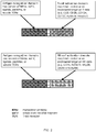
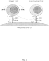
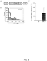
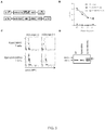
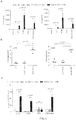

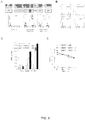

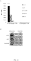


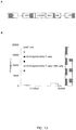


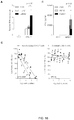

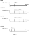


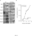


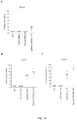
Classifications
-
- A—HUMAN NECESSITIES
- A61—MEDICAL OR VETERINARY SCIENCE; HYGIENE
- A61K—PREPARATIONS FOR MEDICAL, DENTAL OR TOILETRY PURPOSES
- A61K35/00—Medicinal preparations containing materials or reaction products thereof with undetermined constitution
- A61K35/12—Materials from mammals; Compositions comprising non-specified tissues or cells; Compositions comprising non-embryonic stem cells; Genetically modified cells
- A61K35/14—Blood; Artificial blood
- A61K35/17—Lymphocytes; B-cells; T-cells; Natural killer cells; Interferon-activated or cytokine-activated lymphocytes
-
- A—HUMAN NECESSITIES
- A61—MEDICAL OR VETERINARY SCIENCE; HYGIENE
- A61K—PREPARATIONS FOR MEDICAL, DENTAL OR TOILETRY PURPOSES
- A61K39/00—Medicinal preparations containing antigens or antibodies
- A61K39/0005—Vertebrate antigens
- A61K39/0011—Cancer antigens
- A61K39/001102—Receptors, cell surface antigens or cell surface determinants
- A61K39/001111—Immunoglobulin superfamily
- A61K39/001112—CD19 or B4
-
- A—HUMAN NECESSITIES
- A61—MEDICAL OR VETERINARY SCIENCE; HYGIENE
- A61K—PREPARATIONS FOR MEDICAL, DENTAL OR TOILETRY PURPOSES
- A61K39/00—Medicinal preparations containing antigens or antibodies
- A61K39/0005—Vertebrate antigens
- A61K39/0011—Cancer antigens
- A61K39/001102—Receptors, cell surface antigens or cell surface determinants
- A61K39/001122—Ephrin Receptors [Eph]
-
- A—HUMAN NECESSITIES
- A61—MEDICAL OR VETERINARY SCIENCE; HYGIENE
- A61K—PREPARATIONS FOR MEDICAL, DENTAL OR TOILETRY PURPOSES
- A61K39/00—Medicinal preparations containing antigens or antibodies
- A61K39/395—Antibodies; Immunoglobulins; Immune serum, e.g. antilymphocytic serum
- A61K39/39533—Antibodies; Immunoglobulins; Immune serum, e.g. antilymphocytic serum against materials from animals
- A61K39/39558—Antibodies; Immunoglobulins; Immune serum, e.g. antilymphocytic serum against materials from animals against tumor tissues, cells, antigens
-
- A—HUMAN NECESSITIES
- A61—MEDICAL OR VETERINARY SCIENCE; HYGIENE
- A61K—PREPARATIONS FOR MEDICAL, DENTAL OR TOILETRY PURPOSES
- A61K39/00—Medicinal preparations containing antigens or antibodies
- A61K39/46—Cellular immunotherapy
- A61K39/461—Cellular immunotherapy characterised by the cell type used
- A61K39/4611—T-cells, e.g. tumor infiltrating lymphocytes [TIL], lymphokine-activated killer cells [LAK] or regulatory T cells [Treg]
-
- A—HUMAN NECESSITIES
- A61—MEDICAL OR VETERINARY SCIENCE; HYGIENE
- A61K—PREPARATIONS FOR MEDICAL, DENTAL OR TOILETRY PURPOSES
- A61K39/00—Medicinal preparations containing antigens or antibodies
- A61K39/46—Cellular immunotherapy
- A61K39/463—Cellular immunotherapy characterised by recombinant expression
- A61K39/4633—Antibodies or T cell engagers
-
- A—HUMAN NECESSITIES
- A61—MEDICAL OR VETERINARY SCIENCE; HYGIENE
- A61K—PREPARATIONS FOR MEDICAL, DENTAL OR TOILETRY PURPOSES
- A61K39/00—Medicinal preparations containing antigens or antibodies
- A61K39/46—Cellular immunotherapy
- A61K39/464—Cellular immunotherapy characterised by the antigen targeted or presented
- A61K39/4643—Vertebrate antigens
- A61K39/4644—Cancer antigens
- A61K39/464402—Receptors, cell surface antigens or cell surface determinants
- A61K39/464411—Immunoglobulin superfamily
- A61K39/464412—CD19 or B4
-
- A—HUMAN NECESSITIES
- A61—MEDICAL OR VETERINARY SCIENCE; HYGIENE
- A61K—PREPARATIONS FOR MEDICAL, DENTAL OR TOILETRY PURPOSES
- A61K39/00—Medicinal preparations containing antigens or antibodies
- A61K39/46—Cellular immunotherapy
- A61K39/464—Cellular immunotherapy characterised by the antigen targeted or presented
- A61K39/4643—Vertebrate antigens
- A61K39/4644—Cancer antigens
- A61K39/464402—Receptors, cell surface antigens or cell surface determinants
- A61K39/464422—Ephrin Receptors [Eph]
-
- A—HUMAN NECESSITIES
- A61—MEDICAL OR VETERINARY SCIENCE; HYGIENE
- A61K—PREPARATIONS FOR MEDICAL, DENTAL OR TOILETRY PURPOSES
- A61K39/00—Medicinal preparations containing antigens or antibodies
- A61K39/46—Cellular immunotherapy
- A61K39/464—Cellular immunotherapy characterised by the antigen targeted or presented
- A61K39/4643—Vertebrate antigens
- A61K39/4644—Cancer antigens
- A61K39/464402—Receptors, cell surface antigens or cell surface determinants
- A61K39/464429—Molecules with a "CD" designation not provided for elsewhere
-
- A—HUMAN NECESSITIES
- A61—MEDICAL OR VETERINARY SCIENCE; HYGIENE
- A61K—PREPARATIONS FOR MEDICAL, DENTAL OR TOILETRY PURPOSES
- A61K45/00—Medicinal preparations containing active ingredients not provided for in groups A61K31/00 - A61K41/00
- A61K45/06—Mixtures of active ingredients without chemical characterisation, e.g. antiphlogistics and cardiaca
-
- A—HUMAN NECESSITIES
- A61—MEDICAL OR VETERINARY SCIENCE; HYGIENE
- A61N—ELECTROTHERAPY; MAGNETOTHERAPY; RADIATION THERAPY; ULTRASOUND THERAPY
- A61N5/00—Radiation therapy
- A61N5/10—X-ray therapy; Gamma-ray therapy; Particle-irradiation therapy
-
- A—HUMAN NECESSITIES
- A61—MEDICAL OR VETERINARY SCIENCE; HYGIENE
- A61P—SPECIFIC THERAPEUTIC ACTIVITY OF CHEMICAL COMPOUNDS OR MEDICINAL PREPARATIONS
- A61P35/00—Antineoplastic agents
-
- C—CHEMISTRY; METALLURGY
- C07—ORGANIC CHEMISTRY
- C07K—PEPTIDES
- C07K14/00—Peptides having more than 20 amino acids; Gastrins; Somatostatins; Melanotropins; Derivatives thereof
- C07K14/435—Peptides having more than 20 amino acids; Gastrins; Somatostatins; Melanotropins; Derivatives thereof from animals; from humans
- C07K14/705—Receptors; Cell surface antigens; Cell surface determinants
- C07K14/70503—Immunoglobulin superfamily
- C07K14/7051—T-cell receptor (TcR)-CD3 complex
-
- C—CHEMISTRY; METALLURGY
- C07—ORGANIC CHEMISTRY
- C07K—PEPTIDES
- C07K16/00—Immunoglobulins [IGs], e.g. monoclonal or polyclonal antibodies
- C07K16/18—Immunoglobulins [IGs], e.g. monoclonal or polyclonal antibodies against material from animals or humans
- C07K16/28—Immunoglobulins [IGs], e.g. monoclonal or polyclonal antibodies against material from animals or humans against receptors, cell surface antigens or cell surface determinants
- C07K16/2803—Immunoglobulins [IGs], e.g. monoclonal or polyclonal antibodies against material from animals or humans against receptors, cell surface antigens or cell surface determinants against the immunoglobulin superfamily
- C07K16/2809—Immunoglobulins [IGs], e.g. monoclonal or polyclonal antibodies against material from animals or humans against receptors, cell surface antigens or cell surface determinants against the immunoglobulin superfamily against the T-cell receptor (TcR)-CD3 complex
-
- C—CHEMISTRY; METALLURGY
- C07—ORGANIC CHEMISTRY
- C07K—PEPTIDES
- C07K16/00—Immunoglobulins [IGs], e.g. monoclonal or polyclonal antibodies
- C07K16/18—Immunoglobulins [IGs], e.g. monoclonal or polyclonal antibodies against material from animals or humans
- C07K16/28—Immunoglobulins [IGs], e.g. monoclonal or polyclonal antibodies against material from animals or humans against receptors, cell surface antigens or cell surface determinants
- C07K16/2866—Immunoglobulins [IGs], e.g. monoclonal or polyclonal antibodies against material from animals or humans against receptors, cell surface antigens or cell surface determinants against receptors for cytokines, lymphokines, interferons
-
- C—CHEMISTRY; METALLURGY
- C07—ORGANIC CHEMISTRY
- C07K—PEPTIDES
- C07K16/00—Immunoglobulins [IGs], e.g. monoclonal or polyclonal antibodies
- C07K16/18—Immunoglobulins [IGs], e.g. monoclonal or polyclonal antibodies against material from animals or humans
- C07K16/28—Immunoglobulins [IGs], e.g. monoclonal or polyclonal antibodies against material from animals or humans against receptors, cell surface antigens or cell surface determinants
- C07K16/30—Immunoglobulins [IGs], e.g. monoclonal or polyclonal antibodies against material from animals or humans against receptors, cell surface antigens or cell surface determinants from tumour cells
-
- C—CHEMISTRY; METALLURGY
- C07—ORGANIC CHEMISTRY
- C07K—PEPTIDES
- C07K16/00—Immunoglobulins [IGs], e.g. monoclonal or polyclonal antibodies
- C07K16/40—Immunoglobulins [IGs], e.g. monoclonal or polyclonal antibodies against enzymes
-
- C—CHEMISTRY; METALLURGY
- C07—ORGANIC CHEMISTRY
- C07K—PEPTIDES
- C07K16/00—Immunoglobulins [IGs], e.g. monoclonal or polyclonal antibodies
- C07K16/46—Hybrid immunoglobulins
- C07K16/468—Immunoglobulins having two or more different antigen binding sites, e.g. multifunctional antibodies
-
- C—CHEMISTRY; METALLURGY
- C12—BIOCHEMISTRY; BEER; SPIRITS; WINE; VINEGAR; MICROBIOLOGY; ENZYMOLOGY; MUTATION OR GENETIC ENGINEERING
- C12N—MICROORGANISMS OR ENZYMES; COMPOSITIONS THEREOF; PROPAGATING, PRESERVING, OR MAINTAINING MICROORGANISMS; MUTATION OR GENETIC ENGINEERING; CULTURE MEDIA
- C12N5/00—Undifferentiated human, animal or plant cells, e.g. cell lines; Tissues; Cultivation or maintenance thereof; Culture media therefor
- C12N5/06—Animal cells or tissues; Human cells or tissues
- C12N5/0602—Vertebrate cells
- C12N5/0634—Cells from the blood or the immune system
- C12N5/0636—T lymphocytes
-
- A—HUMAN NECESSITIES
- A61—MEDICAL OR VETERINARY SCIENCE; HYGIENE
- A61K—PREPARATIONS FOR MEDICAL, DENTAL OR TOILETRY PURPOSES
- A61K39/00—Medicinal preparations containing antigens or antibodies
- A61K2039/51—Medicinal preparations containing antigens or antibodies comprising whole cells, viruses or DNA/RNA
- A61K2039/515—Animal cells
- A61K2039/5156—Animal cells expressing foreign proteins
-
- A—HUMAN NECESSITIES
- A61—MEDICAL OR VETERINARY SCIENCE; HYGIENE
- A61K—PREPARATIONS FOR MEDICAL, DENTAL OR TOILETRY PURPOSES
- A61K39/00—Medicinal preparations containing antigens or antibodies
- A61K2039/51—Medicinal preparations containing antigens or antibodies comprising whole cells, viruses or DNA/RNA
- A61K2039/515—Animal cells
- A61K2039/5158—Antigen-pulsed cells, e.g. T-cells
-
- A—HUMAN NECESSITIES
- A61—MEDICAL OR VETERINARY SCIENCE; HYGIENE
- A61K—PREPARATIONS FOR MEDICAL, DENTAL OR TOILETRY PURPOSES
- A61K2239/00—Indexing codes associated with cellular immunotherapy of group A61K39/46
- A61K2239/31—Indexing codes associated with cellular immunotherapy of group A61K39/46 characterized by the route of administration
-
- A—HUMAN NECESSITIES
- A61—MEDICAL OR VETERINARY SCIENCE; HYGIENE
- A61K—PREPARATIONS FOR MEDICAL, DENTAL OR TOILETRY PURPOSES
- A61K2239/00—Indexing codes associated with cellular immunotherapy of group A61K39/46
- A61K2239/38—Indexing codes associated with cellular immunotherapy of group A61K39/46 characterised by the dose, timing or administration schedule
-
- C—CHEMISTRY; METALLURGY
- C07—ORGANIC CHEMISTRY
- C07K—PEPTIDES
- C07K2317/00—Immunoglobulins specific features
- C07K2317/10—Immunoglobulins specific features characterized by their source of isolation or production
- C07K2317/14—Specific host cells or culture conditions, e.g. components, pH or temperature
-
- C—CHEMISTRY; METALLURGY
- C07—ORGANIC CHEMISTRY
- C07K—PEPTIDES
- C07K2317/00—Immunoglobulins specific features
- C07K2317/30—Immunoglobulins specific features characterized by aspects of specificity or valency
- C07K2317/31—Immunoglobulins specific features characterized by aspects of specificity or valency multispecific
-
- C—CHEMISTRY; METALLURGY
- C07—ORGANIC CHEMISTRY
- C07K—PEPTIDES
- C07K2317/00—Immunoglobulins specific features
- C07K2317/60—Immunoglobulins specific features characterized by non-natural combinations of immunoglobulin fragments
- C07K2317/62—Immunoglobulins specific features characterized by non-natural combinations of immunoglobulin fragments comprising only variable region components
- C07K2317/622—Single chain antibody (scFv)
-
- C—CHEMISTRY; METALLURGY
- C07—ORGANIC CHEMISTRY
- C07K—PEPTIDES
- C07K2319/00—Fusion polypeptide
- C07K2319/01—Fusion polypeptide containing a localisation/targetting motif
- C07K2319/03—Fusion polypeptide containing a localisation/targetting motif containing a transmembrane segment
-
- C—CHEMISTRY; METALLURGY
- C12—BIOCHEMISTRY; BEER; SPIRITS; WINE; VINEGAR; MICROBIOLOGY; ENZYMOLOGY; MUTATION OR GENETIC ENGINEERING
- C12N—MICROORGANISMS OR ENZYMES; COMPOSITIONS THEREOF; PROPAGATING, PRESERVING, OR MAINTAINING MICROORGANISMS; MUTATION OR GENETIC ENGINEERING; CULTURE MEDIA
- C12N2501/00—Active agents used in cell culture processes, e.g. differentation
- C12N2501/50—Cell markers; Cell surface determinants
- C12N2501/599—Cell markers; Cell surface determinants with CD designations not provided for elsewhere
-
- C—CHEMISTRY; METALLURGY
- C12—BIOCHEMISTRY; BEER; SPIRITS; WINE; VINEGAR; MICROBIOLOGY; ENZYMOLOGY; MUTATION OR GENETIC ENGINEERING
- C12N—MICROORGANISMS OR ENZYMES; COMPOSITIONS THEREOF; PROPAGATING, PRESERVING, OR MAINTAINING MICROORGANISMS; MUTATION OR GENETIC ENGINEERING; CULTURE MEDIA
- C12N2510/00—Genetically modified cells
Definitions
- the fields of the invention include at least immunology, cell biology, molecular biology, and medicine, including oncology.
- Immunotherapy with antigen-specific T-cells has shown promise in the treatment of solid tumors and hematological malignancies in preclinical models as well as in Phase I/II clinical studies.
- Genetically modifying T-cells with chimeric antigen receptors (CARs) or engineered T-cell receptors (TCRs) allows for the rapid generation of antigen-specific T-cells.
- CARs chimeric antigen receptors
- TCRs engineered T-cell receptors
- neither CAR nor engineered TCR expressing T-cells redirect the vast reservoir of resident immune cells to tumors limiting anti-tumor effects.
- WO 2009/091826 discloses compositions and methods related to a human CD19-specific chimeric T cell receptor polypeptide comprising and intracellular signaling domain, a transmembrane domain and an extracellular domain, the extracellular domain comprising a human CD19 binding region.
- composition comprising, or consisting essentially of, or consisting of, an engager, wherein said engager is a molecule that comprises an activation domain that binds to an activation molecule on an immune cell surface or an engineered immune cell surface, and an antigen recognition domain that binds to a target cell antigen, e.g., an antigen expressed on the surface of a tumor cell or cancer cell.
- the cancer cell may be of a solid tumor or a hematological malignancy.
- Engager cells are cells that have the capability of secreting engager molecules ( FIG. 1 ), and in certain aspects of the invention, the individual is provided with cells that provide therapy to the individual.
- the cells include T-cells and NK cells) secrete engagers that activate immune cells.
- the immune cell kills the target cell.
- the engager may be bipartite (e.g ., comprising an activation domain and antigen recognition domain that may optionally be joined by a linker), or may be tripartite or multipartite (e.g ., comprise one or more activation domains and/or antigen recognition domains, or other domains).
- the engager is a protein, e.g., an engineered protein.
- the activation domain of the engager is or comprises an antibody or an antigen-binding fragment or portion thereof, e.g., a single chain variable fragment (scFv).
- the antigen recognition domain is or comprises an antibody or an antigen-binding fragment or portion thereof, e.g., a monoclonal antibody or an scFv, or it may comprise ligands, peptides, soluble T-cell receptors, or combinations thereof ( FIG. 2 ).
- the activation domain and antigen recognition domain are joined by a linker, e.g., a peptide linker.
- the activation domain of an engager molecule can provide activation to immune cells.
- immune cells have different activating receptors.
- CD3 is an activating receptor on T-cells
- CD16, NKG2D, or NKp30 are activating receptors on NK cells
- CD3 or an invariant TCR are the activating receptors on NKT-cells.
- Engager molecules that activate T-cells may therefore have a different activation domain than engager molecules that activate NK cells ( FIG. 2 ).
- the activation molecule is one or more of CD3, e.g ., CD3 ⁇ , CD3 ⁇ or CD3 ⁇ ; or CD27, CD28, CD40, CD134, CD137, and CD278.
- the activation molecule is CD16, NKG2D, or NKp30, or wherein the immune cell is a NKT-cell, the activation molecule is CD3 or an invariant TCR, or wherein the immune cell is a NKT cell, the activation molecule is an invariant TCR is CD1d.
- Activation can either result in a positive or a negative signal.
- Examples for 'positive signal' activation domains include 1) scFvs that are specific for (and activate) CD3 and ligands or receptors of co-stimulatory molecules, such as CD28, CD134, and CD137; 2) domains derived from co-stimulatory molecules, such as CD80, CD70, CD134L (OX40L), and CD137L (41BBL); 3) cytokines, such as IL-2, IL-7, and IL-15; and 4) chemokines such as CCL1, CCL2, CCL 16, CCL 17, CCL 22, CXCL8, or RANTES.
- Examples for 'negative signal' activation domains include 1) domains derived from inhibitory molecules, such as PD-L1; 2) scFvs specific for inhibitory molecules such as CTLA4 and PD-1, and 2) inhibitory cytokines, such as TGF ⁇ and IL-10.
- the activation domain is an scFv.
- the engager additionally comprises one or more accessory domains, e.g ., one or more of a cytokine, a costimulatory domain, a domain that inhibits negative regulatory molecules of T-cell activation, or a combination thereof.
- the cytokine is IL-15, IL-2, and/or IL-7.
- the costimulatory domain is CD27, CD80, CD86, CD134, or CD137.
- the domain that inhibits negative regulatory molecules of T-cell activation is PD-1, PD-L1, CTLA4, or B7-H4.
- the respective domains may be in any order N-terminus to C-terminus, including, e.g ., having the activation domain N-terminal to the antigen recognition domain, having the activation domain C-terminal to the antigen recognition domain, and so forth.
- T-cells are modified to secrete engager molecules that have the antigen recognition domain N-terminal to the activation domain.
- two or more of the domains of an engager molecule are separated by a linker.
- the linker may be of any suitable length, and such a parameter is routinely optimized in the art.
- linkers may be of a length and sequence sufficient to ensure that each of the first and second domains can, independently from one another, retain their differential binding specificities.
- the antigen bound by the antigen recognition domain is a tumor-associated antigen (TAA) or a tumor-specific antigen (TSA).
- TAA tumor-associated antigen
- TSA tumor-specific antigen
- the antigen recognition domain of the engager is an scFv that is specific for a TAA or TSA.
- the TAA or TSA is expressed on a cancer cell.
- the TAA or TSA is expressed on a blood cancer cell.
- the TAA or TSA is expressed on a cell of a solid tumor.
- the solid tumor is a glioblastoma, a non-small cell lung cancer, a lung cancer other than a non-small cell lung cancer, breast cancer, prostate cancer, pancreatic cancer, liver cancer, colon cancer, stomach cancer, a cancer of the spleen, skin cancer, a brain cancer other than a glioblastoma, a kidney cancer, a thyroid cancer, or the like.
- the TAA or TSA is expressed by a tumor cell in an individual.
- the TAA or TSA is one or more of, e.g., an scFv on the engager is specific for one or more of 5T4, 8H9, ⁇ v ⁇ 6 integrin, BCMA, B7-H3, B7-H6, CAIX, CA9, CD19, CD20, CD22, CD30, CD33, CD38, CD44, CD44v6, CD44v7/8, CD70, CD123, CD138, CD171, CEA, CSPG4, EGFR, EGFR family including ErbB2 (HER2), EGFRvIII, EGP2, EGP40, , ERBB3, ERBB4, ErbB3/4, EPCAM, EphA2, EpCAM, FAP, FBP, fetal AchR, FR ⁇ , GD2, GD3, Glypican-3 (GPC3), HLA-A1+MAGE1, HLA-A1+NY-ESO-1, IL-11R ⁇ , IL-13R ⁇ 2, Lambda, Lewis
- the particular cell recognition domain of the engager molecule may be tailored to recognize a corresponding tumor expressing a particular antigen.
- Antigens may be either produced by a malignant cancer cell or by the so-called tumor stroma, which support tumor growth.
- the scFv is specific for EphA2, CD19, CD123, LeY, B7H3, HER2, or EGFR (including EGFRVIII). Exemplary antigens are listed in Table 1.
- Table 1 Examples of targetable antigens Antigen 5T4 ⁇ v ⁇ 6 integrin B7-H3 B7-H6 CD19 CD20 CD22 CD30 CD33 CD44, CD44v6, CD44v7/8 CD70 CD123 CEA CSPG4 EGFR, EGFR family including ErbB2 (HER2) EGFRvIII EGP2 EGP40 EpCAM EphA2 FAP fetal AchR FR ⁇ GD2 GD3 Glypican-3 (GPC3) HLA-A1+MAGE1 HLA-A2+MAGE1 HLA-A3+MAGE1 HLA-A1+NY-ESO-1 HLA-A2+NY-ESO-1 HLA-A3+NY-ESO-1 IL-11R ⁇ IL-13R ⁇ 2 Lambda Lewis-Y Kappa Mesothelin Muc1 Muc1 6 NCAM NKG2D Ligands NY-ESO-1 PRAME PSCA PSMA ROR1 TAG72 TEMs VEGFR2
- the antigen recognition domain can also target other antigens; for example inhibitory molecules expressed on cancer cells or cells within the tumor microenvironment, such as PD-L1 and CTLA4.
- An engager molecule that comprises a PD-L1-specific antigen recognition domain and a CD3-specific T-cell activation domain would overcome tumor-mediated immunosuppression by converting an inhibitory signal into a positive (CD3-activating) signal.
- Other examples are antigens that are present with in the extracelluar matrix of tumors such as oncofetal variants of fibronectin, tenascin, or necrotic regions of tumors.
- a polynucleotide that encodes an engager molecule, e.g., any of the engager molecules described herein.
- the polynucleotide may be comprised within, or comprise, a vector. Any type of suitable vector may be employed, but in specific embodiments the vector is a non-viral vector (e.g., a plasmid, a minicircle DNA vector, a sleeping beauty plasmid, a piggybac plasmid, and so forth) or a viral vector (e.g., a lentiviral vector, adenoviral vector, retroviral vector, adeno-associated viral vector, oncolytic vector, and so forth).
- a non-viral vector e.g., a plasmid, a minicircle DNA vector, a sleeping beauty plasmid, a piggybac plasmid, and so forth
- a viral vector e.g., a lentiviral vector, adenoviral vector, retro
- the polynucleotide stably expresses the engager molecule in a cell that contains the polynucleotide.
- the polynucleotide transiently expresses the engager molecule in a cell that contains the polynucleotide.
- the polynucleotide allows inducible expression of the engager molecule in a cell that contains the polynucleotide.
- the polynucleotide allows inducible repression of expression of the engager molecule in a cell that contains the polynucleotide.
- the polynucleotide preferably encodes the engager in a form that is secretable by the cell.
- the polynucleotide encodes a fusion molecule comprising an activation domain and an antigen recognition domain, and in specific embodiments each domain is a scFv.
- the activation domain may be positioned toward the N-terminus of the polypeptide in relation to the antigen recognition domain, or the activation domain may be positioned toward the C-terminus of the polypeptide in relation to the antigen recognition domain.
- a cell that has been genetically engineered to express one or more engagers e.g ., any of the engager molecules described herein (e.g., referred to as an "engager cell”).
- the genetically engineered cell is, e.g., a T lymphocyte (T-cell), a CAR T-cell, a natural killer (NK) T-cell, or an NK cell.
- the genetically engineered cell is a non-immune cell, e.g., a mesenchymal stem cell (MSC), a neuronal stem cell, a hematopoietic stem cell, an induced pluripotent stem cell (iPS cell), an embryonic stem cell.
- the engager cell secretes the engager into the environment surrounding the engager cell, e.g., culture medium (where the engager cell is cultured in vitro ) or into an individual into which the engager cell has been administered).
- a lentiviral vector may be employed, in some cases a retroviral vector having higher transduction efficiency may be employed.
- An exemplary retroviral vector comprises in a 5' to 3' direction CD19scFv-CD3scFv.
- Another exemplary retroviral vector comprises in a 5' to 3' direction EphA2(4H5)-CD3scFv, optionally followed by IRES followed by a nucleotide sequence encoding a fluorescent or selectable marker, e.g ., mOrange.
- a cell secretes more than one (e.g., 1, 2, 3, 4, 5, 6, 7, 8, 9, 10 or more) type of engager molecule.
- the cell may be modified to harbor non-identical polynucleotides that express non-identical engager molecules.
- one cell may comprise one polynucleotide that encodes an engager molecule and also comprise one or more other polynucleotides that encode other engager molecules.
- the cell harbors a first polynucleotide that encodes an engager molecule that targets one of the antigens listed in Table 1 and a second polynucleotide that encodes an engager molecule that targets another antigen listed in Table 1.
- the engagers described herein may also contain different activation domains so that immune cells can activate T-cells or NK cells, or both.
- T-cells that for example secrete engager molecules that can activate NK cells and/or T-cells, and NK cells that activate NK and/or T-cells.
- the engager cells of the present invention do not also comprise a CAR, an engineered TCR, or any other genetic modification
- the engager T-cells also comprise a CAR, including one that targets antigens listed in Table 1, an engineered TCR, or any other genetic modification that may enhance its function.
- a CAR or engineered TCR expressing T-cell comprises one or more engager molecules.
- an antigen binding domain of a CAR or TCR and an antigen recognition domain of an engager molecule recognize or bind to the same target antigen.
- an antigen binding domain of a CAR and an antigen recognition domain of an engager molecule recognize or bind to different target antigens expressed on the same target cell.
- a cell comprises one polynucleotide that expresses an engager molecule and also comprises one or more polynucleotides that express co-stimulatory molecules, including CD80, CD70, CD134L (OX40L), and CD137L (41BBL).
- the cell harbors a first polynucleotide that encodes an engager molecule that targets CD19 and a second polynucleotide that encodes CD80 and 41BBL ( FIG. 22 ).
- compositions that include an engager molecule and/or one or more cells that secrete an engager molecule, including one that has an activation domain that binds to a target on a native immune cell surface or an engineered immune cell surface and also a recognition domain that binds one or more molecules produced by a target cell.
- An effective amount of the engager molecules or cells that secrete them are given to an individual in need thereof.
- a method of treating an individual having a tumor cell comprising administering to the individual a therapeutically effective amount of an engager molecule, wherein the engager molecule comprises an antigen recognition domain that binds to an antigen on the tumor cell.
- a method of treating an individual having a tumor cell comprising administering to the individual a therapeutically effective amount of engager cells that secrete one or more engager molecules, wherein the engager molecules comprise an antigen recognition domain that binds to an antigen on the tumor cell.
- said administering results in a measurable decrease in the growth of the tumor in the individual.
- said administering results in a measurable decrease in the size of the tumor in the individual.
- the size or growth rate of a tumor may be determinable by, e.g., direct imaging (e.g., CT scan, MRI, PET scan or the like), fluorescent imaging, tissue biopsy, and/or evaluation of relevant physiological markers (e.g ., PSA levels for prostate cancer; HCG levels for choriocarcinoma, and the like).
- the individual has a high level of an antigen, e.g ., EphA2, that is correlated to poor prognosis.
- the individual is provided with an additional cancer therapy, such as surgery, radiation, chemotherapy, hormone therapy, immunotherapy, or a combination thereof.
- a cell that secretes a composition comprising: an activation domain that binds to a target on a native immune cell surface or an engineered immune cell surface; and an antigen recognition domain that binds one or more molecules produced by a target cell.
- the activation domain and the antigen recognition domain are attached by a linker, in at least some cases.
- the activation domain may comprise an antibody or a ligand.
- the immune cell is a T-cell and the antibody recognizes CD3, although the immune cell may be a NK cell and the antibody recognizes CD16, NKG2D, or NKp30.
- the antibody is a single chain fragment variable (scFv) antibody.
- the antigen recognition domain is an antibody that recognizes an antigen produced by target cells, and in some cases the antibody is an scFv fragment.
- the antigen recognition domain is a ligand, a peptide, a soluble TCR, or recognizes an antigen of Table 1.
- compositions of the disclosure comprise the sequence of SEQ ID NO:1, SEQ ID NO:2, SEQ ID NO:3, SEQ ID NO:4, SEQ ID NO:5, SEQ ID NO:6, SEQ ID NO:7, SEQ ID NO:8, or SEQ ID NO:9, for example.
- compositions of the disclosure further comprise one or more of the following: a cytokine; a co-stimulatory domain; or a domain for inhibition of negative regulatory molecules of T-cell activation.
- the cytokine is IL-15.
- the co-stimulatory domain is CD80, CD137, or both.
- the domain for inhibition of negative regulatory molecules of T-cell activation comprises PD-1, PD-L1, CTLA4, or B7-H4.
- Some compositions of the disclosure comprise a detectable marker.
- the composition is secreted by the cells and the composition binds to and activates the cells.
- the composition is secreted by the cells and the composition binds to and activates native immune cells in the individual.
- the cells that are capable of expressing the composition are T-cells and the activation domain comprises an antibody that recognizes CD3.
- the cells that are capable of expressing the composition are NK cells and the activation domain comprises an antibody that recognizes CD16, NKG2D, or NKp30.
- the cancer is EphA2-positive, CD19-positive, CD123-positive, LeY-positive, B7H3-positive, HER2-positive, or EGFR (including EGFRvIII)-positive.
- composition comprising a cell that secretes a polypeptide, said polypeptide comprising: an activation domain that binds to a target on an immune cell surface; and an antigen recognition domain that binds one or more molecules produced by or present on a target cell.
- the activation domain and the antigen recognition domain are attached by a linker.
- the immune cell is a native immune cell.
- the immune cell is an engineered immune cell.
- the activation domain comprises an antibody, ligand, receptor, or peptide.
- the immune cell is a T-cell and the antibody recognizes CD3.
- the immune cell is a NK cell and the antibody recognizes CD16, NKG2D, or NKp30.
- the antibody is a single chain fragment variable (scFv) antibody.
- the antigen recognition domain is an antibody that recognizes an antigen produced by target cells.
- the antibody is a scFv antibody.
- the antigen recognition domain is a ligand, a peptide, or a soluble TCR. In specific aspects described herein, the antigen recognition domain recognizes an antigen of Table 1.
- the polypeptide comprises the sequence of SEQ ID NO:1, SEQ ID NO:2, SEQ ID NO:3, SEQ ID NO:4, SEQ ID NO:5, SEQ ID NO:6, SEQ ID NO:7, SEQ ID NO:8, or SEQ ID NO:9.
- the polypeptide further comprises one or more of the following: a cytokine; a co-stimulatory domain; or a domain for inhibition of negative regulatory molecules of T-cell activation.
- the cytokine is IL-15, IL-12, IL-2, or IL-7.
- the co-stimulatory domain is CD80, CD134, CD137, or a combination thereof.
- the domain for inhibition of negative regulatory molecules of T-cell activation comprises PD-1, PD-L1, CTLA4, or B7-H4.
- the polypeptide further comprises a detectable marker.
- the cell comprises a polynucleotide encoding the polypeptide.
- the polynucleotide is integrated into the genome of the cell.
- expression of the polynucleotide encoding the polypeptide is under the control of one or more tumor-associated factors.
- the one or more tumor-associated factors is a chemokine or cytokine.
- Chemokines may be from a family selected from the group consisting of CC, CXC, C, and CX3C chemokines, including CCL2, CCL3, CCL5, CCL16, CXCL8, CXCL10, XCL1, XCL2, or CX3CL1. Cytokines may be selected from the group consisting of IL2, IL4, IL5, IL7, IL10, IL12, IL15, and IL17.
- expression of the polynucleotide encoding the polypeptide is under the control of a tissue-specific regulatory sequence.
- the tissue-specific regulatory sequence is a hypoxia-specific regulatory sequence.
- the hypoxia-specific regulatory sequence is a hypoxia response element (HRE) or an oxygen-dependent degradation domain (ODDD).
- the HRE comprises 5'-RCGTG-3' (SEQ ID NO:10).
- the polypeptide is secreted by the cells and the composition binds to and activates the cells, and in at least some cases the polypeptide is secreted by the cells and the polypeptide binds to and activates native immune cells in the individual.
- the cells that are capable of expressing the polypeptide are T-cells.
- the activation domain comprises an antibody that recognizes CD3.
- the cells that are capable of expressing the polypeptide are NK cells.
- the activation domain comprises an antibody that recognizes CD16, NKG2D, or NKp30.
- the cancer is EphA2-positive, CD19-positive, CD123-positive, LeY-positive, B7H3-positive, HER2-positive, or EGFR-positive, and may be EGFRvIII-positive.
- the present invention provides:
- the term "engager” or “engager molecule” refers to a molecule that is secreted from a cell that activates immune cells.
- the engager activates specific immune cells according to the domains present in the engager.
- Illustrative examples of cells that secrete engagers include T-cells, NK cells, NKT cells, CAR T-cells, mesenchymal stem cells (MSCs), neuronal stem cells, hematopoietic stem cells, or a mixture thereof, in some cases.
- Antigen recognition domain refers to a part of the engager molecule that recognizes an antigen.
- antigens can be of any nature including, but not limited to, proteins, carbohydrates, and/or synthetic molecules.
- activation domain refers to a part of the engager molecules that interacts with the immune cells and induces a positive or negative immunomodulatory signal.
- positive immunomodulatory signals include signals that induce cell proliferation, cytokine secretion, or cytolytic activity.
- immunomodulatory signals include signals that inhibit cell proliferation, inhibit the secretion of immunosuppressive factors, or induce cell death.
- the term "native immune cell” refers to an immune cell that naturally occurs in the immune system.
- Illustrative examples include, but are not limited to, T-cells, NK cells, NKT cells, B cells, and dendritic cells.
- engineered immune cell refers to an immune cell that is genetically modified.
- co-stimulatory domain or “co-stimulatory signaling domain” refers to an intracellular signaling domain of a co-stimulatory molecule. In particular aspects, it refers to a domain that provides additional signals to the immune cell in conjunction with an activation domain .
- Co-stimulatory molecules are cell surface molecules other than antigen receptors or Fc receptors that provide a second signal required for efficient activation and function of T lymphocytes upon binding to antigen.
- co-stimulatory molecules include CD27, CD28, 4-1BB (CD137), OX40 (CD134), CD30, CD40, ICOS (CD278), LFA-1, CD2, CD7, LIGHT, NKD2C, CD70, CD80, CD86, and CD83.
- cells are genetically modified (including immune cells) with engager molecules comprising at least an antigen recognition domain and an activation domain and, optionally, a cytokine, costimulatory domain, and/or domain for inhibition of negative regulatory molecules of T-cell activation.
- the engager molecule's antigen recognition domain binds to one or more molecules present in and/or on target cells or that are secreted by target cells.
- the target cells are cancer cells, including at least solid tumor cells. Once engager molecules have bound a target molecule, they can activate cells that express the molecule recognized by the activation domain.
- Engager molecules can activate cells that are genetically modified with engager molecules or can activate unmodified cells.
- activation can result in a positive or negative signal.
- positive signals include signals that induce cell proliferation, cytokine secretion, or cytolytic activity.
- negative signals include signals that inhibit T-cell proliferation, inhibit the secretion of immunosuppressive factors, or induce cell death.
- immune cells that secrete engager molecules are able to redirect resident (naturally endogenous to a specific individual) immune cells to cancer cells.
- aspects of the disclosure provide delivery of modified immune cells that secrete an engager molecule to an individual in need thereof (known to have cancer or suspected of having cancer, including a particular cancer) in contrast to delivering the engager molecule only to the individual itself (in the absence of being produced by modified immune cells).
- the individual receives the modified immune cells that allow production of the engager molecule(s).
- the cells produce immunostimulatory cytokines; proliferate in an antigen-specific manner; kill the appropriate target cells; redirect bystander immune cells (including at least T-cells or NK cells) to cancer cells; secrete engager molecules upon activation; and/or are effective against cancer in a loco-regional or systemic manner.
- FIG. 3 illustrates examples of modified T-cells or NK cells that secrete engager molecules.
- a particular T-cell or NK-cell may produce an engager that can target the same cancer cell-specific antigen, the activation domains for a T-cell or NK cell must be different, because NK cells do not express CD3.
- Examples of activation domains for a NK cell include at least CD16, NKG2D, or NKp30, for example.
- compositions comprising a bispecific single chain antibody construct, or a bispecific constructs that has ligands, peptides, or soluble TCRs as antigen recognition domains and/or ligands as immune cell activation domains instead of fragments derived from antibodies ( FIG. 3 ).
- the bispecific single chain antibody construct is characterized to comprise or consist of or consist essentially of at least two domains, whereby one of said at least two domains specifically binds to a tumor antigen listed in Table 1, for example (EphA2, CD19, CD123, LeY, B7H3, HER2, and EGFR, (including EGFRvIII), and a second domain binds to an immune cell surface protein, such as CD3 on T-cells, for example.
- an engager molecule may comprise three or more domains.
- aspects described herein further encompass a process for the production of the composition of the disclosure, a method for the prevention, treatment or amelioration of cancer, and the use of a bispecific engager molecule construct in the prevention, treatment or amelioration of cancer or at least one symptom thereof.
- the engager compositions comprise a bispecific single chain antibody construct in addition to a third domain (although in some cases, more domains may be added), which may be referred to as a tripartite engager.
- the third or more domain may enhance activity of the composition or method, such as by providing a cytokine, costimulatory domain, and/or domain for inhibition of negative regulatory molecules of T-cell activation.
- aspects described herein further encompass a process for the production of the tripartite engager, a method for the prevention, treatment or amelioration of cancer, and the use of the tripartite engager in the prevention, treatment or amelioration of cancer.
- an engager molecule comprises an antigen recognition domain that binds EphA2.
- EphA2 may be referred to as EPH receptor A2 (ephrin type-A receptor 2; EPHA2; ARCC2; CTPA; CTPP1; or ECK), which is a protein that in humans is encoded by the EPHA2 gene in the ephrin receptor subfamily of the protein-tyrosine kinase family.
- Receptors in this subfamily generally comprise a single kinase domain and an extracellular region comprising a Cys-rich domain and 2 fibronectin type III repeats; embodiments of the antibodies of the disclosure may target any of these domains.
- EphA2 encodes a protein that binds ephrin-A ligands.
- An exemplary human EphA2 nucleic sequence is in GenBank® Accession No. NM_004431, and an exemplary human EphA2 polypeptide sequence is in GenBank® Accession No. NP_004422.
- an engager molecule comprises an antigen recognition domain that binds CD19.
- CD19 may be referred to as CD19 molecule, CD19 antigen, Differentiation antigen CD19, B-lymphocyte surface antigen B4, CVID3. Information in regards to gene locus, nucleotide and amino acid sequences can be found at HGNC: 1633, Entrez Gene: 930, Ensembl: ENSG00000177455, UniProtKB: P15391.
- CD19 encodes a cell surface molecule, which assembles with the antigen receptor of B lymphocytes in order to decrease the threshold for antigen receptor-dependent stimulation.
- An exemplary human CD19 molecule is in GenBank® Accession No. NM_001178098, and an exemplary human CD19 polypeptide is in GenBank® Accession No. NP_001171569.
- the engager cells may be generated by any suitable method in the art.
- the engager T-cells are generated by viral transduction of T-cells, although viral transduction of NK cells and other cells (CAR T-cells, NKT-cells, MSCs, neuronal stem cells, hematopoietic stem cells, in some cases) in at least certain cases is also useful, in some aspects described herein.
- the endogenous, native TCR of the transduced T-cells can either be unspecific or specific for an antigen, such as a tumor antigen or a viral antigen.
- the engager compositions of the disclosure include an antigen recognition domain that allows the engager-expressing T-cell and the corresponding engager molecule to target a particular cell of interest that expresses the antigen or to target a secretable antigen.
- any cell may comprise the antigen, but in specific aspects the antigen is displayed on a cancer cell, including a solid tumor cell.
- the cancer cell antigen may be of any kind, but in particular aspects it is specific to a cancer cell and may be specific to a particular type of cancer cell.
- the cancer cell may be breast, lung, prostate, or glioblastoma.
- the cell may be also be a pathogenic cell, such as a bacterial cell.
- any cancer antigen may be targeted by the engager-expressing T-cells described herein or the corresponding engager molecules thereof.
- the antigens are listed in Table 1.
- the engager molecule comprises more than one antigen recognition domain.
- the antigen When targeting a particular cancer antigen, the antigen may be targeted with any suitable scFv or antigen binding fragment of an antibody.
- scFvs which were used to construct engager molecules are listed in Table 2. The functionality of T-cells expressing the respective engager molecules was confirmed, and is described in detail in EXAMPLE 2.
- Table 2 scFVs used for engager molecule construction Target MAb CD19 FMC63 CD123 26292 CD123 32716 LeY Hu3S193 EphA2 4H5 B7H3 8H9 HER2 FRP5 EGFR including EGFRvIII 806
- the engager compositions of the disclosure include an activation domain that allows the immune cell that expresses the engager molecule and/or other immune cells to bind to the engager and a target cell.
- the activation domain is an antibody or antigen-binding fragment thereof, in particular aspects.
- Illustrative examples of activation domains include, but are not limited to antibody or antigen-binding fragment thereof, ligands, peptides, soluble T-cell receptors, or combinations thereof.
- the immune cell to which the engager binds may be an unmodified naturally endogenous (to the recipient individual) immune cell, or it may be a genetically modified immune cell. Binding of the engager to the target immune cell through the activation domain (such as an CD3 monoclonal antibody in the case of T-cells), thereby activates the target immune cell.
- Other activation molecules that can be readily targeted with engagers include co-stimulatory molecules such as CD27, CD28, CD134, and CD137.
- T-cells can be engineered to express one engager molecule with an EphA2-specific antigen recognition domain and a CD3-specific activation domain, and another engager with a HER2-specific antigen recognition domain and a CD28-specific activation domain.
- the activation domain may comprise of an antibody that recognizes CD 16 (such as NM3E2 antibody), or ligands specific for NKG2D (ULBP2), or NKp30 (B7H6).
- the activation domain comprises ligands, receptors, peptides, etc.
- an engager comprises an additional domain that enhances the activity of the engager and/or enhances the activity of the immune cell expressing the engager molecule.
- an additional domain may be of any kind, in specific embodiments the additional domain comprises one or more antigen recognition domains or one or more activation domain.
- the additional domain may comprise a cytokine, a co-stimulatory domain, or an entity that inhibits negative regulatory molecules of T-cell activation.
- only one additional domain is employed in engager molecules, but in other embodiments more than one may be employed, including more than one of the same or different type.
- Additional domains could offset immune escape by targeting an additional antigen.
- an engager molecule could be designed to target CD19 and CD22 for hematological malignancies or EphA2 and HER2 for solid tumors. Additional domains could also provide co-stimulation.
- an engager molecule could be designed to target a tumor antigen (see Table 1), and contain a CD3-specific scFv for T-cell activation and the extracellular domain of 41BBL to provide co-stimulation.
- An additional domain could attract immune cells.
- an additional domain encoding the chemokine RANTES could be used to increase the trafficking of immune cells to tumor sites.
- An additional domain could also be used to provide an immune cell growth factor such a cytokine.
- an additional domain encoding cytokines like IL-2 or IL15 could be used to enhance immune cell proliferation and expansion.
- An additional domain could also be used to change the physical properties of the engager molecule.
- an additional domain encoding a CH2-CH3 domain or a leucine zipper could promote dimerization or trimerization of the engager molecule resulting in enhanced function.
- the additional domain encodes any cytokine.
- the cytokine may be IL-2, IL-7, and/or IL-15, and in some aspects, an engager comprises more than one additional domain encoding cytokines.
- an engager comprises a co-stimulatory domain as described elsewhere herein.
- the co-stimulatory domain comprises CD80, CD137, and the like.
- the engager molecule comprises a domain that inhibits negative regulatory molecules of T-cell activation.
- the domain is PD-L1 or CTLA4, for example.
- an engager instead of an antigen recognition domain being an antibody, an engager comprises a different kind of domain that is not an antibody but that is capable of recognizing a cancer cell.
- the engager comprises one member of a ligand-receptor binding pair, wherein the cognate member is expressed on the cancer cell.
- the engager comprises a soluble T-cell receptor (TCR) domain, such as in lieu of an antibody.
- FIG. 2 illustrates an exemplary embodiment with a T-cell activation domain linked to a soluble TCR.
- an scFv contains a VH and VL domain connected by a linker peptide.
- SEQ ID NO: 1 is a CD19-CD3 T-cell Engager comprising the following formula:
- SEQ ID NO:1 is as follows, where the first underlined section is the leader and the following underlined sections are linker sequences between the respective scFvs, according to the formula above:
- SEQ ID NO:2 is as follows, where the first underlined section is the leader and the following underlined sections are linker sequences between the respective scFvs according to the formula above:
- SEQ ID NO:3 is as follows, where the first underlined section is the leader and the following underlined sections are linker sequences between the respective scFvs according to the formula above:
- SEQ ID NO:4 is as follows, where the first underlined section is the leader and the following underlined sections are linker sequences between the respective scFvs according to the formula above:
- SEQ ID NO:5 is as follows, where the first underlined section is the leader and the following underlined sections are linker sequences between the respective scFvs according to the formula above:
- SEQ ID NO:6 is as follows, where the first underlined section is the leader and the following underlined sections are linker sequences between the respective scFvs according to the formula above:
- SEQ ID NO:7 is as follows, where the first underlined section is the leader and the following underlined sections are linker sequences between the respective scFvs according to the formula above:
- SEQ ID NO:8 is as follows, where the first underlined section is the leader and the following underlined sections are linker sequences between the respective scFvs according to the formula above:
- SEQ ID NO:9 is as follows, where the first underlined section is the leader and the following underlined sections are linker sequences between the respective scFvs according to the formula above:
- bispecific single chain antibody construct relates to a construct comprising two antibody derived binding domains.
- One of the binding domains may comprise variable regions (or parts thereof) of an antibody, antibody fragment or derivative thereof, capable of specifically binding to/interacting with a target antigen, e . g .,EphA2 or CD19.
- the second binding domain may comprise variable regions (or parts thereof) of an antibody, antibody fragment or derivative thereof, capable of specifically binding to/interacting with an activation molecule, e.g., human CD3 antigen.
- a part of a variable region comprises at least one CDR ("Complementary determining region"), such as at least a CDR1, CDR2, or CDR3 region.
- the two domains/regions in the single chain antibody construct are preferably covalently connected to one another as a single chain.
- An scFv in general contains a VH and VL domain connected by a linker peptide.
- the secretable engager is composed of a signal peptide (to allow for secretion) from cells, followed by two or more scFvs connected by linker peptides (Lx, Ly, Lz).
- Linkers may be of a length and sequence sufficient to ensure that each of the first and second domains can, independently from one another, retain their differential binding specificities.
- Bispecific scFvs can be arranged in different formats: V H ⁇ -Lx-V L ⁇ -Ly-V H ⁇ -Lz-V L ⁇ , V L ⁇ -Lx-V H ⁇ -Ly-V H ⁇ -Lz-V L ⁇ , V L ⁇ -Lx-V H ⁇ -Ly-V L ⁇ -Lz-V H ⁇ , V H ⁇ -Lx-V L ⁇ -Ly-V L ⁇ -Lz-V H ⁇ , V H ⁇ -Lx-V L ⁇ -Ly-V H ⁇ -Lz-V L ⁇ , V H ⁇ -Lx-V L ⁇ -Ly-V H ⁇ -Lz-V L ⁇ , V L ⁇ -Lx-V H ⁇ -Ly-V H ⁇ -Lz-V L ⁇ , V L ⁇ -Lx-V H ⁇ -Ly-V L ⁇ -Lz-V H ⁇ , V L ⁇ -L
- the "bispecific single chain antibody construct" to be employed in the composition of the disclosure comprises a bispecific single chain Fv (scFv).
- scFv bispecific single chain Fv
- Illustrative examples of bispecific single chain molecules are known in the art and are described in WO 99/54440 ; Mack, J. Immunol. (1997), 158, 3965-3970 ; Mack, PNAS, (1995), 92, 7021-7025 ; Kufer, Cancer Immunol. Immunother., (1997), 45, 193-197 ; Loffler, Blood, (2000), 95, 6, 2098-2103 ; and Bruhl, J. Immunol., (2001), 166, 2420-2426 .
- an exemplary molecular format of the disclosure provides a polypeptide construct wherein the antibody-derived region comprises one V H and one V L region.
- the intramolecular orientation of the V H -domain and the V L -domain, which are linked to each other by a linker-domain, in the scFv format is not decisive for the recited bispecific single chain constructs.
- scFvs with both possible arrangements V H -domain-linker domain-V L -domain; V L -domain-linker domain-V H -domain) are contemplated in particular aspects of the bispecific single chain construct.
- the engager construct may also comprise additional domains, e.g. , the antigen binding domain may contain multiple antigen recognition binding domains allowing targeting of multiple antigens; the activation domain may contain multiple domains to activate cells.
- single-chain as used in accordance with the present disclosure in some embodiments means that first and second domains of the bispecific single chain construct are covalently linked, preferably in the form of a co-linear amino acid sequence encodable by a single nucleic acid molecule.
- binding to/interacting with defines a binding/interaction of at least two "antigen-interaction-sites" with each other.
- the term "antigen-interaction-site” defines, in accordance with the present disclosure, a motif of a polypeptide that shows the capacity of specific interaction with a specific antigen or a specific group of antigens.
- the binding/interaction is also understood to define a "specific recognition”.
- specifically recognizing means in accordance with this disclosure that the antibody molecule is capable of specifically interacting with and/or binding to at least two amino acids of each of the target molecules as defined herein. The term relates to the specificity of the antibody molecule, i.e.
- the specific interaction of the antigen-interaction-site with its specific antigen may result in an initiation of a signal, e.g. due to the induction of a change of the conformation of the antigen, an oligomerization of the antigen, etc. Further, the binding may be exemplified by the specificity of a "key-lock-principle".
- specific motifs in the amino acid sequence of the antigen-interaction-site and the antigen bind to each other as a result of their primary, secondary or tertiary structure as well as the result of secondary modifications of said structure, in some embodiments.
- the specific interaction of the antigen-interaction-site with its specific antigen may result as well in a simple binding of the site to the antigen.
- the term "specific interaction" as used in accordance with the present disclosure means that the bispecific single chain construct does not or essentially does not cross-react with (poly)peptides of similar structures.
- Cross-reactivity of a panel of bispecific single chain constructs under investigation may be tested, for example, by assessing binding of the panel of bispecific single chain construct under conventional conditions (see, e.g ., Harlow and Lane, Antibodies: A Laboratory Manual, Cold Spring Harbor Laboratory Press, 1988 and Using Antibodies: A Laboratory Manual, Cold Spring Harbor Laboratory Press, 1999 ) to the (poly)peptide of interest as well as to a number of more or less (structurally and/or functionally) closely related (poly)peptides.
- Examples for the specific interaction of an antigen-interaction-site with a specific antigen comprise the specificity of a ligand for its receptor.
- the definition particularly comprises the interaction of ligands which induce a signal upon binding to its specific receptor.
- Examples for corresponding ligands comprise cytokines that interact/bind with/to its specific cytokine-receptors.
- Another example for said interaction is the interaction of an antigenic determinant (epitope) with the antigenic binding site of an antibody.
- binding to/interacting with may also relate to a conformational epitope, a structural epitope or a discontinuous epitope consisting of two regions of the human target molecules or parts thereof.
- a conformational epitope is defined by two or more discrete amino acid sequences separated in the primary sequence which come together on the surface of the molecule when the polypeptide folds to the native protein ( Sela, (1969) Science 166, 1365 and Layer, (1990) Cell 61, 553-6 ).
- constructs of the present disclosure are also envisaged to specifically bind to/interact with a conformational/structural epitope(s) composed of and/or comprising the two regions of the human CD3 complex described herein or parts thereof as disclosed herein below.
- antibody fragment or derivative thereof' relates to single chain antibodies, or fragments thereof, synthetic antibodies, antibody fragments, such as a Camel Ig, Ig NAR, Fab fragments, Fab' fragments, F(ab)'2 fragments, F(ab)'3 fragments, Fv, single chain Fv antibody (“scFv”), bis-scFv, (scFv)2, minibody, diabody, triabody, tetrabody, disulfide stabilized Fv protein (“dsFv”), and single-domain antibody (sdAb, nanobody), etc., or a chemically modified derivative of any of these.
- scFv single chain Fv antibody
- dsFv disulfide stabilized Fv protein
- sdAb single-domain antibody
- Antibodies to be employed in accordance with the disclosure or their corresponding immunoglobulin chain(s) can be further modified using conventional techniques known in the art, for example, by using amino acid deletion(s), insertion(s), substitution(s), addition(s), and/or recombination(s) and/or any other modification(s) (e.g . posttranslational and chemical modifications, such as glycosylation and phosphorylation) known in the art either alone or in combination. Methods for introducing such modifications in the DNA sequence underlying the amino acid sequence of an immunoglobulin chain are well known to the person skilled in the art; see, e.g., Sambrook et al. (1989).
- antibody fragment or derivative thereof particularly relates to (poly)peptide constructs comprising at least one CDR.
- Fragments or derivatives of the recited antibody molecules define (poly)peptides which are parts of the above antibody molecules and/or which are modified by chemical/biochemical or molecular biological methods.
- Corresponding methods are known in the art and described inter alia in laboratory manuals (see Sambrook et al.; Molecular Cloning: A Laboratory Manual; Cold Spring Harbor Laboratory Press, 2nd edition 1989 and 3rd edition 2001 ; Gerhardt et al.; Methods for General and Molecular Bacteriology; ASM Press, 1994 ; Lefkovits; Immunology Methods Manual: The Comprehensive Sourcebook of Techniques; Academic Press, 1997 ; Golemis; Protein-Protein Interactions: A Molecular Cloning Manual; Cold Spring Harbor Laboratory Press, 2002 ).
- Variable domains comprised in the herein described bispecific single chain constructs may be connected by additional linker sequences.
- the term "peptide linker" defines in accordance with the present disclosure an amino acid sequence by which the amino acid sequences of the first domain and the second domain of the defined construct are linked with each other.
- An essential technical feature of such peptide linker is that said peptide linker does not comprise any polymerization activity.
- the characteristics of a peptide linker, which comprise the absence of the promotion of secondary structures, are known in the art and described, e.g., in Dall'Acqua et al. (Biochem. (1998) 37, 9266-9273 ), Cheadle et al.
- peptide linker with less than 5 amino acids can comprise 4, 3, 2 or one amino acids.
- a particularly preferred "single" amino acid in context of the "peptide linker” is Gly. Accordingly, the peptide linker may consist of one or more Gly residues.
- peptide linkers that also do not promote any secondary structures are preferred.
- the linkage of the domains to each other can be provided by, e.g., genetic engineering. Methods for preparing fused and operatively linked bispecific single chain constructs and expressing them in mammalian cells or bacteria are well-known in the art (e.g.
- bispecific single chain antibody constructs described herein above and below may be humanized or deimmunized antibody constructs.
- Methods for the humanization and/or deimmunization of (poly)peptides and, in particular, antibody constructs are known to the person skilled in the art.
- the V H and V L regions of a human CD3 specific domain are derived from a CD3 specific antibody selected from the group consisting of X35-3, VIT3, BMA030 (BW264/56), CLB-T3/3, CRIS7, YTH12.5, F111-409, CLB-T3.4.2, TR-66, WT32, SPv-T3b, 11D8, XIII-141, XIII-46, XIII-87, 12F6, T3/RW2-8C8, T3/RW2-4B6, OKT3D, M-T301, SMC2, WT31 and F101.01.
- a CD3 specific antibody selected from the group consisting of X35-3, VIT3, BMA030 (BW264/56), CLB-T3/3, CRIS7, YTH12.5, F111-409, CLB-T3.4.2, TR-66, WT32, SPv-T3b, 11D8, XIII-141, XIII-46, XIII-87, 12F6, T
- V H and V L regions are derived from antibodies/antibody derivatives and the like that are capable of specifically recognizing the human CD3-8 chain or human CD3- ⁇ chain.
- the present disclosure also encompasses a composition comprising a nucleic acid sequence encoding a bispecific single chain antibody construct as defined above and cells harboring the nucleic acid sequence.
- the nucleic acid molecule is a recombinant nucleic acid molecule, in particular aspects and may be synthetic. It may comprise DNA, RNA as well as PNA (peptide nucleic acid) and it may be a hybrid thereof.
- one or more regulatory sequences may be added to the nucleic acid molecule comprised in the composition of the disclosure.
- promoters, transcriptional enhancers and/or sequences that allow for induced expression of the polynucleotide of the disclosure may be employed.
- a suitable inducible system is for example tetracycline-regulated gene expression as described, e.g., by Gossen and Bujard (Proc. Natl. Acad. Sci. USA 89 (1992), 5547-5551 ) and Gossen et al. (Trends Biotech. 12 (1994), 58-62 ), or a dexamethasone-inducible gene expression system as described, e.g. by Crook (1989) EMBO J. 8, 513-519 .
- nucleic acid molecules may contain, for example, thioester bonds and/or nucleotide analogues.
- the modifications may be useful for the stabilization of the nucleic acid molecule against endo- and/or exonucleases in the cell.
- the nucleic acid molecules may be transcribed by an appropriate vector comprising a chimeric gene that allows for the transcription of said nucleic acid molecule in the cell.
- polynucleotides can be used for "gene targeting" or "gene therapeutic” approaches.
- the nucleic acid molecules are labeled. Methods for the detection of nucleic acids are well known in the art, e.g., Southern and Northern blotting, PCR or primer extension. This embodiment may be useful for screening methods for verifying successful introduction of the nucleic acid molecules described above during gene therapy approaches.
- the nucleic acid molecule(s) may be a recombinantly produced chimeric nucleic acid molecule comprising any of the aforementioned nucleic acid molecules either alone or in combination.
- the nucleic acid molecule is part of a vector.
- the present disclosure therefore also relates to a composition comprising a vector comprising the nucleic acid molecule described in the present disclosure.
- Suitable vectors are known to those skilled in molecular biology, the choice of which would depend on the function desired and include plasmids, cosmids, viruses, bacteriophages and other vectors used conventionally in genetic engineering. Methods that are well known to those skilled in the art can be used to construct various plasmids and vectors; see, for example, the techniques described in Sambrook et al. (1989) and Ausubel, Current Protocols in Molecular Biology, Green Publishing Associates and Wiley Interscience, N.Y. (1989), (1994 ). Alternatively, the polynucleotides and vectors of the disclosure can be reconstituted into liposomes for delivery to target cells. A cloning vector may be used to isolate individual sequences of DNA.
- Typical cloning vectors include pBluescript SK, pGEM, pUC9, pBR322 and pGBT9.
- Typical expression vectors include pTRE, pCAL-n-EK, pESP-1, pOP13CAT.
- a vector that comprises a nucleic acid sequence that is a regulatory sequence operably linked to the nucleic acid sequence encoding a bispecific single chain antibody constructs defined herein.
- regulatory sequences control elements
- control elements are known to the artisan and may include a promoter, a splice cassette, translation initiation codon, translation and insertion site for introducing an insert into the vector.
- the nucleic acid molecule is operatively linked to said expression control sequences allowing expression in eukaryotic or prokaryotic cells.
- a vector is an expression vector comprising the nucleic acid molecule encoding a bispecific single chain antibody constructs defined herein.
- the vector is a viral vector, such as a lentiviral vector.
- Lentiviral vectors are commercially available, including from Clontech (Mountain View, CA) or GeneCopoeia (Rockville, MD), for example.
- control sequence refers to DNA sequences that are necessary to effect the expression of coding sequences to which they are ligated. The nature of such control sequences differs depending upon the host organism. In prokaryotes, control sequences generally include promoters, ribosomal binding sites, and terminators. In eukaryotes generally control sequences include promoters, terminators and, in some instances, enhancers, transactivators or transcription factors.
- control sequence is intended to include, at a minimum, all components the presence of which are necessary for expression, and may also include additional advantageous components.
- operably linked refers to a juxtaposition wherein the components so described are in a relationship permitting them to function in their intended manner.
- a control sequence "operably linked" to a coding sequence is ligated in such a way that expression of the coding sequence is achieved under conditions compatible with the control sequences.
- the control sequence is a promoter, it is obvious for a skilled person that double-stranded nucleic acid is preferably used.
- the recited vector is an expression vector, in certain embodiments.
- An "expression vector” is a construct that can be used to transform a selected host and provides for expression of a coding sequence in the selected host.
- Expression vectors can for instance be cloning vectors, binary vectors or integrating vectors.
- Expression comprises transcription of the nucleic acid molecule preferably into a translatable mRNA.
- Regulatory elements ensuring expression in prokaryotes and/or eukaryotic cells are well known to those skilled in the art. In the case of eukaryotic cells they comprise normally promoters ensuring initiation of transcription and optionally poly-A signals ensuring termination of transcription and stabilization of the transcript.
- Possible regulatory elements permitting expression in prokaryotic host cells comprise, e.g., the P L , lac, trp or tac promoter in E. coli, and examples of regulatory elements permitting expression in eukaryotic host cells are the AOX1 or GAL1 promoter in yeast or the CMV-, SV40-, RSV-promoter (Rous sarcoma virus), CMV-enhancer, SV40-enhancer or a globin intron in mammalian and other animal cells.
- Beside elements that are responsible for the initiation of transcription such regulatory elements may also comprise transcription termination signals, such as the SV40-poly-A site or the tk-poly-A site, downstream of the polynucleotide.
- transcription termination signals such as the SV40-poly-A site or the tk-poly-A site, downstream of the polynucleotide.
- leader sequences capable of directing the polypeptide to a cellular compartment or secreting it into the medium may be added to the coding sequence of the recited nucleic acid sequence and are well known in the art.
- the leader sequence(s) is (are) assembled in appropriate phase with translation, initiation and termination sequences, and preferably, a leader sequence capable of directing secretion of translated protein, or a portion thereof, into the periplasmic space or extracellular medium.
- the heterologous sequence can encode a fusion protein including an N-terminal identification peptide imparting desired characteristics, e.g., stabilization or simplified purification of expressed recombinant product; see supra.
- suitable expression vectors are known in the art such as Okayama-Berg cDNA expression vector pcDV1 (Pharmacia), pEF-Neo, pCDM8, pRc/CMV, pcDNA1, pcDNA3 (Invitrogen), pEF-DHFR and pEF-ADA, ( Kunststoff et al. Cancer Immunol Immunother (2001) 50(3), 141-150 ) or pSPORT1 (GIBCO BRL).
- the expression control sequences are eukaryotic promoter systems in vectors capable of transforming of transfecting eukaryotic host cells, but control sequences for prokaryotic hosts may also be used.
- the vector Once the vector has been incorporated into the appropriate host, the host is maintained under conditions suitable for high level expression of the nucleotide sequences, and as desired, the collection and purification of the polypeptide of the disclosure may follow.
- Additional regulatory elements may include transcriptional as well as translational enhancers.
- the above-described vectors of the disclosure comprises a selectable and/or scorable marker.
- Selectable marker genes useful for the selection of transformed cells are well known to those skilled in the art and comprise, for example, antimetabolite resistance as the basis of selection for dhfr, which confers resistance to methotrexate ( Reiss, Plant Physiol. (Life-Sci. Adv.) 13 (1994), 143-149 ); npt, which confers resistance to the aminoglycosides neomycin, kanamycin and paromycin ( Herrera-Estrella, EMBO J.
- mannose-6-phosphate isomerase which allows cells to utilize mannose ( WO 94/20627 ) and ODC (ornithine decarboxylase) which confers resistance to the ornithine decarboxylase inhibitor, 2-(difluoromethyl)-DL-ornithine, DFMO ( McConlogue, 1987, In: Current Communications in Molecular Biology, Cold Spring Harbor Laboratory ed .) or deaminase from Aspergillus terreus which confers resistance to Blasticidin S ( Tamura, Biosci. Biotechnol. Biochem. 59 (1995), 2336-2338 ).
- luciferase Giacomin, Pl. Sci. 116 (1996), 59-72 ; Scikantha, J. Bact. 178 (1996), 121 ), green fluorescent protein ( Gerdes, FEBS Lett. 389 (1996), 44-47 ) or ⁇ -glucuronidase ( Jefferson, EMBO J. 6 (1987), 3901-3907 ).
- This embodiment is particularly useful for simple and rapid screening of cells, tissues and organisms containing a recited vector.
- the recited nucleic acid molecule can be used in a cell, alone, or as part of a vector to express the encoded polypeptide in cells.
- the nucleic acid molecules or vectors containing the DNA sequence(s) encoding any one of the above described bispecific single chain antibody constructs is introduced into the cells that in turn produce the polypeptide of interest.
- the recited nucleic acid molecules and vectors may be designed for direct introduction or for introduction via liposomes, or viral vectors ( e.g., adenoviral, retroviral) into a cell.
- the cells are T-cells, CAR T-cells, NK cells, NKT-cells, MSCs, neuronal stem cells, or hematopoietic stem cells, for example.
- the present disclosure relates to methods to derive vectors, particularly plasmids, cosmids, viruses and bacteriophages used conventionally in genetic engineering that comprise a nucleic acid molecule encoding the polypeptide sequence of a bispecific single chain antibody constructs defined herein.
- said vector is an expression vector and/or a gene transfer or targeting vector.
- Expression vectors derived from viruses such as retroviruses, vaccinia virus, adeno-associated virus, herpes viruses, or bovine papilloma virus, may be used for delivery of the recited polynucleotides or vector into targeted cell populations.
- nucleic acid molecules and vectors can be reconstituted into liposomes for delivery to target cells.
- the vectors containing the nucleic acid molecules of the disclosure can be transferred into the host cell by well-known methods, which vary depending on the type of cellular host. For example, calcium chloride transfection is commonly utilized for prokaryotic cells, whereas calcium phosphate treatment or electroporation may be used for other cellular hosts; see Sambrook, supra.
- the engager molecules of the present disclosure may be expressed from an expression vector. Recombinant techniques to generate such expression vectors are well known in the art.
- vector is used to refer to a carrier nucleic acid molecule into which a nucleic acid sequence can be inserted for introduction into a cell where it can be replicated.
- a nucleic acid sequence can be "exogenous,” which means that it is foreign to the cell into which the vector is being introduced or that the sequence is homologous to a sequence in the cell but in a position within the host cell nucleic acid in which the sequence is ordinarily not found.
- Vectors include plasmids, cosmids, viruses (bacteriophage, animal viruses, and plant viruses), and artificial chromosomes (e.g ., YACs).
- plasmids include plasmids, cosmids, viruses (bacteriophage, animal viruses, and plant viruses), and artificial chromosomes (e.g ., YACs).
- YACs artificial chromosomes
- expression vector refers to any type of genetic construct comprising a nucleic acid coding for a RNA capable of being transcribed. In some cases, RNA molecules are then translated into a protein, polypeptide, or peptide. In other cases, these sequences are not translated, for example, in the production of antisense molecules or ribozymes.
- Expression vectors can contain a variety of "control sequences,” which refer to nucleic acid sequences necessary for the transcription and possibly translation of an operably linked coding sequence in a particular host cell. In addition to control sequences that govern transcription and translation, vectors and expression vectors may contain nucleic acid sequences that serve other functions as well and are described infra.
- a “promoter” is a control sequence that is a region of a nucleic acid sequence at which initiation and rate of transcription are controlled. It may contain genetic elements at which regulatory proteins and molecules may bind, such as RNA polymerase and other transcription factors, to initiate the specific transcription a nucleic acid sequence.
- the phrases "operatively positioned,” “operatively linked,” “under control,” and “under transcriptional control” mean that a promoter is in a correct functional location and/or orientation in relation to a nucleic acid sequence to control transcriptional initiation and/or expression of that sequence.
- a promoter generally comprises a sequence that functions to position the start site for RNA synthesis.
- the best known example of this is the TATA box, but in some promoters lacking a TATA box, such as, for example, the promoter for the mammalian terminal deoxynucleotidyl transferase gene and the promoter for the SV40 late genes, a discrete element overlying the start site itself helps to fix the place of initiation. Additional promoter elements regulate the frequency of transcriptional initiation. Typically, these are located in the region 30 110 bp upstream of the start site, although a number of promoters have been shown to contain functional elements downstream of the start site as well.
- a coding sequence "under the control of' a promoter, one positions the 5' end of the transcription initiation site of the transcriptional reading frame "downstream" of ( i.e., 3' of) the chosen promoter.
- the "upstream” promoter stimulates transcription of the DNA and promotes expression of the encoded RNA.
- promoter elements frequently are flexible, so that promoter function is preserved when elements are inverted or moved relative to one another.
- the spacing between promoter elements can be increased to 50 bp apart before activity begins to decline.
- individual elements can function either cooperatively or independently to activate transcription.
- a promoter may or may not be used in conjunction with an "enhancer,” which refers to a cis-acting regulatory sequence involved in the transcriptional activation of a nucleic acid sequence.
- a promoter may be one naturally associated with a nucleic acid sequence, as may be obtained by isolating the 5 prime non-coding sequences located upstream of the coding segment and/or exon. Such a promoter can be referred to as "endogenous.”
- an enhancer may be one naturally associated with a nucleic acid sequence, located either downstream or upstream of that sequence.
- certain advantages will be gained by positioning the coding nucleic acid segment under the control of a recombinant or heterologous promoter, which refers to a promoter that is not normally associated with a nucleic acid sequence in its natural environment.
- a recombinant or heterologous enhancer refers also to an enhancer not normally associated with a nucleic acid sequence in its natural environment.
- promoters or enhancers may include promoters or enhancers of other genes, and promoters or enhancers isolated from any other virus, or prokaryotic or eukaryotic cell, and promoters or enhancers not "naturally occurring," i.e., containing different elements of different transcriptional regulatory regions, and/or mutations that alter expression.
- promoters that are most commonly used in recombinant DNA construction include the ⁇ lactamase (penicillinase), lactose and tryptophan (trp) promoter systems.
- sequences may be produced using recombinant cloning and/or nucleic acid amplification technology, including PCRTM, in connection with the compositions disclosed herein (see U.S. Patent Nos. 4,683,202 and 5,928,906 ).
- control sequences that direct transcription and/or expression of sequences within non-nuclear organelles such as mitochondria, chloroplasts, and the like, can be employed as well.
- promoter and/or enhancer that effectively directs the expression of the DNA segment in the organelle, cell type, tissue, organ, or organism chosen for expression.
- Those of skill in the art of molecular biology generally know the use of promoters, enhancers, and cell type combinations for protein expression, (see, for example Sambrook et al. 1989).
- the promoters employed may be constitutive, tissue-specific, inducible, and/or useful under the appropriate conditions to direct high level expression of the introduced DNA segment, such as is advantageous in the large-scale production of recombinant proteins and/or peptides.
- the promoter may be heterologous or endogenous.
- any promoter/enhancer combination could also be used to drive expression.
- Use of a T3, T7 or SP6 cytoplasmic expression system is another possible embodiment.
- Eukaryotic cells can support cytoplasmic transcription from certain bacterial promoters if the appropriate bacterial polymerase is provided, either as part of the delivery complex or as an additional genetic expression construct.
- tissue-specific promoters or elements as well as assays to characterize their activity, is well known to those of skill in the art.
- the expression of a secretable engager molecule polypeptide is modulated.
- the expression may be modulated in a variety of ways, although in specific embodiments one or more regulatory sequences direct expression of a polynucleotide that encodes an engager polypeptide in a spatial and/or temporal manner. In some cases, the expression is modulated to increase expression of a polynucleotide that encodes an engager polypeptide, such that there is a corresponding increase in the level of engager polypeptide in the immune cell or secreted therefrom.
- the expression is modulated to decrease expression of a polynucleotide that encodes an engager molecule, such that there is a corresponding decrease in the level of engager polypeptide in the immune cell or secreted therefrom.
- Situations where the expression may be desired to be decreased include those where the engager is undesired or no longer desired, for example in normal tissue.
- the modulation of expression may be compared to the level of expression in the absence of the particular regulatory sequence or factor(s) that regulates it.
- the expression of an engager polypeptide is modulated upon exposure of a corresponding regulatory sequence to one or more factors.
- the expression is modulated upon exposure to tumor-associated factors.
- tumor-associated factors include factors present in hypoxic tissue.
- the factors are cytokines and/or chemokines.
- hypoxia induces the expresison of HIF-1 ⁇ , a transcription factor that could induce engager expression that is under the control of a hypoxia response element (HRE).
- Hypoxia could also stabilize engager molecules that contain an oxygen-dependent degradation domain (ODDD).
- ODDD oxygen-dependent degradation domain
- a specific initiation signal also may be required for efficient translation of coding sequences. These signals include the ATG initiation codon or adjacent sequences. Exogenous translational control signals, including the ATG initiation codon, may need to be provided. One of ordinary skill in the art would readily be capable of determining this and providing the necessary signals.
- IVS internal ribosome entry sites
- 2A sequences are used to create multigene messages, and these may be used in the embodiments.
- Vectors can include a multiple cloning site (MCS), which is a nucleic acid region that contains multiple restriction enzyme sites, any of which can be used in conjunction with standard recombinant technology to digest the vector.
- MCS multiple cloning site
- Restriction enzyme digestion refers to catalytic cleavage of a nucleic acid molecule with an enzyme that functions only at specific locations in a nucleic acid molecule. Many of these restriction enzymes are commercially available. Use of such enzymes is widely understood by those of skill in the art. Frequently, a vector is linearized or fragmented using a restriction enzyme that cuts within the MCS to enable exogenous sequences to be ligated to the vector.
- Ligand refers to the process of forming phosphodiester bonds between two nucleic acid fragments, which may or may not be contiguous with each other. Techniques involving restriction enzymes and ligation reactions are well known to those of skill in the art of recombinant technology.
- Splicing sites termination signals, origins of replication, and selectable markers may also be employed.
- a plasmid vector is contemplated for use to transform a host cell.
- plasmid vectors containing replicon and control sequences which are derived from species compatible with the host cell are used in connection with these hosts.
- the vector ordinarily carries a replication site, as well as marking sequences which are capable of providing phenotypic selection in transformed cells.
- E. coli is often transformed using derivatives of pBR322, a plasmid derived from an E. coli species.
- pBR322 contains genes for ampicillin and tetracycline resistance and thus provides easy means for identifying transformed cells.
- the pBR plasmid, or other microbial plasmid or phage must also contain, or be modified to contain, for example, promoters which can be used by the microbial organism for expression of its own proteins.
- phage vectors containing replicon and control sequences that are compatible with the host microorganism can be used as transforming vectors in connection with these hosts.
- the phage lambda GEMTM 11 may be utilized in making a recombinant phage vector which can be used to transform host cells, such as, for example, E. coli LE392.
- plasmid vectors include pIN vectors (Inouye et al., 1985); and pGEX vectors, for use in generating glutathione S transferase (GST) soluble fusion proteins for later purification and separation or cleavage.
- GST glutathione S transferase
- Other suitable fusion proteins are those with ⁇ galactosidase, ubiquitin, and the like.
- Bacterial host cells for example, E. coli, comprising the expression vector, are grown in any of a number of suitable media, for example, LB.
- suitable media for example, LB.
- the expression of the recombinant protein in certain vectors may be induced, as would be understood by those of skill in the art, by contacting a host cell with an agent specific for certain promoters, e.g., by adding IPTG to the media or by switching incubation to a higher temperature. After culturing the bacteria for a further period, generally of between 2 and 24 h, the cells are collected by centrifugation and washed to remove residual media.
- Components of the present disclosure may be a viral vector that encodes one or more CARs of the disclosure.
- Non-limiting examples of virus vectors that may be used to deliver a nucleic acid of the present disclosure are described below.
- a particular method for delivery of the nucleic acid involves the use of an adenovirus expression vector.
- adenovirus vectors are known to have a low capacity for integration into genomic DNA, this feature is counterbalanced by the high efficiency of gene transfer afforded by these vectors.
- "Adenovirus expression vector” is meant to include those constructs containing adenovirus sequences sufficient to (a) support packaging of the construct and (b) to ultimately express a tissue or cell specific construct that has been cloned therein.
- Knowledge of the genetic organization or adenovirus, a 36 kb, linear, double stranded DNA virus allows substitution of large pieces of adenoviral DNA with foreign sequences up to 7 kb (Grunhaus and Horwitz, 1992).
- the nucleic acid may be introduced into the cell using adenovirus assisted transfection. Increased transfection efficiencies have been reported in cell systems using adenovirus coupled systems (Kelleher and Vos, 1994; Cotten et al., 1992; Curiel, 1994).
- Adeno associated virus (AAV) is an attractive vector system for use in the cells of the present disclosure as it has a high frequency of integration and it can infect nondividing cells, thus making it useful for delivery of genes into mammalian cells, for example, in tissue culture (Muzyczka, 1992) or in vivo.
- AAV has a broad host range for infectivity (Tratschin et al., 1984; Laughlin et al., 1986; Lebkowski et al., 1988; McLaughlin et al., 1988). Details concerning the generation and use of rAAV vectors are described in U.S. Patent Nos. 5,139,941 and 4,797,368 .
- Retroviruses are useful as delivery vectors because of their ability to integrate their genes into the host genome, transferring a large amount of foreign genetic material, infecting a broad spectrum of species and cell types and of being packaged in special cell lines (Miller, 1992).
- An exemplary retroviral vector comprises in a 5' to 3' direction an EphA2-specific scFv(4H5) linked to a CD3-specific scFv followed by mOrange (mO) separated by an internal ribosomal entry site (IRES) ( FIG. 4 ).
- a nucleic acid e.g., one encoding the desired sequence
- a packaging cell line containing the gag, pol, and env genes but without the LTR and packaging components is constructed (Mann et al., 1983).
- Retroviral vectors are able to infect a broad variety of cell types. However, integration and stable expression require the division of host cells (Paskind et al., 1975).
- Lentiviruses are complex retroviruses, which, in addition to the common retroviral genes gag, pol, and env, contain other genes with regulatory or structural function. Lentiviral vectors are well known in the art (see, for example, Naldini et al., 1996; Zufferey et al., 1997; Blomer et al., 1997; U.S. Pat. Nos. 6,013,516 and 5,994,136 ). Some examples of lentivirus include the Human Immunodeficiency Viruses: HIV-1, HIV-2 and the Simian Immunodeficiency Virus: SIV. Lentiviral vectors have been generated by multiply attenuating the HIV virulence genes, for example, the genes env, vif, vpr, vpu and nef are deleted making the vector biologically safe.
- Recombinant lentiviral vectors are capable of infecting non-dividing cells and can be used for both in vivo and ex vivo gene transfer and expression of nucleic acid sequences.
- recombinant lentivirus capable of infecting a non-dividing cell wherein a suitable host cell is transfected with two or more vectors carrying the packaging functions, namely gag, pol and env, as well as rev and tat is described in U.S. Pat. No. 5,994,136 .
- One may target the recombinant virus by linkage of the envelope protein with an antibody or a particular ligand for targeting to a receptor of a particular cell-type.
- a sequence (including a regulatory region) of interest into the viral vector, along with another gene which encodes the ligand for a receptor on a specific target cell, for example, the vector is now target-specific.
- the retrovirus comprises an envelope proteins (env) protein that determines the range of host cells which can ultimately be infected and transformed by recombinant retroviruses generated from the cell lines.
- envelope proteins env
- retroviral-derived env genes which can be employed in the invention include, but are not limited to: VSV-G, MLV envelopes, 10A1 envelope, BAEV, FeLV-B, RD114, SSAV, Ebola, Sendai, FPV (Fowl plague virus), gp41 and gp120, and influenza virus envelopes.
- the disclosure provides retrovirus pseudotyped with the VSV-G glycoprotein.
- viral vectors may be employed as vaccine constructs in the present disclosure.
- Vectors derived from viruses such as vaccinia virus (Ridgeway, 1988; Baichwal and Sugden, 1986; Coupar et al., 1988), Sindbis virus, cytomegalovirus and herpes simplex virus may be employed. They offer several attractive features for various mammalian cells (Friedmann, 1989; Ridgeway, 1988; Baichwal and Sugden, 1986; Coupar et al., 1988; Horwich et al., 1990).
- a nucleic acid to be delivered may be housed within an infective virus that has been engineered to express a specific binding ligand.
- the virus particle will thus bind specifically to the cognate receptors of the target cell and deliver the contents to the cell.
- a novel approach designed to allow specific targeting of retrovirus vectors was developed based on the chemical modification of a retrovirus by the chemical addition of lactose residues to the viral envelope. This modification can permit the specific infection of hepatocytes via sialoglycoprotein receptors.
- Suitable methods for nucleic acid delivery for transfection or transformation of cells are known to one of ordinary skill in the art. Such methods include, but are not limited to, direct delivery of DNA such as by ex vivo transfection, by injection, and so forth. Through the application of techniques known in the art, cells may be stably or transiently transformed.
- thaT-cells or tissues may be removed and transfected ex vivo using nucleic acids of the present disclosure.
- the transplanted cells or tissues may be placed into an organism.
- a nucleic acid is expressed in the transplanted cells.
- the pharmaceutical composition of the disclosure comprises a host cell transformed or transfected with a vector defined herein above.
- the host cell may be produced by introducing at least one of the above described vectors or at least one of the above described nucleic acid molecules into the host cell.
- the presence of the at least one vector or at least one nucleic acid molecule in the host may mediate the expression of a gene encoding the above described be specific single chain antibody constructs.
- the described nucleic acid molecule or vector that is introduced in the host cell may either integrate into the genome of the host or it may be maintained extrachromosomally.
- the host cell can be any prokaryote or eukaryotic cell, but in specific embodiments it is a eukaryotic cell.
- the host cell is a bacterium, an insect, fungal, plant or animal cell. It is particularly envisaged that the recited host may be a mammalian cell, more preferably a human cell or human cell line.
- Particularly preferred host cells comprise immune cells, CHO cells, COS cells, myeloma cell lines like SP2/0 or NS/0.
- the pharmaceutical composition of the disclosure may also comprise a proteinaceous compound capable of providing an activation signal for immune effector cells useful for cell proliferation or cell stimulation.
- the proteinaceous compound is not understood as an additional domain of the above defined bispecific single chain antibody construct, but at least one additional component of the pharmaceutical composition of the disclosure.
- the "proteinaceous compounds" providing an activation signal for immune effector cells” may be, e.g. a further activation signal for T-cells (e.g. a further costimulatory molecule: molecules of the B7-family, OX40 L, 4-1BBL), or a further cytokine: interleukin ( e.g . IL-2, IL-7, or IL-15), or an NKG-2D engaging compound.
- Preferred formats of proteinaceous compounds comprise additional bispecific antibodies and fragments or derivatives thereof, e.g. bispecific scFv. Proteinaceous compounds can comprise, but are not limited to scFv fragments specific for the T-cell receptor or superantigens.
- the proteinaceous compound may also provide an activation signal for immune effector cell which is a non-T-cell.
- immune effector cells which are non-T-cells comprise, inter alia, NK cells, or NKT-cells.
- One aspect described herein relates to a process for the production of a composition of the disclosure, the process comprising culturing a host cell defined herein above under conditions allowing the expression of the construct and recovering the produced bispecific single chain antibody construct from the culture.
- the cell or a plurality of cells is provided to the individual.
- the host cell is a T-cell comprising an engineered TCR receptor or a CAR.
- Naturally occurring T-cell receptors comprise two subunits, an ⁇ -subunit and a ⁇ -subunit, each of which is a unique protein produced by recombination event in each T-cell's genome.
- Libraries of TCRs may be screened for their selectivity to particular target antigens.
- An "engineered TCR” refers to a natural TCR, which has a high-avidity and reactivity toward target antigens that is selected, cloned, and/or subsequently introduced into a population of T-cells used for adoptive immunotherapy.
- CARs are engineered to bind target antigens in an MHC independent manner.
- a CAR comprises an extracellular binding domain including, but not limited to, an antibody or antigen binding fragment thereof; a transmembrane domain; one or more intracellular costimulatory signaling domains and a primary signaling domain.
- a T-cell comprises one or more polynucleotides encoding engager molecules that recognize the same target antigen as a CAR or engineered TCR expressed by the T-cell.
- a CAR or engineered TCR expressing T-cell comprises one or more polynucleotides encoding engager molecules that recognize a target antigen that is different than the target antigen recognized by a CAR or engineered TCR, but that is expressed on the same target cell.
- the term "pharmaceutical composition” relates to a composition for administration to an individual.
- the pharmaceutical composition comprises a composition for parenteral, transdermal, intraluminal, intra-arterial, intrathecal or intravenous administration or for direct injection into a cancer. It is in particular envisaged that said pharmaceutical composition is administered to the individual via infusion or injection. Administration of the suitable compositions may be effected by different ways, e.g., by intravenous, subcutaneous, intraperitoneal, intramuscular, topical or intradermal administration.
- the pharmaceutical composition of the present disclosure may further comprise a pharmaceutically acceptable carrier.
- suitable pharmaceutical carriers include phosphate buffered saline solutions, water, emulsions, such as oil/water emulsions, various types of wetting agents, sterile solutions, etc.
- Compositions comprising such carriers can be formulated by well known conventional methods. These pharmaceutical compositions can be administered to the subject at a suitable dose.
- the dosage regimen will be determined by the attending physician and clinical factors. As is well known in the medical arts, dosages for any one patient depends upon many factors, including the patient's size, body surface area, age, the particular compound to be administered, sex, time and route of administration, general health, and other drugs being administered concurrently.
- a preferred dosage for administration might be in the range of 0.24 ⁇ g to 48 mg, preferably 0.24 ⁇ g to 24 mg, more preferably 0.24 ⁇ g to 2.4 mg, even more preferably 0.24 ⁇ g to 1.2 mg and most preferably 0.24 ⁇ g to 240 mg units per kilogram of body weight per day. Particularly preferred dosages are recited herein below. Progress can be monitored by periodic assessment. Dosages will vary but a preferred dosage for intravenous administration of DNA is from approximately 10 6 to 10 12 copies of the DNA molecule.
- compositions of the disclosure may be administered locally or systemically. Administration will generally be parenteral, e.g., intravenous; DNA may also be administered directly to the target site, e.g., by biolistic delivery to an internal or external target site or by catheter to a site in an artery. In a preferred aspect, the pharmaceutical composition is administered subcutaneously and in an even more preferred embodiment intravenously. Preparations for parenteral administration include sterile aqueous or non-aqueous solutions, suspensions, and emulsions. Examples of non-aqueous solvents are propylene glycol, polyethylene glycol, vegetable oils such as olive oil, and injectable organic esters such as ethyl oleate.
- Aqueous carriers include water, alcoholic/aqueous solutions, emulsions or suspensions, including saline and buffered media.
- Parenteral vehicles include sodium chloride solution, Ringer's dextrose, dextrose and sodium chloride, lactated Ringer's, or fixed oils.
- Intravenous vehicles include fluid and nutrient replenishes, electrolyte replenishers (such as those based on Ringer's dextrose), and the like. Preservatives and other additives may also be present such as, for example, antimicrobials, anti-oxidants, chelating agents, and inert gases and the like.
- the pharmaceutical composition of the present disclosure might comprise proteinaceous carriers, like, e.g., serum albumin or immunoglobulin, preferably of human origin. It is envisaged that the pharmaceutical composition of the disclosure might comprise, in addition to the proteinaceous bispecific single chain antibody constructs or nucleic acid molecules or vectors encoding the same (as described in this disclosure), further biologically active agents, depending on the intended use of the pharmaceutical composition.
- proteinaceous carriers like, e.g., serum albumin or immunoglobulin, preferably of human origin.
- the pharmaceutical composition of the disclosure might comprise, in addition to the proteinaceous bispecific single chain antibody constructs or nucleic acid molecules or vectors encoding the same (as described in this disclosure), further biologically active agents, depending on the intended use of the pharmaceutical composition.
- compositions described herein may be comprised in a kit.
- one or more cells for use in cell therapy and/or the reagents to generate one or more cells for use in cell therapy that harbors recombinant expression vectors may be comprised in a kit.
- the kit components are provided in suitable container means.
- kits may be packaged either in aqueous media or in lyophilized form.
- the container means of the kits will generally include at least one vial, test tube, flask, bottle, syringe or other container means, into which a component may be placed, and preferably, suitably aliquoted. Where there are more than one component in the kit, the kit also will generally contain a second, third or other additional container into which the additional components may be separately placed. However, various combinations of components may be comprised in a vial.
- the kits also will typically include a means for containing the components in close confinement for commercial sale. Such containers may include injection or blow molded plastic containers into which the desired vials are retained.
- the liquid solution is an aqueous solution, with a sterile aqueous solution being particularly ueful.
- the container means may itself be a syringe, pipette, and/or other such like apparatus, from which the formulation may be applied to an infected area of the body, injected into an animal, and/or even applied to and/or mixed with the other components of the kit.
- kits may be provided as dried powder(s).
- the powder can be reconstituted by the addition of a suitable solvent.
- the solvent may also be provided in another container means.
- kits may also comprise a second container means for containing a sterile, pharmaceutically acceptable buffer and/or other diluent.
- kits that are to be used for cell therapy are provided in a kit, and in some cases the cells are essentially the sole component of the kit.
- the kit may comprise reagents and materials to make the desired cell.
- the reagents and materials include primers for amplifying desired sequences, nucleotides, suitable buffers or buffer reagents, salt, and so forth, and in some cases the reagents include vectors and/or DNA that encodes an engager molecule as described herein and/or regulatory elements therefor.
- the apparatus may be a syringe, scalpel, and so forth.
- the kit in addition to cell therapy aspects, also includes a second cancer therapy, such as chemotherapy, hormone therapy, and/or immunotherapy, for example.
- a second cancer therapy such as chemotherapy, hormone therapy, and/or immunotherapy, for example.
- the kit(s) may be tailored to a particular cancer for an individual and comprise respective second cancer therapies for the individual.
- nucleic acid sequences, vectors, host cells, as contemplated herein and/or pharmaceutical compositions comprising the same are used for the prevention, treatment or amelioration of a cancerous disease, such as a tumorous disease.
- a cancerous disease such as a tumorous disease.
- the pharmaceutical composition of the present disclosure may be particularly useful in preventing, ameliorating and/or treating cancer, including cancer having solid tumors, for example.
- treatment includes any beneficial or desirable effect on the symptoms or pathology of a disease or pathological condition, and may include even minimal reductions in one or more measurable markers of the disease or condition being treated, e.g., cancer. Treatment can involve optionally either the reduction or amelioration of symptoms of the disease or condition, or the delaying of the progression of the disease or condition. “Treatment” does not necessarily indicate complete eradication or cure of the disease or condition, or associated symptoms thereof.
- prevent and similar words such as “prevented,” “preventing” etc., indicate an approach for preventing, inhibiting, or reducing the likelihood of the occurrence or recurrence of, a disease or condition, e.g., cancer. It also refers to delaying the onset or recurrence of a disease or condition or delaying the occurrence or recurrence of the symptoms of a disease or condition. As used herein, "prevention” and similar words also includes reducing the intensity, effect, symptoms and/or burden of a disease or condition prior to onset or recurrence of the disease or condition.
- the present disclosure contemplates, in part, cells, bispecific single chain antibody construct, nucleic acid molecules and vectors that can administered either alone or in any combination using standard vectors and/or gene delivery systems, and in at least some aspects, together with a pharmaceutically acceptable carrier or excipient.
- said nucleic acid molecules or vectors may be stably integrated into the genome of the subject.
- viral vectors may be used that are specific for certain cells or tissues and persist in said cells.
- Suitable pharmaceutical carriers and excipients are well known in the art.
- the compositions prepared according to the disclosure can be used for the prevention or treatment or delaying the above identified diseases.
- the disclosure relates to a method for the prevention, treatment or amelioration of a tumorous disease comprising the step of administering to a subject in the need thereof an effective amount of cells harboring an engager molecule, a nucleic acid sequence, a vector, as contemplated herein and/or produced by a process as contemplated herein.
- Possible indications for administration of the composition(s) of the exemplary EphA2 Engager cells are cancerous diseases, including tumorous diseases, including breast, prostate, lung, and colon cancers or epithelial cancers/carcinomas such as breast cancer, colon cancer, prostate cancer, head and neck cancer, skin cancer, cancers of the genito-urinary tract, e.g . ovarian cancer, endometrial cancer, cervix cancer and kidney cancer, lung cancer, gastric cancer, cancer of the small intestine, liver cancer, pancreas cancer, gall bladder cancer, cancers of the bile duct, esophagus cancer, cancer of the salivary glands and cancer of the thyroid gland.
- the cancer is EphA2-positive, for example.
- Exemplary indications for administration of the composition(s) of CD19 Engager cells are cancerous diseases, including any malignancies that express CD19. These include in general all hematological malignancies that are derived from the B-cell lineage. In addition, it includes malignancies that aberrantly express CD19.
- the administration of the composition(s) of the disclosure is useful for all stages and types of cancer, including for minimal residual disease, early cancer, advanced cancer, and/or metastatic cancer and/or refractory cancer, for example.
- the disclosure further encompasses co-administration protocols with other compounds, e.g. bispecific antibody constructs, targeted toxins or other compounds, which act via immune cells.
- the clinical regimen for co-administration of the inventive compound(s) may encompass co-administration at the same time, before or after the administration of the other component.
- Particular combination therapies include chemotherapy, radiation, surgery, hormone therapy, or other types of immunotherapy.
- kits comprising a bispecific single chain antibody construct as defined above, a nucleic acid sequence as defined above, a vector as defined above and/or a host as defined above. It is also contemplated that the kit of this disclosure comprises a pharmaceutical composition as described herein above, either alone or in combination with further medicaments to be administered to an individual in need of medical treatment or intervention.
- an "anti-cancer” agent is capable of negatively affecting cancer in a subject, for example, by killing cancer cells, inducing apoptosis in cancer cells, reducing the growth rate of cancer cells, reducing the incidence or number of metastases, reducing tumor size, inhibiting tumor growth, reducing the blood supply to a tumor or cancer cells, promoting an immune response against cancer cells or a tumor, preventing or inhibiting the progression of cancer, or increasing the lifespan of a subject with cancer. More generally, these other compositions would be provided in a combined amount effective to kill or inhibit proliferation of the cell.
- This process may involve contacting the cancer cells with the expression construct and the agent(s) or multiple factor(s) at the same time. This may be achieved by contacting the cell with a single composition or pharmacological formulation that includes both agents, or by contacting the cell with two distinct compositions or formulations, at the same time, wherein one composition includes the expression construct and the other includes the second agent(s).
- Tumor cell resistance to chemotherapy and radiotherapy agents represents a major problem in clinical oncology.
- One goal of current cancer research is to find ways to improve the efficacy of chemo- and radiotherapy by combining it with other therapies.
- T-cell therapy could be used similarly in conjunction with chemotherapeutic, radiotherapeutic, or immunotherapeutic intervention, as well as pro-apoptotic or cell cycle regulating agents.
- the present inventive therapy may precede or follow the other agent treatment by intervals ranging from minutes to weeks.
- the other agent and present disclosure are applied separately to the individual, one would generally ensure that a significant period of time did not expire between the time of each delivery, such that the agent and inventive therapy would still be able to exert an advantageously combined effect on the cell.
- Cancer therapies also include a variety of combination therapies with both chemical, radiation-based treatments, and/or non-immune based horrted therapies.
- Combination chemotherapies include all classes of chemotherapeutic agents including alkylating agents, antimetabalites, plant alkaloids, antibiotics, hormonal agents, and miscellaneous anticancer drugs.
- Specific agents include, for example, abraxane, altretamine, docetaxel, herceptin, methotrexate, novantrone, zoladex, cisplatin (CDDP), carboplatin, procarbazine, mechlorethamine, cyclophosphamide, camptothecin, ifosfamide, melphalan, chlorambucil, busulfan, nitrosurea, dactinomycin, daunorubicin, doxorubicin, bleomycin, plicomycin, mitomycin, etoposide (VP16), tamoxifen, raloxifene, estrogen receptor binding agents, taxol, gemcitabine, fuldarabine, navelbine, farnesyl-protein tansferase inhibitors, transplatinum, 5-fluorouracil, vincristin, and vinblastin, or any analog or derivative variant of the foregoing and also combinations thereof.
- CDDP carbop
- chemotherapy for the individual is employed in conjunction with the disclosure, for example before, during and/or after administration of the embodiments.
- Dosage ranges for X-rays range from daily doses of 50 to 200 roentgens for prolonged periods of time (3 to 4 wk), to single doses of 2000 to 6000 roentgens.
- Dosage ranges for radioisotopes vary widely, and depend on the half-life of the isotope, the strength and type of radiation emitted, and the uptake by the neoplastic cells.
- contacted and “exposed,” when applied to a cell are used herein to describe the process by which a therapeutic construct and a chemotherapeutic or radiotherapeutic agent are delivered to a target cell or are placed in direct juxtaposition with the target cell.
- both agents are delivered to a cell in a combined amount effective to kill the cell or prevent it from dividing.
- Cancer therapies also include a variety of combination therapies with non-immune based targeted therapies. These include for example agents that inhibit signaling pathways such WNT, p53, and/or RB-signaling pathways. Other examples include agents that inhibit tyrosine kinases, BRAF, STAT3, c-met, regulate gene expression, induce cell death or block blood vessel formation.
- agents include imatinib mesylate, dasatinib, nilotinib, bosutinib, lapatinib, gefinitib, erlotinib, tensirolimus, everolimus, vemurafenib, crizotinib, vorinostat, romidepsin, bexarotene, alitrionin, tretionin, bortezomib, carfilzomib, pralatrexate, sorafenib, sunitinib, pazopanib, regorafenib, or cabozantinib.
- Immunotherapeutics generally rely on the use of immune effector cells and molecules to target and destroy cancer cells.
- the immune effector may be, for example, an antibody specific for some marker on the surface of a tumor cell.
- the antibody alone may serve as an effector of therapy or it may recruit other cells to actually effecT-cell killing.
- the antibody also may be conjugated to a drug or toxin (chemotherapeutic, radionuclide, ricin A chain, cholera toxin, pertussis toxin, etc .) and serve merely as a targeting agent.
- the effector may be a lymphocyte carrying a surface molecule that interacts, either directly or indirectly, with a tumor cell target.
- Various effector cells include cytotoxic T-cells and NK cells.
- Immunotherapy other than the inventive therapy described herein could thus be used as part of a combined therapy, in conjunction with the presenT-cell therapy.
- the general approach for combined therapy is discussed below.
- the tumor cell must bear some marker that is amenable to targeting, i.e ., is not present on the majority of other cells. Many tumor markers exist and any of these may be suitable for targeting in the context of the present disclosure.
- Common tumor markers include carcinoembryonic antigen, prostate specific antigen, urinary tumor associated antigen, fetal antigen, tyrosinase (p97), gp68, TAG-72, HMFG, Sialyl Lewis Antigen, MucA, MucB, PLAP, estrogen receptor, laminin receptor, erb B and p155 and the like.
- the secondary treatment is a gene therapy in which a therapeutic polynucleotide is administered before, after, or at the same time as the present disclosure clinical embodiments.
- a variety of expression products are encompassed within the disclosure, including inducers of cellular proliferation, inhibitors of cellular proliferation, or regulators of programmed cell death.
- Curative surgery is a cancer treatment that may be used in conjunction with other therapies, such as the treatment of the present disclosure, chemotherapy, radiotherapy, hormonal therapy, gene therapy, immunotherapy and/or alternative therapies.
- Curative surgery includes resection in which all or part of cancerous tissue is physically removed, excised, and/or destroyed.
- Tumor resection refers to physical removal of at least part of a tumor.
- treatment by surgery includes laser surgery, cryosurgery, electrosurgery, and miscopically controlled surgery (Mohs' surgery). It is further contemplated that the present disclosure may be used in conjunction with removal of superficial cancers, precancers, or incidental amounts of normal tissue.
- a cavity may be formed in the body.
- Treatment may be accomplished by perfusion, direct injection or local application of the area with an additional anti-cancer therapy.
- Such treatment may be repeated, for example, every 1, 2, 3, 4, 5, 6, or 7 days, or every 1, 2, 3, 4, and 5 weeks or every 1, 2, 3, 4, 5, 6, 7, 8, 9, 10, 11, or 12 months.
- These treatments may be of varying dosages as well.
- agents may be used in combination with the present disclosure to improve the therapeutic efficacy of treatment.
- additional agents include immunomodulatory agents, agents that affect the upregulation of cell surface receptors and GAP junctions, cytostatic and differentiation agents, inhibitors of cell adhesion, or agents that increase the sensitivity of the hyperproliferative cells to apoptotic inducers.
- Immunomodulatory agents include tumor necrosis factor; interferon alpha, beta, and gamma; IL-2 and other cytokines; F42K and other cytokine analogs; or MIP-1, MIP-1beta, MCP-1, RANTES, and other chemokines.
- cell surface receptors or their ligands such as Fas / Fas ligand, DR4 or DR5 / TRAIL would potentiate the apoptotic inducing abililties of the present disclosure by establishment of an autocrine or paracrine effect on hyperproliferative cells. Increases intercellular signaling by elevating the number of GAP junctions would increase the anti-hyperproliferative effects on the neighboring hyperproliferative cell population.
- cytostatic or differentiation agents can be used in combination with the present disclosure to improve the anti-hyerproliferative efficacy of the treatments.
- Inhibitors of cell adhesion are contemplated to improve the efficacy of the present disclosure.
- cell adhesion inhibitors are focal adhesion kinase (FAKs) inhibitors and Lovastatin. It is further contemplated that other agents that increase the sensitivity of a hyperproliferative cell to apoptosis, such as the antibody c225, could be used in combination with the present disclosure to improve the treatment efficacy.
- FAKs focal adhesion kinase
- Lovastatin Lovastatin
- Engagers comprise an antigen recognition domain and an activation domain.
- Antigen recognition and activation domains can bind single or multiple molecules and comprises, for example, of 1) scFvs, 2) peptides, and/or 3) natural ligands.
- the antigen recognition domain binds to molecules that are present in and/or on target cells or are secreted by target cells.
- the activation domain recognizes molecules expressed on the cell surface of cells or molecules that are secreted by cells.
- Example of activation domains include domains that bind to CD3, CD16, CD28, CD40, CD134, or CD137.
- FIGS. 1 and 3 The exemplary mode of action of engager cells is summarized in FIGS. 1 and 3 .
- T-cells that secrete engagers comprising, as an example, an antigen recognition domain specific for EphA2, CD19. CD123, LeY, B7H3, HER2, or EGFR and an activation domain specific for CD3 or CD16 using retroviral vectors
- EphA2 T-cell engager FIG. 6
- CD19 T-cell engager FIG. 7
- CD123 T-cell engager FIG. 8
- LeY T-cell engager FIG. 9
- B7H3 T-cell engager FIG. 10
- HER2 T-cell engager FIG. 11
- EGFR T-cell engager FIG. 12
- EphA2 NK-cell engager FIG. 13
- EXAMPLE 2 ENGAGER T-CELLS RECOGNIZE TARGET CELLS IN AN ANTIGEN DEPENDENT MANNER
- the inventors provide an alternative strategy to render T-cells specific for antigens that utilizes expressing a secretable bispecific T-cell engager in T-cells that comprises two scFVs.
- One scFV is specific for CD3 and the other scFV is specific for an antigen of choice.
- the inventors constructed 1) a bispecific T-cell engager that recognizes CD3 and the tumor antigen EphA2; 2) a bispecific T-cell engager that recognizes CD3 and the tumor antigen CD19; and 3) a bispecific T-cell engager that recognizes CD3 and the tumor antigen CD123, 4) a bispecific T-cell engager that recognizes CD3 and the tumor antigen LeY, 5) a bispecific T-cell engager that recognizes CD3 and the tumor antigen B7H3, 6) a bispecific T-cell engager that recognizes CD3 and the tumor antigen HER2, and 7) a bispecific T-cell engager that recognizes CD3 and the tumor antigen EGFR.
- Engager T-cells were generated by viral transduction of T-cells as an exemplary method. Because engager molecules cannot be readily detected, an EphA2-specific engager molecule was generated with a 6xHis-myc tag (EphA2-HM ENG; FIG. 5A ). T-cells expressing standard EphA2-ENG and EphA2-HM ENG killed EphA2-positive target cells, demonstrating that EphA2-HM ENG is functional ( FIG. 5B ). Using myc antibodies and 6xHis antibodies, it was demonstrated that EphA2-HM ENG molecules bind to the cell surface of T-cells ( FIG. 5C ) and are secreted into the media ( FIG. 5D ). Part of this embodiment is that a 3 rd domain can be added to the engager molecule to enhance it function. Here there has been added an 'recognition tag', demonstrating that it is feasible to further modify engager molecules.
- EphA2-ENG T-cells recognize EphA2-positive tumor cells.
- T-cells expressing the CD3/EphA2 T-cell engager (EphA2-ENG-T-cells) recognize EphA2-positive cancer cells ( FIG. 6A ).
- EphA2-ENG-T-cells were co-cultured with EphA2-positive (U373, A549) and EphA2-negative (K562) tumor cells. After 24 hours, media was collected for analysis. EphA2-ENG-T-cells recognized U373 and A549 in contrast to K562 as judged by the production of the proinflammatory cytokine IFN ⁇ .
- K562 cells genetically modified to express EphA2 induced IFN ⁇ production, highlighting that EphA2 has to be expressed by target cells to induce the production IFN ⁇ .
- Non-transduced (NT) T-cells cells and T-cells secreting an engager that recognizes an antigen not expressed on these tumor cells (CD19) produced no IFN ⁇ .
- EphA2-ENG-T-cells also proliferated in the presence of EphA2-positive tumor cells in contrast to NT and CD19-specific T-cells. Results for 4 exemplary donors are shown ( FIG. 6B ).
- CD19-ENG-T-cells kill CD19-positive tumor cells ( FIG. 7 ).
- CD19 Engager T-cells were generated by retroviral transduction and ⁇ 50% of T-cells were transduced as judged by FACS analysis ( FIG. 7A ,B ).
- CD19-ENG T-cells recognized CD19-positive target cells as judged by chromium release assay ( FIG. 7C ), and IFN- ⁇ and IL-2 secretion ( FIG. 7D ) in contrast to CD19-negative K562 cells. None of the targets were recognized by non-transduced (NT) T-cells or T-cells secreting engagers specific for an irrelevant antigen (EphA2-ENG T-cells).
- CD123-ENG T-cells kill CD123-positive tumor cells ( FIG. 8 ).
- Two retroviral vectors were generated encoding CD123-specific engager molecules derived from the CD123-specific MAbs 26292 (292) and 32716 (716), which bind to distinct epitopes on the CD123 molecule ( FIG. 8A ).
- CD123(292)- and CD123(716)-specific T-cells were generated by retroviral transduction and greater 80% of T-cells were genetically modified as judged by FACS analysis ( FIG. 8A ).
- CD123(292)- and CD123(716)-specific ENG T-cells recognized CD123-positive target cells, KG1a and JurkaT-cells that were genetically modified to express CD123 (Jurkat-CD123), in co-culture assays as judged by IFN ⁇ secretion ( FIG. 8B ,C ).
- CD123-negative parental JurkaT-cells did not induce IPN ⁇ secretion, and ENG T-cells specific for an irrelevant antigen (CD19) did not release cytokines in response to any of the tested target cells ( FIG. 8C ).
- CD123(292)- and CD123(716)- specific ENG T-cells killed KG1a cells in contrast of CD19-specific ENG T-cells, confirming antigen specificity ( FIG. 8D ).
- LeY-ENG T-cells kill LeY-positive tumor cells ( FIG. 9 ).
- LeY-ENG T-cells kill LeY-positive target cells.
- B7H3-ENG T-cells recognize and kill B7H3-positive tumor cells ( FIG. 10 ).
- B7H3-ENG T-cells specifically recognzie B7H3-postive tumor cells.
- CD19-ENG T-cells served as controls since all tested tumor cells did not express CD19.
- B7H3-ENG T-cells U373, LM7, CHLA255 were incubated with B7H3-ENG T-cells or CD19-ENG T-cells. While B7H3-ENG T-cells killed all tumor cells as judged by crystal violet staining, no killing was observed in the presence of CD19-ENG T-cells ( FIG. 10B ) .
- HER2-ENG T-cells recognize HER2-positive tumor cells ( FIG. 11 ).
- HER2-ENG T-cells recognize HER2-positive target cells in an antigen depdendent manner
- NT non-transduced
- MDA HER2-negative tumor cells
- 806-ENG T-cells recognize EGFR-positive tumor cells ( FIG. 12 ).
- 806-ENG T-cells recognize the conformatinal EGFR epitope 806 in cells in which EGFR is gene amplified or that express EGFRvIII.
- U373 EGFR low positive
- K562 EGFR negative
- K562 genetically modified to express EGFRvIII K562-EGFRvIII.
- Significant IFN ⁇ production was observed in the presence of A431 and K562-EGFRvIII.
- T-cells secreting EphA2-specific NK-cell engagers activate NK cells in an antigen-specific manner ( FIG. 13 ).
- T-cell were transduced with a retroviral vertor encoding a NK-cell engager consisting of a CD16-specific scFv linked to an EphA2-specific scFv ( FIG 13A ).
- Transduced T-cells (CD16.EphA2-ENG T-cells) were incubated on IL13R ⁇ 2- or EphA2-coated plates in the absence or presence of autologous NK cells. IFN ⁇ production was measured after 24 hours.
- CD16.EphA2-ENG T-cells/NK cells cocultures produced high levels of IFNy in the presence of EphA2 but not in the presence of IL13Ra2.
- CD16.EphA2-ENG T-cells or NK cells by themselves did not produce IFN ⁇ indicating that CD16.EphA2-ENG Tcells are able to redirect NK cells specifically to EphA2 ( FIG 13B ).
- EphA2-ENG-T-cells 'arm' non-transduced T-cells to recognize tumor cells ( FIG. 14B,C ).
- EphA2-ENG-T-cells were plated in the transwell of a coculture assay and tumor cells, and non-transduced T-cells were plated in the bottom (platewell). Viable tumor cells were determined by crystal-violett staining. Tumor cell killing dependent on the presence of non-transduced T-cells, demonstrating that EphA2-ENG T-cells actively secrete Engagers.
- CD19-ENG T-cells had no anti-tumor effect, highlighting again the specificity of the approach.
- T-cells expressing a 2 nd generation CAR containing the same EphA2-specific scFv as the engager EphA2-CAR T-cells.
- U373 cells were incubated with 1x10 5 T-cells containing increasing percentages of transduced EphA2-ENG or EphA2-CAR T-cells. After 48 hours viable tumor cells were measured by MTS assay. To achieve greater 99% tumor cell killing, only ⁇ 10% of T-cells had to express EphA2 engagers. The same anti-tumor activity was only observed when ⁇ 75% of T-cells expressed EphA2-CARs (p ⁇ 0.00001).
- CD19-ENG-T-cells 'arm' non-transduced T-cells to recognize tumor cells FIG. 15 .
- EphA2-ENG-T-cells were plated in the transwell of a coculture assay and luciferase expressing BV173 tumor cells, and non-transduced T-cells were plated in the bottom (platewell). Viable tumor cells were determined by luciferase assay. Tumor cell killing dependent on the presence of non-transduced T-cells, demonstrating that CD19-ENG T-cells actively secrete Engagers. EphA2-ENG T-cells had no anti-tumor effect, highlighting again the specificity of the approach.
- EphA2-ENG-T-cells secrete more Engagers upon activation ( FIG.16 ) .
- EphA2-ENG-T-cells were activated with EphA2 protein or control (HER2) protein.
- Activated EphA2-ENG T-cells secreted IFN ⁇ ( FIG. 16A ) and more engager molecules ( FIG. 16B ).
- the antitumor activity of activated and non-activated Engager T-cells was evaluated in a transwell coculture assay with luciferase-expressing, EphA2-positive U373 cells.
- EphA2-activated EphA2-ENG T-cells were ⁇ 10-fold more potent than control EphA2-ENG T-cells, demonstrating that more engagers are secreted upon T-cell activation ( FIG. 16C ).
- No increase in killing of activated EphA2-ENG T-cells was observed against EphA2-negative tumor cells (BV173) confirming specificity ( FIG. 16D ).
- EphA2-engagers induce the expansion of transduced and bystander T-cells in vivo ( FIG. 17 ).
- the human A549 lung cancer SCID xenograft model was used to demonstrate that EphA2-ENG T-cells expand in vivo.
- EphA2-ENG T-cells have potent antitumor activity in glioma model ( FIG. 19A,C ).
- EphA2-ENG T-cells have potent antitumor activity in systemic lung cancer model ( FIG. 19D,F ).
- the antitumor efficacy of EphA2-ENG T-cells in the A549.eGFP.ffLuc metastatic lung cancer model was evaluated.
- mice treated with EphA2-ENG T-cells had a significant reduction (p ⁇ 0.005) in their tumor signal as early as 5 days post the 1 st T-cell dose ( Fig. 19D ,E ), resulting in a survival advantage in comparison to untreated mice and mice treated with CD19-Engager T-cells (p ⁇ 0.005) ( Fig. 19F ).
- CD19-ENG T-cells have potent antitumor leukemia model ( FIG. 20A,B ).
- the antitumor activity of CD19-ENG T-cells in the BV173.ffLuc NSG leukemia model was determined.
- EXAMPLE 6 THE FUNCTION OF ENGAGER T-CELLS CAN BE ENHANCED BY EXPRESSING CO-STIMULATORY MOLECULES ON THE CELL SURFACE OF T-CELLS OR IL15
- FIG. 22 Generation of CD19-ENG T-cells that coexpress co-stimulatory molecules.
- Co-stimulation can be provided by expressing co-stimulatory molecules on the T-cell surface. Once T-cells get activated, they express the corresponding ligands, resulting in sustained T-cell activation.
- a retroviral vector encoding 41BBL and CD80 separated by an IRES FIG. 22A was generated.
- 'Double' transduction of T-cells with retroviral vectors encoding engager molecules or CD80 and 41BBL resulted in the expression of CD80 and 41BBL on the cell surface of T-cells in contrast to T-cells that where only transduced with the retrovirus encoding the engager molecule ( FIG. 22B ).
- EphA2-ENG/IL15 T cells were generated by 'double' transduction of T-cells with retroviral vectors encoding engager molecules or IL15. Post stimulation with EphA2-positive tumor cells, EphA2-ENG/IL15 T cells produced IFN ⁇ , IL2, and IL15 ( FIG. 23 ). There was increased proliferation of EphA2-ENG/IL15 T cells stimulation in comparison to T cells that only express EphA2-ENG ( FIG. 24 ), highlighting that it is feasible to introduce additional genes, besides genes that encode co-stimulatory molecules, into T-cells to enhance their function.
- T-cells secreting antigen-specific engagers effectively target antigen-positive cells.
- These engager T-cells produce immunostimulatory cytokines and proliferate in an antigen-specific manner, induce tumor cytolysis when co-cultured with antigen-positive targets, redirect bystander T-cells to antigen-positive tumor cells, and have potent antitumor activity in vivo.
- BiTEs bispecific T-cell engagers
- DARTs dual affinity re-targeting antibodies
- diabodies diabodies
- T-cells secreting engager molecules can overcome many limitations of bispecific MAbs since they are able to persist and expand post infusion, actively traffic to tumor sites, and increase transgene expression upon activation. Indeed, engager T-cells expanded in vivo , obviating the need for continuous infusion of engager molecules. Once activated, engager cells increased the production of the engager molecules resulting in an enhanced ability to redirect bystander T-cells to tumor cells. In particular aspects, these favorable characteristic of T-cells result in high concentrations of engager molecules at tumor sites while minimizing systemic exposure, which can be toxic.
- engager T-cells present a new class of antigen-specific T-cells with the unique ability to redirect bystander T-cells to tumor cells in an antigen-dependent manner.
- Engager T-cells induced the regression of established tumors in locoregional and systemic xenografts models, and thus are useful to improve current immunotherapy for cancer.
- the lung cancer cell line A549, leukemia cell line K562, and glioblastoma line U373 were purchased from the American Type Culture Collection (ATCC).
- the leukaemia cell line BV173 was purchased from the Leibniz Institute DSMZ - German Collection of Microoganisms and Cell Cultures (Braunschweig, Germany).
- the generation of K562 cells expressing human EphA2 (K562-EphA2) and ffLuc-expressing U373, A549 and BV173 cells were described previously.
- EphA2-specific engager containing the immunoglobulin heavy-chain leader peptide, the EphA2-specific scFv 4H5, a short serine-glycine linker, and a CD3-specific scFV derived from OKT3 is described elsewhere.
- the EphA2-specific Engager was subcloned into pSFG-IRES-mOrange.
- the CD19-specific engager containing the immunoglobulin heavy-chain leader peptide, the CD19-specific scFv (FMC63), a short serine-glycine linker, and a CD3-specific scFV derived from OKT3 was synthesized by Invitrogen (Carlsbad, CA) and subcloned into pSFG-IRES-mOrange. RD114-pseudotyped retroviral particles were generated.
- ENG- or CAR-expressing T-cells were generated as previously described. Prior to blood collection, informed consent was obtained from healthy donors in accordance to protocols approved by the Institutional Review Board of Baylor College of Medicine. PBMCs were stimulated on OKT3 and CD28 antibodies-coated non-tissue culture treated 24-well plates. Human IL2 (Proleukin, Chiron) was added to cultures on day 2, and on day 3 T-cells were transduced with retroviral particles on RetroNectin (Clontech) coated plates in the presence IL2. T-cells were subsequently expanded with IL2. NT T-cells were activated with OKT3/CD28 and expanded in parallel with IL2.
- Human IL2 Proleukin, Chiron
- the expression of mOrange was detected by FACS analysis.
- the surface expression of CAR on T-cells was analyzed using a CH2CH3 Cy5 antibody (Jackson ImmunoResearch Laboratories).
- Isotype controls were immunoglobulin G1-fluorescein isothiocyanate (IgG1-FITC, BD Biosciences), IgG1-peridinin chlorophyll protein (IgG1-PerCP, BD Biosciences), and isotype Cy5 (Jackson ImmunoResearch Laboratories).
- 20,000 cells were analyzed by a FACSCalibur instrument (BD Biosciences) using Cell Quest Software (BD Biosciences).
- EphA2-ENG, CD19-ENG, and NT T-cells were plated at 10:1 ratio with tumor cells.
- IPN ⁇ and IL2 produciton after 24 hr of coculture was measured using ELISA as per the manufacturer's instructions (R&D Systems). Standard chromium ( 51 Cr) release assays were performed as previously described.
- U373, U373.eGFP.ffLuc or BV173.ffLuc cells were plated on bottom wells of 24 well plate. After 24 hours, NT T-cells were added to bottom wells and EphA2-ENG or CD19-ENG T-cells were added to transwell insert wells (6.5 mm in diameter, 0.4 ⁇ m pore, polycarbonate, Corning Inc). After 48 hours, viable tumor cells were detected by crystal violet staining for U373 or by luciferase assay for U373.eGFP.ffLuc or BV173.ffLuc cells.
- U373 cells were plated in 96 well plates at a density of 1x10 4 cells per well. After 24 hours, T-cells were added to plates. After 48 hours co-culture, nonadherenT-cells were removed and viable cells were detected by 3-(4,5- dimethylthiazol-2-yl)-5-(3-carboxymethoxyphenyl)-2-(4-sulfophenyl)-2H-tetrazolium (MTS) assay (CellTiter 96 aqueous one solution cell proliferation assay, Promega).
- MTS 3-(4,5- dimethylthiazol-2-yl)-5-(3-carboxymethoxyphenyl)-2-(4-sulfophenyl)-2H-tetrazolium
- Intracranial model Male 8- to 12-week-old ICR-SCID mice were purchased from Taconic (IcrTac:ICR-Prkdcscid; Fox Chase C.B-17 SCID ICR; Taconic). Briefly, U373.eGFP.ffLuc cells (1x10 5 in 2.0 ⁇ L) were injected 3 mm deep to the bregma, corresponding to the center of the right caudate nucleus over 5 minutes. Seven days after tumor cell injection, animals were treated with 2x10 6 NT or ENG T-cells from the same donor in 2 ⁇ L to the same tumor coordinates. Photons emitted from the luciferase-expressing tumor cells were quantified using Living Image software (Caliper Life Sciences).
- a constant region-of-interest was drawn over the tumor region and the intensity of the signal measured as total photon/second/cm 2 /steradian (p/s/cm 2 /sr). Animals were initially imaged every two days, and once a week thereafter. Mice were euthanized when the tumor radiance was >1x10 9 on two occasions or when they met euthanasia criteria (neurological deficits, weight loss, signs of distress) in accordance with the Center for Comparative Medicine at Baylor College of Medicine.
- mice Male 8 to 12 week old SCID Beige mice were injected i.v. with 2.5x10 6 A549 cells on day 0. On day 7 post tumor challenge, tumor-bearing mice were injected i.v. with an admixture of 5 ⁇ 10 6 EphA2 ENG T-cells and 5 ⁇ 10 6 eGFP.ffLuc-expressing T-cells or an admixture of 5x10 6 CD19 ENG T-cells and 5x10 6 eGFP.ffLuc-expressing T-cells (5 mice per group). All mice received one i.p. dose of IL2 (1,500 U), and expansion and persistence of T-cells was monitored by serial bioluminescence imaging.
- IL2 1,500 U
- GraphPad Prism 5 software (GraphPad software, Inc.) was used for statistical analysis. Measurement data were presented as mean ⁇ standard deviation (SD). For comparison between two groups, two-tailed t-test was used. For comparisons of three or more groups, the values were analyzed by one-way ANOVA with Bonferroni's post test. Linear regression analysis was performed to compare the antitumor activity of ENG and CAR T-cells, and activated and non-activated ENG T-cells. The significance level used was p ⁇ 0.05. For the mouse experiments, 5 mice were planned to detect a large effect size of 2, which provided at least 80% power with 5% type-I error. Although no formal randomization was carried out, a cage of 5 mice was chosen randomly. Survival, determined from the time of tumor cell injection, was analyzed by the Kaplan-Meier method and by the log-rank test.
Description
- The fields of the invention include at least immunology, cell biology, molecular biology, and medicine, including oncology.
- Immunotherapy with antigen-specific T-cells has shown promise in the treatment of solid tumors and hematological malignancies in preclinical models as well as in Phase I/II clinical studies. Genetically modifying T-cells with chimeric antigen receptors (CARs) or engineered T-cell receptors (TCRs) allows for the rapid generation of antigen-specific T-cells. However, neither CAR nor engineered TCR expressing T-cells redirect the vast reservoir of resident immune cells to tumors limiting anti-tumor effects.
- To overcome this limitation, a new approach to render immune cells not only specific for tumor cells but also to allow them to redirect resident immune cells to tumor sites is described herein. The subject matter of this disclosure has applications not only to the field of cancer immunotherapy, but to any other disease in which the immune system is manipulated for therapeutic purposes.
-
WO 2009/091826 discloses compositions and methods related to a human CD19-specific chimeric T cell receptor polypeptide comprising and intracellular signaling domain, a transmembrane domain and an extracellular domain, the extracellular domain comprising a human CD19 binding region. - In a first aspect, provided herein is a composition comprising, or consisting essentially of, or consisting of, an engager, wherein said engager is a molecule that comprises an activation domain that binds to an activation molecule on an immune cell surface or an engineered immune cell surface, and an antigen recognition domain that binds to a target cell antigen, e.g., an antigen expressed on the surface of a tumor cell or cancer cell. The cancer cell may be of a solid tumor or a hematological malignancy.
- Engager cells are cells that have the capability of secreting engager molecules (
FIG. 1 ), and in certain aspects of the invention, the individual is provided with cells that provide therapy to the individual. The cells (including T-cells and NK cells) secrete engagers that activate immune cells. - In certain aspects described herein, when the activation domain binds to the activation molecule on the immune cell, and the antigen recognition domain binds to the target cell antigen, the immune cell kills the target cell. The engager may be bipartite (e.g., comprising an activation domain and antigen recognition domain that may optionally be joined by a linker), or may be tripartite or multipartite (e.g., comprise one or more activation domains and/or antigen recognition domains, or other domains).
- In certain embodiments, the engager is a protein, e.g., an engineered protein. In specific aspects described herein, the activation domain of the engager is or comprises an antibody or an antigen-binding fragment or portion thereof, e.g., a single chain variable fragment (scFv). In other specific aspects described herein, the antigen recognition domain is or comprises an antibody or an antigen-binding fragment or portion thereof, e.g., a monoclonal antibody or an scFv, or it may comprise ligands, peptides, soluble T-cell receptors, or combinations thereof (
FIG. 2 ). In certain embodiments, the activation domain and antigen recognition domain are joined by a linker, e.g., a peptide linker. - The activation domain of an engager molecule can provide activation to immune cells. The skilled artisan recognizes that immune cells have different activating receptors. For example CD3 is an activating receptor on T-cells, whereas CD16, NKG2D, or NKp30 are activating receptors on NK cells, and CD3 or an invariant TCR are the activating receptors on NKT-cells. Engager molecules that activate T-cells may therefore have a different activation domain than engager molecules that activate NK cells (
FIG. 2 ). In specific aspects described herein, e.g., wherein the immune cell is a T-cell, the activation molecule is one or more of CD3, e.g., CD3γ, CD3δ or CD3ε; or CD27, CD28, CD40, CD134, CD137, and CD278. In other specific aspects described herein, e.g., wherein the immune cell is a NK cell, the activation molecule is CD16, NKG2D, or NKp30, or wherein the immune cell is a NKT-cell, the activation molecule is CD3 or an invariant TCR, or wherein the immune cell is a NKT cell, the activation molecule is an invariant TCR is CD1d. - Activation can either result in a positive or a negative signal. Examples for 'positive signal' activation domains include 1) scFvs that are specific for (and activate) CD3 and ligands or receptors of co-stimulatory molecules, such as CD28, CD134, and CD137; 2) domains derived from co-stimulatory molecules, such as CD80, CD70, CD134L (OX40L), and CD137L (41BBL); 3) cytokines, such as IL-2, IL-7, and IL-15; and 4) chemokines such as CCL1, CCL2, CCL 16,
CCL 17, CCL 22, CXCL8, or RANTES. Examples for 'negative signal' activation domains include 1) domains derived from inhibitory molecules, such as PD-L1; 2) scFvs specific for inhibitory molecules such as CTLA4 and PD-1, and 2) inhibitory cytokines, such as TGFβ and IL-10. In a specific embodiment, the activation domain is an scFv. - In certain other embodiments, the engager additionally comprises one or more accessory domains, e.g., one or more of a cytokine, a costimulatory domain, a domain that inhibits negative regulatory molecules of T-cell activation, or a combination thereof. In specific embodiments, the cytokine is IL-15, IL-2, and/or IL-7. In other specific embodiments, the costimulatory domain is CD27, CD80, CD86, CD134, or CD137. In other specific embodiments, the domain that inhibits negative regulatory molecules of T-cell activation is PD-1, PD-L1, CTLA4, or B7-H4.
- For any of the engagers described herein, the respective domains may be in any order N-terminus to C-terminus, including, e.g., having the activation domain N-terminal to the antigen recognition domain, having the activation domain C-terminal to the antigen recognition domain, and so forth. In certain embodiments, T-cells are modified to secrete engager molecules that have the antigen recognition domain N-terminal to the activation domain. In particular embodiments, two or more of the domains of an engager molecule are separated by a linker. The linker may be of any suitable length, and such a parameter is routinely optimized in the art. For example, linkers may be of a length and sequence sufficient to ensure that each of the first and second domains can, independently from one another, retain their differential binding specificities.
- In certain embodiments, the antigen bound by the antigen recognition domain is a tumor-associated antigen (TAA) or a tumor-specific antigen (TSA). In certain embodiments, the antigen recognition domain of the engager is an scFv that is specific for a TAA or TSA. In a specific embodiment, the TAA or TSA is expressed on a cancer cell. In one embodiment, the TAA or TSA is expressed on a blood cancer cell. In another embodiment, the TAA or TSA is expressed on a cell of a solid tumor. In more specific embodiments, the solid tumor is a glioblastoma, a non-small cell lung cancer, a lung cancer other than a non-small cell lung cancer, breast cancer, prostate cancer, pancreatic cancer, liver cancer, colon cancer, stomach cancer, a cancer of the spleen, skin cancer, a brain cancer other than a glioblastoma, a kidney cancer, a thyroid cancer, or the like. In more specific embodiments, the TAA or TSA is expressed by a tumor cell in an individual. In specific aspects described herein, the TAA or TSA is one or more of, e.g., an scFv on the engager is specific for one or more of 5T4, 8H9, αvβ6 integrin, BCMA, B7-H3, B7-H6, CAIX, CA9, CD19, CD20, CD22, CD30, CD33, CD38, CD44, CD44v6, CD44v7/8, CD70, CD123, CD138, CD171, CEA, CSPG4, EGFR, EGFR family including ErbB2 (HER2), EGFRvIII, EGP2, EGP40, , ERBB3, ERBB4, ErbB3/4, EPCAM, EphA2, EpCAM, FAP, FBP, fetal AchR, FRα, GD2, GD3, Glypican-3 (GPC3), HLA-A1+MAGE1, HLA-A1+NY-ESO-1, IL-11Rα, IL-13Rα2, Lambda, Lewis-Y, Kappa, KDR, MCSP, Mesothelin, Muc1, Muc16, NCAM, NKG2D Ligands, NY-ESO-1, PRAME, PSC1, PSCA, PSMA, ROR1, SP17, Survivin, TAG72, TEMs, carcinoembryonic antigen, HMW-MAA, and VEGFR2.
- The particular cell recognition domain of the engager molecule may be tailored to recognize a corresponding tumor expressing a particular antigen. Antigens may be either produced by a malignant cancer cell or by the so-called tumor stroma, which support tumor growth. In some instances, the scFv is specific for EphA2, CD19, CD123, LeY, B7H3, HER2, or EGFR (including EGFRVIII). Exemplary antigens are listed in Table 1.
Table 1: Examples of targetable antigens Antigen 5T4 αvβ6 integrin B7-H3 B7-H6 CD19 CD20 CD22 CD30 CD33 CD44, CD44v6, CD44v7/8 CD70 CD123 CEA CSPG4 EGFR, EGFR family including ErbB2 (HER2) EGFRvIII EGP2 EGP40 EpCAM EphA2 FAP fetal AchR FRα GD2 GD3 Glypican-3 (GPC3) HLA-A1+MAGE1 HLA-A2+MAGE1 HLA-A3+MAGE1 HLA-A1+NY-ESO-1 HLA-A2+NY-ESO-1 HLA-A3+NY-ESO-1 IL-11Rα IL-13Rα2 Lambda Lewis-Y Kappa Mesothelin Muc1 Muc1 6 NCAM NKG2D Ligands NY-ESO-1 PRAME PSCA PSMA ROR1 TAG72 TEMs VEGFR2 - Besides the examples of antigens listed in Table 1, the antigen recognition domain can also target other antigens; for example inhibitory molecules expressed on cancer cells or cells within the tumor microenvironment, such as PD-L1 and CTLA4. An engager molecule that comprises a PD-L1-specific antigen recognition domain and a CD3-specific T-cell activation domain would overcome tumor-mediated immunosuppression by converting an inhibitory signal into a positive (CD3-activating) signal. Other examples are antigens that are present with in the extracelluar matrix of tumors such as oncofetal variants of fibronectin, tenascin, or necrotic regions of tumors.
- In another aspect, provided herein is a polynucleotide that encodes an engager molecule, e.g., any of the engager molecules described herein. The polynucleotide may be comprised within, or comprise, a vector. Any type of suitable vector may be employed, but in specific embodiments the vector is a non-viral vector (e.g., a plasmid, a minicircle DNA vector, a sleeping beauty plasmid, a piggybac plasmid, and so forth) or a viral vector (e.g., a lentiviral vector, adenoviral vector, retroviral vector, adeno-associated viral vector, oncolytic vector, and so forth). In a specific embodiment, the polynucleotide stably expresses the engager molecule in a cell that contains the polynucleotide. In another specific embodiment, the polynucleotide transiently expresses the engager molecule in a cell that contains the polynucleotide. In another specific embodiment, the polynucleotide allows inducible expression of the engager molecule in a cell that contains the polynucleotide. In another specific embodiment, the polynucleotide allows inducible repression of expression of the engager molecule in a cell that contains the polynucleotide. In any of the embodiments provided herein, the polynucleotide preferably encodes the engager in a form that is secretable by the cell. The polynucleotide encodes a fusion molecule comprising an activation domain and an antigen recognition domain, and in specific embodiments each domain is a scFv. The activation domain may be positioned toward the N-terminus of the polypeptide in relation to the antigen recognition domain, or the activation domain may be positioned toward the C-terminus of the polypeptide in relation to the antigen recognition domain.
- In another aspect, provided herein is a cell that has been genetically engineered to express one or more engagers, e.g., any of the engager molecules described herein (e.g., referred to as an "engager cell"). In certain aspects described herein, the genetically engineered cell is, e.g., a T lymphocyte (T-cell), a CAR T-cell, a natural killer (NK) T-cell, or an NK cell. In certain other aspects described herein, the genetically engineered cell is a non-immune cell, e.g., a mesenchymal stem cell (MSC), a neuronal stem cell, a hematopoietic stem cell, an induced pluripotent stem cell (iPS cell), an embryonic stem cell. In certain embodiments, the engager cell secretes the engager into the environment surrounding the engager cell, e.g., culture medium (where the engager cell is cultured in vitro) or into an individual into which the engager cell has been administered). Although in certain embodiments a lentiviral vector may be employed, in some cases a retroviral vector having higher transduction efficiency may be employed. An exemplary retroviral vector comprises in a 5' to 3' direction CD19scFv-CD3scFv. Another exemplary retroviral vector comprises in a 5' to 3' direction EphA2(4H5)-CD3scFv, optionally followed by IRES followed by a nucleotide sequence encoding a fluorescent or selectable marker, e.g., mOrange.
- In certain embodiments, a cell secretes more than one (e.g., 1, 2, 3, 4, 5, 6, 7, 8, 9, 10 or more) type of engager molecule. The cell may be modified to harbor non-identical polynucleotides that express non-identical engager molecules. For example, one cell may comprise one polynucleotide that encodes an engager molecule and also comprise one or more other polynucleotides that encode other engager molecules. In specific embodiments, the cell harbors a first polynucleotide that encodes an engager molecule that targets one of the antigens listed in Table 1 and a second polynucleotide that encodes an engager molecule that targets another antigen listed in Table 1. Besides different antigen recognition domains, the engagers described herein may also contain different activation domains so that immune cells can activate T-cells or NK cells, or both. Thus, aspects of the disclosure provide for T-cells that for example secrete engager molecules that can activate NK cells and/or T-cells, and NK cells that activate NK and/or T-cells.
- Although in specific embodiments the engager cells of the present invention do not also comprise a CAR, an engineered TCR, or any other genetic modification, in certain embodiments the engager T-cells also comprise a CAR, including one that targets antigens listed in Table 1, an engineered TCR, or any other genetic modification that may enhance its function. In one embodiment, a CAR or engineered TCR expressing T-cell comprises one or more engager molecules. In a particular embodiment, an antigen binding domain of a CAR or TCR and an antigen recognition domain of an engager molecule recognize or bind to the same target antigen. In a certain embodiment, an antigen binding domain of a CAR and an antigen recognition domain of an engager molecule recognize or bind to different target antigens expressed on the same target cell. In a specific embodiment, a cell comprises one polynucleotide that expresses an engager molecule and also comprises one or more polynucleotides that express co-stimulatory molecules, including CD80, CD70, CD134L (OX40L), and CD137L (41BBL). In specific embodiments, the cell harbors a first polynucleotide that encodes an engager molecule that targets CD19 and a second polynucleotide that encodes CD80 and 41BBL (
FIG. 22 ). - In particular aspects described herein, there are pharmaceutical compositions that include an engager molecule and/or one or more cells that secrete an engager molecule, including one that has an activation domain that binds to a target on a native immune cell surface or an engineered immune cell surface and also a recognition domain that binds one or more molecules produced by a target cell. An effective amount of the engager molecules or cells that secrete them are given to an individual in need thereof.
- In another aspect, provided herein is a method of treating an individual having a tumor cell, comprising administering to the individual a therapeutically effective amount of an engager molecule, wherein the engager molecule comprises an antigen recognition domain that binds to an antigen on the tumor cell. In a related aspect, provided herein is a method of treating an individual having a tumor cell, comprising administering to the individual a therapeutically effective amount of engager cells that secrete one or more engager molecules, wherein the engager molecules comprise an antigen recognition domain that binds to an antigen on the tumor cell. In a specific aspects described herein, said administering results in a measurable decrease in the growth of the tumor in the individual. In another specific aspects described herein, said administering results in a measurable decrease in the size of the tumor in the individual. In various aspects described herein, the size or growth rate of a tumor may be determinable by, e.g., direct imaging (e.g., CT scan, MRI, PET scan or the like), fluorescent imaging, tissue biopsy, and/or evaluation of relevant physiological markers (e.g., PSA levels for prostate cancer; HCG levels for choriocarcinoma, and the like). In specific aspects described herein, the individual has a high level of an antigen, e.g., EphA2, that is correlated to poor prognosis. In some aspects described herein, the individual is provided with an additional cancer therapy, such as surgery, radiation, chemotherapy, hormone therapy, immunotherapy, or a combination thereof.
- In particular aspects described herein, there is a cell that secretes a composition, said composition comprising: an activation domain that binds to a target on a native immune cell surface or an engineered immune cell surface; and an antigen recognition domain that binds one or more molecules produced by a target cell. The activation domain and the antigen recognition domain are attached by a linker, in at least some cases. The activation domain may comprise an antibody or a ligand. In specific aspects described herein, the immune cell is a T-cell and the antibody recognizes CD3, although the immune cell may be a NK cell and the antibody recognizes CD16, NKG2D, or NKp30. In particular aspects described herein, the antibody is a single chain fragment variable (scFv) antibody. In specific aspects described herein, the antigen recognition domain is an antibody that recognizes an antigen produced by target cells, and in some cases the antibody is an scFv fragment.
- In particular aspects described herein, the antigen recognition domain is a ligand, a peptide, a soluble TCR, or recognizes an antigen of Table 1.
- Particular exemplary compositions of the disclosure comprise the sequence of SEQ ID NO:1, SEQ ID NO:2, SEQ ID NO:3, SEQ ID NO:4, SEQ ID NO:5, SEQ ID NO:6, SEQ ID NO:7, SEQ ID NO:8, or SEQ ID NO:9, for example.
- Some compositions of the disclosure further comprise one or more of the following: a cytokine; a co-stimulatory domain; or a domain for inhibition of negative regulatory molecules of T-cell activation. In specific aspects described herein, the cytokine is IL-15. In certain aspects, the co-stimulatory domain is CD80, CD137, or both. In particular aspects, the domain for inhibition of negative regulatory molecules of T-cell activation comprises PD-1, PD-L1, CTLA4, or B7-H4. Some compositions of the disclosure comprise a detectable marker.
- In one aspect described herein, there is a method of treating an individual with cancer, comprising the step of delivering a therapeutically effective amount of one or more cells of the disclosure to the individual. In specific aspects described herein, the composition is secreted by the cells and the composition binds to and activates the cells. In certain aspects, the composition is secreted by the cells and the composition binds to and activates native immune cells in the individual. In particular aspects described herein, the cells that are capable of expressing the composition are T-cells and the activation domain comprises an antibody that recognizes CD3. In other aspects, the cells that are capable of expressing the composition are NK cells and the activation domain comprises an antibody that recognizes CD16, NKG2D, or NKp30. In one aspect, the cancer is EphA2-positive, CD19-positive, CD123-positive, LeY-positive, B7H3-positive, HER2-positive, or EGFR (including EGFRvIII)-positive.
- In aspects described herein, there is a composition comprising a cell that secretes a polypeptide, said polypeptide comprising: an activation domain that binds to a target on an immune cell surface; and an antigen recognition domain that binds one or more molecules produced by or present on a target cell. In specific aspects described herein, the activation domain and the antigen recognition domain are attached by a linker. In specific embodiments, the immune cell is a native immune cell. In certain aspects described herein, the immune cell is an engineered immune cell. In particular embodiments, the activation domain comprises an antibody, ligand, receptor, or peptide. In particular aspects described herein, the immune cell is a T-cell and the antibody recognizes CD3. In some aspects described herein, the immune cell is a NK cell and the antibody recognizes CD16, NKG2D, or NKp30. In certain aspects described herein, the antibody is a single chain fragment variable (scFv) antibody. In particular aspects described herein, the antigen recognition domain is an antibody that recognizes an antigen produced by target cells. In some aspects described herein, the antibody is a scFv antibody. In particular aspects described herein, the antigen recognition domain is a ligand, a peptide, or a soluble TCR. In specific aspects described herein, the antigen recognition domain recognizes an antigen of Table 1. In particular aspects described herein, the polypeptide comprises the sequence of SEQ ID NO:1, SEQ ID NO:2, SEQ ID NO:3, SEQ ID NO:4, SEQ ID NO:5, SEQ ID NO:6, SEQ ID NO:7, SEQ ID NO:8, or SEQ ID NO:9. In certain aspects described herein, the polypeptide further comprises one or more of the following: a cytokine; a co-stimulatory domain; or a domain for inhibition of negative regulatory molecules of T-cell activation. In specific aspects described herein, the cytokine is IL-15, IL-12, IL-2, or IL-7. In specific aspects described herein, the co-stimulatory domain is CD80, CD134, CD137, or a combination thereof. In certain aspects described herein, the domain for inhibition of negative regulatory molecules of T-cell activation comprises PD-1, PD-L1, CTLA4, or B7-H4. In some aspects described herein, the polypeptide further comprises a detectable marker. In some aspects described herein, the cell comprises a polynucleotide encoding the polypeptide. In certain aspects described herein, the polynucleotide is integrated into the genome of the cell. In particular aspects described herein, expression of the polynucleotide encoding the polypeptide is under the control of one or more tumor-associated factors. In some aspects described herein, the one or more tumor-associated factors is a chemokine or cytokine. Chemokines may be from a family selected from the group consisting of CC, CXC, C, and CX3C chemokines, including CCL2, CCL3, CCL5, CCL16, CXCL8, CXCL10, XCL1, XCL2, or CX3CL1. Cytokines may be selected from the group consisting of IL2, IL4, IL5, IL7, IL10, IL12, IL15, and IL17. In specific aspects described herein, expression of the polynucleotide encoding the polypeptide is under the control of a tissue-specific regulatory sequence. In some aspects described herein, the tissue-specific regulatory sequence is a hypoxia-specific regulatory sequence. In certain aspects described herein, the hypoxia-specific regulatory sequence is a hypoxia response element (HRE) or an oxygen-dependent degradation domain (ODDD). In some aspects, the HRE comprises 5'-RCGTG-3' (SEQ ID NO:10).
- In one aspect described herein t, there is a method of treating an individual with cancer, comprising the step of delivering a therapeutically effective amount of a composition of the disclosure to the individual. In specific aspects described herein, the polypeptide is secreted by the cells and the composition binds to and activates the cells, and in at least some cases the polypeptide is secreted by the cells and the polypeptide binds to and activates native immune cells in the individual. In some aspects described herein, the cells that are capable of expressing the polypeptide are T-cells. In certain aspects described herein, the activation domain comprises an antibody that recognizes CD3. In some aspects described herein, the cells that are capable of expressing the polypeptide are NK cells. In specific aspects described herein, the activation domain comprises an antibody that recognizes CD16, NKG2D, or NKp30. In certain aspects described herein, the cancer is EphA2-positive, CD19-positive, CD123-positive, LeY-positive, B7H3-positive, HER2-positive, or EGFR-positive, and may be EGFRvIII-positive.
- The present invention provides:
- A cell comprising a polynucleotide vector encoding a bipartite molecule comprising an activation domain that binds to one or more cell surface molecules and an antigen recognition domain that binds to EphA2 and/or CD19, wherein the activation domain is a single chain variable fragment (scFv) that recognizes CD3 and wherein the cell is a T cell or NK cell;
- A cell of the invention for use in method of treating an individual with cancer, the method comprising the step of delivering a therapeutically effective amount of the cells to the individual; and
- A polynucleotide vector encoding a bipartite molecule comprising an activation domain that binds to one or more cell surface molecules and an antigen recognition domain that binds to EphA2 and CD19, wherein the activation domain is a single chain variable fragment (scFv) that recognizes CD3.
- For a more complete understanding of the present invention, reference is now made to the following descriptions taken in conjunction with the accompanying drawing, in which:
-
FIG. 1 illustrates the concept of apects of the engager cells. -
FIG. 2 illustrates the concept of aspects of the T-cell and NK-cell engagers. -
FIG. 3 shows an exemplary Engager T-cell. -
FIG. 4 shows the generation of EphA2-ENG T-cells. (A) Scheme of retroviral vector (IRES: internal ribosomal entry site; mO: mOrange). (B) FACS analysis for mOrange of transduced and NT T-cells. (C) qRT-PCR for EphA2-engager of transduced and NT T-cells. -
FIG. 5 demonstrates that EphA2-specific engager molecules bind to the cells surface of T-cells and are secreted by T-cells. (A) Scheme of retroviral vectors encoding EphA2-ENG and EphA2-ENG with a c-terminal 6xHis-Myc tag (EphA2-HM). EphA2-HM was generated by inserting a 6xHisMyc-tag before the stop codon. EphA2-ENG and EphA2-HM ENG T-cells were generated by retroviral transduction and post transduction 73% (EphA2-ENG) or 57% (EphA2-HM ENG) T-cells were positive for mOrange by FACS analysis (data not shown). (B) Cytotoxicity assay using NT, EphA2-ENG and EphA2-HM T-cells as effectors and EphA2-positive U373 cells as target cells. There was no significant difference between EphA2- and EphA2-HM ENG T-cells, indicating that the tag does not interfere with the function of the engager molecule. (C) FACS analysis of EphA2- and EphA2-HM ENG T-cells gated on mOrange positive and mOrange negative cells (Open curve: isotype, Filled curve: αmyc-APC (Abcam, Cambridge, MA). EphA2-ENG T-cells showed no cell surface staining. EphA2-ENG-HM T-cells were stained with αmyc-APC. Transduced (mOrange+) and non-tranduced T-cells (mOrange-) were positive, indicating that transduced T-cells secrete engager molecules that bind to non-transduced T-cells. (D) Media from NT, EphA2-ENG or EphA2-HM ENG T-cells were incubated with His Mag Sepharose excel (GE Healthcare). Beads were washed and the bound fraction was eluted according to the manufacturer's instruction. The eluted fraction was separated by SDS-PAGE, and blotted with αMyc (Abcam) followed by a HRP-conjugated goat mouse IgG antibody (Santa Cruz Biotechnology); *: unspecific band. -
FIG. 6 demonstrates that EphA2-ENG T-cells secrete cytokines, proliferate and kill target cells in an antigen-specific manner. (A) EphA2-ENG, CD19-ENG, and NT T-cells were cocultured with EphA2-positive (U373, A549, K562-EphA2) or -negative (K562) tumor cells at a ratio of 10:1. After 24 hours, IFNγ and IL2 production was determined by ELISA (n=4). (B) EphA2-ENG, CD19-ENG, and NT T-cells were cocultured with EphA2-positive (U373, A549) tumor cells at a ratio of 10:1. After 7 days, number of viable T-cells were enumerated by trypan blue staining (n=4). (C) Cytotoxicity assays were performed using EphA2-ENG, CD19-ENG, and NT T-cells as effectors and EphA2-positive (U373, A549, K562-EphA2) and -negative (K562) tumor cells as targets (mean ± SD; n=4). -
FIG. 7 demonstrates the generation of T-cells secreting CD19-specific engager molecules. (A) Scheme of retroviral vector. (B) FACS analysis for mOrange of transduced and NT T-cells. (C) In cytotoxicity assays only CD19-ENG T-cells killed CD19+ cell lines (Daudi, Raji, BV173) in contrast to non-transduced (NT) T-cells at a E:T ratio of 2.5:1 (n=4; p<0.05). (D) Only CD19-ENG T-cells secreted significant levels of IFN-γ in coculture assays with CD19+ targets (n=3; CD19-ENG vs EphA2-ENG T-cells for BV173, Daudi, Raji: p<0.05). Consistent IL-2 production was specific for CD19+ targets and ws influenced by expression of CD80 and CD86 on target cells (n=3; CD19-ENG vs EphA2-ENG T-cells for Daudi and Raji: p<0.05; CD19-ENG vs EphA2-ENG T-cells for BV173: p=NS). -
FIG. 8 demonstrates that T-cells secreting CD123-specific T-cell engagers recognize CD123-positive tumor cells in an antigen-specific manner. (A) Scheme of retroviral vectors encoding CD123-specific Engagers and FACS analysis of transduced T-cells. (B) FACS analysis of CD123-positive AML cells (THP-1 and KG1a) and negative (Jurkat) and positive control (Jurkat CD123). (C) Coculture assay of CD19-, CD123(292)-, or CD123(716)-specific effectors with CD123-positive target cells (KG1a, Jurkat-CD123), and CD123-negative target cells (Jurkat). IPNγ was determined after 24h. (D) Cytotoxicity assay using CD19-, CD123(292)-, or CD123(716)-specific effectors and CD123-positive target cells (KG1a). -
FIG. 9 demonstrates that T-cells secreting LeY-specific T-cell engagers kill LeY-positive tumor cells in an antigen-specific manner. Standard cytotoxicity assay were performed with LeY-ENG T-cells and CD19-ENG T-cells as effectors and K562 (CD19-, LeY-) and KG1a (CD19-, LeY+) target cells. -
FIG. 10 demonstrates that T-cells secreting B7H3-specific T-cell engagers recognize B7H3-positive tumor cells in an antigen-specific manner. (A) Co-culture assay of B7H3-ENG T-cells or CD19-ENG T-cells with B7H3-positive (U373, LM7, CHLA255) and B7H3-negative (HTB119) tumor cells. After 24 hours IPNγ was determined. (B) Co-culture assay of B7H3-ENG T-cells or CD19-ENG T-cells with U373, LM7, CHLA255. Viable tumor cells were visualized by crystal violet staining. -
FIG. 11 demonstrates that T-cells secreting HER2-specific T-cell engagers recognize HER2-positive tumor cells in an antigen-specific manner. HER2-ENG T-cells and non-transduced (NT) T-cells were co-cultured with HER2-positive (U373) and HER2-negative (MDA) tumor cells. After 24 hours IFNy was determined. -
FIG. 12 demonstrates that T-cells secreting T-cell engagers that are specific for the conformational EGFR epitope 806 recognize EGFR-positive tumor cells in an antigen-specific manner. 806-ENG T-cells and CD19-ENG T-cells were incubated with U373 (EGFR low positive), A431 (EGFR gene amplified), K562 (EGFR negative), and K562 genetically modified to express EGFRvIII (K562-EGFRvIII). After 24 hours IPNγ was determined. -
FIG. 13 demonstrates that T-cells secreting an EphA2-specific NK-cell engagers activate NK cells in an antigen-specific manner. (A) Scheme of retroviral vectors encoding EphA2-specific NK-cell engager. (B) Coculture assay of NK cells, CD16.EphA2-ENG T-cells, or CD16.EphA2-ENG T-cells plus NK cells on IL13Rα2- or EphA2-coated plates. Only CD16.EphA2-ENG T-cells plus NK cells co-cultures in the presence of EphA2 produced high levels of IFNg, demonstrating antigen-specific activation of NK cells. -
FIG. 14 demonstrates that EphA2-ENG T-cells redirect bystander T-cells to tumor cells. (A) A549 cells were cocultured with or without NT T-cells and media of NT T-cells or EphA2-ENG T-cells. After 24 hours, IFN-γ production was determined by ELISA (mean ± SD; n=4). (B) Scheme of coculture transwell assays. (C) NT T-cells and U373 cells were plated in the bottom well, and EphA2-ENG T-cells in the insert well. The number of plated EphA2-ENG T-cells ranged from 103 to 106. CD19-ENG T-cells in the insert well and bottom wells without NT T-cells served as controls. After 48 hours, live U373 cells were visualized by crystal violet staining. Results of one representative experiment are shown. (D) To compare the antitumor activity of EphA2-ENG and EphA2-CAR T-cells U373 cells were incubated with 1x10⁵ T-cells containing increasing percentages of transduced EphA2-ENG or EphA2-CAR T-cells. After 48 hours viable tumor cells were measured by MTS assay (n=4; triplicate assay for each donor; p<0.00001). -
FIG. 15 demonstrates that CD19-ENG T-cells redirect bystander T-cells to tumor cells. Non transduced (NT), EphA2-ENG, or CD19-ENG T-cells were plated in the insert well, and fire fly luciferase (ffLuc)-BV173 or ffLuc-BV173 and NT T-cells were plated in the bottom well. After 48h presence of tumor cells was determined by luc-assay. Only CD19-ENG T-cells redirected NT T-cells to tumor cells (n=3; P<0.05). -
FIG. 16 demonstrates that antigen-specific activation of EphA2-ENG T-cells enhances their ability to redirect bystander T-cells to tumor cells. (A,B) NT T-cells and EphA2-ENG T-cells were cultured on EphA2 (activated) or HER2 (non-activated) protein-coated plates. PBS treated plates served as controls. (A) After 72 hours, expression of EphA2-ENG was determined by qRT-PCR (mean + SD; n=3). (B) IPNγ production was determined by ELISA after 24 hours (mean + SD; n=3). (C) U373.eGFP.ffLuc or BV173.ffLuc cells were cocultured with NT T-cells and increasing numbers of transwell-separated activated or non-activated EphA2-ENG T-cells. After 48 hours, live tumor cells were determined by luciferase assay (n=3; duplicate assay for each donor; activated vs. non-activated T-cells: for U373: p<0.00001; for BV173: p=0.12). -
FIG. 17 demonstrates antigen-dependent expansion of EphA2-ENG T-cells and bystander T-cells in vivo. (A) A549 tumor- or non-tumor-bearing mice received an i.v. injection of an admixture of 5x10⁶ eGFP.ffLuc-expressing EphA2-ENG and 5x10⁶ NT T-cells, and one i.p. dose of IL2 (1,500 units). Serial bioluminescence imaging was performed to track T-cells (means +/- SD are shown; n=5 per group; * p<0.05, ** p<0.01, *** p<0.005). (B) A549 tumor-bearing mice received an i.v. injection of an admixture of 5x10⁶ EphA2-ENG and eGFP.ffLuc-expressing 5x10⁶ NT-cells or 5x10⁶ CD19-ENG and eGFP.ffLuc-expressing 5x10⁶ NT-cells, and one i.p. dose of IL2 (1,500 units). Serial bioluminescence imaging was performed to track T-cells. -
FIG. 18 outlines the experimental schemes of the animal models in which EphA2-ENG and CD19-ENG T-cells were tested. -
FIG. 19 demonstrates that EphA2-ENG T-cells have potent antitumor activity in vivo. (A-C) Antitumor activity of EphA2-ENG T-cells in U373 glioma SCID xenograft model. Seven days after intracranial injection of 1x10⁵ U373.eGFP.ffLuc cells, 2x10⁶ EphA2-ENG (n=8) or CD19-ENG (n=5) T-cells were injected intracranially in the same site. Untreated animals served as controls (n=5). Tumor growth was followed by bioluminescence imaging. (A) Images of representative animals; (B) Quantitative bioluminescence imaging results for each mice (radiance=photons/sec/cm2/sr) over time; (C) Kaplan-Meier survival curve. (D-F) Antitumor activity of EphA2-ENG T-cells in A549 lung tumor SCID xenograft model. Seven, 14, and 21 days after i.v. injection of 2.5x10⁶ A549.eGFP.ffLuc cells mice received an i.v. dose of 1x10⁷ EphA2-ENG (n=5) or CD19-ENG T-cells (n=4) and an i.p. dose of IL2 (1,500 units). Untreated animals served as controls (n=5). Tumor growth was followed by bioluminescence imaging. (D) Images of representative animals; (E) Quantitative bioluminescence imaging results for each mice; (F) Kaplan-Meier survival curve. -
FIG. 20 demonstrates that CD19-ENG T-cells have potent antitumor activity in a leukemia xenograft model. BV173.ffLuc cells were injected i.v. onday 0, and onday -
FIG. 21 demonstrates that CD19-ENG T-cells have potent antitumor activity in a lymphoma xenograft model. Daudi.ffLuc cells were injected i.v. onday 0, and onday -
FIG. 22 demonstrates that T-cells that express a CD19-specific engager molecule and the co- stimulatory molecules CD80 and 4-1BBL consistently produce IL-2 in coculture assays. (A) Scheme of used retroviral vectors and CD19-ENG/Co-stim T-cells. (B) Expression of CD80 and 4-1BBL on CD19-ENG and CD19-ENG/Co-stim T-cells; n=4. (C) Consistent IL-2 production post stimulation with BV173 (CD80-/CD86-) of CD19-ENG/Co-stim T-cells; n=2. No IL-2 production was observed with Co-stim T-cells expressing an irrelevant engager molecule (EphA2-ENG/Co-stim T-cells), confirming antigen-specific IL-2 secretion. -
FIG. 23 demonstrate that T cells that are genetically modified with retroviral vectors encoding an EphA2-ENG and IL15 express not only IFNg and IL2 post activation but also increased level of IL15. NT T cells, CD19-ENG T cells, EphA2-ENG T cells, or EphA2-ENG/IL15 T cells were cocultured with EphA2-positive/CD19-negative cells (U373, A549, K562-EphA2), EphA2-negative/CD19-negative cells (K562) or EphA2-negative/CD19-positive cells (BV173).. After 24 hours, IFNγ (A), IL2 (B), and IL15 (B) production was determined by ELISA (n=4). -
FIG. 24 demonstrate that T cells that are genetically modified with retroviral vectors encoding an EphA2-ENG and IL15 have greater proliferative potential than T cells that are only modified with EphA2-ENG. NT T cells, CD19-ENG, EphA2-ENG, and EphA2/IL15 T-cells were cocultured with EphA2-positive (U373, A549) tumor cells (B, C) at a ratio of 10:1 or media (A). After 7 days, number of viable T-cells were enumerated by trypan blue staining (U373: EphA2-ENG vs EphA2-ENG/IL15 p=0.01; A549: EphA2-ENG vs EphA2-ENG/IL15 p=0.008; n=4). - As used herein, the words "a" and "an" when used in the present specification in concert with the word comprising, including the claims, denote "one or more." Some aspects described herein may consist of or consist essentially of one or more elements, method steps, and/or methods of the invention. It is contemplated that any method or composition described herein can be implemented with respect to any other method or composition described herein.
- As used herein, the term "engager" or "engager molecule" refers to a molecule that is secreted from a cell that activates immune cells. In particular embodiments, the engager activates specific immune cells according to the domains present in the engager. Illustrative examples of cells that secrete engagers, but are not limited to, include T-cells, NK cells, NKT cells, CAR T-cells, mesenchymal stem cells (MSCs), neuronal stem cells, hematopoietic stem cells, or a mixture thereof, in some cases.
- As used herein, the term "Antigen recognition domain" refers to a part of the engager molecule that recognizes an antigen. In particular aspects described herein, antigens can be of any nature including, but not limited to, proteins, carbohydrates, and/or synthetic molecules.
- As used herein, the term "activation domain" refers to a part of the engager molecules that interacts with the immune cells and induces a positive or negative immunomodulatory signal. Ilustrative examples of positive immunomodulatory signals include signals that induce cell proliferation, cytokine secretion, or cytolytic activity. Illustrative examples of negative immunomodulatory signals include signals that inhibit cell proliferation, inhibit the secretion of immunosuppressive factors, or induce cell death.
- As used herein, the term "native immune cell" refers to an immune cell that naturally occurs in the immune system. Illustrative examples include, but are not limited to, T-cells, NK cells, NKT cells, B cells, and dendritic cells.
- As used herein, the term "engineered immune cell" refers to an immune cell that is genetically modified.
- As used herein, the term "co-stimulatory domain" or "co-stimulatory signaling domain" refers to an intracellular signaling domain of a co-stimulatory molecule. In particular aspects, it refers to a domain that provides additional signals to the immune cell in conjunction with an activation domain .Co-stimulatory molecules are cell surface molecules other than antigen receptors or Fc receptors that provide a second signal required for efficient activation and function of T lymphocytes upon binding to antigen. Illustrative examples of such co-stimulatory molecules include CD27, CD28, 4-1BB (CD137), OX40 (CD134), CD30, CD40, ICOS (CD278), LFA-1, CD2, CD7, LIGHT, NKD2C, CD70, CD80, CD86, and CD83.
- In particular embodiments, cells are genetically modified (including immune cells) with engager molecules comprising at least an antigen recognition domain and an activation domain and, optionally, a cytokine, costimulatory domain, and/or domain for inhibition of negative regulatory molecules of T-cell activation. The engager molecule's antigen recognition domain binds to one or more molecules present in and/or on target cells or that are secreted by target cells. In specific aspects, the target cells are cancer cells, including at least solid tumor cells. Once engager molecules have bound a target molecule, they can activate cells that express the molecule recognized by the activation domain. Engager molecules can activate cells that are genetically modified with engager molecules or can activate unmodified cells. Depending on the desired effect, activation can result in a positive or negative signal. Example of positive signals include signals that induce cell proliferation, cytokine secretion, or cytolytic activity. Examples of negative signals include signals that inhibit T-cell proliferation, inhibit the secretion of immunosuppressive factors, or induce cell death.
- In particular aspects, immune cells that secrete engager molecules are able to redirect resident (naturally endogenous to a specific individual) immune cells to cancer cells.
- Aspects of the disclosure provide delivery of modified immune cells that secrete an engager molecule to an individual in need thereof (known to have cancer or suspected of having cancer, including a particular cancer) in contrast to delivering the engager molecule only to the individual itself (in the absence of being produced by modified immune cells). In the present disclosure, the individual receives the modified immune cells that allow production of the engager molecule(s). In particular aspects, the cells produce immunostimulatory cytokines; proliferate in an antigen-specific manner; kill the appropriate target cells; redirect bystander immune cells (including at least T-cells or NK cells) to cancer cells; secrete engager molecules upon activation; and/or are effective against cancer in a loco-regional or systemic manner.
FIG. 3 illustrates examples of modified T-cells or NK cells that secrete engager molecules. Although a particular T-cell or NK-cell may produce an engager that can target the same cancer cell-specific antigen, the activation domains for a T-cell or NK cell must be different, because NK cells do not express CD3. Examples of activation domains for a NK cell include at least CD16, NKG2D, or NKp30, for example. - In various aspects described herein, methods and compositions relate to compositions comprising a bispecific single chain antibody construct, or a bispecific constructs that has ligands, peptides, or soluble TCRs as antigen recognition domains and/or ligands as immune cell activation domains instead of fragments derived from antibodies (
FIG. 3 ). The bispecific single chain antibody construct is characterized to comprise or consist of or consist essentially of at least two domains, whereby one of said at least two domains specifically binds to a tumor antigen listed in Table 1, for example (EphA2, CD19, CD123, LeY, B7H3, HER2, and EGFR, (including EGFRvIII), and a second domain binds to an immune cell surface protein, such as CD3 on T-cells, for example. In addition, an engager molecule may comprise three or more domains. Aspects described herein further encompass a process for the production of the composition of the disclosure, a method for the prevention, treatment or amelioration of cancer, and the use of a bispecific engager molecule construct in the prevention, treatment or amelioration of cancer or at least one symptom thereof. - In some aspects described herein, the engager compositions comprise a bispecific single chain antibody construct in addition to a third domain (although in some cases, more domains may be added), which may be referred to as a tripartite engager. The third or more domain may enhance activity of the composition or method, such as by providing a cytokine, costimulatory domain, and/or domain for inhibition of negative regulatory molecules of T-cell activation. Aspects described herein further encompass a process for the production of the tripartite engager, a method for the prevention, treatment or amelioration of cancer, and the use of the tripartite engager in the prevention, treatment or amelioration of cancer.
- In particular embodiments, an engager molecule comprises an antigen recognition domain that binds EphA2. EphA2 may be referred to as EPH receptor A2 (ephrin type-
A receptor 2; EPHA2; ARCC2; CTPA; CTPP1; or ECK), which is a protein that in humans is encoded by the EPHA2 gene in the ephrin receptor subfamily of the protein-tyrosine kinase family. Receptors in this subfamily generally comprise a single kinase domain and an extracellular region comprising a Cys-rich domain and 2 fibronectin type III repeats; embodiments of the antibodies of the disclosure may target any of these domains. The ephrin receptors are divided into two groups as a result of the similarity of their respective extracellular domain sequences and also their affinities for binding ephrin-A and ephrin-B ligands, and EphA2 encodes a protein that binds ephrin-A ligands. An exemplary human EphA2 nucleic sequence is in GenBank® Accession No. NM_004431, and an exemplary human EphA2 polypeptide sequence is in GenBank® Accession No. NP_004422. - In particular embodiments, an engager molecule comprises an antigen recognition domain that binds CD19. CD19 may be referred to as CD19 molecule, CD19 antigen, Differentiation antigen CD19, B-lymphocyte surface antigen B4, CVID3. Information in regards to gene locus, nucleotide and amino acid sequences can be found at HGNC: 1633, Entrez Gene: 930, Ensembl: ENSG00000177455, UniProtKB: P15391. CD19 encodes a cell surface molecule, which assembles with the antigen receptor of B lymphocytes in order to decrease the threshold for antigen receptor-dependent stimulation. An exemplary human CD19 molecule is in GenBank® Accession No. NM_001178098, and an exemplary human CD19 polypeptide is in GenBank® Accession No. NP_001171569.
- The engager cells may be generated by any suitable method in the art. In specific embodiments, the engager T-cells are generated by viral transduction of T-cells, although viral transduction of NK cells and other cells (CAR T-cells, NKT-cells, MSCs, neuronal stem cells, hematopoietic stem cells, in some cases) in at least certain cases is also useful, in some aspects described herein. The endogenous, native TCR of the transduced T-cells can either be unspecific or specific for an antigen, such as a tumor antigen or a viral antigen.
- The engager compositions of the disclosure include an antigen recognition domain that allows the engager-expressing T-cell and the corresponding engager molecule to target a particular cell of interest that expresses the antigen or to target a secretable antigen. In particular aspects, any cell may comprise the antigen, but in specific aspects the antigen is displayed on a cancer cell, including a solid tumor cell. The cancer cell antigen may be of any kind, but in particular aspects it is specific to a cancer cell and may be specific to a particular type of cancer cell. For example, in embodiments wherein EphA2 is utilized, the cancer cell may be breast, lung, prostate, or glioblastoma. The cell may be also be a pathogenic cell, such as a bacterial cell.
- Any cancer antigen may be targeted by the engager-expressing T-cells described herein or the corresponding engager molecules thereof. In specific aspects, the antigens are listed in Table 1. In particular aspects, the engager molecule comprises more than one antigen recognition domain.
- When targeting a particular cancer antigen, the antigen may be targeted with any suitable scFv or antigen binding fragment of an antibody. Examples of particular scFvs, which were used to construct engager molecules are listed in Table 2. The functionality of T-cells expressing the respective engager molecules was confirmed, and is described in detail in EXAMPLE 2.
Table 2: scFVs used for engager molecule construction Target MAb CD19 FMC63 CD123 26292 CD123 32716 LeY Hu3S193 EphA2 4H5 B7H3 8H9 HER2 FRP5 EGFR including EGFRvIII 806 - Other potential antigens are listed in Table 1 and/or are discussed elsewhere herein.
- The engager compositions of the disclosure include an activation domain that allows the immune cell that expresses the engager molecule and/or other immune cells to bind to the engager and a target cell. The activation domain is an antibody or antigen-binding fragment thereof, in particular aspects. Illustrative examples of activation domains include, but are not limited to antibody or antigen-binding fragment thereof, ligands, peptides, soluble T-cell receptors, or combinations thereof.
- The immune cell to which the engager binds may be an unmodified naturally endogenous (to the recipient individual) immune cell, or it may be a genetically modified immune cell. Binding of the engager to the target immune cell through the activation domain (such as an CD3 monoclonal antibody in the case of T-cells), thereby activates the target immune cell. Other activation molecules that can be readily targeted with engagers include co-stimulatory molecules such as CD27, CD28, CD134, and CD137. For example, T-cells can be engineered to express one engager molecule with an EphA2-specific antigen recognition domain and a CD3-specific activation domain, and another engager with a HER2-specific antigen recognition domain and a CD28-specific activation domain. These engager T-cells would only be fully activated at tumor sites at which both antigens are expressed. When the engager is to target NK cells, the activation domain may comprise of an antibody that recognizes CD 16 (such as NM3E2 antibody), or ligands specific for NKG2D (ULBP2), or NKp30 (B7H6). In specific embodiments, the activation domain comprises ligands, receptors, peptides, etc.
- In particular embodiments, an engager comprises an additional domain that enhances the activity of the engager and/or enhances the activity of the immune cell expressing the engager molecule. Although an additional domain may be of any kind, in specific embodiments the additional domain comprises one or more antigen recognition domains or one or more activation domain. The additional domain may comprise a cytokine, a co-stimulatory domain, or an entity that inhibits negative regulatory molecules of T-cell activation. In some embodiments, only one additional domain is employed in engager molecules, but in other embodiments more than one may be employed, including more than one of the same or different type.
- Additional domains could offset immune escape by targeting an additional antigen. For example, an engager molecule could be designed to target CD19 and CD22 for hematological malignancies or EphA2 and HER2 for solid tumors. Additional domains could also provide co-stimulation. For example, an engager molecule could be designed to target a tumor antigen (see Table 1), and contain a CD3-specific scFv for T-cell activation and the extracellular domain of 41BBL to provide co-stimulation. An additional domain could attract immune cells. For example, an additional domain encoding the chemokine RANTES could be used to increase the trafficking of immune cells to tumor sites. An additional domain could also be used to provide an immune cell growth factor such a cytokine. For example, an additional domain encoding cytokines like IL-2 or IL15 could be used to enhance immune cell proliferation and expansion. An additional domain could also be used to change the physical properties of the engager molecule. For example, an additional domain encoding a CH2-CH3 domain or a leucine zipper could promote dimerization or trimerization of the engager molecule resulting in enhanced function.
- In particular embodiments, the additional domain encodes any cytokine. In specific aspects, the cytokine may be IL-2, IL-7, and/or IL-15, and in some aspects, an engager comprises more than one additional domain encoding cytokines.
- In particular embodiments, an engager comprises a co-stimulatory domain as described elsewhere herein. In particular embodiments, however, the co-stimulatory domain comprises CD80, CD137, and the like.
- In particular aspects, the engager molecule comprises a domain that inhibits negative regulatory molecules of T-cell activation. In specific aspects, the domain is PD-L1 or CTLA4, for example.
- In some aspects described herein, instead of an antigen recognition domain being an antibody, an engager comprises a different kind of domain that is not an antibody but that is capable of recognizing a cancer cell. In one aspect, the engager comprises one member of a ligand-receptor binding pair, wherein the cognate member is expressed on the cancer cell. In certain aspects, the engager comprises a soluble T-cell receptor (TCR) domain, such as in lieu of an antibody.
FIG. 2 illustrates an exemplary embodiment with a T-cell activation domain linked to a soluble TCR. - Below are provided specific examples of engager molecules. In general, an scFv contains a VH and VL domain connected by a linker peptide. For example, SEQ ID NO: 1 is a CD19-CD3 T-cell Engager comprising the following formula:
- Leader-VL FMC63-(G4S1)3-VH FMC63-SG4S-VHOKT3-(G4S1)3-VLOKT3
- SEQ ID NO:1 is as follows, where the first underlined section is the leader and the following underlined sections are linker sequences between the respective scFvs, according to the formula above:

- Leader-VH26292-(G4S1)3-VL26292-SG4S-VHOKT3-(G4S1)3-VLOKT3
- SEQ ID NO:2 is as follows, where the first underlined section is the leader and the following underlined sections are linker sequences between the respective scFvs according to the formula above:

- Leader-VH32716-(G4S1)3-VL32716-SG4S-VHOKT3-(G4S1)3-VLOKT3
- SEQ ID NO:3 is as follows, where the first underlined section is the leader and the following underlined sections are linker sequences between the respective scFvs according to the formula above:

- Leader-VHHu3S193-(G4S1)3-VLHu3S193-SG4S-VHOKT3-(G4S1)3-VLOKT3
- SEQ ID NO:4 is as follows, where the first underlined section is the leader and the following underlined sections are linker sequences between the respective scFvs according to the formula above:

- Leader-VH4H5-(G4S1)3-VL4H5-SG4S-VHOKT3-(G4S1)3-VLOKT3
- SEQ ID NO:5 is as follows, where the first underlined section is the leader and the following underlined sections are linker sequences between the respective scFvs according to the formula above:

- Leader-VH8H9-(G4S1)3-VL8H9-SG4S-VHOKT3-(G4S1)3-VLOKT3
- SEQ ID NO:6 is as follows, where the first underlined section is the leader and the following underlined sections are linker sequences between the respective scFvs according to the formula above:

- Leader-VHFRP5-(G4S1)3-VLFRP5-G4S-VHOKT3-(G4S1)3-VLOKT3
- SEQ ID NO:7 is as follows, where the first underlined section is the leader and the following underlined sections are linker sequences between the respective scFvs according to the formula above:

- Leader-VH806-(G4S1)3-VL806-SG4S-VHOKT3-(G4S1)3-VLOKT3
- SEQ ID NO:8 is as follows, where the first underlined section is the leader and the following underlined sections are linker sequences between the respective scFvs according to the formula above:

- Leader-VHNM3E2-(G4S1)3-VLNM3E2-SG4S-VH4H5-(G4S1)3-VL4H5
- SEQ ID NO:9 is as follows, where the first underlined section is the leader and the following underlined sections are linker sequences between the respective scFvs according to the formula above:

- The term "bispecific single chain antibody construct" relates to a construct comprising two antibody derived binding domains. One of the binding domains may comprise variable regions (or parts thereof) of an antibody, antibody fragment or derivative thereof, capable of specifically binding to/interacting with a target antigen, e.g.,EphA2 or CD19. The second binding domain may comprise variable regions (or parts thereof) of an antibody, antibody fragment or derivative thereof, capable of specifically binding to/interacting with an activation molecule, e.g., human CD3 antigen. In specific embodiments, a part of a variable region comprises at least one CDR ("Complementary determining region"), such as at least a CDR1, CDR2, or CDR3 region. The two domains/regions in the single chain antibody construct are preferably covalently connected to one another as a single chain.
- An scFv in general contains a VH and VL domain connected by a linker peptide. The secretable engager is composed of a signal peptide (to allow for secretion) from cells, followed by two or more scFvs connected by linker peptides (Lx, Ly, Lz). Linkers may be of a length and sequence sufficient to ensure that each of the first and second domains can, independently from one another, retain their differential binding specificities. Bispecific scFvs can be arranged in different formats: VHα-Lx-VLα-Ly-VHβ-Lz-VLβ, VLα-Lx-VHα-Ly-VHβ-Lz-VLβ, VLα-Lx-VHα-Ly-VLβ-Lz-VHβ, VHα-Lx-VLα-Ly-VLβ-Lz-VHβ, VHα-Lx-VLβ-Ly-VHβ-Lz-VLα, VHβ-Lx-VLα-Ly-VHα-Lz-VLβ, VLα-Lx-VHβ-Ly-VLβ-Lz-VHα, VLβ-Lx-VHα-Ly-VLα-Lz-YHβ.
- In specific embodiments, the "bispecific single chain antibody construct" to be employed in the composition of the disclosure comprises a bispecific single chain Fv (scFv). Illustrative examples of bispecific single chain molecules are known in the art and are described in
WO 99/54440 - In specific aspects described herein, an exemplary molecular format of the disclosure provides a polypeptide construct wherein the antibody-derived region comprises one VH and one VL region. In particular aspects, the intramolecular orientation of the VH-domain and the VL-domain, which are linked to each other by a linker-domain, in the scFv format is not decisive for the recited bispecific single chain constructs. scFvs with both possible arrangements (VH-domain-linker domain-VL-domain; VL-domain-linker domain-VH-domain) are contemplated in particular aspects of the bispecific single chain construct.
- In specific embodiments, the engager construct may also comprise additional domains, e.g., the antigen binding domain may contain multiple antigen recognition binding domains allowing targeting of multiple antigens; the activation domain may contain multiple domains to activate cells.
- The term "single-chain" as used in accordance with the present disclosure in some embodiments means that first and second domains of the bispecific single chain construct are covalently linked, preferably in the form of a co-linear amino acid sequence encodable by a single nucleic acid molecule.
- The term "binding to/interacting with" as used in the context with the present disclosure defines a binding/interaction of at least two "antigen-interaction-sites" with each other. The term "antigen-interaction-site" defines, in accordance with the present disclosure, a motif of a polypeptide that shows the capacity of specific interaction with a specific antigen or a specific group of antigens. The binding/interaction is also understood to define a "specific recognition". The term "specifically recognizing" means in accordance with this disclosure that the antibody molecule is capable of specifically interacting with and/or binding to at least two amino acids of each of the target molecules as defined herein. The term relates to the specificity of the antibody molecule, i.e. to its ability to discriminate between the specific regions of the human target molecule as defined herein. The specific interaction of the antigen-interaction-site with its specific antigen may result in an initiation of a signal, e.g. due to the induction of a change of the conformation of the antigen, an oligomerization of the antigen, etc. Further, the binding may be exemplified by the specificity of a "key-lock-principle". Thus, specific motifs in the amino acid sequence of the antigen-interaction-site and the antigen bind to each other as a result of their primary, secondary or tertiary structure as well as the result of secondary modifications of said structure, in some embodiments. The specific interaction of the antigen-interaction-site with its specific antigen may result as well in a simple binding of the site to the antigen.
- The term "specific interaction" as used in accordance with the present disclosure means that the bispecific single chain construct does not or essentially does not cross-react with (poly)peptides of similar structures. Cross-reactivity of a panel of bispecific single chain constructs under investigation may be tested, for example, by assessing binding of the panel of bispecific single chain construct under conventional conditions (see, e.g., Harlow and Lane, Antibodies: A Laboratory Manual, Cold Spring Harbor Laboratory Press, 1988 and Using Antibodies: A Laboratory Manual, Cold Spring Harbor Laboratory Press, 1999) to the (poly)peptide of interest as well as to a number of more or less (structurally and/or functionally) closely related (poly)peptides. Only those antibodies that bind to the (poly)peptide/protein of interest but do not or do not essentially bind to any of the other (poly)peptides are considered specific for the (poly)peptide/protein of interest. Examples for the specific interaction of an antigen-interaction-site with a specific antigen comprise the specificity of a ligand for its receptor. The definition particularly comprises the interaction of ligands which induce a signal upon binding to its specific receptor. Examples for corresponding ligands comprise cytokines that interact/bind with/to its specific cytokine-receptors. Another example for said interaction, which is also particularly comprised by said definition, is the interaction of an antigenic determinant (epitope) with the antigenic binding site of an antibody.
- The term "binding to/interacting with" may also relate to a conformational epitope, a structural epitope or a discontinuous epitope consisting of two regions of the human target molecules or parts thereof. In context of this disclosure, a conformational epitope is defined by two or more discrete amino acid sequences separated in the primary sequence which come together on the surface of the molecule when the polypeptide folds to the native protein (Sela, (1969) Science 166, 1365 and Layer, (1990) Cell 61, 553-6).
- In particular embodiments, the constructs of the present disclosure are also envisaged to specifically bind to/interact with a conformational/structural epitope(s) composed of and/or comprising the two regions of the human CD3 complex described herein or parts thereof as disclosed herein below.
- Accordingly, specificity can be determined experimentally by methods known in the art and methods as disclosed and described herein. Such methods comprise, but are not limited to Western blots, ELISA-, RIA-, ECL-, IRMA-, EIA-tests and peptide scans.
- The term "antibody fragment or derivative thereof' relates to single chain antibodies, or fragments thereof, synthetic antibodies, antibody fragments, such as a Camel Ig, Ig NAR, Fab fragments, Fab' fragments, F(ab)'2 fragments, F(ab)'3 fragments, Fv, single chain Fv antibody ("scFv"), bis-scFv, (scFv)2, minibody, diabody, triabody, tetrabody, disulfide stabilized Fv protein ("dsFv"), and single-domain antibody (sdAb, nanobody), etc., or a chemically modified derivative of any of these. Antibodies to be employed in accordance with the disclosure or their corresponding immunoglobulin chain(s) can be further modified using conventional techniques known in the art, for example, by using amino acid deletion(s), insertion(s), substitution(s), addition(s), and/or recombination(s) and/or any other modification(s) (e.g. posttranslational and chemical modifications, such as glycosylation and phosphorylation) known in the art either alone or in combination. Methods for introducing such modifications in the DNA sequence underlying the amino acid sequence of an immunoglobulin chain are well known to the person skilled in the art; see, e.g., Sambrook et al. (1989).
- The term "antibody fragment or derivative thereof' particularly relates to (poly)peptide constructs comprising at least one CDR.
- Fragments or derivatives of the recited antibody molecules define (poly)peptides which are parts of the above antibody molecules and/or which are modified by chemical/biochemical or molecular biological methods. Corresponding methods are known in the art and described inter alia in laboratory manuals (see Sambrook et al.; Molecular Cloning: A Laboratory Manual; Cold Spring Harbor Laboratory Press, 2nd edition 1989 and 3rd edition 2001; Gerhardt et al.; Methods for General and Molecular Bacteriology; ASM Press, 1994; Lefkovits; Immunology Methods Manual: The Comprehensive Sourcebook of Techniques; Academic Press, 1997; Golemis; Protein-Protein Interactions: A Molecular Cloning Manual; Cold Spring Harbor Laboratory Press, 2002).
- Variable domains comprised in the herein described bispecific single chain constructs may be connected by additional linker sequences. The term "peptide linker" defines in accordance with the present disclosure an amino acid sequence by which the amino acid sequences of the first domain and the second domain of the defined construct are linked with each other. An essential technical feature of such peptide linker is that said peptide linker does not comprise any polymerization activity. The characteristics of a peptide linker, which comprise the absence of the promotion of secondary structures, are known in the art and described, e.g., in Dall'Acqua et al. (Biochem. (1998) 37, 9266-9273), Cheadle et al. (Mol Immunol (1992) 29, 21-30) and Raag and Whitlow (FASEB (1995) 9(1), 73-80). An envisaged peptide linker with less than 5 amino acids can comprise 4, 3, 2 or one amino acids. A particularly preferred "single" amino acid in context of the "peptide linker" is Gly. Accordingly, the peptide linker may consist of one or more Gly residues. Furthermore, peptide linkers that also do not promote any secondary structures are preferred. The linkage of the domains to each other can be provided by, e.g., genetic engineering. Methods for preparing fused and operatively linked bispecific single chain constructs and expressing them in mammalian cells or bacteria are well-known in the art (e.g.
WO 99/54440 - The bispecific single chain antibody constructs described herein above and below may be humanized or deimmunized antibody constructs. Methods for the humanization and/or deimmunization of (poly)peptides and, in particular, antibody constructs are known to the person skilled in the art.
- In one aspect of the pharmaceutical composition of this disclosure, the VH and VL regions of a human CD3 specific domain are derived from a CD3 specific antibody selected from the group consisting of X35-3, VIT3, BMA030 (BW264/56), CLB-T3/3, CRIS7, YTH12.5, F111-409, CLB-T3.4.2, TR-66, WT32, SPv-T3b, 11D8, XIII-141, XIII-46, XIII-87, 12F6, T3/RW2-8C8, T3/RW2-4B6, OKT3D, M-T301, SMC2, WT31 and F101.01. These CD3-specific antibodies are well known in the art and, inter alia, described in Tunnacliffe (1989), Int. Immunol. 1, 546-550. In specific embodiments, VH and VL regions are derived from antibodies/antibody derivatives and the like that are capable of specifically recognizing the human CD3-8 chain or human CD3-ζ chain.
- The present disclosure also encompasses a composition comprising a nucleic acid sequence encoding a bispecific single chain antibody construct as defined above and cells harboring the nucleic acid sequence. The nucleic acid molecule is a recombinant nucleic acid molecule, in particular aspects and may be synthetic. It may comprise DNA, RNA as well as PNA (peptide nucleic acid) and it may be a hybrid thereof.
- It is evident to the person skilled in the art that one or more regulatory sequences may be added to the nucleic acid molecule comprised in the composition of the disclosure. For example, promoters, transcriptional enhancers and/or sequences that allow for induced expression of the polynucleotide of the disclosure may be employed. A suitable inducible system is for example tetracycline-regulated gene expression as described, e.g., by Gossen and Bujard (Proc. Natl. Acad. Sci. USA 89 (1992), 5547-5551) and Gossen et al. (Trends Biotech. 12 (1994), 58-62), or a dexamethasone-inducible gene expression system as described, e.g. by Crook (1989) EMBO J. 8, 513-519.
- Furthermore, it is envisaged for further purposes that nucleic acid molecules may contain, for example, thioester bonds and/or nucleotide analogues. The modifications may be useful for the stabilization of the nucleic acid molecule against endo- and/or exonucleases in the cell. The nucleic acid molecules may be transcribed by an appropriate vector comprising a chimeric gene that allows for the transcription of said nucleic acid molecule in the cell. In this respect, it is also to be understood that such polynucleotides can be used for "gene targeting" or "gene therapeutic" approaches. In another embodiment the nucleic acid molecules are labeled. Methods for the detection of nucleic acids are well known in the art, e.g., Southern and Northern blotting, PCR or primer extension. This embodiment may be useful for screening methods for verifying successful introduction of the nucleic acid molecules described above during gene therapy approaches.
- The nucleic acid molecule(s) may be a recombinantly produced chimeric nucleic acid molecule comprising any of the aforementioned nucleic acid molecules either alone or in combination. In specific aspects, the nucleic acid molecule is part of a vector.
- The present disclosure therefore also relates to a composition comprising a vector comprising the nucleic acid molecule described in the present disclosure.
- Many suitable vectors are known to those skilled in molecular biology, the choice of which would depend on the function desired and include plasmids, cosmids, viruses, bacteriophages and other vectors used conventionally in genetic engineering. Methods that are well known to those skilled in the art can be used to construct various plasmids and vectors; see, for example, the techniques described in Sambrook et al. (1989) and Ausubel, Current Protocols in Molecular Biology, Green Publishing Associates and Wiley Interscience, N.Y. (1989), (1994). Alternatively, the polynucleotides and vectors of the disclosure can be reconstituted into liposomes for delivery to target cells. A cloning vector may be used to isolate individual sequences of DNA. Relevant sequences can be transferred into expression vectors where expression of a particular polypeptide is required. Typical cloning vectors include pBluescript SK, pGEM, pUC9, pBR322 and pGBT9. Typical expression vectors include pTRE, pCAL-n-EK, pESP-1, pOP13CAT.
- In specific embodiments, there is a vector that comprises a nucleic acid sequence that is a regulatory sequence operably linked to the nucleic acid sequence encoding a bispecific single chain antibody constructs defined herein. Such regulatory sequences (control elements) are known to the artisan and may include a promoter, a splice cassette, translation initiation codon, translation and insertion site for introducing an insert into the vector. In specific embodiments, the nucleic acid molecule is operatively linked to said expression control sequences allowing expression in eukaryotic or prokaryotic cells.
- It is envisaged that a vector is an expression vector comprising the nucleic acid molecule encoding a bispecific single chain antibody constructs defined herein. In specific aspects, the vector is a viral vector, such as a lentiviral vector. Lentiviral vectors are commercially available, including from Clontech (Mountain View, CA) or GeneCopoeia (Rockville, MD), for example.
- The term "regulatory sequence" refers to DNA sequences that are necessary to effect the expression of coding sequences to which they are ligated. The nature of such control sequences differs depending upon the host organism. In prokaryotes, control sequences generally include promoters, ribosomal binding sites, and terminators. In eukaryotes generally control sequences include promoters, terminators and, in some instances, enhancers, transactivators or transcription factors. The term "control sequence" is intended to include, at a minimum, all components the presence of which are necessary for expression, and may also include additional advantageous components.
- The term "operably linked" refers to a juxtaposition wherein the components so described are in a relationship permitting them to function in their intended manner. A control sequence "operably linked" to a coding sequence is ligated in such a way that expression of the coding sequence is achieved under conditions compatible with the control sequences. In case the control sequence is a promoter, it is obvious for a skilled person that double-stranded nucleic acid is preferably used.
- Thus, the recited vector is an expression vector, in certain embodiments. An "expression vector" is a construct that can be used to transform a selected host and provides for expression of a coding sequence in the selected host. Expression vectors can for instance be cloning vectors, binary vectors or integrating vectors. Expression comprises transcription of the nucleic acid molecule preferably into a translatable mRNA. Regulatory elements ensuring expression in prokaryotes and/or eukaryotic cells are well known to those skilled in the art. In the case of eukaryotic cells they comprise normally promoters ensuring initiation of transcription and optionally poly-A signals ensuring termination of transcription and stabilization of the transcript. Possible regulatory elements permitting expression in prokaryotic host cells comprise, e.g., the PL, lac, trp or tac promoter in E. coli, and examples of regulatory elements permitting expression in eukaryotic host cells are the AOX1 or GAL1 promoter in yeast or the CMV-, SV40-, RSV-promoter (Rous sarcoma virus), CMV-enhancer, SV40-enhancer or a globin intron in mammalian and other animal cells.
- Beside elements that are responsible for the initiation of transcription such regulatory elements may also comprise transcription termination signals, such as the SV40-poly-A site or the tk-poly-A site, downstream of the polynucleotide. Furthermore, depending on the expression system used leader sequences capable of directing the polypeptide to a cellular compartment or secreting it into the medium may be added to the coding sequence of the recited nucleic acid sequence and are well known in the art. The leader sequence(s) is (are) assembled in appropriate phase with translation, initiation and termination sequences, and preferably, a leader sequence capable of directing secretion of translated protein, or a portion thereof, into the periplasmic space or extracellular medium. Optionally, the heterologous sequence can encode a fusion protein including an N-terminal identification peptide imparting desired characteristics, e.g., stabilization or simplified purification of expressed recombinant product; see supra. In this context, suitable expression vectors are known in the art such as Okayama-Berg cDNA expression vector pcDV1 (Pharmacia), pEF-Neo, pCDM8, pRc/CMV, pcDNA1, pcDNA3 (Invitrogen), pEF-DHFR and pEF-ADA, (Raum et al. Cancer Immunol Immunother (2001) 50(3), 141-150) or pSPORT1 (GIBCO BRL).
- In some embodiments, the expression control sequences are eukaryotic promoter systems in vectors capable of transforming of transfecting eukaryotic host cells, but control sequences for prokaryotic hosts may also be used. Once the vector has been incorporated into the appropriate host, the host is maintained under conditions suitable for high level expression of the nucleotide sequences, and as desired, the collection and purification of the polypeptide of the disclosure may follow.
- Additional regulatory elements may include transcriptional as well as translational enhancers. Advantageously, the above-described vectors of the disclosure comprises a selectable and/or scorable marker. Selectable marker genes useful for the selection of transformed cells are well known to those skilled in the art and comprise, for example, antimetabolite resistance as the basis of selection for dhfr, which confers resistance to methotrexate (Reiss, Plant Physiol. (Life-Sci. Adv.) 13 (1994), 143-149); npt, which confers resistance to the aminoglycosides neomycin, kanamycin and paromycin (Herrera-Estrella, EMBO J. 2 (1983), 987-995) and hygro, which confers resistance to hygromycin (Marsh, Gene 32 (1984), 481-485). Additional selectable genes have been described, namely trpB, which allows cells to utilize indole in place of tryptophan; hisD, which allows cells to utilize histinol in place of histidine (Hartman, Proc. Natl. Acad. Sci. USA 85 (1988), 8047); mannose-6-phosphate isomerase which allows cells to utilize mannose (
WO 94/20627 - Useful scorable markers are also known to those skilled in the art and are commercially available. Advantageously, said marker is a gene encoding luciferase (Giacomin, Pl. Sci. 116 (1996), 59-72; Scikantha, J. Bact. 178 (1996), 121), green fluorescent protein (Gerdes, FEBS Lett. 389 (1996), 44-47) or β-glucuronidase (Jefferson, EMBO J. 6 (1987), 3901-3907). This embodiment is particularly useful for simple and rapid screening of cells, tissues and organisms containing a recited vector.
- As described above, the recited nucleic acid molecule can be used in a cell, alone, or as part of a vector to express the encoded polypeptide in cells. The nucleic acid molecules or vectors containing the DNA sequence(s) encoding any one of the above described bispecific single chain antibody constructs is introduced into the cells that in turn produce the polypeptide of interest. The recited nucleic acid molecules and vectors may be designed for direct introduction or for introduction via liposomes, or viral vectors (e.g., adenoviral, retroviral) into a cell. In certain embodiments, the cells are T-cells, CAR T-cells, NK cells, NKT-cells, MSCs, neuronal stem cells, or hematopoietic stem cells, for example.
- In accordance with the above, the present disclosure relates to methods to derive vectors, particularly plasmids, cosmids, viruses and bacteriophages used conventionally in genetic engineering that comprise a nucleic acid molecule encoding the polypeptide sequence of a bispecific single chain antibody constructs defined herein. Preferably, said vector is an expression vector and/or a gene transfer or targeting vector. Expression vectors derived from viruses such as retroviruses, vaccinia virus, adeno-associated virus, herpes viruses, or bovine papilloma virus, may be used for delivery of the recited polynucleotides or vector into targeted cell populations. Methods which are well known to those skilled in the art can be used to construct recombinant vectors; see, for example, the techniques described in Sambrook et al. (loc cit.), Ausubel (1989, loc cit.) or other standard text books. Alternatively, the recited nucleic acid molecules and vectors can be reconstituted into liposomes for delivery to target cells. The vectors containing the nucleic acid molecules of the disclosure can be transferred into the host cell by well-known methods, which vary depending on the type of cellular host. For example, calcium chloride transfection is commonly utilized for prokaryotic cells, whereas calcium phosphate treatment or electroporation may be used for other cellular hosts; see Sambrook, supra.
- The engager molecules of the present disclosure may be expressed from an expression vector. Recombinant techniques to generate such expression vectors are well known in the art.
- The term "vector" is used to refer to a carrier nucleic acid molecule into which a nucleic acid sequence can be inserted for introduction into a cell where it can be replicated. A nucleic acid sequence can be "exogenous," which means that it is foreign to the cell into which the vector is being introduced or that the sequence is homologous to a sequence in the cell but in a position within the host cell nucleic acid in which the sequence is ordinarily not found. Vectors include plasmids, cosmids, viruses (bacteriophage, animal viruses, and plant viruses), and artificial chromosomes (e.g., YACs). One of skill in the art would be well equipped to construct a vector through standard recombinant techniques (see, for example, Maniatis et al., 1988 and Ausubel et al., 1994).
- The term "expression vector" refers to any type of genetic construct comprising a nucleic acid coding for a RNA capable of being transcribed. In some cases, RNA molecules are then translated into a protein, polypeptide, or peptide. In other cases, these sequences are not translated, for example, in the production of antisense molecules or ribozymes. Expression vectors can contain a variety of "control sequences," which refer to nucleic acid sequences necessary for the transcription and possibly translation of an operably linked coding sequence in a particular host cell. In addition to control sequences that govern transcription and translation, vectors and expression vectors may contain nucleic acid sequences that serve other functions as well and are described infra.
- A "promoter" is a control sequence that is a region of a nucleic acid sequence at which initiation and rate of transcription are controlled. It may contain genetic elements at which regulatory proteins and molecules may bind, such as RNA polymerase and other transcription factors, to initiate the specific transcription a nucleic acid sequence. The phrases "operatively positioned," "operatively linked," "under control," and "under transcriptional control" mean that a promoter is in a correct functional location and/or orientation in relation to a nucleic acid sequence to control transcriptional initiation and/or expression of that sequence.
- A promoter generally comprises a sequence that functions to position the start site for RNA synthesis. The best known example of this is the TATA box, but in some promoters lacking a TATA box, such as, for example, the promoter for the mammalian terminal deoxynucleotidyl transferase gene and the promoter for the SV40 late genes, a discrete element overlying the start site itself helps to fix the place of initiation. Additional promoter elements regulate the frequency of transcriptional initiation. Typically, these are located in the
region 30 110 bp upstream of the start site, although a number of promoters have been shown to contain functional elements downstream of the start site as well. To bring a coding sequence "under the control of' a promoter, one positions the 5' end of the transcription initiation site of the transcriptional reading frame "downstream" of (i.e., 3' of) the chosen promoter. The "upstream" promoter stimulates transcription of the DNA and promotes expression of the encoded RNA. - The spacing between promoter elements frequently is flexible, so that promoter function is preserved when elements are inverted or moved relative to one another. In the tk promoter, the spacing between promoter elements can be increased to 50 bp apart before activity begins to decline. Depending on the promoter, it appears that individual elements can function either cooperatively or independently to activate transcription. A promoter may or may not be used in conjunction with an "enhancer," which refers to a cis-acting regulatory sequence involved in the transcriptional activation of a nucleic acid sequence.
- A promoter may be one naturally associated with a nucleic acid sequence, as may be obtained by isolating the 5 prime non-coding sequences located upstream of the coding segment and/or exon. Such a promoter can be referred to as "endogenous." Similarly, an enhancer may be one naturally associated with a nucleic acid sequence, located either downstream or upstream of that sequence. Alternatively, certain advantages will be gained by positioning the coding nucleic acid segment under the control of a recombinant or heterologous promoter, which refers to a promoter that is not normally associated with a nucleic acid sequence in its natural environment. A recombinant or heterologous enhancer refers also to an enhancer not normally associated with a nucleic acid sequence in its natural environment. Such promoters or enhancers may include promoters or enhancers of other genes, and promoters or enhancers isolated from any other virus, or prokaryotic or eukaryotic cell, and promoters or enhancers not "naturally occurring," i.e., containing different elements of different transcriptional regulatory regions, and/or mutations that alter expression. For example, promoters that are most commonly used in recombinant DNA construction include the β lactamase (penicillinase), lactose and tryptophan (trp) promoter systems. In addition to producing nucleic acid sequences of promoters and enhancers synthetically, sequences may be produced using recombinant cloning and/or nucleic acid amplification technology, including PCR™, in connection with the compositions disclosed herein (see
U.S. Patent Nos. 4,683,202 and5,928,906 ). Furthermore, it is contemplated the control sequences that direct transcription and/or expression of sequences within non-nuclear organelles such as mitochondria, chloroplasts, and the like, can be employed as well. - Naturally, it will be important to employ a promoter and/or enhancer that effectively directs the expression of the DNA segment in the organelle, cell type, tissue, organ, or organism chosen for expression. Those of skill in the art of molecular biology generally know the use of promoters, enhancers, and cell type combinations for protein expression, (see, for example Sambrook et al. 1989). The promoters employed may be constitutive, tissue-specific, inducible, and/or useful under the appropriate conditions to direct high level expression of the introduced DNA segment, such as is advantageous in the large-scale production of recombinant proteins and/or peptides. The promoter may be heterologous or endogenous.
- Additionally any promoter/enhancer combination could also be used to drive expression. Use of a T3, T7 or SP6 cytoplasmic expression system is another possible embodiment. Eukaryotic cells can support cytoplasmic transcription from certain bacterial promoters if the appropriate bacterial polymerase is provided, either as part of the delivery complex or as an additional genetic expression construct.
- The identity of tissue-specific promoters or elements, as well as assays to characterize their activity, is well known to those of skill in the art.
- In particular embodiments, the expression of a secretable engager molecule polypeptide is modulated. The expression may be modulated in a variety of ways, although in specific embodiments one or more regulatory sequences direct expression of a polynucleotide that encodes an engager polypeptide in a spatial and/or temporal manner. In some cases, the expression is modulated to increase expression of a polynucleotide that encodes an engager polypeptide, such that there is a corresponding increase in the level of engager polypeptide in the immune cell or secreted therefrom. In some cases the expression is modulated to decrease expression of a polynucleotide that encodes an engager molecule, such that there is a corresponding decrease in the level of engager polypeptide in the immune cell or secreted therefrom. Situations where the expression may be desired to be decreased include those where the engager is undesired or no longer desired, for example in normal tissue. The modulation of expression may be compared to the level of expression in the absence of the particular regulatory sequence or factor(s) that regulates it.
- In certain embodiments, the expression of an engager polypeptide is modulated upon exposure of a corresponding regulatory sequence to one or more factors. In specific embodiments, the expression is modulated upon exposure to tumor-associated factors. Illustrative examples of tumor-associated factors include factors present in hypoxic tissue. In some embodiments, the factors are cytokines and/or chemokines. For example, hypoxia induces the expresison of HIF-1α, a transcription factor that could induce engager expression that is under the control of a hypoxia response element (HRE). Hypoxia could also stabilize engager molecules that contain an oxygen-dependent degradation domain (ODDD). Another example of a substance, which is produced by tumor cells and could regulate engager gene expression, is lactic acid. A specific initiation signal also may be required for efficient translation of coding sequences. These signals include the ATG initiation codon or adjacent sequences. Exogenous translational control signals, including the ATG initiation codon, may need to be provided. One of ordinary skill in the art would readily be capable of determining this and providing the necessary signals.
- In certain embodiments, the use of internal ribosome entry sites (IRES) elements are used to create multigene, or polycistronic, messages, and these may be used in the embodiments.
- In certain embodiments 2A sequences are used to create multigene messages, and these may be used in the embodiments.
- Vectors can include a multiple cloning site (MCS), which is a nucleic acid region that contains multiple restriction enzyme sites, any of which can be used in conjunction with standard recombinant technology to digest the vector. "Restriction enzyme digestion" refers to catalytic cleavage of a nucleic acid molecule with an enzyme that functions only at specific locations in a nucleic acid molecule. Many of these restriction enzymes are commercially available. Use of such enzymes is widely understood by those of skill in the art. Frequently, a vector is linearized or fragmented using a restriction enzyme that cuts within the MCS to enable exogenous sequences to be ligated to the vector. "Ligation" refers to the process of forming phosphodiester bonds between two nucleic acid fragments, which may or may not be contiguous with each other. Techniques involving restriction enzymes and ligation reactions are well known to those of skill in the art of recombinant technology.
- Splicing sites, termination signals, origins of replication, and selectable markers may also be employed.
- In certain aspects, a plasmid vector is contemplated for use to transform a host cell. In general, plasmid vectors containing replicon and control sequences which are derived from species compatible with the host cell are used in connection with these hosts. The vector ordinarily carries a replication site, as well as marking sequences which are capable of providing phenotypic selection in transformed cells. In a non-limiting example, E. coli is often transformed using derivatives of pBR322, a plasmid derived from an E. coli species. pBR322 contains genes for ampicillin and tetracycline resistance and thus provides easy means for identifying transformed cells. The pBR plasmid, or other microbial plasmid or phage must also contain, or be modified to contain, for example, promoters which can be used by the microbial organism for expression of its own proteins.
- In addition, phage vectors containing replicon and control sequences that are compatible with the host microorganism can be used as transforming vectors in connection with these hosts. For example, the
phage lambda GEMTM 11 may be utilized in making a recombinant phage vector which can be used to transform host cells, such as, for example, E. coli LE392. - Further useful plasmid vectors include pIN vectors (Inouye et al., 1985); and pGEX vectors, for use in generating glutathione S transferase (GST) soluble fusion proteins for later purification and separation or cleavage. Other suitable fusion proteins are those with β galactosidase, ubiquitin, and the like.
- Bacterial host cells, for example, E. coli, comprising the expression vector, are grown in any of a number of suitable media, for example, LB. The expression of the recombinant protein in certain vectors may be induced, as would be understood by those of skill in the art, by contacting a host cell with an agent specific for certain promoters, e.g., by adding IPTG to the media or by switching incubation to a higher temperature. After culturing the bacteria for a further period, generally of between 2 and 24 h, the cells are collected by centrifugation and washed to remove residual media.
- The ability of certain viruses to infect cells or enter cells via receptor mediated endocytosis, and to integrate into host cell genome and express viral genes stably and efficiently have made them attractive candidates for the transfer of foreign nucleic acids into cells (e.g., mammalian cells). Components of the present disclosure may be a viral vector that encodes one or more CARs of the disclosure. Non-limiting examples of virus vectors that may be used to deliver a nucleic acid of the present disclosure are described below.
- A particular method for delivery of the nucleic acid involves the use of an adenovirus expression vector. Although adenovirus vectors are known to have a low capacity for integration into genomic DNA, this feature is counterbalanced by the high efficiency of gene transfer afforded by these vectors. "Adenovirus expression vector" is meant to include those constructs containing adenovirus sequences sufficient to (a) support packaging of the construct and (b) to ultimately express a tissue or cell specific construct that has been cloned therein. Knowledge of the genetic organization or adenovirus, a 36 kb, linear, double stranded DNA virus, allows substitution of large pieces of adenoviral DNA with foreign sequences up to 7 kb (Grunhaus and Horwitz, 1992).
- The nucleic acid may be introduced into the cell using adenovirus assisted transfection. Increased transfection efficiencies have been reported in cell systems using adenovirus coupled systems (Kelleher and Vos, 1994; Cotten et al., 1992; Curiel, 1994). Adeno associated virus (AAV) is an attractive vector system for use in the cells of the present disclosure as it has a high frequency of integration and it can infect nondividing cells, thus making it useful for delivery of genes into mammalian cells, for example, in tissue culture (Muzyczka, 1992) or in vivo. AAV has a broad host range for infectivity (Tratschin et al., 1984; Laughlin et al., 1986; Lebkowski et al., 1988; McLaughlin et al., 1988). Details concerning the generation and use of rAAV vectors are described in
U.S. Patent Nos. 5,139,941 and4,797,368 . - Retroviruses are useful as delivery vectors because of their ability to integrate their genes into the host genome, transferring a large amount of foreign genetic material, infecting a broad spectrum of species and cell types and of being packaged in special cell lines (Miller, 1992).
- An exemplary retroviral vector comprises in a 5' to 3' direction an EphA2-specific scFv(4H5) linked to a CD3-specific scFv followed by mOrange (mO) separated by an internal ribosomal entry site (IRES) (
FIG. 4 ). - In order to construct a retroviral vector, a nucleic acid (e.g., one encoding the desired sequence) is inserted into the viral genome in the place of certain viral sequences to produce a virus that is replication defective. In order to produce virions, a packaging cell line containing the gag, pol, and env genes but without the LTR and packaging components is constructed (Mann et al., 1983). When a recombinant plasmid containing a cDNA, together with the retroviral LTR and packaging sequences is introduced into a special cell line (e.g., by calcium phosphate precipitation for example), the packaging sequence allows the RNA transcript of the recombinant plasmid to be packaged into viral particles, which are then secreted into the culture media (Nicolas and Rubenstein, 1988; Temin, 1986; Mann et al., 1983). The media containing the recombinant retroviruses is then collected, optionally concentrated, and used for gene transfer. Retroviral vectors are able to infect a broad variety of cell types. However, integration and stable expression require the division of host cells (Paskind et al., 1975).
- Lentiviruses are complex retroviruses, which, in addition to the common retroviral genes gag, pol, and env, contain other genes with regulatory or structural function. Lentiviral vectors are well known in the art (see, for example, Naldini et al., 1996; Zufferey et al., 1997; Blomer et al., 1997;
U.S. Pat. Nos. 6,013,516 and5,994,136 ). Some examples of lentivirus include the Human Immunodeficiency Viruses: HIV-1, HIV-2 and the Simian Immunodeficiency Virus: SIV. Lentiviral vectors have been generated by multiply attenuating the HIV virulence genes, for example, the genes env, vif, vpr, vpu and nef are deleted making the vector biologically safe. - Recombinant lentiviral vectors are capable of infecting non-dividing cells and can be used for both in vivo and ex vivo gene transfer and expression of nucleic acid sequences. For example, recombinant lentivirus capable of infecting a non-dividing cell wherein a suitable host cell is transfected with two or more vectors carrying the packaging functions, namely gag, pol and env, as well as rev and tat is described in
U.S. Pat. No. 5,994,136 . One may target the recombinant virus by linkage of the envelope protein with an antibody or a particular ligand for targeting to a receptor of a particular cell-type. By inserting a sequence (including a regulatory region) of interest into the viral vector, along with another gene which encodes the ligand for a receptor on a specific target cell, for example, the vector is now target-specific. - In particular aspects described herein, the retrovirus comprises an envelope proteins (env) protein that determines the range of host cells which can ultimately be infected and transformed by recombinant retroviruses generated from the cell lines. Illustrative examples of retroviral-derived env genes which can be employed in the invention include, but are not limited to: VSV-G, MLV envelopes, 10A1 envelope, BAEV, FeLV-B, RD114, SSAV, Ebola, Sendai, FPV (Fowl plague virus), gp41 and gp120, and influenza virus envelopes.
- In one aspects described herein, the disclosure provides retrovirus pseudotyped with the VSV-G glycoprotein.
- Other viral vectors may be employed as vaccine constructs in the present disclosure. Vectors derived from viruses such as vaccinia virus (Ridgeway, 1988; Baichwal and Sugden, 1986; Coupar et al., 1988), sindbis virus, cytomegalovirus and herpes simplex virus may be employed. They offer several attractive features for various mammalian cells (Friedmann, 1989; Ridgeway, 1988; Baichwal and Sugden, 1986; Coupar et al., 1988; Horwich et al., 1990).
- A nucleic acid to be delivered may be housed within an infective virus that has been engineered to express a specific binding ligand. The virus particle will thus bind specifically to the cognate receptors of the target cell and deliver the contents to the cell. A novel approach designed to allow specific targeting of retrovirus vectors was developed based on the chemical modification of a retrovirus by the chemical addition of lactose residues to the viral envelope. This modification can permit the specific infection of hepatocytes via sialoglycoprotein receptors.
- Another approach to targeting of recombinant retroviruses was designed in which biotinylated antibodies against a retroviral envelope protein and against a specific cell receptor were used. The antibodies were coupled via the biotin components by using streptavidin (Roux et al., 1989). Using antibodies against major histocompatibility complex class I and class II antigens, they demonstrated the infection of a variety of human cells that bore those surface antigens with an ecotropic virus in vitro (Roux et al., 1989).
- Suitable methods for nucleic acid delivery for transfection or transformation of cells are known to one of ordinary skill in the art. Such methods include, but are not limited to, direct delivery of DNA such as by ex vivo transfection, by injection, and so forth. Through the application of techniques known in the art, cells may be stably or transiently transformed.
- Methods for transfecting eukaryotic cells and tissues removed from an organism in an ex vivo setting are known to those of skill in the art. Thus, it is contemplated thaT-cells or tissues may be removed and transfected ex vivo using nucleic acids of the present disclosure. In particular aspects, the transplanted cells or tissues may be placed into an organism. In preferred facets, a nucleic acid is expressed in the transplanted cells.
- It is further envisaged that the pharmaceutical composition of the disclosure comprises a host cell transformed or transfected with a vector defined herein above. The host cell may be produced by introducing at least one of the above described vectors or at least one of the above described nucleic acid molecules into the host cell. The presence of the at least one vector or at least one nucleic acid molecule in the host may mediate the expression of a gene encoding the above described be specific single chain antibody constructs.
- The described nucleic acid molecule or vector that is introduced in the host cell may either integrate into the genome of the host or it may be maintained extrachromosomally.
- The host cell can be any prokaryote or eukaryotic cell, but in specific embodiments it is a eukaryotic cell. In specific embodiments, the host cell is a bacterium, an insect, fungal, plant or animal cell. It is particularly envisaged that the recited host may be a mammalian cell, more preferably a human cell or human cell line. Particularly preferred host cells comprise immune cells, CHO cells, COS cells, myeloma cell lines like SP2/0 or NS/0.
- The pharmaceutical composition of the disclosure may also comprise a proteinaceous compound capable of providing an activation signal for immune effector cells useful for cell proliferation or cell stimulation. In particular aspects, the proteinaceous compound is not understood as an additional domain of the above defined bispecific single chain antibody construct, but at least one additional component of the pharmaceutical composition of the disclosure.
- In the light of the present disclosure, the "proteinaceous compounds" providing an activation signal for immune effector cells" may be, e.g. a further activation signal for T-cells (e.g. a further costimulatory molecule: molecules of the B7-family, OX40 L, 4-1BBL), or a further cytokine: interleukin (e.g. IL-2, IL-7, or IL-15), or an NKG-2D engaging compound. Preferred formats of proteinaceous compounds comprise additional bispecific antibodies and fragments or derivatives thereof, e.g. bispecific scFv. Proteinaceous compounds can comprise, but are not limited to scFv fragments specific for the T-cell receptor or superantigens. Superantigens directly bind to certain subfamilies of T-cell receptor variable regions in an MHC-independent manner thus mediating the primary T-cell activation signal. The proteinaceous compound may also provide an activation signal for immune effector cell which is a non-T-cell. Examples for immune effector cells which are non-T-cells comprise, inter alia, NK cells, or NKT-cells.
- One aspect described herein relates to a process for the production of a composition of the disclosure, the process comprising culturing a host cell defined herein above under conditions allowing the expression of the construct and recovering the produced bispecific single chain antibody construct from the culture. However, in particular aspects, the cell or a plurality of cells is provided to the individual.
- The conditions for the culturing of cells harboring an expression construct that allows the expression of the engager molecules are known in the art, as are procedures for the purification/recovery of the constructs when desired.
- In one aspect, the host cell is a T-cell comprising an engineered TCR receptor or a CAR. Naturally occurring T-cell receptors comprise two subunits, an α-subunit and a β-subunit, each of which is a unique protein produced by recombination event in each T-cell's genome. Libraries of TCRs may be screened for their selectivity to particular target antigens. An "engineered TCR" refers to a natural TCR, which has a high-avidity and reactivity toward target antigens that is selected, cloned, and/or subsequently introduced into a population of T-cells used for adoptive immunotherapy. In contrast to engineered TCRs, CARs are engineered to bind target antigens in an MHC independent manner. In particular aspects, a CAR comprises an extracellular binding domain including, but not limited to, an antibody or antigen binding fragment thereof; a transmembrane domain; one or more intracellular costimulatory signaling domains and a primary signaling domain.
- In various aspects described herein, a T-cell comprises one or more polynucleotides encoding engager molecules that recognize the same target antigen as a CAR or engineered TCR expressed by the T-cell. In particular embodiments, a CAR or engineered TCR expressing T-cell comprises one or more polynucleotides encoding engager molecules that recognize a target antigen that is different than the target antigen recognized by a CAR or engineered TCR, but that is expressed on the same target cell.
- In accordance with this disclosure, the term "pharmaceutical composition" relates to a composition for administration to an individual. In a preferred aspect, the pharmaceutical composition comprises a composition for parenteral, transdermal, intraluminal, intra-arterial, intrathecal or intravenous administration or for direct injection into a cancer. It is in particular envisaged that said pharmaceutical composition is administered to the individual via infusion or injection. Administration of the suitable compositions may be effected by different ways, e.g., by intravenous, subcutaneous, intraperitoneal, intramuscular, topical or intradermal administration.
- The pharmaceutical composition of the present disclosure may further comprise a pharmaceutically acceptable carrier. Examples of suitable pharmaceutical carriers are well known in the art and include phosphate buffered saline solutions, water, emulsions, such as oil/water emulsions, various types of wetting agents, sterile solutions, etc. Compositions comprising such carriers can be formulated by well known conventional methods. These pharmaceutical compositions can be administered to the subject at a suitable dose.
- The dosage regimen will be determined by the attending physician and clinical factors. As is well known in the medical arts, dosages for any one patient depends upon many factors, including the patient's size, body surface area, age, the particular compound to be administered, sex, time and route of administration, general health, and other drugs being administered concurrently. A preferred dosage for administration might be in the range of 0.24 µg to 48 mg, preferably 0.24 µg to 24 mg, more preferably 0.24 µg to 2.4 mg, even more preferably 0.24 µg to 1.2 mg and most preferably 0.24 µg to 240 mg units per kilogram of body weight per day. Particularly preferred dosages are recited herein below. Progress can be monitored by periodic assessment. Dosages will vary but a preferred dosage for intravenous administration of DNA is from approximately 106 to 1012 copies of the DNA molecule.
- The compositions of the disclosure may be administered locally or systemically. Administration will generally be parenteral, e.g., intravenous; DNA may also be administered directly to the target site, e.g., by biolistic delivery to an internal or external target site or by catheter to a site in an artery. In a preferred aspect, the pharmaceutical composition is administered subcutaneously and in an even more preferred embodiment intravenously. Preparations for parenteral administration include sterile aqueous or non-aqueous solutions, suspensions, and emulsions. Examples of non-aqueous solvents are propylene glycol, polyethylene glycol, vegetable oils such as olive oil, and injectable organic esters such as ethyl oleate. Aqueous carriers include water, alcoholic/aqueous solutions, emulsions or suspensions, including saline and buffered media. Parenteral vehicles include sodium chloride solution, Ringer's dextrose, dextrose and sodium chloride, lactated Ringer's, or fixed oils. Intravenous vehicles include fluid and nutrient replenishes, electrolyte replenishers (such as those based on Ringer's dextrose), and the like. Preservatives and other additives may also be present such as, for example, antimicrobials, anti-oxidants, chelating agents, and inert gases and the like. In addition, the pharmaceutical composition of the present disclosure might comprise proteinaceous carriers, like, e.g., serum albumin or immunoglobulin, preferably of human origin. It is envisaged that the pharmaceutical composition of the disclosure might comprise, in addition to the proteinaceous bispecific single chain antibody constructs or nucleic acid molecules or vectors encoding the same (as described in this disclosure), further biologically active agents, depending on the intended use of the pharmaceutical composition.
- Any of the compositions described herein may be comprised in a kit. In a non-limiting example, one or more cells for use in cell therapy and/or the reagents to generate one or more cells for use in cell therapy that harbors recombinant expression vectors may be comprised in a kit. The kit components are provided in suitable container means.
- Some components of the kits may be packaged either in aqueous media or in lyophilized form. The container means of the kits will generally include at least one vial, test tube, flask, bottle, syringe or other container means, into which a component may be placed, and preferably, suitably aliquoted. Where there are more than one component in the kit, the kit also will generally contain a second, third or other additional container into which the additional components may be separately placed. However, various combinations of components may be comprised in a vial. The kits also will typically include a means for containing the components in close confinement for commercial sale. Such containers may include injection or blow molded plastic containers into which the desired vials are retained.
- When the components of the kit are provided in one and/or more liquid solutions, the liquid solution is an aqueous solution, with a sterile aqueous solution being particularly ueful. In some cases, the container means may itself be a syringe, pipette, and/or other such like apparatus, from which the formulation may be applied to an infected area of the body, injected into an animal, and/or even applied to and/or mixed with the other components of the kit.
- However, the components of the kit may be provided as dried powder(s). When reagents and/or components are provided as a dry powder, the powder can be reconstituted by the addition of a suitable solvent. It is envisioned that the solvent may also be provided in another container means. The kits may also comprise a second container means for containing a sterile, pharmaceutically acceptable buffer and/or other diluent.
- In particular aspects described herein, cells that are to be used for cell therapy are provided in a kit, and in some cases the cells are essentially the sole component of the kit. The kit may comprise reagents and materials to make the desired cell. In specific aspects, the reagents and materials include primers for amplifying desired sequences, nucleotides, suitable buffers or buffer reagents, salt, and so forth, and in some cases the reagents include vectors and/or DNA that encodes an engager molecule as described herein and/or regulatory elements therefor.
- In particular aspects described herein, there are one or more apparatuses in the kit suitable for extracting one or more samples from an individual. The apparatus may be a syringe, scalpel, and so forth.
- In some cases, the kit, in addition to cell therapy aspects, also includes a second cancer therapy, such as chemotherapy, hormone therapy, and/or immunotherapy, for example. The kit(s) may be tailored to a particular cancer for an individual and comprise respective second cancer therapies for the individual.
- In various apects bispecific single chain antibody constructs, nucleic acid sequences, vectors, host cells, as contemplated herein and/or pharmaceutical compositions comprising the same are used for the prevention, treatment or amelioration of a cancerous disease, such as a tumorous disease. In particular aspects described herein, the pharmaceutical composition of the present disclosure may be particularly useful in preventing, ameliorating and/or treating cancer, including cancer having solid tumors, for example.
- As used herein "treatment" or "treating," includes any beneficial or desirable effect on the symptoms or pathology of a disease or pathological condition, and may include even minimal reductions in one or more measurable markers of the disease or condition being treated, e.g., cancer. Treatment can involve optionally either the reduction or amelioration of symptoms of the disease or condition, or the delaying of the progression of the disease or condition. "Treatment" does not necessarily indicate complete eradication or cure of the disease or condition, or associated symptoms thereof.
- As used herein, "prevent," and similar words such as "prevented," "preventing" etc., indicate an approach for preventing, inhibiting, or reducing the likelihood of the occurrence or recurrence of, a disease or condition, e.g., cancer. It also refers to delaying the onset or recurrence of a disease or condition or delaying the occurrence or recurrence of the symptoms of a disease or condition. As used herein, "prevention" and similar words also includes reducing the intensity, effect, symptoms and/or burden of a disease or condition prior to onset or recurrence of the disease or condition.
- In particular aspects described herein, the present disclosure contemplates, in part, cells, bispecific single chain antibody construct, nucleic acid molecules and vectors that can administered either alone or in any combination using standard vectors and/or gene delivery systems, and in at least some aspects, together with a pharmaceutically acceptable carrier or excipient. In certain aspects, subsequent to administration, said nucleic acid molecules or vectors may be stably integrated into the genome of the subject.
- In specific aspects, viral vectors may be used that are specific for certain cells or tissues and persist in said cells. Suitable pharmaceutical carriers and excipients are well known in the art. The compositions prepared according to the disclosure can be used for the prevention or treatment or delaying the above identified diseases.
- Furthermore, the disclosure relates to a method for the prevention, treatment or amelioration of a tumorous disease comprising the step of administering to a subject in the need thereof an effective amount of cells harboring an engager molecule, a nucleic acid sequence, a vector, as contemplated herein and/or produced by a process as contemplated herein.
- Possible indications for administration of the composition(s) of the exemplary EphA2 Engager cells are cancerous diseases, including tumorous diseases, including breast, prostate, lung, and colon cancers or epithelial cancers/carcinomas such as breast cancer, colon cancer, prostate cancer, head and neck cancer, skin cancer, cancers of the genito-urinary tract, e.g. ovarian cancer, endometrial cancer, cervix cancer and kidney cancer, lung cancer, gastric cancer, cancer of the small intestine, liver cancer, pancreas cancer, gall bladder cancer, cancers of the bile duct, esophagus cancer, cancer of the salivary glands and cancer of the thyroid gland. In particular aspects, the cancer is EphA2-positive, for example. Exemplary indications for administration of the composition(s) of CD19 Engager cells are cancerous diseases, including any malignancies that express CD19. These include in general all hematological malignancies that are derived from the B-cell lineage. In addition, it includes malignancies that aberrantly express CD19. The administration of the composition(s) of the disclosure is useful for all stages and types of cancer, including for minimal residual disease, early cancer, advanced cancer, and/or metastatic cancer and/or refractory cancer, for example.
- The disclosure further encompasses co-administration protocols with other compounds, e.g. bispecific antibody constructs, targeted toxins or other compounds, which act via immune cells. The clinical regimen for co-administration of the inventive compound(s) may encompass co-administration at the same time, before or after the administration of the other component. Particular combination therapies include chemotherapy, radiation, surgery, hormone therapy, or other types of immunotherapy.
- Aspects described herein relate to a kit comprising a bispecific single chain antibody construct as defined above, a nucleic acid sequence as defined above, a vector as defined above and/or a host as defined above. It is also contemplated that the kit of this disclosure comprises a pharmaceutical composition as described herein above, either alone or in combination with further medicaments to be administered to an individual in need of medical treatment or intervention.
- In certain aspects, methods of the present disclosure for clinical aspects are combined with other agents effective in the treatment of hyperproliferative disease, such as anti-cancer agents. An "anti-cancer" agent is capable of negatively affecting cancer in a subject, for example, by killing cancer cells, inducing apoptosis in cancer cells, reducing the growth rate of cancer cells, reducing the incidence or number of metastases, reducing tumor size, inhibiting tumor growth, reducing the blood supply to a tumor or cancer cells, promoting an immune response against cancer cells or a tumor, preventing or inhibiting the progression of cancer, or increasing the lifespan of a subject with cancer. More generally, these other compositions would be provided in a combined amount effective to kill or inhibit proliferation of the cell. This process may involve contacting the cancer cells with the expression construct and the agent(s) or multiple factor(s) at the same time. This may be achieved by contacting the cell with a single composition or pharmacological formulation that includes both agents, or by contacting the cell with two distinct compositions or formulations, at the same time, wherein one composition includes the expression construct and the other includes the second agent(s).
- Tumor cell resistance to chemotherapy and radiotherapy agents, for example, represents a major problem in clinical oncology. One goal of current cancer research is to find ways to improve the efficacy of chemo- and radiotherapy by combining it with other therapies. In the context of the present disclosure, it is contemplated that T-cell therapy could be used similarly in conjunction with chemotherapeutic, radiotherapeutic, or immunotherapeutic intervention, as well as pro-apoptotic or cell cycle regulating agents.
- Alternatively, the present inventive therapy may precede or follow the other agent treatment by intervals ranging from minutes to weeks. In aspects where the other agent and present disclosure are applied separately to the individual, one would generally ensure that a significant period of time did not expire between the time of each delivery, such that the agent and inventive therapy would still be able to exert an advantageously combined effect on the cell. In such instances, it is contemplated that one may contact the cell with both modalities within about 12-24 h of each other and, more preferably, within about 6-12 h of each other. In some situations, it may be desirable to extend the time period for treatment significantly, however, where several days (2, 3, 4, 5, 6 or 7) to several weeks (1, 2, 3, 4, 5, 6, 7 or 8) lapse between the respective administrations.
- Various combinations may be employed, present disclosure is "A" and the secondary agent, such as radio- or chemotherapy, for example, is "B":
- A/B/A B/A/B B/B/A A/A/B A/B/B B/A/A A/B/B/B B/A/B/B
- B/B/B/A B/B/A/B A/A/B/B A/B/A/B A/B/B/A B/B/A/A
- B/A/B/A B/A/A/B A/A/A/B B/A/A/A A/B/A/A A/A/B/A
- It is expected that the treatment cycles would be repeated as necessary. It also is contemplated that various standard therapies, as well as surgical intervention, may be applied in combination with the inventive cell therapy.
- Cancer therapies also include a variety of combination therapies with both chemical, radiation-based treatments, and/or non-immune based trageted therapies. Combination chemotherapies include all classes of chemotherapeutic agents including alkylating agents, antimetabalites, plant alkaloids, antibiotics, hormonal agents, and miscellaneous anticancer drugs. Specific agents include, for example, abraxane, altretamine, docetaxel, herceptin, methotrexate, novantrone, zoladex, cisplatin (CDDP), carboplatin, procarbazine, mechlorethamine, cyclophosphamide, camptothecin, ifosfamide, melphalan, chlorambucil, busulfan, nitrosurea, dactinomycin, daunorubicin, doxorubicin, bleomycin, plicomycin, mitomycin, etoposide (VP16), tamoxifen, raloxifene, estrogen receptor binding agents, taxol, gemcitabine, fuldarabine, navelbine, farnesyl-protein tansferase inhibitors, transplatinum, 5-fluorouracil, vincristin, and vinblastin, or any analog or derivative variant of the foregoing and also combinations thereof.
- In specific embodiments, chemotherapy for the individual is employed in conjunction with the disclosure, for example before, during and/or after administration of the embodiments.
- Other factors that cause DNA damage and have been used extensively include what are commonly known as γ-rays, X-rays, and/or the directed delivery of radioisotopes to tumor cells. Other forms of DNA damaging factors are also contemplated such as microwaves and UV-irradiation. It is most likely that all of these factors effect a broad range of damage on DNA, on the precursors of DNA, on the replication and repair of DNA, and on the assembly and maintenance of chromosomes. Dosage ranges for X-rays range from daily doses of 50 to 200 roentgens for prolonged periods of time (3 to 4 wk), to single doses of 2000 to 6000 roentgens. Dosage ranges for radioisotopes vary widely, and depend on the half-life of the isotope, the strength and type of radiation emitted, and the uptake by the neoplastic cells.
- The terms "contacted" and "exposed," when applied to a cell, are used herein to describe the process by which a therapeutic construct and a chemotherapeutic or radiotherapeutic agent are delivered to a target cell or are placed in direct juxtaposition with the target cell. To achieve cell killing or stasis, both agents are delivered to a cell in a combined amount effective to kill the cell or prevent it from dividing.
- Cancer therapies also include a variety of combination therapies with non-immune based targeted therapies. These include for example agents that inhibit signaling pathways such WNT, p53, and/or RB-signaling pathways. Other examples include agents that inhibit tyrosine kinases, BRAF, STAT3, c-met, regulate gene expression, induce cell death or block blood vessel formation. Examples of specific agents include imatinib mesylate, dasatinib, nilotinib, bosutinib, lapatinib, gefinitib, erlotinib, tensirolimus, everolimus, vemurafenib, crizotinib, vorinostat, romidepsin, bexarotene, alitrionin, tretionin, bortezomib, carfilzomib, pralatrexate, sorafenib, sunitinib, pazopanib, regorafenib, or cabozantinib.
- Immunotherapeutics generally rely on the use of immune effector cells and molecules to target and destroy cancer cells. The immune effector may be, for example, an antibody specific for some marker on the surface of a tumor cell. The antibody alone may serve as an effector of therapy or it may recruit other cells to actually effecT-cell killing. The antibody also may be conjugated to a drug or toxin (chemotherapeutic, radionuclide, ricin A chain, cholera toxin, pertussis toxin, etc.) and serve merely as a targeting agent. Alternatively, the effector may be a lymphocyte carrying a surface molecule that interacts, either directly or indirectly, with a tumor cell target. Various effector cells include cytotoxic T-cells and NK cells.
- Immunotherapy other than the inventive therapy described herein could thus be used as part of a combined therapy, in conjunction with the presenT-cell therapy. The general approach for combined therapy is discussed below. Generally, the tumor cell must bear some marker that is amenable to targeting, i.e., is not present on the majority of other cells. Many tumor markers exist and any of these may be suitable for targeting in the context of the present disclosure. Common tumor markers include carcinoembryonic antigen, prostate specific antigen, urinary tumor associated antigen, fetal antigen, tyrosinase (p97), gp68, TAG-72, HMFG, Sialyl Lewis Antigen, MucA, MucB, PLAP, estrogen receptor, laminin receptor, erb B and p155 and the like.
- In yet another embodiment, the secondary treatment is a gene therapy in which a therapeutic polynucleotide is administered before, after, or at the same time as the present disclosure clinical embodiments. A variety of expression products are encompassed within the disclosure, including inducers of cellular proliferation, inhibitors of cellular proliferation, or regulators of programmed cell death.
- Approximately 60% of persons with cancer will undergo surgery of some type, which includes preventative, diagnostic or staging, curative and palliative surgery. Curative surgery is a cancer treatment that may be used in conjunction with other therapies, such as the treatment of the present disclosure, chemotherapy, radiotherapy, hormonal therapy, gene therapy, immunotherapy and/or alternative therapies.
- Curative surgery includes resection in which all or part of cancerous tissue is physically removed, excised, and/or destroyed. Tumor resection refers to physical removal of at least part of a tumor. In addition to tumor resection, treatment by surgery includes laser surgery, cryosurgery, electrosurgery, and miscopically controlled surgery (Mohs' surgery). It is further contemplated that the present disclosure may be used in conjunction with removal of superficial cancers, precancers, or incidental amounts of normal tissue.
- Upon excision of part of all of cancerous cells, tissue, or tumor, a cavity may be formed in the body. Treatment may be accomplished by perfusion, direct injection or local application of the area with an additional anti-cancer therapy. Such treatment may be repeated, for example, every 1, 2, 3, 4, 5, 6, or 7 days, or every 1, 2, 3, 4, and 5 weeks or every 1, 2, 3, 4, 5, 6, 7, 8, 9, 10, 11, or 12 months. These treatments may be of varying dosages as well.
- It is contemplated that other agents may be used in combination with the present disclosure to improve the therapeutic efficacy of treatment. These additional agents include immunomodulatory agents, agents that affect the upregulation of cell surface receptors and GAP junctions, cytostatic and differentiation agents, inhibitors of cell adhesion, or agents that increase the sensitivity of the hyperproliferative cells to apoptotic inducers. Immunomodulatory agents include tumor necrosis factor; interferon alpha, beta, and gamma; IL-2 and other cytokines; F42K and other cytokine analogs; or MIP-1, MIP-1beta, MCP-1, RANTES, and other chemokines. It is further contemplated that the upregulation of cell surface receptors or their ligands such as Fas / Fas ligand, DR4 or DR5 / TRAIL would potentiate the apoptotic inducing abililties of the present disclosure by establishment of an autocrine or paracrine effect on hyperproliferative cells. Increases intercellular signaling by elevating the number of GAP junctions would increase the anti-hyperproliferative effects on the neighboring hyperproliferative cell population. In other embodiments, cytostatic or differentiation agents can be used in combination with the present disclosure to improve the anti-hyerproliferative efficacy of the treatments. Inhibitors of cell adhesion are contemplated to improve the efficacy of the present disclosure. Examples of cell adhesion inhibitors are focal adhesion kinase (FAKs) inhibitors and Lovastatin. It is further contemplated that other agents that increase the sensitivity of a hyperproliferative cell to apoptosis, such as the antibody c225, could be used in combination with the present disclosure to improve the treatment efficacy.
- The following examples are presented in order to more fully illustrate the preferred embodiments of the disclosure. They should in no way, however, be construed as limiting the broad scope of the disclosure.
- The inventors have developed a new class of cells (for example, but not limited to T-cells, NK, or NKT-cells) by genetically modifying them with secretable molecules, termed engagers. Engagers comprise an antigen recognition domain and an activation domain. Antigen recognition and activation domains can bind single or multiple molecules and comprises, for example, of 1) scFvs, 2) peptides, and/or 3) natural ligands. The antigen recognition domain binds to molecules that are present in and/or on target cells or are secreted by target cells. The activation domain recognizes molecules expressed on the cell surface of cells or molecules that are secreted by cells. Example of activation domains include domains that bind to CD3, CD16, CD28, CD40, CD134, or CD137. The exemplary mode of action of engager cells is summarized in
FIGS. 1 and3 . - Provided herein are exemplary T-cells that secrete engagers comprising, as an example, an antigen recognition domain specific for EphA2, CD19. CD123, LeY, B7H3, HER2, or EGFR and an activation domain specific for CD3 or CD16 using retroviral vectors (EphA2 T-cell engager (
FIG. 6 ), CD19 T-cell engager (FIG. 7 ), CD123 T-cell engager (FIG. 8 ), LeY T-cell engager (FIG. 9 ), B7H3 T-cell engager (FIG. 10 ), HER2 T-cell engager (FIG. 11 ), or EGFR T-cell engager (FIG. 12 ), and EphA2 NK-cell engager (FIG. 13 ). - In particular aspects, the inventors provide an alternative strategy to render T-cells specific for antigens that utilizes expressing a secretable bispecific T-cell engager in T-cells that comprises two scFVs. One scFV is specific for CD3 and the other scFV is specific for an antigen of choice. The inventors constructed 1) a bispecific T-cell engager that recognizes CD3 and the tumor antigen EphA2; 2) a bispecific T-cell engager that recognizes CD3 and the tumor antigen CD19; and 3) a bispecific T-cell engager that recognizes CD3 and the tumor antigen CD123, 4) a bispecific T-cell engager that recognizes CD3 and the tumor antigen LeY, 5) a bispecific T-cell engager that recognizes CD3 and the tumor antigen B7H3, 6) a bispecific T-cell engager that recognizes CD3 and the tumor antigen HER2, and 7) a bispecific T-cell engager that recognizes CD3 and the tumor antigen EGFR. Engager T-cells were generated by viral transduction of T-cells as an exemplary method. Because engager molecules cannot be readily detected, an EphA2-specific engager molecule was generated with a 6xHis-myc tag (EphA2-HM ENG;
FIG. 5A ). T-cells expressing standard EphA2-ENG and EphA2-HM ENG killed EphA2-positive target cells, demonstrating that EphA2-HM ENG is functional (FIG. 5B ). Using myc antibodies and 6xHis antibodies, it was demonstrated that EphA2-HM ENG molecules bind to the cell surface of T-cells (FIG. 5C ) and are secreted into the media (FIG. 5D ). Part of this embodiment is that a 3rd domain can be added to the engager molecule to enhance it function. Here there has been added an 'recognition tag', demonstrating that it is feasible to further modify engager molecules. - EphA2-ENG T-cells recognize EphA2-positive tumor cells. T-cells expressing the CD3/EphA2 T-cell engager (EphA2-ENG-T-cells) recognize EphA2-positive cancer cells (
FIG. 6A ). EphA2-ENG-T-cells were co-cultured with EphA2-positive (U373, A549) and EphA2-negative (K562) tumor cells. After 24 hours, media was collected for analysis. EphA2-ENG-T-cells recognized U373 and A549 in contrast to K562 as judged by the production of the proinflammatory cytokine IFNγ. K562 cells genetically modified to express EphA2 induced IFNγ production, highlighting that EphA2 has to be expressed by target cells to induce the production IFNγ. Non-transduced (NT) T-cells cells and T-cells secreting an engager that recognizes an antigen not expressed on these tumor cells (CD19) produced no IFNγ. EphA2-ENG-T-cells also proliferated in the presence of EphA2-positive tumor cells in contrast to NT and CD19-specific T-cells. Results for 4 exemplary donors are shown (FIG. 6B ). - CD19-ENG-T-cells kill CD19-positive tumor cells (
FIG. 7 ). CD19 Engager T-cells were generated by retroviral transduction and ∼ 50% of T-cells were transduced as judged by FACS analysis (FIG. 7A ,B). CD19-ENG T-cells recognized CD19-positive target cells as judged by chromium release assay (FIG. 7C ), and IFN-γ and IL-2 secretion (FIG. 7D ) in contrast to CD19-negative K562 cells. None of the targets were recognized by non-transduced (NT) T-cells or T-cells secreting engagers specific for an irrelevant antigen (EphA2-ENG T-cells). - CD123-ENG T-cells kill CD123-positive tumor cells (
FIG. 8 ). Two retroviral vectors were generated encoding CD123-specific engager molecules derived from the CD123-specific MAbs 26292 (292) and 32716 (716), which bind to distinct epitopes on the CD123 molecule (FIG. 8A ). CD123(292)- and CD123(716)-specific T-cells were generated by retroviral transduction and greater 80% of T-cells were genetically modified as judged by FACS analysis (FIG. 8A ). CD123(292)- and CD123(716)-specific ENG T-cells recognized CD123-positive target cells, KG1a and JurkaT-cells that were genetically modified to express CD123 (Jurkat-CD123), in co-culture assays as judged by IFNγ secretion (FIG. 8B ,C). In contrast CD123-negative, parental JurkaT-cells did not induce IPNγ secretion, and ENG T-cells specific for an irrelevant antigen (CD19) did not release cytokines in response to any of the tested target cells (FIG. 8C ). In a standard cytotoxicity assay, CD123(292)- and CD123(716)- specific ENG T-cells killed KG1a cells in contrast of CD19-specific ENG T-cells, confirming antigen specificity (FIG. 8D ). - LeY-ENG T-cells kill LeY-positive tumor cells (
FIG. 9 ). To demonstrate that LeY-ENG T-cells kill LeY-positive target cells we performed standard cytotoxicity assyswith LeY-ENG T-cells and CD19-ENG T-cells as effectors and K562 (CD19-, LeY-) and KG1a (CD19-, LeY+) target cells. Only LeY-positive target cells were killed by LeY-ENG T-cells. In contrast CD19-ENG T-cells had no cytolytic activity. - B7H3-ENG T-cells recognize and kill B7H3-positive tumor cells (
FIG. 10 ). To determine if B7H3-ENG T-cells specifically recognzie B7H3-postive tumor cells, we performed co-culture assay of B7H3-ENG T-cells with B7H3-positive (U373, LM7, CHLA255) and B7H3-negative (HTB119) tumor cells. CD19-ENG T-cells served as controls since all tested tumor cells did not express CD19. Only B7H3-positive trageT-cells induced IFNγ production of B7H3-ENG T-cells (FIG. 10A ) demonstrating antigen-specific activation of B7H3-ENG T-cells. To determine the cytolytic activity of B7H3-ENG T-cells U373, LM7, CHLA255 were incubated with B7H3-ENG T-cells or CD19-ENG T-cells. While B7H3-ENG T-cells killed all tumor cells as judged by crystal violet staining, no killing was observed in the presence of CD19-ENG T-cells (FIG. 10B ). - HER2-ENG T-cells recognize HER2-positive tumor cells (
FIG. 11 ). To demonstrate that HER2-ENG T-cells recognize HER2-positive target cells in an antigen depdendent manner, we co-cultured HER2-ENG T-cells or non-transduced (NT) T-cells were co-cultured with HER2-positive (U373) and HER2-negative (MDA) tumor cells. After 24 hours IPNγ was determined. While U373 induced IFNγ production MDA did not, demonstrating antigen-specific recognition of HER2-positive tumor cells. No IFNγ production was observed with either targets in the presence of NT T-cells. - 806-ENG T-cells recognize EGFR-positive tumor cells (
FIG. 12 ). To demonstrate that 806-ENG T-cells recognize the conformatinal EGFR epitope 806 in cells in which EGFR is gene amplified or that express EGFRvIII we performed co-culture assays with U373 (EGFR low positive), A431(EGFR gene amplified), K562 (EGFR negative), and K562 genetically modified to express EGFRvIII (K562-EGFRvIII). Significant IFNγ production was observed in the presence of A431 and K562-EGFRvIII. In contrast none of the targets (all CD19-negative) induced IFNg production of CD19-ENG T-cells. - T-cells secreting EphA2-specific NK-cell engagers activate NK cells in an antigen-specific manner (
FIG. 13 ). T-cell were transduced with a retroviral vertor encoding a NK-cell engager consisting of a CD16-specific scFv linked to an EphA2-specific scFv (FIG 13A ). Transduced T-cells (CD16.EphA2-ENG T-cells) were incubated on IL13Rα2- or EphA2-coated plates in the absence or presence of autologous NK cells. IFNγ production was measured after 24 hours. CD16.EphA2-ENG T-cells/NK cells cocultures produced high levels of IFNy in the presence of EphA2 but not in the presence of IL13Ra2. In addition, CD16.EphA2-ENG T-cells or NK cells by themselves did not produce IFNγ indicating that CD16.EphA2-ENG Tcells are able to redirect NK cells specifically to EphA2 (FIG 13B ). - Supernatants of EphA2-ENG-T-cells 'arm' non-transduced T-cells to recognize tumor cells (
FIG. 14A ). Media was collected from EphA2-ENG-T-cells and mixed with non-transduced T-cells and tumor cells. Non-transduced T-cells produced IFNγ after exposure to U373, indicative of T-cell activation. In contrast, non-transduced T-cells did not produce IFNγ when mixed with media harvested from non-transduced T-cells. Thus, EphA2-ENG-T-cells secrete T-cell engangers to 'arm' bystander T-cells. Results for 4 exemplary donors are shown. - EphA2-ENG-T-cells 'arm' non-transduced T-cells to recognize tumor cells (
FIG. 14B,C ). EphA2-ENG-T-cells were plated in the transwell of a coculture assay and tumor cells, and non-transduced T-cells were plated in the bottom (platewell). Viable tumor cells were determined by crystal-violett staining. Tumor cell killing dependent on the presence of non-transduced T-cells, demonstrating that EphA2-ENG T-cells actively secrete Engagers. CD19-ENG T-cells had no anti-tumor effect, highlighting again the specificity of the approach.Direct comparison of EphA2-ENG T-cells and EphA2-CAR T-cells (FIG. 14D ). The activity was compared of T-cells expressing a 2nd generation CAR containing the same EphA2-specific scFv as the engager (EphA2-CAR T-cells). U373 cells were incubated with 1x10⁵ T-cells containing increasing percentages of transduced EphA2-ENG or EphA2-CAR T-cells. After 48 hours viable tumor cells were measured by MTS assay. To achieve greater 99% tumor cell killing, only ∼10% of T-cells had to express EphA2 engagers. The same anti-tumor activity was only observed when ∼75% of T-cells expressed EphA2-CARs (p<0.00001). - CD19-ENG-T-cells 'arm' non-transduced T-cells to recognize tumor cells (
FIG. 15 ). EphA2-ENG-T-cells were plated in the transwell of a coculture assay and luciferase expressing BV173 tumor cells, and non-transduced T-cells were plated in the bottom (platewell). Viable tumor cells were determined by luciferase assay. Tumor cell killing dependent on the presence of non-transduced T-cells, demonstrating that CD19-ENG T-cells actively secrete Engagers. EphA2-ENG T-cells had no anti-tumor effect, highlighting again the specificity of the approach. - EphA2-ENG-T-cells secrete more Engagers upon activation (
FIG.16 ). EphA2-ENG-T-cells were activated with EphA2 protein or control (HER2) protein. Activated EphA2-ENG T-cells secreted IFNγ (FIG. 16A ) and more engager molecules (FIG. 16B ). The antitumor activity of activated and non-activated Engager T-cells was evaluated in a transwell coculture assay with luciferase-expressing, EphA2-positive U373 cells. EphA2-activated EphA2-ENG T-cells were ∼10-fold more potent than control EphA2-ENG T-cells, demonstrating that more engagers are secreted upon T-cell activation (FIG. 16C ). No increase in killing of activated EphA2-ENG T-cells was observed against EphA2-negative tumor cells (BV173) confirming specificity (FIG. 16D ). - EphA2-engagers induce the expansion of transduced and bystander T-cells in vivo (
FIG. 17 ). The human A549 lung cancer SCID xenograft model was used to demonstrate that EphA2-ENG T-cells expand in vivo. Tumor-bearing (n=5) or control (n=5) mice were injected intravenously (i.v.) with an admixture of 5x10⁶ eGFP.ffluc-expressing EphA2-ENG T-cells and 5x10⁶ unmodified T-cells, and received one intraperitoneal (i.p.) dose of IL2. While EphA2-ENG T-cells expanded in tumor-bearing mice, no expansion was observed in the absence of tumors (FIG. 17A ). To demonstrate that EphA2-ENG T-cells induce the expansion of bystander T-cells in vivo, tumor-bearing mice were injected i.v. with an admixture of 5x10⁶ EphA2-ENG T-cells and 5x10⁶ eGFP.ffLuc expressing T-cells (n=5) or 5x10⁶ CD19-ENG T-cells and 5x10⁶ eGFP.ffLuc expressing T-cells (n=5). eGFP.ffLuc-expressing T-cells only expanded when co-injected with EphA2-ENG T-cells as judged by bioluminescence imaging (FIG. 17B ). These results indicate that EphA2-engagers induced the expansion of transduced and bystander T-cells in an antigen-dependent manner in vivo. - The in vivo antitumor activity of Engager T-cells was evaluated in 4 animal models. The experimental schema are summarized in
FIG. 18 . - EphA2-ENG T-cells have potent antitumor activity in glioma model (
FIG. 19A,C ). The antitumor activity of EphA2-ENG T-cells in human glioma and lung cancer SCID xenograft models was evaluated. Seven days after intracranial injection of 1x10⁵ U373.eGFP.ffLuc cells, mice were stereotactically injected at the tumor site with 2x10⁶ EphA2-ENG T-cells (n=8), or CD19-ENG T-cells (n=5). Untreated animals served as controls (n=5). Serial bioluminescence imaging was used to track tumor growth (FIG. 19A ,B). Mice treated with EphA2-ENG T-cells had a 2 log or greater reduction in their tumor signal, resulting in a long-term tumor free survival of 5 out of 8 mice (p<0.0005) (FIG. 19C ). - EphA2-ENG T-cells have potent antitumor activity in systemic lung cancer model (
FIG. 19D,F ). The antitumor efficacy of EphA2-ENG T-cells in the A549.eGFP.ffLuc metastatic lung cancer model was evaluated. 2.5x10⁶ A549.eGFP.ffLuc cells were injected i.v. onday 0, and onday Fig. 19D ,E), resulting in a survival advantage in comparison to untreated mice and mice treated with CD19-Engager T-cells (p<0.005) (Fig. 19F ). - CD19-ENG T-cells have potent antitumor leukemia model (
FIG. 20A,B ). The antitumor activity of CD19-ENG T-cells in the BV173.ffLuc NSG leukemia model was determined. BV173.ffLuc cells were injected i.v. onday 0, and onday - CD19-ENG T-cells have potent antitumor lymphoma model (
FIG. 21A,B ). Daudi.ffLuc cells were injected i.v. onday 0, and onday - Generation of CD19-ENG T-cells that coexpress co-stimulatory molecules (
FIG. 22 ). Co-stimulation can be provided by expressing co-stimulatory molecules on the T-cell surface. Once T-cells get activated, they express the corresponding ligands, resulting in sustained T-cell activation. A retroviral vector encoding 41BBL and CD80 separated by an IRES (FIG. 22A ) was generated. 'Double' transduction of T-cells with retroviral vectors encoding engager molecules or CD80 and 41BBL resulted in the expression of CD80 and 41BBL on the cell surface of T-cells in contrast to T-cells that where only transduced with the retrovirus encoding the engager molecule (FIG. 22B ). There was consistent IL2 production in the presence of target cells that do not express co-stimulatory molecules (FIG. 22B ), highlighting that it is feasible to introduce additional genetic modification into T-cells to enhance their function. - Generation of T-cells that express EphA2-ENG and IL15 (
FIGS. 23 and24 ). EphA2-ENG/IL15 T cells were generated by 'double' transduction of T-cells with retroviral vectors encoding engager molecules or IL15. Post stimulation with EphA2-positive tumor cells, EphA2-ENG/IL15 T cells produced IFNγ, IL2, and IL15 (FIG. 23 ). There was increased proliferation of EphA2-ENG/IL15 T cells stimulation in comparison to T cells that only express EphA2-ENG (FIG. 24 ), highlighting that it is feasible to introduce additional genes, besides genes that encode co-stimulatory molecules, into T-cells to enhance their function. - Herein is described the development and characterization of a new class of T-cells that secrete bispecific T-cell engagers, and it is shown that T-cells secreting antigen-specific engagers effectively target antigen-positive cells. These engager T-cells produce immunostimulatory cytokines and proliferate in an antigen-specific manner, induce tumor cytolysis when co-cultured with antigen-positive targets, redirect bystander T-cells to antigen-positive tumor cells, and have potent antitumor activity in vivo.
- Genetic modification of T-cells with CARs or engineered TCRs is an attractive strategy to rapidly generate antigen-specific T-cells. However, neither CAR nor engineered TCR T-cells have been shown to be able to redirect bystander T-cells to cancer cells. Several groups of investigators have developed bispecific antibodies, including bispecific T-cell engagers (BiTEs), dual affinity re-targeting antibodies (DARTs), and diabodies, to redirect resident T-cells to tumor cells. Among these, the CD19-specific BiTE, blinatumomab, has shown encouraging results in Phase I and II clinical studies for patients with hematological malignancies. However, BiTEs have to be given as a continuous infusion, which can be associated with systemic toxicities. In addition, like regular MAbs, BiTEs lack active biodistribution or self amplify once infused. In addition, they do not penetrate tissue planes, which might explain the so far limited activity of BiTEs in humans with solid tumors.
- T-cells secreting engager molecules can overcome many limitations of bispecific MAbs since they are able to persist and expand post infusion, actively traffic to tumor sites, and increase transgene expression upon activation. Indeed, engager T-cells expanded in vivo, obviating the need for continuous infusion of engager molecules. Once activated, engager cells increased the production of the engager molecules resulting in an enhanced ability to redirect bystander T-cells to tumor cells. In particular aspects, these favorable characteristic of T-cells result in high concentrations of engager molecules at tumor sites while minimizing systemic exposure, which can be toxic.
- In vivo, engager T-cells had potent antitumor activity in 4 animal models, demonstrating their great therapeutic potential.
- In conclusion, engager T-cells present a new class of antigen-specific T-cells with the unique ability to redirect bystander T-cells to tumor cells in an antigen-dependent manner. Engager T-cells induced the regression of established tumors in locoregional and systemic xenografts models, and thus are useful to improve current immunotherapy for cancer.
- The present examples describes specific but exemplary aspects related to EphA2. The skilled artisan recognizes that such examples are extrapolatable to other embodiments and well within the routine skill of the artisan.
- The lung cancer cell line A549, leukemia cell line K562, and glioblastoma line U373 were purchased from the American Type Culture Collection (ATCC). The leukaemia cell line BV173 was purchased from the Leibniz Institute DSMZ - German Collection of Microoganisms and Cell Cultures (Braunschweig, Germany). The generation of K562 cells expressing human EphA2 (K562-EphA2) and ffLuc-expressing U373, A549 and BV173 cells were described previously.
- The construction of the EphA2-specific engager containing the immunoglobulin heavy-chain leader peptide, the EphA2-specific scFv 4H5, a short serine-glycine linker, and a CD3-specific scFV derived from OKT3 is described elsewhere. The EphA2-specific Engager was subcloned into pSFG-IRES-mOrange. The CD19-specific engager, containing the immunoglobulin heavy-chain leader peptide, the CD19-specific scFv (FMC63), a short serine-glycine linker, and a CD3-specific scFV derived from OKT3 was synthesized by Invitrogen (Carlsbad, CA) and subcloned into pSFG-IRES-mOrange. RD114-pseudotyped retroviral particles were generated.
- ENG- or CAR-expressing T-cells were generated as previously described. Prior to blood collection, informed consent was obtained from healthy donors in accordance to protocols approved by the Institutional Review Board of Baylor College of Medicine. PBMCs were stimulated on OKT3 and CD28 antibodies-coated non-tissue culture treated 24-well plates. Human IL2 (Proleukin, Chiron) was added to cultures on
day 2, and on day 3 T-cells were transduced with retroviral particles on RetroNectin (Clontech) coated plates in the presence IL2. T-cells were subsequently expanded with IL2. NT T-cells were activated with OKT3/CD28 and expanded in parallel with IL2. - The expression of mOrange was detected by FACS analysis. The surface expression of CAR on T-cells was analyzed using a CH2CH3 Cy5 antibody (Jackson ImmunoResearch Laboratories). For immunophenotyping, cells were stained with CD3-PerCP, CD4-FITC, and CD8-FITC monoclonal antibodies (BD Biosciences). Isotype controls were immunoglobulin G1-fluorescein isothiocyanate (IgG1-FITC, BD Biosciences), IgG1-peridinin chlorophyll protein (IgG1-PerCP, BD Biosciences), and isotype Cy5 (Jackson ImmunoResearch Laboratories). For each sample, 20,000 cells were analyzed by a FACSCalibur instrument (BD Biosciences) using Cell Quest Software (BD Biosciences).
- EphA2-ENG, CD19-ENG, and NT T-cells were plated at 10:1 ratio with tumor cells. IPNγ and IL2 produciton after 24 hr of coculture was measured using ELISA as per the manufacturer's instructions (R&D Systems). Standard chromium (51Cr) release assays were performed as previously described.
- U373, U373.eGFP.ffLuc or BV173.ffLuc cells were plated on bottom wells of 24 well plate. After 24 hours, NT T-cells were added to bottom wells and EphA2-ENG or CD19-ENG T-cells were added to transwell insert wells (6.5 mm in diameter, 0.4 µm pore, polycarbonate, Corning Inc). After 48 hours, viable tumor cells were detected by crystal violet staining for U373 or by luciferase assay for U373.eGFP.ffLuc or BV173.ffLuc cells.
- U373 cells were plated in 96 well plates at a density of 1x10⁴ cells per well. After 24 hours, T-cells were added to plates. After 48 hours co-culture, nonadherenT-cells were removed and viable cells were detected by 3-(4,5- dimethylthiazol-2-yl)-5-(3-carboxymethoxyphenyl)-2-(4-sulfophenyl)-2H-tetrazolium (MTS) assay (CellTiter 96 aqueous one solution cell proliferation assay, Promega).
- RNA was extracted from T-cells using the RNeasy Mini Kit (Qiagen). Relative quantification of EphA2-ENG mRNA expression was done using SYBR Green Reagents (Qiagen).
- All animal experiments followed a protocol approved by the Baylor College of Medicine Institutional Animal Care and Use Committee. Experiments were performed as described previously with minor modifications.
- Intracranial model: Male 8- to 12-week-old ICR-SCID mice were purchased from Taconic (IcrTac:ICR-Prkdcscid; Fox Chase C.B-17 SCID ICR; Taconic). Briefly, U373.eGFP.ffLuc cells (1x10⁵ in 2.0 µL) were injected 3 mm deep to the bregma, corresponding to the center of the right caudate nucleus over 5 minutes. Seven days after tumor cell injection, animals were treated with 2x10⁶ NT or ENG T-cells from the same donor in 2 µL to the same tumor coordinates. Photons emitted from the luciferase-expressing tumor cells were quantified using Living Image software (Caliper Life Sciences). A constant region-of-interest was drawn over the tumor region and the intensity of the signal measured as total photon/second/cm2/steradian (p/s/cm2/sr). Animals were initially imaged every two days, and once a week thereafter. Mice were euthanized when the tumor radiance was >1x10⁹ on two occasions or when they met euthanasia criteria (neurological deficits, weight loss, signs of distress) in accordance with the Center for Comparative Medicine at Baylor College of Medicine.
- Systemic A549 tumor model (antitumor activity): Male 8- to 12-week-old SCID Beige mice were purchased from Charles River (CB17.Cg-PrkdcscidLystbg/Crl; Fox Chase SCIDR Beige mouse; Charles River Laboratories International, Inc.). 2.5x10⁶ A549.eGFP.ffLuc cells in PBS were injected i.v. on
day 0. Seven, 14 and 21 days after tumor cell injection, mice were treated i.v. with 1x10⁷ CD19-ENG T-cells or EphA2-ENG T-cells. All animals received one i.p. dose of IL2 (1,500 U) on the day of the T-cell injections. Untreated animals served as controls. Animals were imaged as described above. Mice were euthanized when they met euthanasia criteria in accordance with the Center for Comparative Medicine at Baylor College of Medicine. - Systemic A549 tumor model (T-cell expansion and persistence): To determine the expansion and persistence of EphA2-ENG T-cells male 8 to 12 week old SCID Beige mice were injected i.v. with 2.5x10⁶ A549 cells in PBS on
day 0. Onday 7 post tumor challenge, 5 tumor-bearing mice and 5 controls were injected i.v. with an admixture of 5x10⁶ eGFP.ffLuc-expressing EphA2 ENG T-cells and 5x10⁶ NT T-cells. All mice received one i.p. dose of IL2 (1,500 U), and expansion and persistence of T-cells was monitored by serial bioluminescence imaging. To determine the expansion and persistence of bystander T-cells, male 8 to 12 week old SCID Beige mice were injected i.v. with 2.5x10⁶ A549 cells onday 0. Onday 7 post tumor challenge, tumor-bearing mice were injected i.v. with an admixture of 5×10⁶ EphA2 ENG T-cells and 5×10⁶ eGFP.ffLuc-expressing T-cells or an admixture of 5x10⁶ CD19 ENG T-cells and 5x10⁶ eGFP.ffLuc-expressing T-cells (5 mice per group). All mice received one i.p. dose of IL2 (1,500 U), and expansion and persistence of T-cells was monitored by serial bioluminescence imaging. -
GraphPad Prism 5 software (GraphPad software, Inc.) was used for statistical analysis. Measurement data were presented as mean ± standard deviation (SD). For comparison between two groups, two-tailed t-test was used. For comparisons of three or more groups, the values were analyzed by one-way ANOVA with Bonferroni's post test. Linear regression analysis was performed to compare the antitumor activity of ENG and CAR T-cells, and activated and non-activated ENG T-cells. The significance level used was p < 0.05. For the mouse experiments, 5 mice were planned to detect a large effect size of 2, which provided at least 80% power with 5% type-I error. Although no formal randomization was carried out, a cage of 5 mice was chosen randomly. Survival, determined from the time of tumor cell injection, was analyzed by the Kaplan-Meier method and by the log-rank test.
Claims (15)
- A cell comprising a polynucleotide vector encoding a bipartite molecule comprising an activation domain that binds to one or more cell surface molecules and an antigen recognition domain that binds to EphA2 and/or CD19, wherein the activation domain is a single chain variable fragment (scFv) that recognizes CD3 and wherein the cell is a T cell or NK cell.
- The cell of claim 1, wherein the antigen recognition domain comprises scFV antibody moieties.
- The cell of any one of claim 1 or 2, wherein the vector is a non-viral or viral vector.
- The cell of claim 3, wherein the viral vector is selected from the group consisting of lentiviral, adenoviral, retroviral, and adeno-associated viral vector.
- The cell of any claim 1 or 2, wherein the vector is an oncolytic vector.
- A cell according to any one of the preceding claims for use in method of treating an individual with cancer, the method comprising the step of delivering a therapeutically effective amount of the cells to the individual.
- The cell of claim 6 for use according to claim 6, wherein the cancer is EphA2-positive or CD19-positive.
- The cell of claim 6 or 7 for use according to claim 6 or 7, wherein the vector is selected from the group consisting of lentiviral, adenoviral, retroviral, and adeno-associated viral vector.
- The cell of any one of claims 6 to 8 for use according to any one of claims 6 to 8, wherein the individual is provided with an additional cancer therapy.
- The cell of claim 9 for use according to claim 9, wherein the additional cancer therapy is surgery, radiation, chemotherapy, hormone therapy, immunotherapy, or a combination thereof.
- The cell of any one of claims 6 to 10 for use according to any one of claims 6 to 10, wherein the cancer is breast cancer, prostate cancer, lung cancer, colon cancers, head and neck cancer, skin cancer, ovarian cancer, endometrial cancer, cervix cancer, kidney cancer, gastric cancer, cancer of the small intestine, liver cancer, pancreas cancer, gall bladder cancer, cancer of the bile duct, esophagus cancer, cancer of the salivary glands, cancer of the thyroid gland, or glioblastoma.
- The cell of any one of claims 6 to 10 for use according to any one of claims 6 to 10, wherein the antigen recognition domain binds to CD19 and the cancer is a malignancy that expresses CD19, a haematological malignancy derived from the B-cell lineage, or a malignancy that aberrantly expresses CD19.
- A polynucleotide vector encoding a bipartite molecule comprising an activation domain that binds to one or more cell surface molecules and an antigen recognition domain that binds to EphA2 and CD19, wherein the activation domain is a single chain variable fragment (scFv) that recognizes CD3.
- The vector of claim 13, wherein the vector is a non-viral or viral vector.
- The vector of claim 14, wherein the viral vector is selected from the group consisting of lentiviral, adenoviral, retroviral, and adeno-associated viral vector.
Priority Applications (1)
| Application Number | Priority Date | Filing Date | Title |
|---|---|---|---|
| EP18171595.4A EP3447072A3 (en) | 2013-03-05 | 2014-03-05 | Engager cells comprising cd3 scfv and an antigen recognition domain that binds a tumor antigen for immunotherapy |
Applications Claiming Priority (5)
| Application Number | Priority Date | Filing Date | Title |
|---|---|---|---|
| US201361772803P | 2013-03-05 | 2013-03-05 | |
| US201361775524P | 2013-03-09 | 2013-03-09 | |
| US201461928383P | 2014-01-16 | 2014-01-16 | |
| US201461941729P | 2014-02-19 | 2014-02-19 | |
| PCT/US2014/020919 WO2014138306A1 (en) | 2013-03-05 | 2014-03-05 | Engager cells for immunotherapy |
Related Child Applications (2)
| Application Number | Title | Priority Date | Filing Date |
|---|---|---|---|
| EP18171595.4A Division EP3447072A3 (en) | 2013-03-05 | 2014-03-05 | Engager cells comprising cd3 scfv and an antigen recognition domain that binds a tumor antigen for immunotherapy |
| EP18171595.4A Division-Into EP3447072A3 (en) | 2013-03-05 | 2014-03-05 | Engager cells comprising cd3 scfv and an antigen recognition domain that binds a tumor antigen for immunotherapy |
Publications (2)
| Publication Number | Publication Date |
|---|---|
| EP2964675A1 EP2964675A1 (en) | 2016-01-13 |
| EP2964675B1 true EP2964675B1 (en) | 2018-06-20 |
Family
ID=51491924
Family Applications (2)
| Application Number | Title | Priority Date | Filing Date |
|---|---|---|---|
| EP14714473.7A Revoked EP2964675B1 (en) | 2013-03-05 | 2014-03-05 | Engager cells for immunotherapy |
| EP18171595.4A Withdrawn EP3447072A3 (en) | 2013-03-05 | 2014-03-05 | Engager cells comprising cd3 scfv and an antigen recognition domain that binds a tumor antigen for immunotherapy |
Family Applications After (1)
| Application Number | Title | Priority Date | Filing Date |
|---|---|---|---|
| EP18171595.4A Withdrawn EP3447072A3 (en) | 2013-03-05 | 2014-03-05 | Engager cells comprising cd3 scfv and an antigen recognition domain that binds a tumor antigen for immunotherapy |
Country Status (8)
| Country | Link |
|---|---|
| US (2) | US20160015749A1 (en) |
| EP (2) | EP2964675B1 (en) |
| JP (2) | JP6420776B2 (en) |
| AU (1) | AU2014225788B2 (en) |
| CA (1) | CA2904230A1 (en) |
| ES (1) | ES2681948T3 (en) |
| HK (1) | HK1219967A1 (en) |
| WO (1) | WO2014138306A1 (en) |
Families Citing this family (54)
| Publication number | Priority date | Publication date | Assignee | Title |
|---|---|---|---|---|
| SI2647707T1 (en) | 2010-11-30 | 2018-12-31 | Chugai Seiyaku Kabushiki Kaisha | Cytotoxicity-inducing therapeutic agent |
| US20160000842A1 (en) * | 2013-03-05 | 2016-01-07 | Baylor College Of Medicine | Oncolytic virus |
| WO2015156268A1 (en) | 2014-04-07 | 2015-10-15 | 中外製薬株式会社 | Immunoactivating antigen-binding molecule |
| BR112016026299A2 (en) * | 2014-05-13 | 2018-02-20 | Chugai Seiyaku Kabushiki Kaisha | The T-lymph cell redirection antigen joint molecule to the cell which has an immunosuppressive function |
| KR20170074243A (en) * | 2014-10-31 | 2017-06-29 | 더 트러스티스 오브 더 유니버시티 오브 펜실바니아 | Methods and compositions for modified t cells |
| EP3218413A4 (en) | 2014-11-12 | 2018-03-28 | Memorial Sloan-Kettering Cancer Center | Anti-chondroitin sulfate proteoglycan 4 antibodies and uses thereof |
| WO2016106004A1 (en) * | 2014-12-23 | 2016-06-30 | Full Spectrum Genetics, Inc. | Novel anti-b7h3 binding compounds and uses thereof |
| US10865245B2 (en) | 2014-12-23 | 2020-12-15 | Full Spectrum Genetics, Inc. | Anti-B7H3 binding compounds and uses thereof |
| DK3298033T4 (en) | 2015-05-18 | 2023-10-02 | Tcr2 Therapeutics Inc | COMPOSITIONS AND MEDICAL USES FOR TCR REPROGRAMMING USING FUSION PROTEINS |
| SG10202002131PA (en) | 2015-05-21 | 2020-05-28 | Harpoon Therapeutics Inc | Trispecific binding proteins and methods of use |
| JP6920786B2 (en) * | 2015-08-26 | 2021-08-18 | ドイチェス クレブスフォルシュンクスツェントルム | RNA virus for immunoviral therapy |
| CN108348551A (en) * | 2015-08-28 | 2018-07-31 | 宾夕法尼亚大学董事会 | Express the method and composition of the cell of signal transduction molecule in chimeric cell |
| CA3001185A1 (en) * | 2015-10-06 | 2017-04-13 | Regents Of The University Of Minnesota | Nk engaging compounds and methods |
| WO2017062708A1 (en) * | 2015-10-09 | 2017-04-13 | Virginia Commonwealth University | T cell delivery of mda-7/il-24 to improve therapeutic eradication of cancer and generate protective antitumor immunity |
| JP6931329B2 (en) | 2015-11-18 | 2021-09-01 | 中外製薬株式会社 | Combination therapy using T cell redirection antigen-binding molecule for cells with immunosuppressive function |
| EP3378488A4 (en) | 2015-11-18 | 2019-10-30 | Chugai Seiyaku Kabushiki Kaisha | Method for enhancing humoral immune response |
| SG11201807286WA (en) * | 2016-03-19 | 2018-10-30 | F1 Oncology Inc | Methods and compositions for transducing lymphocytes and regulated expansion thereof |
| US11325948B2 (en) | 2016-03-19 | 2022-05-10 | Exuma Biotech Corp. | Methods and compositions for genetically modifying lymphocytes to express polypeptides comprising the intracellular domain of MPL |
| US11291721B2 (en) * | 2016-03-21 | 2022-04-05 | Marengo Therapeutics, Inc. | Multispecific and multifunctional molecules and uses thereof |
| US11623958B2 (en) | 2016-05-20 | 2023-04-11 | Harpoon Therapeutics, Inc. | Single chain variable fragment CD3 binding proteins |
| CN116987189A (en) | 2016-05-20 | 2023-11-03 | 哈普恩治疗公司 | Single chain variable fragment CD3 binding proteins |
| CA3024723A1 (en) | 2016-05-20 | 2017-11-23 | Harpoon Therapeutics, Inc. | Single domain serum albumin binding protein |
| EP3478321A4 (en) * | 2016-06-30 | 2020-04-22 | Oncorus, Inc. | Pseudotyped oncolytic viral delivery of therapeutic polypeptides |
| AU2017306432A1 (en) | 2016-08-02 | 2019-03-21 | TCR2 Therapeutics Inc. | Compositions and methods for TCR reprogramming using fusion proteins |
| JP7217970B2 (en) | 2016-10-07 | 2023-02-06 | ティーシーアール2 セラピューティクス インク. | Compositions and methods for reprogramming T-cell receptors using fusion proteins |
| WO2018098365A2 (en) | 2016-11-22 | 2018-05-31 | TCR2 Therapeutics Inc. | Compositions and methods for tcr reprogramming using fusion proteins |
| EA201991168A1 (en) | 2016-11-23 | 2019-12-30 | Харпун Терапьютикс, Инк. | PROTEIN BINDING PROSTATIC SPECIFIC MEMBRANE ANTIGEN |
| BR112019010602A2 (en) | 2016-11-23 | 2019-12-17 | Harpoon Therapeutics Inc | trispecific psma proteins and methods of use |
| US11535668B2 (en) | 2017-02-28 | 2022-12-27 | Harpoon Therapeutics, Inc. | Inducible monovalent antigen binding protein |
| AU2018251206A1 (en) * | 2017-04-14 | 2019-10-31 | The General Hospital Corporation | Chimeric antigen receptor T cells targeting the tumor microenvironment |
| WO2018209304A1 (en) | 2017-05-12 | 2018-11-15 | Harpoon Therapeutics, Inc. | Msln targeting trispecific proteins and methods of use |
| CN113896792A (en) | 2017-05-12 | 2022-01-07 | 哈普恩治疗公司 | Mesothelin binding proteins |
| WO2019018538A1 (en) * | 2017-07-20 | 2019-01-24 | H. Lee Moffitt Cancer Center And Research Institute, Inc. | Recombinant cd123-binding antibodies |
| WO2019035938A1 (en) | 2017-08-16 | 2019-02-21 | Elstar Therapeutics, Inc. | Multispecific molecules that bind to bcma and uses thereof |
| MX2020003856A (en) | 2017-10-13 | 2020-08-13 | Harpoon Therapeutics Inc | B cell maturation antigen binding proteins. |
| AU2018347582A1 (en) | 2017-10-13 | 2020-05-07 | Harpoon Therapeutics, Inc. | Trispecific proteins and methods of use |
| WO2019133847A1 (en) | 2017-12-29 | 2019-07-04 | Oncorus, Inc. | Oncolytic viral delivery of therapeutic polypeptides |
| US20210388382A1 (en) * | 2018-05-08 | 2021-12-16 | Neuracle Science Co., Ltd. | Adeno-associated virus (aav) delivery of anti-fam19a5 antibodies |
| WO2019241315A1 (en) | 2018-06-12 | 2019-12-19 | Obsidian Therapeutics, Inc. | Pde5 derived regulatory constructs and methods of use in immunotherapy |
| JP2021530246A (en) | 2018-07-03 | 2021-11-11 | マレンゴ・セラピューティクス,インコーポレーテッド | Anti-TCR antibody molecule and its use |
| JP7425049B2 (en) | 2018-09-25 | 2024-01-30 | ハープーン セラピューティクス,インク. | DLL3 binding protein and method of use |
| EP4285914A3 (en) * | 2018-10-25 | 2024-02-28 | Baylor College of Medicine | Oncolytic virotherapy and immunotherapy |
| US20240025963A1 (en) * | 2018-12-14 | 2024-01-25 | 2Seventy Bio, Inc. | Dimerizing agent regulated immunoreceptor complexes |
| EP3917548A4 (en) * | 2019-01-30 | 2023-05-03 | The Wistar Institute of Anatomy and Biology | Dna-encoded bispecific t-cell engagers targeting cancer antigens and methods of use in cancer theraputics |
| JP2022526856A (en) * | 2019-04-12 | 2022-05-26 | ザ トラスティーズ オブ ザ ユニバーシティ オブ ペンシルバニア | Compositions and Methods Containing High Affinity Chimeric Antigen Receptors (CARs) with Cross-Reactivity with Clinically Relevant EGFR Mutant Proteins |
| CN111001016B (en) * | 2019-12-20 | 2022-06-17 | 中国人民解放军陆军军医大学 | Detection method for inhibiting DR5 spermatogenesis reduction under hypoxia through VHA |
| EP3865513A1 (en) * | 2020-02-17 | 2021-08-18 | Fundación Para La Investigación Biomédica Del Hospital 12 De Octubre | Anti-cd19/anti-cd3 bispecific antibody, t cells secreting the same, method of preparation and use thereof |
| KR20220144841A (en) | 2020-02-21 | 2022-10-27 | 하푼 테라퓨틱스, 인크. | FLT3 Binding Proteins and Methods of Use |
| CN113481165B (en) * | 2020-07-16 | 2022-06-03 | 山东博安生物技术股份有限公司 | CAR-T secreting bispecific T cell adaptors and uses for treating solid tumors |
| WO2022216993A2 (en) | 2021-04-08 | 2022-10-13 | Marengo Therapeutics, Inc. | Multifuntional molecules binding to tcr and uses thereof |
| WO2023102119A1 (en) * | 2021-12-01 | 2023-06-08 | The Brigham And Women's Hospital, Inc. | Multispecific t cell engager polypeptides in combination with t cells for cancer therapy |
| WO2024010119A1 (en) * | 2022-07-07 | 2024-01-11 | 주식회사 유틸렉스 | Chimeric antigen receptor simultaneously targeting mutant egfr and epha2 |
| KR102532260B1 (en) * | 2022-07-07 | 2023-05-16 | 주식회사 유틸렉스 | Chimeric antigen receptors simultaneously targeting mutant epidermal growth factor receptor and EphA2 |
| WO2024040261A1 (en) * | 2022-08-19 | 2024-02-22 | The Children's Hospital Of Philadelphia | Gd2 bi-specific invariant natural killer t cell engagers and methods of use thereof |
Citations (3)
| Publication number | Priority date | Publication date | Assignee | Title |
|---|---|---|---|---|
| US20050107869A1 (en) | 2000-12-22 | 2005-05-19 | Avantec Vascular Corporation | Apparatus and methods for controlled substance delivery from implanted prostheses |
| US20080004413A1 (en) | 2006-06-30 | 2008-01-03 | Derek Schorzman | Carboxylic M2Dx-like siloxanyl monomers |
| WO2014011988A2 (en) | 2012-07-13 | 2014-01-16 | The Trustees Of The University Of Pennsylvania | Enhancing activity of car t cells by co-introducing a bispecific antibody |
Family Cites Families (17)
| Publication number | Priority date | Publication date | Assignee | Title |
|---|---|---|---|---|
| US4797368A (en) | 1985-03-15 | 1989-01-10 | The United States Of America As Represented By The Department Of Health And Human Services | Adeno-associated virus as eukaryotic expression vector |
| US4683202A (en) | 1985-03-28 | 1987-07-28 | Cetus Corporation | Process for amplifying nucleic acid sequences |
| US5139941A (en) | 1985-10-31 | 1992-08-18 | University Of Florida Research Foundation, Inc. | AAV transduction vectors |
| GB9304200D0 (en) | 1993-03-02 | 1993-04-21 | Sandoz Ltd | Improvements in or relating to organic compounds |
| FR2724320B1 (en) * | 1994-09-13 | 1996-12-20 | Transgene Sa | NEW IMPLANT FOR THE TREATMENT OF ACQUIRED DISEASES |
| US6013516A (en) | 1995-10-06 | 2000-01-11 | The Salk Institute For Biological Studies | Vector and method of use for nucleic acid delivery to non-dividing cells |
| US5928906A (en) | 1996-05-09 | 1999-07-27 | Sequenom, Inc. | Process for direct sequencing during template amplification |
| US5994136A (en) | 1997-12-12 | 1999-11-30 | Cell Genesys, Inc. | Method and means for producing high titer, safe, recombinant lentivirus vectors |
| US7112324B1 (en) | 1998-04-21 | 2006-09-26 | Micromet Ag | CD 19×CD3 specific polypeptides and uses thereof |
| US7820166B2 (en) * | 2002-10-11 | 2010-10-26 | Micromet Ag | Potent T cell modulating molecules |
| DE602004030811D1 (en) * | 2003-10-16 | 2011-02-10 | Micromet Ag | MULTISPECIENT DEIMMUNIZED CD3-BINDING MOLECULES |
| CA2633713A1 (en) * | 2005-12-21 | 2007-06-28 | Micromet Ag | Epha2 bite molecules and uses thereof |
| WO2009091826A2 (en) * | 2008-01-14 | 2009-07-23 | The Board Of Regents Of The University Of Texas System | Compositions and methods related to a human cd19-specific chimeric antigen receptor (h-car) |
| BRPI0919840B1 (en) * | 2008-10-01 | 2023-02-28 | Amgen Research (Munich) Gmbh | BISPECIFIC SINGLE CHAIN ANTIBODY MOLECULE, ITS USE, AND PHARMACEUTICAL COMPOSITION COMPRISING IT |
| JP2014500879A (en) * | 2010-11-16 | 2014-01-16 | ベーリンガー インゲルハイム インターナショナル ゲゼルシャフト ミット ベシュレンクテル ハフツング | Factors and methods for treating diseases correlated with BCMA expression |
| MY169644A (en) * | 2010-12-09 | 2019-04-24 | Univ Pennsylvania | Use of chimeric antigen receptor-modified t cells to treat cancer |
| US20130071414A1 (en) * | 2011-04-27 | 2013-03-21 | Gianpietro Dotti | Engineered cd19-specific t lymphocytes that coexpress il-15 and an inducible caspase-9 based suicide gene for the treatment of b-cell malignancies |
-
2014
- 2014-03-05 JP JP2015561624A patent/JP6420776B2/en not_active Expired - Fee Related
- 2014-03-05 CA CA2904230A patent/CA2904230A1/en not_active Abandoned
- 2014-03-05 US US14/773,327 patent/US20160015749A1/en not_active Abandoned
- 2014-03-05 AU AU2014225788A patent/AU2014225788B2/en active Active
- 2014-03-05 WO PCT/US2014/020919 patent/WO2014138306A1/en active Application Filing
- 2014-03-05 ES ES14714473.7T patent/ES2681948T3/en active Active
- 2014-03-05 EP EP14714473.7A patent/EP2964675B1/en not_active Revoked
- 2014-03-05 EP EP18171595.4A patent/EP3447072A3/en not_active Withdrawn
-
2016
- 2016-07-11 HK HK16108096.8A patent/HK1219967A1/en unknown
-
2018
- 2018-08-22 US US16/109,615 patent/US20190117692A1/en not_active Abandoned
- 2018-10-12 JP JP2018193647A patent/JP6867347B2/en active Active
Patent Citations (3)
| Publication number | Priority date | Publication date | Assignee | Title |
|---|---|---|---|---|
| US20050107869A1 (en) | 2000-12-22 | 2005-05-19 | Avantec Vascular Corporation | Apparatus and methods for controlled substance delivery from implanted prostheses |
| US20080004413A1 (en) | 2006-06-30 | 2008-01-03 | Derek Schorzman | Carboxylic M2Dx-like siloxanyl monomers |
| WO2014011988A2 (en) | 2012-07-13 | 2014-01-16 | The Trustees Of The University Of Pennsylvania | Enhancing activity of car t cells by co-introducing a bispecific antibody |
Non-Patent Citations (9)
| Title |
|---|
| "RNA and protein expressin summary", THE HUMAN PORTEIN ATLAS, pages 1 - 3, Retrieved from the Internet <URL:https://www.proteinatlas.orq/ENSG00000177455- CD 19/tissue> |
| ANONYMOUS, THE HUMAN PORTEIN ATLAS, Retrieved from the Internet <URL:https://www.proteinatlas.orq/ENSG00000177455- CD 19/patholoqy> |
| COMPTE M. ET AL.: "Inhibition of tumor growth in vivo by in situ secretion of bispecific anti-CEA × anti-CD3 diabodies from lentivirally transduced human lymphocytes", CANCER GENE THERAPY, vol. 14, no. 4, 2007, pages 380 - 388, XP055388560 |
| FOURNIER P. ET AL.: "Bispecific antibodies and trispecific immunocytokines for targeting the immune system against cancer: preparing for the future", BIODRUGS, vol. 27, no. 1, 21 December 2012 (2012-12-21), pages 35 - 53, XP008170163 |
| FRANKEL S. R. ET AL.: "Targeting T cells to tumor cells using bispecific antibodies", CURRENT OPINION IN CHEMICAL BIOLOGY, vol. 17, no. 3, 2013, pages 385 - 392, XP002787546 |
| HAMMOND S. A. ET AL.: "Selective targeting and potent control of tumor growth using an EphA2/CD3-bispecific single-chain antibody construct", CANCER RES., vol. 67, no. 8, 2007, pages 3927 - 3935, XP002578205 |
| MOLHOJ M. ET AL.: "CD19-/CD3-bispecific antibody of the BiTE class is far superior to tandem diabody with respect to redirected tumor cell lysis", MOLECULAR IMMUNOLOGY, vol. 44, no. 8, 2007, pages 1935 - 1 943, XP005792730 |
| VELASQUEZ M. P. ET AL.: "T cells expressing CD19-specific Engager Molecules for the Immunotherapy of CD19-positive Malignancies", NATURE SCIENTIFIC REPORTS, vol. 6, 2016, pages 1 - 11, XP055582695 |
| WYKOSKY J. ET AL.: "The EphA2 receptor and ephrinA1 ligand in solid tumors: function and therapeutic targeting", MOL CANCER RES., vol. 6, no. 12, 2008, pages 1795 - 1806, XP009157971 |
Also Published As
| Publication number | Publication date |
|---|---|
| HK1219967A1 (en) | 2017-04-21 |
| AU2014225788B2 (en) | 2018-03-29 |
| EP3447072A3 (en) | 2019-05-22 |
| CA2904230A1 (en) | 2014-09-12 |
| JP2019024499A (en) | 2019-02-21 |
| EP3447072A2 (en) | 2019-02-27 |
| ES2681948T3 (en) | 2018-09-17 |
| JP6420776B2 (en) | 2018-11-07 |
| JP2016514956A (en) | 2016-05-26 |
| US20190117692A1 (en) | 2019-04-25 |
| US20160015749A1 (en) | 2016-01-21 |
| AU2014225788A1 (en) | 2015-10-08 |
| JP6867347B2 (en) | 2021-04-28 |
| EP2964675A1 (en) | 2016-01-13 |
| WO2014138306A1 (en) | 2014-09-12 |
Similar Documents
| Publication | Publication Date | Title |
|---|---|---|
| EP2964675B1 (en) | Engager cells for immunotherapy | |
| US11725061B2 (en) | CSGP4—specific chimeric antigen receptor for cancer | |
| CN109715207B (en) | Chimeric antigen receptors for targeting cancer | |
| US20210252056A1 (en) | Immune effector cells and molecular adaptors with an antigen cytokine complex for effective immunotherapy | |
| CN113286879A (en) | Diverse antigen binding domains, novel platforms and other enhancers for cell therapy | |
| CN111629734A (en) | Novel platform for co-stimulation, novel CAR design and other enhancements of adoptive cell therapy | |
| US20160000842A1 (en) | Oncolytic virus | |
| US20210221906A1 (en) | Carbonic anhydrase ix specific chimeric antigen receptors and methods of use thereof | |
| CN108064252A (en) | Chimeric antigen receptor and its application method | |
| CN112430580A (en) | Compositions and methods for immunotherapy | |
| CA3114788A1 (en) | Immunoresponsive cells expressing dominant negative fas and uses thereof | |
| CN115397439A (en) | Quantitative control of engineered cellular activity of expression universal immunoreceptors | |
| CN111826353A (en) | Methods of modulating T cell function and response |
Legal Events
| Date | Code | Title | Description |
|---|---|---|---|
| PUAI | Public reference made under article 153(3) epc to a published international application that has entered the european phase |
Free format text: ORIGINAL CODE: 0009012 |
|
| 17P | Request for examination filed |
Effective date: 20150923 |
|
| AK | Designated contracting states |
Kind code of ref document: A1 Designated state(s): AL AT BE BG CH CY CZ DE DK EE ES FI FR GB GR HR HU IE IS IT LI LT LU LV MC MK MT NL NO PL PT RO RS SE SI SK SM TR |
|
| AX | Request for extension of the european patent |
Extension state: BA ME |
|
| STAA | Information on the status of an ep patent application or granted ep patent |
Free format text: STATUS: EXAMINATION IS IN PROGRESS |
|
| 17Q | First examination report despatched |
Effective date: 20170111 |
|
| REG | Reference to a national code |
Ref country code: HK Ref legal event code: DE Ref document number: 1219967 Country of ref document: HK |
|
| RIC1 | Information provided on ipc code assigned before grant |
Ipc: C07K 16/30 20060101ALI20170607BHEP Ipc: C07K 16/28 20060101AFI20170607BHEP Ipc: A61K 45/06 20060101ALI20170607BHEP Ipc: C07K 16/46 20060101ALI20170607BHEP Ipc: A61K 39/395 20060101ALI20170607BHEP Ipc: A61K 39/00 20060101ALI20170607BHEP Ipc: C07K 16/40 20060101ALI20170607BHEP Ipc: A61N 5/10 20060101ALI20170607BHEP Ipc: A61K 35/14 20150101ALI20170607BHEP Ipc: A61K 35/17 20150101ALI20170607BHEP |
|
| GRAP | Despatch of communication of intention to grant a patent |
Free format text: ORIGINAL CODE: EPIDOSNIGR1 |
|
| STAA | Information on the status of an ep patent application or granted ep patent |
Free format text: STATUS: GRANT OF PATENT IS INTENDED |
|
| INTG | Intention to grant announced |
Effective date: 20170811 |
|
| RIN1 | Information on inventor provided before grant (corrected) |
Inventor name: VELASQUEZ, MIREYA, PAULINA Inventor name: ABBOTT, STERWART Inventor name: IWAHORI, KOTA Inventor name: GOTTSCHALK, STEPHEN, M. G. Inventor name: SONG, XIAO-TONG |
|
| GRAJ | Information related to disapproval of communication of intention to grant by the applicant or resumption of examination proceedings by the epo deleted |
Free format text: ORIGINAL CODE: EPIDOSDIGR1 |
|
| STAA | Information on the status of an ep patent application or granted ep patent |
Free format text: STATUS: EXAMINATION IS IN PROGRESS |
|
| INTC | Intention to grant announced (deleted) | ||
| GRAP | Despatch of communication of intention to grant a patent |
Free format text: ORIGINAL CODE: EPIDOSNIGR1 |
|
| STAA | Information on the status of an ep patent application or granted ep patent |
Free format text: STATUS: GRANT OF PATENT IS INTENDED |
|
| INTG | Intention to grant announced |
Effective date: 20180110 |
|
| GRAS | Grant fee paid |
Free format text: ORIGINAL CODE: EPIDOSNIGR3 |
|
| GRAA | (expected) grant |
Free format text: ORIGINAL CODE: 0009210 |
|
| STAA | Information on the status of an ep patent application or granted ep patent |
Free format text: STATUS: THE PATENT HAS BEEN GRANTED |
|
| AK | Designated contracting states |
Kind code of ref document: B1 Designated state(s): AL AT BE BG CH CY CZ DE DK EE ES FI FR GB GR HR HU IE IS IT LI LT LU LV MC MK MT NL NO PL PT RO RS SE SI SK SM TR |
|
| AX | Request for extension of the european patent |
Extension state: BA ME |
|
| REG | Reference to a national code |
Ref country code: GB Ref legal event code: FG4D |
|
| REG | Reference to a national code |
Ref country code: IE Ref legal event code: FG4D |
|
| REG | Reference to a national code |
Ref country code: AT Ref legal event code: REF Ref document number: 1010531 Country of ref document: AT Kind code of ref document: T Effective date: 20180715 |
|
| REG | Reference to a national code |
Ref country code: DE Ref legal event code: R096 Ref document number: 602014027267 Country of ref document: DE |
|
| REG | Reference to a national code |
Ref country code: ES Ref legal event code: FG2A Ref document number: 2681948 Country of ref document: ES Kind code of ref document: T3 Effective date: 20180917 |
|
| REG | Reference to a national code |
Ref country code: CH Ref legal event code: NV Representative=s name: VENI GMBH, CH |
|
| REG | Reference to a national code |
Ref country code: NL Ref legal event code: MP Effective date: 20180620 |
|
| PG25 | Lapsed in a contracting state [announced via postgrant information from national office to epo] |
Ref country code: BG Free format text: LAPSE BECAUSE OF FAILURE TO SUBMIT A TRANSLATION OF THE DESCRIPTION OR TO PAY THE FEE WITHIN THE PRESCRIBED TIME-LIMIT Effective date: 20180920 Ref country code: NO Free format text: LAPSE BECAUSE OF FAILURE TO SUBMIT A TRANSLATION OF THE DESCRIPTION OR TO PAY THE FEE WITHIN THE PRESCRIBED TIME-LIMIT Effective date: 20180920 Ref country code: FI Free format text: LAPSE BECAUSE OF FAILURE TO SUBMIT A TRANSLATION OF THE DESCRIPTION OR TO PAY THE FEE WITHIN THE PRESCRIBED TIME-LIMIT Effective date: 20180620 Ref country code: SE Free format text: LAPSE BECAUSE OF FAILURE TO SUBMIT A TRANSLATION OF THE DESCRIPTION OR TO PAY THE FEE WITHIN THE PRESCRIBED TIME-LIMIT Effective date: 20180620 Ref country code: LT Free format text: LAPSE BECAUSE OF FAILURE TO SUBMIT A TRANSLATION OF THE DESCRIPTION OR TO PAY THE FEE WITHIN THE PRESCRIBED TIME-LIMIT Effective date: 20180620 |
|
| REG | Reference to a national code |
Ref country code: LT Ref legal event code: MG4D |
|
| PG25 | Lapsed in a contracting state [announced via postgrant information from national office to epo] |
Ref country code: RS Free format text: LAPSE BECAUSE OF FAILURE TO SUBMIT A TRANSLATION OF THE DESCRIPTION OR TO PAY THE FEE WITHIN THE PRESCRIBED TIME-LIMIT Effective date: 20180620 Ref country code: LV Free format text: LAPSE BECAUSE OF FAILURE TO SUBMIT A TRANSLATION OF THE DESCRIPTION OR TO PAY THE FEE WITHIN THE PRESCRIBED TIME-LIMIT Effective date: 20180620 Ref country code: HR Free format text: LAPSE BECAUSE OF FAILURE TO SUBMIT A TRANSLATION OF THE DESCRIPTION OR TO PAY THE FEE WITHIN THE PRESCRIBED TIME-LIMIT Effective date: 20180620 Ref country code: GR Free format text: LAPSE BECAUSE OF FAILURE TO SUBMIT A TRANSLATION OF THE DESCRIPTION OR TO PAY THE FEE WITHIN THE PRESCRIBED TIME-LIMIT Effective date: 20180921 |
|
| REG | Reference to a national code |
Ref country code: AT Ref legal event code: MK05 Ref document number: 1010531 Country of ref document: AT Kind code of ref document: T Effective date: 20180620 |
|
| PG25 | Lapsed in a contracting state [announced via postgrant information from national office to epo] |
Ref country code: NL Free format text: LAPSE BECAUSE OF FAILURE TO SUBMIT A TRANSLATION OF THE DESCRIPTION OR TO PAY THE FEE WITHIN THE PRESCRIBED TIME-LIMIT Effective date: 20180620 |
|
| PG25 | Lapsed in a contracting state [announced via postgrant information from national office to epo] |
Ref country code: PL Free format text: LAPSE BECAUSE OF FAILURE TO SUBMIT A TRANSLATION OF THE DESCRIPTION OR TO PAY THE FEE WITHIN THE PRESCRIBED TIME-LIMIT Effective date: 20180620 Ref country code: RO Free format text: LAPSE BECAUSE OF FAILURE TO SUBMIT A TRANSLATION OF THE DESCRIPTION OR TO PAY THE FEE WITHIN THE PRESCRIBED TIME-LIMIT Effective date: 20180620 Ref country code: IS Free format text: LAPSE BECAUSE OF FAILURE TO SUBMIT A TRANSLATION OF THE DESCRIPTION OR TO PAY THE FEE WITHIN THE PRESCRIBED TIME-LIMIT Effective date: 20181020 Ref country code: AT Free format text: LAPSE BECAUSE OF FAILURE TO SUBMIT A TRANSLATION OF THE DESCRIPTION OR TO PAY THE FEE WITHIN THE PRESCRIBED TIME-LIMIT Effective date: 20180620 Ref country code: EE Free format text: LAPSE BECAUSE OF FAILURE TO SUBMIT A TRANSLATION OF THE DESCRIPTION OR TO PAY THE FEE WITHIN THE PRESCRIBED TIME-LIMIT Effective date: 20180620 Ref country code: CZ Free format text: LAPSE BECAUSE OF FAILURE TO SUBMIT A TRANSLATION OF THE DESCRIPTION OR TO PAY THE FEE WITHIN THE PRESCRIBED TIME-LIMIT Effective date: 20180620 Ref country code: SK Free format text: LAPSE BECAUSE OF FAILURE TO SUBMIT A TRANSLATION OF THE DESCRIPTION OR TO PAY THE FEE WITHIN THE PRESCRIBED TIME-LIMIT Effective date: 20180620 |
|
| PG25 | Lapsed in a contracting state [announced via postgrant information from national office to epo] |
Ref country code: SM Free format text: LAPSE BECAUSE OF FAILURE TO SUBMIT A TRANSLATION OF THE DESCRIPTION OR TO PAY THE FEE WITHIN THE PRESCRIBED TIME-LIMIT Effective date: 20180620 |
|
| REG | Reference to a national code |
Ref country code: DE Ref legal event code: R026 Ref document number: 602014027267 Country of ref document: DE |
|
| PLBI | Opposition filed |
Free format text: ORIGINAL CODE: 0009260 |
|
| PLAX | Notice of opposition and request to file observation + time limit sent |
Free format text: ORIGINAL CODE: EPIDOSNOBS2 |
|
| 26 | Opposition filed |
Opponent name: SCHLICH, GEORGE Effective date: 20190319 |
|
| PGFP | Annual fee paid to national office [announced via postgrant information from national office to epo] |
Ref country code: DE Payment date: 20190219 Year of fee payment: 6 Ref country code: CH Payment date: 20190314 Year of fee payment: 6 Ref country code: IT Payment date: 20190326 Year of fee payment: 6 Ref country code: GB Payment date: 20190227 Year of fee payment: 6 |
|
| PG25 | Lapsed in a contracting state [announced via postgrant information from national office to epo] |
Ref country code: DK Free format text: LAPSE BECAUSE OF FAILURE TO SUBMIT A TRANSLATION OF THE DESCRIPTION OR TO PAY THE FEE WITHIN THE PRESCRIBED TIME-LIMIT Effective date: 20180620 |
|
| PGFP | Annual fee paid to national office [announced via postgrant information from national office to epo] |
Ref country code: FR Payment date: 20190213 Year of fee payment: 6 |
|
| PGFP | Annual fee paid to national office [announced via postgrant information from national office to epo] |
Ref country code: ES Payment date: 20190401 Year of fee payment: 6 |
|
| PG25 | Lapsed in a contracting state [announced via postgrant information from national office to epo] |
Ref country code: SI Free format text: LAPSE BECAUSE OF FAILURE TO SUBMIT A TRANSLATION OF THE DESCRIPTION OR TO PAY THE FEE WITHIN THE PRESCRIBED TIME-LIMIT Effective date: 20180620 |
|
| RDAF | Communication despatched that patent is revoked |
Free format text: ORIGINAL CODE: EPIDOSNREV1 |
|
| PG25 | Lapsed in a contracting state [announced via postgrant information from national office to epo] |
Ref country code: MC Free format text: LAPSE BECAUSE OF FAILURE TO SUBMIT A TRANSLATION OF THE DESCRIPTION OR TO PAY THE FEE WITHIN THE PRESCRIBED TIME-LIMIT Effective date: 20180620 |
|
| REG | Reference to a national code |
Ref country code: DE Ref legal event code: R064 Ref document number: 602014027267 Country of ref document: DE Ref country code: DE Ref legal event code: R103 Ref document number: 602014027267 Country of ref document: DE |
|
| PG25 | Lapsed in a contracting state [announced via postgrant information from national office to epo] |
Ref country code: AL Free format text: LAPSE BECAUSE OF FAILURE TO SUBMIT A TRANSLATION OF THE DESCRIPTION OR TO PAY THE FEE WITHIN THE PRESCRIBED TIME-LIMIT Effective date: 20180620 Ref country code: LU Free format text: LAPSE BECAUSE OF NON-PAYMENT OF DUE FEES Effective date: 20190305 |
|
| REG | Reference to a national code |
Ref country code: BE Ref legal event code: MM Effective date: 20190331 |
|
| PG25 | Lapsed in a contracting state [announced via postgrant information from national office to epo] |
Ref country code: IE Free format text: LAPSE BECAUSE OF NON-PAYMENT OF DUE FEES Effective date: 20190305 |
|
| RDAG | Patent revoked |
Free format text: ORIGINAL CODE: 0009271 |
|
| STAA | Information on the status of an ep patent application or granted ep patent |
Free format text: STATUS: PATENT REVOKED |
|
| PG25 | Lapsed in a contracting state [announced via postgrant information from national office to epo] |
Ref country code: BE Free format text: LAPSE BECAUSE OF NON-PAYMENT OF DUE FEES Effective date: 20190331 |
|
| REG | Reference to a national code |
Ref country code: CH Ref legal event code: PL |
|
| REG | Reference to a national code |
Ref country code: FI Ref legal event code: MGE |
|
| 27W | Patent revoked |
Effective date: 20191104 |
|
| GBPR | Gb: patent revoked under art. 102 of the ep convention designating the uk as contracting state |
Effective date: 20191104 |
|
| PG25 | Lapsed in a contracting state [announced via postgrant information from national office to epo] |
Ref country code: PT Free format text: LAPSE BECAUSE OF FAILURE TO SUBMIT A TRANSLATION OF THE DESCRIPTION OR TO PAY THE FEE WITHIN THE PRESCRIBED TIME-LIMIT Effective date: 20181022 |
|
| PG25 | Lapsed in a contracting state [announced via postgrant information from national office to epo] |
Ref country code: HU Free format text: LAPSE BECAUSE OF FAILURE TO SUBMIT A TRANSLATION OF THE DESCRIPTION OR TO PAY THE FEE WITHIN THE PRESCRIBED TIME-LIMIT; INVALID AB INITIO Effective date: 20140305 |
|
| PG25 | Lapsed in a contracting state [announced via postgrant information from national office to epo] |
Ref country code: TR Free format text: LAPSE BECAUSE OF FAILURE TO SUBMIT A TRANSLATION OF THE DESCRIPTION OR TO PAY THE FEE WITHIN THE PRESCRIBED TIME-LIMIT Effective date: 20180620 |
|
| PG25 | Lapsed in a contracting state [announced via postgrant information from national office to epo] |
Ref country code: MK Free format text: LAPSE BECAUSE OF FAILURE TO SUBMIT A TRANSLATION OF THE DESCRIPTION OR TO PAY THE FEE WITHIN THE PRESCRIBED TIME-LIMIT Effective date: 20180620 |