EP3854307A1 - Hautdosisabbildung - Google Patents
Hautdosisabbildung Download PDFInfo
- Publication number
- EP3854307A1 EP3854307A1 EP20152826.2A EP20152826A EP3854307A1 EP 3854307 A1 EP3854307 A1 EP 3854307A1 EP 20152826 A EP20152826 A EP 20152826A EP 3854307 A1 EP3854307 A1 EP 3854307A1
- Authority
- EP
- European Patent Office
- Prior art keywords
- subject
- ray source
- skin dose
- ray
- data
- Prior art date
- Legal status (The legal status is an assumption and is not a legal conclusion. Google has not performed a legal analysis and makes no representation as to the accuracy of the status listed.)
- Withdrawn
Links
- 238000013507 mapping Methods 0.000 title claims abstract description 26
- 230000005855 radiation Effects 0.000 claims abstract description 73
- 238000000034 method Methods 0.000 claims description 22
- 238000004590 computer program Methods 0.000 claims description 18
- 230000003287 optical effect Effects 0.000 claims description 18
- 238000002604 ultrasonography Methods 0.000 claims description 10
- 230000001225 therapeutic effect Effects 0.000 claims description 4
- 238000012790 confirmation Methods 0.000 claims description 3
- 238000012544 monitoring process Methods 0.000 abstract description 5
- 238000003384 imaging method Methods 0.000 description 7
- 230000006870 function Effects 0.000 description 6
- 230000008901 benefit Effects 0.000 description 4
- 238000012545 processing Methods 0.000 description 4
- 231100000075 skin burn Toxicity 0.000 description 4
- 230000001419 dependent effect Effects 0.000 description 3
- 238000005516 engineering process Methods 0.000 description 3
- 231100000987 absorbed dose Toxicity 0.000 description 2
- 238000010276 construction Methods 0.000 description 2
- 230000007246 mechanism Effects 0.000 description 2
- 238000012986 modification Methods 0.000 description 2
- 230000004048 modification Effects 0.000 description 2
- 230000002265 prevention Effects 0.000 description 2
- 238000012800 visualization Methods 0.000 description 2
- 230000003936 working memory Effects 0.000 description 2
- FGUUSXIOTUKUDN-IBGZPJMESA-N C1(=CC=CC=C1)N1C2=C(NC([C@H](C1)NC=1OC(=NN=1)C1=CC=CC=C1)=O)C=CC=C2 Chemical compound C1(=CC=CC=C1)N1C2=C(NC([C@H](C1)NC=1OC(=NN=1)C1=CC=CC=C1)=O)C=CC=C2 FGUUSXIOTUKUDN-IBGZPJMESA-N 0.000 description 1
- 206010028980 Neoplasm Diseases 0.000 description 1
- 210000001015 abdomen Anatomy 0.000 description 1
- 238000010521 absorption reaction Methods 0.000 description 1
- 238000003491 array Methods 0.000 description 1
- 230000009286 beneficial effect Effects 0.000 description 1
- 238000004364 calculation method Methods 0.000 description 1
- 201000011510 cancer Diseases 0.000 description 1
- 230000000747 cardiac effect Effects 0.000 description 1
- 210000000038 chest Anatomy 0.000 description 1
- 230000002860 competitive effect Effects 0.000 description 1
- 238000002591 computed tomography Methods 0.000 description 1
- 230000001186 cumulative effect Effects 0.000 description 1
- 201000010099 disease Diseases 0.000 description 1
- 208000037265 diseases, disorders, signs and symptoms Diseases 0.000 description 1
- 210000003414 extremity Anatomy 0.000 description 1
- 239000004744 fabric Substances 0.000 description 1
- 230000014509 gene expression Effects 0.000 description 1
- 230000009931 harmful effect Effects 0.000 description 1
- 230000005865 ionizing radiation Effects 0.000 description 1
- 210000003141 lower extremity Anatomy 0.000 description 1
- 230000009467 reduction Effects 0.000 description 1
- 238000013515 script Methods 0.000 description 1
- 230000002195 synergetic effect Effects 0.000 description 1
Images


Classifications
-
- A—HUMAN NECESSITIES
- A61—MEDICAL OR VETERINARY SCIENCE; HYGIENE
- A61B—DIAGNOSIS; SURGERY; IDENTIFICATION
- A61B6/00—Apparatus or devices for radiation diagnosis; Apparatus or devices for radiation diagnosis combined with radiation therapy equipment
- A61B6/48—Diagnostic techniques
- A61B6/488—Diagnostic techniques involving pre-scan acquisition
-
- A—HUMAN NECESSITIES
- A61—MEDICAL OR VETERINARY SCIENCE; HYGIENE
- A61B—DIAGNOSIS; SURGERY; IDENTIFICATION
- A61B6/00—Apparatus or devices for radiation diagnosis; Apparatus or devices for radiation diagnosis combined with radiation therapy equipment
- A61B6/54—Control of apparatus or devices for radiation diagnosis
- A61B6/542—Control of apparatus or devices for radiation diagnosis involving control of exposure
-
- A—HUMAN NECESSITIES
- A61—MEDICAL OR VETERINARY SCIENCE; HYGIENE
- A61B—DIAGNOSIS; SURGERY; IDENTIFICATION
- A61B6/00—Apparatus or devices for radiation diagnosis; Apparatus or devices for radiation diagnosis combined with radiation therapy equipment
- A61B6/02—Arrangements for diagnosis sequentially in different planes; Stereoscopic radiation diagnosis
- A61B6/03—Computed tomography [CT]
- A61B6/032—Transmission computed tomography [CT]
-
- A—HUMAN NECESSITIES
- A61—MEDICAL OR VETERINARY SCIENCE; HYGIENE
- A61B—DIAGNOSIS; SURGERY; IDENTIFICATION
- A61B6/00—Apparatus or devices for radiation diagnosis; Apparatus or devices for radiation diagnosis combined with radiation therapy equipment
- A61B6/10—Safety means specially adapted therefor
- A61B6/107—Protection against radiation, e.g. shielding
-
- A—HUMAN NECESSITIES
- A61—MEDICAL OR VETERINARY SCIENCE; HYGIENE
- A61B—DIAGNOSIS; SURGERY; IDENTIFICATION
- A61B6/00—Apparatus or devices for radiation diagnosis; Apparatus or devices for radiation diagnosis combined with radiation therapy equipment
- A61B6/44—Constructional features of apparatus for radiation diagnosis
- A61B6/4429—Constructional features of apparatus for radiation diagnosis related to the mounting of source units and detector units
- A61B6/4435—Constructional features of apparatus for radiation diagnosis related to the mounting of source units and detector units the source unit and the detector unit being coupled by a rigid structure
- A61B6/4441—Constructional features of apparatus for radiation diagnosis related to the mounting of source units and detector units the source unit and the detector unit being coupled by a rigid structure the rigid structure being a C-arm or U-arm
-
- A—HUMAN NECESSITIES
- A61—MEDICAL OR VETERINARY SCIENCE; HYGIENE
- A61B—DIAGNOSIS; SURGERY; IDENTIFICATION
- A61B6/00—Apparatus or devices for radiation diagnosis; Apparatus or devices for radiation diagnosis combined with radiation therapy equipment
- A61B6/52—Devices using data or image processing specially adapted for radiation diagnosis
- A61B6/5211—Devices using data or image processing specially adapted for radiation diagnosis involving processing of medical diagnostic data
- A61B6/5229—Devices using data or image processing specially adapted for radiation diagnosis involving processing of medical diagnostic data combining image data of a patient, e.g. combining a functional image with an anatomical image
- A61B6/5247—Devices using data or image processing specially adapted for radiation diagnosis involving processing of medical diagnostic data combining image data of a patient, e.g. combining a functional image with an anatomical image combining images from an ionising-radiation diagnostic technique and a non-ionising radiation diagnostic technique, e.g. X-ray and ultrasound
-
- A—HUMAN NECESSITIES
- A61—MEDICAL OR VETERINARY SCIENCE; HYGIENE
- A61B—DIAGNOSIS; SURGERY; IDENTIFICATION
- A61B6/00—Apparatus or devices for radiation diagnosis; Apparatus or devices for radiation diagnosis combined with radiation therapy equipment
- A61B6/54—Control of apparatus or devices for radiation diagnosis
- A61B6/547—Control of apparatus or devices for radiation diagnosis involving tracking of position of the device or parts of the device
Definitions
- the present invention relates to skin dose mapping.
- the present invention relates in particular to a device for skin dose mapping in medical X-ray radiation, to a medical X-ray radiation system and to a method for skin dose mapping in medical X-ray radiation.
- a subject like a patient should not receive more than a certain amount of skin dose, i.e. X-ray radiation during treatment to avoid radiation skin burn.
- US 9861328 B2 describes X-ray imaging and provides exposure parameter in consideration of dose conditions. So-called load factors (kV, mA, and ms) from the X-ray generator can be used to calculate the cumulative skin dose and skin dose rate of the whole patient. This information may be used to aid a physician in the prevention of skin burns by using alternate projections. Via the skin dose model concept, detailed information about the skin burn risk and warning if the accumulated skin dose of the radiated skin area exceeds the skin burn threshold is provided. However, it has been shown that a required input provided by staff members like a doctor can be cumbersome.
- a device for skin dose mapping in medical X-ray radiation comprises a situation data receiver, a processor and a skin dose reporter.
- the situation receiver is configured to receive subject situation information data that comprises spatial relation data of the subject in relation to an X-ray source and at least one of the group of shape of the subject, size of the subject, posture of the subject, position of the subject and orientation of the subject.
- the situation receiver is also configured to receive settings of the X-ray source for generating X-ray radiation.
- the processor is configured to provide a subject-model based on the subject situation information data.
- the processor is also configured to compute a skin dose map for the subject-model taking into account the spatial relation data and the settings of the X-ray source.
- the skin dose reporter is configured to provide the skin dose map as skin dose report.
- This provides an improved way of skin dose monitoring that provides accurate information for a current situation. Since the processor provides a subject-model based on the subject situation information data, it is ensured that a correct skin dose prediction for the planned X-ray radiation is available.
- the subject situation information data is provided as at least one of the group of optical domain image data, ultrasound data and electromagnetic tracking data.
- external sensors can be used for capturing the current situation.
- the processor is configured to determine if parts of the skin dose map exceed pre-determined threshold dose values. In order to achieve a dose values below the pre-determined threshold dose values, the processor is also configured to compute at least one of the group of: adapted settings of the X-ray source and adapted position of the X-ray source with an adapted spatial relation data of the X-ray source in relation to the subject.
- the skin dose reporter is configured to provide at least one of the group of the adapted settings of the X-ray source and the adapted position of the X-ray source for the medical X-ray radiation.
- the processor is configured to generate the subject-model based on the subject situation information data.
- the skin dose monitoring can be provided independent of any data supply from a storage.
- the processor is provided with a reference model; and the processor is configured to adapt the reference model based on the subject situation information data.
- a medical X-ray radiation system comprising an X-ray source, a device for skin dose mapping in medical X-ray radiation according to one of the preceding examples, and a sensor arrangement with at least one sensor.
- the at least one sensor is configured to provide the subject situation information data of a subject of interest.
- the X-ray source is configured to provide the settings.
- the X-ray source is configured to be operated based on the adapted skin dose map.
- the system is provided for therapeutic or interventional purposes to provide X-ray radiation for radiation treatment of the subject.
- the system is a mobile X-ray radiation system.
- the sensor arrangement comprises at least one optical camera to capture a current spatial relation of a subject and the X-ray source.
- the camera is a 3D camera.
- the sensor arrangement comprises at least one ultrasound or infrared sensor to capture the current spatial relation of the subject and the X-ray source.
- a method for skin dose mapping in medical X-ray radiation comprises the following steps:
- the exact position and shape of a patient is sensed.
- Accurate patient distance and shape is used for accurate skin dose mapping.
- Accurate skin dose mapping will allow accurate dose management to prevent damage to the patient's skin.
- graphical visualization is provided to aid in guidance for the user.
- the present invention relates to skin dose mapping.
- subject situation information data is received that comprises spatial relation data of the subject in relation to an X-ray source and shape of the subject, size of the subject, posture of the subject, position of the subject and/or orientation of the subject.
- settings of the X-ray source for generating X-ray radiation are received.
- a subject-model is provided, i.e. generated, based on the subject situation information data.
- An adapted skin dose map is computed for the subject-model taking into account the spatial relation data and the settings of the X-ray source.
- the skin dose map is provided as skin dose report.
- Fig. 1 shows a device 10 for skin dose mapping in medical X-ray radiation.
- the device 10 comprises a situation data receiver 12, a processor 14 and a skin dose reporter 16.
- the situation receiver 12 is configured to receive subject situation information data that comprises spatial relation data of the subject in relation to an X-ray source and at least one of the group of shape of the subject, size of the subject, posture of the subject, position of the subject and orientation of the subject; and to receive settings of the X-ray source for generating X-ray radiation.
- the processor 14 is configured to provide a subject-model based on the subject situation information data and to compute a skin dose map for the subject-model taking into account the spatial relation data and the settings of the X-ray source.
- the skin dose reporter is configured to provide the skin dose map as skin dose report.
- the situation data receiver 12 is also referred to as subject situation data receiver.
- the situation data receiver 12 is a sort of input or input unit.
- the processor 14 is a sort of data processor or data processing unit.
- the skin dose reporter 16 is a sort of output or output unit.
- the subject situation information data comprises the information about the subject in relation to an X-ray source to determine which radiation dose is present at the entry of the radiation into the subject, i.e. the radiation on the subject's skin surface.
- the subject situation information data comprises data to generate a subject-model.
- the subject situation information data also comprises data relating to the arrangement of the X-ray source in relation to the subject.
- subject may also be referred to as individual.
- the subject may further also be referred to as patient, although it is noted that this term does not indicate whether any illness or disease is actually present with the subject.
- shape of the subject relates to the silhouette or contour of the subject.
- the shape relates to subject's outer surface, i.e. the outer skin surface of the subject.
- size of the subject relates to the actual dimensions of the subject.
- posture of the subject relates to the particular arrangement of the subject, for example when laying on the back or the side, or with angled limbs or arms.
- position of the subject relates to the spatial arrangement of the subject, i.e. the location in the room, for example when sitting on a support or standing in front of a detector. The position and posture thus both relate to the particular arrangement of the subject.
- orientation of the subject relates to how the subject is provided in relation to the space or in relation to the X-ray source.
- spatial relation data of the subject in relation to an X-ray source relates to the arrangement of the subject and the X-ray source to each other. In particular, the distance of the respective skin portion is provided. "orientation of the subject” and the “spatial relation data of the subject in relation to an X-ray source” thus both relate to the relative arrangement of subject and X-ray source.
- skin dose relates to a dose of X-ray radiation present at the skin's surface of the subject.
- the skin dose is related to absorbed dose as a dose quantity which is the measure of the energy deposited in matter by ionizing radiation per unit mass. Absorbed dose is used in the calculation of dose uptake in living tissue in both radiation protection (reduction of harmful effects), and radiology (potential beneficial effects for example in cancer treatment).
- skin dose map relates to an indication of the radiation dose across the subject's skin.
- the skin dose map reflects the shape and size of the subject and the radiation dose for the particular arrangement of subject and X-ray source.
- the skin dose map is thus a situation-adapted skin dose map.
- an X-ray source situation receiver is provided that is configured to receive X-ray source information data that comprises spatial relation data of an X-ray source in relation to a subject.
- the patient situation receiver can also be referred to as patient situation input or patient input.
- the X-ray source situation receiver can also be referred to as X-ray source situation input or X-ray source input.
- the patient situation receiver and the X-ray source situation receiver are provided as separated receivers or inputs. In another example, the patient situation receiver and the X-ray source situation receiver are provided integrated.
- the spatial relation data of the subject in relation to the X-ray source and the shape, size, posture, position and orientation of the subject are provided as current or situation-specific data, thus providing a current or situation-specific skin dose report.
- the spatial relation data of the subject in relation to an X-ray source may also be referred to as location data or relative location data.
- the system is also provided with the patient shape and size, position and, as an option, positions of staff in the examination room.
- the term "to provide a subject-model” refers to generating or computing the subj ect-model.
- a 3D optical camera and/or group of sensors are provided to accurately determine the patient shape/size which can be put into the body zone model to determine the exact dose model. Only then a real accurate skin dose estimation can be made as hence the position to the skin will be known.
- the sensors may also provide sufficient spatial information such that additional collision prevention sensors can be removed.
- the application is extended to prevent collision of C-arc with the subject or staff members.
- the term "to receive settings of the X-ray source for generating X-ray radiation” refers to the provision of the data for the planned operation of the X-ray source, e.g. an X-ray tube.
- the settings of the X-ray source for generating X-ray radiation comprise control signals for loading of the X-ray source.
- the subject situation information data relates to a region of interest of the subject that is planned to be subject of X-ray radiation application, such as the thorax, the abdomen area or the lower limbs.
- the subject situation information data relates to the whole subject.
- the subject situation information data is provided as at least one of the group of: i) optical domain image data, ii) ultrasound data and iii) electromagnetic tracking data.
- the subject situation information is provided by any type of sensor that is capable of detecting at least one the criteria of shape of the subject, size of the subject, posture of the subject, position of the subject and orientation of the subject.
- Optical domain image data can be provided by cameras, for example, when installed for documentation purposes.
- the optical domain image data can also be provided by cameras provided for collision control.
- video cameras are used providing spatial images of the subject such that the subject's shape and size can be determined.
- time-of-flight cameras are used providing spatial images of the subject such that the subject's shape and size can be determined.
- time-of-flight cameras are used providing spatial images of the subject such that the subject's shape and size can be determined.
- optical markers or other positioning marks such as landmarks are provided on the subject to reconstruct the subject's shape and size from the images of the markers.
- Ultrasound data is also suitable, for example, in case of coverage of the subject by cloth drapes.
- Electromagnetic tracking data can be provided in relation with markers which also allows to track the subject if covered or hidden by screens or drapes.
- the processor 14 is configured to determine if parts of the skin dose map exceed pre-determined threshold dose values. In order to achieve dose values below the pre-determined threshold dose values, the processor 14 is configured to compute at least one of the group of: adapted settings of the X-ray source, and adapted position of the X-ray source with an adapted spatial relation data of the X-ray source in relation to the subject.
- the skin dose reporter 16 is configured to provide at least one of the group of the adapted settings of the X-ray source and the adapted position of the X-ray source for the medical X-ray radiation.
- the processor 14 is configured to compute an adapted skin dose map based on at least one of the group of the adapted settings of the X-ray source and the adapted position of the X-ray source with the adapted spatial relation data of the X-ray source in relation to the subject.
- the adapted skin dose map has dose values below the pre-determined threshold.
- the skin dose reporter 16 is configured to provide the adapted skin dose map as an adapted skin dose report for the medical X-ray radiation to-be-applied to the subj ect.
- the skin dose reporter 16 comprises a user interface 18 configured to provide at least one of the group of the adapted settings of the X-ray source and the adapted position of the X-ray source for the medical X-ray radiation to the user and to receive a user confirmation for respective adjustment of the settings and position of the X-ray source for the operation of the X-ray source.
- the interface 18 may be a display.
- the interface 18 is data-connected, as indicated with dashed line 20.
- the processor 14 is configured to generate the subject-model based on the subject situation information data. In this option, the processor 14 is thus provided to compute the subject-model without the use of a predetermined model and purely based on the subject situation information.
- the processor 14 is provided with a reference model.
- the processor 14 is configured to adapt the reference model based on the subject situation information data.
- the processor 14 is thus provided to fine-tune or adapt the provided model to match the current subject situation.
- the reference model is based on a category of the subject chosen from a list of pre-defined categories.
- the reference model is based on pre-taken image data of the subject; for example, based on a previously acquired CT-scan of the patient.
- data input 22 is provided.
- the situation data receiver 12, the processor 14 and the skin dose reporter 16 are provided as separated units.
- situation data receiver 12, the processor 14 and the skin dose reporter 16 are provided in an integral manner, for example within a common housing 24.
- Fig. 2 shows basic steps of a method 100 for skin dose mapping in medical X-ray radiation, the method comprising the following steps:
- step 108 an adapted skin dose map for the subject-model is computed taking into account the spatial relation data and the settings of the X-ray source.
- Fig. 3 shows a medical X-ray radiation system 50.
- the system comprises an X-ray source 52 and an example of the device 10 for skin dose mapping in medical X-ray radiation according to one of the preceding examples.
- a sensor arrangement 54 with at least one sensor 56 is provided.
- the at least one sensor 56 is configured to provide the subject situation information data of a subject of interest.
- the X-ray source is 52 configured to provide the settings.
- the X-ray source 52 is further configured to be operated based on the adapted skin dose map.
- the X-ray source comprises a controller (not shown) for operating the X-ray radiation, and the controller provides the settings to the situation data receiver.
- the medical X-ray radiation system is provided for therapeutic or interventional purposes to provide X-ray radiation for radiation treatment of the subject.
- the medical X-ray radiation system is provided for diagnostic purposes to provide X-ray imaging.
- an X-ray detector 58 is provided.
- a C-arm 60 suspended from a ceiling-rail-system 62 is provided.
- the X-ray source also referred to as X-ray tube or tube
- the table position may be known.
- the skin of the patient's back may be arranged flat on the table such that the entrance skin dose and actual skin dose in such situation can be calculated accurately.
- Absorption of tabletop and mattress have to be taken into account for accurate skin dose estimation.
- Graphical visualization could aid in guidance for the user.
- system 50 is provided for therapeutic or interventional purposes to provide X-ray radiation for radiation treatment of the subject.
- the mobile X-ray system is a C-arm X-ray system, where the X-ray source and an X-ray detector are attached to ends of a movably mounted C-arm structure.
- the X-ray source is mounted to a base that is freely movable across the floor.
- the subject support is freely movable across the floor.
- the X-ray source is movably supported by a base that is fixedly mounted.
- the system 50 is a mobile X-ray radiation system.
- the mobile X-ray radiation system is a mobile C-arm system.
- the sensor arrangement comprises at least one optical camera 64 to capture a current spatial relation of a subject and the X-ray source.
- the camera 64 is a 3D camera.
- the 3D camera is provided as an option. Instead of the 3D camera, also other types of cameras are provided, such as 2D cameras.
- An example for a camera is a visible light video camera.
- Another example for a camera is an infrared video camera.
- the sensor arrangement comprises one or more optical cameras to capture a current spatial relation of a subject and the X-ray source.
- a 3D camera is provided.
- at least two 2D cameras are provided.
- An advantage of 2D/3D optical cameras is that this system that does not require modification of the X-ray system hardware, and therefore is an attractive implementation option.
- An advantage of this embodiment is that the shape of the patient is determined by the system, therefore increasing the accuracy.
- the sensor arrangement comprises at least one ultrasound 66 or infrared sensor 68 to capture the current spatial relation of the subject and the X-ray source.
- a subject 70 is arranged on a subject support 72. Further, displays 74 may be arranged in the vicinity of the subject support.
- infrared cameras provides the advantage to solve workflow issues as the drapes used in the medical application potentially become semitransparent.
- a combination of sensors e.g. ultrasound, infrared or optical 3D cameras, is proposed to determine exact positioning and shape of a patient for accurate skin dose mapping.
- Sensors can be mounted on ceiling and on system to determine shape and position of patient.
- infrared flash or an alternative tracking system is provided.
- the system may be equipped with photo-diodes and the position is calculated by measuring the timing difference of laser sweeps originating from boxes attached to the ceiling.
- Table sensors can be used to detect changes of the patient in the room.
- ultrasound sensors or radio frequency transmitters are placed on the ceiling and ultrasound or radio frequency sensors are placed on the X-ray system.
- the position is calculated by measuring the timing difference or signal strength of acoustic/radio frequency pulses originating from boxes attached to the ceiling.
- the mobile X-ray system is tracked in 3D space using a tracker that may be located on the ceiling of the room.
- the tracking technology may have a large field of view, i.e. capable of detecting of the position variations of the mobile X-ray system in the room, and can be allowed to be less accurate than traditional surgical optical navigation systems.
- an optical camera system or a structured light optical camera system is provided for the tracking technology.
- one or more 2D/3D optical camera system(s) are located on the ceiling.
- the position of the X-ray system is calculated based on the 3D shape model of the X-ray system.
- Table sensors can be used to detect changes of the patient in the room. This system can be used to estimate not only the position of the X-ray system in the room, but also the position and potentially shape of the patient.
- the sensor arrangement is also configured to track staff members and to provide a skin dose report for the staff members.
- Fig. 4 shows an example of a sensor of a medical X-ray radiation system.
- the sensor is provided as a 3D camera 76, e.g. mounted to a ceiling structure.
- a slit light source may generate a fan-shaped layer 78 or screen of vision.
- the subject can be scanned at least along the region of interest.
- spatial relation data of the subject is provided, i.e. spatial information of the subject to provide, e.g. generate, the subj ect-model.
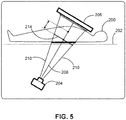
- Fig. 5 shows a schematic setup of an example for X-ray imaging.
- a subject 200 is arranged on a subject support 202.
- An X-ray source 204 is provided underneath the subject support 202.
- a detector 206 is arranged on the upper side of the patient.
- a centerline 208 indicates the central direction of the X-ray beam, being shaped with outer lines 210.
- a radiation entry plane 212 is indicated below the subject 200.
- a projection 214 perpendicular to the centerline 208 is indicated.
- the radiation entry plane 212 is relevant for skin dose control.
- Fig. 6 shows a mobile unit 300 as an example for the medical X-ray radiation system in a schematic setup of an example for X-ray imaging.
- the mobile unit 300 comprises a base 302, for examples having wheels. Further, a C-arm 304 is attached to the base 302. An X-ray source 306 and an X-ray detector 310 are provided mounted to opposing ends of the C-arm 304. A subject 312 is arranged on a subject support 314. A driving mechanism 316 for moving the C-arm is provided. Further, a device 10 for skin dose mapping in medical X-ray radiation is provided, e.g. integrated into the mobile base unit. Still further, display units 318 are provided in the vicinity to the subject support 314, e.g.
- a first sensor 322 is provided attached to a housing of the X-ray source, and a second sensor 324 is provided attached to a housing of the X-ray detector.
- the two sensors provide the subject situation information data of a subject of interest to the situation data receiver 12 of the device 10 for skin dose mapping in medical X-ray radiation.
- a computer program is provided enabling a processor to carry out the method of the example above.
- a computer program or program element for controlling an apparatus according to one of the examples above is provided, which program or program element, when being executed by a processing unit, is adapted to perform the method steps of one of the method examples above.
- a computer readable medium having stored the program element of the above example is provided.
- a computer program or a computer program element is provided that is characterized by being adapted to execute the method steps of the method according to one of the preceding embodiments, on an appropriate system.
- the computer program element might therefore be stored on a computer unit or be distributed over more than one computer units, which might also be part of an embodiment of the present invention.
- This computing unit may be adapted to perform or induce a performing of the steps of the method described above. Moreover, it may be adapted to operate the components of the above described apparatus.
- the computing unit can be adapted to operate automatically and/or to execute the orders of a user.
- a computer program may be loaded into a working memory of a data processor. The data processor may thus be equipped to carry out the method of the invention.
- aspects of the invention may be implemented in a computer program product, which may be a collection of computer program instructions stored on a computer readable storage device which may be executed by a computer.
- the instructions of the present invention may be in any interpretable or executable code mechanism, including but not limited to scripts, interpretable programs, dynamic link libraries (DLLs) or Java classes.
- the instructions can be provided as complete executable programs, partial executable programs, as modifications to existing programs (e.g. updates) or extensions for existing programs (e.g. plugins).
- parts of the processing of the present invention may be distributed over multiple computers or processors.
- the processing unit for instance a controller implements the control method.
- the controller can be implemented in numerous ways, with software and/or hardware, to perform the various functions required.
- a processor is one example of a controller which employs one or more microprocessors that may be programmed using software (e.g., microcode) to perform the required functions.
- a controller may however be implemented with or without employing a processor, and also may be implemented as a combination of dedicated hardware to perform some functions and a processor (e.g., one or more programmed microprocessors and associated circuitry) to perform other functions.
- controller components that may be employed in various embodiments of the present disclosure include, but are not limited to, conventional microprocessors, application specific integrated circuits (ASICs), and field-programmable gate arrays (FPGAs).
- ASICs application specific integrated circuits
- FPGAs field-programmable gate arrays
- This exemplary embodiment of the invention covers both, a computer program that right from the beginning uses the invention and a computer program that by means of an up-date turns an existing program into a program that uses the invention.
- the computer program element might be able to provide all necessary steps to fulfil the procedure of an exemplary embodiment of the method as described above.
- a computer readable medium such as a CD-ROM
- the computer readable medium has a computer program element stored on it which computer program element is described by the preceding section.
- a computer program may be stored and/or distributed on a suitable medium, such as an optical storage medium or a solid-state medium supplied together with or as part of other hardware, but may also be distributed in other forms, such as via the internet or other wired or wireless telecommunication systems.
- the computer program may also be presented over a network like the World Wide Web and can be downloaded into the working memory of a data processor from such a network.
- a medium for making a computer program element available for downloading is provided, which computer program element is arranged to perform a method according to one of the previously described embodiments of the invention.
Landscapes
- Health & Medical Sciences (AREA)
- Life Sciences & Earth Sciences (AREA)
- Medical Informatics (AREA)
- Engineering & Computer Science (AREA)
- Radiology & Medical Imaging (AREA)
- Biomedical Technology (AREA)
- Biophysics (AREA)
- Nuclear Medicine, Radiotherapy & Molecular Imaging (AREA)
- Optics & Photonics (AREA)
- Pathology (AREA)
- Physics & Mathematics (AREA)
- High Energy & Nuclear Physics (AREA)
- Heart & Thoracic Surgery (AREA)
- Molecular Biology (AREA)
- Surgery (AREA)
- Animal Behavior & Ethology (AREA)
- General Health & Medical Sciences (AREA)
- Public Health (AREA)
- Veterinary Medicine (AREA)
- Apparatus For Radiation Diagnosis (AREA)
Priority Applications (1)
| Application Number | Priority Date | Filing Date | Title |
|---|---|---|---|
| EP20152826.2A EP3854307A1 (de) | 2020-01-21 | 2020-01-21 | Hautdosisabbildung |
Applications Claiming Priority (1)
| Application Number | Priority Date | Filing Date | Title |
|---|---|---|---|
| EP20152826.2A EP3854307A1 (de) | 2020-01-21 | 2020-01-21 | Hautdosisabbildung |
Publications (1)
| Publication Number | Publication Date |
|---|---|
| EP3854307A1 true EP3854307A1 (de) | 2021-07-28 |
Family
ID=69185476
Family Applications (1)
| Application Number | Title | Priority Date | Filing Date |
|---|---|---|---|
| EP20152826.2A Withdrawn EP3854307A1 (de) | 2020-01-21 | 2020-01-21 | Hautdosisabbildung |
Country Status (1)
| Country | Link |
|---|---|
| EP (1) | EP3854307A1 (de) |
Citations (4)
| Publication number | Priority date | Publication date | Assignee | Title |
|---|---|---|---|---|
| US20100290591A1 (en) * | 2009-05-14 | 2010-11-18 | Martin Spahn | Method for monitoring the X-ray dosage administered to a patient by a radiation source when using an X-ray device, and X-ray device |
| US20160213329A1 (en) * | 2015-01-22 | 2016-07-28 | Siemens Aktiengesellschaft | X-ray recording system |
| US20170220716A1 (en) * | 2014-08-04 | 2017-08-03 | Universite De Strasbourg | Method for estimating the spatial distribution of the hazardousness of radiation doses |
| US9861328B2 (en) | 2013-03-04 | 2018-01-09 | Samsung Electronics Co., Ltd. | Mobile X-ray imaging apparatus and control method for the same |
-
2020
- 2020-01-21 EP EP20152826.2A patent/EP3854307A1/de not_active Withdrawn
Patent Citations (4)
| Publication number | Priority date | Publication date | Assignee | Title |
|---|---|---|---|---|
| US20100290591A1 (en) * | 2009-05-14 | 2010-11-18 | Martin Spahn | Method for monitoring the X-ray dosage administered to a patient by a radiation source when using an X-ray device, and X-ray device |
| US9861328B2 (en) | 2013-03-04 | 2018-01-09 | Samsung Electronics Co., Ltd. | Mobile X-ray imaging apparatus and control method for the same |
| US20170220716A1 (en) * | 2014-08-04 | 2017-08-03 | Universite De Strasbourg | Method for estimating the spatial distribution of the hazardousness of radiation doses |
| US20160213329A1 (en) * | 2015-01-22 | 2016-07-28 | Siemens Aktiengesellschaft | X-ray recording system |
Similar Documents
| Publication | Publication Date | Title |
|---|---|---|
| US9904998B2 (en) | Patient-specific and automatic x-ray system adjustment based on optical 3D scene detection and interpretation | |
| US7603155B2 (en) | Method and system of acquiring images with a medical imaging device | |
| KR101766193B1 (ko) | X-선 레코딩 시스템 | |
| JP5801717B2 (ja) | 軌道推定および標的位置監視のための連続立体撮像方法及びシステム、コンピュータ製品 | |
| JP6118465B2 (ja) | 対象物トラッキング装置 | |
| EP1832313A1 (de) | Steuergerät für eine Strahlentherapievorrichtung und Strahlungs-/Bestrahlungsverfahren | |
| US9795357B2 (en) | Positioning distance control for X-ray imaging systems | |
| GB2433668A (en) | Image registration based on the relative position of an imager to an imaged object | |
| US10143438B2 (en) | System for 3D object modeling and tracking in X-ray imaging | |
| JP2019037350A (ja) | 放射線治療用追跡装置 | |
| CN107019522A (zh) | 对操作者提供图像支持的方法、x射线装置和计算机程序 | |
| US20240065773A1 (en) | Navigation support | |
| US10786220B2 (en) | Device for imaging an object | |
| EP3854307A1 (de) | Hautdosisabbildung | |
| US11925500B2 (en) | Synchronisation device and method for determining an instant of the respiratory cycle of a patient, and assembly comprising a medical robot | |
| US11123025B2 (en) | Iso-centering in C-arm computer tomography | |
| US20240065663A1 (en) | Contactless measurement and visualization of respiration for chest radiography image examinations | |
| EP4129181A1 (de) | System zur medizinischen bildgebung | |
| WO2012073109A1 (en) | Device and method for detecting images of moving anatomical parts | |
| JP2019072324A (ja) | 放射線撮像装置および放射線治療装置 |
Legal Events
| Date | Code | Title | Description |
|---|---|---|---|
| PUAI | Public reference made under article 153(3) epc to a published international application that has entered the european phase |
Free format text: ORIGINAL CODE: 0009012 |
|
| STAA | Information on the status of an ep patent application or granted ep patent |
Free format text: STATUS: THE APPLICATION HAS BEEN PUBLISHED |
|
| AK | Designated contracting states |
Kind code of ref document: A1 Designated state(s): AL AT BE BG CH CY CZ DE DK EE ES FI FR GB GR HR HU IE IS IT LI LT LU LV MC MK MT NL NO PL PT RO RS SE SI SK SM TR |
|
| STAA | Information on the status of an ep patent application or granted ep patent |
Free format text: STATUS: THE APPLICATION IS DEEMED TO BE WITHDRAWN |
|
| 18D | Application deemed to be withdrawn |
Effective date: 20220129 |