EP3852129A1 - Interface unit - Google Patents
Interface unit Download PDFInfo
- Publication number
- EP3852129A1 EP3852129A1 EP19860396.1A EP19860396A EP3852129A1 EP 3852129 A1 EP3852129 A1 EP 3852129A1 EP 19860396 A EP19860396 A EP 19860396A EP 3852129 A1 EP3852129 A1 EP 3852129A1
- Authority
- EP
- European Patent Office
- Prior art keywords
- unit
- region
- sample
- interface
- opening
- Prior art date
- Legal status (The legal status is an assumption and is not a legal conclusion. Google has not performed a legal analysis and makes no representation as to the accuracy of the status listed.)
- Granted
Links
Images



Classifications
-
- H—ELECTRICITY
- H01—ELECTRIC ELEMENTS
- H01J—ELECTRIC DISCHARGE TUBES OR DISCHARGE LAMPS
- H01J49/00—Particle spectrometers or separator tubes
- H01J49/02—Details
- H01J49/04—Arrangements for introducing or extracting samples to be analysed, e.g. vacuum locks; Arrangements for external adjustment of electron- or ion-optical components
- H01J49/0459—Arrangements for introducing or extracting samples to be analysed, e.g. vacuum locks; Arrangements for external adjustment of electron- or ion-optical components for solid samples
- H01J49/0463—Desorption by laser or particle beam, followed by ionisation as a separate step
-
- H—ELECTRICITY
- H01—ELECTRIC ELEMENTS
- H01J—ELECTRIC DISCHARGE TUBES OR DISCHARGE LAMPS
- H01J49/00—Particle spectrometers or separator tubes
- H01J49/02—Details
- H01J49/10—Ion sources; Ion guns
- H01J49/14—Ion sources; Ion guns using particle bombardment, e.g. ionisation chambers
- H01J49/145—Ion sources; Ion guns using particle bombardment, e.g. ionisation chambers using chemical ionisation
-
- H—ELECTRICITY
- H01—ELECTRIC ELEMENTS
- H01J—ELECTRIC DISCHARGE TUBES OR DISCHARGE LAMPS
- H01J49/00—Particle spectrometers or separator tubes
- H01J49/02—Details
- H01J49/10—Ion sources; Ion guns
- H01J49/16—Ion sources; Ion guns using surface ionisation, e.g. field-, thermionic- or photo-emission
- H01J49/168—Ion sources; Ion guns using surface ionisation, e.g. field-, thermionic- or photo-emission field ionisation, e.g. corona discharge
Definitions
- the present invention relates to an interface unit that can be used in a laser ablation (LA)-DART-MS system, and more specifically to an interface unit that may be configured between a Direct Analysis in Real Time (DART) unit and a mass spectrometry (MS) unit to improve detection sensitivity of a sample ablated with a laser beam.
- LA laser ablation
- MS mass spectrometry
- a DART-MS (Direct Analysis in Real Time-Mass Spectrometry) system is a device that can perform molecular weight and structural analysis of a material by ablating and ionizing a target material using a heated metastable He gas discharged from an ion source and reactive ions produced from it.
- this has an advantage that can perform simple analysis by locating a sample between the ion source and the MS unit under the atmospheric pressure, application to a wider range of the sample requires to develop a technology for increasing a concentration of the sample in the atmosphere and thereby improving a signal-to-noise ratio of spectrum.
- ablation efficiency and ionization efficiency of the sample efficient collection of generated ions, transmission, etc.
- a laser ablation technique is applied to increase the concentration of the sample under the atmosphere, but due to exposed space in the atmosphere, it is still required to improve efficient collection of the ablated and ionized components and transmission to the mass spectrometry unit.
- the laser ablation-DART-MS system is needed to improve the detection sensitivity by introducing a quartz tube interface between an exit of the DART ionization and an inlet of the MS unit to restrict flow of ablated components and generated ions at an irradiation point of each laser beam.
- the present invention is to provide an interface unit that can be used in a laser ablation (LA) -DART-MS system, and more specifically to an interface unit that can be configured between a DART (Direct Analysis in Real Time) ionization unit and a MS (Mass Spectrometry) unit to improve detection sensitivity of a sample ablated with a laser beam.
- LA laser ablation
- MS Mass Spectrometry
- An interface unit of the present invention comprises a tube-shaped main body which can be located between an exit of a DART ionization unit and an inlet of a mass spectrometry unit; and a first opening provided on one side surface of the main body, the first opening being configured such that an analyte ablated from a sample is introduced into the main body, wherein the interface unit is used in a laser ablation-DART-MS system and the main body may receive a helium beam emitted from the DART ionization unit and the analyte ablated from the sample and transmit them to the mass spectrometry unit.
- the laser ablation-DART-MS system using the interface unit of the present invention comprises: a sample mounting unit on which the sample is mounted; an optical unit including a laser unit for irradiating the sample with a laser beam to ablate the sample; a DART ionization unit for providing a helium beam to ionize the analyte ablated from the sample; and a mass spectrometry (MS) unit for performing analysis on the ionized analyte.
- the laser ablation-DART-MS system further comprises an optical unit support member capable of mounting the optical unit at a desired position and supporting the optical unit, wherein the optical unit support member may be fixed to the mass spectrometry unit.
- a laser ablation-DART-MS system can improve detection sensitivity by introducing a quartz tube interface between an exit of a DART ionization unit and an inlet of a MS unit to restrict flow of ablated components and generated ions at an irradiation point of each laser beam.
- a main body of a first region according to the present invention is formed to be narrower as it is adjacent to a second region, whereby the helium gas emitted from the DART ionization unit and the analyte ablated from a sample are collected in a sufficient amount to be focused and transmitted to the second region together with the generated ionic components.
- An inner diameter of the main body in the second region is formed to be equal to or smaller than an inner diameter of the main body in the other end side of the first region, so that the gas stream received from the first region is transferred to the inlet of the mass spectrometry unit in a radial compression state, and thus the components to be analyzed can be efficiently collected and transferred.
- the laser ablation-DART-MS system can enhance reproducibility of an experiment by fixing a relative positional relationship between the laser and the sample.
- An interface unit of the present invention comprises a tube-shaped main body which can be located between an exit of a DART ionization unit and an inlet of a mass spectrometry unit; and a first opening provided on one side surface of the main body, the first opening being configured such that an analyte ablated from a sample is introduced into the main body, wherein the interface unit is used in a laser ablation-DART-MS system and the main body may receive a helium beam emitted from the DART ionization unit and the analyte ablated from the sample and transfer them to the mass spectrometry unit.
- the main body includes a first region into which a helium beam emitted from the DART ionization unit and an analyte ablated from the sample are introduced, and a second region that is connected to the first region and into which a gas stream is injected from the first region to transfer it to the mass spectrometry unit, wherein the helium beam emitted from the DART ionization unit is introduced into one end of the first region and the other end of the first region is connected to the second region, and wherein an inner diameter of the main body in the first region may be reduced from the one end of the first region toward the other end of the first region.
- an internal space of the main body may be formed to be tapered.
- the first opening may be provided in the first region.
- the interface unit of the present invention further comprises a protrusion tube extending from the first opening toward a sample mounting unit perpendicular to a longitudinal direction of the interface unit, wherein the analytes ablated from the sample mounted on the sample mounting unit may be introduced into the interface unit through the protrusion tube and then through the first opening.
- the other side surface of the main body in the first region is provided with a second opening configured to pass through a laser beam emitted from a laser unit.
- the second opening faces the first opening and the laser beam may be irradiated to the sample through the first opening and the second opening.
- the first region of the interface unit of the present invention may be provided with at least one or more third openings through which a corona pin is inserted into the main body.
- An inlet of the mass spectrometry unit in the interface unit of the present invention includes an orifice provided with a hole through which an analyte outside the mass spectrometry unit is introduced into an analysis space provided inside the mass spectrometry unit, and an interface flange connected to the orifice.
- One end of the second region is connected to the other end of the first region and the other end of the second region is connected to the inlet of the mass spectrometry unit, wherein an outer diameter of the body in the other end of the second region may be smaller than an inner diameter of a suction hole formed to face the hole of the orifice in the interface flange.
- the interface unit of the present invention further comprises a second opening configured to pass through the laser beam emitted from the laser unit, wherein the second opening is located at a point opposite to the first opening in the side of the main body and the laser beam may be irradiated to the sample through the second opening and then through the first opening.
- the interface unit of the present invention may further comprise one or more third openings arranged to insert an end of the corona pin inside the main body of the interface unit, the third openings being located near the second openings.
- the laser ablation-DART-MS system using the interface unit of the present invention comprises: a sample mounting unit on which the sample is mounted; an optical unit including a laser unit for irradiating a laser beam to the sample to ablate the sample; a DART ionization unit for providing a helium beam to ionize the analyte ablated from the sample; and a mass spectrometry (MS) unit for performing analysis on the ionized analyte.
- the laser ablation-DART-MS system further comprises an optical unit support member capable of mounting the optical unit at a desired position and supporting the optical unit, wherein the optical unit support member may be fixed to the mass spectrometry unit.
- An inlet of the mass spectrometry unit in the laser ablation-DART-MS system of the present invention includes an orifice provided with a hole through which an analyte outside the mass spectrometry unit is introduced into an analysis space provided inside the mass spectrometry unit, and an interface flange connected to the orifice, wherein the interface flange is fixed to a surface of the mass spectrometry unit having the orifice, and the optical unit support member is fixed to the interface flange.
- the optical unit support member in the laser ablation-DART-MS system of the present invention includes a plurality of fastening portions, wherein the plurality of fastening portions includes at least one interface flange connecting portion provided at a position corresponding to a tab portion of the interface flange and each interface flange connecting portion may be coupled to the tab portion of each interface flange with a first fastening member.
- the plurality of fastening portions in the laser ablation-DART-MS system of the present invention further includes at least one optical unit connecting portion to which the optical unit may be coupled, wherein each optical unit connecting portion is coupled to a fastening portion of the optical unit with a second fastening member and the optical unit may further include at least one of a mirror, a translation stage, an iris, and a lens.
- the optical unit support member in the laser ablation-DART-MS system of the present invention consists of an upper plate and a lower plate, and the plurality of fastening portions includes at least one upper and lower plate coupling portion to which the upper plate and the lower plate are coupled with each other and may be fixed at a position on which the upper and lower plate coupling portion of the lower plate and the upper and lower plate coupling portion of the upper plate are overlapped, by a third fastening member.
- FIG. 1 is a schematic diagram of a laser ablation-DART-MS system 1.
- the laser ablation-DART-MS system 1 is a device that performs a molecular weight and a structural analysis of a sample 2 by irradiating the sample 2 with a laser beam to ablate the sample 2, and then ionizing an ablated analyte using a helium beam (He beam) emitted from a DART ionization unit 10 (DART ion source) and reactive ions produced therefrom.
- He beam helium beam
- DART ion source DART ionization unit 10
- the laser ablation-DART-MS system 1 comprises a DART ionization unit 10, a mass spectrometry unit 20, a sample mounting unit 30, a laser unit 41, and a corona discharge unit (not shown).
- the DART ionization unit 10 irradiates a laser beam with the laser unit 41 and ionizes an analyte ablated from a sample 2 mounted on the sample mounting unit 30 using a helium beam emitted from the DART ionization unit 10 and reactive ions generated therefrom.
- the helium beam is emitted from an exit 11 of the DART ionization unit 10 to ionize the analyte ablated from the sample 2 mounted on the sample mounting unit 30.
- the DART ionization unit 10 may be, for example, DART-SVP manufactured by IonSense.
- the mass spectrometry (MS) unit 20 receives the ionized analyte and performs a molecular weight and a structural analysis of the ionized analyte.
- the mass spectrometry unit 20 may be, for example, LTQ Orbitrap Elite manufactured by Thermo Fisher Scientific.
- the sample mounting unit 30 is located between an exit of the DART ionization unit 10 and an inlet 21 of the mass spectrometry unit 20.
- the inlet 21 of the mass spectrometry unit 20 may include an orifice 21a having a hole through which an external analyte is introduced into an analysis space provided inside the mass spectrometry unit 20, and an interface flange 21b connected to the orifice 21a.
- the interface flange 21b in the inlet 21 of the mass spectrometry unit 20 may selectively be provided according to an analysis situation.
- the analyte ablated from the sample 2 mounted on the sample mounting unit 30 is introduced into the inlet of the mass spectrometry unit 20.
- the sample mounting unit 30 is may be located at a predetermined distance spaced away from a virtual straight line connecting the exit of the DART ionization unit 10 and the inlet 21 of the mass spectrometry unit 20.
- the sample mounting unit 30 may be located downward a path between the exit of the DART ionization unit 10 and the inlet 21 of the mass spectrometry unit 20.
- the sample mounting unit 30 may be, for example, a sample plate of a stainless steel on which a glass substrate or a thin layer chromatography (TLC) substrate containing the sample 2 may be placed.
- the laser unit 41 irradiates a sample 2 with a laser beam to ablate an analyte from the sample 2.
- the laser unit 41 may be, for example, LMD-XT series manufactured by LASOS.
- the corona discharge unit includes a corona pin.
- the corona pin is directed toward a path between the exit of the DART ionization unit 10 and the inlet 21 of the mass spectrometry unit 20. That is, the corona pin is directed toward an area at which the helium beam emitted from the DART ionization unit 10 meets with the analyte ablated from the sample 2.
- the ionization of the analyte ablated from the sample 2 is facilitated by a high voltage of the corona discharge unit, for example, a positive DC voltage of 1 kV or more, thereby increasing ionization efficiency of the analyte.
- a relative position of the laser unit 41 or an irradiation angle and power of the laser beam can be adjusted so that an ion peak intensity of the analyte derived from the sample 2 is maximized.
- An interface unit 100 of the present invention may be located between the exit of the DART ionization unit 10 and the inlet 21 of the mass spectrometry unit 20 in the laser ablation-DART-MS system 1.
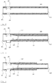
- FIG. 2 is a longitudinal sectional view of the interface unit 100 according to an embodiment of the present invention.
- the interface unit 100 may have a tube-shaped main body having both ends opened, and be a tube including a plurality of openings as will be described below.
- the interface unit 100 may be, for example, a quartz tube including a plurality of openings.
- the interface unit 100 may be a tube made of a glass or a ceramic, in addition to the above-described quartz.
- One end 101 of both the ends of the interface unit 100 may be arranged to overlap an end portion of the exit of the DART ionization unit 10 (that is, some or all of the end portion of the exit of the DART ionization unit 10 is built inside one end of the interface unit 100).
- one end 101 of both the ends of the interface unit 100 may directly contact or be adjacent to the exit of the DART ionization unit 10.
- a helium beam emitted from the exit of the DART ionization unit 10 is introduced into the interface unit 100 through the opened one end 101 of the interface unit 100.
- the other end 102 of both the ends of the interface unit 100 may be coupled with an inlet of the mass spectrometry unit 20.
- the inlet 21 of the mass spectrometry unit 20 may include an interface flange 21b for connecting an external tube and the mass spectrometry unit 20, wherein the other end 102 of the interface unit 100 may be inserted into the interface flange 21b such that the interface unit 100 and the inlet 21 of the mass spectrometry unit 20 may be coupled with each other.
- the interface unit 100 may be connected to the inlet 21 of the mass spectrometry unit 20 to be in contact to the orifice 21a or to be spaced apart by a predetermined distance (about 2 mm).
- the inlet 21 of the mass spectrometry unit 20 further includes an extension tube 21c fixed to the interface flange 21b and transferring a gas stream to the orifice 21a and the extension tube 21c may be fixed to the interface unit 100.
- the interface unit 100 of the present invention includes a tube-shaped main body that can be located between the exit of the DART ionization unit 10 and the inlet 21 of the mass spectrometry unit 20.
- the main body may include a first region 110 into which the helium beam emitted from the DART ionization unit 10 and the analyte ablated from the sample 2 are introduced, and a second region 120 connected to the first region 110 and having a gas stream of the first region 110 injected and transferred to the mass spectrometry unit 20.
- the gas stream may include a helium gas and components ablated and ionized from the sample.
- the body of the second region 120 may be coupled with the inlet 21 of the mass spectrometry unit 20.
- one end 111 of the first region 110 faces the DART ionization unit 10 to be adjacent to the DART ionization unit 10 and the other end 112 of the first region 110 is connected to one end of the second region 120.
- the other end 122 of the second region 120 faces the mass spectrometry unit 20 to be adjacent to the mass spectrometry unit 20. That is, they may be arranged in the order of [DART ionization unit 10]-[first region 110]-[second region 120]-[mass spectrometry unit 20].
- the interface unit 100 may be configured to have a uniform inner diameter along a longitudinal direction thereof.
- the inner diameter of the main body in the first region 110 may be decreased from one end 111 of the first region 110 toward the other end 112 of the first region 110.
- an internal space of the main body in the first region 110 may be configured to be tapered. That is, the internal space of the main body in the first region 110 may have a conical shape.
- the main body in the first region 110 of the present invention is configured to be narrower as it is adjacent to the second region 120, so that a helium gas emitted from the DART ionization unit 10 and an analyte ablated from a sample 2 can be collected in a sufficient amount to be focused and transferred to the second region 120 together with the generated ionic components.
- the inner diameter of the main body at one end 111 of the first region 110 may be larger than the inner diameter of the inlet of the mass spectrometry unit 20.
- the inner diameter of the main body in the second region 120 is configured to be equal to or smaller than the inner diameter of the main body in the other end 112 of the first region 110, and thus a gas stream transmitted from the first region 110 can be transferred to the inlet of the mass spectrometry unit 20 in radial compression.
- the inner diameter of the main body in the second region 120 may be kept constant. Specifically, since the gas stream is transferred in radial compression through the second region 120, it is possible to reduce a loss in the vicinity of the inlet of the mass spectrometry unit 20, which is a sub-ambient pressure region.
- the first region 110 may include a sample mounting unit 30, more specifically, a first opening 130 formed on one side of the main body adjacent to the sample 2, a second opening 140 formed on the other side of the main body so that the laser beam emitted from the laser unit 41 passes through, and at least one third opening 150 for inserting a corona pin into the body.
- the analyte ablated from the sample 2 mounted on the sample mounting unit 30 may be introduced into the interface unit 100 of the first region 110 through the first opening 130.
- the analyte introduced into the interface unit 100 may be ionized by a helium beam irradiated through an opened end 101 of the interface unit 100 and reactive ions generated therefrom.
- the first opening 130 is also a path through which the laser beam introduced through the second opening 140 passes toward the sample 2, as will be described later. That is, the laser beam emitted from the laser unit 41 may firstly pass through the second opening 140 and then the first opening 130 to irradiate the sample 2 mounted on the sample mounting unit 30.
- the first opening 130 may have, for example, a circular shape.
- the second opening 140 may be located at a point opposite to the first opening 130. That is, the second opening 140 may face the first opening 130.
- the second opening 140 may have, for example, a circular shape.
- the laser beam may penetrate the center of the second opening 140.
- the second opening 140 may be covered with a planar cover of a material that transmits light in the wavelength range of the laser beam.
- the planar cover may cover the second opening 140 such that the plane of the planar cover is perpendicular to a light path of the laser beam.
- At least one third opening 150 may be included in a portion of the side surface of the interface unit 100 that is directed toward the corona pin of a corona discharge unit.
- the third opening 150 may be located near the second opening 140.
- An end of the corona pin of the corona discharge unit may be located near the third opening 150 to face the inside of the interface unit 100, or the end of the corona pin of the corona discharge unit may be inserted into interface unit 100 through the third opening 150.
- the third opening 150 applied to the corona pin may be provided in the number of one or a plurality.
- the distance between the laser beam and the corona pin may be variously changed.
- the third opening 150 may have, for example, a circular shape.
- FIGS. 4 and 5 are longitudinal cross-sectional views showing a structure in which a protrusion tube 131 is included in the interface unit 100 of the present invention.
- the first opening 130 further includes the protrusion tube 131 vertically extending from the longitudinal direction of the interface unit 100.
- the protrusion tube 131 extends from a first opening 130 in the direction of a sample mounting unit 30.
- the protrusion tube 131 has a shape which extends downward and protrudes. That is, the protrusion tube may be a tube which extends toward the sample mounting unit 30 perpendicular to the longitudinal direction of the interface unit 100 at the first opening.
- the protrusion tube 131 may be, for example, a tube shape as shown in FIGS. 4 and 5 .
- the present invention is not limited to the above description, and various modifications and changes may be made, such as a tapered shape widening in the direction of the sample mounting unit 30 from the first opening 130.
- an outer diameter of the body of the other end 122 of the second region 120 may be smaller than an inner diameter of a suction hole formed to face a hole of the orifice 21a in the interface flange 21b.
- the other end 102 of the interface unit 100 may be inserted into the suction hole to fix the interface unit 100 to the mass spectrometry unit 20.
- a guide protrusion for securing a length into which the interface unit 100 is inserted may be provided at the suction hole side of the interface flange 21b.
- the outer diameter and the inner diameter of one end 101 of the interface unit 100 are made larger to allow the helium beam emitted from the DART ionization unit 10 and the analyte ablated from the sample 2 to be sufficiently introduced into the interface.
- the conventional laser ablation-DART-MS system to which the interface unit 100 of the present invention is not applied has the problem that the detection sensitivity of the analyte is low, because the ablated and ionized components may be lost due to a space exposed in the atmosphere between the exit of the DART ionization unit 10 and the inlet of the mass spectrometry unit 20 during the process of ionizing the analyte ablated from the sample 2 and introducing it into the inlet of the mass spectrometry unit 20.
- the interface unit 100 is located in a path between the exit of the DART ionization unit 10 and the inlet 21 of the mass spectrometry unit 20 and has a tube shape located between the exit of the DART ionization unit 10 and the inlet 21 of the mass spectrometry unit 20, wherein the interface unit 100 includes the first opening 130 in the portion adjacent to the sample 2. Since the interface unit 100 is connected to the exit of the DART ionization unit 10 (i.e., it may be adjacent to the exit or include some or all of the end of the exit), there is an advantage that can effectively contact the ablated components by confining flow of the helium beam.
- the main body of the first region 110 of the present invention is formed to be narrower as it is adjacent to the second region 120, whereby the helium gas emitted from the DART ionization unit 10 and the analyte ablated from the sample 2 are collected in a sufficient amount to be focused and transferred to the second region 120 together with the generated ionic components.
- An inner diameter of the main body in the second region is formed to be equal to or smaller than an inner diameter of the main body in the other end 112 side of the first region 110, so that the gas stream received from the first region 110 is transferred to the inlet of the mass spectrometry unit 20 in a radial compression state, whereby there is an advantage that the components to be analyzed can be efficiently collected and transferred.
- the analyte ablated from the sample 2 is introduced into the interface unit 100 through the first opening 130.
- the ablated analyte can be collected more effectively and guided to the ionization region that contacts the helium beam.
- the analyte introduced into the interface unit 100 is ionized and introduced into the inlet 21 of the mass spectrometry unit 20 along the tubular interface unit 100 with minimal loss.
- the laser ablation-DART-MS system 1 to which the interface unit 100 of the present invention is applied has an advantage that the detection sensitivity is significantly increased as compared with the conventional laser ablation-DART-MS system.
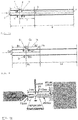
- interface units 100 which include a first region 110 in which the inner diameter of the body varies along the longitudinal direction and a second region 120 in which the inner diameter of the body is uniform in the longitudinal direction will be described with reference to FIG. 7a .
- the inner diameter of the main body at one end 111 of the first region 110 may be determined in consideration of an effect of the emission pattern of a helium gas emitted from a DART ionization unit 10 and a degree of the helium gas introduced into one end 101 of the interface unit 100 on the detection sensitivity of a laser ablation-DART-MS system 1.
- the inner diameter C of the main body at one end 111 of the first region 110 may be 1 mm to 10 mm or 2 mm to 8 mm.
- a length from one end 111 of the first region 110 to the other end 112 of the first region 110 and an inner diameter of the main body at the other end 112 of the first region 110 are determined in consideration of an effect of focusing of the gas stream on the detection sensitivity of the laser ablation-DART-MS system 1.
- the length A from one end 111 of the first region 110 to the other end 112 of the first region 110 may be 10 mm to 200 mm or 10 mm to 150 mm
- the inner diameter D of the main body at the other end 112 of the first region 110 may be greater than 0 mm and less than or equal to 8 mm or between 0.5 mm and 5 mm.
- Formation of the second region 120, a length from one end 121 of the second region 120 to the other end 122 of the second region 120, and an inner diameter of the main body at the second region 120 may be determined taking account of an effect of a radial compression degree of the gas stream on the detection sensitivity of the laser ablation-DART-MS system 1.
- the length B from one end 121 of the second region 120 to the other end 122 of the second region 120 is from greater than 0 mm to 190 mm or less, or from greater than 0 mm to 140 mm or less.
- the inner diameter E of the main body adjacent to the mass spectrometry unit 20 at the second region 120 may range from greater than 0 mm to 8 mm or less or from 0.5 mm to 5 mm. If the second region 120 is omitted, the other end 112 of the first region 110 may be coupled to the inlet 21 of the mass spectrometry unit 20.
- a first opening 130, a second opening 140, and a third opening 150 may function as follows.
- the first opening 130 serves to efficiently collect components ablated by the laser beam to guide them to an ionization region that contacts with the helium beam.
- a diameter H of the first opening 130 may be from 1 mm to 5 mm or from 2 mm to 5 mm.
- the second opening 140 serves to cause the laser beam to be irradiated onto a sample 2 without scattering, refracting or reflecting thereof so that effective ablation of the sample 2can occur.
- the size and formation of the diameter of the second opening 140 may be determined considering that a degree of scattering and power loss of the laser beam vs a deviation degree of the analyte ablated and ionized through the second opening 140 from the interface unit 100 (that is, a loss degree of the analyte occurred by deviation of the analyte from a path between an exit of the DART ionization unit 10 and an inlet of the mass spectrometry unit 20) affect the detection sensitivity.
- the diameter F of the second opening 140 may be between greater than 0 mm and less than or equal to 5 mm or between 2 mm and 5 mm.
- the third opening 150 serves to allow a corona pin to be inserted into the interface unit 100 to facilitate ionization through high voltage supply in a region in which the helium beam and the ablated components contact and are ionized.
- the size, formation, and number of the diameter of the third openings 150 may be determined considering that an increase of the ionization efficiency by a corona discharge vs a deviation degree of the analyte ablated and ionized through the third opening 150 from the interface unit 100 (that is, a loss degree of the analyte occurred by deviation of the analyte from the path between the exit of the DART ionization unit 10 and the inlet of the mass spectrometry unit 20) affect the detection sensitivity.
- the diameter G of the third opening 150 may be between greater than 0 mm and 5 mm or less, or between 1 mm and 3 mm.
- Formation and length of a protrusion tube 131 may be determined considering that a degree in which the ablated analyte is introduced into the interface unit 100 to effectively contact the helium gas beam, more specifically a limitation degree of the ablated analyte (a degree to which the analyte to be ablated in the sample 2 does not flow to any other part except the region where it contacts the helium gas beam) and a guiding degree(that is, the ablated analytes flow toward the center of the interface unit 100 at which contacts the helium gas beam) vs a relative distance between an ablation point of the sample 2 and the interface unit 100 affect the detection sensitivity.
- the length M that the protrusion tube protrudes from the first opening 130 may be between greater than 0 mm and 20 mm or less, or between greater than 0 mm and 10 mm or less.
- the interface unit 100 of the present invention can be applied to the laser ablation-DART-MS system 1 so that the laser beam passes through the center of the second opening 140.
- the length I from one end 111 of the first region 110 to the center of the second opening 140 may be between5 mm and 175 mm or between 5 mm and 125 mm and the length J from the center of the second opening 140 to the other end 112 of the first region 110 may be between 5 mm and 195 mm or between 5 mm and 145 mm.
- the distance L from the center of the body of the interface unit 100 to the center of the third opening 150 may be between -3 mm and 3 mm or between -2 mm and 2 mm.
- the distance from the center of the second opening 140 to the center of the third opening 150 may be determined in consideration of an influence of the relative distance between the laser beam and the corona pin on the detection sensitivity.
- the distance K from the center of the second opening 140 to the center of the third opening 150 may range from 1 mm to 10 mm or from 2 mm to 6 mm.
- Bs means a distance between the center of the second opening 140 and the exit of the DART ionization unit 10, which may, for example, range from 5 mm to 175 mm or from 5 mm to 125 mm.
- Bs' means a distance between the center of the second opening 140 and the inlet of the mass spectrometry unit 20, which may, for example, range from 5 mm to 195 mm or from 5 mm to 145 mm.
- Cs means a length of the portion fixed to the inlet of the mass spectrometry unit 20, which may, for example, range from 10 mm to 190 mm or from 10 mm to 140 mm.
- Ds means an inner diameter of one end of the DART ionization unit 10 side of the interface units 100 and 100', which may, for example, range from 1 mm to 10 mm or from 2 mm to 8 mm.
- Es means a diameter of the second opening 140 for passing the laser beam, which may, for example, range from greater than 0 mm to 5 mm or less or from 2 mm to 5 mm.
- Fs means a diameter of the third opening 150, which may, for example, range from greater than 0 mm to 5 mm or less or from 1 mm to 3 mm.
- Gs means a distance between the center of the second opening 140 and the center of the third opening 150, which may, for example, range from 1 mm to 10 mm or from 2 mm to 6 mm.
- Hs means a height from the center of the interface units 100, 100 'to the center of the third opening 150, which may, for example, range from -3 mm to 3 mm or from -2 mm to 2 mm.
- Is refers to a diameter of the first opening 130 for passing the laser beam and ionizing the ablated analyte, which may, for example, range from 1 mm to 5 mm or from 2 mm to 5 mm.
- Js means a length (height) of the protrusion tube 131 extending from the first opening 130, which may, for example, range from greater than 0 mm to 10 mm or less or from greater than 0 mm to 20 mm or less. In case Js is 0 mm, the first opening 130 does not have the protrusion tube 131.
- the present invention is not limited to the above-described dimensions, and may be variously changed according to various environments in which the present invention is implemented.
- a UV absorber material (C14H16N2O2, ethyl (Z)-2-cyano-3-(4-(dimethylamino)phenyl-acrylate) with a molecular weight of 244 Da was completely dissolved in PYR13-FSI (1-methyl-1-propylpyrrolidinium bis(fluorosulfonyl)imide) which is an ionic liquid in a concentration of 10 mg/mL.
- the ionic liquid as a solute was mixed evenly with a solvent having properties such as a low vapor pressure, a good solubility, a thermal stability, and a high viscosity, whereby the mixture can be used as a matrix having advantages of both of a liquid matrix and a solid matrix in that the solute is not volatilized.
- An analyte ablated from a sample 2 by a laser beam was dissolved in the ionic liquid for securing homogeneity and shot-to-shot reproducibility of the sample.
- a laser power is 180 mW as a continuous wave
- a DC voltage ranging from 0 to 1.5 kV is applied to a needle
- a temperature of the DART source is 400 °C
- the mass spectrometry unit 20 has a positive mode (ionization mode), an FTMS (analyzer) set as 240,000 (resolution).
- the inside of the main body of the first region 110 was formed in a conical shape, and the interface unit 100 having no the protrusion tube 131 was applied.
- 1 ⁇ L of the sample 2 was dropped onto a glass substrate using a pipette. Then, as shown in FIG. 8 , the glass substrate was placed on a sample plate to adjust a relative distance between the DART ionization unit 10, the laser beam, the inlet 21 of the mass spectrometry unit 20 and the sample plate. Next, a laser power, a DC voltage, a temperature of the DART source and the mass spectrometry unit 20 were set to the above experimental condition 2). Thereafter, a mass spectrum of the analyte was obtained.
- a laser power is 180 mW as a continuous wave
- a DC voltage ranging from 0 to 1.5 kV is applied to a needle
- a temperature of the DART source is 400 °C
- the mass spectrometry unit 20 has a positive mode (ionization mode), an FTMS (analyzer) set as 240,000 (resolution).
- the interface unit 100 having a uniform inner diameter in the longitudinal direction without the protrusion tube 131 was applied.
- FIG. 9a is a graph showing an experimental result when the experiment was performed without the interface unit 100 according to the present invention.
- FIG. 9b is a graph showing an experimental result of Example 2
- FIG. 9c is a graph showing an experimental result of Example 1.
- Example 1 shows that the detection sensitivity of the laser ablation-DART-MS system 1 to which the interface unit 100 of the present invention is applied is more excellent than that of the system without the interface unit 100. Comparison of the experimental result of Example 1 with the experimental result without the interface unit 100 can confirm a difference of about 35 times in the detection sensitivity.
- an optical unit support member 400 for supporting an optical unit 40 including a laser unit 41 will be described in detail.
- An interface flange 21b is mounted to the mass spectrometry unit 20 so that ions generated by the DART ionization unit 10 are transmitted to the mass spectrometry (MS) unit 20.
- the interface flange 21b may be fixed to the surface of the mass spectrometry unit in which an orifice 21a is provided.
- the interface flange 21b may further include a tab portion 22a as shown in FIG. 12 .
- the optical unit support member 400 as will be described later may be fixed to the tab portion 22a of the interface flange 21b.
- the interface flange 21b may be provided or not be provided with the tab portion 22a. If the interface flange 21b is not provided with the tab portion 22a, the tab portion 22a may be formed at a desired position to fix the optical unit support member 400.
- FIG. 10 shows a schematic diagram of optical units 40.
- the optical units 40 include a laser unit 41, a mirror 42, a translation stage 43, an iris 44, a lens 45, and the like.
- the laser unit 41 irradiates a sample 2 with a laser beam to ablate an analyte from the sample.
- the elements that should be optimized for improving the detection sensitivity are a power of the laser determined by the optical units 40, a distance between the sample 2 and a focal point (that is, the point at which the laser beam is focused in one place by the lens 45), a beam size at an ablation point (that is, the point at which ablation occurs by contacting the laser beam with the sample 2), and the likes. That is, the alignment and focusibility of the laser beam may be adjusted through the optimized arrangement of the optical units 40.
- a head portion of the optical fiber may be mounted to the optical unit support member 400 regardless of the size of the laser module 41.
- the mirror 42 serves to adjust a path of the laser beam such that the laser beam generated by the laser unit 41 can reach the sample 2. That is, when the laser beam does not reach the straight path from the laser unit 41 to the sample 2, the path of the laser beam is adjusted by changing an advancing direction of the laser beam with at least one mirror 42.
- the translation stage 43 can move along at least one axial direction.
- it may be an XY stage movable on a plane.
- the lens 45 may be mounted on the translation stage 43 so that the lens 45 may move in a predetermined direction.
- the position of the lens 45 can be adjusted to change a focal point of the laser beam on the sample 2.
- the focal point may be placed on the sample or placed slightly away from the sample.
- the iris 44 serves as a guide for aligning the laser beam in a desired path. Further, the beam size may be controlled by adjusting an aperture size of the iris 44.
- the lens 45 may adjust the focal point of the laser beam on the surface of the sample 2.
- a relative distance between the focal point of the laser beam and the sample surface may affect the detection sensitivity. If the sample is placed at the focal point, the ablation degree of the sample per area may be high, the ablation area may be reduced, and the detection sensitivity of fragment ions may be higher compared to molecular ions. If the sample is placed at an off-center focal point, the ablation degree of the sample per area may be low. As the off-center focal point increases, the beam size for the sample is larger, and thus the ablation area may be increased, and the detection sensitivity of the molecular ions may be higher compared to the fragment ions.
- the optical unit having high detection sensitivity is arranged with optimization by adjusting the positional relationship between the laser unit 41, the lens 45, and the sample in consideration of such a correlation.
- the wavelength and power of the laser are factors that greatly affect the detection sensitivity according to the sample, and thus it is necessary to optimize the arrangement of the optical unit depending on sample characteristics and laser characteristics at the time of the experiment.
- the present invention has an advantage that a plurality of fastening portions 410 are provided on the optical unit support member 400 and the optical units 40 are mounted with the plurality of fastening portions 410 on the optical unit support member 400 in various arrangement and combination manners according to the above-mentioned purposes.
- the laser ablation-DART-MS system 1 of the present invention includes the optical unit support member 400 for supporting the optical units 40.
- the optical unit support member 400 may be manufactured, for example, in a plate shape.
- the optical unit support member 400 includes a plurality of fastening portions 410 arranged at predetermined intervals.
- the plurality of fastening portions 410 may be, for example, an M6 tab or have a through hole shape.
- the plurality of fastening portions 410 include at least two interface flange connecting portions 410a.
- some of the plurality of fastening portions 410 formed at the predetermined intervals may function as the interface flange connecting portion 410a or may be provided at a position corresponding to a tab portion 22a of the interface flange.
- the interface flange connecting portion 410a as shown in FIG. 11 may be located at a position corresponding to the tab portion 22a of the interface flange of FIG. 12 .
- Each interface flange connecting portion 410a may be fixed to the tab portion 22a of each interface flange with a first fastening member.
- the tab portion 22a of the interface flange may have a female screw shape on an inner circumferential surface thereof and the first fastening member may have a male screw shape on which the inner circumferential surface of the tab portion 22a is engaged.
- the first fastening member may be, for example, an M6 bolt.
- the optical unit support member 400 is fixed to a front surface of the interface flange 21b, in a manner that the optical unit support member 400 is located at a desired position of the front surface of the interface flange 21b in the mass spectrometry unit 20, and the first fastening members are inserted into the fastening portions (i.e., the interface flange connecting portions 410a) corresponding to the positions of the tab portions 22a of the interface flange 21b among the plurality of fastening portions 410.
- the fastening portions i.e., the interface flange connecting portions 410a
- the plurality of fastening portions 410 includes an optical unit connecting portion 410b. That is, some of the plurality of fastening portions 410 may function as the optical unit connecting portion 410b.
- the optical units 40 may include the above-described laser unit 41, the mirror 42, the translation stage 43, the iris 44, the lens 45, and the like.
- Each of the optical units 40 (the laser unit 41, the mirror 42, the translation stage 43, the iris 44, the lens 45, etc.) may include at least one fastening portion for connecting to the optical unit connecting portion 410b.
- the fastening portion may be, for example, in the shape of a through hole, or the inner circumferential surface thereof may be in the shape of a female screw.
- the fastening portion and the optical unit connecting portion 410b of each optical unit 40 may be fixed with a second fastening member.
- the second fastening member may have the shape of a male screw coupled to the optical unit connecting portion 410b and the fastening portion.
- the second fastening member may be, for example, an M6 bolt or an M6 tanned bolt.
- the second fastening member may be provided with a nut behind a bolt.
- each of the optical units 40 is fixed to the optical unit support member 400, in a manner that each of the optical units 40 is arranged at a desired position of the optical unit support member 400 and the second fastening members are inserted into the fastening portions (i.e., the optical unit connecting portions 410b) corresponding to those of the optical units 40 among the plurality of fastening portions 410.
- the fastening portions i.e., the optical unit connecting portions 410b
- a corona discharge unit 50 may be fixed to the optical unit support member 400.
- the corona discharge unit 50 may also include at least one fastening portion.
- the fastening portion may have the shape of a through hole and the through hole may have an inner circumferential surface of a female screw shape.
- the corona discharge unit 50 may be fixed to a desired position of the optical unit support member 400 in a manner that the corona discharge unit 50 is fixed to the fastening portions (that is, the corona discharge unit connecting portion 410c) corresponding to those of the corona discharge unit 50 among the plurality of fastening portions 410 of the optical unit support member 400, with the second fastening member.
- the optical unit support member 400 consists of a lower plate 401 and an upper plate 402, the lower plate 401 and the upper plate 402 being combined with each other, as shown in FIG. 11 .
- some of the plurality of fastening portions 410 may be upper and lower plate coupling portions 410d. That is, some of a top portion of the lower plate 401 is overlapped with some of a bottom portion of the upper plate 402, and an overlapped portion between the plurality of fastening portions 410 of the lower plate 401 and the plurality of fastening portions 410 of the upper plate 402 can be fixed with a third fastening member.
- the third fastening member may, for example, be an M6 bolt.
- the holes of the lower plate in the upper and lower plate coupling portion 410d are perforated in the shape of a counterbore for a M6 bolt so that the head of the M6 bolt does not protrude above the plate.
- FIG. 11 shows that four holes located at the top row of the lower plate 401 and four holes located at the next row can be the upper and lower plate coupling portions 410d.
- the reference numerals are indicated in the leftmost holes.
- four holes located in the bottom row of the upper plate 402 and four holes located in the next row may be the upper and lower plate coupling portions 410d.
- the lower plate 401 is fixed to the tab portion 22a of the interface flange 21b, and each of the optical units 40 may be fixed at a desired position among the lower plate 401 and the upper plate 402.
- the present invention has an advantage that the dimension of the upper plate 402 may be freely changed according to the size and configuration of the optical units 40.
- the optical unit support member 400 may be made of, for example, a metal, or the like, and made of a stainless steel, an aluminum, or the like.
- FIG. 12 is a front view illustratively showing an interface flange 21b that may be used in the laser ablation-DART-MS system 1 of FIG. 1
- FIG. 13 is a view showing that a lower plate 401 is mounted on the interface flange 21b of FIG. 12 .
- the interface flange 21b includes a tab portion 22a.
- FIG. 14 is a conceptual diagram showing that a member 400 for supporting an optical unit and the optical units 40 are mounted on an interface flange 21b.
- the lower plate 401 has, for example, width x height x thickness of 190 mm x 130 mm x 10 mm or 15 mm, respectively.
- the lower plate 401 is composed of a first portion 401a connected to the interface flange 21b and having a thickness of 10 mm, and a second portion 401b connected to the upper plate 402 and having a thickness of 15 mm.
- the reason having the different thicknesses as such is that the second portion 401b at the lower portion is made slightly thicker so that the upper plate 402 is positioned further inward in order to secure a minimum distance between the laser beam irradiated onto the sample and the mass spectrometry unit 20 as short as possible.
- the distance between the mass spectrometry unit 20 and the ablation point of the sample may be extended as desired through a spacer 46 according to an environment in which the present invention is implemented, but reduction of the distance may be limited depending on a size of the laser unit 41 and a dimension of the optical unit support member 400.
- the second plate 401b may be made slightly thicker so that the upper plate 402 may be positioned inward in order to minimize the limitation due to the dimension of the optical unit support member 400. Referring to FIG.
- the shape of the lower plate 401 may be variously modified and changed to conform to the structure or shape of the interface flange 21b coupled to the mass spectrometry unit 20.
- the upper plate 402 has, for example, width x height x thickness of 190 mm x 310 mm x 10 mm.
- the lower plate 401 and the upper plate 402 may be, for example, made of an aluminum material.
- a plurality of fastening portions 410 is arranged, for example, at the intervals of 12.5 mm or 25 mm such that the optical units 40 can be installed cross the lower plate 401 and the upper plate 402.
- the interface flange connecting portion 410a is provided in a position corresponding to the tab portion 22a of the interface flange of FIG. 12 , and the interface flange connecting portion 410a is, for example, four.
- the present invention is not limited to the above-described embodiments, and the intervals, positions, and numbers of the plurality of fastening portions 410 may be variously changed to conform with positions of the tab portions 22a in the interface flange 21b or a setting of the optical units 40.
- An extension tube 21c may be connected to a suction port 24 formed in the interface flange 21b. One end of the extension tube 21c may be connected to the suction port 24, and the other end of the extension tube 21c may extend in a direction apposite to an exit 11 of the DART ionization unit 10.
- the interface unit 100 may be connected to the other end of the extension tube 21c or be directly coupled to an inlet 24 of the interface flange 21b without the extension tube 21c.
- the other end of the extension tube 21c may be spaced apart from the exit 11 of the DART ionization unit 10 by a certain distance, thereby enabling the laser beam irradiated from the laser unit 41 to be irradiated to the sample mounting unit 30 side without any interruption. That is, the extension tube 21c may extend to a distance that does not invade an optical path of the laser beam. By providing the extension tube 21c, it is possible to reduce an amount of ionized analytes that are lost before they are introduced into the mass spectrometry unit 20.
- a laser ablation-DART-MS system can improve detection sensitivity by introducing a quartz tube interface between an exit of a DART ionization unit and an inlet of a MS unit to restrict flow of ablated components and generated ions at an irradiation point of each laser beam.
- a main body of a first region according to the present invention is formed to be narrower as it is adjacent to a second region, whereby a helium gas emitted from the DART ionization unit and an analyte ablated from a sample are collected in a sufficient amount to be focused and transmitted to the second region together with the generated ionic components.
- An inner diameter of the main body in the second region is formed to be equal to or smaller than an inner diameter of the main body in the other end side of the first region, so that a gas stream transferred from the first region is delivered to the inlet of the mass spectrometry (MS) unit in a radial compression state, and thus the components to be analyzed can be efficiently collected and transferred.
- MS mass spectrometry
- the laser ablation-DART-MS system can enhance reproducibility of an experiment by fixing a relative positional relationship between the laser and the sample.
Landscapes
- Physics & Mathematics (AREA)
- Chemical & Material Sciences (AREA)
- Analytical Chemistry (AREA)
- Engineering & Computer Science (AREA)
- Plasma & Fusion (AREA)
- Optics & Photonics (AREA)
- Other Investigation Or Analysis Of Materials By Electrical Means (AREA)
- Electron Tubes For Measurement (AREA)
Abstract
Description
- The present application claims the benefit of priorities based on
Korean Patent Application No. 10-2018-0108208 filed on September 11, 2018 Korean Patent Application No. 10-2018-0114885 filed on September 27, 2018 Korean Patent Application No. 10-2019-0110755 filed on September 6, 2019 Korean Patent Application No. 10-2019-0111487 filed on September 9, 2019 Korean Patent Application No. 10-2019-0112165 filed on September 10, 2019 - The present invention relates to an interface unit that can be used in a laser ablation (LA)-DART-MS system, and more specifically to an interface unit that may be configured between a Direct Analysis in Real Time (DART) unit and a mass spectrometry (MS) unit to improve detection sensitivity of a sample ablated with a laser beam.
- In general, a DART-MS (Direct Analysis in Real Time-Mass Spectrometry) system is a device that can perform molecular weight and structural analysis of a material by ablating and ionizing a target material using a heated metastable He gas discharged from an ion source and reactive ions produced from it. Although this has an advantage that can perform simple analysis by locating a sample between the ion source and the MS unit under the atmospheric pressure, application to a wider range of the sample requires to develop a technology for increasing a concentration of the sample in the atmosphere and thereby improving a signal-to-noise ratio of spectrum. In this regard, ablation efficiency and ionization efficiency of the sample, efficient collection of generated ions, transmission, etc. may be important factors for improving the detection sensitivity. As a part of this effort, a laser ablation technique is applied to increase the concentration of the sample under the atmosphere, but due to exposed space in the atmosphere, it is still required to improve efficient collection of the ablated and ionized components and transmission to the mass spectrometry unit.
- Accordingly, the laser ablation-DART-MS system is needed to improve the detection sensitivity by introducing a quartz tube interface between an exit of the DART ionization and an inlet of the MS unit to restrict flow of ablated components and generated ions at an irradiation point of each laser beam.
- The present invention is to provide an interface unit that can be used in a laser ablation (LA) -DART-MS system, and more specifically to an interface unit that can be configured between a DART (Direct Analysis in Real Time) ionization unit and a MS (Mass Spectrometry) unit to improve detection sensitivity of a sample ablated with a laser beam.
- Technical problems that the present invention seeks to achieve are not limited to the above-mentioned technical problem, and other technical problems not mentioned above will be clearly understood by a person who has an ordinary knowledge in the technical field to which the present invention belongs from the following description.
- An interface unit of the present invention comprises a tube-shaped main body which can be located between an exit of a DART ionization unit and an inlet of a mass spectrometry unit; and a first opening provided on one side surface of the main body, the first opening being configured such that an analyte ablated from a sample is introduced into the main body, wherein the interface unit is used in a laser ablation-DART-MS system and the main body may receive a helium beam emitted from the DART ionization unit and the analyte ablated from the sample and transmit them to the mass spectrometry unit.
- The laser ablation-DART-MS system using the interface unit of the present invention comprises: a sample mounting unit on which the sample is mounted; an optical unit including a laser unit for irradiating the sample with a laser beam to ablate the sample; a DART ionization unit for providing a helium beam to ionize the analyte ablated from the sample; and a mass spectrometry (MS) unit for performing analysis on the ionized analyte. The laser ablation-DART-MS system further comprises an optical unit support member capable of mounting the optical unit at a desired position and supporting the optical unit, wherein the optical unit support member may be fixed to the mass spectrometry unit.
- According to the present invention, a laser ablation-DART-MS system can improve detection sensitivity by introducing a quartz tube interface between an exit of a DART ionization unit and an inlet of a MS unit to restrict flow of ablated components and generated ions at an irradiation point of each laser beam.
- A main body of a first region according to the present invention is formed to be narrower as it is adjacent to a second region, whereby the helium gas emitted from the DART ionization unit and the analyte ablated from a sample are collected in a sufficient amount to be focused and transmitted to the second region together with the generated ionic components. An inner diameter of the main body in the second region is formed to be equal to or smaller than an inner diameter of the main body in the other end side of the first region, so that the gas stream received from the first region is transferred to the inlet of the mass spectrometry unit in a radial compression state, and thus the components to be analyzed can be efficiently collected and transferred.
- According to the present invention, the laser ablation-DART-MS system can enhance reproducibility of an experiment by fixing a relative positional relationship between the laser and the sample. In addition, there is an advantage that can optimize the system for improving the detection sensitivity of the sample by adjusting positions of the optical units such as a laser unit using a laser support member. Further, it is possible to increase convenience of the equipment operation of the laser ablation-DART-MS system.
-
-
FIG. 1 is a schematic diagram of a laser ablation-DART-MS system to which an interface unit of the present invention is applied. -
FIG. 2 is a longitudinal sectional view showing an embodiment of an interface unit of the present invention. -
FIG. 3 is a longitudinal sectional view showing the other embodiment of an interface unit of the present invention. -
FIG. 4 is a longitudinal sectional view showing an embodiment in which a protrusion tube is provided in an interface unit of the present invention. -
FIG. 5 is a longitudinal sectional view showing the other embodiment in which a protrusion tube is provided in an interface unit of the present invention. -
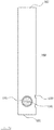
FIG. 6 is a bottom view illustrating the interface unit ofFIG. 4 . -
FIG. 7a is a conceptual diagram showing dimensions of respective portions according to an embodiment of an interface unit. -
FIG. 7b is a conceptual diagram showing dimensions of respective portions according to the other embodiment of an interface unit. -
FIG. 8 shows that an experiment is performed in a laser ablation-DART-MS system equipped with the interface unit ofFIG. 2 . -
FIG. 9a is a graph showing an experimental result in a laser ablation-DART-MS system to which an interface unit is not applied. -
FIG. 9b and9c are graphs showing experimental results in a laser ablation-DART-MS system to which an interface unit is applied. -
FIG. 10 is a schematic diagram of an optical unit in the laser ablation-DART-MS system ofFIG. 1 . -
FIG. 11 is a front view of a member for supporting an optical unit. -
FIG. 12 is a view showing an example of an interface flange. -
FIG. 13 is a view illustrating that a lower plate is mounted on an interface flange. -
FIG. 14 is a conceptual diagram showing that a member for supporting optical units and a part of the optical units are mounted on an interface flange. - An interface unit of the present invention comprises a tube-shaped main body which can be located between an exit of a DART ionization unit and an inlet of a mass spectrometry unit; and a first opening provided on one side surface of the main body, the first opening being configured such that an analyte ablated from a sample is introduced into the main body, wherein the interface unit is used in a laser ablation-DART-MS system and the main body may receive a helium beam emitted from the DART ionization unit and the analyte ablated from the sample and transfer them to the mass spectrometry unit.
- In the interface unit of the present invention, the main body includes a first region into which a helium beam emitted from the DART ionization unit and an analyte ablated from the sample are introduced, and a second region that is connected to the first region and into which a gas stream is injected from the first region to transfer it to the mass spectrometry unit, wherein the helium beam emitted from the DART ionization unit is introduced into one end of the first region and the other end of the first region is connected to the second region, and wherein an inner diameter of the main body in the first region may be reduced from the one end of the first region toward the other end of the first region.
- In the first region of the interface unit of the present invention, an internal space of the main body may be formed to be tapered.
- In the interface unit of the present invention, the first opening may be provided in the first region.
- The interface unit of the present invention further comprises a protrusion tube extending from the first opening toward a sample mounting unit perpendicular to a longitudinal direction of the interface unit, wherein the analytes ablated from the sample mounted on the sample mounting unit may be introduced into the interface unit through the protrusion tube and then through the first opening.
- In the interface unit of the present invention, the other side surface of the main body in the first region is provided with a second opening configured to pass through a laser beam emitted from a laser unit. The second opening faces the first opening and the laser beam may be irradiated to the sample through the first opening and the second opening.
- The first region of the interface unit of the present invention may be provided with at least one or more third openings through which a corona pin is inserted into the main body.
- An inlet of the mass spectrometry unit in the interface unit of the present invention includes an orifice provided with a hole through which an analyte outside the mass spectrometry unit is introduced into an analysis space provided inside the mass spectrometry unit, and an interface flange connected to the orifice. One end of the second region is connected to the other end of the first region and the other end of the second region is connected to the inlet of the mass spectrometry unit, wherein an outer diameter of the body in the other end of the second region may be smaller than an inner diameter of a suction hole formed to face the hole of the orifice in the interface flange.
- The interface unit of the present invention further comprises a second opening configured to pass through the laser beam emitted from the laser unit, wherein the second opening is located at a point opposite to the first opening in the side of the main body and the laser beam may be irradiated to the sample through the second opening and then through the first opening.
- The interface unit of the present invention may further comprise one or more third openings arranged to insert an end of the corona pin inside the main body of the interface unit, the third openings being located near the second openings.
- The laser ablation-DART-MS system using the interface unit of the present invention comprises: a sample mounting unit on which the sample is mounted; an optical unit including a laser unit for irradiating a laser beam to the sample to ablate the sample; a DART ionization unit for providing a helium beam to ionize the analyte ablated from the sample; and a mass spectrometry (MS) unit for performing analysis on the ionized analyte. The laser ablation-DART-MS system further comprises an optical unit support member capable of mounting the optical unit at a desired position and supporting the optical unit, wherein the optical unit support member may be fixed to the mass spectrometry unit.
- An inlet of the mass spectrometry unit in the laser ablation-DART-MS system of the present invention includes an orifice provided with a hole through which an analyte outside the mass spectrometry unit is introduced into an analysis space provided inside the mass spectrometry unit, and an interface flange connected to the orifice, wherein the interface flange is fixed to a surface of the mass spectrometry unit having the orifice, and the optical unit support member is fixed to the interface flange.
- The optical unit support member in the laser ablation-DART-MS system of the present invention includes a plurality of fastening portions, wherein the plurality of fastening portions includes at least one interface flange connecting portion provided at a position corresponding to a tab portion of the interface flange and each interface flange connecting portion may be coupled to the tab portion of each interface flange with a first fastening member.
- The plurality of fastening portions in the laser ablation-DART-MS system of the present invention further includes at least one optical unit connecting portion to which the optical unit may be coupled, wherein each optical unit connecting portion is coupled to a fastening portion of the optical unit with a second fastening member and the optical unit may further include at least one of a mirror, a translation stage, an iris, and a lens.
- The optical unit support member in the laser ablation-DART-MS system of the present invention consists of an upper plate and a lower plate, and the plurality of fastening portions includes at least one upper and lower plate coupling portion to which the upper plate and the lower plate are coupled with each other and may be fixed at a position on which the upper and lower plate coupling portion of the lower plate and the upper and lower plate coupling portion of the upper plate are overlapped, by a third fastening member.
- Hereinafter, an
interface unit 100 according to an embodiment of the present invention will be described in detail. The accompanying drawings show example forms of the present invention, which are provided to explain the present invention in more detail, and the technical scope of the present invention is not limited thereto. - Further, regardless of the reference numerals, the same or corresponding constitutive elements will be given the same reference numerals, and redundant description thereof will be omitted herein. For convenience of the description, sizes and shapes of each constitutive element as shown may be exaggerated or reduced.
-
FIG. 1 is a schematic diagram of a laser ablation-DART-MS system 1. First, the laser ablation-DART-MS system 1 is a device that performs a molecular weight and a structural analysis of asample 2 by irradiating thesample 2 with a laser beam to ablate thesample 2, and then ionizing an ablated analyte using a helium beam (He beam) emitted from a DART ionization unit 10 (DART ion source) and reactive ions produced therefrom. - The laser ablation-DART-
MS system 1 comprises aDART ionization unit 10, amass spectrometry unit 20, asample mounting unit 30, alaser unit 41, and a corona discharge unit (not shown). - The
DART ionization unit 10 irradiates a laser beam with thelaser unit 41 and ionizes an analyte ablated from asample 2 mounted on thesample mounting unit 30 using a helium beam emitted from theDART ionization unit 10 and reactive ions generated therefrom. The helium beam is emitted from anexit 11 of theDART ionization unit 10 to ionize the analyte ablated from thesample 2 mounted on thesample mounting unit 30. TheDART ionization unit 10 may be, for example, DART-SVP manufactured by IonSense. - The mass spectrometry (MS)
unit 20 receives the ionized analyte and performs a molecular weight and a structural analysis of the ionized analyte. Themass spectrometry unit 20 may be, for example, LTQ Orbitrap Elite manufactured by Thermo Fisher Scientific. - The
sample mounting unit 30 is located between an exit of theDART ionization unit 10 and aninlet 21 of themass spectrometry unit 20. Theinlet 21 of themass spectrometry unit 20 may include anorifice 21a having a hole through which an external analyte is introduced into an analysis space provided inside themass spectrometry unit 20, and aninterface flange 21b connected to theorifice 21a. Theinterface flange 21b in theinlet 21 of themass spectrometry unit 20 may selectively be provided according to an analysis situation. The analyte ablated from thesample 2 mounted on thesample mounting unit 30 is introduced into the inlet of themass spectrometry unit 20. More concretely, thesample mounting unit 30 is may be located at a predetermined distance spaced away from a virtual straight line connecting the exit of theDART ionization unit 10 and theinlet 21 of themass spectrometry unit 20. For example, thesample mounting unit 30 may be located downward a path between the exit of theDART ionization unit 10 and theinlet 21 of themass spectrometry unit 20. Thesample mounting unit 30 may be, for example, a sample plate of a stainless steel on which a glass substrate or a thin layer chromatography (TLC) substrate containing thesample 2 may be placed. - The
laser unit 41 irradiates asample 2 with a laser beam to ablate an analyte from thesample 2. Thelaser unit 41 may be, for example, LMD-XT series manufactured by LASOS. - Further, the corona discharge unit includes a corona pin. The corona pin is directed toward a path between the exit of the
DART ionization unit 10 and theinlet 21 of themass spectrometry unit 20. That is, the corona pin is directed toward an area at which the helium beam emitted from theDART ionization unit 10 meets with the analyte ablated from thesample 2. The ionization of the analyte ablated from thesample 2 is facilitated by a high voltage of the corona discharge unit, for example, a positive DC voltage of 1 kV or more, thereby increasing ionization efficiency of the analyte. - While a mass spectrum is checked in a real time by the analyzer, a relative position of the
laser unit 41 or an irradiation angle and power of the laser beam can be adjusted so that an ion peak intensity of the analyte derived from thesample 2 is maximized. - An
interface unit 100 of the present invention may be located between the exit of theDART ionization unit 10 and theinlet 21 of themass spectrometry unit 20 in the laser ablation-DART-MS system 1.FIG. 2 is a longitudinal sectional view of theinterface unit 100 according to an embodiment of the present invention. - The
interface unit 100 may have a tube-shaped main body having both ends opened, and be a tube including a plurality of openings as will be described below. Theinterface unit 100 may be, for example, a quartz tube including a plurality of openings. Alternatively, theinterface unit 100 may be a tube made of a glass or a ceramic, in addition to the above-described quartz. Oneend 101 of both the ends of theinterface unit 100 may be arranged to overlap an end portion of the exit of the DART ionization unit 10 (that is, some or all of the end portion of the exit of theDART ionization unit 10 is built inside one end of the interface unit 100). Alternatively, oneend 101 of both the ends of theinterface unit 100 may directly contact or be adjacent to the exit of theDART ionization unit 10. A helium beam emitted from the exit of theDART ionization unit 10 is introduced into theinterface unit 100 through the opened oneend 101 of theinterface unit 100. Theother end 102 of both the ends of theinterface unit 100 may be coupled with an inlet of themass spectrometry unit 20. For example, theinlet 21 of themass spectrometry unit 20 may include aninterface flange 21b for connecting an external tube and themass spectrometry unit 20, wherein theother end 102 of theinterface unit 100 may be inserted into theinterface flange 21b such that theinterface unit 100 and theinlet 21 of themass spectrometry unit 20 may be coupled with each other. Theinterface unit 100 may be connected to theinlet 21 of themass spectrometry unit 20 to be in contact to theorifice 21a or to be spaced apart by a predetermined distance (about 2 mm). - Alternatively, for example, the
inlet 21 of themass spectrometry unit 20 further includes anextension tube 21c fixed to theinterface flange 21b and transferring a gas stream to theorifice 21a and theextension tube 21c may be fixed to theinterface unit 100. - As shown in
FIG. 2 , theinterface unit 100 of the present invention includes a tube-shaped main body that can be located between the exit of theDART ionization unit 10 and theinlet 21 of themass spectrometry unit 20. The main body may include afirst region 110 into which the helium beam emitted from theDART ionization unit 10 and the analyte ablated from thesample 2 are introduced, and asecond region 120 connected to thefirst region 110 and having a gas stream of thefirst region 110 injected and transferred to themass spectrometry unit 20. The gas stream may include a helium gas and components ablated and ionized from the sample. Specifically, the body of thesecond region 120 may be coupled with theinlet 21 of themass spectrometry unit 20. - Specifically, one
end 111 of thefirst region 110 faces theDART ionization unit 10 to be adjacent to theDART ionization unit 10 and theother end 112 of thefirst region 110 is connected to one end of thesecond region 120. Theother end 122 of thesecond region 120 faces themass spectrometry unit 20 to be adjacent to themass spectrometry unit 20. That is, they may be arranged in the order of [DART ionization unit 10]-[first region 110]-[second region 120]-[mass spectrometry unit 20]. - Alternatively, as shown in
FIG. 3 , theinterface unit 100 may be configured to have a uniform inner diameter along a longitudinal direction thereof. - As shown in
FIGS. 2 ,4 and 5 , the inner diameter of the main body in thefirst region 110 may be decreased from oneend 111 of thefirst region 110 toward theother end 112 of thefirst region 110. Specifically, an internal space of the main body in thefirst region 110 may be configured to be tapered. That is, the internal space of the main body in thefirst region 110 may have a conical shape. The main body in thefirst region 110 of the present invention is configured to be narrower as it is adjacent to thesecond region 120, so that a helium gas emitted from theDART ionization unit 10 and an analyte ablated from asample 2 can be collected in a sufficient amount to be focused and transferred to thesecond region 120 together with the generated ionic components. The inner diameter of the main body at oneend 111 of thefirst region 110 may be larger than the inner diameter of the inlet of themass spectrometry unit 20. - The inner diameter of the main body in the
second region 120 is configured to be equal to or smaller than the inner diameter of the main body in theother end 112 of thefirst region 110, and thus a gas stream transmitted from thefirst region 110 can be transferred to the inlet of themass spectrometry unit 20 in radial compression. The inner diameter of the main body in thesecond region 120 may be kept constant. Specifically, since the gas stream is transferred in radial compression through thesecond region 120, it is possible to reduce a loss in the vicinity of the inlet of themass spectrometry unit 20, which is a sub-ambient pressure region. - As shown in
FIGS. 2 and4 to 6 , thefirst region 110 may include asample mounting unit 30, more specifically, afirst opening 130 formed on one side of the main body adjacent to thesample 2, asecond opening 140 formed on the other side of the main body so that the laser beam emitted from thelaser unit 41 passes through, and at least onethird opening 150 for inserting a corona pin into the body. - The analyte ablated from the
sample 2 mounted on thesample mounting unit 30 may be introduced into theinterface unit 100 of thefirst region 110 through thefirst opening 130. - The analyte introduced into the
interface unit 100 may be ionized by a helium beam irradiated through an openedend 101 of theinterface unit 100 and reactive ions generated therefrom. Thefirst opening 130 is also a path through which the laser beam introduced through thesecond opening 140 passes toward thesample 2, as will be described later. That is, the laser beam emitted from thelaser unit 41 may firstly pass through thesecond opening 140 and then thefirst opening 130 to irradiate thesample 2 mounted on thesample mounting unit 30. Thefirst opening 130 may have, for example, a circular shape. - Since the laser beam emitted from the
laser unit 41 is irradiated to thesample 2 mounted on thesample mounting unit 30, thesecond opening 140 may be located at a point opposite to thefirst opening 130. That is, thesecond opening 140 may face thefirst opening 130. Thesecond opening 140 may have, for example, a circular shape. The laser beam may penetrate the center of thesecond opening 140. - The
second opening 140 may be covered with a planar cover of a material that transmits light in the wavelength range of the laser beam. For example, the planar cover may cover thesecond opening 140 such that the plane of the planar cover is perpendicular to a light path of the laser beam. As such, by covering thesecond opening 140 with the planar cover, the gas stream can be irradiated onto the sample without refracting or scattering the laser beam simultaneously with preventing the gas stream from leaking through thesecond opening 140. - Further, at least one
third opening 150 may be included in a portion of the side surface of theinterface unit 100 that is directed toward the corona pin of a corona discharge unit. In addition, thethird opening 150 may be located near thesecond opening 140. An end of the corona pin of the corona discharge unit may be located near thethird opening 150 to face the inside of theinterface unit 100, or the end of the corona pin of the corona discharge unit may be inserted intointerface unit 100 through thethird opening 150. Thethird opening 150 applied to the corona pin may be provided in the number of one or a plurality. In case the plurality ofthird openings 150 are provided and each of thethird openings 150 is provided at various distances from thesecond opening 140, the distance between the laser beam and the corona pin may be variously changed. In addition, thethird opening 150 may have, for example, a circular shape. -
FIGS. 4 and 5 are longitudinal cross-sectional views showing a structure in which aprotrusion tube 131 is included in theinterface unit 100 of the present invention. Specifically, thefirst opening 130 further includes theprotrusion tube 131 vertically extending from the longitudinal direction of theinterface unit 100. Theprotrusion tube 131 extends from afirst opening 130 in the direction of asample mounting unit 30. Specifically, in the laser ablation-DART-MS system 1, theprotrusion tube 131 has a shape which extends downward and protrudes. That is, the protrusion tube may be a tube which extends toward thesample mounting unit 30 perpendicular to the longitudinal direction of theinterface unit 100 at the first opening. Thus, the analytes ablated from thesample 2 mounted on thesample mounting unit 30 are introduced into theinterface unit 100 through theprotrusion tube 131 extending from thefirst opening 130, so that the loss of the analytes can be prevented more efficiently. Theprotrusion tube 131 may be, for example, a tube shape as shown inFIGS. 4 and 5 . However, the present invention is not limited to the above description, and various modifications and changes may be made, such as a tapered shape widening in the direction of thesample mounting unit 30 from thefirst opening 130. - As shown in
FIG. 5 , when oneend 121 of thesecond region 120 is connected to theother end 112 of thefirst region 110 and theother end 122 of thesecond region 120 is connected to an inlet of themass spectrometry unit 20, an outer diameter of the body of theother end 122 of thesecond region 120 may be smaller than an inner diameter of a suction hole formed to face a hole of theorifice 21a in theinterface flange 21b. Theother end 102 of theinterface unit 100 may be inserted into the suction hole to fix theinterface unit 100 to themass spectrometry unit 20. A guide protrusion for securing a length into which theinterface unit 100 is inserted may be provided at the suction hole side of theinterface flange 21b. That is, while a dimension of theother end 102 of theinterface unit 100 that is directly coupled to theinlet 21 of themass spectrometry unit 20 is formed to be coupled to theinlet 21 in accordance with the structure and size of theinlet 21 of themass spectrometry unit 20, the outer diameter and the inner diameter of oneend 101 of theinterface unit 100 are made larger to allow the helium beam emitted from theDART ionization unit 10 and the analyte ablated from thesample 2 to be sufficiently introduced into the interface. - The conventional laser ablation-DART-MS system to which the
interface unit 100 of the present invention is not applied has the problem that the detection sensitivity of the analyte is low, because the ablated and ionized components may be lost due to a space exposed in the atmosphere between the exit of theDART ionization unit 10 and the inlet of themass spectrometry unit 20 during the process of ionizing the analyte ablated from thesample 2 and introducing it into the inlet of themass spectrometry unit 20. - However, according to the present invention, as described above, the
interface unit 100 is located in a path between the exit of theDART ionization unit 10 and theinlet 21 of themass spectrometry unit 20 and has a tube shape located between the exit of theDART ionization unit 10 and theinlet 21 of themass spectrometry unit 20, wherein theinterface unit 100 includes thefirst opening 130 in the portion adjacent to thesample 2. Since theinterface unit 100 is connected to the exit of the DART ionization unit 10 (i.e., it may be adjacent to the exit or include some or all of the end of the exit), there is an advantage that can effectively contact the ablated components by confining flow of the helium beam. - Further, the main body of the
first region 110 of the present invention is formed to be narrower as it is adjacent to thesecond region 120, whereby the helium gas emitted from theDART ionization unit 10 and the analyte ablated from thesample 2 are collected in a sufficient amount to be focused and transferred to thesecond region 120 together with the generated ionic components. An inner diameter of the main body in the second region is formed to be equal to or smaller than an inner diameter of the main body in theother end 112 side of thefirst region 110, so that the gas stream received from thefirst region 110 is transferred to the inlet of themass spectrometry unit 20 in a radial compression state, whereby there is an advantage that the components to be analyzed can be efficiently collected and transferred. - Furthermore, the analyte ablated from the
sample 2 is introduced into theinterface unit 100 through thefirst opening 130. Thus, there is an advantage that the ablated analyte can be collected more effectively and guided to the ionization region that contacts the helium beam. - In addition, the analyte introduced into the
interface unit 100 is ionized and introduced into theinlet 21 of themass spectrometry unit 20 along thetubular interface unit 100 with minimal loss. Thus, the laser ablation-DART-MS system 1 to which theinterface unit 100 of the present invention is applied has an advantage that the detection sensitivity is significantly increased as compared with the conventional laser ablation-DART-MS system. - Hereinafter, specific embodiments of
interface units 100 which include afirst region 110 in which the inner diameter of the body varies along the longitudinal direction and asecond region 120 in which the inner diameter of the body is uniform in the longitudinal direction will be described with reference toFIG. 7a . - The inner diameter of the main body at one
end 111 of thefirst region 110 may be determined in consideration of an effect of the emission pattern of a helium gas emitted from aDART ionization unit 10 and a degree of the helium gas introduced into oneend 101 of theinterface unit 100 on the detection sensitivity of a laser ablation-DART-MS system 1. For example, the inner diameter C of the main body at oneend 111 of thefirst region 110 may be 1 mm to 10 mm or 2 mm to 8 mm. - A length from one
end 111 of thefirst region 110 to theother end 112 of thefirst region 110 and an inner diameter of the main body at theother end 112 of thefirst region 110 are determined in consideration of an effect of focusing of the gas stream on the detection sensitivity of the laser ablation-DART-MS system 1. For example, the length A from oneend 111 of thefirst region 110 to theother end 112 of thefirst region 110 may be 10 mm to 200 mm or 10 mm to 150 mm, and the inner diameter D of the main body at theother end 112 of thefirst region 110 may be greater than 0 mm and less than or equal to 8 mm or between 0.5 mm and 5 mm. - Formation of the
second region 120, a length from oneend 121 of thesecond region 120 to theother end 122 of thesecond region 120, and an inner diameter of the main body at thesecond region 120 may be determined taking account of an effect of a radial compression degree of the gas stream on the detection sensitivity of the laser ablation-DART-MS system 1. For example, the length B from oneend 121 of thesecond region 120 to theother end 122 of thesecond region 120 is from greater than 0 mm to 190 mm or less, or from greater than 0 mm to 140 mm or less. The inner diameter E of the main body adjacent to themass spectrometry unit 20 at thesecond region 120 may range from greater than 0 mm to 8 mm or less or from 0.5 mm to 5 mm. If thesecond region 120 is omitted, theother end 112 of thefirst region 110 may be coupled to theinlet 21 of themass spectrometry unit 20. - A
first opening 130, asecond opening 140, and athird opening 150 may function as follows. Thefirst opening 130 serves to efficiently collect components ablated by the laser beam to guide them to an ionization region that contacts with the helium beam. In consideration of this, a diameter H of thefirst opening 130 may be from 1 mm to 5 mm or from 2 mm to 5 mm. - The
second opening 140 serves to cause the laser beam to be irradiated onto asample 2 without scattering, refracting or reflecting thereof so that effective ablation of the sample 2can occur. The size and formation of the diameter of thesecond opening 140 may be determined considering that a degree of scattering and power loss of the laser beam vs a deviation degree of the analyte ablated and ionized through thesecond opening 140 from the interface unit 100 (that is, a loss degree of the analyte occurred by deviation of the analyte from a path between an exit of theDART ionization unit 10 and an inlet of the mass spectrometry unit 20) affect the detection sensitivity. For example, the diameter F of thesecond opening 140 may be between greater than 0 mm and less than or equal to 5 mm or between 2 mm and 5 mm. - The
third opening 150 serves to allow a corona pin to be inserted into theinterface unit 100 to facilitate ionization through high voltage supply in a region in which the helium beam and the ablated components contact and are ionized. The size, formation, and number of the diameter of thethird openings 150 may be determined considering that an increase of the ionization efficiency by a corona discharge vs a deviation degree of the analyte ablated and ionized through thethird opening 150 from the interface unit 100 (that is, a loss degree of the analyte occurred by deviation of the analyte from the path between the exit of theDART ionization unit 10 and the inlet of the mass spectrometry unit 20) affect the detection sensitivity. For example, the diameter G of thethird opening 150 may be between greater than 0 mm and 5 mm or less, or between 1 mm and 3 mm. - Formation and length of a
protrusion tube 131 may be determined considering that a degree in which the ablated analyte is introduced into theinterface unit 100 to effectively contact the helium gas beam, more specifically a limitation degree of the ablated analyte (a degree to which the analyte to be ablated in thesample 2 does not flow to any other part except the region where it contacts the helium gas beam) and a guiding degree(that is, the ablated analytes flow toward the center of theinterface unit 100 at which contacts the helium gas beam) vs a relative distance between an ablation point of thesample 2 and theinterface unit 100 affect the detection sensitivity. For example, the length M that the protrusion tube protrudes from thefirst opening 130 may be between greater than 0 mm and 20 mm or less, or between greater than 0 mm and 10 mm or less. - The
interface unit 100 of the present invention can be applied to the laser ablation-DART-MS system 1 so that the laser beam passes through the center of thesecond opening 140. The length I from oneend 111 of thefirst region 110 to the center of thesecond opening 140 may be between5 mm and 175 mm or between 5 mm and 125 mm and the length J from the center of thesecond opening 140 to theother end 112 of thefirst region 110 may be between 5 mm and 195 mm or between 5 mm and 145 mm. - The distance L from the center of the body of the
interface unit 100 to the center of thethird opening 150 may be between -3 mm and 3 mm or between -2 mm and 2 mm. - The distance from the center of the
second opening 140 to the center of thethird opening 150 may be determined in consideration of an influence of the relative distance between the laser beam and the corona pin on the detection sensitivity. For example, the distance K from the center of thesecond opening 140 to the center of thethird opening 150 may range from 1 mm to 10 mm or from 2 mm to 6 mm. - Hereinafter, a specific embodiment of the
interface unit 100 in which the inner diameter of the body is uniform along the longitudinal direction will be described with reference toFIG. 7b . - As refers to a distance between the exit of the
DART ionization unit 10 and the inlet of themass spectrometry unit 20, which may, for example, range from 10 mm to 200 mm or from 10 mm to 150 mm. Bs means a distance between the center of thesecond opening 140 and the exit of theDART ionization unit 10, which may, for example, range from 5 mm to 175 mm or from 5 mm to 125 mm. Bs' means a distance between the center of thesecond opening 140 and the inlet of themass spectrometry unit 20, which may, for example, range from 5 mm to 195 mm or from 5 mm to 145 mm. Cs means a length of the portion fixed to the inlet of themass spectrometry unit 20, which may, for example, range from 10 mm to 190 mm or from 10 mm to 140 mm. Ds means an inner diameter of one end of theDART ionization unit 10 side of theinterface units 100 and 100', which may, for example, range from 1 mm to 10 mm or from 2 mm to 8 mm. Es means a diameter of thesecond opening 140 for passing the laser beam, which may, for example, range from greater than 0 mm to 5 mm or less or from 2 mm to 5 mm. Fs means a diameter of thethird opening 150, which may, for example, range from greater than 0 mm to 5 mm or less or from 1 mm to 3 mm. Gs means a distance between the center of thesecond opening 140 and the center of thethird opening 150, which may, for example, range from 1 mm to 10 mm or from 2 mm to 6 mm. Hs means a height from the center of theinterface units 100, 100 'to the center of thethird opening 150, which may, for example, range from -3 mm to 3 mm or from -2 mm to 2 mm. Is refers to a diameter of thefirst opening 130 for passing the laser beam and ionizing the ablated analyte, which may, for example, range from 1 mm to 5 mm or from 2 mm to 5 mm. Js means a length (height) of theprotrusion tube 131 extending from thefirst opening 130, which may, for example, range from greater than 0 mm to 10 mm or less or from greater than 0 mm to 20 mm or less. In case Js is 0 mm, thefirst opening 130 does not have theprotrusion tube 131. - The present invention is not limited to the above-described dimensions, and may be variously changed according to various environments in which the present invention is implemented.
- A UV absorber material (C14H16N2O2, ethyl (Z)-2-cyano-3-(4-(dimethylamino)phenyl-acrylate) with a molecular weight of 244 Da was completely dissolved in PYR13-FSI (1-methyl-1-propylpyrrolidinium bis(fluorosulfonyl)imide) which is an ionic liquid in a concentration of 10 mg/mL.
- Then, the ionic liquid as a solute was mixed evenly with a solvent having properties such as a low vapor pressure, a good solubility, a thermal stability, and a high viscosity, whereby the mixture can be used as a matrix having advantages of both of a liquid matrix and a solid matrix in that the solute is not volatilized. An analyte ablated from a
sample 2 by a laser beam was dissolved in the ionic liquid for securing homogeneity and shot-to-shot reproducibility of the sample. Thus, when the experiment was performed using the laser ablation-DART-MS system, a signal reduction due to continuous consumption of thesample 2 during the analysis time was reduced to a minimum, so that a constant signal sensitivity can be kept. - A laser power is 180 mW as a continuous wave, a DC voltage ranging from 0 to 1.5 kV is applied to a needle, a temperature of the DART source is 400 °C, and the
mass spectrometry unit 20 has a positive mode (ionization mode), an FTMS (analyzer) set as 240,000 (resolution). As shown inFIG. 2 , the inside of the main body of thefirst region 110 was formed in a conical shape, and theinterface unit 100 having no theprotrusion tube 131 was applied. - 1 µL of the
sample 2 was dropped onto a glass substrate using a pipette. Then, as shown inFIG. 8 , the glass substrate was placed on a sample plate to adjust a relative distance between theDART ionization unit 10, the laser beam, theinlet 21 of themass spectrometry unit 20 and the sample plate. Next, a laser power, a DC voltage, a temperature of the DART source and themass spectrometry unit 20 were set to the above experimental condition 2). Thereafter, a mass spectrum of the analyte was obtained. - The Sample prepared in the same manner as in Example 1 was used.
- A laser power is 180 mW as a continuous wave, a DC voltage ranging from 0 to 1.5 kV is applied to a needle, a temperature of the DART source is 400 °C, and the
mass spectrometry unit 20 has a positive mode (ionization mode), an FTMS (analyzer) set as 240,000 (resolution). As shown inFIG. 3 , theinterface unit 100 having a uniform inner diameter in the longitudinal direction without theprotrusion tube 131 was applied. - The experiment was carried out by the same experimental method as in Example 1.
-
FIG. 9a is a graph showing an experimental result when the experiment was performed without theinterface unit 100 according to the present invention. -
FIG. 9b is a graph showing an experimental result of Example 2, andFIG. 9c is a graph showing an experimental result of Example 1. - The experimental results of Examples 1 and 2 show that the detection sensitivity of the laser ablation-DART-
MS system 1 to which theinterface unit 100 of the present invention is applied is more excellent than that of the system without theinterface unit 100. Comparison of the experimental result of Example 1 with the experimental result without theinterface unit 100 can confirm a difference of about 35 times in the detection sensitivity. - Hereinafter, in the laser ablation-DART-
MS system 1 to which theinterface unit 100 of the present invention is applied, an opticalunit support member 400 for supporting anoptical unit 40 including alaser unit 41 will be described in detail. - An
interface flange 21b is mounted to themass spectrometry unit 20 so that ions generated by theDART ionization unit 10 are transmitted to the mass spectrometry (MS)unit 20. Specifically, theinterface flange 21b may be fixed to the surface of the mass spectrometry unit in which anorifice 21a is provided. - The
interface flange 21b may further include atab portion 22a as shown inFIG. 12 . The opticalunit support member 400 as will be described later may be fixed to thetab portion 22a of theinterface flange 21b. In other words, theinterface flange 21b may be provided or not be provided with thetab portion 22a. If theinterface flange 21b is not provided with thetab portion 22a, thetab portion 22a may be formed at a desired position to fix the opticalunit support member 400. -
FIG. 10 shows a schematic diagram ofoptical units 40. - The
optical units 40 include alaser unit 41, amirror 42, atranslation stage 43, aniris 44, alens 45, and the like. Thelaser unit 41 irradiates asample 2 with a laser beam to ablate an analyte from the sample. In this case, the elements that should be optimized for improving the detection sensitivity are a power of the laser determined by theoptical units 40, a distance between thesample 2 and a focal point (that is, the point at which the laser beam is focused in one place by the lens 45), a beam size at an ablation point (that is, the point at which ablation occurs by contacting the laser beam with the sample 2), and the likes. That is, the alignment and focusibility of the laser beam may be adjusted through the optimized arrangement of theoptical units 40. Alternatively, when an optical fiber is coupled to thelaser module 41, a head portion of the optical fiber may be mounted to the opticalunit support member 400 regardless of the size of thelaser module 41. - The
mirror 42 serves to adjust a path of the laser beam such that the laser beam generated by thelaser unit 41 can reach thesample 2. That is, when the laser beam does not reach the straight path from thelaser unit 41 to thesample 2, the path of the laser beam is adjusted by changing an advancing direction of the laser beam with at least onemirror 42. - The
translation stage 43 can move along at least one axial direction. For example, it may be an XY stage movable on a plane. Thelens 45 may be mounted on thetranslation stage 43 so that thelens 45 may move in a predetermined direction. Thus, the position of thelens 45 can be adjusted to change a focal point of the laser beam on thesample 2. For example, the focal point may be placed on the sample or placed slightly away from the sample. - The
iris 44 serves as a guide for aligning the laser beam in a desired path. Further, the beam size may be controlled by adjusting an aperture size of theiris 44. - The
lens 45 may adjust the focal point of the laser beam on the surface of thesample 2. - On the other hand, regarding the
optical units 40, a relative distance between the focal point of the laser beam and the sample surface may affect the detection sensitivity. If the sample is placed at the focal point, the ablation degree of the sample per area may be high, the ablation area may be reduced, and the detection sensitivity of fragment ions may be higher compared to molecular ions. If the sample is placed at an off-center focal point, the ablation degree of the sample per area may be low. As the off-center focal point increases, the beam size for the sample is larger, and thus the ablation area may be increased, and the detection sensitivity of the molecular ions may be higher compared to the fragment ions. Accordingly, it is required that the optical unit having high detection sensitivity is arranged with optimization by adjusting the positional relationship between thelaser unit 41, thelens 45, and the sample in consideration of such a correlation. In addition, even if the optimized positional relationship is established at a specific wavelength and power, the wavelength and power of the laser are factors that greatly affect the detection sensitivity according to the sample, and thus it is necessary to optimize the arrangement of the optical unit depending on sample characteristics and laser characteristics at the time of the experiment. The present invention has an advantage that a plurality offastening portions 410 are provided on the opticalunit support member 400 and theoptical units 40 are mounted with the plurality offastening portions 410 on the opticalunit support member 400 in various arrangement and combination manners according to the above-mentioned purposes. - The laser ablation-DART-
MS system 1 of the present invention includes the opticalunit support member 400 for supporting theoptical units 40. The opticalunit support member 400 may be manufactured, for example, in a plate shape. In addition, the opticalunit support member 400 includes a plurality offastening portions 410 arranged at predetermined intervals. The plurality offastening portions 410 may be, for example, an M6 tab or have a through hole shape. - The plurality of
fastening portions 410 include at least two interfaceflange connecting portions 410a. For example, some of the plurality offastening portions 410 formed at the predetermined intervals may function as the interfaceflange connecting portion 410a or may be provided at a position corresponding to atab portion 22a of the interface flange. For example, the interfaceflange connecting portion 410a as shown inFIG. 11 may be located at a position corresponding to thetab portion 22a of the interface flange ofFIG. 12 . Each interfaceflange connecting portion 410a may be fixed to thetab portion 22a of each interface flange with a first fastening member. For example, thetab portion 22a of the interface flange may have a female screw shape on an inner circumferential surface thereof and the first fastening member may have a male screw shape on which the inner circumferential surface of thetab portion 22a is engaged. The first fastening member may be, for example, an M6 bolt. - Specifically, the optical
unit support member 400 is fixed to a front surface of theinterface flange 21b, in a manner that the opticalunit support member 400 is located at a desired position of the front surface of theinterface flange 21b in themass spectrometry unit 20, and the first fastening members are inserted into the fastening portions (i.e., the interfaceflange connecting portions 410a) corresponding to the positions of thetab portions 22a of theinterface flange 21b among the plurality offastening portions 410. - Further, the plurality of
fastening portions 410 includes an opticalunit connecting portion 410b. That is, some of the plurality offastening portions 410 may function as the opticalunit connecting portion 410b. Theoptical units 40 may include the above-describedlaser unit 41, themirror 42, thetranslation stage 43, theiris 44, thelens 45, and the like. Each of the optical units 40 (thelaser unit 41, themirror 42, thetranslation stage 43, theiris 44, thelens 45, etc.) may include at least one fastening portion for connecting to the opticalunit connecting portion 410b. The fastening portion may be, for example, in the shape of a through hole, or the inner circumferential surface thereof may be in the shape of a female screw. The fastening portion and the opticalunit connecting portion 410b of eachoptical unit 40 may be fixed with a second fastening member. For example, the second fastening member may have the shape of a male screw coupled to the opticalunit connecting portion 410b and the fastening portion. The second fastening member may be, for example, an M6 bolt or an M6 tanned bolt. Alternatively, when the fastening portion is in the shape of the through hole, for example, the second fastening member may be provided with a nut behind a bolt. - Specifically, each of the
optical units 40 is fixed to the opticalunit support member 400, in a manner that each of theoptical units 40 is arranged at a desired position of the opticalunit support member 400 and the second fastening members are inserted into the fastening portions (i.e., the opticalunit connecting portions 410b) corresponding to those of theoptical units 40 among the plurality offastening portions 410. - Additionally, a
corona discharge unit 50 may be fixed to the opticalunit support member 400. Similarly, thecorona discharge unit 50 may also include at least one fastening portion. For example, the fastening portion may have the shape of a through hole and the through hole may have an inner circumferential surface of a female screw shape. Thecorona discharge unit 50 may be fixed to a desired position of the opticalunit support member 400 in a manner that thecorona discharge unit 50 is fixed to the fastening portions (that is, the corona dischargeunit connecting portion 410c) corresponding to those of thecorona discharge unit 50 among the plurality offastening portions 410 of the opticalunit support member 400, with the second fastening member. - On the other hand, the optical
unit support member 400 consists of alower plate 401 and anupper plate 402, thelower plate 401 and theupper plate 402 being combined with each other, as shown inFIG. 11 . In this case, some of the plurality offastening portions 410 may be upper and lowerplate coupling portions 410d. That is, some of a top portion of thelower plate 401 is overlapped with some of a bottom portion of theupper plate 402, and an overlapped portion between the plurality offastening portions 410 of thelower plate 401 and the plurality offastening portions 410 of theupper plate 402 can be fixed with a third fastening member. The third fastening member may, for example, be an M6 bolt. The holes of the lower plate in the upper and lowerplate coupling portion 410d are perforated in the shape of a counterbore for a M6 bolt so that the head of the M6 bolt does not protrude above the plate.FIG. 11 shows that four holes located at the top row of thelower plate 401 and four holes located at the next row can be the upper and lowerplate coupling portions 410d. For convenience, the reference numerals are indicated in the leftmost holes. Similarly, four holes located in the bottom row of theupper plate 402 and four holes located in the next row may be the upper and lowerplate coupling portions 410d. Thelower plate 401 is fixed to thetab portion 22a of theinterface flange 21b, and each of theoptical units 40 may be fixed at a desired position among thelower plate 401 and theupper plate 402. - Meanwhile, in case the
lower plate 401 and theupper plate 402 is implemented to be combined with each other, there is an advantage that the load of theupper plate 402 is dispersed over the bolt and theinterface flange 21b by allowing theupper plate 402 to be placed on theinterface flange 21b. In addition, the present invention has an advantage that the dimension of theupper plate 402 may be freely changed according to the size and configuration of theoptical units 40. - The optical
unit support member 400 may be made of, for example, a metal, or the like, and made of a stainless steel, an aluminum, or the like. - Hereinafter, embodiments of the present invention will be described in detail with reference to
FIGS. 11 to 14 .FIG. 12 is a front view illustratively showing aninterface flange 21b that may be used in the laser ablation-DART-MS system 1 ofFIG. 1 , andFIG. 13 is a view showing that alower plate 401 is mounted on theinterface flange 21b ofFIG. 12 . As shown inFig. 12 , theinterface flange 21b includes atab portion 22a. - Further,
FIG. 14 is a conceptual diagram showing that amember 400 for supporting an optical unit and theoptical units 40 are mounted on aninterface flange 21b. - The
lower plate 401 has, for example, width x height x thickness of 190 mm x 130 mm x 10 mm or 15 mm, respectively. Thelower plate 401 is composed of afirst portion 401a connected to theinterface flange 21b and having a thickness of 10 mm, and asecond portion 401b connected to theupper plate 402 and having a thickness of 15 mm. The reason having the different thicknesses as such is that thesecond portion 401b at the lower portion is made slightly thicker so that theupper plate 402 is positioned further inward in order to secure a minimum distance between the laser beam irradiated onto the sample and themass spectrometry unit 20 as short as possible. In other words, the shorter a distance between the ablation point of the sample and themass spectrometry unit 20 is, the shorter a distance that the ionized components move to themass spectrometry unit 40 is, so that less loss during movement allows higher detection sensitivity. The distance between themass spectrometry unit 20 and the ablation point of the sample may be extended as desired through aspacer 46 according to an environment in which the present invention is implemented, but reduction of the distance may be limited depending on a size of thelaser unit 41 and a dimension of the opticalunit support member 400. Thus, thesecond plate 401b may be made slightly thicker so that theupper plate 402 may be positioned inward in order to minimize the limitation due to the dimension of the opticalunit support member 400. Referring toFIG. 11 , thefirst portion 401a and thesecond portion 401b are shown in dashed lines for convenience. The shape of thelower plate 401 may be variously modified and changed to conform to the structure or shape of theinterface flange 21b coupled to themass spectrometry unit 20. Theupper plate 402 has, for example, width x height x thickness of 190 mm x 310 mm x 10 mm. Thelower plate 401 and theupper plate 402 may be, for example, made of an aluminum material. - Further, a plurality of
fastening portions 410 is arranged, for example, at the intervals of 12.5 mm or 25 mm such that theoptical units 40 can be installed cross thelower plate 401 and theupper plate 402. The interfaceflange connecting portion 410a is provided in a position corresponding to thetab portion 22a of the interface flange ofFIG. 12 , and the interfaceflange connecting portion 410a is, for example, four. - However, the present invention is not limited to the above-described embodiments, and the intervals, positions, and numbers of the plurality of
fastening portions 410 may be variously changed to conform with positions of thetab portions 22a in theinterface flange 21b or a setting of theoptical units 40. - An
extension tube 21c may be connected to asuction port 24 formed in theinterface flange 21b. One end of theextension tube 21c may be connected to thesuction port 24, and the other end of theextension tube 21c may extend in a direction apposite to anexit 11 of theDART ionization unit 10. Theinterface unit 100 may be connected to the other end of theextension tube 21c or be directly coupled to aninlet 24 of theinterface flange 21b without theextension tube 21c. - The other end of the
extension tube 21c may be spaced apart from theexit 11 of theDART ionization unit 10 by a certain distance, thereby enabling the laser beam irradiated from thelaser unit 41 to be irradiated to thesample mounting unit 30 side without any interruption. That is, theextension tube 21c may extend to a distance that does not invade an optical path of the laser beam. By providing theextension tube 21c, it is possible to reduce an amount of ionized analytes that are lost before they are introduced into themass spectrometry unit 20. - It will be appreciated that the technical configuration of the present invention described above may be embodied in other specific forms by those ordinarily skilled in the technical field to which the invention belongs without changing the technical spirit or essential features of the present invention. Therefore, the embodiments described above should be understood to be illustrative in all respects and not restrictive. Further, the scope of the present invention is shown by the claims to be described below, rather than the above detailed description of the specification. In addition, it should be construed that all changes or modifications derived from the meaning and range of the claims and equivalent concepts thereof are included in the scope of the present invention.
- According to the present invention, a laser ablation-DART-MS system can improve detection sensitivity by introducing a quartz tube interface between an exit of a DART ionization unit and an inlet of a MS unit to restrict flow of ablated components and generated ions at an irradiation point of each laser beam.
- A main body of a first region according to the present invention is formed to be narrower as it is adjacent to a second region, whereby a helium gas emitted from the DART ionization unit and an analyte ablated from a sample are collected in a sufficient amount to be focused and transmitted to the second region together with the generated ionic components. An inner diameter of the main body in the second region is formed to be equal to or smaller than an inner diameter of the main body in the other end side of the first region, so that a gas stream transferred from the first region is delivered to the inlet of the mass spectrometry (MS) unit in a radial compression state, and thus the components to be analyzed can be efficiently collected and transferred.
- According to the present invention, the laser ablation-DART-MS system can enhance reproducibility of an experiment by fixing a relative positional relationship between the laser and the sample. In addition, there is an advantage that can optimize the system for improving the detection sensitivity of the sample by adjusting positions of the optical units such as a laser unit using a laser support member. Further, it is possible to increase convenience of the equipment operation of the laser ablation-DART-MS system.
Claims (15)
- An interface unit comprising:a tube-shaped main body which is located between an exit of a DART (Direct Analysis in Real Time) ionization unit and an inlet of a MS (Mass Spectrometry) unit; anda first opening provided on one side surface of the main body, the first opening being configured such that an analyte ablated from a sample is introduced into the main body,wherein the interface unit is used in a laser ablation-DART-MS system, and the main body receives a helium beam emitted from the DART ionization unit and the analyte ablated from the sample and transfer them to the mass spectrometry unit.
- The interface unit according to claim 1,wherein the main body includes a first region into which a helium beam emitted from the DART ionization unit and the analyte ablated from the sample are introduced, and a second region that is connected to the first region and into which a gas stream is injected from the first region to transfer it to the mass spectrometry unit, andwherein the helium beam emitted from the DART ionization unit is introduced into one end of the first region, with the other end of the first region being connected to the second region, and an inner diameter of the main body in the first region is reduced from the one end of the first region toward the other end of the first region.
- The interface unit according to claim 2,
wherein an internal space of the main body is formed to be tapered. - The interface unit according to claim 2,
wherein the first opening is provided in the first region. - The interface unit according to claim 4, further comprising,a protrusion tube extending from the first opening toward a sample mounting unit perpendicular to a longitudinal direction of the interface unit,wherein the analytes ablated from the sample mounted on the sample mounting unit are introduced into the interface unit through the protrusion tube and then through the first opening.
- The interface unit according to claim 4,wherein the other side surface of the main body in the first region is provided with a second opening configured to pass through a laser beam emitted from a laser unit,the second opening faces the first opening, andthe laser beam is irradiated to the sample through the first opening and the second opening.
- The interface unit according to claim 6,
wherein the first region is provided with at least one or more third openings through which a corona pin is inserted into the main body. - The interface unit according to claim 2,wherein the inlet of the mass spectrometry unit includes an orifice provided with a hole through which the analyte outside the mass spectrometry unit is introduced into an analysis space formed inside the mass spectrometry unit, and an interface flange connected to the orifice,one end of the second region is connected to the other end of the first region,the other end of the second region is connected to the inlet of the mass spectrometry unit, andan outer diameter of the body in the other end of the second region is smaller than an inner diameter of a suction hole formed to face the hole of the orifice in the interface flange.
- The interface unit according to claim 1, further comprising,a second opening configured to pass through the laser beam emitted from the laser unit,wherein the second opening is located at a point opposite to the first opening in the side of the main body, andthe laser beam is irradiated to the sample through the second opening and then through the first opening.
- The interface unit according to claim 9, further comprising,one or more third openings through which an end of the corona pin is inserted inside the main body of the interface unit,wherein the third openings are located near the second opening.
- A laser ablation-DART-MS system using the interface unit according to any one of claims 1 to 10, comprising:a sample mounting unit on which a sample is mounted;an optical unit including a laser unit for irradiating a laser beam to the sample to ablate the sample;a DART ionization unit for providing a helium beam to ionize an analyte ablated from the sample; anda mass spectrometry (MS) unit for performing analysis on the ionized analyte, andfurther comprising an optical unit support member capable of mounting the optical unit at a desired position and supporting the optical unit,wherein the optical unit support member is fixed to the mass spectrometry unit.
- The laser ablation-DART-MS system according to claim 11,wherein an inlet of the mass spectrometry unit includes an orifice provided with a hole through which an analyte outside the mass spectrometry unit is introduced into an analysis space provided inside the mass spectrometry unit, and an interface flange connected to the orifice,the interface flange is fixed to a surface of the mass spectrometry unit having the orifice, andthe optical unit support member is fixed to the interface flange.
- The laser ablation-DART-MS system according to claim 12,wherein the optical unit support member includes a plurality of fastening portions,the plurality of fastening portions includes at least one interface flange connecting portion provided at a position corresponding to a tab portion of the interface flange, andeach interface flange connecting portion is coupled to each tab portion of the interface flange with a first fastening member.
- The laser ablation-DART-MS system according to claim 13,wherein the plurality of fastening portions further includes at least one optical unit connecting portion to which the optical unit can be coupled,each optical unit connecting portion is coupled to a fastening portion of the optical unit with a second fastening member, andthe optical unit further includes at least one of a mirror, a translation stage, an iris, and a lens.
- The laser ablation-DART-MS system according to claim 13,wherein the optical unit support member consists of an upper plate and a lower plate,the plurality of fastening portions includes at least one upper and lower plate coupling portion to which the upper plate and the lower plate can be coupled with each other, and is fixed at a position on which the upper and lower plate coupling portion of the lower plate and the upper and lower plate coupling portion of the upper plate are overlapped, by a third fastening member.
Applications Claiming Priority (6)
| Application Number | Priority Date | Filing Date | Title |
|---|---|---|---|
| KR20180108208 | 2018-09-11 | ||
| KR20180114885 | 2018-09-27 | ||
| KR1020190110755A KR102790955B1 (en) | 2018-09-27 | 2019-09-06 | A laser ablation-corona-DART-MS system and an member for supporting optical units |
| KR1020190111487A KR102577694B1 (en) | 2018-09-11 | 2019-09-09 | A LA-DART-MS system for improving detection sensitivity and an Interface unit therefor |
| KR1020190112165A KR102762853B1 (en) | 2019-09-10 | 2019-09-10 | Interface unit |
| PCT/KR2019/011778 WO2020055133A1 (en) | 2018-09-11 | 2019-09-11 | Interface unit |
Publications (3)
| Publication Number | Publication Date |
|---|---|
| EP3852129A1 true EP3852129A1 (en) | 2021-07-21 |
| EP3852129A4 EP3852129A4 (en) | 2021-10-27 |
| EP3852129B1 EP3852129B1 (en) | 2026-04-22 |
Family
ID=73337619
Family Applications (1)
| Application Number | Title | Priority Date | Filing Date |
|---|---|---|---|
| EP19860396.1A Active EP3852129B1 (en) | 2018-09-11 | 2019-09-11 | Interface unit |
Country Status (5)
| Country | Link |
|---|---|
| US (1) | US11488816B2 (en) |
| EP (1) | EP3852129B1 (en) |
| JP (1) | JP7294620B2 (en) |
| CN (1) | CN111954917B (en) |
| ES (1) | ES3076166T3 (en) |
Family Cites Families (35)
| Publication number | Priority date | Publication date | Assignee | Title |
|---|---|---|---|---|
| JP2731512B2 (en) * | 1994-10-07 | 1998-03-25 | 株式会社日立製作所 | Plasma mass spectrometer |
| KR19990041082U (en) | 1998-05-13 | 1999-12-15 | 구자홍 | Laser processing part support device of laser processing machine |
| US20070164209A1 (en) | 2002-05-31 | 2007-07-19 | Balogh Michael P | High speed combination multi-mode ionization source for mass spectrometers |
| US7388195B2 (en) * | 2004-09-30 | 2008-06-17 | Charles Stark Draper Laboratory, Inc. | Apparatus and systems for processing samples for analysis via ion mobility spectrometry |
| US7262841B2 (en) * | 2005-03-17 | 2007-08-28 | Agilent Technologies, Inc. | Laser alignment for ion source |
| JP4866098B2 (en) | 2006-02-21 | 2012-02-01 | 大学共同利用機関法人自然科学研究機構 | Mass spectrometer |
| US7642510B2 (en) | 2006-08-22 | 2010-01-05 | E.I. Du Pont De Nemours And Company | Ion source for a mass spectrometer |
| US7723676B2 (en) | 2007-12-18 | 2010-05-25 | Science & Engineering Services, Inc. | Method and apparatus for ion fragmentation in mass spectrometry |
| CN101520432B (en) * | 2008-02-28 | 2013-04-24 | 岛津分析技术研发(上海)有限公司 | Desorption ionization device used in mass spectrometer |
| US8207494B2 (en) * | 2008-05-01 | 2012-06-26 | Indiana University Research And Technology Corporation | Laser ablation flowing atmospheric-pressure afterglow for ambient mass spectrometry |
| JP2010014539A (en) | 2008-07-03 | 2010-01-21 | Tokyo Metropolitan Univ | Liquid sample ionization method, mass spectrometry, and devices thereof |
| JP2010271219A (en) * | 2009-05-22 | 2010-12-02 | Fujifilm Corp | Mass spectrometer and mass spectrometry method using the same |
| JP5521177B2 (en) | 2010-04-28 | 2014-06-11 | 株式会社島津製作所 | Mass spectrometer |
| KR20150134373A (en) | 2013-03-22 | 2015-12-01 | 에테하 취리히 | Laser ablation cell |
| JP6253893B2 (en) | 2013-04-16 | 2017-12-27 | 株式会社 資生堂 | Mass spectrometry method, ion generation apparatus, and mass spectrometry system |
| RU2637795C2 (en) * | 2013-04-17 | 2017-12-07 | Флуидигм Кэнада Инк. | Analysis of samples for mass-cytometry |
| MX359727B (en) | 2013-07-19 | 2018-10-08 | Smiths Detection Inc | Mass spectrometer inlet with reduced average flow. |
| EP3018695A4 (en) | 2013-08-02 | 2016-07-20 | Shimadzu Corp | IONIZATION DEVICE AND MASS SPECTROSCOPY DEVICE |
| CN103698385B (en) * | 2013-12-06 | 2015-09-23 | 宁波检验检疫科学技术研究院 | A kind of Direct Analysis device of solid sample |
| JP5787457B1 (en) * | 2014-02-04 | 2015-09-30 | 株式会社バイオクロマト | Connecting device for mass spectrometer |
| JP6107978B2 (en) | 2014-02-10 | 2017-04-05 | 株式会社島津製作所 | Mass spectrometer and mass spectrometry method |
| WO2015140491A1 (en) * | 2014-03-18 | 2015-09-24 | Micromass Uk Limited | Liquid extraction matrix assisted laser desorption ionisation ion source |
| CN105021718B (en) | 2014-04-28 | 2017-06-30 | 南京工业大学 | Liquid chromatography and open-type ionization mass spectrometry online combined interface and detection method |
| DE102014226039A1 (en) | 2014-12-16 | 2016-06-16 | Carl Zeiss Smt Gmbh | Ionization device and mass spectrometer with it |
| CN110706996B (en) | 2015-03-06 | 2023-08-11 | 英国质谱公司 | Impact surface for improved ionization |
| EP3570315B1 (en) | 2015-03-06 | 2024-01-31 | Micromass UK Limited | Rapid evaporative ionisation mass spectrometry ("reims") and desorption electrospray ionisation mass spectrometry ("desi-ms") analysis of biopsy samples |
| KR101748525B1 (en) | 2016-02-02 | 2017-06-16 | 주식회사 엘지화학 | A method for analyzing material distribution of target surface and a system for the same |
| KR101787579B1 (en) | 2016-03-31 | 2017-10-19 | 주식회사 아스타 | Apparatus and method for mass spectrometry using near infrared fluorescence |
| KR101845733B1 (en) | 2016-04-22 | 2018-04-05 | 주식회사 아스타 | Mass spectrometer with adjustable laser beam size and intensity and method for irradiating laser on sample for mass spectrometry |
| EP3240014A1 (en) * | 2016-04-29 | 2017-11-01 | ETH Zurich | Laser ablation cell |
| WO2018154463A1 (en) | 2017-02-22 | 2018-08-30 | Basf Corporation | Exhaust gas treatment catalyst for abatement of nitrogen oxides |
| KR102375568B1 (en) | 2017-03-24 | 2022-03-16 | 도레이첨단소재 주식회사 | Coating composition having selective removal of divalent cation and filtration membrane using the same |
| KR102171076B1 (en) | 2018-03-21 | 2020-10-28 | 삼성에스디아이 주식회사 | Composition for organic optoelectric device and organic optoelectric device and display device |
| KR102532321B1 (en) | 2018-03-23 | 2023-05-15 | 현대자동차주식회사 | Method for controlling pulley of continuously variable transmission |
| KR102143093B1 (en) | 2018-10-12 | 2020-08-10 | (주) 세종이엠씨 | Electromagnetic supply device |
-
2019
- 2019-09-11 US US17/049,105 patent/US11488816B2/en active Active
- 2019-09-11 CN CN201980024368.2A patent/CN111954917B/en active Active
- 2019-09-11 EP EP19860396.1A patent/EP3852129B1/en active Active
- 2019-09-11 JP JP2020552313A patent/JP7294620B2/en active Active
- 2019-09-11 ES ES19860396T patent/ES3076166T3/en active Active
Also Published As
| Publication number | Publication date |
|---|---|
| JP7294620B2 (en) | 2023-06-20 |
| US20210233758A1 (en) | 2021-07-29 |
| JP2021517964A (en) | 2021-07-29 |
| ES3076166T3 (en) | 2026-08-12 |
| US11488816B2 (en) | 2022-11-01 |
| EP3852129B1 (en) | 2026-04-22 |
| CN111954917A (en) | 2020-11-17 |
| EP3852129A4 (en) | 2021-10-27 |
| CN111954917B (en) | 2023-11-07 |
Similar Documents
| Publication | Publication Date | Title |
|---|---|---|
| EP2140478B1 (en) | Laser desorption - electrospray ion (esi) source for mass spectrometers | |
| US7582863B2 (en) | Sample support for desorption | |
| US7087898B2 (en) | Laser desorption ion source | |
| Park et al. | Infrared laser ablation sample transfer for on‐line liquid chromatography electrospray ionization mass spectrometry | |
| KR102577694B1 (en) | A LA-DART-MS system for improving detection sensitivity and an Interface unit therefor | |
| US20260011542A1 (en) | Desorption ion source with post-desorption ionization in transmission geometry | |
| CN114137059A (en) | Mass spectrometer for on-line measurement of chemical components of gas and particles in aerosol | |
| JP3160050B2 (en) | Mass spectrometer | |
| US6963066B2 (en) | Rod assembly in ion source | |
| EP3852129B1 (en) | Interface unit | |
| US5969351A (en) | Mass spectrometer | |
| EP2107594A3 (en) | Apparatus and method for detecting organic trace components | |
| KR102762853B1 (en) | Interface unit | |
| GB2569639A (en) | Ion supply system and method to control an ion supply system | |
| WO2020055133A1 (en) | Interface unit | |
| KR102790955B1 (en) | A laser ablation-corona-DART-MS system and an member for supporting optical units | |
| JP7803436B2 (en) | mass spectrometer | |
| LU505964B1 (en) | Post-ionization laser module for AP-MALDI ion source | |
| US20240213012A1 (en) | Ion generator, mass spectrometer and method of generating ions | |
| KR102614315B1 (en) | Mass spectrometer | |
| US7449684B2 (en) | Mass spectrometer | |
| KR100816482B1 (en) | Devices for High Spatial Resolution Laser Photodesorption Ionization Imaging Mass Spectrometry | |
| Diologent et al. | On the origin of increased sensitivity and mass resolution using silicon masks in MALDI | |
| US10636641B2 (en) | Proton transfer reaction mass spectrometer | |
| WO2025099435A1 (en) | Ion attenuation device |
Legal Events
| Date | Code | Title | Description |
|---|---|---|---|
| STAA | Information on the status of an ep patent application or granted ep patent |
Free format text: STATUS: THE INTERNATIONAL PUBLICATION HAS BEEN MADE |
|
| PUAI | Public reference made under article 153(3) epc to a published international application that has entered the european phase |
Free format text: ORIGINAL CODE: 0009012 |
|
| STAA | Information on the status of an ep patent application or granted ep patent |
Free format text: STATUS: REQUEST FOR EXAMINATION WAS MADE |
|
| 17P | Request for examination filed |
Effective date: 20200929 |
|
| AK | Designated contracting states |
Kind code of ref document: A1 Designated state(s): AL AT BE BG CH CY CZ DE DK EE ES FI FR GB GR HR HU IE IS IT LI LT LU LV MC MK MT NL NO PL PT RO RS SE SI SK SM TR |
|
| A4 | Supplementary search report drawn up and despatched |
Effective date: 20210924 |
|
| RIC1 | Information provided on ipc code assigned before grant |
Ipc: H01J 49/14 20060101ALI20210920BHEP Ipc: H01J 49/26 20060101ALI20210920BHEP Ipc: H01J 49/06 20060101ALI20210920BHEP Ipc: H01J 49/00 20060101ALI20210920BHEP Ipc: H01J 49/16 20060101ALI20210920BHEP Ipc: H01J 49/04 20060101AFI20210920BHEP |
|
| DAV | Request for validation of the european patent (deleted) | ||
| DAX | Request for extension of the european patent (deleted) | ||
| RAP1 | Party data changed (applicant data changed or rights of an application transferred) |
Owner name: LG ENERGY SOLUTION LTD. |
|
| RAP3 | Party data changed (applicant data changed or rights of an application transferred) |
Owner name: LG ENERGY SOLUTION, LTD. |
|
| STAA | Information on the status of an ep patent application or granted ep patent |
Free format text: STATUS: EXAMINATION IS IN PROGRESS |
|
| 17Q | First examination report despatched |
Effective date: 20230921 |
|
| GRAP | Despatch of communication of intention to grant a patent |
Free format text: ORIGINAL CODE: EPIDOSNIGR1 |
|
| STAA | Information on the status of an ep patent application or granted ep patent |
Free format text: STATUS: GRANT OF PATENT IS INTENDED |
|
| INTG | Intention to grant announced |
Effective date: 20260115 |
|
| GRAS | Grant fee paid |
Free format text: ORIGINAL CODE: EPIDOSNIGR3 |
|
| GRAA | (expected) grant |
Free format text: ORIGINAL CODE: 0009210 |
|
| STAA | Information on the status of an ep patent application or granted ep patent |
Free format text: STATUS: THE PATENT HAS BEEN GRANTED |
|
| P01 | Opt-out of the competence of the unified patent court (upc) registered |
Free format text: CASE NUMBER: UPC_APP_0006065_3852129/2026 Effective date: 20260219 |
|
| AK | Designated contracting states |
Kind code of ref document: B1 Designated state(s): AL AT BE BG CH CY CZ DE DK EE ES FI FR GB GR HR HU IE IS IT LI LT LU LV MC MK MT NL NO PL PT RO RS SE SI SK SM TR |
|
| REG | Reference to a national code |
Ref country code: CH Ref legal event code: F10 Free format text: ST27 STATUS EVENT CODE: U-0-0-F10-F00 (AS PROVIDED BY THE NATIONAL OFFICE) Effective date: 20260422 Ref country code: GB Ref legal event code: FG4D |
|
| REG | Reference to a national code |
Ref country code: DE Ref legal event code: R096 Ref document number: 602019084012 Country of ref document: DE |
|
| REG | Reference to a national code |
Ref country code: IE Ref legal event code: FG4D |
|
| REG | Reference to a national code |
Ref country code: ES Ref legal event code: FG2A Ref document number: 3076166 Country of ref document: ES Kind code of ref document: T3 Effective date: 20260812 |