EP3851516A1 - Method and device for culturing and examining neurons - Google Patents
Method and device for culturing and examining neurons Download PDFInfo
- Publication number
- EP3851516A1 EP3851516A1 EP20152016.0A EP20152016A EP3851516A1 EP 3851516 A1 EP3851516 A1 EP 3851516A1 EP 20152016 A EP20152016 A EP 20152016A EP 3851516 A1 EP3851516 A1 EP 3851516A1
- Authority
- EP
- European Patent Office
- Prior art keywords
- image data
- neurons
- microfluidic
- test device
- examining
- Prior art date
- Legal status (The legal status is an assumption and is not a legal conclusion. Google has not performed a legal analysis and makes no representation as to the accuracy of the status listed.)
- Pending
Links
Images


Classifications
-
- C—CHEMISTRY; METALLURGY
- C12—BIOCHEMISTRY; BEER; SPIRITS; WINE; VINEGAR; MICROBIOLOGY; ENZYMOLOGY; MUTATION OR GENETIC ENGINEERING
- C12M—APPARATUS FOR ENZYMOLOGY OR MICROBIOLOGY; APPARATUS FOR CULTURING MICROORGANISMS FOR PRODUCING BIOMASS, FOR GROWING CELLS OR FOR OBTAINING FERMENTATION OR METABOLIC PRODUCTS, i.e. BIOREACTORS OR FERMENTERS
- C12M23/00—Constructional details, e.g. recesses, hinges
- C12M23/02—Form or structure of the vessel
- C12M23/16—Microfluidic devices; Capillary tubes
-
- C—CHEMISTRY; METALLURGY
- C12—BIOCHEMISTRY; BEER; SPIRITS; WINE; VINEGAR; MICROBIOLOGY; ENZYMOLOGY; MUTATION OR GENETIC ENGINEERING
- C12M—APPARATUS FOR ENZYMOLOGY OR MICROBIOLOGY; APPARATUS FOR CULTURING MICROORGANISMS FOR PRODUCING BIOMASS, FOR GROWING CELLS OR FOR OBTAINING FERMENTATION OR METABOLIC PRODUCTS, i.e. BIOREACTORS OR FERMENTERS
- C12M23/00—Constructional details, e.g. recesses, hinges
- C12M23/38—Caps; Covers; Plugs; Pouring means
-
- C—CHEMISTRY; METALLURGY
- C12—BIOCHEMISTRY; BEER; SPIRITS; WINE; VINEGAR; MICROBIOLOGY; ENZYMOLOGY; MUTATION OR GENETIC ENGINEERING
- C12M—APPARATUS FOR ENZYMOLOGY OR MICROBIOLOGY; APPARATUS FOR CULTURING MICROORGANISMS FOR PRODUCING BIOMASS, FOR GROWING CELLS OR FOR OBTAINING FERMENTATION OR METABOLIC PRODUCTS, i.e. BIOREACTORS OR FERMENTERS
- C12M23/00—Constructional details, e.g. recesses, hinges
- C12M23/40—Manifolds; Distribution pieces
-
- C—CHEMISTRY; METALLURGY
- C12—BIOCHEMISTRY; BEER; SPIRITS; WINE; VINEGAR; MICROBIOLOGY; ENZYMOLOGY; MUTATION OR GENETIC ENGINEERING
- C12M—APPARATUS FOR ENZYMOLOGY OR MICROBIOLOGY; APPARATUS FOR CULTURING MICROORGANISMS FOR PRODUCING BIOMASS, FOR GROWING CELLS OR FOR OBTAINING FERMENTATION OR METABOLIC PRODUCTS, i.e. BIOREACTORS OR FERMENTERS
- C12M41/00—Means for regulation, monitoring, measurement or control, e.g. flow regulation
- C12M41/46—Means for regulation, monitoring, measurement or control, e.g. flow regulation of cellular or enzymatic activity or functionality, e.g. cell viability
Definitions
- the present invention relates to a microfluidic test device for culturing and / or examining neurons; a method for the examination of neurons and a system for the automatic examination of neurons.
- the invention relates to a microfluidic test device for cultivating and / or examining neurons, comprising at least one carrier plate and a body, the body having a plurality of microfluidic units, each microfluidic unit comprising at least two opposing chambers, characterized in that the Body is designed monolithically.
- a method for examining neurons comprising the steps of generating microscopic image data of neurons in at least one microfluidic unit, the neurons each being in a microfluidic unit, with at least one microscopic image being generated by a microfluidic unit, each microscopic image has a plurality of pixels, generating secondary image data using an image processing algorithm, the image processing algorithm determining at least one class membership for each pixel of the generated images and outputting and / or receiving automatically generated secondary image data on the state of the neurons.
- a system for studying neurons and a method for culturing neurons are provided.
- Nerve processes are cable-like outgrowths of the cell bodies of neurons that conduct stimuli to and from the brain.
- the function of nerve processes results from a complex physiology that includes a large number of molecules for the provision of energy, stability and stimulus transmission. Many of these molecules have to be transported from the cell bodies to the more distant parts of the nerve processes.
- Nerve processes can be understood to mean axons and dendrites, each of which can fulfill different functions.
- the morphology of axons and dendrites is diverse and complex. Axons and / or dendrites can have different lengths, diameters and ramifications.
- Axonal degeneration In addition to the investigation of physiological parameters of the axons and / or dendrites, a main focus of research is the investigation of damage to the axons (axonal degeneration). Axonal degeneration in humans can, for example, lead to restricted mobility or even inability to move, influence the perception of the environment and limit the ability to think. The investigation of the degeneration of nerve processes, especially axonal degeneration, is necessary in order to develop and test new active substances or other therapeutic methods. In the context of industrial applications, the investigation of axonal degeneration has so far been a hurdle in the development of new active ingredients and therapies for currently incurable diseases such as paraplegia, Alzheimer's and stroke, in which axonal degeneration is an important process in the context of the disease.
- a second problem is that the systematic evaluation of biological processes on axons requires software that can quantitatively analyze morphological changes in cellular components. This means that not only specifically nerve processes, in particular axons, must be recognized, but also the extent of the axonal changes must be recognized.
- Morphological changes over time can be recorded using time course experiments and / or time lapse microscopy, which Produce large amounts of data, so that a manual analysis would hardly be feasible, but also prone to errors, since the evaluation would be made subjectively and would only be feasible with the appropriate expertise.
- Fluorescence staining improves the contrast between target structures and background, but can only be carried out at one point in time, with a temporal resolution only being possible in different devices, or by means of genetically modified nerve processes that are marked with a fluorescent molecule, for example.
- these genetic changes in nerve cells are only possible to a limited extent, labor and cost-intensive and can also impair the functions of the nerve processes.
- axonal physiology and degeneration cannot be comprehensively investigated by means of these experimental applications, since the overserving temporal sequence on the same nerve processes is crucial for understanding these processes.
- an automatic method is required that specifically detects and quantifies nerve processes in microscopic recordings, for example in the contrast microscopy phases, preferably without the need for manipulation, for example genetic manipulation, of the biological material.
- the terms “have”, “have”, “comprise” or “include” or any grammatical deviations therefrom are used in a non-exclusive manner. Accordingly, these terms can relate to situations in which, apart from the features introduced by these terms, no further features are present, or to situations in which one or more further features are present.
- the phrase “A has B”, “A has B”, “A includes B” or “A includes B” can both refer to the situation in which, apart from B, no further element is present in A (ie on a situation in which A consists exclusively of B), as well as on the situation in which, in addition to B, one or more further elements are present in A, for example Element C, Elements C and D or even more elements.
- the present invention proposes a microfluidic test device for culturing and / or examining neurons, preferably for examining axons.
- the microfluidic test device comprises at least one carrier plate and a body, the body having a plurality of microfluidic units. Each microfluidic unit comprises at least two opposing chambers.
- the body of the test device is designed monolithically.
- monolithic can be used in to be understood in this context that the body has been formed from one piece and / or consists of one piece.
- a plurality of microfluidic units can preferably mean a number from 2 to 200 microfluidic units, in particular a number from 6 to 100.
- the body can preferably have at least 6, more preferably at least 12, even more preferably at least 16 microfluidic units.
- the microfluidic test device can preferably consist of a carrier plate and a body having a plurality of microfluidic units, for example at least 16 microfluidic units. More preferably, the plurality of microfluidic devices can be molded together using a single mold to form the monolithic body. Even more preferably, the plurality of microfluidic units can be molded together using a casting mold which has troughs corresponding to the plurality or number of microfluidic units.
- the microfluidic test device can comprise a body made of a suitable material in which the microfluidic units are formed.
- Suitable materials for the body of the microfluidic test device can be: glass, such as, for example, quartz glass, lead glass, soda lime glass, borosilica glass, silicone, quartz, ceramic; a silicon based polymer such as polydimethylsiloxane (PDMS); Elastomers, for example Thermoset polyester and thermoplastic polymers such as polystyrene, polycarbonate, polyoxymethylene, polyethylene terephthalate (PET), polyether ketone (PEEK), polypropylene (PP), polymethacrylate, polymethyl methacrylate (PMMA), polyethylene glycol diacrylate, Teflon, perfluoroalkoxy polymer, tetrafluoroethylene -Hexafluoropropylene copolymer, polyfluoropolyether diol methacrylate, polyurethane; composite materials such as cyclic olefin polymers, a mixture of PMMA / PDMS, a mixture of paper / PDMS;
- the body of the test device can particularly preferably be formed by using a casting mold which has troughs for the microfluidic units.
- the formation of the microfluidic units in a common monolithic body can be advantageous, since the individual microfluidic units are permanently localized to one another and thus automated recording of microscopic images by means of high throughput and / or screening microscopes can be made possible.
- the microfluidic units can be examined in parallel.
- time history examinations can be made possible and / or simplified require the parallel and / or sequential acquisition of microscopic images.
- the presence of a plurality of microfluidic units on a test device makes it possible to use multichannel pipettes, which can also mean a time saving and thus conserve resources.
- the experimental throughput can thus be increased in comparison to conventional microfluidic test devices, which do not allow the microfluidic units to be parallelized.
- the test device can preferably have at least 2, at least 6, at least 8, at least 16, at least 24, at least 36, or at least 48 microfluidic units or more preferably up to 96 or more microfluidic units.
- the body can preferably be fixed on the carrier plate.
- the fixation (bonding) can be done by means of plasma treatment.
- silanol groups (Si-OH) can be formed in the PDMS, which on contact with hydroxide (OH), carboxylic acid (COOH) and / or ketones on the carrier plate (e.g. glass) form an irreversible bond as Si with splitting off a water molecule.
- O-Si can enter (Xiong et al. (2014).
- a microfluidic unit comprises at least two opposing chambers.
- the opposing chambers of a microfluidic unit can each have a compartment that extends in the direction of the other chamber.
- the two opposing chambers can be designed so that the cell bodies of the neurons are located in a first of the two chambers and the nerve processes of the neurons, preferably the axons, are located in the chamber opposite this chamber, the second chamber. These can be in contact with signal receiving cells such as glial cells, muscle cells, endothelial cells, gland cells, intestinal cells and / or tumor cells.
- the compartment which extends from the first chamber in the direction of the second chamber can preferably be referred to as the first compartment and more preferably a further compartment which extends from the second chamber in the direction of the first chamber can be referred to as the second or axonal compartment.
- a microfluidic unit preferably consists of two opposing chambers which are fluidically connected.
- the compartments preferably microchannels, more preferably the microchannels represent a connection between the two compartments and / or chambers, for example a fluid-technical connection which can preferably allow the fluids necessary for cell culture to flow between the compartments.
- the test device preferably has a base which closes the compartments and / or the chambers on the underside, more preferably closes the base, the chambers, the compartments and the microchannels on the underside.
- the base of the test device is preferably formed by the carrier plate.
- the base is further preferably formed by a carrier plate comprising or consisting of at least one material selected from glass and / or plastic.
- the base of the carrier plate is particularly preferably selected from a material from: silicone; Glass such as quartz glass, lead glass, soda lime glass, borosilica glass; Quartz; Ceramics; Elastomers such as PDMS, Thermoset polyester, thermoplastic polymers such as polystyrene, polycarbonate, polyoxymethylene, polyethylene, polymethyl methacrylate, polyethylene glycol diacrylate, polyethylene terephthalate, glycol-modified polytetrafluoroethylene, polyurethane.
- the carrier plate can in particular have a coating; for example a coating comprising a substance selected from a protein, a peptide, a carbohydrate, an amino acid.
- the coating can increase the adhesion of the neurons or other cells, such as the above-mentioned signal receiver cells, to the carrier plate and / or improve their growth.
- Suitable coatings are known to the person skilled in the art and can include, for example, laminin, lysine, ornithine, gelatin, collagen, fibronectin, elastin, extracellular matrix coating, growth factors and / or others.
- the carrier plate can preferably have a width of 80 to 100 mm and / or a length of 110 to 140 mm and / or a height of 0.5 to 2 mm; a width of 86 mm, a length of 128 mm and a height of 1 mm can be particularly preferred.
- the dimensions mentioned are advantageous because they allow clamping in a conventional microscope table, in particular in a microscope table of a high-throughput microscope, such as a BioTek Lionheart, BioTek Cytation, Perkin Elmer Operetta CLS, or Perkin Elmer Opera Phenix CLS.
- microfluidic test device as used here is a broad term to which its customary and common meaning should be assigned, as understood by the person skilled in the art. The term is not limited to any specific or adapted meaning. The term refers to any device that enables a microfluidic flow between fluidically interconnected elements for receiving liquids; in particular between chambers connected to one another by fluid technology within the meaning of the present invention.
- chamber can be understood to mean any type of cavities or other units suitable for receiving liquids; for example, chambers can also include wells of microtiter plates. “Chambers” within the meaning of the present invention are further elaborated below.
- the test device can further comprise or consist of at least one of the following materials: glass, such as, for example, quartz glass, lead glass, soda lime glass, borosilica glass, silicone, quartz, ceramic; a silicon based polymer such as polydimethylsiloxane (PDMS); Elastomers, for example Thermoset polyester and thermoplastic polymers such as polystyrene, polycarbonate, polyoxymethylene, polymethyl methacrylate (PMMA), polyethylene glycol diacrylate, polyethylene terephthalate, Teflon, perfluoroalkoxy polymer, tetrafluoroethylene-hexafluoropropylene copolymer, polyfluoropolyether diol methacrylate; composite materials such as cyclic olefin polymers, a mixture of PMMA / PDMS, a mixture of paper / PDMS; Paper; and / or hydrogels.
- glass such as, for example, quartz glass, lead glass, soda lime glass, borosili
- Neurite process and “neurite” have the same meaning in connection with the present application and are therefore used interchangeably.
- Neurites are extensions of neurons, for example axons and / or dendrites.
- Axons can in particular be responsible for the transmission of electrical stimuli to other neurons and / or effector cells and / or signal receiver cells in target tissues.
- dendrites can be responsible for the absorption of electrical stimuli that are transmitted via axons, for example.
- fluid connection is a broad term to be given its ordinary and common meaning as understood by those skilled in the art. The term is not limited to any specific or adapted meaning. The term can, without limitation, relate in particular to connections between two chambers of a test device which allow a flow of liquid. In particular, such connections can be realized through microchannels.
- microchannels is a broad term to be given its ordinary and common meaning as understood by those skilled in the art. The term is not limited to any specific or adapted meaning.
- the term “microchannels” can, without limitation, refer in particular to channels between two chambers, which preferably have a maximum height of 5 ⁇ m, further preferably a maximum width of 10 ⁇ m and also preferably a maximum length of 4350 ⁇ m or more preferably a maximum Have a length of 1500 ⁇ m. In particular, these can be understood to mean those channels which have a height of 4 to 6 ⁇ m, a width of 8 to 12 ⁇ m and a length of 750 to 1500 ⁇ m. Microchannels with a height of 5 ⁇ m, a maximum length of 1000 ⁇ m and a width of 10 ⁇ m can be particularly preferred.
- microfluidic unit is a broad term to which its customary and common meaning is to be assigned, as understood by the person skilled in the art. The term is not limited to any specific or adapted meaning.
- microfluidic unit can in particular refer, without limitation, to a unit of the test device, such as, for example, a microfluidic unit which comprises at least two, for example, opposing chambers which are fluidically connected.
- the fluidic connection can preferably consist of microchannels. There is preferably a microfluidic flow between the opposing chambers.
- the microfluidic flow can in particular be achieved and / or adjusted by adding different amounts of liquid to the opposing chambers. For example, 10, 20, 30, 40 or 50% by volume more liquid can be added to one of the opposing chambers than into the other of the opposing chambers. In this way, for example, a microfluidic flow can be generated in the direction of the other chamber.
- microfluidic can be understood in the sense of the present application that the liquid-conducting components of the device (for example the microchannels) preferably have a scale in the micrometer range.
- the control of the liquids can preferably be based in the liquid-conducting components of the device on capillary forces which, due to the dimensions of the liquid-conducting components of the device, such as the microchannels, act in the micrometer range, take place.
- microfluidic flow as used here is a broad term to which its customary and common meaning should be assigned, as understood by the person skilled in the art. The term is not limited to any specific or adapted meaning.
- microfluidic flow can in particular denote a direction of flow of a liquid in the microfluidic test device caused by capillary forces.
- the microfluidic flow can bring about a flow direction between the at least two opposing chambers;
- the microfluidic flow can have a direction of flow, for example a nutrient fluid or a nutrient medium, from a first chamber of the microfluidic unit, in which at least one cell body of a neuron can be located, in the direction of a second chamber of the microfluidic unit, in which at least one of the cell body outgoing nerve process, in particular an axon, can cause.
- capillary force Under “capillary force”, the behavior exhibited by liquids, which they experience when they come into contact with capillaries, e.g. B. narrow tubes, microchannels, crevices or cavities in solids can be understood. These effects can be caused by the surface tension of liquids themselves and the interfacial tension between liquids and the solid surface.
- the term “capillary force” in the context of the present invention can be understood to mean an adhesion force which can act between a liquid and the wall of a tube surrounding it and / or a microchannel surrounding it. This force can in particular influence the height of rise and the transport of the liquid within the tube and / or the microchannel and / or within the microfluidic test device.
- a compartment is a broad term to which its customary and common meaning should be given, as understood by those skilled in the art. The term is not limited to any specific or adapted meaning.
- the term “compartment” can, without limitation, relate in particular to a recess which extends from the respective chamber to the other opposite chamber.
- a compartment can, for example, have a width of 2 to 15 mm; preferably a width of 4 to 8 mm; particularly preferably have a width of 4 to 5 mm.
- a compartment within the meaning of the present invention can be closed at the top and bottom and on two sides; on the side which is oriented towards the opposite chamber, the compartment can have at least one opening; this opening can connect to microchannels; on the side that is oriented towards the respective chamber, there can be a direct connection to this chamber.
- the compartments can preferably be connected directly to the respective chamber.
- a “direct connection” can be understood to mean an opening which extends over the entire width of the compartment.
- the dimensions of the opening can be limited by the dimensions of the compartment.
- Exemplary dimensions of the opening can be: a maximum height of 14.9 mm, a maximum width of 15 mm, and / or a maximum length of 2.5 mm.
- the opening can preferably have a height of 100 to 200 ⁇ m, a width of 0.5 to 8 mm, more preferably a width of 4 to 5 mm, and / or a length of 1 to 2.3 mm.
- the compartments can also have microchannels.
- the microchannels can represent a connection between the two compartments or the chambers.
- the microchannels can preferably form a connection starting from one compartment of one chamber to a compartment of the opposite chamber. Even more preferably, the microchannels can form a fluidic connection between the opposing compartments.
- a fluidic connection can be advantageous because it can enable a microfluidic flow to be provided between the chambers and thus a direction for the growth of the nerve processes can be specified.
- the microchannels can be advantageous because of their small dimensions, since they can only allow single or a few nerve processes to pass through. Such a separation can be used to examine individual or only a few nerve processes.
- the microchannels can preferably be spaced apart from one another by a maximum of 100 ⁇ m. More preferably, the microchannels can have a distance of a maximum of 75 ⁇ m, more preferably of a maximum of 50 ⁇ m. Even more preferably, the distance between individual microchannels can be between 20 and 50 ⁇ m. This distance can be advantageous because it allows the nerve processes to be displayed separately in the microscope in different microchannels.
- the microchannels can preferably be located on the same plane, more preferably on the same focal plane. Even more preferably, the microchannels can connect directly to the carrier plate at the bottom.
- the compartments are preferably connected to one another via a multiplicity of microchannels.
- the number of microchannels between the compartments is preferably a maximum of 250; particularly preferably 20 to 200, even more preferably 50 to 100.
- Such a number can have the advantage that a sufficient microfluidic flow can be achieved and nerve processes, in particular axons, along the microfluidic flow of one or more cell bodies of the neurons in the first chamber starting into the microchannels can grow and / or can preferably extend into the opposite second chamber.
- a smaller number of microchannels can have the disadvantage that the microfluidic flow cannot be sufficient to bring about a directed growth of the axons.
- Another advantage can be that the number of microchannels described can lead to a spacing between the microchannels which can be well suited for microscopic analysis; for example, in that the axons that have grown through a microchannel can be viewed separately.
- a higher number of microchannels can have the disadvantage that the axons in the second chamber can become too high in number and therefore have a high density, so that a separate microscopic analysis of the axons is no longer possible due to an excessively high density of the axons can.
- the compartments and / or the microchannels can also have an overlap.
- the covering can be designed in such a way that it largely covers the neurons and / or axons from above.
- the presence of an overlap can be advantageous because when the culture medium is changed, for example with a multichannel pipette, the cell bodies of the neurons and / or the axons are largely protected by the overlap and are therefore less likely to be injured.
- the chambers of a microfluidic unit preferably have a cylindrical shape. Even more preferably, the chambers have a cylindrical shape; the chambers can preferably have a diameter of 1 to 10 mm, more preferably 2 to 8 mm. Such a diameter can be advantageous in order to achieve a media volume that can enable the cultivation of cell bodies and nerve processes. Furthermore, the chambers can have a maximum height of 15 mm, preferably the chambers can have a height of 1 to 15 mm. Such a height can be advantageous in order to achieve a volume, for example a volume of at least 20 ⁇ l or at least 50 ⁇ l, which can enable the cultivation of cell bodies and nerve processes.
- a chamber according to the present invention is designed to have a volume of 20 to 350 ⁇ l, in particular a volume of 50 to 200 ⁇ l.
- Such volumes can be particularly advantageous since they can be suitable for adjusting the microfluidic flow.
- a volume of 50 to 350 ⁇ l may be necessary. Said volumes can be advantageous since the cultivation of the cells / cell cultures and / or the supply of the cells / cell cultures with nutrients can be made possible over a period of 1 to 60 days.
- axons can grow from sown cell bodies of the neurons, which axons can extend through the microchannels from one chamber, preferably from the first chamber, into the opposite chamber, preferably into the second chamber.
- Suitable media, culture media, nutrient media or nutrient liquids are known to the person skilled in the art and are published, for example, in Muramatsu and Yamashita (2013).
- culture media relate to media which are suitable for growing and / or cultivating cells, in particular neurons, and are used interchangeably here.
- Nutrient media particularly suitable for nerve cells can include in particular the following media: Minimum Essential Medium (MEM) + Glutamax containing 10% horse serum and / or Dulbecco's Modified Eagle's Medium (DMEM) containing 10% horse serum, growth factors and / or DMEM-F12 medium containing 10 % Horse serum, 1mM sodium pyruvate and / or Eagle's MEM containing 0.5 mM glutamine, 20 mM glucose and / or Neurobasal A medium containing 2% B-27 or B-27 Plus supplement, 1mM sodium pyruvate and / or Neurobasal Plus medium containing 2% B-27 or B-27 Plus Supplement, 1mM sodium pyruvate (commercially available, for example, from Thermo Fisher Scientific) and / or neuronal base medium containing 2% N21-
- MEM Minimum Essential Medium
- DMEM D
- the distance between the opposing chambers can preferably be between 2 and 10 mm.
- the length of the microchannels connecting the chambers can be lengthened in accordance with the distances between the chambers. This can lead to a greater distance that an axon has to cover as it grows from one chamber into the opposite chamber.
- the cultivation times of the axons can be extended accordingly. Too great a distance between the opposing chambers can be disadvantageous that the cultivation times of the axons can be prolonged too much.
- the term “cultivation” is a broad term to be given its ordinary and common meaning as understood by those skilled in the art. The term is not limited to any specific or adapted meaning.
- the term “cultivation” can, without limitation, relate in particular to the cultivation of cells or cell cultures in a suitable growth medium under conditions which allow the respective cells or cell cultures to grow. Cultivation can be understood to mean growing the cells or cell cultures at a humidity of preferably 95%, with a CO 2 content of preferably 5% by volume and / or at a temperature of at least 20 ° C to a maximum of 45 ° C, A temperature of 37 ° C. is particularly suitable.
- evaporation in particular can advantageously be avoided and the pH can be kept constant.
- Cultivation can take place, for example, in a suitable, commercially available incubator, such as, for example, CO 2 incubators with hot air sterilization and heat-sterilizable CO 2 sensors of the CB series from Binder (Tuttlingen, Germany).
- a suitable, commercially available incubator such as, for example, CO 2 incubators with hot air sterilization and heat-sterilizable CO 2 sensors of the CB series from Binder (Tuttlingen, Germany).
- a suitable, commercially available incubator such as, for example, CO 2 incubators with hot air sterilization and heat-sterilizable CO 2 sensors of the CB series from Binder (Tuttlingen, Germany).
- a suitable, commercially available incubator such as, for example, CO 2 incubators with hot air sterilization and heat-sterilizable CO 2 sensors of the CB series from Binder (Tuttlingen, Germany).
- One or more of the abovementioned culture media can be used as a suitable medium.
- Step a) of providing a microfluidic test device can in particular comprise the creation of a microfluidic test device by means of suitable lithographic means and / or methods.
- suitable lithographic means and / or methods can be photo- and / or soft-lithographic means and / or methods, such as from Whitesides et al., Annu Rev Biomed Eng. 2001; 3: 335-373 famous.
- a suitable casting mold for example a casting mold made of epoxy, can be used to create the microfluidic test device.
- a mixture of the monomers of a suitable material, preferably PDMS, is preferably introduced into this casting mold and polymerized with suitable means.
- These agents are known to those skilled in the art, for example from Whitesides et al. (2001) known.
- Seeding the neurons in step b) can in particular take place with a cell density of 1000 to 20,000 cells per mm 2 based on the base area of the compartment, preferably with a cell density of 6,000 cells per mm 2 related to the base area of the compartment. Furthermore, the seeding can include additional steps, such as, for example, adhering the cells to the test device.
- the sowing can furthermore comprise the previous isolation of nerve precursor cells, such as, for example, the isolation of primary neurons from the brain, spinal cord or dorsal root ganglia of a suitable donor organism, such as, for example, humans, mice, rats, rabbits and / or others.
- iPSC induced pluripotent stem cells
- Step c) of bringing at least some of the sown chambers into contact with a culture medium can preferably include the introduction of a suitable culture medium by means of a conventional laboratory pipette or a multi-channel pipette.
- Step c) can preferably include the introduction of at least 20 ⁇ l of a suitable culture medium into each of the opposing chambers and / or compartments.
- a different amount of culture medium can preferably be introduced into the opposing chambers of the microfluidic units. More preferably, the amount of culture medium that is introduced into the first chamber may exceed the amount of the culture medium that is introduced into the second chamber.
- the amount of culture medium that is introduced into the first chamber, the amount of the culture medium that is introduced into the second chamber by 10% by volume to 100% by volume, for example by 50% by volume. exceed in terms of the amount of culture medium in the second chamber.
- a suitable amount of culture medium can be, for example, between 20 ⁇ l and 350 ⁇ l.
- the microfluidic flow can be generated by introducing different amounts of culture medium into the opposing chambers. This can be advantageous, since in this way a directed growth of the axons can take place starting from the cell body in the first chamber towards the second chamber.
- the culture medium introduced into the two opposing chambers can be the same, but alternatively different culture media can also be introduced into the first and second chambers. Such an approach can be advantageous in order to be able to study specific effects on a part, the axon or the cell body of the neuron.
- Step d) comprises culturing the neurons.
- This step can in particular include the incubation of the neurons at a suitable temperature, suitable humidity and / or a suitable CO 2 content.
- this can be understood to mean incubation at 37 ° C. in a 5% by volume CO 2 atmosphere with 95% air humidity.
- the method for cultivating neurons can preferably further comprise: e) co-culturing further cells, for example glial cells, muscle cells, endothelial cells, gland cells, intestinal cells, tumor cells.
- further cells for example glial cells, muscle cells, endothelial cells, gland cells, intestinal cells, tumor cells.
- the co-cultivation of further cells can enable the investigation of cell-cell interactions and can therefore be advantageous.
- the method for culturing can furthermore comprise: step f) repeating at least steps c) and d) and / or comprise further steps.
- the neurons can preferably each be present in one of the microfluidic units of the test device according to the present invention.
- a plurality of microscopic image data can preferably be generated by a microfluidic unit.
- the image data can preferably depict at least the two opposing chambers and / or the compartments.
- the image data can depict the entire microfluidic unit, for example the entire microfluidic unit in terms of its surface area.
- 1 to 12 images preferably 3 to 8 images, can be generated of the opposing chambers, the compartments and / or the microfluidic unit.
- the preferred number of images and / or image data and / or the microscopic resolution can vary depending on the purpose and / or question of the investigation. The smallest possible number of images can be advantageous, since this saves time and resources, such as storage capacities.
- the term “entire microfluidic unit” should be understood to include in particular at least the two opposing chambers and the microchannels; the two opposing chambers, the compartments and the microchannels can particularly preferably be included.
- step a) can include generating image data over time.
- the generation of image data over time can include, for example, the generation of image data after a specific time interval has elapsed, for example after 30 or 60 minutes over a period of several hours and / or days. Depending on the purpose and / or question of the particular investigation, shorter or longer time intervals and / or time periods can also be used.
- the generation of the microscopic image data in step a) can preferably include the recording of microscopic image data, preferably data from light microscopic images and / or fluorescence microscopic images; more preferably data from phase contrast images.
- Light microscopes such as light microscopes designed for phase contrast microscopy, and / or fluorescence microscopes, in particular, can be particularly suitable for generating the microscopic image data.
- High-throughput microscopes and / or screening microscopes can also be used to generate the image data be used. It is particularly preferable to use a high-throughput microscope designed for phase contrast microscopy, such as BioTek Lionheart, BioTek Cytation, Perkin Elmer Operetta CLS, or Perkin Elmer Opera Phenix CLS.
- the use of high-throughput or screening microscopes can be advantageous, since a largely automated generation of the image data can become possible.
- step a) at least one microscopic image is also generated by a microfluidic unit, each microscopic image having a plurality of pixels.
- a plurality of pixels can mean 64x64 to 1024x1024 pixels, preferably 512x512. If the number of pixels is too small, the classification can be negatively influenced by the image processing algorithm.
- the generation of the microscopic image can preferably include the storage of the image in an evaluation unit.
- the generation of the image data in steps a) and b) can preferably include microscopic image data of axons.
- automatically generated secondary image data in this context relates in particular to the secondary image data generated using the image processing algorithm.
- the generation of secondary image data using an image processing algorithm in step b) can preferably include an image processing algorithm which was trained on the basis of microscopic image data with known information. More preferably, the generation of secondary image data using an image processing algorithm in step b) can comprise a machine learning algorithm which was trained on the basis of microscopic image data with known information. Furthermore, the image processing algorithm can preferably assign the probability of a class to each pixel.
- the generation of secondary image data with an image processing algorithm in step b) can comprise a deep learning algorithm which was trained on the basis of microscopic image data with known information;
- the generation of secondary image data with an image processing algorithm in step b) can include a convolutional neural network (CNN) which was trained on the basis of microscopic image data with known information.
- CNN convolutional neural network
- the Image processing algorithm in step b) be a CNN.
- the generation of secondary image data can take place automatically in step b).
- microscopic image data with known information is a broad term to which its customary and common meaning should be assigned, as understood by the person skilled in the art.
- the term is not limited to any specific or adapted meaning.
- the term can, without limitation, relate in particular to image data which were recorded by means of a microscope and which include pixels.
- microscopic images consisting of a plurality of pixels can be generated from the image data.
- a plurality of pixels can be understood here to mean, for example, 64x64, 128x128, 256x256, 512x512 or 1024x1024 pixels per image.
- the term “microscopic image data with known information” can relate to image data or images comprising pixels, with at least information about a class affiliation being known for each individual pixel.
- at least the membership is selected from one of two classes, more preferably selected from the classes background and neuron, even more preferably selected from the classes background and axon.
- the class affiliation can be selected from more than two classes, preferably from two to ten classes including three, four, five, six, seven, eight, nine or ten classes. However, more than ten classes can also be possible.
- the class affiliation can particularly preferably be selected from four classes.
- Affiliation can particularly preferably be selected from one of the following classes: background, axon, axonal fragment, axonal spheroid.
- Microscopic image data with known information can be obtained, for example, by manually marking image elements using suitable software. Suitable software for this includes pixel-based graphics programs such as GIMP (GNU Image Manipulation Program; https://docs.gimp.org/2.10/de/).
- GIMP GPU Image Manipulation Program
- image elements comprising the desired cells or their components, such as neurons or axons, can be marked by hand in an existing microscopic image.
- the picture elements can preferably consist of or comprise contiguous pixels.
- the existing microscopic images preferably include microscopic images of neurons and / or axons, preferably phase contrast images or fluorescence microscopic images.
- the marked picture elements can then preferably be of the neuron or axon class be assigned; more preferably, the marked picture elements can be assigned to one of three classes: axon, axonal spheroid or axonal fragment.
- the picture elements marked by hand can in particular represent the basic truth for the class allocation of the pixels.
- Unmarked picture elements can preferably be assigned to the background class.
- the marking can preferably be set on the basis of a threshold value. Suitable threshold value methods are known to the person skilled in the art (Meijering, 2012).
- the threshold value can in particular include a brightness value and / or values such as the class probability. This value can preferably be determined by the image processing algorithm. More preferably, the marking can be assigned on the basis of the greatest probability, for example on the basis of a threshold value of 0.5 for the axon / background classes.
- the highest value reflects the highest probability of belonging to a specific class; more preferably the highest probability value determines the class affiliation.
- the known information in the image data can include further information, in particular on: axonal growth, axonal degeneration, axonal remodeling, and / or axonal regeneration.
- the training of the image processing algorithm on the basis of microscopic image data with known information can take place, for example, in a U-Net based architecture.
- a U-Net variant with a VGG16 encoder or ResNet, DenseNet, Hourlass, MobileNet, or EfficientNet can be used.
- the training can preferably include a change in the microscopic image data; for this purpose, for example, the microscopic image data can be standardized (preprocessing) and / or the microscopic image data can be mirrored (data augmentation; O. Ronneberger, P. Fischer, and T. Brox, "U-net: Convolutional networks for biomedical image segmentation," in International Conference on Medical image computing and computer-assisted intervention. Springer, 2015, pp. 234-241 ).
- the training may more preferably include the use of one or more models.
- the training particularly preferably comprises several repetitions with at least 5, even more preferably with at least 10 image data sets.
- an image data set can preferably comprise or consist of a microscopic recording comprising the microscopic image data with known information, as explained above.
- This image data set can be referred to as a training sample.
- a suitable number of image data sets or training samples can comprise 10 to 100 image data sets, preferably at least 15, more preferably at least 30; a number of 10 image data records can already be sufficient.
- Training with the specified number of image data sets or training samples can be advantageous, since it can lead to a sufficiently precise assignment of pixels from unknown microscopic images or microscopic image data. Using a larger number can have the disadvantage of requiring more time and effort to train.
- Each training sample can preferably each comprise at least one image which consists of or comprises pixels and wherein at least information about a class affiliation is known for each individual pixel.
- the image processing algorithm can furthermore preferably be used for at least 5 and a maximum of 1000 epochs using a suitable optimizer, such as an Adam, SGD, AdaDelta, Lamb, or RMSProb optimizer; a batch size of 2 to 32; and a learning rate between 10 and 10 -6 , in particular using backpropagation.
- the image data with known information can, for example, be normalized using ImageNet mean value and standard deviation.
- an inference can preferably have been carried out.
- the above-mentioned methods are known to the person skilled in the art, for example, from Ronneberger et al. famous.
- a median filter can be carried out on the temporal level with a suitable time window, for example 3.5 hours for 7 images.
- the data set can be artificially enlarged for training using data augmentation.
- Such an enlargement of the data set can in particular comprise random removal of the image data, for example with a size of 448x448 pixels and / or random rotation and flipping of the image data sets along the vertical or horizontal axis or both.
- the image data records can preferably be rotated between 0 and 270 degrees, more preferably at 0 °, 90 °, 180 ° and / or 270 °.
- methods such as additive noise, color inversion, swapping color channels, affine transformations, image warping, deleting individual pixels or areas and / or random brightness and / or contrast transformation can be used to augment the data set.
- the training can in particular include a method for stochastic optimization, in particular a stochastic gradient method, such as stochastic gradient descent (SGD).
- a stochastic gradient method such as stochastic gradient descent (SGD).
- SGD stochastic gradient descent
- the image processing algorithm determines at least one class association for each pixel of the image data generated in step a).
- the image data generated in step a) can be referred to as primary image data. More preferably, these primary image data comprise or consist of pixels.
- the image data generated in step a) can include or consist of pixels.
- the images can consist of 512x512 pixels or 1024x1024 pixels, for example.
- the image processing algorithm can preferably determine the affiliation selected from one of two classes, more preferably the affiliation selected from background and neuron, even more preferably determine the affiliation selected from background and axon. Furthermore, the image processing algorithm can determine the class membership selected from more than two classes, preferably from two to ten classes including three, four, five, six, seven, eight, nine or ten classes. However, more than ten classes can also be possible. Particularly preferably, the image processing algorithm in step b) can determine for each pixel of the image data generated in step a) the class membership of at least or precisely four classes. Particularly preferably, the image processing algorithm can determine belonging to one of the following classes: background, axon, axonal spheroid, axonal fragment. Most preferably, the Image processing algorithm in step b) comprise and / or execute a method of semantic segmentation.
- the determination of a class association for each pixel of the image data generated in step a) by the image processing algorithm can include the output of a probability for the association with each of the classes.
- the image processing algorithm can preferably output a probability for each class, on the basis of which the pixel can be assigned to a class.
- the pixel can be assigned to one of the classes background or axon on the basis of a probability output by the image processing algorithm; even more preferably to one of the classes background, axon, axonal spheroid or axonal fragment.
- an assignment to one of the background or neuron classes can take place in particular.
- the class with the highest value of the probability can be decisive for the assignment;
- the image processing algorithm can assign the pixel to the class for which the value of the output probability is highest.
- the generation of secondary image data in step b) can preferably take place using a convolutional neural network (CNN), also known as a “convolution network”.
- CNN convolutional neural network
- the generation of secondary image data in step b) can take place using a U-Net;
- a U-Net with VGG16 encoder, or ResNet, DenseNet, MobileNet, EfficientNet can be used.
- ResNet DenseNet
- MobileNet efficientNet
- other segmentation architectures such as Hourglass or DeepLab can also be used.
- the generation of secondary image data in step b) can preferably include segmenting the image data.
- segmenting the image data is understood to mean the assignment of the image data, in particular the pixels, to at least one class. As described above, the assignment can include the determination of a class membership for each pixel.
- image data comprising pixels which are assigned to a class, belong to this or which have a class membership, can be referred to as secondary image data in the context of the present invention.
- Secondary image data is understood to mean, in particular, image data which can be derived from the above-explained primary image data by means of the image processing algorithm.
- image data comprising pixels with at least one class can be used as "secondary image data" in the context of the present invention be understood.
- the secondary image data preferably comprise microscopic images comprising or consisting of pixels, with at least one class association having been determined for each of these pixels, or consisting thereof.
- the image processing algorithm can output automatically generated secondary image data which comprise or consist of an image with a number of color channels corresponding to the number of classes.
- a specific color channel can preferably be assigned to each pixel depending on the class it belongs to.
- the generation of secondary image data can in particular take place automatically and / or in a standardized manner, that is to say without external human intervention; for example purely by machine based on the effect of the image processing algorithm.
- the segmentation of the image data can in particular include the creation of a mask on the basis of the secondary image data.
- the secondary image data can preferably be represented graphically in the form of a mask.
- a “mask” can be understood to mean a picture element in an image that comprises or consists of a group of adjacent pixels of a class.
- a specific color value can be assigned to the pixels of a class.
- coherent picture elements with a specific color value can be derived from the pixels of a class, which can in particular be placed as a mask over the original picture.
- the generation of secondary image data in step b) can particularly preferably include semantic segmentation of the image data.
- Semantic segmentation can be advantageous because it can classify into several classes and / or irregularly shaped objects can be clearly recognized.
- the semantic segmentation can preferably be applied image-wise.
- the training in step b) can be followed by a step bb) of validation.
- the validation of the image processing algorithm can include the use of a leave-one-out cross validation method or a k-fold cross validation method.
- a class-wise F1 score can be used as a quality measure during validation.
- the binary classification: "Belongs to class? Yes / no" can be considered. Then it can be compared with a fundamental truth for each pixel The basic truth corresponds to the picture elements marked by hand: True positives (True positives, TPs) can be all pixels that the algorithm assigns to the class that actually belong to the class. If they really don't belong, they can be false positives (FPs).
- the validation can include the determination of a precision value and / or a recall value.
- the precision value can denote the ratio of true positive assignments (TP) divided by the sum of true positive and false positive assignments (FP); ie a high precision is based on a low false-positive rate.
- recall TP / TP + FN.
- an independent data set i.e. a data set that was not used in training, can be used for the validation. In the event that several models have been trained, the model that performed best on the validation data set can be used.
- Step c) of the method for examining neurons comprises outputting and / or receiving automatically generated secondary image data on the state of the neurons.
- the secondary data automatically generated by the image processing algorithm can preferably be output in the form of a graphic representation and / or in the form of a table.
- the secondary image data can in particular be output and / or displayed as a digitally processed image, such as, for example, as an incorrect color image.
- a specific color value in particular a specific gray value
- a digitally processed image can be combined therefrom in an optional step d) (see for example Figure 3 ).
- the gray value can be determined as follows: a gray value image can in particular have pixel values between 0 and 255.
- the pixel values 0, 85, 170 and 255 can be used.
- 0, 1, 2, 3 could also be used as pixel values.
- the secondary image data can be output as tables, for example. Such tables can in particular include the number of pixels that belong to a specific class. Furthermore, an output of the secondary image data in the form of a graphic and / or any other method of representation known to a person skilled in the art is conceivable.
- step cc data on the state of the neurons can optionally be generated from the secondary image data.
- These data can, for example, in particular be tables and / or graphic representations of the state of the neurons, as explained here.
- the method for examining neurons preferably comprises step d) of combining a digitally processed image based on the secondary image data, as described above.
- the merging of the digitally processed image can preferably be carried out automatically using the image processing algorithm.
- the method for examining neurons can optionally include a step e) of displaying the automatically generated secondary image data or the digitally processed image on an image reproduction device.
- the display of the digitally processed image can preferably be carried out automatically using the image processing algorithm.
- Suitable image reproduction devices are known to the person skilled in the art and can in particular include computer screens, tablet computers and / or smartphones.
- the method for examining neurons can optionally include a step f) of outputting and / or receiving automatically generated tertiary and / or quaternary image data.
- state of neurons is a broad term to be given its ordinary and common meaning as understood by those skilled in the art.
- state of the neurons can in particular be understood to mean a state of the axons.
- the term is not limited to any specific or adjusted meaning.
- the term can, without limitation, refer in particular to a state of a physical nature, such as, for example, the presence, the size, the area, the shape and / or a state of a biological nature.
- a state of a biological nature can in particular be understood to mean one of the following states, selected from the group consisting of: axon, in particular healthy axon; and degenerate axon, especially axonal spheroid and / or axonal fragment.
- the term “state of the neurons” can in particular be understood to mean a state of the axons, for example: axonal growth, axonal degeneration, axonal remodeling and / or axonal regeneration.
- the merging of the digitally processed image in step d) can take place based on tertiary image data derived from the secondary image data.
- the image processing algorithm can derive tertiary image data from the secondary image data.
- Tertiary image data can preferably be understood to mean the distribution of the class membership of the pixels, in particular the pixels of an image, over time. For example, the class membership of a pixel can change over time to a different class, preferably to one of the other three classes.
- These tertiary image data can optionally be output or obtained in a step f) of the method.
- the image processing algorithm in step d) can derive tertiary image data from time course experiments in which secondary image data are present for each pixel at more than one point in time.
- This can mean, in particular, that in tertiary image data for each pixel the information on class affiliation is present at more than one point in time.
- Tertiary image data can preferably include or consist of the class membership of a pixel at at least two different points in time, more preferably at least three, at least ten or at least 100 different points in time.
- the information at different points in time can in particular be recorded by means of time course experiments and / or time-lapse recordings. The person skilled in the art is familiar with carrying out time course experiments and / or time-lapse recordings.
- Time course experiments can in particular include the generation of microscopic image data, for example from the same neurons and / or axons, which are preferably present in the microfluidic test device, at specific points in time during a time course.
- time-lapse recordings can be derived from the microscopic image data at several specific points in time.
- step d) can preferably take place using the same neural network as in step b) or a different neural network.
- step d) can take place using a recurrent neural network (RNN), also known as a recurrent or feedback neural network.
- RNN recurrent neural network
- an encoder-decoder RNN can be used in step d) as from Bahdanau et al. known.
- the method for examining neurons according to the present invention can preferably comprise repeating steps a) to c) at specific times.
- the image data of steps a) to b) can also preferably be generated at a plurality of specific points in time.
- step c) of the method for examining neurons can additionally include the outputting and / or receiving of automatically generated image data on the state of the neurons at specific times.
- Specific points in time can, for example, include the point in time 0 as before the start of a time course experiment, the point in time after 12 hours have elapsed, and / or the point in time after 24 hours have elapsed. Other times may be conceivable depending on the purpose or question of the investigation.
- the tertiary and / or quaternary image data can be obtained, output and / or displayed in step f), in particular using the image processing algorithm, as a digitally processed image, such as, for example, an incorrect color image or in a heat map.
- a digitally processed image such as, for example, an incorrect color image or in a heat map.
- a specific color value can be assigned to each pixel and the digitally processed image can be combined therefrom, for example an incorrect color image or a heat map.
- the tertiary image data can be output as tables and / or cluster analyzes, for example.
- Such tables and / or cluster analyzes can in particular include the number and / or area of the pixels that have made a specific change in class and / or the point in time at which this change took place.
- so-called connected components analyzes can be used, for example, from Dillencourt et al. (1992) are known.
- output of the tertiary image data in the form of a graphic is conceivable.
- a cluster image is exemplified in Figure 5 shown.
- a certain type of degeneration can preferably be assigned to the pixel in the digitally processed image obtainable from quaternary image data depending on the change in the class affiliation of the pixel over a course of time. More preferably, the image processing algorithm can assign a specific color value to each specific degeneration variant and combine the digitally processed image therefrom.
- the quaternary image data can be output as tables, for example; Such tables can in particular include the number and / or area of the pixels that have made a certain change in class membership over time and / or have been assigned to a degenerative variant and, in particular, the point in time at which this change took place. It is also conceivable to output or receive the quaternary image data in the form of a graphic and / or any other method of representation known to a person skilled in the art.
- the axonal degeneration variants can preferably be determined on the basis of the temporal information and the change in class membership and / or the optical flow on the image data. This determination can preferably include the use of the image processing algorithm.
- the term "optical flow" can refer to that Measure the movement of pixels in video and / or relate microscopic image data over time.
- a vector can preferably be obtained for each pixel at two or more points in time. More preferably, the vector can designate the movement of the pixel between the two or more points in time.
- an encoder-decoder Recurrent Neural Network can preferably be used, as it is, for example, from Bahdanau et al. is known.
- RNN Recurrent Neural Network
- image data from a time course experiment can be analyzed in the form of a video using the aforementioned RNN.
- a suitable encoder can be, for example, a gated recurrent unit (GRU) or a long short-term memory (LSTM) unit, which can preferably receive an image sequence as input.
- the image sequence can preferably be preprocessed and represented, for example, as a sequence of histograms.
- the context vector can more preferably use the weighted sum of the hidden input vectors and its own outputs to determine a probability of belonging to a class.
- the output of the vector can, for example, be a C + 1 vector with values [0,1], where C represents the number of classes.
- each position in the vector can designate the probability with which the class in the input data set, e.g. B. in one of the histograms of the sequence was designated.
- class C + 1 can preferably be referred to as "nothing present”.
- This output vector can preferably be initialized with zeros.
- the RNN can preferably run through several iterations to determine which class it belongs to.
- the RNN can more preferably decide the membership of a class on the basis of the determination of other class memberships.
- the context vector in the decoder can preferably represent the weighted sum of all hidden vectors (N-1 hidden vectors).
- the RNN can preferably use a different set of hidden vectors for each repetition.
- a further optional step f) can additionally include the outputting and / or receiving of tertiary and / or quaternary image data.
- the state of the neurons can be selected from: axonal growth, axonal degeneration, axonal remodeling, axonal regeneration.
- the method for examining neurons can preferably run fully automatically.
- "fully automatic” can mean that only the preparation of the neurons is carried out manually and the further steps are then carried out via an automated process, such as automated sowing and treatment of the neurons in the microfluidic units by means of a pipetting robot, e.g. BioTek MultiFlo FX Multi Mode dispenser and a suitable incubator BioTek BioSpa Automated Incubator, an automated introduction of the microfluidic units into the focus of a high-throughput microscope and / or an automated process for generating the microscopic image data, for example using an automated microscope table.
- the sowing and / or the cultivation of the neurons can take place in parallel, preferably using multichannel pipettes.
- the outputting and / or receiving of the secondary and tertiary image data also takes place automatically.
- the present invention also relates to a system for automatically examining neurons.
- the system comprises at least one test device according to the present invention, at least one microscope, and at least one evaluation unit, for example a computer, a computing cloud, a network, a tablet computer and / or a smartphone.
- the evaluation unit is preferably suitable for executing a Python script.
- the evaluation unit is preferably suitable for carrying out the method for examining neurons according to the present invention; furthermore, the evaluation unit preferably carries out the method for examining neurons according to the present invention.
- a computer program which, when running on a computer or computer network, executes the method according to the invention in one of its configurations.
- a computer program with program code means is proposed in order to carry out the method according to the invention in one of its configurations when the program is executed on a computer or computer network.
- the program code means can be stored on a computer-readable data carrier and / or a computer-readable storage medium.
- computer-readable data carrier and “computer-readable storage medium”, as used here, can in particular relate to non-transitory data storage media, for example a hardware data storage medium on which computer-executable instructions are stored.
- the computer-readable data carrier or the computer-readable storage medium can in particular be or comprise a storage medium such as a random access memory (RAM) and / or a read-only memory (ROM).
- RAM random access memory
- ROM read-only memory
- a data carrier is proposed on which a data structure is stored which, after being loaded into a working and / or main memory of a computer or computer network, can execute the method according to the invention in one of its configurations.
- a computer program product with program code means stored on a machine-readable carrier is also provided within the scope of the present invention in order to carry out the method according to the invention in one of its configurations when the program is executed on a computer or computer network.
- a computer program product is understood to mean the program as a tradable product. In principle, it can be in any form, for example on paper or a computer-readable data carrier, and can in particular be distributed via a data transmission network.
- a modulated data signal which contains instructions that can be executed by a computer system or computer network for executing a method according to one of the described embodiments.
- any of the method steps can be carried out by means of a computer or computer network.
- any of the method steps can be carried out by means of a computer or computer network.
- these steps can include any of the method steps, except for the steps that involve manual labor require, for example, providing samples and / or certain aspects of performing actual measurements.
- Example 1 Creating the microfluidic test device
- PDMS Polydimethylsiloxane
- Biesterfeld Polydimethylsiloxane
- a vacuum was applied for 30 minutes (-0.006 Pa in a vacuum desiccator, Jeio Tech).
- PDMS was poured into the negative epoxy mold and vacuum was applied again for 30 minutes.
- the liquid PDMS was cured at 73 ° C to initiate polymerization of the PDMS.
- the microfluidic test device was removed from the casting mold and the chambers were punched with an 8 mm biopsy punch (DocCheck Shop GmbH). Glass plates were cleaned by means of ultrasound treatment (Sonicator Elmasonic S, Elma Schmidbauer GmbH) and then washed with ethanol.
- a plasma treatment took place for 2 minutes at the highest level in order to activate the silanol groups between the glass plate and the microfluidic test device and thus to ensure permanent attachment.
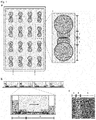
- An exemplary test device with 16 microfluidic units was obtained.
- the exemplary test device has the following dimensions: length 115 mm, width 73.5 mm; Height 14.4 mm.
- the chambers have a diameter of 8 mm.
- the distance between the opposing chambers is 4.5 mm; the compartments are 4.58 mm wide; the microchannels have a length of 150 ⁇ m.
- the chambers, compartments and microchannels of the test device were then washed with ethanol and twice with distilled water to remove dirt.
- the distilled water was aspirated from the chambers and there was a coating with 0.1 mg / mL poly-D-lysine (PDL, Sigma Aldrich) in 0.02 M borate buffer (0.25% boric acid, 0.38% sodium tetraborate ( Merck) in distilled water, pH 8.5) at 4 ° C overnight.
- the PDL was sucked out of the chambers the next morning and coated with 50 ⁇ g / mL laminin (Sigma Aldrich) at 4 ° C. overnight.
- the neurons were isolated, the chambers, compartments and microchannels of the test device were washed twice with heated medium after the laminin had been aspirated off. Immediately before sowing, the medium was removed from the chambers.
- Example 2 Obtaining and cultivating the neurons in the test device
- PCNs Primary cortical neurons
- PCNs Primary cortical neurons
- the cortical hemispheres were transferred from the PBS into CO 2 -saturated papain solution (0.33% papain (Carl Roth), 0.017% L-cysteine (Sigma-Aldrich), 0.1% 0.5 M EDTA (Carl Roth), 1% Sodium acetate (Carl Roth) and 30 mL EBSS (10% Earle's Balanced Salt Solution (Sigma Aldrich) and 0.22% sodium bicarbonate (Möller Chemie), pH 7.3)) and incubated at 37 ° C for 20 minutes.
- CO 2 -saturated papain solution 0.33% papain (Carl Roth), 0.017% L-cysteine (Sigma-Aldrich), 0.1% 0.5 M EDTA (Carl Roth), 1% Sodium acetate (Carl Roth) and 30 mL EBSS (10% Earle's Balanced Salt Solution (Sigma Aldrich) and 0.22% sodium bicarbon
- the tissue was broken up mechanically using serological pipettes and incubated at 37 ° C. for a further 3 minutes. Then 1 mg / mL DNAse was added (Sigma Aldrich) added and the cell suspension centrifuged at 250 xg for 5 minutes. After removing the supernatant, the cell pellet was resuspended in 5 mL EBSS (1%) + bovine serum albumin (BSA) / trypsin inhibitor (1% BSA (Sigma Aldrich) and 1% trypsin inhibitor Chicken Egg White (Sigma Aldrich) in 50 mL EBSS ) and another 5 mL BSA / trypsin inhibitor were carefully added without swirling the cell suspension.
- BSA bovine serum albumin
- trypsin inhibitor 1% BSA (Sigma Aldrich) and 1% trypsin inhibitor Chicken Egg White (Sigma Aldrich) in 50 mL EBSS
- another 5 mL BSA / trypsin inhibitor were carefully added without swirling the
- the suspension was then centrifuged at 250 ⁇ g for 10 minutes, the supernatant was aspirated and the pellet in 5 mL MEM + Glutamax medium (Thermo Fisher Scientific) plus 10% fetal calf serum (Thermo Fisher Scientific), 1% horse serum (GE Health Life Sciences ) and 1% penicillin / streptomycin (Biochrom) resuspended.
- the cell suspension was then filtered using a 70 ⁇ m cell sieve (VWR).
- the volume differences of the medium between the chambers were maintained in order to continue to allow the microfluidic flow for the directed growth of the axons. After 4 to 5 days in culture, the first axons are visible in the axonal compartment and continue to grow over the next few days and weeks. A long-term culture of 1-2 months is possible by changing the medium once a week.
- each pixel was assigned the value 0, 1, 2 or 3, depending on which class the pixel belonged to, with 0 corresponding to the background class, 1 to the axon class, 2 to the axonal spheroid class and 3 to the axonal fragment class.
- a variant of the U-net architecture (Ronneberger et al.) was used for the training and the Vgg16 network (Simonyan et al.) was used as the encoder.
- the network was trained with 120 epochs and the Adam Optimizer, a batch size of 2 and a learning rate of 0.01.
- the input images were standardized using the mean and standard deviation of Imagenet (Deng et al.).
- an independent validation set which the CNN had not yet seen, was generated.
- masks were also created by manual labeling, which represent the basic truth.
- the output of the CNN was compared with the basic truth for each class separately using Precision, Recall and F1 Score.
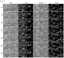
- the CNN's performance was calculated by averaging the F1 scores of all validation images per class ( Figure 2c ).
- the F1 score is defined as 2 * correct positives / (2 * correct positives + false positives + false negatives)
- Example 5 Automated study of axonal degeneration over time
- the axonal compartment was treated with 0 (vehicle), 50, 100 and 200 ⁇ M hemin (Sigma Aldrich), the volume of the medium between the two chambers being equalized in order to prevent the microfluidic flow. Hemin is oxidized heme and is used to simulate damage caused by bleeding in the brain in cell culture (Zille et al., 2017).
- the test device was then observed by means of time-lapse microscopy (Olympus IX81) over 24 hours with a time interval of 30 minutes, the culture conditions being kept constant (37 ° C. in a humid atmosphere with 5% CO 2 ).
- An RNN was trained to recognize the variants of axonal degeneration ( Fig. 2d ). Time courses of the four variants of axonal degeneration were used for this: i) Instant granular degeneration, ii) Axonal swelling, iii) Dying-back degeneration and iv) Transport degeneration.
- the RNN has been trained to take advantage of the change in class.
- the RNN identified 7 clusters whose different combinations predict belonging to one of the four variants of axonal degeneration ( Fig. 5 ).
Abstract
Die vorliegende Erfindung befasst sich mit einer mikrofluidischen Testvorrichtung zur Kultivierung und/oder Untersuchung von Neuronen. Insbesondere betrifft die Erfindung eine mikrofluidische Testvorrichtung zur Kultivierung und/oder Untersuchung von Neuronen, umfassend mindestens eine Trägerplatte und einen Körper, wobei der Körper eine Mehrzahl an mikrofluidische Einheiten aufweist, wobei jede mikrofluidische Einheit mindestens zwei sich gegenüberliegende Kammern umfasst, dadurch gekennzeichnet, dass der Körper monolithisch ausgestaltet ist. Weiterhin wird ein Verfahren zur Untersuchung von Neuronen bereitgestellt, umfassend die Schritte Generieren mikroskopischer Bilddaten von Neuronen in mindestens einer mikrofluidischen Einheit, wobei die Neuronen jeweils in einer mikrofluidischen Einheit vorliegen, wobei von einer mikrofluidischen Einheit mindestens ein mikroskopisches Bild erzeugt wird, wobei jedes mikroskopische Bild eine Mehrzahl an Pixeln aufweist, Generieren von sekundären Bilddaten unter Verwendung eines Bildverarbeitungsalgorithmus, wobei der Bildverarbeitungsalgorithmus für jedes Pixel der generierten Bilder mindestens eine Klassenzugehörigkeit bestimmt und Ausgeben und/oder Erhalten automatisch generierter sekundärer Bilddaten zum Zustand der Neurone. Schließlich wird ein System zur Untersuchung von Neuronen und ein Verfahren zur Kultivierung von Neuronen bereitgestellt.The present invention relates to a microfluidic test device for culturing and / or examining neurons. In particular, the invention relates to a microfluidic test device for cultivating and / or examining neurons, comprising at least one carrier plate and a body, the body having a plurality of microfluidic units, each microfluidic unit comprising at least two opposing chambers, characterized in that the Body is designed monolithically. Furthermore, a method for examining neurons is provided, comprising the steps of generating microscopic image data of neurons in at least one microfluidic unit, the neurons each being in a microfluidic unit, with at least one microscopic image being generated by a microfluidic unit, each microscopic image has a plurality of pixels, generating secondary image data using an image processing algorithm, the image processing algorithm determining at least one class membership for each pixel of the generated images and outputting and / or receiving automatically generated secondary image data on the state of the neurons. Finally, a system for studying neurons and a method for culturing neurons are provided.
Description
Die vorliegende Erfindung befasst sich mit einer mikrofluidischen Testvorrichtung zur Kultivierung und/oder Untersuchung von Neuronen; einem Verfahren zur Untersuchung von Neuronen und einem System zur automatischen Untersuchung von Neuronen. Insbesondere betrifft die Erfindung eine mikrofluidische Testvorrichtung zur Kultivierung und/oder Untersuchung von Neuronen, umfassend mindestens eine Trägerplatte und einen Körper, wobei der Körper eine Mehrzahl an mikrofluidische Einheiten aufweist, wobei jede mikrofluidische Einheit mindestens zwei sich gegenüberliegende Kammern umfasst, dadurch gekennzeichnet, dass der Körper monolithisch ausgestaltet ist. Weiterhin wird ein Verfahren zur Untersuchung von Neuronen bereitgestellt, umfassend die Schritte Generieren mikroskopischer Bilddaten von Neuronen in mindestens einer mikrofluidischen Einheit, wobei die Neuronen jeweils in einer mikrofluidischen Einheit vorliegen, wobei von einer mikrofluidischen Einheit mindestens ein mikroskopisches Bild erzeugt wird, wobei jedes mikroskopische Bild eine Mehrzahl an Pixeln aufweist, Generieren von sekundären Bilddaten unter Verwendung eines Bildverarbeitungsalgorithmus, wobei der Bildverarbeitungsalgorithmus für jedes Pixel der generierten Bilder mindestens eine Klassenzugehörigkeit bestimmt und Ausgeben und/oder Erhalten automatisch generierter sekundärer Bilddaten zum Zustand der Neurone. Schließlich wird ein System zur Untersuchung von Neuronen und ein Verfahren zur Kultivierung von Neuronen bereitgestellt.The present invention relates to a microfluidic test device for culturing and / or examining neurons; a method for the examination of neurons and a system for the automatic examination of neurons. In particular, the invention relates to a microfluidic test device for cultivating and / or examining neurons, comprising at least one carrier plate and a body, the body having a plurality of microfluidic units, each microfluidic unit comprising at least two opposing chambers, characterized in that the Body is designed monolithically. Furthermore, a method for examining neurons is provided, comprising the steps of generating microscopic image data of neurons in at least one microfluidic unit, the neurons each being in a microfluidic unit, with at least one microscopic image being generated by a microfluidic unit, each microscopic image has a plurality of pixels, generating secondary image data using an image processing algorithm, the image processing algorithm determining at least one class membership for each pixel of the generated images and outputting and / or receiving automatically generated secondary image data on the state of the neurons. Finally, a system for studying neurons and a method for culturing neurons are provided.
Untersuchungen von Neuronen und deren kabelartigen und/oder filamentartigen Auswüchsen, auch Nervenfortsätze oder Neurite genannt, ist Gegenstand zahlreicher Forschungsvorhaben, die sich mit dem Verständnis und der therapeutischen Behandlung des menschlichen und/oder tierischen Nervensystems beschäftigen.Studies of neurons and their cable-like and / or filament-like outgrowths, also called nerve processes or neurites, are the subject of numerous research projects that deal with the understanding and therapeutic treatment of the human and / or animal nervous system.
Nervenfortsätze sind kabelartige Auswüchse von Zellkörpern von Neuronen, die Reize zum und vom Gehirn leiten. Die Funktion von Nervenfortsätzen ergibt sich aus einer komplexen Physiologie, die eine Vielzahl Molekülen zur Bereitstellung von Energie, Stabilität und Reizweiterleitung beinhaltet. Viele dieser Moleküle müssen von den Zellkörpern zu den weiter entfernten Teilen der Nervenfortsätze transportiert werden. Unter Nervenfortsätzen können Axone und Dendrite verstanden werden, die jeweils unterschiedliche Funktionen erfüllen können. Die Morphologie von Axonen und Dendriten ist divers und komplex. Axone und/oder Dendrite können unterschiedlichen Längen, Durchmesser und Verästelungen aufweisen. Zusätzlich zur Untersuchung physiologischer Parameter der Axone und/oder Dendrite liegt ein Hauptaugenmerk der Forschung in der Untersuchung von Schädigungen der Axone (axonale Degeneration). Axonale Degeneration beim Menschen kann beispielsweise zu Bewegungseinschränkung bis hin zu Bewegungsunfähigkeit führen, die Wahrnehmung der Umgebung beeinflussen und das Denkvermögen beschränken. Die Untersuchung der Degeneration von Nervenfortsätzen insbesondere von axonaler Degeneration ist erforderlich, um neue Wirkstoffe oder andere therapeutische Verfahren zu entwickeln und zu testen. Im Rahmen industrieller Anwendungen ist die Untersuchung axonaler Degeneration bislang eine Hürde in der Entwicklung neuer Wirkstoffe und Therapien für derzeit unheilbare Erkrankungen wie Querschnittslähmung, Alzheimer und Schlaganfall, bei denen die axonale Degeneration ein wichtiger Prozess im Rahmen der Erkrankung darstellt.Nerve processes are cable-like outgrowths of the cell bodies of neurons that conduct stimuli to and from the brain. The function of nerve processes results from a complex physiology that includes a large number of molecules for the provision of energy, stability and stimulus transmission. Many of these molecules have to be transported from the cell bodies to the more distant parts of the nerve processes. Nerve processes can be understood to mean axons and dendrites, each of which can fulfill different functions. The morphology of axons and dendrites is diverse and complex. Axons and / or dendrites can have different lengths, diameters and ramifications. In addition to the investigation of physiological parameters of the axons and / or dendrites, a main focus of research is the investigation of damage to the axons (axonal degeneration). Axonal degeneration in humans can, for example, lead to restricted mobility or even inability to move, influence the perception of the environment and limit the ability to think. The investigation of the degeneration of nerve processes, especially axonal degeneration, is necessary in order to develop and test new active substances or other therapeutic methods. In the context of industrial applications, the investigation of axonal degeneration has so far been a hurdle in the development of new active ingredients and therapies for currently incurable diseases such as paraplegia, Alzheimer's and stroke, in which axonal degeneration is an important process in the context of the disease.
Eine entscheidende Rolle dabei spielt, dass mikroskopische Untersuchungen von Prozessen in Nervenfortsätzen unabhängig von deren Zellkörpern in Zellkultur zurzeit nicht mit einem hinreichenden Durchsatz möglich sind. Es gibt bislang Systeme, die in einem Objekt verbaut sind und die separate Untersuchung von zellularen Vorgängen an Axonen ermöglichen (auch Campenot Chambers oder Axon Isolation Device genannt; siehe beispielsweise Li, et al. 2014). Diese lassen aber nur Einzeluntersuchungen und keine systematischen Untersuchen zu, da diese extrem aufwendig wären.A decisive role is played by the fact that microscopic examinations of processes in nerve processes independently of their cell bodies in cell culture are currently not possible with a sufficient throughput. There are currently systems that are built into an object and enable the separate investigation of cellular processes on axons (also called Campenot Chambers or Axon Isolation Device; see, for example, Li, et al. 2014). However, these only allow individual examinations and no systematic examinations, as these would be extremely time-consuming.
Ein zweites Problem ist, dass die systematische Auswertung biologischer Prozesse an Axonen eine Software erfordert, die morphologische Veränderungen zellularer Bestandteile quantitativ analysieren kann. Dies bedeutet, dass nicht nur spezifisch Nervenfortsätze, insbesondere Axone, erkannt werden müssen, sondern auch das Ausmaß der axonalen Veränderungen erkannt werden muss.A second problem is that the systematic evaluation of biological processes on axons requires software that can quantitatively analyze morphological changes in cellular components. This means that not only specifically nerve processes, in particular axons, must be recognized, but also the extent of the axonal changes must be recognized.
Morphologische Veränderungen über die Zeit können mithilfe von Zeitverlaufsexperimenten und/oder Zeitraffermikroskopie aufgenommen werden, welche große Mengen an Daten produzieren, sodass eine manuelle Analyse kaum durchführbar, aber auch fehleranfällig wäre, da die Bewertung subjektiv erfolgen würde und nur mit entsprechender Expertise machbar wäre. Fluoreszenzfärbungen verbessern zwar den Kontrast zwischen Zielstrukturen und Hintergrund, können aber lediglich zu einem Zeitpunkt durchgeführt werden, wobei eine zeitliche Auflösung nur in unterschiedlichen Devices möglich ist, oder mittels genetisch veränderter Nervenfortsätze, die beispielsweise mit einem Fluoreszenzmolekül markiert sind. Jedoch sind diese genetischen Veränderungen in Nervenzellen nur limitiert möglich, arbeits- und kostenintensiv und können auch die Funktionen die Nervenfortsätze beeinträchtigen. Des Weiteren können die axonale Physiologie und Degeneration mittels dieser experimentellen Anwendungen nicht umfassend untersucht werden, da der oberservierende zeitliche Ablauf an denselben Nervenfortsätzen ausschlaggebend für das Verständnis dieser Prozesse ist.Morphological changes over time can be recorded using time course experiments and / or time lapse microscopy, which Produce large amounts of data, so that a manual analysis would hardly be feasible, but also prone to errors, since the evaluation would be made subjectively and would only be feasible with the appropriate expertise. Fluorescence staining improves the contrast between target structures and background, but can only be carried out at one point in time, with a temporal resolution only being possible in different devices, or by means of genetically modified nerve processes that are marked with a fluorescent molecule, for example. However, these genetic changes in nerve cells are only possible to a limited extent, labor and cost-intensive and can also impair the functions of the nerve processes. Furthermore, axonal physiology and degeneration cannot be comprehensively investigated by means of these experimental applications, since the overserving temporal sequence on the same nerve processes is crucial for understanding these processes.
Deswegen bedarf es eines automatischen Verfahrens, das spezifisch Nervenfortsätze in mikroskopischen Aufnahmen, wie beispielsweise in der Phasen Kontrast-Mikroskopie, erkennt und quantifizieren kann, vorzugsweise ohne dass es einer Manipulation, beispielsweise genetischen Manipulation, des biologischen Materials bedarf.Therefore, an automatic method is required that specifically detects and quantifies nerve processes in microscopic recordings, for example in the contrast microscopy phases, preferably without the need for manipulation, for example genetic manipulation, of the biological material.
Es wäre daher wünschenswert, eine mikrofluidische Testvorrichtung und ein Verfahren zur Kultivierung und/oder Untersuchung von Neuronen bereitzustellen, welche die Nachteile und/oder Limitationen bekannter Testvorrichtungen und Verfahren zumindest weitgehend vermeiden. Insbesondere soll eine schnelle und einfache Untersuchung ermöglicht werden, die automatisch und objektiv ablaufen kann und somit einen hohen Durchsatz ermöglicht.It would therefore be desirable to provide a microfluidic test device and a method for cultivating and / or examining neurons which at least largely avoid the disadvantages and / or limitations of known test devices and methods. In particular, a quick and simple examination is to be made possible, which can run automatically and objectively and thus enables a high throughput.
Diese Aufgabe wird adressiert durch eine mikrofluidische Testvorrichtung, ein Verfahren zur Kultivierung und/oder Untersuchung von Neuronen und ein System zur automatischen Untersuchung von Neuronen mit den Merkmalen der unabhängigen Patentansprüche. Vorteilhafte Weiterbildungen, welche einzeln oder in beliebiger Kombination realisierbar sind, sind in den abhängigen Ansprüchen dargestellt.This object is addressed by a microfluidic test device, a method for cultivating and / or examining neurons and a system for automatically examining neurons with the features of the independent claims. Advantageous developments, which can be implemented individually or in any combination, are presented in the dependent claims.
Im Folgenden werden die Begriffe "haben", "aufweisen", "umfassen" oder "einschließen" oder beliebige grammatikalische Abweichungen davon in nicht-ausschließlicher Weise verwendet. Dementsprechend können sich diese Begriffe sowohl auf Situationen beziehen, in welchen, neben den durch diese Begriffe eingeführten Merkmalen, keine weiteren Merkmale vorhanden sind, oder auf Situationen, in welchen ein oder mehrere weitere Merkmale vorhanden sind. Beispielsweise kann sich der Ausdruck "A hat B", "A weist B auf', "A umfasst B" oder "A schließt B ein" sowohl auf die Situation beziehen, in welcher, abgesehen von B, kein weiteres Element in A vorhanden ist (d.h. auf eine Situation, in welcher A ausschließlich aus B besteht), als auch auf die Situation, in welcher, zusätzlich zu B, ein oder mehrere weitere Elemente in A vorhanden sind, beispielsweise Element C, Elemente C und D oder sogar weitere Elemente.In the following, the terms “have”, “have”, “comprise” or “include” or any grammatical deviations therefrom are used in a non-exclusive manner. Accordingly, these terms can relate to situations in which, apart from the features introduced by these terms, no further features are present, or to situations in which one or more further features are present. For example, the phrase "A has B", "A has B", "A includes B" or "A includes B" can both refer to the situation in which, apart from B, no further element is present in A (ie on a situation in which A consists exclusively of B), as well as on the situation in which, in addition to B, one or more further elements are present in A, for example Element C, Elements C and D or even more elements.
Weiterhin wird darauf hingewiesen, dass die Begriffe "mindestens ein" und "ein oder mehrere" sowie grammatikalische Abwandlungen dieser Begriffe, wenn diese in Zusammenhang mit einem oder mehreren Elementen oder Merkmalen verwendet werden und ausdrücken sollen, dass das Element oder Merkmal einfach oder mehrfach vorgesehen sein kann, in der Regel lediglich einmalig verwendet werden, beispielsweise bei der erstmaligen Einführung des Merkmals oder Elementes. Bei einer nachfolgenden erneuten Erwähnung des Merkmals oder Elementes wird der entsprechende Begriff "mindestens ein" oder "ein oder mehrere" in der Regel nicht mehr verwendet, ohne Einschränkung der Möglichkeit, dass das Merkmal oder Element einfach oder mehrfach vorgesehen sein kann.It is also pointed out that the terms “at least one” and “one or more” as well as grammatical modifications of these terms, if they are used in connection with one or more elements or features and are intended to express that the element or feature is provided once or several times can be used, as a rule, only once, for example when the feature or element is introduced for the first time. If the feature or element is subsequently mentioned again, the corresponding term “at least one” or “one or more” is generally no longer used, without restricting the possibility that the feature or element can be provided once or several times.
Weiterhin werden im Folgenden die Begriffe "vorzugsweise", "insbesondere", "beispielsweise" oder ähnliche Begriffe in Verbindung mit optionalen Merkmalen verwendet, ohne dass alternative Ausführungsformen hierdurch beschränkt werden. So sind Merkmale, welche durch diese Begriffe eingeleitet werden, optionale Merkmale, und es ist nicht beabsichtigt, durch diese Merkmale den Schutzumfang der Ansprüche und insbesondere der unabhängigen Ansprüche einzuschränken. So kann die Erfindung, wie der Fachmann erkennen wird, auch unter Verwendung anderer Ausgestaltungen durchgeführt werden. In ähnlicher Weise werden Merkmale, welche durch "in einer Ausführungsform der Erfindung" oder durch "in einem Ausführungsbeispiel der Erfindung" eingeleitet werden, als optionale Merkmale verstanden, ohne dass hierdurch alternative Ausgestaltungen oder der Schutzumfang der unabhängigen Ansprüche eingeschränkt werden soll. Weiterhin sollen durch diese einleitenden Ausdrücke sämtliche Möglichkeiten, die hierdurch eingeleiteten Merkmale mit anderen Merkmalen zu kombinieren, seien es optionale oder nicht-optionale Merkmale, unangetastet bleiben.Furthermore, the terms “preferably”, “in particular”, “for example” or similar terms are used below in connection with optional features, without this limiting alternative embodiments. Features introduced by these terms are optional features, and it is not intended to use these features to restrict the scope of protection of the claims and in particular of the independent claims. Thus, as the person skilled in the art will recognize, the invention can also be carried out using other configurations. In a similar way, features introduced by “in an embodiment of the invention” or by “in an exemplary embodiment of the invention” are understood as optional features, without any intention that this should limit alternative configurations or the scope of protection of the independent claims. Furthermore, by means of these introductory expressions, all possibilities of combining the features introduced thereby with other features, be it optional or non-optional features, remain untouched.
Die vorliegende Erfindung schlägt eine mikrofluidische Testvorrichtung zur Kultivierung und/oder Untersuchung von Neuronen, vorzugsweise zur Untersuchung von Axonen, vor. Die mikrofluidische Testvorrichtung umfasst mindestens eine Trägerplatte und einen Körper, wobei der Körper eine Mehrzahl an mikrofluidischen Einheiten aufweist. Jede mikrofluidische Einheit umfasst mindestens zwei sich gegenüberliegenden Kammer. Der Körper der Testvorrichtung ist monolithisch ausgestaltet. Der Begriff monolithisch kann in diesem Zusammenhang so verstanden werden, dass der Körper aus einem Stück geformt worden ist und/oder aus einem Stück besteht.The present invention proposes a microfluidic test device for culturing and / or examining neurons, preferably for examining axons. The microfluidic test device comprises at least one carrier plate and a body, the body having a plurality of microfluidic units. Each microfluidic unit comprises at least two opposing chambers. The body of the test device is designed monolithically. The term monolithic can be used in to be understood in this context that the body has been formed from one piece and / or consists of one piece.
Eine Mehrzahl an mikrofluidischen Einheiten kann vorzugsweise eine Anzahl von 2 bis 200 mikrofluidische Einheiten bedeuten, insbesondere eine Anzahl von 6 bis 100. Vorzugsweise kann der Körper mindestens 6, mehr bevorzugt mindestens 12, noch mehr bevorzugt mindestens 16 mikrofluidische Einheiten aufweisen. Vorzugsweise kann die mikrofluidische Testvorrichtung aus einer Trägerplatte und einem Körper aufweisend eine Mehrzahl an mikrofluidischen Einheiten, beispielsweise mindestens 16 mikrofluidische Einheiten, bestehen. Mehr bevorzugt kann die Mehrzahl an mikrofluidischen Einheiten zusammen unter Verwendung einer einzigen Gussform geformt werden, um den monolithischen Körper zu bilden. Noch mehr bevorzugt kann die Mehrzahl an mikrofluidischen Einheiten zusammen unter Verwendung einer Gussform, die Mulden entsprechend der Mehrzahl oder Anzahl der mikrofluidischen Einheiten aufweist, geformt werden. Beispielsweise kann die mikrofluidische Testvorrichtung einen Körper aus einem geeigneten Material umfassen, in dem die mikrofluidischen Einheiten ausgebildet sind.A plurality of microfluidic units can preferably mean a number from 2 to 200 microfluidic units, in particular a number from 6 to 100. The body can preferably have at least 6, more preferably at least 12, even more preferably at least 16 microfluidic units. The microfluidic test device can preferably consist of a carrier plate and a body having a plurality of microfluidic units, for example at least 16 microfluidic units. More preferably, the plurality of microfluidic devices can be molded together using a single mold to form the monolithic body. Even more preferably, the plurality of microfluidic units can be molded together using a casting mold which has troughs corresponding to the plurality or number of microfluidic units. For example, the microfluidic test device can comprise a body made of a suitable material in which the microfluidic units are formed.
Geeignete Materialien für den Körper der mikrofluidischen Testvorrichtung können sein: Glas, wie beispielsweise Quarzglas, Bleiglas, Kalknatronglas, Borsilikaglas, Silicon, Quarz, Keramik; ein Polymer auf Siliziumbasis, beispielsweise Polydimethylsiloxan (PDMS); Elastomere, beispielsweise Thermoset Polyester und thermoplastische Polymere wie Polystyrol, Polycarbonat, Polyoxymethylen, Polyethylenterephthalat (PET), Polyetherketon (PEEK), Polypropylen (PP), Polymethacrylat, Polymethylmethacrylat (PMMA), Polyethyleneglycol-Diacrylat, Teflon, Perfluoroalkoxy-Polymer, Tetrafluo-rethylen-Hexafluorpropylen-Copolymer, Polyfluoropolyetherdiol-Methacrylat, Polyurethan; zusammengesetzte Materialeien wie zyklische Olefin-Polymere, eine Mischung aus PMMA/PDMS, eine Mischung aus Papier/PDMS; Papier; und/oder Hydrogele. PDMS kann ein bevorzugtes Material sein.Suitable materials for the body of the microfluidic test device can be: glass, such as, for example, quartz glass, lead glass, soda lime glass, borosilica glass, silicone, quartz, ceramic; a silicon based polymer such as polydimethylsiloxane (PDMS); Elastomers, for example Thermoset polyester and thermoplastic polymers such as polystyrene, polycarbonate, polyoxymethylene, polyethylene terephthalate (PET), polyether ketone (PEEK), polypropylene (PP), polymethacrylate, polymethyl methacrylate (PMMA), polyethylene glycol diacrylate, Teflon, perfluoroalkoxy polymer, tetrafluoroethylene -Hexafluoropropylene copolymer, polyfluoropolyether diol methacrylate, polyurethane; composite materials such as cyclic olefin polymers, a mixture of PMMA / PDMS, a mixture of paper / PDMS; Paper; and / or hydrogels. PDMS can be a preferred material.
Wie oben erwähnt, kann der Körper der Testvorrichtung besonders bevorzugt durch Verwendung einer Gussform, die Mulden für die mikrofluidischen Einheiten aufweist, gebildet werden. Die Ausbildung der mikrofluidischen Einheiten in einem gemeinsamen monolithischen Körper kann vorteilhaft sein, da die einzelnen mikrofluidischen Einheiten fest zueinander lokalisiert sind und so ein automatisiertes Aufnehmen von mikroskopischen Bilder mittels Hochdurchsatz- und/oder Screening-Mikroskopen ermöglicht werden kann. Beispielsweise können die mikrofluidischen Einheiten parallel untersucht werden. Insbesondere können Zeitverlaufsuntersuchungen möglich und/oder vereinfacht werden, die das parallele und/oder sequentielle Aufnehmen mikroskopischer Bilder erfordern. Weiterhin kann durch das Vorhandensein einer Mehrzahl an mikrofluidischen Einheiten auf einer Testvorrichtung die Verwendung von Mehrkanalpipetten ermöglicht werden, was zusätzlich eine Zeitersparnis bedeuten kann und somit Ressourcen schonen kann. Somit kann der experimentelle Durchsatz im Vergleich zu herkömmlichen mikrofluidischen Testvorrichtungen, die keine Parallelisierung der mikrofluidischen Einheiten ermöglichen, erhöht werden.As mentioned above, the body of the test device can particularly preferably be formed by using a casting mold which has troughs for the microfluidic units. The formation of the microfluidic units in a common monolithic body can be advantageous, since the individual microfluidic units are permanently localized to one another and thus automated recording of microscopic images by means of high throughput and / or screening microscopes can be made possible. For example, the microfluidic units can be examined in parallel. In particular, time history examinations can be made possible and / or simplified require the parallel and / or sequential acquisition of microscopic images. Furthermore, the presence of a plurality of microfluidic units on a test device makes it possible to use multichannel pipettes, which can also mean a time saving and thus conserve resources. The experimental throughput can thus be increased in comparison to conventional microfluidic test devices, which do not allow the microfluidic units to be parallelized.
Vorzugsweise kann die Testvorrichtung mindestens 2, mindestens 6, mindestens 8, mindestens 16, mindestens 24, mindestens 36, oder mindestens 48 mikrofluidische Einheiten oder weiter bevorzugt bis zu 96 oder mehr mikrofluidische Einheiten aufweisen.The test device can preferably have at least 2, at least 6, at least 8, at least 16, at least 24, at least 36, or at least 48 microfluidic units or more preferably up to 96 or more microfluidic units.
Verfahren zur Herstellung der mikrofluidischen Testvorrichtung sind dem Fachmann bekannt und untenstehend beschrieben.Methods for producing the microfluidic test device are known to the person skilled in the art and are described below.
Vorzugweise kann der Körper auf der Trägerplatte fixiert sein. Die Fixierung (Bonding) kann mittels Plasmabehandlung erfolgen. Dabei können im PDMS Silanolgruppen (Si-OH) geformt werden, die bei Kontakt mit Hydroxid (OH), Karbonsäure (COOH) und/oder Ketonen der Trägerplatte (z. B. Glas) unter Abspaltung von einem Wassermolekül eine irreversible Bindung als Si-O-Si eingehen können (Xiong et al. (2014).The body can preferably be fixed on the carrier plate. The fixation (bonding) can be done by means of plasma treatment. In the process, silanol groups (Si-OH) can be formed in the PDMS, which on contact with hydroxide (OH), carboxylic acid (COOH) and / or ketones on the carrier plate (e.g. glass) form an irreversible bond as Si with splitting off a water molecule. O-Si can enter (Xiong et al. (2014).
Eine mikrofluidische Einheit umfasst mindestens zwei sich gegenüberliegende Kammern. Die sich gegenüberliegenden Kammer einer mikrofluidischen Einheit können jeweils ein Kompartiment aufweisen, das sich in Richtung der anderen Kammer erstreckt. Die zwei sich gegenüberliegenden Kammern können so ausgestaltet sein, dass sich in einer ersten der beiden Kammern die Zellkörper der Neuronen befinden und sich in der dieser Kammer gegenüberliegenden Kammer, der zweiten Kammer, die Nervenfortsätze der Neuronen, vorzugsweise die Axone, befinden. Diese können mit Signalempfängerzellen, wie beispielsweise Gliazellen, Muskelzellen, Endothelzellen, Drüsenzellen, Darmzellen, und/oder Tumorzellen in Kontakt stehen. Vorzugsweise kann das Kompartiment, das sich von der ersten Kammer in Richtung der zweiten Kammer erstreckt als erstes Kompartiment bezeichnet werden und weiter bevorzugt kann ein weiteres Kompartiment, das sich von der zweiten Kammer in Richtung der ersten Kammer erstreckt als zweites oder axonales Kompartiment bezeichnet werden.A microfluidic unit comprises at least two opposing chambers. The opposing chambers of a microfluidic unit can each have a compartment that extends in the direction of the other chamber. The two opposing chambers can be designed so that the cell bodies of the neurons are located in a first of the two chambers and the nerve processes of the neurons, preferably the axons, are located in the chamber opposite this chamber, the second chamber. These can be in contact with signal receiving cells such as glial cells, muscle cells, endothelial cells, gland cells, intestinal cells and / or tumor cells. The compartment which extends from the first chamber in the direction of the second chamber can preferably be referred to as the first compartment and more preferably a further compartment which extends from the second chamber in the direction of the first chamber can be referred to as the second or axonal compartment.
Vorzugsweise besteht eine mikrofluidische Einheit aus zwei sich gegenüberliegenden Kammern, die fluidtechnisch in Verbindung stehen. Dabei weisen die Kompartimente bevorzugt Mikrokanäle auf, mehr bevorzugt stellen die Mikrokanäle eine Verbindung zwischen den beiden Kompartimenten und/oder Kammern dar, beispielsweise eine fluidtechnische Verbindung, die vorzugsweise das Fließen der für die Zellkultur notwendigen Flüssigkeiten zwischen den Kompartimenten ermöglichen kann.A microfluidic unit preferably consists of two opposing chambers which are fluidically connected. The compartments preferably microchannels, more preferably the microchannels represent a connection between the two compartments and / or chambers, for example a fluid-technical connection which can preferably allow the fluids necessary for cell culture to flow between the compartments.
Die Testvorrichtung gemäß der vorliegenden Erfindung weist vorzugsweise einen Boden auf, der die Kompartimente und/oder die Kammern auf der Unterseite verschließt, mehr bevorzugt verschließt der Boden, die Kammern, die Kompartimente und die Mikrokanäle auf der Unterseite.The test device according to the present invention preferably has a base which closes the compartments and / or the chambers on the underside, more preferably closes the base, the chambers, the compartments and the microchannels on the underside.
Vorzugsweise wird der Boden der Testvorrichtung von der Trägerplatte gebildet. Weiter bevorzugt wird der Boden von einer Trägerplatte umfassend oder bestehend aus mindestens einem Material ausgewählt aus Glas und/oder Kunststoff gebildet. Besonders bevorzugt wird der Boden von der Trägerplatte von einem Material ausgewählt aus: Silicon; Glas, wie beispielsweise Quarzglas, Bleiglas, Kalknatronglas, Borsilikaglas; Quarz; Keramik; Elastomere, wie beispielsweise PDMS, Thermoset Polyester, thermoplastische Polymere wie Polystyrol, Polycarbonat, Polyoxymethylen, Polyethylen, Polymethylmethacrylat, Polyethylenglycol-Diacrylat, Polyethylenterephthalat, mit Glycol modifiziertes Polytetrafluorethylen, Polyurethan, gebildet.The base of the test device is preferably formed by the carrier plate. The base is further preferably formed by a carrier plate comprising or consisting of at least one material selected from glass and / or plastic. The base of the carrier plate is particularly preferably selected from a material from: silicone; Glass such as quartz glass, lead glass, soda lime glass, borosilica glass; Quartz; Ceramics; Elastomers such as PDMS, Thermoset polyester, thermoplastic polymers such as polystyrene, polycarbonate, polyoxymethylene, polyethylene, polymethyl methacrylate, polyethylene glycol diacrylate, polyethylene terephthalate, glycol-modified polytetrafluoroethylene, polyurethane.
Die Trägerplatte kann insbesondere eine Beschichtung aufweisen; beispielsweise eine Beschichtung umfassend eine Substanz ausgewählt aus einem Protein, einem Peptid, einem Kohlenhydrat, einer Aminosäure. Die Beschichtung kann die Haftung der Neuronen oder anderer Zellen, wie beispielsweise der oben genannten Signalempfängerzellen, auf der Trägerplatte erhöhen und/oder deren Wachstum verbessern. Geeignete Beschichtungen sind dem Fachmann bekannt und können beispielsweise Laminin, Lysin, Ornithin, Gelatine, Kollagen, Fibronektin, Elastin, Extrazellulärmatrix-Coating, Wachstumsfaktoren und/oder andere umfassen.The carrier plate can in particular have a coating; for example a coating comprising a substance selected from a protein, a peptide, a carbohydrate, an amino acid. The coating can increase the adhesion of the neurons or other cells, such as the above-mentioned signal receiver cells, to the carrier plate and / or improve their growth. Suitable coatings are known to the person skilled in the art and can include, for example, laminin, lysine, ornithine, gelatin, collagen, fibronectin, elastin, extracellular matrix coating, growth factors and / or others.
Vorzugsweise kann die Trägerplatte eine Breite von 80 bis 100 mm und/oder eine Länge von 110 bis 140 mm und/oder eine Höhe von 0,5 bis 2 mm haben; besonders bevorzugt kann eine Breite von 86 mm, eine Länge von 128 mm und eine Höhe von 1 mm sein. Die genannten Ausmaße sind vorteilhaft, da sie das Einspannen in einen herkömmlichen Mikroskopiertisch ermöglichen, insbesondere in einen Mikroskopiertisch eines Hochdurchsatz Mikroskops, wie beispielsweise eines BioTek Lionheart, BioTek Cytation, Perkin Elmer Operetta CLS, oder Perkin Elmer Opera Phenix CLS.The carrier plate can preferably have a width of 80 to 100 mm and / or a length of 110 to 140 mm and / or a height of 0.5 to 2 mm; a width of 86 mm, a length of 128 mm and a height of 1 mm can be particularly preferred. The dimensions mentioned are advantageous because they allow clamping in a conventional microscope table, in particular in a microscope table of a high-throughput microscope, such as a BioTek Lionheart, BioTek Cytation, Perkin Elmer Operetta CLS, or Perkin Elmer Opera Phenix CLS.
Der Begriff "mikrofluidische Testvorrichtung", wie er hier verwendet wird, ist ein weiter Begriff, dem seine gewöhnliche und gängige Bedeutung beigemessen werden soll, wie der Fachmann sie versteht. Der Begriff ist nicht beschränkt auf eine spezielle oder angepasste Bedeutung. Der Begriff bezeichnet jede Vorrichtung die einen mikrofluidischen Fluss zwischen fluidtechnisch miteinander verbundenen Elementen zur Aufnahme von Flüssigkeiten, ermöglicht; insbesondere zwischen fluidtechnisch miteinander verbundenen Kammern im Sinne der vorliegenden Erfindung. Unter "Kammer" kann im Sinne der vorliegenden Erfindung jede Art von Hohlräume oder andere Einheiten geeignet für die Aufnahme von Flüssigkeiten verstanden werden; beispielsweise können Kammern auch Wells von Mikrotiterplatten umfassen. "Kammern" im Sinne der vorliegenden Erfindung sind untenstehend weiter ausgeführt.The term “microfluidic test device” as used here is a broad term to which its customary and common meaning should be assigned, as understood by the person skilled in the art. The term is not limited to any specific or adapted meaning. The term refers to any device that enables a microfluidic flow between fluidically interconnected elements for receiving liquids; in particular between chambers connected to one another by fluid technology within the meaning of the present invention. In the context of the present invention, “chamber” can be understood to mean any type of cavities or other units suitable for receiving liquids; for example, chambers can also include wells of microtiter plates. “Chambers” within the meaning of the present invention are further elaborated below.
Die Testvorrichtung gemäß der vorliegenden Erfindung kann weiterhin mindestens eines der folgenden Materialien umfassen oder daraus bestehen: Glas, wie beispielsweise Quarzglas, Bleiglas, Kalknatronglas, Borsilikaglas, Silicon, Quarz, Keramik; ein Polymer auf Siliziumbasis, beispielsweise Polydimethylsiloxan (PDMS); Elastomere, beispielsweise Thermoset Polyester und thermoplastische Polymere wie Polystyrol, Polycarbonat, Polyoxymethylen, Polynmethylmethacrylat (PMMA), Polyethyleneglycol-Diacrylat, Polyethylenterephthalat, Teflon, Perfluoroalkoxy-Polymer, Tetrafluorethylen-Hexafluorpropylen-Copolymer, Polyfluoropolyetherdiol-Methacrylat, Polyurethan; zusammengesetzte Materialien wie zyklische Olefin-Polymere, eine Mischung aus PMMA/PDMS, eine Mischung aus Papier/PDMS; Papier; und/oder Hydrogele.The test device according to the present invention can further comprise or consist of at least one of the following materials: glass, such as, for example, quartz glass, lead glass, soda lime glass, borosilica glass, silicone, quartz, ceramic; a silicon based polymer such as polydimethylsiloxane (PDMS); Elastomers, for example Thermoset polyester and thermoplastic polymers such as polystyrene, polycarbonate, polyoxymethylene, polymethyl methacrylate (PMMA), polyethylene glycol diacrylate, polyethylene terephthalate, Teflon, perfluoroalkoxy polymer, tetrafluoroethylene-hexafluoropropylene copolymer, polyfluoropolyether diol methacrylate; composite materials such as cyclic olefin polymers, a mixture of PMMA / PDMS, a mixture of paper / PDMS; Paper; and / or hydrogels.
Die Begriffe "Nervenfortsatz" und "Neurit" haben im Zusammenhang mit der vorliegenden Anmeldung dieselbe Bedeutung und werden daher austauschbar verwendet. Bei Neuriten handelt es sich um Fortsätze von Neuronen, beispielsweise um Axone und/oder Dendrite. Axone können insbesondere für die Weiterleitung elektrischer Reize an andere Neuronen und/oder Effektorzellen und/oder Signalempfängerzellen in Zielgeweben verantwortlich sein. Demgegenüber können Dendrite für die Aufnahme von elektrischen Reizen, die z.B. über Axone weitergeleitet werden, verantwortlich sein.The terms “nerve process” and “neurite” have the same meaning in connection with the present application and are therefore used interchangeably. Neurites are extensions of neurons, for example axons and / or dendrites. Axons can in particular be responsible for the transmission of electrical stimuli to other neurons and / or effector cells and / or signal receiver cells in target tissues. On the other hand, dendrites can be responsible for the absorption of electrical stimuli that are transmitted via axons, for example.
Der Begriff "fluidtechnische Verbindung", wie er hier verwendet wird, ist ein weiter Begriff, dem seine gewöhnliche und gängige Bedeutung beigemessen werden soll, wie der Fachmann sie versteht. Der Begriff ist nicht beschränkt auf eine spezielle oder angepasste Bedeutung. Der Begriff kann, ohne Beschränkung, sich insbesondere auf Verbindungen zwischen zwei Kammern einer Testvorrichtung beziehen, die einen Flüssigkeitsdurchfluss erlauben. Insbesondere können solche Verbindungen durch Mikrokanäle verwirklicht werden.As used herein, the term "fluidic connection" is a broad term to be given its ordinary and common meaning as understood by those skilled in the art. The term is not limited to any specific or adapted meaning. The term can, without limitation, relate in particular to connections between two chambers of a test device which allow a flow of liquid. In particular, such connections can be realized through microchannels.
Der Begriff "Mikrokanäle", wie er hier verwendet wird, ist ein weiter Begriff, dem seine gewöhnliche und gängige Bedeutung beigemessen werden soll, wie der Fachmann sie versteht. Der Begriff ist nicht beschränkt auf eine spezielle oder angepasste Bedeutung. Der Begriff "Mikrokanäle" kann, ohne Beschränkung, sich insbesondere auf Kanäle zwischen zwei Kammern beziehen, die vorzugsweise eine maximale Höhe von 5 µm, weiterhin bevorzugt eine maximale Breite von 10 µm und außerdem bevorzugt eine maximale Länge von 4350 µm oder mehr bevorzugt eine maximale Länge von 1500 µm aufweisen. Insbesondere können darunter solche Kanäle verstanden werden, die eine Höhe von 4 bis 6 µm, eine Breite von 8 bis 12 µm und eine Länge von 750 bis 1500 µm aufweisen. Besonders bevorzugt können Mikrokanäle mit einer Höhe von 5 µm, einer Länge von maximal 1000 µm und einer Breite von 10 µm sein.As used herein, the term "microchannels" is a broad term to be given its ordinary and common meaning as understood by those skilled in the art. The term is not limited to any specific or adapted meaning. The term "microchannels" can, without limitation, refer in particular to channels between two chambers, which preferably have a maximum height of 5 μm, further preferably a maximum width of 10 μm and also preferably a maximum length of 4350 μm or more preferably a maximum Have a length of 1500 µm. In particular, these can be understood to mean those channels which have a height of 4 to 6 μm, a width of 8 to 12 μm and a length of 750 to 1500 μm. Microchannels with a height of 5 μm, a maximum length of 1000 μm and a width of 10 μm can be particularly preferred.
Der Begriff "mikrofluidische Einheit", wie er hier verwendet wird, ist ein weiter Begriff, dem seine gewöhnliche und gängige Bedeutung beigemessen werden soll, wie der Fachmann sie versteht. Der Begriff ist nicht beschränkt auf eine spezielle oder angepasste Bedeutung. Der Begriff "mikrofluidische Einheit" kann, ohne Beschränkung, sich insbesondere auf eine Einheit der Testvorrichtung beziehen, wie beispielsweise eine mikrofluidische Einheit, die mindestens zwei z.B. sich gegenüberliegenden Kammern umfasst, die fluidtechnisch verbunden sind. Die fluidtechnische Verbindung kann vorzugsweise aus Mikrokanälen bestehen. Vorzugsweise besteht zwischen den sich gegenüberliegenden Kammern ein mikrofluidischer Fluss. Mehr bevorzugt kann zwischen den Kammern ein Gefälle bestehen, beispielsweise der Flüssigkeiten, in derart, dass sich der Flüssigkeitsstand zwischen den Kammern unterscheidet, das heißt eine der Kammern einen höheren Flüssigkeitsstand aufweist als die andere, so dass ein Flüssigkeitsstrom von der Kammer mit dem höheren Flüssigkeitsstand hinüber zu der Kammer mit dem niedrigeren Flüssigkeitsstand bestehen kann. Der mikrofluidische Fluss kann insbesondere durch das Zugeben von unterschiedlichen Mengen an Flüssigkeit zu den sich gegenüberliegenden Kammern erreicht und/oder eingestellt werden. Beispielsweise kann in eine der gegenüberliegenden Kammer 10, 20, 30, 40 oder auch 50 Vol.-% mehr Flüssigkeit zugegeben werden wie in die andere der gegenüberliegenden Kammern. Dadurch kann beispielsweise ein mikrofluidischer Fluss in Richtung der anderen Kammer erzeugt werden.The term “microfluidic unit”, as used here, is a broad term to which its customary and common meaning is to be assigned, as understood by the person skilled in the art. The term is not limited to any specific or adapted meaning. The term "microfluidic unit" can in particular refer, without limitation, to a unit of the test device, such as, for example, a microfluidic unit which comprises at least two, for example, opposing chambers which are fluidically connected. The fluidic connection can preferably consist of microchannels. There is preferably a microfluidic flow between the opposing chambers. More preferably, there can be a gradient between the chambers, for example the liquids, in such a way that the liquid level differs between the chambers, that is, one of the chambers has a higher liquid level than the other, so that a liquid flow from the chamber with the higher liquid level over to the chamber with the lower fluid level. The microfluidic flow can in particular be achieved and / or adjusted by adding different amounts of liquid to the opposing chambers. For example, 10, 20, 30, 40 or 50% by volume more liquid can be added to one of the opposing chambers than into the other of the opposing chambers. In this way, for example, a microfluidic flow can be generated in the direction of the other chamber.
Unter dem Begriff "mikrofluidisch" kann im Sinne der vorliegenden Anmeldung verstanden werden, dass die flüssigkeitsleitenden Komponenten der Vorrichtung (z.B. die Mikrokanäle) bevorzugt einen Maßstab im Mikrometerbereich aufweisen. In den flüssigkeitsleitenden Komponenten der Vorrichtung kann die Kontrolle der Flüssigkeiten vorzugsweise basierend auf Kapillarkräften, die aufgrund der Ausmaße der flüssigkeitsleitenden Komponenten der Vorrichtung, wie beispielsweise der Mikrokanäle, im Mikrometerbereich wirken, stattfinden.The term “microfluidic” can be understood in the sense of the present application that the liquid-conducting components of the device (for example the microchannels) preferably have a scale in the micrometer range. The control of the liquids can preferably be based in the liquid-conducting components of the device on capillary forces which, due to the dimensions of the liquid-conducting components of the device, such as the microchannels, act in the micrometer range, take place.
Der Begriff "mikrofluidischer Fluss" wie er hier verwendet wird, ist ein weiter Begriff, dem seine gewöhnliche und gängige Bedeutung beigemessen werden soll, wie der Fachmann sie versteht. Der Begriff ist nicht beschränkt auf eine spezielle oder angepasste Bedeutung. Der Begriff "mikrofluidischer Fluss" kann insbesondere eine durch Kapillarkräfte bewirkte Fließrichtung einer Flüssigkeit in der mikrofluidischen Testvorrichtung bezeichnen. Insbesondere kann der mikrofluidische Fluss eine Fließrichtung zwischen den mindestens zwei sich gegenüberliegenden Kammer bewirken; ganz besonders kann der mikrofluidische Fluss eine Fließrichtung, beispielsweise einer Nährflüssigkeit oder eines Nährmediums, von einer ersten Kammer der mikrofluidischen Einheit, in der sich mindestens ein Zellkörper eines Neurons befinden kann, in Richtung einer zweiten Kammer der mikrofluidischen Einheit, in der sich mindestens ein von dem Zellkörper ausgehender Nervenfortsatz, insbesondere ein Axon befinden kann, bewirken.The term “microfluidic flow” as used here is a broad term to which its customary and common meaning should be assigned, as understood by the person skilled in the art. The term is not limited to any specific or adapted meaning. The term “microfluidic flow” can in particular denote a direction of flow of a liquid in the microfluidic test device caused by capillary forces. In particular, the microfluidic flow can bring about a flow direction between the at least two opposing chambers; In particular, the microfluidic flow can have a direction of flow, for example a nutrient fluid or a nutrient medium, from a first chamber of the microfluidic unit, in which at least one cell body of a neuron can be located, in the direction of a second chamber of the microfluidic unit, in which at least one of the cell body outgoing nerve process, in particular an axon, can cause.
Unter "Kapillarkraft" kann das von Flüssigkeiten gezeigte Verhalten, das sie bei Kontakt mit Kapillaren, z. B. engen Röhren, Mikrokanälen, Spalten oder Hohlräumen in Feststoffen zeigen, verstanden werden. Diese Effekte können durch die Oberflächenspannung von Flüssigkeiten selbst und die Grenzflächenspannung zwischen Flüssigkeiten und der festen Oberfläche hervorgerufen werden. Insbesondere kann unter dem Begriff "Kapillarkraft" im Sinne der vorliegenden Erfindung eine Adhäsionskraft verstanden werden, die zwischen einer Flüssigkeit und der Wandung einer sie umgebenden Röhre und/oder eines sie umgebenen Mikrokanals wirken kann. Diese Kraft kann insbesondere die Steighöhe und den Transport der Flüssigkeit innerhalb der Röhre und/oder des Mikrokanals und/oder innerhalb der mikrofluidischen Testvorrichtung beeinflussen.Under "capillary force", the behavior exhibited by liquids, which they experience when they come into contact with capillaries, e.g. B. narrow tubes, microchannels, crevices or cavities in solids can be understood. These effects can be caused by the surface tension of liquids themselves and the interfacial tension between liquids and the solid surface. In particular, the term “capillary force” in the context of the present invention can be understood to mean an adhesion force which can act between a liquid and the wall of a tube surrounding it and / or a microchannel surrounding it. This force can in particular influence the height of rise and the transport of the liquid within the tube and / or the microchannel and / or within the microfluidic test device.
Der Begriff "Kompartiment", wie er hier verwendet wird, ist ein weiter Begriff, dem seine gewöhnliche und gängige Bedeutung beigemessen werden soll, wie der Fachmann sie versteht. Der Begriff ist nicht beschränkt auf eine spezielle oder angepasste Bedeutung. Der Begriff "Kompartiment" kann, ohne Beschränkung, sich insbesondere auf eine Aussparung beziehen, die sich von der jeweiligen Kammer zu der anderen gegenüberliegenden Kammer hin erstreckt. Ein Kompartiment im Sinne der vorliegenden Erfindung kann beispielsweise eine Breite von 2 bis 15 mm aufweisen; vorzugsweise eine Breite von 4 bis 8 mm; besonders bevorzugt eine Breite von 4 bis 5 mm aufweisen.As used herein, the term "compartment" is a broad term to which its customary and common meaning should be given, as understood by those skilled in the art. The term is not limited to any specific or adapted meaning. The term “compartment” can, without limitation, relate in particular to a recess which extends from the respective chamber to the other opposite chamber. For the purposes of the present invention, a compartment can, for example, have a width of 2 to 15 mm; preferably a width of 4 to 8 mm; particularly preferably have a width of 4 to 5 mm.
Ein Kompartiment im Sinne der vorliegenden Erfindung kann nach oben und unten und auf zwei Seiten geschlossen sein; auf der Seite, die zu der gegenüberliegenden Kammer hin orientiert ist, kann das Kompartiment mindestens eine Öffnung aufweisen; diese Öffnung kann an Mikrokanäle anschließen; auf der Seite, die zu der jeweiligen Kammer hin orientiert ist, kann eine direkte Verbindung zu dieser Kammer bestehen. Vorzugsweise können die Kompartimente mit der jeweiligen Kammer direkt verbunden sein. Unter einer "direkten Verbindung" kann in diesem Zusammenhang eine Öffnung, die sich über die gesamte Breite des Kompartiments erstreckt, verstanden werden. Somit können die Ausmaße der Öffnung durch die Ausmaße des Kompartiments begrenzt sein. Beispielhafte Ausmaße der Öffnung können sein: eine maximale Höhe von 14,9 mm, eine maximale Breite von 15 mm, und/oder eine maximale Länge von 2,5 mm. Bevorzugt kann die Öffnung eine Höhe von 100 bis 200 µm, eine Breite von 0,5 bis 8 mm, weiter bevorzugt eine Breite von 4 bis 5 mm, und/oder eine Länge von 1 bis 2,3 mm aufweisen.A compartment within the meaning of the present invention can be closed at the top and bottom and on two sides; on the side which is oriented towards the opposite chamber, the compartment can have at least one opening; this opening can connect to microchannels; on the side that is oriented towards the respective chamber, there can be a direct connection to this chamber. The compartments can preferably be connected directly to the respective chamber. In this context, a “direct connection” can be understood to mean an opening which extends over the entire width of the compartment. Thus, the dimensions of the opening can be limited by the dimensions of the compartment. Exemplary dimensions of the opening can be: a maximum height of 14.9 mm, a maximum width of 15 mm, and / or a maximum length of 2.5 mm. The opening can preferably have a height of 100 to 200 μm, a width of 0.5 to 8 mm, more preferably a width of 4 to 5 mm, and / or a length of 1 to 2.3 mm.
Die Kompartimente können weiterhin Mikrokanäle aufweisen. Insbesondere, können die Mikrokanäle eine Verbindung zwischen den beiden Kompartimenten oder den Kammern darstellen. Vorzugsweise können die Mikrokanäle ausgehend von einem Kompartiment einer Kammer zu einem Kompartiment der gegenüberliegenden Kammer eine Verbindung ausbilden. Noch mehr bevorzugt kann durch die Mikrokanäle eine fluidtechnische Verbindung zwischen den sich gegenüberliegenden Kompartimenten ausgebildet werden. Eine fluidtechnische Verbindung kann vorteilhaft sein, da sie das Bereitstellen eines mikrofluidischen Flusses zwischen den Kammern ermöglichen kann und somit eine Richtung für das Wachstum der Nervenfortsätze vorgegeben werden kann. Weiterhin können die Mikrokanäle aufgrund ihrer geringen Ausmaße vorteilhaft sein, da sie den Durchtritt nur einzelner oder einiger weniger Nervenfortsätze ermöglichen können. Durch eine solche Separation kann die Untersuchung einzelner oder nur weniger Nervenfortsätze bewerkstelligt werden.The compartments can also have microchannels. In particular, the microchannels can represent a connection between the two compartments or the chambers. The microchannels can preferably form a connection starting from one compartment of one chamber to a compartment of the opposite chamber. Even more preferably, the microchannels can form a fluidic connection between the opposing compartments. A fluidic connection can be advantageous because it can enable a microfluidic flow to be provided between the chambers and thus a direction for the growth of the nerve processes can be specified. Furthermore, the microchannels can be advantageous because of their small dimensions, since they can only allow single or a few nerve processes to pass through. Such a separation can be used to examine individual or only a few nerve processes.
Vorzugsweise können die Mikrokanäle voneinander einen Abstand von maximal 100 µm aufweisen. Mehr bevorzugt können die Mikrokanäle einen Abstand von maximal 75 µm, mehr bevorzugt von maximal 50 µm auf weisen. Noch mehr bevorzugt kann der Abstand zwischen einzelnen Mikrokanälen zwischen 20 und 50 µm betragen. Dieser Abstand kann vorteilhaft sein, da so die Nervenfortsätze in verschiedenen Mikrokanälen separat im Mikroskop abgebildet werden können. Vorzugsweise können sich die Mikrokanäle auf derselben Ebene befinden, mehr bevorzugt auf derselben Fokusebene. Noch mehr bevorzugt können die Mikrokanäle nach unten hin direkt an die Trägerplatte anschließen.The microchannels can preferably be spaced apart from one another by a maximum of 100 μm. More preferably, the microchannels can have a distance of a maximum of 75 μm, more preferably of a maximum of 50 μm. Even more preferably, the distance between individual microchannels can be between 20 and 50 μm. This distance can be advantageous because it allows the nerve processes to be displayed separately in the microscope in different microchannels. The microchannels can preferably be located on the same plane, more preferably on the same focal plane. Even more preferably, the microchannels can connect directly to the carrier plate at the bottom.
Vorzugsweise sind die Kompartimente über eine Vielzahl von Mikrokanälen miteinander verbunden. Bevorzugt beträgt die Anzahl der Mikrokanäle zwischen den Kompartimenten maximal 250; besonders bevorzugt 20 bis 200, noch mehr bevorzugt 50 bis 100. Eine solche Anzahl kann den Vorteil haben, dass ein ausreichender mikrofluidischer Fluss bewerkstelligt werden kann und Nervenfortsätze, insbesondere Axone, entlang des mikrofluidischen Flusses von einem oder mehreren Zellkörpern der Neurone in der ersten Kammer ausgehend in die Mikrokanäle einwachsen können und/oder sich vorzugsweise bis in die gegenüberliegende zweite Kammer erstrecken können. Eine geringere Anzahl an Mikrokanälen kann den Nachteil haben, dass der mikrofluidische Fluss nicht ausreichend sein kann, um ein gerichtetes Wachstum der Axone zu bewerkstelligen. Ein weiterer Vorteil kann darin bestehen, dass die beschriebene Anzahl an Mikrokanälen zu einem Abstand der Mikrokanäle untereinander führen kann, der für die mikroskopische Analyse gut geeignet sein kann; beispielsweise dadurch, dass die Axone, die durch einen Mikrokanal gewachsen sind, separat betrachtet werden können. Eine höhere Anzahl an Mikrokanälen kann den Nachteil haben, dass es zu einer zu hohen Anzahl und somit zu einer hohen Dichte der Axone in der zweiten Kammer kommen kann und so eine separate mikroskopische Analyse der Axone aufgrund einer zu hohen Dichte der Axone nicht mehr möglich sein kann.The compartments are preferably connected to one another via a multiplicity of microchannels. The number of microchannels between the compartments is preferably a maximum of 250; particularly preferably 20 to 200, even more preferably 50 to 100. Such a number can have the advantage that a sufficient microfluidic flow can be achieved and nerve processes, in particular axons, along the microfluidic flow of one or more cell bodies of the neurons in the first chamber starting into the microchannels can grow and / or can preferably extend into the opposite second chamber. A smaller number of microchannels can have the disadvantage that the microfluidic flow cannot be sufficient to bring about a directed growth of the axons. Another advantage can be that the number of microchannels described can lead to a spacing between the microchannels which can be well suited for microscopic analysis; for example, in that the axons that have grown through a microchannel can be viewed separately. A higher number of microchannels can have the disadvantage that the axons in the second chamber can become too high in number and therefore have a high density, so that a separate microscopic analysis of the axons is no longer possible due to an excessively high density of the axons can.
Die Kompartimente und/oder die Mikrokanäle können weiterhin eine Überdeckung aufweisen. Die Überdeckung kann derart gestaltet sein, dass sie die Neuronen und/oder Axone größtenteils von oben abdeckt. Das Vorhandensein einer Überdeckung kann vorteilhaft sein, da beim Auswechseln des Kulturmediums, beispielsweise mit einer Mehrkanalpipette, die Zellkörper der Neurone und/oder die Axone größtenteils von der Überdeckung geschützt werden und daher weniger leicht verletzt werden können.The compartments and / or the microchannels can also have an overlap. The covering can be designed in such a way that it largely covers the neurons and / or axons from above. The presence of an overlap can be advantageous because when the culture medium is changed, for example with a multichannel pipette, the cell bodies of the neurons and / or the axons are largely protected by the overlap and are therefore less likely to be injured.
Die Kammern einer mikrofluidischen Einheit haben vorzugsweise eine zylindrische Form. Noch mehr bevorzugt weisen die Kammern eine zylindrische Form auf, vorzugsweise können die Kammern einen Durchmesser von 1 bis 10 mm, mehr bevorzugt von 2 bis 8 mmm aufweisen. Ein solcher Durchmesser kann vorteilhaft sein, um ein Medienvolumen zu erreichen, dass die Kultivierung von Zellkörpern und Nervenfortsätzen ermöglichen kann. Weiterhin können die Kammern eine maximale Höhe von 15 mm aufweisen, vorzugsweise können die Kammern eine Höhe von 1 bis 15 mmm aufweisen. Eine solche Höhe kann vorteilhaft sein, um ein Volumen zu erreichen, beispielsweise ein Volumen von mindestens 20 µl oder mindestens 50 µl, das die Kultivierung von Zellkörpern und Nervenfortsätzen ermöglichen kann.The chambers of a microfluidic unit preferably have a cylindrical shape. Even more preferably, the chambers have a cylindrical shape; the chambers can preferably have a diameter of 1 to 10 mm, more preferably 2 to 8 mm. Such a diameter can be advantageous in order to achieve a media volume that can enable the cultivation of cell bodies and nerve processes. Furthermore, the chambers can have a maximum height of 15 mm, preferably the chambers can have a height of 1 to 15 mm. Such a height can be advantageous in order to achieve a volume, for example a volume of at least 20 μl or at least 50 μl, which can enable the cultivation of cell bodies and nerve processes.
Typischerweise ist eine Kammer gemäß der vorliegenden Erfindung ausgestaltet, um ein Volumen von 20 bis 350 µl, insbesondere ein Volumen von 50 bis 200 µl, zu umfassen. Solche Volumina können insbesondere vorteilhaft sein, da sie für das Einstellen des mikrofluidischen Flusses geeignet sein können. Für eine Kultivierung von Zellen und oder Zellkulturen im Sinne der vorliegenden Erfindung kann ein Volumen von 50 bis 350 µl notwendig sein. Genannte Volumen können vorteilhaft sein, da die Kultivierung der Zellen/Zellkulturen und/oder die Versorgung der Zellen/Zellkulturen mit Nährstoffen über einen Zeitraum von 1 bis 60 Tagen ermöglicht werden kann. Während der Kultivierung können aus angesäten Zellkörpern der Neurone Axone auswachsen, die sich durch die Mikrokanäle hindurch von der einen Kammer, vorzugsweise von der ersten Kammer, in die gegenüberliegende Kammer, vorzugsweise in die zweite Kammer, hin erstrecken können. Geeignete Medien, Kulturmedien, Nährmedien oder Nährflüssigkeiten sind dem Fachmann bekannt und beispielsweise in Muramatsu and Yamashita (2013) veröffentlicht.Typically, a chamber according to the present invention is designed to have a volume of 20 to 350 μl, in particular a volume of 50 to 200 μl. Such volumes can be particularly advantageous since they can be suitable for adjusting the microfluidic flow. For the cultivation of cells and / or cell cultures within the meaning of the present invention, a volume of 50 to 350 μl may be necessary. Said volumes can be advantageous since the cultivation of the cells / cell cultures and / or the supply of the cells / cell cultures with nutrients can be made possible over a period of 1 to 60 days. During the cultivation, axons can grow from sown cell bodies of the neurons, which axons can extend through the microchannels from one chamber, preferably from the first chamber, into the opposite chamber, preferably into the second chamber. Suitable media, culture media, nutrient media or nutrient liquids are known to the person skilled in the art and are published, for example, in Muramatsu and Yamashita (2013).
Die Begriffe "Kulturmedien", "Nährmedien" oder "Nährflüssigkeiten" beziehen sich auf Medien, die für die Anzucht und/oder Kultivierung von Zellen, insbesondere von Neuronen, geeignet sind und werden hier austauschbar verwendet. Für Nervenzellen besonders geeignete Nährmedien können insbesondere die folgenden Medien ein schließen: Minimum Essential Medium (MEM) + Glutamax enthaltend 10% Pferdeserum und/oder Dulbecco's Modified Eagle's Medium (DMEM) enthaltend 10% Pferdeserum, Wachstumsfaktoren und/oder DMEM-F12 Medium enthaltend 10% Pferdeserum, 1mM Natriumpyruvat und/oder Eagle's MEM enthaltend 0,5 mM Glutamin, 20 mM Glucose und/oder Neurobasal A Medium enthaltend 2% B-27 oder B-27 Plus Supplement, 1mM Natriumpyruvat und/oder Neurobasal Plus Medium enthaltend 2% B-27 oder B-27 Plus Supplement, 1mM Natriumpyruvat (kommerziell erhältlich beispielsweise bei Thermo Fisher Scientific) und/oder Neuronal Base Medium enthaltend 2% N21-MAX Media Supplement, Wachstumsfaktoren (kommerziell erhältlich beispielsweise bei R&D Systems).The terms “culture media”, “nutrient media” or “nutrient fluids” relate to media which are suitable for growing and / or cultivating cells, in particular neurons, and are used interchangeably here. Nutrient media particularly suitable for nerve cells can include in particular the following media: Minimum Essential Medium (MEM) + Glutamax containing 10% horse serum and / or Dulbecco's Modified Eagle's Medium (DMEM) containing 10% horse serum, growth factors and / or DMEM-F12 medium containing 10 % Horse serum, 1mM sodium pyruvate and / or Eagle's MEM containing 0.5 mM glutamine, 20 mM glucose and / or Neurobasal A medium containing 2% B-27 or B-27 Plus supplement, 1mM sodium pyruvate and / or Neurobasal Plus medium containing 2% B-27 or B-27 Plus Supplement, 1mM sodium pyruvate (commercially available, for example, from Thermo Fisher Scientific) and / or neuronal base medium containing 2% N21-MAX Media Supplement, growth factors (commercially available, for example, from R&D Systems).
Der Abstand der gegenüberliegenden Kammern kann vorzugsweise zwischen 2 und 10 mm betragen. Entsprechend der Abstände zwischen den Kammern kann sich die Länge der die Kammern verbindenden Mikrokanäle verlängern. Daraus kann entsprechend eine größere Distanz entstehen, die ein Axon beim Hineinwachsen von der einen Kammer in die gegenüberliegende Kammer überbrücken muss. Die Kultivierungszeiten der Axone können sich so entsprechend verlängern. Zu große Abständen zwischen den gegenüberliegenden Kammern können nachteilig sein, dass sich die Kultivierungszeiten der Axone zu stark verlängern können.The distance between the opposing chambers can preferably be between 2 and 10 mm. The length of the microchannels connecting the chambers can be lengthened in accordance with the distances between the chambers. This can lead to a greater distance that an axon has to cover as it grows from one chamber into the opposite chamber. The cultivation times of the axons can be extended accordingly. Too great a distance between the opposing chambers can be disadvantageous that the cultivation times of the axons can be prolonged too much.
Die vorliegende Erfindung stellt weiterhin ein Verfahren zur Kultivierung von Neuronen bereit. Das Verfahren umfasst die folgenden Schritte:
- a) Bereitstellen einer mikrofluidischen Testvorrichtung gemäß der vorliegenden Erfindung;
- b) Aussäen von Neuronen in mindestens eine der Kammern und/oder Kompartimente, so dass mindestens eine angesäte Kammer entsteht,
- c) Inkontaktbringen zumindest eines Teils der angesäten Kammern mit einem Kulturmedium, und
- d) Kultivieren der Neuronen.
- a) providing a microfluidic test device according to the present invention;
- b) sowing neurons in at least one of the chambers and / or compartments, so that at least one sown chamber is created,
- c) bringing at least a part of the sown chambers into contact with a culture medium, and
- d) culturing the neurons.
Der Begriff "Kultivierung", wie er hier verwendet wird, ist ein weiter Begriff, dem seine gewöhnliche und gängige Bedeutung beigemessen werden soll, wie der Fachmann sie versteht. Der Begriff ist nicht beschränkt auf eine spezielle oder angepasste Bedeutung. Der Begriff "Kultivierung" kann, ohne Beschränkung, sich insbesondere auf die Anzucht von Zellen oder Zellkulturen in einem geeigneten Wachstumsmedium unter Bedingungen, die das Wachstum der jeweiligen Zellen oder Zellkulturen erlauben, beziehen. Als Kultivierung kann insbesondere die Anzucht der Zellen oder Zellkulturen bei einer Luftfeuchtigkeit von vorzugsweise 95%, bei einem CO2-Gehalt von vorzugsweise 5 Vol.-% und/oder bei einer Temperatur von mindestens 20 °C bis maximal 45 °C verstanden werden, besonders geeignet ist eine Temperatur von 37°C. Durch die Kultivierung unter den genannten Bedingungen kann vorteilhafterweise insbesondere Verdunstung vermieden und der pH-Wert konstant gehalten werden. Die Anzucht kann beispielsweise in einem geeigneten kommerziell erhältlichen Inkubator erfolgen, wie beispielsweise CO2-Inkubatoren mit Heißluftsterilisation und hitzesterilisierbarem CO2-Sensor der CB Serie von Binder (Tuttlingen, Deutschland). Als geeignetes Medium kann eines oder können mehrere der oben genannten Kulturmedien verwendet werden.As used herein, the term "cultivation" is a broad term to be given its ordinary and common meaning as understood by those skilled in the art. The term is not limited to any specific or adapted meaning. The term “cultivation” can, without limitation, relate in particular to the cultivation of cells or cell cultures in a suitable growth medium under conditions which allow the respective cells or cell cultures to grow. Cultivation can be understood to mean growing the cells or cell cultures at a humidity of preferably 95%, with a CO 2 content of preferably 5% by volume and / or at a temperature of at least 20 ° C to a maximum of 45 ° C, A temperature of 37 ° C. is particularly suitable. By cultivating under the conditions mentioned, evaporation in particular can advantageously be avoided and the pH can be kept constant. Cultivation can take place, for example, in a suitable, commercially available incubator, such as, for example, CO 2 incubators with hot air sterilization and heat-sterilizable CO 2 sensors of the CB series from Binder (Tuttlingen, Germany). One or more of the abovementioned culture media can be used as a suitable medium.
Schritt a) des Bereitstellens einer mikrofluidischen Testvorrichtung kann insbesondere das Erstellen einer mikrofluidischen Testvorrichtung mittels geeigneter lithographischer Mittel und/oder Verfahren umfassen. Diese Verfahren und Mittel sind dem Fachmann bekannt. Insbesondere kann es sich dabei um photo- und/oder softlithographische Mittel und/oder Verfahren handeln, wie aus
Das Aussäen der Neuronen in Schritt b) kann insbesondere mit einer Zelldichte von 1000 bis 20000 Zellen pro mm2 bezogen auf die Grundfläche des Kompartiments, vorzugsweise bei einer Zelldichte von 6000 Zellen pro mm2 bezogen auf die Grundfläche des Kompartiments erfolgen. Des Weiteren kann das Aussäen zusätzliche Schritte, wie beispielsweise das Anhaften der Zellen an die Testvorrichtung, beinhalten. Das Aussäen kann weiterhin die vorausgehende Isolierung von Nervenvorläuferzellen, wie beispielsweise die Isolierung von primären Neuronen aus dem Gehirn, Rückenmark oder Spinalganglien eines geeigneten Spenderorganismuses, wie beispielsweise Mensch, Maus, Ratte, Kaninchen, und/oder weiteren, umfassen. Auch eine vorausgehende Gewinnung induzierter pluripotenter Stammzellen (iPSC) aus beispielweise menschlichen Fibroblasten, die dann erst zu Neuronen ausdifferenziert werden können, kann denkbar sein.Seeding the neurons in step b) can in particular take place with a cell density of 1000 to 20,000 cells per mm 2 based on the base area of the compartment, preferably with a cell density of 6,000 cells per mm 2 related to the base area of the compartment. Furthermore, the seeding can include additional steps, such as, for example, adhering the cells to the test device. The sowing can furthermore comprise the previous isolation of nerve precursor cells, such as, for example, the isolation of primary neurons from the brain, spinal cord or dorsal root ganglia of a suitable donor organism, such as, for example, humans, mice, rats, rabbits and / or others. A previous extraction of induced pluripotent stem cells (iPSC) from, for example, human fibroblasts, which can only then be differentiated into neurons, may also be conceivable.
Schritt c) des Inkontaktbringens zumindest eines Teils der angesäten Kammern mit einem Kulturmedium kann vorzugsweise das Einbringen eines geeigneten Kulturmediums mittels einer herkömmlichen Laborpipette oder einer Mehrkanalpipette umfassen. Vorzugsweise kann Schritt c) das Einbringen von mindestens 20 µl eines geeigneten Kulturmediums in jede der gegenüberliegenden Kammern und/oder Kompartimente umfassen. Bevorzugt kann eine unterschiedliche Menge an Kulturmedium in die gegenüberliegenden Kammern der mikrofluidischen Einheiten eingebracht werden. Mehr bevorzugt kann die Menge an Kulturmedium, das in die erste Kammer eingebracht wird, die Menge des Kulturmediums, das in die zweite Kammer eingebracht wird, übersteigen. Noch mehr bevorzugt kann die Menge an Kulturmedium, das in die in die erste Kammer eingebracht wird, die Menge des Kulturmediums das in die zweite Kammer eingebracht wird um 10 Vol.-% bis 100 Vol.-%, beispielsweise um 50 Vol.-%, übersteigen in Bezug auf die Menge an Kulturmedium in der zweiten Kammer. Eine geeignete Menge an Kulturmedium kann beispielsweise zwischen 20 µl und 350 µl betragen. Durch das Einbringen unterschiedlicher Mengen an Kulturmedium in die gegenüberliegenden Kammern kann der mikrofluidische Fluss erzeugt werden. Dies kann vorteilhaft sei, da so ein gerichtetes Wachstum der Axone ausgehend vom Zellkörper in der ersten Kammer hin zur zweiten Kammer erfolgen kann.Step c) of bringing at least some of the sown chambers into contact with a culture medium can preferably include the introduction of a suitable culture medium by means of a conventional laboratory pipette or a multi-channel pipette. Step c) can preferably include the introduction of at least 20 μl of a suitable culture medium into each of the opposing chambers and / or compartments. A different amount of culture medium can preferably be introduced into the opposing chambers of the microfluidic units. More preferably, the amount of culture medium that is introduced into the first chamber may exceed the amount of the culture medium that is introduced into the second chamber. Even more preferably, the amount of culture medium that is introduced into the first chamber, the amount of the culture medium that is introduced into the second chamber by 10% by volume to 100% by volume, for example by 50% by volume. , exceed in terms of the amount of culture medium in the second chamber. A suitable amount of culture medium can be, for example, between 20 μl and 350 μl. The microfluidic flow can be generated by introducing different amounts of culture medium into the opposing chambers. This can be advantageous, since in this way a directed growth of the axons can take place starting from the cell body in the first chamber towards the second chamber.
Das in die beiden gegenüberliegenden Kammern eingebrachte Kulturmedium kann dasselbe sein, alternativ können aber auch unterschiedliche Kulturmedien in die erste und die zweite Kammer eingebracht werden. Ein solches Vorgehen kann vorteilhaft sein, um spezifische Auswirkungen auf einen Teil, das Axon oder den Zellkörper des Neurons untersuchen zu können.The culture medium introduced into the two opposing chambers can be the same, but alternatively different culture media can also be introduced into the first and second chambers. Such an approach can be advantageous in order to be able to study specific effects on a part, the axon or the cell body of the neuron.
Schritt d) umfasst das Kultivieren der Neuronen. Dieser Schritt kann insbesondere die Inkubation der Neuronen bei einer geeigneten Temperatur, geeigneter Luftfeuchte und/oder einem geeigneten CO2-Gehalt umfassen. Insbesondere kann darunter das Inkubieren bei 37°C in einer 5 Vol.-% CO2 Atmosphäre bei 95% Luftfeuchtigkeit verstanden werden.Step d) comprises culturing the neurons. This step can in particular include the incubation of the neurons at a suitable temperature, suitable humidity and / or a suitable CO 2 content. In particular, this can be understood to mean incubation at 37 ° C. in a 5% by volume CO 2 atmosphere with 95% air humidity.
Vorzugsweise kann das Verfahren zur Kultivierung von Neuronen weiterhin umfassen: e) Co-Kultivieren von weiteren Zellen, beispielsweise Gliazellen, Muskelzellen, Endothelzellen, Drüsenzellen, Darmzellen, Tumorzellen. Das Co-Kultivieren weiterer Zellen kann die Untersuchung von Zell-Zell-Interaktionen ermöglichen und kann daher vorteilhaft sein.The method for cultivating neurons can preferably further comprise: e) co-culturing further cells, for example glial cells, muscle cells, endothelial cells, gland cells, intestinal cells, tumor cells. The co-cultivation of further cells can enable the investigation of cell-cell interactions and can therefore be advantageous.
Das Verfahren zur Kultivierung kann weiterhin umfassen: Schritt f) wiederholen zumindest der Schritte c) und d) und/oder weitere Schritte umfassen.The method for culturing can furthermore comprise: step f) repeating at least steps c) and d) and / or comprise further steps.
Weiterhin stellt die vorliegende Erfindung ein Verfahren zur Untersuchung von Neuronen bereit. Das Verfahren umfasst die folgenden Schritte:
- a) Generieren mikroskopischer Bilddaten von Neuronen in mindestens einer mikrofluidischen Einheit, wobei die Neuronen jeweils in einer mikrofluidischen Einheit vorliegen; wobei von einer mikrofluidischen Einheit mindestens ein mikroskopisches Bild erzeugt wird, wobei jedes mikroskopische Bild eine Mehrzahl an Pixeln aufweist;
- b) Generieren von sekundären Bilddaten unter Verwendung eines Bildverarbeitungsalgorithmus, wobei der Bildverarbeitungsalgorithmus für jedes jeden Pixel der in Schritt a) generierten Bilder mindestens eine Klassenzugehörigkeit bestimmt; und
- c) Ausgeben und/oder Erhalten automatisch generierter sekundärer Bilddaten zum Zustand der Neurone.
- a) generating microscopic image data of neurons in at least one microfluidic unit, the neurons each being present in a microfluidic unit; wherein at least one microscopic image is generated by a microfluidic unit, each microscopic image having a plurality of pixels;
- b) generating secondary image data using an image processing algorithm, the image processing algorithm determining at least one class for each pixel of the images generated in step a); and
- c) Outputting and / or receiving automatically generated secondary image data on the state of the neurons.
Das Verfahren kann weiterhin die folgenden optionalen Schritte umfassen:
- cc) Erzeugen von Daten zum Zustand der Neurone aus den sekundärer Bilddaten; und/oder
- d) Zusammenfügen eines digital aufbereiteten Bildes basierend auf den sekundären Bilddaten;
- e) Darstellen der automatisch generierten sekundären Bilddaten des digital aufbereiteten Bildes auf einer Bildwiedergabevorrichtung; und/oder
- f) Ausgeben und/oder Erhalten automatisch generierter tertiärer und/oder quartärer Bilddaten.
- cc) generating data on the state of the neurons from the secondary image data; and or
- d) Merging a digitally processed image based on the secondary image data;
- e) displaying the automatically generated secondary image data of the digitally processed image on an image reproduction device; and or
- f) Outputting and / or receiving automatically generated tertiary and / or quaternary image data.
In Schritt a) können die Neuronen vorzugsweise jeweils in einer der mikrofluidischen Einheiten der Testvorrichtung gemäß der vorliegenden Erfindung vorliegen. Vorzugsweise können von einer mikrofluidischen Einheit mehrere mikroskopische Bilddaten erzeugt werden. Weiterhin bevorzugt können die Bilddaten mindestens die beiden gegenüberliegenden Kammern und/oder die Kompartimente abbilden. Besonders bevorzugt können die Bilddaten die gesamte mikrofluidische Einheit, beispielsweise die gesamte mikrofluidische Einheit in ihrer flächenmäßigen Ausdehnung, abbilden. Beispielsweise können bei einer bevorzugten mikroskopischen Auflösung von 10-fach von den gegenüberliegenden Kammern, den Kompartimenten und/oder der mikrofluidischen Einheit 1 bis 12 Bilder erzeugt werden, vorzugsweise 3 bis 8 Bilder. Die bevorzugte Anzahl der Bilder und/oder Bilddaten und/oder die mikroskopische Auflösung kann je nach Zweck und/oder Fragestellung der Untersuchung variieren. Eine möglichst geringe Anzahl an Bildern kann vorteilhaft sein, da so Zeit und Ressourcen, wie zum Beispiel Speicherkapazitäten, gespart werden können.In step a), the neurons can preferably each be present in one of the microfluidic units of the test device according to the present invention. A plurality of microscopic image data can preferably be generated by a microfluidic unit. Furthermore, the image data can preferably depict at least the two opposing chambers and / or the compartments. Particularly preferably, the image data can depict the entire microfluidic unit, for example the entire microfluidic unit in terms of its surface area. For example, with a preferred microscopic resolution of 10 times, 1 to 12 images, preferably 3 to 8 images, can be generated of the opposing chambers, the compartments and / or the microfluidic unit. The preferred number of images and / or image data and / or the microscopic resolution can vary depending on the purpose and / or question of the investigation. The smallest possible number of images can be advantageous, since this saves time and resources, such as storage capacities.
Der Begriff "gesamte mikrofluidische Einheit" soll so verstanden werden, dass insbesondere mindestens die beiden sich gegenüberliegenden Kammer und die Mikrokanäle umfasst sind, besonders bevorzugt können die beiden sich gegenüberliegenden Kammer, die Kompartimente und die Mikrokanäle umfasst sein.The term “entire microfluidic unit” should be understood to include in particular at least the two opposing chambers and the microchannels; the two opposing chambers, the compartments and the microchannels can particularly preferably be included.
Weiterhin kann Schritt a) ein Generieren von Bilddaten über einen Zeitverlauf umfassen. Das Generieren von Bilddaten über einen Zeitverlauf kann beispielsweise das Erzeugen von Bilddaten jeweils nach Ablauf eines bestimmten Zeitintervalls umfassen, beispielsweise jeweils nach 30 oder nach 60 Minuten über einen Zeitraum von mehreren Stunden und/oder Tagen. Je nach Zweck und/oder Fragestellung der jeweiligen Untersuchung können auch kürzere oder längere Zeitintervalle und/oder Zeiträume verwendet werden.Furthermore, step a) can include generating image data over time. The generation of image data over time can include, for example, the generation of image data after a specific time interval has elapsed, for example after 30 or 60 minutes over a period of several hours and / or days. Depending on the purpose and / or question of the particular investigation, shorter or longer time intervals and / or time periods can also be used.
Vorzugsweise kann das Generieren der mikroskopischen Bilddaten in Schritt a) das Aufnehmen von mikroskopischen Bilddaten umfassen, bevorzugt Daten von lichtmikroskopischen Aufnahmen und/oder fluoreszenzmikroskopischen Aufnahmen; mehr bevorzugt Daten von Phasenkontrastaufnahmen.The generation of the microscopic image data in step a) can preferably include the recording of microscopic image data, preferably data from light microscopic images and / or fluorescence microscopic images; more preferably data from phase contrast images.
Für das Generieren der mikroskopischen Bilddaten besonders geeignet können insbesondere Lichtmikroskope, wie beispielsweise Lichtmikroskope ausgebildet für die Phasenkontrastmikroskopie, und/oder Fluoreszenzmikroskope sein. Zum Generieren der Bilddaten können auch Hochdurchsatzmikroskope und/oder Screening-Mikroskope verwendet werden. Besonders bevorzugt kann die Verwendung eines Hochdurchsatzmikroskops ausgebildet für die Phasenkontrastmikroskopie sein, wie beispielsweise BioTek Lionheart, BioTek Cytation, Perkin Elmer Operetta CLS, oder Perkin Elmer Opera Phenix CLS. Die Verwendung von Hochdurchsatz- oder Screening-Mikroskopen kann vorteilhaft sein, da eine weitgehend automatisierte Erzeugung der Bilddaten möglich werden kann.Light microscopes, such as light microscopes designed for phase contrast microscopy, and / or fluorescence microscopes, in particular, can be particularly suitable for generating the microscopic image data. High-throughput microscopes and / or screening microscopes can also be used to generate the image data be used. It is particularly preferable to use a high-throughput microscope designed for phase contrast microscopy, such as BioTek Lionheart, BioTek Cytation, Perkin Elmer Operetta CLS, or Perkin Elmer Opera Phenix CLS. The use of high-throughput or screening microscopes can be advantageous, since a largely automated generation of the image data can become possible.
In Schritt a) wird weiterhin von einer mikrofluidischen Einheit mindestens ein mikroskopisches Bild erzeugt, wobei jedes mikroskopische Bild eine Mehrzahl an Pixeln aufweist. Eine Mehrzahl an Pixeln kann in diesem Zusammenhang 64x64 bis 1024x1024 Pixel bedeuten, vorzugsweise 512x512. Wenn die Anzahl an Pixeln zu klein ist, kann die Klassifizierung durch den Bildverarbeitungsalgorithmus negativ beeinflusst werden. Vorzugsweise kann das Erzeugen des mikroskopischen Bildes die Speicherung des Bildes in einer Auswerteeinheit umfassen.In step a), at least one microscopic image is also generated by a microfluidic unit, each microscopic image having a plurality of pixels. In this context, a plurality of pixels can mean 64x64 to 1024x1024 pixels, preferably 512x512. If the number of pixels is too small, the classification can be negatively influenced by the image processing algorithm. The generation of the microscopic image can preferably include the storage of the image in an evaluation unit.
Bevorzugt kann das Generieren der Bilddaten in Schritt a) und b) mikroskopische Bilddaten von Axonen umfassen.The generation of the image data in steps a) and b) can preferably include microscopic image data of axons.
Der Begriff "automatisch generierte sekundäre Bilddaten" bezieht sich in diesem Zusammenhang insbesondere auf die unter Verwendung des Bildverarbeitungsalgorithmus generierten sekundären Bilddaten.The term “automatically generated secondary image data” in this context relates in particular to the secondary image data generated using the image processing algorithm.
Das Generieren von sekundären Bilddaten unter Verwendung eines Bildverarbeitungsalgorithmus in Schritt b) kann vorzugsweise einen Bildverarbeitungsalgorithmus umfassen, welcher anhand von mikroskopischen Bilddaten mit bekannten Informationen trainiert wurde. Mehr bevorzugt kann das Generieren von sekundären Bilddaten unter Verwendung eines Bildverarbeitungsalgorithmus in Schritt b) einen Machine-Learning-Algorithmus umfassen, welcher anhand von mikroskopischen Bilddaten mit bekannten Informationen trainiert wurde. Weiterhin bevorzugt kann der Bildverarbeitungsalgorithmus jedem Pixel die Wahrscheinlichkeit einer Klassenzugehörigkeit zuordnen. Noch mehr bevorzugt kann das Generieren von sekundären Bilddaten mit einem Bildverarbeitungsalgorithmus in Schritt b) einen Deep Learning Algorithmus, welcher anhand von mikroskopischen Bilddaten mit bekannten Informationen trainiert wurde, umfassen; am weiterhin bevorzugt kann das Generieren von sekundären Bilddaten mit einem Bildverarbeitungsalgorithmus in Schritt b) ein Convolutional Neural Network (CNN) umfassen, welches anhand von mikroskopischen Bilddaten mit bekannten Informationen trainiert wurde. Am meisten bevorzugt kann der Bildverarbeitungsalgorithmus in Schritt b) ein CNN sein. Insbesondere kann das Generieren von sekundären Bilddaten in schritt b) automatisch ablaufen.The generation of secondary image data using an image processing algorithm in step b) can preferably include an image processing algorithm which was trained on the basis of microscopic image data with known information. More preferably, the generation of secondary image data using an image processing algorithm in step b) can comprise a machine learning algorithm which was trained on the basis of microscopic image data with known information. Furthermore, the image processing algorithm can preferably assign the probability of a class to each pixel. Even more preferably, the generation of secondary image data with an image processing algorithm in step b) can comprise a deep learning algorithm which was trained on the basis of microscopic image data with known information; Most preferably, the generation of secondary image data with an image processing algorithm in step b) can include a convolutional neural network (CNN) which was trained on the basis of microscopic image data with known information. Most preferably, the Image processing algorithm in step b) be a CNN. In particular, the generation of secondary image data can take place automatically in step b).
Der Begriff "mikroskopische Bilddaten mit bekannten Informationen", wie er hier verwendet wird, ist ein weiter Begriff, dem seine gewöhnliche und gängige Bedeutung beigemessen werden soll, wie der Fachmann sie versteht. Der Begriff ist nicht beschränkt auf eine spezielle oder angepasste Bedeutung. Der Begriff kann, ohne Beschränkung, sich insbesondere auf Bilddaten beziehen, die mittels eines Mikroskops aufgenommen wurden und Pixel umfassen.The term "microscopic image data with known information", as used here, is a broad term to which its customary and common meaning should be assigned, as understood by the person skilled in the art. The term is not limited to any specific or adapted meaning. The term can, without limitation, relate in particular to image data which were recorded by means of a microscope and which include pixels.
Weiterhin können aus den Bilddaten mikroskopische Bilder bestehend aus einer Mehrzahl an Pixeln erzeugt werden. Unter einer Mehrzahl an Pixeln können hier beispielsweise 64x64, 128x128, 256x256, 512x512 oder 1024x1024 Pixel pro Bild verstanden werden.Furthermore, microscopic images consisting of a plurality of pixels can be generated from the image data. A plurality of pixels can be understood here to mean, for example, 64x64, 128x128, 256x256, 512x512 or 1024x1024 pixels per image.
Beispielsweise kann der Begriff "mikroskopische Bilddaten mit bekannten Informationen" sich auf Bilddaten oder Bilder umfassend Pixel beziehen, wobei zu jedem einzelnen Pixel mindestens Informationen über eine Klassenzugehörigkeit bekannt sind. Vorzugsweise ist mindestens die Zugehörigkeit ausgewählt aus einer von zwei Klassen, mehr bevorzugt ausgewählt aus den Klassen Hintergrund und Neuron, noch mehr bevorzugt ausgewählt aus den Klassen Hintergrund und Axon. Weiterhin kann die Klassenzugehörigkeit ausgewählt aus mehr als zwei Klassen, vorzugsweise aus zwei bis zehn Klassen einschließlich drei, vier, fünf, sechs, sieben, acht, neun oder zehn Klassen, ausgewählt sein. Es können aber auch mehr als zehn Klassen möglich sein. Besonders bevorzugt kann die Klassenzugehörigkeit aus vier Klassen ausgewählt sein. Insbesondere bevorzugt kann die Zugehörigkeit aus einer der folgenden Klassen ausgewählt sein: Hintergrund, Axon, axonales Fragment, axonaler Spheroid. Mikroskopische Bilddaten mit bekannten Informationen können beispielsweise über manuelle Markierung von Bildelementen mittels geeigneter Software erhalten werden. Zu hierfür geeigneter Software zählen unter anderem pixelbasierte Grafikprogramme wie GIMP (GNU Image Manipulation Program; https://docs.gimp.org/2.10/de/). Zur Erstellung mikroskopischer Bilddaten mit bekannten Informationen können beispielsweise Bildelemente umfassend die gewünschten Zellen oder deren Bestandteile, wie beispielsweise Neuronen oder Axone, in einem existierenden mikroskopischen Bild von Hand markiert werden. Vorzugsweise können die Bildelemente aus zusammenhängenden Pixeln bestehen oder diese umfassen. Weiterhin vorzugsweise umfassen die existierenden mikroskopischen Bilder mikroskopische Aufnahmen von Neuronen und/oder Axonen vorzugsweise Phasenkontrast-Aufnahmen oder fluoreszenzmikroskopische Aufnahmen. Die markierten Bildelemente können dann vorzugsweise der Klasse Neuron oder Axon zugeordnet sein; mehr bevorzugt können die markierten Bildelemente einer von drei Klassen: Axon, axonaler Spheroid oder axonales Fragment zugeordnet sein. Die von Hand (manuell) markierten Bildelemente können insbesondere die Grundwahrheit für die Klassenzuordnung der Pixel darstellen.For example, the term “microscopic image data with known information” can relate to image data or images comprising pixels, with at least information about a class affiliation being known for each individual pixel. Preferably, at least the membership is selected from one of two classes, more preferably selected from the classes background and neuron, even more preferably selected from the classes background and axon. Furthermore, the class affiliation can be selected from more than two classes, preferably from two to ten classes including three, four, five, six, seven, eight, nine or ten classes. However, more than ten classes can also be possible. The class affiliation can particularly preferably be selected from four classes. Affiliation can particularly preferably be selected from one of the following classes: background, axon, axonal fragment, axonal spheroid. Microscopic image data with known information can be obtained, for example, by manually marking image elements using suitable software. Suitable software for this includes pixel-based graphics programs such as GIMP (GNU Image Manipulation Program; https://docs.gimp.org/2.10/de/). To create microscopic image data with known information, for example, image elements comprising the desired cells or their components, such as neurons or axons, can be marked by hand in an existing microscopic image. The picture elements can preferably consist of or comprise contiguous pixels. Furthermore, the existing microscopic images preferably include microscopic images of neurons and / or axons, preferably phase contrast images or fluorescence microscopic images. The marked picture elements can then preferably be of the neuron or axon class be assigned; more preferably, the marked picture elements can be assigned to one of three classes: axon, axonal spheroid or axonal fragment. The picture elements marked by hand (manually) can in particular represent the basic truth for the class allocation of the pixels.
Unmarkierte Bildelemente können vorzugsweise der Klasse Hintergrund zugeordnet sein. Vorzugsweise kann die Markierung auf Basis eines Schwellwerts gesetzt werden. Geeignete Schwellwertverfahren sind dem Fachmann bekannt (Meijering, 2012). Der Schwellwert kann insbesondere einen Helligkeitswert, und/oder Werte wie die Klassenwahrscheinlichkeit umfassen. Dieser Wert kann bevorzugt durch den Bildverarbeitungsalgorithmus festgelegt werden. Mehr bevorzugt kann die Markierung auf Basis der größten Wahrscheinlichkeit, beispielsweise auf Basis eines Schwellwerts von 0,5 bei den Klassen Axon/Hintergrund, zugeordnet werden.Unmarked picture elements can preferably be assigned to the background class. The marking can preferably be set on the basis of a threshold value. Suitable threshold value methods are known to the person skilled in the art (Meijering, 2012). The threshold value can in particular include a brightness value and / or values such as the class probability. This value can preferably be determined by the image processing algorithm. More preferably, the marking can be assigned on the basis of the greatest probability, for example on the basis of a threshold value of 0.5 for the axon / background classes.
Beispielsweise können sich bei 4 Klassen für jedes Pixel vier Werte ergeben, die sich alle zu 1 aufsummieren können; z.B. (0.8, 0.05, 0.05, 0.1) für ein Pixel. Vorzugsweise reflektiert der höchste Wert, die höchste Wahrscheinlichkeit zur Zugehörigkeit zu einer bestimmten Klasse; mehr bevorzugt bestimmt der höchste Wahrscheinlichkeitswert die Klassenzughörigkeit.
Die genannten Schwellwert-Verfahren können vorzugsweise auf den primären/und oder sekundären Bilddaten angewendet werden. Im Falle von nur 2 Klassen, beispielsweise Neuron (oder Axon) und Hintergrund, kann vorzugsweise ein einziger Schwellwert ausreichend sein. Dann können sich nur zwei Werte Pro Pixel (x, y) ergeben, wobei gilt: x+y=1 und somit y = 1 - x. Man kann vorzugsweise als Ausgabe ein Bild der Größe [Eingangshöhe x Eingangsbreite x 1] bekommen, wobei jedes Pixel einen Wert zwischen 0 und 1 annehmen kann. Es kann beispielsweise einen Schwellwert t (beispielsweise 0.5) bestimmt werden und die Aussage "alle Pixel <= t gehören zu Klasse 0 alle anderen zu Klasse 1" kann getroffen werden.For example, with 4 classes, four values can result for each pixel, which can all add up to 1; e.g. (0.8, 0.05, 0.05, 0.1) for a pixel. Preferably, the highest value reflects the highest probability of belonging to a specific class; more preferably the highest probability value determines the class affiliation.
The mentioned threshold value methods can preferably be applied to the primary / and / or secondary image data. In the case of only 2 classes, for example neuron (or axon) and background, a single threshold value can preferably be sufficient. Then only two values per pixel (x, y) can result, where: x + y = 1 and thus y = 1 - x. You can preferably get an image of the size [entrance height x entrance width x 1] as output, whereby each pixel can assume a value between 0 and 1. For example, a threshold value t (for example 0.5) can be determined and the statement “all pixels <= t belong to
Zusätzlich können die bekannten Informationen der Bilddaten weitere Informationen umfassen insbesondere zu: axonalem Wachstum, axonaler Degeneration, axonaler Remodelierung, und/oder axonaler Regeneration.In addition, the known information in the image data can include further information, in particular on: axonal growth, axonal degeneration, axonal remodeling, and / or axonal regeneration.
Das Trainieren des Bildverarbeitungsalgorithmus anhand von mikroskopischen Bilddaten mit bekannten Informationen kann beispielsweise in einer U-Net basierten Architektur stattfinden. Insbesondere kann eine U-Net Variante mit einem VGG16 Encoder oder ResNet, DenseNet, Hourlass, MobileNet, oder EfficientNet verwendet werden. Vorzugsweise können das Trainieren eine Veränderung der mikroskopischen Bilddaten umfassen; dafür kann beispielsweise eine Standardisierung der mikroskopischen Bilddaten stattfinden (preprocessing) und/oder eine Spiegelung der mikroskopischen Bilddaten (data augmentation;
Besonders bevorzugt umfasst das Trainieren mehrere Wiederholungen mit mindestens 5, noch mehr bevorzugt mit mindestens 10 Bilddatensätzen. Dabei kann ein Bilddatensatz vorzugsweise eine mikroskopische Aufnahme umfassend die mikroskopischen Bilddaten mit bekannten Informationen, wie obenstehend ausgeführt, umfassen oder daraus bestehen. Dieser Bilddatensatz kann als Trainingsprobe bezeichnet werden. Eine geeignete Anzahl an Bilddatensätzen oder Trainingsproben kann 10 bis 100 Bilddatensätze umfassen, bevorzugt mindestens 15, mehr bevorzugt mindestens 30; eine Anzahl von 10 Bilddatensätzen kann bereits ausreichend sein. Ein Trainieren mit der genannten Anzahl an Bilddatensätzen oder Trainingsproben kann vorteilhaft sein, da es zu einer ausreichend genauen Zuordnung von Pixeln aus unbekannten mikroskopischen Bildern oder mikroskopischen Bilddaten führen kann. Die Verwendung einer größeren Anzahl kann den Nachteil haben, dass das Trainieren mehr Zeit und mehr Aufwand erfordert.The training particularly preferably comprises several repetitions with at least 5, even more preferably with at least 10 image data sets. In this case, an image data set can preferably comprise or consist of a microscopic recording comprising the microscopic image data with known information, as explained above. This image data set can be referred to as a training sample. A suitable number of image data sets or training samples can comprise 10 to 100 image data sets, preferably at least 15, more preferably at least 30; a number of 10 image data records can already be sufficient. Training with the specified number of image data sets or training samples can be advantageous, since it can lead to a sufficiently precise assignment of pixels from unknown microscopic images or microscopic image data. Using a larger number can have the disadvantage of requiring more time and effort to train.
Vorzugsweise kann jede Trainingsprobe mindestens je ein Bild umfassen, welches aus Pixeln besteht oder diese umfasst und wobei zu jedem einzelnen Pixel mindestens Informationen über eine Klassenzugehörigkeit bekannt sind. Der Bildverarbeitungsalgorithmus kann weiterhin vorzugsweise für mindestens 5 und maximal 1000 Epochen unter Verwendung eines geeigneten Optimierers, wie beispielsweise eines Adam, SGD, AdaDelta, Lamb, oder RMSProb Optimierers; einer Batchgröße von 2 bis 32; und einer Lernrate zwischen 10 und 10-6, insbesondere mittels Backpropagation, trainiert worden sein. Die Bilddaten mit bekannten Informationen können beispielsweise mittels ImageNet Mittelwert und Standardabweichung normalisiert werden. Weiterhin kann vorzugsweise eine Inferenz ausgeführt worden sein. Die oben genannten Verfahren sind dem Fachmann beispielsweise aus Ronneberger et al. bekannt.Each training sample can preferably each comprise at least one image which consists of or comprises pixels and wherein at least information about a class affiliation is known for each individual pixel. The image processing algorithm can furthermore preferably be used for at least 5 and a maximum of 1000 epochs using a suitable optimizer, such as an Adam, SGD, AdaDelta, Lamb, or RMSProb optimizer; a batch size of 2 to 32; and a learning rate between 10 and 10 -6 , in particular using backpropagation. The image data with known information can, for example, be normalized using ImageNet mean value and standard deviation. Furthermore, an inference can preferably have been carried out. The above-mentioned methods are known to the person skilled in the art, for example, from Ronneberger et al. famous.
Zusätzlich kann beim Trainieren mit Bilddaten aus Zeitverlaufsexperimenten ein Medianfilter auf der zeitlichen Ebene mit einem geeigneten Zeitfenster durchgeführt werden, beispielsweise 3,5 h bei 7 Bildern.In addition, when training with image data from time course experiments, a median filter can be carried out on the temporal level with a suitable time window, for example 3.5 hours for 7 images.
Weiterhin kann für das Trainieren der Datensatz mittels data augmentation künstlich vergrößert werden. Eine solche Vergrößerung des Datensatzes kann insbesondere zufälliges Ausscheiden der Bilddaten umfassen, beispielsweise mit einer Größe von 448x448 Pixeln und/oder zufälliges Drehen und Umklappen der Bilddatensätze entlang der vertikalen oder horizontalen Achse oder beiden umfassen. Vorzugsweise kann das Drehen der Bilddatensätze zwischen 0 und 270 Grad erfolgen, mehr bevorzugt mit 0°, 90°, 180° und/oder 270°. Weiterhin können beispielsweise Verfahren wie additive noise, Farbinvertierung, Farbkanäle vertauschen, affine Transformationen, Image Warping, Löschen einzelner Pixel oder Bereiche und/oder zufällige Helligkeits- und/oder Kontrast-Transformation zur Augmentation des Datensatzes verwendet werden.Furthermore, the data set can be artificially enlarged for training using data augmentation. Such an enlargement of the data set can in particular comprise random removal of the image data, for example with a size of 448x448 pixels and / or random rotation and flipping of the image data sets along the vertical or horizontal axis or both. The image data records can preferably be rotated between 0 and 270 degrees, more preferably at 0 °, 90 °, 180 ° and / or 270 °. Furthermore, methods such as additive noise, color inversion, swapping color channels, affine transformations, image warping, deleting individual pixels or areas and / or random brightness and / or contrast transformation can be used to augment the data set.
Das Trainieren kann insbesondere ein Verfahren zur stochastischen Optimierung umfassen, insbesondere ein stochastisches Gradientenverfahren, wie stochastisch gradient descent (SGD).
In Schritt b) des Verfahrens zur Untersuchung von Neuronen werden sekundäre Bilddaten unter Verwendung eines Bildverarbeitungsalgorithmus generiert.The training can in particular include a method for stochastic optimization, in particular a stochastic gradient method, such as stochastic gradient descent (SGD).
In step b) of the method for examining neurons, secondary image data are generated using an image processing algorithm.
Der Bildverarbeitungsalgorithmus bestimmt in Schritt b) für jedes Pixel der in Schritt a) generierten Bilddaten mindestens eine Klassenzugehörigkeit. Die in Schritt a) generierten Bilddaten können als primäre Bilddaten bezeichnet werden. Mehr bevorzugt umfassen diese primären Bilddaten Pixel oder bestehen daraus. Beispielsweise können die in Schritt a) generierten Bilddaten Bilder, die Pixel umfassen oder daraus bestehen. Die Bilder können beispielsweise aus 512x512 Pixeln oder aus 1024x1024 Pixeln bestehen.In step b), the image processing algorithm determines at least one class association for each pixel of the image data generated in step a). The image data generated in step a) can be referred to as primary image data. More preferably, these primary image data comprise or consist of pixels. For example, the image data generated in step a) can include or consist of pixels. The images can consist of 512x512 pixels or 1024x1024 pixels, for example.
Vorzugsweise kann der Bildverarbeitungsalgorithmus die Zugehörigkeit ausgewählt aus einer von zwei Klassen bestimmen, mehr bevorzugt die Zugehörigkeit ausgewählt aus Hintergrund und Neuron, noch mehr bevorzugt die Zugehörigkeit ausgewählt aus Hintergrund und Axon bestimmen. Weiterhin kann der Bildverarbeitungsalgorithmus die Klassenzugehörigkeit ausgewählt aus mehr als zwei Klassen bestimmen, vorzugsweise aus zwei bis zehn Klassen einschließlich drei, vier, fünf, sechs, sieben, acht, neun oder zehn Klassen. Es können aber auch mehr als zehn Klassen möglich sein. Besonders bevorzugt kann der Bildverarbeitungsalgorithmus in Schritt b) für jedes Pixel der in Schritt a) generierten Bilddaten die Klassenzugehörigkeit zu mindestens oder zu genau vier Klassen bestimmen. Insbesondere bevorzugt kann der Bildverarbeitungsalgorithmus die Zugehörigkeit zu einer der folgenden Klassen bestimmen: Hintergrund, Axon, axonaler Spheroid, axonales Fragment. Ganz besonders bevorzugt, kann der Bildverarbeitungsalgorithmus in Schritt b) ein Verfahren der semantischen Segmentierung umfassen und/oder ausführen.The image processing algorithm can preferably determine the affiliation selected from one of two classes, more preferably the affiliation selected from background and neuron, even more preferably determine the affiliation selected from background and axon. Furthermore, the image processing algorithm can determine the class membership selected from more than two classes, preferably from two to ten classes including three, four, five, six, seven, eight, nine or ten classes. However, more than ten classes can also be possible. Particularly preferably, the image processing algorithm in step b) can determine for each pixel of the image data generated in step a) the class membership of at least or precisely four classes. Particularly preferably, the image processing algorithm can determine belonging to one of the following classes: background, axon, axonal spheroid, axonal fragment. Most preferably, the Image processing algorithm in step b) comprise and / or execute a method of semantic segmentation.
Insbesondere kann die Bestimmung zu einer Klassenzugehörigkeit für jedes Pixel der in Schritt a) generierten Bilddaten durch den Bildverarbeitungsalgorithmus die Ausgabe einer Wahrscheinlichkeit für die Zugehörigkeit zu jeder der Klassen umfassen. Vorzugsweise kann der Bildverarbeitungsalgorithmus zu jedem Pixel eine Wahrscheinlichkeit für jede Klasse ausgeben, anhand derer das Pixel einer Klasse zugeordnet werden kann. Besonders bevorzugt kann das Pixel anhand einer durch den Bildverarbeitungsalgorithmus ausgegebenen Wahrscheinlichkeit zu einer der Klassen Hintergrund oder Axon zugeordnet werden; noch mehr bevorzugt zu einer der Klassen Hintergrund, Axon, axonaler Spheroid oder axonales Fragment. Alternativ kann insbesondere eine Zuordnung zu einer der Klassen Hintergrund oder Neuron erfolgen. Weiterhin bevorzugt kann die Klasse mit dem höchsten Wert der Wahrscheinlichkeit für die Zuordnung ausschlaggebend sein; insbesondere kann der Bildverarbeitungsalgorithmus das Pixel der Klasse, für die der Wert der ausgegebenen Wahrscheinlichkeit am höchsten ist, zuordnen.In particular, the determination of a class association for each pixel of the image data generated in step a) by the image processing algorithm can include the output of a probability for the association with each of the classes. For each pixel, the image processing algorithm can preferably output a probability for each class, on the basis of which the pixel can be assigned to a class. Particularly preferably, the pixel can be assigned to one of the classes background or axon on the basis of a probability output by the image processing algorithm; even more preferably to one of the classes background, axon, axonal spheroid or axonal fragment. Alternatively, an assignment to one of the background or neuron classes can take place in particular. Furthermore, the class with the highest value of the probability can be decisive for the assignment; In particular, the image processing algorithm can assign the pixel to the class for which the value of the output probability is highest.
Das Generieren von sekundären Bilddaten in Schritt b) kann vorzugsweise unter Verwendung eines Convolutional Neural Network (CNN), auch als "Faltungsnetzwerk" bekannt, stattfinden. Insbesondere kann das Generieren von sekundären Bilddaten in Schritt b) unter Verwendung eines U-Net stattfinden; beispielsweise kann ein U-Net mit VGG16-Encoder, oder ResNet, DenseNet, MobileNet, EfficientNet verwendet werden. Beispielsweise können auch andere Segmentierungsarchitekturen wie Hourglass oder DeepLab verwendet werden.The generation of secondary image data in step b) can preferably take place using a convolutional neural network (CNN), also known as a “convolution network”. In particular, the generation of secondary image data in step b) can take place using a U-Net; For example, a U-Net with VGG16 encoder, or ResNet, DenseNet, MobileNet, EfficientNet can be used. For example, other segmentation architectures such as Hourglass or DeepLab can also be used.
Vorzugsweise kann das Generieren von sekundären Bilddaten in Schritt b) das Segmentieren der Bilddaten umfassen. Unter "Segmentieren der Bilddaten" wird im Hinblick auf die vorliegende Erfindung das Zuordnen der Bilddaten, insbesondere der Pixel, zu mindestens einer Klasse verstanden. Das Zuordnen kann, wie obenstehend beschrieben, die Bestimmung einer Klassenzugehörigkeit für jedes Pixel umfassen. Diese Bilddaten, umfassend Pixel, die einer Klassen zugeordnet sind, zu dieser gehören oder die eine Klassenzugehörigkeit haben, können im Sinne der vorliegenden Erfindung als sekundäre Bilddaten bezeichnet werden.The generation of secondary image data in step b) can preferably include segmenting the image data. With regard to the present invention, “segmenting the image data” is understood to mean the assignment of the image data, in particular the pixels, to at least one class. As described above, the assignment can include the determination of a class membership for each pixel. These image data, comprising pixels which are assigned to a class, belong to this or which have a class membership, can be referred to as secondary image data in the context of the present invention.
Unter "sekundären Bilddaten" werden insbesondere Bilddaten verstanden, die mittels des Bildverarbeitungsalgorithmus aus den oben erläuterten primären Bilddaten abgeleitet werden können. Als "Sekundäre Bilddaten" im Sinne der vorliegenden Erfindung können insbesondere Bilddaten umfassend Pixel mit mindestens einer Klassenzugehörigkeit verstanden werden. Vorzugsweise umfassen die sekundären Bilddaten mikroskopische Bilder umfassend oder bestehend aus Pixeln, wobei für jedes dieser Pixel mindestens eine Klassenzugehörigkeit bestimmt worden ist, oder bestehen daraus.“Secondary image data” is understood to mean, in particular, image data which can be derived from the above-explained primary image data by means of the image processing algorithm. In particular, image data comprising pixels with at least one class can be used as "secondary image data" in the context of the present invention be understood. The secondary image data preferably comprise microscopic images comprising or consisting of pixels, with at least one class association having been determined for each of these pixels, or consisting thereof.
Beispielsweise kann der Bildverarbeitungsalgorithmus nach der Bestimmung der Klassenzugehörigkeit in Schritt b) in Schritt c) automatisch generierte sekundäre Bilddaten ausgeben, die ein Bild mit einer der Anzahl der Klassen entsprechenden Anzahl an Farbkanälen umfassen oder daraus bestehen. Vorzugsweise kann dabei je nach Klassenzugehörigkeit jedem Pixel ein bestimmter Farbkanal zugeordnet werden.For example, after determining the class affiliation in step b) in step c), the image processing algorithm can output automatically generated secondary image data which comprise or consist of an image with a number of color channels corresponding to the number of classes. A specific color channel can preferably be assigned to each pixel depending on the class it belongs to.
Das Generieren von sekundären Bilddaten kann insbesondere automatisch und/oder standardisiert ablaufen, das heißt ohne menschliches Zutun von außen; beispielsweise rein maschinell basierend auf der Wirkung des Bildverarbeitungsalgorithmus.The generation of secondary image data can in particular take place automatically and / or in a standardized manner, that is to say without external human intervention; for example purely by machine based on the effect of the image processing algorithm.
Das Segmentieren der Bilddaten kann insbesondere das Erstellen einer Maske anhand der sekundären Bilddaten umfassen. Die sekundären Bilddaten können vorzugsweise in Form einer Maske graphisch dargestellt werden. Unter einer "Maske" kann im Sinne der vorliegenden Erfindung ein Bildelement in einem Bild verstanden werden, dass eine Gruppe aneinander angrenzender Pixel einer Klasse umfasst oder aus dieser besteht. Dabei kann den Pixeln einer Klasse beispielsweise ein bestimmter Farbwert zugeordnet sein. So können aus den Pixeln einer Klasse zusammenhängende Bildelemente mit einem bestimmten Farbwert abgeleitet werden, die insbesondere als Maske über das ursprüngliche Bild gelegt werden können.The segmentation of the image data can in particular include the creation of a mask on the basis of the secondary image data. The secondary image data can preferably be represented graphically in the form of a mask. In the context of the present invention, a “mask” can be understood to mean a picture element in an image that comprises or consists of a group of adjacent pixels of a class. For example, a specific color value can be assigned to the pixels of a class. In this way, coherent picture elements with a specific color value can be derived from the pixels of a class, which can in particular be placed as a mask over the original picture.
Besonders bevorzugt kann das Generieren von sekundären Bilddaten in Schritt b) semantisches Segmentieren der Bilddaten umfassen. Semantisches Segmentieren kann vorteilhaft sein, da es eine Klassifizierung in mehrere Klassen vornehmen kann und/oder sich unregelmäßig geformte Objekte klar erkennen lassen. Vorzugsweise kann das semantische Segmentieren bildweise angewendet werden.The generation of secondary image data in step b) can particularly preferably include semantic segmentation of the image data. Semantic segmentation can be advantageous because it can classify into several classes and / or irregularly shaped objects can be clearly recognized. The semantic segmentation can preferably be applied image-wise.
Optional kann dem Trainieren in Schritt b) ein Schritt bb) des Validierens folgen. Das Validieren des Bildverarbeitungsalgorithmus kann die Verwendung eines leave-one-out cross Validierungsverfahrens oder eines k-fold cross Validierungsverfahrens umfassen.
Als Qualitätsmaß beim Validieren kann insbesondere ein Klassen-weiser F1-Score verwendet werden. Für jede Klasse kann dafür die binäre Klassifikation: "Gehört-zu-Klasse? ja/nein" betrachtet werden. Dann kann mit einer Grundwahrheit für jedes Pixel verglichen werden, wobei die Grundwahrheit den von Hand markierten Bildelementen entspricht: wahre Positive (True positives, TPs) können alle Pixel sein, die der Algorithmus der Klasse zuordnet, die auch wirklich zu der Klasse gehören. Gehören sie in Wirklichkeit nicht dazu, können es falsch Positive (False Positives, FPs) sein. Für den Fall, dass der Algorithmus aussagen sollte: "Das Pixel gehört nicht zur Klasse", in Wirklichkeit gehört dieses Pixel aber zu dieser Klasse, kann dieses Pixel ein falsch Negativ (False Negative, FN) sein. Der F1 Score kann dann anhand folgender Formel berechnet werden: ![]()
Recall kann das Verhältnis aus richtig positiven Zuordnungen geteilt durch die Summe aus richtig positiven (TP) und falsch negativen Zuordnungen (FN) bezeichnen und der Sensitivität der Klassenzuordnung entsprechen. Dabei kann recall anhand folgender Formel bestimmt werden: recall = TP/TP+FN.
Für die Validierung können insbesondere die Werte für precision, recall und/oder der F1-Score mit einer Grundwahrheit für jede Klasse separat verglichen werden.
Alternativ kann ein unabhängiger Datensatz, also ein Datensatz der nicht im Training verwendet wurde, für die Validierung verwendet werden. Für den Fall das mehrere Modelle trainiert wurden, kann das Modell, welches am besten auf dem Validierungsdatensatz abgeschnitten hat, verwendet werden.
Schritt c) des Verfahrens zur Untersuchung von Neuronen umfasst das Ausgeben und/oder Erhalten automatisch generierter sekundärer Bilddaten zum Zustand der Neurone. Dabei können die durch den Bildverarbeitungsalgorithmus automatisch generierten sekundären Daten bevorzugt in Form einer graphischen Darstellung und/oder in Form einer Tabelle ausgegeben werden. Die sekundären Bilddaten können insbesondere als digital aufbereitetes Bild, wie beispielsweise als Fehlfarbenbild ausgegeben werden und/oder dargestellt werden. Vorzugsweise kann in dem digital aufbereiteten Bild je nach Klassenzugehörigkeit des Pixels dem Pixel ein bestimmter Farbwert, insbesondere ein bestimmter Grauwert zugeordnet werden und daraus beispielsweise in einem optionalen Schritt d) ein digital aufbereitetes Bild zusammengefügt werden (siehe beispielsweise
In particular, a class-wise F1 score can be used as a quality measure during validation. For each class, the binary classification: "Belongs to class? Yes / no" can be considered. Then it can be compared with a fundamental truth for each pixel The basic truth corresponds to the picture elements marked by hand: True positives (True positives, TPs) can be all pixels that the algorithm assigns to the class that actually belong to the class. If they really don't belong, they can be false positives (FPs). In the event that the algorithm should say: "The pixel does not belong to the class", but in reality this pixel belongs to this class, this pixel can be a false negative (FN). The F1 score can then be calculated using the following formula:![]()
Recall can denote the ratio of correctly positive assignments divided by the sum of correctly positive (TP) and false negative assignments (FN) and correspond to the sensitivity of the class assignment. Recall can be determined using the following formula: recall = TP / TP + FN.
For the validation, in particular the values for precision, recall and / or the F1 score can be compared with a basic truth for each class separately.
Alternatively, an independent data set, i.e. a data set that was not used in training, can be used for the validation. In the event that several models have been trained, the model that performed best on the validation data set can be used.
Step c) of the method for examining neurons comprises outputting and / or receiving automatically generated secondary image data on the state of the neurons. The secondary data automatically generated by the image processing algorithm can preferably be output in the form of a graphic representation and / or in the form of a table. The secondary image data can in particular be output and / or displayed as a digitally processed image, such as, for example, as an incorrect color image. Preferably, depending on the class of the pixel, a specific color value, in particular a specific gray value, can be assigned to the pixel in the digitally processed image and a digitally processed image can be combined therefrom in an optional step d) (see for example
Weiterhin können die sekundären Bilddaten beispielsweise als Tabellen ausgegeben werden. Solche Tabellen können insbesondere die Anzahl der Pixel umfassen, die einer bestimmten Klasse angehören. Weiterhin ist eine Ausgabe der sekundären Bilddaten in Form einer Grafik und/oder jeder anderen dem Fachmann bekannten Darstellungsweise denkbar.Furthermore, the secondary image data can be output as tables, for example. Such tables can in particular include the number of pixels that belong to a specific class. Furthermore, an output of the secondary image data in the form of a graphic and / or any other method of representation known to a person skilled in the art is conceivable.
In Schritt cc) können optional Daten zum Zustand der Neurone aus den sekundären Bilddaten erzeugt werden. Diese Daten können beispielsweise insbesondere Tabellen und/oder graphische Darstellungen zum Zustand der Neurone sein wie vorliegend erläutert.In step cc), data on the state of the neurons can optionally be generated from the secondary image data. These data can, for example, in particular be tables and / or graphic representations of the state of the neurons, as explained here.
Bevorzugt umfasst das Verfahren zur Untersuchung von Neuronen Schritt d) des Zusammenfügens eines digital aufbereiteten Bildes basierend auf den sekundären Bilddaten, wie obenstehend beschrieben. Das Zusammenfügen des digital aufbereiteten Bildes kann vorzugsweise unter Verwendung des Bildverarbeitungsalgorithmus automatisch durchgeführt werden.The method for examining neurons preferably comprises step d) of combining a digitally processed image based on the secondary image data, as described above. The merging of the digitally processed image can preferably be carried out automatically using the image processing algorithm.
Weiterhin optional kann das Verfahren zur Untersuchung von Neuronen einen Schritt e) des Darstellens der automatisch generierten sekundären Bilddaten oder des digital aufbereiteten Bildes auf einer Bildwiedergabevorrichtung umfassen. Das Darstellen des digital aufbereiteten Bildes kann vorzugsweise unter Verwendung des Bildverarbeitungsalgorithmus automatisch durchgeführt werden. Geeignete Bildwiedergabevorrichtungen sind dem Fachmann bekannt und können insbesondere Computerbildschirme, Tabletcomputer und/oder Smartphones umfassen.Furthermore, the method for examining neurons can optionally include a step e) of displaying the automatically generated secondary image data or the digitally processed image on an image reproduction device. The display of the digitally processed image can preferably be carried out automatically using the image processing algorithm. Suitable image reproduction devices are known to the person skilled in the art and can in particular include computer screens, tablet computers and / or smartphones.
Weiterhin optional kann das Verfahren zur Untersuchung von Neuronen einen Schritt f) des Ausgebens und/oder Erhaltens automatisch generierter tertiärer und/oder quartärer Bilddaten umfassen.Furthermore, the method for examining neurons can optionally include a step f) of outputting and / or receiving automatically generated tertiary and / or quaternary image data.
Der Begriff "Zustand der Neurone", wie er hier verwendet wird, ist ein weiter Begriff, dem seine gewöhnliche und gängige Bedeutung beigemessen werden soll, wie der Fachmann sie versteht. Weiterhin kann unter dem Begriff "Zustand der Neurone" insbesondere ein Zustand der Axone verstanden werden. Der Begriff ist nicht beschränkt auf eine spezielle oder angepasste Bedeutung. Der Begriff kann, ohne Beschränkung, sich insbesondere auf einen Zustand physischer Art, wie beispielsweise das Vorhandensein, die Größe, die Fläche, die Form und/oder einen Zustand biologischer Art beziehen. Unter einem Zustand biologischer Art kann insbesondere einer der folgenden Zustände verstanden werden, ausgewählt aus der Gruppe bestehend aus: Axon, insbesondere gesundes Axon; und degeneriertes Axon, insbesondere axonaler Spheroid und/oder axonales Fragment. Weiterhin kann unter dem Begriff "Zustand der Neurone" insbesondere ein Zustand der Axone verstanden werden, beispielsweise: axonales Wachstum, axonale Degeneration, axonale Remodelierung und/oder axonale Regeneration.As used herein, the term "state of neurons" is a broad term to be given its ordinary and common meaning as understood by those skilled in the art. Furthermore, the term “state of the neurons” can in particular be understood to mean a state of the axons. The term is not limited to any specific or adjusted meaning. The term can, without limitation, refer in particular to a state of a physical nature, such as, for example, the presence, the size, the area, the shape and / or a state of a biological nature. A state of a biological nature can in particular be understood to mean one of the following states, selected from the group consisting of: axon, in particular healthy axon; and degenerate axon, especially axonal spheroid and / or axonal fragment. Furthermore, the term “state of the neurons” can in particular be understood to mean a state of the axons, for example: axonal growth, axonal degeneration, axonal remodeling and / or axonal regeneration.
Im Verfahren zur Untersuchung von Neuronen gemäß der vorliegenden Erfindung kann insbesondere das Zusammenfügen des digital aufbereiteten Bildes in Schritt d) basierend auf tertiären Bilddaten abgeleitet aus den sekundären Bilddaten stattfinden. Besonders bevorzugt kann der Bildverarbeitungsalgorithmus aus den sekundären Bilddaten tertiäre Bilddaten ableiten. Unter tertiären Bilddaten kann vorzugsweise die Verteilung der Klassenzugehörigkeit der Pixel, insbesondere der Pixel eines Bildes, über einen Zeitverlauf verstanden werden. Beispielsweise kann sich die Klassenzugehörigkeit eines Pixels im Zeitverlauf jeweils zu einer anderen Klasse, vorzugsweise zu einer der anderen drei Klassen, ändern. Diese tertiären Bilddaten können optional in einen Schritt f) des Verfahrens ausgegeben oder erhalten werden.In the method for examining neurons according to the present invention, in particular, the merging of the digitally processed image in step d) can take place based on tertiary image data derived from the secondary image data. Particularly preferably, the image processing algorithm can derive tertiary image data from the secondary image data. Tertiary image data can preferably be understood to mean the distribution of the class membership of the pixels, in particular the pixels of an image, over time. For example, the class membership of a pixel can change over time to a different class, preferably to one of the other three classes. These tertiary image data can optionally be output or obtained in a step f) of the method.
Insbesondere kann der Bildverarbeitungsalgorithmus in Schritt d) tertiäre Bilddaten aus Zeitverlaufsexperimenten ableiten, bei denen für jeden Pixel sekundäre Bilddaten zu mehr als einem Zeitpunkt vorliegen. Dies kann insbesondere bedeuten, dass in tertiären Bilddaten für jedes Pixel die Informationen zur Klassenzugehörigkeit zu mehr als einem Zeitpunkt vorliegen. Bevorzugt können tertiäre Bilddaten die Klassenzugehörigkeit eines Pixels zu mindestens zwei verschiedenen Zeitpunkten umfassen oder daraus bestehen, mehr bevorzugt zu mindesten drei, mindestens zehn oder mindestens 100 verschiedenen Zeitpunkten. Die Informationen zu verschiedenen Zeitpunkten können insbesondere mittels Zeitverlaufsexperimenten und/oder Zeitrafferaufnahmen erfasst werden. Die Durchführung von Zeitverlaufsexperimenten und/oder Zeitrafferaufnahmen ist dem Fachmann bekannt. Zeitverlaufsexperimente können insbesondere das Generieren von mikroskopischen Bilddaten, beispielsweise von jeweils denselben Neuronen und/oder Axonen, die vorzugsweise in der mikrofluidischen Testvorrichtung vorliegen, zu bestimmten Zeitpunkten während eines Zeitverlaufs umfassen. Aus den mikroskopischen Bilddaten zu mehreren bestimmten Zeitpunkten können insbesondere Zeitrafferaufnahmen abgeleitet werden.In particular, the image processing algorithm in step d) can derive tertiary image data from time course experiments in which secondary image data are present for each pixel at more than one point in time. This can mean, in particular, that in tertiary image data for each pixel the information on class affiliation is present at more than one point in time. Tertiary image data can preferably include or consist of the class membership of a pixel at at least two different points in time, more preferably at least three, at least ten or at least 100 different points in time. The information at different points in time can in particular be recorded by means of time course experiments and / or time-lapse recordings. The person skilled in the art is familiar with carrying out time course experiments and / or time-lapse recordings. Time course experiments can in particular include the generation of microscopic image data, for example from the same neurons and / or axons, which are preferably present in the microfluidic test device, at specific points in time during a time course. In particular, time-lapse recordings can be derived from the microscopic image data at several specific points in time.
Das Generieren von tertiären Bilddaten in Schritt d) kann vorzugsweise unter Verwendung des gleichen neuronalen Netzwerks wie in Schritt b) oder eines anderen neuronalen Netzwerks stattfinden. Insbesondere kann Schritt d) unter Verwendung eines Recurrent Neural Network (RNN), auch als rekurrentes oder rückgekoppeltes neuronales Netz bekannt, stattfinden. Insbesondere kann in Schritt d) ein Encoder-Decoder RNN verwendet werden wie aus Bahdanau et al. bekannt.The generation of tertiary image data in step d) can preferably take place using the same neural network as in step b) or a different neural network. In particular, step d) can take place using a recurrent neural network (RNN), also known as a recurrent or feedback neural network. In particular, an encoder-decoder RNN can be used in step d) as from Bahdanau et al. known.
Das Verfahren zur Untersuchung von Neuronen gemäß der vorliegenden Erfindung kann vorzugsweise das Wiederholen der Schritte a) bis c) zu bestimmten Zeitpunkten umfassen. Somit können weiterhin bevorzugt die Bilddaten der Schritte a) bis b) zu mehreren bestimmten Zeitpunkten generiert werden. Weiterhin bevorzugt kann Schritt c) des Verfahrens zur Untersuchung von Neuronen zusätzlich das Ausgeben und/oder Erhalten automatisch generierter Bilddaten zum Zustand der Neurone zu bestimmten Zeitpunkten umfassen. Bestimmte Zeitpunkte können beispielsweise den Zeitpunkt 0, als vor Beginn eines Zeitverlaufsexperiments, den Zeitpunkt nach Ablauf von 12 Stunden, und/oder den Zeitpunkt nach Ablauf von 24 Stunden umfassen. Weitere Zeitpunkte können je nach Zweck oder Fragestellung der Untersuchung denkbar sein.The method for examining neurons according to the present invention can preferably comprise repeating steps a) to c) at specific times. Thus, the image data of steps a) to b) can also preferably be generated at a plurality of specific points in time. Further preferably, step c) of the method for examining neurons can additionally include the outputting and / or receiving of automatically generated image data on the state of the neurons at specific times. Specific points in time can, for example, include the point in
Vorzugsweise können aus den tertiären Bilddaten quartäre Bilddaten abgeleitet werden. Besonders bevorzugt kann der Bildverarbeitungsalgorithmus aus den tertiären Bilddaten quartäre Bilddaten ableiten. Vorzugsweise kann der Bildverarbeitungsalgorithmus den quartären Bilddaten axonale Degenerationsvarianten zuordnen. Axonale Degenerationsvarianten im Sinne der vorliegenden Erfindung umfassen beispielsweise:
- i) Instant granular degeneration: Die Degeneration des Axons resultiert in granuläre, voneinander abgetrennte Strukturen (Kerschensteiner, et al. (2005);
- ii) Axonal swelling: Die Degeneration des Axons erfolgt durch das Anschwellen bestehender axonaler Spheroide und/oder das Erzeugen neuer axonaler Spheroide und die darauffolgende granuläre Degeneration (Kerschensteiner, et al. (2005); Beirowski, et al. (2005);
- iii) Dying-back degeneration: Die Degeneration des Axons, bei der sich das Axon zurückzieht und retrograd degeneriert und die darauffolgende granuläre Degeneration (Beirowski, et al. (2005); und/oder
- iv) Transport degeneration: Die Degeneration des Axons, bei der es zu einer erhöhten Transportrate von axonalen Spheroiden innerhalb des Axons kommt in Abwesenheit von Axonal swelling und die darauffolgende granuläre Degeneration (Beirowski, et al. (2005).
- i) Instant granular degeneration: The degeneration of the axon results in granular, separated structures (Kerschensteiner, et al. (2005);
- ii) Axonal swelling: The degeneration of the axon occurs through the swelling of existing axonal spheroids and / or the creation of new axonal spheroids and the subsequent granular degeneration (Kerschensteiner, et al. (2005); Beirowski, et al. (2005);
- iii) Dying-back degeneration: The degeneration of the axon in which the axon retreats and degenerates retrograde and the subsequent granular degeneration (Beirowski, et al. (2005); and / or
- iv) Transport degeneration: The degeneration of the axon, in which there is an increased transport rate of axonal spheroids within the axon in the absence of axonal swelling and the subsequent granular degeneration (Beirowski, et al. (2005).
Die tertiären und/oder quartären Bilddaten können, insbesondere unter Verwendung des Bildverarbeitungsalgorithmus, als digital aufbereitetes Bild, wie beispielsweise als Fehlfarbenbild oder in einer Heatmap, in Schritt f) erhalten, ausgegeben und/oder dargestellt werden. Vorzugsweise kann in dem digital aufbereiteten Bild erhältlich aus tertiären Bilddaten je nach Änderung der Klassenzugehörigkeit des Pixels jedem Pixel ein bestimmter Farbwert zugeordnet werden und daraus das digital aufbereitete Bild zusammengefügt werden, beispielsweise ein Fehlfarbenbild oder eine Heatmap.The tertiary and / or quaternary image data can be obtained, output and / or displayed in step f), in particular using the image processing algorithm, as a digitally processed image, such as, for example, an incorrect color image or in a heat map. In the digitally processed image obtainable from tertiary image data, depending on the change in the class of the pixel, a specific color value can be assigned to each pixel and the digitally processed image can be combined therefrom, for example an incorrect color image or a heat map.
Auch diese Zuordnung und/oder das Zusammenfügen des digital aufbereiteten Bildes kann vorzugsweise unter Verwendung des Bildverarbeitungsalgorithmus durchgeführt werden. Weiterhin können die tertiären Bilddaten beispielsweise als Tabellen und/oder Clusteranalysen ausgegeben werden. Solche Tabellen und/oder Clusteranalysen können insbesondere die Anzahl und/oder Fläche der Pixel umfassen, die eine bestimmte Veränderung der Klassenzugehörigkeit vollzogen haben und/oder den Zeitpunkt, an dem diese Veränderung stattgefunden hat. In Bezug auf die Anzahl der Pixel können insbesondere sogenannte Connected Components Analysen zur Anwendung kommen, die beispielsweise aus Dillencourt et al. (1992) bekannt sind. Weiterhin ist eine Ausgabe der tertiären Bilddaten in Form einer Grafik, wie beispielsweise in Form einer Zeitverlaufskurve, eines Clusterbilds und/oder eines Balkendiagramms und/oder jeder anderen dem Fachmann bekannten Darstellungsweise denkbar. Ein Clusterbild ist beispielhaft in
Bevorzugt können die axonalen Degenerationsvarianten anhand der zeitlichen Information und der Veränderung der Klassenzugehörigkeit und/oder dem optischen Fluss auf den Bilddaten bestimmt werden. Diese Bestimmung kann vorzugsweise die Verwendung des Bildverarbeitungsalgorithmus umfassen. Der Begriff "optischer Fluss" kann sich auf das Messen der Bewegung von Pixeln in Videos und/oder mikroskopischen Bilddaten über die Zeit beziehen. Bevorzugt kann für jeden Pixel zu zwei oder mehreren Zeitpunkten ein Vektor erhalten werden. Mehr bevorzugt kann der Vektor die Bewegung des Pixels zwischen den zwei oder mehreren Zeitpunkten bezeichnen.The axonal degeneration variants can preferably be determined on the basis of the temporal information and the change in class membership and / or the optical flow on the image data. This determination can preferably include the use of the image processing algorithm. The term "optical flow" can refer to that Measure the movement of pixels in video and / or relate microscopic image data over time. A vector can preferably be obtained for each pixel at two or more points in time. More preferably, the vector can designate the movement of the pixel between the two or more points in time.
Für die Klassifizierung der axonalen Degenerationsvarianten kann vorzugsweise ein Encoder-Decoder Recurrent Neural Network (RNN) verwendet werden, wie es beispielsweise aus Bahdanau et al. bekannt ist. Dafür können beispielswiese Bilddaten aus einem Zeitverlaufsexperiments in Form eines Videos mittels des genannten RNN analysiert werden. Ein geeigneter Encoder kann beispielsweise eine gated recurrent unit (GRU) oder eine long short-term memory (LSTM) Einheit sein, die vorzugsweise eine Bildsequenz als Input erhalten kann. Die Bildsequenz kann vorzugswiese vorverarbeitet sein und beispielsweise als eine Abfolge von Histogrammen dargestellt sein.For the classification of the axonal degeneration variants, an encoder-decoder Recurrent Neural Network (RNN) can preferably be used, as it is, for example, from Bahdanau et al. is known. For this purpose, for example, image data from a time course experiment can be analyzed in the form of a video using the aforementioned RNN. A suitable encoder can be, for example, a gated recurrent unit (GRU) or a long short-term memory (LSTM) unit, which can preferably receive an image sequence as input. The image sequence can preferably be preprocessed and represented, for example, as a sequence of histograms.
Als Decoder kann beispielsweise eine weitere GRU oder LSTM in Frage kommen, die vorzugsweise auf Basis eines Kontextvektors trainiert worden sein kann. Der Kontextvektor kann mehr bevorzugt die gewichtete Summe der versteckten Eingangsvektoren und seinen eigenen Ausgaben verwenden, um eine Wahrscheinlichkeit für die Zugehörigkeit zu einer Klasse zu bestimmen. Die Ausgabe des Vektors kann beispielsweise ein C+1 Vektor mit Werten [0,1] sein, wobei C die Anzahl der Klassen repräsentiert. Besonders bevorzugt kann jede Position in dem Vektor die Wahrscheinlichkeit bezeichnen, mit der die Klasse im Eingangsdatensatz, z. B. in einem der Histogramme der Abfolge, bezeichnet war. Im Gegensatz dazu kann die Klasse C+1 vorzugsweise als "nichts vorhanden" bezeichnet werden. Dieser Ausgabevektor kann bevorzugt mit Nullen initialisiert sein.Another GRU or LSTM, which can preferably have been trained on the basis of a context vector, can be considered as a decoder. The context vector can more preferably use the weighted sum of the hidden input vectors and its own outputs to determine a probability of belonging to a class. The output of the vector can, for example, be a C + 1 vector with values [0,1], where C represents the number of classes. Particularly preferably, each position in the vector can designate the probability with which the class in the input data set, e.g. B. in one of the histograms of the sequence was designated. In contrast, class C + 1 can preferably be referred to as "nothing present". This output vector can preferably be initialized with zeros.
Das RNN kann vorzugweise mehrere Wiederholungen für die Bestimmung der Klassenzugehörigkeit durchlaufen. Das RNN kann mehr bevorzugt die Zugehörigkeit zu einer Klasse anhand der Bestimmung anderer Klassenzugehörigkeiten entscheiden.
Der Kontextvektor im Decoder kann vorzugsweise die gewichtete Summe aller versteckten Vektoren (N-1 hidden vectors) darstellen. Der RNN kann für jede Wiederholung vorzugsweise auf ein anderes Set an versteckten Vektoren zurückgreifen.The RNN can preferably run through several iterations to determine which class it belongs to. The RNN can more preferably decide the membership of a class on the basis of the determination of other class memberships.
The context vector in the decoder can preferably represent the weighted sum of all hidden vectors (N-1 hidden vectors). The RNN can preferably use a different set of hidden vectors for each repetition.
Vorzugsweise kann im Anschluss an das Ausgeben und/oder Erhalten automatisch generierter sekundärer Bilddaten zum Zustand der Neurone ein weiterer optionaler Schritt f) zusätzlich das Ausgeben und/oder Erhalten tertiärer und/oder quartärer Bilddaten umfassen.Preferably, following the outputting and / or receiving of automatically generated secondary image data on the state of the neurons, a further optional step f) can additionally include the outputting and / or receiving of tertiary and / or quaternary image data.
Insbesondere kann dabei der Zustand der Neurone ausgewählt sein aus: axonalem Wachstum, axonaler Degeneration, axonaler Remodelierung, axonaler Regeneration.In particular, the state of the neurons can be selected from: axonal growth, axonal degeneration, axonal remodeling, axonal regeneration.
Bevorzugt kann das Verfahren zur Untersuchung von Neuronen vollautomatisch ablaufen. "Vollautomatisch" kann in diesem Zusammenhang heißen, dass lediglich die Präparation der Neuronen manuell erfolgt und die weiteren Schritte dann über einen automatisierten Ablauf, wie beispielsweise eine automatisierte Aussaat und Behandlung der Neuronen in den mikrofluidischen Einheiten mittels eines Pipettierroboters, z.B. BioTek MultiFlo FX Multi-Mode Dispenser und eines dafür passenden Inkubators BioTek BioSpa Automated Incubator, ein automatisiertes Einbringen der mikrofluidischen Einheiten in den Fokus eines Hochdurchsatz-Mikroskops und/oder ein automatisierter Ablauf des Generierens der mikroskopischen Bilddaten beispielswiese unter Verwendung eines automatisierten Mikroskopiertischs. Weiterhin kann die Aussaat, und/oder die Kultivierung der Neuronen vorzugsweise unter der Verwendung von Mehrkanalpipetten parallelisiert ablaufen kann. Besonders bevorzugt läuft auch das Ausgeben und/oder Erhalten der sekundären und tertiären Bilddaten automatisch ab.The method for examining neurons can preferably run fully automatically. In this context, "fully automatic" can mean that only the preparation of the neurons is carried out manually and the further steps are then carried out via an automated process, such as automated sowing and treatment of the neurons in the microfluidic units by means of a pipetting robot, e.g. BioTek MultiFlo FX Multi Mode dispenser and a suitable incubator BioTek BioSpa Automated Incubator, an automated introduction of the microfluidic units into the focus of a high-throughput microscope and / or an automated process for generating the microscopic image data, for example using an automated microscope table. Furthermore, the sowing and / or the cultivation of the neurons can take place in parallel, preferably using multichannel pipettes. Particularly preferably, the outputting and / or receiving of the secondary and tertiary image data also takes place automatically.
Ferner bezieht sich die vorliegende Erfindung auf ein System zur automatischen Untersuchung von Neuronen. Das System umfasst mindestens eine Testvorrichtung gemäß der vorliegenden Erfindung, mindestens ein Mikroskop, und mindestens eine Auswerteeinheit, beispielweise einen Computer, eine Computing Cloud, ein Netzwerk, einen Tabletcomputer und/oder ein Smartphone. Vorzugsweise ist die Auswerteeinheit geeignet ein Python-Skript auszuführen. Die Auswerteeinheit ist vorzugsweise geeignet das Verfahren zur Untersuchung von Neuronen gemäß der vorliegenden Erfindung auszuführen; weiterhin vorzugsweise führt die Auswerteeinheit das Verfahren zur Untersuchung von Neuronen gemäß der vorliegenden Erfindung aus.The present invention also relates to a system for automatically examining neurons. The system comprises at least one test device according to the present invention, at least one microscope, and at least one evaluation unit, for example a computer, a computing cloud, a network, a tablet computer and / or a smartphone. The evaluation unit is preferably suitable for executing a Python script. The evaluation unit is preferably suitable for carrying out the method for examining neurons according to the present invention; furthermore, the evaluation unit preferably carries out the method for examining neurons according to the present invention.
Ferner wird im Rahmen der vorliegenden Erfindung ein Computerprogramm vorgeschlagen, das bei Ablauf auf einem Computer oder Computer-Netzwerk das erfindungsgemäße Verfahren in einer seiner Ausgestaltungen ausführt.Furthermore, within the scope of the present invention, a computer program is proposed which, when running on a computer or computer network, executes the method according to the invention in one of its configurations.
Weiterhin wird im Rahmen der vorliegenden Erfindung ein Computerprogramm mit Programmcode-Mitteln vorgeschlagen, um das erfindungsgemäße Verfahren in einer seiner Ausgestaltungen durchzuführen, wenn das Programm auf einem Computer oder Computer-Netzwerk ausgeführt wird. Insbesondere können die Programmcode-Mittel auf einem computerlesbaren Datenträger und/oder einem computerlesbaren Speichermedium gespeichert sein.Furthermore, within the scope of the present invention, a computer program with program code means is proposed in order to carry out the method according to the invention in one of its configurations when the program is executed on a computer or computer network. In particular, the program code means can be stored on a computer-readable data carrier and / or a computer-readable storage medium.
Die Begriffe "computerlesbarer Datenträger" und "computerlesbares Speichermedium", wie sie hier verwendet werden, können sich insbesondere auf nicht-transitorische Datenspeicher beziehen, beispielsweise ein Hardware-Datenspeichermedium, auf welchem computerausführbare Instruktionen gespeichert sind. Der computerlesbare Datenträger oder das computerlesbare Speichermedium können insbesondere ein Speichermedium wie ein Random-Access Memory (RAM) und/oder ein Read-Only Memory (ROM) sein oder umfassen.The terms “computer-readable data carrier” and “computer-readable storage medium”, as used here, can in particular relate to non-transitory data storage media, for example a hardware data storage medium on which computer-executable instructions are stored. The computer-readable data carrier or the computer-readable storage medium can in particular be or comprise a storage medium such as a random access memory (RAM) and / or a read-only memory (ROM).
Außerdem wird im Rahmen der vorliegenden Erfindung ein Datenträger vorgeschlagen, auf dem eine Datenstruktur gespeichert ist, die nach einem Laden in einen Arbeits- und/oder Hauptspeicher eines Computers oder Computer-Netzwerkes das erfindungsgemäße Verfahren in einer seiner Ausgestaltungen ausführen kann.In addition, within the scope of the present invention, a data carrier is proposed on which a data structure is stored which, after being loaded into a working and / or main memory of a computer or computer network, can execute the method according to the invention in one of its configurations.
Auch wird im Rahmen der vorliegenden Erfindung ein Computerprogramm-Produkt mit auf einem maschinenlesbaren Träger gespeicherten Programmcode-Mitteln bereitgestellt, um das erfindungsgemäße Verfahren in einer seiner Ausgestaltungen durchzuführen, wenn das Programm auf einem Computer oder Computer-Netzwerk ausgeführt wird.A computer program product with program code means stored on a machine-readable carrier is also provided within the scope of the present invention in order to carry out the method according to the invention in one of its configurations when the program is executed on a computer or computer network.
Dabei wird unter einem Computer-Programmprodukt das Programm als handelbares Produkt verstanden. Es kann grundsätzlich in beliebiger Form vorliegen, so zum Beispiel auf Papier oder einem computerlesbaren Datenträger und kann insbesondere über ein Datenübertragungsnetz verteilt werden.A computer program product is understood to mean the program as a tradable product. In principle, it can be in any form, for example on paper or a computer-readable data carrier, and can in particular be distributed via a data transmission network.
Schließlich wird im Rahmen der vorliegenden Erfindung ein moduliertes Datensignal vorgeschlagen, welches von einem Computersystem oder Computernetzwerk ausführbare Instruktionen zum Ausführen eines Verfahrens nach einer der beschriebenen Ausführungsformen enthält.Finally, within the scope of the present invention, a modulated data signal is proposed which contains instructions that can be executed by a computer system or computer network for executing a method according to one of the described embodiments.
Im Hinblick auf die computer-implementierten Aspekte der Erfindung können einer, mehrere oder sogar alle Verfahrensschritte des Verfahrens gemäß einer oder mehreren der hier vorgeschlagenen Ausgestaltungen mittels eines Computers oder Computer-Netzwerks durchgeführt werden. Somit können, allgemein, jegliche der Verfahrensschritte, einschließlich der Bereitstellung und/oder Manipulation von Daten mittels eines Computers oder Computer-Netzwerks durchgeführt werden. Allgemein können diese Schritte jegliche der Verfahrensschritte umfassen, ausgenommen der Schritte, welche manuelle Arbeit erfordern, beispielsweise das Bereitstellen von Proben und/oder bestimmte Aspekte der Durchführung tatsächlicher Messungen.With regard to the computer-implemented aspects of the invention, one, several or even all method steps of the method according to one or more of the embodiments proposed here can be carried out by means of a computer or computer network. Thus, in general, any of the method steps, including the provision and / or manipulation of data, can be carried out by means of a computer or computer network. In general, these steps can include any of the method steps, except for the steps that involve manual labor require, for example, providing samples and / or certain aspects of performing actual measurements.
-
Aspekt 1. Mikrofluidische Testvorrichtung zur Kultivierung und/oder Untersuchung von Neuronen, vorzugsweise von Axonen, umfassend mindestens eine Trägerplatte und einen Körper, wobei der Körper eine Mehrzahl an mikrofluidische Einheiten aufweist, wobei jede mikrofluidische Einheit mindestens zwei sich gegenüberliegende Kammern umfasst, dadurch gekennzeichnet, dass der Körper monolithisch ausgestaltet ist.
Aspect 1. Microfluidic test device for culturing and / or examining neurons, preferably axons, comprising at least one carrier plate and a body, the body having a plurality of microfluidic units, each microfluidic unit comprising at least two opposing chambers, characterized in that that the body is designed monolithically. -
Aspekt 2. Mikrofluidische Testvorrichtung nach Aspekt 1, wobei der Körper mindestens 6, vorzugsweise mindestens 12 oder mindestens 16 mikrofluidische Einheiten aufweist.
Aspect 2. Microfluidic test device according toaspect 1, wherein the body has at least 6, preferably at least 12 or at least 16 microfluidic units. -
Aspekt 2. Mikrofluidische Testvorrichtung nach Aspekt 1 oder 2, dadurch gekennzeichnet, dass die mindestens zwei sich gegenüberliegenden Kammern jeweils ein Kompartiment aufweisen, das sich in Richtung der anderen Kammer erstreckt.
Aspect 2. Microfluidic test device according toaspect -
Aspekt 3. Mikrofluidische Testvorrichtung nach Aspekt 1 oder 2, dadurch gekennzeichnet, dass die Kompartimente Mikrokanäle aufweisen.
Aspect 3. Microfluidic test device according toaspect -
Aspekt 4. Mikrofluidische Testvorrichtung nach einem der Aspekte 1 bis 3, dadurch gekennzeichnet, dass die Mikrokanäle eine Verbindung zwischen den beiden Kompartimenten darstellen.
Aspect 4. Microfluidic test device according to one ofaspects 1 to 3, characterized in that the microchannels represent a connection between the two compartments. -
Aspekt 5. Mikrofluidische Testvorrichtung nach einem der Aspekte 1 bis 4, dadurch gekennzeichnet, dass die Mikrokanäle eine maximale Breite von 10 µm und/oder eine maximale Höhe von 5 µm und/oder eine maximale Länge von 4350 µm aufweisen.
Aspect 5. Microfluidic test device according to one ofaspects 1 to 4, characterized in that the microchannels have a maximum width of 10 μm and / or a maximum height of 5 μm and / or a maximum length of 4350 μm. -
Aspekt 6. Mikrofluidische Testvorrichtung nach einem der Aspekte 1 bis 5, dadurch gekennzeichnet, dass die mindestens zwei sich gegenüberliegende Kammern einen Durchmesser von 1 bis 10 mm und/oder eine maximale Höhe von 15 mm aufweisen.
Aspect 6. Microfluidic test device according to one ofaspects 1 to 5, characterized in that the at least two opposing chambers have a diameter of 1 to 10 mm and / or a maximum height of 15 mm. -
Aspekt 7. Mikrofluidische Testvorrichtung nach einem der Aspekte 2 bis 6, dadurch gekennzeichnet, dass die Kompartimente mit der jeweiligen Kammer direkt verbunden sind.
Aspect 7. Microfluidic test device according to one ofaspects 2 to 6, characterized in that the compartments are directly connected to the respective chamber. -
Aspekt 8. Mikrofluidische Testvorrichtung nach einem der Aspekte 1 bis 7, dadurch gekennzeichnet, dass die Mikrokanäle einen Maximalabstand zueinander von maximal 100 µm aufweisen, bevorzugt von maximal 75 µm, mehr bevorzugt von maximal 50 µm.
Aspect 8. Microfluidic test device according to one ofaspects 1 to 7, characterized in that the microchannels have a maximum spacing from one another of a maximum of 100 μm, preferably a maximum of 75 μm, more preferably a maximum of 50 μm. -
Aspekt 9. Mikrofluidische Testvorrichtung nach einem der Aspekte 2 bis 8, dadurch gekennzeichnet, dass die Kompartimente über maximal 250 Mikrokanäle miteinander verbunden sind.
Aspect 9. Microfluidic test device according to one ofaspects 2 to 8, characterized in that the compartments are connected to one another via a maximum of 250 microchannels. -
Aspekt 10. Mikrofluidische Testvorrichtung nach einem der Aspekte 1 bis 9, dadurch gekennzeichnet, dass die Testvorrichtung mindestens eines der folgenden Materialien umfasst oder daraus besteht: Glas, wie beispielsweise Quarzglas, Bleiglas, Kalknatronglas, Borsilikaglas, Silicon, Quarz, Keramik; ein Polymer auf Siliziumbasis, beispielsweise Polydimethylsiloxan (PDMS); Elastomere, beispielsweise Thermoset Polyester und thermoplastische Polymer wie Polystyrol, Polycarbonat, Polyoxymethylen, Polynmethyl methacrylate (PMMA), Polyethyleneglycol-Diacrylat, Polyethylenterephthalat, Teflon, Perfluoroalkoxy-Polymere, Tetrafluorethylen-Hexafluorpropylen-Copolymer, Polyfluoropolyetherdiol-Methacrylat, Polyurethan; zusammengesetzte Materialeien wie zyklische Olefin-Polymere, eine Mischung aus PMMA)/PDMS, eine Mischung aus Papier/PDMS; Papier; und/oder Hydrogele.
Aspect 10. Microfluidic test device according to one ofaspects 1 to 9, characterized in that the test device comprises or consists of at least one of the following materials: glass, such as quartz glass, lead glass, soda-lime glass, borosilica glass, silicone, quartz, ceramic; a silicon based polymer such as polydimethylsiloxane (PDMS); Elastomers, for example Thermoset polyester and thermoplastic polymers such as polystyrene, polycarbonate, polyoxymethylene, polymethyl methacrylate (PMMA), polyethylene glycol diacrylate, polyethylene terephthalate, Teflon, perfluoroalkoxy polymers, tetrafluoroethylene-hexafluoropropylene copolymer, polyfluoropolyether diol methacrylate; composite materials such as cyclic olefin polymers, a mixture of PMMA / PDMS, a mixture of paper / PDMS; Paper; and / or hydrogels. -
Aspekt 11. Mikrofluidische Testvorrichtung nach einem der Aspekte 1 bis 10, dadurch gekennzeichnet, dass die Testvorrichtung einen Boden aufweist, der die Kompartimente und/oder die Kammern auf der Unterseite verschließt, vorzugsweise verschließt der Boden, die Kammern, die Kompartimente und die Mikrokanäle auf der Unterseite.Aspect 11. Microfluidic test device according to one of
aspects 1 to 10, characterized in that the test device has a base which closes the compartments and / or the chambers on the underside, preferably the base closes the chambers, the compartments and the microchannels the bottom. -
Aspekt 12. Mikrofluidische Testvorrichtung nach einem der Aspekte 2 bis 11, dadurch gekennzeichnet, dass die Kompartimente und/oder die Mikrokanäle eine Überdeckung aufweisen.
Aspect 12. Microfluidic test device according to one ofaspects 2 to 11, characterized in that the compartments and / or the microchannels have an overlap. -
Aspekt 13. Mikrofluidische Testvorrichtung nach einem der Aspekte 1 bis 12, dadurch gekennzeichnet, dass der Abstand der gegenüberliegenden Kammern zwischen 2 und 10 mm beträgt.Aspect 13. Microfluidic test device according to one of
aspects 1 to 12, characterized in that the distance between the opposing chambers is between 2 and 10 mm. -
Aspekt 14. Mikrofluidische Testvorrichtung nach einem der Aspekte 1 bis 13, dadurch gekennzeichnet, dass der Boden der Kammern, der Kompartimente und der Mikrokanäle auf derselben Ebene liegen.Aspect 14. Microfluidic test device according to one of
Aspects 1 to 13, characterized in that the bottom of the chambers, the compartments and the microchannels lie on the same plane. -
Aspekt 15. Mikrofluidische Testvorrichtung nach einem der Aspekte 1 bis 14, dadurch gekennzeichnet, dass der Boden der Testvorrichtung von der Trägerplatte, vorzugsweise von einer Trägerplatte umfassend oder bestehend aus mindestens einem Material ausgewählt aus Glas und/oder Kunststoff gebildet wird; vorzugsweise ausgewählt aus: Silicon; Glas, wie beispielsweise Quarzglas, Bleiglas, Kalknatronglas, Borsilikaglas; Quarz; Keramik; Elastomere, wie beispielsweise PDMS, Thermoset Polyester, thermoplastische Polymere wie Polystyrol, Polycarbonat, Polyoxymethylen, Polyethylen, Polymethylmethacrylate, Polyethylenglycol-Diacrylat, Polyethylenterephthalat, mit Glycol modifiziertes Polytetrafluorethylen, Polyurethan, gebildet wird.
Aspect 15. Microfluidic test device according to one ofaspects 1 to 14, characterized in that the bottom of the test device is formed by the carrier plate, preferably comprising or consisting of a carrier plate comprising at least one material selected from glass and / or plastic; preferably selected from: silicone; Glass such as quartz glass, lead glass, soda lime glass, borosilica glass; Quartz; Ceramics; Elastomers, such as PDMS, Thermoset polyester, thermoplastic polymers such as polystyrene, polycarbonate, polyoxymethylene, polyethylene, polymethyl methacrylate, polyethylene glycol diacrylate, polyethylene terephthalate, glycol-modified polytetrafluoroethylene, polyurethane. -
Aspekt 16. Verfahren zur Untersuchung von Neuronen, umfassend die folgenden Schritte:
- a) Generieren mikroskopischer Bilddaten von Neuronen in mindestens einer mikrofluidischen Einheit, wobei die Neuronen jeweils in einer mikrofluidischen Einheit vorliegen, vorzugsweise in einer Testvorrichtung nach einem der Ansprüche 1 bis 15 vorliegen, wobei von einer mikrofluidischen Einheit mindestens ein mikroskopisches Bild erzeugt wird, wobei jedes mikroskopische Bild eine Mehrzahl an Pixeln aufweist;
- b) Generieren von sekundären Bilddaten unter Verwendung eines Bildverarbeitungsalgorithmus, wobei der Bildverarbeitungsalgorithmus für jedes Pixel der in Schritt a) generierten Bilder mindestens eine Klassenzugehörigkeit bestimmt; bevorzugt unter Verwendung eines Machine-Learning-Algorithmus, mehr bevorzugt unter Verwendung eines Deep Learning Algorithmus; welcher anhand von mikroskopischen Bilddaten mit bekannten Informationen trainiert wurde;
- c) Ausgeben und/oder Erhalten automatisch generierter sekundärer Bilddaten zum Zustand der Neurone; und optional
- cc) Erzeugen von Daten zum Zustand der Neurone aus den sekundären Bilddaten; und optional;
- d) Zusammenfügen eines digital aufbereiteten Bildes basierend auf den sekundären Bilddaten;
- e) Darstellen der automatisch generierten sekundärer Bilddaten oder des digital aufbereiteten Bildes auf einer Bildwiedergabevorrichtung; und/oder
- f) Ausgeben und/oder Erhalten automatisch generierter tertiärer und/oder quartärer Bilddaten.
Aspect 16. A method of examining neurons, comprising the steps of:- a) Generating microscopic image data of neurons in at least one microfluidic unit, the neurons each being present in a microfluidic unit, preferably in a test device according to one of
claims 1 to 15, wherein at least one microscopic image is generated from a microfluidic unit, each microscopic image has a plurality of pixels; - b) generating secondary image data using an image processing algorithm, the image processing algorithm determining at least one class for each pixel of the images generated in step a); preferably using a machine learning algorithm, more preferably using a deep learning algorithm; which was trained on the basis of microscopic image data with known information;
- c) outputting and / or receiving automatically generated secondary image data on the state of the neurons; and optional
- cc) generating data on the state of the neurons from the secondary image data; and optional;
- d) Merging a digitally processed image based on the secondary image data;
- e) displaying the automatically generated secondary image data or the digitally processed image on an image reproduction device; and or
- f) Outputting and / or receiving automatically generated tertiary and / or quaternary image data.
-
Aspekt 17. Verfahren zur Untersuchung von Neuronen nach Aspekt 16, wobei das Generieren der mikroskopischen Bilddaten in Schritt a) das Aufnehmen von mikroskopischen Bilddaten mittels eines Mikroskops umfasst, bevorzugt Daten von lichtmikroskopischen Aufnahmen und/oder fluoreszenzmikroskopischen Aufnahmen; mehr bevorzugt Daten von Phasenkontrastaufnahmen umfasst.Aspect 17. The method for examining neurons according to
aspect 16, wherein the generation of the microscopic image data in step a) comprises the recording of microscopic image data by means of a microscope, preferably data from light microscopic recordings and / or fluorescence microscopic recordings; more preferably comprises data from phase contrast recordings. -
Aspekt 18. Verfahren zur Untersuchung von Neuronen nach Aspekt 16 oder 17, wobei die Bilddaten in Schritt a) und b) mikroskopische Bilddaten von Axonen umfassen.Aspect 18. Method for examining neurons according to
aspect 16 or 17, wherein the image data in step a) and b) comprise microscopic image data of axons. -
Aspekt 19. Verfahren zur Untersuchung von Neuronen nach einem der Aspekte 16 bis 18, wobei der Bildverarbeitungsalgorithmus in Schritt b) für jedes Pixel der in Schritt a) generierten Bilddaten mindestens eine Klassenzugehörigkeit bestimmt; vorzugsweise mindestens die Zugehörigkeit zu einer der folgenden Klassen bestimmt: Hintergrund, Axon, axonaler Spheroid, axonales Fragment.Aspect 19. Method for examining neurons according to one of
Aspects 16 to 18, wherein the image processing algorithm in step b) determines at least one class membership for each pixel of the image data generated in step a); preferably at least one of the following classes is determined: background, axon, axonal spheroid, axonal fragment. -
Aspekt 20. Verfahren zur Untersuchung von Neuronen nach einem der Aspekte 16 bis 19, wobei das Generieren von sekundären Bilddaten in Schritt b) mit einem Convolutional Neural Network (CNN) stattfindet; insbesondere unter Verwendung eines U-Net; beispielsweise kann ein U-Net mit VGG16-Encoder verwendet werden.
Aspect 20. Method for examining neurons according to one ofAspects 16 to 19, wherein the generation of secondary image data in step b) takes place with a convolutional neural network (CNN); especially using a U-Net; For example, a U-Net with a VGG16 encoder can be used. -
Aspekt 21. Verfahren zur Untersuchung von Neuronen nach einem der Aspekte 16 bis 20, wobei das Generieren von sekundären Bilddaten in Schritt b) das Segmentieren der Bilddaten umfasst.Aspect 21. Method for examining neurons according to one of
Aspects 16 to 20, wherein the generation of secondary image data in step b) comprises segmenting the image data. -
Aspekt 22. Verfahren zur Untersuchung von Neuronen nach einem der Aspekte 16 bis 21, wobei das Zusammenfügen des digital aufbereiteten Bildes in Schritt d) basierend auf tertiären Bilddaten abgeleitet aus den sekundären Bilddaten stattfindet.Aspect 22. Method for examining neurons according to one of
Aspects 16 to 21, the merging of the digitally processed image in step d) taking place based on tertiary image data derived from the secondary image data. -
Aspekt 23. Verfahren zur Untersuchung von Neuronen nach einem der Aspekte 16 bis 22, wobei das Ausgeben und/oder Erhalten automatisch generierter sekundärer Bilddaten zum Zustand der Neurone zusätzlich das Erhalten und/oder Ausgeben tertiärer und/oder quartärer Bilddaten umfasst; wobei vorzugsweise der Zustand der Neurone ausgewählt ist aus: axonalem Wachstum, axonaler Degeneration, axonaler Remodelierung, axonaler Regeneration.Aspect 23. Method for examining neurons according to one of
Aspects 16 to 22, wherein the outputting and / or receiving of automatically generated secondary image data on the state of the neurons additionally comprises the receiving and / or outputting of tertiary and / or quaternary image data; where the state of the neurons is preferably selected from: axonal growth, axonal degeneration, axonal remodeling, axonal regeneration. -
Aspekt 24. Verfahren zur Untersuchung von Neuronen nach einem der Aspekte 16 bis 23, wobei die sekundären Bilddaten automatisch generierte sekundäre Bilddaten zum Zustand der Neurone umfassen, wobei diese vorzugsweise in Form einer graphischen Darstellung und/oder in Form einer Tabelle vorliegen.
Aspect 24. Method for examining neurons according to one ofaspects 16 to 23, the secondary image data comprising automatically generated secondary image data on the state of the neurons, these preferably being in the form of a graphic representation and / or in the form of a table. -
Aspekt 25. Verfahren zur Untersuchung von Neuronen nach einem der Aspekte 16 bis 24, wobei der Bildverarbeitungsalgorithmus mit mindestens 15 Trainingsproben, vorzugsweise mit mindestens 30 Trainingsproben, trainiert wurde; wobei vorzugsweise jede Trainingsprobe mindestens je ein Bild umfasst, welches aus Pixeln besteht oder diese umfasst und wobei zu jedem einzelnen Pixel mindestens Informationen über eine Klassenzugehörigkeit bekannt sind.Aspect 25. Method for examining neurons according to one of
Aspects 16 to 24, wherein the image processing algorithm was trained with at least 15 training samples, preferably with at least 30 training samples; wherein each training sample preferably comprises at least one image which consists of or comprises pixels and wherein at least information about a class affiliation is known for each individual pixel. -
Aspekt 26. System zur automatischen Untersuchung von Neuronen umfassend mindestens eine Testvorrichtung einem der Aspekte 1 bis 15, mindestens ein Mikroskop, mindestens eine Auswerteeinheit, beispielweise einen Computer, eine Computing Cloud, ein Netzwerk, einen Tabletcomputer und/oder ein Smartphone; wobei die Auswerteeinheit vorzugsweise geeignet ist das Verfahren nach einem der Aspekte 16 bis 25 auszuführen, vorzugsweise ist die Auswerteeinheit geeignet ein Python-Skript auszuführen.Aspect 26. System for the automatic examination of neurons comprising at least one test device from one of
aspects 1 to 15, at least one microscope, at least one evaluation unit, for example a computer, a computing cloud, a network, a tablet computer and / or a smartphone; wherein the evaluation unit is preferably suitable for executing the method according to one of theaspects 16 to 25, preferably the evaluation unit is suitable for executing a Python script. -
Aspekt 27. Verfahren zur Kultivierung von Neuronen umfassend:
- a) Bereitstellen einer mikrofluidischen Testvorrichtung nach einem der Aspekte 1 bis 15,
- b) Aussäen von Neuronen in mindestens eine der Kammern und/oder Kompartimente, so dass mindestens eine angesäte Kammer entsteht,
- c) Inkontaktbringen zumindest eines Teils der angesäten Kammern mit einem Kulturmedium,
- d) Kultivieren der Neuronen.
- a) providing a microfluidic test device according to one of
aspects 1 to 15, - b) sowing neurons in at least one of the chambers and / or compartments, so that at least one sown chamber is created,
- c) bringing at least part of the sown chambers into contact with a culture medium,
- d) culturing the neurons.
-
Aspekt 28. Verfahren nach Aspekt 27, weiterhin umfassend:
e) Co-Kultivieren von weiteren Zellen, beispielsweise Gliazellen, Muskelzellen, Endothelzellen, Drüsenzellen, Darmzellen, Tumorzellen.Aspect 28. The method according to aspect 27, further comprising:
e) co-culturing further cells, for example glial cells, muscle cells, endothelial cells, gland cells, intestinal cells, tumor cells.
Der Inhalt sämtlicher hierin zitierten Literaturstellen ist hiermit durch Bezugnahme auf den jeweiligen speziellen Offenbarungsgehalt und in ihrer Gesamtheit aufgenommen.The content of all literature references cited herein is hereby incorporated by reference to the respective specific disclosure content and in its entirety.
-
Fig. 1 . Mikrofluidische Testvorrichtung zur Untersuchung von Neuronen. a, Der Körper der Testvorrichtung (1) ist auf einer Trägerplatte (2) befestigt und beinhaltet 16 mikrofluidische Einheiten. Eine mikrofluidische Einheit (3) besteht aus zwei gegenüberliegenden Kammern (4), die über Kompartimente (5) und Mikrokanäle (6) miteinander verbunden sind. b, Neurone werden in ein erstes der beiden Kompartimente ausgesät (Seite der Zellkörper und Dendrite (8)) und ihre Axone (9) wachsen durch die Mikrokanäle (6) in das zweite Kompartiment. Dies erfolgt entlang eines mikrofluidischen Flusses (7), der durch einen Unterschied im Flüssigkeitsstand der beiden Kammern hergestellt wird. c, Phasenkontrastaufnahme der Neurone in der Testvorrichtung und der räumlichen Trennung von Zellkörpern und Axonen. Messbalken = 100 µm.Fig. 1 . Microfluidic test device for examining neurons . a , The body of the test device (1) is attached to a carrier plate (2) and contains 16 microfluidic units. A microfluidic unit (3) consists of two opposing chambers (4) which are connected to one another via compartments (5) and microchannels (6). b , Neurons are sown in a first of the two compartments (side of the cell body and dendrites (8)) and their axons (9) grow through the microchannels (6) into the second compartment. This takes place along a microfluidic flow (7), which is created by a difference in the liquid level of the two chambers. c , phase contrast image of the neurons in the test device and the spatial separation of cell bodies and axons. Measuring bar = 100 µm. -
Fig. 2 . Verfahren zur automatisierten Untersuchung von Neuronen. a, Für das Training wurden die die Trainingsproben/Bilddatensätze wurden manuell gelabelt, wobei jedem Pixel eine von vier Klassen zugeordnet wurde: Hintergrund, Axon, axonaler Spheroid und axonales Fragment. Das convolutional neural network lernte dann diese Klassenzuordnung und passte die Gewichte zwischen einzelnen Neuronen der versteckten Ebenen nach jeder Epoche entsprechend an. b, Das trainierte convolutional neural network wurde dann mit einem unabhängigen, vorher nicht gesehen, Validierungsdatensatz validiert. Der Graph zeigt die Precision (1, Ratio aus richtig positiven Zuordnungen geteilt durch die Summe aus richtig positiven und falsch positiven Zuordnungen, d.h. einer hohen Precision liegt eine niedrige falsch positiv Rate zugrunde), Recall (2, Ratio aus richtig positiven Zuordnungen geteilt durch die Summe aus richtig positiven und falsch negativen Zuordnungen, entspricht der Sensitivität der Klassenzuordnung) und den F1 Score (3, das harmonische Mittel aus Precision und Recall, berechnet als 2 ∗ (Recall ∗ Precision) / (Recall + Precision) bzw. als 2∗TPs / (2∗TPs + FP + FN)) der Klassen Hintergrund (4), Axon (5), axonalem Spheroid (6) und Fragment (7). c. Das trainierte convolutional neural network kann dann angewendet werden und erzeugt sekundäre (segmentierte Bilddatensätze) und tertiäre (Zeitverlauf der Pixelverteilung) Bilddaten. d, Die erzeugten tertiären Bilddaten wurden für das Training eines rekurrenten Neuronalen Netzes genutzt, welches mittels Rückkopplungen (#) quartäre Bilddaten (Varianten axonaler Degeneration) erzeugt.Fig. 2 . Method for the automated examination of neurons . a , The training samples / image data sets were manually labeled for the training, with each pixel being assigned one of four classes: background, axon, axonal spheroid and axonal fragment. The convolutional neural network then learned this class assignment and adapted the weights between individual neurons of the hidden levels according to each epoch. b , The trained convolutional neural network was then validated with an independent, previously not seen, validation data set. The graph shows the Precision (1, ratio from correctly positive assignments divided by the sum of correctly positive and false positive assignments, ie a high precision is based on a low false positive rate), Recall (2, ratio from correctly positive assignments divided by the Sum of true positive and false negative assignments, corresponds to the sensitivity of the class assignment) and the F1 score (3, the harmonic mean of Precision and Recall, calculated as 2 ∗ (Recall ∗ Precision) / (Recall + Precision) or as 2 ∗ TPs / (2 ∗ TPs + FP + FN)) of the classes background (4), axon (5), axonal spheroid (6) and fragment (7). c . The trained convolutional neural network can then be used and generates secondary (segmented image data sets) and tertiary (time course of the pixel distribution) image data. d , The generated tertiary image data were used for training a recurrent neural network, which generates quaternary image data (variants of axonal degeneration) by means of feedback (#). -
Fig. 3 . Das Verfahren zur automatisierten Untersuchung von Nervenfortsätzen erkennt axonales Wachstum, Remodelierung und Degeneration. Exemplarische Phasenkontrast-Bilder (linke Spalte) und die vom CNN generierten dazugehörigen Maskenbilder (rechte Spalte). Auf den Maskenbildern ist der Hintergrund in schwarz, Axone in dunkelgrau, axonale Spheroide in mittelgrau und Fragmente in hellgrau dargestellt. Es werden axonales Wachstum (x), axonale Remodelierung (y) und axonale Degeneration (z) erkannt. Messbalken = 50 µm.Fig. 3 . The procedure for the automated examination of nerve processes detects axonal growth, remodeling and degeneration. Exemplary phase contrast images (left column) and the associated mask images generated by the CNN (right column). On the mask images, the background is shown in black, axons in dark gray, axonal spheroids in medium gray and fragments in light gray. Axonal growth (x), axonal remodeling (y) and axonal degeneration (z) are recognized. Measuring bar = 50 µm. -
Fig. 4 . Das Verfahren zur automatisierten Untersuchung von Neuronen erkennt zeit- und konzentrationsabhängige axonale Degeneration in einem experimentellen Modell für hämorrhagischen Schlaganfall. a, Axone, die über 24 Stunden mit Hämin behandelt wurden (50, 100, 200 µM), weisen morphologische Veränderungen im Vergleich zu Vehikel-behandelten Axonen (0 µM) auf, die weiter wuchsen. Messbalken = 50 µm. b, Die Analyse der Phasenkontrast-Bilder über 24 Stunden zeigte, dass die Axonfläche im Vergleich zur Kontrolle (0 µM) ab 9,5h (200 µM), 11,5h (100 µM) und 18,5h (50 µM) signifikant kleiner war. c, Bei der Fläche der axonalen Spheroide ergaben sich keine Unterschiede zwischen den Konzentrationen. d, Die Fläche der Fragmente war zwischen 8,5h und 23h (200 µM vs. 0 µM) signifikant erhöht ebenso wie e, der axonale Degenerationsindex erhöht war zwischen 8h und 24h (200 µM vs. 0 µM) und zwischen 18,5h und 23h (100 µM vs. 0 µM). Die x-Achse zeigt die Zeit, die y-Achse die normalisierte Fläche im Vergleich zum Zeitpunkt 0h (b-d) bzw. den axonalen Degenerationsindex (e). n=6 unabhängige Neuronenpräparationen, p-Werte: +, *, # < 0.05; + = 50 µM vs. 0 µM, * = 100 µM vs. 0 µM, # = 200 µM vs. 0 µM.Fig. 4 . The method for the automated examination of neurons detects time- and concentration-dependent axonal degeneration in an experimental model for hemorrhagic stroke . a , axons which were treated with hemin for 24 hours (50, 100, 200 μM), show morphological changes compared to vehicle-treated axons (0 μM), which continued to grow. Measuring bar = 50 µm. b , The analysis of the phase contrast images over 24 hours showed that the axon area was significantly smaller compared to the control (0 µM) from 9.5h (200 µM), 11.5h (100 µM) and 18.5h (50 µM) was. c , There were no differences between the concentrations in the area of the axonal spheroids. d , The area of the fragments was significantly increased between 8.5h and 23h (200 µM vs. 0 µM), as was e , the axonal degeneration index was increased between 8h and 24h (200 µM vs. 0 µM) and between 18.5h and 23h (100 µM vs. 0 µM). The x-axis shows the time, the y-axis the normalized area compared to the time 0h ( bd ) or the axonal degeneration index ( e ). n = 6 independent neuron preparations, p-values: +, *, # <0.05; + = 50 µM vs. 0 µM, * = 100 µM vs. 0 µM, # = 200 µM vs. 0 µM. -
Fig. 5 . Das Verfahren zur Untersuchung von Neuronen erkennt unterschiedliche Varianten der axonalen Degeneration. Es wurden 7 verschiedene Cluster identifiziert, die die vier Varianten der axonalen Degeneration i) Instant granular degeneration, ii) Axonal swelling, iii) Dying-back degeneration und iv) Transport degeneration beschreiben. Graue Punkte gehören zur jeweiligen Variante der axonalen Degeneration, während schwarze Punkte nicht dazu gehören.Fig. 5 . The procedure for examining neurons recognizes different variants of axonal degeneration. 7 different clusters were identified which describe the four variants of axonal degeneration i) Instant granular degeneration, ii) Axonal swelling, iii) Dying-back degeneration and iv) Transport degeneration. Gray dots belong to the respective variant of axonal degeneration, while black dots do not.
Die nachfolgenden Beispiele dienen der Illustration der Erfindung. Sie dürfen im Hinblick auf den Schutzumfang nicht in einschränkender Weise ausgelegt werden.The following examples serve to illustrate the invention. With regard to the scope of protection, they must not be interpreted in a restrictive manner.
Polydimethylsiloxane (PDMS, Biesterfeld) wurde im Verhältnis 1:10 gemischt und Vakuum wurde 30 Minuten angelegt (-0.006 Pa in einem Vakuumexsikkator, Jeio Tech). PDMS wurde in die Negativ-Gussform aus Epoxid gegossen und erneut Vakuum 30 Minuten angelegt. Das flüssige PDMS wurde bei 73 °C ausgehärtet, um die Polymerisierung des PDMS einzuleiten. Die mikrofluidische Testvorrichtung wurde aus der Gussform entnommen und die Kammern mit einer 8-mm Biopsiestanze (DocCheck Shop GmbH) gestanzt. Glasplatten wurden mittels Ultraschallbehandlung (Sonicator Elmasonic S, Elma Schmidbauer GmbH) gereinigt und anschließend mit Äthanol gewaschen. Es erfolgte eine Plasmabehandlung (Harrick Plasma) 2 Minuten auf der höchsten Stufe, um die Silanolgruppen zwischen Glasplatte und mikrofluidischer Testvorrichtung zu aktivieren und so eine beständige Anheftung zu gewährleisten. Eine beispielhafte Testvorrichtung mit 16 mikrofluidischen Einheiten wurde erhalten. Die beispielhafte Testvorrichtung hat die folgenden Ausmaße: Länge 115 mm, Breite 73,5 mm; Höhe 14,4 mm. Dabei haben die Kammern einen Durchmesser von 8 mm. Der Abstand zwischen den sich gegenüberliegenden Kammern beträgt 4,5 mm; die Kompartimente haben eine Breite von 4,58 mm; die Mikrokanäle haben eine Länge von 150 µm.Polydimethylsiloxane (PDMS, Biesterfeld) was mixed in a ratio of 1:10 and a vacuum was applied for 30 minutes (-0.006 Pa in a vacuum desiccator, Jeio Tech). PDMS was poured into the negative epoxy mold and vacuum was applied again for 30 minutes. The liquid PDMS was cured at 73 ° C to initiate polymerization of the PDMS. The microfluidic test device was removed from the casting mold and the chambers were punched with an 8 mm biopsy punch (DocCheck Shop GmbH). Glass plates were cleaned by means of ultrasound treatment (Sonicator Elmasonic S, Elma Schmidbauer GmbH) and then washed with ethanol. A plasma treatment (Harrick plasma) took place for 2 minutes at the highest level in order to activate the silanol groups between the glass plate and the microfluidic test device and thus to ensure permanent attachment. An exemplary test device with 16 microfluidic units was obtained. The exemplary test device has the following dimensions: length 115 mm, width 73.5 mm; Height 14.4 mm. The chambers have a diameter of 8 mm. The distance between the opposing chambers is 4.5 mm; the compartments are 4.58 mm wide; the microchannels have a length of 150 µm.
Die Kammern, Kompartimente und Mikrokanäle der Testvorrichtung wurden anschließend mit Äthanol sowie zweimal mit destilliertem Wasser gewaschen, um Schmutz zu entfernen. Das destillierte Wasser wurden von den Kammern abgesaugt und es erfolgte eine Beschichtung mit 0,1 mg/mL Poly-D-Lysin (PDL, Sigma Aldrich) in 0,02 M Boratpuffer (0,25 % Boratsaeure, 0,38 % Natriumtetraborat (Merck) in destilliertem Wasser, pH 8,5) bei 4 °C über Nacht. Das PDL wurde am nächsten Morgen aus den Kammern abgesaugt und es erfolgt eine Beschichtung mit 50 µg/mL Laminin (Sigma Aldrich) bei 4 °C über Nacht. Am Tag der Isolation der Neuronen wurden die Kammern, Kompartimente und Mikrokanäle der Testvorrichtung nach Absaugen des Laminins zweimal mit erwärmten Medium gewaschen. Unmittelbar vor der Aussaat wurde das Medium aus den Kammern entfernt.The chambers, compartments and microchannels of the test device were then washed with ethanol and twice with distilled water to remove dirt. The distilled water was aspirated from the chambers and there was a coating with 0.1 mg / mL poly-D-lysine (PDL, Sigma Aldrich) in 0.02 M borate buffer (0.25% boric acid, 0.38% sodium tetraborate ( Merck) in distilled water, pH 8.5) at 4 ° C overnight. The PDL was sucked out of the chambers the next morning and coated with 50 μg / mL laminin (Sigma Aldrich) at 4 ° C. overnight. On the day the neurons were isolated, the chambers, compartments and microchannels of the test device were washed twice with heated medium after the laminin had been aspirated off. Immediately before sowing, the medium was removed from the chambers.
Primäre kortikale Neurone (PCNs) wurden aus E14 Embryonen der Maus gewonnen. Während der Isolation wurden die Gehirne in PBS auf Eis gelagert. Die kortikalen Hemisphären wurden aus dem PBS in CO2-gesättigte Papainlösung überführt (0,33% Papain (Carl Roth), 0,017 % L-Cysteine (Sigma-Aldrich), 0,1 % 0.5 M EDTA (Carl Roth), 1 % Natriumacetat (Carl Roth) und 30 mL EBSS (10 % Earle's Balanced Salt Solution (Sigma Aldrich) und 0,22 % Natriumbicarbonat (Möller Chemie), pH 7.3)) und 20 Minuten bei 37 °C inkubiert. Das Gewebe wurde mechanisch mittels serologischen Pipetten aufgebrochen und weitere 3 Minuten bei 37 °C inkubiert. Dann wurde 1 mg/mL DNAse (Sigma Aldrich) hinzugefügt und die Zellsuspension bei 250 x g 5 Minuten zentrifugiert. Nach der Entfernung des Überstandes wurde das Zellpellet in 5 mL EBSS (1%) + bovinem Serumalbumin (BSA)/Trypsin Inhibitor resuspendiert (1% BSA (Sigma Aldrich) und 1% Trypsin Inhibitor Chicken Egg White (Sigma Aldrich) in 50 mL EBSS) und weitere 5 mL BSA/Trypsin Inhibitor wurden vorsichtig hinzugefügt ohne die Zellsuspension zu verwirbeln. Die Suspension wurde dann bei 250 x g 10 Minuten zentrifugiert, der Überstand wurde abgesaugt und das Pellet in 5 mL MEM+Glutamax Medium (Thermo Fisher Scientific) zzgl. 10 % fetales Kälberserum (Thermo Fisher Scientific), 1% Pferdeserum (GE Health Life Sciences) und 1 % Penicillin/Streptomycin (Biochrom) resuspendiert. Die Zellsuspension wurde dann mittel 70 µm Zellsieb (VWR) gefiltert.Primary cortical neurons (PCNs) were obtained from E14 mouse embryos. During the isolation, the brains were stored in PBS on ice. The cortical hemispheres were transferred from the PBS into CO 2 -saturated papain solution (0.33% papain (Carl Roth), 0.017% L-cysteine (Sigma-Aldrich), 0.1% 0.5 M EDTA (Carl Roth), 1% Sodium acetate (Carl Roth) and 30 mL EBSS (10% Earle's Balanced Salt Solution (Sigma Aldrich) and 0.22% sodium bicarbonate (Möller Chemie), pH 7.3)) and incubated at 37 ° C for 20 minutes. The tissue was broken up mechanically using serological pipettes and incubated at 37 ° C. for a further 3 minutes. Then 1 mg / mL DNAse was added (Sigma Aldrich) added and the cell suspension centrifuged at 250 xg for 5 minutes. After removing the supernatant, the cell pellet was resuspended in 5 mL EBSS (1%) + bovine serum albumin (BSA) / trypsin inhibitor (1% BSA (Sigma Aldrich) and 1% trypsin inhibitor Chicken Egg White (Sigma Aldrich) in 50 mL EBSS ) and another 5 mL BSA / trypsin inhibitor were carefully added without swirling the cell suspension. The suspension was then centrifuged at 250 × g for 10 minutes, the supernatant was aspirated and the pellet in 5 mL MEM + Glutamax medium (Thermo Fisher Scientific) plus 10% fetal calf serum (Thermo Fisher Scientific), 1% horse serum (GE Health Life Sciences ) and 1% penicillin / streptomycin (Biochrom) resuspended. The cell suspension was then filtered using a 70 µm cell sieve (VWR).
Primäre kortikale Neurone wurden mit einer Dichte von 6000 Zellen pro mm2 Kompartimentfläche in die mikrofluidischen Einheiten ausgesät und adhärierten innerhalb von 30 Minuten bei 37 °C. Zur Herstellung des mikrofluidischen Flusses wurden dann 150 µL Medium in das erste Kompartiment mit Zellkörpern hinzugefügt und 100 µL in das axonale Kompartiment. Die Neurone wurden bei 37 °C, 95% Luftfeuchtigkeit und 5 % CO2 kultiviert. Das MEM+Glutamax Medium wurde durch Neurobasal Plus Medium zzgl. 2 % B-27 Plus Supplement, 1 % Natriumpyruvat (alles Thermo Fisher Scientific) und 1 % Penicillin/Streptomycin (Biochrom) am ersten Tag in Kultur ersetzt. Die Volumenunterschiede des Mediums zwischen den Kammern wurden beibehalten, um den mikrofluidischen Fluss für das gerichtete Wachstum der Axone weiterhin zu ermöglichen. Nach 4 bis 5 Tagen in Kultur sind die ersten Axone im axonalen Kompartiment sichtbar und wachsen weiterhin innerhalb der nächsten Tage und Wochen. Durch einen wöchentlichen Mediumwechsel ist eine Langzeitkultur von 1-2 Monaten möglich.Primary cortical neurons were sown into the microfluidic units with a density of 6000 cells per mm 2 of compartment area and adhered within 30 minutes at 37 ° C. To create the microfluidic flow, 150 µL of medium were then added to the first compartment with cell bodies and 100 µL to the axonal compartment. The neurons were cultivated at 37 ° C., 95% humidity and 5% CO 2. The MEM + Glutamax medium was replaced by Neurobasal Plus medium plus 2% B-27 Plus supplement, 1% sodium pyruvate (all Thermo Fisher Scientific) and 1% penicillin / streptomycin (Biochrom) on the first day in culture. The volume differences of the medium between the chambers were maintained in order to continue to allow the microfluidic flow for the directed growth of the axons. After 4 to 5 days in culture, the first axons are visible in the axonal compartment and continue to grow over the next few days and weeks. A long-term culture of 1-2 months is possible by changing the medium once a week.
Für das Training der Segmentierung der Klassen mittels CNN (
Zur Validierung des Trainingssets/Bildverarbeitungsalgorithmus wurde ein unabhängiges Validierungsset, welches das CNN noch nicht gesehen hatte, generiert. Dafür wurden ebenfalls Masken durch manuelles Labeln erstellt, die die Grundwahrheit darstellen. Der Output des CNN wurde mittels Precision, Recall und F1 Score mit der Grundwahrheit für jede Klasse separat verglichen. Dabei wurde die Leistung des CNN berechnet, indem die F1 Scores aller Validierungsbilder pro Klasse gemittelt wurden (
Das axonale Kompartiment wurde mit 0 (Vehikel), 50, 100 und 200 µM Hämin (Sigma Aldrich) behandelt, wobei das Volumen des Mediums zwischen den zwei Kammern ausgeglichen wurde, um den mikrofluidischen Fluss zu unterbinden. Hämin ist oxidiertes Häm und wird verwendet um Schaden durch Blutungen im Gehirn in Zellkultur zu simulieren (Zille et al., 2017). Die Testvorrichtung wurde dann mittels Zeitraffermikroskopie (Olympus IX81) über 24 Stunden mit einem Zeitintervall von 30 Minuten beobachtet, wobei die Kulturbedingungen konstant gehalten wurden (37 °C in einer feuchten Atmosphäre bei 5 % CO2).The axonal compartment was treated with 0 (vehicle), 50, 100 and 200 μM hemin (Sigma Aldrich), the volume of the medium between the two chambers being equalized in order to prevent the microfluidic flow. Hemin is oxidized heme and is used to simulate damage caused by bleeding in the brain in cell culture (Zille et al., 2017). The test device was then observed by means of time-lapse microscopy (Olympus IX81) over 24 hours with a time interval of 30 minutes, the culture conditions being kept constant (37 ° C. in a humid atmosphere with 5% CO 2 ).
Alle Formen des Zustands der Neurone, wie axonales Wachstum, axonale Degeneration, axonales Remodelierung und axonale Regeneration konnte vom CNN identifiziert werden (
Zur Erkennung der Varianten axonaler Degeneration wurde ein RNN trainiert (
-
Bahdanau et al. (2015) "Neural machine translation by jointly learning to align and translate," in 3rd International Conference on Learning Representations, ICLR 2015 - Conference Track Proceedings Bahdanau et al. (2015) "Neural machine translation by jointly learning to align and translate," in 3rd International Conference on Learning Representations, ICLR 2015 - Conference Track Proceedings -
Beirowski, et al. (2005) "The Progressive Nature of Wallerian Degeneration in Wild-Type and Slow Wallerian Degeneration (WldS) Nerves." BMC Neuroscience 6 (1): 6 Beirowski, et al. (2005) "The Progressive Nature of Wallerian Degeneration in Wild-Type and Slow Wallerian Degeneration (WldS) Nerves." BMC Neuroscience 6 (1): 6 -
Deng, J., Dong, W., Socher, R., Li, L. J., Li, K., & Fei-Fei, L. (2009, June). Imagenet: A large-scale hierarchical image database. In 2009 IEEE conference on Computer vision and pattern recognition (pp. 248-255 Deng, J., Dong, W., Socher, R., Li, LJ, Li, K., & Fei-Fei, L. (2009, June). Imagenet: A large-scale hierarchical image database. In 2009 IEEE conference on Computer vision and pattern recognition (pp. 248-255 -
Dillencourt et al. (1992). "A general approach to connected-component labeling for arbitrary image representations". Journal of the ACM. 39 (2): 253Dillencourt, et al. (1992). "A general approach to connected-component labeling for arbitrary image representations". Journal of the ACM. 39 (2): 253 -
Kerschensteiner, Martin, Martin E Schwab, Jeff W Lichtman, and Thomas Misgeld. 2005. "In Vivo Imaging of Axonal Degeneration and Regeneration in the Injured Spinal Cord." Nature Medicine 11 (5): 572-77 Kerschensteiner, Martin, Martin E Schwab, Jeff W Lichtman, and Thomas Misgeld. 2005. "In Vivo Imaging of Axonal Degeneration and Regeneration in the Injured Spinal Cord." Nature Medicine 11 (5): 572-77 -
Li, et al. (2014) "AxonQuant: A Microfluidic Chamber Culture-Coupled Algorithm That Allows High-Throughput Quantification of Axonal Damage." Neurosignals 22 (1): 14-29 Li, et al. (2014) "AxonQuant: A Microfluidic Chamber Culture-Coupled Algorithm That Allows High-Throughput Quantification of Axonal Damage." Neurosignals 22 (1): 14-29 -
Muramatsu and Yamashita (2013), "Primary Culture of Cortical Neurons." Bio-protocol 3(8): e496. DOI: 10.21769/BioProtoc.496 Muramatsu and Yamashita (2013), "Primary Culture of Cortical Neurons." Bio-protocol 3 (8): e496. DOI: 10.21769 / BioProtoc.496 -
Meijering, (2012)"Cell segmentation: 50 years down throad [life sciences]," IEEE Signal Processing Magazine, vol. 29, no. 5, pp. 140-145 Meijering, (2012) "Cell segmentation: 50 years down throad [life sciences]," IEEE Signal Processing Magazine, vol. 29, no. 5, pp. 140-145 -
Ronneberger et al., "U-net: Convolutional networks for biomedical image segmentation," in International Conference on Medical image computing and com-puter-assisted intervention. Springer, 2015, pp. 234-241 Ronneberger et al., "U-net: Convolutional networks for biomedical image segmentation," in International Conference on Medical Image Computing and Computer-Assisted Intervention. Springer, 2015, pp. 234-241 -
Simonyan, K., & Zisserman, A. (2014). Very deep convolutional networks for large-scale image recognition. arXiv preprint ar Xiv: 1409.1556 Simonyan, K., & Zisserman, A. (2014). Very deep convolutional networks for large-scale image recognition. arXiv preprint ar Xiv: 1409.1556 -
Whitesides et al., Soft lithography in biology and biochemistry. Annu Rev Biomed Eng. 2001; 3:335-373 Whitesides et al., Soft lithography in biology and biochemistry. Annu Rev Biomed Eng. 2001; 3: 335-373 -
Xiong et al. (2014) J. Adhesion Science and Technology, 28, 1046-1054Xiong et al. (2014) J. Adhesion Science and Technology, 28, 1046-1054 -
Zille et al., "Neuronal Death After Hemorrhagic Stroke In Vitro and In Vivo Shares Features of Ferroptosis and Necroptosis," in Stroke. 2017 Apr;48(4): 1033-1043. doi: 10.1161/STROKEAHA.116.015609 Zille et al., "Neuronal Death After Hemorrhagic Stroke In Vitro and In Vivo Shares Features of Ferroptosis and Necroptosis," in Stroke. 2017 Apr; 48 (4): 1033-1043. doi: 10.1161 / STROKEAHA.116.015609 Mar 1.
Claims (15)
Priority Applications (1)
| Application Number | Priority Date | Filing Date | Title |
|---|---|---|---|
| EP20152016.0A EP3851516A1 (en) | 2020-01-15 | 2020-01-15 | Method and device for culturing and examining neurons |
Applications Claiming Priority (1)
| Application Number | Priority Date | Filing Date | Title |
|---|---|---|---|
| EP20152016.0A EP3851516A1 (en) | 2020-01-15 | 2020-01-15 | Method and device for culturing and examining neurons |
Publications (1)
| Publication Number | Publication Date |
|---|---|
| EP3851516A1 true EP3851516A1 (en) | 2021-07-21 |
Family
ID=69172721
Family Applications (1)
| Application Number | Title | Priority Date | Filing Date |
|---|---|---|---|
| EP20152016.0A Pending EP3851516A1 (en) | 2020-01-15 | 2020-01-15 | Method and device for culturing and examining neurons |
Country Status (1)
| Country | Link |
|---|---|
| EP (1) | EP3851516A1 (en) |
Cited By (1)
| Publication number | Priority date | Publication date | Assignee | Title |
|---|---|---|---|---|
| CN113699109A (en) * | 2021-08-31 | 2021-11-26 | 北京理工大学 | Construction method of in-vitro modular neuron network |
Citations (7)
| Publication number | Priority date | Publication date | Assignee | Title |
|---|---|---|---|---|
| US20080257735A1 (en) * | 2004-09-24 | 2008-10-23 | Regents Of The University Of California | Microfluidic Device for Enabling the Controlled Growth of Cells and Methods Relating to Same |
| US20140141497A1 (en) * | 2004-01-16 | 2014-05-22 | California Institute Of Technology | Microfluidic chemostat |
| KR20150036919A (en) * | 2013-09-30 | 2015-04-08 | 재단법인대구경북과학기술원 | Neuron growth growth measuring device and method for growth measuring neuron using the same |
| WO2017078190A1 (en) * | 2015-11-02 | 2017-05-11 | 재단법인대구경북과학기술원 | Microfluidic channel device for reconstructing hippocampal neural circuit, and method for reconstructing hippocampal neural circuit by using same |
| US20180298317A1 (en) * | 2015-04-24 | 2018-10-18 | President And Fellows Of Harvard College | Devices for simulating a function of a tissue and methods of use and manufacturing thereof |
| US20190151367A1 (en) * | 2016-04-04 | 2019-05-23 | Mayo Foundation For Medical Education And Research | Treating optic neuritis with induced pluripotent stem cell-derived oligodendrocyte precursor cells |
| EP3494877A1 (en) * | 2017-12-11 | 2019-06-12 | Nmi Naturwissenschaftliches Und Medizinisches Institut An Der Universität Tübingen | Device for the examination of neurons |
-
2020
- 2020-01-15 EP EP20152016.0A patent/EP3851516A1/en active Pending
Patent Citations (7)
| Publication number | Priority date | Publication date | Assignee | Title |
|---|---|---|---|---|
| US20140141497A1 (en) * | 2004-01-16 | 2014-05-22 | California Institute Of Technology | Microfluidic chemostat |
| US20080257735A1 (en) * | 2004-09-24 | 2008-10-23 | Regents Of The University Of California | Microfluidic Device for Enabling the Controlled Growth of Cells and Methods Relating to Same |
| KR20150036919A (en) * | 2013-09-30 | 2015-04-08 | 재단법인대구경북과학기술원 | Neuron growth growth measuring device and method for growth measuring neuron using the same |
| US20180298317A1 (en) * | 2015-04-24 | 2018-10-18 | President And Fellows Of Harvard College | Devices for simulating a function of a tissue and methods of use and manufacturing thereof |
| WO2017078190A1 (en) * | 2015-11-02 | 2017-05-11 | 재단법인대구경북과학기술원 | Microfluidic channel device for reconstructing hippocampal neural circuit, and method for reconstructing hippocampal neural circuit by using same |
| US20190151367A1 (en) * | 2016-04-04 | 2019-05-23 | Mayo Foundation For Medical Education And Research | Treating optic neuritis with induced pluripotent stem cell-derived oligodendrocyte precursor cells |
| EP3494877A1 (en) * | 2017-12-11 | 2019-06-12 | Nmi Naturwissenschaftliches Und Medizinisches Institut An Der Universität Tübingen | Device for the examination of neurons |
Non-Patent Citations (17)
| Title |
|---|
| BAHDANAU ET AL.: "Neural machine translation by jointly learning to align and translate", 3RD INTERNATIONAL CONFERENCE ON LEARNING REPRESENTATIONS, ICLR 2015 - CONFERENCE TRACK PROCEEDINGS, 2015 |
| BEIROWSKI ET AL.: "The Progressive Nature of Wallerian Degeneration in Wild-Type and Slow Wallerian Degeneration (WldS) Nerves", BMC NEUROSCIENCE, vol. 6, no. 1, 2005, pages 6, XP021002953, DOI: 10.1186/1471-2202-6-6 |
| CECILIA A BRUNELLO ET AL: "Microtechnologies to fuel neurobiological research with nanometer precision", JOURNAL OF NANOBIOTECHNOLOGY, BIOMED CENTRAL, GB, vol. 11, no. 1, 10 April 2013 (2013-04-10), pages 11, XP021144228, ISSN: 1477-3155, DOI: 10.1186/1477-3155-11-11 * |
| DATABASE WPI Week 201535, Derwent World Patents Index; AN 2015-247733, XP002799448 * |
| DATABASE WPI Week 201738, Derwent World Patents Index; AN 2017-30157D, XP002799449 * |
| DENG, J.DONG, W.SOCHER, R.LI, L. J.LI, K.FEI-FEI, L.: "Imagenet: A large-scale hierarchical image database", 2009 IEEE CONFERENCE ON COMPUTER VISION AND PATTERN RECOGNITION, June 2009 (2009-06-01), pages 248 - 255 |
| DILLENCOURT ET AL.: "A general approach to connected-component labeling for arbitrary image representations", JOURNAL OF THE ACM., vol. 39, no. 2, 1992, pages 253, XP058159756, DOI: 10.1145/128749.128750 |
| KERSCHENSTEINER, MARTINMARTIN E SCHWABJEFF W LICHTMANTHOMAS MISGELD: "In Vivo Imaging of Axonal Degeneration and Regeneration in the Injured Spinal Cord", NATURE MEDICINE, vol. 11, no. 5, 2005, pages 572 - 77 |
| LI ET AL.: "AxonQuant: A Microfluidic Chamber Culture-Coupled Algorithm That Allows High-Throughput Quantification of Axonal Damage", NEUROSIGNALS, vol. 22, no. 1, 2014, pages 14 - 29 |
| MEIJERING: "Cell segmentation: 50 years down throad [life sciences", IEEE SIGNAL PROCESSING MAGAZINE, vol. 29, no. 5, 2012, pages 140 - 145 |
| MURAMATSUYAMASHITA: "Primary Culture of Cortical Neurons", BIO-PROTOCOL, vol. 3, no. 8, 2013, pages e496 |
| O. RONNEBERGERP. FISCHERT. BROX: "International Conference on Medical image computing and com-puter-assisted intervention", 2015, SPRINGER, article "U-net: Convolutional networks for biomedical image segmentation", pages: 234 - 241 |
| SIMONYAN, K.ZISSERMAN, A.: "Very deep convolutional networks for large-scale image recognition", ARXIV PREPRINT ARXIV, 2014, pages 1409.1556 |
| WHITESIDES ET AL.: "Soft lithography in biology and biochemistry", ANNU REV BIOMED ENG., vol. 3, 2001, pages 335 - 373, XP002961528, DOI: 10.1146/annurev.bioeng.3.1.335 |
| WIERINGA P A ET AL: "Neural growth into a microchannel network: Towards A regenerative neural interface", NEURAL ENGINEERING, 2009. NER '09. 4TH INTERNATIONAL IEEE/EMBS CONFERENCE ON, IEEE, PISCATAWAY, NJ, USA, 29 April 2009 (2009-04-29), pages 51 - 55, XP031478272, ISBN: 978-1-4244-2072-8 * |
| XIONG ET AL., J. ADHESION SCIENCE AND TECHNOLOGY, vol. 28, 2014, pages 1046 - 1054 |
| ZILLE ET AL.: "Neuronal Death After Hemorrhagic Stroke In Vitro and In Vivo Shares Features of Ferroptosis and Necroptosis", STROKE, vol. 48, no. 4, April 2017 (2017-04-01), pages 1033 - 1043 |
Cited By (2)
| Publication number | Priority date | Publication date | Assignee | Title |
|---|---|---|---|---|
| CN113699109A (en) * | 2021-08-31 | 2021-11-26 | 北京理工大学 | Construction method of in-vitro modular neuron network |
| CN113699109B (en) * | 2021-08-31 | 2023-08-11 | 北京理工大学 | Construction method of in-vitro modularized neuron network |
Similar Documents
| Publication | Publication Date | Title |
|---|---|---|
| Giandomenico et al. | Cerebral organoids at the air–liquid interface generate diverse nerve tracts with functional output | |
| Henrici | Morphologic variation and the rate of growth of bacteria | |
| JP2021515586A (en) | Using machine learning and / or neural networks to validate stem cells and their derivatives for use in cell therapy, drug discovery and diagnostics | |
| Lee et al. | Non-linear dimensionality reduction on extracellular waveforms reveals cell type diversity in premotor cortex | |
| JP6931870B2 (en) | Methods and devices for detecting cell reprogramming | |
| DE102008059788B4 (en) | Analysis and classification of biological or biochemical objects on the basis of time series images, applicable to cytometric time-lapse cell analysis in image-based cytometry | |
| EP3380825B1 (en) | Device and method for analyzing biological objects with raman spectroscopy | |
| WO2015135893A1 (en) | Obtaining brain region-specific neuronal cultures from three-dimensional tissue cultures of stem cells | |
| DE2626733B2 (en) | Method for automatically and continuously growing living tissues or cells by tissue culture | |
| Palazzolo et al. | Fast wide-volume functional imaging of engineered in vitro brain tissues | |
| Motta et al. | Big data in nanoscale connectomics, and the greed for training labels | |
| Gritti et al. | Rethinking embryology in vitro: A synergy between engineering, data science and theory | |
| EP3491364B1 (en) | Combined optical-spectroscopic method for identifying microbial pathogens | |
| EP3851516A1 (en) | Method and device for culturing and examining neurons | |
| EP3149150B1 (en) | Automatic method for monitoring cell culture growth | |
| JPH07121220B2 (en) | Animal cell culture device, culture method and activity diagnostic device | |
| Palmer et al. | The role of a population of descending neurons in the optomotor response in flying Drosophila | |
| EP3907007A1 (en) | Microfluidic device | |
| Ayasreh et al. | Dynamic and functional alterations of neuronal networks in vitro upon physical damage: a proof of concept | |
| Potter et al. | Hybrots: hybrids of living neurons and robots for studying neural computation | |
| Das Gupta et al. | Comprehensive monitoring of tissue composition using in vivo imaging of cell nuclei and deep learning | |
| Kim et al. | In vitro visualization of cell-to-cell interactions between natural killer cells and sensory neurons | |
| DE102009038520B4 (en) | Method and device for cell sample diagnostics | |
| Cardenas De La Hoz et al. | Measuring process dynamics and nuclear migration for clones of neural progenitor cells | |
| Kung et al. | Creating custom neural circuits on multiple electrode arrays utilizing optical tweezers for precise nerve cell placement |
Legal Events
| Date | Code | Title | Description |
|---|---|---|---|
| PUAI | Public reference made under article 153(3) epc to a published international application that has entered the european phase |
Free format text: ORIGINAL CODE: 0009012 |
|
| STAA | Information on the status of an ep patent application or granted ep patent |
Free format text: STATUS: THE APPLICATION HAS BEEN PUBLISHED |
|
| AK | Designated contracting states |
Kind code of ref document: A1 Designated state(s): AL AT BE BG CH CY CZ DE DK EE ES FI FR GB GR HR HU IE IS IT LI LT LU LV MC MK MT NL NO PL PT RO RS SE SI SK SM TR |
|
| STAA | Information on the status of an ep patent application or granted ep patent |
Free format text: STATUS: REQUEST FOR EXAMINATION WAS MADE |
|
| 17P | Request for examination filed |
Effective date: 20220104 |
|
| RBV | Designated contracting states (corrected) |
Designated state(s): AL AT BE BG CH CY CZ DE DK EE ES FI FR GB GR HR HU IE IS IT LI LT LU LV MC MK MT NL NO PL PT RO RS SE SI SK SM TR |
|
| RAP3 | Party data changed (applicant data changed or rights of an application transferred) |
Owner name: UNIVERSITAET ZU LUEBECK Owner name: FRAUNHOFER-GESELLSCHAFT ZUR FOERDERUNG DER ANGEWANDTEN FORSCHUNG E.V. |