EP3839514A1 - Immunoassay to detect cleaved high molecular weight kininogen - Google Patents
Immunoassay to detect cleaved high molecular weight kininogen Download PDFInfo
- Publication number
- EP3839514A1 EP3839514A1 EP20209278.9A EP20209278A EP3839514A1 EP 3839514 A1 EP3839514 A1 EP 3839514A1 EP 20209278 A EP20209278 A EP 20209278A EP 3839514 A1 EP3839514 A1 EP 3839514A1
- Authority
- EP
- European Patent Office
- Prior art keywords
- hmwk
- cleaved
- antibody
- disease
- subject
- Prior art date
- Legal status (The legal status is an assumption and is not a legal conclusion. Google has not performed a legal analysis and makes no representation as to the accuracy of the status listed.)
- Granted
Links
- 108010000487 High-Molecular-Weight Kininogen Proteins 0.000 title claims abstract description 382
- 238000003018 immunoassay Methods 0.000 title claims abstract description 102
- 102000002261 High-Molecular-Weight Kininogen Human genes 0.000 title abstract description 364
- 208000037265 diseases, disorders, signs and symptoms Diseases 0.000 claims description 138
- 206010019860 Hereditary angioedema Diseases 0.000 claims description 123
- 201000010099 disease Diseases 0.000 claims description 101
- 238000000034 method Methods 0.000 claims description 100
- 239000000523 sample Substances 0.000 claims description 86
- 239000003795 chemical substances by application Substances 0.000 claims description 85
- 241000282414 Homo sapiens Species 0.000 claims description 69
- 238000011282 treatment Methods 0.000 claims description 68
- 238000003556 assay Methods 0.000 claims description 62
- 108090000113 Plasma Kallikrein Proteins 0.000 claims description 57
- 102000003827 Plasma Kallikrein Human genes 0.000 claims description 53
- 108700040183 Complement C1 Inhibitor Proteins 0.000 claims description 39
- 102000055157 Complement C1 Inhibitor Human genes 0.000 claims description 39
- 102100035792 Kininogen-1 Human genes 0.000 claims description 38
- 108090000765 processed proteins & peptides Proteins 0.000 claims description 37
- 239000000872 buffer Substances 0.000 claims description 35
- 239000012472 biological sample Substances 0.000 claims description 34
- 230000001404 mediated effect Effects 0.000 claims description 31
- 239000003814 drug Substances 0.000 claims description 28
- 108010047041 Complementarity Determining Regions Proteins 0.000 claims description 27
- 210000004369 blood Anatomy 0.000 claims description 26
- 239000008280 blood Substances 0.000 claims description 26
- 239000003112 inhibitor Substances 0.000 claims description 26
- 229940124597 therapeutic agent Drugs 0.000 claims description 24
- 230000000903 blocking effect Effects 0.000 claims description 22
- 239000011592 zinc chloride Substances 0.000 claims description 22
- JIAARYAFYJHUJI-UHFFFAOYSA-L zinc dichloride Chemical compound [Cl-].[Cl-].[Zn+2] JIAARYAFYJHUJI-UHFFFAOYSA-L 0.000 claims description 22
- 239000013068 control sample Substances 0.000 claims description 20
- 101800004538 Bradykinin Proteins 0.000 claims description 17
- 102100035360 Cerebellar degeneration-related antigen 1 Human genes 0.000 claims description 17
- 238000001262 western blot Methods 0.000 claims description 17
- QXZGBUJJYSLZLT-UHFFFAOYSA-N H-Arg-Pro-Pro-Gly-Phe-Ser-Pro-Phe-Arg-OH Natural products NC(N)=NCCCC(N)C(=O)N1CCCC1C(=O)N1C(C(=O)NCC(=O)NC(CC=2C=CC=CC=2)C(=O)NC(CO)C(=O)N2C(CCC2)C(=O)NC(CC=2C=CC=CC=2)C(=O)NC(CCCN=C(N)N)C(O)=O)CCC1 QXZGBUJJYSLZLT-UHFFFAOYSA-N 0.000 claims description 16
- QXZGBUJJYSLZLT-FDISYFBBSA-N bradykinin Chemical compound NC(=N)NCCC[C@H](N)C(=O)N1CCC[C@H]1C(=O)N1[C@H](C(=O)NCC(=O)N[C@@H](CC=2C=CC=CC=2)C(=O)N[C@@H](CO)C(=O)N2[C@@H](CCC2)C(=O)N[C@@H](CC=2C=CC=CC=2)C(=O)N[C@@H](CCCNC(N)=N)C(O)=O)CCC1 QXZGBUJJYSLZLT-FDISYFBBSA-N 0.000 claims description 16
- 108010011867 ecallantide Proteins 0.000 claims description 15
- VBGWSQKGUZHFPS-VGMMZINCSA-N kalbitor Chemical compound C([C@H]1C(=O)N[C@@H](CC(N)=O)C(=O)N[C@H](C(N[C@@H](CC=2C=CC=CC=2)C(=O)N[C@H](C(=O)N[C@@H](CCCNC(N)=N)C(=O)N[C@@H](CCC(N)=O)C(=O)N[C@@H]2C(=O)N[C@@H](CCC(O)=O)C(=O)N[C@@H](CCC(O)=O)C(=O)N[C@@H](CC=3C=CC=CC=3)C(=O)N[C@H](C(=O)N[C@@H](CC=3C=CC(O)=CC=3)C(=O)NCC(=O)NCC(=O)N[C@H]3CSSC[C@H](NC(=O)[C@@H]4CCCN4C(=O)CNC(=O)[C@H](CC(O)=O)NC(=O)[C@H](CC(O)=O)NC(=O)[C@H](C)NC(=O)[C@H](CCCCN)NC(=O)[C@H](CC=4C=CC=CC=4)NC(=O)[C@H](C)NC(=O)[C@@H](NC(=O)[C@H](CC=4C=CC=CC=4)NC(=O)[C@H](CO)NC(=O)[C@H](CC=4NC=NC=4)NC(=O)[C@H](CCSC)NC(=O)[C@H](C)NC(=O)[C@@H](N)CCC(O)=O)CSSC[C@H](NC(=O)[C@H](CCSC)NC(=O)[C@H](CCCCN)NC(=O)[C@H](CCCCN)NC(=O)[C@@H](NC(=O)[C@H](CCC(O)=O)NC(=O)[C@H](CCC(O)=O)NC(=O)[C@H](CC(C)C)NC(=O)[C@H](CO)NC(=O)[C@H](CCC(O)=O)NC(=O)[C@H](CC=4C=CC=CC=4)NC(=O)[C@H](CCCNC(N)=N)NC(=O)[C@H](CC(N)=O)NC(=O)[C@H](CCC(N)=O)NC(=O)[C@H](CC(N)=O)NC(=O)CNC(=O)[C@H](CCC(O)=O)NC3=O)CSSC2)C(=O)N[C@@H]([C@H](C)O)C(=O)N[C@@H](CCCNC(N)=N)C(=O)N[C@@H](CC(O)=O)C(O)=O)C(=O)N[C@@H](CCCNC(N)=N)C(=O)N[C@@H](C)C(=O)N[C@@H](C)C(=O)N[C@@H](CC=2NC=NC=2)C(=O)N2CCC[C@H]2C(=O)N[C@@H](CCCNC(N)=N)C(=O)N[C@@H](CC=2C3=CC=CC=C3NC=2)C(=O)N[C@@H](CC=2C=CC=CC=2)C(=O)N1)[C@@H](C)CC)[C@H](C)O)=O)[C@@H](C)CC)C1=CC=CC=C1 VBGWSQKGUZHFPS-VGMMZINCSA-N 0.000 claims description 13
- 229950005287 lanadelumab Drugs 0.000 claims description 12
- 239000003446 ligand Substances 0.000 claims description 12
- 238000012286 ELISA Assay Methods 0.000 claims description 11
- 108010090804 Streptavidin Proteins 0.000 claims description 10
- 210000002966 serum Anatomy 0.000 claims description 10
- 229940042399 direct acting antivirals protease inhibitors Drugs 0.000 claims description 9
- 239000000137 peptide hydrolase inhibitor Substances 0.000 claims description 9
- YBJHBAHKTGYVGT-ZKWXMUAHSA-N (+)-Biotin Chemical compound N1C(=O)N[C@@H]2[C@H](CCCCC(=O)O)SC[C@@H]21 YBJHBAHKTGYVGT-ZKWXMUAHSA-N 0.000 claims description 8
- 101100112922 Candida albicans CDR3 gene Proteins 0.000 claims description 8
- 229940009550 c1 esterase inhibitor Drugs 0.000 claims description 8
- 229960001174 ecallantide Drugs 0.000 claims description 8
- QURWXBZNHXJZBE-SKXRKSCCSA-N icatibant Chemical compound NC(N)=NCCC[C@@H](N)C(=O)N[C@@H](CCCN=C(N)N)C(=O)N1CCC[C@H]1C(=O)N1[C@H](C(=O)NCC(=O)N[C@@H](CC=2SC=CC=2)C(=O)N[C@@H](CO)C(=O)N2[C@H](CC3=CC=CC=C3C2)C(=O)N2[C@@H](C[C@@H]3CCCC[C@@H]32)C(=O)N[C@@H](CCCN=C(N)N)C(O)=O)C[C@@H](O)C1 QURWXBZNHXJZBE-SKXRKSCCSA-N 0.000 claims description 8
- 230000002401 inhibitory effect Effects 0.000 claims description 8
- 239000000203 mixture Substances 0.000 claims description 7
- 108700023918 icatibant Proteins 0.000 claims description 6
- 229960001062 icatibant Drugs 0.000 claims description 6
- 229960002685 biotin Drugs 0.000 claims description 4
- 235000020958 biotin Nutrition 0.000 claims description 4
- 239000011616 biotin Substances 0.000 claims description 4
- 102100035361 Cerebellar degeneration-related protein 2 Human genes 0.000 claims 2
- 101000737796 Homo sapiens Cerebellar degeneration-related protein 2 Proteins 0.000 claims 2
- 101000737793 Homo sapiens Cerebellar degeneration-related antigen 1 Proteins 0.000 claims 1
- 230000035945 sensitivity Effects 0.000 abstract description 19
- 210000002381 plasma Anatomy 0.000 description 94
- 125000003275 alpha amino acid group Chemical group 0.000 description 91
- 230000027455 binding Effects 0.000 description 86
- 238000009739 binding Methods 0.000 description 85
- 102000010631 Kininogens Human genes 0.000 description 75
- 108010077861 Kininogens Proteins 0.000 description 75
- 108010058188 Low-Molecular-Weight Kininogen Proteins 0.000 description 47
- 238000002965 ELISA Methods 0.000 description 42
- 239000000427 antigen Substances 0.000 description 42
- 102000036639 antigens Human genes 0.000 description 41
- 108091007433 antigens Proteins 0.000 description 41
- 238000001514 detection method Methods 0.000 description 39
- 208000035475 disorder Diseases 0.000 description 37
- 108090000623 proteins and genes Proteins 0.000 description 37
- 238000001994 activation Methods 0.000 description 30
- 238000004458 analytical method Methods 0.000 description 30
- 102000004169 proteins and genes Human genes 0.000 description 30
- 230000004913 activation Effects 0.000 description 29
- 235000001014 amino acid Nutrition 0.000 description 28
- 235000018102 proteins Nutrition 0.000 description 28
- 239000012131 assay buffer Substances 0.000 description 26
- 150000001413 amino acids Chemical class 0.000 description 24
- 102000004196 processed proteins & peptides Human genes 0.000 description 23
- 229920001184 polypeptide Polymers 0.000 description 21
- 239000012634 fragment Substances 0.000 description 17
- 229940126155 plasma kallikrein inhibitor Drugs 0.000 description 17
- AFSDNFLWKVMVRB-UHFFFAOYSA-N Ellagic acid Chemical compound OC1=C(O)C(OC2=O)=C3C4=C2C=C(O)C(O)=C4OC(=O)C3=C1 AFSDNFLWKVMVRB-UHFFFAOYSA-N 0.000 description 13
- ATJXMQHAMYVHRX-CPCISQLKSA-N Ellagic acid Natural products OC1=C(O)[C@H]2OC(=O)c3cc(O)c(O)c4OC(=O)C(=C1)[C@H]2c34 ATJXMQHAMYVHRX-CPCISQLKSA-N 0.000 description 13
- 229920002079 Ellagic acid Polymers 0.000 description 13
- 229960002852 ellagic acid Drugs 0.000 description 13
- 235000004132 ellagic acid Nutrition 0.000 description 13
- FAARLWTXUUQFSN-UHFFFAOYSA-N methylellagic acid Natural products O1C(=O)C2=CC(O)=C(O)C3=C2C2=C1C(OC)=C(O)C=C2C(=O)O3 FAARLWTXUUQFSN-UHFFFAOYSA-N 0.000 description 13
- 101000984722 Bos taurus Pancreatic trypsin inhibitor Proteins 0.000 description 12
- 230000002950 deficient Effects 0.000 description 12
- KCXVZYZYPLLWCC-UHFFFAOYSA-N EDTA Chemical compound OC(=O)CN(CC(O)=O)CCN(CC(O)=O)CC(O)=O KCXVZYZYPLLWCC-UHFFFAOYSA-N 0.000 description 11
- 108010001336 Horseradish Peroxidase Proteins 0.000 description 11
- 108060003951 Immunoglobulin Proteins 0.000 description 11
- 102000001399 Kallikrein Human genes 0.000 description 11
- 108060005987 Kallikrein Proteins 0.000 description 11
- 206010042674 Swelling Diseases 0.000 description 11
- 102000018358 immunoglobulin Human genes 0.000 description 11
- 230000035772 mutation Effects 0.000 description 11
- 238000003118 sandwich ELISA Methods 0.000 description 11
- 230000008961 swelling Effects 0.000 description 11
- 238000002560 therapeutic procedure Methods 0.000 description 11
- 102000014914 Carrier Proteins Human genes 0.000 description 10
- 230000003247 decreasing effect Effects 0.000 description 10
- 230000000694 effects Effects 0.000 description 10
- 238000005516 engineering process Methods 0.000 description 10
- 108010054477 Immunoglobulin Fab Fragments Proteins 0.000 description 9
- 102000001706 Immunoglobulin Fab Fragments Human genes 0.000 description 9
- 102100030951 Tissue factor pathway inhibitor Human genes 0.000 description 9
- 108091008324 binding proteins Proteins 0.000 description 9
- 210000004027 cell Anatomy 0.000 description 9
- 238000011161 development Methods 0.000 description 9
- 230000018109 developmental process Effects 0.000 description 9
- 210000004408 hybridoma Anatomy 0.000 description 9
- 108010013555 lipoprotein-associated coagulation inhibitor Proteins 0.000 description 9
- 238000004519 manufacturing process Methods 0.000 description 9
- 239000012528 membrane Substances 0.000 description 9
- 208000011231 Crohn disease Diseases 0.000 description 8
- FAPWRFPIFSIZLT-UHFFFAOYSA-M Sodium chloride Chemical compound [Na+].[Cl-] FAPWRFPIFSIZLT-UHFFFAOYSA-M 0.000 description 8
- 238000002823 phage display Methods 0.000 description 8
- 238000012216 screening Methods 0.000 description 8
- 101710085045 B2 bradykinin receptor Proteins 0.000 description 7
- 101000975003 Homo sapiens Kallistatin Proteins 0.000 description 7
- 101001077723 Homo sapiens Serine protease inhibitor Kazal-type 6 Proteins 0.000 description 7
- 229940122920 Kallikrein inhibitor Drugs 0.000 description 7
- 102100023012 Kallistatin Human genes 0.000 description 7
- HCHKCACWOHOZIP-UHFFFAOYSA-N Zinc Chemical compound [Zn] HCHKCACWOHOZIP-UHFFFAOYSA-N 0.000 description 7
- 230000001154 acute effect Effects 0.000 description 7
- 125000000151 cysteine group Chemical group N[C@@H](CS)C(=O)* 0.000 description 7
- 230000001419 dependent effect Effects 0.000 description 7
- 238000006467 substitution reaction Methods 0.000 description 7
- 208000024891 symptom Diseases 0.000 description 7
- 229910052725 zinc Inorganic materials 0.000 description 7
- 239000011701 zinc Substances 0.000 description 7
- 208000023275 Autoimmune disease Diseases 0.000 description 6
- 102000004190 Enzymes Human genes 0.000 description 6
- 108090000790 Enzymes Proteins 0.000 description 6
- 206010020751 Hypersensitivity Diseases 0.000 description 6
- 208000026935 allergic disease Diseases 0.000 description 6
- 230000007815 allergy Effects 0.000 description 6
- 235000018417 cysteine Nutrition 0.000 description 6
- 229940088598 enzyme Drugs 0.000 description 6
- 230000005764 inhibitory process Effects 0.000 description 6
- 239000000047 product Substances 0.000 description 6
- 238000000159 protein binding assay Methods 0.000 description 6
- 239000000243 solution Substances 0.000 description 6
- 239000000758 substrate Substances 0.000 description 6
- 210000001519 tissue Anatomy 0.000 description 6
- UAIUNKRWKOVEES-UHFFFAOYSA-N 3,3',5,5'-tetramethylbenzidine Chemical compound CC1=C(N)C(C)=CC(C=2C=C(C)C(N)=C(C)C=2)=C1 UAIUNKRWKOVEES-UHFFFAOYSA-N 0.000 description 5
- 108091003079 Bovine Serum Albumin Proteins 0.000 description 5
- 108010080865 Factor XII Proteins 0.000 description 5
- 102000000429 Factor XII Human genes 0.000 description 5
- 208000005139 Hereditary Angioedema Types I and II Diseases 0.000 description 5
- 206010061218 Inflammation Diseases 0.000 description 5
- 241001465754 Metazoa Species 0.000 description 5
- 206010030113 Oedema Diseases 0.000 description 5
- 239000011230 binding agent Substances 0.000 description 5
- 229940098773 bovine serum albumin Drugs 0.000 description 5
- 239000011248 coating agent Substances 0.000 description 5
- 238000000576 coating method Methods 0.000 description 5
- 238000010790 dilution Methods 0.000 description 5
- 239000012895 dilution Substances 0.000 description 5
- 238000000338 in vitro Methods 0.000 description 5
- 230000004054 inflammatory process Effects 0.000 description 5
- 230000009870 specific binding Effects 0.000 description 5
- -1 sulfosuccinimide ester Chemical class 0.000 description 5
- 238000005406 washing Methods 0.000 description 5
- 208000028185 Angioedema Diseases 0.000 description 4
- 229940123765 Bradykinin B2 receptor antagonist Drugs 0.000 description 4
- 241000588724 Escherichia coli Species 0.000 description 4
- NTYJJOPFIAHURM-UHFFFAOYSA-N Histamine Chemical compound NCCC1=CN=CN1 NTYJJOPFIAHURM-UHFFFAOYSA-N 0.000 description 4
- 101710111227 Kininogen-1 Proteins 0.000 description 4
- 208000002193 Pain Diseases 0.000 description 4
- NBIIXXVUZAFLBC-UHFFFAOYSA-N Phosphoric acid Chemical compound OP(O)(O)=O NBIIXXVUZAFLBC-UHFFFAOYSA-N 0.000 description 4
- 230000003187 abdominal effect Effects 0.000 description 4
- 239000011324 bead Substances 0.000 description 4
- 239000003359 bradykinin B2 receptor antagonist Substances 0.000 description 4
- BECPQYXYKAMYBN-UHFFFAOYSA-N casein, tech. Chemical compound NCCCCC(C(O)=O)N=C(O)C(CC(O)=O)N=C(O)C(CCC(O)=N)N=C(O)C(CC(C)C)N=C(O)C(CCC(O)=O)N=C(O)C(CC(O)=O)N=C(O)C(CCC(O)=O)N=C(O)C(C(C)O)N=C(O)C(CCC(O)=N)N=C(O)C(CCC(O)=N)N=C(O)C(CCC(O)=N)N=C(O)C(CCC(O)=O)N=C(O)C(CCC(O)=O)N=C(O)C(COP(O)(O)=O)N=C(O)C(CCC(O)=N)N=C(O)C(N)CC1=CC=CC=C1 BECPQYXYKAMYBN-UHFFFAOYSA-N 0.000 description 4
- 238000006243 chemical reaction Methods 0.000 description 4
- 230000000295 complement effect Effects 0.000 description 4
- 230000007423 decrease Effects 0.000 description 4
- 230000006870 function Effects 0.000 description 4
- 230000002163 immunogen Effects 0.000 description 4
- 229940072221 immunoglobulins Drugs 0.000 description 4
- 230000003993 interaction Effects 0.000 description 4
- 239000002609 medium Substances 0.000 description 4
- 229940126619 mouse monoclonal antibody Drugs 0.000 description 4
- 108020004707 nucleic acids Proteins 0.000 description 4
- 102000039446 nucleic acids Human genes 0.000 description 4
- 150000007523 nucleic acids Chemical class 0.000 description 4
- 108091033319 polynucleotide Proteins 0.000 description 4
- 239000002157 polynucleotide Substances 0.000 description 4
- 102000040430 polynucleotide Human genes 0.000 description 4
- 239000011780 sodium chloride Substances 0.000 description 4
- NFGXHKASABOEEW-UHFFFAOYSA-N 1-methylethyl 11-methoxy-3,7,11-trimethyl-2,4-dodecadienoate Chemical compound COC(C)(C)CCCC(C)CC=CC(C)=CC(=O)OC(C)C NFGXHKASABOEEW-UHFFFAOYSA-N 0.000 description 3
- 101150072419 F12 gene Proteins 0.000 description 3
- 108091006905 Human Serum Albumin Proteins 0.000 description 3
- 102000008100 Human Serum Albumin Human genes 0.000 description 3
- 241001494479 Pecora Species 0.000 description 3
- 102000035195 Peptidases Human genes 0.000 description 3
- 108091005804 Peptidases Proteins 0.000 description 3
- 206010035226 Plasma cell myeloma Diseases 0.000 description 3
- 229920001213 Polysorbate 20 Polymers 0.000 description 3
- 239000004365 Protease Substances 0.000 description 3
- 108010071390 Serum Albumin Proteins 0.000 description 3
- 102000007562 Serum Albumin Human genes 0.000 description 3
- HEMHJVSKTPXQMS-UHFFFAOYSA-M Sodium hydroxide Chemical compound [OH-].[Na+] HEMHJVSKTPXQMS-UHFFFAOYSA-M 0.000 description 3
- 238000002835 absorbance Methods 0.000 description 3
- 238000003149 assay kit Methods 0.000 description 3
- 230000008901 benefit Effects 0.000 description 3
- 239000000090 biomarker Substances 0.000 description 3
- 230000015572 biosynthetic process Effects 0.000 description 3
- 230000015271 coagulation Effects 0.000 description 3
- 238000005345 coagulation Methods 0.000 description 3
- 150000002019 disulfides Chemical class 0.000 description 3
- 229940079593 drug Drugs 0.000 description 3
- 238000011156 evaluation Methods 0.000 description 3
- 238000002474 experimental method Methods 0.000 description 3
- 239000012530 fluid Substances 0.000 description 3
- 230000002068 genetic effect Effects 0.000 description 3
- 125000003147 glycosyl group Chemical group 0.000 description 3
- 238000003384 imaging method Methods 0.000 description 3
- 238000011534 incubation Methods 0.000 description 3
- 238000002347 injection Methods 0.000 description 3
- 239000007924 injection Substances 0.000 description 3
- 238000013507 mapping Methods 0.000 description 3
- 238000005259 measurement Methods 0.000 description 3
- 238000012544 monitoring process Methods 0.000 description 3
- 201000000050 myeloid neoplasm Diseases 0.000 description 3
- 239000000256 polyoxyethylene sorbitan monolaurate Substances 0.000 description 3
- 235000010486 polyoxyethylene sorbitan monolaurate Nutrition 0.000 description 3
- 239000002243 precursor Substances 0.000 description 3
- 230000008569 process Effects 0.000 description 3
- 238000003127 radioimmunoassay Methods 0.000 description 3
- 108020003175 receptors Proteins 0.000 description 3
- 102000005962 receptors Human genes 0.000 description 3
- 230000000306 recurrent effect Effects 0.000 description 3
- 150000003384 small molecules Chemical class 0.000 description 3
- 241000894007 species Species 0.000 description 3
- 239000000126 substance Substances 0.000 description 3
- 239000004094 surface-active agent Substances 0.000 description 3
- 230000004572 zinc-binding Effects 0.000 description 3
- UCTWMZQNUQWSLP-VIFPVBQESA-N (R)-adrenaline Chemical compound CNC[C@H](O)C1=CC=C(O)C(O)=C1 UCTWMZQNUQWSLP-VIFPVBQESA-N 0.000 description 2
- 229930182837 (R)-adrenaline Natural products 0.000 description 2
- 206010048962 Brain oedema Diseases 0.000 description 2
- 201000007120 C1 inhibitor deficiency Diseases 0.000 description 2
- 241000283707 Capra Species 0.000 description 2
- 101800004419 Cleaved form Proteins 0.000 description 2
- 108091035707 Consensus sequence Proteins 0.000 description 2
- 208000007342 Diabetic Nephropathies Diseases 0.000 description 2
- 208000000998 Hereditary Angioedema Type III Diseases 0.000 description 2
- 101001081555 Homo sapiens Plasma protease C1 inhibitor Proteins 0.000 description 2
- 241000699666 Mus <mouse, genus> Species 0.000 description 2
- 229910019142 PO4 Inorganic materials 0.000 description 2
- 239000002033 PVDF binder Substances 0.000 description 2
- 101150097162 SERPING1 gene Proteins 0.000 description 2
- 240000004808 Saccharomyces cerevisiae Species 0.000 description 2
- UIIMBOGNXHQVGW-DEQYMQKBSA-M Sodium bicarbonate-14C Chemical compound [Na+].O[14C]([O-])=O UIIMBOGNXHQVGW-DEQYMQKBSA-M 0.000 description 2
- 208000006011 Stroke Diseases 0.000 description 2
- 208000007536 Thrombosis Diseases 0.000 description 2
- 239000002253 acid Substances 0.000 description 2
- 150000007513 acids Chemical class 0.000 description 2
- 229910000147 aluminium phosphate Inorganic materials 0.000 description 2
- 230000003321 amplification Effects 0.000 description 2
- 239000005557 antagonist Substances 0.000 description 2
- 239000003146 anticoagulant agent Substances 0.000 description 2
- 229940127219 anticoagulant drug Drugs 0.000 description 2
- 239000000739 antihistaminic agent Substances 0.000 description 2
- 208000006673 asthma Diseases 0.000 description 2
- 229940075791 berinert Drugs 0.000 description 2
- 239000003124 biologic agent Substances 0.000 description 2
- 210000004204 blood vessel Anatomy 0.000 description 2
- 210000001124 body fluid Anatomy 0.000 description 2
- 239000010839 body fluid Substances 0.000 description 2
- 208000006752 brain edema Diseases 0.000 description 2
- 239000003153 chemical reaction reagent Substances 0.000 description 2
- 238000004587 chromatography analysis Methods 0.000 description 2
- 229940088949 cinryze Drugs 0.000 description 2
- 238000002648 combination therapy Methods 0.000 description 2
- 150000001875 compounds Chemical class 0.000 description 2
- 239000012141 concentrate Substances 0.000 description 2
- 108700005721 conestat alfa Proteins 0.000 description 2
- 238000007796 conventional method Methods 0.000 description 2
- 239000013078 crystal Substances 0.000 description 2
- 208000033679 diabetic kidney disease Diseases 0.000 description 2
- 238000000502 dialysis Methods 0.000 description 2
- 239000010432 diamond Substances 0.000 description 2
- 238000001378 electrochemiluminescence detection Methods 0.000 description 2
- 238000001493 electron microscopy Methods 0.000 description 2
- 210000002889 endothelial cell Anatomy 0.000 description 2
- 229960005139 epinephrine Drugs 0.000 description 2
- 229940050762 firazyr Drugs 0.000 description 2
- 239000007850 fluorescent dye Substances 0.000 description 2
- 239000004023 fresh frozen plasma Substances 0.000 description 2
- 230000004927 fusion Effects 0.000 description 2
- 210000001035 gastrointestinal tract Anatomy 0.000 description 2
- 238000001502 gel electrophoresis Methods 0.000 description 2
- 210000004392 genitalia Anatomy 0.000 description 2
- 239000001963 growth medium Substances 0.000 description 2
- 230000036541 health Effects 0.000 description 2
- 229960001340 histamine Drugs 0.000 description 2
- 102000044507 human SERPING1 Human genes 0.000 description 2
- 238000009396 hybridization Methods 0.000 description 2
- 230000007062 hydrolysis Effects 0.000 description 2
- 238000006460 hydrolysis reaction Methods 0.000 description 2
- 230000003100 immobilizing effect Effects 0.000 description 2
- 230000003053 immunization Effects 0.000 description 2
- 238000002649 immunization Methods 0.000 description 2
- 238000003119 immunoblot Methods 0.000 description 2
- 208000015181 infectious disease Diseases 0.000 description 2
- 208000014674 injury Diseases 0.000 description 2
- 238000001990 intravenous administration Methods 0.000 description 2
- 229940018902 kalbitor Drugs 0.000 description 2
- 210000004698 lymphocyte Anatomy 0.000 description 2
- 230000007246 mechanism Effects 0.000 description 2
- 238000002483 medication Methods 0.000 description 2
- 229910021645 metal ion Inorganic materials 0.000 description 2
- 210000003657 middle cerebral artery Anatomy 0.000 description 2
- 230000003278 mimic effect Effects 0.000 description 2
- 230000004048 modification Effects 0.000 description 2
- 238000012986 modification Methods 0.000 description 2
- 238000003199 nucleic acid amplification method Methods 0.000 description 2
- 239000002773 nucleotide Substances 0.000 description 2
- 125000003729 nucleotide group Chemical group 0.000 description 2
- 230000003287 optical effect Effects 0.000 description 2
- 238000004806 packaging method and process Methods 0.000 description 2
- NBIIXXVUZAFLBC-UHFFFAOYSA-K phosphate Chemical compound [O-]P([O-])([O-])=O NBIIXXVUZAFLBC-UHFFFAOYSA-K 0.000 description 2
- 239000010452 phosphate Substances 0.000 description 2
- 125000002467 phosphate group Chemical group [H]OP(=O)(O[H])O[*] 0.000 description 2
- 239000004033 plastic Substances 0.000 description 2
- 229920003023 plastic Polymers 0.000 description 2
- 229920002981 polyvinylidene fluoride Polymers 0.000 description 2
- 238000002360 preparation method Methods 0.000 description 2
- 230000000770 proinflammatory effect Effects 0.000 description 2
- 238000011321 prophylaxis Methods 0.000 description 2
- 230000002797 proteolythic effect Effects 0.000 description 2
- 238000000746 purification Methods 0.000 description 2
- 230000002829 reductive effect Effects 0.000 description 2
- 238000011160 research Methods 0.000 description 2
- 238000011895 specific detection Methods 0.000 description 2
- QAOWNCQODCNURD-UHFFFAOYSA-L sulfate group Chemical group S(=O)(=O)([O-])[O-] QAOWNCQODCNURD-UHFFFAOYSA-L 0.000 description 2
- 238000001356 surgical procedure Methods 0.000 description 2
- 230000002123 temporal effect Effects 0.000 description 2
- 238000012360 testing method Methods 0.000 description 2
- 239000011534 wash buffer Substances 0.000 description 2
- FALRKNHUBBKYCC-UHFFFAOYSA-N 2-(chloromethyl)pyridine-3-carbonitrile Chemical compound ClCC1=NC=CC=C1C#N FALRKNHUBBKYCC-UHFFFAOYSA-N 0.000 description 1
- JKMHFZQWWAIEOD-UHFFFAOYSA-N 2-[4-(2-hydroxyethyl)piperazin-1-yl]ethanesulfonic acid Chemical compound OCC[NH+]1CCN(CCS([O-])(=O)=O)CC1 JKMHFZQWWAIEOD-UHFFFAOYSA-N 0.000 description 1
- IJRKANNOPXMZSG-SSPAHAAFSA-N 2-hydroxypropane-1,2,3-tricarboxylic acid;(2r,3s,4r,5r)-2,3,4,5,6-pentahydroxyhexanal Chemical compound OC[C@@H](O)[C@@H](O)[C@H](O)[C@@H](O)C=O.OC(=O)CC(O)(C(O)=O)CC(O)=O IJRKANNOPXMZSG-SSPAHAAFSA-N 0.000 description 1
- 208000004998 Abdominal Pain Diseases 0.000 description 1
- 206010001052 Acute respiratory distress syndrome Diseases 0.000 description 1
- 208000024827 Alzheimer disease Diseases 0.000 description 1
- 206010002198 Anaphylactic reaction Diseases 0.000 description 1
- 206010003178 Arterial thrombosis Diseases 0.000 description 1
- 108010039206 Biotinidase Proteins 0.000 description 1
- 229920002799 BoPET Polymers 0.000 description 1
- 201000006474 Brain Ischemia Diseases 0.000 description 1
- 208000024172 Cardiovascular disease Diseases 0.000 description 1
- 108010078791 Carrier Proteins Proteins 0.000 description 1
- 108010001857 Cell Surface Receptors Proteins 0.000 description 1
- 102000000844 Cell Surface Receptors Human genes 0.000 description 1
- 208000018380 Chemical injury Diseases 0.000 description 1
- 208000006545 Chronic Obstructive Pulmonary Disease Diseases 0.000 description 1
- 101000749287 Clitocybe nebularis Clitocypin Proteins 0.000 description 1
- 101000767029 Clitocybe nebularis Clitocypin-1 Proteins 0.000 description 1
- 102000008186 Collagen Human genes 0.000 description 1
- 108010035532 Collagen Proteins 0.000 description 1
- 208000035473 Communicable disease Diseases 0.000 description 1
- 102100037078 Complement component 1 Q subcomponent-binding protein, mitochondrial Human genes 0.000 description 1
- 229940094664 Cysteine protease inhibitor Drugs 0.000 description 1
- 201000003883 Cystic fibrosis Diseases 0.000 description 1
- 208000005156 Dehydration Diseases 0.000 description 1
- 206010012689 Diabetic retinopathy Diseases 0.000 description 1
- 206010012735 Diarrhoea Diseases 0.000 description 1
- 206010061818 Disease progression Diseases 0.000 description 1
- 206010013952 Dysphonia Diseases 0.000 description 1
- 238000008157 ELISA kit Methods 0.000 description 1
- 206010015150 Erythema Diseases 0.000 description 1
- 206010015216 Erythema marginatum Diseases 0.000 description 1
- 108010054265 Factor VIIa Proteins 0.000 description 1
- 108010071241 Factor XIIa Proteins 0.000 description 1
- 108010074860 Factor Xa Proteins 0.000 description 1
- 238000012413 Fluorescence activated cell sorting analysis Methods 0.000 description 1
- SXRSQZLOMIGNAQ-UHFFFAOYSA-N Glutaraldehyde Chemical compound O=CCCCC=O SXRSQZLOMIGNAQ-UHFFFAOYSA-N 0.000 description 1
- 244000068988 Glycine max Species 0.000 description 1
- 235000010469 Glycine max Nutrition 0.000 description 1
- 201000005569 Gout Diseases 0.000 description 1
- 239000007995 HEPES buffer Substances 0.000 description 1
- 206010019196 Head injury Diseases 0.000 description 1
- 241000590002 Helicobacter pylori Species 0.000 description 1
- 206010062506 Heparin-induced thrombocytopenia Diseases 0.000 description 1
- 208000010473 Hoarseness Diseases 0.000 description 1
- 241000282412 Homo Species 0.000 description 1
- 101000740725 Homo sapiens Complement component 1 Q subcomponent-binding protein, mitochondrial Proteins 0.000 description 1
- 101001091365 Homo sapiens Plasma kallikrein Proteins 0.000 description 1
- 101000653189 Homo sapiens Tissue factor pathway inhibitor Proteins 0.000 description 1
- 238000012450 HuMAb Mouse Methods 0.000 description 1
- 206010055171 Hypertensive nephropathy Diseases 0.000 description 1
- 206010073257 Idiopathic angioedema Diseases 0.000 description 1
- 102000008394 Immunoglobulin Fragments Human genes 0.000 description 1
- 108010021625 Immunoglobulin Fragments Proteins 0.000 description 1
- 102000006496 Immunoglobulin Heavy Chains Human genes 0.000 description 1
- 108010019476 Immunoglobulin Heavy Chains Proteins 0.000 description 1
- 102000013463 Immunoglobulin Light Chains Human genes 0.000 description 1
- 108010065825 Immunoglobulin Light Chains Proteins 0.000 description 1
- 206010065390 Inflammatory pain Diseases 0.000 description 1
- 108010003195 Kallidin Proteins 0.000 description 1
- FYSKZKQBTVLYEQ-FSLKYBNLSA-N Kallidin Chemical compound NCCCC[C@H](N)C(=O)N[C@@H](CCCN=C(N)N)C(=O)N1CCC[C@H]1C(=O)N1[C@H](C(=O)NCC(=O)N[C@@H](CC=2C=CC=CC=2)C(=O)N[C@@H](CO)C(=O)N2[C@@H](CCC2)C(=O)N[C@@H](CC=2C=CC=CC=2)C(=O)N[C@@H](CCCN=C(N)N)C(O)=O)CCC1 FYSKZKQBTVLYEQ-FSLKYBNLSA-N 0.000 description 1
- 102100038297 Kallikrein-1 Human genes 0.000 description 1
- 101710176219 Kallikrein-1 Proteins 0.000 description 1
- 102100022905 Keratin, type II cytoskeletal 1 Human genes 0.000 description 1
- 101710194922 Keratin, type II cytoskeletal 1 Proteins 0.000 description 1
- 101800000201 Kininogen-1 heavy chain Proteins 0.000 description 1
- 102400000965 Kininogen-1 heavy chain Human genes 0.000 description 1
- 101800000979 Kininogen-1 light chain Proteins 0.000 description 1
- 102400000969 Kininogen-1 light chain Human genes 0.000 description 1
- 102000002397 Kinins Human genes 0.000 description 1
- 108010093008 Kinins Proteins 0.000 description 1
- 102100031607 Kunitz-type protease inhibitor 1 Human genes 0.000 description 1
- 101710165137 Kunitz-type protease inhibitor 1 Proteins 0.000 description 1
- 102100039020 Kunitz-type protease inhibitor 2 Human genes 0.000 description 1
- 101710165138 Kunitz-type protease inhibitor 2 Proteins 0.000 description 1
- 102100033320 Lysosomal Pro-X carboxypeptidase Human genes 0.000 description 1
- 208000001344 Macular Edema Diseases 0.000 description 1
- 206010025415 Macular oedema Diseases 0.000 description 1
- 102000005741 Metalloproteases Human genes 0.000 description 1
- 108010006035 Metalloproteases Proteins 0.000 description 1
- 241001529936 Murinae Species 0.000 description 1
- 101000836058 Mus musculus Serine protease inhibitor A3C Proteins 0.000 description 1
- 241000699670 Mus sp. Species 0.000 description 1
- 239000005041 Mylar™ Substances 0.000 description 1
- NQTADLQHYWFPDB-UHFFFAOYSA-N N-Hydroxysuccinimide Chemical compound ON1C(=O)CCC1=O NQTADLQHYWFPDB-UHFFFAOYSA-N 0.000 description 1
- 206010028980 Neoplasm Diseases 0.000 description 1
- 208000012902 Nervous system disease Diseases 0.000 description 1
- 208000025966 Neurological disease Diseases 0.000 description 1
- 239000000020 Nitrocellulose Substances 0.000 description 1
- 108700026244 Open Reading Frames Proteins 0.000 description 1
- 102000057297 Pepsin A Human genes 0.000 description 1
- 108090000284 Pepsin A Proteins 0.000 description 1
- 102000007079 Peptide Fragments Human genes 0.000 description 1
- 108010033276 Peptide Fragments Proteins 0.000 description 1
- 206010034829 Pharyngeal oedema Diseases 0.000 description 1
- 239000002202 Polyethylene glycol Substances 0.000 description 1
- 239000004793 Polystyrene Substances 0.000 description 1
- 206010054048 Postoperative ileus Diseases 0.000 description 1
- 102000001708 Protein Isoforms Human genes 0.000 description 1
- 108010029485 Protein Isoforms Proteins 0.000 description 1
- 108010076504 Protein Sorting Signals Proteins 0.000 description 1
- 208000010378 Pulmonary Embolism Diseases 0.000 description 1
- 206010037884 Rash pruritic Diseases 0.000 description 1
- 101000836071 Rattus norvegicus Serine protease inhibitor A3K Proteins 0.000 description 1
- 108010008281 Recombinant Fusion Proteins Proteins 0.000 description 1
- 102000007056 Recombinant Fusion Proteins Human genes 0.000 description 1
- 206010063837 Reperfusion injury Diseases 0.000 description 1
- 208000013616 Respiratory Distress Syndrome Diseases 0.000 description 1
- 229920002684 Sepharose Polymers 0.000 description 1
- 206010040047 Sepsis Diseases 0.000 description 1
- 206010040070 Septic Shock Diseases 0.000 description 1
- 238000012300 Sequence Analysis Methods 0.000 description 1
- 102000012479 Serine Proteases Human genes 0.000 description 1
- 108010022999 Serine Proteases Proteins 0.000 description 1
- 229940122055 Serine protease inhibitor Drugs 0.000 description 1
- 101710102218 Serine protease inhibitor Proteins 0.000 description 1
- GUGOEEXESWIERI-UHFFFAOYSA-N Terfenadine Chemical compound C1=CC(C(C)(C)C)=CC=C1C(O)CCCN1CCC(C(O)(C=2C=CC=CC=2)C=2C=CC=CC=2)CC1 GUGOEEXESWIERI-UHFFFAOYSA-N 0.000 description 1
- 208000001435 Thromboembolism Diseases 0.000 description 1
- 102000002262 Thromboplastin Human genes 0.000 description 1
- 108010000499 Thromboplastin Proteins 0.000 description 1
- 108010034949 Thyroglobulin Proteins 0.000 description 1
- 102000009843 Thyroglobulin Human genes 0.000 description 1
- 239000007983 Tris buffer Substances 0.000 description 1
- 229940122618 Trypsin inhibitor Drugs 0.000 description 1
- 101710162629 Trypsin inhibitor Proteins 0.000 description 1
- 208000007814 Unstable Angina Diseases 0.000 description 1
- 102000004504 Urokinase Plasminogen Activator Receptors Human genes 0.000 description 1
- 108010042352 Urokinase Plasminogen Activator Receptors Proteins 0.000 description 1
- 208000024780 Urticaria Diseases 0.000 description 1
- 206010047115 Vasculitis Diseases 0.000 description 1
- 206010047249 Venous thrombosis Diseases 0.000 description 1
- 206010047700 Vomiting Diseases 0.000 description 1
- 208000027418 Wounds and injury Diseases 0.000 description 1
- 238000012452 Xenomouse strains Methods 0.000 description 1
- 208000020560 abdominal swelling Diseases 0.000 description 1
- 230000002159 abnormal effect Effects 0.000 description 1
- 238000009825 accumulation Methods 0.000 description 1
- 230000009471 action Effects 0.000 description 1
- 239000012190 activator Substances 0.000 description 1
- 239000003463 adsorbent Substances 0.000 description 1
- 201000000028 adult respiratory distress syndrome Diseases 0.000 description 1
- 230000009824 affinity maturation Effects 0.000 description 1
- 238000012867 alanine scanning Methods 0.000 description 1
- 125000000217 alkyl group Chemical group 0.000 description 1
- 230000000172 allergic effect Effects 0.000 description 1
- 238000012870 ammonium sulfate precipitation Methods 0.000 description 1
- 230000036783 anaphylactic response Effects 0.000 description 1
- 208000003455 anaphylaxis Diseases 0.000 description 1
- 239000003098 androgen Substances 0.000 description 1
- 230000033115 angiogenesis Effects 0.000 description 1
- 238000002399 angioplasty Methods 0.000 description 1
- 238000010171 animal model Methods 0.000 description 1
- 239000003242 anti bacterial agent Substances 0.000 description 1
- 230000001387 anti-histamine Effects 0.000 description 1
- 229940088710 antibiotic agent Drugs 0.000 description 1
- 210000000628 antibody-producing cell Anatomy 0.000 description 1
- 229940125715 antihistaminic agent Drugs 0.000 description 1
- 208000007474 aortic aneurysm Diseases 0.000 description 1
- 238000002820 assay format Methods 0.000 description 1
- 210000003719 b-lymphocyte Anatomy 0.000 description 1
- 102000005936 beta-Galactosidase Human genes 0.000 description 1
- 108010005774 beta-Galactosidase Proteins 0.000 description 1
- 230000001588 bifunctional effect Effects 0.000 description 1
- 230000008236 biological pathway Effects 0.000 description 1
- 230000031018 biological processes and functions Effects 0.000 description 1
- 108091006004 biotinylated proteins Proteins 0.000 description 1
- 230000023555 blood coagulation Effects 0.000 description 1
- 238000009534 blood test Methods 0.000 description 1
- 244000078885 bloodborne pathogen Species 0.000 description 1
- 239000012888 bovine serum Substances 0.000 description 1
- 239000008366 buffered solution Substances 0.000 description 1
- 125000001314 canonical amino-acid group Chemical group 0.000 description 1
- 150000001720 carbohydrates Chemical class 0.000 description 1
- HFNQLYDPNAZRCH-UHFFFAOYSA-N carbonic acid Chemical compound OC(O)=O.OC(O)=O HFNQLYDPNAZRCH-UHFFFAOYSA-N 0.000 description 1
- 239000005018 casein Substances 0.000 description 1
- 235000021240 caseins Nutrition 0.000 description 1
- 230000003197 catalytic effect Effects 0.000 description 1
- 230000007910 cell fusion Effects 0.000 description 1
- 229920002301 cellulose acetate Polymers 0.000 description 1
- 230000008859 change Effects 0.000 description 1
- 238000012512 characterization method Methods 0.000 description 1
- 230000009920 chelation Effects 0.000 description 1
- 238000007385 chemical modification Methods 0.000 description 1
- 239000003593 chromogenic compound Substances 0.000 description 1
- 238000003776 cleavage reaction Methods 0.000 description 1
- 239000000701 coagulant Substances 0.000 description 1
- 229920001436 collagen Polymers 0.000 description 1
- 230000000052 comparative effect Effects 0.000 description 1
- 239000002299 complementary DNA Substances 0.000 description 1
- 238000003271 compound fluorescence assay Methods 0.000 description 1
- 230000021615 conjugation Effects 0.000 description 1
- 238000010276 construction Methods 0.000 description 1
- 208000029078 coronary artery disease Diseases 0.000 description 1
- 239000003246 corticosteroid Substances 0.000 description 1
- 238000012258 culturing Methods 0.000 description 1
- 230000006378 damage Effects 0.000 description 1
- POZRVZJJTULAOH-LHZXLZLDSA-N danazol Chemical compound C1[C@]2(C)[C@H]3CC[C@](C)([C@](CC4)(O)C#C)[C@@H]4[C@@H]3CCC2=CC2=C1C=NO2 POZRVZJJTULAOH-LHZXLZLDSA-N 0.000 description 1
- 229960000766 danazol Drugs 0.000 description 1
- 230000034994 death Effects 0.000 description 1
- 230000007812 deficiency Effects 0.000 description 1
- 230000018044 dehydration Effects 0.000 description 1
- 238000006297 dehydration reaction Methods 0.000 description 1
- 238000009795 derivation Methods 0.000 description 1
- UQLDLKMNUJERMK-UHFFFAOYSA-L di(octadecanoyloxy)lead Chemical compound [Pb+2].CCCCCCCCCCCCCCCCCC([O-])=O.CCCCCCCCCCCCCCCCCC([O-])=O UQLDLKMNUJERMK-UHFFFAOYSA-L 0.000 description 1
- 238000003745 diagnosis Methods 0.000 description 1
- 238000002405 diagnostic procedure Methods 0.000 description 1
- 230000029087 digestion Effects 0.000 description 1
- 230000005750 disease progression Effects 0.000 description 1
- 208000037765 diseases and disorders Diseases 0.000 description 1
- 238000010494 dissociation reaction Methods 0.000 description 1
- 230000005593 dissociations Effects 0.000 description 1
- 239000000975 dye Substances 0.000 description 1
- 230000005284 excitation Effects 0.000 description 1
- 230000007717 exclusion Effects 0.000 description 1
- 239000000284 extract Substances 0.000 description 1
- 208000030533 eye disease Diseases 0.000 description 1
- 229940012414 factor viia Drugs 0.000 description 1
- 238000009459 flexible packaging Methods 0.000 description 1
- 238000012921 fluorescence analysis Methods 0.000 description 1
- 238000002825 functional assay Methods 0.000 description 1
- 108020001507 fusion proteins Proteins 0.000 description 1
- 102000037865 fusion proteins Human genes 0.000 description 1
- 238000002523 gelfiltration Methods 0.000 description 1
- 238000010353 genetic engineering Methods 0.000 description 1
- 210000004602 germ cell Anatomy 0.000 description 1
- 239000011521 glass Substances 0.000 description 1
- 230000013595 glycosylation Effects 0.000 description 1
- 238000006206 glycosylation reaction Methods 0.000 description 1
- 229940037467 helicobacter pylori Drugs 0.000 description 1
- 238000013537 high throughput screening Methods 0.000 description 1
- 230000002209 hydrophobic effect Effects 0.000 description 1
- 230000001900 immune effect Effects 0.000 description 1
- 230000028993 immune response Effects 0.000 description 1
- 238000001114 immunoprecipitation Methods 0.000 description 1
- 238000000099 in vitro assay Methods 0.000 description 1
- 238000001727 in vivo Methods 0.000 description 1
- 238000005462 in vivo assay Methods 0.000 description 1
- 208000027866 inflammatory disease Diseases 0.000 description 1
- 238000003331 infrared imaging Methods 0.000 description 1
- 238000001802 infusion Methods 0.000 description 1
- 108010093564 inter-alpha-inhibitor Proteins 0.000 description 1
- 230000002452 interceptive effect Effects 0.000 description 1
- 230000000968 intestinal effect Effects 0.000 description 1
- 210000000936 intestine Anatomy 0.000 description 1
- 230000000302 ischemic effect Effects 0.000 description 1
- 238000002955 isolation Methods 0.000 description 1
- 108010045069 keyhole-limpet hemocyanin Proteins 0.000 description 1
- 229940039088 kininogenase Drugs 0.000 description 1
- 210000000867 larynx Anatomy 0.000 description 1
- 150000002632 lipids Chemical class 0.000 description 1
- 239000012669 liquid formulation Substances 0.000 description 1
- 206010025135 lupus erythematosus Diseases 0.000 description 1
- 125000003588 lysine group Chemical group [H]N([H])C([H])([H])C([H])([H])C([H])([H])C([H])([H])C([H])(N([H])[H])C(*)=O 0.000 description 1
- 108010057284 lysosomal Pro-X carboxypeptidase Proteins 0.000 description 1
- 201000010230 macular retinal edema Diseases 0.000 description 1
- 230000014759 maintenance of location Effects 0.000 description 1
- 206010025482 malaise Diseases 0.000 description 1
- 230000005541 medical transmission Effects 0.000 description 1
- 230000005906 menstruation Effects 0.000 description 1
- 108020004999 messenger RNA Proteins 0.000 description 1
- 229910052751 metal Inorganic materials 0.000 description 1
- 239000002184 metal Substances 0.000 description 1
- MYWUZJCMWCOHBA-VIFPVBQESA-N methamphetamine Chemical compound CN[C@@H](C)CC1=CC=CC=C1 MYWUZJCMWCOHBA-VIFPVBQESA-N 0.000 description 1
- 238000010369 molecular cloning Methods 0.000 description 1
- 210000003097 mucus Anatomy 0.000 description 1
- 238000002703 mutagenesis Methods 0.000 description 1
- 231100000350 mutagenesis Toxicity 0.000 description 1
- 230000000869 mutational effect Effects 0.000 description 1
- 201000008383 nephritis Diseases 0.000 description 1
- 208000004296 neuralgia Diseases 0.000 description 1
- 208000021722 neuropathic pain Diseases 0.000 description 1
- 229920001220 nitrocellulos Polymers 0.000 description 1
- 230000009871 nonspecific binding Effects 0.000 description 1
- 238000001543 one-way ANOVA Methods 0.000 description 1
- 238000005457 optimization Methods 0.000 description 1
- 201000008482 osteoarthritis Diseases 0.000 description 1
- 239000002245 particle Substances 0.000 description 1
- 230000007170 pathology Effects 0.000 description 1
- 229940111202 pepsin Drugs 0.000 description 1
- 239000000816 peptidomimetic Substances 0.000 description 1
- 230000002093 peripheral effect Effects 0.000 description 1
- 230000002085 persistent effect Effects 0.000 description 1
- 239000002831 pharmacologic agent Substances 0.000 description 1
- 238000000678 plasma activation Methods 0.000 description 1
- 229920001223 polyethylene glycol Polymers 0.000 description 1
- 229920000136 polysorbate Polymers 0.000 description 1
- 229920002223 polystyrene Polymers 0.000 description 1
- 230000003389 potentiating effect Effects 0.000 description 1
- 238000012545 processing Methods 0.000 description 1
- 238000004393 prognosis Methods 0.000 description 1
- 108020001580 protein domains Proteins 0.000 description 1
- 238000002818 protein evolution Methods 0.000 description 1
- 229940121649 protein inhibitor Drugs 0.000 description 1
- 239000012268 protein inhibitor Substances 0.000 description 1
- 230000017854 proteolysis Effects 0.000 description 1
- 230000005180 public health Effects 0.000 description 1
- 230000002285 radioactive effect Effects 0.000 description 1
- 230000009257 reactivity Effects 0.000 description 1
- 239000013074 reference sample Substances 0.000 description 1
- 208000023504 respiratory system disease Diseases 0.000 description 1
- 208000037803 restenosis Diseases 0.000 description 1
- 230000000717 retained effect Effects 0.000 description 1
- 206010039073 rheumatoid arthritis Diseases 0.000 description 1
- 206010039083 rhinitis Diseases 0.000 description 1
- 229940009560 ruconest Drugs 0.000 description 1
- 230000007017 scission Effects 0.000 description 1
- 230000036303 septic shock Effects 0.000 description 1
- 239000003001 serine protease inhibitor Substances 0.000 description 1
- 230000035939 shock Effects 0.000 description 1
- 239000001509 sodium citrate Substances 0.000 description 1
- NLJMYIDDQXHKNR-UHFFFAOYSA-K sodium citrate Chemical compound O.O.[Na+].[Na+].[Na+].[O-]C(=O)CC(O)(CC([O-])=O)C([O-])=O NLJMYIDDQXHKNR-UHFFFAOYSA-K 0.000 description 1
- 239000007790 solid phase Substances 0.000 description 1
- 210000001082 somatic cell Anatomy 0.000 description 1
- 238000001179 sorption measurement Methods 0.000 description 1
- 238000004611 spectroscopical analysis Methods 0.000 description 1
- 208000005198 spinal stenosis Diseases 0.000 description 1
- 230000002269 spontaneous effect Effects 0.000 description 1
- 239000007921 spray Substances 0.000 description 1
- 230000000087 stabilizing effect Effects 0.000 description 1
- 150000003431 steroids Chemical class 0.000 description 1
- 230000004936 stimulating effect Effects 0.000 description 1
- 230000000638 stimulation Effects 0.000 description 1
- 208000003265 stomatitis Diseases 0.000 description 1
- 238000003860 storage Methods 0.000 description 1
- 230000035882 stress Effects 0.000 description 1
- 229940014800 succinic anhydride Drugs 0.000 description 1
- 239000006228 supernatant Substances 0.000 description 1
- 238000002198 surface plasmon resonance spectroscopy Methods 0.000 description 1
- 238000003786 synthesis reaction Methods 0.000 description 1
- 230000009897 systematic effect Effects 0.000 description 1
- 201000000596 systemic lupus erythematosus Diseases 0.000 description 1
- 210000001138 tear Anatomy 0.000 description 1
- 230000001225 therapeutic effect Effects 0.000 description 1
- 229940104230 thymidine Drugs 0.000 description 1
- 229960002175 thyroglobulin Drugs 0.000 description 1
- 208000037816 tissue injury Diseases 0.000 description 1
- 102000055046 tissue-factor-pathway inhibitor 2 Human genes 0.000 description 1
- 108010016054 tissue-factor-pathway inhibitor 2 Proteins 0.000 description 1
- 238000012546 transfer Methods 0.000 description 1
- 230000009261 transgenic effect Effects 0.000 description 1
- 230000008733 trauma Effects 0.000 description 1
- 238000011277 treatment modality Methods 0.000 description 1
- 239000002753 trypsin inhibitor Substances 0.000 description 1
- 238000000108 ultra-filtration Methods 0.000 description 1
- 210000005166 vasculature Anatomy 0.000 description 1
- 230000002861 ventricular Effects 0.000 description 1
- 230000003612 virological effect Effects 0.000 description 1
- 230000000007 visual effect Effects 0.000 description 1
- 230000008673 vomiting Effects 0.000 description 1
- 235000005074 zinc chloride Nutrition 0.000 description 1
Images


Classifications
-
- G—PHYSICS
- G01—MEASURING; TESTING
- G01N—INVESTIGATING OR ANALYSING MATERIALS BY DETERMINING THEIR CHEMICAL OR PHYSICAL PROPERTIES
- G01N33/00—Investigating or analysing materials by specific methods not covered by groups G01N1/00 - G01N31/00
- G01N33/48—Biological material, e.g. blood, urine; Haemocytometers
- G01N33/50—Chemical analysis of biological material, e.g. blood, urine; Testing involving biospecific ligand binding methods; Immunological testing
- G01N33/86—Chemical analysis of biological material, e.g. blood, urine; Testing involving biospecific ligand binding methods; Immunological testing involving blood coagulating time or factors, or their receptors
-
- A—HUMAN NECESSITIES
- A61—MEDICAL OR VETERINARY SCIENCE; HYGIENE
- A61P—SPECIFIC THERAPEUTIC ACTIVITY OF CHEMICAL COMPOUNDS OR MEDICINAL PREPARATIONS
- A61P7/00—Drugs for disorders of the blood or the extracellular fluid
- A61P7/10—Antioedematous agents; Diuretics
-
- C—CHEMISTRY; METALLURGY
- C07—ORGANIC CHEMISTRY
- C07K—PEPTIDES
- C07K16/00—Immunoglobulins [IGs], e.g. monoclonal or polyclonal antibodies
- C07K16/18—Immunoglobulins [IGs], e.g. monoclonal or polyclonal antibodies against material from animals or humans
- C07K16/36—Immunoglobulins [IGs], e.g. monoclonal or polyclonal antibodies against material from animals or humans against blood coagulation factors
-
- G—PHYSICS
- G01—MEASURING; TESTING
- G01N—INVESTIGATING OR ANALYSING MATERIALS BY DETERMINING THEIR CHEMICAL OR PHYSICAL PROPERTIES
- G01N33/00—Investigating or analysing materials by specific methods not covered by groups G01N1/00 - G01N31/00
- G01N33/48—Biological material, e.g. blood, urine; Haemocytometers
- G01N33/50—Chemical analysis of biological material, e.g. blood, urine; Testing involving biospecific ligand binding methods; Immunological testing
- G01N33/53—Immunoassay; Biospecific binding assay; Materials therefor
- G01N33/543—Immunoassay; Biospecific binding assay; Materials therefor with an insoluble carrier for immobilising immunochemicals
- G01N33/54306—Solid-phase reaction mechanisms
-
- G—PHYSICS
- G01—MEASURING; TESTING
- G01N—INVESTIGATING OR ANALYSING MATERIALS BY DETERMINING THEIR CHEMICAL OR PHYSICAL PROPERTIES
- G01N33/00—Investigating or analysing materials by specific methods not covered by groups G01N1/00 - G01N31/00
- G01N33/48—Biological material, e.g. blood, urine; Haemocytometers
- G01N33/50—Chemical analysis of biological material, e.g. blood, urine; Testing involving biospecific ligand binding methods; Immunological testing
- G01N33/53—Immunoassay; Biospecific binding assay; Materials therefor
- G01N33/577—Immunoassay; Biospecific binding assay; Materials therefor involving monoclonal antibodies binding reaction mechanisms characterised by the use of monoclonal antibodies; monoclonal antibodies per se are classified with their corresponding antigens
-
- G—PHYSICS
- G01—MEASURING; TESTING
- G01N—INVESTIGATING OR ANALYSING MATERIALS BY DETERMINING THEIR CHEMICAL OR PHYSICAL PROPERTIES
- G01N33/00—Investigating or analysing materials by specific methods not covered by groups G01N1/00 - G01N31/00
- G01N33/48—Biological material, e.g. blood, urine; Haemocytometers
- G01N33/50—Chemical analysis of biological material, e.g. blood, urine; Testing involving biospecific ligand binding methods; Immunological testing
- G01N33/58—Chemical analysis of biological material, e.g. blood, urine; Testing involving biospecific ligand binding methods; Immunological testing involving labelled substances
-
- G—PHYSICS
- G01—MEASURING; TESTING
- G01N—INVESTIGATING OR ANALYSING MATERIALS BY DETERMINING THEIR CHEMICAL OR PHYSICAL PROPERTIES
- G01N33/00—Investigating or analysing materials by specific methods not covered by groups G01N1/00 - G01N31/00
- G01N33/48—Biological material, e.g. blood, urine; Haemocytometers
- G01N33/50—Chemical analysis of biological material, e.g. blood, urine; Testing involving biospecific ligand binding methods; Immunological testing
- G01N33/68—Chemical analysis of biological material, e.g. blood, urine; Testing involving biospecific ligand binding methods; Immunological testing involving proteins, peptides or amino acids
- G01N33/6854—Immunoglobulins
-
- G—PHYSICS
- G01—MEASURING; TESTING
- G01N—INVESTIGATING OR ANALYSING MATERIALS BY DETERMINING THEIR CHEMICAL OR PHYSICAL PROPERTIES
- G01N33/00—Investigating or analysing materials by specific methods not covered by groups G01N1/00 - G01N31/00
- G01N33/48—Biological material, e.g. blood, urine; Haemocytometers
- G01N33/50—Chemical analysis of biological material, e.g. blood, urine; Testing involving biospecific ligand binding methods; Immunological testing
- G01N33/68—Chemical analysis of biological material, e.g. blood, urine; Testing involving biospecific ligand binding methods; Immunological testing involving proteins, peptides or amino acids
- G01N33/6893—Chemical analysis of biological material, e.g. blood, urine; Testing involving biospecific ligand binding methods; Immunological testing involving proteins, peptides or amino acids related to diseases not provided for elsewhere
-
- C—CHEMISTRY; METALLURGY
- C07—ORGANIC CHEMISTRY
- C07K—PEPTIDES
- C07K2317/00—Immunoglobulins specific features
- C07K2317/50—Immunoglobulins specific features characterized by immunoglobulin fragments
- C07K2317/56—Immunoglobulins specific features characterized by immunoglobulin fragments variable (Fv) region, i.e. VH and/or VL
- C07K2317/565—Complementarity determining region [CDR]
-
- C—CHEMISTRY; METALLURGY
- C07—ORGANIC CHEMISTRY
- C07K—PEPTIDES
- C07K2317/00—Immunoglobulins specific features
- C07K2317/70—Immunoglobulins specific features characterized by effect upon binding to a cell or to an antigen
-
- G—PHYSICS
- G01—MEASURING; TESTING
- G01N—INVESTIGATING OR ANALYSING MATERIALS BY DETERMINING THEIR CHEMICAL OR PHYSICAL PROPERTIES
- G01N2333/00—Assays involving biological materials from specific organisms or of a specific nature
- G01N2333/435—Assays involving biological materials from specific organisms or of a specific nature from animals; from humans
- G01N2333/745—Assays involving non-enzymic blood coagulation factors
-
- G—PHYSICS
- G01—MEASURING; TESTING
- G01N—INVESTIGATING OR ANALYSING MATERIALS BY DETERMINING THEIR CHEMICAL OR PHYSICAL PROPERTIES
- G01N2800/00—Detection or diagnosis of diseases
- G01N2800/50—Determining the risk of developing a disease
Definitions
- HMWK high molecular-weight kininogen
- LMWK low molecular-weight kininogen
- Plasma kallikrein is the primary bradykinin-generating enzyme in the circulation.
- the activation of pKal occurs via the contact system which has been linked to disease pathology associated with hereditary angioedema (HAE).
- HMWK a single-chain polypeptide
- HMWK a single-chain polypeptide
- HAE hereditary angioedema
- Some aspects of the present disclosure provide an immunoassay for detecting a cleaved high molecular weight kininogen (HMWK) with high sensitivity and specificity.
- the method comprises (i) providing a support member on which a first agent (e.g., an antibody such as 559B-M004-B04) that specifically binds a cleaved HMWK is attached; (ii) contacting the support member of (i) with a biological sample suspected of containing a cleaved HMWK; (iii) contacting the support member obtained in (ii) with a second agent that binds HMWK, wherein the second agent is conjugated to a label; and (iv) detecting a signal released from the label of the second agent that is bound to the support member, directly or indirectly, to determine the level of the cleaved HMWK in the biological sample.
- step (ii) may be performed in the presence of ZnCl 2 .
- the support member of (i) is incubated with a blocking buffer.
- the second agent is a polyclonal antibody, a monoclonal antibodies, or a mixture of two or more monoclonal antibodies that bind to HMWK.
- the two or more monoclonal antibodies in the mixture may bind to different epitopes in HMWK.
- the label is a signal releasing agent.
- the label is a member of a receptor-ligand pair.
- the immunoassay may further comprise, prior to step (iv), contacting the second agent in (iii), which is immobilized on the support member, with the other member of the receptor-ligand pair, wherein the other member is conjugated to a signal releasing agent.
- the receptor-ligand pair is biotin and streptavidin.
- step (i) is performed in the presence of ZnCl 2 .
- step (i) is performed using an enzyme-linked immunosorbent assay (ELISA) or an immunoblotting assay.
- ELISA enzyme-linked immunosorbent assay
- the sample may be a biological sample obtained from a subject (e.g., a human patient), such as a serum sample of a plasma sample.
- the method further comprises collecting the sample into an evacuated blood collection tube, which comprises one or more protease inhibitors.
- any of the assay methods may be a ELISA assay, a Western blot assay, or lateral flow assay.
- the biological sample is obtained from a subject (e.g., a human patient) having a disease.
- the assay method may further comprise determining whether the disease is mediated by plasma kallikrein based on the level of the cleaved HMWK, a deviation of the level of the cleaved HMWK in the sample from that of a control sample being indicative that the disease is mediated by plasma kallikrein.
- any of the assay methods described herein may further comprise identifying patients with diseases or disorders mediated by plasma kallikrein, or evaluating the efficacy of a treatment of the disease or disorder based on the levels of cleaved HMWK.
- the method may further comprises administering to the subject an effective amount of a therapeutic agent, such as a plasma kallikrein (pKal) inhibitor, a bradykinin 2 receptor (B2R) inhibitor, and/or a C1 esterase inhibitor, for treating the disorder, if the subject is identified as having the disorder.
- a therapeutic agent such as a plasma kallikrein (pKal) inhibitor, a bradykinin 2 receptor (B2R) inhibitor, and/or a C1 esterase inhibitor, for treating the disorder, if the subject is identified as having the disorder.
- the pKal inhibitor is an anti-pKal antibody.
- the therapeutic agent is lanadelumab, ecallantide, icatibant, or human plasma-derived
- the subject is a human patient who is on a treatment for the disorder
- the method further comprises assessing the efficacy of the treatment based on the level of the cleaved HMWK determining in step (iii), a deviation of the level of the cleaved HMWK in the sample from the subject from that of a control sample being indicative of the treatment efficacy.
- the method further comprises identifying a suitable treatment for the subject based on the level of the cleaved HMWK.
- the method further comprises identifying the subject as a candidate for a treatment of the disease based on the level of the cleaved HMWK.
- the human patient has a history of the disease (e.g ., HAE).
- the method further comprises assessing the risk of disease attack in the subject based on the level of the cleaved HMWK, a deviation of the level of the cleaved HMWK in the sample from the subject from that of a control sample being indicative of the risk of disease attack.
- the method further comprises administering a therapeutic agent to the subject, if the subject is at risk of disease attack.
- kits for detecting a cleaved high molecular weight kininogen (HMWK), the kit comprising a first agent (e.g., an antibody as described herein) that specifically binds a cleaved HMWK.
- a first agent e.g., an antibody as described herein
- the kit further comprises a second agent that binds HMWK, a support member, or both, and optionally instructions for detecting the cleaved HMWK.
- the support member is a 96-well plate.
- an isolated antibody which specifically binds a cleaved high molecular weight kininogen (HMWK).
- HMWK high molecular weight kininogen
- the antibody binds the same epitope as 559B-M004-B04 or competes against 559B-M004-B04 for binding to the cleaved HMWK.
- the antibody comprises the same heavy chain and light chain complementary determining regions as 559B-M004-B04, e.g., the same heavy chain and light variable regions as 559B-M004-B04.
- the antibody is 559B-M004-B04.
- any of the antibodies specific to a cleaved HMWK as described herein can be used in a method for detecting a cleaved high molecular kininogen (HMWK) in a sample.
- a method for detecting a cleaved high molecular kininogen (HMWK) in a sample may comprise (i) contacting a sample suspected of containing a cleaved HMWK with the antibody; (ii) measuring a complex of the cleaved HMWK and the antibody formed in step (i); and determining the level of the cleaved HMWK in the sample based on the result of step (ii).
- the sample is a biological sample such as a serum sample or a plasma sample obtained from a human subject.
- step (i) can be performed in the presence of ZnCl 2 .
- any of the immunoassay methods described herein can be in Western blot format or ELISA format.
- an isolated antibody that binds both intact high molecular weight kininogen (HMWK) and a cleaved HMWK.
- HMWK high molecular weight kininogen
- the antibody that binds both intact and cleaved HMWK does not bind to low molecular weight kininogen (LMWK).
- LMWK low molecular weight kininogen
- the antibody binds the same epitope as 559B-M0067-E02, 559B-M0039-G07, 559B-M0044-E09, 559B-M0003-C08, 559B-M0039-H06, 559B-M0039-D08, 559B-M0068-C07, 559B-M0021-G11, 559B-M0061-G06, 559B-M0036-G12, 559B-M0042-E06, 559B-M0070-H10, 559B-M0068-D01, or 559B-M0004-E08.
- the antibody competes against 559B-M0067-E02, 559B-M0039-G07, 559B-M0044-E09, 559B-M0003-C08, 559B-M0039-H06, 559B-M0039-D08, 559B-M0068-C07, 559B-M0021-G11, 559B-M0061-G06, 559B-M0036-G12, 559B-M0042-E06, 559B-M0070-H10, 559B-M0068-D01, or 559B-M0004-E08 for binding to the intact HMWK and/or the cleaved HMWK.
- the antibody comprising the same heavy chain and light chain CDRs as 559B-M0067-E02, 559B-M0039-G07, 559B-M0044-E09, 559B-M0003-C08, 559B-M0039-H06, 559B-M0039-D08, 559B-M0068-C07, 559B-M0021-G11, 559B-M0061-G06, 559B-M0036-G12, 559B-M0042-E06, 559B-M0070-H10, 559B-M0068-D01, or 559B-M0004-E08.
- the antibody is selected from the group consisting of 559B-M0067-E02, 559B-M0039-G07, 559B-M0044-E09, 559B-M0003-C08, 559B-M0039-H06, 559B-M0039-D08, 559B-M0068-C07, 559B-M0021-G11, 559B-M0061-G06, 559B-M0036-G12, 559B-M0042-E06, 559B-M0070-H10, 559B-M0068-D01, and 559B-M0004-E08.
- the antibody that binds both intact and cleaved HMWK also binds LMWK.
- the antibody binds the same epitope as 559B-M0069-C09, 559B-M0038-F04, 559B-M0044-C05, 559B-M0047-H01, 559B-M0019-E12, 559B-X0004-B05, 559B-M0048-D12, 559B-M0053-G01, 559B-M0038-H03, 559B-M0017-H08, 559B-M0035-F05, 559B-M0035-H09, 559B-M0043-C06, 559B-M0003-A08, 559B-M0054-B11, 559B-M0067-G11, 559B-M0064-H02, or 559B-M0065-B10.
- the antibody competes against 559B-M0069-C09, 559B-M0038-F04, 559B-M0044-C05, 559B-M0047-H01, 559B-M0019-E12, 559B-X0004-B05, 559B-M0048-D12, 559B-M0053-G01, 559B-M0038-H03, 559B-M0017-H08, 559B-M0035-F05, 559B-M0035-H09, 559B-M0043-C06, 559B-M0003-A08, 559B-M0054-B11, 559B-M0067-G11, 559B-M0064-H02, or 559B-M0065-B 10 for binding to the intact HMWK, the cleaved HMWK, and/or the LMWK.
- the antibody comprises the same heavy chain and light chain CDRs as 559B-M0069-C09, 559B-M0038-F04, 559B-M0044-C05, 559B-M0047-H01, 559B-M0019-E12, 559B-X0004-B05, 559B-M0048-D12, 559B-M0053-G01, 559B-M0038-H03, 559B-M0017-H08, 559B-M0035-F05, 559B-M0035-H09, 559B-M0043-C06, 559B-M0003-A08, 559B-M0054-B11, 559B-M0067-G11, 559B-M0064-H02, or 559B-M0065-B10.
- the antibody is selected from the group consisting of 559B-M0069-C09, 559B-M0038-F04, 559B-M0044-C05, 559B-M0047-H01, 559B-M0019-E12, 559B-X0004-B05, 559B-M0048-D12, 559B-M0053-G01, 559B-M0038-H03, 559B-M0017-H08, 559B-M0035-F05, 559B-M0035-H09, 559B-M0043-C06, 559B-M0003-A08, 559B-M0054-B11, 559B-M0067-G11, 559B-M0064-H02, and 559B-M0065-B10.
- Plasma kallikrein is a serine protease component of the contact system and is the primary bradykinin-generating enzyme in the circulation.
- the contact system is activated by either factor XIIa (the active form of Factor XII or FXII) upon exposure to foreign or negatively charged surfaces or on endothelial cell surfaces by prolylcarboxypeptidases ( Sainz I.M. et al., Thromb Haemost 98, 77-83, 2007 ).
- HMWK high molecular weight kininogen
- C1-inhibitor protein C1-INH
- HAE hereditary angioedema
- patients with HAE suffer from acute attacks of painful edema often precipitated by unknown triggers ( Zuraw B.L. et al., N Engl J Med 359, 1027-1036, 2008 ).
- plasma kallikrein-kinin system plasma kallikrein-kinin system
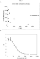
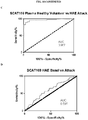
- cleaved HMWK The level of cleaved HMWK was found to be elevated in HAE attack, as well as in other pKal-associated disorders. Thus, cleaved HMWK can serve as a biomarker for monitoring disease development and/or treatment efficacy.
- the art lacks suitable agents and/or suitable assays that can effectively distinguish intact HMWK from its cleaved version.
- the present disclosure is based, at least in part, on the development of specific immunoassays that allows for detection of cleaved HMWK with high specificity and sensitivity. It was observed that a Sandwich ELISA in which an agent that specifically binds cleaved HMWK is immobilized on a support member (e.g., a multi-well plate) unexpectedly enhanced detection efficiency as compared to the setting of ELISA in which the antigen (in this case, the cleaved HMWK) is immobilized on the support member.
- a support member e.g., a multi-well plate
- the detection specificity and sensitivity was further enhanced when a 96-well plate was used, as compared with a 384-well plate.
- the present disclosure is also based on, at least in part, the isolation of antibodies that specifically bind a cleaved HMWK.
- immunoassays for detecting the presence or measuring the level of a cleaved HMWK in a biological sample suspected of containing HMWK species, using an agent (e.g., an antibody) that specifically binds a cleaved HMWK (e.g., the cleaved HMWK having a molecular weight of 46 kDa).
- an agent e.g., an antibody
- the imunoassays described herein can be applied to identify patients who are at risk of such diseases, to monitor disease progression, and/or to monitor efficacy of a treatment against such a disorder.
- One aspect of the present disclosure relates to immunoassays for detecting cleaved HMWK with high sensitivity and specificity.
- Such immunoassays may involve a Sandwich ELISA in which an agent that specifically binds a cleaved HMWK is immobilized on a support member, which can be a 96-well plate.
- the immunoassays described herein allows for selective detection of cleaved HMWK in biological samples, e.g., serum samples or plasma samples, which may contain both intact and cleaved HMWK, as well as LMWK.
- High molecular-weight kininogen exists in the plasma as a single polypeptide (1-chain) multi-domain (domains 1-6) protein with a molecular weight of approximately 110 kDa, referred to herein as intact HWMK.
- the human gene encoding HMWK is kininogen 1 (KNG1).
- KNG1 is transcribed and alternatively spliced to form mRNAs that encode either HMWK or low molecular weight kininogen (LMWK).
- An exemplary protein sequence of HMWK is provided below: >gi
- Intact HMWK also referred to herein as "intact kininogen”
- intact kininogen can be assayed, for example, using coagulant or immunological methods, e.g., radioimmunoassay (see, e.g., Kerbiriou-Nabias, D.M., Br J Haematol, 1984, 56(2):2734-86 ).
- a monoclonal antibody to the light chain of human HMWK is known. See, e.g., Reddigari, S.R. & Kaplan, A.P., Blood, 1999, 74:695-702 .
- An assay for HMWK that relies on a chromogenic substrate can also be used. See, e.g., Scott, C.F. et al. Thromb Res, 1987, 48(6):685-700 ; Gallimore, M.J. et al. Thromb Res, 2004, 114(2):91-96 .
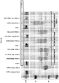
- HMWK is cleaved by pKal within domain 4 to release the 9 amino acid, proinflammatory peptide bradykinin, and a 2-chain form of HMWK, referred to herein as cleaved HMWK.
- the 2 chains of HMWK are the heavy chain, which contains domains 1-3, and the light chain, which contains domains 5 and 6, joined by a disulfide bond.
- the heavy and light chains Upon initial cleavage of intact HMWK, the heavy and light chains have a molecular weight of approximately 65 kDa and 56 kDa, respectively. Further proteolytic processing results in generation of a 46 kDa light chain.
- the immunoassays described herein may use any agent that can specifically bind a cleaved HMWK, for example, an agent that recognizes a neoepitope on cleaved HMWK that is not present on intact HMWK.
- the cleaved HMWK-binding agent is an antibody.
- An antibody is an immunoglobulin molecule capable of specific binding to a target, such as a carbohydrate, polynucleotide, lipid, polypeptide, etc., through at least one antigen recognition site, located in the variable region of the immunoglobulin molecule.
- antibody encompasses not only intact (i.e., full-length) polyclonal or monoclonal antibodies, but also antigen-binding fragments thereof (such as Fab, Fab', F(ab') 2 , Fv), single chain (scFv), mutants thereof, fusion proteins comprising an antibody portion, humanized antibodies, chimeric antibodies, diabodies, linear antibodies, single chain antibodies, multispecific antibodies (e.g., bispecific antibodies) and any other modified configuration of the immunoglobulin molecule that comprises an antigen recognition site of the required specificity, including glycosylation variants of antibodies, amino acid sequence variants of antibodies, and covalently modified antibodies.
- antigen-binding fragments thereof such as Fab, Fab', F(ab') 2 , Fv), single chain (scFv), mutants thereof, fusion proteins comprising an antibody portion, humanized antibodies, chimeric antibodies, diabodies, linear antibodies, single chain antibodies, multispecific antibodies (e.g., bispecific antibodies) and any other modified
- An antibody includes an antibody of any class, such as IgD, IgE, IgG, IgA, or IgM (or sub-class thereof), and the antibody need not be of any particular class.
- immunoglobulins can be assigned to different classes. There are five major classes of immunoglobulins: IgA, IgD, IgE, IgG, and IgM, and several of these may be further divided into subclasses (isotypes), e.g., IgG1, IgG2, IgG3, IgG4, IgA1 and IgA2.
- the heavy-chain constant domains that correspond to the different classes of immunoglobulins are called alpha, delta, epsilon, gamma, and mu, respectively.
- the subunit structures and three-dimensional configurations of different classes of immunoglobulins are well known.
- any of the antibodies described herein can be either monoclonal or polyclonal.
- a “monoclonal antibody” refers to a homogenous antibody population and a “polyclonal antibody” refers to a heterogeneous antibody population. These two terms do not limit the source of an antibody or the manner in which it is made.
- An antibody that "specifically binds" a cleaved HMWK or an epitope thereof is a term well understood in the art, and methods to determine such specific binding are also well known in the art.
- a molecule is said to exhibit "specific binding” if it reacts or associates more frequently, more rapidly, with greater duration and/or with greater affinity with a particular target antigen (here a cleaved HMWK) than it does with alternative targets (e.g., intact HMWK and/or LMWK).
- An antibody "specifically binds" to a target antigen if it binds with greater affinity, avidity, more readily, and/or with greater duration than it binds to other substances.
- an antibody that specifically (or preferentially) binds to cleaved HMWK or an epitope therein is an antibody that binds this target antigen with greater affinity, avidity, more readily, and/or with greater duration than it binds to other antigens ( e.g., intact HMWK or LMWK) or other epitopes in the same antigen. It is also understood by reading this definition that, for example, an antibody that specifically binds to a first target antigen may or may not specifically or preferentially bind to a second target antigen. As such, “specific binding” or “preferential binding” does not necessarily require (although it can include) exclusive binding. Generally, but not necessarily, reference to binding means preferential binding.
- the antibodies that specifically binds cleaved HMWK (as well the other antibodies that bind both cleaved and intact, and optionally LMWK) described herein have a suitable binding affinity to a cleaved HMWK (or another target antigen as described herein).
- binding affinity refers to the apparent association constant or K A .
- the K A is the reciprocal of the dissociation constant (K D ).
- the antibody described herein may have a binding affinity (K D ) of at least 10 -5 , 10 -6 , 10 -7 , 10 -8 , 10 -9 , 10 -10 M, or lower.
- An increased binding affinity corresponds to a decreased K D .
- Higher affinity binding of an antibody to a first target relative to a second target can be indicated by a higher K A (or a smaller numerical value K D ) for binding the first target than the K A (or numerical value K D ) for binding the second target.
- the antibody has specificity for the first target (e.g., a protein in a first conformation or mimic thereof) relative to the second target (e.g., the same protein in a second conformation or mimic thereof; or a second protein).
- Differences in binding affinity e.g., for specificity or other comparisons
- the binding affinity of an antibody that specifically binds a cleaved HMWK as described herein may be 10-fold, 100-fold, 10,000-fold, or 10 5 -fold higher than the binding affinity of that antibody to intact HMWK and/or LMWK.
- Binding affinity can be determined by a variety of methods including equilibrium dialysis, equilibrium binding, gel filtration, ELISA, surface plasmon resonance, or spectroscopy (e.g., using a fluorescence assay).
- Exemplary conditions for evaluating binding affinity are in HBS-P buffer (10 mM HEPES pH7.4, 150 mM NaCl, 0.005% (v/v) Surfactant P20). These techniques can be used to measure the concentration of bound binding protein as a function of target protein concentration.
- K A it is not always necessary to make an exact determination of K A , though, since sometimes it is sufficient to obtain a quantitative measurement of affinity, e.g., determined using a method such as ELISA or FACS analysis, is proportional to K A , and thus can be used for comparisons, such as determining whether a higher affinity is, e.g., 2-fold higher, to obtain a qualitative measurement of affinity, or to obtain an inference of affinity, e.g., by activity in a functional assay, e.g., an in vitro or in vivo assay.
- a functional assay e.g., an in vitro or in vivo assay.
- the antibody that specifically binds to cleaved HMWK (also referred to as an anti-cleaved HMWK antibody) binds to the same epitope of a cleaved HMWK as 559B-M004-B04.
- An "epitope” refers to the site on a target antigen that is bound by a binding protein (e.g., an antibody such as a Fab or full length antibody).
- the site can be entirely composed of amino acid components, entirely composed of chemical modifications of amino acids of the protein (e.g., glycosyl moieties), or composed of combinations thereof.
- Overlapping epitopes include at least one common amino acid residue, glycosyl group, phosphate group, sulfate group, or other molecular feature. In some cases, the epitope is linear; in other instances, the epitope is conformational.
- a first antibody "binds to the same epitope" as a second antibody if the first antibody binds to the same site on a target antigen that the second antibody binds, or binds to a site that overlaps (e.g., 50%, 60%, 70%, 80%, 90%, or 100% overlap, e.g., in terms of amino acid sequence or other molecular feature (e.g., glycosyl group, phosphate group, or sulfate group) with the site that the second antigen binds.
- overlaps e.g., 50%, 60%, 70%, 80%, 90%, or 100% overlap, e.g., in terms of amino acid sequence or other molecular feature (e.g., glycosyl group, phosphate group, or sulfate group) with the site that the second antigen binds.
- the antibody that specifically binds to cleaved HMWK competes against 559B-M004-B04 for binding to HMWK.
- a first antibody "competes for binding" with a second antibody if the binding of the first antibody to its epitope decreases ( e.g., by 10%, 20%, 30%, 40%, 50%, 60%, 70%, 80%, 90%, 100%, or more) the amount of the second antibody that binds to its epitope.
- the competition can be direct (e.g., the first antibody binds to an epitope that is the same as, or overlaps with, the epitope bound by the second antibody), or indirect (e.g., the binding of the first antibody to its epitope causes a steric change in the target antigen that decreases the ability of the second antibody to bind to its epitope).
- the antibody that specifically binds to cleaved HMWK comprises a V H chain that includes a V H CDR1, a V H CDR2, and/or a V H CDR3 at least 75% ( e.g., 80%, 85%, 90%, 95%, 96%, 97%, 98%, or 99%) identical to the corresponding V H CDRs of 559B-M004-B04.
- the antibody that specifically binds to cleaved HMWK comprises a V L CDR1, a V L CDR2, and/or a V L CDR3 at least 75% (e.g., 80%, 85%, 90%, 95%, 96%, 97%, 98%, or 99%) identical to the corresponding V L CDRs of 559B-M004-B04.
- the antibody that specifically binds to cleaved HMWK has the same heavy chain and/or light chain complementarity determining regions (CDRs) as 559B-M004-B04.
- CDRs Complementarity determining regions
- CDRs are known in the art as referring to non-contiguous sequences of amino acids within antibody variable regions, which confer antigen specificity and binding affinity. In general, there are three (3) CDRs in each heavy chain variable region and three (3) CDRs in each light chain variable region. The precise amino acid sequence boundaries of a given CDR can be readily determined using any of a number of well-known schemes, including those described by Kabat et al. (1991), 5th Ed. Public Health Service, National Institutes of Health, Bethesda, Md.
- CDR complementary determining region
- an antibody has the same V H and/or V L CDRs as 559B-M004-B04 (as well as other exemplary antibodies disclosed herein), such an antibody is deemed as having the same CDRs as 559B-M004-B04 (or the other exemplary antibodies disclosed herein) and is within the scope of the present disclosure.
- such an antibody may have the same V H and/or V L CDRs as clone 559B-M004-B04 as determined by the Chothia numbering scheme.
- an anti-cleaved HMWK antibody within the scope of the present disclosure may have the same V H and/or V L CDRs as clone 559B-M004-B04, as determined by the Kabat numbering scheme.
- the anti-cleaved HMWK antibody comprises a V H chain at least 75% (e.g., 80%, 85%, 90%, 95%, 96%, 97%, 98%, or 99%) identical to the V H chain of 559B-M004-B04 and/or a V L chain at least 75% ( e.g., 80%, 85%, 90%, 95%, 96%, 97%, 98%, or 99%) identical to the V L chain of 559B-M004-B04.
- the antibody is 559B-M004-B04.
- Gapped BLAST can be utilized as described in Altschul et al., Nucleic Acids Res. 25(17):3389-3402, 1997 .
- the default parameters of the respective programs e.g., XBLAST and NBLAST.
- Heavy chain CDR1, CDR2, and CDR3 sequences and light chain CDR1, CDR2, and CDR3 sequences are underlined and in boldface (identified by one scheme as an example).
- the antibody that specifically binds a cleaved HMWK may contain one or more (e.g., up to 5, up to 3, or up to 1) conservative mutations in one or more of the heavy chain CDRs, or one or more of the light chain CDRs in 559B-M0004-B04, e.g., at positions where the residues are not likely to be involved in interacting with the cleaved HMWK.
- a "conservative amino acid substitution” refers to an amino acid substitution that does not alter the relative charge or size characteristics of the protein in which the amino acid substitution is made.
- Variants can be prepared according to methods for altering polypeptide sequence known to one of ordinary skill in the art such as are found in references which compile such methods, e.g. Molecular Cloning: A Laboratory Manual, J. Sambrook, et al., eds., Second Edition, Cold Spring Harbor Laboratory Press, Cold Spring Harbor, New York, 1989 , or Current Protocols in Molecular Biology, F.M. Ausubel, et al., eds., John Wiley & Sons, Inc., New York .
- amino acids include substitutions made amongst amino acids within the following groups: (a) M, I, L, V; (b) F, Y, W; (c) K, R, H; (d) A, G; (e) S, T; (f) Q, N; and (g) E, D.
- Antibodies capable of binding to cleaved HMWK can be made by any method known in the art. See, for example, Harlow and Lane, (1988) Antibodies: A Laboratory Manual, Cold Spring Harbor Laboratory, New York .
- antibodies specific to a target antigen can be made by the conventional hybridoma technology.
- the full-length target antigen or a fragment thereof, optionally coupled to a carrier protein such as KLH, can be used to immunize a host animal for generating antibodies binding to that antigen.
- the route and schedule of immunization of the host animal are generally in keeping with established and conventional techniques for antibody stimulation and production, as further described herein. General techniques for production of mouse, humanized, and human antibodies are known in the art and are described herein.
- any mammalian subject including humans or antibody producing cells therefrom can be manipulated to serve as the basis for production of mammalian, including human hybridoma cell lines.
- the host animal is inoculated intraperitoneally, intramuscularly, orally, subcutaneously, intraplantar, and/or intradermally with an amount of immunogen, including as described herein.
- Hybridomas can be prepared from the lymphocytes and immortalized myeloma cells using the general somatic cell hybridization technique of Kohler, B. and Milstein, C. (1975) Nature 256:495-497 or as modified by Buck, D. W., et al., In Vitro, 18:377-381 (1982 ).
- Available myeloma lines including but not limited to X63-Ag8.653 and those from the Salk Institute, Cell Distribution Center, San Diego, Calif., USA, may be used in the hybridization.
- the technique involves fusing myeloma cells and lymphoid cells using a fusogen such as polyethylene glycol, or by electrical means well known to those skilled in the art.
- the cells are separated from the fusion medium and grown in a selective growth medium, such as hypoxanthine-aminopterin-thymidine (HAT) medium, to eliminate unhybridized parent cells.
- a selective growth medium such as hypoxanthine-aminopterin-thymidine (HAT) medium
- HAT hypoxanthine-aminopterin-thymidine
- Any of the media described herein, supplemented with or without serum, can be used for culturing hybridomas that secrete monoclonal antibodies.
- EBV immortalized B cells may be used to produce the anti-PKal monoclonal antibodies described herein.
- hybridomas are expanded and subcloned, if desired, and supernatants are assayed for anti-immunogen activity by conventional immunoassay procedures (e.g., radioimmunoassay, enzyme immunoassay, or fluorescence immunoassay).
- immunoassay procedures e.g., radioimmunoassay, enzyme immunoassay, or fluorescence immunoassay.
- Hybridomas that may be used as source of antibodies encompass all derivatives, progeny cells of the parent hybridomas that produce monoclonal antibodies capable of interfering with the PKal activity.
- Hybridomas that produce such antibodies may be grown in vitro or in vivo using known procedures.
- the monoclonal antibodies may be isolated from the culture media or body fluids, by conventional immunoglobulin purification procedures such as ammonium sulfate precipitation, gel electrophoresis, dialysis, chromatography, and ultrafiltration, if desired.
- Undesired activity if present, can be removed, for example, by running the preparation over adsorbents made of the immunogen attached to a solid phase and eluting or releasing the desired antibodies off the immunogen.
- a target antigen or a fragment containing the target amino acid sequence conjugated to a protein that is immunogenic in the species to be immunized e.g., keyhole limpet hemocyanin, serum
- an antibody (monoclonal or polyclonal) of interest may be sequenced and the polynucleotide sequence may then be cloned into a vector for expression or propagation.
- the sequence encoding the antibody of interest may be maintained in vector in a host cell and the host cell can then be expanded and frozen for future use.
- the polynucleotide sequence may be used for genetic manipulation to improve the affinity (affinity maturation), or other characteristics of the antibody. It may be desirable to genetically manipulate the antibody sequence to obtain greater affinity and/or specificity to the target antigen. It will be apparent to one of skill in the art that one or more polynucleotide changes can be made to the antibody and still maintain its binding specificity to the target antigen.
- Fully human antibodies can be obtained by using commercially available mice that have been engineered to express specific human immunoglobulin proteins.
- Transgenic animals that are designed to produce a more desirable (e.g., fully human antibodies) or more robust immune response may also be used for generation of humanized or human antibodies. Examples of such technology are Xenomouse RTM from Amgen, Inc. (Fremont, Calif.) and HuMAb-Mouse RTM and TC MouseTM from Medarex, Inc. (Princeton, N.J.).
- antibodies may be made recombinantly by phage display or yeast technology. See, for example, U.S. Pat. Nos.
- Antigen-binding fragments of an intact antibody can be prepared via routine methods.
- F(ab')2 fragments can be produced by pepsin digestion of an antibody molecule, and Fab fragments that can be generated by reducing the disulfide bridges of F(ab')2 fragments.
- a single-chain antibody can be prepared via recombinant technology by linking a nucleotide sequence coding for a heavy chain variable region and a nucleotide sequence coding for a light chain variable region.
- a flexible linker is incorporated between the two variable regions.
- techniques described for the production of single chain antibodies U.S. Patent Nos. 4,946,778 and 4,704,692 ) can be adapted to produce a phage or yeast scFv library and scFv clones specific to a PKal can be identified from the library following routine procedures. Positive clones can be subjected to further screening to identify those that specifically bind a target antigen, such as a cleaved HMWK.
- the antibodies specific to a cleaved HMWK may be isolated from an antibody library, which may be a synthetic library or a natural library.
- a natural antibody library refers to a library derived from a natural source (e.g., a human donor) following routine practice.
- a synthetic antibody library refers to a library the members of which are designed following predetermined rules (e.g., having a complete randomized CDR region such as CDRs or a semi randomized CDR region such as CDR1 or CDR2 of the heavy chain, the light chain, or both).
- the antibody library is a display library (e.g., a phage display library or a yeast display library).
- a display library is a collection of entities; each entity includes an accessible polypeptide component and a recoverable component that encodes or identifies the polypeptide component.
- the polypeptide component is varied so that different amino acid sequences are represented.
- the polypeptide component can be of any length, e.g., from three amino acids to over 300 amino acids.
- a display library entity can include more than one polypeptide component, for example, the two polypeptide chains of a sFab.
- a display library can be used to identify proteins that bind to a cleaved HMWK (as well as other target antigens described herein).
- the polypeptide component of each member of the library is probed with a cleaved HMWK (or a fragment thereof) and if the polypeptide component binds to the cleaved HMWK, the display library member is identified, typically by retention on a support.
- An exemplary illustration for identifying antibodies specific to cleaved HMWK using a phage display antibody library is provided in Figure 12 .
- Retained display library members are recovered from the support and analyzed.
- the analysis can include amplification and a subsequent selection under similar or dissimilar conditions. For example, positive and negative selections can be alternated.
- the analysis can also include determining the amino acid sequence of the polypeptide component and purification of the polypeptide component for detailed characterization.
- Antibodies obtained following a method known in the art and described herein can be characterized using methods well known in the art. For example, one method is to identify the epitope to which the antigen binds, or "epitope mapping.” There are many methods known in the art for mapping and characterizing the location of epitopes on proteins, including solving the crystal structure of an antibody-antigen complex, competition assays, gene fragment expression assays, and synthetic peptide-based assays, as described, for example, in Chapter 11 of Harlow and Lane, Using Antibodies, a Laboratory Manual, Cold Spring Harbor Laboratory Press, Cold Spring Harbor, N.Y., 1999 . In an additional example, epitope mapping can be used to determine the sequence to which an antibody binds.
- the epitope can be a linear epitope, i.e., contained in a single stretch of amino acids, or a conformational epitope formed by a three-dimensional interaction of amino acids that may not necessarily be contained in a single stretch (primary structure linear sequence).
- Peptides of varying lengths e.g., at least 4-6 amino acids long
- the epitope to which the antibody binds can be determined in a systematic screening by using overlapping peptides derived from the target antigen sequence and determining binding by the antibody.
- the open reading frame encoding the target antigen is fragmented either randomly or by specific genetic constructions and the reactivity of the expressed fragments of the antigen with the antibody to be tested is determined.
- the gene fragments may, for example, be produced by PCR and then transcribed and translated into protein in vitro, in the presence of radioactive amino acids. The binding of the antibody to the radioactively labeled antigen fragments is then determined by immunoprecipitation and gel electrophoresis.
- Certain epitopes can also be identified by using large libraries of random peptide sequences displayed on the surface of phage particles (phage libraries). Alternatively, a defined library of overlapping peptide fragments can be tested for binding to the test antibody in simple binding assays.
- mutagenesis of an antigen binding domain, domain swapping experiments and alanine scanning mutagenesis can be performed to identify residues required, sufficient, and/or necessary for epitope binding.
- domain swapping experiments can be performed using a mutant of a target antigen in which various fragments of the HMWK polypeptide have been replaced (swapped) with sequences from a closely related, but antigenically distinct protein. By assessing binding of the antibody to the mutant HMWK, the importance of the particular antigen fragment to antibody binding can be assessed.
- competition assays can be performed using other antibodies known to bind to the same antigen to determine whether an antibody binds to the same epitope as the other antibodies. Competition assays are well known to those of skill in the art.
- anti-cleaved HMWK antibodies is also within the scope of the present disclosure.
- immunoassays for detecting a cleaved HMWK.
- the term "immunoassay” may be referred to interchangeably as an immune-based assay or immuno-based assay.
- an immunoassay detects the presence and/or concentration (level) of a molecule (e.g., HMWK), in a sample using an agent that binds to the molecule, such as an antibody.
- immunoassays include Western blots, enzyme linked immunosorbent assays (ELISAs), lateral flow assay, radioimmunoassays, electrochemiluminescence-based detection assays, magnetic immunoassays, and related techniques.
- the immunoassay is an ELISA assay.
- the immunoassay is a sandwich ELISA assay.
- the immunoassay is a lateral flow assay.
- ELISAs are known in the art (see, e.g., Crowther, John R (2009) . “The ELISA Guidebook.” 2nd ed. Humana Press and Lequin R (2005 ). " Enzyme immunoassay (EIA)/enzyme-linked immunosorbent assay (ELISA)". Clin. Chem. 51 (12): 2415-8 ) and exemplary ELISAs are described herein. Kits for performing ELISAs are also known in the art and commercially available (see, e.g., ELISA kits from Life Technologies and BD Biosciences).
- a sample may be obtained from a subject.
- a sample refers to a composition that comprises tissue, e.g., blood, plasma or protein, from a subject.
- a sample includes both an initial unprocessed sample taken from a subject as well as subsequently processed, e.g., partially purified or preserved forms.
- Exemplary samples include blood, plasma, tears, or mucus.
- the sample is a body fluid sample such as a serum or plasma sample.
- a sample to be analyzed by the immunoassay described herein can be either an initial unprocessed sample taken from a subject or subsequently processed, e.g., partially purified or preserved forms.
- multiple (e.g., at least 2, 3, 4, 5, or more) samples may be collected from the subject, over time or at particular time intervals, for example to assess the progression of a disease or disorder or evaluate the efficacy of a treatment.
- the multiple samples may be obtained before and after a treatment, or during the course of a treatment.
- a sample can be obtained from a subject using any means known in the art.
- the sample is obtained from the subject by collecting the sample (e.g., a blood sample) into an evacuated collection tube (e.g., an evacuated blood collection tube).
- the evacuated collection tube contains one or more protease inhibitors, for example, to reduce or prevent ex vivo activation of the contact system during sample collection.
- protease inhibitors may be contained in a liquid formulation.
- the protease inhibitors comprise at least one serine protease inhibitor and at least one cysteine protease inhibitor.
- Such evacuated collection tubes are known in the art. See, for example, PCT Application No. US2016/046681 .
- an evacuated blood collection tube may further comprise one or more anti-coagulants.
- a "patient,” “subject” or “host” (these terms are used interchangeably) to be treated by the subject method may mean either a human or non-human animal.
- a subject is suspected of or is at risk for or suffers from a kallikrein-mediated disorder, e.g., a bradykinin-mediated disorder, such as hereditary angioedema (HAE), non-histamine-dependent idiopathic angioedema, rheumatoid arthritis, Crohn's disease, lupus, Alzheimer's disease, septic shock, burn injury, brain ischemia/reperfusion injury, cerebral edema, diabetic retinopathy, diabetic nephropathy, macular edema, vasculitis, arterial or venous thrombosis, thrombosis associated with ventricular assist devices or stents, heparin-induced thrombocytopenia with thrombosis, thromboembolic disease, and coronary heart
- the subject who needs the analysis described herein may be a patient of the disease or disorder.
- a subject may be under the attack of the disease (e.g., HAE) currently, or may suffer from the disease in the past (e.g., during disease quiescence currently).
- the subject is a human patient who may be on a treatment of the disease, for example, a treatment involving a C1 esterase inhibitor (C1-INH), a plasma kallikrein inhibitor, or a bradykinin inhibitor.
- a human patient may be free of such a treatment.
- the sample described herein can be subject to analysis using an agent that specifically binds a cleaved HMWK to determine the level of the cleaved HMWK in the sample.
- the immunoassays described herein may in the format of a sandwich ELISA, in which a first agent (e.g., the antibody described herein) that specifically binds the cleaved HMWK is immobilized on a support member.
- the support member can then be incubated with a sample as described herein for a suitable period of time under conditions that allow for the formation of cleaved HMWK/first agent (e.g., antibody) complex.
- Such a complex can then be detected using a second agent that binds HMWK.
- the second agent can be conjugated to a label, which can release a signal directly or indirectly. The intensity of the signal represents the level of the cleaved HMWK in the sample.
- Any support member known in the art may be used in the method, including but not limited to a membrane, a bead, a slide, or a multi-well plate. Selection of an appropriate support member for the immunoassay will depend on various factor such as the number of samples and method of detecting the signal released from label conjugated to the second agent.
- the support member is a membrane, such as a nitrocellulose membrane, a polyvinylidene fluoride (PVDF) membrane, or a cellulose acetate membrane.
- the immunoassay may be in a Western blot assay format or a lateral flow assay format.
- the support member is a multi-well plate, such as an ELISA plate.
- the immunoassays described herein can be carried out on high throughput platforms.
- multi-well plates e.g., 24-, 48-, 96-, 384- or greater well plates, may be used for high throughput immunoassays.
- Individual immunoassays can be carried out in each well in parallel. Therefore, it is generally desirable to use a plate reader to measure multiple wells in parallel to increase assay throughput.
- plate readers that are capable of imaging multi-wells (e.g., 4, 16, 24, 48, 96, 384, or greater wells) in parallel can be used for this platform.
- a commercially available plate reader e.g., the plate::vision system available from Perkin Elmer, Waltham, MA
- This plate reader is capable of kinetic-based fluorescence analysis.
- the plate::vision system has high collection efficiency optics and has special optics designed for the analysis of 96 wells in parallel.
- Additional suitable parallel plate readers include but are not limited to the SAFIRE (Tecan, San Jose, CA), the FLIPRTETRA® (Molecular Devices, Union City, CA), the FDSS7000 (Hamamatsu, Bridgewater, NJ), and the CellLux (Perkin Elmer, Waltham, MA).
- the described immunoassays are performed in 96-well plates, such as a 96-well ELISA plate.
- high-throughput screening immunoassays of the present disclosure can be automated ( e.g., adapted to robotic assays).
- the immunoassays may be performed on low-throughput platforms, including single immunoassay format.
- a low-throughput platform may be used to measure the presence and amount of cleaved HMWK in biological samples (e.g., biological tissues, tissue extracts) for diagnostic methods, monitoring of disease and/or treatment progression, and/or predicting whether a disease or disorder may benefit from a particular treatment.
- any method known in the art can be used to immobilize an agent that specifically binds a cleaved HMWK such as the antibodies described herein onto a support member as also described herein.
- the immobilization involves binding the agent (e.g., the antibody) to the support member.
- the immobilization involves adsorbing the antibody to the support member. Such adsorption methods may be performed, for example, by incubating the antibody in a buffer in the wells of a multi-well plate.
- the agent such as the antibody is provided in a coating buffer and incubated in the wells of a multi-well plate.
- Coating buffers will be evident to one of skill in the art and may be prepared or obtained from a commercial source.
- Non-limiting examples of coating buffers include 50 mM sodium bicarbonate, pH 9.6; 0.2 M sodium bicarbonate, pH 9.4; phosphate buffered solution (50 mM phosphate, pH 8.0, 0.15 M NaCl); carbonate-bicarbonate solution; and TBS (50 mM TRIS, pH 8.0, 0.15 M NaCl).
- the first agent is immobilized on the support member by hydrophobic interactions between the first agent and the support member. In some embodiments, the first agent is immobilized on the support member using electrophoretic transfer.
- the support member may be incubated with a blocking buffer.
- blocking buffers are used to block any of the exposed surface of the support membrane (e.g., sites on the support membrane unoccupied by the first agent).
- Use of a blocking buffer may reduce the baseline signal detected (i.e., "background interference") and/or improve the sensitivity of the immunoassay and/or reduce non-specific binding of components of the sample to the support membrane.
- background interference i.e., "background interference”
- selection of the blocking buffer affected the results of the immunoassay.
- the blocking buffer contains serum albumin, such as bovine serum albumin or human serum albumin.
- the blocking buffer is a BSA buffer (e.g., 2% BSA in PBS buffer).
- the blocking buffer is free from serum albumin, such as bovine serum albumin or human serum albumin.
- the blocking buffer comprises casein fragments, and optionally NaCl and Tween and may have a pH 7.0-7.4.
- the casein fragments are high purity casein fragments.
- Such a blocking buffer may be prepared or obtained from a commercial source (e.g., The Blocking Solution LowCross from CANDOR Bioscience).
- the support member on which the agent specific to a cleaved HMWK is attached, can be brought in contact (incubated) with a sample as described herein, which is suspected of containing the cleaved HMWK.
- the term "contact” refers to an exposure of the support member with the biological sample or agent for a suitable period sufficient for the formation of complexes between the agent, such as an antibody, and the cleaved HMWK in the sample, if any.
- the sample may be removed from the support member, which can then be washed for multiple times to remove any unbound cleaved HMWK.
- the contacting is performed by capillary action in which a biological sample or agent is moved across a surface of the support membrane.
- the support member can then be incubated with a second agent that binds HMWK for a suitable period allowing for the binding of the second agent to HMWK attached to the support member.
- the second agent can be any agent capable of binding to HMWK, such as an antibody capable of binding to HMWK (either specific to the cleaved form of HMWK or can cross react to both the cleaved HMWK and intact HMWK).
- the second agent comprises one or more antibodies that bind HWMK (cleaved and/or intact).
- the antibody is a mouse monoclonal antibody or a monoclonal sheep antibody. It is conjugated with a label, which is a compound capable of releasing a signal either directly or indirectly (e.g., via interaction with one or more additional compounds).
- the label is a signal releasing agent, which is an agent that either directly releases a signal (e.g., a dye or fluorophore) or releases a signal upon interacting with a substrate (e.g., an enzyme such as HRP or ⁇ -galactosidase, which can convert a colorless substrate to a colored product).
- a signal e.g., a dye or fluorophore
- a substrate e.g., an enzyme such as HRP or ⁇ -galactosidase, which can convert a colorless substrate to a colored product.
- fluorophore also referred to as "fluorescent label” or “fluorescent dye” refers to moieties that absorb light energy at a defined excitation wavelength and emit light energy at a different wavelength.
- the label can be a member of a receptor-ligand pair.
- a "ligand-receptor pair” refers to a pair of molecules (e.g., biological molecules) that have a specific affinity for each other, e.g., biotin-streptavidin.
- the support member carrying the first agent-cleaved HMWK-second agent may be further incubated with the other member of the ligand-receptor pair for a suitable period such that the two members of the receptor-ligand pair interact.
- the other member of the receptor-ligand pair is conjugated with a signal releasing agent as described herein.
- the second agent is conjugated to biotin and HRP-conjugated streptavidin is used for detection.
- a substrate solution may be added to aid in detection.
- the reaction may be stopped (e.g., by adding 1 N NaOH) and the concentration of colored product formed may be measured in a spectrophotometer. The intensity of color is proportional to the concentration of bound antigen.
- the signal released from the label as described herein can be detected/measured by routine methodology, which would depend on the specific format of an immunoassay and the signal releasing agent used therein.
- the terms “measuring” or “measurement,” or alternatively “detecting” or “detection,” means assessing the presence, absence, quantity or amount (which can be an effective amount) of a substance within a sample, including the derivation of qualitative or quantitative concentration levels of such substances, or otherwise evaluating the values or categorization of a subject.
- Assays may further involve use of a quantitative imaging system, e.g., LICOR imaging technology, which is commercially available (see, e.g., the Odyssey® CLx infrared imaging system from LI-COR Biosciences).
- LICOR imaging technology which is commercially available (see, e.g., the Odyssey® CLx infrared imaging system from LI-COR Biosciences).
- an electrochemiluminescence detection assay or an assay relying on a combination of electrochemiluminescence and patterned array technology is used (e.g., an ECL or MULTI-ARRAY technology assay from Meso Scale Discovery (MSD)).
- any of the immunoassays described herein, e.g., one or more steps of the immunoassays, may be carried out in a suitable assay buffer, which will be evident to one of skill in the art.
- the assay buffer contains or has been supplemented with ZnCl 2 .
- the assay buffer contains at least about 10 ⁇ M, 20 ⁇ M, 30 ⁇ M, 40 ⁇ M, 50 ⁇ M, 60 ⁇ M, 70 ⁇ M, 80 ⁇ M, 90 ⁇ M, 100 ⁇ M, 150 ⁇ M, 200 ⁇ M, 250 ⁇ M, 300 ⁇ M, 350 ⁇ M, 400 ⁇ M, 450 ⁇ M, 500 ⁇ M or more ZnCl 2 .
- such a ZnCl 2 -containing assay buffer is used in the step in which the agent specific to cleaved HMWK (e.g., an antibody specific to cleaved HMWK) binds a cleaved HMWK.
- ZnCl 2 enhances the binding activity of the agent (e.g., antibody) to the cleaved HMWK.
- the assay buffer contains serum albumin, such as bovine serum albumin or human serum albumin. In some embodiments, the assay buffer contains at least about 0.01%. 0.02%, 0.03%, 0.04%. 0.05%, 0.06%, 0.07%, 0.08%, 0.09%, 0.1%, 0.12%, 0.014%, 0.16%, 0.18%, 0.2%, 0.25%, 0.3%, 0.4%, or more BSA. In some embodiments, the assay buffer contains a surfactant, such as Tween-20. In some embodiments, the assay buffer contains 0.01%. 0.02%, 0.03%, 0.04%. 0.05%, 0.06%, 0.07%, 0.08%, 0.09%, 0.1% or more of a surfactant. In one example, the assay buffer contains 0.1% BSA and 0.05% Tween-20 in PBS.
- the assay methods and kits described herein can be applied for evaluation of a disease or disorder associated with plasma kallikrein, such as those described herein (e.g., HAE), given the correlation between the level of cleaved HMWK and such diseases or disorders (e.g. as a biomarker).
- a disease or disorder associated with plasma kallikrein such as those described herein (e.g., HAE)
- the assay methods and kits described herein may be used to monitor the progress of such a disease, assess the efficacy of a treatment for the disease, identify patients suitable for a particular treatment, and/or predict disease status (e.g., attack versus quiescence) in a subject.
- the level of cleaved HMWK determined by the immunoassay described herein can be relied on to evaluate whether a subject (e.g., a human patient) from whom the biological sample is obtained, has or is at risk for a disease or disorder associated with plasma kallikrein, such as HAE or autoimmune disease such as RA, UC, and Crohn's disease).
- the level of cleaved kininogen can then be compared with either the intact kininogen or the total amount of kininogen in the sample to determine a value ( e.g., percentage) of cleaved kininogen, a value of intact kininogen, or both, in the sample.
- the value of cleaved kininogen and/or intact kininogen can be compared to a reference value to determine whether the subject has or is at risk for the PKal-mediated disorder, e.g., HAE or an autoimmune disease, such as RA, UC, and Crohn's disease.
- a reference value e.g., HAE or an autoimmune disease, such as RA, UC, and Crohn's disease.
- the percentage of cleaved kininogen is at or higher than a reference number
- the subject can be identified as having or at risk for a pKal-mediated disorder such as HAE, RA, UC, and Crohn's disease.
- the percentage of intact kininogen is at or lower than a reference number
- the subject can be identified as having or at risk for a pKal-mediated disorder such as HAE, RA, UC, and Crohn's disease.
- the sample for analysis of the methods described herein is derived from a human subject who has or is at risk of having hereditary angioedema (HAE).
- HAE hereditary angioedema
- HAE is also known as "Quincke edema,” C1 esterase inhibitor deficiency, C1 inhibitor deficiency, and hereditary angioneurotic edema (HANE).
- HAE is characterized by recurrent episodes of severe swelling (angioedema), which can affect, e.g., the limbs, face, genitals, gastrointestinal tract, and airway.
- Symptoms of HAE include, e.g., swelling in the arms, legs, lips, eyes, tongue, and/or throat; airway blockage that can involve throat swelling and sudden hoarseness; repeat episodes of abdominal cramping without obvious cause; and/or swelling of the intestines, which can be severe and can lead to abdominal cramping, vomiting, dehydration, diarrhea, pain, and/or shock. About one-third of individuals with this HAE develop a non-itchy rash called erythema marginatum during an attack.
- sickness e.g., viral illnesses such as colds and the flu
- menstruation e.g., a sarcoma
- an attack occurs without a known trigger.
- HAE symptoms first appear in childhood and worsen during puberty.
- untreated individuals have an attack every 1 to 2 weeks, and most episodes last for about 3 to 4 days (ghr.nlm.nih.gov/condition/hereditary-angioedema).
- the frequency and duration of attacks vary greatly among people with hereditary angioedema, even among people in the same family.
- HAE HAE affects 1 in 50,000 people
- type I accounts for about 85 percent of cases
- type II accounts for about 15 percent of cases
- type III is very rare.
- Type III is the most newly described form and was originally thought to occur only in women, but families with affected males have been identified.
- HAE is inherited in an autosomal dominant pattern, such that an affected person can inherit the mutation from one affected parent. New mutations in the gene can also occur, and thus HAE can also occur in people with no history of the disorder in their family. It is estimated that 20-25% of cases result from a new spontaneous mutation.
- the SERPING1 gene provides instructions for making the C1 inhibitor protein, which is important for controlling inflammation.
- C1 inhibitor blocks the activity of certain proteins that promote inflammation. Mutations that cause hereditary angioedema type I lead to reduced levels of C1 inhibitor in the blood. In contrast, mutations that cause type II result in the production of a C1 inhibitor that functions abnormally. Without the proper levels of functional C1 inhibitor, excessive amounts of bradykinin are generated. Bradykinin promotes inflammation by increasing the leakage of fluid through the walls of blood vessels into body tissues. Excessive accumulation of fluids in body tissues causes the episodes of swelling seen in individuals with hereditary angioedema type I and type II.
- Mutations in the F12 gene are associated with some cases of hereditary angioedema type III.
- the F12 gene provides instructions for making coagulation factor XII.
- factor XII is also an important stimulator of inflammation and is involved in the production of bradykinin.
- Certain mutations in the F12 gene result in the production of factor XII with increased activity. As a result, more bradykinin is generated and blood vessel walls become more leaky, which leads to episodes of swelling.
- the cause of other cases of hereditary angioedema type III remains unknown. Mutations in one or more as-yet unidentified genes may be responsible for the disorder in these cases.
- HAE can present similarly to other forms of angioedema resulting from allergies or other medical conditions, but it differs significantly in cause and treatment.
- HAE is misdiagnosed as an allergy, it is most commonly treated with antihistamines, steroids, and/or epinephrine, which are typically ineffective in HAE, although epinephrine can be used for life-threatening reactions.
- Misdiagnoses have also resulted in unnecessary exploratory surgery for patients with abdominal swelling, and in some HAE patients abdominal pain has been incorrectly diagnosed as psychosomatic.
- C1 inhibitor concentrate from donor blood which is administered intravenously, is one acute treatment; however, this treatment is not available in many countries.
- fresh frozen plasma FFP
- C1 inhibitor concentrate can be used as an alternative, as it also contains C1 inhibitor.
- C1 inhibitor derived from human blood
- Berinert P CSL Behring
- CINRYZE® which is nanofiltered
- Rhucin/Ruconest is a recombinant C1 inhibitor under development that does not carry the risk of infectious disease transmission due to human blood-borne pathogens.
- Treatment of an acute HAE attack also can include medications for pain relief and/or IV fluids.
- Treatment modalities can stimulate the synthesis of C1 inhibitor, or reduce C1 inhibitor consumption.
- Androgen medications such as danazol, can reduce the frequency and severity of attacks by stimulating production of C1 inhibitor.
- H. pylori can trigger abdominal attacks. Antibiotics to treat H. pylori will decrease abdominal attacks.
- Ecallantide inhibits plasma kallikrein and has been approved in the U.S..
- Icatibant inhibits the bradykinin B2 receptor, and has been approved in Europe and the U.S.
- HAE Diagnosis of HAE can rely on, e.g., family history and/or blood tests. Laboratory findings associated with HAE types I, II, and III are described, e.g., in Kaplan, A.P., J Allergy Clin Immunol, 2010, 126(5):918-925 .
- type I HAE the level of C1 inhibitor is decreased, as is the level of C4, whereas C1q level is normal.
- type II HAE the level of C1 inhibitor is normal or increased; however, C1 inhibitor function is abnormal.
- C4 level is decreased and C1q level is normal.
- type III the levels of C1 inhibitor, C4, and C1q can all be normal.
- the present disclosure is based, at least in part, on the identification of additional proteins that have differential levels in samples from HAE patients as compared to healthy individuals (Table 1). Measuring the level or presence of 2-HMWK can be used to identify whether a subject has a disease, such as HAE. In some embodiments, the methods may be used to determine whether a patient has had or is having an HAE attack.
- Symptoms of HAE can be assessed, for example, using questionnaires, e.g., questionnaires that are completed by patients, clinicians, or family members. Such questionnaires are known in the art and include, for example, visual analog scales. See, e.g., McMillan, C.V. et al. Patient. 2012;5(2):113-26 .
- the value of cleaved kininogen and/or intact kininogen detected in a sample from a subject can be compared to a reference value to determine whether the subject has or is at risk for the PKal-mediated disorder (e.g., HAE).
- the level of the cleaved kininogen and/or intact kininogen detected in a sample from the subject can be compared to a reference value to assess the efficacy of a treatment for the disorder, the prognosis or severity of the disorder, and/or identifying a subject as a candidate for treatment.
- the reference value can be a control level of cleaved kininogen percentage.
- the control level is the percentage of cleaved kininogen in a control sample, such as a sample ( e.g., blood or plasma sample) obtained from a healthy subject or population of healthy subjects, which preferably are of the same species as the candidate subject.
- a healthy subject is a subject that is apparently free of the target disease (e.g., a PKal-mediated disorder such as HAE or autoimmune diseases such as RA, US, and Crohn's disease) at the time the level of cleaved and/or intact kininogen is measured or has no history of the disease.
- the control level can also be a predetermined level or threshold.
- a predetermined level can represent the percentage of cleaved kininogen in a population of subjects that do not have or are not at risk for the target disease. It can also represent the percentage of cleaved kininogen in a population of subjects that have the target disease.
- the predetermined level can take a variety of forms. For example, it can be single cutoff value, such as a median or mean. In some embodiments, such a predetermined level can be established based upon comparative groups, such as where one defined group is known to have a target disease and another defined group is known to not have the target disease. Alternatively, the predetermined level can be a range, for example, a range representing the percentages of cleaved kininogen in a control population within a predetermined percentile.
- control level as described herein can be determined by routine technology.
- the control level can be obtained by performing a conventional method (e.g., the same assay for obtaining the level of cleaved and/or intact kininogen in a test sample as described herein) on a control sample as also described herein.
- levels of cleaved and/or intact kininogen can be obtained from members of a control population and the results can be analyzed by, e.g., a computational program, to obtain the control level (a predetermined level) that represents the level of cleaved and/or intact kininogen in the control population.
- the candidate subject By comparing the percentage of cleaved kininogen in a sample obtained from a candidate subject to the reference value as described herein, it can be determined as to whether the candidate subject has or is at risk for the PKal-mediated disease (e.g., HAE or an autoimmune disease such as RA, UC, and Crohn's disease). For example, if the percentage of cleaved kininogen in a sample of the candidate subject deviates from the reference value ( e.g., increased as compared to the reference value or decreased as compared to the reference value), the candidate subject might be identified as having or at risk for the disease.
- the PKal-mediated disease e.g., HAE or an autoimmune disease such as RA, UC, and Crohn's disease.
- the reference value represents represent the percentage range of cleaved kininogen in a population of subjects that have the target disease
- the percentage of cleaved kininogen in a sample of a candidate falling in the range indicates that the candidate subject has or is at risk for the target disease.
- a reference value may represent a background level indicating absence of cleaved kininogen. Presence of cleaved kininogen is deemed as a deviation from such a background reference value.
- a "deviation from" a control sample or reference value encompasses levels of cleaved HMWK as well as the presence or absence of cleaved HMWK in the sample.
- an elevated level or a level above a reference value means that the level/percentage of cleaved kininogen is higher than a reference value, such as a pre-determined threshold of a level/percentage of cleaved kininogen in a control sample. Control levels are described in detail herein.
- An elevated percentage of cleaved kininogen includes a cleaved kininogen percentag that is, for example, 1%, 5%, 10%, 20%, 30%, 40%, 50%, 60%, 70%, 80%, 90%, 100%, 150%, 200%, 300%, 400%, 500% or more above a reference value.
- An elevated percentage of cleaved kininogen also includes increasing a phenomenon from a zero state (e.g., no or undetectable cleaved kininogen and/or intact kininogen that binds to a capture reagent in a sample) to a non-zero state (e.g., some or detectable cleaved kininogen and/or intact kininogen).
- a decreased percentage/level or a percentage/level below a reference value means that the percentage/level of cleaved is lower than a reference value, such as a pre-determined threshold of cleaved kininogen in a control sample. Control levels are described in detail herein.
- An decreased level of cleaved kininogen includes a cleaved kininogen that is, for example, 1%, 5%, 10%, 20%, 30%, 40%, 50%, 60%, 70%, 80%, 90%, 100%, 150%, 200%, 300%, 400%, 500% or more lower than a reference value.
- a decreased level of cleaved kininogen that binds to a capture reagent also includes decreasing a phenomenon from a non-zero state (e.g., some or detectable cleaved kininogen in a sample) to a zero state ( e.g., no or undetectable cleaved kininogen in a sample).
- the candidate subject is a human patient having a symptom of a pKal-mediated disorder, e.g., such as HAE or an autoimmune disease such as RA, UC, and Crohn's disease.
- a pKal-mediated disorder e.g., such as HAE or an autoimmune disease such as RA, UC, and Crohn's disease.
- the subject has edema, swelling wherein said swelling is completely or predominantly peripheral; hives; redness, pain, and swelling in the absence of evidence of infection; non-histamine-mediated edema, recurrent attacks of swelling, or a combination thereof.
- the subject has no symptom of a pKal-mediated disorder at the time the sample is collected, has no history of a symptom of a pKal-mediated disorder, or no history of a pKal-mediated disorder such as HAE.
- the subject is resistant to an anti-histamine therapy, a corticosteroid therapy, or both.
- a subject identified in the methods described herein may be subject to a suitable treatment.
- the assay methods and kits described herein can be applied for evaluation of the efficacy of a treatment for a disease associated with plasma kallikrein, such as those described herein, given the correlation between the level of cleaved HMWK and such diseases.
- multiple biological samples e.g., blood or plasma samples
- the levels of cleaved and/or intact kininogen can be measured by any of the assay methods as described herein and values ( e.g., percentages) of cleaved and/or intact kininogen can be determined accordingly.
- the treatment involves a therapeutic agent, such as a kallikrein inhibitor, a bradykinin B2 receptor antagonist, or a C1-INH replacement agent.
- a therapeutic agent such as a kallikrein inhibitor, a bradykinin B2 receptor antagonist, or a C1-INH replacement agent.
- the therapeutic agents include, but not limited to, landadelumab (DX-2930), ecallantide (DX-88), icantibant, and human plasma-derived C1-INH.
- a higher dose and/or frequency of dosage of the therapeutic agent are administered to the subject identified.
- the dosage or frequency of dosage of the therapeutic agent is maintained, lowered, or ceased in a subject identified as responsive to the treatment or not in need of further treatment.
- a different treatment can be applied to the subject who is found as not responsive to the first treatment.
- the values of cleaved kininogen can also be relied on to identify a disorder that may be treatable by a pKal inhibitor.
- the level of cleaved kiniogen and/or the level of intact kininogen in a sample collected from a subject can be measured by a suitable assay, e.g., those described herein such as a Western blot or ELISA assay. Values such as percentages of the cleaved and/or intact kininogen can be determined as described herein.
- the values of cleaved kininogen and/or intact kininogen can be compared with a reference value as described herein. If the value of cleaved kininogen/intact kininogen deviates from the reference value (e.g., elevated or decreased), it indicates that a pKal inhibitor may be effective in treating the disease. For example, if the percentages of cleaved kininogen are decreasing after the treatment or over the course of the treatment, the treatment can be identified as being effective. Alternatively, if the percentages of intact kininogen are increasing after the treatment or over the course of the treatment, the treatment is identified as being effective.
- the method can further comprise administering to the subject having the disease an effective amount of a pKal inhibitor, e.g., ecallantide (DX-88), EPIKAL-2, or landadelumab (DX-2930).
- a pKal inhibitor e.g., ecallantide (DX-88), EPIKAL-2, or landadelumab (DX-2930).
- HAE may be in the quiescent state (basal state), during which the subject does not experience symptoms of the disease.
- HAE attacks are typically recurrent episodes in which the subject may experience pain and swelling, for example in the hands, feet, face, gastrointestinal tract, genitals, and larynx (throat) that can last from two to five days.
- the level of 2-HMWK is indicative of whether the subject will experience, is experiencing, or will soon experience an HAE attack.
- the methods involve comparing the level of 2-HMWK in a sample obtained from a subjecting having HAE to the level of 2-HMWK in a sample from the same subject, for example a sample obtained from the same subject at basal state or a sample obtained from the same subject during a HAE attack.
- assays for detecting the levels of cleaved 2-HMWK described herein may be used for research purposes. Although many diseases and disorders associated with or mediated by plasma kallikrein have been identified, it is possible that other diseases are mediated by similar mechanisms or involve similar components. In some embodiments, the methods described herein may be used to identify a disease as being associated with or mediated by plasma kallikrein or with components of the contact activation system. In some embodiments, the methods described herein may be used to study mechanisms ( e.g., the discovery of novel biological pathways or processes involved in disease development) or progression of a disease.
- the levels of cleaved 2-HMWK as measured using the assays described herein may be relied on in the development of new therapeutics for a disease associated with the contact activation system.
- the levels of cleaved 2-HMWK may be measured in samples obtained from a subject having been administered a new therapy (e.g., a clinical trial).
- the levels of cleaved 2-HMWK may indicate the efficacy of the new therapeutic or the progression of the disease in the subject prior to, during, or after the new therapy.
- a subject at risk for or suffering from a disease associated with plasma kallikrein, as identified using the methods and assays described herein, may be treated with any appropriate therapeutic agent.
- provided methods include selecting a treatment for a subject based on the output of the described method, e.g., measuring the level of cleaved 2-HMWK.
- the method comprises one or both of selecting or administering a therapeutic agent, e.g., a kallikrein inhibitor, a bradykinin B2 receptor inhibitor, and/or a C1 esterase inhibitor, for administration to the subject based on the output of the assay, e.g., 2-HMWK detection.
- a therapeutic agent e.g., a kallikrein inhibitor, a bradykinin B2 receptor inhibitor, and/or a C1 esterase inhibitor
- the therapeutic agent is administered one or more times to the subject.
- a plasma kallikrein inhibitor is administered to a subject.
- kallikrein inhibitor is a peptide, a small molecule inhibitor, a kallikrein antibody, or a fragment thereof.
- an antagonist of bradykinin B2 receptor is administered to a subject.
- a C1-INH is administered to a subject.
- the therapeutic agent e.g., kallikrein inhibitor, bradykinin B2 receptor inhibitor, and/or C1-INH
- Combination therapy e.g., with one or more of a kallikrein inhibitor, bradykinin B2 receptor antagonist, or C1-INH replacement agent, e.g., with one or more of a kallikrein inhibitor, bradykinin B2 receptor antagonist or C1-INH replacement agent and another therapy, may be provided in multiple different configurations.
- the first agent may be administered before or after the administration of the other therapy.
- the first agent and another therapy are administered concurrently, or in close temporal proximity (e.g., a short time interval between the injections, such as during the same treatment session).
- the first agent and the other therapy may also be administered at greater temporal intervals.
- Plasma kallikrein binding agents e.g., binding proteins, e.g., polypeptides, e.g., inhibitory polypeptides, e.g., antibodies, e.g., inhibitory antibodies, or other binding agents, e.g., small molecules
- the disease or condition that involves plasma kallikrein activity is hereditary angioedema (HAE).
- a plasma kallikrein binding agent such as a plasma kallikrein inhibitor is administered to a subject at risk or suffering from a disease associated with the contact activation system.
- a number of useful protein inhibitors of kallikrein, either tissue and/or plasma kallikrein, include a Kunitz domain.
- a "Kunitz domain” is a polypeptide domain having at least 51 amino acids and containing at least two, and preferably three, disulfides.
- the domain is folded such that the first and sixth cysteines, the second and fourth, and the third and fifth cysteines form disulfide bonds (e.g., in a Kunitz domain having 58 amino acids, cysteines can be present at positions corresponding to amino acids 5, 14, 30, 38, 51, and 55, according to the number of the BPTI homologous sequences provided below, and disulfides can form between the cysteines at position 5 and 55, 14 and 38, and 30 and 51), or, if two disulfides are present, they can form between a corresponding subset of cysteines thereof.
- disulfides can form between the cysteines at position 5 and 55, 14 and 38, and 30 and 51
- the spacing between respective cysteines can be within 7, 5, 4, 3, 2, 1 or 0 amino acids of the following spacing between positions corresponding to: 5 to 55, 14 to 38, and 30 to 51, according to the numbering of the BPTI sequence provided below.
- the BPTI sequence can be used as a reference to refer to specific positions in any generic Kunitz domain. Comparison of a Kunitz domain of interest to BPTI can be performed by identifying the best fit alignment in which the number of aligned cysteines in maximized.
- the 3D structure (at high resolution) of the Kunitz domain of BPTI is known.
- One of the X-ray structures is deposited in the Brookhaven Protein Data Bank as "6PTI”.
- the 3D structure of some BPTI homologues Eigenbrot et al., Protein Engineering (1990) 3(7):591-598 ; Hynes et al., Biochemistry (1990) 29:10018-10022 ) are known.
- At least eighty one Kunitz domain sequences are known.
- Known human homologues include three Kunitz domains of LACI also known as tissue factor pathway inhibitor (TFPI) ( Wun et al., J. Biol. Chem.
- LACI is a human serum phosphoglycoprotein with a molecular weight of 39 kDa (amino acid sequence in Table 1) containing three Kunitz domains.
- LACI Natural Kunitz Domains
- the signal sequence (1-28) is uppercase and underscored LACI-K1 (50-107) is uppercase LACI-K2 (121-178) is underscored LACI-K3 (211-270) is bold BPTI (SEQ ID NO:79)
- LACI-K1 (residues 50 to 107), LACI-K2 (residues 121 to 178), and LACI-K3 (213 to 270).
- the cDNA sequence of LACI is reported in Wun et al. (J. Biol. Chem. (1988) 263(13):6001-6004 ).
- Girard et al. (Nature (1989) 338:518-20 ) reports mutational studies in which the P1 residues of each of the three Kunitz domains were altered.
- LACI-K1 inhibits Factor VIIa (F.VIIa) when F.VIIa is complexed to tissue factor and LACI-K2 inhibits Factor Xa.
- a variety of methods can be used to identify a Kunitz domain from a sequence database.
- a known amino acid sequence of a Kunitz domain, a consensus sequence, or a motif e.g., the ProSite Motif
- GenBank sequence databases National Center for Biotechnology Information, National Institutes of Health, Bethesda MD
- Pfam database of HMMs Hidden Markov Models
- Pfam Accession Number PF00014 of Pfam Release 9 provides numerous Kunitz domains and an HMM for identify Kunitz domains.
- the SMART database (Simple Modular Architecture Research Tool, EMBL, Heidelberg, DE) of HMMs as described in Schultz et al. Proc. Natl. Acad. Sci. USA (1998) 95:5857 and Schultz et al. Nucl. Acids Res (2000) 28:231 .
- the SMART database contains domains identified by profiling with the hidden Markov models of the HMMer2 search program ( R. Durbin et al. (1998) Biological sequence analysis: probabilistic models of proteins and nucleic acids. Cambridge University Press ).
- the database also is annotated and monitored.
- the ProDom protein domain database consists of an automatic compilation of homologous domains ( Corpet et al. Nucl. Acids Res.
- Kunitz domains interact with target protease using, primarily, amino acids in two loop regions ("binding loops").
- the first loop region is between about residues corresponding to amino acids 13-20 of BPTI.
- the second loop region is between about residues corresponding to amino acids 31-39 of BPTI.
- An exemplary library of Kunitz domains varies one or more amino acid positions in the first and/or second loop regions.
- Particularly useful positions to vary, when screening for Kunitz domains that interact with kallikrein or when selecting for improved affinity variants include: positions 13, 15, 16, 17, 18, 19, 31, 32, 34, and 39 with respect to the sequence of BPTI. At least some of these positions are expected to be in close contact with the target protease. It is also useful to vary other positions, e.g., positions that are adjacent to the aforementioned positions in the three-dimensional structure.
- the "framework region" of a Kunitz domain is defined as those residues that are a part of the Kunitz domain, but specifically excluding residues in the first and second binding loops regions, i.e., about residues corresponding to amino acids 13-20 of BPTI and 31-39 of BPTI. Conversely, residues that are not in the binding loop may tolerate a wider range of amino acid substitution (e.g., conservative and/or non-conservative substitutions).
- these Kunitz domains are variant forms of the looped structure including Kunitz domain 1 of human lipoprotein-associated coagulation inhibitor (LACI) protein.
- LACI contains three internal, well-defined, peptide loop structures that are paradigm Kunitz domains ( Girard, T. et al., Nature (1989) 338:518-520 ).
- Variants of Kunitz domain 1 of LACI described herein have been screened, isolated and bind kallikrein with enhanced affinity and specificity (see, for example, U.S. Pat. Nos. 5,795,865 and 6,057,287 ). These methods can also be applied to other Kunitz domain frameworks to obtain other Kunitz domains that interact with kallikrein, e.g., plasma kallikrein.
- Useful modulators of kallikrein function typically bind and/or inhibit kallikrein, as determined using kallikrein binding and inhibition assays.
- the plasma kallikrein inhibitor binds to the active form of plasma kallikrein. In some embodiments, the plasma kallikrein inhibitor, binds to and inhibits plasma kallikrein, e.g., human plasma kallikrein and/or murine kallikrein.
- Exemplary polypeptide plasma kallikrein agents are disclosed in U.S. Patent No. 5,795,865 , U.S. Patent No. 5,994,125 , U.S. Patent No. 6,057,287 , U.S. Patent No. 6,333,402 , U.S. Patent No. 7,628,983 , and U.S. Patent No. 8,283,321 , U.S. Patent No.
- the plasma kallikrein inhibitor is an inhibitory polypeptide or peptide.
- the inhibitory peptide is ecallantide (also referred to as DX-88 or KALBITOR®; SEQ ID NO:80).
- the kallikrein inhibitor comprises or consists of an about 58-amino acid sequence of amino acids 3-60 of SEQ ID NO: 80 or the DX-88 polypeptide having the 60-amino acid sequence of SEQ ID NO: 80.
- the plasma kallikrein inhibitor can be full-length antibodies (e.g., an IgG (e.g., an IgG1, IgG2, IgG3, IgG4), IgM, IgA (e.g., IgA1, IgA2), IgD, and IgE) or can include only an antigen-binding fragment (e.g., a Fab, F(ab')2 or scFv fragment).
- the binding protein can include two heavy chain immunoglobulins and two light chain immunoglobulins, or can be a single chain antibody.
- the plasma kallikrein inhibitor can be recombinant proteins such as humanized, CDR grafted, chimeric, deimmunized, or in vitro generated antibodies, and may optionally include constant regions derived from human germline immunoglobulin sequences.
- the plasma kallikrein inhibitor is a monoclonal antibody.
- the kallikrein binding protein is an antibody (e.g., a human antibody) having the light and/or heavy chains of antibodies selected from the group consisting of M162-A04, M160-G12, M142-H08, X63-G06, X101-A01 (also referred to as DX-2922), X81-B01, X67-D03, X67-G04, X81-B01, X67-D03, X67-G04, X115-B07, X115-D05, X115-E09, X115-H06, X115-A03, X115-D01, X115-F02, X124-G01 (also referred to herein as DX-2930 or lanadelumab), X115-G04, M29
- an antibody e.g., a human antibody having the light and/or heavy chains of antibodies selected from the group consisting of M162-A04, M160-G12, M142
- the plasma kallikrein binding protein competes with or binds the same epitope as M162-A04, M160-G12, M142-H08, X63-G06, X101-A01 (also referred to herein as DX-2922), X81-B01, X67-D03, X67-G04, X81-B01, X67-D03, X67-G04, X115-B07, X115-D05, X115-E09, X115-H06, X115-A03, X115-D01, X115-F02, X124-G01,, X115-G04, M29-D09, M145-D11, M06-D09 and M35-G04.
- the plasma kallikrein binding protein is lanadelumab. See US 20110200611 and US 20120201756 , which are incorporated by reference herein.
- lanadelumab An example of a plasma kallikrein inhibitory antibody is lanadelumab.
- the amino acid sequences of the heavy chain and light chain variable regions of lanadelumab are provided below with the CDR regions identified in boldface and underlined.
- a plasma kallikrein inhibitor can have about 85%, 90%, 91%, 92%, 93%, 94%, 95%, 96%, 97%, 98%, 99% or higher sequence identity to a plasma kallikrein inhibitor described herein. In some embodiments, a plasma kallikrein inhibitor can have about 85%, 90%, 91%, 92%, 93%, 94%, 95%, 96%, 97%, 98%, 99% or higher sequence identity in the HC and/or LC framework regions ( e.g., HC and/or LC FR 1, 2, 3, and/or 4) to a plasma kallikrein inhibitor described herein.
- HC and/or LC framework regions e.g., HC and/or LC FR 1, 2, 3, and/or 4
- a plasma kallikrein inhibitor can have about 85%, 90%, 91%, 92%, 93%, 94%, 95%, 96%, 97%, 98%, 99% or higher sequence identity in the HC and/or LC CDRs ( e.g., HC and/or LC CDR1, 2, and/or 3) to a plasma kallikrein inhibitor described herein.
- a plasma kallikrein inhibitor can have about 85%, 90%, 91%, 92%, 93%, 94%, 95%, 96%, 97%, 98%, 99% or higher sequence identity in the constant region (e.g., CH1, CH2, CH3, and/or CL1) to a plasma kallikrein inhibitor described herein.
- a small molecule binds and inhibits the active form of plasma kallikrein.
- a bradykinin B2 receptor inhibitor e.g., antagonist
- exemplary bradykinin B2 receptor antagonists include icatibant (Firazyr®), which is a peptidomimetic drug containing 10 amino acids which block binding of native bradykinin to the bradykinin B2 receptor.
- a C1 esterase inhibitor such as a replacement C1-INH agent is administered to a subject.
- C1-INH replacement agents are publicly available and include, for example, human plasma-derived C1-INH, e.g. Berinert® and CINRYZE®.
- kits for use in evaluating cleaved HMWK in samples suspected of containing a cleaved HWMK can comprise a first agent that specifically binds to cleaved HMWK as compared to intact HMWK or LMWK.
- the first agent is an antibody, such as any of the antibodies described herein that specifically bind cleaved HMWK (e.g., 559B-M004 or functional variants thereof as described herein).
- the kits further comprise a second agent (e.g., an antibody binding to HMWK) for detecting binding of the first agent to the cleaved HMWK.
- the second agent can be conjugated to a label.
- the second agent is an antibody that specifically binds cleaved HMWK.
- the second agent is an antibody that cross reacts with both cleaved and intact HMWK.
- the kit may further comprise a support member for performing the immunoassay and immobilizing the first agent.
- the support member is a 96-well plate, such as a 96-well ELISA plate.
- the kit can also comprise one or more buffers as described herein but not limited to a coating buffer; an assay buffer, such as an assay buffer containing ZnCl 2 ; a blocking buffer; a wash buffer; and/or a stopping buffer.
- the kit can comprise instructions for use in accordance with any of the methods described herein.
- the included instructions can comprise a description of how to use the components contained in the kit for measuring the level of cleaved and/or intact HMWK in a sample, which can be a biological sample collected from a human patient.
- the kit may comprise may comprise a description of how to use components contained in the kit for measuring the level of LMWK.
- the instructions relating to the use of the kit generally include information as to the amount of each component and suitable conditions for performing the assay methods described herein.
- the components in the kits may be in unit doses, bulk packages ( e.g., multi-dose packages), or sub-unit doses.
- Instructions supplied in the kits of the present disclosure are typically written instructions on a label or package insert (e.g., a paper sheet included in the kit), but machine-readable instructions (e.g., instructions carried on a magnetic or optical storage disk) are also acceptable.
- the label or package insert indicates that the kit is used for evaluating the level of cleaved and/or intact HMWK. In some embodiments, the kit is used for evaluating the level of LWMK. Instructions may be provided for practicing any of the methods described herein.
- kits of this present disclosure are in suitable packaging.
- suitable packaging includes, but is not limited to, vials, bottles, jars, flexible packaging (e.g., sealed Mylar or plastic bags), and the like.
- packages for use in combination with a specific device such as an inhaler, nasal administration device (e.g., an atomizer) or an infusion device such as a minipump.
- a kit may have a sterile access port (for example the container may be an intravenous solution bag or a vial having a stopper pierceable by a hypodermic injection needle).
- the container may also have a sterile access port (for example the container may be an intravenous solution bag or a vial having a stopper pierceable by a hypodermic injection needle).
- Kits may optionally provide additional components such as interpretive information, such as a control and/or standard or reference sample.
- the kit comprises a container and a label or package insert(s) on or associated with the container.
- the present disclosure provides articles of manufacture comprising contents of the kits described above.
- isolated antibodies that bind both cleaved HMWK and intact HMWK. In some embodiments, such antibodies do not bind LMWK or bind to LMWK with a low affinity. In other embodiments, such antibodies also bind to LMWK.
- the antibodies that specifically binds a cleaved HMWK and intact HMWK (or additionally LMWK) described herein have a suitable binding affinity to one or more of the target antigens.
- the antibody described herein may have a binding affinity (K D ) of at least 10 -5 , 10 -6 , 10 -7 , 10 -8 , 10 -9 , 10 -10 M, or lower.
- a functional equivalent may bind to the same epitope of a cleaved HMWK and/or intact HMWK, or the sample epitope of LMWK as one of the above listed exemplary antibodies. In some embodiments, the functional equivalent competes against one of the above-listed exemplary antibodies for binding to a target antigen.
- the functional equivalent comprises a V H chain that includes a V H CDR1, a V H CDR2, and/or a V H CDR3 at least 75% (e.g., 80%, 85%, 90%, 95%, 96%, 97%, 98%, or 99%) identical to the corresponding V H CDRs of one of the above-listed exemplary antibodies.
- the functional equivalent comprises a V L CDR1, a V L CDR2, and/or a V L CDR3 at least 75% ( e.g., 80%, 85%, 90%, 95%, 96%, 97%, 98%, or 99%) identical to the exemplary antibody as listed above.
- the functional equivalent has the same heavy chain and/or light chain complementarity determining regions (CDRs) as one of the above-listed exemplary antibodies.
- the functional equivalent comprises a V H chain at least 75% (e.g., 80%, 85%, 90%, 95%, 96%, 97%, 98%, or 99%) identical to the V H chain of an exemplary antibody and/or a V L chain at least 75% ( e.g., 80%, 85%, 90%, 95%, 96%, 97%, 98%, or 99%) identical to the V L chain of the exemplary antibody.
- the functional equivalent may contain one or more (e.g., up to 5, up to 3, or up to 1) conservative mutations in one or more of the heavy chain CDRs, or one or more of the light chain CDRs in an exemplary antibody, e.g., at positions where the residues are not likely to be involved in interacting with a target antigen.
- An ELISA-based immunoassay screen was initially developed to identify Fab fragments in a phage display library that bound to cleaved or intact HMWK.
- the assay conditions relied on biotinylated intact or cleaved HMWK immobilized on streptavidin coated 384-well assay plates, blocking using a bovine serum albumin (BSA) blocking buffer, and contacting the immobilized HMWK with Fab displayed on phage from an overnight culture in E. coli (detected with anti-M13-HRP antibody).
- BSA bovine serum albumin
- the selection was directed towards obtaining 2-chain HMWK specific antibodies by first preforming a negative selection of the library with an input of approximately 1 x 10 12 phage against biotinylated 1-chain HMWK immobilized streptavidin coated magnetic beads (Dynabeads M280, Thermo Fisher). The depleted library was then contacted with biotinylated 2-chain HMWK immobilized on streptavidin coated magnetic beads. The beads were extensively washed with PBS buffer and used to infect E.coli for phage output amplification to complete a round of selection.
- each purified Fab was determined by coating 384 well plates and measuring binding to biotinylated 1-chain HMWK, to biotinylated 2-chain HMWK, or to biotinylated LMWK, followed by detection with streptavidin conjugated to HRP and TMB detection. These assay conditions led to the identification of the 559B-M004-B04 isolate, which specifically binds cleaved HMWK over intact HMWK ( FIG. 1 ).
- the immobilized HMWK was also contacted with a crude (unpurified) 559B-M004-B04 Fab preparation from an overnight culture in E. coli.
- Fab bound to the HMWK was detected using an anti-human Fab-HRP antibody, but did not result in specific binding to cleaved HMWK ( FIG. 1 ).
- the configuration of the immunoassay was reversed by passively immobilizing the purified Fab fragment of 559B-M004-B04 on polystyrene 384-well assay plate.
- the Fab was contacted with biotinylated HMWK, and the bound HMWK were detected with streptavidin-HRP. ( FIG. 1 ).
- ELISA immunoassay
- a Fab is coated on to a multiwell plate overnight. The following day the plate is washed then blocked with BSA Buffer. Following a wash samples, standards, and QCs diluted in LowCross Buffer are added to the plate and after a subsequent incubation and then wash, any bound 2-Chain HMWK is detected by adding HRP-labeled sheep anti-HMWK polyclonal detection antibody. Following incubation with the detection antibody, the plate is washed and TMB substrate is added to the plate. After a short incubation the reaction is stopped with phosphoric acid. The optical density is then measured at 450nm-630nm.
- Biotinylated intact HMWK, biotinylated cleaved HMWK or biotinylated LMWK (1 ⁇ g/L each) was added to each well in a total volume of 100 ⁇ L and incubated for 2 hours prior to washing with a wash buffer.
- HRP-labeled streptavidin was added to each well at a concentration of 100ng/mL, and the signal was developed using Ultra TMB Substrate. The signal to noise ratio was calculated using the signal observed upon the addition of the biotinylated protein to an uncoated well. ( FIG. 2 , panels A and B). Based on the ELISA results, the antibodies can be divided among 5 categories (Table 2).
- Table 2 Binding characteristics of Fab fragments ELISA Binding Fab Fragment Low affinity binder 559B-M0035-A01, 559B-M0052-E02, 559B-M0003-E08 Bind to cleaved and intact HMWK, not LMWK 559B-M0067-E02, 559B-M0039-G07, 559B-M0044-E09, 559B-M0039-H06, 559B-M0039-D08, 559B-M0068-C07, 559B-M0021-G11, 559B-M0061-G06, 559B-M0036-G12, 559B-M0042-E06, 559B-M0070-H10, 559B-M0068-D01, 559B-M0004-E08 Bind to cleaved and intact HMWK and LMWK 559B-M0069-
- M070-H10 is an example of an antibody presumed to bind an epitope shared between 1-chain and 2-chain HMWK but not on LMWK.
- LMWK is a kininogen splice variant leads to a truncated protein composed of domains 1 through 4 and part of domain 5 ( Colman et al. Blood (1997) 90: 3819-3843 ). Consequently, antibodies such as M070-H10 are likely to bind domain 5 or domain 6.
- panel A 559B-M0004-B04 exhibited selectivity for 2-chain over both 1-chain HMWK and LMWK and was selected for further assay optimization.
- a sandwich ELISA was developed to detect cleaved HMWK in human plasma samples in which 559B-M0004-B04 (100 ⁇ L of 2 ⁇ g/mL) was passively immobilized on a 96 well plate (Nunc Maxisorp plate) ( FIG. 12 , panel B). The following day, the plate was washed and then blocked with 2% BSA (Protease/IgG free) in PBS buffer.
- BSA Protease/IgG free
- Unbound detection antibodies were washed and a 1:2000 dilution of goat anti-mouse secondary antibody conjugated to horseradish peroxidase (HRP) was added.
- the assay containing the secondary antibody was incubated for 1 hour at room temperature, and unbound secondary antibody was removed by washing with 2-chain HMWK assay buffer.
- Signal was detected by the addition of 3, 3',5,5'-tetramethylbenzidine (TMB), an HRP substrate.
- TMB 3, 3',5,5'-tetramethylbenzidine
- the reaction was stopped with phosphoric acid. Hydrolysis of a TMB substrate was detected using a microplate reader at 450 nm-630nm ( FIG. 3 ).
- the ELISA assay was evaluated for detection of cleaved HMWK generated upon contact activation in human plasma ( FIGs. 5A and 5B ).
- the amount of cleaved HMWK in normal human plasma was measured in the absence or presence of a catalytic amount of FXIIa, pKal, or ellagic acid, which causes FXII auto-activation to FXIIa and consequently generation of cleaved HMWK ( FIGs. 5 , panels A and B).
- the results from the 2-Chain HMWK ELISA were corroborated by detecting cleaved HMWK generated upon contact activation in human plasma by Western blot analysis using the mouse monoclonal antibody, 11H05 ( FIG. 10 ).
- the 11H05 antibody specifically binds the light chain of HMWK and illuminates both the 56 kDa light chain and the further proteolyzed 46 kDa light chain, which is subsequently generated through the proteolytic activity of plasma kallikrein at a site near the N-terminus of the HMWK light chain ( Colman et al. Blood (1997) 90: 3819-3843 ).
- the ELISA assay was also evaluated for the ability to detect cleaved HMWK generated in plasma from 12 normal donors ( FIG. 6 ). Following ellagic acid activation of the contact activation system, cleaved HMWK was detected in each of the 12 samples. The amount of cleaved HMWK was also measured after the contact activation system was inhibited in normal plasma using various concentrations of landadelumab (DX-2930; a specific inhibitor of plasma kallikrein) or an inhibitor of the serpin C1-INH, then activated with ellagic acid ( FIG. 7 , panels A and B).
- DX-2930 landadelumab
- an inhibitor of the serpin C1-INH an inhibitor of the serpin C1-INH
- lanadelumab was spiked into citrated plasma at different concentrations it effectively inhibited the generation of 2-chain HMWK induced by FXIIa as shown by Western blot and sandwich ELISA ( FIG. 7B ).
- the IC 50 for lanadelumab inhibition of 2-chain HMWK generation was 212 ⁇ 28 nM, which is consistent with the value expected for the activation of all prekallikrein in neat plasma (approximately 500 nM).
- the complete inhibition of signal by landadelumab in plasma treated with a contact system activator confirms that M004-B04 is specific for 2-chain HMWK generated by plasma kallikrein.
- a zinc binding site on HMWK in domain 5 (amino acids 479-498) of the light chain was previously identified and shown to mediate kininogen interactions with the endothelial cell surface receptors gC1qR, cytokeratin 1, and the urokinase plasminogen activator receptor and thereby enhance contact system activation ( Kaplan et al. Adv. Immunol.(2014) 121: 41-89 ; Bjorkqvist et al. Biol. Chem. (2013) 394: 1195-1204 ).
- the addition of ZnCl 2 to the assay buffer was tested at various concentrations and was found to enhance binding of the antibody to cleaved HMWK ( FIG. 11 ).
- the EDTA concentration in plasma collected in commercially available spray coated K 2 EDTA tubes is approximately 4 mM, which following a 1:20 dilution converts to an in-well concentration of approximately 200 ⁇ M and is consistent with the restoration of Zinc-dependent binding upon addition of sufficient ZnCl 2 to overwhelm the chelating capacity of EDTA.
- the ELISA signal citrated plasma activated using ellagic acid was not increased in the presence of 25 or 50 ⁇ M ZnCl 2 (in well concentrations) but at concentrations above 100 ⁇ M ZnCl 2 the ELISA signal increased to a maximum above 200 ⁇ M ZnCl 2 .
- the HKH20 peptide, as well as the GCP28 peptide corresponding to sequences in domain 3 were tested for their ability to inhibit 2-chain HMWK binding to 559B-M004-B04 by ELISA.
- the HKH20 peptide but not the GCP28 peptide inhibits 2-chain HMWK binding to M004-B04, which suggests that the M004-B04 epitope could reside within domain 5 in the vicinity of the zinc binding site.
- the kininogen peptides were diluted to 250 ⁇ g/mL and allowed to preincubate on assay plate. Then, purified 2-chain HMWK in deficient human plasma was diluted 160 and then added to plate.
- M004-B04 specifically binds a neo-epitope on 2-chain HMWK that is not present on 1-chain HMWK or LMWK and demonstrating that the antibody binding is dependent on plasma kallikrein activity
- the assay was also tested using a pair of mouse monoclonal antibodies (11H05 and 13B12) for the detection ( FIG. 16 ).
- Antibody 13B12 appears to bind the heavy chain of HMWK and 11H05 appears to bind the light chain of HMWK, the combination of both antibodies for detection resulted in a signal boost, possibly due to their non-overlapping binding epitopes in the antigen.
- the standard curve prepared in SCAT169 plasma is less sensitive than the curve prepared in citrated plasma, likely due to the inclusion of 2 mM EDTA in the collected plasma.
- this concentration of EDTA 3.1 ⁇ M does not interfere significantly with the 2-chain HMWK and may assist in stabilizing the plasma from proteolytic degradation due to metalloproteases.
- the Western blot method of detecting 2-chain HMWK in SCAT169 plasma was capable of differentiating samples from HAE patients from samples from healthy volunteers (HV), as shown by ROC analysis an AUC value of 0.915 for the comparison of basal to HV, or 0.967 for the comparison of attack to HV.
- SCAT169 samples from HAE patients during quiescence (basal) were differentiated from from attack samples with an AUC of 0.597 ( FIG. 18 , panel D).
- the 2-chain ELISA method of detecting 2-chain HMWK in citrated plasma was capable of differentiating samples from HAE patients from healthy volunteers, as shown by ROC analysis with an AUC value of 0.915 for the comparison of basal to HV, or 0.866 for the comparison of attack to HV.
- Citrated plasma samples from HAE patients during quiescence (basal) were differentiated from from attack samples with an AUC of 0.709 ( FIG. 19 , panel D).
- the 2-chain ELISA method of detecting 2-chain HMWK in SCAT169 samples was capable of differentiating samples from HAE patients from healthy volunteers, as shown by ROC analysis with an AUC value of 0.999 for the comparison of basal to HV, or 1.0 for the comparison of attack to HV. Citrated plasma samples from HAE patients during quiescence (basal) were differentiated from from attack samples with an AUC of 0.8176 ( FIG. 20 , panel D).
- both the 2-chain HMWK Western blot and the 2-chain HMWK ELISA demonstrated herein may be useful in differentiating patients having or at risk of having HAE based on the levels of cleaved kininogen in plasma, as compared to healthy volunteers.
- the presence of protease inhibitors in SCAT169 plasma reduced the ex vivo plasma activation during blood collection.
- the present disclosure encompasses all variations, combinations, and permutations in which one or more limitations, elements, clauses, and descriptive terms from one or more of the listed claims is introduced into another claim.
- any claim that is dependent on another claim can be modified to include one or more limitations found in any other claim that is dependent on the same base claim.
- elements are presented as lists, e.g., in Markush group format, each subgroup of the elements is also disclosed, and any element(s) can be removed from the group. It should it be understood that, in general, where the present disclosure, or aspects of the present disclosure, is/are referred to as comprising particular elements and/or features, certain embodiments of the present disclosure or aspects of the present disclosure consist, or consist essentially of, such elements and/or features.
Abstract
Description
- This application claims the benefit of
U.S. Provisional Application Numbers 62/243,505, filed October 19, 2015 62/335,311, filed May 12, 2016 - Kininogens are precursors of kinin, such as bradykinin and kallidin. There are two types of human kininogens, high molecular-weight kininogen (HMWK) and low molecular-weight kininogen (LMWK), which are splicing variants. HMWK acts mainly as a cofactor on coagulation and inflammation and is the preferred substrate for plasma kallikrein (pKal)-mediated bradykinin generation.
- Plasma kallikrein (pKal) is the primary bradykinin-generating enzyme in the circulation. The activation of pKal occurs via the contact system which has been linked to disease pathology associated with hereditary angioedema (HAE). pKal cleaves HMWK (a single-chain polypeptide) to produce bradykinin and a cleaved form HMWK, which contains two polypeptide chains held together by a disulfide bond. Cugno et al., Blood (1997) 89:3213-3218.
- Cleaved HMWK increased to about 47% of total kininogen during a hereditary angioedema (HAE) attack. Cugno et al., Blood (1997) 89:3213-3218, making it a biomarker for monitoring HAE attack. It is therefore of interest to develop sensitive and reliable assays for detecting the level of cleaved HMWK in biological samples.
- Some aspects of the present disclosure provide an immunoassay for detecting a cleaved high molecular weight kininogen (HMWK) with high sensitivity and specificity. The method comprises (i) providing a support member on which a first agent (e.g., an antibody such as 559B-M004-B04) that specifically binds a cleaved HMWK is attached; (ii) contacting the support member of (i) with a biological sample suspected of containing a cleaved HMWK; (iii) contacting the support member obtained in (ii) with a second agent that binds HMWK, wherein the second agent is conjugated to a label; and (iv) detecting a signal released from the label of the second agent that is bound to the support member, directly or indirectly, to determine the level of the cleaved HMWK in the biological sample. In some instances, step (ii) may be performed in the presence of ZnCl2.
- In some embodiments, prior to step (ii), the support member of (i) is incubated with a blocking buffer.
- In some embodiments, the second agent is a polyclonal antibody, a monoclonal antibodies, or a mixture of two or more monoclonal antibodies that bind to HMWK. The two or more monoclonal antibodies in the mixture may bind to different epitopes in HMWK. In some embodiments, the label is a signal releasing agent. In some embodiments, the label is a member of a receptor-ligand pair. In that case, the immunoassay may further comprise, prior to step (iv), contacting the second agent in (iii), which is immobilized on the support member, with the other member of the receptor-ligand pair, wherein the other member is conjugated to a signal releasing agent. In one example, the receptor-ligand pair is biotin and streptavidin.
- Another aspect of the present disclosure provides methods for detecting a cleaved high molecular kininogen (HMWK) in a sample, the method comprising (i) contacting a sample suspected of containing a cleaved HMWK with any of the antibodies described herein (e.g. 559B-M004-B04); (ii) measuring a complex of the cleaved HMWK and the antibody formed in step (i); and (iii) determining the level of the cleaved HMWK in the sample based on the result of step (ii). In some embodiments, step (i) is performed in the presence of ZnCl2. In some embodiments, step (i) is performed using an enzyme-linked immunosorbent assay (ELISA) or an immunoblotting assay.
- In any of the methods described herein, the sample may be a biological sample obtained from a subject (e.g., a human patient), such as a serum sample of a plasma sample. In some embodiments, the method further comprises collecting the sample into an evacuated blood collection tube, which comprises one or more protease inhibitors.
- Any of the assay methods (e.g., immunoassays) described herein may be a ELISA assay, a Western blot assay, or lateral flow assay.
- In some embodiments, the biological sample is obtained from a subject (e.g., a human patient) having a disease. The assay method may further comprise determining whether the disease is mediated by plasma kallikrein based on the level of the cleaved HMWK, a deviation of the level of the cleaved HMWK in the sample from that of a control sample being indicative that the disease is mediated by plasma kallikrein.
- Any of the assay methods described herein may further comprise identifying patients with diseases or disorders mediated by plasma kallikrein, or evaluating the efficacy of a treatment of the disease or disorder based on the levels of cleaved HMWK. In some embodiments, the method may further comprises administering to the subject an effective amount of a therapeutic agent, such as a plasma kallikrein (pKal) inhibitor, a
bradykinin 2 receptor (B2R) inhibitor, and/or a C1 esterase inhibitor, for treating the disorder, if the subject is identified as having the disorder. In some embodiments the pKal inhibitor is an anti-pKal antibody. In some embodiments, the therapeutic agent is lanadelumab, ecallantide, icatibant, or human plasma-derived C1 esterase inhibitor. - In some embodiments, the subject is a human patient who is on a treatment for the disorder, and wherein the method further comprises assessing the efficacy of the treatment based on the level of the cleaved HMWK determining in step (iii), a deviation of the level of the cleaved HMWK in the sample from the subject from that of a control sample being indicative of the treatment efficacy. In some embodiments, the method further comprises identifying a suitable treatment for the subject based on the level of the cleaved HMWK. In some embodiments, the method further comprises identifying the subject as a candidate for a treatment of the disease based on the level of the cleaved HMWK.
- In some embodiments, the human patient has a history of the disease (e.g., HAE). In some embodiments, the method further comprises assessing the risk of disease attack in the subject based on the level of the cleaved HMWK, a deviation of the level of the cleaved HMWK in the sample from the subject from that of a control sample being indicative of the risk of disease attack. In some embodiments, the method further comprises administering a therapeutic agent to the subject, if the subject is at risk of disease attack.
- In another aspect, a kit is provided for detecting a cleaved high molecular weight kininogen (HMWK), the kit comprising a first agent (e.g., an antibody as described herein) that specifically binds a cleaved HMWK. In some embodiments, the kit further comprises a second agent that binds HMWK, a support member, or both, and optionally instructions for detecting the cleaved HMWK. In some examples, the support member is a 96-well plate.
- In another aspect of the disclosure, an isolated antibody is provided, which specifically binds a cleaved high molecular weight kininogen (HMWK). In some embodiments, the antibody binds the same epitope as 559B-M004-B04 or competes against 559B-M004-B04 for binding to the cleaved HMWK. In some embodiments, the antibody comprises the same heavy chain and light chain complementary determining regions as 559B-M004-B04, e.g., the same heavy chain and light variable regions as 559B-M004-B04. In one example, the antibody is 559B-M004-B04.
- Any of the antibodies specific to a cleaved HMWK as described herein can be used in a method for detecting a cleaved high molecular kininogen (HMWK) in a sample. Such a method may comprise (i) contacting a sample suspected of containing a cleaved HMWK with the antibody; (ii) measuring a complex of the cleaved HMWK and the antibody formed in step (i); and determining the level of the cleaved HMWK in the sample based on the result of step (ii). In some embodiments, the sample is a biological sample such as a serum sample or a plasma sample obtained from a human subject. The result obtained from this method may be relied on to determine the risk of a subject from whom the sample is obtained for developing a disorder mediated by plasma kallikrein such as HAE. In some instances, step (i) can be performed in the presence of ZnCl2.
- Any of the immunoassay methods described herein can be in Western blot format or ELISA format.
- In yet another aspect, an isolated antibody is provided that binds both intact high molecular weight kininogen (HMWK) and a cleaved HMWK.
- In some embodiments, the antibody that binds both intact and cleaved HMWK does not bind to low molecular weight kininogen (LMWK). In some embodiments, the antibody binds the same epitope as 559B-M0067-E02, 559B-M0039-G07, 559B-M0044-E09, 559B-M0003-C08, 559B-M0039-H06, 559B-M0039-D08, 559B-M0068-C07, 559B-M0021-G11, 559B-M0061-G06, 559B-M0036-G12, 559B-M0042-E06, 559B-M0070-H10, 559B-M0068-D01, or 559B-M0004-E08. In some embodiments, the antibody competes against 559B-M0067-E02, 559B-M0039-G07, 559B-M0044-E09, 559B-M0003-C08, 559B-M0039-H06, 559B-M0039-D08, 559B-M0068-C07, 559B-M0021-G11, 559B-M0061-G06, 559B-M0036-G12, 559B-M0042-E06, 559B-M0070-H10, 559B-M0068-D01, or 559B-M0004-E08 for binding to the intact HMWK and/or the cleaved HMWK.
- In some embodiments, the antibody comprising the same heavy chain and light chain CDRs as 559B-M0067-E02, 559B-M0039-G07, 559B-M0044-E09, 559B-M0003-C08, 559B-M0039-H06, 559B-M0039-D08, 559B-M0068-C07, 559B-M0021-G11, 559B-M0061-G06, 559B-M0036-G12, 559B-M0042-E06, 559B-M0070-H10, 559B-M0068-D01, or 559B-M0004-E08. In some examples, the antibody is selected from the group consisting of 559B-M0067-E02, 559B-M0039-G07, 559B-M0044-E09, 559B-M0003-C08, 559B-M0039-H06, 559B-M0039-D08, 559B-M0068-C07, 559B-M0021-G11, 559B-M0061-G06, 559B-M0036-G12, 559B-M0042-E06, 559B-M0070-H10, 559B-M0068-D01, and 559B-M0004-E08.
- In other embodiments, the antibody that binds both intact and cleaved HMWK also binds LMWK. In some embodiments, the antibody binds the same epitope as 559B-M0069-C09, 559B-M0038-F04, 559B-M0044-C05, 559B-M0047-H01, 559B-M0019-E12, 559B-X0004-B05, 559B-M0048-D12, 559B-M0053-G01, 559B-M0038-H03, 559B-M0017-H08, 559B-M0035-F05, 559B-M0035-H09, 559B-M0043-C06, 559B-M0003-A08, 559B-M0054-B11, 559B-M0067-G11, 559B-M0064-H02, or 559B-M0065-B10. In some embodiments, the antibody competes against 559B-M0069-C09, 559B-M0038-F04, 559B-M0044-C05, 559B-M0047-H01, 559B-M0019-E12, 559B-X0004-B05, 559B-M0048-D12, 559B-M0053-G01, 559B-M0038-H03, 559B-M0017-H08, 559B-M0035-F05, 559B-M0035-H09, 559B-M0043-C06, 559B-M0003-A08, 559B-M0054-B11, 559B-M0067-G11, 559B-M0064-H02, or 559B-M0065-
B 10 for binding to the intact HMWK, the cleaved HMWK, and/or the LMWK. - In some embodiments, the antibody comprises the same heavy chain and light chain CDRs as 559B-M0069-C09, 559B-M0038-F04, 559B-M0044-C05, 559B-M0047-H01, 559B-M0019-E12, 559B-X0004-B05, 559B-M0048-D12, 559B-M0053-G01, 559B-M0038-H03, 559B-M0017-H08, 559B-M0035-F05, 559B-M0035-H09, 559B-M0043-C06, 559B-M0003-A08, 559B-M0054-B11, 559B-M0067-G11, 559B-M0064-H02, or 559B-M0065-B10. In some examples, the antibody is selected from the group consisting of 559B-M0069-C09, 559B-M0038-F04, 559B-M0044-C05, 559B-M0047-H01, 559B-M0019-E12, 559B-X0004-B05, 559B-M0048-D12, 559B-M0053-G01, 559B-M0038-H03, 559B-M0017-H08, 559B-M0035-F05, 559B-M0035-H09, 559B-M0043-C06, 559B-M0003-A08, 559B-M0054-B11, 559B-M0067-G11, 559B-M0064-H02, and 559B-M0065-B10.
- The details of one or more embodiments of the disclosure are set forth in the description below. Other features or advantages of the present disclosure will be apparent from the following drawings and detailed description of several embodiments, and also from the appended claims.
- The following drawings form part of the present specification and are included to further demonstrate certain aspects of the present disclosure, which can be better understood by reference to one or more of these drawings in combination with the detailed description of specific embodiments presented herein.
-
FIG. 1 is a graph showing binding of 559B-M0004-B04 to intact HMWK (dark gray bars) or cleaved HMWK (light gray bars) under the indicated ELISA conditions. -
FIG. 2 presents graphs showing binding of various Fab clones to intact 1-chain (intact) HMWK, 2-chain (cleaved) HMWK, or LMWK. A: Fab clones identified using the phage display screening methods described herein. Intact HWMK is shown in dark gray bars, cleaved HMWK in light gray bars, and LMWK in medium gray bars. B: binding for several example Fab clones. LWMK is shown in dark gray bars, intact HMWK in light gray bars, and cleaved HWMK in medium gray bars. -
FIG. 3 is a graph showing specificity of 559B-M0004-B04 towards intact HMWK, cleaved HMWK, or LMWK. Purified cleaved HMWK was spiked into SBT assay buffer (circles) or HMWK-deficient plasma (squares). Purified intact HMWK was spiked into SBT assay buffer (triangles). Purified LMWK was spiked into SBT assay buffer (diamonds). The y-axis presents the ELISA signal in absorbance units, and the x-axis presents the concentration of kininogen in µg/mL. -
FIG. 4 is a graph showing detection of 2-Chain HMWK (cleaved HMWK) in plasma or assay buffer. Purified cleaved HMWK was spiked into SBT assay buffer (open circles), SBT assay buffer and analyzed in the presence of 10% plasma (squares), or HMWK-deficient plasma and analyzed in the presence of 10% plasma (triangles). Purified cleaved HMWK was also spiked into assay buffer and analyzed in the presence of 2.5% plasma (diamonds) or HMWK deficient plasma and analyzed in the presence of 2.5% plasma (closed circles). The y-axis presents the ELISA signal in absorbance units, and the x-axis presents the concentration of kininogen in µg/mL. -
FIG. 5 is a graph showing levels of cleaved HMWK in the indicated human plasma samples prior to and after contact system activation. A: prior to and after contact system activation with FXIIa or ellagic acid. B: prior to and after contact system activation with FXIIa, pKal, or ellagic acid. -
FIG. 6 is a graph showing levels of cleaved HMWK in plasma samples from 12 normal human donors prior to and after activation of the contact system with ellagic acid. -
FIG. 7 presents graphs showing levels of cleaved HMWK following inhibition with a pKal inhibitor. A: inhibition with landadelumab/DX-2930 or C1-INH prior to contact system activation with ellagic acid. B: inhibition of pooled sodium citrate plasma samples with landadelumab/DX-2930 prior to contact system activation with 10 nM FXIIa. -
FIG. 8 is a graph showing cleaved HMWK generation at the indicated time points following contact system activation with FXIIa or ellagic acid. -
FIG. 9 is a graph showing levels of 2-chain HMWK in plasma samples from normal subjects and subjects having HAE. -
FIG. 10 is photo showing results obtained from a HMWK Western blot analysis, which are consistent with the results obtained from the 2-Chain HMWK ELISA assay described herein. Human citrated plasma samples (normal plasma, FXII-deficient plasma, and prekallikrein-deficient plasma) were probed with a mouse monoclonal anti-HMWK light chain antibody followed by a goat anti-mouse detection antibody. The analyzed plasma samples were either untreated or activated with 100 nM pKal, 10 nM FXIIa, or 10% ellagic acid. -
FIG. 11 is a graph showing that the addition of ZnCl2 to either citrated or EDTA plasma samples increased the signal of the 2-Chain HMWK in an ELISA assay. The x-axis shows the concentration of ZnCl2 in the assay well after a 40-fold dilution. -
FIG. 12 presents schematics of the discovery and development of assays using the antibodies described herein. A: schematic of the phage display methods used to discover 2-chain HMWK binding antibodies. B: an example sandwich ELISA assay in which the 2-chain HMWK specific antibody/Fab (e.g., 559B-M0004-B04) is immobilized in 96-well plates to capture 2-chain HMWK in citrated plasma, followed by washing and detection with an anti-HMWK antibody conjugated to a label (anti-HMWK-HRP). -
FIG. 13 is a graph showing results from a 2-chain HMWK sandwich ELISA standard curve, in which citrated plasma samples were spiked with 2-chain HMWK (10% final dilution). -
FIG. 14 shows the identification of 2-chain HMWK-specific antibodies by phage display selection and screening. A: plots the ratio of the result of a 2-chain HWMK binding assay to a LMWK binding assay on the y-axis compared to the ratio of the result of a 2-chain HMWK binding assay to a 1-chain HMWK binding assay on the x-axis for each antibody (Fab) tested. Recombinant Fab fragments were passively immobilized onto 384-well plates prior to addition of biotinylated 2-chain HMWK, 1-chain HMWK, or LMWK, followed by streptavidin-HRP. B: shows binding to 1-chain HMWK, 2-chain HMWK, or LMWK for the indicated isolated Fab fragments. -
FIG. 15 is a graph showing competition of 2-chain HMWK and kininogen peptides (HKH20 and GCP28) for binding to 559B-M0004-B04. -
FIG. 16 is a graph showing a standard curve for an optimized sandwich ELISA for the detection of 2-chain HMWK in human plasma samples. -
FIG. 17 presents graphs of Western blotting analyses comparing the level of 2-chain HMWK in citrated plasma samples from healthy subjects and HAE patients. A: scatter plot comparing the percent 2-chaim HMWK in samples from healthy subjects ("HV") and HAE patients between HAE attacks ("Basal") and during an HAE attack ("Attack"). B: ROC (receiver operating characteristic) analysis comparing the sensitivity and specificity for the detection of HAE basal samples versus samples from healthy subjects (AUC = 0.977). C: ROC analysis comparing the sensitivity and specificity for the detection of HAE attack samples versus samples from healthy subjects (AUC = 1). D: ROC analysis comparing the sensitivity and specificity for the detection of HAE attack samples versus HAE basal samples (AUC = 0.625). -
FIG. 18 presents graphs of Western blotting analyses comparing the level of 2-chain HMWK in SCAT169 plasma samples from healthy subjects and HAE patients. A: scatter plot comparing the percent 2-chaim HMWK in samples from healthy subjects ("HV") and HAE patients between HAE attacks ("Basal") and during an HAE attack ("Attack"). B: ROC analysis comparing the sensitivity and specificity for the detection of HAE basal samples versus samples from healthy subjects (AUC = 0.915). C: ROC analysis comparing the sensitivity and specificity for the detection of HAE attack samples versus samples from healthy subjects (AUC = 0.967). D: ROC analysis comparing the sensitivity and specificity for the detection of HAE attack samples versus HAE basal samples (AUC = 0.597). -
FIG. 19 presents graphs of ELISA analyses comparing the level of 2-chain HMWK in citrated plasma samples from healthy subjects and HAE patients. A: scatter plot comparing the percent 2-chaim HMWK in samples from healthy subjects ("HV") and HAE patients between HAE attacks ("Basal") and during an HAE attack ("Attack"). B: ROC analysis comparing the sensitivity and specificity for the detection of HAE basal samples versus samples from healthy subjects (AUC = 0.795). C: ROC analysis comparing the sensitivity and specificity for the detection of HAE attack samples versus samples from healthy subjects (AUC = 0.866). D: ROC analysis comparing the sensitivity and specificity for the detection of HAE attack samples versus HAE basal samples (AUC = 0.709). -
FIG. 20 presents graphs of ELISA analyses comparing the level of 2-chain HMWK in SCAT169 samples from healthy subjects and HAE patients. A: scatter plot comparing the percent 2-chaim HMWK in samples from healthy subjects ("HV") and HAE patients between HAE attacks ("Basal") and during an HAE attack ("Attack"). B: ROC analysis comparing the sensitivity and specificity for the detection of HAE basal samples versus samples from healthy subjects (AUC = 0.999). C: ROC analysis comparing the sensitivity and specificity for the detection of HAE attack samples versus samples from healthy subjects (AUC = 1). D: ROC analysis comparing the sensitivity and specificity for the detection of HAE attack samples versus HAE basal samples (AUC = 0.8176). - Plasma kallikrein (PKal) is a serine protease component of the contact system and is the primary bradykinin-generating enzyme in the circulation. The contact system is activated by either factor XIIa (the active form of Factor XII or FXII) upon exposure to foreign or negatively charged surfaces or on endothelial cell surfaces by prolylcarboxypeptidases (Sainz I.M. et al., Thromb Haemost 98, 77-83, 2007). Activation of the plasma kallikrein amplifies intrinsic coagulation via its feedback activation of factor XII and proteolytically cleaves the kininogen precursor, high molecular weight kininogen (HMWK), releasing the proinflammatory nonapeptide bradykinin and a cleaved HMWK, which contains two polypeptide chains linked by a disulfide bond (also known as 2-chain HMWK).
- As the primary kininogenase in the circulation, plasma kallikrein is largely responsible for the generation of bradykinin in the vasculature. A genetic deficiency in the C1-inhibitor protein (C1-INH) leads to hereditary angioedema (HAE). Patients with HAE suffer from acute attacks of painful edema often precipitated by unknown triggers (Zuraw B.L. et al., N Engl J Med 359, 1027-1036, 2008). Through the use of pharmacological agents or genetic studies in animal models, the plasma kallikrein-kinin system (plasma KKS) has been implicated in various diseases.
- The level of cleaved HMWK was found to be elevated in HAE attack, as well as in other pKal-associated disorders. Thus, cleaved HMWK can serve as a biomarker for monitoring disease development and/or treatment efficacy. However, the art lacks suitable agents and/or suitable assays that can effectively distinguish intact HMWK from its cleaved version.
- The present disclosure is based, at least in part, on the development of specific immunoassays that allows for detection of cleaved HMWK with high specificity and sensitivity. It was observed that a Sandwich ELISA in which an agent that specifically binds cleaved HMWK is immobilized on a support member (e.g., a multi-well plate) unexpectedly enhanced detection efficiency as compared to the setting of ELISA in which the antigen (in this case, the cleaved HMWK) is immobilized on the support member. Further, it was observed, unexpectedly, that using the LowCross blocking buffer (containing casein), rather than a blocking buffer containing bovine serum album (BSA), enhanced detection specificity and sensitivity during the initial screening to discover antibodies specific for cleaved HMWK. Moreover, the detection specificity and sensitivity was further enhanced when a 96-well plate was used, as compared with a 384-well plate. The present disclosure is also based on, at least in part, the isolation of antibodies that specifically bind a cleaved HMWK.
- Accordingly, provided herein are immunoassays for detecting the presence or measuring the level of a cleaved HMWK in a biological sample suspected of containing HMWK species, using an agent (e.g., an antibody) that specifically binds a cleaved HMWK (e.g., the cleaved HMWK having a molecular weight of 46 kDa). Given the correlation between the level of cleaved HMWK and disorders associated with or mediated by pKal (e.g., HAE), the imunoassays described herein can be applied to identify patients who are at risk of such diseases, to monitor disease progression, and/or to monitor efficacy of a treatment against such a disorder.
- One aspect of the present disclosure relates to immunoassays for detecting cleaved HMWK with high sensitivity and specificity. Such immunoassays may involve a Sandwich ELISA in which an agent that specifically binds a cleaved HMWK is immobilized on a support member, which can be a 96-well plate. The immunoassays described herein allows for selective detection of cleaved HMWK in biological samples, e.g., serum samples or plasma samples, which may contain both intact and cleaved HMWK, as well as LMWK.
- High molecular-weight kininogen (HMWK) exists in the plasma as a single polypeptide (1-chain) multi-domain (domains 1-6) protein with a molecular weight of approximately 110 kDa, referred to herein as intact HWMK. The human gene encoding HMWK is kininogen 1 (KNG1). KNG1 is transcribed and alternatively spliced to form mRNAs that encode either HMWK or low molecular weight kininogen (LMWK). An exemplary protein sequence of HMWK is provided below:
>gi|156231037|ref|NP_001095886.1| kininogen-1isoform 1 precursor [Homo sapiens]
- Intact HMWK, also referred to herein as "intact kininogen," can be assayed, for example, using coagulant or immunological methods, e.g., radioimmunoassay (see, e.g., Kerbiriou-Nabias, D.M., Br J Haematol, 1984, 56(2):2734-86). A monoclonal antibody to the light chain of human HMWK is known. See, e.g., Reddigari, S.R. & Kaplan, A.P., Blood, 1999, 74:695-702. An assay for HMWK that relies on a chromogenic substrate can also be used. See, e.g., Scott, C.F. et al. Thromb Res, 1987, 48(6):685-700; Gallimore, M.J. et al. Thromb Res, 2004, 114(2):91-96.
- HMWK is cleaved by pKal within
domain 4 to release the 9 amino acid, proinflammatory peptide bradykinin, and a 2-chain form of HMWK, referred to herein as cleaved HMWK. The 2 chains of HMWK are the heavy chain, which contains domains 1-3, and the light chain, which containsdomains - Exemplary sequences of the heavy and light chains of cleaved kininogen are provided below.
- > cleaved kininogen-1 heavy chain

- > cleaved kininogen-1 light chain

- The immunoassays described herein may use any agent that can specifically bind a cleaved HMWK, for example, an agent that recognizes a neoepitope on cleaved HMWK that is not present on intact HMWK. In some embodiments, the cleaved HMWK-binding agent is an antibody.
- An antibody (interchangeably used in plural form) is an immunoglobulin molecule capable of specific binding to a target, such as a carbohydrate, polynucleotide, lipid, polypeptide, etc., through at least one antigen recognition site, located in the variable region of the immunoglobulin molecule. As used herein, the term "antibody" encompasses not only intact (i.e., full-length) polyclonal or monoclonal antibodies, but also antigen-binding fragments thereof (such as Fab, Fab', F(ab')2, Fv), single chain (scFv), mutants thereof, fusion proteins comprising an antibody portion, humanized antibodies, chimeric antibodies, diabodies, linear antibodies, single chain antibodies, multispecific antibodies (e.g., bispecific antibodies) and any other modified configuration of the immunoglobulin molecule that comprises an antigen recognition site of the required specificity, including glycosylation variants of antibodies, amino acid sequence variants of antibodies, and covalently modified antibodies. An antibody includes an antibody of any class, such as IgD, IgE, IgG, IgA, or IgM (or sub-class thereof), and the antibody need not be of any particular class. Depending on the antibody amino acid sequence of the constant domain of its heavy chains, immunoglobulins can be assigned to different classes. There are five major classes of immunoglobulins: IgA, IgD, IgE, IgG, and IgM, and several of these may be further divided into subclasses (isotypes), e.g., IgG1, IgG2, IgG3, IgG4, IgA1 and IgA2. The heavy-chain constant domains that correspond to the different classes of immunoglobulins are called alpha, delta, epsilon, gamma, and mu, respectively. The subunit structures and three-dimensional configurations of different classes of immunoglobulins are well known.
- Any of the antibodies described herein can be either monoclonal or polyclonal. A "monoclonal antibody" refers to a homogenous antibody population and a "polyclonal antibody" refers to a heterogeneous antibody population. These two terms do not limit the source of an antibody or the manner in which it is made.
- An antibody that "specifically binds" a cleaved HMWK or an epitope thereof is a term well understood in the art, and methods to determine such specific binding are also well known in the art. A molecule is said to exhibit "specific binding" if it reacts or associates more frequently, more rapidly, with greater duration and/or with greater affinity with a particular target antigen (here a cleaved HMWK) than it does with alternative targets (e.g., intact HMWK and/or LMWK). An antibody "specifically binds" to a target antigen if it binds with greater affinity, avidity, more readily, and/or with greater duration than it binds to other substances. For example, an antibody that specifically (or preferentially) binds to cleaved HMWK or an epitope therein is an antibody that binds this target antigen with greater affinity, avidity, more readily, and/or with greater duration than it binds to other antigens (e.g., intact HMWK or LMWK) or other epitopes in the same antigen. It is also understood by reading this definition that, for example, an antibody that specifically binds to a first target antigen may or may not specifically or preferentially bind to a second target antigen. As such, "specific binding" or "preferential binding" does not necessarily require (although it can include) exclusive binding. Generally, but not necessarily, reference to binding means preferential binding.
- In some embodiments, the antibodies that specifically binds cleaved HMWK (as well the other antibodies that bind both cleaved and intact, and optionally LMWK) described herein have a suitable binding affinity to a cleaved HMWK (or another target antigen as described herein). As used herein, "binding affinity" refers to the apparent association constant or KA. The KA is the reciprocal of the dissociation constant (KD). The antibody described herein may have a binding affinity (KD) of at least 10-5, 10-6, 10-7, 10-8, 10-9, 10-10 M, or lower. An increased binding affinity corresponds to a decreased KD. Higher affinity binding of an antibody to a first target relative to a second target can be indicated by a higher KA (or a smaller numerical value KD) for binding the first target than the KA (or numerical value KD) for binding the second target. In such cases, the antibody has specificity for the first target (e.g., a protein in a first conformation or mimic thereof) relative to the second target (e.g., the same protein in a second conformation or mimic thereof; or a second protein). Differences in binding affinity (e.g., for specificity or other comparisons) can be at least 1.5, 2, 3, 4, 5, 10, 15, 20, 37.5, 50, 70, 80, 91, 100, 500, 1000, 10,000 or 105 fold. For example, the binding affinity of an antibody that specifically binds a cleaved HMWK as described herein may be 10-fold, 100-fold, 10,000-fold, or 105-fold higher than the binding affinity of that antibody to intact HMWK and/or LMWK.
- Binding affinity can be determined by a variety of methods including equilibrium dialysis, equilibrium binding, gel filtration, ELISA, surface plasmon resonance, or spectroscopy (e.g., using a fluorescence assay). Exemplary conditions for evaluating binding affinity are in HBS-P buffer (10 mM HEPES pH7.4, 150 mM NaCl, 0.005% (v/v) Surfactant P20). These techniques can be used to measure the concentration of bound binding protein as a function of target protein concentration. The concentration of bound binding protein ([Bound]) is related to the concentration of free target protein ([Free]) and the concentration of binding sites for the binding protein on the target where (N) is the number of binding sites per target molecule by the following equation:

- It is not always necessary to make an exact determination of KA, though, since sometimes it is sufficient to obtain a quantitative measurement of affinity, e.g., determined using a method such as ELISA or FACS analysis, is proportional to KA, and thus can be used for comparisons, such as determining whether a higher affinity is, e.g., 2-fold higher, to obtain a qualitative measurement of affinity, or to obtain an inference of affinity, e.g., by activity in a functional assay, e.g., an in vitro or in vivo assay.
- In some embodiments, the antibody that specifically binds to cleaved HMWK (also referred to as an anti-cleaved HMWK antibody) binds to the same epitope of a cleaved HMWK as 559B-M004-B04. An "epitope" refers to the site on a target antigen that is bound by a binding protein (e.g., an antibody such as a Fab or full length antibody). The site can be entirely composed of amino acid components, entirely composed of chemical modifications of amino acids of the protein (e.g., glycosyl moieties), or composed of combinations thereof. Overlapping epitopes include at least one common amino acid residue, glycosyl group, phosphate group, sulfate group, or other molecular feature. In some cases, the epitope is linear; in other instances, the epitope is conformational.
- A first antibody "binds to the same epitope" as a second antibody if the first antibody binds to the same site on a target antigen that the second antibody binds, or binds to a site that overlaps (e.g., 50%, 60%, 70%, 80%, 90%, or 100% overlap, e.g., in terms of amino acid sequence or other molecular feature (e.g., glycosyl group, phosphate group, or sulfate group) with the site that the second antigen binds.
- In some embodiments, the antibody that specifically binds to cleaved HMWK competes against 559B-M004-B04 for binding to HMWK. A first antibody "competes for binding" with a second antibody if the binding of the first antibody to its epitope decreases (e.g., by 10%, 20%, 30%, 40%, 50%, 60%, 70%, 80%, 90%, 100%, or more) the amount of the second antibody that binds to its epitope. The competition can be direct (e.g., the first antibody binds to an epitope that is the same as, or overlaps with, the epitope bound by the second antibody), or indirect (e.g., the binding of the first antibody to its epitope causes a steric change in the target antigen that decreases the ability of the second antibody to bind to its epitope).
- In some examples, the antibody that specifically binds to cleaved HMWK comprises a VH chain that includes a VH CDR1, a VH CDR2, and/or a VH CDR3 at least 75% (e.g., 80%, 85%, 90%, 95%, 96%, 97%, 98%, or 99%) identical to the corresponding VH CDRs of 559B-M004-B04. Alternatively or in addition, the antibody that specifically binds to cleaved HMWK comprises a VL CDR1, a VL CDR2, and/or a VL CDR3 at least 75% (e.g., 80%, 85%, 90%, 95%, 96%, 97%, 98%, or 99%) identical to the corresponding VL CDRs of 559B-M004-B04. In some embodiments, the antibody that specifically binds to cleaved HMWK has the same heavy chain and/or light chain complementarity determining regions (CDRs) as 559B-M004-B04.
- "Complementarity determining regions" or "CDRs" are known in the art as referring to non-contiguous sequences of amino acids within antibody variable regions, which confer antigen specificity and binding affinity. In general, there are three (3) CDRs in each heavy chain variable region and three (3) CDRs in each light chain variable region. The precise amino acid sequence boundaries of a given CDR can be readily determined using any of a number of well-known schemes, including those described by Kabat et al. (1991), 5th Ed. Public Health Service, National Institutes of Health, Bethesda, Md. (the Kabat" numbering scheme), Al-Lazikani et al., (1997) JMB 273,927-948 (the Chothia" numbering scheme), MacCallum et al., J. Mol. Biol. 262:732-745 (1996) (the Contact numbering scheme), Lefranc M P et al., Dev Comp Immunol, 2003 January; 27(1):55-77 (the IMGT numbering scheme), and Honegger A and Pluckthun A, J Mol Biol, 2001 Jun. 8; 309(3):657-70, (the AHo numbering scheme).
- The boundaries of a given CDR may vary depending on the scheme used for identification. For example, the Kabat scheme is based structural alignments, while the Chothia scheme is based on structural information. The Contact scheme is based on analysis of complex crystal structures and is similar in many respects to the Chothia numbering scheme. Thus, unless otherwise specified, the term "complementary determining region" or "CDR" of a given antibody should be understood to encompass the complementary determining region as defined by any of the known schemes described herein above.
- If, determined by the same numbering scheme, an antibody has the same VH and/or VL CDRs as 559B-M004-B04 (as well as other exemplary antibodies disclosed herein), such an antibody is deemed as having the same CDRs as 559B-M004-B04 (or the other exemplary antibodies disclosed herein) and is within the scope of the present disclosure. For example, such an antibody may have the same VH and/or VL CDRs as
clone 559B-M004-B04 as determined by the Chothia numbering scheme. In another example, an anti-cleaved HMWK antibody within the scope of the present disclosure may have the same VH and/or VL CDRs asclone 559B-M004-B04, as determined by the Kabat numbering scheme. - Alternatively or in addition, the anti-cleaved HMWK antibody comprises a VH chain at least 75% (e.g., 80%, 85%, 90%, 95%, 96%, 97%, 98%, or 99%) identical to the VH chain of 559B-M004-B04 and/or a VL chain at least 75% (e.g., 80%, 85%, 90%, 95%, 96%, 97%, 98%, or 99%) identical to the VL chain of 559B-M004-B04. In some embodiments, the antibody is 559B-M004-B04.
- The "percent identity" of two amino acid sequences is determined using the algorithm of Karlin and Altschul Proc. Natl. Acad. Sci. USA 87:2264-68, 1990, modified as in Karlin and Altschul Proc. Natl. Acad. Sci. USA 90:5873-77, 1993. Such an algorithm is incorporated into the NBLAST and XBLAST programs (version 2.0) of Altschul, et al. J. Mol. Biol. 215:403-10, 1990. BLAST protein searches can be performed with the XBLAST program, score=50, wordlength=3 to obtain amino acid sequences homologous to the protein molecules of interest. Where gaps exist between two sequences, Gapped BLAST can be utilized as described in Altschul et al., Nucleic Acids Res. 25(17):3389-3402, 1997. When utilizing BLAST and Gapped BLAST programs, the default parameters of the respective programs (e.g., XBLAST and NBLAST) can be used.
- The sequences of the heavy chain variable region and the light chain variable region of 559B-M004-B04 are shown below. Heavy chain CDR1, CDR2, and CDR3 sequences and light chain CDR1, CDR2, and CDR3 sequences are underlined and in boldface (identified by one scheme as an example).
- >559B-R0048-A01 (559B-M0004-B04) Heavy Chain Amino Acid Sequence (SEQ ID NO: 4)

- >559B-R0048-A01 (559B-M0004-B04) Light Chain Amino Acid Sequence (SEQ ID NO: 5)

- In some instances, the antibody that specifically binds a cleaved HMWK may contain one or more (e.g., up to 5, up to 3, or up to 1) conservative mutations in one or more of the heavy chain CDRs, or one or more of the light chain CDRs in 559B-M0004-B04, e.g., at positions where the residues are not likely to be involved in interacting with the cleaved HMWK. As used herein, a "conservative amino acid substitution" refers to an amino acid substitution that does not alter the relative charge or size characteristics of the protein in which the amino acid substitution is made. Variants can be prepared according to methods for altering polypeptide sequence known to one of ordinary skill in the art such as are found in references which compile such methods, e.g. Molecular Cloning: A Laboratory Manual, J. Sambrook, et al., eds., Second Edition, Cold Spring Harbor Laboratory Press, Cold Spring Harbor, New York, 1989, or Current Protocols in Molecular Biology, F.M. Ausubel, et al., eds., John Wiley & Sons, Inc., New York. Conservative substitutions of amino acids include substitutions made amongst amino acids within the following groups: (a) M, I, L, V; (b) F, Y, W; (c) K, R, H; (d) A, G; (e) S, T; (f) Q, N; and (g) E, D.
- Antibodies capable of binding to cleaved HMWK (as well as antibodies capable of binding to intact HMWK and/or LMWK) as described herein can be made by any method known in the art. See, for example, Harlow and Lane, (1988) Antibodies: A Laboratory Manual, Cold Spring Harbor Laboratory, New York.
- In some embodiments, antibodies specific to a target antigen (a cleaved HMWK, the intact HMWK, and/or LMWK) can be made by the conventional hybridoma technology. The full-length target antigen or a fragment thereof, optionally coupled to a carrier protein such as KLH, can be used to immunize a host animal for generating antibodies binding to that antigen. The route and schedule of immunization of the host animal are generally in keeping with established and conventional techniques for antibody stimulation and production, as further described herein. General techniques for production of mouse, humanized, and human antibodies are known in the art and are described herein. It is contemplated that any mammalian subject including humans or antibody producing cells therefrom can be manipulated to serve as the basis for production of mammalian, including human hybridoma cell lines. Typically, the host animal is inoculated intraperitoneally, intramuscularly, orally, subcutaneously, intraplantar, and/or intradermally with an amount of immunogen, including as described herein.
- Hybridomas can be prepared from the lymphocytes and immortalized myeloma cells using the general somatic cell hybridization technique of Kohler, B. and Milstein, C. (1975) Nature 256:495-497 or as modified by Buck, D. W., et al., In Vitro, 18:377-381 (1982). Available myeloma lines, including but not limited to X63-Ag8.653 and those from the Salk Institute, Cell Distribution Center, San Diego, Calif., USA, may be used in the hybridization. Generally, the technique involves fusing myeloma cells and lymphoid cells using a fusogen such as polyethylene glycol, or by electrical means well known to those skilled in the art. After the fusion, the cells are separated from the fusion medium and grown in a selective growth medium, such as hypoxanthine-aminopterin-thymidine (HAT) medium, to eliminate unhybridized parent cells. Any of the media described herein, supplemented with or without serum, can be used for culturing hybridomas that secrete monoclonal antibodies. As another alternative to the cell fusion technique, EBV immortalized B cells may be used to produce the anti-PKal monoclonal antibodies described herein. The hybridomas are expanded and subcloned, if desired, and supernatants are assayed for anti-immunogen activity by conventional immunoassay procedures (e.g., radioimmunoassay, enzyme immunoassay, or fluorescence immunoassay).
- Hybridomas that may be used as source of antibodies encompass all derivatives, progeny cells of the parent hybridomas that produce monoclonal antibodies capable of interfering with the PKal activity. Hybridomas that produce such antibodies may be grown in vitro or in vivo using known procedures. The monoclonal antibodies may be isolated from the culture media or body fluids, by conventional immunoglobulin purification procedures such as ammonium sulfate precipitation, gel electrophoresis, dialysis, chromatography, and ultrafiltration, if desired. Undesired activity if present, can be removed, for example, by running the preparation over adsorbents made of the immunogen attached to a solid phase and eluting or releasing the desired antibodies off the immunogen. Immunization of a host animal with a target antigen or a fragment containing the target amino acid sequence conjugated to a protein that is immunogenic in the species to be immunized, e.g., keyhole limpet hemocyanin, serum albumin, bovine thyroglobulin, or soybean trypsin inhibitor using a bifunctional or derivatizing agent, for example maleimidobenzoyl sulfosuccinimide ester (conjugation through cysteine residues), N-hydroxysuccinimide (through lysine residues), glutaraldehyde, succinic anhydride, SOCl, or R1N=C=NR, where R and R1 are different alkyl groups, can yield a population of antibodies (e.g., monoclonal antibodies).
- If desired, an antibody (monoclonal or polyclonal) of interest (e.g., produced by a hybridoma) may be sequenced and the polynucleotide sequence may then be cloned into a vector for expression or propagation. The sequence encoding the antibody of interest may be maintained in vector in a host cell and the host cell can then be expanded and frozen for future use. In an alternative, the polynucleotide sequence may be used for genetic manipulation to improve the affinity (affinity maturation), or other characteristics of the antibody. It may be desirable to genetically manipulate the antibody sequence to obtain greater affinity and/or specificity to the target antigen. It will be apparent to one of skill in the art that one or more polynucleotide changes can be made to the antibody and still maintain its binding specificity to the target antigen.
- In other embodiments, fully human antibodies can be obtained by using commercially available mice that have been engineered to express specific human immunoglobulin proteins. Transgenic animals that are designed to produce a more desirable (e.g., fully human antibodies) or more robust immune response may also be used for generation of humanized or human antibodies. Examples of such technology are XenomouseRTM from Amgen, Inc. (Fremont, Calif.) and HuMAb-MouseRTM and TC Mouse™ from Medarex, Inc. (Princeton, N.J.). In another alternative, antibodies may be made recombinantly by phage display or yeast technology. See, for example,
U.S. Pat. Nos. 5,565,332 ;5,580,717 ;5,733,743 ; and6,265,150 ; and Winter et al., (1994) Annu. Rev. Immunol. 12:433-455, and. Alternatively, the phage display technology (McCafferty et al., (1990) Nature 348:552-553) can be used to produce human antibodies and antibody fragments in vitro, from immunoglobulin variable (V) domain gene repertoires from unimmunized donors. - Antigen-binding fragments of an intact antibody (full-length antibody) can be prepared via routine methods. For example, F(ab')2 fragments can be produced by pepsin digestion of an antibody molecule, and Fab fragments that can be generated by reducing the disulfide bridges of F(ab')2 fragments.
- A single-chain antibody can be prepared via recombinant technology by linking a nucleotide sequence coding for a heavy chain variable region and a nucleotide sequence coding for a light chain variable region. Preferably, a flexible linker is incorporated between the two variable regions. Alternatively, techniques described for the production of single chain antibodies (
U.S. Patent Nos. 4,946,778 and4,704,692 ) can be adapted to produce a phage or yeast scFv library and scFv clones specific to a PKal can be identified from the library following routine procedures. Positive clones can be subjected to further screening to identify those that specifically bind a target antigen, such as a cleaved HMWK. - In some embodiments, the antibodies specific to a cleaved HMWK (or to intact HMWK or LMWK) may be isolated from an antibody library, which may be a synthetic library or a natural library. A natural antibody library refers to a library derived from a natural source (e.g., a human donor) following routine practice. A synthetic antibody library refers to a library the members of which are designed following predetermined rules (e.g., having a complete randomized CDR region such as CDRs or a semi randomized CDR region such as CDR1 or CDR2 of the heavy chain, the light chain, or both).
- In some instances, the antibody library is a display library (e.g., a phage display library or a yeast display library). A display library is a collection of entities; each entity includes an accessible polypeptide component and a recoverable component that encodes or identifies the polypeptide component. The polypeptide component is varied so that different amino acid sequences are represented. The polypeptide component can be of any length, e.g., from three amino acids to over 300 amino acids. A display library entity can include more than one polypeptide component, for example, the two polypeptide chains of a sFab. In one exemplary implementation, a display library can be used to identify proteins that bind to a cleaved HMWK (as well as other target antigens described herein). In a selection, the polypeptide component of each member of the library is probed with a cleaved HMWK (or a fragment thereof) and if the polypeptide component binds to the cleaved HMWK, the display library member is identified, typically by retention on a support. An exemplary illustration for identifying antibodies specific to cleaved HMWK using a phage display antibody library is provided in
Figure 12 . - Retained display library members are recovered from the support and analyzed. The analysis can include amplification and a subsequent selection under similar or dissimilar conditions. For example, positive and negative selections can be alternated. The analysis can also include determining the amino acid sequence of the polypeptide component and purification of the polypeptide component for detailed characterization.
- Antibodies obtained following a method known in the art and described herein can be characterized using methods well known in the art. For example, one method is to identify the epitope to which the antigen binds, or "epitope mapping." There are many methods known in the art for mapping and characterizing the location of epitopes on proteins, including solving the crystal structure of an antibody-antigen complex, competition assays, gene fragment expression assays, and synthetic peptide-based assays, as described, for example, in Chapter 11 of Harlow and Lane, Using Antibodies, a Laboratory Manual, Cold Spring Harbor Laboratory Press, Cold Spring Harbor, N.Y., 1999. In an additional example, epitope mapping can be used to determine the sequence to which an antibody binds. The epitope can be a linear epitope, i.e., contained in a single stretch of amino acids, or a conformational epitope formed by a three-dimensional interaction of amino acids that may not necessarily be contained in a single stretch (primary structure linear sequence). Peptides of varying lengths (e.g., at least 4-6 amino acids long) can be isolated or synthesized (e.g., recombinantly) and used for binding assays with an antibody. In another example, the epitope to which the antibody binds can be determined in a systematic screening by using overlapping peptides derived from the target antigen sequence and determining binding by the antibody. According to the gene fragment expression assays, the open reading frame encoding the target antigen is fragmented either randomly or by specific genetic constructions and the reactivity of the expressed fragments of the antigen with the antibody to be tested is determined. The gene fragments may, for example, be produced by PCR and then transcribed and translated into protein in vitro, in the presence of radioactive amino acids. The binding of the antibody to the radioactively labeled antigen fragments is then determined by immunoprecipitation and gel electrophoresis.
- Certain epitopes can also be identified by using large libraries of random peptide sequences displayed on the surface of phage particles (phage libraries). Alternatively, a defined library of overlapping peptide fragments can be tested for binding to the test antibody in simple binding assays. In an additional example, mutagenesis of an antigen binding domain, domain swapping experiments and alanine scanning mutagenesis can be performed to identify residues required, sufficient, and/or necessary for epitope binding. For example, domain swapping experiments can be performed using a mutant of a target antigen in which various fragments of the HMWK polypeptide have been replaced (swapped) with sequences from a closely related, but antigenically distinct protein. By assessing binding of the antibody to the mutant HMWK, the importance of the particular antigen fragment to antibody binding can be assessed.
- Alternatively, competition assays can be performed using other antibodies known to bind to the same antigen to determine whether an antibody binds to the same epitope as the other antibodies. Competition assays are well known to those of skill in the art.
- Any of the anti-cleaved HMWK antibodies is also within the scope of the present disclosure.
- Provided herein are immunoassays for detecting a cleaved HMWK. As used herein, the term "immunoassay" may be referred to interchangeably as an immune-based assay or immuno-based assay. In general, an immunoassay detects the presence and/or concentration (level) of a molecule (e.g., HMWK), in a sample using an agent that binds to the molecule, such as an antibody. Examples of immunoassays include Western blots, enzyme linked immunosorbent assays (ELISAs), lateral flow assay, radioimmunoassays, electrochemiluminescence-based detection assays, magnetic immunoassays, and related techniques. In some embodiments, the immunoassay is an ELISA assay. In some embodiments, the immunoassay is a sandwich ELISA assay. In some embodiments, the immunoassay is a lateral flow assay.
- ELISAs are known in the art (see, e.g., Crowther, John R (2009) . "The ELISA Guidebook." 2nd ed. Humana Press and Lequin R (2005). "Enzyme immunoassay (EIA)/enzyme-linked immunosorbent assay (ELISA)". Clin. Chem. 51 (12): 2415-8) and exemplary ELISAs are described herein. Kits for performing ELISAs are also known in the art and commercially available (see, e.g., ELISA kits from Life Technologies and BD Biosciences).
- To perform the immunoassay described herein, a sample may be obtained from a subject. As used herein, a "sample" refers to a composition that comprises tissue, e.g., blood, plasma or protein, from a subject. A sample includes both an initial unprocessed sample taken from a subject as well as subsequently processed, e.g., partially purified or preserved forms. Exemplary samples include blood, plasma, tears, or mucus. In some embodiments, the sample is a body fluid sample such as a serum or plasma sample. A sample to be analyzed by the immunoassay described herein can be either an initial unprocessed sample taken from a subject or subsequently processed, e.g., partially purified or preserved forms. In some embodiments, multiple (e.g., at least 2, 3, 4, 5, or more) samples may be collected from the subject, over time or at particular time intervals, for example to assess the progression of a disease or disorder or evaluate the efficacy of a treatment. The multiple samples may be obtained before and after a treatment, or during the course of a treatment.
- A sample can be obtained from a subject using any means known in the art. In some embodiments, the sample is obtained from the subject by collecting the sample (e.g., a blood sample) into an evacuated collection tube (e.g., an evacuated blood collection tube). In some embodiments, the evacuated collection tube contains one or more protease inhibitors, for example, to reduce or prevent ex vivo activation of the contact system during sample collection. Such protease inhibitors may be contained in a liquid formulation. In some embodiments, the protease inhibitors comprise at least one serine protease inhibitor and at least one cysteine protease inhibitor. Such evacuated collection tubes are known in the art. See, for example,
PCT Application No. US2016/046681 . Optionally, an evacuated blood collection tube may further comprise one or more anti-coagulants. - A "patient," "subject" or "host" (these terms are used interchangeably) to be treated by the subject method may mean either a human or non-human animal. In some embodiments, a subject is suspected of or is at risk for or suffers from a kallikrein-mediated disorder, e.g., a bradykinin-mediated disorder, such as hereditary angioedema (HAE), non-histamine-dependent idiopathic angioedema, rheumatoid arthritis, Crohn's disease, lupus, Alzheimer's disease, septic shock, burn injury, brain ischemia/reperfusion injury, cerebral edema, diabetic retinopathy, diabetic nephropathy, macular edema, vasculitis, arterial or venous thrombosis, thrombosis associated with ventricular assist devices or stents, heparin-induced thrombocytopenia with thrombosis, thromboembolic disease, and coronary heart disease with unstable angina pectoris, edema, eye disease, gout, intestinal bowel disease, oral mucositis, neuropathic pain, inflammatory pain, spinal stenosis-degenerative spine disease, post operative ileus, aortic aneurysm, osteoarthritis, hereditary angioedema, pulmonary embolism, stroke, head trauma or peri-tumor brain edema, sepsis, acute middle cerebral artery (MCA) ischemic event (stroke), restenosis (e.g., after angioplasty), systemic lupus erythematosis nephritis, an autoimmune disease, an inflammatory disease, a cardiovascular disease, a neurological disease, a disease associated with protein misfolding, a disease associated with angiogenesis, hypertensive nephropathy and diabetic nephropathy, allergic and respiratory diseases (e.g., anaphylaxis, asthma, chronic obstructive pulmonary disease, acute respiratory distress syndrome, cystic fibrosis, persistent, rhinitis) and tissue injuries (e.g., burn or chemical injury).
- Alternatively or in addition, the subject who needs the analysis described herein may be a patient of the disease or disorder. Such a subject may be under the attack of the disease (e.g., HAE) currently, or may suffer from the disease in the past (e.g., during disease quiescence currently). In some examples, the subject is a human patient who may be on a treatment of the disease, for example, a treatment involving a C1 esterase inhibitor (C1-INH), a plasma kallikrein inhibitor, or a bradykinin inhibitor. In other instances, such a human patient may be free of such a treatment.
- The sample described herein can be subject to analysis using an agent that specifically binds a cleaved HMWK to determine the level of the cleaved HMWK in the sample. In some embodiments, the immunoassays described herein may in the format of a sandwich ELISA, in which a first agent (e.g., the antibody described herein) that specifically binds the cleaved HMWK is immobilized on a support member. The support member can then be incubated with a sample as described herein for a suitable period of time under conditions that allow for the formation of cleaved HMWK/first agent (e.g., antibody) complex. Such a complex can then be detected using a second agent that binds HMWK. The second agent can be conjugated to a label, which can release a signal directly or indirectly. The intensity of the signal represents the level of the cleaved HMWK in the sample.
- Any support member known in the art may be used in the method, including but not limited to a membrane, a bead, a slide, or a multi-well plate. Selection of an appropriate support member for the immunoassay will depend on various factor such as the number of samples and method of detecting the signal released from label conjugated to the second agent.
- In some embodiments, the support member is a membrane, such as a nitrocellulose membrane, a polyvinylidene fluoride (PVDF) membrane, or a cellulose acetate membrane. In some examples, the immunoassay may be in a Western blot assay format or a lateral flow assay format.
- In some embodiments, the support member is a multi-well plate, such as an ELISA plate. In some embodiments, the immunoassays described herein can be carried out on high throughput platforms. In some embodiments, multi-well plates, e.g., 24-, 48-, 96-, 384- or greater well plates, may be used for high throughput immunoassays. Individual immunoassays can be carried out in each well in parallel. Therefore, it is generally desirable to use a plate reader to measure multiple wells in parallel to increase assay throughput. In some embodiments, plate readers that are capable of imaging multi-wells (e.g., 4, 16, 24, 48, 96, 384, or greater wells) in parallel can be used for this platform. For example, a commercially available plate reader (e.g., the plate::vision system available from Perkin Elmer, Waltham, MA) may be used. This plate reader is capable of kinetic-based fluorescence analysis. The plate::vision system has high collection efficiency optics and has special optics designed for the analysis of 96 wells in parallel. Additional suitable parallel plate readers include but are not limited to the SAFIRE (Tecan, San Jose, CA), the FLIPRTETRA® (Molecular Devices, Union City, CA), the FDSS7000 (Hamamatsu, Bridgewater, NJ), and the CellLux (Perkin Elmer, Waltham, MA).
- As described in Example 1, it was unexpectedly discovered that the surface area and/or volume of the wells of the multi-well plate may affect the results of the immunoassay. In some embodiments, the described immunoassays are performed in 96-well plates, such as a 96-well ELISA plate.
- In other embodiments, high-throughput screening immunoassays of the present disclosure can be automated (e.g., adapted to robotic assays).
- In some embodiments, the immunoassays may be performed on low-throughput platforms, including single immunoassay format. For example, a low-throughput platform may be used to measure the presence and amount of cleaved HMWK in biological samples (e.g., biological tissues, tissue extracts) for diagnostic methods, monitoring of disease and/or treatment progression, and/or predicting whether a disease or disorder may benefit from a particular treatment.
- Any method known in the art can be used to immobilize an agent that specifically binds a cleaved HMWK such as the antibodies described herein onto a support member as also described herein. In some embodiments, the immobilization involves binding the agent (e.g., the antibody) to the support member. In other embodiments, the immobilization involves adsorbing the antibody to the support member. Such adsorption methods may be performed, for example, by incubating the antibody in a buffer in the wells of a multi-well plate. In some embodiments, the agent such as the antibody is provided in a coating buffer and incubated in the wells of a multi-well plate. Coating buffers will be evident to one of skill in the art and may be prepared or obtained from a commercial source. Non-limiting examples of coating buffers include 50 mM sodium bicarbonate, pH 9.6; 0.2 M sodium bicarbonate, pH 9.4; phosphate buffered solution (50 mM phosphate, pH 8.0, 0.15 M NaCl); carbonate-bicarbonate solution; and TBS (50 mM TRIS, pH 8.0, 0.15 M NaCl).
- In some embodiments, the first agent is immobilized on the support member by hydrophobic interactions between the first agent and the support member. In some embodiments, the first agent is immobilized on the support member using electrophoretic transfer.
- Either before or after immobilization, or both, the support member may be incubated with a blocking buffer. In general, blocking buffers are used to block any of the exposed surface of the support membrane (e.g., sites on the support membrane unoccupied by the first agent). Use of a blocking buffer may reduce the baseline signal detected (i.e., "background interference") and/or improve the sensitivity of the immunoassay and/or reduce non-specific binding of components of the sample to the support membrane. As described in Example 1, selection of the blocking buffer affected the results of the immunoassay. In some embodiments, the blocking buffer contains serum albumin, such as bovine serum albumin or human serum albumin. In some embodiments, the blocking buffer is a BSA buffer (e.g., 2% BSA in PBS buffer). In some embodiments, the blocking buffer is free from serum albumin, such as bovine serum albumin or human serum albumin. In some embodiments, the blocking buffer comprises casein fragments, and optionally NaCl and Tween and may have a pH 7.0-7.4. In some embodiments, the casein fragments are high purity casein fragments. Such a blocking buffer may be prepared or obtained from a commercial source (e.g., The Blocking Solution LowCross from CANDOR Bioscience).
- The support member, on which the agent specific to a cleaved HMWK is attached, can be brought in contact (incubated) with a sample as described herein, which is suspected of containing the cleaved HMWK. In general, the term "contact" refers to an exposure of the support member with the biological sample or agent for a suitable period sufficient for the formation of complexes between the agent, such as an antibody, and the cleaved HMWK in the sample, if any. Afterwards, the sample may be removed from the support member, which can then be washed for multiple times to remove any unbound cleaved HMWK. In some embodiments, the contacting is performed by capillary action in which a biological sample or agent is moved across a surface of the support membrane.
- The support member can then be incubated with a second agent that binds HMWK for a suitable period allowing for the binding of the second agent to HMWK attached to the support member.
- The second agent can be any agent capable of binding to HMWK, such as an antibody capable of binding to HMWK (either specific to the cleaved form of HMWK or can cross react to both the cleaved HMWK and intact HMWK). In some embodiments, the second agent comprises one or more antibodies that bind HWMK (cleaved and/or intact). In some embodiments, the antibody is a mouse monoclonal antibody or a monoclonal sheep antibody. It is conjugated with a label, which is a compound capable of releasing a signal either directly or indirectly (e.g., via interaction with one or more additional compounds).
- In some embodiments, the label is a signal releasing agent, which is an agent that either directly releases a signal (e.g., a dye or fluorophore) or releases a signal upon interacting with a substrate (e.g., an enzyme such as HRP or β-galactosidase, which can convert a colorless substrate to a colored product). As used herein, the term "fluorophore" (also referred to as "fluorescent label" or "fluorescent dye") refers to moieties that absorb light energy at a defined excitation wavelength and emit light energy at a different wavelength.
- In other embodiments, the label can be a member of a receptor-ligand pair. As used herein, a "ligand-receptor pair" refers to a pair of molecules (e.g., biological molecules) that have a specific affinity for each other, e.g., biotin-streptavidin. In this case, the support member carrying the first agent-cleaved HMWK-second agent may be further incubated with the other member of the ligand-receptor pair for a suitable period such that the two members of the receptor-ligand pair interact. The other member of the receptor-ligand pair is conjugated with a signal releasing agent as described herein. In one example, the second agent is conjugated to biotin and HRP-conjugated streptavidin is used for detection.
- After washing away any unbound conjugate, a substrate solution may be added to aid in detection. For example, after a set interval, the reaction may be stopped (e.g., by adding 1 N NaOH) and the concentration of colored product formed may be measured in a spectrophotometer. The intensity of color is proportional to the concentration of bound antigen.
- Next, the signal released from the label as described herein can be detected/measured by routine methodology, which would depend on the specific format of an immunoassay and the signal releasing agent used therein. As used herein, the terms "measuring" or "measurement," or alternatively "detecting" or "detection," means assessing the presence, absence, quantity or amount (which can be an effective amount) of a substance within a sample, including the derivation of qualitative or quantitative concentration levels of such substances, or otherwise evaluating the values or categorization of a subject.
- Assays, e.g., Western blot assays, may further involve use of a quantitative imaging system, e.g., LICOR imaging technology, which is commercially available (see, e.g., the Odyssey® CLx infrared imaging system from LI-COR Biosciences). In some embodiments, an electrochemiluminescence detection assay or an assay relying on a combination of electrochemiluminescence and patterned array technology is used (e.g., an ECL or MULTI-ARRAY technology assay from Meso Scale Discovery (MSD)).
- Any of the immunoassays described herein, e.g., one or more steps of the immunoassays, may be carried out in a suitable assay buffer, which will be evident to one of skill in the art. In some embodiments, the assay buffer contains or has been supplemented with ZnCl2. In some embodiments, the assay buffer contains at least about 10 µM, 20 µM, 30 µM, 40 µM, 50 µM, 60 µM, 70 µM, 80 µM, 90 µM, 100 µM, 150 µM, 200 µM, 250 µM, 300 µM, 350 µM, 400 µM, 450 µM, 500 µM or more ZnCl2. In some embodiments, such a ZnCl2-containing assay buffer is used in the step in which the agent specific to cleaved HMWK (e.g., an antibody specific to cleaved HMWK) binds a cleaved HMWK. ZnCl2 enhances the binding activity of the agent (e.g., antibody) to the cleaved HMWK.
- In some embodiments, the assay buffer contains serum albumin, such as bovine serum albumin or human serum albumin. In some embodiments, the assay buffer contains at least about 0.01%. 0.02%, 0.03%, 0.04%. 0.05%, 0.06%, 0.07%, 0.08%, 0.09%, 0.1%, 0.12%, 0.014%, 0.16%, 0.18%, 0.2%, 0.25%, 0.3%, 0.4%, or more BSA. In some embodiments, the assay buffer contains a surfactant, such as Tween-20. In some embodiments, the assay buffer contains 0.01%. 0.02%, 0.03%, 0.04%. 0.05%, 0.06%, 0.07%, 0.08%, 0.09%, 0.1% or more of a surfactant. In one example, the assay buffer contains 0.1% BSA and 0.05% Tween-20 in PBS.
- The assay methods and kits described herein can be applied for evaluation of a disease or disorder associated with plasma kallikrein, such as those described herein (e.g., HAE), given the correlation between the level of cleaved HMWK and such diseases or disorders (e.g. as a biomarker). Alternatively or in addition, the assay methods and kits described herein may be used to monitor the progress of such a disease, assess the efficacy of a treatment for the disease, identify patients suitable for a particular treatment, and/or predict disease status (e.g., attack versus quiescence) in a subject.
- In some embodiments, the level of cleaved HMWK determined by the immunoassay described herein can be relied on to evaluate whether a subject (e.g., a human patient) from whom the biological sample is obtained, has or is at risk for a disease or disorder associated with plasma kallikrein, such as HAE or autoimmune disease such as RA, UC, and Crohn's disease). The level of cleaved kininogen can then be compared with either the intact kininogen or the total amount of kininogen in the sample to determine a value (e.g., percentage) of cleaved kininogen, a value of intact kininogen, or both, in the sample. The value of cleaved kininogen and/or intact kininogen can be compared to a reference value to determine whether the subject has or is at risk for the PKal-mediated disorder, e.g., HAE or an autoimmune disease, such as RA, UC, and Crohn's disease. For example, if the percentage of cleaved kininogen is at or higher than a reference number, the subject can be identified as having or at risk for a pKal-mediated disorder such as HAE, RA, UC, and Crohn's disease. Alternatively, if the percentage of intact kininogen is at or lower than a reference number, the subject can be identified as having or at risk for a pKal-mediated disorder such as HAE, RA, UC, and Crohn's disease.
- In some embodiments, the sample for analysis of the methods described herein is derived from a human subject who has or is at risk of having hereditary angioedema (HAE). HAE is also known as "Quincke edema," C1 esterase inhibitor deficiency, C1 inhibitor deficiency, and hereditary angioneurotic edema (HANE). HAE is characterized by recurrent episodes of severe swelling (angioedema), which can affect, e.g., the limbs, face, genitals, gastrointestinal tract, and airway. Symptoms of HAE include, e.g., swelling in the arms, legs, lips, eyes, tongue, and/or throat; airway blockage that can involve throat swelling and sudden hoarseness; repeat episodes of abdominal cramping without obvious cause; and/or swelling of the intestines, which can be severe and can lead to abdominal cramping, vomiting, dehydration, diarrhea, pain, and/or shock. About one-third of individuals with this HAE develop a non-itchy rash called erythema marginatum during an attack.
- Swelling of the airway can be life threatening and causes death in some patients. Mortality rates are estimated at 15-33%. HAE leads to about 15,000-30,000 emergency department visits per year.
- Trauma or stress, e.g., dental procedures, sickness (e.g., viral illnesses such as colds and the flu), menstruation, and surgery can trigger an attack of angioedema. To prevent acute attacks of HAE, patients can attempt to avoid specific stimuli that have previously caused attacks. However, in many cases, an attack occurs without a known trigger. Typically, HAE symptoms first appear in childhood and worsen during puberty. On average, untreated individuals have an attack every 1 to 2 weeks, and most episodes last for about 3 to 4 days (ghr.nlm.nih.gov/condition/hereditary-angioedema). The frequency and duration of attacks vary greatly among people with hereditary angioedema, even among people in the same family.
- There are three types of HAE, known as types I, II, and III. It is estimated that HAE affects 1 in 50,000 people, that type I accounts for about 85 percent of cases, type II accounts for about 15 percent of cases, and type III is very rare. Type III is the most newly described form and was originally thought to occur only in women, but families with affected males have been identified.
- HAE is inherited in an autosomal dominant pattern, such that an affected person can inherit the mutation from one affected parent. New mutations in the gene can also occur, and thus HAE can also occur in people with no history of the disorder in their family. It is estimated that 20-25% of cases result from a new spontaneous mutation.
- Mutations in the SERPING1 gene cause hereditary angioedema type I and type II. The SERPING1 gene provides instructions for making the C1 inhibitor protein, which is important for controlling inflammation. C1 inhibitor blocks the activity of certain proteins that promote inflammation. Mutations that cause hereditary angioedema type I lead to reduced levels of C1 inhibitor in the blood. In contrast, mutations that cause type II result in the production of a C1 inhibitor that functions abnormally. Without the proper levels of functional C1 inhibitor, excessive amounts of bradykinin are generated. Bradykinin promotes inflammation by increasing the leakage of fluid through the walls of blood vessels into body tissues. Excessive accumulation of fluids in body tissues causes the episodes of swelling seen in individuals with hereditary angioedema type I and type II.
- Mutations in the F12 gene are associated with some cases of hereditary angioedema type III. The F12 gene provides instructions for making coagulation factor XII. In addition to playing a critical role in blood clotting (coagulation), factor XII is also an important stimulator of inflammation and is involved in the production of bradykinin. Certain mutations in the F12 gene result in the production of factor XII with increased activity. As a result, more bradykinin is generated and blood vessel walls become more leaky, which leads to episodes of swelling. The cause of other cases of hereditary angioedema type III remains unknown. Mutations in one or more as-yet unidentified genes may be responsible for the disorder in these cases.
- HAE can present similarly to other forms of angioedema resulting from allergies or other medical conditions, but it differs significantly in cause and treatment. When HAE is misdiagnosed as an allergy, it is most commonly treated with antihistamines, steroids, and/or epinephrine, which are typically ineffective in HAE, although epinephrine can be used for life-threatening reactions. Misdiagnoses have also resulted in unnecessary exploratory surgery for patients with abdominal swelling, and in some HAE patients abdominal pain has been incorrectly diagnosed as psychosomatic.
- C1 inhibitor therapies, as well as other therapies for HAE, are described in Kaplan, A.P., J Allergy Clin Immunol, 2010, 126(5):918-925.
- Acute treatment of HAE attacks is provided to halt progression of the edema as quickly as possible. C1 inhibitor concentrate from donor blood, which is administered intravenously, is one acute treatment; however, this treatment is not available in many countries. In emergency situations where C1 inhibitor concentrate is not available, fresh frozen plasma (FFP) can be used as an alternative, as it also contains C1 inhibitor.
- Purified C1 inhibitor, derived from human blood, has been used in Europe since 1979. Several C1 inhibitor treatments are now available in the U.S. and two C1 inhibitor products are now available in Canada. Berinert P (CSL Behring), which is pasteurized, was approved by the F.D.A. in 2009 for acute attacks. CINRYZE®, which is nanofiltered, was approved by the F.D.A. in 2008 for prophylaxis. Rhucin/Ruconest (Pharming) is a recombinant C1 inhibitor under development that does not carry the risk of infectious disease transmission due to human blood-borne pathogens.
- Treatment of an acute HAE attack also can include medications for pain relief and/or IV fluids.
- Other treatment modalities can stimulate the synthesis of C1 inhibitor, or reduce C1 inhibitor consumption. Androgen medications, such as danazol, can reduce the frequency and severity of attacks by stimulating production of C1 inhibitor.
- Helicobacter pylori can trigger abdominal attacks. Antibiotics to treat H. pylori will decrease abdominal attacks.
- Newer treatments attack the contact cascade. Ecallantide (KALBITOR®) inhibits plasma kallikrein and has been approved in the U.S.. Icatibant (FIRAZYR®, Shire) inhibits the bradykinin B2 receptor, and has been approved in Europe and the U.S.
- Diagnosis of HAE can rely on, e.g., family history and/or blood tests. Laboratory findings associated with HAE types I, II, and III are described, e.g., in Kaplan, A.P., J Allergy Clin Immunol, 2010, 126(5):918-925. In type I HAE, the level of C1 inhibitor is decreased, as is the level of C4, whereas C1q level is normal. In type II HAE, the level of C1 inhibitor is normal or increased; however, C1 inhibitor function is abnormal. C4 level is decreased and C1q level is normal. In type III, the levels of C1 inhibitor, C4, and C1q can all be normal. The present disclosure is based, at least in part, on the identification of additional proteins that have differential levels in samples from HAE patients as compared to healthy individuals (Table 1). Measuring the level or presence of 2-HMWK can be used to identify whether a subject has a disease, such as HAE. In some embodiments, the methods may be used to determine whether a patient has had or is having an HAE attack.
- Symptoms of HAE can be assessed, for example, using questionnaires, e.g., questionnaires that are completed by patients, clinicians, or family members. Such questionnaires are known in the art and include, for example, visual analog scales. See, e.g., McMillan, C.V. et al. Patient. 2012;5(2):113-26.
- The value of cleaved kininogen and/or intact kininogen detected in a sample from a subject can be compared to a reference value to determine whether the subject has or is at risk for the PKal-mediated disorder (e.g., HAE). Alternatively or in addition, the level of the cleaved kininogen and/or intact kininogen detected in a sample from the subject can be compared to a reference value to assess the efficacy of a treatment for the disorder, the prognosis or severity of the disorder, and/or identifying a subject as a candidate for treatment.
- The reference value can be a control level of cleaved kininogen percentage. In some embodiments, the control level is the percentage of cleaved kininogen in a control sample, such as a sample (e.g., blood or plasma sample) obtained from a healthy subject or population of healthy subjects, which preferably are of the same species as the candidate subject. As used herein, a healthy subject is a subject that is apparently free of the target disease (e.g., a PKal-mediated disorder such as HAE or autoimmune diseases such as RA, US, and Crohn's disease) at the time the level of cleaved and/or intact kininogen is measured or has no history of the disease.
- The control level can also be a predetermined level or threshold. Such a predetermined level can represent the percentage of cleaved kininogen in a population of subjects that do not have or are not at risk for the target disease. It can also represent the percentage of cleaved kininogen in a population of subjects that have the target disease.
- The predetermined level can take a variety of forms. For example, it can be single cutoff value, such as a median or mean. In some embodiments, such a predetermined level can be established based upon comparative groups, such as where one defined group is known to have a target disease and another defined group is known to not have the target disease. Alternatively, the predetermined level can be a range, for example, a range representing the percentages of cleaved kininogen in a control population within a predetermined percentile.
- The control level as described herein can be determined by routine technology. In some examples, the control level can be obtained by performing a conventional method (e.g., the same assay for obtaining the level of cleaved and/or intact kininogen in a test sample as described herein) on a control sample as also described herein. In other examples, levels of cleaved and/or intact kininogen can be obtained from members of a control population and the results can be analyzed by, e.g., a computational program, to obtain the control level (a predetermined level) that represents the level of cleaved and/or intact kininogen in the control population.
- By comparing the percentage of cleaved kininogen in a sample obtained from a candidate subject to the reference value as described herein, it can be determined as to whether the candidate subject has or is at risk for the PKal-mediated disease (e.g., HAE or an autoimmune disease such as RA, UC, and Crohn's disease). For example, if the percentage of cleaved kininogen in a sample of the candidate subject deviates from the reference value (e.g., increased as compared to the reference value or decreased as compared to the reference value), the candidate subject might be identified as having or at risk for the disease. When the reference value represents represent the percentage range of cleaved kininogen in a population of subjects that have the target disease, the percentage of cleaved kininogen in a sample of a candidate falling in the range indicates that the candidate subject has or is at risk for the target disease. In some instances, a reference value may represent a background level indicating absence of cleaved kininogen. Presence of cleaved kininogen is deemed as a deviation from such a background reference value. As used herein, a "deviation from" a control sample or reference value encompasses levels of cleaved HMWK as well as the presence or absence of cleaved HMWK in the sample.
- As used herein, "an elevated level or a level above a reference value" means that the level/percentage of cleaved kininogen is higher than a reference value, such as a pre-determined threshold of a level/percentage of cleaved kininogen in a control sample. Control levels are described in detail herein.
- An elevated percentage of cleaved kininogen includes a cleaved kininogen percentag that is, for example, 1%, 5%, 10%, 20%, 30%, 40%, 50%, 60%, 70%, 80%, 90%, 100%, 150%, 200%, 300%, 400%, 500% or more above a reference value. An elevated percentage of cleaved kininogen also includes increasing a phenomenon from a zero state (e.g., no or undetectable cleaved kininogen and/or intact kininogen that binds to a capture reagent in a sample) to a non-zero state (e.g., some or detectable cleaved kininogen and/or intact kininogen).
- As used herein, "a decreased percentage/level or a percentage/level below a reference value" means that the percentage/level of cleaved is lower than a reference value, such as a pre-determined threshold of cleaved kininogen in a control sample. Control levels are described in detail herein.
- An decreased level of cleaved kininogen includes a cleaved kininogen that is, for example, 1%, 5%, 10%, 20%, 30%, 40%, 50%, 60%, 70%, 80%, 90%, 100%, 150%, 200%, 300%, 400%, 500% or more lower than a reference value. A decreased level of cleaved kininogen that binds to a capture reagent also includes decreasing a phenomenon from a non-zero state (e.g., some or detectable cleaved kininogen in a sample) to a zero state (e.g., no or undetectable cleaved kininogen in a sample).
- In some embodiments, the candidate subject is a human patient having a symptom of a pKal-mediated disorder, e.g., such as HAE or an autoimmune disease such as RA, UC, and Crohn's disease. For example, the subject has edema, swelling wherein said swelling is completely or predominantly peripheral; hives; redness, pain, and swelling in the absence of evidence of infection; non-histamine-mediated edema, recurrent attacks of swelling, or a combination thereof. In other embodiments, the subject has no symptom of a pKal-mediated disorder at the time the sample is collected, has no history of a symptom of a pKal-mediated disorder, or no history of a pKal-mediated disorder such as HAE. In yet other embodiments, the subject is resistant to an anti-histamine therapy, a corticosteroid therapy, or both.
- A subject identified in the methods described herein may be subject to a suitable treatment.
- The assay methods and kits described herein can be applied for evaluation of the efficacy of a treatment for a disease associated with plasma kallikrein, such as those described herein, given the correlation between the level of cleaved HMWK and such diseases. For examples, multiple biological samples (e.g., blood or plasma samples) can be collected from a subject to whom a treatment is performed either before and after the treatment or during the course of the treatment. The levels of cleaved and/or intact kininogen can be measured by any of the assay methods as described herein and values (e.g., percentages) of cleaved and/or intact kininogen can be determined accordingly. If the percentage of the cleaved kininogen decreases after the treatment or over the course of the treatment (the cleaved kininogen percentage in a later collected sample as compared to that in an earlier collected sample) or the percentage of intact kininogen increases after the treatment or over the course of the treatment, it indicates that the treatment is effective. In some examples, the treatment involves a therapeutic agent, such as a kallikrein inhibitor, a bradykinin B2 receptor antagonist, or a C1-INH replacement agent. Examples of the therapeutic agents include, but not limited to, landadelumab (DX-2930), ecallantide (DX-88), icantibant, and human plasma-derived C1-INH.
- If the subject is identified as not responsive to the treatment, a higher dose and/or frequency of dosage of the therapeutic agent are administered to the subject identified. In some embodiments, the dosage or frequency of dosage of the therapeutic agent is maintained, lowered, or ceased in a subject identified as responsive to the treatment or not in need of further treatment. Alternatively, a different treatment can be applied to the subject who is found as not responsive to the first treatment.
- In other embodiments, the values of cleaved kininogen, either alone or in combination with that of intact kininogen, can also be relied on to identify a disorder that may be treatable by a pKal inhibitor. To practice this method, the level of cleaved kiniogen and/or the level of intact kininogen in a sample collected from a subject (e.g., a blood sample or a plasma sample) having a target disease can be measured by a suitable assay, e.g., those described herein such as a Western blot or ELISA assay. Values such as percentages of the cleaved and/or intact kininogen can be determined as described herein. The values of cleaved kininogen and/or intact kininogen can be compared with a reference value as described herein. If the value of cleaved kininogen/intact kininogen deviates from the reference value (e.g., elevated or decreased), it indicates that a pKal inhibitor may be effective in treating the disease. For example, if the percentages of cleaved kininogen are decreasing after the treatment or over the course of the treatment, the treatment can be identified as being effective. Alternatively, if the percentages of intact kininogen are increasing after the treatment or over the course of the treatment, the treatment is identified as being effective.
- If the disease is identified as being susceptible (can be treated by) to a pKal inhibitor, the method can further comprise administering to the subject having the disease an effective amount of a pKal inhibitor, e.g., ecallantide (DX-88), EPIKAL-2, or landadelumab (DX-2930).
- Also within the scope of the present disclosure are methods of evaluating the severity of a disease or disorder associated with plasma kallikrein or the disease state. For example, as described herein, HAE may be in the quiescent state (basal state), during which the subject does not experience symptoms of the disease. HAE attacks are typically recurrent episodes in which the subject may experience pain and swelling, for example in the hands, feet, face, gastrointestinal tract, genitals, and larynx (throat) that can last from two to five days. In some embodiments, the level of 2-HMWK is indicative of whether the subject will experience, is experiencing, or will soon experience an HAE attack. In some embodiments, the methods involve comparing the level of 2-HMWK in a sample obtained from a subjecting having HAE to the level of 2-HMWK in a sample from the same subject, for example a sample obtained from the same subject at basal state or a sample obtained from the same subject during a HAE attack.
- Further, assays for detecting the levels of cleaved 2-HMWK described herein may be used for research purposes. Although many diseases and disorders associated with or mediated by plasma kallikrein have been identified, it is possible that other diseases are mediated by similar mechanisms or involve similar components. In some embodiments, the methods described herein may be used to identify a disease as being associated with or mediated by plasma kallikrein or with components of the contact activation system. In some embodiments, the methods described herein may be used to study mechanisms (e.g., the discovery of novel biological pathways or processes involved in disease development) or progression of a disease.
- In some embodiments, the levels of cleaved 2-HMWK as measured using the assays described herein, may be relied on in the development of new therapeutics for a disease associated with the contact activation system. For example, the levels of cleaved 2-HMWK may be measured in samples obtained from a subject having been administered a new therapy (e.g., a clinical trial). In some embodiments, the levels of cleaved 2-HMWK may indicate the efficacy of the new therapeutic or the progression of the disease in the subject prior to, during, or after the new therapy.
- A subject at risk for or suffering from a disease associated with plasma kallikrein, as identified using the methods and assays described herein, may be treated with any appropriate therapeutic agent. In some embodiments, provided methods include selecting a treatment for a subject based on the output of the described method, e.g., measuring the level of cleaved 2-HMWK.
- In some embodiments, the method comprises one or both of selecting or administering a therapeutic agent, e.g., a kallikrein inhibitor, a bradykinin B2 receptor inhibitor, and/or a C1 esterase inhibitor, for administration to the subject based on the output of the assay, e.g., 2-HMWK detection.
- In some embodiments, the therapeutic agent is administered one or more times to the subject. In some embodiments, a plasma kallikrein inhibitor is administered to a subject. In some embodiments, kallikrein inhibitor is a peptide, a small molecule inhibitor, a kallikrein antibody, or a fragment thereof. In some embodiments, an antagonist of bradykinin B2 receptor is administered to a subject. In some embodiments, a C1-INH is administered to a subject.
- The therapeutic agent, e.g., kallikrein inhibitor, bradykinin B2 receptor inhibitor, and/or C1-INH, may be administered along with another therapy as part of a combination therapy for treatment of the disease or condition that involves the contact activation system. Combination therapy, e.g., with one or more of a kallikrein inhibitor, bradykinin B2 receptor antagonist, or C1-INH replacement agent, e.g., with one or more of a kallikrein inhibitor, bradykinin B2 receptor antagonist or C1-INH replacement agent and another therapy, may be provided in multiple different configurations. The first agent may be administered before or after the administration of the other therapy. In some situations, the first agent and another therapy (e.g., a therapeutic agent) are administered concurrently, or in close temporal proximity (e.g., a short time interval between the injections, such as during the same treatment session). The first agent and the other therapy may also be administered at greater temporal intervals.
- Plasma kallikrein binding agents (e.g., binding proteins, e.g., polypeptides, e.g., inhibitory polypeptides, e.g., antibodies, e.g., inhibitory antibodies, or other binding agents, e.g., small molecules) are useful therapeutic agents for a variety of diseases and conditions, e.g., diseases and conditions that involve plasma kallikrein activity. For example, in some embodiments, the disease or condition that involves plasma kallikrein activity is hereditary angioedema (HAE). In some embodiments a plasma kallikrein binding agent such as a plasma kallikrein inhibitor is administered to a subject at risk or suffering from a disease associated with the contact activation system.
- A number of useful protein inhibitors of kallikrein, either tissue and/or plasma kallikrein, include a Kunitz domain. As used herein, a "Kunitz domain" is a polypeptide domain having at least 51 amino acids and containing at least two, and preferably three, disulfides. The domain is folded such that the first and sixth cysteines, the second and fourth, and the third and fifth cysteines form disulfide bonds (e.g., in a Kunitz domain having 58 amino acids, cysteines can be present at positions corresponding to
amino acids position - The 3D structure (at high resolution) of the Kunitz domain of BPTI is known. One of the X-ray structures is deposited in the Brookhaven Protein Data Bank as "6PTI". The 3D structure of some BPTI homologues (Eigenbrot et al., Protein Engineering (1990) 3(7):591-598; Hynes et al., Biochemistry (1990) 29:10018-10022) are known. At least eighty one Kunitz domain sequences are known. Known human homologues include three Kunitz domains of LACI also known as tissue factor pathway inhibitor (TFPI) (Wun et al., J. Biol. Chem. (1988) 263(13):6001-6004; Girard et al., Nature (1989) 338:518-20; Novotny et al, J. Biol. Chem. (1989) 264(31):18832-18837) two Kunitz domains of Inter-α-Trypsin Inhibitor, APP-I (Kido et al. J. Biol. Chem. (1988) 263(34):18104-18107), a Kunitz domain from collagen, three Kunitz domains of TFPI-2 (Sprecher et al., PNAS USA (1994) 91:3353-3357), the Kunitz domains of hepatocyte growth factor
activator inhibitor type 1, the Kunitz domains of Hepatocyte growth factoractivator inhibitor type 2, the Kunitz domains described inU.S. Patent Publication No.: 2004-0152633 . LACI is a human serum phosphoglycoprotein with a molecular weight of 39 kDa (amino acid sequence in Table 1) containing three Kunitz domains.Table 1: Exemplary Natural Kunitz Domains LACI (SEQ ID NO: 78) 
The signal sequence (1-28) is uppercase and underscored LACI-K1 (50-107) is uppercase LACI-K2 (121-178) is underscored LACI-K3 (211-270) is bold BPTI (SEQ ID NO:79) 
- The Kunitz domains above are referred to as LACI-K1 (
residues 50 to 107), LACI-K2 (residues 121 to 178), and LACI-K3 (213 to 270). The cDNA sequence of LACI is reported in Wun et al. (J. Biol. Chem. (1988) 263(13):6001-6004). Girard et al. (Nature (1989) 338:518-20) reports mutational studies in which the P1 residues of each of the three Kunitz domains were altered. LACI-K1 inhibits Factor VIIa (F.VIIa) when F.VIIa is complexed to tissue factor and LACI-K2 inhibits Factor Xa. - A variety of methods can be used to identify a Kunitz domain from a sequence database. For example, a known amino acid sequence of a Kunitz domain, a consensus sequence, or a motif (e.g., the ProSite Motif) can be searched against the GenBank sequence databases (National Center for Biotechnology Information, National Institutes of Health, Bethesda MD), e.g., using BLAST; against Pfam database of HMMs (Hidden Markov Models) (e.g., using default parameters for Pfam searching; against the SMART database; or against the ProDom database. For example, the Pfam Accession Number PF00014 of Pfam Release 9 provides numerous Kunitz domains and an HMM for identify Kunitz domains. A description of the Pfam database can be found in Sonhammer et al. Proteins (1997) 28(3):405-420 and a detailed description of HMMs can be found, for example, in Gribskov et al. Meth. Enzymol. (1990) 183:146-159; Gribskov et al. Proc. Natl. Acad. Sci. USA (1987) 84:4355-4358; Krogh et al. J. Mol. Biol. (1994) 235:1501-1531; and Stultz et al. Protein Sci. (1993) 2:305-314. The SMART database (Simple Modular Architecture Research Tool, EMBL, Heidelberg, DE) of HMMs as described in Schultz et al. Proc. Natl. Acad. Sci. USA (1998) 95:5857 and Schultz et al. Nucl. Acids Res (2000) 28:231. The SMART database contains domains identified by profiling with the hidden Markov models of the HMMer2 search program (R. Durbin et al. (1998) Biological sequence analysis: probabilistic models of proteins and nucleic acids. Cambridge University Press). The database also is annotated and monitored. The ProDom protein domain database consists of an automatic compilation of homologous domains (Corpet et al. Nucl. Acids Res. (1999) 27:263-267). Current versions of ProDom are built using recursive PSI-BLAST searches (Altschul et al. Nucleic Acids Res. (1997) 25:3389-3402; Gouzy et al. Computers and Chemistry (1999) 23:333-340.) of the SWISS-
PROT 38 and TREMBL protein databases. The database automatically generates a consensus sequence for each domain. Prosite lists the Kunitz domain as a motif and identifies proteins that include a Kunitz domain. See, e.g., Falquet et al. Nucleic Acids Res. (2002) 30:235-238. - Kunitz domains interact with target protease using, primarily, amino acids in two loop regions ("binding loops"). The first loop region is between about residues corresponding to amino acids 13-20 of BPTI. The second loop region is between about residues corresponding to amino acids 31-39 of BPTI. An exemplary library of Kunitz domains varies one or more amino acid positions in the first and/or second loop regions. Particularly useful positions to vary, when screening for Kunitz domains that interact with kallikrein or when selecting for improved affinity variants, include:
positions - The "framework region" of a Kunitz domain is defined as those residues that are a part of the Kunitz domain, but specifically excluding residues in the first and second binding loops regions, i.e., about residues corresponding to amino acids 13-20 of BPTI and 31-39 of BPTI. Conversely, residues that are not in the binding loop may tolerate a wider range of amino acid substitution (e.g., conservative and/or non-conservative substitutions).
- In one embodiment, these Kunitz domains are variant forms of the looped structure including
Kunitz domain 1 of human lipoprotein-associated coagulation inhibitor (LACI) protein. LACI contains three internal, well-defined, peptide loop structures that are paradigm Kunitz domains (Girard, T. et al., Nature (1989) 338:518-520). Variants ofKunitz domain 1 of LACI described herein have been screened, isolated and bind kallikrein with enhanced affinity and specificity (see, for example,U.S. Pat. Nos. 5,795,865 and6,057,287 ). These methods can also be applied to other Kunitz domain frameworks to obtain other Kunitz domains that interact with kallikrein, e.g., plasma kallikrein. Useful modulators of kallikrein function typically bind and/or inhibit kallikrein, as determined using kallikrein binding and inhibition assays. - In some aspects, the plasma kallikrein inhibitor binds to the active form of plasma kallikrein. In some embodiments, the plasma kallikrein inhibitor, binds to and inhibits plasma kallikrein, e.g., human plasma kallikrein and/or murine kallikrein. Exemplary polypeptide plasma kallikrein agents are disclosed in
U.S. Patent No. 5,795,865 ,U.S. Patent No. 5,994,125 ,U.S. Patent No. 6,057,287 ,U.S. Patent No. 6,333,402 ,U.S. Patent No. 7,628,983 , andU.S. Patent No. 8,283,321 ,U.S. Patent No. 7,064,107 ,U.S. Patent No. 7,276,480 ,U.S. Patent No. 7,851,442 ,U.S. Patent No. 8,124,586 ,U.S. Patent No. 7,811,991 , andU.S. Publication No. 20110086801 , the entire contents of each of which is incorporated herein by reference. In some embodiments, the plasma kallikrein inhibitor is an inhibitory polypeptide or peptide. In some embodiments, the inhibitory peptide is ecallantide (also referred to as DX-88 or KALBITOR®; SEQ ID NO:80). In some embodiments, the kallikrein inhibitor comprises or consists of an about 58-amino acid sequence of amino acids 3-60 of SEQ ID NO: 80 or the DX-88 polypeptide having the 60-amino acid sequence of SEQ ID NO: 80.
- The plasma kallikrein inhibitor can be full-length antibodies (e.g., an IgG (e.g., an IgG1, IgG2, IgG3, IgG4), IgM, IgA (e.g., IgA1, IgA2), IgD, and IgE) or can include only an antigen-binding fragment (e.g., a Fab, F(ab')2 or scFv fragment). The binding protein can include two heavy chain immunoglobulins and two light chain immunoglobulins, or can be a single chain antibody. The plasma kallikrein inhibitor can be recombinant proteins such as humanized, CDR grafted, chimeric, deimmunized, or in vitro generated antibodies, and may optionally include constant regions derived from human germline immunoglobulin sequences. In one embodiment, the plasma kallikrein inhibitor is a monoclonal antibody.
- Exemplary plasma kallikrein binding proteins are disclosed in
U.S. Publication No. 20120201756 , the entire contents of which are incorporated herein by reference. In some embodiments, the kallikrein binding protein is an antibody (e.g., a human antibody) having the light and/or heavy chains of antibodies selected from the group consisting of M162-A04, M160-G12, M142-H08, X63-G06, X101-A01 (also referred to as DX-2922), X81-B01, X67-D03, X67-G04, X81-B01, X67-D03, X67-G04, X115-B07, X115-D05, X115-E09, X115-H06, X115-A03, X115-D01, X115-F02, X124-G01 (also referred to herein as DX-2930 or lanadelumab), X115-G04, M29-D09, M145-D11, M06-D09 and M35-G04. In some embodiments, the plasma kallikrein binding protein competes with or binds the same epitope as M162-A04, M160-G12, M142-H08, X63-G06, X101-A01 (also referred to herein as DX-2922), X81-B01, X67-D03, X67-G04, X81-B01, X67-D03, X67-G04, X115-B07, X115-D05, X115-E09, X115-H06, X115-A03, X115-D01, X115-F02, X124-G01,, X115-G04, M29-D09, M145-D11, M06-D09 and M35-G04. In some embodiments, the plasma kallikrein binding protein is lanadelumab. SeeUS 20110200611 andUS 20120201756 , which are incorporated by reference herein. - An example of a plasma kallikrein inhibitory antibody is lanadelumab. The amino acid sequences of the heavy chain and light chain variable regions of lanadelumab are provided below with the CDR regions identified in boldface and underlined.
- Lanadelumab heavy chain variable region sequence (SEQ ID NO: 81)

- Lanadelumab light chain variable region sequence (SEQ ID NO: 82)

- In some embodiments, a plasma kallikrein inhibitor can have about 85%, 90%, 91%, 92%, 93%, 94%, 95%, 96%, 97%, 98%, 99% or higher sequence identity to a plasma kallikrein inhibitor described herein. In some embodiments, a plasma kallikrein inhibitor can have about 85%, 90%, 91%, 92%, 93%, 94%, 95%, 96%, 97%, 98%, 99% or higher sequence identity in the HC and/or LC framework regions (e.g., HC and/or
LC FR - In some aspects, a small molecule binds and inhibits the active form of plasma kallikrein.
- In some embodiments, a bradykinin B2 receptor inhibitor (e.g., antagonist) is administered to a subject. Exemplary bradykinin B2 receptor antagonists include icatibant (Firazyr®), which is a peptidomimetic drug containing 10 amino acids which block binding of native bradykinin to the bradykinin B2 receptor.
- In some embodiment, a C1 esterase inhibitor (C1-INH), such as a replacement C1-INH agent is administered to a subject. Exemplary C1-INH replacement agents are publicly available and include, for example, human plasma-derived C1-INH, e.g. Berinert® and CINRYZE®.
- The present disclosure also provides kits for use in evaluating cleaved HMWK in samples suspected of containing a cleaved HWMK, e.g., biological samples from human patients. Such kits can comprise a first agent that specifically binds to cleaved HMWK as compared to intact HMWK or LMWK. In some embodiments, the first agent is an antibody, such as any of the antibodies described herein that specifically bind cleaved HMWK (e.g., 559B-M004 or functional variants thereof as described herein). In some embodiments, the kits further comprise a second agent (e.g., an antibody binding to HMWK) for detecting binding of the first agent to the cleaved HMWK. The second agent can be conjugated to a label. In some embodiments, the second agent is an antibody that specifically binds cleaved HMWK. In other embodiments, the second agent is an antibody that cross reacts with both cleaved and intact HMWK.
- The kit may further comprise a support member for performing the immunoassay and immobilizing the first agent. In some embodiments, the support member is a 96-well plate, such as a 96-well ELISA plate. The kit can also comprise one or more buffers as described herein but not limited to a coating buffer; an assay buffer, such as an assay buffer containing ZnCl2; a blocking buffer; a wash buffer; and/or a stopping buffer.
- In some embodiments, the kit can comprise instructions for use in accordance with any of the methods described herein. The included instructions can comprise a description of how to use the components contained in the kit for measuring the level of cleaved and/or intact HMWK in a sample, which can be a biological sample collected from a human patient. Alternatively or in addition, the kit may comprise may comprise a description of how to use components contained in the kit for measuring the level of LMWK.
- The instructions relating to the use of the kit generally include information as to the amount of each component and suitable conditions for performing the assay methods described herein. The components in the kits may be in unit doses, bulk packages (e.g., multi-dose packages), or sub-unit doses. Instructions supplied in the kits of the present disclosure are typically written instructions on a label or package insert (e.g., a paper sheet included in the kit), but machine-readable instructions (e.g., instructions carried on a magnetic or optical storage disk) are also acceptable.
- The label or package insert indicates that the kit is used for evaluating the level of cleaved and/or intact HMWK. In some embodiments, the kit is used for evaluating the level of LWMK. Instructions may be provided for practicing any of the methods described herein.
- The kits of this present disclosure are in suitable packaging. Suitable packaging includes, but is not limited to, vials, bottles, jars, flexible packaging (e.g., sealed Mylar or plastic bags), and the like. Also contemplated are packages for use in combination with a specific device, such as an inhaler, nasal administration device (e.g., an atomizer) or an infusion device such as a minipump. A kit may have a sterile access port (for example the container may be an intravenous solution bag or a vial having a stopper pierceable by a hypodermic injection needle). The container may also have a sterile access port (for example the container may be an intravenous solution bag or a vial having a stopper pierceable by a hypodermic injection needle).
- Kits may optionally provide additional components such as interpretive information, such as a control and/or standard or reference sample. Normally, the kit comprises a container and a label or package insert(s) on or associated with the container. In some embodiments, the present disclosure provides articles of manufacture comprising contents of the kits described above.
- Also provided herein are isolated antibodies that bind both cleaved HMWK and intact HMWK. In some embodiments, such antibodies do not bind LMWK or bind to LMWK with a low affinity. In other embodiments, such antibodies also bind to LMWK.
- In some embodiments, the antibodies that specifically binds a cleaved HMWK and intact HMWK (or additionally LMWK) described herein have a suitable binding affinity to one or more of the target antigens. The antibody described herein may have a binding affinity (KD) of at least 10-5, 10-6, 10-7, 10-8, 10-9, 10-10 M, or lower.
- Examples of the antibodies noted above and their binding specificities are provided in Table 2 in Example 2 below. The amino acid sequences of the heavy chain and light chain variable regions are provided below with the CDR regions identified in boldface and underlined (determined by one scheme as an example):
- >559B-R0049-A01 (559B-M0067-E02) Heavy Chain Amino Acid Sequence (SEQ ID NO: 6)

- >559B-R0049-A01 (559B-M0067-E02) Light Chain Amino Acid Sequence (SEQ ID NO: 7)

- >559B-R0049-G05 (559B-M0039-G07) Heavy Chain Amino Acid Sequence (SEQ ID NO: 8)

- >559B-R0049-G05 (559B-M0039-G07) Light Chain Amino Acid Sequence (SEQ ID NO: 9)

- >559B-R0048-A09 (559B-M0044-E09) Heavy Chain Amino Acid Sequence (SEQ ID NO: 10)

- >559B-R0048-A09 (559B-M0044-E09) Light Chain Amino Acid Sequence (SEQ ID NO: 11)

- >559B-R0048-E01 (559B-M0003-C08) Heavy Chain Amino Acid Sequence (SEQ ID NO: 12)

- >559B-R0048-E01 (559B-M0003-C08) Light Chain Amino Acid Sequence (SEQ ID NO: 13)

- >559B-R0049-G01 (559B-M0039-H06) Heavy Chain Amino Acid Sequence (SEQ ID NO: 14)

- >559B-R0049-G01 (559B-M0039-H06) Light Chain Amino Acid Sequence (SEQ ID NO: 15)

- >559B-R0049-E05 (559B-M0039-D08) Heavy Chain Amino Acid Sequence (SEQ ID NO: 16)

- >559B-R0049-E05 (559B-M0039-D08) Light Chain Amino Acid Sequence (SEQ ID NO: 17)

- >559B-R0048-A11 (559B-M0068-C07) Heavy Chain Amino Acid Sequence (SEQ ID NO: 18)

- >559B-R0048-A11 (559B-M0068-C07) Light Chain Amino Acid Sequence (SEQ ID NO: 19)

- >559B-R0048-A03 (559B-M0021-G11) Heavy Chain Amino Acid Sequence (SEQ ID NO: 20)

- >559B-R0048-A03 (559B-M0021-G11) Light Chain Amino Acid Sequence (SEQ ID NO: 21)

- >559B-R0048-C05 (559B-M0061-G06) Heavy Chain Amino Acid Sequence (SEQ ID NO: 22)

- >559B-R0048-C05 (559B-M0061-G06) Light Chain Amino Acid Sequence (SEQ ID NO: 23)

- >559B-R0049-A03 (559B-M0036-G12) Heavy Chain Amino Acid Sequence (SEQ ID NO: 24)

- >559B-R0049-A03 (559B-M0036-G12) Light Chain Amino Acid Sequence (SEQ ID NO: 25)

- >559B-R0048-C09 (559B-M0042-E06) Heavy Chain Amino Acid Sequence (SEQ ID NO: 26)

- >559B-R0048-C09 (559B-M0042-E06) Light Chain Amino Acid Sequence (SEQ ID NO: 27)

- >559B-R0048-E09 (559B-M0070-H10) Heavy Chain Amino Acid Sequence (SEQ ID NO: 28)

- >559B-R0048-E09 (559B-M0070-H10) Light Chain Amino Acid Sequence (SEQ ID NO: 29)

- >559B-R0048-E05 (559B-M0068-D01) Heavy Chain Amino Acid Sequence (SEQ ID NO: 30)

- >559B-R0048-E05 (559B-M0068-D01) Light Chain Amino Acid Sequence (SEQ ID NO: 31)

- >559B-R0048-C01 (559B-M0004-E08) Heavy Chain Amino Acid Sequence (SEQ ID NO: 32)

- >559B-R0048-C01 (559B-M0004-E08) Light Chain Amino Acid Sequence (SEQ ID NO: 33)

- >559B-R0049-C01 (559B-M0069-C09) Heavy Chain Amino Acid Sequence (SEQ ID NO: 34)

- >559B-R0049-C01 (559B-M0069-C09) Light Chain Amino Acid Sequence (SEQ ID NO: 35)

- >559B-R0049-A05 (559B-M0038-F04) Heavy Chain Amino Acid Sequence (SEQ ID NO: 36)

- >559B-R0049-A05 (559B-M0038-F04) Light Chain Amino Acid Sequence (SEQ ID NO: 37)

- >559B-R0048-G05 (559B-M0044-C05) Heavy Chain Amino Acid Sequence (SEQ ID NO: 38)

- >559B-R0048-G05 (559B-M0044-C05) Light Chain Amino Acid Sequence (SEQ ID NO: 39)

- >559B-R0048-C11 (559B-M0047-H01) Heavy Chain Amino Acid Sequence (SEQ ID NO: 40)

- >559B-R0048-C11 (559B-M0047-H01) Light Chain Amino Acid Sequence (SEQ ID NO: 41)

- >559B-R0048-C03 (559B-M0019-E12) Heavy Chain Amino Acid Sequence (SEQ ID NO: 42)

- >559B-R0048-C03 (559B-M0019-E12) Light Chain Amino Acid Sequence (SEQ ID NO: 43)

- >559B-R0048-A05 (559B-X0004-B05) Heavy Chain Amino Acid Sequence (SEQ ID NO: 44)

- >559B-R0048-A05 (559B-X0004-B05) Light Chain Amino Acid Sequence (SEQ ID NO: 45)

- >559B-R0048-E11 (559B-M0048-D12) Heavy Chain Amino Acid Sequence (SEQ ID NO: 46)

- >559B-R0048-E11 (559B-M0048-D12) Light Chain Amino Acid Sequence (SEQ ID NO: 47)

- >559B-R0048-G11 (559B-M0053-G01) Heavy Chain Amino Acid Sequence (SEQ ID NO: 48)

- >559B-R0048-G11 (559B-M0053-G01) Light Chain Amino Acid Sequence (SEQ ID NO: 49)

- >559B-R0049-C05 (559B-M0038-H03) Heavy Chain Amino Acid Sequence (SEQ ID NO: 50)

- >559B-R0049-C05 (559B-M0038-H03) Light Chain Amino Acid Sequence (SEQ ID NO: 51)

- >559B-R0048-E03 (559B-M0017-H08) Heavy Chain Amino Acid Sequence (SEQ ID NO: 52)

- >559B-R0048-E03 (559B-M0017-H08) Light Chain Amino Acid Sequence (SEQ ID NO: 53)

- >559B-R0049-E03 (559B-M0035-F05) Heavy Chain Amino Acid Sequence (SEQ ID NO: 54)

- >559B-R0049-E03 (559B-M0035-F05) Light Chain Amino Acid Sequence (SEQ ID NO: 55)

- >559B-R0049-G03 (559B-M0035-H09) Heavy Chain Amino Acid Sequence (SEQ ID NO: 56)

- >559B-R0049-G03 (559B-M0035-H09) Light Chain Amino Acid Sequence (SEQ ID NO: 57)

- >559B-R0048-A07 (559B-M0043-C06) Heavy Chain Amino Acid Sequence (SEQ ID NO: 58)

- >559B-R0048-A07 (559B-M0043-C06) Light Chain Amino Acid Sequence (SEQ ID NO: 59)

- >559B-R0048-G01 (559B-M0003-A08) Heavy Chain Amino Acid Sequence (SEQ ID NO: 60)

- >559B-R0048-G01 (559B-M0003-A08) Light Chain Amino Acid Sequence (SEQ ID NO:61)

- >559B-R0048-G09 (559B-M0054-B11) Heavy Chain Amino Acid Sequence (SEQ ID NO: 62)

- >559B-R0048-G09 (559B-M0054-B11) Light Chain Amino Acid Sequence (SEQ ID NO: 63)

- >559B-R0048-E07 (559B-M0067-G11) Heavy Chain Amino Acid Sequence (SEQ ID NO: 64)

- >559B-R0048-E07 (559B-M0067-G11) Light Chain Amino Acid Sequence (SEQ ID NO: 65)

- >559B-R0048-C07 (559B-M0065-B10) Heavy Chain Amino Acid Sequence (SEQ ID NO: 66)

- >559B-R0048-C07 (559B-M0065-B10) Light Chain Amino Acid Sequence (SEQ ID NO: 67)

- >559B-R0049-C03 (559B-M0037-E08) Heavy Chain Amino Acid Sequence (SEQ ID NO: 68)

- >559B-R0049-C03 (559B-M0037-E08) Light Chain Amino Acid Sequence (SEQ ID NO: 69)

- >559B-R0049-E01 (559B-M0035-A01) Heavy Chain Amino Acid Sequence (SEQ ID NO: 70)

- >559B-R0049-E01 (559B-M0035-A01) Light Chain Amino Acid Sequence (SEQ ID NO: 71)

- >559B-R0048-G03 (559B-M0003-E08) Heavy Chain Amino Acid Sequence (SEQ ID NO: 72)

- >559B-R0048-G03 (559B-M0003-E08) Light Chain Amino Acid Sequence (SEQ ID NO: 73)

- >559B-R0048-G07 (559B-M0052-E02) Heavy Chain Amino Acid Sequence (SEQ ID NO: 74)

- >559B-R0048-G07 (559B-M0052-E02) Light Chain Amino Acid Sequence (SEQ ID NO: 75)

- >559B-M0064-H02 Heavy Chain Amino Acid Sequence (SEQ ID NO: 76)

- >559B-M0064-H02 Light Chain Amino Acid Sequence (SEQ ID NO: 77)

- Also within the scope of this disclosure are functional equivalents of any of the exemplary antibodies listed above. Such a functional equivalent may bind to the same epitope of a cleaved HMWK and/or intact HMWK, or the sample epitope of LMWK as one of the above listed exemplary antibodies. In some embodiments, the functional equivalent competes against one of the above-listed exemplary antibodies for binding to a target antigen.
- In some embodiments, the functional equivalent comprises a VH chain that includes a VH CDR1, a VH CDR2, and/or a VH CDR3 at least 75% (e.g., 80%, 85%, 90%, 95%, 96%, 97%, 98%, or 99%) identical to the corresponding VH CDRs of one of the above-listed exemplary antibodies. Alternatively or in addition, the functional equivalent comprises a VL CDR1, a VL CDR2, and/or a VL CDR3 at least 75% (e.g., 80%, 85%, 90%, 95%, 96%, 97%, 98%, or 99%) identical to the exemplary antibody as listed above. In some embodiments, the functional equivalent has the same heavy chain and/or light chain complementarity determining regions (CDRs) as one of the above-listed exemplary antibodies.
- Alternatively or in addition, the functional equivalent comprises a VH chain at least 75% (e.g., 80%, 85%, 90%, 95%, 96%, 97%, 98%, or 99%) identical to the VH chain of an exemplary antibody and/or a VL chain at least 75% (e.g., 80%, 85%, 90%, 95%, 96%, 97%, 98%, or 99%) identical to the VL chain of the exemplary antibody.
- In some instances, the functional equivalent may contain one or more (e.g., up to 5, up to 3, or up to 1) conservative mutations in one or more of the heavy chain CDRs, or one or more of the light chain CDRs in an exemplary antibody, e.g., at positions where the residues are not likely to be involved in interacting with a target antigen.
- Without further elaboration, it is believed that one skilled in the art can, based on the above description, utilize the present present disclosure to its fullest extent. The following specific embodiments are, therefore, to be construed as merely illustrative, and not limitative of the remainder of the disclosure in any way whatsoever. All publications cited herein are incorporated by reference for the purposes or subject matter referenced herein.
- An ELISA-based immunoassay screen was initially developed to identify Fab fragments in a phage display library that bound to cleaved or intact HMWK. In general, the assay conditions relied on biotinylated intact or cleaved HMWK immobilized on streptavidin coated 384-well assay plates, blocking using a bovine serum albumin (BSA) blocking buffer, and contacting the immobilized HMWK with Fab displayed on phage from an overnight culture in E. coli (detected with anti-M13-HRP antibody).
- As shown in
FIG. 12 , panel A, the selection was directed towards obtaining 2-chain HMWK specific antibodies by first preforming a negative selection of the library with an input of approximately 1 x 10¹² phage against biotinylated 1-chain HMWK immobilized streptavidin coated magnetic beads (Dynabeads M280, Thermo Fisher). The depleted library was then contacted with biotinylated 2-chain HMWK immobilized on streptavidin coated magnetic beads. The beads were extensively washed with PBS buffer and used to infect E.coli for phage output amplification to complete a round of selection. Three rounds of selection were performed prior to screening individual phage colonies by ELISA with biotinylated 1-chain and 2-chain HMWK immobilized on streptavidin coated plates followed by detection with horse radish peroxidase (HRP) conjugated anti-M13 antibody and absorbance detection due to substrate hydrolysis for 3,3',5,5'-Tetramethylbenzidine (TMB). Recombinant Fab fragments were expressed in E. coli and purified by protein A sepharose chromatography (Wassaf et al. Anal. Biochem. (2006) 351: 241-253). The specificity of each purified Fab was determined by coating 384 well plates and measuring binding to biotinylated 1-chain HMWK, to biotinylated 2-chain HMWK, or to biotinylated LMWK, followed by detection with streptavidin conjugated to HRP and TMB detection. These assay conditions led to the identification of the 559B-M004-B04 isolate, which specifically binds cleaved HMWK over intact HMWK (FIG. 1 ). - The immobilized HMWK was also contacted with a crude (unpurified) 559B-M004-B04 Fab preparation from an overnight culture in E. coli. Fab bound to the HMWK was detected using an anti-human Fab-HRP antibody, but did not result in specific binding to cleaved HMWK (
FIG. 1 ). - The configuration of the immunoassay was reversed by passively immobilizing the purified Fab fragment of 559B-M004-B04 on polystyrene 384-well assay plate. The Fab was contacted with biotinylated HMWK, and the bound HMWK were detected with streptavidin-HRP. (
FIG. 1 ). - Unexpectedly, the specificity of the 559B-M004-B04 Fab to cleaved HMWK was enhanced when the BSA blocking buffer was replaced with a commercially available blocking buffer, the LowCross Blocking Solution from Candor Biosciences during the initial screening analyses (
FIG. 1 ). Further, performing the immunoassay using 96-well assay plates rather than 384-well plates further increased the observed specificity of 559B-M004-B04 to cleaved HMWK (FIG. 1 ). - The results obtained using the 559B-M004-B04 isolate led to the development of an immunoassay (ELISA) for the detection of 2-HMWK in samples (
FIG. 12 , panel B). This assay can also be used to further evaluate binding characteristics of other Fab fragments and antibodies. Briefly, a Fab is coated on to a multiwell plate overnight. The following day the plate is washed then blocked with BSA Buffer. Following a wash samples, standards, and QCs diluted in LowCross Buffer are added to the plate and after a subsequent incubation and then wash, any bound 2-Chain HMWK is detected by adding HRP-labeled sheep anti-HMWK polyclonal detection antibody. Following incubation with the detection antibody, the plate is washed and TMB substrate is added to the plate. After a short incubation the reaction is stopped with phosphoric acid. The optical density is then measured at 450nm-630nm. - Thirty-six purified Fab clones (see Table 2 below) were assessed for binding to cleaved HWMK, intact HMWK, and LWMK using the immunoassay described in Example 1. Specifically, each of the purified Fab clones was immobilized on 96-well assay plates at a concentration of 1 µg/L) in a total volume of 100 µL in PBS and incubated overnight at 2-8°C. The assay plates were blocked using LowCross blocking buffer. Biotinylated intact HMWK, biotinylated cleaved HMWK or biotinylated LMWK (1µg/L each) was added to each well in a total volume of 100 µL and incubated for 2 hours prior to washing with a wash buffer. HRP-labeled streptavidin was added to each well at a concentration of 100ng/mL, and the signal was developed using Ultra TMB Substrate. The signal to noise ratio was calculated using the signal observed upon the addition of the biotinylated protein to an uncoated well. (
FIG. 2 , panels A and B). Based on the ELISA results, the antibodies can be divided among 5 categories (Table 2).Table 2: Binding characteristics of Fab fragments ELISA Binding Fab Fragment Low affinity binder 559B-M0035-A01, 559B-M0052-E02, 559B-M0003-E08 Bind to cleaved and intact HMWK, not LMWK 559B-M0067-E02, 559B-M0039-G07, 559B-M0044-E09, 559B-M0003-C08, 559B-M0039-H06, 559B-M0039-D08, 559B-M0068-C07, 559B-M0021-G11, 559B-M0061-G06, 559B-M0036-G12, 559B-M0042-E06, 559B-M0070-H10, 559B-M0068-D01, 559B-M0004-E08 Bind to cleaved and intact HMWK and LMWK 559B-M0069-C09, 559B-M0038-F04, 559B-M0044-C05, 559B-M0047-H01, 559B-M0019-E12, 559B-X0004-B05, 559B-M0048-D12, 559B-M0053-G01, 559B-M0038-H03, 559B-M0017-H08, 559B-M0035-F05, 559B-M0035-H09, 559B-M0043-C06, 559B-M0003-A08, 559B-M0054-B11, 559B-M0067-G11, 559B-M0065-B10, 559B-M0064-H02 Mainly bind to LMWK 559B-M0037-E08 Specifically Bind to cleaved HMWK 559B-M0004-B04 - Several antibodies were obtained that bound to both 1-chain, 2-chain HMWK and LMWK, such as 559B-M0064-H02. These antibodies are likely to bind an epitope in
domains 1 through 4, which are shared between HMWK and LMWK. M070-H10 is an example of an antibody presumed to bind an epitope shared between 1-chain and 2-chain HMWK but not on LMWK. LMWK is a kininogen splice variant leads to a truncated protein composed ofdomains 1 through 4 and part of domain 5 (Colman et al. Blood (1997) 90: 3819-3843). Consequently, antibodies such as M070-H10 are likely to binddomain 5 ordomain 6. - As shown in
FIG. 14 , panel A, 559B-M0004-B04 exhibited selectivity for 2-chain over both 1-chain HMWK and LMWK and was selected for further assay optimization. A sandwich ELISA was developed to detect cleaved HMWK in human plasma samples in which 559B-M0004-B04 (100 µL of 2 µg/mL) was passively immobilized on a 96 well plate (Nunc Maxisorp plate) (FIG. 12 , panel B). The following day, the plate was washed and then blocked with 2% BSA (Protease/IgG free) in PBS buffer. Following a wash, samples containing cleaved HMWK in 0.1% BSA buffer in PBS with 0.05% Tween-20 (2-Chain HMWK assay buffer). Purified protein standards (e.g., 2-chain HMWK, intact HMWK or LMWK) were spiked into HNKW HMWK-deficient plasma and diluted 1:320 in 2-chain HMWK assay buffer. Following plate washing with PBST, a mixture of 2 mouse monoclonal antibodies (11H05 and 13B12) at 1 µg/mL in 2-chain HMWK assay buffer were added for 1 hour at room temperature. Unbound detection antibodies were washed and a 1:2000 dilution of goat anti-mouse secondary antibody conjugated to horseradish peroxidase (HRP) was added. The assay containing the secondary antibody was incubated for 1 hour at room temperature, and unbound secondary antibody was removed by washing with 2-chain HMWK assay buffer. Signal was detected by the addition of 3, 3',5,5'-tetramethylbenzidine (TMB), an HRP substrate. The reaction was stopped with phosphoric acid. Hydrolysis of a TMB substrate was detected using a microplate reader at 450 nm-630nm (FIG. 3 ). Additionally, performing the ELISA assay using samples containing cleaved HMWK in 2-chain HMWK assay buffer buffer or HMWK-deficient plasma and analyzed in the presence of either 2.5% or 10% plasma resulted in similar binding (FIG. 4 ). Using these immunoassay conditions, specifically binding to cleaved HMWK was detected. The assay resulted in comparable performance when HMWK was provided in either 2-chaim HMWK assay buffer or HMWK-deficient plasma (FIGs. 3 and4 ). Furthermore, there was no binding of 559B-M0004-B04 to LMWK. - The ELISA assay was evaluated for detection of cleaved HMWK generated upon contact activation in human plasma (
FIGs. 5A and 5B ). The amount of cleaved HMWK in normal human plasma was measured in the absence or presence of a catalytic amount of FXIIa, pKal, or ellagic acid, which causes FXII auto-activation to FXIIa and consequently generation of cleaved HMWK (FIGs. 5 , panels A and B). Consistent with the role of plasma kallikrein as the primary plasma enzyme required for the generation of 2-chain HMWK, neither ellagic acid nor FXIIa addition lead to the generation of cleaved HMWK in prekallikrein-deficient plasma. The contact system in FXI deficient plasma was equally activated using either FXIIa, pKal, or ellagic; a result consistent with the understanding that FXIa is generated by FXIIa and does not produce 2-chain HMWK. - The results from the 2-Chain HMWK ELISA were corroborated by detecting cleaved HMWK generated upon contact activation in human plasma by Western blot analysis using the mouse monoclonal antibody, 11H05 (
FIG. 10 ). The 11H05 antibody specifically binds the light chain of HMWK and illuminates both the 56 kDa light chain and the further proteolyzed 46 kDa light chain, which is subsequently generated through the proteolytic activity of plasma kallikrein at a site near the N-terminus of the HMWK light chain (Colman et al. Blood (1997) 90: 3819-3843). - The ELISA assay was also evaluated for the ability to detect cleaved HMWK generated in plasma from 12 normal donors (
FIG. 6 ). Following ellagic acid activation of the contact activation system, cleaved HMWK was detected in each of the 12 samples. The amount of cleaved HMWK was also measured after the contact activation system was inhibited in normal plasma using various concentrations of landadelumab (DX-2930; a specific inhibitor of plasma kallikrein) or an inhibitor of the serpin C1-INH, then activated with ellagic acid (FIG. 7 , panels A and B). Landadelumab (DX-2930) is a fully human antibody potent (Ki = 0.12 nM) and specific inhibitor of plasma kallikrein that was discovered using phage and is in clinical development for the prophylactic treatment of HAE-C 1INH attacks (Chyung et al. Ann. Allergy Asthma Immunol. (2014) 113: 460-466; Kenniston et al. J. Biol. Chem. (1994) 289: 23596-23608). When lanadelumab was spiked into citrated plasma at different concentrations it effectively inhibited the generation of 2-chain HMWK induced by FXIIa as shown by Western blot and sandwich ELISA (FIG. 7B ). The IC50 for lanadelumab inhibition of 2-chain HMWK generation was 212 ± 28 nM, which is consistent with the value expected for the activation of all prekallikrein in neat plasma (approximately 500 nM). The complete inhibition of signal by landadelumab in plasma treated with a contact system activator confirms that M004-B04 is specific for 2-chain HMWK generated by plasma kallikrein. - Activation of the contact system in kininogen-deficient plasma did not yield an increase in ELISA signal in this preliminary assay using M004-B04 as the capture antibody and a HRP-conjugated sheep polyclonal anti-kininogen as the detection antibody (data not shown).
- It is also evident from
FIG. 10 that plasma collected from a healthy subject using EDTA as an anti-coagulant was activated similarly as citrated plasma; supporting the observation that metal ions are not required for contact system activation (Colman et al. Blood (1997) 90: 3819-3843). However, 2-chain HMWK was not detected by ELISA in EDTA plasma (FIG. 5B ) suggesting that M004-B04 antibody binding to 2-chain HMWK is dependent upon a metal ion. A zinc binding site on HMWK in domain 5 (amino acids 479-498) of the light chain was previously identified and shown to mediate kininogen interactions with the endothelial cell surface receptors gC1qR,cytokeratin 1, and the urokinase plasminogen activator receptor and thereby enhance contact system activation (Kaplan et al. Adv. Immunol.(2014) 121: 41-89; Bjorkqvist et al. Biol. Chem. (2013) 394: 1195-1204). The addition of ZnCl2 to the assay buffer was tested at various concentrations and was found to enhance binding of the antibody to cleaved HMWK (FIG. 11 ). Increasing concentrations of ZnCl2 on the ELISA signal observed with ellagic acid activated citrated and EDTA plasma was investigated. The ELISA signal in EDTA plasma increased to an apparent maximum at ZnCl2 concentrations above 400 µM (in well concentration). - Binding of 1-chain HMWK to zinc was previously shown using electron microscopy to promote a more compact and spherical quaternary structure (Herwald et al. Eur J. Biochem. (2001) 268: 396-404). It was also shown by electron microscopy that 2-chain HMWK adopts a more elongated, less spherical, quaternary structure than 1-chain HMWK in a buffer containing EDTA (Herwald et al. Eur J. Biochem. (2001) 268: 396-404). Though the effect of zinc on the structure of 2-chain HMWK was not previously reported, the apparent zinc dependent binding of M004-B04 described herein suggests the 2-chain HMWK exists in a unique conformation in the presence of zinc.
- The EDTA concentration in plasma collected in commercially available spray coated K2EDTA tubes is approximately 4 mM, which following a 1:20 dilution converts to an in-well concentration of approximately 200 µM and is consistent with the restoration of Zinc-dependent binding upon addition of sufficient ZnCl2 to overwhelm the chelating capacity of EDTA. In contrast, the ELISA signal citrated plasma activated using ellagic acid was not increased in the presence of 25 or 50 µM ZnCl2 (in well concentrations) but at concentrations above 100 µM ZnCl2 the ELISA signal increased to a maximum above 200 µM ZnCl2. (
FIG. 11 ) The normal concentration for zinc in plasma from healthy volunteers is 10-17 µM (Wessells et al. J. Nutr. (2014) 144: 1204-1210). Since the ELISA signal observed in activated citrated plasma only increased when with in-well ZnCl2 concentrations > 50 µM, which would equates to in-plasma concentrations >1 mM, it appears that the ELISA is not susceptible to physiologic fluctuations in the concentration of zinc in the plasma. Consequently, the subsequent experiments did not add ZnCl2 to the assay buffer. - As described above, the binding of 559B-M004-B04 to 2-chain HMWK was enhanced by supra-physiologic concentrations of ZnCl2 and inhibited by metal chelation with high concentrations of EDTA. A zinc binding site has been described in domain of 2-chain HWMK and a synthetic peptide encompassing this site (HKH20, HKHGHGHGKHKNKGKKNGKH (SEQ ID NO: 83) was shown to inhibit contact system activation via an attenuation of cell surface association (Nakazawa et al. Int. Immunopharmacol. (2002) 2: 1875-1885). Consequently, the HKH20 peptide, as well as the GCP28 peptide corresponding to sequences in
domain 3 were tested for their ability to inhibit 2-chain HMWK binding to 559B-M004-B04 by ELISA. As shown inFIG. 15 , the HKH20 peptide but not the GCP28 peptide inhibits 2-chain HMWK binding to M004-B04, which suggests that the M004-B04 epitope could reside withindomain 5 in the vicinity of the zinc binding site. To perform the assay, the kininogen peptides were diluted to 250 µg/mL and allowed to preincubate on assay plate. Then, purified 2-chain HMWK in deficient human plasma was diluted 160 and then added to plate. - Time dependence of generation of cleaved HMWK in normal citrated human plasma was assessed at various time points following activation of the contact activation system with ellagic acid or FXIIa (
FIG. 8 ). Finally, the ELISA assay was used to assess the presence and quantity of cleaved HMWK in plasma samples from patients with hereditary angioedema (HAE) compared to citrated plasma samples from normal patients (without HAE). The samples from patients with HAE were found to contain elevated levels of 2-chain HMWK (1423 ± 603 ng/mL) relative to samples from normal donors (432.4 ± 186 ng/mL) (FIG. 9 ), which are statistically different (P= 0.017) by one way ANOVA analysis. - Having determined that M004-B04 specifically binds a neo-epitope on 2-chain HMWK that is not present on 1-chain HMWK or LMWK and demonstrating that the antibody binding is dependent on plasma kallikrein activity, the assay was also tested using a pair of mouse monoclonal antibodies (11H05 and 13B12) for the detection (
FIG. 16 ). Antibody 13B12 appears to bind the heavy chain of HMWK and 11H05 appears to bind the light chain of HMWK, the combination of both antibodies for detection resulted in a signal boost, possibly due to their non-overlapping binding epitopes in the antigen. - The importance of plasma collection on the assessment of contact system has been previously described (Suffritti et al. Clin. Exp. Allergy (2014) 44: 1503-1514). It is well known that contact of plasma with glass or other polar surfaces results in extensive ex vivo contact system activation that can mask the accurate determination of endogenous contact system activation (Colman et al. Blood (1997) 90: 3819-3843). The ability of the optimized sandwich ELISA to detect 2-chain HMWK was compared in different plasma types, including a customized plasma containing a mixture of protease inhibitors in acid citrate dextrose in an evacuated, plastic blood collection tube referred to as SCAT169 (HTI, Essex Vt). As shown in
FIG. 6 , the standard curve prepared in SCAT169 plasma is less sensitive than the curve prepared in citrated plasma, likely due to the inclusion of 2 mM EDTA in the collected plasma. At the plasma dilution used in this assay (1:320) this concentration of EDTA (3.1 µM) does not interfere significantly with the 2-chain HMWK and may assist in stabilizing the plasma from proteolytic degradation due to metalloproteases. - Citrated and SCAT169 plasma from healthy volunteers was compared to samples from HAE patients by Western blot and the sandwich ELISA assay. In
FIG. 17 , panels A-C, the Western blot method of detecting 2-chain HMWK (i.e. cleaved kininogen) in citrated plasma was capable of differentiating samples from HAE patients from healthy volunteers (HV), as shown by receiver operator characteristic (ROC) analysis with an area under the curve (AUC) value of 0.977 for the comparison of basal to HV, or 1.0 for the comparison of attack to HV. Citrated plasma samples from HAE patients during quiescence (basal) were differentiated from attack samples with an AUC of 0.625 (FIG. 17 , panel D). - As shown in
FIG. 18 , panels A-C, the Western blot method of detecting 2-chain HMWK in SCAT169 plasma was capable of differentiating samples from HAE patients from samples from healthy volunteers (HV), as shown by ROC analysis an AUC value of 0.915 for the comparison of basal to HV, or 0.967 for the comparison of attack to HV. SCAT169 samples from HAE patients during quiescence (basal) were differentiated from from attack samples with an AUC of 0.597 (FIG. 18 , panel D). - In
FIG. 19 , panels A-C, the 2-chain ELISA method of detecting 2-chain HMWK in citrated plasma was capable of differentiating samples from HAE patients from healthy volunteers, as shown by ROC analysis with an AUC value of 0.915 for the comparison of basal to HV, or 0.866 for the comparison of attack to HV. Citrated plasma samples from HAE patients during quiescence (basal) were differentiated from from attack samples with an AUC of 0.709 (FIG. 19 , panel D). - As shown in
FIG. 20 , panels A-C. the 2-chain ELISA method of detecting 2-chain HMWK in SCAT169 samples was capable of differentiating samples from HAE patients from healthy volunteers, as shown by ROC analysis with an AUC value of 0.999 for the comparison of basal to HV, or 1.0 for the comparison of attack to HV. Citrated plasma samples from HAE patients during quiescence (basal) were differentiated from from attack samples with an AUC of 0.8176 (FIG. 20 , panel D). - For the above ROC analysis, both the 2-chain HMWK Western blot and the 2-chain HMWK ELISA demonstrated herein may be useful in differentiating patients having or at risk of having HAE based on the levels of cleaved kininogen in plasma, as compared to healthy volunteers. The presence of protease inhibitors in SCAT169 plasma reduced the ex vivo plasma activation during blood collection.
- All of the features disclosed in this specification may be combined in any combination. Each feature disclosed in this specification may be replaced by an alternative feature serving the same, equivalent, or similar purpose. Thus, unless expressly stated otherwise, each feature disclosed is only an example of a generic series of equivalent or similar features.
- From the above description, one skilled in the art can easily ascertain the essential characteristics of the present disclosure, and without departing from the spirit and scope thereof, can make various changes and modifications of the present disclosure to adapt it to various usages and conditions. Thus, other embodiments are also within the claims.
- Those skilled in the art will recognize, or be able to ascertain using no more than routine experimentation, many equivalents to the specific embodiments of the present disclosure described herein. The scope of the present disclosure is not intended to be limited to the above description, but rather is as set forth in the appended claims.
- In the claims articles such as "a," "an," and "the" may mean one or more than one unless indicated to the contrary or otherwise evident from the context. Claims or descriptions that include "or" between one or more members of a group are considered satisfied if one, more than one, or all of the group members are present in, employed in, or otherwise relevant to a given product or process unless indicated to the contrary or otherwise evident from the context. The present disclosure includes embodiments in which exactly one member of the group is present in, employed in, or otherwise relevant to a given product or process. The present disclosure includes embodiments in which more than one, or all of the group members are present in, employed in, or otherwise relevant to a given product or process.
- Furthermore, the present disclosure encompasses all variations, combinations, and permutations in which one or more limitations, elements, clauses, and descriptive terms from one or more of the listed claims is introduced into another claim. For example, any claim that is dependent on another claim can be modified to include one or more limitations found in any other claim that is dependent on the same base claim. Where elements are presented as lists, e.g., in Markush group format, each subgroup of the elements is also disclosed, and any element(s) can be removed from the group. It should it be understood that, in general, where the present disclosure, or aspects of the present disclosure, is/are referred to as comprising particular elements and/or features, certain embodiments of the present disclosure or aspects of the present disclosure consist, or consist essentially of, such elements and/or features. For purposes of simplicity, those embodiments have not been specifically set forth in haec verba herein. It is also noted that the terms "comprising" and "containing" are intended to be open and permits the inclusion of additional elements or steps. Where ranges are given, endpoints are included. Furthermore, unless otherwise indicated or otherwise evident from the context and understanding of one of ordinary skill in the art, values that are expressed as ranges can assume any specific value or sub-range within the stated ranges in different embodiments of the present disclosure, to the tenth of the unit of the lower limit of the range, unless the context clearly dictates otherwise.
- This application refers to various issued patents, published patent applications, journal articles, and other publications, all of which are incorporated herein by reference. If there is a conflict between any of the incorporated references and the instant specification, the specification shall control. In addition, any particular embodiment of the present present disclosure that falls within the prior art may be explicitly excluded from any one or more of the claims. Because such embodiments are deemed to be known to one of ordinary skill in the art, they may be excluded even if the exclusion is not set forth explicitly herein. Any particular embodiment of the present disclosure can be excluded from any claim, for any reason, whether or not related to the existence of prior art.
- Those skilled in the art will recognize or be able to ascertain using no more than routine experimentation many equivalents to the specific embodiments described herein. The scope of the present embodiments described herein is not intended to be limited to the above Description, but rather is as set forth in the appended claims. Those of ordinary skill in the art will appreciate that various changes and modifications to this description may be made without departing from the spirit or scope of the present disclosure, as defined in the following claims.
- The invention provides the following numbered embodiments:
- 1. An immunoassay for detecting a cleaved high molecular weight kininogen (HMWK), the method comprising:
- (i) providing a support member, on which a first agent that specifically binds a cleaved HMWK is immobilized;
- (ii) contacting the support member of (i) with a biological sample suspected of containing a cleaved HMWK;
- (iii) contacting the support member obtained in (ii) with a second agent that binds HMWK, wherein the second agent is conjugated to a label; and
- (iv) detecting a signal released from the label of the second agent that is bound to the support member, directly or indirectly, to determine the level of the cleaved HMWK in the biological sample.
- 2. The immunoassay of
embodiment 1, wherein the first agent is an antibody that specifically binds the cleaved HMWK. - 3. The immunoassay of
embodiment 2, wherein the antibody is 559B-M004-B04. - 4. The immunoassay of any one of embodiments 1-3, wherein the support member is a 96-well plate.
- 5. The immunoassay of any one of embodiments 1-4, wherein, prior to step (ii), the support member of (i) is incubated with a blocking buffer.
- 6. The immunoassay of any one of embodiments 1-5, wherein the second agent is a polyclonal antibody, a monoclonal antibody, or a mixture of two or more monoclonal antibodies that bind to HMWK.
- 7. The immunoassay of any one of embodiments 1-6, wherein the label is a signal releasing agent.
- 8. The immunoassay of any one of embodiments 1-7, wherein the label is a member of a receptor-ligand pair and the immunoassay further comprises, prior to step (iv), contacting the second agent in (iii) that is bound to the support member, with the other member of the receptor-ligand pair, wherein the other member is conjugated to a signal releasing agent.
- 9. The immunoassay of
embodiment 8, wherein the receptor-ligand pair is biotin and streptavidin. - 10. The immunoassay of any one of embodiments 1-9, wherein the immunoassay is a Western blot assay, an ELISA assay, or a lateral flow assay.
- 11. The immunoassay of any one of embodiments 1-10, wherein step (ii) is performed in the presence of ZnCl2.
- 12. The immunoassay of any one of embodiments 1-11, wherein the biological sample is obtained from a human subject.
- 13. The immunoassay of
embodiment 12, wherein the biological sample is a serum sample or plasma sample, which is processed from a blood sample collected in an evacuated blood collection tube comprising one or more protease inhibitors. - 14. The immunoassay of
embodiment 12 or embodiment 13, wherein human subject has a disease and wherein the immunoassay further comprises determining whether the disease is mediated by pKal based on the level of the cleaved HMWK determined in step (iv), a deviation of the level of the cleaved HMWK in the biological sample from that of a control sample being indicative that the disease is mediated by pKal. - 15. The immunoassay of
embodiment 12 or embodiment 13, further comprising determining whether the human subject has or is at risk for a disease mediated by plasma kallikrein based on the level of the cleaved HMWK determined in step (iv), wherein if the level of the cleaved HMWK of the biological sample from the subject deviates from the level of the cleaved HMWK of a control sample, the subject is identified as having or at risk of having the disease. - 16. The immunoassay of
embodiment 15, further comprising administering to the subject an effective amount of a therapeutic agent for treating the disease, if the subject is identified as having the disease. - 17. The immunoassay of
embodiment 16, wherein the therapeutic agent is a plasma kallikrein (pKal) inhibitor, abradykinin 2 receptor (B2R) inhibitor, and/or a C1 esterase inhibitor. - 18. The immunoassay of embodiment 17, wherein the pKal inhibitor is an anti-pKal antibody or an inhibitory peptide.
- 19. The immunoassay of
embodiment 18, wherein the therapeutic agent is lanadelumab, ecallantide, icatibant, or human plasma-derived C1-INH - 20. The
immunoassay embodiment 12 or embodiment 13, wherein the human subject is on a treatment for the disease, and wherein the method further comprises assessing the efficacy of the treatment based on the level of the cleaved HMWK determining in step (iv), a deviation of the level of the cleaved HMWK in the biological sample from the subject from that of a control sample being indicative of the treatment efficacy. - 21. The immunoassay of
embodiment 12 or embodiment 13, further comprising identifying a suitable treatment for the subject based on the level of the cleaved HMWK. - 22. The immunoassay of
embodiment 12 or embodiment 13, further comprising identifying the subject as a candidate for a treatment of the disease based on the level of the cleaved HMWK. - 23. The immunoassay of any one of
embodiments 12, 13 or 15-22, wherein the human subject has a history of the disease. - 24. The immunoassay of embodiment 23, wherein the disease is HAE.
- 25. The immunoassay of
embodiment 12 or embodiment 13, wherein the human subject has a history of HAE, and wherein the immunoassay further comprises assessing the risk of disease attack in the subject based on the level of the cleaved HMWK, a deviation of the level of the cleaved HMWK in the biological sample from the subject from that of a control sample being indicative of the risk of disease attack. - 26. The method of
embodiment 25, further comprising administering a therapeutic agent to the subject, if the subject is at risk of disease attack. - 27. An isolated antibody, which specifically binds a cleaved high molecular weight kininogen (HMWK).
- 28. The isolated antibody of embodiment 27, wherein the antibody binds the same epitope as 559B-M004-B04 or competes against 559B-M004-B04 for binding to the cleaved HMWK.
- 29. The isolated antibody of embodiment 28, wherein the antibody comprises the same heavy chain and light chain complementary determining regions as 559B-M004-B04.
- 30. The isolated antibody of embodiment 29, wherein the antibody is 559B-M004-B04.
- 31. A kit for detecting a cleaved high molecular weight kininogen (HMWK), the kit comprising a first agent that specifically binds a cleaved HMWK; wherein the first agent is an antibody of any one of embodiments 27-30.
- 32. The kit of embodiment 31, wherein the kit further comprises a second agent that binds HMWK, a support member, or both.
- 33. The kit of
embodiment 31 or 32, wherein the support member is a 96-well plate. - 34. The kit of any one of embodiments 31-33, wherein the kit further comprises instructions for detecting the cleaved HMWK.
- 35. An isolated antibody that binds both intact high molecular weight kininogen (HMWK) and a cleaved HMWK.
- 36. The isolated antibody of
embodiment 35, wherein the antibody does not bind to low molecular weight kininogen (LMWK). - 37. The isolated antibody of embodiment 36, wherein the antibody binds the same epitope as 559B-M0067-E02, 559B-M0039-G07, 559B-M0044-E09, 559B-M0003-C08, 559B-M0039-H06, 559B-M0039-D08, 559B-M0068-C07, 559B-M0021-G11, 559B-M0061-G06, 559B-M0036-G12, 559B-M0042-E06, 559B-M0070-H10, 559B-M0068-D01, or 559B-M0004-E08; or competes against 559B-M0067-E02, 559B-M0039-G07, 559B-M0044-E09, 559B-M0003-C08, 559B-M0039-H06, 559B-M0039-D08, 559B-M0068-C07, 559B-M0021-G11, 559B-M0061-G06, 559B-M0036-G12, 559B-M0042-E06, 559B-M0070-H10, 559B-M0068-D01, or 559B-M0004-E08 for binding to the intact HMWK and/or the cleaved HMWK.
- 38. The isolated antibody of embodiment 37, wherein the antibody comprising the same heavy chain and light chain CDRs as 559B-M0067-E02, 559B-M0039-G07, 559B-M0044-E09, 559B-M0003-C08, 559B-M0039-H06, 559B-M0039-D08, 559B-M0068-C07, 559B-M0021-G11, 559B-M0061-G06, 559B-M0036-G12, 559B-M0042-E06, 559B-M0070-H10, 559B-M0068-D01, or 559B-M0004-E08.
- 39. The isolated antibody of
embodiment 38, wherein the antibody is selected from the group consisting of 559B-M0067-E02, 559B-M0039-G07, 559B-M0044-E09, 559B-M0003-C08, 559B-M0039-H06, 559B-M0039-D08, 559B-M0068-C07, 559B-M0021-G11, 559B-M0061-G06, 559B-M0036-G12, 559B-M0042-E06, 559B-M0070-H10, 559B-M0068-D01, and 559B-M0004-E08. - 40. The isolated antibody of
embodiment 35, wherein the antibody also binds
LMWK. - 41. The isolated antibody of embodiment 40, wherein the antibody binds the same epitope as 559B-M0069-C09, 559B-M0038-F04, 559B-M0044-C05, 559B-M0047-H01, 559B-M0019-E12, 559B-X0004-B05, 559B-M0048-D12, 559B-M0053-G01, 559B-M0038-H03, 559B-M0017-H08, 559B-M0035-F05, 559B-M0035-H09, 559B-M0043-C06, 559B-M0003-A08, 559B-M0054-B11, 559B-M0067-G11, 559B-M0064-H02, or 559B-M0065-B10; or competes against 559B-M0069-C09, 559B-M0038-F04, 559B-M0044-C05, 559B-M0047-H01, 559B-M0019-E12, 559B-X0004-B05, 559B-M0048-D12, 559B-M0053-G01, 559B-M0038-H03, 559B-M0017-H08, 559B-M0035-F05, 559B-M0035-H09, 559B-M0043-C06, 559B-M0003-A08, 559B-M0054-B11, 559B-M0067-G11, 559-M0064-H02, or 559B-M0065-B10 for binding to the intact HMWK, the cleaved HMWK, and/or the LMWK.
- 42. The isolated antibody of embodiment 41, wherein the antibody comprises the same heavy chain and light chain CDRs as 559B-M0069-C09, 559B-M0038-F04, 559B-M0044-C05, 559B-M0047-H01, 559B-M0019-E12, 559B-X0004-B05, 559B-M0048-D12, 559B-M0053-G01, 559B-M0038-H03, 559B-M0017-H08, 559B-M0035-F05, 559B-M0035-H09, 559B-M0043-C06, 559B-M0003-A08, 559B-M0054-B11, 559B-M0067-G11, 559B-M0064-H02, or 559B-M0065-B10.
- 43. The isolated antibody of embodiment 42, wherein the antibody is selected from the group consisting of 559B-M0069-C09, 559B-M0038-F04, 559B-M0044-C05, 559B-M0047-H01, 559B-M0019-E12, 559B-X0004-B05, 559B-M0048-D12, 559B-M0053-G01, 559B-M0038-H03, 559B-M0017-H08, 559B-M0035-F05, 559B-M0035-H09, 559B-M0043-C06, 559B-M0003-A08, 559B-M0054-B11, 559B-M0067-G11, 559B-M0064-H02, and 559B-M0065-B10.
- 44. A method for detecting a cleaved high molecular kininogen (HMWK) in a sample, the method comprising:
- (i) contacting a sample suspected of containing a cleaved HMWK with an antibody of any one of embodiments 27-30;
- (ii) measuring a complex of the cleaved HMWK and the antibody formed in step (i); and
- (iii) determining the level of the cleaved HMWK in the sample based on the result of step (ii).
- 45. The method of embodiment 44, wherein the sample is a biological sample obtained from a subject.
- 46. The method of
embodiment 45, wherein the biological sample is a serum sample or a plasma sample. - 47. The method of
embodiment 45 or embodiment 46, wherein the subject is a
human patient. - 48. The method of any one of embodiment 46-48, further comprising collecting the biological sample into an evacuated blood collection tube, which comprises one or more protease inhibitors.
- 49. The method of any one of embodiments 44-48, wherein the method is performed using an enzyme-linked immunosorbent assay (ELISA), immunoblotting assay, or lateral flow assay.
- 50. The method of embodiments 45-49, wherein the subject has a disease and wherein the method further comprises determining whether the disease is mediated by pKal based on the level of the cleaved HMWK determined in step (iii), a deviation of the level of the cleaved HMWK in the sample from that of a control sample being indicated that the disease is mediated by pKal.
- 51. The method of any one of embodiments 47-49, further comprising determining whether the subject has or is at risk for a disorder mediated by plasma kallikrein based on the level of the cleaved HMWK determined in step (iii), wherein if the level of the cleaved HMWK of the sample from the subject deviates from the level of the cleaved HMWK of a control sample, the subject is identified as having or at risk of having the disorder.
- 52. The method of embodiment 51, further comprising administering to the subject an effective amount of a therapeutic agent for treating the disorder, if the subject is identified as having the disorder.
- 53. The method of embodiment 52, wherein the therapeutic agent is a plasma kallikrein (pKal) inhibitor, a
bradykinin 2 receptor (B2R) inhibitor, and/or a C1 esterase inhibitor. - 54. The method of embodiment 53, wherein the pKal inhibitor is an anti-pKal antibody or an inhibitory peptide.
- 55. The method of embodiment 54, wherein the therapeutic agent is lanadelumab, ecallantide, icatibant, or human plasma-derived C1-INH
- 56. The method of embodiment 48, or 49, wherein the subject is a human patient who is on a treatment for the disorder, and wherein the method further comprises assessing the efficacy of the treatment based on the level of the cleaved HMWK determining in step (iii), a deviation of the level of the cleaved HMWK in the sample from the subject from that of a control sample being indicative of the treatment efficacy.
- 57. The method of any one of embodiments 48-51, further comprising identifying a suitable treatment for the subject based on the level of the cleaved HMWK.
- 58. The method of any one of embodiments 48-51, further comprising identifying the subject as a candidate for a treatment of the disease based on the level of the cleaved HMWK.
- 59. The method of any one of embodiments 48-58, wherein the human patient has a history of the disease.
- 60. The method of embodiment 59, wherein the disease is HAE
- 61. The method of any one of embodiments 48-51, wherein the human patient has a history of HAE and the method further comprises assessing the risk of disease attack in the subject based on the level of the cleaved HMWK, a deviation of the level of the cleaved HMWK in the sample from the subject from that of a control sample being indicative of the risk of disease attack.
- 62. The method of embodiment 61, further comprising administering a therapeutic agent to the subject, if the subject is at risk of disease attack.
- 63. The method of any one of embodiments 44-62, wherein step (i) is performed in the presence of ZnCh.


Claims (15)
- An immunoassay method for detecting a cleaved high molecular weight kininogen (HMWK), the method comprising:(i) providing a support member, on which a first agent that specifically binds a cleaved HMWK is immobilized;(ii) contacting the support member of (i) with a biological sample suspected of containing a cleaved HMWK;(iii) contacting the support member obtained in (ii) with a second agent that binds HMWK, wherein the second agent is conjugated to a label; and(iv) detecting a signal released from the label of the second agent that is bound to the support member, directly or indirectly, to determine the level of the cleaved HMWK in the biological sample.
- The immunoassay method of claim 1, wherein step (ii) is performed in the presence of ZnCl2.
- The immunoassay method of claim 1 or 2, wherein the first agent is an antibody comprising a heavy chain complementarity determining region (CDR) 1 sequence FSFYVMV, a heavy chain CDR2 sequence GISPSGGNTAYADSVK, and a heavy chain CDR3 sequence KLFYYDDTKGYFDF and a light chain CDR1 sequence SGSSSNIGSNYVY, a light chain CDR2 sequence RNNQRPS, and a light chain CDR3 sequence AWDDSLNGRV.
- The immunoassay method of claim 3, wherein the antibody comprises a heavy chain variable domain comprising
EVQLLESGGGLVQPGGSLRLSCAASGFTFSFYVMVWVRQAPGKGLEWVSGISPSGGNT AYADSVKGRFTISRDNSKNTLYLQMNSLRAEDTAVYYCARKLFYYDDTKGYFDFWGQGTLVT VSS (SEQ ID NO: 4) and a light chain variable domain that comprises comprising
- The immunoassay method of any one of claims 1-4, wherein:(a) the support member is a 96-well plate;(b) prior to step (ii), the support member of (i) is incubated with a blocking buffer;(c) the second agent is a polyclonal antibody, a monoclonal antibody, or a mixture of two or more monoclonal antibodies that bind to HMWK; and/or(d) the immunoassay method is a Western blot assay, an ELISA assay, or a lateral flow assay.
- The immunoassay method of any one of claims 1-5, wherein the label is a signal releasing agent; and/or the label is a member of a receptor-ligand pair and the immunoassay method further comprises, prior to step (iv), contacting the second agent in (iii) that is bound to the support member, with the other member of the receptor-ligand pair, wherein the other member is conjugated to a signal releasing agent, optionally wherein the receptor-ligand pair is biotin and streptavidin.
- The immunoassay method of any one of claims 1-6, wherein the biological sample is obtained from a human subject, optionally wherein the biological sample is a serum sample or plasma sample.
- The immunoassay method of claim 7, wherein the serum sample or plasma sample is processed from a blood sample collected in an evacuated blood collection tube comprising one or more protease inhibitors.
- The immunoassay method of claim 7 or 8, wherein the human subject has a disease and wherein the immunoassay method further comprises determining whether the disease is mediated by plasma kallikrein (pKal) based on the level of the cleaved HMWK determined in step (iv), a deviation of the level of the cleaved HMWK in the biological sample from that of a control sample being indicative that the disease is mediated by pKal.
- The immunoassay method of claim 7 or 8, further comprising determining whether the human subject has or is at risk for a disease mediated by plasma kallikrein (pKal) based on the level of the cleaved HMWK determined in step (iv), wherein if the level of the cleaved HMWK of the biological sample from the human subject deviates from the level of the cleaved HMWK of a control sample, the human subject is identified as having or at risk of having the disease.
- The immunoassay method of claim 7 or 8, further comprising identifying the human subject as a candidate for administration of an effective amount of a therapeutic agent for treating a disease, if the human subject is identified as having the disease, optionally wherein the therapeutic agent is a plasma kallikrein (pKal) inhibitor, a bradykinin 2 receptor (B2R) inhibitor, and/or a C1 esterase inhibitor, for example wherein the pKal inhibitor is an anti-pKal antibody such as lanadelumab or an inhibitory peptide such as ecallantide, icatibant, or human plasma-derived C1-INH.
- The immunoassay method of claim 7 or 8, wherein the human subject is on a treatment for a disease, and wherein the method further comprises assessing the efficacy of the treatment based on the level of the cleaved HMWK determining in step (iv), a deviation of the level of the cleaved HMWK in the biological sample from the human subject from that of a control sample being indicative of the treatment efficacy.
- The immunoassay method of claim 7 or 8, further comprising identifying a suitable treatment for the subject based on the level of the cleaved HMWK; and/or identifying the subject as a candidate for a treatment of a disease based on the level of the cleaved HMWK.
- The immunoassay method of claim 7 or 8, wherein the human subject has a history of hereditary angioedema (HAE), and wherein the immunoassay method further comprises assessing the risk of disease attack in the subject based on the level of the cleaved HMWK, a deviation of the level of the cleaved HMWK in the biological sample from the subject from that of a control sample being indicative of the risk of disease attack, optionally further comprising identifying the subject as a candidate for administration of a therapeutic agent, if the subject is at risk of disease attack.
- The immunoassay method of any one of claims 9-14, wherein the disease is hereditary angioedema (HAE).
Priority Applications (1)
| Application Number | Priority Date | Filing Date | Title |
|---|---|---|---|
| EP23215946.7A EP4354146A2 (en) | 2015-10-19 | 2016-10-19 | Immunoassay for the detection of cleaved high molecular weight kininogen |
Applications Claiming Priority (4)
| Application Number | Priority Date | Filing Date | Title |
|---|---|---|---|
| US201562243505P | 2015-10-19 | 2015-10-19 | |
| US201662335311P | 2016-05-12 | 2016-05-12 | |
| EP16791171.8A EP3365685B1 (en) | 2015-10-19 | 2016-10-19 | Immunoassay to detect cleaved high molecular weight kininogen |
| PCT/US2016/057640 WO2017070170A1 (en) | 2015-10-19 | 2016-10-19 | Immunoassay to detect cleaved high molecular weight kininogen |
Related Parent Applications (1)
| Application Number | Title | Priority Date | Filing Date |
|---|---|---|---|
| EP16791171.8A Division EP3365685B1 (en) | 2015-10-19 | 2016-10-19 | Immunoassay to detect cleaved high molecular weight kininogen |
Related Child Applications (1)
| Application Number | Title | Priority Date | Filing Date |
|---|---|---|---|
| EP23215946.7A Division EP4354146A2 (en) | 2015-10-19 | 2016-10-19 | Immunoassay for the detection of cleaved high molecular weight kininogen |
Publications (2)
| Publication Number | Publication Date |
|---|---|
| EP3839514A1 true EP3839514A1 (en) | 2021-06-23 |
| EP3839514B1 EP3839514B1 (en) | 2023-12-13 |
Family
ID=57233867
Family Applications (3)
| Application Number | Title | Priority Date | Filing Date |
|---|---|---|---|
| EP23215946.7A Pending EP4354146A2 (en) | 2015-10-19 | 2016-10-19 | Immunoassay for the detection of cleaved high molecular weight kininogen |
| EP16791171.8A Active EP3365685B1 (en) | 2015-10-19 | 2016-10-19 | Immunoassay to detect cleaved high molecular weight kininogen |
| EP20209278.9A Active EP3839514B1 (en) | 2015-10-19 | 2016-10-19 | Immunoassay to detect cleaved high molecular weight kininogen |
Family Applications Before (2)
| Application Number | Title | Priority Date | Filing Date |
|---|---|---|---|
| EP23215946.7A Pending EP4354146A2 (en) | 2015-10-19 | 2016-10-19 | Immunoassay for the detection of cleaved high molecular weight kininogen |
| EP16791171.8A Active EP3365685B1 (en) | 2015-10-19 | 2016-10-19 | Immunoassay to detect cleaved high molecular weight kininogen |
Country Status (17)
| Country | Link |
|---|---|
| US (2) | US10914747B2 (en) |
| EP (3) | EP4354146A2 (en) |
| JP (3) | JP6903648B2 (en) |
| KR (1) | KR20180093897A (en) |
| CN (2) | CN113655228A (en) |
| AU (2) | AU2016340826C1 (en) |
| BR (1) | BR112018007842A2 (en) |
| CA (1) | CA3002747A1 (en) |
| CO (1) | CO2018004289A2 (en) |
| DK (1) | DK3365685T3 (en) |
| EA (1) | EA201891002A1 (en) |
| ES (1) | ES2857552T3 (en) |
| IL (1) | IL258637A (en) |
| MX (2) | MX2018004763A (en) |
| PL (1) | PL3365685T3 (en) |
| PT (1) | PT3365685T (en) |
| WO (1) | WO2017070170A1 (en) |
Families Citing this family (4)
| Publication number | Priority date | Publication date | Assignee | Title |
|---|---|---|---|---|
| CN105073778B (en) | 2013-01-20 | 2020-01-10 | 戴埃克斯有限公司 | Assessment and treatment of bradykinin-mediated conditions |
| IL289514B2 (en) | 2013-10-21 | 2024-01-01 | Takeda Pharmaceuticals Co | Assays for determining plasma kallikrein system biomarkers |
| PT3060908T (en) | 2013-10-21 | 2021-05-27 | Dyax Corp | Diagnosis and treatment of autoimmune diseases |
| EP4116327A1 (en) * | 2017-10-11 | 2023-01-11 | Board Of Regents, The University Of Texas System | Human pd-l1 antibodies and methods of use therefor |
Citations (17)
| Publication number | Priority date | Publication date | Assignee | Title |
|---|---|---|---|---|
| US4704692A (en) | 1986-09-02 | 1987-11-03 | Ladner Robert C | Computer based system and method for determining and displaying possible chemical structures for converting double- or multiple-chain polypeptides to single-chain polypeptides |
| US4946778A (en) | 1987-09-21 | 1990-08-07 | Genex Corporation | Single polypeptide chain binding molecules |
| US5047323A (en) * | 1986-01-22 | 1991-09-10 | Temple University Of The Commonwealth System Of Higher Education | Method for detecting human kininogen using monoclonal antibodies thereto |
| US5565332A (en) | 1991-09-23 | 1996-10-15 | Medical Research Council | Production of chimeric antibodies - a combinatorial approach |
| US5580717A (en) | 1990-05-01 | 1996-12-03 | Affymax Technologies N.V. | Recombinant library screening methods |
| US5733743A (en) | 1992-03-24 | 1998-03-31 | Cambridge Antibody Technology Limited | Methods for producing members of specific binding pairs |
| US5795865A (en) | 1994-01-11 | 1998-08-18 | Dyax Corp. | Kallikrein-inhibiting "kunitz domain" proteins and analogues thereof |
| US6057287A (en) | 1994-01-11 | 2000-05-02 | Dyax Corp. | Kallikrein-binding "Kunitz domain" proteins and analogues thereof |
| US6265150B1 (en) | 1995-06-07 | 2001-07-24 | Becton Dickinson & Company | Phage antibodies |
| US20040152633A1 (en) | 2001-05-31 | 2004-08-05 | Jorgensen Marianne Ulrich | Kunitz-type sequences and polypeptides |
| US7064107B2 (en) | 2002-06-07 | 2006-06-20 | Dyax Corp. | Prevention and reduction of blood loss |
| US7276480B1 (en) | 2005-12-30 | 2007-10-02 | Dyax Corp. | Prevention and reduction of blood loss |
| US20110200611A1 (en) | 2010-01-06 | 2011-08-18 | Dyax Corp. | Plasma kallikrein binding proteins |
| US20120201756A1 (en) | 2011-01-06 | 2012-08-09 | Dyax Corp. | Plasma kallikrein binding proteins |
| WO2014113712A1 (en) * | 2013-01-20 | 2014-07-24 | Dyax Corp. | Evaluation and treatment of bradykinin-mediated disorders |
| WO2015061182A1 (en) * | 2013-10-21 | 2015-04-30 | Dyax Corp. | Diagnosis and treatment of autoimmune diseases |
| WO2015061183A1 (en) * | 2013-10-21 | 2015-04-30 | Dyax Corp. | Assays for determining plasma kallikrein system biomarkers |
Family Cites Families (33)
| Publication number | Priority date | Publication date | Assignee | Title |
|---|---|---|---|---|
| CA1312564C (en) | 1985-07-12 | 1993-01-12 | Robert W. Colman | Monoclonal antibodies to human plasma prekallikrein and methods of preparing and using same |
| US4908431A (en) | 1986-01-22 | 1990-03-13 | Temple University-Of The Commonwealth System Of Higher Education | Monoclonal antibodies to human kininogen and methods of preparing same |
| KR960009766B1 (en) | 1986-09-10 | 1996-07-24 | 니혼 소오끼 세이야꾸 가부시기가이샤 | Method of assaying kallikrein formation substances |
| US4882272A (en) | 1986-10-01 | 1989-11-21 | Temple University - Of The Commonwealth System Of Higher Education | High molecular weight kininogen assay |
| US5025796A (en) | 1988-12-01 | 1991-06-25 | The United States Of America As Represented By The Department Of Health And Human Services | Apparatus and methods for determining in vivo response to thermal stimulation in in an unrestrained subject |
| AU2548992A (en) | 1991-08-13 | 1993-03-16 | Temple University - Of The Commonwealth System Of Higher Education | Modulation of blood pressure and inhibition of platelet activation with kininogen fragment |
| JPH1084995A (en) * | 1996-09-12 | 1998-04-07 | Nippon Zoki Pharmaceut Co Ltd | Activation of blood coagulation factor xii |
| US5789261A (en) * | 1996-10-30 | 1998-08-04 | Temple University Of The Commonwealth System Of Higher Education | Solid phase immunoassay |
| US6242210B1 (en) | 1997-05-20 | 2001-06-05 | Actinova Limited | Treatment of bacterial infections |
| JP4346798B2 (en) * | 1999-08-19 | 2009-10-21 | 協和メデックス株式会社 | HCV core antigen detection or quantification method and detection or quantification reagent used therefor. |
| US7067713B2 (en) | 2000-01-31 | 2006-06-27 | Pharming Intellectual Property B.V. | C1 Inhibitor produced in the milk of transgenic non-human mammals |
| US7678541B2 (en) | 2000-11-21 | 2010-03-16 | Hologic, Inc. | Methods and compositions for the detection of a nucleic acid using a non-invasive cleavage reaction |
| JP2002221519A (en) * | 2001-01-29 | 2002-08-09 | Sysmex Corp | Anticoagulant and anticoagulant treating method using it |
| US6913900B2 (en) * | 2001-08-29 | 2005-07-05 | Nippon Zoki Pharmaceutical Co., Ltd. | Plasma prekallikrein activation and kallikrein production assay |
| US20070003552A1 (en) | 2002-07-09 | 2007-01-04 | Gebbink Martijn F B | Cross-beta structure comprising amyloid binding proteins and methods for detection of the cross-beta structure, for modulating cross-beta structures fibril formation and for modulating cross-beta structure-mediated toxicity and method for interfering with blood coagulation |
| EP1598428A1 (en) | 2004-05-18 | 2005-11-23 | Georg Dewald | Methods and kits to detect Hereditary angioedema type III |
| US7235530B2 (en) | 2004-09-27 | 2007-06-26 | Dyax Corporation | Kallikrein inhibitors and anti-thrombolytic agents and uses thereof |
| JP5632126B2 (en) | 2005-12-29 | 2014-11-26 | ダイアックス コーポレーション | Protease inhibition |
| CA2658523C (en) | 2006-07-31 | 2012-06-12 | Activesite Pharmaceuticals, Inc. | Inhibitors of plasma kallikrein |
| AU2008289005A1 (en) | 2007-08-21 | 2009-02-26 | Genzyme Corporation | Treatment with kallikrein inhibitors |
| AU2008315938B2 (en) | 2007-10-22 | 2014-11-27 | Georg Dewald | Disorders of vasoregulation and methods of diagnosing them |
| JP2012507723A (en) | 2008-11-03 | 2012-03-29 | シェーリング コーポレイション | Inflammatory bowel disease biomarkers and related treatment methods |
| EP2369930A4 (en) | 2008-12-02 | 2012-10-03 | Joslin Diabetes Ct | Method for reducing blood pressure using inhibitors of plasma kallikrein |
| FR2942233B1 (en) | 2009-02-19 | 2015-03-13 | Lfb Biotechnologies | MEANS FOR PURIFYING BLOOD PLASMA PROTEIN, AND METHODS FOR ITS IMPLEMENTATION |
| JO3244B1 (en) * | 2009-10-26 | 2018-03-08 | Amgen Inc | Human il-23 antigen binding proteins |
| WO2011075684A1 (en) | 2009-12-18 | 2011-06-23 | Activesite Pharmaceuticals, Inc. | Prodrugs of inhibitors of plasma kallikrein |
| US9206123B2 (en) | 2009-12-18 | 2015-12-08 | Activesite Pharmaceuticals, Inc. | Prodrugs of inhibitors of plasma kallikrein |
| WO2012009447A2 (en) | 2010-07-13 | 2012-01-19 | University Of Rochester | THE cAMP/PKA/HDAC5 PATHWAY AND USES THEREOF |
| US10299714B2 (en) * | 2010-12-02 | 2019-05-28 | Becton, Dickinson And Company | Blood collection devices containing blood stabilization agent including variegin or analog thereof and/or a polysulfated disaccharide |
| WO2012170947A2 (en) | 2011-06-10 | 2012-12-13 | Isis Pharmaceuticals, Inc. | Methods for modulating factor 12 expression |
| WO2012170945A2 (en) | 2011-06-10 | 2012-12-13 | Isis Pharmaceuticals, Inc. | Methods for modulating kallikrein (klkb1) expression |
| CN105188535A (en) * | 2013-02-01 | 2015-12-23 | 贝克顿·迪金森公司 | Blood collection devices containing contact pathway inhibition additives |
| KR20220107077A (en) * | 2014-01-21 | 2022-08-01 | 다케다 파머수티컬 컴패니 리미티드 | Plasma kallikrein binding proteins and uses thereof in treating hereditary angioedema |
-
2016
- 2016-10-19 BR BR112018007842-5A patent/BR112018007842A2/en unknown
- 2016-10-19 WO PCT/US2016/057640 patent/WO2017070170A1/en active Application Filing
- 2016-10-19 CN CN202110923341.6A patent/CN113655228A/en active Pending
- 2016-10-19 DK DK16791171.8T patent/DK3365685T3/en active
- 2016-10-19 EP EP23215946.7A patent/EP4354146A2/en active Pending
- 2016-10-19 JP JP2018519871A patent/JP6903648B2/en active Active
- 2016-10-19 US US15/769,237 patent/US10914747B2/en active Active
- 2016-10-19 EA EA201891002A patent/EA201891002A1/en unknown
- 2016-10-19 EP EP16791171.8A patent/EP3365685B1/en active Active
- 2016-10-19 PT PT167911718T patent/PT3365685T/en unknown
- 2016-10-19 PL PL16791171T patent/PL3365685T3/en unknown
- 2016-10-19 KR KR1020187014261A patent/KR20180093897A/en not_active Application Discontinuation
- 2016-10-19 AU AU2016340826A patent/AU2016340826C1/en active Active
- 2016-10-19 EP EP20209278.9A patent/EP3839514B1/en active Active
- 2016-10-19 MX MX2018004763A patent/MX2018004763A/en unknown
- 2016-10-19 ES ES16791171T patent/ES2857552T3/en active Active
- 2016-10-19 CA CA3002747A patent/CA3002747A1/en active Pending
- 2016-10-19 CN CN201680069922.5A patent/CN108291917B/en active Active
-
2018
- 2018-04-11 IL IL258637A patent/IL258637A/en unknown
- 2018-04-18 MX MX2023005689A patent/MX2023005689A/en unknown
- 2018-04-23 CO CONC2018/0004289A patent/CO2018004289A2/en unknown
-
2021
- 2021-01-05 US US17/141,690 patent/US20210270842A1/en active Pending
- 2021-06-23 JP JP2021104359A patent/JP7238027B2/en active Active
-
2023
- 2023-01-30 AU AU2023200462A patent/AU2023200462A1/en active Pending
- 2023-03-01 JP JP2023031415A patent/JP2023078186A/en active Pending
Patent Citations (25)
| Publication number | Priority date | Publication date | Assignee | Title |
|---|---|---|---|---|
| US5047323A (en) * | 1986-01-22 | 1991-09-10 | Temple University Of The Commonwealth System Of Higher Education | Method for detecting human kininogen using monoclonal antibodies thereto |
| US4704692A (en) | 1986-09-02 | 1987-11-03 | Ladner Robert C | Computer based system and method for determining and displaying possible chemical structures for converting double- or multiple-chain polypeptides to single-chain polypeptides |
| US4946778A (en) | 1987-09-21 | 1990-08-07 | Genex Corporation | Single polypeptide chain binding molecules |
| US5580717A (en) | 1990-05-01 | 1996-12-03 | Affymax Technologies N.V. | Recombinant library screening methods |
| US5565332A (en) | 1991-09-23 | 1996-10-15 | Medical Research Council | Production of chimeric antibodies - a combinatorial approach |
| US5733743A (en) | 1992-03-24 | 1998-03-31 | Cambridge Antibody Technology Limited | Methods for producing members of specific binding pairs |
| US5795865A (en) | 1994-01-11 | 1998-08-18 | Dyax Corp. | Kallikrein-inhibiting "kunitz domain" proteins and analogues thereof |
| US5994125A (en) | 1994-01-11 | 1999-11-30 | Dyax Corp. | Kallikrein-inhibiting "Kunitz Domain" proteins and analogues thereof |
| US6057287A (en) | 1994-01-11 | 2000-05-02 | Dyax Corp. | Kallikrein-binding "Kunitz domain" proteins and analogues thereof |
| US6333402B1 (en) | 1994-01-11 | 2001-12-25 | Dyax Corp. | Kallikrein-binding “Kunitz domain” proteins and analogues thereof |
| US7628983B2 (en) | 1994-01-11 | 2009-12-08 | Dyax Corp. | Kallikrein-binding “Kunitz domain” proteins and analogues thereof |
| US8283321B2 (en) | 1994-01-11 | 2012-10-09 | Dyax Corp. | Kallikrein-binding “Kunitz domain” proteins and analogues thereof |
| US6265150B1 (en) | 1995-06-07 | 2001-07-24 | Becton Dickinson & Company | Phage antibodies |
| US20040152633A1 (en) | 2001-05-31 | 2004-08-05 | Jorgensen Marianne Ulrich | Kunitz-type sequences and polypeptides |
| US7064107B2 (en) | 2002-06-07 | 2006-06-20 | Dyax Corp. | Prevention and reduction of blood loss |
| US7811991B2 (en) | 2002-06-07 | 2010-10-12 | Dyax Corp. | Prevention and reduction of blood loss |
| US7851442B2 (en) | 2002-06-07 | 2010-12-14 | Dyax Corp. | Prevention and reduction of blood loss |
| US20110086801A1 (en) | 2002-06-07 | 2011-04-14 | Dyax Corp. | Prevention and reduction of blood loss |
| US8124586B2 (en) | 2002-06-07 | 2012-02-28 | Dyax Corp. | Prevention and reduction of blood loss |
| US7276480B1 (en) | 2005-12-30 | 2007-10-02 | Dyax Corp. | Prevention and reduction of blood loss |
| US20110200611A1 (en) | 2010-01-06 | 2011-08-18 | Dyax Corp. | Plasma kallikrein binding proteins |
| US20120201756A1 (en) | 2011-01-06 | 2012-08-09 | Dyax Corp. | Plasma kallikrein binding proteins |
| WO2014113712A1 (en) * | 2013-01-20 | 2014-07-24 | Dyax Corp. | Evaluation and treatment of bradykinin-mediated disorders |
| WO2015061182A1 (en) * | 2013-10-21 | 2015-04-30 | Dyax Corp. | Diagnosis and treatment of autoimmune diseases |
| WO2015061183A1 (en) * | 2013-10-21 | 2015-04-30 | Dyax Corp. | Assays for determining plasma kallikrein system biomarkers |
Non-Patent Citations (50)
| Title |
|---|
| AL-LAZIKANI ET AL., JMB, vol. 273, 1997, pages 927 - 948 |
| ALTSCHUL ET AL., J. MOL. BIOL., vol. 215, 1990, pages 403 - 10 |
| ALTSCHUL ET AL., NUCLEIC ACIDS RES, vol. 25, no. 17, 1997, pages 3389 - 3402 |
| BJORKQVIST ET AL., BIOL. CHEM., vol. 394, 2013, pages 1195 - 1204 |
| BUCK, D. W. ET AL., IN VITRO, vol. 18, 1982, pages 377 - 381 |
| CHYUNG ET AL., ANN. ALLERGY ASTHMA IMMUNOL., vol. 113, 2014, pages 460 - 466 |
| COLMAN ET AL., BLOOD, vol. 90, 1997, pages 3819 - 3843 |
| CORPET ET AL., NUCL. ACIDS RES., vol. 27, 1999, pages 263 - 267 |
| CROWTHER, JOHN R: "The ELISA Guidebook", 2009, HUMANA PRESS |
| EIGENBROT ET AL., PROTEIN ENGINEERING, vol. 3, no. 7, 1990, pages 591 - 598 |
| FALQUET ET AL., NUCLEIC ACIDS RES, vol. 30, 2002, pages 235 - 238 |
| GALLIMORE, M.J. ET AL., THROMB RES, vol. 114, no. 2, 2004, pages 91 - 96 |
| GIRARD, T. ET AL., NATURE, vol. 338, 1989, pages 518 - 520 |
| GOUZY ET AL., COMPUTERS AND CHEMISTRY, vol. 23, 1999, pages 333 - 340 |
| GRIBSKOV ET AL., METH. ENZYMOL., vol. 183, 1990, pages 146 - 159 |
| GRIBSKOV ET AL., PROC. NATL. ACAD. SCI. USA, vol. 84, 1987, pages 4355 - 4358 |
| HERWALD ET AL., EUR J. BIOCHEM., vol. 268, 2001, pages 396 - 404 |
| HONEGGER APLUCKTHUN A, J MOL BIOL, vol. 309, no. 3, 8 June 2001 (2001-06-08), pages 657 - 70 |
| HYNES ET AL., BIOCHEMISTRY, vol. 29, 1990, pages 10018 - 10022 |
| KAPLAN ET AL., ADV. IMMUNOL., vol. 121, 2014, pages 41 - 89 |
| KAPLAN, A.P., J ALLERGY CLIN IMMUNOL, vol. 126, no. 5, 2010, pages 918 - 925 |
| KARLINALTSCHUL, PROC. NATL. ACAD. SCI. USA, vol. 87, 1990, pages 2264 - 68 |
| KARLINALTSCHUL, PROC. NATL. ACAD. SCI. USA, vol. 90, 1993, pages 5873 - 77 |
| KENNISTON ET AL., J. BIOL. CHEM., vol. 289, 1994, pages 23596 - 23608 |
| KERBIRIOU-NABIAS, D.M., BR J HAEMATOL, vol. 56, no. 2, 1984, pages 2734 - 86 |
| KIDO ET AL., J. BIOL. CHEM., vol. 263, no. 34, 1988, pages 18104 - 18107 |
| KOHLER, B.MILSTEIN, C., NATURE, vol. 256, 1975, pages 495 - 497 |
| KROGH ET AL., J. MOL. BIOL., vol. 235, 1994, pages 1501 - 1531 |
| LEFRANC M P ET AL., DEV COMP IMMUNOL, vol. 27, no. 1, January 2003 (2003-01-01), pages 55 - 77 |
| LEQUIN R: "Enzyme immunoassay (EIA)/enzyme-linked immunosorbent assay (ELISA", CLIN. CHEM., vol. 51, no. 12, 2005, pages 2415 - 8 |
| M. BERRETTINI ET AL: "Detection of In Vitro and In Vivo Cleavage of High Molecular Weight Kininogen in Human Plasma by Immunoblotting With Monoclonal Antibodies", BLOOD, vol. 68, no. 2, 1 August 1986 (1986-08-01), pages 455 - 462, XP055335425 * |
| MACCALLUM ET AL., J. MOL. BIOL., vol. 262, 1996, pages 732 - 745 |
| MCCAFFERTY ET AL., NATURE, vol. 348, 1990, pages 552 - 553 |
| MCMILLAN, C.V. ET AL., PATIENT, vol. 5, no. 2, 2012, pages 113 - 26 |
| NAKAZAWA ET AL., INT. IMMUNOPHARMACOL., vol. 2, 2002, pages 1875 - 1885 |
| NOVOTNY ET AL., J. BIOL. CHEM., vol. 264, no. 31, 1989, pages 18832 - 18837 |
| R. DURBIN ET AL.: "Biological sequence analysis: probabilistic models of proteins and nucleic acids", 1998, CAMBRIDGE UNIVERSITY PRESS |
| REDDIGARI, S.R.KAPLAN, A.P., BLOOD, vol. 74, 1999, pages 695 - 702 |
| SAINZ I.M. ET AL., THROMB HAEMOST, vol. 98, 2007, pages 77 - 83 |
| SCHULTZ ET AL., NUCL. ACIDS RES, vol. 28, 2000, pages 231 |
| SCHULTZ ET AL., PROC. NATL. ACAD. SCI. USA, vol. 95, 1998, pages 5857 |
| SCOTT, C.F. ET AL., THROMB RES, vol. 48, no. 6, 1987, pages 685 - 700 |
| SONHAMMER ET AL., PROTEINS, vol. 28, no. 3, 1997, pages 405 - 420 |
| SPRECHER ET AL., PNAS USA, vol. 91, 1994, pages 3353 - 3357 |
| STULTZ ET AL., PROTEIN SCI., vol. 2, 1993, pages 305 - 314 |
| SUFFRITTI ET AL., CLIN. EXP. ALLERGY, vol. 44, 2014, pages 1503 - 1514 |
| WASSAF ET AL., ANAL. BIOCHEM., vol. 351, 2006, pages 241 - 253 |
| WESSELLS ET AL., J. NUTR., vol. 144, 2014, pages 1204 - 1210 |
| WINTER ET AL., ANNU. REV. IMMUNOL., vol. 12, 1994, pages 433 - 455 |
| ZURAW B.L. ET AL., N ENGL J MED, vol. 359, 2008, pages 1027 - 1036 |
Also Published As
Similar Documents
| Publication | Publication Date | Title |
|---|---|---|
| JP7441662B2 (en) | Evaluation, assay and treatment of pKal-related diseases | |
| AU2018260845B2 (en) | Evaluation and treatment of bradykinin-mediated disorders | |
| US20220291232A1 (en) | Assays for determining plasma kallikrein system biomarkers | |
| US20210270842A1 (en) | Immunoassay to detect cleaved high molecular weight kininogen | |
| NZ741448B2 (en) | Immunoassay for the detection of cleaved high molecular weight kininogen |
Legal Events
| Date | Code | Title | Description |
|---|---|---|---|
| PUAI | Public reference made under article 153(3) epc to a published international application that has entered the european phase |
Free format text: ORIGINAL CODE: 0009012 |
|
| STAA | Information on the status of an ep patent application or granted ep patent |
Free format text: STATUS: THE APPLICATION HAS BEEN PUBLISHED |
|
| AC | Divisional application: reference to earlier application |
Ref document number: 3365685 Country of ref document: EP Kind code of ref document: P |
|
| AK | Designated contracting states |
Kind code of ref document: A1 Designated state(s): AL AT BE BG CH CY CZ DE DK EE ES FI FR GB GR HR HU IE IS IT LI LT LU LV MC MK MT NL NO PL PT RO RS SE SI SK SM TR |
|
| RAP1 | Party data changed (applicant data changed or rights of an application transferred) |
Owner name: TAKEDA PHARMACEUTICAL COMPANY LIMITED |
|
| STAA | Information on the status of an ep patent application or granted ep patent |
Free format text: STATUS: REQUEST FOR EXAMINATION WAS MADE |
|
| 17P | Request for examination filed |
Effective date: 20211222 |
|
| RBV | Designated contracting states (corrected) |
Designated state(s): AL AT BE BG CH CY CZ DE DK EE ES FI FR GB GR HR HU IE IS IT LI LT LU LV MC MK MT NL NO PL PT RO RS SE SI SK SM TR |
|
| REG | Reference to a national code |
Ref country code: HK Ref legal event code: DE Ref document number: 40055446 Country of ref document: HK |
|
| STAA | Information on the status of an ep patent application or granted ep patent |
Free format text: STATUS: EXAMINATION IS IN PROGRESS |
|
| 17Q | First examination report despatched |
Effective date: 20221031 |
|
| GRAP | Despatch of communication of intention to grant a patent |
Free format text: ORIGINAL CODE: EPIDOSNIGR1 |
|
| STAA | Information on the status of an ep patent application or granted ep patent |
Free format text: STATUS: GRANT OF PATENT IS INTENDED |
|
| INTG | Intention to grant announced |
Effective date: 20230623 |
|
| GRAS | Grant fee paid |
Free format text: ORIGINAL CODE: EPIDOSNIGR3 |
|
| GRAA | (expected) grant |
Free format text: ORIGINAL CODE: 0009210 |
|
| STAA | Information on the status of an ep patent application or granted ep patent |
Free format text: STATUS: THE PATENT HAS BEEN GRANTED |
|
| AC | Divisional application: reference to earlier application |
Ref document number: 3365685 Country of ref document: EP Kind code of ref document: P |
|
| AK | Designated contracting states |
Kind code of ref document: B1 Designated state(s): AL AT BE BG CH CY CZ DE DK EE ES FI FR GB GR HR HU IE IS IT LI LT LU LV MC MK MT NL NO PL PT RO RS SE SI SK SM TR |
|
| REG | Reference to a national code |
Ref country code: GB Ref legal event code: FG4D |
|
| REG | Reference to a national code |
Ref country code: CH Ref legal event code: EP |
|
| REG | Reference to a national code |
Ref country code: DE Ref legal event code: R096 Ref document number: 602016084790 Country of ref document: DE |
|
| REG | Reference to a national code |
Ref country code: IE Ref legal event code: FG4D |
|
| REG | Reference to a national code |
Ref country code: NL Ref legal event code: FP |
|
| REG | Reference to a national code |
Ref country code: SE Ref legal event code: TRGR |
|
| PG25 | Lapsed in a contracting state [announced via postgrant information from national office to epo] |
Ref country code: GR Free format text: LAPSE BECAUSE OF FAILURE TO SUBMIT A TRANSLATION OF THE DESCRIPTION OR TO PAY THE FEE WITHIN THE PRESCRIBED TIME-LIMIT Effective date: 20240314 |
|
| REG | Reference to a national code |
Ref country code: LT Ref legal event code: MG9D |